EP1330656B1 - Moyens de detection de la transformation pathologique de la proteine app et leurs applications - Google Patents
Moyens de detection de la transformation pathologique de la proteine app et leurs applications Download PDFInfo
- Publication number
- EP1330656B1 EP1330656B1 EP01992896A EP01992896A EP1330656B1 EP 1330656 B1 EP1330656 B1 EP 1330656B1 EP 01992896 A EP01992896 A EP 01992896A EP 01992896 A EP01992896 A EP 01992896A EP 1330656 B1 EP1330656 B1 EP 1330656B1
- Authority
- EP
- European Patent Office
- Prior art keywords
- app
- fragments
- cter
- kda
- markers
- Prior art date
- Legal status (The legal status is an assumption and is not a legal conclusion. Google has not performed a legal analysis and makes no representation as to the accuracy of the status listed.)
- Expired - Lifetime
Links
- 108090000623 proteins and genes Proteins 0.000 title claims abstract description 42
- 230000001575 pathological effect Effects 0.000 title claims abstract description 38
- 102000004169 proteins and genes Human genes 0.000 title claims abstract description 22
- 230000009466 transformation Effects 0.000 title claims description 22
- 239000012634 fragment Substances 0.000 claims abstract description 134
- 238000000034 method Methods 0.000 claims abstract description 44
- 208000024827 Alzheimer disease Diseases 0.000 claims abstract description 35
- 230000007170 pathology Effects 0.000 claims abstract description 19
- 230000004048 modification Effects 0.000 claims abstract description 17
- 238000012986 modification Methods 0.000 claims abstract description 17
- 230000000626 neurodegenerative effect Effects 0.000 claims abstract description 11
- 230000001225 therapeutic effect Effects 0.000 claims abstract description 9
- 230000001925 catabolic effect Effects 0.000 claims abstract description 7
- 230000002503 metabolic effect Effects 0.000 claims abstract description 6
- 210000001519 tissue Anatomy 0.000 claims description 37
- 210000004027 cell Anatomy 0.000 claims description 36
- 230000026731 phosphorylation Effects 0.000 claims description 19
- 238000006366 phosphorylation reaction Methods 0.000 claims description 19
- 230000001537 neural effect Effects 0.000 claims description 15
- 210000000265 leukocyte Anatomy 0.000 claims description 13
- 238000001514 detection method Methods 0.000 claims description 10
- 230000014509 gene expression Effects 0.000 claims description 10
- 230000004481 post-translational protein modification Effects 0.000 claims description 9
- 230000004064 dysfunction Effects 0.000 claims description 8
- 230000004770 neurodegeneration Effects 0.000 claims description 8
- 238000001155 isoelectric focusing Methods 0.000 claims description 7
- 230000008569 process Effects 0.000 claims description 6
- 230000021736 acetylation Effects 0.000 claims description 5
- 238000006640 acetylation reaction Methods 0.000 claims description 5
- 238000010171 animal model Methods 0.000 claims description 5
- 210000004369 blood Anatomy 0.000 claims description 5
- 239000008280 blood Substances 0.000 claims description 5
- 238000003745 diagnosis Methods 0.000 claims description 5
- 230000004060 metabolic process Effects 0.000 claims description 5
- 230000011987 methylation Effects 0.000 claims description 5
- 238000007069 methylation reaction Methods 0.000 claims description 5
- 238000011282 treatment Methods 0.000 claims description 5
- 230000015556 catabolic process Effects 0.000 claims description 4
- 238000011161 development Methods 0.000 claims description 4
- 239000003814 drug Substances 0.000 claims description 4
- 230000000694 effects Effects 0.000 claims description 4
- 208000015122 neurodegenerative disease Diseases 0.000 claims description 4
- 238000012216 screening Methods 0.000 claims description 4
- -1 cerebrospinal fluid Substances 0.000 claims description 3
- 210000001175 cerebrospinal fluid Anatomy 0.000 claims description 3
- 206010059245 Angiopathy Diseases 0.000 claims description 2
- 201000002832 Lewy body dementia Diseases 0.000 claims description 2
- 238000003748 differential diagnosis Methods 0.000 claims description 2
- 238000012544 monitoring process Methods 0.000 claims 2
- 206010067889 Dementia with Lewy bodies Diseases 0.000 claims 1
- 239000007788 liquid Substances 0.000 claims 1
- 230000003412 degenerative effect Effects 0.000 abstract description 4
- 210000005013 brain tissue Anatomy 0.000 description 46
- 238000004458 analytical method Methods 0.000 description 20
- 239000000872 buffer Substances 0.000 description 19
- 238000011830 transgenic mouse model Methods 0.000 description 19
- 235000018102 proteins Nutrition 0.000 description 18
- 108090000765 processed proteins & peptides Proteins 0.000 description 15
- 241000699660 Mus musculus Species 0.000 description 14
- 108010026424 tau Proteins Proteins 0.000 description 13
- PEDCQBHIVMGVHV-UHFFFAOYSA-N Glycerine Chemical compound OCC(O)CO PEDCQBHIVMGVHV-UHFFFAOYSA-N 0.000 description 12
- 238000001962 electrophoresis Methods 0.000 description 11
- 239000000523 sample Substances 0.000 description 11
- 102000013498 tau Proteins Human genes 0.000 description 11
- 102000002659 Amyloid Precursor Protein Secretases Human genes 0.000 description 10
- 108010043324 Amyloid Precursor Protein Secretases Proteins 0.000 description 10
- 241000699670 Mus sp. Species 0.000 description 10
- 230000035772 mutation Effects 0.000 description 10
- 230000008859 change Effects 0.000 description 9
- 230000000875 corresponding effect Effects 0.000 description 8
- 238000003119 immunoblot Methods 0.000 description 8
- 239000008188 pellet Substances 0.000 description 8
- 239000000243 solution Substances 0.000 description 8
- 102100022033 Presenilin-1 Human genes 0.000 description 7
- 238000005119 centrifugation Methods 0.000 description 7
- 238000006206 glycosylation reaction Methods 0.000 description 7
- 239000012528 membrane Substances 0.000 description 7
- QKNYBSVHEMOAJP-UHFFFAOYSA-N 2-amino-2-(hydroxymethyl)propane-1,3-diol;hydron;chloride Chemical compound Cl.OCC(N)(CO)CO QKNYBSVHEMOAJP-UHFFFAOYSA-N 0.000 description 6
- FAPWRFPIFSIZLT-UHFFFAOYSA-M Sodium chloride Chemical compound [Na+].[Cl-] FAPWRFPIFSIZLT-UHFFFAOYSA-M 0.000 description 6
- DBMJMQXJHONAFJ-UHFFFAOYSA-M Sodium laurylsulphate Chemical compound [Na+].CCCCCCCCCCCCOS([O-])(=O)=O DBMJMQXJHONAFJ-UHFFFAOYSA-M 0.000 description 6
- 229920004890 Triton X-100 Polymers 0.000 description 6
- XSQUKJJJFZCRTK-UHFFFAOYSA-N Urea Chemical compound NC(N)=O XSQUKJJJFZCRTK-UHFFFAOYSA-N 0.000 description 6
- 230000008034 disappearance Effects 0.000 description 6
- 201000010099 disease Diseases 0.000 description 6
- 208000037265 diseases, disorders, signs and symptoms Diseases 0.000 description 6
- 230000013595 glycosylation Effects 0.000 description 6
- 210000004698 lymphocyte Anatomy 0.000 description 6
- 239000012139 lysis buffer Substances 0.000 description 6
- 210000002569 neuron Anatomy 0.000 description 6
- UMGDCJDMYOKAJW-UHFFFAOYSA-N thiourea Chemical compound NC(N)=S UMGDCJDMYOKAJW-UHFFFAOYSA-N 0.000 description 6
- CFBILACNYSPRPM-UHFFFAOYSA-N 2-amino-2-(hydroxymethyl)propane-1,3-diol;2-[[1,3-dihydroxy-2-(hydroxymethyl)propan-2-yl]amino]acetic acid Chemical compound OCC(N)(CO)CO.OCC(CO)(CO)NCC(O)=O CFBILACNYSPRPM-UHFFFAOYSA-N 0.000 description 5
- 101000823051 Homo sapiens Amyloid-beta precursor protein Proteins 0.000 description 5
- 241001529936 Murinae Species 0.000 description 5
- 241000699666 Mus <mouse, genus> Species 0.000 description 5
- 239000007983 Tris buffer Substances 0.000 description 5
- UDSAIICHUKSCKT-UHFFFAOYSA-N bromophenol blue Chemical compound C1=C(Br)C(O)=C(Br)C=C1C1(C=2C=C(Br)C(O)=C(Br)C=2)C2=CC=CC=C2S(=O)(=O)O1 UDSAIICHUKSCKT-UHFFFAOYSA-N 0.000 description 5
- 238000003776 cleavage reaction Methods 0.000 description 5
- 230000007850 degeneration Effects 0.000 description 5
- 102000046783 human APP Human genes 0.000 description 5
- 238000004519 manufacturing process Methods 0.000 description 5
- 230000007017 scission Effects 0.000 description 5
- 238000012546 transfer Methods 0.000 description 5
- LENZDBCJOHFCAS-UHFFFAOYSA-N tris Chemical compound OCC(N)(CO)CO LENZDBCJOHFCAS-UHFFFAOYSA-N 0.000 description 5
- 208000037259 Amyloid Plaque Diseases 0.000 description 4
- KCXVZYZYPLLWCC-UHFFFAOYSA-N EDTA Chemical compound OC(=O)CN(CC(O)=O)CCN(CC(O)=O)CC(O)=O KCXVZYZYPLLWCC-UHFFFAOYSA-N 0.000 description 4
- 101100268553 Homo sapiens APP gene Proteins 0.000 description 4
- 241000699729 Muridae Species 0.000 description 4
- 238000009825 accumulation Methods 0.000 description 4
- 210000004556 brain Anatomy 0.000 description 4
- 229940071106 ethylenediaminetetraacetate Drugs 0.000 description 4
- 238000011534 incubation Methods 0.000 description 4
- 239000000047 product Substances 0.000 description 4
- 239000006228 supernatant Substances 0.000 description 4
- HRPVXLWXLXDGHG-UHFFFAOYSA-N Acrylamide Chemical compound NC(=O)C=C HRPVXLWXLXDGHG-UHFFFAOYSA-N 0.000 description 3
- 229920000936 Agarose Polymers 0.000 description 3
- 241001465754 Metazoa Species 0.000 description 3
- 206010029260 Neuroblastoma Diseases 0.000 description 3
- 208000037273 Pathologic Processes Diseases 0.000 description 3
- 239000013504 Triton X-100 Substances 0.000 description 3
- 239000002671 adjuvant Substances 0.000 description 3
- 238000001574 biopsy Methods 0.000 description 3
- 230000001413 cellular effect Effects 0.000 description 3
- 230000009089 cytolysis Effects 0.000 description 3
- 230000018109 developmental process Effects 0.000 description 3
- 229940079593 drug Drugs 0.000 description 3
- 238000002474 experimental method Methods 0.000 description 3
- 238000001502 gel electrophoresis Methods 0.000 description 3
- 230000002163 immunogen Effects 0.000 description 3
- 238000001114 immunoprecipitation Methods 0.000 description 3
- 238000002347 injection Methods 0.000 description 3
- 239000007924 injection Substances 0.000 description 3
- 239000011159 matrix material Substances 0.000 description 3
- 230000005012 migration Effects 0.000 description 3
- 230000009054 pathological process Effects 0.000 description 3
- USRGIUJOYOXOQJ-GBXIJSLDSA-N phosphothreonine Chemical class OP(=O)(O)O[C@H](C)[C@H](N)C(O)=O USRGIUJOYOXOQJ-GBXIJSLDSA-N 0.000 description 3
- 238000000926 separation method Methods 0.000 description 3
- 210000002966 serum Anatomy 0.000 description 3
- 239000011780 sodium chloride Substances 0.000 description 3
- 230000002123 temporal effect Effects 0.000 description 3
- 238000000539 two dimensional gel electrophoresis Methods 0.000 description 3
- DGVVWUTYPXICAM-UHFFFAOYSA-N β‐Mercaptoethanol Chemical compound OCCS DGVVWUTYPXICAM-UHFFFAOYSA-N 0.000 description 3
- 206010012289 Dementia Diseases 0.000 description 2
- 238000009007 Diagnostic Kit Methods 0.000 description 2
- 102000004190 Enzymes Human genes 0.000 description 2
- 108090000790 Enzymes Proteins 0.000 description 2
- 101150089079 PS1 gene Proteins 0.000 description 2
- 108010022181 Phosphopyruvate Hydratase Proteins 0.000 description 2
- 229920001213 Polysorbate 20 Polymers 0.000 description 2
- 239000004473 Threonine Substances 0.000 description 2
- 108010064539 amyloid beta-protein (1-42) Proteins 0.000 description 2
- 230000003941 amyloidogenesis Effects 0.000 description 2
- 101150031224 app gene Proteins 0.000 description 2
- 238000003556 assay Methods 0.000 description 2
- 238000011888 autopsy Methods 0.000 description 2
- 239000011324 bead Substances 0.000 description 2
- 230000008901 benefit Effects 0.000 description 2
- 239000012148 binding buffer Substances 0.000 description 2
- 230000015572 biosynthetic process Effects 0.000 description 2
- 125000003178 carboxy group Chemical group [H]OC(*)=O 0.000 description 2
- 238000012512 characterization method Methods 0.000 description 2
- 210000004978 chinese hamster ovary cell Anatomy 0.000 description 2
- 210000000349 chromosome Anatomy 0.000 description 2
- 230000034994 death Effects 0.000 description 2
- 239000003599 detergent Substances 0.000 description 2
- 230000004069 differentiation Effects 0.000 description 2
- 238000009826 distribution Methods 0.000 description 2
- VHJLVAABSRFDPM-QWWZWVQMSA-N dithiothreitol Chemical compound SC[C@@H](O)[C@H](O)CS VHJLVAABSRFDPM-QWWZWVQMSA-N 0.000 description 2
- 210000002919 epithelial cell Anatomy 0.000 description 2
- 238000005194 fractionation Methods 0.000 description 2
- 230000001900 immune effect Effects 0.000 description 2
- 230000003053 immunization Effects 0.000 description 2
- 238000002649 immunization Methods 0.000 description 2
- 230000000984 immunochemical effect Effects 0.000 description 2
- 230000004807 localization Effects 0.000 description 2
- 238000013508 migration Methods 0.000 description 2
- 230000000144 pharmacologic effect Effects 0.000 description 2
- 239000000256 polyoxyethylene sorbitan monolaurate Substances 0.000 description 2
- 235000010486 polyoxyethylene sorbitan monolaurate Nutrition 0.000 description 2
- 229920000136 polysorbate Polymers 0.000 description 2
- 102000004196 processed proteins & peptides Human genes 0.000 description 2
- 238000011002 quantification Methods 0.000 description 2
- 235000020183 skimmed milk Nutrition 0.000 description 2
- 238000012360 testing method Methods 0.000 description 2
- 235000008521 threonine Nutrition 0.000 description 2
- 230000009261 transgenic effect Effects 0.000 description 2
- UMCMPZBLKLEWAF-BCTGSCMUSA-N 3-[(3-cholamidopropyl)dimethylammonio]propane-1-sulfonate Chemical compound C([C@H]1C[C@H]2O)[C@H](O)CC[C@]1(C)[C@@H]1[C@@H]2[C@@H]2CC[C@H]([C@@H](CCC(=O)NCCC[N+](C)(C)CCCS([O-])(=O)=O)C)[C@@]2(C)[C@@H](O)C1 UMCMPZBLKLEWAF-BCTGSCMUSA-N 0.000 description 1
- MPVDXIMFBOLMNW-ISLYRVAYSA-N 7-hydroxy-8-[(E)-phenyldiazenyl]naphthalene-1,3-disulfonic acid Chemical compound OC1=CC=C2C=C(S(O)(=O)=O)C=C(S(O)(=O)=O)C2=C1\N=N\C1=CC=CC=C1 MPVDXIMFBOLMNW-ISLYRVAYSA-N 0.000 description 1
- 102000007469 Actins Human genes 0.000 description 1
- 108010085238 Actins Proteins 0.000 description 1
- 102100038910 Alpha-enolase Human genes 0.000 description 1
- 101710137189 Amyloid-beta A4 protein Proteins 0.000 description 1
- 101710151993 Amyloid-beta precursor protein Proteins 0.000 description 1
- 102100022704 Amyloid-beta precursor protein Human genes 0.000 description 1
- 235000011330 Armoracia rusticana Nutrition 0.000 description 1
- 240000003291 Armoracia rusticana Species 0.000 description 1
- 241000283707 Capra Species 0.000 description 1
- 206010051290 Central nervous system lesion Diseases 0.000 description 1
- 108020004705 Codon Proteins 0.000 description 1
- 241000699800 Cricetinae Species 0.000 description 1
- 241000699802 Cricetulus griseus Species 0.000 description 1
- 238000002965 ELISA Methods 0.000 description 1
- 102100028652 Gamma-enolase Human genes 0.000 description 1
- 108010001336 Horseradish Peroxidase Proteins 0.000 description 1
- 108060003951 Immunoglobulin Proteins 0.000 description 1
- 102100024319 Intestinal-type alkaline phosphatase Human genes 0.000 description 1
- 101710184243 Intestinal-type alkaline phosphatase Proteins 0.000 description 1
- 208000009829 Lewy Body Disease Diseases 0.000 description 1
- 239000000020 Nitrocellulose Substances 0.000 description 1
- 241000283977 Oryctolagus Species 0.000 description 1
- 108010058846 Ovalbumin Proteins 0.000 description 1
- 108010036933 Presenilin-1 Proteins 0.000 description 1
- 102100022036 Presenilin-2 Human genes 0.000 description 1
- 108010050254 Presenilins Proteins 0.000 description 1
- 102000015499 Presenilins Human genes 0.000 description 1
- 241000283984 Rodentia Species 0.000 description 1
- 229920002684 Sepharose Polymers 0.000 description 1
- 229920006362 Teflon® Polymers 0.000 description 1
- AYFVYJQAPQTCCC-UHFFFAOYSA-N Threonine Natural products CC(O)C(N)C(O)=O AYFVYJQAPQTCCC-UHFFFAOYSA-N 0.000 description 1
- 108700019146 Transgenes Proteins 0.000 description 1
- 239000007997 Tricine buffer Substances 0.000 description 1
- 230000005856 abnormality Effects 0.000 description 1
- 239000008351 acetate buffer Substances 0.000 description 1
- 230000002776 aggregation Effects 0.000 description 1
- 238000004220 aggregation Methods 0.000 description 1
- 230000032683 aging Effects 0.000 description 1
- 235000001014 amino acid Nutrition 0.000 description 1
- 125000000539 amino acid group Chemical group 0.000 description 1
- 150000001413 amino acids Chemical class 0.000 description 1
- DZHSAHHDTRWUTF-SIQRNXPUSA-N amyloid-beta polypeptide 42 Chemical compound C([C@@H](C(=O)N[C@@H](C)C(=O)N[C@@H](CCC(O)=O)C(=O)N[C@@H](CC(O)=O)C(=O)N[C@H](C(=O)NCC(=O)N[C@@H](CO)C(=O)N[C@@H](CC(N)=O)C(=O)N[C@@H](CCCCN)C(=O)NCC(=O)N[C@@H](C)C(=O)N[C@H](C(=O)N[C@@H]([C@@H](C)CC)C(=O)NCC(=O)N[C@@H](CC(C)C)C(=O)N[C@@H](CCSC)C(=O)N[C@@H](C(C)C)C(=O)NCC(=O)NCC(=O)N[C@@H](C(C)C)C(=O)N[C@@H](C(C)C)C(=O)N[C@@H]([C@@H](C)CC)C(=O)N[C@@H](C)C(O)=O)[C@@H](C)CC)C(C)C)NC(=O)[C@H](CC=1C=CC=CC=1)NC(=O)[C@@H](NC(=O)[C@H](CC(C)C)NC(=O)[C@H](CCCCN)NC(=O)[C@H](CCC(N)=O)NC(=O)[C@H](CC=1N=CNC=1)NC(=O)[C@H](CC=1N=CNC=1)NC(=O)[C@@H](NC(=O)[C@H](CCC(O)=O)NC(=O)[C@H](CC=1C=CC(O)=CC=1)NC(=O)CNC(=O)[C@H](CO)NC(=O)[C@H](CC(O)=O)NC(=O)[C@H](CC=1N=CNC=1)NC(=O)[C@H](CCCNC(N)=N)NC(=O)[C@H](CC=1C=CC=CC=1)NC(=O)[C@H](CCC(O)=O)NC(=O)[C@H](C)NC(=O)[C@@H](N)CC(O)=O)C(C)C)C(C)C)C1=CC=CC=C1 DZHSAHHDTRWUTF-SIQRNXPUSA-N 0.000 description 1
- 238000013459 approach Methods 0.000 description 1
- 238000012742 biochemical analysis Methods 0.000 description 1
- 238000002306 biochemical method Methods 0.000 description 1
- 239000013060 biological fluid Substances 0.000 description 1
- 239000007853 buffer solution Substances 0.000 description 1
- 210000004899 c-terminal region Anatomy 0.000 description 1
- 244000309466 calf Species 0.000 description 1
- 238000004113 cell culture Methods 0.000 description 1
- 210000003169 central nervous system Anatomy 0.000 description 1
- 238000006243 chemical reaction Methods 0.000 description 1
- 238000003759 clinical diagnosis Methods 0.000 description 1
- 238000005345 coagulation Methods 0.000 description 1
- 230000015271 coagulation Effects 0.000 description 1
- 230000000052 comparative effect Effects 0.000 description 1
- 230000000295 complement effect Effects 0.000 description 1
- 238000012937 correction Methods 0.000 description 1
- 230000002596 correlated effect Effects 0.000 description 1
- 230000006378 damage Effects 0.000 description 1
- 230000003247 decreasing effect Effects 0.000 description 1
- 230000001419 dependent effect Effects 0.000 description 1
- 238000010790 dilution Methods 0.000 description 1
- 239000012895 dilution Substances 0.000 description 1
- 238000013399 early diagnosis Methods 0.000 description 1
- 230000002255 enzymatic effect Effects 0.000 description 1
- 210000000981 epithelium Anatomy 0.000 description 1
- 210000003743 erythrocyte Anatomy 0.000 description 1
- 238000011156 evaluation Methods 0.000 description 1
- 230000001747 exhibiting effect Effects 0.000 description 1
- 238000000605 extraction Methods 0.000 description 1
- 239000012530 fluid Substances 0.000 description 1
- 230000006870 function Effects 0.000 description 1
- 208000005017 glioblastoma Diseases 0.000 description 1
- 102000035122 glycosylated proteins Human genes 0.000 description 1
- 108091005608 glycosylated proteins Proteins 0.000 description 1
- 210000001320 hippocampus Anatomy 0.000 description 1
- 102000018358 immunoglobulin Human genes 0.000 description 1
- 230000016784 immunoglobulin production Effects 0.000 description 1
- 239000012133 immunoprecipitate Substances 0.000 description 1
- 238000012744 immunostaining Methods 0.000 description 1
- 238000000099 in vitro assay Methods 0.000 description 1
- 230000002401 inhibitory effect Effects 0.000 description 1
- 238000009434 installation Methods 0.000 description 1
- 230000003933 intellectual function Effects 0.000 description 1
- 230000002427 irreversible effect Effects 0.000 description 1
- 230000003902 lesion Effects 0.000 description 1
- 238000012417 linear regression Methods 0.000 description 1
- 125000003588 lysine group Chemical group [H]N([H])C([H])([H])C([H])([H])C([H])([H])C([H])([H])C([H])(N([H])[H])C(*)=O 0.000 description 1
- 239000003550 marker Substances 0.000 description 1
- 239000000463 material Substances 0.000 description 1
- 235000013336 milk Nutrition 0.000 description 1
- 239000008267 milk Substances 0.000 description 1
- 210000004080 milk Anatomy 0.000 description 1
- 239000000203 mixture Substances 0.000 description 1
- 239000013642 negative control Substances 0.000 description 1
- 210000004498 neuroglial cell Anatomy 0.000 description 1
- 230000016273 neuron death Effects 0.000 description 1
- 231100000189 neurotoxic Toxicity 0.000 description 1
- 230000002887 neurotoxic effect Effects 0.000 description 1
- 229920001220 nitrocellulos Polymers 0.000 description 1
- 229940092253 ovalbumin Drugs 0.000 description 1
- 230000002611 ovarian Effects 0.000 description 1
- 230000002018 overexpression Effects 0.000 description 1
- 230000020477 pH reduction Effects 0.000 description 1
- 231100000915 pathological change Toxicity 0.000 description 1
- 230000036285 pathological change Effects 0.000 description 1
- 230000007310 pathophysiology Effects 0.000 description 1
- 230000002093 peripheral effect Effects 0.000 description 1
- 229920001184 polypeptide Polymers 0.000 description 1
- 230000001323 posttranslational effect Effects 0.000 description 1
- 238000002360 preparation method Methods 0.000 description 1
- 238000000746 purification Methods 0.000 description 1
- 238000011160 research Methods 0.000 description 1
- 230000000717 retained effect Effects 0.000 description 1
- 230000028327 secretion Effects 0.000 description 1
- 235000004400 serine Nutrition 0.000 description 1
- 150000003355 serines Chemical class 0.000 description 1
- 238000000528 statistical test Methods 0.000 description 1
- 238000003756 stirring Methods 0.000 description 1
- 239000000126 substance Substances 0.000 description 1
- 238000003786 synthesis reaction Methods 0.000 description 1
- 230000008685 targeting Effects 0.000 description 1
- 238000002560 therapeutic procedure Methods 0.000 description 1
- 150000003588 threonines Chemical class 0.000 description 1
- 238000001890 transfection Methods 0.000 description 1
- 238000000844 transformation Methods 0.000 description 1
- 230000032258 transport Effects 0.000 description 1
- 235000002374 tyrosine Nutrition 0.000 description 1
- 150000003668 tyrosines Chemical class 0.000 description 1
- 238000011144 upstream manufacturing Methods 0.000 description 1
- 238000002255 vaccination Methods 0.000 description 1
- XLYOFNOQVPJJNP-UHFFFAOYSA-N water Substances O XLYOFNOQVPJJNP-UHFFFAOYSA-N 0.000 description 1
- 238000001262 western blot Methods 0.000 description 1
Images







Classifications
-
- G—PHYSICS
- G01—MEASURING; TESTING
- G01N—INVESTIGATING OR ANALYSING MATERIALS BY DETERMINING THEIR CHEMICAL OR PHYSICAL PROPERTIES
- G01N33/00—Investigating or analysing materials by specific methods not covered by groups G01N1/00 - G01N31/00
- G01N33/48—Biological material, e.g. blood, urine; Haemocytometers
- G01N33/50—Chemical analysis of biological material, e.g. blood, urine; Testing involving biospecific ligand binding methods; Immunological testing
- G01N33/68—Chemical analysis of biological material, e.g. blood, urine; Testing involving biospecific ligand binding methods; Immunological testing involving proteins, peptides or amino acids
- G01N33/6893—Chemical analysis of biological material, e.g. blood, urine; Testing involving biospecific ligand binding methods; Immunological testing involving proteins, peptides or amino acids related to diseases not provided for elsewhere
- G01N33/6896—Neurological disorders, e.g. Alzheimer's disease
-
- G—PHYSICS
- G01—MEASURING; TESTING
- G01N—INVESTIGATING OR ANALYSING MATERIALS BY DETERMINING THEIR CHEMICAL OR PHYSICAL PROPERTIES
- G01N2500/00—Screening for compounds of potential therapeutic value
-
- G—PHYSICS
- G01—MEASURING; TESTING
- G01N—INVESTIGATING OR ANALYSING MATERIALS BY DETERMINING THEIR CHEMICAL OR PHYSICAL PROPERTIES
- G01N2800/00—Detection or diagnosis of diseases
- G01N2800/28—Neurological disorders
- G01N2800/2814—Dementia; Cognitive disorders
- G01N2800/2821—Alzheimer
Definitions
- the invention relates to means for detecting the pathological transformation of APP protein (for Amyloid Precursor Protein) in neurodegenerative diseases such as Alzheimer's disease.
- It relates more particularly to a method for detecting such a transformation, the kits for its implementation, as well as its therapeutic and diagnostic applications.
- AD Alzheimer's disease
- Amyloidogenesis which results from a dysfunction of the APP protein
- DNF neurofibrillary degeneration
- the dysfunction of the APP protein is at the origin of Alzheimer's pathology, but the precise cause of degeneration and neuronal death are not yet known.
- the degeneration results from a loss or a gain of function of the APP, which would be translated in parallel and secondarily by an accumulation of the A ⁇ peptide in the form of amyloid plaques.
- mice with APP or APP + PS1 mutated genes. These mice progressively develop amyloid plaques, but DNF is not observed. These mice are already used by the pharmaceutical industry to test molecules capable of inhibiting the formation of the A ⁇ peptide.
- the main strategy is to block the enzymes responsible for the production of A ⁇ peptide.
- the enzymes of this type known to date are the secretases ⁇ and ⁇ (1).
- Molecules that bind to amyloid deposits are also tested. They are breakers of ⁇ ( ⁇ sheet breakers) or anti-amyloid structures (2).
- Vaccination against amyloid peptide is also being evaluated (3).
- sAPP A change in the secretion of the N-terminal part of APP, called sAPP, has also been reported in in vitro assays testing the 715 mutation on APP (4).
- US5,234,814 describes an increase in the amount of APP-Cter fragments in the cerebrospinal fluid of patients with Alzheimer's disease.
- WO9407144 discloses an increase in the amount of APP-Cter fragments exhibiting post-translational modifications, particularly phosphorylation, in the epithelium of patients with Alzheimer's disease, and suggests detection based on two-dimensional electrophoresis.
- the invention therefore aims to use these modifications observed in human brain tissue as markers in a method for detecting the pathological transformation of APP, in particular in a neurodegenerative process.
- the invention also relates to a method for evaluating said pathologies including the definition of an index established with respect to a reference system.
- the method for detecting, in a sample to be analyzed, pathological modifications of the APP protein is characterized in that markers constituted by catabolic and / or metabolic fragments of the carboxyl terminal part of the APP, referred to hereinafter, are used. after APP-Cter fragments, specifically modified in neurodegenerative pathological situations where APP participates in etiology.
- fragments in question are catabolic and / or metabolic products of APP that have retained the primary structure of the C-terminus of the APP and will be therefore recognizable by an antibody directed against this C-terminal part of the APP.
- fragment as used in the specification and claims therefore encompasses the different charge and isoelectric point variants following post-translational modifications, such as, for example, phosphorylation and / or methylation and / or acetylation. and / or glycosylation, insofar as these variants are recognized by an anti-APP-Cter antibody as described in the examples.
- fragments as identified by biochemical analysis of a sample to be analyzed, by electrophoresis 1-D (electrophoresis monodimentionnelle) or 2-D (two-dimensional electrophoresis) coupled to an immunoblot and revealing by a set of antibodies polyclonal and / or monoclonal anti-APP-Cter, and complemented by a set of antibodies against post-translational modifications of the pathological transformation of APP-Cter.
- electrophoresis 1-D epiphoresis monodimentionnelle
- 2-D two-dimensional electrophoresis
- the electrophoresis can be mono and / or two-dimensional.
- Such fragments are specifically modified in pathological tissues. We note the decrease, or even the disappearance of some of these fragments with the progression of the neurodegenerative process. At the same time, there is a change in the electrical charge of some of these fragments and a change in their solubility.
- the invention is aimed especially at the application of this method to the diagnosis of the Alzheimerization process and, as such, aims at the use as markers of dysfunctions of APP, of 5 major fragments which are modified during Alzheimerization. , whose molecular weights determined by one-dimensional electrophoresis (1-D) are 14.5; 13.5; 12; 10.5 and 9.5 kDa. For convenience, these fragments are designated by the letters A, B, C, D, E. It should be noted that a 9 kDa band (F) is also present and a band of 6.5 kDa, in smaller quantities and variables.
- the disappearing variants correspond to the 14.5 kDa 1-D electrophoretic band, named A, split in two components of 15 kDa and 14.5 kDa in 2-D, with isoelectric variants of isoelectric points of 5.13 (represented by 15 / 5.13) and 15 / 5.37 for the 15 kDa component and 14, 5 / 5.64 and 14.5 / 5.95 for the other component.
- Fragments of interest are also modified in the early phase of Alzheimer's disease. They correspond to electrophoretic variants 15 / 4.55; 14 / 5.2; 14 / 5.37; 12.7 / 5.45; 12.7 / 5.64; 10.5 / 5.91; 12.7 / 6.05. These variants are particularly informative insofar as they are early markers.
- the fragments that appear during Alzheimerization correspond to the 10.5 kDa electrophoretic band 1-D, split into two components of 11 kDa and 10.5 kDa in 2-D, with isoelectric variants 11 / 4.94 ; 10.5 / 5.35 and 10.5 / 7.5 and the fragment of 9.5 kDa and pI 5.77.
- Such fragments are modified in so-called alzheimerized tissues or cells. They can be modified in a similar way in experimental models, such as transgenic animals carrying a gene or a combination of mutated or non-mutated genes involved in Alzheimer's pathology.
- neuronal cell models eg, neuroblastoma cells such as SKNSH strain SY5Y, strain NT2, Kelly strain), glioblastoma strains (CCF, U118, T98) but also in non-neuronal cells, such as CHO, COS, HT-29, and Hela strains.
- neuroblastoma cells such as SKNSH strain SY5Y, strain NT2, Kelly strain
- glioblastoma strains CCF, U118, T98
- non-neuronal cells such as CHO, COS, HT-29, and Hela strains.
- each cell line naturally possesses a pattern of expression of these fragments which is specific to it (variation of the total quantity of the fragments of molecular mass 13.5, 12, 10.5, 9.5, 9, 8, 5 and 6.5 kDa expressed relative to the total proteins of the cellular model, variations in the ratio between each fragment.
- the expression of the fragments A-F is also modified according to the state of differentiation of the cell strains.
- the samples to be analyzed used in said method therefore comprise neuronal tissue or neuronal cell strains as well as non-neuronal tissues or cell lines, for example biological fluids such as cerebrospinal fluid, blood and all or part of its cells.
- neuronal tissue or neuronal cell strains as well as non-neuronal tissues or cell lines, for example biological fluids such as cerebrospinal fluid, blood and all or part of its cells.
- biological fluids such as cerebrospinal fluid, blood and all or part of its cells.
- tissues are advantageously homogenized or fractionated, for example by centrifugation, with a view to their implementation in the detection method.
- the above method is also applicable to the differential diagnosis of neurodegenerative pathologies similar to Alzheimer's disease, such as Lewy body dementia or amyloid angiopathies.
- the detection method defined above makes it possible to define a transformation index of the APP protein into pathological protein.
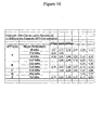
- the invention aims in particular at a method for evaluating a neurodegenerative pathological process in a sample to be analyzed, by immunochemical analysis, as described in the examples, characterized in that an index is assigned to the fragments identified in the studied sample, compared to a reference system where the index 0 (zero) corresponds to the detection in healthy tissue and the index 100 (one hundred) in the pathological tissue, whether human, of transgenic mice with mutations of genes implicated in Alzheimer pathology, or native or transfected cell strains with said genes.
- Such an index has an advantage in diagnosis and therapy. It allows to quickly reflect the pathological state. The value of the index makes it possible to determine whether there is a risk of developing Alzheimer's disease. Obtaining a high index may help to confirm a clinical diagnosis.
- this index can be used to slow down or even to induce degeneration to establish new experimental models. This index can also provide information to cause targeted degeneration in the context of therapeutic actions requiring the disappearance of a specific cell population.
- the method according to the invention may advantageously use in addition a set of polyclonal and / or monoclonal antibodies in order to reveal post-translational modifications of the pathological transformation of APP-Cter fragments, in particular a phosphorylation type modification, and / or methylation and / or acetylation and / or glycosylation.
- the set of polyclonal and / or monoclonal antibodies used makes it possible to reveal the phosphorylations and the expression variation of the fragments of 14.5; 12 and 10.5 kDa.
- the detection method according to the invention is advantageously usable for establishing and validating experimental models, cells or animal models of Alzheimer's disease. Relevant models will reproduce changes in APP-Cter fragments as observed in human tissue, as described in the examples.
- Another application of great interest relates to the pharmacological screening for the selection of drugs effective against neurodegenerative diseases of the Alzheimer type.
- the selected molecules are characterized in that they are capable of modifying the Alzheimer profile mentioned above, leading to a restoration of the properties of APP-Cter markers, or to a restoration of their physico-chemical properties (solubility, electrical charge).
- the invention thus makes it possible to carry out a rapid and relevant pharmacological screening of molecules of interest.
- the invention also relates to a diagnostic kit for the pathological transformation of APP, characterized in that it comprises a set of polyclonal and / or monoclonal antibodies directed against the carboxy-terminal region of APP.
- a diagnostic kit for the pathological transformation of APP characterized in that it comprises a set of polyclonal and / or monoclonal antibodies directed against the carboxy-terminal region of APP.
- the use of a set of antibodies directed against the post-translational modifications related to the pathological transformation located on APP-Cter fragments, for example sites of phosphorylation, methylation, acetylation or glycosylation on these fragments, may advantageously provide additional information on the detection and quantification of the pathological transformation of APP-Cter fragments.
- APP-Cter fragments demonstrated by 2D electrophoresis is explained in part by phosphorylation modifications, as demonstrated by a phosphorylated phosphorylated threonine 668 polyclonal antibody (nomenclature with APP 695), located in a common region to APP-Cter fragments.
- Methylation and acetylation are two post-translational modifications that are also involved in the pathological transformation of APP-Cter fragments. Indeed, they are located mainly on lysine residues, which causes acidification and thus a change of isoelectric point.
- kits according to the invention will therefore advantageously contain the elements necessary for carrying out such assays.
- a polyclonal antibody directed against the carboxy-terminal portion of APP was produced according to the immunization protocol described by Vaitukaitis et al (5). New Zealand rabbits were immunized with a peptide comprising the last 17 amino acids of the human APP sequence, corresponding to its carboxy terminal end. The peptide was covalently coupled to ovalbumin. 200 ⁇ g of immunogen were injected every two weeks. Before injection, the immunogenic solution was supplemented with 1 volume of adjuvant complete with Freund's first booster and subsequent boosters, with incomplete Freund's adjuvant instead of full adjuvant. Blood was collected one week after the 4th injection and one week after each subsequent injection. Pure serum was obtained after coagulation of the blood and centrifugation for 10 min at 2000g. To the pure serum was added 1 volume of glycerol and the mixture was kept at -20 ° C.
- 1 mg of peptide used for immunization was coupled to an NHS-Fast Flow Sepharose® matrix (Amersham-Pharmacia Biotech) according to the manufacturer's instructions.
- the coupled matrix was contacted with 200 ⁇ l of pure serum diluted in one volume of 2X binding buffer (50 mM Tris pH 8.0, 300 mM NaCl and 0.1% (v / v) Tween-20). and gently stirred overnight at 4 ° C. All of the solutions were passed on a PD-10 column.
- the matrix was washed with 10 volumes of binding buffer.
- the purified antibody was eluted with 2 volumes of pH 3.5 acetate buffer.
- the protein concentration was determined using the Pierce BCA Protein Quantification Kit.
- Brain tissue samples obtained after autopsy or biopsy and stored at - 80 ° C, were dissected using an anatomical atlas, and then homogenized with a Teflon® potter in 10 volumes of 1X 1-D lysis buffer (50 mM Tris-HCl pH 6.8, 4mM EDTA; 5% (w / v) SDS, 10% (v / v) glycerol, 2% (v / v) ⁇ -mercaptoethanol and 0.05% Bromophenol Blue) for 1-D analysis. The samples were heated to 100 ° C for 10 minutes and then held at -80 ° C until use.
- 1X 1-D lysis buffer 50 mM Tris-HCl pH 6.8, 4mM EDTA; 5% (w / v) SDS, 10% (v / v) glycerol, 2% (v / v) ⁇ -mercaptoethanol and 0.05% Bromophenol Blue
- the tissue is homogenized in 10 mM Tris-HCl buffer pH 6.8, then supplemented with 2X 2X buffer volume (7M urea, 2M thiourea, 0.4% Pharmalytes® 3- (W / v) 8% (v / v) Triton X-100, 10mM dithiothreitol, 0.1% Bromophenol Blue) and stored at -80 ° C until use.
- 2X 2X buffer volume 7M urea, 2M thiourea, 0.4% Pharmalytes® 3- (W / v) 8% (v / v) Triton X-100, 10mM dithiothreitol, 0.1% Bromophenol Blue
- Murine brain tissue samples were prepared following the same protocol as for samples of human brain tissue. Brain tissue samples from non-transgenic mice and transgenic mice were used with the wild-type human APP (APPwt) gene or the human APP gene mutated at codons 670 and 671 (APPsw). This mutation of APP is linked to a familial form of Alzheimer's disease, called Swedish mutation (7). Other samples were also used, including non-transgenic mice, and different transgenic mouse lines with the APPwt and APPsw gene. A sample of murine brain tissue similar to the transgenic mouse developed by Hsiao K. et al. (8).
- the brain tissue samples were homogenized in a 1/10 (w / v) ratio in 10 mM Tris-HCl pH 6.8 buffer and centrifuged at 100,000 g for 1 hour at 4 ° C.
- the supernatant (F1) was stored and the pellet re-homogenized in the same buffer supplemented with 0.5% (v / v) Triton X-100.
- fraction F2 the soluble Triton X100 fraction
- An additional extraction and centrifugation step in the same buffer was carried out under the same conditions, giving the fraction F2 '.
- the pellet is then directly taken up in lysis buffer 1-D (see below) and corresponds to fraction F3.
- lysis buffer 1-D 2X 100 mM Tris-HCl pH 6.8, 8 mM EDTA, 10% p / v) SDS, 20% (v / v) glycerol, 4% (v / v) ⁇ -mercaptoethanol and 0.1% Bromophenol blue) for one-dimensional (1-D) or 1-dimensional electrophoresis volume of lysis buffer 2-D (7M urea, 2M thiourea, 0.4% Pharmalytes® 3-10 (w / v), 4% (v / v) Triton X-100, 10 mM dithiothreitol, 0.1 % Bromophenol Blue) for two-dimensional (2-D) electrophoresis study.
- the F2 fraction is used to immunoprecipitate APP-Cter fragments.
- 100 ⁇ l of fraction F2 are diluted in 300 ⁇ l of 10 mM Tris buffer pH 7.4 containing 1% of NP-40 (immunoprecipitation buffer).
- 10 ⁇ l of APP-CterC17 or APP668P antibodies are added and all are incubated with stirring overnight at 4 ° C.
- 40 ⁇ l of protein A fixed on agarose beads (Pierce) are added to the solution and the whole is incubated with shaking at 4 ° C. for one hour.
- the agarose beads are washed three times in the immunoprecipitation buffer and the APP-Cter fragments are removed by treatment with 50 ⁇ l of SDS buffer and recovered in the centrifugation supernatant. The APP-Cter fragments are then analyzed by immunoblotting after Tris-Tricine gel electrophoresis.
- the cells are stably transfected with the wild type human APP gene or the human APP gene carrying the Swedish mutation.
- the cell pellet was taken up in 10mM Tris-HCl buffer pH 6.8 and then sonicated. Before use, the sample is supplemented with 1 volume of lysis buffer corresponding to the experiment (1-D or 2-D) 2X. In the case of a 1-D analysis, the sample is heated for 10 min at 100 ° C.
- Preparation of human white blood cells 10 milliliters of blood are collected in EDTA (ethylene diamine tetraacetate) tubes. Centrifugation at 4500 rpm (rotation per minute) for 15 minutes. The plasma is eliminated. The tube is filled with two-thirds of a lysis solution of red blood cells (lysis solution: 0.91 mM NH 4 CO 3 and 0.132 mM NH 4 Cl). The tube is gently shaken and placed in a water bath refrigerated at 5 ° C. Centrifugation at 4000 rpm for 15 minutes and the supernatant is removed. The white blood cell pellet is rinsed twice with the lysis solution following the same methodology.
- EDTA ethylene diamine tetraacetate
- the pellet is then drained and is either used within 24 hours or frozen at -20 ° C for transport or subsequent use.
- the white blood cell pellet is taken up in 10 mM Tris-HCl buffer pH 6.8 and then sonicated. Before use, the sample is supplemented with 1 volume of 2X lysis buffer corresponding to the experiment (1-D or 2-D). In the case of a 1-D analysis, the sample is heated for 10 min at 100 ° C.
- the 1-D electrophoreses were carried out according to the protocols described by Laemmli (9) for the manufacture of the gel and in the migration conditions described by Shugger and von Jagow (10).
- the concentration gel contains 4% acrylamide and the separation gel used contains 16.5% acrylamide.
- the migration is done in Tris-Tricine buffer.
- the migration program used is as follows: 1h to 30V constant then 16h to 45mA. An equivalent amount of protein was loaded into each well (approximately 100 ⁇ g / well).
- the first dimension or focus is achieved using pre-cast gel strips called IPG-Strip® covering a 3-10 pH gradient, according to the manufacturer's instructions.
- the equipment used for isoelectric focusing is the IEF Protean Cell system (Biorad).
- An amount of 250 ⁇ g of protein is supplemented with 1X rehydration buffer (7M urea, 2M thiourea, 4% triton X100, 0.5% CHAPS, 0.2% (w / v) Pharmalytes® pH 3-10; 10mM DTT, 0.01% orange G) to have 400 ⁇ l to be deposited.
- the 400 ⁇ l of samples are placed on the strips placed in the device. Let passively rehydrate 1 hour then actively 10h at 50V / strip and then run the program according to the manufacturer's instructions. After focusing, the strips are used or stored at -80 ° C.
- the strips are equilibrated for 30 minutes in a buffer for 1-D (50 mM Tris pH 6.8, 10% (v / v) glycerol, 2% (v / v) ⁇ -mercaptoethanol, 2% (w / v) SDS, 0.05% (w / v) bromophenol blue) then deposited on the top of a separation gel for 1-D of 16.5% acrylamide.
- the strip is covered with a 1% (w / v) agarose solution. The rest is identical to a simple 1-D electrophoresis.
- the transfer was performed using the Pharmacia LKB multiphor® semi-dry transfer system following the manufacturer's instructions (Amersham-Pharmacia Biotech). The proteins were transferred to 0.8 mA / cm 2 on a Hybond® ECL nitrocellulose membrane (Pharmacia-Amersham).
- the membrane is incubated for 60 min. in buffer (15 mM Tris pH 8.0, 150 mM NaCl, 0.5% (v / v) Tween®-20) containing 5% (w / v) skim milk and then washed with the same buffer free of milk containing 0.1% Tween®-20 instead of 0.5%.
- the membrane is incubated for 2 hours at room temperature or overnight at 4 ° C. with APP-Cter-C17 antibody diluted to 1 / 2000th (v / v) final in incubation buffer (15 mM Tris pH 8). 0.10 mM NaCl, 0.1% (v / v) Tween-20 and 3% (w / v) skim milk).
- the membrane is washed 3 times for 10 minutes in the milk-free incubation buffer.
- the membrane is then incubated for 1 hour at room temperature with horseradish peroxidase-coupled horseradish goat immunoglobulin at a final dilution of 1 / 4000th (v / v) in milk-free incubation buffer.
- the membrane is washed 3 times for 10 minutes in milk-free incubation buffer and the immunoreactive polypeptides are revealed using the ECL® chemiluminescence kit. (Pharmacia-Amersham Biotech) according to the manufacturer's instructions.
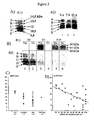
- the Figure 2B represents the analysis, on the same samples and the same immunoblights, of the tau pathology according to the classification of Delacourte et al, 1999 ( figure 2 , B1) and APP-Cter fragments ( figure 2 , B2) in a control subject (Tracks 1 and 2) and two patients with Alzheimer's disease (lanes 3 to 6).
- Two brain regions were studied: the hippocampus (Hip, lanes 1, 3 and 5) and the occipital cortex (Oc, lanes 2, 4 and 6).
- Hip hippocampus
- Oc occipital cortex
- the quantities of APP-Cter were determined in the cortex of 4 groups of patients: the control cases (Ctrl), subclinical cases (infraMA) which correspond to non-demented patients, but with the lesions characteristic of the disease.
- Alzheimer's clinical cases of Alzheimer's disease (AD) and autosomal dominant familial cases of Alzheimer's disease (ADF.MA) ( Figure 2, C ).
- the non-parametric Mann-Whitney statistical test indicates a significant decrease between the infraMA and MA groups compared to controls Ctrl (infraMA: p ⁇ 0.03, MA p ⁇ 0.002).
- the average decrease in APP-Cter is 1.5 times for the infraMA group and 1.7 times for the MA group, compared to the control group.
- the decrease in APP-Cer in the infraMA and MA groups is not significantly different. This demonstrates that this decrease in APP-Cter is an early event in Alzheimer's pathophysiology.
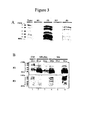
- the APP-Cter fragments are preferentially extracted in two phases: a buffer solution containing a non-ionic detergent, Triton X100 (Fraction F2), and a solution containing an ionic detergent, sodium dodecyl sulfate (SDS) (F3) ( Figure 3, A ).
- the arrows indicate the spots detected by APP-Cter-C17 polyclonal antibody. Their molecular weights are similar to those observed on 1-D immunoblots.
- the isoelectric points are in the pH range of 4.5 to 7.5.
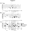
- the APP-Cter variants detected by 2D analysis are represented schematically ( Figure 4, C ) on a grid calibrated in two dimensions, the Y axis representing the molecular weights and the X axis the isoelectric points.
- the assembly was calibrated with internal controls of molecular weight and isoelectric point.
- the isoelectric point controls are gamma-enolase (47 / 4.94), alpha-actin (42 / 5.29) and alpha-enolase (47 / 6.99).
- Isoelectric points at 5.54 and 8.05 are internal standards of brain tissue.
- the scheme of the figure 4 represents the superposition of 2D profiles of APP-Cter fragments detected with APP-Cter-C17 antibody in the human brain tissue of a control subject and Alzheimer's patients at the subclinical stage of the disease and at the early clinical stage of the disease. Alzheimer's disease.
- the spots that is to say the different isoelectric variants of 2-D, observed in both healthy and pathological tissue are represented by empty circles.
- the specific spots of the healthy tissue are represented by circles with hatched background and those specific to the Alzheimerized tissue by a black background.
- APP-Cter products were analyzed (in the same way as in human brain tissue) in a rat and non-transgenic mice but also in transgenic mice for wild-type APP (APPwt) or Swedish mutation (APPsw) gene.
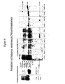
- the Figure 5A represents immunostaining of APP-Cter fragments of control and transgenic mice, compared to the profile of human brain tissue ( Figure 5A , track 4).
- Identical deposits of homogenates of brain tissue were deposited in each well of electrophoresis. APP-Cter fragments were detected with APP-Cter-C17 polyclonal antibody.
- Track 1 of the figure 5 corresponds to the analysis of the untransfected mouse (negative control)
- lane 2 corresponds to the mouse with a single copy of the human APPwt gene
- lane 3 corresponds to the transgenic mouse with the human mutated APPsw gene
- lane 4 corresponds to to control human brain tissue.
- the arrows indicate the molecular weights of the electrophoretic bands detected.
- mice of the APPwt gene essentially synthesize 4 APP-Cter fragments, whereas mice with the APPsw gene and normal human brain tissue synthesize 5 APP-Cter fragments, with a molecular mass of 14, 5; 13.5; 12; 10.5 and 9.5 kDa.
- CHO Choinese Hamster ovarian cells, stably transfected with the wild-type human APP (APPwt) gene or the APPsw gene, were analyzed according to the protocol described.
- the results of the figure 8 show APP-Cter fragments of CHO cells, compared to brain tissue of trangenic mice with the APPwt or APPsw gene.
- CCF, U118 and T98 glial cell strains have a profile with 6 bands at 13.5, 12.10.5, 9.5, 9 and 8.5 kDa, with an abundance of 3 bands between 9, 5 and 8.5 kDa ( Figure 8B ).
- a similar profile is observed in Hela, COS and SY-5Y cell lines transfected with APPsw ( Figure 8B ).
- the HT-29 epithelial cell strain has a 14.5, 13.5, 12, 10.5, 9.5 and 9 kDa 5-band profile ( figure 8 : HT-29).
- the 9 kDa band which is discrete in HT-29 epithelial cells becomes more abundant after treatment with benzyl-O-GalNac which inhibits glycosylation and targeting of glycosylated proteins.
- glycosylation is a post-translational modification that regulates APP-Cter expression and pattern. This modification is therefore an important element to take into account in the search for molecular pathological transformations of APP-Cter during Alzheimerization.
- SKNSH-SY5Y Human neuroblastoma cells stably transfected with mutated APPsw, APPwt, PS1 and Tau genes were analyzed in comparison to untransfected cells. The results are represented on the figure 9 .
- the APP-Cter signal corresponds to the 10.5 kDa band ( figure 9 : Native).
- 6 fragments at 13.5, 12, 10.5, 9.5, 9 and 8.5 kDa are perfectly identifiable in the non-differentiated SKNSH cells transfected with APPsw ( figure 9 : APPsw).
- SKNSH-like profiles such as Kelly cells, or elsewhere, such as SKNSH cells transfected with a mutated APP or PS1 gene, such as NT2 and hNT cells.
- the 14-day differentiation of SY5Y cells caused a profile change, with the appearance of the gamma band at 6.5 kDa and a decrease of the band at 10.5 kDa.
- the cells transfected with the tau 3R gene and undifferentiated see the appearance of the 13.5 kDa band, corresponding to an increase in the release of "beta secretase" fragments.
- the disappearance of the gamma band at 6.5 kDa is observed.
- APP-Cter-C17 antibody detects three molecular weight fragments of 14 kDa, 10.5 kDa and 9.5 kDa.
- a comparative 1-D analysis between white blood cells of control subjects and patients diagnosed with Alzheimer's disease revealed (as in the study of human brain tissue) a decrease in APP-Cter fragments in white blood cells of patients (lanes 6 to 11) relative to the control white blood cells (lanes 1 to 5).
- An antibody directed against the N-terminal end of the A ⁇ peptide makes it possible to locate the beta-secretase cleavage fragment.
- This is the FCA18 antibody (4).
- the 14 kDa fragment of the white blood cells is detected by this antibody, indicating that it is the APP-Cter fragment produced by beta secretase ( Figure 11B ).
- the two bands A (14.5 kDa) and B (13.5 kDa) are recognized by WO2, a monoclonal antibody (Abeta, GmBH, Heidelberg, Germany) directed against the 5-11 portion of the A ⁇ peptide.
- Phosphorylation of APP-Cter fragments is a determining factor in their pathological transformation
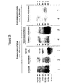
- the APP-Cter of the brain tissue of a control subject and an Alzheimer patient were immunoprecipitated using the APP-CterC17 antibody and the fragments at 14.5, 13.5, 12, 10.5 , and 9.5 kDa (fragments A, B, C, D and E) are revealed after tris-Tricine gel electrophoresis with the same antibody ( figure 13 tracks 1 and 2).
- the immunoprecipitated APP-Cter fragments were dephosphorylated using calf intestinal alkaline phosphatase ( figure 13 lanes 3 and 4) and then revealed with the APP-CterC17 antibody according to the same protocol.
- Phosphorylation on threonines, serines and tyrosines of APP-Cter fragments contribute to the general profile of APP-Cter.
- a difference in the phosphorylation index of these different APP-Cter fragments is observed, as shown in particular by phosphorylated threonine 668 ( figures 11 and 13 ).
- the profile of the dephosphorylated APP-Cter fragments is modified, in particular with the disappearance of the band of 14.5 kDa, and the new profile then resembles that of the SKNSH cell models transfected with APPsw ( Figure 13 ).
- the dephosporylation of the APP-Cter fragments also reveals a change in profile between the normal tissue and the Alzheimer's tissue, with in particular an accumulation of the C, D and E bands of the APP-Cter fragments, corresponding to the cut products of the alpha secretase. ( figure 13 ). Phosphorylation is therefore an important post-translational event, and a marker of the pathological transformation of APP-Cter fragments.
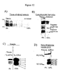
- Detection of APP-Cter fragments allows the definition of a pathological transformation index, applicable to neuronal human tissue ( figure 12A : human brain tissue) or non-neuronal ( figure 12B human lymphocytes), as well as experimental models, animals ( figure 12C brain tissue of transgene mouse APPsw and wt) or cellular, neuronal ( figure 12D neuroblastoma cells) or non-neuronal cells (CHO cells, COS cells, etc.). This index varies from 0 (healthy tissue) to 100% (Alzheimerized pathological tissue) ( figure 12 ).
Landscapes
- Life Sciences & Earth Sciences (AREA)
- Health & Medical Sciences (AREA)
- Engineering & Computer Science (AREA)
- Biomedical Technology (AREA)
- Hematology (AREA)
- Chemical & Material Sciences (AREA)
- Urology & Nephrology (AREA)
- Molecular Biology (AREA)
- Immunology (AREA)
- Proteomics, Peptides & Aminoacids (AREA)
- Analytical Chemistry (AREA)
- Microbiology (AREA)
- Biotechnology (AREA)
- Neurosurgery (AREA)
- Neurology (AREA)
- Food Science & Technology (AREA)
- Medicinal Chemistry (AREA)
- Physics & Mathematics (AREA)
- Cell Biology (AREA)
- Biochemistry (AREA)
- General Health & Medical Sciences (AREA)
- General Physics & Mathematics (AREA)
- Pathology (AREA)
- Investigating Or Analysing Biological Materials (AREA)
- Peptides Or Proteins (AREA)
- Pharmaceuticals Containing Other Organic And Inorganic Compounds (AREA)
- Medicines Containing Antibodies Or Antigens For Use As Internal Diagnostic Agents (AREA)
Priority Applications (1)
| Application Number | Priority Date | Filing Date | Title |
|---|---|---|---|
| CY20081100718T CY1108182T1 (el) | 2000-11-03 | 2008-07-09 | Μεσα εντοπισμου του παθολογικου μετασχηματισμου της πρωτεϊνης app και εφαρμογες τους |
Applications Claiming Priority (3)
| Application Number | Priority Date | Filing Date | Title |
|---|---|---|---|
| FR0014143 | 2000-11-03 | ||
| FR0014143A FR2816411B1 (fr) | 2000-11-03 | 2000-11-03 | Moyens de detection de la transformation pathologique de la proteine app et leurs applications |
| PCT/FR2001/003410 WO2002037118A1 (fr) | 2000-11-03 | 2001-11-05 | Moyens de detection de la transformation pathologique de la proteine app et leurs applications |
Publications (2)
| Publication Number | Publication Date |
|---|---|
| EP1330656A1 EP1330656A1 (fr) | 2003-07-30 |
| EP1330656B1 true EP1330656B1 (fr) | 2008-04-16 |
Family
ID=8856055
Family Applications (1)
| Application Number | Title | Priority Date | Filing Date |
|---|---|---|---|
| EP01992896A Expired - Lifetime EP1330656B1 (fr) | 2000-11-03 | 2001-11-05 | Moyens de detection de la transformation pathologique de la proteine app et leurs applications |
Country Status (13)
| Country | Link |
|---|---|
| US (1) | US7723117B2 (da) |
| EP (1) | EP1330656B1 (da) |
| JP (1) | JP4294953B2 (da) |
| AT (1) | ATE392620T1 (da) |
| AU (2) | AU2002223743B2 (da) |
| CA (1) | CA2427604A1 (da) |
| CY (1) | CY1108182T1 (da) |
| DE (1) | DE60133661T2 (da) |
| DK (1) | DK1330656T3 (da) |
| ES (1) | ES2305126T3 (da) |
| FR (1) | FR2816411B1 (da) |
| PT (1) | PT1330656E (da) |
| WO (1) | WO2002037118A1 (da) |
Families Citing this family (17)
| Publication number | Priority date | Publication date | Assignee | Title |
|---|---|---|---|---|
| US7598049B2 (en) * | 2003-11-19 | 2009-10-06 | Satoris, Inc. | Methods for diagnosis of Alzheimer's Disease in blood samples |
| US20060094064A1 (en) * | 2003-11-19 | 2006-05-04 | Sandip Ray | Methods and compositions for diagnosis, stratification, and monitoring of alzheimer's disease and other neurological disorders in body fluids |
| CA2585983C (en) | 2004-11-10 | 2014-03-25 | Institut National De La Sante Et De La Recherche Medicale (Inserm) | Use of 1,4-bis (3-aminoalkyl) piperazine derivatives in the treatment of neurodegenerative diseases |
| US20090099062A1 (en) * | 2007-05-31 | 2009-04-16 | Ethan Lee | Pyrvinium For The Treatment of Cancer |
| DE202007015863U1 (de) * | 2007-11-12 | 2009-03-26 | Melitta Haushaltsprodukte Gmbh & Co. Kommanditgesellschaft | Filterpapiereinsatz |
| MX340916B (es) | 2008-01-18 | 2016-07-29 | President And Fellows Of Harvard College * | Metodos para detectar señales de identificacion de enfermedad o condiciones en fluidos corporales. |
| EP2513062A1 (en) | 2009-12-16 | 2012-10-24 | Institut National de la Santé et de la Recherche Médicale | 7-chloro-quinolin-4-amine compounds and uses thereof for the prevention or treatment of diseases involving formation of amyloid plaques and/or where a dysfunction of the app metabolism occurs |
| TW201209171A (en) | 2010-07-23 | 2012-03-01 | Harvard College | Methods of detecting diseases or conditions using phagocytic cells |
| US20130203624A1 (en) | 2010-07-23 | 2013-08-08 | President And Fellows Of Harvard College | Methods of Detecting Prenatal or Pregnancy-Related Diseases or Conditions |
| AU2011280997A1 (en) | 2010-07-23 | 2013-02-28 | President And Fellows Of Harvard College | Methods of detecting autoimmune or immune-related diseases or conditions |
| EP2596132A4 (en) | 2010-07-23 | 2013-12-18 | Harvard College | METHOD FOR DETECTING SIGNS FOR ILLNESS OR SUFFERING IN BODY FLUIDS |
| US20130096552A1 (en) * | 2011-10-14 | 2013-04-18 | Christopher L. Brace | Hydrodissection Material with Reduced Migration |
| MX2014015434A (es) | 2012-06-15 | 2015-07-14 | Harry Stylli | Metodos para detectar enfermedades o condiciones utilizando celulas enfermas en circulacion. |
| NZ771629A (en) | 2013-03-09 | 2022-12-23 | Harry Stylli | Methods of detecting cancer |
| BR112015024234B1 (pt) | 2013-03-21 | 2022-11-16 | Eupraxia Pharmaceuticals USA LLC | Composição farmacêutica de liberação sustentada injetável, uso da mesma para diminuir inflamação ou controlar dor e método para formar micropartículas revestidas |
| DK3206672T3 (da) | 2015-10-27 | 2018-06-18 | Eupraxia Pharmaceuticals Inc | Formuleringer til vedvarende frigivelse af lokalbedøvelsesmidler |
| US11397188B2 (en) * | 2017-03-30 | 2022-07-26 | Board Of Regents, The University Of Texas System | Method of detecting an APP Alzheimer's disease marker peptide in patients with Alzheimer's disease |
Family Cites Families (6)
| Publication number | Priority date | Publication date | Assignee | Title |
|---|---|---|---|---|
| US5234814A (en) * | 1989-06-01 | 1993-08-10 | Du Pont Merck Pharmaceutical Company | Diagnostic assay for alzheimer's disease |
| WO1992000521A1 (en) * | 1990-06-29 | 1992-01-09 | Case Western Reserve University | Diagnostic and prognostic methods based on soluble derivatives of the beta amyloid protein precursor |
| CA2086165A1 (en) * | 1992-04-09 | 1993-10-10 | Paul P. Tamburini | Diagnostic assay for alzheimer's disease based on the proteolysis of alzheimer's precursor protein |
| US5441870A (en) * | 1992-04-15 | 1995-08-15 | Athena Neurosciences, Inc. | Methods for monitoring cellular processing of β-amyloid precursor protein |
| WO1994007144A1 (en) * | 1992-09-21 | 1994-03-31 | United States Of America, As Represented By The Secretary, Department Of Health And Human Services | A method of screening compounds and methods for treating alzheimer's disease |
| US6114133A (en) * | 1994-11-14 | 2000-09-05 | Elan Pharmaceuticals, Inc. | Methods for aiding in the diagnosis of Alzheimer's disease by measuring amyloid-β peptide (x-≧41) |
-
2000
- 2000-11-03 FR FR0014143A patent/FR2816411B1/fr not_active Expired - Fee Related
-
2001
- 2001-11-05 CA CA002427604A patent/CA2427604A1/fr not_active Abandoned
- 2001-11-05 EP EP01992896A patent/EP1330656B1/fr not_active Expired - Lifetime
- 2001-11-05 AT AT01992896T patent/ATE392620T1/de active
- 2001-11-05 WO PCT/FR2001/003410 patent/WO2002037118A1/fr not_active Ceased
- 2001-11-05 DK DK01992896T patent/DK1330656T3/da active
- 2001-11-05 PT PT01992896T patent/PT1330656E/pt unknown
- 2001-11-05 AU AU2002223743A patent/AU2002223743B2/en not_active Ceased
- 2001-11-05 DE DE60133661T patent/DE60133661T2/de not_active Expired - Lifetime
- 2001-11-05 ES ES01992896T patent/ES2305126T3/es not_active Expired - Lifetime
- 2001-11-05 JP JP2002539821A patent/JP4294953B2/ja not_active Expired - Fee Related
- 2001-11-05 AU AU2374302A patent/AU2374302A/xx active Pending
- 2001-11-05 US US10/415,479 patent/US7723117B2/en not_active Expired - Fee Related
-
2008
- 2008-07-09 CY CY20081100718T patent/CY1108182T1/el unknown
Also Published As
| Publication number | Publication date |
|---|---|
| EP1330656A1 (fr) | 2003-07-30 |
| US7723117B2 (en) | 2010-05-25 |
| WO2002037118A1 (fr) | 2002-05-10 |
| US20070026527A1 (en) | 2007-02-01 |
| JP2004522940A (ja) | 2004-07-29 |
| DE60133661T2 (de) | 2009-05-28 |
| AU2002223743B2 (en) | 2007-01-11 |
| AU2374302A (en) | 2002-05-15 |
| FR2816411B1 (fr) | 2003-07-04 |
| CY1108182T1 (el) | 2014-02-12 |
| CA2427604A1 (fr) | 2002-05-10 |
| ES2305126T3 (es) | 2008-11-01 |
| JP4294953B2 (ja) | 2009-07-15 |
| DE60133661D1 (de) | 2008-05-29 |
| PT1330656E (pt) | 2008-07-24 |
| FR2816411A1 (fr) | 2002-05-10 |
| DK1330656T3 (da) | 2008-08-11 |
| ATE392620T1 (de) | 2008-05-15 |
Similar Documents
| Publication | Publication Date | Title |
|---|---|---|
| EP1330656B1 (fr) | Moyens de detection de la transformation pathologique de la proteine app et leurs applications | |
| Begcevic et al. | Brain-related proteins as potential CSF biomarkers of Alzheimer's disease: A targeted mass spectrometry approach | |
| Johnson et al. | Behavioral and neural network abnormalities in human APP transgenic mice resemble those of App knock-in mice and are modulated by familial Alzheimer’s disease mutations but not by inhibition of BACE1 | |
| Lachén-Montes et al. | An early dysregulation of FAK and MEK/ERK signaling pathways precedes the β-amyloid deposition in the olfactory bulb of APP/PS1 mouse model of Alzheimer's disease | |
| Butterfield et al. | Mass spectrometry and redox proteomics: applications in disease | |
| Zellner et al. | Proteomic profiling in cerebral amyloid angiopathy reveals an overlap with CADASIL highlighting accumulation of HTRA1 and its substrates | |
| US8354236B2 (en) | Detection of neurodegenerative disease | |
| Adav et al. | ITRAQ quantitative clinical proteomics revealed role of Na+ K+-ATPase and its correlation with deamidation in vascular dementia | |
| Lionnet et al. | Characterisation of tau in the human and rodent enteric nervous system under physiological conditions and in tauopathy | |
| Farrell et al. | Genome-wide association study and functional validation implicates JADE1 in tauopathy | |
| JP7654126B2 (ja) | アルツハイマー病の判定薬および判定方法 | |
| Nuzzo et al. | Cerebrospinal fluid and serum d-serine concentrations are unaltered across the whole clinical spectrum of Alzheimer's disease | |
| Begcevic et al. | Targeted mass spectrometry-based assays for relative quantification of 30 brain-related proteins and their clinical applications | |
| Castle et al. | Application of high-throughput, capillary-based Western analysis to modulated cleavage of the cellular prion protein | |
| Ellis et al. | Identification of high-performing antibodies for the reliable detection of Tau proteoforms by Western blotting and immunohistochemistry | |
| Jimenez-Harrison et al. | DJ-1 molecular chaperone activity depresses Tau aggregation propensity through interaction with monomers | |
| Trist et al. | Novel tools to quantify total, phospho-Ser129 and aggregated alpha-synuclein in the mouse brain | |
| CA2884356C (fr) | Procede pour le diagnostic de dystrophies musculaires | |
| Chen et al. | Amyloid P component as a plasma marker for Parkinson's disease identified by a proteomic approach | |
| Bilousova et al. | Parallel age-associated changes in brain and plasma neuronal pentraxin receptor levels in a transgenic APP/PS1 rat model of Alzheimer's disease | |
| EP2870481A1 (en) | Tropomyosin isoforms related to alzheimers disease and mild cognitive impairment | |
| Trivedi et al. | Performance of novel tau antibodies across multiple modalities for Alzheimer's disease assessment | |
| Tang et al. | Analysis of Brain Protein Stability Changes in a Mouse Model of Alzheimer’s Disease | |
| Lapcinski et al. | The β-amyloid oligomer Aβ* 56 is associated with Alzheimer’s dementia independently of amyloid pathology | |
| US20250281564A1 (en) | Dysregulation of neuroproteasomes by apoe isoforms drives endogenous tau aggregation |
Legal Events
| Date | Code | Title | Description |
|---|---|---|---|
| PUAI | Public reference made under article 153(3) epc to a published international application that has entered the european phase |
Free format text: ORIGINAL CODE: 0009012 |
|
| 17P | Request for examination filed |
Effective date: 20030520 |
|
| AK | Designated contracting states |
Designated state(s): AT BE CH CY DE DK ES FI FR GB GR IE IT LI LU MC NL PT SE TR |
|
| AX | Request for extension of the european patent |
Extension state: AL LT LV MK RO SI |
|
| 17Q | First examination report despatched |
Effective date: 20050322 |
|
| 17Q | First examination report despatched |
Effective date: 20050322 |
|
| GRAP | Despatch of communication of intention to grant a patent |
Free format text: ORIGINAL CODE: EPIDOSNIGR1 |
|
| GRAC | Information related to communication of intention to grant a patent modified |
Free format text: ORIGINAL CODE: EPIDOSCIGR1 |
|
| GRAS | Grant fee paid |
Free format text: ORIGINAL CODE: EPIDOSNIGR3 |
|
| GRAA | (expected) grant |
Free format text: ORIGINAL CODE: 0009210 |
|
| AK | Designated contracting states |
Kind code of ref document: B1 Designated state(s): AT BE CH CY DE DK ES FI FR GB GR IE IT LI LU MC NL PT SE TR |
|
| REG | Reference to a national code |
Ref country code: CH Ref legal event code: EP |
|
| REG | Reference to a national code |
Ref country code: IE Ref legal event code: FG4D Free format text: LANGUAGE OF EP DOCUMENT: FRENCH |
|
| REF | Corresponds to: |
Ref document number: 60133661 Country of ref document: DE Date of ref document: 20080529 Kind code of ref document: P |
|
| REG | Reference to a national code |
Ref country code: PT Ref legal event code: SC4A Free format text: AVAILABILITY OF NATIONAL TRANSLATION Effective date: 20080715 |
|
| REG | Reference to a national code |
Ref country code: CH Ref legal event code: NV Representative=s name: ISLER & PEDRAZZINI AG |
|
| REG | Reference to a national code |
Ref country code: SE Ref legal event code: TRGR |
|
| REG | Reference to a national code |
Ref country code: DK Ref legal event code: T3 |
|
| REG | Reference to a national code |
Ref country code: GR Ref legal event code: EP Ref document number: 20080401776 Country of ref document: GR |
|
| REG | Reference to a national code |
Ref country code: ES Ref legal event code: FG2A Ref document number: 2305126 Country of ref document: ES Kind code of ref document: T3 |
|
| PLBE | No opposition filed within time limit |
Free format text: ORIGINAL CODE: 0009261 |
|
| STAA | Information on the status of an ep patent application or granted ep patent |
Free format text: STATUS: NO OPPOSITION FILED WITHIN TIME LIMIT |
|
| 26N | No opposition filed |
Effective date: 20090119 |
|
| PGFP | Annual fee paid to national office [announced via postgrant information from national office to epo] |
Ref country code: LU Payment date: 20141024 Year of fee payment: 14 |
|
| PGFP | Annual fee paid to national office [announced via postgrant information from national office to epo] |
Ref country code: DK Payment date: 20141022 Year of fee payment: 14 |
|
| PGFP | Annual fee paid to national office [announced via postgrant information from national office to epo] |
Ref country code: SE Payment date: 20141024 Year of fee payment: 14 Ref country code: DE Payment date: 20141023 Year of fee payment: 14 Ref country code: IE Payment date: 20141024 Year of fee payment: 14 Ref country code: MC Payment date: 20141024 Year of fee payment: 14 Ref country code: GR Payment date: 20141023 Year of fee payment: 14 Ref country code: FI Payment date: 20141023 Year of fee payment: 14 Ref country code: TR Payment date: 20141105 Year of fee payment: 14 Ref country code: CH Payment date: 20141024 Year of fee payment: 14 Ref country code: GB Payment date: 20141024 Year of fee payment: 14 Ref country code: ES Payment date: 20141103 Year of fee payment: 14 |
|
| PGFP | Annual fee paid to national office [announced via postgrant information from national office to epo] |
Ref country code: AT Payment date: 20141023 Year of fee payment: 14 Ref country code: PT Payment date: 20141103 Year of fee payment: 14 Ref country code: NL Payment date: 20141024 Year of fee payment: 14 Ref country code: FR Payment date: 20141124 Year of fee payment: 14 |
|
| PGFP | Annual fee paid to national office [announced via postgrant information from national office to epo] |
Ref country code: IT Payment date: 20141030 Year of fee payment: 14 |
|
| PGFP | Annual fee paid to national office [announced via postgrant information from national office to epo] |
Ref country code: BE Payment date: 20141022 Year of fee payment: 14 |
|
| PGFP | Annual fee paid to national office [announced via postgrant information from national office to epo] |
Ref country code: CY Payment date: 20141022 Year of fee payment: 14 |
|
| REG | Reference to a national code |
Ref country code: PT Ref legal event code: MM4A Free format text: LAPSE DUE TO NON-PAYMENT OF FEES Effective date: 20160505 |
|
| REG | Reference to a national code |
Ref country code: DE Ref legal event code: R119 Ref document number: 60133661 Country of ref document: DE |
|
| REG | Reference to a national code |
Ref country code: DK Ref legal event code: EBP Effective date: 20151130 |
|
| PG25 | Lapsed in a contracting state [announced via postgrant information from national office to epo] |
Ref country code: LU Free format text: LAPSE BECAUSE OF NON-PAYMENT OF DUE FEES Effective date: 20151105 Ref country code: MC Free format text: LAPSE BECAUSE OF NON-PAYMENT OF DUE FEES Effective date: 20151130 |
|
| REG | Reference to a national code |
Ref country code: CH Ref legal event code: PL |
|
| REG | Reference to a national code |
Ref country code: AT Ref legal event code: MM01 Ref document number: 392620 Country of ref document: AT Kind code of ref document: T Effective date: 20151105 |
|
| GBPC | Gb: european patent ceased through non-payment of renewal fee |
Effective date: 20151105 |
|
| PG25 | Lapsed in a contracting state [announced via postgrant information from national office to epo] |
Ref country code: GR Free format text: LAPSE BECAUSE OF NON-PAYMENT OF DUE FEES Effective date: 20160602 Ref country code: CH Free format text: LAPSE BECAUSE OF NON-PAYMENT OF DUE FEES Effective date: 20151130 Ref country code: CY Free format text: LAPSE BECAUSE OF NON-PAYMENT OF DUE FEES Effective date: 20151105 Ref country code: IT Free format text: LAPSE BECAUSE OF NON-PAYMENT OF DUE FEES Effective date: 20151105 Ref country code: LI Free format text: LAPSE BECAUSE OF NON-PAYMENT OF DUE FEES Effective date: 20151130 |
|
| REG | Reference to a national code |
Ref country code: NL Ref legal event code: MM Effective date: 20151201 |
|
| REG | Reference to a national code |
Ref country code: IE Ref legal event code: MM4A |
|
| REG | Reference to a national code |
Ref country code: FR Ref legal event code: ST Effective date: 20160729 |
|
| PG25 | Lapsed in a contracting state [announced via postgrant information from national office to epo] |
Ref country code: PT Free format text: LAPSE BECAUSE OF NON-PAYMENT OF DUE FEES Effective date: 20160505 Ref country code: AT Free format text: LAPSE BECAUSE OF NON-PAYMENT OF DUE FEES Effective date: 20151105 Ref country code: SE Free format text: LAPSE BECAUSE OF NON-PAYMENT OF DUE FEES Effective date: 20151106 |
|
| PG25 | Lapsed in a contracting state [announced via postgrant information from national office to epo] |
Ref country code: NL Free format text: LAPSE BECAUSE OF NON-PAYMENT OF DUE FEES Effective date: 20151201 |
|
| REG | Reference to a national code |
Ref country code: GR Ref legal event code: ML Ref document number: 20080401776 Country of ref document: GR Effective date: 20160602 |
|
| PG25 | Lapsed in a contracting state [announced via postgrant information from national office to epo] |
Ref country code: DK Free format text: LAPSE BECAUSE OF NON-PAYMENT OF DUE FEES Effective date: 20151130 Ref country code: IE Free format text: LAPSE BECAUSE OF NON-PAYMENT OF DUE FEES Effective date: 20151105 Ref country code: GB Free format text: LAPSE BECAUSE OF NON-PAYMENT OF DUE FEES Effective date: 20151105 Ref country code: DE Free format text: LAPSE BECAUSE OF NON-PAYMENT OF DUE FEES Effective date: 20160601 |
|
| PG25 | Lapsed in a contracting state [announced via postgrant information from national office to epo] |
Ref country code: FR Free format text: LAPSE BECAUSE OF NON-PAYMENT OF DUE FEES Effective date: 20151130 |
|
| PG25 | Lapsed in a contracting state [announced via postgrant information from national office to epo] |
Ref country code: FI Free format text: LAPSE BECAUSE OF NON-PAYMENT OF DUE FEES Effective date: 20151105 Ref country code: ES Free format text: LAPSE BECAUSE OF NON-PAYMENT OF DUE FEES Effective date: 20151106 |
|
| PG25 | Lapsed in a contracting state [announced via postgrant information from national office to epo] |
Ref country code: BE Free format text: LAPSE BECAUSE OF NON-PAYMENT OF DUE FEES Effective date: 20151130 |
|
| PG25 | Lapsed in a contracting state [announced via postgrant information from national office to epo] |
Ref country code: TR Free format text: LAPSE BECAUSE OF NON-PAYMENT OF DUE FEES Effective date: 20151105 |
|
| REG | Reference to a national code |
Ref country code: ES Ref legal event code: FD2A Effective date: 20180704 |