EP1329719A1 - Method for analyzing antibody molecule structure - Google Patents
Method for analyzing antibody molecule structure Download PDFInfo
- Publication number
- EP1329719A1 EP1329719A1 EP01972608A EP01972608A EP1329719A1 EP 1329719 A1 EP1329719 A1 EP 1329719A1 EP 01972608 A EP01972608 A EP 01972608A EP 01972608 A EP01972608 A EP 01972608A EP 1329719 A1 EP1329719 A1 EP 1329719A1
- Authority
- EP
- European Patent Office
- Prior art keywords
- chains
- antibody
- analysis
- chain
- fragments
- Prior art date
- Legal status (The legal status is an assumption and is not a legal conclusion. Google has not performed a legal analysis and makes no representation as to the accuracy of the status listed.)
- Withdrawn
Links
Images










Classifications
-
- G—PHYSICS
- G01—MEASURING; TESTING
- G01N—INVESTIGATING OR ANALYSING MATERIALS BY DETERMINING THEIR CHEMICAL OR PHYSICAL PROPERTIES
- G01N33/00—Investigating or analysing materials by specific methods not covered by groups G01N1/00 - G01N31/00
- G01N33/48—Biological material, e.g. blood, urine; Haemocytometers
- G01N33/50—Chemical analysis of biological material, e.g. blood, urine; Testing involving biospecific ligand binding methods; Immunological testing
- G01N33/68—Chemical analysis of biological material, e.g. blood, urine; Testing involving biospecific ligand binding methods; Immunological testing involving proteins, peptides or amino acids
- G01N33/6803—General methods of protein analysis not limited to specific proteins or families of proteins
- G01N33/6848—Methods of protein analysis involving mass spectrometry
-
- G—PHYSICS
- G01—MEASURING; TESTING
- G01N—INVESTIGATING OR ANALYSING MATERIALS BY DETERMINING THEIR CHEMICAL OR PHYSICAL PROPERTIES
- G01N33/00—Investigating or analysing materials by specific methods not covered by groups G01N1/00 - G01N31/00
- G01N33/48—Biological material, e.g. blood, urine; Haemocytometers
- G01N33/50—Chemical analysis of biological material, e.g. blood, urine; Testing involving biospecific ligand binding methods; Immunological testing
- G01N33/68—Chemical analysis of biological material, e.g. blood, urine; Testing involving biospecific ligand binding methods; Immunological testing involving proteins, peptides or amino acids
- G01N33/6803—General methods of protein analysis not limited to specific proteins or families of proteins
- G01N33/6818—Sequencing of polypeptides
- G01N33/6821—Sequencing of polypeptides involving C-terminal degradation
-
- G—PHYSICS
- G01—MEASURING; TESTING
- G01N—INVESTIGATING OR ANALYSING MATERIALS BY DETERMINING THEIR CHEMICAL OR PHYSICAL PROPERTIES
- G01N33/00—Investigating or analysing materials by specific methods not covered by groups G01N1/00 - G01N31/00
- G01N33/48—Biological material, e.g. blood, urine; Haemocytometers
- G01N33/50—Chemical analysis of biological material, e.g. blood, urine; Testing involving biospecific ligand binding methods; Immunological testing
- G01N33/68—Chemical analysis of biological material, e.g. blood, urine; Testing involving biospecific ligand binding methods; Immunological testing involving proteins, peptides or amino acids
- G01N33/6803—General methods of protein analysis not limited to specific proteins or families of proteins
- G01N33/6818—Sequencing of polypeptides
- G01N33/6824—Sequencing of polypeptides involving N-terminal degradation, e.g. Edman degradation
-
- G—PHYSICS
- G01—MEASURING; TESTING
- G01N—INVESTIGATING OR ANALYSING MATERIALS BY DETERMINING THEIR CHEMICAL OR PHYSICAL PROPERTIES
- G01N33/00—Investigating or analysing materials by specific methods not covered by groups G01N1/00 - G01N31/00
- G01N33/48—Biological material, e.g. blood, urine; Haemocytometers
- G01N33/50—Chemical analysis of biological material, e.g. blood, urine; Testing involving biospecific ligand binding methods; Immunological testing
- G01N33/68—Chemical analysis of biological material, e.g. blood, urine; Testing involving biospecific ligand binding methods; Immunological testing involving proteins, peptides or amino acids
- G01N33/6854—Immunoglobulins
Definitions
- antibody molecules In primary structure analysis of antibody molecules, fragmentation is performed by enzymatic or chemical methods.
- antibody molecules generally have multimeric structures, and also have high molecular weights.
- the molecular weight of IgG a representative antibody molecule, is approximately 150,000 daltons. Therefore, direct fragmentation of an antibody molecule produces a complex mixture of peptide fragments, which makes the separation and assignment of each fragment difficult. Thus, high reproducibility cannot be achieved according to such a method.
- An objective of this invention is to provide a method for recovering peptide fragments that are obtained by fragmenting an antibody molecule in good yield, in terms of both the kind and the amount of the fragment, and analyzing them for primary structure analysis of the antibody molecule.
- another objective of the present invention is to provide a method for more conveniently and more quickly analyzing heterogeneity and evaluating the quality of antibody molecules by separating and fractionating peptide fragments that were obtained by fragmenting an antibody molecule via appropriate separation techniques (for example RP-HPLC), then constructing a peptide map having a good reproducibility based on the results of analysis.
- this invention provides a method for efficiently analyzing the variable regions by readily identifying the subclass of the H-chain and the type of the L-chain of a structurally unidentified antibody via a comparison of the obtained peptide maps.
- the present inventors focused on the structural characteristics of antibody molecules. As a result, they found out that besides the large molecular weight of an antibody molecule, the multimeric structure thereof is an obstacle in analyzing the antibody molecule. In addition, they demonstrated that the aforementioned obstacles can be overcome by separating in advance an antibody molecule into its H-chains and L-chains, and fragmenting the chains in the separated state. This invention was accomplished based on these findings. That is, the present invention relates to the following method of analysis, and use of the same.
- H-chains and L-chains can be separated by any method.
- H-chains and L-chains can be separated based on the differences in their properties (for example, molecular weight, electric charge, hydrophobicity).
- separation techniques such as electrophoresis and liquid chromatography can be used.
- the separation must be conducted under conditions that hinder recombination of the H-chains and L-chains.
- the term "denaturing gel electrophoresis” refers to gel electrophoresis that enables separation of the H-chains and L-chains of antibody molecules. Electrophoresis which utilizes an acrylamide gel medium containing SDS and urea is well known as an example for such electrophoresis. Particularly, SDS-PAGE is an especially advantageous separation technique for use in this invention. SDS-PAGE can be performed according to the method of Laemmli (Laemmli (1970) Nature 227, 680-685). The H-chains and L-chains are strongly bound by covalent bonds (S-S bonds) as well as non-covalent bonds, but by using SDS-PAGE, the H-chains and L-chains can be readily separated.
- S-S bonds covalent bonds
- denaturing gel electrophoresis can be also carried out without performing reductive acylation and such.
- H-chains and L-chains can be separated even by SDS-PAGE under reducing conditions, and the primary structure can be analyzed in a similar manner. Therefore, the amino acid sequence without the cysteine residues can be investigated.
- Each of the separated H-chains and L-chains are fragmented. Fragmentation methods for primary structure analyses of proteins are well known. Generally, for structural analyses of proteins, enzymatic digestion and chemical cleavage are utilized. For example, the following enzymes may be used. All of these enzymes are commercially available, and their digestion conditions are known. Lysyl endopeptidase Endoproteinase LysC Mouse submandibular gland protease D Staphylococcus aureuse V8 protease Endoproteinase Asp-N
- Protein digestion by enzymatic digestion can be performed under a mild condition, and therefore, modifications and such of the side chains of amino acid residues are unlikely to happen. Furthermore, by fixing the conditions for digestion, reproducibility can be easily maintained.
- chemical cleavage can be utilized instead of enzymatic digestion.
- Cleavage at the Carboxyl terminus of a methionine residue by cyanogen bromide is a representative chemical cleavage method.
- Fragmentation of the separated H-chains and L-chains can be performed by any method.
- a technique recently developed for structural analysis of proteins there is a method wherein bands or spots that are obtained by subjecting a protein to SDS-PAGE or two-dimensional electrophoresis are excised, and then chemically or enzymatically fragmented in a gel (Jahnen-Dechent and Simpson (1990) Plant Mol. Biol. Reporter 8, 92-103; Hellman U. et al., (1995) Anal. Biochem. 224, 451-455; Kawasaki et al. (1990) Anal. Biochem. 191, 332-336).
- proteins are fragmented in a gel.
- the method is called an in-gel digestion method.
- proteins separated in a gel can be fragmented on membranes, such as polyvinylidene difluoride (PVDF) membrane and nitrocellulose membrane, after transferring the separated protein on the membrane.
- PVDF membranes are commercially available under product names such as "Immobilon" (MILLIPORE).
- the in-gel digestion method one of the analysis methods of proteome analysis, is normally carried out by the steps of enzymatically digesting a protein separated and purified by two-dimensional electrophoresis directly in a gel, and then obtaining information on its amino acid sequence from the recovered peptide fragments.
- an antibody can be easily separated into H-chains and L-chains by denaturing gel electrophoresis, such as SDS-PAGE. Therefore, the inventors anticipated that fragmentation for the present invention could be accomplished by restricting electrophoresis to denaturing gel electrophoresis and performing enzymatic digestion of the separated H-chains and L-chains directly in the gel. That is, the only electrophoresis performed prior to enzymatic digestion is, not two-dimensional electrophoresis, but denaturing gel electrophoresis.
- the in-gel digestion method can be performed by excising the bands of H-chains and L-chains separated by SDS-PAGE; incubating them after adding a digestion buffer to soak the gel; and then adding a protease, such as Lys-C, for digestion.
- the digest solution is recovered, eluting buffer is freshly added to the gel, and the fragments are extracted from the gel. This operation is repeated, and the obtained digest solution and eluate are mixed for following structural analysis.
- H-chains and L-chains can be readily fragmented in few steps, and the subsequent separation and fractionation of the peptide fragments can be performed with a good recovery.
- the H-chains and L-chains separated by SDS-PAGE can be transferred onto a PVDF membrane to fragment them onto the PVDF membrane. Transfer to a PVDF membrane and fragmentation thereon can be performed according to conventional methods.
- the three-dimensional structure of an antibody molecule is supported by diverse bonding patterns, including S-S bonds. Therefore, for example, when only the S-S bonds are dissociated and other bonds cannot be sufficiently blocked, various three-dimensional structures are formed during digestion. Construction of such three-dimensional structures is one of the factors that prevent fragmentation of the peptides.
- digestion of the fragments due to the restricted movement of separated H-chains and L-chains, obtained by combining the in-gel digestion method or the fragmentation method with a protein adsorption membrane, digestion of the fragments can be carried out under conditions wherein the construction of other three-dimensional structure is greatly suppressed. As a result, analysis can be performed with a good reproducibility.
- the present invention can be utilized to analyze any kind of antibody. According to the present invention, structural analyses can be conducted for. naturally occurring antibodies, chimeric antibodies and human antibodies, as well as derivatives or modified derivatives thereof.
- the present invention can not only be applied to complete antibody molecules (whole antibodies) but also to antibody fragments, such as Fab, F(ab')2, and Fab'.
- the method of this invention can not only be applied to IgG but also to all classes of antibodies, such as IgA, IgM, IgD, and IgE.
- IgA, IgM, IgD, and IgE there are no restrictions with regard to the particular animal specie or cell line from which the antibody to be analyzed is derived.
- antibodies prepared by any method can be analyzed by the method of the present invention, so long as the antibody molecules contain H-chains and L-chains that are separable.
- Peptide fragments obtained by the digestion of H-chains and L-chains of an antibody molecule can be analyzed according to known protein analysis methods.
- the analysis of a peptide fragment is defined as revealing the biochemical information possessed by the peptide fragment.
- biochemical information include information regarding amino acid sequence of the peptide fragment, molecular weight, mass, presence or absence and type of modification, and so on.
- Modifications of peptide fragments include deamidation, oxidation, methylation, succinylation, glycosylation, and so on.
- the peptide fragments obtained by digestion may be analyzed after separating them by appropriate techniques. Such techniques include RP-HPLC, SDS-PAGE, ion-exchange chromatography, and such.
- Separation patterns of a peptide fragment obtained by the above-mentioned separation technique can be used as a peptide map. Furthermore, regarding peptide fragments related to heterogeneity, the type of heterogeneity can be determined by applying appropriate analysis methods. In this invention, since the heterogeneity existing in the antibody molecule is analyzed after fragmentation of both of the H-chains and L-chains, analysis can be performed readily. Methods for analysis are well known in the art.
- Mass spectrometry is the most reliable analysis method at present among analysis methods based on the difference in molecular weight. In the present invention as well, mass spectrometry is one of the preferred analysis methods.
- a mass spectrometer is composed of an ion source, a mass detection element, and a detector. Various methods for ionization and various methods for a detection piece are known, and instruments are composed of combinations of part utilizing these methods. Representative methods are shown below.
- MALDI Microx Assisted Laser Desorption Ionization
- EI Electro Ionization
- FAB Fluor Atom Bombardment
- API Alignment Pressure Ionization
- TOF Time of Flight
- Sector Magnetic, Electrostatic
- Quadrupole FT Quadrupole FT
- an automatic analyzer using the Edman degradation method exists and analysis using this instrument is generally performed.
- the instrument was also used in the examples of this invention discussed in detail below.
- amino acid sequence analysis can be performed by the Edman degradation method after deblocking the N-terminus. Specifically, deblocking can be easily and reliably performed by conducting the deblocking step after the fragmentation of the peptide. Methods for deblocking the N-terminus are well known in the art.
- the post-column method using ninhydrin has been automated and has a high reproducibility.
- pre-column method using PITC is often utilized for the primary structure analysis of proteins.
- the subclass and type of a structurally unidentified antibody can be identified.
- the subclass of the H-chain and the type of L-chain of a structurally unidentified antibody can be identified by preparing peptide maps based on the method of the present invention for a structurally identified antibody and the structurally unidentified antibody, respectively; and comparing the constant regions of the two.
- the subclasses and types the mixture is composed can be easily determined by comparing its peptide map with peptide maps prepared for structurally identified antibodies of different subclasses and types.
- variable region the amino acid sequence of fragments that are derived from the variable region can be analyzed by selecting peaks of the fragments in a chromatogram of a reverse-phase chromatography, and fractionating the selected peaks. According to the present invention, structural analysis of variable region can efficiently conducted in this manner.
- H-chains and L-chains were separated electrophoretically by SDS-PAGE. Electrophoresis was performed according to the method of Laemmli. SDS-PAGE mini 12% (TEFCO) was used as the gel. Electrophoresis was performed in running buffer (0.1% SDS, 200 mM Glycnie, 25 mM Tris) at 25 mA (constant current) , and was terminated when BPB had completely phoresed. The bands of each chain were detected upon staining with staining solution (0.2% Coomassie Brilliant Blue R-250, 40% ethanol, 10% acetic acid). After destaining with destaining solution (40% methanol, 10% acetic acid) , the gel was stored in a refrigerator by soaking in Milli Q water.
- Method file that can cover the range of theoretical mass numbers for the sample was selected for the measurement. Conditions for such measurements were: accelerated voltage: 20 kV; delay: 30 ns; and mode: linear, positive. All operations were performed according to the manual.
- the digest mixture solution was subjected to RP-HPLC, and the digested fragments were fractionated.
- CH7 was regarded as a fragment linked to CH8 due to the facts that CH7 has a Lys-Pro sequence, a sequence difficult to cleave with Lys-C, and a peak with a mass for a linked fragment was detected by TOF-MS.
- CH15 and CH30 were detected simultaneouly in the 34.1-minute peak.
- the sequence of CH30 was read from both 30.6-minute and 34.1-minute peaks, and the amount of recovery of the residue Met429 from the 30.6-minute peak was particularly poor in the sequence analysis results.
- the Met seemed to have been modified.
- this was suggested to be caused by the formation of the oxidized form of Met.
- CH4 and CH15 were identified as the 48.4-minute and 34.1-minute peaks, respectively, revealing that the inability of detection by TOF-MS of the digest mixture was due to problems with ionization efficiency.
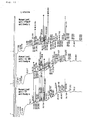
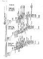
- peaks for the humanized PTHrP antibody and humanized anti-HM1.24 antibody could be connected with straight lines, but only two lines could be drawn to the peaks for the humanized anti-TF antibody.
- the subclass of H-chain is ⁇ 1 for humanized PTHrP antibody and humanized anti-HM1.24 antibody, and ⁇ 4 for humanized anti-TF antibody.
- all peaks that could be connected with straight lines were confirmed to correspond to fragments derived from the constant region.
- comparison of chromatograms allows prediction of the subclass and type of an antibody. It has been known that unidentified antibodies, and even a mixture of antibodies, can be identified by comparing chromatograms.
- the peaks derived from the variable region can be specified.
- efficient structural analysis of an antibody can be conducted by preferentially analyzing those peaks.
- the present method enables simultaneous and easy separation and purification of H-chains and L-chains that was difficult according to conventional methods.
- Enzymatic digestion in gel, fragment recovery from the gel, and sequence analysis of the recovered fragments were shown to be possible under the present conditions. Since most of the sequence can be identified from the recovered fragments, the method was found out to be useable to identify the amino acid sequence of antibodies. Furthermore, the recovery rate, which was generally believed to be the problem in the in-gel digestion until now, was good with a value of 30% to 60% or so. The reproducibility of the results was also good, and the present method that utilizes the in-gel digestion can be used as a peptide mapping system for antibodies.
- the assigned fragment numbers determined by examining the amino acid sequences of the fractions with a sequencer is shown at the peak tops.
- the number of residues that can be analyzed with a sequencer is limited. Therefore, whether a long fragment that could not be read through the end has the full length of the predicted sequence was determined in combination with the results of examination on the fraction by TOF-MS.
- TOF-MS measurements of the digest mixture can be utilized for typing of antibodies besides confirmation of the progress of a reaction and mass mapping. Examination of the RP-HPLC fractionated fractions by a sequencer and TOF-MS revealed that most of the fragments of each chain of each antibody were recovered.
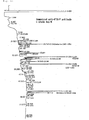
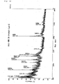
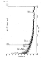
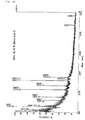
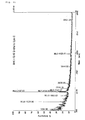
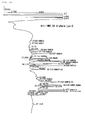
- the H-chain of humanized anti-PTHrP antibody was digested with Asp-N under the same conditions as in Example 2, and was analyzed by RP-HPLC. The result of analysis (chromatogram) is shown in Fig. 30.
Landscapes
- Life Sciences & Earth Sciences (AREA)
- Health & Medical Sciences (AREA)
- Engineering & Computer Science (AREA)
- Molecular Biology (AREA)
- Immunology (AREA)
- Hematology (AREA)
- Chemical & Material Sciences (AREA)
- Urology & Nephrology (AREA)
- Physics & Mathematics (AREA)
- Biomedical Technology (AREA)
- Microbiology (AREA)
- Analytical Chemistry (AREA)
- Biotechnology (AREA)
- Proteomics, Peptides & Aminoacids (AREA)
- Pathology (AREA)
- Bioinformatics & Computational Biology (AREA)
- Bioinformatics & Cheminformatics (AREA)
- Food Science & Technology (AREA)
- Medicinal Chemistry (AREA)
- Cell Biology (AREA)
- Biochemistry (AREA)
- General Health & Medical Sciences (AREA)
- General Physics & Mathematics (AREA)
- Biophysics (AREA)
- Spectroscopy & Molecular Physics (AREA)
- Peptides Or Proteins (AREA)
- Other Investigation Or Analysis Of Materials By Electrical Means (AREA)
- Investigating Or Analysing Biological Materials (AREA)
Abstract
The present invention provides a method for analyzing the primary structure of an antibody molecule by separating the H-chain and the L-chain of the antibody molecule, digesting each separated chain, and analyzing the peptide fragments obtained from each chain. The reproducibility can be elevated by combining in-gel digestion. According to the present method, an antibody molecule can be analyzed with a few steps and a high reproducibility. Moreover, quality of antibody molecules can be easily evaluated by the present method.
Description
The present invention relates to a method for analyzing the
primary structure of an antibody molecule. More specifically, the
present invention relates to a method for analyzing the primary
structure of an antibody molecule by separating the H-chains and the
L-chains of the antibody molecule, fragmenting each separated chain,
and analyzing the peptide fragments obtained from each chain.
Moreover, this invention provides a method for producing
peptide maps for the H-chains and the L-chains of an antibody molecule,
respectively, by analyzing the peptide fragments obtained by
digestion. In addition, this invention provides a method for
estimating the subclass of the H-chain and the type of the L-chain
of a structurally unidentified antibody.
An antibody molecule consists of H-chains and L-chains. For
example, IgG is composed of a tetramer wherein two H-chains and two
L-chains are linked via disulfide bonds (S-S bonds). Several
structural heterogeneities may occur among antibody molecules.
Commonly reported examples include N-terminal blocking, C-terminal
amino acid deletion, deamidation, oxidation, methylation,
succinylation, and glycosylation. Because these structural
heterogeneities influence the function and stability of antibodies,
identification of heterogeneity is important. Particularly, when
utilizing antibody molecules as pharmaceuticals, analysis of
heterogeneity becomes important for quality evaluation. Furthermore,
confirmation of the amino acid sequence in a preliminary evaluation
of physical properties, and construction of a peptide mapping system
for a drug substance standardization test and quality assurance are
mandatory for advancing drug development. Therefore, an analysis
system that enables more convenient and more rapid examination becomes
necessary.
In primary structure analysis of antibody molecules,
fragmentation is performed by enzymatic or chemical methods. However,
antibody molecules generally have multimeric structures, and also
have high molecular weights. For example, the molecular weight of
IgG, a representative antibody molecule, is approximately 150,000
daltons. Therefore, direct fragmentation of an antibody molecule
produces a complex mixture of peptide fragments, which makes the
separation and assignment of each fragment difficult. Thus, high
reproducibility cannot be achieved according to such a method.
To avoid the influence of an antibody being a multimer,
fragmentation after separation into H-chains and L-chains by liquid
chromatography, such as reverse phase high performance liquid
chromatography (RP-HPLC), can be considered. However, besides S-S
bonds (covalent bonds), H-chains and L-chains are strongly bound by
non-covalent bonds such as ionic bonds, hydrophobic bonds, and
hydrogen bonds; thus, fractionation of H-chains and L-chains by liquid
chromatography with good recovery is difficult. Therefore, in
conventional methods for analysis, large amounts of antibodies are
needed as samples.
An objective of this invention is to provide a method for
recovering peptide fragments that are obtained by fragmenting an
antibody molecule in good yield, in terms of both the kind and the
amount of the fragment, and analyzing them for primary structure
analysis of the antibody molecule. In addition, another objective
of the present invention is to provide a method for more conveniently
and more quickly analyzing heterogeneity and evaluating the quality
of antibody molecules by separating and fractionating peptide
fragments that were obtained by fragmenting an antibody molecule via
appropriate separation techniques (for example RP-HPLC), then
constructing a peptide map having a good reproducibility based on
the results of analysis. Furthermore, this invention provides a
method for efficiently analyzing the variable regions by readily
identifying the subclass of the H-chain and the type of the L-chain
of a structurally unidentified antibody via a comparison of the
obtained peptide maps.
To accomplish the aforementioned objective, the present
inventors focused on the structural characteristics of antibody
molecules. As a result, they found out that besides the large
molecular weight of an antibody molecule, the multimeric structure
thereof is an obstacle in analyzing the antibody molecule. In
addition, they demonstrated that the aforementioned obstacles can
be overcome by separating in advance an antibody molecule into its
H-chains and L-chains, and fragmenting the chains in the separated
state. This invention was accomplished based on these findings.
That is, the present invention relates to the following method of
analysis, and use of the same.
In this invention, first, the antibody molecule is separated
into H-chains and L-chains to facilitate primary structure analysis
of the antibody molecule. Next, both of the separated chains are
fragmented independently, and the obtained peptide fragments are
analyzed. Based on the results of analysis of the obtained peptide
fragments, a peptide map can be prepared. The peptide map can be used
to evaluate the quality of the antibody molecule, and such.
In the present invention, the H-chains and L-chains of an
antibody molecule can be separated by any method. For example, the
antibody molecule can be cleaved at the S-S bond by a treatment with
reducing agent, such as dithiothreitol (DTT) and 2-mercaptoethanol
(2-ME), and then, separated into the H-chains and L-chains based on
a difference in molecular weight. By modifying the thiol group (SH
group) after reductive treatment, recombination of the S-S bond can
be prevented. S-S bonds are not only formed between the originally
bonded SH groups, but also between completely unrelated SH groups.
Therefore, recombination of S-S bonds results in the formation of
a complexed mixture.
SH groups can be modified by alkylation, acylation, and such.
More specifically, treatments such as reduction and
carboxymethylation, reduction and pyridylethylation, and reduction
and carboxyamidemethylation can be presented as examples. The S-S
bond becomes irreversible by modifying the SH group, and thus
production of a complexed mixture due to recombination is suppressed.
Therefore, modification of the SH group is expected to increase the
accuracy of the analysis.
In the present invention, modification of the SH group can be
performed at any time, e.g., before separation of the H-chains and
L-chains, in the gel after separation but before fragmentation, or
after fragmentation. Particularly, separation by SDS-PAGE, as
mentioned in Examples, can be accomplished with good yield and also
at a high reproducibility by performing the modification before the
separation. Specifically, the separation by SDS-PAGE is performed
after denaturation by sodium dodecyl sulfate (SDS) , reduction by DTT,
and carboxymethylation treatment.
In this invention, antibody molecules cleaved at S-S bonds are
separated into H-chains and L-chains prior to fragmentation.
H-chains and L-chains can be separated by any method. H-chains and
L-chains can be separated based on the differences in their properties
(for example, molecular weight, electric charge, hydrophobicity).
Specifically, separation techniques such as electrophoresis and
liquid chromatography can be used. However, the separation must be
conducted under conditions that hinder recombination of the H-chains
and L-chains. More specifically, it is advantageous to separate the
H-chains and L-chains by denaturing gel electrophoresis. By
employing denaturing gel electrophoresis, the H-chains and L-chains
can be physically separated, and a higher reproducibility can be
expected.
In the present invention, the term "denaturing gel
electrophoresis" refers to gel electrophoresis that enables
separation of the H-chains and L-chains of antibody molecules.
Electrophoresis which utilizes an acrylamide gel medium containing
SDS and urea is well known as an example for such electrophoresis.
Particularly, SDS-PAGE is an especially advantageous separation
technique for use in this invention. SDS-PAGE can be performed
according to the method of Laemmli (Laemmli (1970) Nature 227,
680-685). The H-chains and L-chains are strongly bound by covalent
bonds (S-S bonds) as well as non-covalent bonds, but by using SDS-PAGE,
the H-chains and L-chains can be readily separated.
Alternatively, denaturing gel electrophoresis can be also
carried out without performing reductive acylation and such. For
example, when only information of a part of the internal sequence
of an antibody molecule is required, H-chains and L-chains can be
separated even by SDS-PAGE under reducing conditions, and the primary
structure can be analyzed in a similar manner. Therefore, the amino
acid sequence without the cysteine residues can be investigated.
Each of the separated H-chains and L-chains are fragmented.
Fragmentation methods for primary structure analyses of proteins are
well known. Generally, for structural analyses of proteins,
enzymatic digestion and chemical cleavage are utilized. For example,
the following enzymes may be used. All of these enzymes are
commercially available, and their digestion conditions are known.
Lysyl endopeptidase
Endoproteinase LysC
Mouse submandibular gland protease D
Staphylococcus aureuse V8 protease
Endoproteinase Asp-N
Lysyl endopeptidase
Endoproteinase LysC
Mouse submandibular gland protease D
Staphylococcus aureuse V8 protease
Endoproteinase Asp-N
Protein digestion by enzymatic digestion can be performed under
a mild condition, and therefore, modifications and such of the side
chains of amino acid residues are unlikely to happen. Furthermore,
by fixing the conditions for digestion, reproducibility can be easily
maintained.
In this invention, chemical cleavage can be utilized instead
of enzymatic digestion. Cleavage at the Carboxyl terminus of a
methionine residue by cyanogen bromide is a representative chemical
cleavage method.
Fragmentation of the separated H-chains and L-chains can be
performed by any method. For example, as a technique recently
developed for structural analysis of proteins, there is a method
wherein bands or spots that are obtained by subjecting a protein to
SDS-PAGE or two-dimensional electrophoresis are excised, and then
chemically or enzymatically fragmented in a gel (Jahnen-Dechent and
Simpson (1990) Plant Mol. Biol. Reporter 8, 92-103; Hellman U. et
al., (1995) Anal. Biochem. 224, 451-455; Kawasaki et al. (1990) Anal.
Biochem. 191, 332-336). According to this method, proteins are
fragmented in a gel. Therefore, the method is called an in-gel
digestion method. Alternatively, proteins separated in a gel can be
fragmented on membranes, such as polyvinylidene difluoride (PVDF)
membrane and nitrocellulose membrane, after transferring the
separated protein on the membrane. In this invention, the
reproducibility of the analysis can be further raised by combining
the in-gel digestion method or the fragmentation method on a protein
adsorption membrane to the aforementioned fragmentation process.
PVDF membranes are commercially available under product names such
as "Immobilon" (MILLIPORE).
The in-gel digestion method, one of the analysis methods of
proteome analysis, is normally carried out by the steps of
enzymatically digesting a protein separated and purified by
two-dimensional electrophoresis directly in a gel, and then obtaining
information on its amino acid sequence from the recovered peptide
fragments. In this invention, an antibody can be easily separated
into H-chains and L-chains by denaturing gel electrophoresis, such
as SDS-PAGE. Therefore, the inventors anticipated that
fragmentation for the present invention could be accomplished by
restricting electrophoresis to denaturing gel electrophoresis and
performing enzymatic digestion of the separated H-chains and L-chains
directly in the gel. That is, the only electrophoresis performed
prior to enzymatic digestion is, not two-dimensional electrophoresis,
but denaturing gel electrophoresis.
The in-gel digestion method can be performed by excising the
bands of H-chains and L-chains separated by SDS-PAGE; incubating them
after adding a digestion buffer to soak the gel; and then adding a
protease, such as Lys-C, for digestion. The digest solution is
recovered, eluting buffer is freshly added to the gel, and the
fragments are extracted from the gel. This operation is repeated,
and the obtained digest solution and eluate are mixed for following
structural analysis. According to the in-gel digestion method,
H-chains and L-chains can be readily fragmented in few steps, and
the subsequent separation and fractionation of the peptide fragments
can be performed with a good recovery. Alternatively, in this
invention, the H-chains and L-chains separated by SDS-PAGE can be
transferred onto a PVDF membrane to fragment them onto the PVDF
membrane. Transfer to a PVDF membrane and fragmentation thereon can
be performed according to conventional methods.
The three-dimensional structure of an antibody molecule is
supported by diverse bonding patterns, including S-S bonds.
Therefore, for example, when only the S-S bonds are dissociated and
other bonds cannot be sufficiently blocked, various three-dimensional
structures are formed during digestion. Construction of such
three-dimensional structures is one of the factors that prevent
fragmentation of the peptides. In the analysis method of this
invention, due to the restricted movement of separated H-chains and
L-chains, obtained by combining the in-gel digestion method or the
fragmentation method with a protein adsorption membrane, digestion
of the fragments can be carried out under conditions wherein the
construction of other three-dimensional structure is greatly
suppressed. As a result, analysis can be performed with a good
reproducibility.
The present invention can be utilized to analyze any kind of
antibody. According to the present invention, structural analyses
can be conducted for. naturally occurring antibodies, chimeric
antibodies and human antibodies, as well as derivatives or modified
derivatives thereof. The present invention can not only be applied
to complete antibody molecules (whole antibodies) but also to antibody
fragments, such as Fab, F(ab')2, and Fab'. Furthermore, the method
of this invention can not only be applied to IgG but also to all classes
of antibodies, such as IgA, IgM, IgD, and IgE. Moreover, there are
no restrictions with regard to the particular animal specie or cell
line from which the antibody to be analyzed is derived. In addition,
antibodies prepared by any method can be analyzed by the method of
the present invention, so long as the antibody molecules contain
H-chains and L-chains that are separable.
Peptide fragments obtained by the digestion of H-chains and
L-chains of an antibody molecule can be analyzed according to known
protein analysis methods. In this invention, the analysis of a
peptide fragment is defined as revealing the biochemical information
possessed by the peptide fragment. Examples of biochemical
information include information regarding amino acid sequence of the
peptide fragment, molecular weight, mass, presence or absence and
type of modification, and so on. Modifications of peptide fragments
include deamidation, oxidation, methylation, succinylation,
glycosylation, and so on. If necessary, the peptide fragments
obtained by digestion may be analyzed after separating them by
appropriate techniques. Such techniques include RP-HPLC, SDS-PAGE,
ion-exchange chromatography, and such. Separation patterns of a
peptide fragment obtained by the above-mentioned separation technique
can be used as a peptide map. Furthermore, regarding peptide
fragments related to heterogeneity, the type of heterogeneity can
be determined by applying appropriate analysis methods. In this
invention, since the heterogeneity existing in the antibody molecule
is analyzed after fragmentation of both of the H-chains and L-chains,
analysis can be performed readily. Methods for analysis are well
known in the art.
General methods for analyzing peptide fragments include mass
spectrometry, amino acid sequence analysis, amino acid composition
analysis, and so on. Each of these methods for structural analysis
can be performed according to conventional methods. Mass
spectrometry is the most reliable analysis method at present among
analysis methods based on the difference in molecular weight. In the
present invention as well, mass spectrometry is one of the preferred
analysis methods. A mass spectrometer is composed of an ion source,
a mass detection element, and a detector. Various methods for
ionization and various methods for a detection piece are known, and
instruments are composed of combinations of part utilizing these
methods. Representative methods are shown below.
Ionization methods-
MALDI (Matrix Assisted Laser Desorption Ionization)
EI (Electron Ionization)
FAB (Fast Atom Bombardment)
API (Atmosphere Pressure Ionization)
Detection methods-
TOF (Time of Flight)
Sector (Magnetic, Electrostatic)
Quadrupole
FT (Fourier Transform)
Ionization methods-
MALDI (Matrix Assisted Laser Desorption Ionization)
EI (Electron Ionization)
FAB (Fast Atom Bombardment)
API (Atmosphere Pressure Ionization)
Detection methods-
TOF (Time of Flight)
Sector (Magnetic, Electrostatic)
Quadrupole
FT (Fourier Transform)
Among the combinations, MALDI-TOF-MS (TOF-MS) is an especially
advantageous analysis instrument for the present invention. There
also are instruments, such as LC-MS wherein liquid chromatography
(LC) is combined with mass spectrometry, tandem MS (MS/MS) wherein
two mass spectrometers are connected together, and LC-MS/MS wherein
the LC and MS/MS are combined; and each of them are used depending
on the objective of the analysis.
Amino acid sequence is the most important data in the primary
structure analysis of a protein. In this invention as well,
identification of peptide fragments is based on the result of sequence
analysis. Representative methods for amino acid sequence analysis
are shown below.
N-terminus analysis methods-
Edman degredation method (PITC method)
DABITC method
Dansyl method (DNS-Cl method)
Aminopeptidase method
C-terminus analysis methods-
Hydrazinolysis method
Carboxypeptidase method
N-terminus analysis methods-
Edman degredation method (PITC method)
DABITC method
Dansyl method (DNS-Cl method)
Aminopeptidase method
C-terminus analysis methods-
Hydrazinolysis method
Carboxypeptidase method
At present, an automatic analyzer using the Edman degradation
method exists and analysis using this instrument is generally
performed. The instrument was also used in the examples of this
invention discussed in detail below. In the Edman degradation method,
blocking of the N-terminus of the peptide fragment becomes an obstacle.
In this invention, amino acid sequence analysis can be performed by
the Edman degradation method after deblocking the N-terminus.
Specifically, deblocking can be easily and reliably performed by
conducting the deblocking step after the fragmentation of the peptide.
Methods for deblocking the N-terminus are well known in the art.
Blocking of the N-terminus refers to a condition wherein the
α-amino group of the N-terminal amino acid is modified by acetylation
and such. It is difficult to identify amino acids whose α-amino group
is modified by methods such as the Edman degradation method. However,
the blocked N-terminus can be analyzed by removing the modification.
For example, the modification of an acetylated amino acid can be
enzymatically removed with an amino acid acylase. Generally, the
amino acid acylase acts poorly on large proteins. In this invention,
the amino acid acylase will be made to act on digested fragments.
Therefore, in this invention, the N-terminus can be easily and
reliably deblocked.
Amino acid composition analysis is necessary to understand the
basic characteristics of a protein. Conventionally, the amino acid
composition of a protein is detected by derivatization of the amino
acids composing the protein that are formed due to hydrolysis. The
detection methods are categorized into two types, depending on whether
the derivatization is carried out before or after the amino acid
separation. Representative reagents for derivatization are shown
below.
Post-column methods-
ninhydrin
o-phthalaldehyde
Pre-column methods-
PITC
NBD-F
FMO-Cl
Dabsyl-Cl
o-phthalaldehyde
DNS-Cl
Post-column methods-
ninhydrin
o-phthalaldehyde
Pre-column methods-
PITC
NBD-F
FMO-Cl
Dabsyl-Cl
o-phthalaldehyde
DNS-Cl
The post-column method using ninhydrin has been automated and
has a high reproducibility. However, due to the problem of detection
limit, pre-column method using PITC is often utilized for the primary
structure analysis of proteins.
Furthermore, the present invention relates to peptide maps that
can be obtained by the analysis methods of this invention. Based on
such analysis results, peptide maps relating to the structure of
antibody molecules can be prepared. In this invention, the term
"peptide map" refers to a set of information of each peptide fragment
that compose a certain peptide mixture, which can characterize the
peptide mixture. The term "peptide map" generally refers to a
condition wherein a mixture of peptide fragments has been separated
by some method. However, herein, not only separated peptide
fragments themselves but also a set of information obtained by
analyzing the peptide fragments are referred to as the peptide map.
Therefore, electrophoretic image developed by electrophoresis of
digested fragments is included in the peptide map of this invention.
Furthermore, the results of mass spectrometry of peptide fragments
are also included in the peptide map of this invention.
Structural equivalence or difference in original proteins can
be determined by comparing peptide maps in order to compare the
characteristic of a certain peptide fragment mixture to that of
another peptide fragment mixture. A high possibility that the
original antibodies share a common structure is suggested by peptide
fragment mixtures yielding common peptide maps. On the contrary,
difference in the peptide maps indicate structural differences of
the original antibody molecules. Therefore, antibody molecule
structures can be compared by comparing peptide maps.
For example, a quality of an antibody can be evaluated by
comparing the analysis result with that of a structurally identified
antibody. More specifically, a peptide map of a produced antibody
molecule is prepared according to the present invention, and is
compared with a peptide map that is used as the standard. As a result,
when the difference between both peptide maps is within the range
determined based on the desired precision of analysis, the produced
antibody is regarded as equivalent, including its heterogeneity. In
the present invention, heterogeneity refers to structural differences
among antibody molecules. Structural differences of antibody
molecules are caused, for example, by the presence of N-terminal
blocking, deletion of the C-terminal amino acid, deamidation,
oxidation, methylation, succinylation, and glycosylation. However,
a heterogeneity that can be discovered according to the present
invention is not limited to those caused by the factors illustrated
herein.
Using a peptide map prepared based on the method of this
invention, the subclass and type of a structurally unidentified
antibody can be identified. Specifically, the subclass of the
H-chain and the type of L-chain of a structurally unidentified
antibody can be identified by preparing peptide maps based on the
method of the present invention for a structurally identified antibody
and the structurally unidentified antibody, respectively; and
comparing the constant regions of the two. Furthermore, from a
mixture of multiple structurally unidentified antibodies, the
subclasses and types the mixture is composed can be easily determined
by comparing its peptide map with peptide maps prepared for
structurally identified antibodies of different subclasses and types.
An immunoglobulin is composed of constant regions and variable
regions. The variable regions have various structures due to antigen
recognition, whereas the structure of the constant regions is limited.
Therefore, peptide maps for all expected constant regions can be
prepared in advance. The structure of the constant region is
categorized into the subclasses of the H-chains and into the types
of the L-chains. When peptide maps are prepared for all the subclasses
and types, the subclass of the H-chain or the type of the L-chain
composing a structurally unidentified immunoglobulin can be
determined by preparing its peptide map by the same method, and
comparing the peptide map to the known peptide maps.
By utilizing peptide map information on fragments derived from
the constant region, primary structure analysis of an unidentified
antibody can be efficiently carried out. As already mentioned above,
an antibody molecule consists of a constant region and a variable
region. Therefore, to obtain information on fragments derived from
the constant region from a peptide map is nothing but to predict peaks
of fragments derived from the variable region. Thus, the structure
of fragment peaks estimated to stem from the variable region can be
preferentially analyzed. In the evaluation of antibody molecules,
structural information on the variable region that affects the binding
specificity with an antigen is important. Thus, the technique of the
present invention that enables one to efficiently proceed to the
analysis of the variable region is useful as a method for evaluating
antibody molecules.
For example, as indicated in Examples below, when the analysis
of peptide fragments are performed by reverse-phase chromatography
and mass spectrometry, then the peptide map is obtained as a
chromatogram or a mass spectrum. Evaluation of an antibody molecule
by comparing peptide maps is carried out via the decision of peak
assignment and such. With regard to the evaluation, once the peaks
of the fragments derived from the constant region are identified,
the variable region can be analyzed by comparing the remaining peaks.
Furthermore, the amino acid sequence of fragments that are
derived from the variable region can be analyzed by selecting peaks
of the fragments in a chromatogram of a reverse-phase chromatography,
and fractionating the selected peaks. According to the present
invention, structural analysis of variable region can efficiently
conducted in this manner.
The present invention will be explained in detail below with
reference to Examples.
Hereinafter, an example of analysis according to the present
invention will be described using an anti-Parathyroid hormone-related
peptide humanized antibody (IgG1) (method for preparing the antibody
is described in WO98/13388) as the example.
The amino acid sequences of humanized PTHrP antibody L-chain
and H-chain are shown in SEQ ID NOs: 1 and 2, respectively. The
humanized PTHrP antibody is an IgG1 antibody which has a structure
wherein two each of H-chains and L-chains are cross-linked by S-S
bonds; it is a huge protein, consisting of round about 1,300 amino
acids in total. Therefore, when the entire antibody molecule was
analyzed at once, reproducibility was difficult to maintain regarding
two points: enzymatic digestion, separation and fractionation of the
fragments. Furthermore, even when the S-S bonds were irreversibly
cleaved by reductive modification, the H-chains and L-chains were
separated by RP-HPLC, and then were analyzed respectively, the
separation of the H-chains and L-chains was insufficient, recovery
was low, and precision of analysis was not good. Since an antibody
is a huge protein, the situation was such that a highly accurate data
could not be obtained by just applying conventional methods for
protein analysis. Therefore, the method of this invention was
developed.
Humanized PTHrP antibody solution (13.4 mg protein/ml; 150 mM
NaCl, 20 mM acetic acid/sodium acetate buffer, pH 5.7) and SDS sample
buffer (5% SDS, 2 mM ethylenediamine tetraacetic acid (EDTA), 0.005%
bromophenol blue (BPB), 20 mM tris(hydroxymethyl)aminomethane
(Tris)-HCl, pH 8.0) were mixed at a ratio of 1:5 (v/v) while stirring,
and denaturation was carried out by heating at 95°C in a water bath
for 5 minutes. Reduction was carried out by adding DTT to a final
concentration of 40 mM, substituting the upper space in the tube with
nitrogen gas, and then incubating in a thermoregulated air bath at
37°C for 2 hours. Carboxymethylation was carried out by adding
iodoacetic acid to a final concentration of 80 mM, purging the upper
space in the tube with nitrogen gas, and performing the reaction in
a thermoregulated air bath at 37°C for 30 minutes. The mixture was
neutralized to pH 6.8 with 1 N NaOH, and was used as the sample for
electrophoresis.
Humanized PTHrP antibody solution and SDS sample buffer for
reduction (5% 2-ME, 5% SDS, 2 mM EDTA, 0.005% BPB, 20 mM Tris-HCl,
pH 8.0) were mixed at a ratio of 1: 5 (v/v) while stirring, denaturation
and reduction were carried out by heating at 95°C in a water bath
for 5 minutes, and this was used as the sample for electrophoresis.
H-chains and L-chains were separated electrophoretically by
SDS-PAGE. Electrophoresis was performed according to the method of
Laemmli. SDS-PAGE mini 12% (TEFCO) was used as the gel.
Electrophoresis was performed in running buffer (0.1% SDS, 200 mM
Glycnie, 25 mM Tris) at 25 mA (constant current) , and was terminated
when BPB had completely phoresed. The bands of each chain were
detected upon staining with staining solution (0.2% Coomassie
Brilliant Blue R-250, 40% ethanol, 10% acetic acid). After
destaining with destaining solution (40% methanol, 10% acetic acid) ,
the gel was stored in a refrigerator by soaking in Milli Q water.
Portions corresponding to each of the bands of the H-chains and
L-chains were excised, digestion buffer (0.1% SDS, 100 mM Tris-HCl,
pH 9.0) enough to soak the gel was added, and this was pre-incubated
in a thermoregulated air bath at 37°C for 30 minutes. Lys-C enzyme
was added to a final concentration of 5 µg/ml, and digestion was
carried out in a thermoregulated air bath at 37°C for 18 hours. Lys-C
specifically cleaves the C-termini of lysines in the amino acids
constituting the protein.
The digest solution was poured into another tube, elution buffer
(0.1% SDS, 100 mM Tris-HCl, pH 9.0) was freshly added, and was eluted
in a thermoregulated air bath at 37°C for 1 hour. This elution process
was repeated 3 times, and the obtained digest solution and elute were
mixed and then used as a digest mixture solution in the following
analysis.
The digest mixture solution was desalted using ZipTip
(MILLIPORE) to prepare a TOF-MS sample.
3,5-dimethoxy-4-hydroxycinnamic acid (Sinapinic Acid) was used as
the matrix. The sample and matrix were dissolved in a 50% acetonitrile
solution containing 0.1% trifluoroacetic acid (TFA), and was dried
on a sample plate. Voyager-DE STR (PerSeptive Biosystems) was used
as the instrument. This instrument utilizes the matrix-assisted
laser desorption ionization (MALDI) method as the ionization method
and time-of-flight (TOF) type for detection.
Method file that can cover the range of theoretical mass numbers
for the sample was selected for the measurement. Conditions for such
measurements were: accelerated voltage: 20 kV; delay: 30 ns; and mode:
linear, positive. All operations were performed according to the
manual.
The digest mixture solution was subjected to RP-HPLC, and the
digested fragments were fractionated.
ODS-A (250x 4.6 mm) (YMC) was used as the separation column,
and TSKguardgel DEAE-SW (10x 6.0mm) (Tosoh) was used as the guard
column. SCL-6A System Controller, LC-6A Pump, SPD-6A UV Detector,
and C-R3A Chromatopac integrator (Shimadzu) were used as equipments.
Milli Q water and acetonitrile containing 0.1% TFA were used
as the mobile phase. Elution was carried out at a flow rate of 1.0
ml/min using a 5-60% linear gradient of acetonitrile. Detection was
carried out at a UV frequency of 215 nm.
After freeze drying, the fractions were dissolved with small
amounts of formic acid to prepare samples for fragment identification
using an amino acid sequencer and TOF-MS.
The fractionated samples were placed directly onto sample
filters, dried, and then analyzed. Protein Sequencer 476A (Applied
Biosystems) was used as the instrument, and all operations were
performed according to the manual.
The fractionated samples were dried on sample plates. The
matrix (Sinapinic acid) dissolved in 50% acetonitrile solution
containing 0.1% TFA was dropped on each sample to prepare the analysis
sample. The same instrument as that used in 5) was used. Method file
that can cover the range of theoretical mass numbers for the sample
was used for the measurement. Conditions for the measurement were:
accelerated voltage: 20 kV; delay: 100 ns; and mode: linear, positive.
All operations were performed according to the manual.
Humanized PTHrP antibody is an IgG1 antibody, and forms a
structure wherein two each of the H-chains and 1-chains are
cross-linked by S-S bonds. When the S-S bonds are reductively cleaved
leaving the highly reactive SH groups as they are, reorganization
of the S-S bonds or addition of reagents or such will occur which
makes interpretation of data complicated and laborious. Therefore,
this system is inappropriate for mapping. Thus, to avoid unnecessary
confusion, incorporation of a treatment for reductive modification
of the S-S bonds to the process was considered. Three objects, the
sample before electrophoresis, the gel before enzymatic digestion,
and the digest mixture solution after elution, were considered as
the candidates that should be subjected to the treatment. According
to preliminary studies, it had been confirmed that something is
already attached to the SH group in the digest mixture solution after
elution from the gel (data not shown). Thus, this object was rejected
as the candidate.
Generally, reductive modification is carried out in gel.
However, herein, reduction and carboxymethylation were performed on
samples before electrophoresis. A sample before electrophoresis was
denatured in SDS sample buffer, i.e., in SDS, and then reduction and
carboxymethylation treatments were performed. The treated sample
could be directly phoresed, and the migration pattern was not affected
by the treatment. Even in TOF-MS of digest mixture solutions,
fragments involved in S-S bonds were detected in accordance with the
theoretical mass numbers, and carboxymethylation reaction had
progressed. Problems arose neither in the chromatogram of RP-HPLC
nor to the amount of recovery.
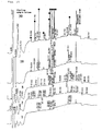
Comparison of chromatograms with and without
carboxymethylation treatment is indicated in Fig. 8 (without
carboxymethylation treatment) and Fig. 3 (with carboxymethylation
treatment). Except the carboxymethylation treatment, all
manipulations were conducted following the method of the present
invention.
The results obtained by the sequencer and TOF-MS on the
fractions suggested that reagents and such are added to the SH group
in peak fragments involved in S-S bonds. The same sequence detected
in several positions was considered to correspond to the different
SH group adducts, according to the results of TOF-MS. Furthermore,
all fragments not involved in S-S bonds were detected in the same
manner in both Figs. 8 and 3.
Finally, separation of the H-chains and L-chains by SDS-PAGE
under reducing conditions, in-gel fragmentation, and structure
analysis were found to be possible even without the carboxymethylation
treatment. Therefore, when just a partial knowledge of the internal
sequence of an antibody molecule is required, the amino acid sequence
without the cysteine residues can be determined. Carboxymethylation
treatment was performed for the following experiments and results,
except otherwise stated.
After reduction and carboxymethylation treatments and SDS-PAGE
of the humanized PTHrP antibody, the bands for the H-chain and L-chain
were excised, and the gel was directly digested with Lys-C enzyme.
Sinapinic Acid was used as the matrix, and the examination results
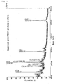
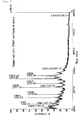
by TOF-MS for the digests mixture of each chain are shown in Figs.
1 and 2. The numbers at the peak tops indicate the measured mass and
fragment numbers presumed to corresponding thereto.
7 of 16 fragments predicted from the sequence of the L-chain
were detected (Fig. 1). Fragments that should be theoretically
generated according to the amino acid sequence of the L-chain (SEQ
ID NO: 1) are shown in Table 1. The table shows from the left, the
fragment number (CLn), amino acid sequence of the fragment, and the
theoretical mass. Furthermore, "constant" and "variable" indicate
the part that divides the respective constant region and variable
region. The seven detected peaks had nearly the theoretical mass,
and included a conjectural N-terminal blocked fragment (CL1) that
could not be identified with the sequencer.
The peak of CL9 was small and its existence was unclear.
Difficulty in the detection of fragments having low ionization
efficiency is inevitable in TOF-MS. The existence of CL9 was
confirmed due to the analysis of fractions using a sequencer.
Therefore, it was determined that the CL9 was hardly detected due
to its low ionization efficiency. The other eight fragments that were
not detected are fragments with a mass of 1,000 or less. These
fragments were regarded as undetected because their peaks were lost
in the noise of the low molecular weight region or in the peaks of
extracts from the reagents and gel, and consequently their assignments
were unclear.
| CL3 | 43-45 | GPK | 300.3581 |
| CL12 | 173-175 | PSK | 330.3844 |
| CL13 | 176-180 | QSNNK | 589.6063 |
| CL10 | 159-165 | ADGSP VK | 672.7364 |
| CL11 | 166-172 | AGVET TK | 704.7786 |
| CL4 | 46-51 | YLMDL K | 781.9716 |
| CL7 | 112-119 | LTVLG QPK | 855.0456 |
| variable ← → constant | |||
| CL16 | 214-221 | TVAPT EBS | 864.9289 |
| CL6 | 100-111 | EQFVY VFGGG TK | 1331.4912 |
| CL14 | 181-195 | YAASS YLSLT PEQWK | 1743.9342 |
| CL1 | 1-19 | XLVLT QSPSA SASLG ASVK | 1827.0656 |
| CL8 | 120-138 | ANPTV TLFPP SSEEL QANK | 2043.2618 |
| CL15 | 196-213 | SHRSY SBQVT HEGST VEK | 2093.2192 |
| CL9 | 139-158 | ATLVB LISDF YPGAV TVAWK | 2212.5954 |
| CL2 | 20-42 | LTBTL SSQHS TYTIE WYQQQ PEK | 2830.0820 |
| CL5 | 52-99 | QDGSH STGDG IPDRF SGSSS GAERY LTISS LQSED EADYI BGVGD TIK | 5068.3031 |
15 out of 31 fragments predicted from the sequence of the H-chain
were detected with nearly the theoretical mass (Fig. 2). The
fragments predicted from the amino acid sequence of the H-chain (SEQ
ID NO: 2) are shown in Tables 2 and 3. The tables show from the left:
the fragment number (CHn) , amino acid sequence of the fragment, and
the theoretical mass. Furthermore, "constant" and "variable"
indicate the part that divides the respective constant region and
variable region. Among the observed peaks, CH7-8, 13-14, 17-18 were
detected as linked fragments wherein the Lys-Pro sequences that are
believed to be difficult to cleave with Lys-C had not been cleaved.
Except fragments with a mass of 1,000 or less whose assignments
are unclear due to the same reasons as for the above-mentioned L-chain,
fragments that could not be detected were CH4 and CH15. However, the
existence of both of these fragments was confirmed by analyzing these
fractions with a sequencer. Thus, it was suggested that these
fragments could not be detected due to their low ionization
efficiency.
Most types of fragments could be recovered from the gel by the
current method. Therefore, enzymatic digestion in the gel was
considered to be possible. Furthermore, in terms of amount, 80% to
90% of the fragments were estimated to be recovered according to the
spectrum intensity of TOF-MS.
| CH10 | 215-215 | K | 146.1894 |
| CH24 | 340-341 | AK | 217.2683 |
| CH14 | 248-249 | PK | 243.3062 |
| CH17 | 290-291 | TK | 247.2946 |
| | 322-323 | BK | 307.3709 |
| | 212-214 | VDK | 360.4107 |
| CH 19 | 319-321 | EYK | 438.4810 |
| CH 21 | 324-327 | VSNK | 446.5042 |
| CH23 | 336-339 | TISK | 447.5323 |
| | 216-219 | VEPK | 471.5543 |
| CH 12 | 220-223 | SBDK | 509.5378 |
| CH 8 | 207-211 | PSNTK | 545.5934 |
| CH 29 | 411-415 | LTVDK | 574.6753 |
| CH 31 | 441-447 | SLSLS PG | 659.7376 |
| CH 22 | 328-335 | ALPAP IEK | 838.0150 |
| CH 26 | 362-371 | NQVSL TBLVK | 1162.3732 |
| CH 5 | 123-134 | GPSVF FLAPS SK | 1186.3738 |
| CH 3 | 66-76 | GRFTI SRDNS K | 1280.4069 |
| | 135-148 | STSGG TAALG BLVK | 1322.5028 |
| CH16 | 276-289 | FNWYV DGVEV HNAK | 1677.8371 |
| CH 28 | 394-410 | TTPPV LDSDG SFFLY SK | 1874.0778 |
| CH 25 | 342-361 | GQPRE PQVYT LPPSR DELTK | 2311.5809 |
| | 44-65 | GLEWV ATISS GGSYT YYPDS VK | 2380.5937 |
| CH 27 | 372-393 | GFYPS DIAVE WESNG QPENN YK | 2544.6738 |
| | 224-247 | THTBP PBPAP ELLGG PSVFL FPPK | 2622.0617 |
| CH 15 | 250-275 | DTLMI SRTPE VTBVV VDVSH EDPEV K | 2957.3307 |
| CH30 | 416-440 | SRWQQ GNVFS BSVMH EALHN HYTQK . | 3046.3577 |
| CH18 | 292-318 | PREEQ YNSTY RVVSV LTVLH QDWLN GK | 3232.6046 |
| CH1 | 1-43 | XVQLV ESGGG WQPG RSLRL SBAAS GFTFS SYGMS WVRQA PGK | 4528.1109 |
| CH4 | 77-122 | NTLYL QMNSL RAEDT AVYYB ARQTT MTYFA YWGQG TLVTV SS AST K | 5264.9055 |
| variable← → constant | |||
| CH7 | 149-206 | DYFPE PVTVS WNSGA LTSGV HTFPA VLQSS GLYSL SSVVT VPSSS LGTQT YIBNV NHK | 6189.8668 |
In the above study, TOF-MS measurements of the digest mixture
were used to confirm the extent of progress of the reduction and
carboxymethylation reactions. As a result, within the measurable
range, most of the peaks corresponding to the fragments were detected
in accordance with the theoretical mass. Therefore, this spectrum
may be utilized for mass mapping. When the sequence is known, the
theoretical mass of the fragment can be calculated, and thus the
measured value can be assigned. The type of modification can be
predicted from the shift of mass even when the modification is one
that cannot be detected with a sequencer such as that of a N-terminus
modified fragment, or when the modification does not change the
RP-HPLC elution position. A more reliable mapping data can be
obtained using both TOF-MS spectra and RP-HPLC chromatograms.
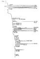
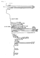
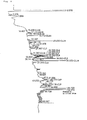
Chromatograms of the digest mixture solutions fractionated by
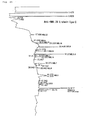
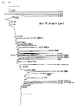
RP-HPLC are shown in Figs. 3 and 4. The fraction number assigned by
examining the amino acid sequence of the fraction using a sequencer
is indicated at the peak tops. Since there is a limit to the number
of residues that can be analyzed using a sequencer, whether or not
a long fragment for which the sequence could not be read to the end
has the full length of the predicted sequence was determined by
combining the result of examination of the fraction by TOF-MS.
13 out of 16 fragments of the predicted sequence could be
assigned for the L-chain (Fig. 3). CL11-12 was identified in the form
of a linked fragment as the 19.5-minute peak. This was because the
Lys-Pro sequence was not cleaved by Lys-C. Furthermore, CL10 was
detected simultaneously in this fraction. Similarly, CL2 and CL8
were detected simultaneously at the 32.8-minute peak.
Furthermore, the sequence of CL4 was read from both the
26.5-minute and 32.2-minute peaks. The recovery of the residue Met48
of the 26.5-minute peak was extremely poor in the sequence analysis
results, which suggested the occurrence of modification at the Met.
From the result of LC-MS, the low recovery was suggested to be the
result of the generation of oxidized form of Met. CL9 was identified
as the 48.9-minute peak (data not shown), and revealed that the
inability of detection by TOF-MS of the digest mixture solution was
due to problems with ionization efficiency. Among the fragments that
could not be assigned, CL1 could not be identified using a sequencer
due to pyroglutamylation of the N-terminal Gln. However, the
32.5-minute peak was regarded as CL1 from the fact that nothing could
be read from this peak using a sequencer and the results of TOF-MS
of this fraction. CL3 and CL13 that could not be assigned are small
fragments comprising 3 and 5 amino acid residues, respectively. Thus,
they were supposed to have been passed through and were not retained
by RP-HPLC using the ODS column.
19 out of 31 fragments from the predicted sequence could be
assigned for the H-chain (Fig. 4). CH13-14 and CH17-18 were
identified as the 39.3-minute and 46.7-minute peaks, respectively.
This was because the Lys-Pro sequence was not cleaved by Lys-C. This
does not oppose the fact that peaks with masses corresponding to the
linked fragments were detected in the TOF-MS of the digest mixture
solution (Fig. 2). Although the sequence of CH7 was read from the
45.9-minute peak, this fragment was too long to confirm the full length
sequence. However, CH7 was regarded as a fragment linked to CH8 due
to the facts that CH7 has a Lys-Pro sequence, a sequence difficult
to cleave with Lys-C, and a peak with a mass for a linked fragment
was detected by TOF-MS.
CH15 and CH30 were detected simultaneouly in the 34.1-minute
peak. The sequence of CH30 was read from both 30.6-minute and
34.1-minute peaks, and the amount of recovery of the residue Met429
from the 30.6-minute peak was particularly poor in the sequence
analysis results. Thus, the Met seemed to have been modified.
Similarly to CL4, this was suggested to be caused by the formation
of the oxidized form of Met. CH4 and CH15 were identified as the
48.4-minute and 34.1-minute peaks, respectively, revealing that the
inability of detection by TOF-MS of the digest mixture was due to
problems with ionization efficiency.
A good reproducibility was confirmed for the above-mentioned
RP-HPLC chromatogram. Furthermore, recovery of most of the fragments
was confirmed from mass spectrometry by TOF-MS and sequence analysis
using a sequencer. Furthermore, the recovery of the fragments
calculated using the amount of applied sample that were calculated
from the yields of repeated analysis using the sequencer was good
with a recovery of 30% to 60%. From the above, the above system nearly
fulfils the essential conditions for producing a mapping system.
Therefore, the present method that uses the in-gel digestion method
can be utilized as a peptide mapping system for antibodies.
Glycosylation of the residue Asn298 of the H-chain is predicted
in humanized PTHrP antibody. The residue 298 exists in the CH17-18
fragment, whose peak was at 46.6 minutes (Fig. 4) according to the
analysis by a sequencer.
The broad shape thereof compared to other fragment peaks is
suggested to be the result of heterogeneity of sugar chains. The
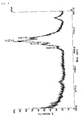
result of TOF-MS of this fraction is shown in Fig. 5. No peaks were
detected near the theoretical mass of 3461 corresponding to the
peptide fragment portion alone, and peaks were detected near 4500
to 6900. The shift in mass corresponds to the added sugar chains,
and the observed multiple peaks suggests that the heterogeneity of
the sugar chain was detected. Furthermore, using this fraction,
information relating to sugar chains can be obtained by combining
mass spectrometry and glycosidase digestion.
Humanized antibodies except humanized PTHrP antibody, i.e.,
humanized anti-HM1.24 antibody and humanized anti-TF antibody, were
analyzed according to the method of this invention to obtain their
chromatograms. Humanized anti-HM1.24 antibody and humanized anti-TF
antibody were produced according to WO98/1458 and WO99/51743,
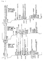
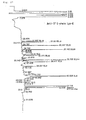
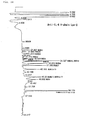
respectively. Comparison of the chromatograms of the L-chains and
H-chains of each antibody are shown in Fig. 6 and Fig. 7, respectively.
Peaks at the same detection positions with similar appearance on the
chromatograms were connected with straight lines.
In Fig. 6, several peaks of humanized anti-HM1.24 antibody and
humanized anti-TF antibody could be connected with straight lines
but none of the humanized PTHrP antibody. According to the predicted
nucleotide sequence of cDNA, the type of L-chain is known to be the
same, κ-chain, for humanized anti-HM1.24 antibody and humanized
anti-TF antibody, and different, λ-chain, for humanized PTHrP
antibody. Furthermore, according to the analysis results of
fractions from humanized anti-TF antibody, all peaks that could be
connected with straight lines were confirmed to correspond to
fragments derived from the constant region. In the figure, peaks
marked with stars ( ) correspond to fragments of the constant region.
These findings revealed that, when antibodies of the same type are
analyzed, fragments derived from the constant region are detected
in a similar manner, thus showing similar chromatograms; the
chromatogram of a different type of antibody will be different.
In Fig. 7, many peaks for the humanized PTHrP antibody and
humanized anti-HM1.24 antibody could be connected with straight lines,
but only two lines could be drawn to the peaks for the humanized anti-TF
antibody. According to the predicted nucleotide sequence of cDNA,
the subclass of H-chain is γ1 for humanized PTHrP antibody and
humanized anti-HM1.24 antibody, and γ4 for humanized anti-TF antibody.
Furthermore, according to the analysis results of fractions from
humanized PTHrP antibody, all peaks that could be connected with
straight lines were confirmed to correspond to fragments derived from
the constant region. Analyses of humanized anti-TF antibody
fractions have confirmed that peaks that could be connected to those
of the humanized anti-TF antibody correspond to fragments derived
from a segment that has the same sequence in the constant regions,
γ1 and 4. In a similar manner to the analysis of the type of L-chains,
this revealed that similar chromatograms are obtained by the same
subclass and different chromatograms by different subclasses.
Conversely, comparison of chromatograms allows prediction of
the subclass and type of an antibody. It has been known that
unidentified antibodies, and even a mixture of antibodies, can be
identified by comparing chromatograms.
When the subclass and type of an antibody are identified, the
peaks derived from the variable region can be specified. Thus,
efficient structural analysis of an antibody can be conducted by
preferentially analyzing those peaks.
50 µg of samples were used in this examination. However, for
the confirmation of a sequence, 20 µg or so is sufficient, and
comparison of peptide maps is possible even with 10 µg. With the use
of microbore HPLC and such, analysis may be conducted with even smaller
quantities of samples. Compared to conventional methods, which
require several hundred micrograms of samples, analysis can be
performed using an extremely small amount of sample. The present
method technically enables preparation of samples with an even smaller
amount, and the minimum amount necessary at the start of the experiment
depends on the sensitivity of analyzer (sequencer, mass spectrometer,
etc.) used for identification. By using a highly sensitive analyzer,
much smaller quantities of samples can be analyzed and identified.
The present method enables simultaneous and easy separation and
purification of H-chains and L-chains that was difficult according
to conventional methods. Enzymatic digestion in gel, fragment
recovery from the gel, and sequence analysis of the recovered
fragments were shown to be possible under the present conditions.
Since most of the sequence can be identified from the recovered
fragments, the method was found out to be useable to identify the
amino acid sequence of antibodies. Furthermore, the recovery rate,
which was generally believed to be the problem in the in-gel digestion
until now, was good with a value of 30% to 60% or so. The
reproducibility of the results was also good, and the present method
that utilizes the in-gel digestion can be used as a peptide mapping
system for antibodies.
With the exception that (1) endoproteinase Asp-N was used as
the digestive enzyme, and (2) 50 mM sodium phosphate buffer (pH 8.0)
was used as the digestion buffer, the following three types of
antibodies were analyzed under the same conditions as described in
Example 1. Humanized anti-IL6-R antibody was produced according to
WO92/19759. Asp-N is an endoproteinase that cleaves the N-terminal
side of aspartic acid.
Humanized anti-PTHrP antibody
Humanized anti-TF antibody
Humanized anti-IL6-R antibody
The results of analysis on the digest mixture of the H-chains and L-chains of humanized anti-PTHrP antibody are shown in Fig. 9 (L-chain) and Fig. 10 (H-chain). The fragment numbers and theoretical mass (in parenthesis) are indicated at the peak tops for the assigned peaks. For the L-chain, 5 out of 10 fragments that were predicted from the amino acid sequence had been detected. On the other hand, for the H-chain, 10 out of 17 fragments that were predicted from the amino acid sequence had been detected. The fragments predicted from the amino acid sequence of the L-chain (SEQ ID NO: 1) are shown in Table 4, and the fragments predicted from the amino acid sequence of the H-chain (SEQ ID NO: 2) are shown in Table 5. The tables indicate from the left, fragment number (CLn), amino acid sequence of the fragment, and the theoretical mass. For both of the L-chain and H-chain, the mass of the detected fragments were nearly in accordance with the theoretical mass.
The other five undetected fragments of the L-chain had a mass
of 1000 or less, and were regarded as undetected because their peaks
were lost in the noise of the low molecular weight region or in the
peaks corresponding to the extracts from the reagents and gel, and
consequently their assignments were unclear. In the H-chain, for the
same reason, fragments having a mass of 1000 or less were regarded
as undetected.
| CLD6 | 86-88 | DEA | 333.2983 |
| CLD4 | 60-63 | DGIP | 400.4321 |
| CLD2 | 49-52 | DLKQ | 502.5684 |
| CLD3 | 53-59 | DGSHSTG | 659.6106 |
| CLD7 | 89-95 | DYIBGVG | 783.8574 |
| CLD9 | 147-159 | DFYPGAVTVAWKA | 1424.6195 |
| CLD5 | 64-85 | DRFSGSSSGAERYLTISSLQSE | 2377.5076 |
| CLD1 | 1-48 | XLVLTQSPSASASLGASVKLTBTLS SQHSTYTIEWYQQQPEKGPKYLM | 5329.0092 |
| CLD8 | 96-146 | DTIKEQFVYVFGGGTKLTVLGQPKA | 5510.3502 |
| variable←→constant | |||
| NPTVTLFPPSSEELQANKATLVBLIS | |||
| CLD10 | 160-221 | DGSPVKAGVETTKPSKQSNNKYAAS | 6820.4176 |
| SYLSLTPEQWKSHRSYSBQVTHEGS TVEKTVAPTEBS |
| CHD15 | 400-401 | DS | 220.1822 |
| CHD9 | 266-270 | DVSHE | 585.5715 |
| CHD6 | 213-221 | DKKVEPKSB | 1091.2510 |
| CHD2 | 62-72 | DSVKGRFTISR | 1265.4356 |
| CHD10 | 271-280 | DPEVKFNWYV | 1296.4453 |
| CHD16 | 402-413 | DGSFFLYSKLTV | 1376.5724 |
| CHD8 | 250-265 | DTLMISRTPEVTBVVV | 1821.1469 |
| CHD3 | 73-89 | DNSKNTLYLQMNSLRAE | 1997.2173 |
| CHD13 | 357-376 | DELTKNQVSLTBLVKGFYPS | 2300.6156 |
| CHD14 | 377-399 | DIAVEWESNGQPENNYKTTPPVL | 2601.8101 |
| CHD7 | 222-249 | DKTHTBPPBPAPELLGGPSVFLFPP KPK | 3090.6154 |
| CHD11 | 281-312 | DGVEVHNAKTKPREEQYNSTYRVVS VLTVLHQ | 3698.1118 |
| CHD17 | 414-447 | DKSRWQQGNVFSBSVMHEALHNHYT QKSLSLSPG | 3931.3429 |
| CHD12 | 313-356 | DWLNGKEYKBKVSNKALPAPIEKTI SKAKGQPREPQVYTLPPSR | 5025.8094 |
| CHD4 | 90-148 | DTAVYYBARQTTMTYFAYWGQGTL VTVSSASTKGPSVFPLAPSSKSTSG | 6202.9944 |
| variable←→ constant | |||
| GTAALGBLVK | |||
| CHD1 | 1-61 | XVQLVESGGGWQPGRSLRLSBAAS GFTFSSYGMSWVRQAPGKGLEWVAT | 6461.2157 |
| ISSGGSYTYYP | |||
| CHD5 | 149-212 | DYFPEPVTVSWNSGALTSGVHTFPA VLQSSGLYSLSSWTVPSSSLGTQT | 6816.5775 |
| YIBNVNHKPSNTKV |
Next, analyses of the digest mixtures were performed by RP-HPLC
for humanized anti-PTHrP antibody and humanized anti-IL6-R antibody.
The chromatograms obtained by subjecting the digest mixture
solutions to RP-HPLC for fractionation are shown in Figs. 11 and 12.
Fig. 11 is the RP-HPLC chromatogram of the Asp-N digest mixture of
the L-chain of humanized anti-PTHrP antibody. Fig. 12 is the RP-HPLC
chromatogram of the Asp-N digest mixture of the L-chain of humanized
anti-IL6-R antibody.
The assigned fragment numbers determined by examining the amino
acid sequences of the fractions with a sequencer is shown at the peak
tops. The number of residues that can be analyzed with a sequencer
is limited. Therefore, whether a long fragment that could not be read
through the end has the full length of the predicted sequence was
determined in combination with the results of examination on the
fraction by TOF-MS.
For the L-chain of humanized anti-PTHrP antibody (Fig. 11), 7
out of 10 fragments from the predicted sequence were assigned. For
the L-chain of humanized anti-IL6-R antibody (Fig. 12), 8 out of 10
fragments from the predicted sequence were assigned. Fragments
cleaved at the N-terminus of Glu(E) besides Asp(D) were detected as
side-reaction products. The substrate specificity of Asp-N used for
digestion is incomplete. Thus, formation of such side-reaction
products seemed to be inevitable. Fragments predicted from the amino
acid sequence of the L-chain of humanized anti-IL6-R antibody are
shown in Table 6. The table indicates from the left, fragment number,
amino acid sequence of the fragment, and the theoretical mass.
| CLD6 | 86-88 | DEA | 333.2983 |
| CLD4 | 60-63 | DGIP | 400.4321 |
| CLD2 | 49-52 | DLKQ | 502.5684 |
| CLD3 | 53-59 | DGSHSTG | 659.6106 |
| CLD7 | 89-95 | DYIBGVG | 783.8574 |
| CLD9 | 147-159 | DFYPGAVTVAWKA | 1424.6195 |
| CLD5 | 64-85 | DRFSGSSSGAERYLTISSLQSE | 2377.5076 |
| CLD1 | 1-48 | XLVLTQSPSASASLGASVKLTBTLS SQHSTYTIEWYQQQPEKGPKYLM | 5329.0092 |
| CLD8 | 96-146 | DTIKEQFVYVFGGGTKLTVLGQPKA | 5510.3502 |
| variable ←→ constant | |||
| NPTVTLFPPSSEELQANKATLVBLIS | |||
| CLD10 | 160-221 | DGSPVKAGVETTKPSKQSNNKYAAS SYLSLTPEQWKSHRSYSBQVTHEGS TVEKTVAPTEBS | 6820.4176 |
Furthermore, the results of comparison of RP-HPLC chromatograms
among the respective H-chains and L-chains of three samples, i.e.,
humanized anti-PTHrP antibody, humanized anti-TF antibody, and
humanized anti-IL6-R antibody, are shown in Fig. 13 (L-chains) and
Fig. 14 (H-chains). Peaks at the same detection positions with
similar detection patterns were connected with straight lines. In
Fig. 12 (L-chains), fragments that could be identified as being
derived from the constant region were indicated with stars ( ).
Among them, side-reaction products were marked with black upside-down
triangles (▾). The results revealed that the same side-reaction
products are similarly detected for antibodies of the same type. This
shows that information on side-reaction products is also useful for
identifying the type of antibodies.
Regarding the H-chain, many straight lines could be drawn
between the peaks of humanized anti-PTHrP antibody (Fig. 14, top)
and humanized anti-IL6-R antibody (Fig. 14, middle), which antibodies
'belong to the same subclass (γ1). However, only one line could be
drawn from the peaks of these antibodies to that of the humanized
anti-TF antibody (Fig. 14, bottom). Analysis of fractions using a
sequencer confirmed that this peak corresponds to a fragment derived
from the portion having a sequence that is the same in both constant
regions, γ1 and γ4. Similarly to the case with Lys-C, when digested
with Asp-N, the subclass and type of antibody can be predicted by
the comparison of chromatograms.
Using the L-chain of humanized anti-PTHrP antibody, the amount
of sample necessary for the method for analyzing an antibody molecule
of the present invention was examined. Under the same conditions as
in Example 1, a mixture obtained by digesting the L-chain of humanized
anti-PTHrP antibody with Lys-C was analyzed by RP-HPLC.
The comparison of RP-HPLC chromatograms, wherein the amounts
of L-chain at the start of the experiments were 30, 10, 5, 2, and
1 µg, is shown in Fig. 15. Although the sizes of the peaks changed
depending on the amount of sample, the overall shape of the
chromatogram did not greatly change. Since the peaks at around 24
minutes, 48 minutes, and 49 minutes had approximately the same size
regardless of the amount of sample, these seemed to be contaminants
during the process of the analysis procedure. These peaks were not
confirmed from the sequencer, and thus were regarded to be other than
proteins.
The above-mentioned results confirmed that each fragment can
be recovered nearly quantitatively with an amount of sample within
the range that were tested in this experiment. Therefore, according
to the present invention, even if the amount of sample varies,
identification of the subclass and type based on comparison of the
chromatograms was considered to be possible. Furthermore, even with
an amount of sample of 1 µg, those samples that could be detected
as peaks could be fractionated. Furthermore, the amino acid sequence
of the fractionated samples could be determined with a sequencer.
Therefore, the method for analyzing an antibody molecule based on
the present invention was shown to be an excellent analysis method
that can be carried out with a small amount of sample of even 1 µg.
Furthermore, the same analysis was performed 5 times on
different days and the daily variation of the analysis result was
examined. As a result, a high reproducibility was confirmed for the
method of this invention.
Under the same conditions as in Example 1, the following three
types of antibodies were digested with Lys-C, and utilizing TOF-MS
and RP-HPLC, the analysis method based on this invention was performed.
The results are shown in the following figures. In the figures, the
peaks were marked with fragment numbers at the peak tops.
| TOF-MS/ H-chain | TOF-MS/ L-chain | RP-HPLC/ H-chain | RP-HPLC/ L-chain | |
| Humanized anti-HM1.24 antibody | Fig. 16 | Fig. 17 | Fig. 24 | Fig. 25 |
| Humanized anti-TF antibody | Fig. 18 | Fig. 19 | Fig. 26 | Fig. 27 |
| Humanized anti-IL-6R antibody | Fig. 20 | Fig. 21 | Fig. 28 | Fig. 29 |
Furthermore, the results of comparison of the chromatograms of
TOF-MS for the respective H-chains and L-chains of 4 samples,
humanized anti-PTHrP antibody obtained previously in Example 1, and
the above-mentioned humanized anti-HM1.24 antibody, humanized
anti-TF antibody, and humanized anti-IL6-R antibody are shown in Fig.
22 (L-chains) and Fig. 23 (H-chains). Peaks at the same detection
positions with similar detection patterns were connected with a
straight line. The peaks were marked with stars ( ) at their peak
tops.
By TOF-MS of the digest mixture, fragments having a mass of about
1000 or less were undetected because their peaks were lost in the
noise of the low molecular weight region or in the peaks corresponding
to the extracts of reagents and gel, and consequently assignments
were unclear. However, most fragments having a mass of about 1000
or more had been detected. The existence of those peaks that were
not detected could be confirmed by analyzing the fractions with a
sequencer. Therefore, these fragments were regarded as being
difficult to be detected due to their low ionization efficiency. When
the spectra of each chain were aligned and compared, the same kinds
of antibodies showed similar spectra (Figs. 22 and 23). Therefore,
the subclass or type of an antibody can be also identified from TOF-MS
spectra. Specifically, TOF-MS measurements of the digest mixture can
be utilized for typing of antibodies besides confirmation of the
progress of a reaction and mass mapping. Examination of the RP-HPLC
fractionated fractions by a sequencer and TOF-MS revealed that most
of the fragments of each chain of each antibody were recovered.
Fragments that are predicted to be generated by the digestion
with Lys-C based on the amino acid sequences of the L-chains and
H-chains of the above-mentioned humanized anti-HM1.24 antibody,
humanized anti-TF antibody, and humanized anti-IL6-R antibody are
listed in Tables 7 to 12. The tables show from the left, the fragment
number, amino acid number, amino acid sequence of the fragment, and
the theoretical mass. Furthermore, in the tables, fragments to which
the peaks could be assigned in the TOF-MS spectra (Figs. 16 to 21)
were marked with circles (○). Furthermore, fragments in which the
assignments could be confirmed by analyzing the fractions by RP-HPLC
(Figs. 24 to 29) were marked with stars ( ).

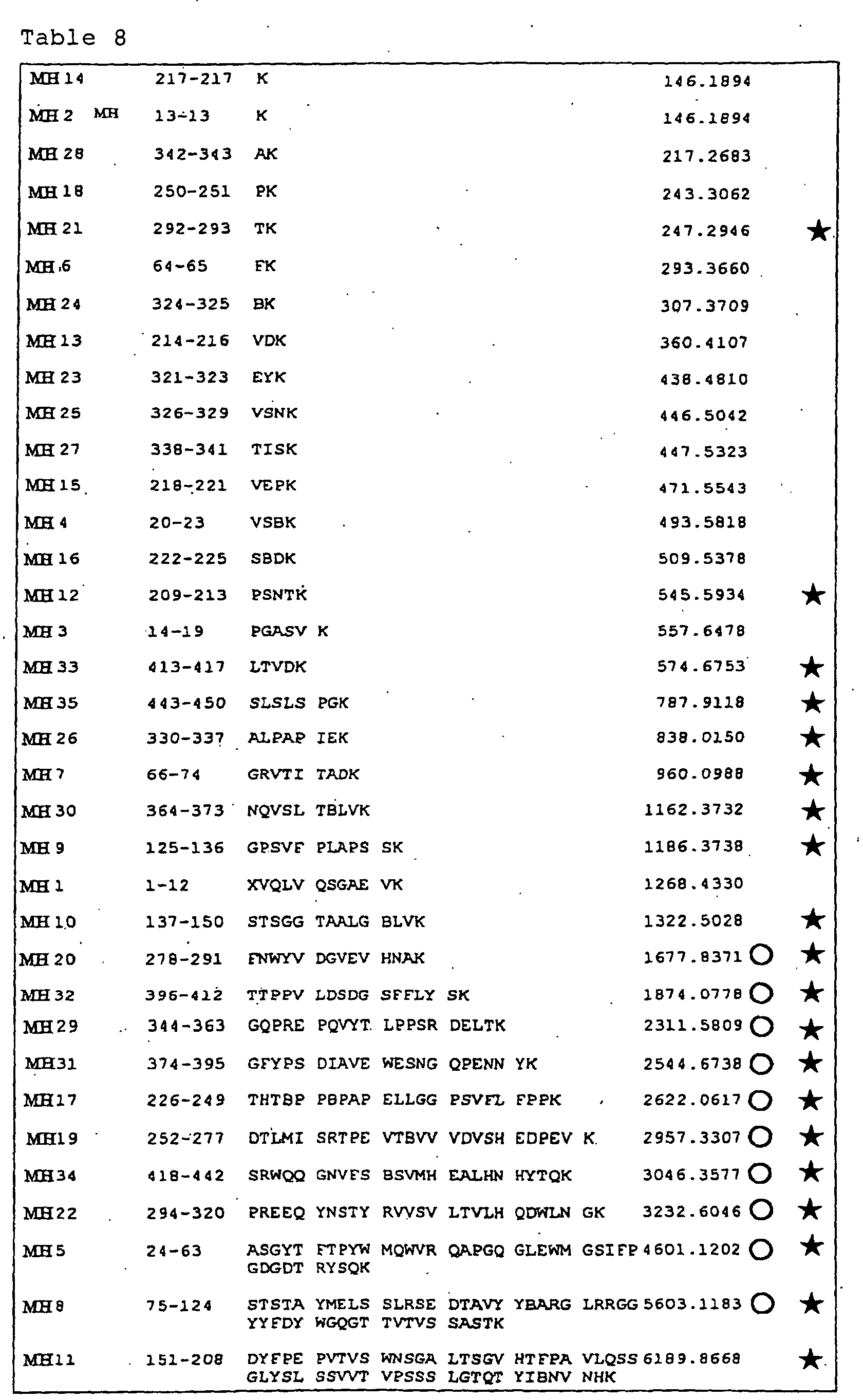
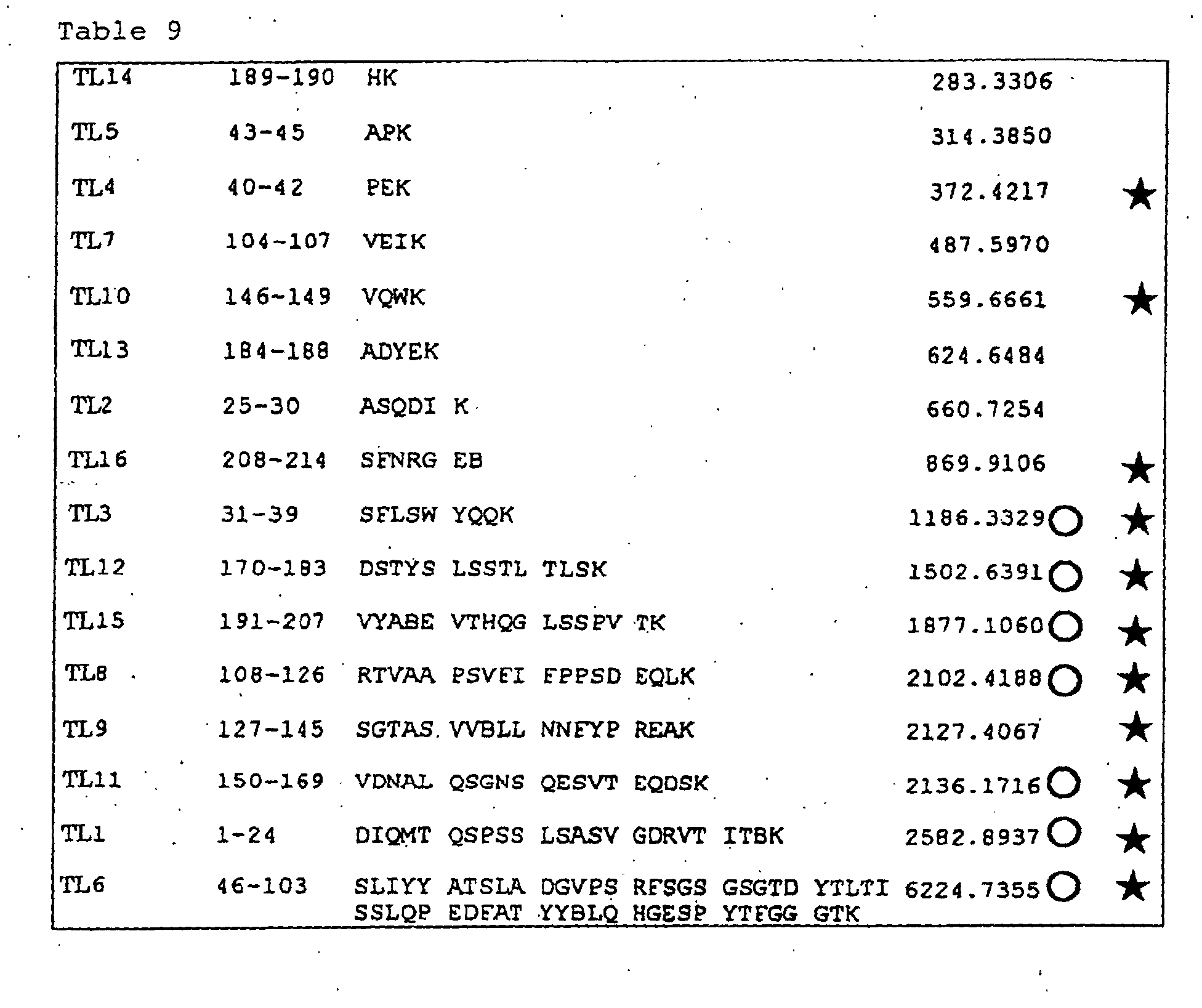
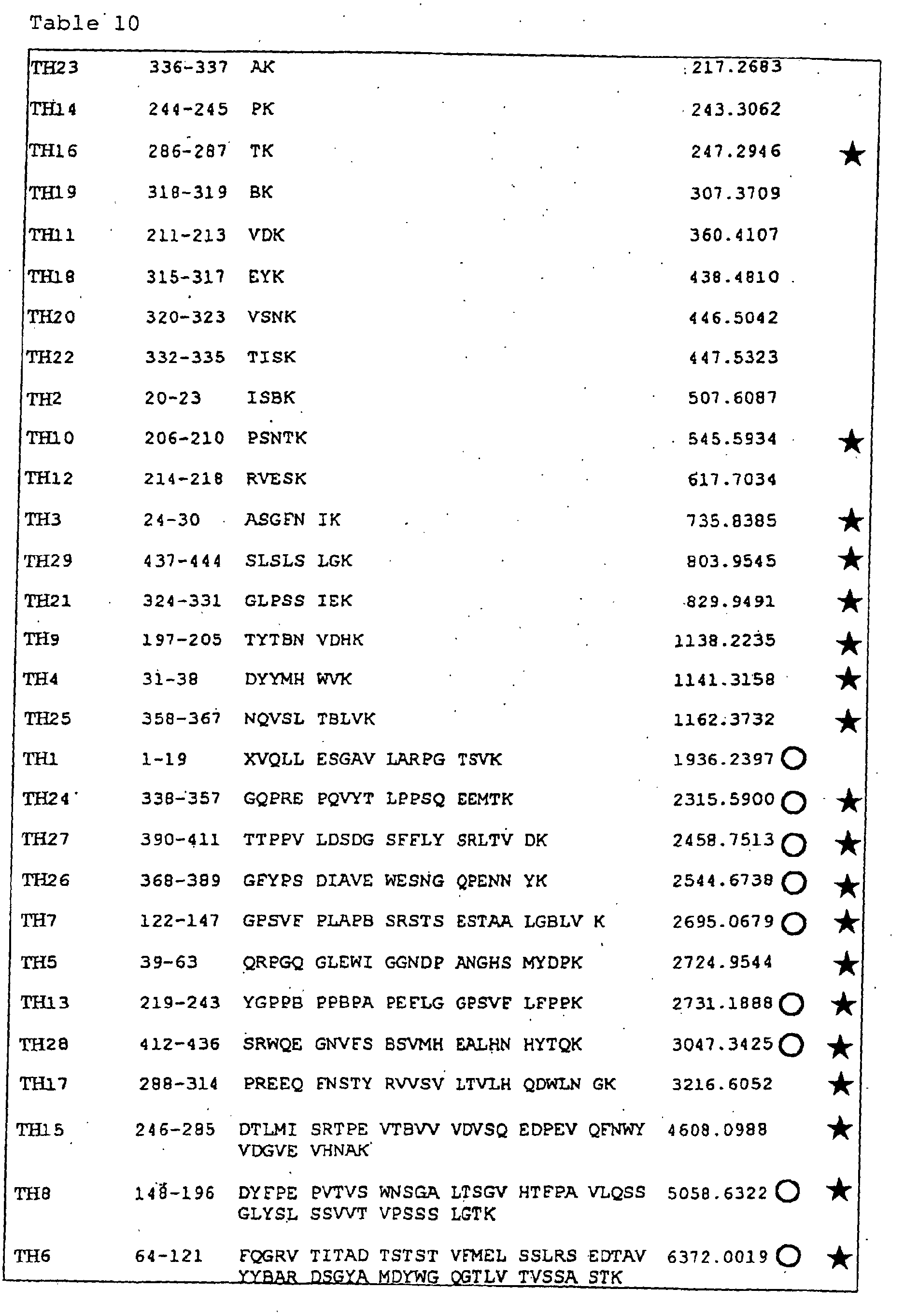
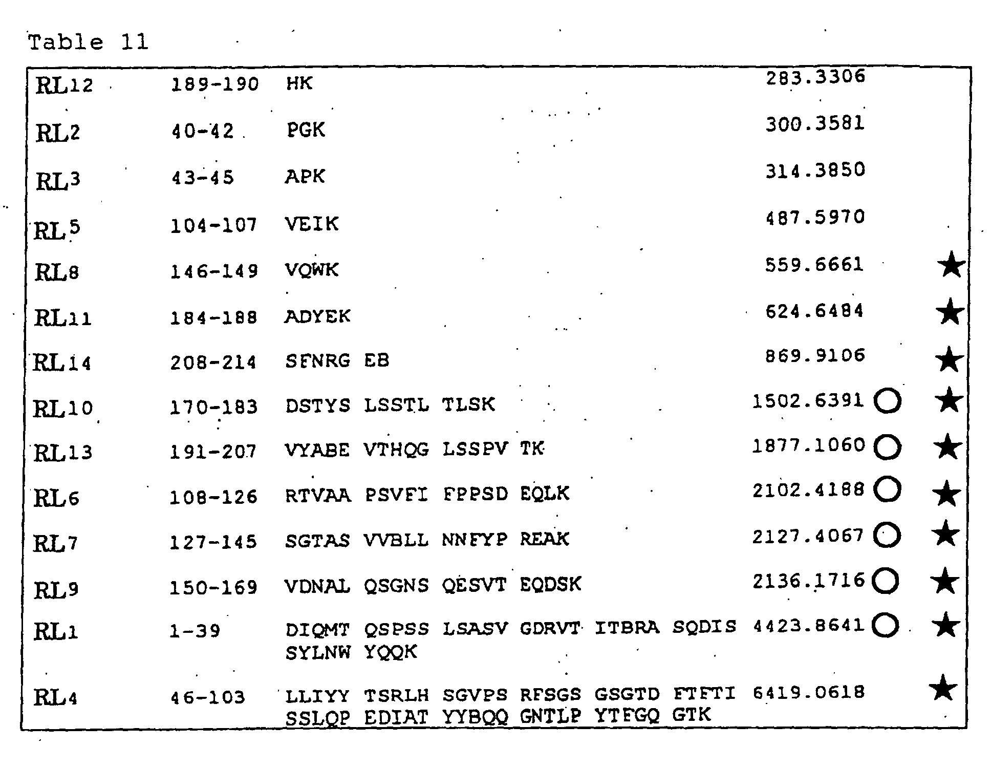

| L-chain | H-chain | |
| Humanized anti-HM1.24 antibody | Table 7 | Table 8 |
| Humanized anti-TF antibody | Table 9 | Table 10 |
| Humanized anti-IL-6R antibody | Table 11 | Table 12 |
The H-chain of humanized anti-PTHrP antibody was digested with
Asp-N under the same conditions as in Example 2, and was analyzed
by RP-HPLC. The result of analysis (chromatogram) is shown in Fig.
30.
According to the present invention, peptide fragments of a
fragmented antibody molecule can be analyzed accurately in terms of
both the kind and amount. Consequently, peptide maps with a good
reproducibility can be prepared, which allows analysis of
heterogeneity and evaluation of the quality of the antibody molecule
to be more conveniently and more quickly carried out. Furthermore,
by comparing the peptide maps obtained according to the present
invention, identification of the subclass of structurally
unidentified antibodies, and identification of the variable region
can be easily carried out.
Additionally, according to the method for analyzing antibody
molecules of the present invention, accurate analysis of an antibody
molecule can be performed with a small amount of antibody sample.
The high reproducibility of the analysis method of the present
invention allows analysis with a small amount of sample.
Furthermore, according to the present invention,
reproducibility could be also confirmed for the side-reaction
products in the digest mixture, indicating that, not only
theoretically predicted reaction products but also side-reaction
products that are theoretically difficult to predict can be detected
with a high reproducibility according to the analysis method of the
present invention. This means that peptide maps of the present
invention allow more precise and more accurate characterization of
antibody molecules.


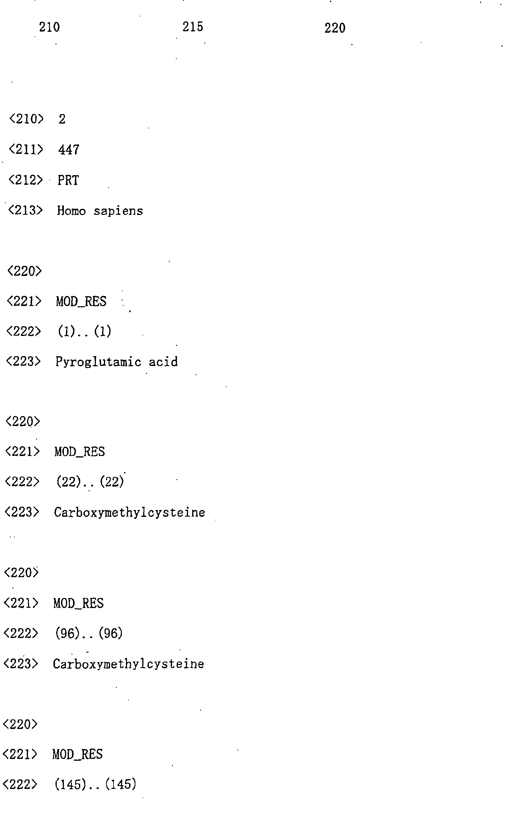
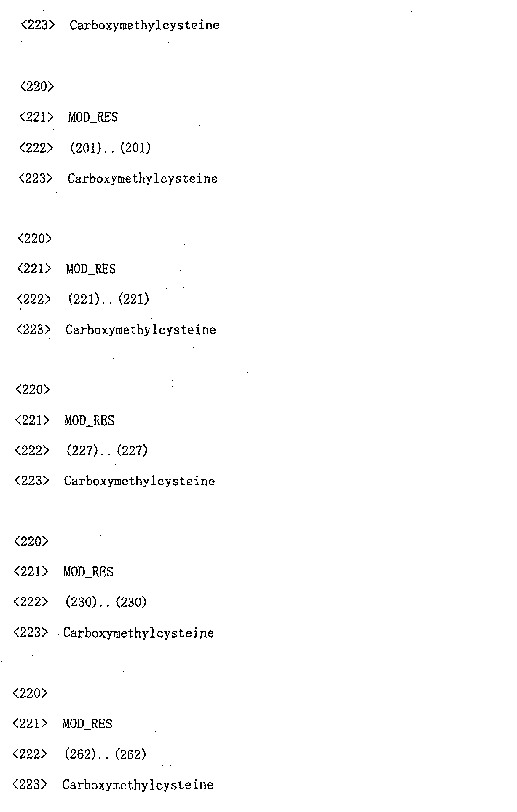

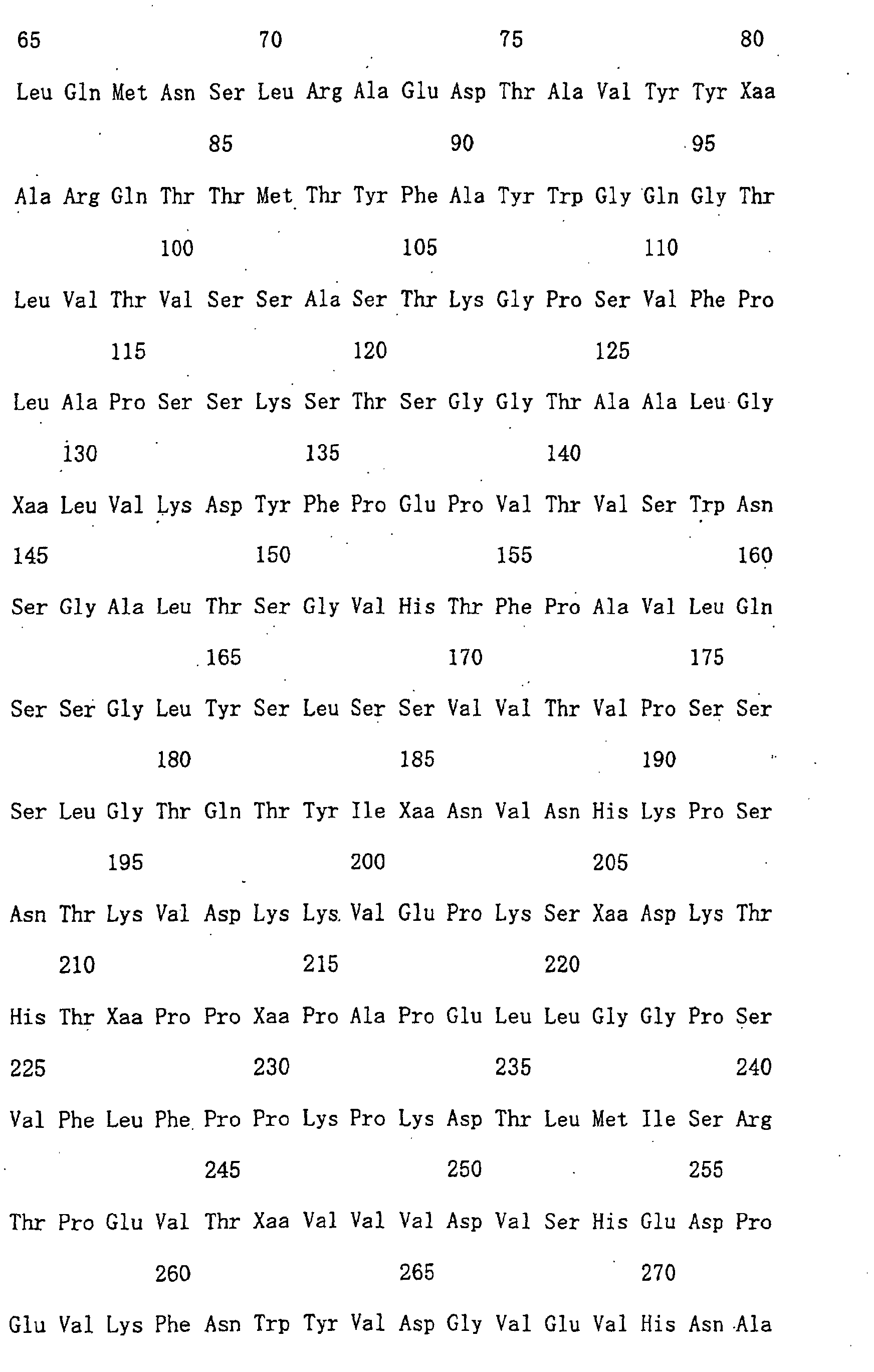
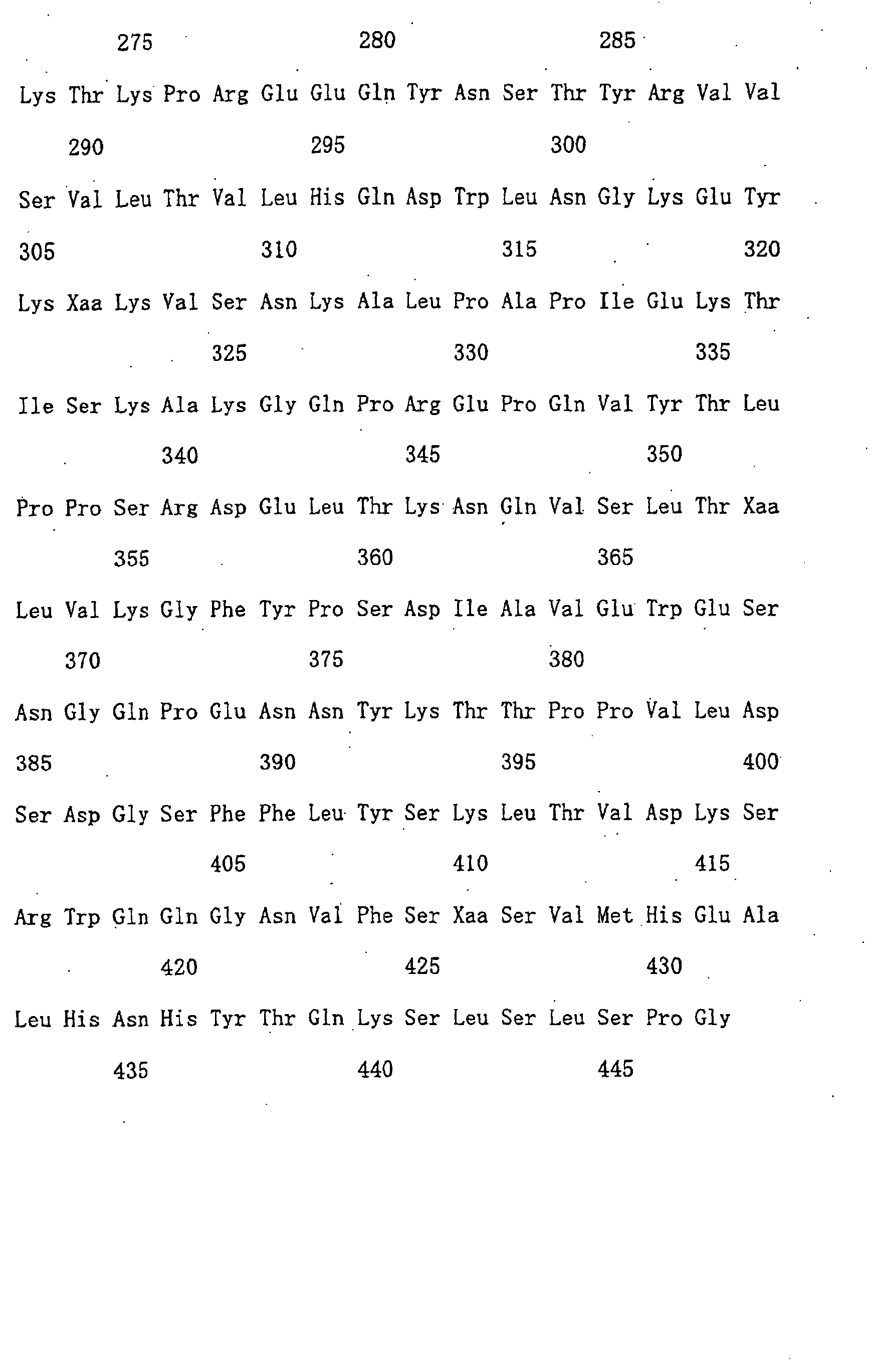
Claims (19)
- A method for structural analysis of an antibody molecule, which comprises the steps of:a) separating the antibody molecule into H-chains and L-chains;b) fragmenting the separated H-chains and L-chains; andc) analyzing the peptide fragments obtained by fragmentation.
- The method of claim 1, wherein the antibody molecule is separated into H-chains and L-chains by denaturing gel electrophoresis.
- The method of claim 1, wherein disulfide bonds are cleaved by reduction prior to separating the antibody molecule into H-chains and L-chains.
- The method of claim 3, wherein the thiol groups formed by reducing the disulfide bonds of the antibody molecule are made irreversible by modification.
- The method of claim 2, wherein the separated H-chains and L-chains are fragmented in gel.
- The method of claim 5 which comprises the step of eluting the fragmented peptides from the gel.
- The method of claim 2, wherein the separated H-chains and L-chains are transferred to a protein adsorption membrane, and then are fragmented on the protein adsorption membrane.
- The method of claim 1 comprising the step of separating the peptide fragments obtained by fragmentation.
- The method of claim 8 additionally comprising the step of deblocking peptide fragments whose N-termini are blocked.
- The method of claim 1, wherein the analysis of peptide fragments is performed by amino acid sequence analysis.
- The method of claim 1, wherein the analysis of peptide fragments is performed by mass spectrometry.
- The method of claim 1, wherein the analysis of peptide fragments is performed by amino acid composition analysis.
- A peptide map which is produced based on results of structural analysis obtainable by the method of claim 1.
- The method of claim 1 wherein the type of heterogeneity is determined by analyzing peptide fragments involved in heterogenity among the peptide fragments obtained by separation.
- The method of claim 1, wherein the antibody is an antibody for pharmaceutical use.
- A method of evaluating the quality of an antibody molecule, which comprises the steps of:a) separating the antibody molecule into H-chains and L-chains;b) fragmenting the separated H-chains and L-chains;c) analyzing the peptide fragments obtained by fragmentation;d) comparing the obtained analysis result with an analysis result obtained by analyzing a structurally identified antibody; ande) determining that the two antibodies are the same when there is no difference between the two analysis results, or when the difference is within a specified range.
- A method for identifying the subclass of'the H-chain and/or the type of the L-chain of a structurally unidentified antibody by analyzing the structures of a structurally identified antibody and a structurally unidentified antibody, respectively, by the method of claim 1, and then comparing the analysis results relating to both constant regions.
- A method for analyzing the variable region of an antibody molecule, which comprises the steps of:a) analyzing the structure of the antibody molecule by the method of claim 1; andb) selecting information on the variable region by removing information on the constant region from the analyzed information of a) based on the analysis result of a structurally identified antibody.
- The method of claim 18, wherein the analysis method is reverse-phase liquid chromatography, and step b) involves selecting peaks corresponding to the variable region from peaks corresponding to the constant region.
Applications Claiming Priority (3)
| Application Number | Priority Date | Filing Date | Title |
|---|---|---|---|
| JP2000299438 | 2000-09-29 | ||
| JP2000299438 | 2000-09-29 | ||
| PCT/JP2001/008577 WO2002027328A1 (en) | 2000-09-29 | 2001-09-28 | Method of analyzing antibody molecule structure |
Publications (2)
| Publication Number | Publication Date |
|---|---|
| EP1329719A1 true EP1329719A1 (en) | 2003-07-23 |
| EP1329719A4 EP1329719A4 (en) | 2004-07-21 |
Family
ID=18781246
Family Applications (1)
| Application Number | Title | Priority Date | Filing Date |
|---|---|---|---|
| EP01972608A Withdrawn EP1329719A4 (en) | 2000-09-29 | 2001-09-28 | METHOD FOR ANALYZING THE MOLECULAR STRUCTURE OF AN ANTIBODY |
Country Status (4)
| Country | Link |
|---|---|
| EP (1) | EP1329719A4 (en) |
| JP (1) | JPWO2002027328A1 (en) |
| AU (1) | AU2001292312A1 (en) |
| WO (1) | WO2002027328A1 (en) |
Cited By (7)
| Publication number | Priority date | Publication date | Assignee | Title |
|---|---|---|---|---|
| WO2008077889A1 (en) * | 2006-12-22 | 2008-07-03 | Ares Trading S.A. | Analytical method for analyzing c-terminus truncation |
| US10955420B2 (en) | 2016-09-07 | 2021-03-23 | Mayo Foundation For Medical Education And Research | Identification and monitoring of cleaved immunoglobulins by molecular mass |
| US11209439B2 (en) | 2015-09-24 | 2021-12-28 | Mayo Foundation For Medical Education And Research | Identification of immunoglobulin free light chains by mass spectrometry |
| US11604196B2 (en) | 2014-04-04 | 2023-03-14 | Mayo Foundation For Medical Education And Research | Isotyping immunoglobulins using accurate molecular mass |
| US11946937B2 (en) | 2017-09-13 | 2024-04-02 | Mayo Foundation For Medical Education And Research | Identification and monitoring of apoptosis inhibitor of macrophage |
| US12153052B2 (en) | 2017-09-13 | 2024-11-26 | Mayo Foundation For Medical Education And Research | Identification and monitoring of immunoglobulin J chains |
| US12546782B2 (en) | 2013-03-15 | 2026-02-10 | Mayo Foundation For Medical Education And Research | Identification and monitoring of monoclonal immunoglobulins related to monoclonal gammopathy by molecular mass with mass spectrometry |
Families Citing this family (3)
| Publication number | Priority date | Publication date | Assignee | Title |
|---|---|---|---|---|
| JP2006078192A (en) * | 2004-09-07 | 2006-03-23 | Shimadzu Corp | Biopolymer mass spectrometry |
| WO2016147839A1 (en) * | 2015-03-17 | 2016-09-22 | アサヒビール株式会社 | Method for quantification of hydrophobins, and method for predicting gushing of sparkling malt beverage |
| EP4240747A1 (en) * | 2020-11-05 | 2023-09-13 | Amgen Inc. | Materials and methods for protein processing |
Family Cites Families (2)
| Publication number | Priority date | Publication date | Assignee | Title |
|---|---|---|---|---|
| JP2971842B2 (en) * | 1997-09-22 | 1999-11-08 | 株式会社荏原製作所 | Absorption heat pump and air conditioning system using the same as heat source |
| US6030792A (en) * | 1997-11-13 | 2000-02-29 | Pfizer Inc | Assays for measurement of protein fragments in biological media |
-
2001
- 2001-09-28 EP EP01972608A patent/EP1329719A4/en not_active Withdrawn
- 2001-09-28 WO PCT/JP2001/008577 patent/WO2002027328A1/en not_active Ceased
- 2001-09-28 JP JP2002530856A patent/JPWO2002027328A1/en active Pending
- 2001-09-28 AU AU2001292312A patent/AU2001292312A1/en not_active Abandoned
Cited By (8)
| Publication number | Priority date | Publication date | Assignee | Title |
|---|---|---|---|---|
| WO2008077889A1 (en) * | 2006-12-22 | 2008-07-03 | Ares Trading S.A. | Analytical method for analyzing c-terminus truncation |
| US12546782B2 (en) | 2013-03-15 | 2026-02-10 | Mayo Foundation For Medical Education And Research | Identification and monitoring of monoclonal immunoglobulins related to monoclonal gammopathy by molecular mass with mass spectrometry |
| US11604196B2 (en) | 2014-04-04 | 2023-03-14 | Mayo Foundation For Medical Education And Research | Isotyping immunoglobulins using accurate molecular mass |
| US12099065B2 (en) | 2014-04-04 | 2024-09-24 | Mayo Foundation For Medical Education And Research | Isotyping immunoglobulins using accurate molecular mass |
| US11209439B2 (en) | 2015-09-24 | 2021-12-28 | Mayo Foundation For Medical Education And Research | Identification of immunoglobulin free light chains by mass spectrometry |
| US10955420B2 (en) | 2016-09-07 | 2021-03-23 | Mayo Foundation For Medical Education And Research | Identification and monitoring of cleaved immunoglobulins by molecular mass |
| US11946937B2 (en) | 2017-09-13 | 2024-04-02 | Mayo Foundation For Medical Education And Research | Identification and monitoring of apoptosis inhibitor of macrophage |
| US12153052B2 (en) | 2017-09-13 | 2024-11-26 | Mayo Foundation For Medical Education And Research | Identification and monitoring of immunoglobulin J chains |
Also Published As
| Publication number | Publication date |
|---|---|
| JPWO2002027328A1 (en) | 2004-02-05 |
| AU2001292312A1 (en) | 2002-04-08 |
| EP1329719A4 (en) | 2004-07-21 |
| WO2002027328A1 (en) | 2002-04-04 |
Similar Documents
| Publication | Publication Date | Title |
|---|---|---|
| US6629040B1 (en) | Isotope distribution encoded tags for protein identification | |
| CN101600959B (en) | Quantitative methods for peptides and proteins | |
| Formolo et al. | Determination of the NISTmAb primary structure | |
| Ashcroft | Protein and peptide identification: the role of mass spectrometry in proteomics | |
| US10281473B2 (en) | Compositions and methods for analysis of protein sequences and post-translational modifications | |
| US20100015652A1 (en) | Antibody quantitation | |
| EP1397686B1 (en) | Method for characterizing polypeptides | |
| JP5468073B2 (en) | Protein quantification method | |
| WO2008053399A1 (en) | Compounds and methods for double labelling of polypeptides to allow multiplexing in mass spectrometric analysis | |
| US20080241862A1 (en) | Propionyl and butyryl lysine modifications in proteins | |
| Hoffert et al. | Taking aim at shotgun phosphoproteomics | |
| KR101070247B1 (en) | A new biomarker protein and a new detection method in the biological fluid with preeclampsia using proteomics technology | |
| JPWO2002027328A1 (en) | Structural analysis of antibody molecules | |
| CA2393726A1 (en) | Quantitative proteomics via isotopically differentiated derivatization | |
| US20040106150A1 (en) | Inverse labeling method for the rapid identification of marker/target proteins | |
| KR100611313B1 (en) | N-terminal substitution of polypeptide, amino acid sequencing and quantification method in polypeptide | |
| WO2015026909A2 (en) | Msia-srm assay for biomarker analysis | |
| CA2468875C (en) | Process for the selective alkylation of -sh groups in proteins and peptides for the study of complex protein mixtures | |
| US20230243841A1 (en) | Methods to prevent disulfide scrambling for ms-based proteomics | |
| Wu et al. | A hybrid LC–Gel-MS method for proteomics research and its application to protease functional pathway mapping | |
| US20060024660A1 (en) | Method for relative quantification of proteins | |
| KR20110121842A (en) | Peptide amino group substitution compound En-methyl piperazine acetic isomer of acetic acid and peptide quantification method using mass spectrometry | |
| US20250076307A1 (en) | Methods for characterizing a protein of interest | |
| JP2018193372A (en) | Method for producing and designing anti-peptide antibody | |
| Licker et al. | Characterisation of human cerebrospinal fluid (CSF) after tandem mass tag (tmt0) labelling |
Legal Events
| Date | Code | Title | Description |
|---|---|---|---|
| PUAI | Public reference made under article 153(3) epc to a published international application that has entered the european phase |
Free format text: ORIGINAL CODE: 0009012 |
|
| 17P | Request for examination filed |
Effective date: 20030429 |
|
| AK | Designated contracting states |
Designated state(s): AT BE CH CY DE DK ES FI FR GB GR IE IT LI LU MC NL PT SE TR |
|
| AX | Request for extension of the european patent |
Extension state: AL LT LV MK RO SI |
|
| XX | Miscellaneous (additional remarks) |
Free format text: THE APPLICANT HAS REQUESTED SEVERAL CORRECTIONS IN THE DESCRIPTION. |
|
| A4 | Supplementary search report drawn up and despatched |
Effective date: 20040604 |
|
| STAA | Information on the status of an ep patent application or granted ep patent |
Free format text: STATUS: THE APPLICATION HAS BEEN WITHDRAWN |
|
| 18W | Application withdrawn |
Effective date: 20070104 |