Background of the Invention
Field of the Invention
The present invention relates to a method for
analyzing a biomolecule. More precisely, the present
invention relates to a technique that enables convenient
and efficient detection of a biomolecule immobilized on
a substrate such as a glass substrate.
Description of the Related Art
In conventional techniques for analyzing a
biomolecule such as nucleic acids and proteins using a
two-dimensional signal, including electrophoresis and
Southern blotting, the following two kinds of methods
are mainly used as a method for detecting the
biomolecule (International Patent Publication In
Japanese (Kohyo) No, 10-503841, W097/10365 etc.):
However, as for the method of (1) among the
aforementioned methods, although it shows superior
detection sensitivity, an extremely expensive
fluorescence image analyzer or the like must be
purchased, and in addition, it has drawbacks that, for
example, the fluorescent substance is readily degraded
by irradiation of light such as visible lights and
ultraviolet rays. Further, although the method of (2)
does not require purchase of an expensive detector,
drawbacks thereof have been pointed out, for example,
detection results obtained by the method are
insufficient for quantitative determination, because the
detection is mainly performed by visual inspection.
Summary of the Invention
An object of the present invention is to provide a
method for performing analysis of a biomolecule based on
detection of a two-dimensional signal in a simple and
quantitative manner.
The inventors of the present invention found that,
if a two-dimensional signal of a biomolecule is detected
by using an image scanner, an expensive apparatus is not
needed, and the biomolecule could be quantitatively
analyzed with sufficient reproducibility, and thus
accomplished the present invention.
That is, the present invention provides the
followings.
In the present invention, a two-dimensional signal
means a signal of a biomolecule two-dimensionally
separated or disposed on a solid phase. Typically, the
two-dimensional signal includes a signal of an
individual biomolecule localized on a solid phase and a
background signal.
According to the present invention, a biomolecule
can be detected with sufficient sensitivity by using an
inexpensive commercially available scanner, and further,
the detected image can be stored in a personal computer
or the like and easily analyzed. Thus, it can be seen
that the method of the present invention is a simple
method showing superior cost performance.
Furthermore, since a commercially available
scanner can be used, the method of the present invention
can be a method for detecting a biomolecule excellent in
reproducibility and quantitative determination ability.
which is effective in use for DNA chips and so forth.
Brief explanation of the Drawings
Fig. 1 shows enlarged images of areas of DNA-immobilized
slide glass on which DNA was spotted.
Preferred Embodiments of the Invention
Hereafter, the present invention will be explained
in detail.
In the method of the present invention, a
biomolecule is localized on a solid phase, and the
biomoleoule is detected as a visible two-dimensional
signal. The method used for this operation is not
particularly limited, so long as a two-dimensional
signal of the biomolecule can be formed on a solid phase,
and various methods conventionally used for the analyses
of biomolecules such as nucleic acid detection methods
based on hybridization can be used. Further, even a
currently unknown method may be employed, so long as it
can be applied with the method of the present invention.
Hereafter, embodiments of the present invention
will be explained mainly for a method of detecting a
nucleic acid based on hybridization of nucleic acids
using a nucleic acid immobilized on a solid phase.
<1> Localization of biomolecule on solid phase
The solid phase used for the present invention is
used for localizing a biomolecule. Examples of the
biomolecule include a nucleic acid, protein, enzyme,
antigen, antibody, saccharide and so forth. The nucleic
acid may be, for example, a naturally occurring or
synthesized DNA (including an oligodeoxyribonucleotide)
or RNA (including an oligoribonucleotide). The nucleic
acid may be single-stranded or double-stranded.
Localization of a biomolecule on a solid phase can
be attained by separating the biomolecule in the solid
phase based on a physical or chemical characteristic by
means of electrophoresis or the like, or by artificially
disposing the biomolecule at a determined position on
the solid phase. To dispose the biomolecule on the
solid phase, there can be used, for example, a method of
dropping a solution containing the biomolecule on the
solid phase by using a pipette, dispenser, pin or the
like. Apparatuses for providing solutions in such small
amounts are commercially available, and they can be used
for the present invention. For example, if a spotter
used for the production of DNA chips or the like is used,
the biomolecule can be disposed on the solid phase at a
high density. Preferred size of spots for a DNA chip is
10 µm to 10 mm.
The biomolecule localized on the solid phase may
be immobilized on the solid phase by chemical or
physical binding, or immobilized by gel matrix or the
like.
Different kinds of biomolecules or the same kind
of biomelecule may be immobilized on multiple spots on
the substrate. When multiple kinds of biomolecules are
immobilized on one solid phase, their configuration and
so forth may be suitably selected depending on the types
of biomolecules, detection method, intended use and so
forth.
The biomolecule may be directly immobilized on a
substrate constituting the solid phase, or immobilized
via a ligand showing binding property to the biomolecule
(biomolecule-immobilizing reagent).
The substrate is not particularly limited so long
as it can immobilize a biomolecule and bear usual
conditions for analyses of biomolecules such as
hybridization. Specifically, there can be mentioned
those insoluble in solvents used for immobilization and
hybridization of nucleic acids etc. and being in the
form of solid or gel at an ordinary temperature or
within a temperature range around it (e.g., 0°C to
100°C). The expression that "a substrate is insoluble
in a solvent" means that the substrate is substantially
insoluble in various solvents including aqueous solvents
and organic solvents used in various process steps of
allowing the substrate to carry a ligand having a
property of binding to a nucleic acid, such as
carbodiimide group, then immobilizing the nucleic acid
on the solid phase and thereafter using the substrate as
a DNA chip as described later.
Specific examples of the material of such a
substrate as described above include plastics, inorganic
polymers, metals, naturally occurring polymers, ceramics
and so forth.
Specific examples of the plastics include
polyethylene, polystyrene, polycarbonate, polypropylene,
polyamide, phenol resin, epoxy resin, polycarbodiimide
resin, polyvinyl chloride, polyvinylidene fluoride,
polyethylene fluoride, polyimide, acrylic resin and so
forth.
Specific examples of the inorganic polymers
include glass, carbon, silica gel, graphite and so forth.
Specific examples of the metals include gold,
platinum, silver, copper, iron, aluminum, paramagnet,
apatite and so forth.
Examples of the naturally occurring polymers
include cellulose, chitin, chitosan, alginic acid and
derivatives thereof.
Specific examples of the ceramics include alumina,
silica, silicon carbide, silicon nitride, boron carbide
and so forth.
Further, examples of the aforementioned
biomolecule-immobilizing reagent include, for example,
nitrogen yperite, poly-L-lysine, polycarbodiimide,
membrane, cellulose nitrate and so forth.
Nitrogen yperite can be produced by the methods
disclosed in U.S. Patent Nos. 2,141,090, 5,273,991 and
5,387,707 and so forth. Further, cellulose nitrate,
poly-L-lysine and membrane can be produced by the
methods disclosed in J. Sambrook, E.F. Fritsch and T.
Maniatis, Molecular Cloning, Cold Spring Harbor
Laboratory Press, Second Edition, pages 2.109-2.113 and
pages 9.34-9.46, International Patent Publication in
Japanese (Kohyo) No. 10-503841 and so forth.
In the present invention, so long as the
biomolecule-immobilizing reagent can be carried on the
aforementioned substrate, it may be carried by simply
utilizing a physical adhesive property, or it may be
chemically carried via a covalent bond or the like.
Further, the biomolecule-immobilizing reagent may
be carried on the whole surface of the substrate or a
part thereof, as required. Further, when a biomolecule
is immobilized, it may be irradiated with an
electromagnetic wave such as an ultraviolet ray.
As the aforementioned biomolecule-immobilizing
reagent used for the production of a carrier comprising
a substrate carrying the biomolecule-immobilizing
reagent using physical adhesion, any compounds
comprising the biomolecule-immobilizing reagent bound to
a polymer compound can be used without any particular
limitation.
Such a biomolecule-immobilizing reagent binding
polymer compound shows high adhesion to the
aforementioned substrate, and it is carried by the
substrate by utilizing this adhesion. A typical form of
the biomolecule-immobilizing reagent binding polymer
compound carried on the substrate using physical
adhesion is a coated film.
The method for coating the aforementioned
biomolecule-immobllizing reagent binding polymer
compound on the substrate as a coated film may be a
known method, for example, spraying, immersing, brushing,
stamping, vapor deposition, coating by using a film
coater and so forth.
In the method of the present invention, the solid
phase is frequently brought into contact with
biomolecules other than the biomolecule immobilized on
the solid phase and so forth. Therefore, in order to
prevent such biomolecules other than the immobilized
biomolecule and so forth from non-specifically binding
to unreacted regent remaining on the substrate, active
sites are preferably blocked by bringing the solid phase
into contact with an excessive amount of bovine serum
albumin (BSA), casein, salmon sperm DNA or the like
after an objective biomolecule is immobilized on the
solid phase.
<2> Detection of the two-dimensional signal of
biomolecule
The biomolecule on the solid phase is detected as
a visible two-dimensional signal. Such a two-dimensional
signal is conventionally detected by visual
inspection or by using a fluorescence image analyzer or
the like, directly or after being recorded as a
photograph. On the other hand, in the present invention,
a two-dimensional signal of a biomolecule is detected by
capturing a two-dimensional image on the solid phase
with use of a scanner, and analyzing the obtained two-dimensional
image data. The aforementioned scanner is
an apparatus that has a light source for irradiating the
solid phase with a light and an image sensor for
receiving a reflected light from the solid phase and can
capture a two-dimensional image on the solid phase by
two-dimensionally scanning the solid phase. As such a
two-dimensional scanner, there can be mentioned an image
scanner used as a peripheral equipment of a computer,
and products marketed from various electrical equipment
manufacturers can be used. Further, such a scanner is
built in various apparatuses such as an
electrophotographic apparatus (copying machine) and
facsimile. If a TWAIN compliant apparatus is used as
the scanner, storage and analysis of data by a personal
computer become easy, and quantitative analysis also
becomes possible.
A signal of the biomolecule may be directly
detected by irradiating the solid phase with a light
from the aforementioned light source. Further, the
biomolecule may also be detected after it is visualized
by binding it with a labeling substance showing a
transmittance, refractive index or reflectance different
from that of the solid phase for a light irradiated from
the light source or a substance labeled with such a
labeling substance and specifically binding to the
biomolecule. The method for the visualization may be
suitably selected depending on the type of the
biomolecule.
For example, electrophoresis gel or a nucleic acid
or protein transferred from or the gel by using a filter
can be visualized by staining it with a suitable dye.
Further, a particular protein can be visualized by using
a protein such as an antibody labeled with a labeling
substance. Similarly, a particular nucleic acid can be
visualized by using a nucleic acid probe specifically
binding to the nucleic acid.
Although the labeling substance itself may be
visible, it may be invisible so long as it can be
visualized by a color development reaction. Examples of
such a labeling substance include enzymes such as
alkaline phosphatase and horseradish peroxidase. It is
also possible to use an antigen or antibody, or biotin
or avidin or the like as a labeling substance so that an
enzyme should specifically bind to the labeling
substance via a linkage of antigen-antibody, biotin-avidin
or the like.
Specific examples of the labeling substance
include color developing substrates, photochromic
compounds and so forth.
Examples of the aforementioned color developing
substrates include benzidines and so forth, and color
development can be obtained by using a combination of
digoxigenin or the like and an enzyme or the like.
Examples of the aforementioned photochromio
compounds include azobenzenes, spiropyrans, fulgides,
diarylethenes, stilbenes, phenoxynaphthacenequinone,
thioindigo, Malachite Green and so forth.
Since P-type photochromic compounds also take an
optically steady state to show color development at the
time of detection by irradiation of a light, they can
also be used for the detection of a biomolecule.
Furthermore, azo dyes such as azobenzene can be
colored by irradiation of an ultraviolet ray or the like
before the detection and detected.
As the method of labeling a biomolecule by using
such photochromic compounds, a functional group such as
succinimide ester group, isothiocyanate group,
carbodiimide group, isocyanate group, amino group,
halogen, nitrogen yperite or thiol group can be
introduced into a photochromic compound at an
appropriate position not interfering the coloration of
the photochromic compound and then it can be used for
labeling (reaction) of a biomolecule in water or buffer.
Further, when a coloring substance or a
photochromic compound is introduced into a nucleic acid,
it is also possible to introduce the coloring substance
or photochromic compound into an intercalator having an
appropriate linker and intercalate it in the nucleic
acid in water or buffer.
A two-dimensional image on the solid phase is
captured in the same manner as capture of a two-dimensional
image of an ordinary photograph, document or
the like by using an image scanner. When the solid
phase is placed on a scanning surface of the scanner,
the side of the solid phase on which the biomolecule is
immobilized is preferably faced the capturing surface.
Further, a spacer may be inserted between a portion of
the solid phase on which the biomolecule is not
immobilized and the capturing surface. Even when
flatness of the capturing surface or the solid phase is
not sufficient, use of such a spacer prevents formation
of small gaps and generation of moire (interference
pattern) formed by the small gaps, and thus an image can
be stably obtained. Further, the use of a spacer
prevents direct contact of a portion of the solid phase
on which the biomolecule is immobilized with the
capturing surface, and thus it is effective for
protection of the solid phase and prevention of fouling
of the scanner. The spacer preferably has a thickness
of about 100 to 200 µm.
The image capturing by a scanner is performed, for
example, with a resolution degree of about 600 dpi. The
captured two-dimensional image data are analyzed by
using a computer and appropriate image analysis software.
For example, the captured image data are stored as data
in monochromatic 256 gradation degrees in a memory
device of a computer. The data are expanded in a memory
as an image and indicated on a display. Grids are
automatically set by the software on the image expanded
on the memory, or grids are manually set on the image
indicated on the display. Then, filtering for upper or
lower limits is performed to eliminate background noise,
and subsequently the dot data in monochromatic 256
gradation degrees are once transformed into reflection
densities and compared with grid locations to integrate
them on each grid. Gamma control is performed for the
obtained integrated values for normalization and
conversion of them so that they have a linear signal
level corresponding to the color developing
characteristic and characteristics of the scanner and so
forth, and the results are calculated and outputted.
Based on the obtained results, locations, sizes and
densities of spots or bands can be measured.
The method for detecting a biomolecule of the
present invention does not require purchase of an
expensive detector, and if this method is used for
analysis, analysis with superior reproducibility and
quantitative determination ability is enabled. The
detection method of the present invention can be
suitably used for a DNA chip or the like used in
techniques for determining nucleotide sequences by
hybridization using a large number of biomolecules, such
as sequencing by hybridization (SBH) and sequencing by
hybridization with oligonucleotide matrix (SHOM).
Examples
Hereafter, the present invention will be explained
more specifically with reference of the following
examples.
Preparation Example: Preparation of polycarbodiimidated
slide glass
(1) Preparation of aminated slide glass
In an amount of 20 ml of 10%(v/v) solution of 3-aminopropyltriethoxysilane
in ethanol was added to 180
ml of distilled water and stirred. After 6 N HCl was
added to the solution to adjust pH of the solution to 3
to 4, 15 pieces of slide glass were immersed into the
solution and heated at 75°C for 2 hours. After the
heating was finished, the slide glass was pulled up from
the solution, and the solution was sufficiently washed
down with distilled water. Then, the slide glass was
subjected to a heat treatment at 115°C for 4 hours to
obtain aminated slide glass. The above procedure was
repeated to further obtain aminated slide glass.
(2) Preparation of carbodiimide resin
In an amount of 12.5 g of cyclohexyl isocyanate
(Tokyo Kasei Kogyo) and 1.3 g of 3-methyl-1-phenyl-2-phosphorene-1-oxide
(Aldrich) were added to 117.9 g of
4,4'-dicyclohexylmethanediisocyanate (Aldrich). Then,
the mixture was stirred at 185°C for 96 hours, while
nitrogen was added at a flow rate of 0.5 ml/minute.
After cooling, polycarbodiimide resin was obtained as
powder. The average polymerization degree of the
obtained polycarbodiimide resin was 10, and the number
average molecular weight was about 2400.
(3) Preparation of polycarbodiimidated slide glass
A 10% chloroform solution of the carbodiimide
resin prepared in the above (2) was prepared, and 15
pieces of the aminated slide glass prepared in the above
(1) were immersed in the solution and immediately pulled
up. Then, the slide glass was washed twice with 200 ml
of chloroform for 10 minutes, and dried at 40°C for 2
hours to obtain polycarbodiimidated slide glass. The
above procedure was repeated to further obtain
polycarbodiimidated slide glass.
Example 1
(1) Immobilization of nucleic acid
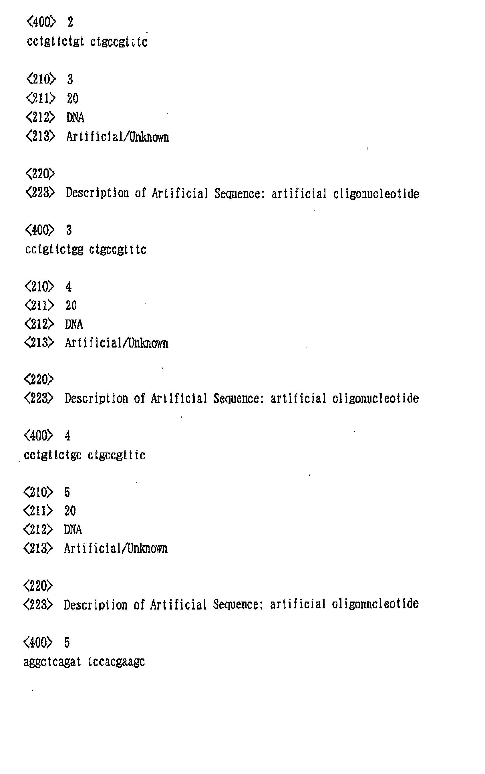
Oligonucleotides having each of the nucleotide
sequences of SEQ ID NOS: 1 to 5 were prepared. The
nucleotide sequence of SEQ ID NO: 4 corresponds to a
partial nucleotide sequence of λ-phage DNA, and can be
hybridized with the probe mentioned below. The other
sequences correspond to the nucleotide sequence of SEQ
ID NO: 4 of which 10th nucleotide is replaced with
another nuoleotide. In addition, only the
oligonucleotide of SEQ ID NO: 5 was labeled with biotin
at the 5' end as a positive control. Each of these
oligonucleotides was dissolved in 2 M NaCl at a
concentration of 100 pmol/µl to obtain a DNA solution.
The DNA solution was spotted on five predetermined
positions of the polycarbodiimidated slide glass
obtained in the above preparation example by using a
spotter (Certesian). The slide glass was put into a
dryer, dried at 37°C for 15 minutes and further
irradiated with ultraviolet ray. Then, the slide glass
was immersed in Buffer A (0.2 M sodium chloride, 0.1 M
Tris-HCl (pH 7.5), 0.05% Triton X-100) containing 3% BSA
(bovine serum albumin), and dried at 37°C for 15 minutes.
Subsequently, this slide glass was washed with TE buffer
(10 mM Tris-HCl, pH 7.2/1 mM EDTA) and dried at 37°C for
15 minutes. Eighteen pieces of DNA microarrays were
prepared as described above.
[Composition of Buffer A]
0.2 M NaCl
0.1 M Tris-HCl (pH 7.5)
0.05% Triton X-100
[Composition of Buffer B]
0.1 M NaCl
0.1 M Tris-HCl (pH 9.5)
(2) Hybridization
On the DNA-immobilized portions of the
aforementioned slide glass, a hybridization solution [3
x SSC (SSC: 1.5 M NaCl, 0.15 M sodium citrate), 10%
dextran, 1 pmol of biotinylated probe, 30 µl each] was
placed, and heated overnight on a water bath at 42°C.
The biotinylated probe was prepared by amplifying a λDNA
fragment having a sequence complementary to the oligomer
of SEQ ID NO: 4 through PCR using a primer labeled with
biotin. The obtained fragment was subjected to agarose
gel electrophoresis and detected by staining with
ethidium bromide. As a result, it was found that the
fragment had a length of about 100 b.
(3) Post-Hybridization
After the hybridization, the hybridization
solution was lightly absorbed from the slide glass, and
the slide glass was subjected to post-hybridization
washing under the following conditions to remove non-specifically
adsorbed probe.
[Post-hybridization washing solution and condition]
(i) 2 x SSC, 1% SDS; room temperature, 5 minutes, twice
(ii) 0.2 x SSC, 1% SDS; 40°C, 5 minutes, twice
(iii) 2 x SSC; room temperature, 5 minutes, once
(4) Detection of hybridization
The slide glass after the aforementioned post-hybridization
washing was blocked by immersing it in
Buffer A (500 ml) containing 3% BSA at room temperature
for 30 minutes. Then, it was immersed in 45 ml of a
solution of streptavidin-alkaline phosphatase conjugate
(prepared by diluting 2000 times a stock solution with
Buffer A containing 3% BSA, Boehringer Mannheim) and
allowed to react at room temperature for 30 minutes.
Then, the slide glass was immersed in Buffer A (50 ml)
and left at room temperature for 5 minutes. This
procedure was repeated twice to remove the conjugate not
bound to the biotin. Then, the slide glass was washed
once with Buffer B (30 ml). Finally, it was immersed in
a substrate solution (20 ml of Buffer B, 18 µl of BCIP
(5-bromo-4-chloro-3-indolyl phosphate) solution. 36 µl
of NBT (nitroblue tetrazolium) solution and left at room
temperature for 3 hours to perform the color development
reaction.
Eighteen pieces of the slide glass obtained as
described above were placed (3 pieces along the
longitudinal direction and 6 pieces along the transverse
direction) on a scanner (GT8700F, EPSON), and their
images were captured simultaneously.
The results of the analysis of the obtained images
are shown in Table 1. The image analysis values shown
in the table indicate average values for 5 spots of the
same oligonucleotide immobilized on one DNA microarray.
The values indicate contrast of signal with a numeral of
0 for the minimum value and a numeral of 100 for the
maximum value in the scanned area.
Further, enlarged images of DNA-spotted areas of
arbitrarily selected four pieces of the DNA microarrays
are shown in Fig. 1.
| Hybridization signals |
| | Detection of hybridization signals |
| | DNA microarray 1 | DNA microarray 2 | DNA microarray 3 | DNA microarray 4 |
| SEQ ID NO:1 | 3 | 5 | 2 | 1 |
| SEQ ID NO:2 | 2 | 2 | 3 | 5 |
| SEQ ID NO:3 | 1 | 1 | 2 | 3 |
| SEQ ID NO:4 | 99 | 96 | 89 | 92 |
| DNA(-) | 1 | 1 | 1 | 1 |
| | DNA microarray 5 | DNA microarray 6 | DNA microarray 7 | DNA microarray 8 |
| SEQ ID NO:1 | 3 | 1 | 2 | 1 |
| SEQ ID NO:2 | 2 | 1 | 6 | 5 |
| SEQ ID NO:3 | 1 | 3 | 2 | 3 |
| SEQ ID NO:4 | 98 | 99 | 99 | 95 |
| DNA(-) | 1 | 1 | 1 | 1 |
| | DNA microarray 9 | DNA microarray 10 | DNA microarray 11 | DNA microarray 12 |
| SEQ ID NO:1 | 3 | 4 | 2 | 6 |
| SEQ ID NO:2 | 1 | 6 | 3 | 5 |
| SEQ ID NO:3 | 1 | 1 | 2 | 3 |
| SEQ ID NO:4 | 92 | 89 | 91 | 92 |
| DNA(-) | 1 | 1 | 1 | 1 |
| | DNA microarray 13 | DNA microarray 14 | DNA microarray 15 | DNA microarray 16 |
| SEQ ID NO:1 | 2 | 5 | 2 | 1 |
| SEQ ID NO:2 | 1 | 2 | 3 | 5 |
| SEQ ID NO:3 | 3 | 4 | 2 | 2 |
| SEQ ID NO:4 | 93 | 95 | 99 | 97 |
| DNA(-) | 1 | 1 | 1 | 1 |
| | DNA microarray 17 | DNA microarray 18 |
| SEQ ID NO:1 | 4 | 5 |
| SEQ ID NO:2 | 2 | 2 |
| SEQ ID NO:3 | 1 | 5 |
| SEQ ID NO:4 | 99 | 96 |
| DNA(-) | 1 | 1 |
From the results shown in Table 1, it can be seen that
a hybridization signal was specifically obtained only from
SEQ ID NO: 4 in each DNA microarray. Therefore, it can be
seen that detection of a nucleic acid can be attained with
an extremely definite signal according to the method for
detecting a nucleic acid of the present invention.
Furthermore, the obtained hybridization signals could be
represented with numeric values, and thus results showing
superior quantitative determination ability were obtained.