EP1314397A2 - System and method of medical imaging having override capability - Google Patents
System and method of medical imaging having override capability Download PDFInfo
- Publication number
- EP1314397A2 EP1314397A2 EP02258042A EP02258042A EP1314397A2 EP 1314397 A2 EP1314397 A2 EP 1314397A2 EP 02258042 A EP02258042 A EP 02258042A EP 02258042 A EP02258042 A EP 02258042A EP 1314397 A2 EP1314397 A2 EP 1314397A2
- Authority
- EP
- European Patent Office
- Prior art keywords
- tube current
- current profile
- noise index
- voi
- scan
- Prior art date
- Legal status (The legal status is an assumption and is not a legal conclusion. Google has not performed a legal analysis and makes no representation as to the accuracy of the status listed.)
- Granted
Links
Images




Classifications
-
- A—HUMAN NECESSITIES
- A61—MEDICAL OR VETERINARY SCIENCE; HYGIENE
- A61B—DIAGNOSIS; SURGERY; IDENTIFICATION
- A61B6/00—Apparatus or devices for radiation diagnosis; Apparatus or devices for radiation diagnosis combined with radiation therapy equipment
- A61B6/46—Arrangements for interfacing with the operator or the patient
- A61B6/461—Displaying means of special interest
- A61B6/465—Displaying means of special interest adapted to display user selection data, e.g. graphical user interface, icons or menus
-
- A—HUMAN NECESSITIES
- A61—MEDICAL OR VETERINARY SCIENCE; HYGIENE
- A61B—DIAGNOSIS; SURGERY; IDENTIFICATION
- A61B6/00—Apparatus or devices for radiation diagnosis; Apparatus or devices for radiation diagnosis combined with radiation therapy equipment
- A61B6/02—Arrangements for diagnosis sequentially in different planes; Stereoscopic radiation diagnosis
- A61B6/03—Computed tomography [CT]
- A61B6/032—Transmission computed tomography [CT]
-
- A—HUMAN NECESSITIES
- A61—MEDICAL OR VETERINARY SCIENCE; HYGIENE
- A61B—DIAGNOSIS; SURGERY; IDENTIFICATION
- A61B6/00—Apparatus or devices for radiation diagnosis; Apparatus or devices for radiation diagnosis combined with radiation therapy equipment
- A61B6/58—Testing, adjusting or calibrating thereof
- A61B6/582—Calibration
- A61B6/583—Calibration using calibration phantoms
Definitions
- the present invention relates generally to medical imaging and, more particularly, to a system and method of medical imaging having an override capability for a default noise index selection, preferably in computed tomography systems.
- an X-ray source emits a fan-shaped beam toward an object, such as a patient.
- the beam after being attenuated by the patient, impinges upon an array of radiation detectors.
- the intensity of the attenuated beam radiation received at the detector array is typically dependent upon the attenuation of the X-ray beam by the patient.
- Each detector element of the detector array produces a separate electrical signal indicative of the attenuated beam received by each detector element.
- the electrical signals are transmitted to a data processing unit for analysis which ultimately results in the formation of an image.
- X-ray sources typically include X-ray tubes, which conduct a tube current and emit the X-ray beam at a focal point.
- X-ray detectors typically include a collimator for collimating X-ray beams received at the detector, a scintillator for converting X-rays to light energy adjacent the collimator, and photodiodes for receiving the light energy from the adjacent scintillator.
- imaging of a patient is conducted by moving the patient through a gantry.
- improved signal processing has allowed the use of lower dose CT scans, such as the commercially available 0.5 second CT scanner.
- image noise is related to the inverse square root of the dose.
- higher than needed X-ray exposure doses are known to occur due to the selection of a default noise index which automatically generates a normal default tube current profile.
- One proposed solution to limit X-ray dosage from the automatically generated tube current profile is to generate an IQ or low dose default tube current profile that automatically generates the default current profile above or below the normal default current profile by a fixed amount. Problems arise, however, when an operator desires to adjust the noise index for the VOI, and in particular, for sub-volumes of the VOI such as when the operator desires to modify X-ray exposure to the subject's sensitive organs, or alternatively when the operator determines that a lower diagnostic quality is sufficient in a sub-volume to achieve the desired patient image.
- the present invention provides a system and method of processing imaging data to generate a default tube current profile and enabling adjustment of the default tube current profile based on whether a user override is selected that solves the aforementioned problems.
- a system and method of medical imaging is designed to permit adjustment of a patient's X-ray exposure during scanning of an VOI.
- the system automatically generates a predicted noise index from a received set of scan parameter values that specifies a tube current of an X-ray source for use in scanning a patient.
- the predicted noise index can then be overridden based on a user override selection that permits adjustment of the predicted noise index according to a desired diagnostic quality for one or more sub-volumes of the VOI and a diagnostic objective.
- a method of processing imaging data for a radiation emitting medical imaging device includes the steps of receiving an image application identifier and a set of scan parameter values, and automatically generating a predicted noise index from the received set of scan parameter values.
- the method also includes the steps of generating an X-ray tube current profile of an X-ray source based at least on the predicted noise index, and allowing user override of the automatically generated predicted noise index by setting a preferred noise index if so selected by a user. If a preferred noise index is set, the method generates a final tube current profile, and acquires imaging data with the final tube current profile.
- a method of processing imaging data for a radiation emitting medical imaging device includes the steps of acquiring imaging data of a subject, generating a set of projections for an VOI having a plurality of sub-volumes, and acquiring a target noise index. The method also includes the steps of generating a current profile according to the target noise index, and enabling interactive adjustment of the generated current profile to convey a minimum allowable dose for each sub-volume in the VOI.
- a computed tomography system includes a projection source to project towards an object, and a detector to receive high frequency electromagnetic energy attenuated by the object.
- the detector produces outputs that are transmitted to a data processing system by a plurality of electrical interconnects.
- the system further includes a computer programmed to construct a plurality of initial projections for an VOI, receive a user input to generate a target noise index, and generate a tube current profile according to the target noise index and a predicted noise index.
- the computer is further programmed to display the tube current profile on a console such that the tube current profile can be modulated for one or more portions of the VOI.
- the computer is also programmed to adjust a scan dose for one or more portions of the VOI based on modulation of the tube current, and acquire imaging data for the VOI.
- a computer-readable medium has stored thereon a computer program having a set of instructions that, when executed by a computer, causes the computer to acquire pre-scan data of an VOI of a subject and generate a predicted noise index from the pre-scan data.
- the computer program also has instructions to generate a tube current profile based upon the predicted noise index and a target noise index, and receive selective adjustments of at least a portion of the tube current profile to adjust a scan dose for a corresponding portion of the VOI based on user analysis of the tube current profile.
- a radiation emitting medical device comprises a means for receiving scan parameters, and a means for adjusting the scan parameters automatically to generate a desired target image quality for a patient.
- the medical device also includes a means for modifying a tube current profile based on the adjusted scan parameters, and a means for scanning the patient using the modified tube current profile to reconstruct an image of the patient.
- CT computed tomography
- an exemplary computed tomography (CT) imaging system 10 is shown as including a gantry 12 representative of a "third generation" CT scanner.
- Gantry 12 has an X-ray source 14 that projects a beam of X-rays 16 toward a detector array 18 on the opposite side of the gantry 12.
- Detector array 18 is formed by a plurality of detectors 20 which together sense the projected X-rays that pass through a medical patient 22.
- Each detector 20 produces an electrical signal that represents the intensity of an impinging X-ray beam and hence the attenuated beam as it passes through the patient 22.
- gantry 12 and the components mounted thereon rotate about a center of rotation 24.
- Detector array 18 and detectors 20 can be any number of high frequency electromagnetic energy detectors, such as gas-filled, scintillation cell-photodiode, and semiconductor detectors as is know to those skilled in the art of detector design.
- Control mechanism 26 includes an X-ray controller 28 that provides power and timing signals to an X-ray source 14 and a gantry motor controller 30 that controls the rotational speed and position of gantry 12.
- a data acquisition system (DAS) 32 in control mechanism 26 samples analog data from detectors 20 and converts the data to digital signals for subsequent processing.
- An image reconstructor 34 receives sampled and digitized X-ray data from DAS 32 and performs highspeed reconstruction. The reconstructed image is applied as an input to a computer 36 which stores the image in a mass storage device 38.
- DAS data acquisition system
- Computer 36 also receives commands and scanning parameters from an operator via console 40 that has a keyboard for entering commands and scanning parameters.
- An associated cathode ray tube display 42 allows the operator to observe the reconstructed image and other data from computer 36.
- the operator supplied commands and parameters are used by computer 36 to provide control signals and information to DAS 32, X-ray controller 28 and gantry motor controller 30.
- computer 36 operates a table speed controller 44 which controls a variable speed table 46 during imaging of a patient 22 within gantry 12.
- table 46 is configured to move a patient 22 through a gantry opening 48 along an axis 50, and may include a single or multiple speed settings.
- a patient 22 or object is positioned within the CT scanner or imaging device 10 on the table 46 with a selected volume of the patient chosen for scanning adjacent to the gantry 12.
- a technician or health-care operator enters input into the operator console 40, thereby defining an VOI or scanning volume such as a thorax of the patient 22, which includes a cardiac volume 52 and a pair of non-cardiac volumes 54.
- the computer 36 then instructs the table speed controller 44 to move the table 46 towards the gantry opening 48 causing the patient 22 to enter the gantry opening 48.
- Control mechanism 26 causes X-ray controller 28 to provide power and timing signals to X-ray source 14 while the gantry motor controller 30 causes rotation of gantry 12 to conduct an imaging scan of the patient 22 passing through the gantry 12.
- detectors 20 While scanning the VOI, detectors 20 send the X-ray data acquired to DAS 32 and image reconstructor 34 for digitalization and image reconstruction.
- Computer 36 then processes the digitized X-ray data to provide a reconstructed image of the VOI on display 42.
- the operator can also initiate a pre-scan of the medical patient or subject to acquire pre-scan imaging data.
- a scout scan can then be generated for the pre-scan imaging data and displayed on console 40.
- a default tube current profile capable of being modulated and based on an operator selected diagnostic quality may also be displayed on the console 40 according to the steps and/or acts discussed with respect to Figs. 3-9.
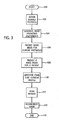
- FIG. 3 a flowchart illustrating the steps of a method and acts associated with a computer program in accordance with the present invention implemented in the system of Figs. 1 and 2 is shown.
- the method and/or computer program is initiated at 100 by a technician or CT scanner operator who provides input into the computer to access desired protocol 102, and select or generate noise prediction coefficients 104 for a patient or object to be imaged.
- operator-entered input can include defining an VOI for a particular anatomy, wherein the VOI has a starting position and an ending position along a common axis, such as axis 50 of Fig. 1, for conducting a scan of the patient to reconstruct an image.
- Other operator-input can include selection of a target noise index, an image application identifying a type of scan, and a set of scan parameter values, such as slice thickness, for the selected scan type.
- a predicted noise index profile for clinical protocols is then determined from the selected scan parameter values at 106, and the method and/or program predicts a noise index for a patient 108. After predicting the noise index 108, the method and/or computer program proceeds to generate a final tube current profile 110, which varies depending on whether an operator or user chooses to override a default or predicted tube current profile for imaging the VOI.
- the tube current profile predicts an electric current amperage passing though an X-ray source for each sub-volume of the VOI to perform an imaging scan of a patient.
- the profile is at least partially based on one or more determined noise index values and indicates a lowest possible dose of X-rays to the patient to maintain a consistent image quality.
- An operator may determine to override either directly or by other methods the tube current profile based on a number of factors.
- One factor for example, is patient anatomy which has different image quality needs. That is, the patient anatomy may require operator interaction to vary the dose in sub-volumes of the VOI where noise artifacts interfere with image quality.
- the patient is scanned 112 to acquire image data.
- the method and/or computer program uses the acquired imaging data to reconstruct an image of the patient 114, which can be displayed on a console or other display device, and the method ends at 116.
- one method of predicting a noise index starts at 120.
- An operator enters patient data representing gender, age, weight, and anatomic volume of the patient which is processed by the computer.
- the computer accesses a patient demographic database 124, which stores data for constructing projection sets based on the operator input of patient data.
- the computer uses the stored data to generate a set of initial projections 126 having a plurality of sub-volumes for the specified patient. If desired, the set of initial projections can be displayed on the console 40 of Fig. 2.
- an image noise or noise index profile is predicted 128 for the patient using the noise prediction coefficients 102 of Fig. 3 and the generated set of projections 126.
- the method then ends 130, and a noise index profile is predicted that can be used to generate a tube current profile to achieve a desired noise index.
- Fig. 5 is a flow chart showing another process of predicting a noise index profile for the patient.
- An operator enters scan parameters to select a scan protocol for a scout scan 134.
- the scout scan is performed and data is acquired 136.
- the data and the generated noise prediction coefficients 102 of Fig. 3 are used to predict the noise index profile 138.
- the method then ends at 140, and the predicted noise index profile can again be used to generate a tube current profile to achieve a desired noise index.
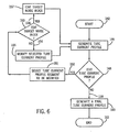
- one method and/or computer program configured to enable direct entry by an operator to select the noise index 110 of Fig. 3 starts at 142.
- a selected noise index input is directly entered by an operator to generate a tube current profile at 146 from the predicted noise index profile for the patient. Selection of the noise index by the operator can be based on the operator's experience of a preferred target noise index for a particular scan type, diagnostic quality requirements, and/or the VOI.
- a decision is made whether to edit the tube current profile 148 using user override. If user override is selected 150, a tube current profile segment to be modified is selected 151, and a query whether to edit the target noise index is made 153.
- the target noise index is not edited 154, the selected tube current profile is modified directly 155 and a new tube current profile is generated at 146 based on the edited segments. If the target noise index is edited 156, the method and/or computer program edits the target noise index for the subset 157, and a new tube current profile is generated at 146.
- Editing of either the entire tube current profile or one or more subsets continues until a desired noise index and tube current profile is selected for the VOI. Editing can be accomplished by an operator interactively modulating either the entire tube current profile or a portion of the profile. Modulation can occur on a graphical user interface, or alternatively by direct entry selection of tube current values for sub-volumes of the VOI.
- editing of the tube current profile is not selected 158 and a final tube current profile is generated 160.
- the final tube current profile is edited to lower the tube current and reduce X-ray exposure to the patient in volumes where a lower image quality is sufficient and/or the diagnostic objective permits a lower dose.
- the method and/or computer program then ends at 162.
- Fig. 7 is a flow chart of another embodiment of the present invention for predicting and selecting a target noise index.
- Selection of the target noise index begins at 164 wherein a diagnostic tube current input method and/or computer program is retrieved from a stored clinical protocol.
- a target noise index is generated using a final reference phantom and a diagnostic tube current value 168.
- the data from the reference phantom can vary according to a selected projection area of the phantom and an oval ratio indicative of the ratio of projection measures taken at zero and ninety degree gantry rotation positions.
- a tube current profile is generated at 172 from the predicted noise index for the patient.
- the tube current profile is indicative of a predicted low dose X-ray exposure to the patient based on the entered diagnostic tube current value and reference phantom.
- a decision is made whether to edit the tube current profile 174 using user override. If user override is selected 176, then the operator selects the tube current profile segment to be modified 177.
- the method and/or computer program proceeds to decide whether to edit the diagnostic tube current value 178. If editing of the diagnostic tube current value is not selected 179, the selected tube current profile is modified at 180 and a new tube current profile is generated at 172. If editing of the diagnostic tube current value is selected 181, the diagnostic tube current value is adjusted 182 and a new target noise index is generated at 168 to produce a new tube current profile 172.
- Editing of either the entire tube current profile or one or more subsets continues until a desired noise index and tube current profile is created for the VOI. Once the desired tube current is determined, editing of the tube current profile is not selected 184 and a final tube current profile is generated at 186. Once again, the final tube current profile is preferably edited to lower the tube current and reduce X-ray exposure to the patient in volumes where a lower image quality is sufficient and/or the diagnostic objective permits a lower dose. The method and/or computer program then ends at 188.
- a flow chart of another method and/or computer program for selecting a noise index begins at 190.
- a decision to increase the relative dose occurs at 192, and if so 194, an operator may increase the relative dose which causes a decrease in the noise index 196.
- a tube current profile is generated at 198.
- the relative dose is not increased 200, a decision to decrease the relative dose 202 occurs. if the reiative dose is not decreased 204, then the tube current profile is generated based on the pre-selected target noise index. Otherwise, the relative dose is decreased 206 and the noise index is increased 208.
- the method and/or computer program uses the increased noise index to generate the tube current profile at 198.
- a decision similar to the other noise selection processes is made concerning whether to edit the tube current profile 210 using user override. If user override is selected 212, then the operator selects one or more tube current profile segments to modify 213 and a query to modify the relative dose is determined 214. If the relative dose is not modified 215, the selected tube current profile is modified 216 and a new tube current profile is generated at 198. If the relative dose is modified 218, the method and/or computer program loops to the beginning and again determines whether to increase the relative dose at 192.
- Editing of either the entire tube current profile or one or more subsets continues until a desired noise index and tube current profile is created for the VOI. Once the desired tube current is determined, editing of the tube current profile is not selected 220 and a final tube current profile is generated at 222. As with the other noise index selection processes, the final tube current profile is preferably edited to lower the tube current and reduce X-ray exposure to the patient in volumes where a lower image quality is sufficient and/or the diagnostic objective permits a lower dose. The method and/or computer program then ends at 224.
- Figs. 6-8 indicate methods and/or computer programs acts that enable interactive adjustment of the generated tube current profile to convey a minimum allowable dose for each sub-volume in the VOI, and generate a final tube current profile to produce a selected noise index.
- Each of these methods and/or acts that apply user analysis to generate the final tube current profile can be further modified in accordance with the steps of Fig. 9.
- the method and/or computer program begins at 226, wherein a final tube current profile is generated.
- a decision block queries whether to enable smart scan at 228.
- Smart scan is a process wherein an X-ray dose is varied with the gantry angle to account for patient asymmetry. If smart scan is selected 230, an effective tube current profile is generated 232. The effective tube current profile can be plotted on a graphical user interface or displayed as a set of data for operator modulation of the tube current profile. After generating an effective tube current profile, a decision to adjust for sensitive organs 234 is made. If smart scan is not selected 236, the method and/or computer program immediately proceeds to decide whether to adjust for sensitive organs at 234.
- an operator may desire to limit the X-ray exposure to certain anatomical volumes sensitive to radiation, such as the eyes, ovaries, breasts, and gonads.
- the tube current or effective tube current profile is adjusted according to the gantry angular position 240 to lower X-ray exposure to sensitive organs when the X-ray source is closest to each of the sensitive organs. That is, during each gantry rotation the tube current decreases as the gantry is positioned above a sensitive organ, such as the eyes of a patient, and increases as the gantry rotates behind the patient's head.
- the method and/or computer program After the tube current adjustment 240, the method and/or computer program generates an adjusted tube current profile at 242which is used to generate the selected noise index. If no adjustment for sensitive organs is desired 244, the adjusted tube current profile is again generated at 242 and the method and/or computer program ends at 246.
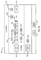
- a graphical user interface 248 configured to permit operator control of a tube current profile is shown.
- An operator can select automatic tube current generation 250, and specify a range of tube current values between a minimum value 252 and a maximum value 254 that the tube current can be between.
- the operator can select manual tube current generation 256 and specify a desired tube current value 258.
- a default noise index 260 is generated by selecting one of the IQ 262, normal 264, and lowdose 266 selections.
- the IQ selection 262 provides an X-ray tube current profile having tube current values a fixed amount higher than the normal dose 264, which is a fixed amount above the lowdose selection 266.
- the relative dose 268 or noise index values can be increased or decreased by adjustment selectors 270.
- the adjustment selectors are configured to change the noise index in five percent increments, although other incremental or continuous adjustments are contemplated with the present invention.
- the noise index 272 may be directly modified by entering a direct input at 274. Upon choosing the OK selection 276, the final tube current profile is created. If improper data is entered, or an operator prefers to enter a new set of data, the cancel selection 278 may be utilized to reselect generation of the tube current profile.
- FIG. 10 shows one computer user interface method for editing and generating a tube current profile
- a method of processing imaging data for a radiation emitting medical imaging device includes the steps of receiving an image application identifier and a set of scan parameter values, and automatically generating a predicted noise index from the received set of scan parameter values.
- the method also includes the step of generating an X-ray tube current profile based at least partially on the predicted noise index if so selected by a user. If a preferred noise index is set, the method generates a final tube current profile and acquires imaging data with the final tube current profile.
- a method of processing imaging data for a radiation emitting medical imaging device includes the steps of acquiring imaging data of a subject, such as a patient or object, and generating a set of projections for an VOI having a plurality of sub-volumes. The method also acquires a target noise index, which may be acquired using a scout scan. The method further includes the steps of generating an X-ray current profile according to the target noise index, and enabling interactive adjustment by a user of the generated current profile to convey a minimum allowable dose for each sub-volume in the VOI.
- a computed tomography system includes a projection source to project towards an object, and a detector to receive high frequency electromagnetic energy attenuated by the object.
- the detector produces outputs that are transmitted to a data processing system by a plurality of electrical interconnects.
- the system also includes a computer programmed to construct a plurality of initial projections for an VOI and receive a user input to generate a target noise index.
- the computer is also programmed to generate a tube current profile according to the target noise index and a predicted noise index, and display the tube current profile on a console such that the tube current profile can be modulated for one or more portions of the VOI.
- the tube current profile can be displayed as a set of data values, or alternatively as a graphical user interface.
- the computer is further programmed to adjust a patient X-ray dose for one or more portions of the VOI based on modulation of the tube current, and acquire imaging data for the VOI.
- a computer-readable medium having stored thereon a computer program having a set of instructions that, when executed by a computer, causes the computer to acquire pre-scan data of an VOI of a subject and generate a predicted noise index from the pre-scan data.
- the computer program also has instructions to generate an X-ray tube current profile based upon the predicted noise index and a target noise index, and receive selective adjustments by an operator of at least a portion of the tube current profile. The selective adjustments are configured to adjust a scan dose for a corresponding portion of the VOI based on user analysis of the tube current profile.
- a radiation emitting medical device comprises a means for receiving scan parameters, and a means for adjusting the scan parameters automatically to generate a desired target image quality for a patient.
- the medical device also includes a means for modifying a tube current profile based on the adjusted scan parameters, and a means for scanning the patient using the modified tube current profile to reconstruct an image of the patient.
Landscapes
- Health & Medical Sciences (AREA)
- Life Sciences & Earth Sciences (AREA)
- Engineering & Computer Science (AREA)
- Medical Informatics (AREA)
- Radiology & Medical Imaging (AREA)
- Biomedical Technology (AREA)
- Biophysics (AREA)
- High Energy & Nuclear Physics (AREA)
- Veterinary Medicine (AREA)
- Nuclear Medicine, Radiotherapy & Molecular Imaging (AREA)
- Optics & Photonics (AREA)
- Pathology (AREA)
- Public Health (AREA)
- Physics & Mathematics (AREA)
- Heart & Thoracic Surgery (AREA)
- Molecular Biology (AREA)
- Surgery (AREA)
- Animal Behavior & Ethology (AREA)
- General Health & Medical Sciences (AREA)
- Pulmonology (AREA)
- Theoretical Computer Science (AREA)
- Human Computer Interaction (AREA)
- Apparatus For Radiation Diagnosis (AREA)
- X-Ray Techniques (AREA)
Abstract
Description
Claims (10)
- A method (100) of processing imaging data for a radiation emitting medical imaging device (10), the method comprising:receiving an image application identifier (102);receiving a set of scan parameter values (104);automatically generating a predicted noise index from the received set of scan parameter values (106);generating an X-ray tube current profile (146, 172, 198) based at least on the predicted noise index;allowing user override (148, 174, 210) of the automatically generated predicted noise index based on the X-ray tube current profile and setting a preferred noise index if so selected by a user, and if a preferred noise index is set, generating a final tube current profile (110); andacquiring imaging data with the final tube current profile (112).
- The method (100) of claim 1 wherein the step of determining the preferred noise index further comprises the step of adjusting the automatically generated predicted noise index according to a direct user input (122).
- The method (100) of claim 1 further comprising the steps of:initiating a pre-scan of a subject (134);acquiring pre-scan imaging data (136);generating a scout image from the pre-scan imaging data;displaying the scout image; anddisplaying a tube current profile for a selected diagnostic quality, wherein one or more portions of the tube current profile are adjustable.
- A method (100) of processing imaging data for a radiation emitting medical imaging device, the method comprising:acquiring imaging data of a subject (134);generating a set of projections for an VOI having a plurality of sub-volumes (136);acquiring a target noise index (138);generating a current profile (146, 172, 198) according to the target noise index; andenabling interactive adjustment of the generated current profile to convey a minimum allowable dose for each sub-volume in the VOI (151, 177, 213).
- The method (100) of claim 4 further comprising the step of varying an application of a patient dose for each rotation of an X-ray source within a sub-volume in the VOI (240) to limit X-ray exposure to sensitive organs of a patient.
- A computed tomography system (10) comprising:a high frequency electromagnetic energy projection source (14) to project high frequency energy towards an object (22);a detector (20) to receive high frequency electromagnetic energy attenuated by the object (22);a plurality of electrical interconnects configured to transmit detector outputs to a data processing system (32); anda computer (36) programmed to:construct a plurality of initial projections for an VOI (126);receive a user input to generate a target noise index (128);generate a tube current profile (146, 174, 210) according to the target noise index and a predicted noise index;display the tube current profile on a console wherein the tube current profile can be modulated for one or more portions of the VOI;adjust a scan dose for one or more portions of the VOI (148, 174, 210) based on user modulation of the tube current profile; andacquire imaging data for the VOI (112).
- The system (10) of claim 6 wherein the computer (36) is further programmed to display the plurality of initial projections on the console.
- The system (10) of claim 6 wherein the computer (36) is further programmed to modulate the tube current profile based on a direct user target noise input (122) for selected volumes of the VOI.
- A computer-readable medium having stored thereon a computer program having a set of instructions that, when executed by a computer (36), causes the computer (36) to:acquire pre-scan data of an VOI of a subject (104);generate a predicted noise index from the pre-scan data (106);generate a tube current profile based upon the predicted noise index and a target noise index (146, 172, 198); andreceive selective adjustments (151, 177, 213) of at least a portion of the tube current profile to adjust a scan dose for a corresponding portion of the VOI based on user analysis of the tube current profile.
- A radiation emitting medical device (10) comprising:means for receiving scan parameters (36);means for adjusting the scan parameters automatically to generate a desired target image quality for a patient;means for modifying a tube current profile (148, 174, 210) based on the adjusted scan parameters; andmeans for scanning the patient using the modified tube current profile to reconstruct an image of the patient (112).
Applications Claiming Priority (2)
| Application Number | Priority Date | Filing Date | Title |
|---|---|---|---|
| US683128 | 2001-11-21 | ||
| US09/683,128 US6904127B2 (en) | 2001-11-21 | 2001-11-21 | System and method of medical imaging having default noise index override capability |
Publications (3)
| Publication Number | Publication Date |
|---|---|
| EP1314397A2 true EP1314397A2 (en) | 2003-05-28 |
| EP1314397A3 EP1314397A3 (en) | 2003-07-16 |
| EP1314397B1 EP1314397B1 (en) | 2013-09-18 |
Family
ID=24742683
Family Applications (1)
| Application Number | Title | Priority Date | Filing Date |
|---|---|---|---|
| EP02258042.7A Expired - Lifetime EP1314397B1 (en) | 2001-11-21 | 2002-11-21 | System and method of medical imaging having override capability |
Country Status (3)
| Country | Link |
|---|---|
| US (1) | US6904127B2 (en) |
| EP (1) | EP1314397B1 (en) |
| JP (1) | JP4370091B2 (en) |
Cited By (4)
| Publication number | Priority date | Publication date | Assignee | Title |
|---|---|---|---|---|
| NL1029526C2 (en) * | 2004-07-21 | 2007-08-07 | Ge Med Sys Global Tech Co Llc | Dosage indexing phantom selection for dosage reporting in computed tomography. |
| NL1032847C2 (en) * | 2005-11-15 | 2007-11-06 | Ge Med Sys Global Tech Co Llc | CT device operating with X-rays and CT fluoroscopic device operating with X-rays. |
| NL1033544C2 (en) * | 2007-03-15 | 2008-09-16 | Gen Electric | Imaging system for producing image of medical patient, has identifiable and imageable components of patient determining relationship between radiation profile coefficients and effective dose |
| EP1943951A4 (en) * | 2005-09-15 | 2009-03-25 | Hitachi Medical Corp | X-ray ct device |
Families Citing this family (42)
| Publication number | Priority date | Publication date | Assignee | Title |
|---|---|---|---|---|
| JP2004173924A (en) * | 2002-11-27 | 2004-06-24 | Ge Medical Systems Global Technology Co Llc | Method for controlling x-ray, and x-ray image photographing equipment |
| EP1603461A2 (en) * | 2003-03-10 | 2005-12-14 | Philips Intellectual Property & Standards GmbH | Device and method for adapting the recording parameters of a radiograph |
| JP2005080748A (en) * | 2003-09-05 | 2005-03-31 | Ge Medical Systems Global Technology Co Llc | Imaging condition set up method and x-ray ct apparatus |
| US6980623B2 (en) * | 2003-10-29 | 2005-12-27 | Ge Medical Systems Global Technology Company Llc | Method and apparatus for z-axis tracking and collimation |
| US7894570B2 (en) * | 2003-11-25 | 2011-02-22 | Koninklijke Philips Electronics N.V. | Automatic dose adaptation as a function of patient body mass index in CT calcium scoring |
| DE102004030833A1 (en) * | 2004-06-25 | 2006-01-26 | Siemens Ag | Röngtendiagnostikverfahren and associated device |
| JP2006055635A (en) * | 2004-07-23 | 2006-03-02 | Toshiba Corp | X-ray computed tomography system |
| JP4731151B2 (en) * | 2004-10-22 | 2011-07-20 | 株式会社日立メディコ | X-ray tube current determination method and X-ray CT apparatus |
| US20060218251A1 (en) * | 2005-03-14 | 2006-09-28 | Matsushita Electric Industrial Co., Ltd. | AV server, AV contents reproducing apparatus, AV contents transmitting method, program and recording medium |
| JP2006320523A (en) * | 2005-05-19 | 2006-11-30 | Ge Medical Systems Global Technology Co Llc | Method of setting scan parameter of shuttle mode helical scan and x-ray ct apparatus |
| US20070076842A1 (en) * | 2005-09-30 | 2007-04-05 | Tkaczyk John E | Adaptable energy discriminating computed tomography system |
| JP5034221B2 (en) * | 2005-11-15 | 2012-09-26 | 株式会社島津製作所 | Radiography equipment |
| US7983457B2 (en) * | 2005-11-23 | 2011-07-19 | General Electric Company | Method and system for automatically determining regions in a scanned object |
| JP4509971B2 (en) * | 2006-06-09 | 2010-07-21 | ジーイー・メディカル・システムズ・グローバル・テクノロジー・カンパニー・エルエルシー | X-ray CT system |
| US8000445B2 (en) * | 2006-07-31 | 2011-08-16 | Koninklijke Philips Electronics N.V. | Rotational X-ray scan planning system |
| US20080033748A1 (en) * | 2006-08-02 | 2008-02-07 | Erik Busch | System to measure and analyze the usage of dose reduction features |
| CN101327128B (en) * | 2007-06-22 | 2010-12-08 | Ge医疗系统环球技术有限公司 | Scan and detection device of X ray CT device as well as system and operation method thereof |
| JP5389345B2 (en) * | 2007-10-04 | 2014-01-15 | ジーイー・メディカル・システムズ・グローバル・テクノロジー・カンパニー・エルエルシー | X-ray CT system |
| DE102008014738A1 (en) * | 2008-03-18 | 2009-09-24 | Siemens Aktiengesellschaft | Medical imaging method and medical imaging device |
| JP5455903B2 (en) * | 2008-07-04 | 2014-03-26 | 株式会社日立メディコ | X-ray CT system |
| US20140072108A1 (en) * | 2010-07-16 | 2014-03-13 | David P. Rohler | Methods and apparatus for extended low contrast detectability for radiographic imaging systems |
| EP2724739B1 (en) | 2009-07-30 | 2015-07-01 | Tandem Diabetes Care, Inc. | Portable infusion pump system |
| US8611627B2 (en) | 2009-12-23 | 2013-12-17 | General Electric Company | CT spectral calibration |
| JP5580220B2 (en) * | 2011-01-17 | 2014-08-27 | ヤマハ発動機株式会社 | Radiation inspection apparatus and radiation inspection method |
| WO2012129374A1 (en) | 2011-03-22 | 2012-09-27 | Corindus, Inc. | Robotic catheter system including imaging system control |
| JP6257948B2 (en) * | 2012-08-07 | 2018-01-10 | 東芝メディカルシステムズ株式会社 | X-ray imaging system |
| CN104812305B (en) * | 2012-12-27 | 2018-03-30 | 东芝医疗系统株式会社 | X ray CT device and control method |
| US11229490B2 (en) | 2013-06-26 | 2022-01-25 | Corindus, Inc. | System and method for monitoring of guide catheter seating |
| US20150005745A1 (en) | 2013-06-26 | 2015-01-01 | Corindus, Inc. | 3-d mapping for guidance of device advancement out of a guide catheter |
| KR101534098B1 (en) * | 2013-09-13 | 2015-07-07 | 삼성전자주식회사 | Computed tomography apparatus and method for controlling x-ray by using the same |
| DE102013219249A1 (en) * | 2013-09-25 | 2015-03-26 | Siemens Aktiengesellschaft | Method and system for automatic selection of a scan protocol |
| CN104287768A (en) * | 2014-09-30 | 2015-01-21 | 沈阳东软医疗系统有限公司 | Method and system for controlling CT scan dose |
| US9848847B2 (en) * | 2015-04-07 | 2017-12-26 | Toshiba Medical Systems Corporation | Using detectability index to determine X-ray tube current |
| CN106994021B (en) * | 2016-01-22 | 2022-10-14 | 通用电气公司 | Method and device for calculating noise on CT image |
| US10085698B2 (en) * | 2016-01-26 | 2018-10-02 | Genereal Electric Company | Methods and systems for automated tube current modulation |
| CN106725570B (en) * | 2016-12-30 | 2019-12-20 | 上海联影医疗科技有限公司 | Imaging method and system |
| US10973489B2 (en) * | 2017-09-29 | 2021-04-13 | General Electric Company | CT imaging system and method using a task-based image quality metric to achieve a desired image quality |
| EP3910644A1 (en) * | 2020-05-12 | 2021-11-17 | Koninklijke Philips N.V. | Quality control in medical imaging |
| US12347100B2 (en) | 2020-11-19 | 2025-07-01 | Mazor Robotics Ltd. | Systems and methods for generating virtual images |
| CN115245343B (en) * | 2021-04-28 | 2026-02-24 | 西安大医集团股份有限公司 | Method for determining bulb tube electrical parameters, host and medical imaging system |
| US12458302B2 (en) * | 2021-05-05 | 2025-11-04 | Koninklijke Philips N.V. | Systems and methods for processing and visualizing tube current modulations in medical imaging devices |
| CN117275701B (en) * | 2023-11-21 | 2024-02-02 | 拓庄医疗科技有限公司 | CT equipment fault diagnosis data analysis system and method based on artificial intelligence |
Family Cites Families (12)
| Publication number | Priority date | Publication date | Assignee | Title |
|---|---|---|---|---|
| US5450462A (en) | 1993-11-19 | 1995-09-12 | General Electric Company | Modulation of x-ray tube current during CT scanning with modulation limit |
| US5400378A (en) * | 1993-11-19 | 1995-03-21 | General Electric Company | Dynamic dose control in multi-slice CT scan |
| US5379333A (en) | 1993-11-19 | 1995-01-03 | General Electric Company | Variable dose application by modulation of x-ray tube current during CT scanning |
| US5485494A (en) * | 1994-08-03 | 1996-01-16 | General Electric Company | Modulation of X-ray tube current during CT scanning |
| US5772585A (en) * | 1996-08-30 | 1998-06-30 | Emc, Inc | System and method for managing patient medical records |
| US5696807A (en) | 1996-09-05 | 1997-12-09 | General Electric Company | Methods and apparatus for modulating x-ray tube current |
| US5867555A (en) | 1997-03-04 | 1999-02-02 | Siemens Aktiengesellschaft | Adaptive dose modulation during CT scanning |
| US5822393A (en) * | 1997-04-01 | 1998-10-13 | Siemens Aktiengesellschaft | Method for adaptively modulating the power level of an x-ray tube of a computer tomography (CT) system |
| DE69840290D1 (en) * | 1997-06-26 | 2009-01-15 | Koninkl Philips Electronics Nv | ADJUSTABLE COMPUTER-BASED TOMOGRAPHY SYSTEM |
| DE19810069B4 (en) * | 1998-03-09 | 2007-05-10 | Siemens Ag | Device with several adjustable by an operator via a control unit operating parameters |
| US6285741B1 (en) * | 1998-08-25 | 2001-09-04 | General Electric Company | Methods and apparatus for automatic image noise reduction |
| US6507639B1 (en) * | 2001-08-30 | 2003-01-14 | Siemens Aktiengesellschaft | Method and apparatus for modulating the radiation dose from x-ray tube |
-
2001
- 2001-11-21 US US09/683,128 patent/US6904127B2/en not_active Expired - Lifetime
-
2002
- 2002-11-21 EP EP02258042.7A patent/EP1314397B1/en not_active Expired - Lifetime
- 2002-11-21 JP JP2002337848A patent/JP4370091B2/en not_active Expired - Fee Related
Cited By (4)
| Publication number | Priority date | Publication date | Assignee | Title |
|---|---|---|---|---|
| NL1029526C2 (en) * | 2004-07-21 | 2007-08-07 | Ge Med Sys Global Tech Co Llc | Dosage indexing phantom selection for dosage reporting in computed tomography. |
| EP1943951A4 (en) * | 2005-09-15 | 2009-03-25 | Hitachi Medical Corp | X-ray ct device |
| NL1032847C2 (en) * | 2005-11-15 | 2007-11-06 | Ge Med Sys Global Tech Co Llc | CT device operating with X-rays and CT fluoroscopic device operating with X-rays. |
| NL1033544C2 (en) * | 2007-03-15 | 2008-09-16 | Gen Electric | Imaging system for producing image of medical patient, has identifiable and imageable components of patient determining relationship between radiation profile coefficients and effective dose |
Also Published As
| Publication number | Publication date |
|---|---|
| EP1314397A3 (en) | 2003-07-16 |
| US20030097062A1 (en) | 2003-05-22 |
| US6904127B2 (en) | 2005-06-07 |
| EP1314397B1 (en) | 2013-09-18 |
| JP4370091B2 (en) | 2009-11-25 |
| JP2003190139A (en) | 2003-07-08 |
Similar Documents
| Publication | Publication Date | Title |
|---|---|---|
| US6904127B2 (en) | System and method of medical imaging having default noise index override capability | |
| US7813471B2 (en) | X-ray CT apparatus | |
| US6744846B2 (en) | Method and apparatus for automatic exposure control in CT scanning | |
| JP4187289B2 (en) | Method and system for modulating x-ray tube current and system for dynamically adjusting at least one component of a computed tomography imaging system | |
| US5822393A (en) | Method for adaptively modulating the power level of an x-ray tube of a computer tomography (CT) system | |
| US6236706B1 (en) | Methods and apparatus for predicting contrast agent uptake in a computed tomography system | |
| KR100457607B1 (en) | Tomographic imaging scan condition determining method, tomographic imaging method and x-ray ct apparatus | |
| US6501828B1 (en) | Method and apparatus for influencing X-rays in a beam path | |
| CN100393281C (en) | X-ray computed tomography device | |
| US5400378A (en) | Dynamic dose control in multi-slice CT scan | |
| JP4822478B2 (en) | X-ray CT system | |
| US8155263B2 (en) | Apparatus and method for voltage modulation in X-ray computed tomography | |
| US6198789B1 (en) | Variable current CT scanning | |
| EP1304077A2 (en) | X-ray computed tomography apparatus | |
| JP4159188B2 (en) | Tube current adjusting method and apparatus, and X-ray CT apparatus | |
| US20050152493A1 (en) | Radiation tomography system and tomography method | |
| JP5985836B2 (en) | Method for reducing the amount of radiation emitted by an imaging system | |
| JP4729519B2 (en) | Radiation imaging method and system with organ-based radiation profile settings | |
| JP2021137259A (en) | Medical diagnostic system, medical diagnostic apparatus, and medical information processing apparatus | |
| US7848790B2 (en) | System and method of imaging using a variable speed for thorax imaging | |
| US6687327B2 (en) | System and method of medical imaging having task and/or patient size dependent processing | |
| EP1103221B1 (en) | Methods and apparatus for optimizing CT image quality with optimized data acquisition | |
| JP6490912B2 (en) | X-ray CT system | |
| JP3950612B2 (en) | X-ray CT system | |
| US12004284B2 (en) | Methods and systems for x-ray imaging |
Legal Events
| Date | Code | Title | Description |
|---|---|---|---|
| PUAI | Public reference made under article 153(3) epc to a published international application that has entered the european phase |
Free format text: ORIGINAL CODE: 0009012 |
|
| AK | Designated contracting states |
Designated state(s): AT BE BG CH CY CZ DE DK EE ES FI FR GB GR IE IT LI LU MC NL PT SE SK TR |
|
| AX | Request for extension of the european patent |
Extension state: AL LT LV MK RO SI |
|
| PUAL | Search report despatched |
Free format text: ORIGINAL CODE: 0009013 |
|
| AK | Designated contracting states |
Designated state(s): AT BE BG CH CY CZ DE DK EE ES FI FR GB GR IE IT LI LU MC NL PT SE SK TR |
|
| AX | Request for extension of the european patent |
Extension state: AL LT LV MK RO SI |
|
| 17P | Request for examination filed |
Effective date: 20040116 |
|
| AKX | Designation fees paid |
Designated state(s): DE NL |
|
| 17Q | First examination report despatched |
Effective date: 20080313 |
|
| GRAP | Despatch of communication of intention to grant a patent |
Free format text: ORIGINAL CODE: EPIDOSNIGR1 |
|
| INTG | Intention to grant announced |
Effective date: 20130419 |
|
| GRAS | Grant fee paid |
Free format text: ORIGINAL CODE: EPIDOSNIGR3 |
|
| GRAA | (expected) grant |
Free format text: ORIGINAL CODE: 0009210 |
|
| AK | Designated contracting states |
Kind code of ref document: B1 Designated state(s): DE NL |
|
| REG | Reference to a national code |
Ref country code: DE Ref legal event code: R096 Ref document number: 60245540 Country of ref document: DE Effective date: 20131114 |
|
| REG | Reference to a national code |
Ref country code: NL Ref legal event code: T3 |
|
| REG | Reference to a national code |
Ref country code: DE Ref legal event code: R097 Ref document number: 60245540 Country of ref document: DE |
|
| PLBE | No opposition filed within time limit |
Free format text: ORIGINAL CODE: 0009261 |
|
| STAA | Information on the status of an ep patent application or granted ep patent |
Free format text: STATUS: NO OPPOSITION FILED WITHIN TIME LIMIT |
|
| 26N | No opposition filed |
Effective date: 20140619 |
|
| REG | Reference to a national code |
Ref country code: DE Ref legal event code: R097 Ref document number: 60245540 Country of ref document: DE Effective date: 20140619 |
|
| PGFP | Annual fee paid to national office [announced via postgrant information from national office to epo] |
Ref country code: NL Payment date: 20171126 Year of fee payment: 16 Ref country code: DE Payment date: 20171129 Year of fee payment: 16 |
|
| REG | Reference to a national code |
Ref country code: DE Ref legal event code: R119 Ref document number: 60245540 Country of ref document: DE |
|
| REG | Reference to a national code |
Ref country code: NL Ref legal event code: MM Effective date: 20181201 |
|
| PG25 | Lapsed in a contracting state [announced via postgrant information from national office to epo] |
Ref country code: NL Free format text: LAPSE BECAUSE OF NON-PAYMENT OF DUE FEES Effective date: 20181201 |
|
| PG25 | Lapsed in a contracting state [announced via postgrant information from national office to epo] |
Ref country code: DE Free format text: LAPSE BECAUSE OF NON-PAYMENT OF DUE FEES Effective date: 20190601 |