EP1312681A1 - Method for identifying therapeutic targets by use of genetic screens in Drosophila melanogaster - Google Patents
Method for identifying therapeutic targets by use of genetic screens in Drosophila melanogaster Download PDFInfo
- Publication number
- EP1312681A1 EP1312681A1 EP01127195A EP01127195A EP1312681A1 EP 1312681 A1 EP1312681 A1 EP 1312681A1 EP 01127195 A EP01127195 A EP 01127195A EP 01127195 A EP01127195 A EP 01127195A EP 1312681 A1 EP1312681 A1 EP 1312681A1
- Authority
- EP
- European Patent Office
- Prior art keywords
- drosophila
- strain
- polymorphisms
- strains
- mutation
- Prior art date
- Legal status (The legal status is an assumption and is not a legal conclusion. Google has not performed a legal analysis and makes no representation as to the accuracy of the status listed.)
- Withdrawn
Links
- 238000000034 method Methods 0.000 title claims abstract description 58
- 230000002068 genetic effect Effects 0.000 title claims abstract description 41
- 241000255601 Drosophila melanogaster Species 0.000 title claims abstract description 20
- 230000001225 therapeutic effect Effects 0.000 title description 2
- 108090000623 proteins and genes Proteins 0.000 claims abstract description 80
- 102000054765 polymorphisms of proteins Human genes 0.000 claims abstract description 78
- 241000255581 Drosophila <fruit fly, genus> Species 0.000 claims abstract description 61
- 238000013507 mapping Methods 0.000 claims abstract description 44
- 208000037265 diseases, disorders, signs and symptoms Diseases 0.000 claims abstract description 41
- 238000003780 insertion Methods 0.000 claims abstract description 41
- 230000037431 insertion Effects 0.000 claims abstract description 41
- 201000010099 disease Diseases 0.000 claims abstract description 40
- 230000006798 recombination Effects 0.000 claims abstract description 21
- 238000005215 recombination Methods 0.000 claims abstract description 21
- 230000031018 biological processes and functions Effects 0.000 claims abstract description 4
- 230000003990 molecular pathway Effects 0.000 claims abstract description 4
- 230000035772 mutation Effects 0.000 claims description 75
- 210000000349 chromosome Anatomy 0.000 claims description 41
- 238000003556 assay Methods 0.000 claims description 38
- 238000007894 restriction fragment length polymorphism technique Methods 0.000 claims description 18
- 239000003550 marker Substances 0.000 claims description 17
- 238000012163 sequencing technique Methods 0.000 claims description 13
- 241000255925 Diptera Species 0.000 claims description 12
- 241001465754 Metazoa Species 0.000 claims description 11
- 239000002299 complementary DNA Substances 0.000 claims description 10
- 238000004458 analytical method Methods 0.000 claims description 9
- 238000010367 cloning Methods 0.000 claims description 9
- 231100000225 lethality Toxicity 0.000 claims description 9
- 108020004414 DNA Proteins 0.000 claims description 7
- 230000014509 gene expression Effects 0.000 claims description 7
- 108091028043 Nucleic acid sequence Proteins 0.000 claims description 6
- 108010046276 FLP recombinase Proteins 0.000 claims description 4
- 206010068052 Mosaicism Diseases 0.000 claims description 4
- 210000002230 centromere Anatomy 0.000 claims description 3
- 238000004519 manufacturing process Methods 0.000 claims description 3
- 210000003411 telomere Anatomy 0.000 claims description 3
- 108091035539 telomere Proteins 0.000 claims description 3
- 102000055501 telomere Human genes 0.000 claims description 3
- 239000003623 enhancer Substances 0.000 claims 2
- 239000000203 mixture Substances 0.000 claims 2
- 230000009897 systematic effect Effects 0.000 abstract description 4
- 238000013459 approach Methods 0.000 description 17
- 239000002773 nucleotide Substances 0.000 description 12
- 125000003729 nucleotide group Chemical group 0.000 description 12
- 108700028369 Alleles Proteins 0.000 description 11
- 238000002474 experimental method Methods 0.000 description 11
- 230000006870 function Effects 0.000 description 9
- 231100000350 mutagenesis Toxicity 0.000 description 9
- 238000003205 genotyping method Methods 0.000 description 8
- 102000004169 proteins and genes Human genes 0.000 description 8
- 238000006243 chemical reaction Methods 0.000 description 7
- 239000012634 fragment Substances 0.000 description 7
- 238000002703 mutagenesis Methods 0.000 description 7
- 230000007547 defect Effects 0.000 description 6
- 238000012300 Sequence Analysis Methods 0.000 description 5
- 230000003321 amplification Effects 0.000 description 5
- 238000003199 nucleic acid amplification method Methods 0.000 description 5
- 108091008146 restriction endonucleases Proteins 0.000 description 5
- 230000003252 repetitive effect Effects 0.000 description 4
- 239000000126 substance Substances 0.000 description 4
- 230000000007 visual effect Effects 0.000 description 4
- 206010028980 Neoplasm Diseases 0.000 description 3
- 238000012408 PCR amplification Methods 0.000 description 3
- 238000002944 PCR assay Methods 0.000 description 3
- 238000012217 deletion Methods 0.000 description 3
- 230000037430 deletion Effects 0.000 description 3
- 238000013461 design Methods 0.000 description 3
- 230000004770 neurodegeneration Effects 0.000 description 3
- 230000035790 physiological processes and functions Effects 0.000 description 3
- 238000011084 recovery Methods 0.000 description 3
- 238000010561 standard procedure Methods 0.000 description 3
- 108010067770 Endopeptidase K Proteins 0.000 description 2
- 102000004190 Enzymes Human genes 0.000 description 2
- 108090000790 Enzymes Proteins 0.000 description 2
- 229930186657 Lat Natural products 0.000 description 2
- TWRXJAOTZQYOKJ-UHFFFAOYSA-L Magnesium chloride Chemical compound [Mg+2].[Cl-].[Cl-] TWRXJAOTZQYOKJ-UHFFFAOYSA-L 0.000 description 2
- 108091034117 Oligonucleotide Proteins 0.000 description 2
- FAPWRFPIFSIZLT-UHFFFAOYSA-M Sodium chloride Chemical compound [Na+].[Cl-] FAPWRFPIFSIZLT-UHFFFAOYSA-M 0.000 description 2
- 102000044209 Tumor Suppressor Genes Human genes 0.000 description 2
- 108700025716 Tumor Suppressor Genes Proteins 0.000 description 2
- 239000011543 agarose gel Substances 0.000 description 2
- 238000010171 animal model Methods 0.000 description 2
- 230000008901 benefit Effects 0.000 description 2
- 230000008238 biochemical pathway Effects 0.000 description 2
- 238000000423 cell based assay Methods 0.000 description 2
- 230000004663 cell proliferation Effects 0.000 description 2
- 230000001413 cellular effect Effects 0.000 description 2
- 230000002380 cytological effect Effects 0.000 description 2
- 238000001514 detection method Methods 0.000 description 2
- 239000003814 drug Substances 0.000 description 2
- 230000009368 gene silencing by RNA Effects 0.000 description 2
- 231100000118 genetic alteration Toxicity 0.000 description 2
- 230000004077 genetic alteration Effects 0.000 description 2
- 210000005260 human cell Anatomy 0.000 description 2
- 231100000518 lethal Toxicity 0.000 description 2
- 230000001665 lethal effect Effects 0.000 description 2
- 230000001537 neural effect Effects 0.000 description 2
- 208000015122 neurodegenerative disease Diseases 0.000 description 2
- 230000037361 pathway Effects 0.000 description 2
- 239000002831 pharmacologic agent Substances 0.000 description 2
- 238000009790 rate-determining step (RDS) Methods 0.000 description 2
- 230000028617 response to DNA damage stimulus Effects 0.000 description 2
- 230000000717 retained effect Effects 0.000 description 2
- 239000000523 sample Substances 0.000 description 2
- 229940124597 therapeutic agent Drugs 0.000 description 2
- 229920000936 Agarose Polymers 0.000 description 1
- 208000024827 Alzheimer disease Diseases 0.000 description 1
- 241000244203 Caenorhabditis elegans Species 0.000 description 1
- 102100025064 Cellular tumor antigen p53 Human genes 0.000 description 1
- 108091026890 Coding region Proteins 0.000 description 1
- 238000001712 DNA sequencing Methods 0.000 description 1
- 108700002304 Drosophila can Proteins 0.000 description 1
- 108700008634 Drosophila p53 Proteins 0.000 description 1
- 108700007057 Drosophila ras Proteins 0.000 description 1
- 108700020323 Drosophila wts Proteins 0.000 description 1
- KCXVZYZYPLLWCC-UHFFFAOYSA-N EDTA Chemical compound OC(=O)CN(CC(O)=O)CCN(CC(O)=O)CC(O)=O KCXVZYZYPLLWCC-UHFFFAOYSA-N 0.000 description 1
- 244000148064 Enicostema verticillatum Species 0.000 description 1
- 244000060234 Gmelina philippensis Species 0.000 description 1
- 241000282412 Homo Species 0.000 description 1
- 102000003839 Human Proteins Human genes 0.000 description 1
- 108090000144 Human Proteins Proteins 0.000 description 1
- 208000023105 Huntington disease Diseases 0.000 description 1
- 241000124008 Mammalia Species 0.000 description 1
- 241000699666 Mus <mouse, genus> Species 0.000 description 1
- 241000699670 Mus sp. Species 0.000 description 1
- 108700026244 Open Reading Frames Proteins 0.000 description 1
- 208000018737 Parkinson disease Diseases 0.000 description 1
- 238000012228 RNA interference-mediated gene silencing Methods 0.000 description 1
- 108091030071 RNAI Proteins 0.000 description 1
- 108010006785 Taq Polymerase Proteins 0.000 description 1
- 241000255588 Tephritidae Species 0.000 description 1
- 108010078814 Tumor Suppressor Protein p53 Proteins 0.000 description 1
- 210000001766 X chromosome Anatomy 0.000 description 1
- JLCPHMBAVCMARE-UHFFFAOYSA-N [3-[[3-[[3-[[3-[[3-[[3-[[3-[[3-[[3-[[3-[[3-[[5-(2-amino-6-oxo-1H-purin-9-yl)-3-[[3-[[3-[[3-[[3-[[3-[[5-(2-amino-6-oxo-1H-purin-9-yl)-3-[[5-(2-amino-6-oxo-1H-purin-9-yl)-3-hydroxyoxolan-2-yl]methoxy-hydroxyphosphoryl]oxyoxolan-2-yl]methoxy-hydroxyphosphoryl]oxy-5-(5-methyl-2,4-dioxopyrimidin-1-yl)oxolan-2-yl]methoxy-hydroxyphosphoryl]oxy-5-(6-aminopurin-9-yl)oxolan-2-yl]methoxy-hydroxyphosphoryl]oxy-5-(6-aminopurin-9-yl)oxolan-2-yl]methoxy-hydroxyphosphoryl]oxy-5-(6-aminopurin-9-yl)oxolan-2-yl]methoxy-hydroxyphosphoryl]oxy-5-(6-aminopurin-9-yl)oxolan-2-yl]methoxy-hydroxyphosphoryl]oxyoxolan-2-yl]methoxy-hydroxyphosphoryl]oxy-5-(5-methyl-2,4-dioxopyrimidin-1-yl)oxolan-2-yl]methoxy-hydroxyphosphoryl]oxy-5-(4-amino-2-oxopyrimidin-1-yl)oxolan-2-yl]methoxy-hydroxyphosphoryl]oxy-5-(5-methyl-2,4-dioxopyrimidin-1-yl)oxolan-2-yl]methoxy-hydroxyphosphoryl]oxy-5-(5-methyl-2,4-dioxopyrimidin-1-yl)oxolan-2-yl]methoxy-hydroxyphosphoryl]oxy-5-(6-aminopurin-9-yl)oxolan-2-yl]methoxy-hydroxyphosphoryl]oxy-5-(6-aminopurin-9-yl)oxolan-2-yl]methoxy-hydroxyphosphoryl]oxy-5-(4-amino-2-oxopyrimidin-1-yl)oxolan-2-yl]methoxy-hydroxyphosphoryl]oxy-5-(4-amino-2-oxopyrimidin-1-yl)oxolan-2-yl]methoxy-hydroxyphosphoryl]oxy-5-(4-amino-2-oxopyrimidin-1-yl)oxolan-2-yl]methoxy-hydroxyphosphoryl]oxy-5-(6-aminopurin-9-yl)oxolan-2-yl]methoxy-hydroxyphosphoryl]oxy-5-(4-amino-2-oxopyrimidin-1-yl)oxolan-2-yl]methyl [5-(6-aminopurin-9-yl)-2-(hydroxymethyl)oxolan-3-yl] hydrogen phosphate Polymers Cc1cn(C2CC(OP(O)(=O)OCC3OC(CC3OP(O)(=O)OCC3OC(CC3O)n3cnc4c3nc(N)[nH]c4=O)n3cnc4c3nc(N)[nH]c4=O)C(COP(O)(=O)OC3CC(OC3COP(O)(=O)OC3CC(OC3COP(O)(=O)OC3CC(OC3COP(O)(=O)OC3CC(OC3COP(O)(=O)OC3CC(OC3COP(O)(=O)OC3CC(OC3COP(O)(=O)OC3CC(OC3COP(O)(=O)OC3CC(OC3COP(O)(=O)OC3CC(OC3COP(O)(=O)OC3CC(OC3COP(O)(=O)OC3CC(OC3COP(O)(=O)OC3CC(OC3COP(O)(=O)OC3CC(OC3COP(O)(=O)OC3CC(OC3COP(O)(=O)OC3CC(OC3COP(O)(=O)OC3CC(OC3COP(O)(=O)OC3CC(OC3CO)n3cnc4c(N)ncnc34)n3ccc(N)nc3=O)n3cnc4c(N)ncnc34)n3ccc(N)nc3=O)n3ccc(N)nc3=O)n3ccc(N)nc3=O)n3cnc4c(N)ncnc34)n3cnc4c(N)ncnc34)n3cc(C)c(=O)[nH]c3=O)n3cc(C)c(=O)[nH]c3=O)n3ccc(N)nc3=O)n3cc(C)c(=O)[nH]c3=O)n3cnc4c3nc(N)[nH]c4=O)n3cnc4c(N)ncnc34)n3cnc4c(N)ncnc34)n3cnc4c(N)ncnc34)n3cnc4c(N)ncnc34)O2)c(=O)[nH]c1=O JLCPHMBAVCMARE-UHFFFAOYSA-N 0.000 description 1
- 230000002159 abnormal effect Effects 0.000 description 1
- 230000004913 activation Effects 0.000 description 1
- 238000000246 agarose gel electrophoresis Methods 0.000 description 1
- 125000003275 alpha amino acid group Chemical group 0.000 description 1
- 239000000074 antisense oligonucleotide Substances 0.000 description 1
- 238000012230 antisense oligonucleotides Methods 0.000 description 1
- 230000006907 apoptotic process Effects 0.000 description 1
- 230000004009 axon guidance Effects 0.000 description 1
- 238000010256 biochemical assay Methods 0.000 description 1
- 238000003766 bioinformatics method Methods 0.000 description 1
- 201000011510 cancer Diseases 0.000 description 1
- 210000004027 cell Anatomy 0.000 description 1
- 230000012820 cell cycle checkpoint Effects 0.000 description 1
- 230000003915 cell function Effects 0.000 description 1
- 238000005119 centrifugation Methods 0.000 description 1
- 238000012512 characterization method Methods 0.000 description 1
- 239000003795 chemical substances by application Substances 0.000 description 1
- 239000003086 colorant Substances 0.000 description 1
- 238000004440 column chromatography Methods 0.000 description 1
- 230000000295 complement effect Effects 0.000 description 1
- 150000001875 compounds Chemical class 0.000 description 1
- 238000010276 construction Methods 0.000 description 1
- 238000011161 development Methods 0.000 description 1
- 230000018109 developmental process Effects 0.000 description 1
- 208000035475 disorder Diseases 0.000 description 1
- 238000009826 distribution Methods 0.000 description 1
- 101150116456 dri gene Proteins 0.000 description 1
- 239000003596 drug target Substances 0.000 description 1
- 239000011536 extraction buffer Substances 0.000 description 1
- 230000002349 favourable effect Effects 0.000 description 1
- 238000001914 filtration Methods 0.000 description 1
- 238000003197 gene knockdown Methods 0.000 description 1
- 238000012252 genetic analysis Methods 0.000 description 1
- 238000010448 genetic screening Methods 0.000 description 1
- 230000036541 health Effects 0.000 description 1
- 238000004128 high performance liquid chromatography Methods 0.000 description 1
- 230000001771 impaired effect Effects 0.000 description 1
- 238000011534 incubation Methods 0.000 description 1
- 230000002452 interceptive effect Effects 0.000 description 1
- 238000005342 ion exchange Methods 0.000 description 1
- 230000004777 loss-of-function mutation Effects 0.000 description 1
- 229910001629 magnesium chloride Inorganic materials 0.000 description 1
- 238000002844 melting Methods 0.000 description 1
- 230000008018 melting Effects 0.000 description 1
- 230000003278 mimic effect Effects 0.000 description 1
- 230000004048 modification Effects 0.000 description 1
- 238000012986 modification Methods 0.000 description 1
- 238000010369 molecular cloning Methods 0.000 description 1
- 230000004879 molecular function Effects 0.000 description 1
- 230000009456 molecular mechanism Effects 0.000 description 1
- 230000007935 neutral effect Effects 0.000 description 1
- 230000007170 pathology Effects 0.000 description 1
- 108091008695 photoreceptors Proteins 0.000 description 1
- 230000010287 polarization Effects 0.000 description 1
- 238000002264 polyacrylamide gel electrophoresis Methods 0.000 description 1
- 238000002360 preparation method Methods 0.000 description 1
- 238000002731 protein assay Methods 0.000 description 1
- 230000005855 radiation Effects 0.000 description 1
- 108700042226 ras Genes Proteins 0.000 description 1
- 238000011160 research Methods 0.000 description 1
- 238000012216 screening Methods 0.000 description 1
- 238000007423 screening assay Methods 0.000 description 1
- 238000005204 segregation Methods 0.000 description 1
- 230000019491 signal transduction Effects 0.000 description 1
- 238000004513 sizing Methods 0.000 description 1
- 150000003384 small molecules Chemical class 0.000 description 1
- 239000011780 sodium chloride Substances 0.000 description 1
- 241000894007 species Species 0.000 description 1
- 238000012360 testing method Methods 0.000 description 1
- 210000001519 tissue Anatomy 0.000 description 1
- 238000010200 validation analysis Methods 0.000 description 1
- 238000012795 verification Methods 0.000 description 1
- 238000011179 visual inspection Methods 0.000 description 1
Images


Classifications
-
- C—CHEMISTRY; METALLURGY
- C12—BIOCHEMISTRY; BEER; SPIRITS; WINE; VINEGAR; MICROBIOLOGY; ENZYMOLOGY; MUTATION OR GENETIC ENGINEERING
- C12Q—MEASURING OR TESTING PROCESSES INVOLVING ENZYMES, NUCLEIC ACIDS OR MICROORGANISMS; COMPOSITIONS OR TEST PAPERS THEREFOR; PROCESSES OF PREPARING SUCH COMPOSITIONS; CONDITION-RESPONSIVE CONTROL IN MICROBIOLOGICAL OR ENZYMOLOGICAL PROCESSES
- C12Q1/00—Measuring or testing processes involving enzymes, nucleic acids or microorganisms; Compositions therefor; Processes of preparing such compositions
- C12Q1/68—Measuring or testing processes involving enzymes, nucleic acids or microorganisms; Compositions therefor; Processes of preparing such compositions involving nucleic acids
- C12Q1/6813—Hybridisation assays
- C12Q1/6827—Hybridisation assays for detection of mutation or polymorphism
- C12Q1/683—Hybridisation assays for detection of mutation or polymorphism involving restriction enzymes, e.g. restriction fragment length polymorphism [RFLP]
-
- C—CHEMISTRY; METALLURGY
- C12—BIOCHEMISTRY; BEER; SPIRITS; WINE; VINEGAR; MICROBIOLOGY; ENZYMOLOGY; MUTATION OR GENETIC ENGINEERING
- C12Q—MEASURING OR TESTING PROCESSES INVOLVING ENZYMES, NUCLEIC ACIDS OR MICROORGANISMS; COMPOSITIONS OR TEST PAPERS THEREFOR; PROCESSES OF PREPARING SUCH COMPOSITIONS; CONDITION-RESPONSIVE CONTROL IN MICROBIOLOGICAL OR ENZYMOLOGICAL PROCESSES
- C12Q1/00—Measuring or testing processes involving enzymes, nucleic acids or microorganisms; Compositions therefor; Processes of preparing such compositions
- C12Q1/68—Measuring or testing processes involving enzymes, nucleic acids or microorganisms; Compositions therefor; Processes of preparing such compositions involving nucleic acids
- C12Q1/6813—Hybridisation assays
- C12Q1/6827—Hybridisation assays for detection of mutation or polymorphism
-
- C—CHEMISTRY; METALLURGY
- C12—BIOCHEMISTRY; BEER; SPIRITS; WINE; VINEGAR; MICROBIOLOGY; ENZYMOLOGY; MUTATION OR GENETIC ENGINEERING
- C12Q—MEASURING OR TESTING PROCESSES INVOLVING ENZYMES, NUCLEIC ACIDS OR MICROORGANISMS; COMPOSITIONS OR TEST PAPERS THEREFOR; PROCESSES OF PREPARING SUCH COMPOSITIONS; CONDITION-RESPONSIVE CONTROL IN MICROBIOLOGICAL OR ENZYMOLOGICAL PROCESSES
- C12Q1/00—Measuring or testing processes involving enzymes, nucleic acids or microorganisms; Compositions therefor; Processes of preparing such compositions
- C12Q1/68—Measuring or testing processes involving enzymes, nucleic acids or microorganisms; Compositions therefor; Processes of preparing such compositions involving nucleic acids
- C12Q1/6876—Nucleic acid products used in the analysis of nucleic acids, e.g. primers or probes
- C12Q1/6888—Nucleic acid products used in the analysis of nucleic acids, e.g. primers or probes for detection or identification of organisms
-
- C—CHEMISTRY; METALLURGY
- C12—BIOCHEMISTRY; BEER; SPIRITS; WINE; VINEGAR; MICROBIOLOGY; ENZYMOLOGY; MUTATION OR GENETIC ENGINEERING
- C12Q—MEASURING OR TESTING PROCESSES INVOLVING ENZYMES, NUCLEIC ACIDS OR MICROORGANISMS; COMPOSITIONS OR TEST PAPERS THEREFOR; PROCESSES OF PREPARING SUCH COMPOSITIONS; CONDITION-RESPONSIVE CONTROL IN MICROBIOLOGICAL OR ENZYMOLOGICAL PROCESSES
- C12Q2600/00—Oligonucleotides characterized by their use
- C12Q2600/156—Polymorphic or mutational markers
Definitions
- the invention relates to methods for genetic mapping in the model organism Drosophila melanogaster, as a means to identify novel conserved genes useful as therapeutic targets in the treatment of human diseases.
- the invention relates to the use of certain Drosophila strains, sequence polymorphisms, and SNP genotyping assays that facilitate the identification of novel Drosophila genes.
- the methods can be used to identify genes in Drosophila that are implicated in physiological processes of relevance to human diseases. Identification of these Drosophila genes allows the subsequent identification of their human orthologues, which may then be used to develop therapeutic agents for the treatment of these human diseases.
- Drosophila melanogaster Due to the evolutionary conservation of major biochemical pathways, Drosophila melanogaster has been proven to be a useful model system for the identification and characterisation of novel genes and molecular processes of relevance for human health.
- Drosophila melanogaster In a recent survey of 289 human genes that are mutated, altered, amplified or deleted in a variety of human diseases, 177 (61%) were found to have orthologues (i.e. functionally-related homologues) in Drosophila melanogaster (Rubin et al., 2000b). Drosophila thus offers the opportunity to study the physiological, cellular and molecular functions of these and other disease genes in greater detail than is possible in humans, human cells, or mammalian models. More importantly, genetic mutagenesis experiments in Drosophila can be used to identify other genes that act in the same biochemical pathway. These Drosophila genes can then be used to identify their human orthologues. Although these may not be mutated in any human disorder, they are nevertheless likely to contribute to disease pathology. These genes and their products may therefore be useful targets for therapeutic agents to be used in the treatment of these diseases.
- orthologues i.e. functionally-related homologues
- a genetic screen comprises the following steps: once a suitable Drosophila model for the human disease has been established by one of the approaches outlined below, it can then be used to identify novel Drosophila genes that contribute to this condition. Mutations in these genes can be recovered using a variety of different chemical mutagenesis strategies (Greenspan, 1997); a common strategy is to search for loss-of-function mutations that dominantly modify the phenotype that results from misexpression of a human disease gene or its Drosophila orthologue.
- Such screens have been performed, for example, to isolate mutations that suppress phenotypes resulting from Ras activation (Karim et al., 1996) or the expression of mutant forms of human neurodegenerative disease genes (Fernandez-Funez et al., 2000). These mutagenesis screens would define genes that, when their function is impaired genetically, reduce the severity of the disease condition in the Drosophila model. Disrupting the function of the corresponding human proteins by pharmacological agents might similarly ameliorate the human disease condition.
- genetic screens in Drosophila melanogaster comprise, as a first step, a model system in which animals are genetically altered and selected according to disease-related phenotype. In the next step, mutations are recovered that modify that phenotype.
- sequence polymorphisms single nucleotide polymophisms, or SNPs, and insertions/deletions, InDels
- the present invention relates to a method for producing a human cDNA molecule derived from a gene that is causally involved in a human disease by
- Step A.3 comprises the steps of
- step A. 1.a) and b) represent two methodologies of Drosophila melanogaster assays, the selection of which depends upon the available prior knowledge of genes and molecular pathways involved in the human disease of interest.
- step A.1.a represents the scenario when one or more genes have been implicated in a given disease.
- Drosophila model for this disease based on the misexpression of the human disease gene in Drosophila, or of its Drosophila homologue, in either their normal or mutated forms.
- the Drosophila genome encodes homologues of the Ras oncogene (Neuman-Silberberg et al., 1984), the p53 tumor suppressor gene (Brodsky et al., 2000; Jin et al., 2000; Ollmann et al., 2000). Expression of activated forms of Drosophila Ras leads to excessive cell proliferation, as in human cancers (Karim and Rubin, 1998).
- Drosophila p53 function blocks apoptosis in response to DNA damage, analogous to the failure of the cell cycle checkpoint in response to DNA damage in human cells lacking p53 (Brodsky et al., 2000; Ollmann et al., 2000).
- the Drosophila genome also encodes homologues of genes known mutated in neurodegenerative disorders such as Alzheimers, Huntingtons and Parkinsons diseases (Adams et al., 2000; Chan and Bonini, 2000).
- step A.1.b) is used when there is no prior knowledge of the genes involved in the human disease of interest. In this case, it may nevertheless be possible to establish a Drosophila model based on the cellular defects underlying the disease. For example, it is possible to identify in Drosophila defects in cellular proliferation (Xu et al., 1995; Moberg et al., 2001; Tapon et al., 2001) or neurodegeneration (Min and Benzer, 1999).
- step 2. mutations can be sought that affect these phenotypes. These mutations can be induced at random by chemical or radiation treatment according to known methods, e.g. as described in WO01/51604. In the embodiment of steop A.1.a, these mutations can be screened for modification of the "disease" phenotype in Drosophila (step A.2.).
- This approach has been described in WO 01/51604; genetic alteration of the animal according to step A.1.a) may may be carried out according to known methods, e.g. as described in WO 01/51604 for obtaining "recombinant animals” or recombinantly modified animals", as termed therein.
- step A.1.b mutations can be directly screened for the disease-related phenotype.
- These screens may take the form of standard homozygous loss of function screens, for example, (Min and Benzer, 1999), or employ more sophisticated genetic methods to generate mosaic animals in which only small patches of cells are homozygous mutant (Chou et al., 1993; Xu and Rubin, 1993).
- homozygous mutant clones may be generated in a random manner (Xu et al., 1995), or specifically in the tissue of interest (Newsome et al., 2000; Tapon et al., 2001).
- step 3. these newly-induced random mutations are mapped, and in step 4.) the mutated gene is identified.
- a strain is considered sufficiently divergent from the strain carrying the mutation if it contains on average at least one DNA sequence polymorphism every 10 kb, which can be routinely determined by DNA sequence analysis.
- a P element is a naturally occurring Drosophila transposon that has been genetically modified to carry various genetic markers (Greenspan, 1997).
- either one P element insertion known to lie within a genetic distance of approximately 5cM from the mutation of interest, or two P element insertions that flank the mutation at any distance, may be used.
- P elements carrying dominant genetic or molecular markers may be used, e.g. white+ (e.g. EP (Rorth, 1996), yellow+, or FRT (Xu and Rubin, 1993) markers.
- Each of these P elements may be placed either in cis or in trans to the mutation.
- each of these insertions is located either within a genetic distance of approximately 5cM from the mutation of interest, or near the proximal or distal end of the same chromosome arm.
- distal EP (Rorth, 1996)
- proximal FRT Xu and Rubin, 1993
- the proximal FRT insertions may also be used with similar EP insertions.
- Such a EP element can be identied from a series of EP elements by phenotypic analysis of recombinants between the mutated strain and the EP strain.
- Another variation of this strategy is to use two P element insertions that flank the mutation of interest, not necessarily the FRT and EP elements selected in the experiments. It is also possible to use just a single P element insertion, as illustrated in the experiments. This method is not restricted to FRT and EP elements. It is essential only that a dominant mutation at a defined sequence location is used. P elements carrying dominant markers such as white+ (as the EP elements), rosy+, yellow+, or green fluorescence protein (GFP) markers are an attractive and abundant source of such markers (Spradling et al., 1995; Spradling et al., 1999). Other types of transposable element insertions, such as Hobo elements (Smith et al., 1993), can also be used.
- a set of strains with tandem EP insertions is used for the generation of recombinants within a defined interval of 1-4 Mb; this strategy is depicted in Fig. 4:
- Two flanking transposon insertions (for example P ⁇ white+ ⁇ elements, as shown), can be used to select for recombination events specifically within the vicinity of the mutation of interest. These two transposons may initially be placed either in cis (A) or in trans (B). In either case, 4 distinct classes of recombinant chromosomes can be derived, as illustrated. These recombinants can be identified based on the segregation of the dominant markers carried on the transposon insertions.
- strains consisting of tandem dominantly-marked transposon insertions separated by less than 4 Mb are used in order to isolate recombination events within that interval.
- paired EP insertions in the trans configuration i.e. one EP located on the same chromosome as the mutation, the other on the opposite chromosome
- the trans configuration i.e. one EP located on the same chromosome as the mutation, the other on the opposite chromosome
- recombinant progeny are identified by selection for recombination events either between the mutation of interest and the P element, or between the two flanking P elements.
- an eye-specific source of FLP recombinase (Newsome et al., 2000) was used to identify recombination events between a proximal FRT insertion and a distal EP insertion. This method can be used with any other pair of FRT and EP insertions.
- Recombination events between other pairs of P elements can be identified by virtue of the dominant markers they carry (e.g. by the different eye colours of animals carrying one or two white+-marked P elements).
- Recombination events between the mutation of interest and a P element can be identified by selecting on the basis of a phenotype associated with the mutation, such as lethality, or the phenotype used in step 2.), and the dominant marker carried on the P element.
- the phenotype of step A.1. is associated with lethality.
- step A.3.d recombinant progeny are recovered by selecting against the mutation-associated lethality.
- the lethal phenotype may be associated to the mutation by the presence of a lethal insertion transposon within a 2 kb interval from the mutation; e.g. the transposon contains a dead-ringer (dri) gene insertion.
- step A.3.c sequence polymorphisms are identified by DNA sequence analysis of selected genomic regions from each of the two strains.
- Tables A - D sequence polymorphisms were identified between FRT and EP strains (Tables A - D). Tables A - D list identified polymorphisms between FRT and EP strains, and the Release 2 Drosophila genome sequence (Adams et al., 2000). The nucleotide positions of the polymorphic site in the indicated Release 2 contig are shown. Specific allelic variants present in the FRT and EP strains, and the Release 2 sequence, are listed where known. Idenitified polymorphic restriction sites are also indicated. Table A lists polymorphisms on chromosome arm 2L, Table B lists polymorphisms on 2R, Table C polymorphisms on 3L, and Table D polymorphisms on 3R.
- polymorphisms between these strains can be identified in the same manner. This involves the directed PCR amplification and sequencing of selected genomic fragments. PCR products of appox. 1 kb in size are sequenced in two separate reactions, each using one of the PCR primers as a sequencing primer. Individual sequence traces are called with PHRED 0.000925.c (Ewing et al., 1998a and 1998b). Pair-wise alignments to the genome sequence are prepared for POLYBAYES using CrossMatch (Marth et al., 1999). Sequences are then multiply realigned using the POLYBAYES 3.0 (release 2001-03-27) anchored alignment algorithm (Marth et al., 1999).
- Genome sequences are assigned a PHRED score of 40. Polymorphisms reported by POLYBAYES are retained only if the PHRED quality score of each variant nucleotide exceeded 20, and the P SNP value assigned by POLYBAYES exceeded 0.5 (corresponding to a PHRED score of 33 in a pair-wise alignment). FRT and EP sequence traces can also be aligned by the SeqMan program (DNAStar), or any other commercially available sequence analysis software, and visually inspected. These alignments can be used to make minor adjustments to the POLYBAYES alignment, and are particularly useful for the detection of most InDels larger than 8 nucleotides.
- step A.3.d the presence or absence of the mutation of interest is determined according to phenotypic criteria, as in step 2.
- step A.3.e the specific allelic variant of one or more sequence polymorphisms is determined for one or more recombinant progeny.
- the number of sequence polymorphisms tested will depend on the desired resolution ⁇ and the number of recombinant progeny available. Typically, between 10 and 100 sites would be assayed.
- the recombinant progeny can be individually or en masse.
- the allelic variant at each polymorphic site can be determined by any suitable method, for example by DNA sequence analysis, by PCR product length polymorphisms (PLP), or restriction fragment length polymorphisms (RFLP) as outlined below.
- a PLP assay is defined as a PCR reaction that allows the two alleles to be distinguished based on the size of the resulting fragments when separated by agarose or polyacrylamide gel electrophoresis.
- PCR primers are designed to amplify a product of 100-300 bp, including the polymorphic InDel. The exact sizes of the PCR product amplified from each allele using these primers will therefore differ by the size of the InDel.
- An RFLP assay is defined as a restriction digest of a PCR product using any commercially available restriction enzyme that results in fragments of different sizes from each of the two alleles.
- PCR primers are designed to amplify a product of 100-1000bp, including the polymorphic site. The precise position of these primers is selected so that the fragments generated after the restriction digest will differ for the two alleles, taking into account the number and distribution of non-polymorphic sites for the selected restriction enzyme in the region of the polymorphic restriction site.
- Table E lists verified PLP assays. For each indicated polymorphism (as listed in Tables A-D) the indicated primer pair will amplify a product of different sizes from the FRT and EP strains. The sizes of these PCR products are indicated.
- Table F lists verified RFLP assays. For each indicated polymorphism (as listed in Tables A-D) PCR amplification with the indicated primer followed by restriction digest with the indicated enzyme will result in a different pattern of restriction fragments for the FRT and EP alleles.
- SNPs single nucleotide polymorphisms
- InDels 1392 insertions/deletions
- PBP PCR product length polymorphism
- RFLP restriction fragment length polymorphism
- a third mutation was also mapped to a resolution of approximately 2 kb, sufficient to localise the mutation within a single gene.
- step A.3. of the invention is described in detail, i.e. the prerequisites and considerations that it was based on, the strategies, the methods and the results:
- a dense map of sequence polymorphisms is a valuable resource for mapping human disease loci (Collins et al., 1997), and experimentally induced mutations in model organisms Winzeler, E.A. et al ., 1998; Cho, R.J. et al .; 1999; Wicks et al., 2001; Hoskins et al .,2001;Lindblad-Toh et al .,2000).
- SNPs and InDels offer several key advantages over conventional genetic markers: they are highly abundant, exist in only two co-dominant variants, and, in most cases, are phenotypically neutral.
- SNP markers it is first necessary to establish a map of sequence polymorphisms between the strain used for mutagenesis and a divergent strain that can be used to generate recombinants for mapping. If laboratory strains are largely isogenic, as is the case for example in Caenorhabditis elegans , then a sufficiently divergent natural isolate must be selected for mapping purposes.
- a polymorphism map can then be constructed simply by performing light shotgun sequencing of this strain and aligning these sequences to the genome sequence of the laboratory strain (Cho et al, 1999).
- the difficulty in this case is that the genetic background of the new isolate is poorly defined, and does not include any visible genetic markers to assist in the recovery of recombinants (Cho et al, 1999).
- well-defined laboratory strains would themselves have sufficient nucleotide diversity for mapping purposes, as shown here to be the case for Drosophila.
- One strategy would be to perform light shotgun sequencing of one strain, align this to the reference genome, and then genotype the second strain for any polymorphisms thus identified.
- An alternative approach is to directly amplify and resequence specific loci from each of the two strains of interest. A mathematical analysis of this problem (see the Example) indicates that the directed approach is more likely to be preferable if polymorphisms are abundant; the shotgun approach if they are rare.
- polymorphisms are sufficiently abundant to favor the directed approach. Indeed, sequence tagged sites generated during the Drosophila genome project (Kimmerly et al ., 1996). have recently been resequenced from several different wild type strains and natural isolates in order to construct a map of 474 SNP marker loci (Hoskins et al .,2001;Teeter et al .,2000). The inventors intended to construct a similar map for useful laboratory strains, also using the directed approach.
- One additional advantage of this strategy is that it allows different strains to be selected for different genomic regions.
- the inventors therefore selected a set of paired strains for each chromosome arm: one strain carrying an FRT insertion (Xu and Rubin, 1993) at the base of the arm, and a corresponding strain carrying an insertion of an EP element (R ⁇ rth. et al., 1998) at the tip.
- the choice of these strains was based on the following criteria: First, the FRT strains, by virtue of their utility in the generation of genetic mosaics, are frequently used as parental strains in large scale mutagenesis screens (e.g. Xu et al., 1995; Liu and Montell, 1999; Newsome et al., 2000; Benlali et al., 2000; Pichaud and Desplan, 2001).
- the inventors amplified and sequenced PCR products, typically ⁇ 1 kb in size, from each pair of FRT and EP homozygotes. Primers were designed to amplify non-repetitive, non-coding regions spaced at regular intervals across each autosomal arm. Sequences from the two strains were then screened for polymorphisms using POLYBAYES (Marth et al., 1999) and limited visual inspection. In total, 821 pairs of PCR products were screened. 787 (95.9%) of these contained at least one sequence polymorphism, and thus define a set of marker loci useful for genetic mapping experiments. These marker loci are evenly distributed across each of the four major autosomal arms, with an average resolution of 1 marker every 114 kb. The largest gap is 578 kb. A total of 8615 polymorphisms were identified (Tables A-D): 7223 SNPs (83.8%) and 1392 InDels (16.2%).
- Sequence polymorphisms between FRT and EP strains were detected on average once every 200 bp, and between each of these strains and the Release 2 genome sequence once every 157 and 153 bp respectively.
- the density of polymorphisms is not uniform across each arm, being generally lower nearer the centromere and, to a lesser extent, towards the telomere. Polymorphism frequency thus correlates with the recombination rate (Begun, D.J. & Aquadro, 1992), consistent with the prediction that selective sweeps should eliminate polymorphisms more effectively in regions with lower recombination rates (Begun and Aquadro, 1992). Nevertheless, sequence polymorphisms could still be detected even in regions of low recombination.
- the largest gap between polymorphisms identified in any two strains was 1.86 Mb; the second largest just 804 kb. Within any 2 Mb window, the average polymorphism frequency between any pair of strains is at least one polymorphism per kb. In existing laboratory strains of Drosophila , sequence polymorphisms are thus sufficiently abundant and widespread to support high resolution genome-wide genetic mapping.
- PCR product length polymorphism (PLP) assays were established for 110 of the 134 InDels of 7 or more base pairs (Table E). These PLP assays can be used to genotype 93 markers.
- PLP assays were established for 110 of the 134 InDels of 7 or more base pairs (Table E). These PLP assays can be used to genotype 93 markers.
- the inventors examined all SNPs for restriction site polymorphisms. This identified 1592 predicted RFLP-SNPs (46.6% of all FRT versus EP SNPs). Since most marker loci contain several SNPs, nearly all markers (83.0%) can potentially be scored using a restriction digest of PCR products.
- the inventors have verified 239 of these RFLP-SNPs by a restriction digest of PCR products from FRT and EP homozygotes and FRT/EP heterozygotes (Table F). These verified RFLP assays can be used to genotype 162 markers. Using either of these established PCR assays, genotypes can be determined for 215 markers on the FRT versus EP map, at an average resolution of 419 kb.
- the SNP map and PCR assays established in the present invention for the FRT and EP strains will be of immediate use for mapping the many mutations that have been induced on FRT chromosomes in screens involving genetic mosaics. If mutations induced on other genetic backgrounds are to be mapped using these markers and assays, a sufficient number of these markers must also be polymorphic in other pairs of strains. To assess this, the PLP assays were used to genotype 107 InDels in several additional strains: two "wild type” strains, two strains carrying multiple recessive markers, and one "balancer” strain for each autosome. 82 (76.6%) of these InDels were indeed polymorphic between at least one other pair of strains in addition to the FRT and EP strains. Most of these markers and assays are therefore also useful for mapping mutations using different pairs of strains.
- the inventors used an eye-specific source of FLP recombinase (Newsome et al., 2000) so that recombinants could be identified by their mosaicism for the white + eye-colour marker located on the EP element (Fig. 2a,b).
- the inventors then scored each recombinant for the connectivity phenotype, and determined marker genotypes by PLP or RFLP assays, or by direct sequencing of PCR products. This analysis placed the 3L-H1135 and 3R-I256 mutations within 169 kb and 307 kb intervals, respectively (Fig. 2c,d).
- the region containing 3L-H1135 also includes the gene misshapen, which had previously been shown to function in photoreceptor axon pathfinding (Ruan et al., 1999). Complementation and phenotypic analysis identified the 3L-H1135 mutation as a new allele of misshapen , confirming the accuracy of this mapping strategy.
- Fig. 2 depicts this experiment as follows:
- the inventors generated a set of 71 recombination events between the 2R-R971 mutation and a P ⁇ white + ⁇ insertion within the 59D-F region (Fig. 3). Of 11 markers tested, 8 were found to discriminate between the parental 2R-R971 and P ⁇ white + ⁇ chromosomes. The inventors then scored these markers on the recombinant progeny, thereby placing the 2R-R971 mutation within a 2 kb interval that includes the 3' region of the dead-ringer ( dri ) gene (Fig. 3). Complementation, phenotypic and sequence analysis confirmed that 2R-R971 is indeed an allele of dri. The practical limit of SNP mapping is thus likely to be imposed only by the density of identified SNPs, rather than a lack of available polymorphisms or the presence of recombination hot-spots.
- Fig. 3 shows this strategy as follows:
- the inset in the upper right illustrates the strategy used to recover a set of recombinants between the 2R-R971 mutation (asterisk) and a closely linked P ⁇ white + ⁇ insertion.
- Light grey indicates the FRT chromosome carrying the 2R-R971 mutation.
- Dark grey indicates the P ⁇ white + ⁇ chromosome that carries a wild type allele at this locus (+).
- Black indicates the CyO balancer chromosome.
- the map shows nucleotide positions in the AE002575 contig, in Mb. Proximal is to the left. Boxes indicate SNP genotypes determined for each recombinant (rows) at the indicated SNP marker (columns).
- step A.3. of the invention comprises the use of three principal components for genetic mapping in Drosophila:
- mutated gene i.e. wild type or mutant
- polymorphic sites for each member of a set of recombinant chromosomes generated using these strains, allows the mutated gene to be localized to a defined genomic interval. Provided a sufficient number of recombinant chromosomes and polymorphic sites are examined, this interval may contain only a single gene, or part of a gene.
- Fig. 1 shows the general principle of SNP mapping: The mutation to be mapped is indicated by the asterisk. The wild type allele at this locus on the chosen mapping strain is indicated by "+". These two chromosomes can be distinguished by a series of sequence polymorphisms (circles). Recombinant chromosomes are then derived by standard genetic crosses, yielding chromosomes consisting in part of each of the two parental chromosomes. The map position of the mutation is determined by scoring these recombinant chromosomes for the presence (*) or absence (+) of the mutation, and for the specific allele at selected polymorphic sites (black or white circle).
- recombinant chromosomes between two P element insertions may be generated that are both closely linked to and flank the mutation of interest.
- Each of these two P-elements may initially be placed either in trans or in cis to the mutation of interest.
- the gene mapping methods developed in the experiments of the present invention are suitable to greatly accelerate the rate of positional cloning in Drosophila .
- the SNP map and PCR assays invention provide a means for fast, inexpensive, and high-resolution genetic mapping using existing laboratory strains of Drosophila .
- the inventors have demonstrated both the feasibility and accuracy of this method.
- the obtained SNP map now allows high-throughput high-resolution genetic mapping in Drosophila.
- Positional cloning is no longer the rate-limiting step in the forward genetic analysis of complex biological phenomena.
- the gene mapping method of step A.3. can be generally used for mapping any Drosophila melanogaster gene.
- the mapped Drosophila melanogaster gene can be cloned using any suitable cloning method, for example, hybridizing a probe with Drosophila melanogaster cDNA libraries (Rubin et al., 2000), rapid amplification of cDNA ends (RACE) (Frohman, PCR Methods App., (1994) 4:540-558 ), and long-range PCR (Wilton et al. , Trends Genet (1996) 12:458).
- RACE rapid amplification of cDNA ends
- Human or other mammalian homologues of this gene can then be identified by standard bioinformatics methods (homology searches) or by molecular approaches, e.g. by using probes from the Drosophila melanogaster DNA for cross-hybridizing with a mammalian, in particular human cDNA or genomic library.
- a segment of the human orthologue may be cloned and sequenced by standard techniques, and utilized to isolate a complete cDNA or genomic clone (Current Protocols in Molecular Biology, Vol. 1, Chap. 2.10, John Wiley & Sons, Publishers (1994); Sambrook et al ., Molecular Cloning, Cold Spring Harbor (1989); Frohman, see above).
- the gene product can be isolated using standard methods (e.g. ion exchange, affinity, and sizing column chromatography; centrifugation; differential solubility).
- the cDNA can be used for production of the deduced protein in recombinant form, according to standard methods.
- the amino acid sequence of the protein can be deduced from the nucleotide sequence.
- the protein can also be synthesized by standard chemical methods known in the art (Hunkapiller et al ., Nature (1984) 310:105-111).
- the biological relevance of the identified gene and protein for the disease in question can be verified using either the Drosophila model, another animal model, or in various cellular assays. Examples for animal models are knock-out or conditional knock-out in mice. So-called “knock-down” approaches employ antisense oligonucleotide or RNAi (RNA interference). Also, the gene may be overexpressed. Small chemical compounds may also be used for validation of a target gene.
- cellular or biochemical assays can be used to identify small molecules that modulate, in particular inhibit, the function of this protein.
- the cDNA derived from the target gene, or the protein, are used in screening assays for modulators of its function.
- the FRT strain and the EP strain that were used in the experiments were isogenized prior to construction of the SNP map.
- Canton S, Oregon R, and the strains with multiple recessive markers were obtained from the Bloomington Drosophila Stock Center.
- CyO and TM6B balancer chromosomes each carried a P ⁇ yellow + ⁇ insertion.
- the generation of these strains, and the isolation of the visual system connectivity mutants 2R-R971, 3L-H1135 and 3R-I256 are described in Newsome et al., 2000.
- D and S are calculated for a wide range of values for these parameters. Extreme values can be found under which, irrespective of polymorphism frequency (p AB ), either D is always less than S (e.g. a highly repetitive genome (r ⁇ 1)), or S is always less than D (e.g. genotyping assays are difficult to design using the original primers (s ⁇ 0), B is largely isogenic with G (p BG ⁇ 0), or primers and assays are much cheaper than sequencing reactions (C p ,C a ⁇ 0)). However, over a broad range of realistic values, there is a cut-off polymorphism frequency, above which D ⁇ S and below which S ⁇ D.
- p AB polymorphism frequency
- the inventors determined that the directed approach is favorable if one is interested in any SNP or only RFLP-SNPs, while the shotgun approach would be more suitable if one were interested only in larger ( ⁇ 7bp) InDels.
- PCR amplification was performed with genomic DNA prepared from FRT or EP homozygotes, primers (0.4 ⁇ M), MgCl 2 (1.5mM), dNTPs (0.2mM each), and Taq DNA polymerase (TaKaRa, 1.25U). Cycle conditions were 94 °C for 5 minutes, followed by 35 cycles of 94 °C for 30 seconds, 62 °C for 30 seconds, and 72 °C for 2 minutes, and a final extension at 72 °C for 7 minutes. PCR was performed using either a GeneAmp 9700 (Perkin-Elmer) or Primus HT (MWG Biotech).
- PCR products were treated with ExoSAP-IT (US Biochemicals) to remove primers and excess nucleotides, and sequenced on an ABI377 or ABI3100 in two separate reactions, each using one of the PCR primers as a sequencing primer.
- ExoSAP-IT US Biochemicals
- PCR primers were designed using Primer3, which is available from Whitehead Institute/MIT Center for Genome Research (http://www-genome.wi.mit.edu/cgi-bin/primer/primer3_www.cgi ).
- primers were selected with melting temperatures of 67-73 °C, maximum T m difference 1 °C, length 20-30 nucleotides, 20-80% GC content, maximum self-complementarity 4.0, and maximum 3' self-complementarity 1.0.
- Primers for SNP identification were chosen to give a product of 1.0-1.3 kb.
- Genomic DNA was prepared either from 20-50 flies in 1ml reaction tubes, or from one or two flies in 96-well plates. For the former, DNA was resuspended in 50 ⁇ l, of which 1-2 ⁇ l used for PCR. For preparation in 96-well format, flies were manually crushed in 50 ⁇ l of extraction buffer (10mM Tris-Cl, 1mM EDTA, 25mM NaCl, 200 ⁇ g/ml Proteinase K) and incubated at 37 °C for 30 minutes. Proteinase K was then inactivated by incubation at 95 °C for 5 minutes, and 1 ⁇ l used for PCR. PCR conditions were identical to those described above, except that extension times were reduced to 1 minute for PLP assays.
- amplification products were separated on 2.5% agarose gels.
- 5 ⁇ l PCR product was digested with the appropriate restriction enzyme in a 20 ⁇ l reaction, and separated on a 1% agarose gel.
- a set of 274 predicted RFLP-SNPs were selected for verification, based on the presence of a recognition site for a readily obtainable restriction endonuclease. 87% of these predicted RFLP-SNPs were experimentally confirmed. A negative result may be due to the presence of multiple recognition sites for the selected enzyme, similarities in the fragment sizes from the two strains, or a false prediction. It may be expected that, with optimal primer design, RFLP assays could be designed for more than 90% of the predicted RFLP-SNPs, allowing genotyping of 98% of those markers that contain at least one RFLP-SNP (89% of all markers).
- the product size was typically 130-230 bp (for InDels of up to 20bp) or 200-500 bp (for larger InDels).
- Primers used for SNP identification were also used for RFLP assays, although they are not necessarily the optimal choice for distinguishing the alternative fragment sizes. All oligonucleotides were synthesized by MWG Biotech.
- Recombinants were recovered between the 2R-R971 mutation and one of four selected P ⁇ white + ⁇ insertions in the 59DE region: l(2)k07136 , l(2)s4830, l(2)k06908 , or l(2)k05606. Recombinants were identified by screening for chromosomes that lacked the white + marker but were viable in trans to 2R-J1609 , an independent mutation that has the same visual system connectivity defect, maps to 59DF, and fails to complement the lethality of 2R-R971. 71 recombinants were recovered from approximately 30,000 progeny.
Landscapes
- Chemical & Material Sciences (AREA)
- Life Sciences & Earth Sciences (AREA)
- Organic Chemistry (AREA)
- Proteomics, Peptides & Aminoacids (AREA)
- Zoology (AREA)
- Health & Medical Sciences (AREA)
- Engineering & Computer Science (AREA)
- Wood Science & Technology (AREA)
- Analytical Chemistry (AREA)
- Microbiology (AREA)
- Physics & Mathematics (AREA)
- Molecular Biology (AREA)
- Immunology (AREA)
- Biotechnology (AREA)
- Biophysics (AREA)
- Biochemistry (AREA)
- Bioinformatics & Cheminformatics (AREA)
- General Engineering & Computer Science (AREA)
- General Health & Medical Sciences (AREA)
- Genetics & Genomics (AREA)
- Measuring Or Testing Involving Enzymes Or Micro-Organisms (AREA)
- Investigating Or Analysing Biological Materials (AREA)
Abstract
A method for obtaining disease-related mammalian target gene is based
on prior identification of a Drosophila melanogaster gene that is
involved in a molecular process or pathway related to the disease.
Mapping of the Drosophila gene is achieved by a genetic cross between
Drosophila strains that contain one or more P element insertions that
allow recombination events within a defined genomic interval. The set
of polymorphisms identified in the recombinant progeny allows for
rapid, systematic, and high-resolution genetic mapping in Drosophila.
Description
- The invention relates to methods for genetic mapping in the model organism Drosophila melanogaster, as a means to identify novel conserved genes useful as therapeutic targets in the treatment of human diseases.
- More specifically, the invention relates to the use of certain Drosophila strains, sequence polymorphisms, and SNP genotyping assays that facilitate the identification of novel Drosophila genes. The methods can be used to identify genes in Drosophila that are implicated in physiological processes of relevance to human diseases. Identification of these Drosophila genes allows the subsequent identification of their human orthologues, which may then be used to develop therapeutic agents for the treatment of these human diseases.
- There is a need for systematic methods to identify drug targets by exploiting the specific genetic alterations that characterize disease-relevant molecular mechanisms. To this end, several genetic screening methods in model organisms have been developed. Large-scale, systematic genetic screens in model organisms provide a means for functionally analyzing genes and gene products within an organism that relate to a physiological process of interest. This approach has been used routinely to dissect physiologically important pathways in a number of species including the fruit fly Drosophila melanogaster.
- Due to the evolutionary conservation of major biochemical pathways, Drosophila melanogaster has been proven to be a useful model system for the identification and characterisation of novel genes and molecular processes of relevance for human health.
- As discussed in WO 01/51604, for example, genetic screens in Drosophila have led to the discovery of novel components of cancer associated signal transduction pathways, including the ras (Karim et al., 1996), Notch (Go and Artavanis-Tsakonas,1998), dpp (Raftery et al., 1995), and hedgehog (Hooper and Scott, 1989) pathways. The physiological relevance of Drosophila tumors to those of animals has been demonstrated by studies of the lats tumor suppressor gene. Mutations in the lats gene in Drosophila were first discovered in genetic screens (Xu and Rubin, 1993). Human and mouse homologues of the Drosophila lats gene were identified and the physiological relevance of tumor suppressor genes in Drosophila with counterparts in mammals was confirmed.
- In a recent survey of 289 human genes that are mutated, altered, amplified or deleted in a variety of human diseases, 177 (61%) were found to have orthologues (i.e. functionally-related homologues) in Drosophila melanogaster (Rubin et al., 2000b). Drosophila thus offers the opportunity to study the physiological, cellular and molecular functions of these and other disease genes in greater detail than is possible in humans, human cells, or mammalian models. More importantly, genetic mutagenesis experiments in Drosophila can be used to identify other genes that act in the same biochemical pathway. These Drosophila genes can then be used to identify their human orthologues. Although these may not be mutated in any human disorder, they are nevertheless likely to contribute to disease pathology. These genes and their products may therefore be useful targets for therapeutic agents to be used in the treatment of these diseases.
- Usually, a genetic screen comprises the following steps: once a suitable Drosophila model for the human disease has been established by one of the approaches outlined below, it can then be used to identify novel Drosophila genes that contribute to this condition. Mutations in these genes can be recovered using a variety of different chemical mutagenesis strategies (Greenspan, 1997); a common strategy is to search for loss-of-function mutations that dominantly modify the phenotype that results from misexpression of a human disease gene or its Drosophila orthologue. Such screens have been performed, for example, to isolate mutations that suppress phenotypes resulting from Ras activation (Karim et al., 1996) or the expression of mutant forms of human neurodegenerative disease genes (Fernandez-Funez et al., 2000). These mutagenesis screens would define genes that, when their function is impaired genetically, reduce the severity of the disease condition in the Drosophila model. Disrupting the function of the corresponding human proteins by pharmacological agents might similarly ameliorate the human disease condition.
- Typically, genetic screens in Drosophila melanogaster comprise, as a first step, a model system in which animals are genetically altered and selected according to disease-related phenotype. In the next step, mutations are recovered that modify that phenotype.
- From this recovery step, a collection of chemically-induced mutations are obtained that modify or mimic the human disease condition in Drosophila. These mutations disrupt the functions of unknown Drosophila genes. Before the human or or other mammalian homologues can be identified - and consequently pharmacological agents to treat the relevant diseases can be developed - it is first necessary to identify the mutated Drosophila genes. This step - the genetic mapping and "positional cloning" (i.e. positive identification of the mutated gene) of Drosophila genes defined only by their mutant phenotype - is presently one of the major bottlenecks in this approach. This is due to the time-consuming, error-prone and ad hoc nature of traditional methods for genetic mapping in Drosophila, and the difficulties in aligning the genetic and cytological maps with the genome sequence.
- It has been an object of the invention to overcome the shortcomings of the methods available for genetic mapping and "positional cloning" in Drosophila.
- To solve the problems underlying the invention, the presence of sequence polymorphisms (single nucleotide polymophisms, or SNPs, and insertions/deletions, InDels) that have been identified in the Drosophila genome have been exploited to provide a method for rapid, systematic, and high-resolution genetic mapping in Drosophila that can be readily integrated into methods for identifying disease-related human genes.
- Thus, the present invention relates to a method for producing a human cDNA molecule derived from a gene that is causally involved in a human disease by
- A. identifying a Drosophila melanogaster gene that is
involved in a molecular process or pathway related to a
disease of interest by
- 1. obtaining genetically altered flies that exhibit a disease-related
phenotype by
- a) expressing in flies a human gene known to be implicated in the disease, or its Drosophila homologue, is in its normal or mutated form such that expression of the gene results in a gene-specific phenotype, or
- b) randomly mutating flies and identifying flies that exhibit a disease-related phenotype,
- 2. mutagenizing animals obtained in 1.a) and recovering mutations that modify said phenotype of 1.a); or recovering animals that result in phenotype 1.b),
- 3. mapping the mutated Drosophila gene, and
- 4. cloning the Drosophila gene;
- B. identifying the orthologue human gene and cloning and amplifying the human cDNA.
-
-
- (a) setting up a genetic cross between the Drosophila strain containing the mutation recovered in step 2., and another strain that is sufficiently divergent at the DNA sequence level, and that contains one or more P element insertions that allow recombination events within a defined genomic interval to be identified;
- (b) recovering a series of recombinant progeny from this cross, each of which contains a genome that is a mixture of the two parental strains,
- (c) identifying a set of sequence polymorphisms that discriminates between the two parental strains;
- (d) determining the phenotype of some or all of the recombinant progeny
- (e) assaying for some or all of the sequence polymorphisms identified in (c) in some or all of the recombinant progeny.
-
- The embodiments of step A. 1.a) and b) represent two methodologies of Drosophila melanogaster assays, the selection of which depends upon the available prior knowledge of genes and molecular pathways involved in the human disease of interest.
- The embodiment of step A.1.a) represents the scenario when one or more genes have been implicated in a given disease.In this case, it is possible to establish a Drosophila model for this disease based on the misexpression of the human disease gene in Drosophila, or of its Drosophila homologue, in either their normal or mutated forms. For example, the Drosophila genome encodes homologues of the Ras oncogene (Neuman-Silberberg et al., 1984), the p53 tumor suppressor gene (Brodsky et al., 2000; Jin et al., 2000; Ollmann et al., 2000). Expression of activated forms of Drosophila Ras leads to excessive cell proliferation, as in human cancers (Karim and Rubin, 1998). Similarly, interfering with Drosophila p53 function blocks apoptosis in response to DNA damage, analogous to the failure of the cell cycle checkpoint in response to DNA damage in human cells lacking p53 (Brodsky et al., 2000; Ollmann et al., 2000). The Drosophila genome also encodes homologues of genes known mutated in neurodegenerative disorders such as Alzheimers, Huntingtons and Parkinsons diseases (Adams et al., 2000; Chan and Bonini, 2000). The functions of these genes in the fly are unknown, but expression of mutated forms of the human genes also results in neurodegeneration in Drosophila (Jackson et al., 1998; Warrick et al., 1998; Feany and Bender, 2000; Wittmann et al., 2001). These examples clearly illustrate the manner in which normal or mutant human or Drosophila genes can be used to establish Drosophila models for human diseases.
- The embodiment of step A.1.b) is used when there is no prior knowledge of the genes involved in the human disease of interest. In this case, it may nevertheless be possible to establish a Drosophila model based on the cellular defects underlying the disease. For example, it is possible to identify in Drosophila defects in cellular proliferation (Xu et al., 1995; Moberg et al., 2001; Tapon et al., 2001) or neurodegeneration (Min and Benzer, 1999).
- In
step 2.), once such models are established, mutations can be sought that affect these phenotypes. These mutations can be induced at random by chemical or radiation treatment according to known methods, e.g. as described in WO01/51604. In the embodiment of steop A.1.a, these mutations can be screened for modification of the "disease" phenotype in Drosophila (step A.2.). This approach has been described in WO 01/51604; genetic alteration of the animal according to step A.1.a) may may be carried out according to known methods, e.g. as described in WO 01/51604 for obtaining "recombinant animals" or recombinantly modified animals", as termed therein. In the embodiment of step A.1.b), mutations can be directly screened for the disease-related phenotype. These screens may take the form of standard homozygous loss of function screens, for example, (Min and Benzer, 1999), or employ more sophisticated genetic methods to generate mosaic animals in which only small patches of cells are homozygous mutant (Chou et al., 1993; Xu and Rubin, 1993). In this latter approach, homozygous mutant clones may be generated in a random manner (Xu et al., 1995), or specifically in the tissue of interest (Newsome et al., 2000; Tapon et al., 2001). - In
step 3.) these newly-induced random mutations are mapped, and instep 4.) the mutated gene is identified. - According to step A.3.a), a strain is considered sufficiently divergent from the strain carrying the mutation if it contains on average at least one DNA sequence polymorphism every 10 kb, which can be routinely determined by DNA sequence analysis.
- A P element is a naturally occurring Drosophila transposon that has been genetically modified to carry various genetic markers (Greenspan, 1997).
- For the purposes of the present invention, either one P element insertion known to lie within a genetic distance of approximately 5cM from the mutation of interest, or two P element insertions that flank the mutation at any distance, may be used. P elements carrying dominant genetic or molecular markers may be used, e.g. white+ (e.g. EP (Rorth, 1996), yellow+, or FRT (Xu and Rubin, 1993) markers. Each of these P elements may be placed either in cis or in trans to the mutation. In configurations in which two flanking P element insertions are used, each of these insertions is located either within a genetic distance of approximately 5cM from the mutation of interest, or near the proximal or distal end of the same chromosome arm.
- In a preferred embodiment of the invention, which was used in the experiments, distal EP (Rorth, 1996), and proximal FRT (Xu and Rubin, 1993) insertions are used as flanking P elements. Alternatively, the proximal FRT insertions may also be used with similar EP insertions. For example, one can select from a series of EP elements across the chromosome arm, and in this way identify an element that lies distal to but near the mutation of interest. Such a EP element can be identied from a series of EP elements by phenotypic analysis of recombinants between the mutated strain and the EP strain.
- Another variation of this strategy is to use two P element insertions that flank the mutation of interest, not necessarily the FRT and EP elements selected in the experiments. It is also possible to use just a single P element insertion, as illustrated in the experiments. This method is not restricted to FRT and EP elements. It is essential only that a dominant mutation at a defined sequence location is used. P elements carrying dominant markers such as white+ (as the EP elements), rosy+, yellow+, or green fluorescence protein (GFP) markers are an attractive and abundant source of such markers (Spradling et al., 1995; Spradling et al., 1999). Other types of transposable element insertions, such as Hobo elements (Smith et al., 1993), can also be used.
- In an embodiment, a set of strains with tandem EP insertions is used for the generation of recombinants within a defined interval of 1-4 Mb; this strategy is depicted in Fig. 4: Two flanking transposon insertions (for example P{white+} elements, as shown), can be used to select for recombination events specifically within the vicinity of the mutation of interest. These two transposons may initially be placed either in cis (A) or in trans (B). In either case, 4 distinct classes of recombinant chromosomes can be derived, as illustrated. These recombinants can be identified based on the segregation of the dominant markers carried on the transposon insertions.
- In a variation of this embodiment, similarly constructed strains consisting of tandem dominantly-marked transposon insertions separated by less than 4 Mb are used in order to isolate recombination events within that interval.
- Alternatively, paired EP insertions in the trans configuration (i.e. one EP located on the same chromosome as the mutation, the other on the opposite chromosome) may be used.
- In step A.3.b), recombinant progeny are identified by selection for recombination events either between the mutation of interest and the P element, or between the two flanking P elements. In the experiments of the invention, an eye-specific source of FLP recombinase (Newsome et al., 2000) was used to identify recombination events between a proximal FRT insertion and a distal EP insertion. This method can be used with any other pair of FRT and EP insertions. Recombination events between other pairs of P elements can be identified by virtue of the dominant markers they carry (e.g. by the different eye colours of animals carrying one or two white+-marked P elements). Recombination events between the mutation of interest and a P element can be identified by selecting on the basis of a phenotype associated with the mutation, such as lethality, or the phenotype used in
step 2.), and the dominant marker carried on the P element. - In an embodiment, the phenotype of step A.1. is associated with lethality. In this case, in step A.3.d recombinant progeny are recovered by selecting against the mutation-associated lethality. This embodiment is illustrated in the Example and in Fig. 3. In particular, the lethal phenotype may be associated to the mutation by the presence of a lethal insertion transposon within a 2 kb interval from the mutation; e.g. the transposon contains a dead-ringer (dri) gene insertion. In step A.3.c), sequence polymorphisms are identified by DNA sequence analysis of selected genomic regions from each of the two strains.
- In the present experiments, sequence polymorphisms were identified between FRT and EP strains (Tables A - D). Tables A - D list identified polymorphisms between FRT and EP strains, and the
Release 2 Drosophila genome sequence (Adams et al., 2000). The nucleotide positions of the polymorphic site in the indicatedRelease 2 contig are shown. Specific allelic variants present in the FRT and EP strains, and theRelease 2 sequence, are listed where known. Idenitified polymorphic restriction sites are also indicated. Table A lists polymorphisms on chromosome arm 2L, Table B lists polymorphisms on 2R, Table C polymorphisms on 3L, and Table D polymorphisms on 3R. - Additional polymorphisms between these strains, or polymorphisms between other strains can be identified in the same manner. This involves the directed PCR amplification and sequencing of selected genomic fragments. PCR products of appox. 1 kb in size are sequenced in two separate reactions, each using one of the PCR primers as a sequencing primer. Individual sequence traces are called with PHRED 0.000925.c (Ewing et al., 1998a and 1998b). Pair-wise alignments to the genome sequence are prepared for POLYBAYES using CrossMatch (Marth et al., 1999). Sequences are then multiply realigned using the POLYBAYES 3.0 (release 2001-03-27) anchored alignment algorithm (Marth et al., 1999). Genome sequences are assigned a PHRED score of 40. Polymorphisms reported by POLYBAYES are retained only if the PHRED quality score of each variant nucleotide exceeded 20, and the PSNP value assigned by POLYBAYES exceeded 0.5 (corresponding to a PHRED score of 33 in a pair-wise alignment). FRT and EP sequence traces can also be aligned by the SeqMan program (DNAStar), or any other commercially available sequence analysis software, and visually inspected. These alignments can be used to make minor adjustments to the POLYBAYES alignment, and are particularly useful for the detection of most InDels larger than 8 nucleotides.
- In step A.3.d) the presence or absence of the mutation of interest is determined according to phenotypic criteria, as in
step 2. - In step A.3.e), the specific allelic variant of one or more sequence polymorphisms is determined for one or more recombinant progeny. The number of sequence polymorphisms tested will depend on the desired resolution <and the number of recombinant progeny available. Typically, between 10 and 100 sites would be assayed. The recombinant progeny can be individually or en masse. The allelic variant at each polymorphic site can be determined by any suitable method, for example by DNA sequence analysis, by PCR product length polymorphisms (PLP), or restriction fragment length polymorphisms (RFLP) as outlined below.
- A PLP assay is defined as a PCR reaction that allows the two alleles to be distinguished based on the size of the resulting fragments when separated by agarose or polyacrylamide gel electrophoresis. Typically, PCR primers are designed to amplify a product of 100-300 bp, including the polymorphic InDel. The exact sizes of the PCR product amplified from each allele using these primers will therefore differ by the size of the InDel.
- An RFLP assay is defined as a restriction digest of a PCR product using any commercially available restriction enzyme that results in fragments of different sizes from each of the two alleles. Typically, PCR primers are designed to amplify a product of 100-1000bp, including the polymorphic site. The precise position of these primers is selected so that the fragments generated after the restriction digest will differ for the two alleles, taking into account the number and distribution of non-polymorphic sites for the selected restriction enzyme in the region of the polymorphic restriction site.
- Other currently available methods that are useful for determining polymorphisms in the present invention, such as fluoresence polarization and denaturing high performance liquid chromatography, are reviewed in (Kwok, 2001).
- Table E lists verified PLP assays. For each indicated polymorphism (as listed in Tables A-D) the indicated primer pair will amplify a product of different sizes from the FRT and EP strains. The sizes of these PCR products are indicated.
- Table F lists verified RFLP assays. For each indicated polymorphism (as listed in Tables A-D) PCR amplification with the indicated primer followed by restriction digest with the indicated enzyme will result in a different pattern of restriction fragments for the FRT and EP alleles.
- PLP and RFLP assays for other polymorphisms can be established similarly.
- In the experiments the invention, 7223 single nucleotide polymorphisms (SNPs) and 1392 insertions/deletions (InDels) in FRT and EP strains of Drosophila were identified. These sequence polymorphisms define a map of 787 autosomal marker loci with a resolution of 114 kb. PCR product length polymorphism (PLP) or restriction fragment length polymorphism (RFLP) assays for 215 of these markers were established. The use of this map is demonstrated by delimiting two mutations to intervals of 169 kb and 307 kb respectively.
- Using a local high-density SNP map, a third mutation was also mapped to a resolution of approximately 2 kb, sufficient to localise the mutation within a single gene.
- In the following, the development of the genetic mapping according to step A.3. of the invention is described in detail, i.e. the prerequisites and considerations that it was based on, the strategies, the methods and the results:
A dense map of sequence polymorphisms is a valuable resource for mapping human disease loci (Collins et al., 1997), and experimentally induced mutations in model organisms Winzeler, E.A. et al., 1998; Cho, R.J. et al.; 1999; Wicks et al., 2001; Hoskins et al.,2001;Lindblad-Toh et al.,2000). - For genetic mapping in model organisms, SNPs and InDels offer several key advantages over conventional genetic markers: they are highly abundant, exist in only two co-dominant variants, and, in most cases, are phenotypically neutral. To enable genetic mapping with SNP markers, it is first necessary to establish a map of sequence polymorphisms between the strain used for mutagenesis and a divergent strain that can be used to generate recombinants for mapping. If laboratory strains are largely isogenic, as is the case for example in Caenorhabditis elegans, then a sufficiently divergent natural isolate must be selected for mapping purposes. A polymorphism map can then be constructed simply by performing light shotgun sequencing of this strain and aligning these sequences to the genome sequence of the laboratory strain (Cho et al, 1999). The difficulty in this case is that the genetic background of the new isolate is poorly defined, and does not include any visible genetic markers to assist in the recovery of recombinants (Cho et al, 1999). Ideally, well-defined laboratory strains would themselves have sufficient nucleotide diversity for mapping purposes, as shown here to be the case for Drosophila. However, this poses the challenge of constructing a map of polymorphisms between two strains, neither of which may correspond to the reference genome. Two general approaches to constructing such a map might be considered. One strategy would be to perform light shotgun sequencing of one strain, align this to the reference genome, and then genotype the second strain for any polymorphisms thus identified. An alternative approach is to directly amplify and resequence specific loci from each of the two strains of interest. A mathematical analysis of this problem (see the Example) indicates that the directed approach is more likely to be preferable if polymorphisms are abundant; the shotgun approach if they are rare.
- In Drosophila, polymorphisms are sufficiently abundant to favor the directed approach. Indeed, sequence tagged sites generated during the Drosophila genome project (Kimmerly et al., 1996). have recently been resequenced from several different wild type strains and natural isolates in order to construct a map of 474 SNP marker loci (Hoskins et al.,2001;Teeter et al.,2000). The inventors intended to construct a similar map for useful laboratory strains, also using the directed approach. One additional advantage of this strategy is that it allows different strains to be selected for different genomic regions. The inventors therefore selected a set of paired strains for each chromosome arm: one strain carrying an FRT insertion (Xu and Rubin, 1993) at the base of the arm, and a corresponding strain carrying an insertion of an EP element (Rørth. et al., 1998) at the tip. The choice of these strains was based on the following criteria: First, the FRT strains, by virtue of their utility in the generation of genetic mosaics, are frequently used as parental strains in large scale mutagenesis screens (e.g. Xu et al., 1995; Liu and Montell, 1999; Newsome et al., 2000; Benlali et al., 2000; Pichaud and Desplan, 2001). Second, traditional methods are particularly inadequate for mapping mutations that can be detected only in genetic mosaics, for which few visible markers are available and transheterozygous complementation tests are impossible. Third, the EP elements carry a dominant white+ marker and, unlike many other transposon insertions, they have not been preselected for any mutant phenotype (Rørth et al., 1998). Finally, flies carrying both a proximal FRT and a distal EP insertion on the same chromosome arm can readily be identified. This greatly facilitates the recovery of a set of recombination events spanning almost an entire chromosome arm. Paired FRT and EP strains were selected for each of the four major autosomal arms. The X chromosome was omitted from this analysis, as it is less frequently included in mutagenesis screens involving genetic mosaics. Recombination is exceedingly rare on the small fourth chromosome, and so it too was excluded.
- The inventors amplified and sequenced PCR products, typically ∼1 kb in size, from each pair of FRT and EP homozygotes. Primers were designed to amplify non-repetitive, non-coding regions spaced at regular intervals across each autosomal arm. Sequences from the two strains were then screened for polymorphisms using POLYBAYES (Marth et al., 1999) and limited visual inspection. In total, 821 pairs of PCR products were screened. 787 (95.9%) of these contained at least one sequence polymorphism, and thus define a set of marker loci useful for genetic mapping experiments. These marker loci are evenly distributed across each of the four major autosomal arms, with an average resolution of 1 marker every 114 kb. The largest gap is 578 kb. A total of 8615 polymorphisms were identified (Tables A-D): 7223 SNPs (83.8%) and 1392 InDels (16.2%).
- Sequence polymorphisms between FRT and EP strains were detected on average once every 200 bp, and between each of these strains and the
Release 2 genome sequence once every 157 and 153 bp respectively. The density of polymorphisms is not uniform across each arm, being generally lower nearer the centromere and, to a lesser extent, towards the telomere. Polymorphism frequency thus correlates with the recombination rate (Begun, D.J. & Aquadro, 1992), consistent with the prediction that selective sweeps should eliminate polymorphisms more effectively in regions with lower recombination rates (Begun and Aquadro, 1992). Nevertheless, sequence polymorphisms could still be detected even in regions of low recombination. The largest gap between polymorphisms identified in any two strains was 1.86 Mb; the second largest just 804 kb. Within any 2 Mb window, the average polymorphism frequency between any pair of strains is at least one polymorphism per kb. In existing laboratory strains of Drosophila, sequence polymorphisms are thus sufficiently abundant and widespread to support high resolution genome-wide genetic mapping. - To facilitate mapping using the sequence polymorphisms the inventors have identified, it was next sought to establish a set of assays for rapidly genotyping a subset of those polymorphisms that discriminate between the FRT and EP strains. First, PCR product length polymorphism (PLP) assays were established for 110 of the 134 InDels of 7 or more base pairs (Table E). These PLP assays can be used to genotype 93 markers. Second, the inventors examined all SNPs for restriction site polymorphisms. This identified 1592 predicted RFLP-SNPs (46.6% of all FRT versus EP SNPs). Since most marker loci contain several SNPs, nearly all markers (83.0%) can potentially be scored using a restriction digest of PCR products. The inventors have verified 239 of these RFLP-SNPs by a restriction digest of PCR products from FRT and EP homozygotes and FRT/EP heterozygotes (Table F). These verified RFLP assays can be used to genotype 162 markers. Using either of these established PCR assays, genotypes can be determined for 215 markers on the FRT versus EP map, at an average resolution of 419 kb.
- The SNP map and PCR assays established in the present invention for the FRT and EP strains will be of immediate use for mapping the many mutations that have been induced on FRT chromosomes in screens involving genetic mosaics. If mutations induced on other genetic backgrounds are to be mapped using these markers and assays, a sufficient number of these markers must also be polymorphic in other pairs of strains. To assess this, the PLP assays were used to genotype 107 InDels in several additional strains: two "wild type" strains, two strains carrying multiple recessive markers, and one "balancer" strain for each autosome. 82 (76.6%) of these InDels were indeed polymorphic between at least one other pair of strains in addition to the FRT and EP strains. Most of these markers and assays are therefore also useful for mapping mutations using different pairs of strains.
- To assess the feasibility of mapping using these markers and assays, it was next sought to localize two mutations, 3L-H1135 and 3R-1256, that had been isolated in a mosaic screen for abnormal patterns of neuronal connectivity in the adult visual system (Newsome et al., 2000). These two mutations had been induced by chemical mutagenesis on the FRT80A and FRT82B chromosomes respectively. The inventors first isolated sets of 45 and 94 recombinants, respectively, between the proximal FRT and distal EP insertions. For this, the inventors used an eye-specific source of FLP recombinase (Newsome et al., 2000) so that recombinants could be identified by their mosaicism for the white+ eye-colour marker located on the EP element (Fig. 2a,b). The inventors then scored each recombinant for the connectivity phenotype, and determined marker genotypes by PLP or RFLP assays, or by direct sequencing of PCR products. This analysis placed the 3L-H1135 and 3R-I256 mutations within 169 kb and 307 kb intervals, respectively (Fig. 2c,d). The region containing 3L-H1135 also includes the gene misshapen, which had previously been shown to function in photoreceptor axon pathfinding (Ruan et al., 1999). Complementation and phenotypic analysis identified the 3L-H1135 mutation as a new allele of misshapen, confirming the accuracy of this mapping strategy.
- Fig. 2 depicts this experiment as follows:
- a , Schematic of the crossing strategy used to isolate a set of recombinants spanning an entire chromosome arm. The asterisk indicates a mutation initially present on the parental FRT chromosome, and on all recombinant chromosomes for which the crossover has occurred distal to the mutation.
- b, In the presence of eyFLP, eye-colour mosaicism can be used to identify recombinant chromosomes carrying both the FRT and EP insertions.
- c and d, Mapping of the 3L-H1135 ( c ) and 3R-I256 (d) mutations. Mapping proceeded in three stages. First, a low resolution map position was obtained by the analysis of a random selection of 12 recombinants (top panels). The left columns indicate the recombinant number, and the presence (light grey) or absence (dark grey) of the visual system connectivity defect. The columns on the right show the results of PLP assays used to genotype each of the indicated SNP markers (light grey, FRT; dark grey, EP). This initial stage placed each mutation between a pair of markers separated by 1-3 Mb. All remaining recombinants were then screened for informative crossover events that had occurred between these two markers (middle panels). One intervening marker was also scored for each mutation (white, not determined). These informative recombinants were then used to obtain a high resolution map position (lower panels). In this final stage, SNP genotypes were determined by DNA sequencing.
-
- This mapping procedure proved to be extremely rapid. Once the recombinant progeny had been generated and scored for the connectivity defect, it took only 2-3 days to map each mutation. The rate-limiting step in this procedure was the histological analysis of neuronal connectivity in each of the recombinant progeny. Clearly, more visible phenotypes could be mapped with even greater efficiency. In such cases it may also be desirable to perform PCR genotyping assays on DNA samples prepared from multiple rather than individual recombinants. While this approach sacrifices the ability to localize a mutation unambiguously between a specific pair of markers, it would allow an approximate map location to be determined within just a few hours of obtaining the recombinants.
- With an average density of 1 sequence polymorphism every 150-200 bp, it will be possible to map mutations down to the sub-genic level. To assess the feasibility of such high-resolution genetic mapping, the inventors examined a third mutation from the same connectivity screen, 2R-R971. Both the mosaic visual system connectivity defect and homozygous lethality associated with this mutation had previously been localised by traditional methods to the cytological interval 59EF. The inventors first constructed a high-resolution SNP map within this interval, comparing again the parental FRT and the distal EP chromosomes. To localise the 2R-R971 mutation on this map, the inventors generated a set of 71 recombination events between the 2R-R971 mutation and a P{white+ } insertion within the 59D-F region (Fig. 3). Of 11 markers tested, 8 were found to discriminate between the parental 2R-R971 and P{white+ } chromosomes. The inventors then scored these markers on the recombinant progeny, thereby placing the 2R-R971 mutation within a 2 kb interval that includes the 3' region of the dead-ringer (dri) gene (Fig. 3). Complementation, phenotypic and sequence analysis confirmed that 2R-R971 is indeed an allele of dri. The practical limit of SNP mapping is thus likely to be imposed only by the density of identified SNPs, rather than a lack of available polymorphisms or the presence of recombination hot-spots.
- The inset in the upper right illustrates the strategy used to recover a set of recombinants between the 2R-R971 mutation (asterisk) and a closely linked P {white+ } insertion. Light grey indicates the FRT chromosome carrying the 2R-R971 mutation. Dark grey indicates the P{white+ } chromosome that carries a wild type allele at this locus (+). Black indicates the CyO balancer chromosome. The map shows nucleotide positions in the AE002575 contig, in Mb. Proximal is to the left. Boxes indicate SNP genotypes determined for each recombinant (rows) at the indicated SNP marker (columns). All SNP genotypes shown here were determined by sequence analysis (light grey, FRT; dark grey, P{white+ }; white, not determined). Of 71 recombination events, 24 occurred proximal to the 2R162 marker (not shown), 46 occurred in the 83 kb interval between 2R162 and 2R179, and one distal to 2R179 . The 2R-R971 mutation therefore lies distal to 2R179, but most likely less than 2 kb from it (given the average of one recombination event every 1.8 kb). Sequencing the entire dri gene from the 2R-R971 chromosome revealed a 1005 bp deletion, 661 bp distal to the 2R179 marker. No evidence of recombination hotspots could be detected at this resolution.
- In summary, step A.3. of the invention comprises the use of three principal components for genetic mapping in Drosophila:
- (1) a set of Drosophila strains containing dominantly marked P-element insertions that can be used to generate recombinant chromosomes for any desired genomic region - either an entire chromosome arm or just a few Mb;
- (2) a set of defined sequence polymorphisms that discriminate between these strains; and (3) a set of assays that can be used to score these polymorphisms on the recombinant chromosomes.
-
- Knowledge of the specific allele present for the mutated gene (i.e. wild type or mutant) and at defined polymorphic sites, for each member of a set of recombinant chromosomes generated using these strains, allows the mutated gene to be localized to a defined genomic interval. Provided a sufficient number of recombinant chromosomes and polymorphic sites are examined, this interval may contain only a single gene, or part of a gene.
- Fig. 1 shows the general principle of SNP mapping: The mutation to be mapped is indicated by the asterisk. The wild type allele at this locus on the chosen mapping strain is indicated by "+". These two chromosomes can be distinguished by a series of sequence polymorphisms (circles). Recombinant chromosomes are then derived by standard genetic crosses, yielding chromosomes consisting in part of each of the two parental chromosomes. The map position of the mutation is determined by scoring these recombinant chromosomes for the presence (*) or absence (+) of the mutation, and for the specific allele at selected polymorphic sites (black or white circle).
- Several variations of the basic strategy may be made, e.g. as shown in the Example: the generation of recombinant chromosomes between a proximal and a distal P-element insertion on the same chromosome arm, as presented in Fig. 2; or the generation of recombinant chromosomes between the mutation of interest and a closely linked P-element insertion, as presented in Fig. 3.
- In addition, recombinant chromosomes between two P element insertions may be generated that are both closely linked to and flank the mutation of interest. Each of these two P-elements may initially be placed either in trans or in cis to the mutation of interest.
- The gene mapping methods developed in the experiments of the present invention are suitable to greatly accelerate the rate of positional cloning in Drosophila. The SNP map and PCR assays invention provide a means for fast, inexpensive, and high-resolution genetic mapping using existing laboratory strains of Drosophila. The inventors have demonstrated both the feasibility and accuracy of this method. In conjunction with the many methods currently being developed for high-throughput SNP genotyping (Kwok, 2001), the obtained SNP map now allows high-throughput high-resolution genetic mapping in Drosophila.
- Positional cloning is no longer the rate-limiting step in the forward genetic analysis of complex biological phenomena.
- Apart from its usefulness for mapping disease-related target genes, the gene mapping method of step A.3. can be generally used for mapping any Drosophila melanogaster gene.
- The mapped Drosophila melanogaster gene can be cloned using any suitable cloning method, for example, hybridizing a probe with Drosophila melanogaster cDNA libraries (Rubin et al., 2000), rapid amplification of cDNA ends (RACE) (Frohman, PCR Methods App., (1994) 4:540-558), and long-range PCR (Wilton et al., Trends Genet (1996) 12:458).
- Human or other mammalian homologues of this gene can then be identified by standard bioinformatics methods (homology searches) or by molecular approaches, e.g. by using probes from the Drosophila melanogaster DNA for cross-hybridizing with a mammalian, in particular human cDNA or genomic library.
- As described in WO 01/51604, after amplification of a segment of the human orthologue using PCR or other suitable methods, that segment may be cloned and sequenced by standard techniques, and utilized to isolate a complete cDNA or genomic clone (Current Protocols in Molecular Biology, Vol. 1, Chap. 2.10, John Wiley & Sons, Publishers (1994); Sambrook et al., Molecular Cloning, Cold Spring Harbor (1989); Frohman, see above). The gene product can be isolated using standard methods (e.g. ion exchange, affinity, and sizing column chromatography; centrifugation; differential solubility). The cDNA can be used for production of the deduced protein in recombinant form, according to standard methods. The amino acid sequence of the protein can be deduced from the nucleotide sequence. As a result, the protein can also be synthesized by standard chemical methods known in the art (Hunkapiller et al., Nature (1984) 310:105-111).
- The biological relevance of the identified gene and protein for the disease in question can be verified using either the Drosophila model, another animal model, or in various cellular assays. Examples for animal models are knock-out or conditional knock-out in mice. So-called "knock-down" approaches employ antisense oligonucleotide or RNAi (RNA interference). Also, the gene may be overexpressed. Small chemical compounds may also be used for validation of a target gene.
- Depending upon the specific nature of the protein, cellular or biochemical assays can be used to identify small molecules that modulate, in particular inhibit, the function of this protein. The cDNA derived from the target gene, or the protein, are used in screening assays for modulators of its function.
-
- Fig. 1:
- General principle of SNP mapping
- Fig. 2:
- Genetic mapping with SNP markers
- Fig. 3:
- High-resolution mapping
- Fig. 4:
- Generation of recombinants using tandem P insertions
- The FRT strain and the EP strain that were used in the experiments were isogenized prior to construction of the SNP map. Canton S, Oregon R, and the strains with multiple recessive markers were obtained from the Bloomington Drosophila Stock Center. CyO and TM6B balancer chromosomes each carried a P{yellow+ } insertion. The generation of these strains, and the isolation of the visual
system connectivity mutants 2R-R971, 3L-H1135 and 3R-I256 are described in Newsome et al., 2000. - As outlined above when describing the general strategy, given the task of constructing a SNP map for two strains, A and B, and given a reference genome, G, one approach is to perform light shotgun sequencing of A, align these sequences to G, and determine the genotype of B for any polymorphic site thus identified. Alternatively, G might be used to design primers for the amplification and sequencing of selected loci from both A and B, which can then be directly compared. The cost per validated SNP for the shotgun (S) and directed (D) approaches can be estimated (see the general strategy above) by:
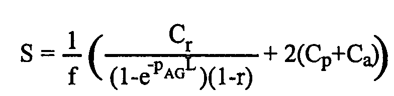 where: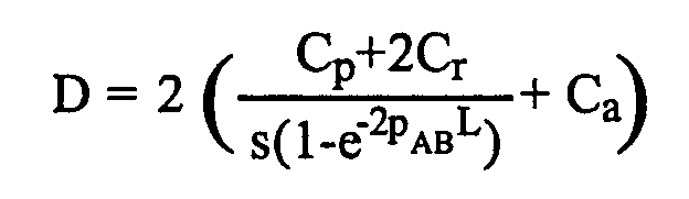
- Cr=the cost of a sequencing reaction,
- Cp=the cost of a primer for PCR and sequencing,
- Ca=the cost of a genotyping assay (excluding primers),
- pXY=SNP frequency between strains X and Y,
- L=read length,
- r=repetitive fraction of the genome,
- s=the success rate for genotyping assays using the original pair of primers, and
- f=the probabilty of any A versus G polymorphism also being an A versus B polymorphism, given by the formula: f=(1+(pAB-pBG)/pAG)/2.
-
- The inventors have calculated D and S for a wide range of values for these parameters. Extreme values can be found under which, irrespective of polymorphism frequency (pAB), either D is always less than S (e.g. a highly repetitive genome (r≈1)), or S is always less than D (e.g. genotyping assays are difficult to design using the original primers (s≈0), B is largely isogenic with G (pBG≈0), or primers and assays are much cheaper than sequencing reactions (Cp,Ca≈0)). However, over a broad range of realistic values, there is a cut-off polymorphism frequency, above which D < S and below which S < D. Specifically, for the FRT and EP Drosophila strains, the inventors determined that the directed approach is favorable if one is interested in any SNP or only RFLP-SNPs, while the shotgun approach would be more suitable if one were interested only in larger (≥7bp) InDels.
- The inventors searched for sequence polymorphisms in each of the 10 autosomal contigs in
Release 2 of the Drosophila genome with a length of at least 1 Mb (Adams et al., 2000). Together, these contigs comprise 90 043 kb. Loci for amplification were selected from non-coding and non-repetitive regions evenly distributed across each contig. Noncoding regions were defined by the lack of any known or predicted open reading frame. Non-repetitive regions were defined by the presence of a single high-scoring BLAST hit on theRelease 2 genome sequence. - PCR amplification was performed with genomic DNA prepared from FRT or EP homozygotes, primers (0.4µM), MgCl2 (1.5mM), dNTPs (0.2mM each), and Taq DNA polymerase (TaKaRa, 1.25U). Cycle conditions were 94 °C for 5 minutes, followed by 35 cycles of 94 °C for 30 seconds, 62 °C for 30 seconds, and 72 °C for 2 minutes, and a final extension at 72 °C for 7 minutes. PCR was performed using either a GeneAmp 9700 (Perkin-Elmer) or Primus HT (MWG Biotech). PCR products were treated with ExoSAP-IT (US Biochemicals) to remove primers and excess nucleotides, and sequenced on an ABI377 or ABI3100 in two separate reactions, each using one of the PCR primers as a sequencing primer.
- Individual sequence traces were called with PHRED 0.000925.c (Ewing et al., 1998a; Ewing et al., 1998b). Sequences were then preclustered into 1 Mb genomic intervals according to the initial PCR primer selection, and pair-wise alignments to the
corresponding Release 2 genome sequence prepared for POLYBAYES using CrossMatch (Marth et al., 1999). Sequences were then multiply realigned using the POLYBAYES 3.0 (release 2001-03-27) anchored alignment algorithm16. Default parameters were used, with the exception that paralog filtering was unnecessary due to the preselection of unique regions, and therefore suppressed. TheRelease 2 genome sequence was assigned a PHRED score of 40. Polymorphisms reported by POLYBAYES were retained only if the PHRED quality score of each variant nucleotide exceeded 20, and the PSNP value assigned by POLYBAYES exceeded 0.5 (corresponding to a PHRED score of 33 in a pair-wise alignment). FRT and EP sequence traces were also aligned by the SeqMan program (DNAStar) and visually inspected. These alignments were used to make minor adjustments to the POLYBAYES alignment, and for the detection of most InDels larger than 8 nucleotides. SNPs were screened for the presence of recognition sites for a panel of 186 restriction endonucleases. - All PCR primers were designed using Primer3, which is available from Whitehead Institute/MIT Center for Genome Research (http://www-genome.wi.mit.edu/cgi-bin/primer/primer3_www.cgi). For SNP identification and PLP assays, primers were selected with melting temperatures of 67-73 °C, maximum Tm difference 1 °C, length 20-30 nucleotides, 20-80% GC content, maximum self-complementarity 4.0, and maximum 3' self-complementarity 1.0. Primers for SNP identification were chosen to give a product of 1.0-1.3 kb.
- Genomic DNA was prepared either from 20-50 flies in 1ml reaction tubes, or from one or two flies in 96-well plates. For the former, DNA was resuspended in 50µl, of which 1-2µl used for PCR. For preparation in 96-well format, flies were manually crushed in 50µl of extraction buffer (10mM Tris-Cl, 1mM EDTA, 25mM NaCl, 200µg/ml Proteinase K) and incubated at 37 °C for 30 minutes. Proteinase K was then inactivated by incubation at 95 °C for 5 minutes, and 1µl used for PCR. PCR conditions were identical to those described above, except that extension times were reduced to 1 minute for PLP assays. For PLP assays, amplification products were separated on 2.5% agarose gels. For RFLP assays, 5µl PCR product was digested with the appropriate restriction enzyme in a 20µl reaction, and separated on a 1% agarose gel.
- A set of 274 predicted RFLP-SNPs were selected for verification, based on the presence of a recognition site for a readily obtainable restriction endonuclease. 87% of these predicted RFLP-SNPs were experimentally confirmed. A negative result may be due to the presence of multiple recognition sites for the selected enzyme, similarities in the fragment sizes from the two strains, or a false prediction. It may be expected that, with optimal primer design, RFLP assays could be designed for more than 90% of the predicted RFLP-SNPs, allowing genotyping of 98% of those markers that contain at least one RFLP-SNP (89% of all markers).
- For PLP assays, the product size was typically 130-230 bp (for InDels of up to 20bp) or 200-500 bp (for larger InDels). Primers used for SNP identification were also used for RFLP assays, although they are not necessarily the optimal choice for distinguishing the alternative fragment sizes. All oligonucleotides were synthesized by MWG Biotech.
- Recombinants were recovered between the 2R-R971 mutation and one of four selected P {white+ } insertions in the 59DE region: l(2)k07136, l(2)s4830, l(2)k06908, or l(2)k05606. Recombinants were identified by screening for chromosomes that lacked the white+ marker but were viable in trans to 2R-J1609, an independent mutation that has the same visual system connectivity defect, maps to 59DF, and fails to complement the lethality of 2R-R971. 71 recombinants were recovered from approximately 30,000 progeny.
-
- Adams, M.D. et al. The genome sequence of Drosophila melanogaster. Science 287, 2185-2195 (2000).
- Begun, D.J. and Aquadro, C.F. Levels of naturally occurring DNA polymorphism correlate with recombination rates in D. melanogaster. Nature 356, 519-520 (1992).
- Benlali, A., Draskovic, I., Hazelett, D.J. & Treisman, J.E. act up controls actin polymerization to alter cell shape and restrict Hedgehog signaling in the Drosophila eye disc. Cell 101, 271-281 (2000).
- Brodsky, M. H., Nordstrom, W., Tsang, G., Kwan, E., Rubin, G. M., and Abrams, J. M. (2000). Drosophila p53 binds a damage response element at the reaper locus. Cell 101, 103-13.
- Chan, H. Y., and Bonini, N. M. (2000). Drosophila models of human neurodegenerative disease. Cell Death Differ 7, 1075-80.
- Cho, R.J. et al. Genome-wide mapping with biallelic markers in Arabidopsis thaliana. Nat. Genet. 23, 203-207 (1999).
- Chou, T. B., Noll, E., and Perrimon, N. (1993). Autosomal P[ovoD1] dominant female-sterile insertions in Drosophila and their use in generating germ-line chimeras. Development 119, 1359-69.
- Collins, F.S., Guyer, M.S. & Charkravarti, A. Variations on a theme: cataloging human DNA sequence variation. Science 278, 1580-1581 (1997).
- Current Protocols in Molecular Biology, Vol. 1, Chap. 2.10, John Wiley & Sons, Pubhishers (1994)
- Ewing, B., Hillier, L., Wendl, M.C. & Green, P. Base-calling of automated sequencer traces using phred. I. Accuracy assessment. Genome Res. 8, 175-185 (1998a).
- Ewing, B. & Green, P. Base-calling of automated sequencer traces using phred. II. Error probabilities. Genome Res. 8, 186-194 (1998b).
- Feany, M. B., and Bender, W. W. (2000). A Drosophila model of Parkinson's disease. Nature 404, 394-398.
- Fernandez-Funez, P., Nino-Rosales, M. L., de Gouyon, B., She, W. C., Luchak, J. M., Martinez, P., Turiegano, E., Benito, J., Capovilla, M., Skinner, P. J., McCall, A., Canal, I., Orr, H. T., Zoghbi, H. Y., and Botas, J. (2000). Identification of genes that modify ataxin-1-induced neurodegeneration. Nature 408, 101-6.
- Frohman, PCR Methods App., (1994) 4:540-558
- Go and Artavanis-Tsakonas, Genetics (1998) 150:211-220
- Greenspan, R. Fly Pushing : The Theory and Practice of Drosophila Genetics, Cold Spring Harbor Laboratory Press, 1997
- Hooper and Scott, Cell (1989) 59:751-765
- Hoskins, R.A. et al. Single Nucleotide Polymorphism Markers for Genetic Mapping in Drosophila melanogaster. Genome Res. 11, 1100-1113(2001).
- Hunkapiller et al., Nature (1984) 310:105-111
- Jackson, G. R., Salecker, I., Dong, X., Yao, X., Arnheim, N., Faber, P. W., MacDonald, M. E., and Zipursky, S. L. (1998). Polyglutamine-expanded human huntingtin transgenes induce degeneration of Drosophila photoreceptor neurons. Neuron 21, 633-642.
- Jin, S., Martinek, S., Joo, W. S., Wortman, J. R., Mirkovic, N., Sali, A., Yandell, M. D., Pavletich, N. P., Young, M. W., and Levine, A. J. (2000). Identification and characterization of a p53 homologue in Drosophila melanogaster. Proc Natl Acad Sci U S A 97, 7301-6.
- Karim, F. D., Chang, H. C., Therrien, M., Wassarman, D. A., Laverty, T., and Rubin, G. M. (1996). A screen for genes that function downstream of Ras1 during Drosophila eye development. Genetics 143, 315-29.
- Karim, F. D., and Rubin, G. M. (1998). Ectopic expression of activated Ras1 induces hyperplastic growth and increased cell death in Drosophila imaginal tissues. Development 125, 1-9.
- Kimmerly, W. et al. A P1-based physical map of the Drosophila euchromatic genome. Genome Res. 6, 414-430 (1996).
- Kwok, P.-Y. Methods for Genotyping Single Nucleotide Polymorphisms. Ann. Rev. Genomics Hum. Genet. 2, 235-258 (2001).
- Lindblad-Toh, K. et al. Large-scale discovery and genotyping of single-nucleotide polymorphisms in the mouse. Nat. Genet. 24, 381-386 (2000).
- Liu, Y. and Montell, D.J. Identification of mutations that cause cell migration defects in mosaic clones. Development 126, 1869-1878 (1999).
- Marth, G.T. et al. A general approach to single-nucleotide polymorphism discovery. Nat. Genet. 23, 452-456 (1999).
- Min, K. T., and Benzer, S. (1999). Preventing neurodegeneration in the Drosophila mutant bubblegum. Science 284, 1985-8.
- Moberg, K. H., Bell, D. W., Wahrer, D. C., Haber, D. A., and Hariharan, I. K. (2001). Archipelago regulates Cyclin E levels in Drosophila and is mutated in human cancer cell lines. Nature 413, 311-6.
- Neuman-Silberberg, F. S., Schejter, E., Hoffmann, F. M., and Shib, B. Z. (1984). The Drosophila ras oncogenes: structure and nucleotide sequence. Cell 37, 1027-33.
- Newsome, T. P., Asling, B., and Dickson, B. J. (2000). Analysis of Drosophila photoreceptor axon guidance in eye-specific mosaics. Development 127, 851-860.
- Ollmann, M., Young, L. M., Di Como, C. J., Karim, F., Belvin, M., Robertson, S., Whittaker, K., Demsky, M., Fisher, W. W., Buchman, A., Duyk, G., Friedman, L., Prives, C., and Kopczynski, C. (2000). Drosophila p53 is a structural and functional homolog of the tumor suppressor p53. Cell 101, 91-101.
- Pichaud, F. and Desplan, C. A new visualization approach for identifying mutations that affect differentiation and organization of the Drosophila ommatidia. Development 128, 815-826 (2001).
- Raftery et al., Genetics 139:241-254 (1995)
- Rorth, P. A modular misexpression screen in Drosophila detecting tissue-specific phenotypes. Proc Natl Acad Sci U S A 93, 12418-22 (1996).
- Rørth, P. et al. Systematic gain-of-function genetics in Drosophila. Development 125, 1049-1057 (1998).
- Ruan, W., Pang, P. & Rao, Y. The SH2/SH3 adaptor protein dock interacts with the Ste20-like kinase misshapen in controlling growth cone motility. Neuron 24, 595-605 (1999).
- Rubin, G. M., Hong, L., Brokstein, P., Evans-Holm, M., Frise, E., Stapleton, M., and Harvey, D. A. (2000a). A Drosophila complementary DNA resource. Science 287, 2222-4.
- Rubin, G. M., Yandell, M. D., Wortman, J. R., Gabor Miklos, G. L., Nelson, C. R., Hariharan, I. K., Fortini, M. E., Li, P. W., Apweiler, R., Fleischmann, W., Cherry, J. M., Henikoff, S., Skupski, M. P., Misra, S., Ashburner, M., Birney, E., Boguski, M. S., Brody, T., Brokstein, P., Celniker, S. E., Chervitz, S. A., Coates, D., Cravchik, A., Gabrielian, A., Galle, R. F., Gelbart, W. M., George, R. A., Goldstein, L. S., Gong, F., Guan, P., Harris, N. L., Hay, B. A., Hoskins, R. A., Li, J., Li, Z., Hynes, R. O., Jones, S. J., Kuehl, P. M., Lemaitre, B., Littleton, J. T., Morrison, D. K., Mungall, C., O'Farrell, P. H., Pickeral, O. K., Shue, C., Vosshall, L. B., Zhang, J., Zhao, Q., Zheng, X. H., Zhong, F., Zhong, W., Gibbs, R., Venter, J. C., Adams, M. D., and Lewis, S. (2000b). Comparative genomics of the eukaryotes. Science 287, 2204-2215.
- Sambrook et al., Molecular Cloning, Cold Spring Harbor (1989)
- Smith, D., Wohlgemuth, J., Calvi, B. R., Franklin, I., and Gelbart, W. M. (1993). hobo enhancer trapping mutagenesis in Drosophila reveals an insertion specificity different from P elements. Genetics 135, 1063-76.
- Spradling, A. C., Stern, D. M., Kiss, I., Roote, J., Laverty, T., and Rubin, G. M. (1995). Gene disruptions using P transposable elements: an integral component of the Drosophila genome project. Proc Natl Acad Sci U S A 92, 10824-30.
- Spradling, A. C., Stern, D., Beaton, A., Rhem, E. J., Laverty, T., Mozden, N., Misra, S., and Rubin, G. M. (1999). The Berkeley Drosophila Genome Project gene disruption project: Single P-element insertions mutating 25% of vital Drosophila genes. Genetics 153, 135-77.
- Tapon, N., Ito, N., Dickson, B. J., Treisman, J. E., and Hariharan, I. K. (2001). The Drosophila tuberous sclerosis complex gene homologs restrict cell growth and cell proliferation. Cell 105, 345-55.
- Teeter, K. et al. Haplotype dimorphism in a SNP collection from Drosophila melanogaster. J. Exp. Zool. 288, 63-75 (2000).
- Warrick, J. M., Paulson, H. L., Gray-Board, G. L., Bui, Q. T., Fischbeck, K. H., Pittman, R. N., and Bonini, N. M. (1998). Expanded polyglutamine protein forms nuclear inclusions and causes neural degeneration in Drosophila. Cell 93, 939-49.
- Wicks, S.R., Yeh, R.T., Gish, W.R., Waterston, R.H. & Plasterk, R.H. Rapid gene mapping in Caenorhabditis elegans using a high density polymorphism map. Nat. Genet. 28, 160-164 (2001).
- Wilton et al., Trends Genet (1996) 12:458.
- Winzeler, E.A. et al. Direct allelic variation scanning of the yeast genome. Science 281, 1194-1197 (1998).
- Wittmann, C. W., Wszolek, M. F., Shulman, J. M., Salvaterra, P. M., Lewis, J., Hutton, M., and Feany, M. B. (2001). Tauopathy in Drosophila: neurodegeneration without neurofibrillary tangles. Science 293, 711-4.
- Xu, T., and Rubin, G. M. (1993). Analysis of genetic mosaics in developing and adult Drosophila tissues. Development 117, 1223-37.
- Xu, T., Wang, W., Zhang, S., Stewart, R. A., and Yu, W. (1995). Identifying tumor suppressors in genetic mosaics: the Drosophila lats gene encodes a putative protein kinase. Development 121, 1053-1063.
-
Claims (31)
- A method for producing a human cDNA molecule derived from a gene that is causally involved in a human disease bycharacterized in that step A.3 comprises the steps ofA. identifying a Drosophila melanogaster gene that is involved in a molecular process or pathway related to a disease of interest by1. obtaining genetically altered flies that exhibit a disease-related phenotype bya) expressing in flies a human gene known to be implicated in the disease, or its Drosophila homologue, is in its normal or mutated form such that expression of the gene results in a gene-specific phenotype; orb) randomly mutating flies and identifying flies that exhibit a disease-related phenotype;2. mutagenizing animals obtained in 1.a) and recovering mutations that modify said phenotype of 1.a); or recovering animals that result in phenotype 1.b);3. mapping the mutated Drosophila gene, and4. cloning the Drosophila gene;B. identifying the orthologue human gene and cloning and amplifying the human cDNA,(a) setting up a genetic cross between the Drosophila strain containing the mutation recovered in step 2., and another strain that is sufficiently divergent at the DNA sequence level, and that contains one or more P element insertions that allow recombination events within a defined genomic interval to be identified;(b) recovering a series of recombinant progeny from this cross, each of which contains a genome that is a mixture of the two parental strains;(c) identifying a set of sequence polymorphisms that discriminates between the two parental strains;(d) determining the phenotype of some or all of the recombinant progeny;(e) assaying for some or all of the sequence polymorphisms identified in (c) in some or all of the recombinant progeny.
- The method of claim 1, wherein in step A.3.a) two strains are used each of which carries a P element located within 1 Mb of the centromere or the telomere, thereby generating a set of recombinants spanning an entire chromsome arm.
- The method of claim 2, whererein in step A.3.a) the strain carrying the mutation of interest is a FRT strain and the other strain is an EP strain carrying an EP element distal to the mutation.
- The method of claim 3, wherein the EP strain carries an EP element that lies distal to but near the mutation of interest and that has been identied from a series of EP elements across the chromosome arm by phenotypic analysis of recombinants between the mutated strain and the EP strain.
- The method of claim 1, wherein in step A.3.b) the recombinants are reocovered due to the expression of an FLP-recombinase that is under the control of an eye-specific enhancer so that recombinants are identified by their mosaicism for an eye-colour marker.
- The method of claim 1, wherein in step A.3.c) the polymorphisms are identified by selecting regions from a set of paired Drosophila melanogaster strains for each chromosome arm, amplifying the selected regions, sequencing the amplified DNA from each pair of homozygotes and determining polymorphisms between the two strains.
- The method of claim 6, wherein the Drosophila strains are a FRT and an EP strain.
- The method of claim 1 or 7, wherein the polymorphisms of step A.3.c) and A.3.d) are selected from the set listed in Tables A - D.
- The method of claim 1, wherein in step A.3.e) the recombinant progeny is assayed for polymorphism(s) by PCR product length polymorphisms (PLP).
- The method of claim 9, wherein the recombinant progeny is assayed for polymorphism(s) listed in Tables A - D by the corresponding PLP assay(s) of Table E.
- The method of claim 1, wherein in step A.3.e) the recombinant progeny is assayed for polymorphism(s) by restriction fragment length polymorphisms (RFLP).
- The method of claim 11, wherein the recombinant progeny is assayed for polymorphism(s) listed in Tables A - D by the corresponding RFPL assay(s) of Table F.
- The method of claim 1, wherein the phenotype of step A.1. is associated with lethality and in step A.3.d) recombinant progeny is recovered by selecting against the mutation-associated lethality.
- The method of claim 1, wherein the recombinant progeny in step A.3.d) are recovered by selecting for recombination between two transposon insertions separated by less than 4 Mb, in either the cis or the trans configuration.
- A method for mapping a mutated gene in Drosophila melanogaster, comprising the steps of(a) setting up a genetic cross between the Drosophila strain containing the mutation and another strain that is sufficiently divergent at the DNA sequence level, and that contains one or more P element insertions that allow recombination events within a defined genomic interval to be identified;(b) recovering a series of recombinant progeny from this cross, each of which contains a genome that is a mixture of the two parental strains;(c) identifying a set of sequence polymorphisms that discriminates between the two parental strains;(d) determining the phenotype of some or all of the recombinant progeny;(e) assaying for some or all of the sequence polymorphisms identified in (c) in some or all of the recombinant progeny.
- The method of claim 15, wherein in step a) two strains are used each of which carries a P element located within 1 Mb of the centromere or the telomere, thereby generating a set of recombinants spanning an entire chromsome arm.
- The method of claim 16, whererein in step a) the strain carrying the mutation is a FRT strain and the other strain is an EP strain element distal to the mutation.
- The method of claim 17, wherein the EP strain carries an EP element that lies distal to but near the mutation of interest and that has been identied from a series of EP elements across the chromosome arm by phenotypic analysis of recombinants between the mutated strain and the EP strain.
- The method of claim 15, wherein in step b) the recombinants are reocovered due to the expression of an FLP-recombinase that is under the control of an eye-specific enhancer so that recombinants are identified by their mosaicism for an eye-colour marker.
- The method of claim 15, wherein in step c) the polymorphisms are identified by selecting regions from a set of paired Drosophila melanogaster strains for each chromosome arm, amplifying the selected regions, sequencing the amplified DNA from each pair of homozygotes and determining polymorphisms between the two strains.
- The method of claim 20, wherein the Drosophila strains are a FRT and an EP strain.
- The method of claim 15 or 20, wherein the polymorphisms of step c) and d) are selected from the set listed in Tables A - D.
- The method of claim 15, wherein in step e) the recombinant progeny is assayed for polymorphism(s) by PCR product length polymorphisms (PLP).
- The method of claim 15, wherein in step e) the recombinant progeny is assayed for polymorphism(s) by restriction fragment length polymorphisms (RFLP).
- The method of claim 23, wherein the recombinant progeny is assayed for polymorphism(s) listed in Tables A - D by the corresponding PLP assay(s) of Table E.
- The method of claim 24, wherein the recombinant progeny is assayed for polymorphism(s) listed in Tables A - D by the corresponding RFPL assay(s) of Table F.
- The method of claim 15, wherein the mutation is associated with lethality and in step d) recombinant progeny is recovered by selecting against the mutation-associated lethality.
- The method of claim 15, wherein the recombinant progeny are recovered by selecting for recombination between two transposon insertions separated by less than 4 Mb, in either the cis or the trans configuration.
- The use of the set of polymorphisms listed in Tables A - D , or a subset thereof, for mapping genes in Drosophila melanogaster.
- The use of a PCR product length polymorphisms (PLP) assay of Table E for for determining a polymorphism in Drosophila melanogaster.
- The use of a restriction fragment length polymorphism (RFLP) assay of Table F for determining a polymorphism in Drosophila melanogaster.
Priority Applications (2)
| Application Number | Priority Date | Filing Date | Title |
|---|---|---|---|
| EP01127195A EP1312681A1 (en) | 2001-11-16 | 2001-11-16 | Method for identifying therapeutic targets by use of genetic screens in Drosophila melanogaster |
| PCT/EP2002/012669 WO2003042407A2 (en) | 2001-11-16 | 2002-11-13 | Method for identifying therapeutic targets by use of genetic screens in drosophila melanogaster |
Applications Claiming Priority (1)
| Application Number | Priority Date | Filing Date | Title |
|---|---|---|---|
| EP01127195A EP1312681A1 (en) | 2001-11-16 | 2001-11-16 | Method for identifying therapeutic targets by use of genetic screens in Drosophila melanogaster |
Publications (1)
| Publication Number | Publication Date |
|---|---|
| EP1312681A1 true EP1312681A1 (en) | 2003-05-21 |
Family
ID=8179247
Family Applications (1)
| Application Number | Title | Priority Date | Filing Date |
|---|---|---|---|
| EP01127195A Withdrawn EP1312681A1 (en) | 2001-11-16 | 2001-11-16 | Method for identifying therapeutic targets by use of genetic screens in Drosophila melanogaster |
Country Status (2)
| Country | Link |
|---|---|
| EP (1) | EP1312681A1 (en) |
| WO (1) | WO2003042407A2 (en) |
Cited By (2)
| Publication number | Priority date | Publication date | Assignee | Title |
|---|---|---|---|---|
| WO2003071268A3 (en) * | 2002-02-20 | 2004-03-11 | Paion Gmbh | Method for discovering pharmacologically active substances |
| WO2010023605A3 (en) * | 2008-08-27 | 2010-07-01 | Inis Biotech Llc | Method for the identification of genes involved in neurodegenerative processes |
Families Citing this family (1)
| Publication number | Priority date | Publication date | Assignee | Title |
|---|---|---|---|---|
| CN103952486B (en) * | 2014-04-23 | 2016-06-22 | 深圳市慢性病防治中心 | A kind of method analyzing VDR gene polymorphism sites Cdx-2 and application |
Citations (1)
| Publication number | Priority date | Publication date | Assignee | Title |
|---|---|---|---|---|
| WO2001051604A2 (en) * | 2000-01-14 | 2001-07-19 | Exelixis, Inc. | Methods for identifying anti-cancer drug targets |
-
2001
- 2001-11-16 EP EP01127195A patent/EP1312681A1/en not_active Withdrawn
-
2002
- 2002-11-13 WO PCT/EP2002/012669 patent/WO2003042407A2/en not_active Ceased
Patent Citations (1)
| Publication number | Priority date | Publication date | Assignee | Title |
|---|---|---|---|---|
| WO2001051604A2 (en) * | 2000-01-14 | 2001-07-19 | Exelixis, Inc. | Methods for identifying anti-cancer drug targets |
Non-Patent Citations (7)
| Title |
|---|
| BERGER JURG ET AL: "Genetic mapping with SNP markers in Drosophila.", NATURE GENETICS, vol. 29, no. 4, December 2001 (2001-12-01), pages 475 - 481, XP002201472, ISSN: 1061-4036 * |
| HOSKINS ROGER A ET AL: "Single nucleotide polymorphism markers for genetic mapping in Drosophila melanogaster.", GENOME RESEARCH, vol. 11, no. 6, June 2001 (2001-06-01), pages 1100 - 1113, XP002201468, ISSN: 1088-9051 * |
| KOCH ROMKE ET AL: "Single nucleotide polymorphisms in wild isolates of Caenorhabditis elegans.", GENOME RESEARCH, vol. 10, no. 11, November 2000 (2000-11-01), pages 1690 - 1696, XP001077876, ISSN: 1088-9051 * |
| TEETER KATHERINE ET AL: "Haplotype dimorphism in a SNP collection from Drosophila melanogaster.", JOURNAL OF EXPERIMENTAL ZOOLOGY, vol. 288, no. 1, 15 April 2000 (2000-04-15), pages 63 - 75, XP002201469, ISSN: 0022-104X * |
| WANG D G ET AL: "Large-scale identification, mapping, and genotyping of single-nucleotide polymorphisms in the human genome", SCIENCE, AMERICAN ASSOCIATION FOR THE ADVANCEMENT OF SCIENCE,, US, vol. 280, 1998, pages 1077 - 1082, XP002089398, ISSN: 0036-8075 * |
| XU TIAN ET AL: "Analysis of genetic mosaics in developing and adult Drosophila tissues.", DEVELOPMENT (CAMBRIDGE), vol. 117, no. 4, 1993, pages 1223 - 1237, XP002201471, ISSN: 0950-1991 * |
| ZIMMERMAN ERIKA ET AL: "Quantitative trait loci affecting components of wing shape in Drosophila melanogaster.", GENETICS, vol. 155, no. 2, June 2000 (2000-06-01), pages 671 - 683, XP002201470, ISSN: 0016-6731 * |
Cited By (2)
| Publication number | Priority date | Publication date | Assignee | Title |
|---|---|---|---|---|
| WO2003071268A3 (en) * | 2002-02-20 | 2004-03-11 | Paion Gmbh | Method for discovering pharmacologically active substances |
| WO2010023605A3 (en) * | 2008-08-27 | 2010-07-01 | Inis Biotech Llc | Method for the identification of genes involved in neurodegenerative processes |
Also Published As
| Publication number | Publication date |
|---|---|
| WO2003042407A2 (en) | 2003-05-22 |
| WO2003042407A3 (en) | 2003-12-18 |
Similar Documents
| Publication | Publication Date | Title |
|---|---|---|
| Berger et al. | Genetic mapping with SNP markers in Drosophila | |
| Malagnac et al. | An Arabidopsis SET domain protein required for maintenance but not establishment of DNA methylation | |
| Ryder et al. | Transposable elements as tools for genomics and genetics in Drosophila | |
| US5994075A (en) | Methods for identifying a mutation in a gene of interest without a phenotypic guide | |
| Doitsidou et al. | Next-generation sequencing-based approaches for mutation mapping and identification in Caenorhabditis elegans | |
| Addo-Quaye et al. | Forward genetics by sequencing EMS variation-induced inbred lines | |
| Chen et al. | Reshaping of global gene expression networks and sex‐biased gene expression by integration of a young gene | |
| Masuya et al. | A series of ENU-induced single-base substitutions in a long-range cis-element altering Sonic hedgehog expression in the developing mouse limb bud | |
| Chen et al. | High-resolution, high-throughput SNP mapping in Drosophila melanogaster | |
| Carlier et al. | Loss of EZH2-like or SU (VAR) 3–9-like proteins causes simultaneous perturbations in H3K27 and H3K9 tri-methylation and associated developmental defects in the fungus Podospora anserina | |
| Besnard et al. | A broad mutational target explains a fast rate of phenotypic evolution | |
| Hentges et al. | Checks and balancers: balancer chromosomes to facilitate genome annotation | |
| Martin et al. | A rapid method to map mutations in Drosophila | |
| Gómez‐Skarmeta et al. | New technologies, new findings, and new concepts in the study of vertebrate cis‐regulatory sequences | |
| Nairz et al. | High-resolution SNP mapping by denaturing HPLC | |
| Garfinkel et al. | Molecular genetics of the Drosophila melanogaster ovo locus, a gene required for sex determination of germline cells. | |
| US6015670A (en) | Methods for identifying a mutation in a gene of interest without a phenotypic guide using ES cells | |
| Hu | Whole genome sequencing and the transformation of C. elegans forward genetics | |
| Brown | Understanding a genome sequence | |
| US20130004951A1 (en) | Bulked mutant analysis | |
| Sharakhova et al. | Chromosomal rearrangements in mosquitoes: from micro-to macroevolution | |
| Jin et al. | A mutation linked to degt-1 (ok3307) in C. elegans strain VC2633 affects rpm-1 | |
| EP0914469A1 (en) | Methods for identifying a mutation in a gene of interest | |
| Park et al. | Isolation of deletion mutants by reverse genetics in Caenorhabditis elegans | |
| WO2003042407A2 (en) | Method for identifying therapeutic targets by use of genetic screens in drosophila melanogaster |
Legal Events
| Date | Code | Title | Description |
|---|---|---|---|
| PUAI | Public reference made under article 153(3) epc to a published international application that has entered the european phase |
Free format text: ORIGINAL CODE: 0009012 |
|
| AK | Designated contracting states |
Designated state(s): AT BE CH CY DE DK ES FI FR GB GR IE IT LI LU MC NL PT SE TR |
|
| AX | Request for extension of the european patent |
Extension state: AL LT LV MK RO SI |
|
| AKX | Designation fees paid | ||
| REG | Reference to a national code |
Ref country code: DE Ref legal event code: 8566 |
|
| STAA | Information on the status of an ep patent application or granted ep patent |
Free format text: STATUS: THE APPLICATION IS DEEMED TO BE WITHDRAWN |
|
| 18D | Application deemed to be withdrawn |
Effective date: 20031122 |