This invention finds industrial applicability in
measuring x-ray and synchrotron radiation powder
diffraction data using one- and two-dimensional detectors
such as a position sensitive proportional counter (PSPC),
an imaging plate and a large CCD detector.
An essential problem that should be addressed in
measuring powder x-ray diffraction data is how to achieve
efficient improvement in the angular resolution and the
statistical precision of data. One means for solving the
problem of statistical precision is using a one- or two-dimensional
detector. This not only shortens the time
required to measure the whole powder diffraction data; in
addition, the time for measurement at an individual point
is equal to the time for measuring the whole powder
diffraction data and, hence, the overall time of
measurement can be shortened by a factor of at least 10,
enabling a marked improvement in statistical precision to
be realized efficiently.
For improvement of angular resolution, it is
effective to perform a parallel-beam x-ray diffraction
experiment using a highly directional x-ray source such as
synchrotron radiation. On the other hand, the effort to
improve the angular resolution of data is limited by the
position resolution of the detector, or the size of a
detection unit. For example, it is impossible to get
information finer than a data detection unit (pixel) in the
detector such as an imaging plate. Hence, in order to
measure finer (higher-resolution) data, various efforts
have been made to reduce the actual size of the detection
unit or improve the apparent resolution of the detection
unit by increasing the size of a measuring instrument such
as a diffractometer on which the detector is to be mounted.
Angular resolution is the resolution of a diffraction
angle in x-ray diffraction data and high angular resolution
means a small half-peak width of powder x-ray profile
(which is the width at half intensity as expressed by
diffraction angle). Angular resolution is expressed in
degrees.
The imaging plate is a new x-ray film prepared by
coating a plastic film with a stimulable phosphor that
serves as a memory of x-ray energy. Unlike PSPC and CCD
that perform measurement with sensing elements that are
fixed in absolute positions on the detector, the imaging
plate is capable of detecting x-rays in a desired position
on the film (20 cm x 40 cm) and the detection unit can be
set in any desired position. In addition, the imaging
plate has high sensitivity and wide exposure latitude.
Because of these advantages, the imaging plate is currently
one of the most commonly used two-dimensional detectors.
The following may be mentioned about the measurement
of x-ray diffraction data using the imaging plate.
Thus, a new approach has been desired in order to
improve the resolution in measurement of x-ray diffraction
data using a one- or two-dimensional detector.
A prior art technology for providing improved
position resolution in measurement with a one- or two-dimensional
detector is a radiation imaging device for
typical use in x-ray medical equipment that is designed to
obtain two-dimensional image data by displacing a two-dimensional
array sensor in both a horizontal and a
vertical direction by a distance equal to one half the
pixel width (Japanese Patent Laid-Open No. 372892/1992).
Similar prior art techniques include the following: a
mask is placed before a two-dimensional detector that is
smaller than the detection unit and moved relative to the
detector relative so as to provide a fine two-dimensional
image (Japanese Patent Laid-Open No. 82305/1994); a two-dimensional
detector is moved by small distances in an
imaging device in an x-ray interferometer to produce
composite imaging data and the detection efficiency of each
sensing element is corrected to enhance the space
resolution (Japanese Patent Laid-Open No. 51881/1999); and
a one-dimensional x-ray detector is moved by small
distances to produce a fine two-dimensional image (Japanese
Patent Laid-Open No. 10220/1989).
There are two other techniques based on a similar
concept. In one approach, a detector array consisting of
multiple one-dimensional rows of radiation sensing elements
is moved in a predetermined direction as the output data
from each element is gathered by a photon counting method
so as to capture a two-dimensional radiation image
(Japanese Patent Laid-Open No. 204283/1992); the other
approach is a radiation CT apparatus which allows an object
to be exposed to a radiation such as x-rays or γ-rays at a
fan beam angle and in which the step of translating the
radiation source and the detector relative to each other
and the step of rotating the detector about the object are
repeated by turns to reconstruct slices through the object
to produce a tomogram (Japanese Patent Laid-Open No.
201253/1990).
As described above, various apparatuses and methods
have been proposed with a view to improving resolution in
x-ray imaging using a one- or two-dimensional detector.
However, in order to apply those techniques to measurement
of powder x-ray diffraction data, the following problems
must be solved.
An object, therefore, of the present invention is to
improve the angular resolution in the measurement of x-ray
powder diffraction data with a one- or two-dimensional
detector to thereby ensure that the aforementioned problems
with employing parallel-beam x-rays such as synchrotron
radiation that inherently undergo attenuation of incident
strength over time and which are highly directional are
solved while permitting powder diffraction data of high
angular resolution to be measured in high precision using a
one- or two-dimensional detector.
In the conventional methods of measuring powder x-ray
diffraction data, a one-dimensional detector (PSPC) or a
two-dimensional detector (imaging plate, IP) remains fixed
or is moved during data measurement. According to the
invention, a high-resolution powder diffraction experiment
is performed using synchrotron radiation or other highly
directional, parallel-beam x-rays as an x-ray source in
such a way that a detector mounted on a measuring
instrument such as a diffractometer is finely moved by
distances smaller than the distance between adjacent x-ray
detection units (pixels) in order to measure data for
interpolation between pixels and the obtained interpolating
data are put together to thereby improve the spatial
resolution in measurement that has been limited by the
detection unit in the detector (i.e., the fineness of each
pixel in the imaging plate). This can theoretically
improve resolution without increasing the size of the
measuring instrument or decreasing the size of the
detector.
The idea of the present invention which relies upon
detector movement for improving spatial resolution finds a
lot of parallels in the prior art. What is unique about
the invention is that it solved the first problem of the
prior art by synthesizing final powder diffraction data
with individual pieces of data being scaled on the basis of
their background intensities. By this technology of the
invention, variations in the intensity of synchrotron
radiation as x-rays can be corrected simultaneously with
the synthesis of data. In the method of the invention,
scaling the intensity of measurement for two kinds of data
on the basis of the averages of their background
intensities is an indispensable step and particularly
effective in an experiment using synchrotron radiation from
an accelerator as an x-ray source which inherently
experiences intensity attenuation over time.
By employing a large detector such as an imaging
plate, measurement can be performed over a very wide area
(20 cm x 40 cm) at a time with the detection unit being set
in a desired position. In addition, by using a two-dimensional
detector that does not require the correction
of detection efficiency, data synthesis can be accomplished
without correcting the shape or detection efficiency of
sensing elements. This contributes to solving the second
problem of the prior art.
As for the improvement in statistical precision that
can be achieved with a two-dimensional detector, data in a
direction perpendicular to the direction of detector
movement are integrated over a few pixels to construct
one-dimensional powder diffraction data and, as a result,
the statistical precision of data is improved by a degree
corresponding to the number of pixels over which
integration was made. When a two-dimensional detector is
to be used in the invention, it is particularly noteworthy
that a train of data in a direction perpendicular to the
direction of data synthesis may be integrated over a few
pixels to an extent that will not deteriorate the
resolution of powder diffraction profiles but which is
sufficient to improve the statistical precision of
intensity. This technique has no parallel in the prior
art.
The present invention is now described in greater
detail by reference to the following examples which are
intended for illustrative purposes only.
Example 1:
Fig. 1 shows schematically an example of applying the
method of data synthesis of the invention to measurements
with a one-dimensional detector. The measurement was
directed to an intensity distribution having a Gaussian
distribution. In the example, the size of a minimum
detection unit (pixel) in the detector was 100 µm square
and the angular resolution in measurement of diffraction
data is expressed by the angle subtended by a source of
signal transmission having the intensity distribution of
interest.
Example 2:
When synchrotron radiation data was used, the
intensity of incident x-rays attenuated and two kinds of
diffraction intensity data, which underwent relative
changes as shown in Fig. 2A, could not be put together in
the correct way. In that case, the intensity ratio between
data 1 and data 2 was calculated from the mean background
intensities in relatively flat portions of curves which
should theoretically be identical between the two kinds of
data. By scaling the intensity data with the calculated
ratio which was 1.149:1.000 in the illustrated case, data 1
and data 2 could be put together with the variations in the
intensity data for the incident x-rays being properly
corrected as shown in Fig. 2B. This is a very unique
feature of the method of the invention.
Example 3:
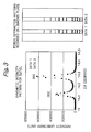
Fig. 3 shows the result obtained when the method of
Example 2 was actually applied to an x-ray powder
diffraction experiment at SPring-8, BL02B2. The size of
pixels in the detector was 50 µm square. Obviously, data 1
effectively interpolated data 2 and vice versa so that a
profile of x-ray diffraction intensity could be closely
measured by the concept of the invention.
Example 4:
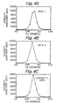
Figs. 4A-4C show results of applying the method of
the invention to a powder x-ray diffraction experiment at
SPring-8, BL02B2. The sample was Standard Reference
Material 660, a LaB6 sample prepared at NIST (the National
Institute of Standards & Technology), and an imaging plate
was used as a detector. The respective points of data in
Figs. 4A-4C represent the intensity of diffracted x-rays as
measured with corresponding pixels. By using the invention
technique of raising the statistical precision, intensity
was integrated over 50 pixels in the direction of pixel
arrangement which is perpendicular to the paper; this
achieved a 50-fold increase in intensity.
In the first place, a diffraction profile was
measured with the imaging plate fixed and this gave data 1
(Fig. 4A). Then, the imaging plate was moved by a distance
equal to one half the pixel size and a diffraction profile
was measured to give data 2 (Fig. 4B). Obviously, no neat
diffraction profile could be measured for data 1 or data 2
by the conventional technique.
Fig. 4C shows the synthetic data measured by the
method of the invention and the shape of a diffraction
profile is represented in detail, clearly demonstrating the
effectiveness of the invention which was already
illustrated in Fig. 1. In structural analysis by powder
x-ray diffraction, the area represented by the profile
shown in Fig. 4C provides the integral reflection intensity
necessary for crystal structure determination. Therefore,
the present invention provides an important technique for
measuring the correct integral reflection intensity,
namely, for determining the correct crystal structure.
Example 5:
Rietveld analysis which was a common method of
structural analysis by powder x-ray diffraction was
performed on both data of measurement that had been
subjected to data synthesis and data of measurement that
had been subjected to data synthesis by the invention; the
results are shown in Figs. 5A and 5B, respectively. The
reliability factor in structural analysis is expressed by
100 x |Ical - Iobs|/Iobs, where Iobs is an integral reflection
intensity and Ical is the intensity calculated from a
crystal's structure model. The reliability factor is an
index for the agreement between the observed intensity and
the calculated intensity. Without data synthesis, the
reliability factor (R1) in the determined crystal structure
was 1.9% whereas the data of measurement obtained by the
invention had R1 = 1.5%.
The crystal structure of LaB6 used as the reference
sample is well known and the lower the value of reliability
factor, the higher the reliability of the intensity data
obtained. If the reliability factor in a structural
analysis of the reference sample decreases from 1.9% to
1.5%, the improvement is about 20% which is an outstanding
value in terms of analytical precision. It is therefore
clear that the invention contributes a lot to improving the
resolution and reliability in the measurement of powder
x-ray diffraction data.
Example 6:
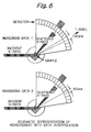
Fig. 6 shows schematically an apparatus for
performing measurement by the method of the invention using
either an imaging plate or a curved PSPC as a detector.
The size of each detection unit is supposed to be 100 µm.
The detector measures scattered x-rays from the sample with
respective detection units. After data 1 is measured, the
curved detector is rotated around the sample such that each
detection unit is displaced by 50 µm. A second measurement
is performed to obtain data 2, which is subsequently
composited with data 1. Depending on the object, the
detector may be planar rather than curved but processing is
still the same.
In the upper part of Fig. 6, x-rays at both ends
strike two pixels and can be measured but the center ray is
not measured since it falls in the gap between the two
pixels. In the lower part of Fig. 6, each pixel is
displaced by a distance equal to one half its size, so the
center x-ray hits a pixel to be measured but the two rays
at both ends fall in inter-pixel gaps and are not measured.
In order to measure the diffraction of x-rays with
unstable intensity such as synchrotron radiation by the
method of the invention, a detector mounted on the
measuring instrument is finely moved by one nth (1/n) of
the distance between adjacent x-ray detection units
(pixels) and data for interpolation between pixels is
measured n times so that the variation in incident
intensity is corrected by using part of the obtained data
and then the measured data are put together to ensure that
the spatial resolution of measurement that has been limited
by the detection unit in the detector is improved by a
factor of n; this is a characteristic, marked advantage of
the invention.
The present invention can also improve resolution
without increasing the size of the measuring instrument or
decreasing the size of the detection unit. When a two-dimensional
detector is to be used in the invention, data
in a direction perpendicular to the direction of detector
movement may be integrated over a few pixels to construct
one-dimensional powder diffraction data and, as a result,
the statistical precision of data is improved by a degree
corresponding to the number of pixels over which
integration was made. This is another characteristic,
marked advantage of the invention.