EP1309703B1 - Kallikrein gene - Google Patents
Kallikrein gene Download PDFInfo
- Publication number
- EP1309703B1 EP1309703B1 EP01966837A EP01966837A EP1309703B1 EP 1309703 B1 EP1309703 B1 EP 1309703B1 EP 01966837 A EP01966837 A EP 01966837A EP 01966837 A EP01966837 A EP 01966837A EP 1309703 B1 EP1309703 B1 EP 1309703B1
- Authority
- EP
- European Patent Office
- Prior art keywords
- protein
- klk15
- nucleic acid
- substance
- gene
- Prior art date
- Legal status (The legal status is an assumption and is not a legal conclusion. Google has not performed a legal analysis and makes no representation as to the accuracy of the status listed.)
- Expired - Lifetime
Links
- 108060005987 Kallikrein Proteins 0.000 title abstract description 68
- 108090000623 proteins and genes Proteins 0.000 claims abstract description 247
- 102000004169 proteins and genes Human genes 0.000 claims abstract description 159
- 239000000203 mixture Substances 0.000 claims abstract description 18
- 150000007523 nucleic acids Chemical class 0.000 claims description 134
- 238000000034 method Methods 0.000 claims description 121
- 102000039446 nucleic acids Human genes 0.000 claims description 110
- 108020004707 nucleic acids Proteins 0.000 claims description 110
- 239000000126 substance Substances 0.000 claims description 78
- 206010028980 Neoplasm Diseases 0.000 claims description 64
- 210000001519 tissue Anatomy 0.000 claims description 57
- 230000014509 gene expression Effects 0.000 claims description 55
- 201000011510 cancer Diseases 0.000 claims description 49
- 150000001875 compounds Chemical class 0.000 claims description 43
- 108090000765 processed proteins & peptides Proteins 0.000 claims description 40
- 102000004196 processed proteins & peptides Human genes 0.000 claims description 37
- 239000000523 sample Substances 0.000 claims description 33
- 206010060862 Prostate cancer Diseases 0.000 claims description 28
- 208000000236 Prostatic Neoplasms Diseases 0.000 claims description 28
- 238000003752 polymerase chain reaction Methods 0.000 claims description 25
- 238000012360 testing method Methods 0.000 claims description 24
- 108091028043 Nucleic acid sequence Proteins 0.000 claims description 23
- 239000003795 chemical substances by application Substances 0.000 claims description 20
- 229960005486 vaccine Drugs 0.000 claims description 20
- 239000013598 vector Substances 0.000 claims description 18
- 238000011282 treatment Methods 0.000 claims description 16
- 230000001225 therapeutic effect Effects 0.000 claims description 15
- 241001465754 Metazoa Species 0.000 claims description 14
- 238000009396 hybridization Methods 0.000 claims description 13
- 229920001184 polypeptide Polymers 0.000 claims description 13
- 230000004071 biological effect Effects 0.000 claims description 12
- 230000003993 interaction Effects 0.000 claims description 11
- 238000004519 manufacturing process Methods 0.000 claims description 11
- 230000015572 biosynthetic process Effects 0.000 claims description 10
- 239000011541 reaction mixture Substances 0.000 claims description 10
- 230000000295 complement effect Effects 0.000 claims description 9
- 238000001514 detection method Methods 0.000 claims description 9
- 239000003814 drug Substances 0.000 claims description 9
- 230000002708 enhancing effect Effects 0.000 claims description 7
- 238000000338 in vitro Methods 0.000 claims description 7
- 239000012472 biological sample Substances 0.000 claims description 6
- 239000008194 pharmaceutical composition Substances 0.000 claims description 6
- 230000004936 stimulating effect Effects 0.000 claims description 6
- 238000012544 monitoring process Methods 0.000 claims description 5
- 230000009918 complex formation Effects 0.000 claims description 4
- 239000003085 diluting agent Substances 0.000 claims description 4
- 239000003112 inhibitor Substances 0.000 claims description 4
- 238000009472 formulation Methods 0.000 claims description 3
- 230000003389 potentiating effect Effects 0.000 claims description 3
- 231100000419 toxicity Toxicity 0.000 claims description 3
- 230000001988 toxicity Effects 0.000 claims description 3
- 238000012258 culturing Methods 0.000 claims description 2
- 239000003937 drug carrier Substances 0.000 claims description 2
- 238000007876 drug discovery Methods 0.000 claims description 2
- 239000000546 pharmaceutical excipient Substances 0.000 claims description 2
- 239000000825 pharmaceutical preparation Substances 0.000 claims description 2
- 125000003275 alpha amino acid group Chemical group 0.000 claims 1
- 102000001399 Kallikrein Human genes 0.000 abstract description 51
- 101000605518 Homo sapiens Kallikrein-15 Proteins 0.000 description 210
- 102100038301 Kallikrein-15 Human genes 0.000 description 210
- 235000018102 proteins Nutrition 0.000 description 118
- 241000282414 Homo sapiens Species 0.000 description 87
- 210000004027 cell Anatomy 0.000 description 76
- 108020004414 DNA Proteins 0.000 description 58
- 150000001413 amino acids Chemical group 0.000 description 32
- 208000037265 diseases, disorders, signs and symptoms Diseases 0.000 description 28
- 101150028944 KLK15 gene Proteins 0.000 description 26
- 239000002299 complementary DNA Substances 0.000 description 24
- 230000001105 regulatory effect Effects 0.000 description 22
- 108091032973 (ribonucleotides)n+m Proteins 0.000 description 21
- 230000027455 binding Effects 0.000 description 21
- 238000004458 analytical method Methods 0.000 description 20
- 208000035475 disorder Diseases 0.000 description 20
- 239000013615 primer Substances 0.000 description 20
- 108010072866 Prostate-Specific Antigen Proteins 0.000 description 19
- 102100038358 Prostate-specific antigen Human genes 0.000 description 18
- 235000001014 amino acid Nutrition 0.000 description 18
- 229940024606 amino acid Drugs 0.000 description 18
- 239000013604 expression vector Substances 0.000 description 18
- 239000002773 nucleotide Substances 0.000 description 17
- 125000003729 nucleotide group Chemical group 0.000 description 17
- 102000012479 Serine Proteases Human genes 0.000 description 16
- 108010022999 Serine Proteases Proteins 0.000 description 16
- 239000012634 fragment Substances 0.000 description 16
- 230000000692 anti-sense effect Effects 0.000 description 15
- 108020004999 messenger RNA Proteins 0.000 description 15
- 210000002307 prostate Anatomy 0.000 description 15
- 230000009261 transgenic effect Effects 0.000 description 15
- 238000013518 transcription Methods 0.000 description 14
- 230000035897 transcription Effects 0.000 description 14
- 102000004190 Enzymes Human genes 0.000 description 13
- 108090000790 Enzymes Proteins 0.000 description 13
- 229940088598 enzyme Drugs 0.000 description 13
- 238000003259 recombinant expression Methods 0.000 description 13
- 230000035772 mutation Effects 0.000 description 12
- 108700028369 Alleles Proteins 0.000 description 11
- 108700024394 Exon Proteins 0.000 description 11
- 102000057032 Tissue Kallikreins Human genes 0.000 description 11
- 238000003776 cleavage reaction Methods 0.000 description 11
- 210000001072 colon Anatomy 0.000 description 11
- 230000000694 effects Effects 0.000 description 11
- 108020001507 fusion proteins Proteins 0.000 description 11
- 102000037865 fusion proteins Human genes 0.000 description 11
- 230000007017 scission Effects 0.000 description 11
- 210000001685 thyroid gland Anatomy 0.000 description 11
- 210000003734 kidney Anatomy 0.000 description 10
- 239000003550 marker Substances 0.000 description 10
- 101001091385 Homo sapiens Kallikrein-6 Proteins 0.000 description 9
- 239000005557 antagonist Substances 0.000 description 9
- 210000004436 artificial bacterial chromosome Anatomy 0.000 description 9
- 238000003556 assay Methods 0.000 description 9
- 102000054765 polymorphisms of proteins Human genes 0.000 description 9
- 108091060211 Expressed sequence tag Proteins 0.000 description 8
- 102100034870 Kallikrein-8 Human genes 0.000 description 8
- 239000000556 agonist Substances 0.000 description 8
- 201000010099 disease Diseases 0.000 description 8
- 230000028993 immune response Effects 0.000 description 8
- 238000012216 screening Methods 0.000 description 8
- 238000010561 standard procedure Methods 0.000 description 8
- 238000010240 RT-PCR analysis Methods 0.000 description 7
- 108700019146 Transgenes Proteins 0.000 description 7
- 230000001689 kallikreinlike Effects 0.000 description 7
- 239000000463 material Substances 0.000 description 7
- 230000007170 pathology Effects 0.000 description 7
- 238000013519 translation Methods 0.000 description 7
- 108090000994 Catalytic RNA Proteins 0.000 description 6
- 102000053642 Catalytic RNA Human genes 0.000 description 6
- 108020004705 Codon Proteins 0.000 description 6
- LFQSCWFLJHTTHZ-UHFFFAOYSA-N Ethanol Chemical compound CCO LFQSCWFLJHTTHZ-UHFFFAOYSA-N 0.000 description 6
- 108091092195 Intron Proteins 0.000 description 6
- 108700005084 Multigene Family Proteins 0.000 description 6
- 102000007056 Recombinant Fusion Proteins Human genes 0.000 description 6
- 108010008281 Recombinant Fusion Proteins Proteins 0.000 description 6
- 208000024313 Testicular Neoplasms Diseases 0.000 description 6
- 206010057644 Testis cancer Diseases 0.000 description 6
- 239000013543 active substance Substances 0.000 description 6
- 230000003321 amplification Effects 0.000 description 6
- 230000001413 cellular effect Effects 0.000 description 6
- 238000006243 chemical reaction Methods 0.000 description 6
- 230000002068 genetic effect Effects 0.000 description 6
- 210000004602 germ cell Anatomy 0.000 description 6
- 238000003199 nucleic acid amplification method Methods 0.000 description 6
- 239000013612 plasmid Substances 0.000 description 6
- 108091092562 ribozyme Proteins 0.000 description 6
- 201000003120 testicular cancer Diseases 0.000 description 6
- 108010036927 trypsin-like serine protease Proteins 0.000 description 6
- 108091026890 Coding region Proteins 0.000 description 5
- 102000005720 Glutathione transferase Human genes 0.000 description 5
- 108010070675 Glutathione transferase Proteins 0.000 description 5
- 101001091388 Homo sapiens Kallikrein-7 Proteins 0.000 description 5
- 101710176220 Kallikrein-2 Proteins 0.000 description 5
- 101710176225 Kallikrein-8 Proteins 0.000 description 5
- 108091034117 Oligonucleotide Proteins 0.000 description 5
- 101710131039 Opsin-5 Proteins 0.000 description 5
- 108010076504 Protein Sorting Signals Proteins 0.000 description 5
- 108700008625 Reporter Genes Proteins 0.000 description 5
- 238000012300 Sequence Analysis Methods 0.000 description 5
- 208000024799 Thyroid disease Diseases 0.000 description 5
- 108700022175 Tissue Kallikreins Proteins 0.000 description 5
- 238000013459 approach Methods 0.000 description 5
- 230000003197 catalytic effect Effects 0.000 description 5
- 238000007796 conventional method Methods 0.000 description 5
- 210000001671 embryonic stem cell Anatomy 0.000 description 5
- 239000003623 enhancer Substances 0.000 description 5
- 230000006870 function Effects 0.000 description 5
- 238000001727 in vivo Methods 0.000 description 5
- 230000008488 polyadenylation Effects 0.000 description 5
- 238000002360 preparation method Methods 0.000 description 5
- 230000002265 prevention Effects 0.000 description 5
- 238000012163 sequencing technique Methods 0.000 description 5
- 239000003270 steroid hormone Substances 0.000 description 5
- 238000002560 therapeutic procedure Methods 0.000 description 5
- IJGRMHOSHXDMSA-UHFFFAOYSA-N Atomic nitrogen Chemical compound N#N IJGRMHOSHXDMSA-UHFFFAOYSA-N 0.000 description 4
- 206010009944 Colon cancer Diseases 0.000 description 4
- 101000605522 Homo sapiens Kallikrein-1 Proteins 0.000 description 4
- 101001008919 Homo sapiens Kallikrein-10 Proteins 0.000 description 4
- 101000605514 Homo sapiens Kallikrein-13 Proteins 0.000 description 4
- 101001091365 Homo sapiens Plasma kallikrein Proteins 0.000 description 4
- 101000605534 Homo sapiens Prostate-specific antigen Proteins 0.000 description 4
- 102100038315 Kallikrein-13 Human genes 0.000 description 4
- 208000008839 Kidney Neoplasms Diseases 0.000 description 4
- 108010085220 Multiprotein Complexes Proteins 0.000 description 4
- 102000007474 Multiprotein Complexes Human genes 0.000 description 4
- 108010029485 Protein Isoforms Proteins 0.000 description 4
- 102000001708 Protein Isoforms Human genes 0.000 description 4
- 108020004511 Recombinant DNA Proteins 0.000 description 4
- 206010038389 Renal cancer Diseases 0.000 description 4
- 239000002671 adjuvant Substances 0.000 description 4
- 210000004100 adrenal gland Anatomy 0.000 description 4
- 230000033228 biological regulation Effects 0.000 description 4
- 238000004113 cell culture Methods 0.000 description 4
- 239000003153 chemical reaction reagent Substances 0.000 description 4
- 238000002512 chemotherapy Methods 0.000 description 4
- 210000000349 chromosome Anatomy 0.000 description 4
- 208000029742 colonic neoplasm Diseases 0.000 description 4
- 238000012217 deletion Methods 0.000 description 4
- 230000037430 deletion Effects 0.000 description 4
- 238000003745 diagnosis Methods 0.000 description 4
- 238000009826 distribution Methods 0.000 description 4
- 238000004520 electroporation Methods 0.000 description 4
- 230000002255 enzymatic effect Effects 0.000 description 4
- 238000002474 experimental method Methods 0.000 description 4
- 230000004927 fusion Effects 0.000 description 4
- 201000010982 kidney cancer Diseases 0.000 description 4
- 230000003211 malignant effect Effects 0.000 description 4
- 230000001404 mediated effect Effects 0.000 description 4
- 238000002493 microarray Methods 0.000 description 4
- 238000010369 molecular cloning Methods 0.000 description 4
- 239000012071 phase Substances 0.000 description 4
- 238000000746 purification Methods 0.000 description 4
- 238000001959 radiotherapy Methods 0.000 description 4
- 210000003079 salivary gland Anatomy 0.000 description 4
- 238000001356 surgical procedure Methods 0.000 description 4
- 238000003786 synthesis reaction Methods 0.000 description 4
- 210000001550 testis Anatomy 0.000 description 4
- 229940124597 therapeutic agent Drugs 0.000 description 4
- 239000003053 toxin Substances 0.000 description 4
- 231100000765 toxin Toxicity 0.000 description 4
- 108700012359 toxins Proteins 0.000 description 4
- 238000001890 transfection Methods 0.000 description 4
- 241000283707 Capra Species 0.000 description 3
- 108020004635 Complementary DNA Proteins 0.000 description 3
- 102000053602 DNA Human genes 0.000 description 3
- 101001091371 Homo sapiens Kallikrein-8 Proteins 0.000 description 3
- 101001091356 Homo sapiens Kallikrein-9 Proteins 0.000 description 3
- 108060003951 Immunoglobulin Proteins 0.000 description 3
- 102100034343 Integrase Human genes 0.000 description 3
- 101150009482 KLK1 gene Proteins 0.000 description 3
- 102100038297 Kallikrein-1 Human genes 0.000 description 3
- 102100027613 Kallikrein-10 Human genes 0.000 description 3
- 102100034866 Kallikrein-6 Human genes 0.000 description 3
- 102100034867 Kallikrein-7 Human genes 0.000 description 3
- 238000001347 McNemar's test Methods 0.000 description 3
- 108020004711 Nucleic Acid Probes Proteins 0.000 description 3
- 241000283973 Oryctolagus cuniculus Species 0.000 description 3
- 206010061535 Ovarian neoplasm Diseases 0.000 description 3
- 102100034869 Plasma kallikrein Human genes 0.000 description 3
- 108010092799 RNA-directed DNA polymerase Proteins 0.000 description 3
- MTCFGRXMJLQNBG-UHFFFAOYSA-N Serine Natural products OCC(N)C(O)=O MTCFGRXMJLQNBG-UHFFFAOYSA-N 0.000 description 3
- 108091081024 Start codon Proteins 0.000 description 3
- 108091036066 Three prime untranslated region Proteins 0.000 description 3
- JLCPHMBAVCMARE-UHFFFAOYSA-N [3-[[3-[[3-[[3-[[3-[[3-[[3-[[3-[[3-[[3-[[3-[[5-(2-amino-6-oxo-1H-purin-9-yl)-3-[[3-[[3-[[3-[[3-[[3-[[5-(2-amino-6-oxo-1H-purin-9-yl)-3-[[5-(2-amino-6-oxo-1H-purin-9-yl)-3-hydroxyoxolan-2-yl]methoxy-hydroxyphosphoryl]oxyoxolan-2-yl]methoxy-hydroxyphosphoryl]oxy-5-(5-methyl-2,4-dioxopyrimidin-1-yl)oxolan-2-yl]methoxy-hydroxyphosphoryl]oxy-5-(6-aminopurin-9-yl)oxolan-2-yl]methoxy-hydroxyphosphoryl]oxy-5-(6-aminopurin-9-yl)oxolan-2-yl]methoxy-hydroxyphosphoryl]oxy-5-(6-aminopurin-9-yl)oxolan-2-yl]methoxy-hydroxyphosphoryl]oxy-5-(6-aminopurin-9-yl)oxolan-2-yl]methoxy-hydroxyphosphoryl]oxyoxolan-2-yl]methoxy-hydroxyphosphoryl]oxy-5-(5-methyl-2,4-dioxopyrimidin-1-yl)oxolan-2-yl]methoxy-hydroxyphosphoryl]oxy-5-(4-amino-2-oxopyrimidin-1-yl)oxolan-2-yl]methoxy-hydroxyphosphoryl]oxy-5-(5-methyl-2,4-dioxopyrimidin-1-yl)oxolan-2-yl]methoxy-hydroxyphosphoryl]oxy-5-(5-methyl-2,4-dioxopyrimidin-1-yl)oxolan-2-yl]methoxy-hydroxyphosphoryl]oxy-5-(6-aminopurin-9-yl)oxolan-2-yl]methoxy-hydroxyphosphoryl]oxy-5-(6-aminopurin-9-yl)oxolan-2-yl]methoxy-hydroxyphosphoryl]oxy-5-(4-amino-2-oxopyrimidin-1-yl)oxolan-2-yl]methoxy-hydroxyphosphoryl]oxy-5-(4-amino-2-oxopyrimidin-1-yl)oxolan-2-yl]methoxy-hydroxyphosphoryl]oxy-5-(4-amino-2-oxopyrimidin-1-yl)oxolan-2-yl]methoxy-hydroxyphosphoryl]oxy-5-(6-aminopurin-9-yl)oxolan-2-yl]methoxy-hydroxyphosphoryl]oxy-5-(4-amino-2-oxopyrimidin-1-yl)oxolan-2-yl]methyl [5-(6-aminopurin-9-yl)-2-(hydroxymethyl)oxolan-3-yl] hydrogen phosphate Polymers Cc1cn(C2CC(OP(O)(=O)OCC3OC(CC3OP(O)(=O)OCC3OC(CC3O)n3cnc4c3nc(N)[nH]c4=O)n3cnc4c3nc(N)[nH]c4=O)C(COP(O)(=O)OC3CC(OC3COP(O)(=O)OC3CC(OC3COP(O)(=O)OC3CC(OC3COP(O)(=O)OC3CC(OC3COP(O)(=O)OC3CC(OC3COP(O)(=O)OC3CC(OC3COP(O)(=O)OC3CC(OC3COP(O)(=O)OC3CC(OC3COP(O)(=O)OC3CC(OC3COP(O)(=O)OC3CC(OC3COP(O)(=O)OC3CC(OC3COP(O)(=O)OC3CC(OC3COP(O)(=O)OC3CC(OC3COP(O)(=O)OC3CC(OC3COP(O)(=O)OC3CC(OC3COP(O)(=O)OC3CC(OC3COP(O)(=O)OC3CC(OC3CO)n3cnc4c(N)ncnc34)n3ccc(N)nc3=O)n3cnc4c(N)ncnc34)n3ccc(N)nc3=O)n3ccc(N)nc3=O)n3ccc(N)nc3=O)n3cnc4c(N)ncnc34)n3cnc4c(N)ncnc34)n3cc(C)c(=O)[nH]c3=O)n3cc(C)c(=O)[nH]c3=O)n3ccc(N)nc3=O)n3cc(C)c(=O)[nH]c3=O)n3cnc4c3nc(N)[nH]c4=O)n3cnc4c(N)ncnc34)n3cnc4c(N)ncnc34)n3cnc4c(N)ncnc34)n3cnc4c(N)ncnc34)O2)c(=O)[nH]c1=O JLCPHMBAVCMARE-UHFFFAOYSA-N 0.000 description 3
- 230000005856 abnormality Effects 0.000 description 3
- 108091006088 activator proteins Proteins 0.000 description 3
- 239000000427 antigen Substances 0.000 description 3
- 108091007433 antigens Proteins 0.000 description 3
- 102000036639 antigens Human genes 0.000 description 3
- 239000002246 antineoplastic agent Substances 0.000 description 3
- 230000001580 bacterial effect Effects 0.000 description 3
- 239000011324 bead Substances 0.000 description 3
- 210000004899 c-terminal region Anatomy 0.000 description 3
- 210000002230 centromere Anatomy 0.000 description 3
- 229940044683 chemotherapy drug Drugs 0.000 description 3
- 230000002759 chromosomal effect Effects 0.000 description 3
- 238000010367 cloning Methods 0.000 description 3
- 229920001577 copolymer Polymers 0.000 description 3
- 230000002950 deficient Effects 0.000 description 3
- 229940079593 drug Drugs 0.000 description 3
- 210000002257 embryonic structure Anatomy 0.000 description 3
- 238000005516 engineering process Methods 0.000 description 3
- 230000012010 growth Effects 0.000 description 3
- 239000001963 growth medium Substances 0.000 description 3
- 230000003054 hormonal effect Effects 0.000 description 3
- 230000002209 hydrophobic effect Effects 0.000 description 3
- 230000001900 immune effect Effects 0.000 description 3
- 238000003018 immunoassay Methods 0.000 description 3
- 102000018358 immunoglobulin Human genes 0.000 description 3
- 238000003780 insertion Methods 0.000 description 3
- 230000037431 insertion Effects 0.000 description 3
- 230000036210 malignancy Effects 0.000 description 3
- 239000003607 modifier Substances 0.000 description 3
- 239000002853 nucleic acid probe Substances 0.000 description 3
- 230000001575 pathological effect Effects 0.000 description 3
- 230000008569 process Effects 0.000 description 3
- 238000011160 research Methods 0.000 description 3
- 238000007894 restriction fragment length polymorphism technique Methods 0.000 description 3
- 238000000926 separation method Methods 0.000 description 3
- 239000007787 solid Substances 0.000 description 3
- 210000001082 somatic cell Anatomy 0.000 description 3
- 230000000638 stimulation Effects 0.000 description 3
- 208000024891 symptom Diseases 0.000 description 3
- 108091035539 telomere Proteins 0.000 description 3
- 102000055501 telomere Human genes 0.000 description 3
- 210000003411 telomere Anatomy 0.000 description 3
- 231100000331 toxic Toxicity 0.000 description 3
- 230000002588 toxic effect Effects 0.000 description 3
- 230000009466 transformation Effects 0.000 description 3
- 238000010396 two-hybrid screening Methods 0.000 description 3
- 241001430294 unidentified retrovirus Species 0.000 description 3
- 239000003981 vehicle Substances 0.000 description 3
- 102000007469 Actins Human genes 0.000 description 2
- 108010085238 Actins Proteins 0.000 description 2
- 102000002260 Alkaline Phosphatase Human genes 0.000 description 2
- 108020004774 Alkaline Phosphatase Proteins 0.000 description 2
- 108020005544 Antisense RNA Proteins 0.000 description 2
- 102100026189 Beta-galactosidase Human genes 0.000 description 2
- 108091003079 Bovine Serum Albumin Proteins 0.000 description 2
- 206010006187 Breast cancer Diseases 0.000 description 2
- 208000026310 Breast neoplasm Diseases 0.000 description 2
- UXVMQQNJUSDDNG-UHFFFAOYSA-L Calcium chloride Chemical compound [Cl-].[Cl-].[Ca+2] UXVMQQNJUSDDNG-UHFFFAOYSA-L 0.000 description 2
- 241000282693 Cercopithecidae Species 0.000 description 2
- 239000003155 DNA primer Substances 0.000 description 2
- 229920002307 Dextran Polymers 0.000 description 2
- 238000000729 Fisher's exact test Methods 0.000 description 2
- 241001326189 Gyrodactylus prostae Species 0.000 description 2
- 101100453983 Homo sapiens KLK14 gene Proteins 0.000 description 2
- 101710176219 Kallikrein-1 Proteins 0.000 description 2
- 102100034872 Kallikrein-4 Human genes 0.000 description 2
- 102100034876 Kallikrein-9 Human genes 0.000 description 2
- 101150068025 Klk2 gene Proteins 0.000 description 2
- CKLJMWTZIZZHCS-REOHCLBHSA-N L-aspartic acid Chemical compound OC(=O)[C@@H](N)CC(O)=O CKLJMWTZIZZHCS-REOHCLBHSA-N 0.000 description 2
- 108060001084 Luciferase Proteins 0.000 description 2
- 239000005089 Luciferase Substances 0.000 description 2
- KDXKERNSBIXSRK-UHFFFAOYSA-N Lysine Natural products NCCCCC(N)C(O)=O KDXKERNSBIXSRK-UHFFFAOYSA-N 0.000 description 2
- TWRXJAOTZQYOKJ-UHFFFAOYSA-L Magnesium chloride Chemical compound [Mg+2].[Cl-].[Cl-] TWRXJAOTZQYOKJ-UHFFFAOYSA-L 0.000 description 2
- 239000000020 Nitrocellulose Substances 0.000 description 2
- 238000000636 Northern blotting Methods 0.000 description 2
- 239000004677 Nylon Substances 0.000 description 2
- 206010033128 Ovarian cancer Diseases 0.000 description 2
- 241001494479 Pecora Species 0.000 description 2
- ISWSIDIOOBJBQZ-UHFFFAOYSA-N Phenol Chemical compound OC1=CC=CC=C1 ISWSIDIOOBJBQZ-UHFFFAOYSA-N 0.000 description 2
- 108010001441 Phosphopeptides Proteins 0.000 description 2
- 241000700159 Rattus Species 0.000 description 2
- 238000002105 Southern blotting Methods 0.000 description 2
- 208000024770 Thyroid neoplasm Diseases 0.000 description 2
- 241000251539 Vertebrata <Metazoa> Species 0.000 description 2
- 230000004913 activation Effects 0.000 description 2
- 239000012190 activator Substances 0.000 description 2
- 230000004520 agglutination Effects 0.000 description 2
- 230000001028 anti-proliverative effect Effects 0.000 description 2
- 230000002622 anti-tumorigenesis Effects 0.000 description 2
- 230000000890 antigenic effect Effects 0.000 description 2
- 108010005774 beta-Galactosidase Proteins 0.000 description 2
- 239000013060 biological fluid Substances 0.000 description 2
- 230000005540 biological transmission Effects 0.000 description 2
- 239000001110 calcium chloride Substances 0.000 description 2
- 229960002713 calcium chloride Drugs 0.000 description 2
- 235000011148 calcium chloride Nutrition 0.000 description 2
- 229910001628 calcium chloride Inorganic materials 0.000 description 2
- 150000001720 carbohydrates Chemical class 0.000 description 2
- 235000014633 carbohydrates Nutrition 0.000 description 2
- 239000000969 carrier Substances 0.000 description 2
- 239000003184 complementary RNA Substances 0.000 description 2
- 238000004590 computer program Methods 0.000 description 2
- 125000000151 cysteine group Chemical group N[C@@H](CS)C(=O)* 0.000 description 2
- 230000007423 decrease Effects 0.000 description 2
- 230000001419 dependent effect Effects 0.000 description 2
- 238000002405 diagnostic procedure Methods 0.000 description 2
- 238000001493 electron microscopy Methods 0.000 description 2
- 230000002616 endonucleolytic effect Effects 0.000 description 2
- 230000001747 exhibiting effect Effects 0.000 description 2
- 239000000284 extract Substances 0.000 description 2
- 239000012091 fetal bovine serum Substances 0.000 description 2
- 239000000499 gel Substances 0.000 description 2
- 238000010363 gene targeting Methods 0.000 description 2
- 239000011521 glass Substances 0.000 description 2
- HNDVDQJCIGZPNO-UHFFFAOYSA-N histidine Natural products OC(=O)C(N)CC1=CN=CN1 HNDVDQJCIGZPNO-UHFFFAOYSA-N 0.000 description 2
- 102000055383 human KLK7 Human genes 0.000 description 2
- 230000036039 immunity Effects 0.000 description 2
- 230000002163 immunogen Effects 0.000 description 2
- 230000002637 immunotoxin Effects 0.000 description 2
- 239000002596 immunotoxin Substances 0.000 description 2
- 231100000608 immunotoxin Toxicity 0.000 description 2
- 229940051026 immunotoxin Drugs 0.000 description 2
- 230000001939 inductive effect Effects 0.000 description 2
- 238000002347 injection Methods 0.000 description 2
- 239000007924 injection Substances 0.000 description 2
- 238000001990 intravenous administration Methods 0.000 description 2
- 238000005304 joining Methods 0.000 description 2
- 108010024383 kallikrein 4 Proteins 0.000 description 2
- 108010045069 keyhole-limpet hemocyanin Proteins 0.000 description 2
- 229940039088 kininogenase Drugs 0.000 description 2
- 101150066555 lacZ gene Proteins 0.000 description 2
- 239000007788 liquid Substances 0.000 description 2
- 210000004962 mammalian cell Anatomy 0.000 description 2
- 239000011159 matrix material Substances 0.000 description 2
- 239000012528 membrane Substances 0.000 description 2
- 238000000520 microinjection Methods 0.000 description 2
- 229920001220 nitrocellulos Polymers 0.000 description 2
- 229910052757 nitrogen Inorganic materials 0.000 description 2
- 229920001778 nylon Polymers 0.000 description 2
- 230000003287 optical effect Effects 0.000 description 2
- 230000002018 overexpression Effects 0.000 description 2
- 239000004033 plastic Substances 0.000 description 2
- 229920003023 plastic Polymers 0.000 description 2
- 238000010837 poor prognosis Methods 0.000 description 2
- 239000002243 precursor Substances 0.000 description 2
- 239000002987 primer (paints) Substances 0.000 description 2
- 230000000644 propagated effect Effects 0.000 description 2
- 208000023958 prostate neoplasm Diseases 0.000 description 2
- 230000001681 protective effect Effects 0.000 description 2
- 230000006916 protein interaction Effects 0.000 description 2
- 230000002285 radioactive effect Effects 0.000 description 2
- 230000002829 reductive effect Effects 0.000 description 2
- 230000004044 response Effects 0.000 description 2
- 238000010532 solid phase synthesis reaction Methods 0.000 description 2
- 239000000243 solution Substances 0.000 description 2
- 238000007619 statistical method Methods 0.000 description 2
- 201000002510 thyroid cancer Diseases 0.000 description 2
- 238000012546 transfer Methods 0.000 description 2
- 230000001810 trypsinlike Effects 0.000 description 2
- 210000004881 tumor cell Anatomy 0.000 description 2
- 239000000439 tumor marker Substances 0.000 description 2
- 241000701161 unidentified adenovirus Species 0.000 description 2
- 230000003612 virological effect Effects 0.000 description 2
- NWUYHJFMYQTDRP-UHFFFAOYSA-N 1,2-bis(ethenyl)benzene;1-ethenyl-2-ethylbenzene;styrene Chemical compound C=CC1=CC=CC=C1.CCC1=CC=CC=C1C=C.C=CC1=CC=CC=C1C=C NWUYHJFMYQTDRP-UHFFFAOYSA-N 0.000 description 1
- OWEGMIWEEQEYGQ-UHFFFAOYSA-N 100676-05-9 Natural products OC1C(O)C(O)C(CO)OC1OCC1C(O)C(O)C(O)C(OC2C(OC(O)C(O)C2O)CO)O1 OWEGMIWEEQEYGQ-UHFFFAOYSA-N 0.000 description 1
- NVKAWKQGWWIWPM-ABEVXSGRSA-N 17-β-hydroxy-5-α-Androstan-3-one Chemical group C1C(=O)CC[C@]2(C)[C@H]3CC[C@](C)([C@H](CC4)O)[C@@H]4[C@@H]3CC[C@H]21 NVKAWKQGWWIWPM-ABEVXSGRSA-N 0.000 description 1
- QKNYBSVHEMOAJP-UHFFFAOYSA-N 2-amino-2-(hydroxymethyl)propane-1,3-diol;hydron;chloride Chemical compound Cl.OCC(N)(CO)CO QKNYBSVHEMOAJP-UHFFFAOYSA-N 0.000 description 1
- 108010066676 Abrin Proteins 0.000 description 1
- 102000012440 Acetylcholinesterase Human genes 0.000 description 1
- 108010022752 Acetylcholinesterase Proteins 0.000 description 1
- 101800001401 Activation peptide Proteins 0.000 description 1
- 102400000069 Activation peptide Human genes 0.000 description 1
- 229930024421 Adenine Natural products 0.000 description 1
- GFFGJBXGBJISGV-UHFFFAOYSA-N Adenine Chemical compound NC1=NC=NC2=C1N=CN2 GFFGJBXGBJISGV-UHFFFAOYSA-N 0.000 description 1
- 229920000936 Agarose Polymers 0.000 description 1
- 208000024827 Alzheimer disease Diseases 0.000 description 1
- 102000009091 Amyloidogenic Proteins Human genes 0.000 description 1
- 108010048112 Amyloidogenic Proteins Proteins 0.000 description 1
- 101001007348 Arachis hypogaea Galactose-binding lectin Proteins 0.000 description 1
- 239000004475 Arginine Substances 0.000 description 1
- 108090001008 Avidin Proteins 0.000 description 1
- 241000894006 Bacteria Species 0.000 description 1
- 231100000699 Bacterial toxin Toxicity 0.000 description 1
- 101001011741 Bos taurus Insulin Proteins 0.000 description 1
- 229920002134 Carboxymethyl cellulose Polymers 0.000 description 1
- 102000014914 Carrier Proteins Human genes 0.000 description 1
- 241000700198 Cavia Species 0.000 description 1
- 108091035707 Consensus sequence Proteins 0.000 description 1
- 241000699800 Cricetinae Species 0.000 description 1
- CKLJMWTZIZZHCS-UWTATZPHSA-N D-aspartic acid Chemical compound OC(=O)[C@H](N)CC(O)=O CKLJMWTZIZZHCS-UWTATZPHSA-N 0.000 description 1
- 230000004568 DNA-binding Effects 0.000 description 1
- 108010014303 DNA-directed DNA polymerase Proteins 0.000 description 1
- 102000016928 DNA-directed DNA polymerase Human genes 0.000 description 1
- 241000702421 Dependoparvovirus Species 0.000 description 1
- 208000007342 Diabetic Nephropathies Diseases 0.000 description 1
- 238000009007 Diagnostic Kit Methods 0.000 description 1
- 102000016607 Diphtheria Toxin Human genes 0.000 description 1
- 108010053187 Diphtheria Toxin Proteins 0.000 description 1
- 238000002965 ELISA Methods 0.000 description 1
- 241000196324 Embryophyta Species 0.000 description 1
- YQYJSBFKSSDGFO-UHFFFAOYSA-N Epihygromycin Natural products OC1C(O)C(C(=O)C)OC1OC(C(=C1)O)=CC=C1C=C(C)C(=O)NC1C(O)C(O)C2OCOC2C1O YQYJSBFKSSDGFO-UHFFFAOYSA-N 0.000 description 1
- 241000588724 Escherichia coli Species 0.000 description 1
- 101100284223 Escherichia phage N15 gene 14 gene Proteins 0.000 description 1
- 241000282326 Felis catus Species 0.000 description 1
- 102000008857 Ferritin Human genes 0.000 description 1
- 108050000784 Ferritin Proteins 0.000 description 1
- 238000008416 Ferritin Methods 0.000 description 1
- 108090000331 Firefly luciferases Proteins 0.000 description 1
- 241000233866 Fungi Species 0.000 description 1
- WHUUTDBJXJRKMK-UHFFFAOYSA-N Glutamic acid Natural products OC(=O)C(N)CCC(O)=O WHUUTDBJXJRKMK-UHFFFAOYSA-N 0.000 description 1
- 108091027305 Heteroduplex Proteins 0.000 description 1
- 241000238631 Hexapoda Species 0.000 description 1
- 241000282412 Homo Species 0.000 description 1
- 101000605516 Homo sapiens Kallikrein-12 Proteins 0.000 description 1
- 101001091379 Homo sapiens Kallikrein-5 Proteins 0.000 description 1
- 108010001336 Horseradish Peroxidase Proteins 0.000 description 1
- 206010020772 Hypertension Diseases 0.000 description 1
- XQFRJNBWHJMXHO-RRKCRQDMSA-N IDUR Chemical compound C1[C@H](O)[C@@H](CO)O[C@H]1N1C(=O)NC(=O)C(I)=C1 XQFRJNBWHJMXHO-RRKCRQDMSA-N 0.000 description 1
- 206010061218 Inflammation Diseases 0.000 description 1
- 108700001097 Insect Genes Proteins 0.000 description 1
- 101150003872 KLK3 gene Proteins 0.000 description 1
- 102100038318 Kallikrein-12 Human genes 0.000 description 1
- 101710115873 Kallikrein-15 Proteins 0.000 description 1
- 102100034868 Kallikrein-5 Human genes 0.000 description 1
- 102000010631 Kininogens Human genes 0.000 description 1
- 108010077861 Kininogens Proteins 0.000 description 1
- 102000002397 Kinins Human genes 0.000 description 1
- 108010093008 Kinins Proteins 0.000 description 1
- HNDVDQJCIGZPNO-YFKPBYRVSA-N L-histidine Chemical compound OC(=O)[C@@H](N)CC1=CN=CN1 HNDVDQJCIGZPNO-YFKPBYRVSA-N 0.000 description 1
- ROHFNLRQFUQHCH-YFKPBYRVSA-N L-leucine Chemical compound CC(C)C[C@H](N)C(O)=O ROHFNLRQFUQHCH-YFKPBYRVSA-N 0.000 description 1
- ROHFNLRQFUQHCH-UHFFFAOYSA-N Leucine Natural products CC(C)CC(N)C(O)=O ROHFNLRQFUQHCH-UHFFFAOYSA-N 0.000 description 1
- 239000004472 Lysine Substances 0.000 description 1
- GUBGYTABKSRVRQ-PICCSMPSSA-N Maltose Natural products O[C@@H]1[C@@H](O)[C@H](O)[C@@H](CO)O[C@@H]1O[C@@H]1[C@@H](CO)OC(O)[C@H](O)[C@H]1O GUBGYTABKSRVRQ-PICCSMPSSA-N 0.000 description 1
- 206010027476 Metastases Diseases 0.000 description 1
- 108091092878 Microsatellite Proteins 0.000 description 1
- 241001529936 Murinae Species 0.000 description 1
- 241000699666 Mus <mouse, genus> Species 0.000 description 1
- 101000605526 Mus musculus Kallikrein-1 Proteins 0.000 description 1
- 101100180428 Mus musculus Klk1b1 gene Proteins 0.000 description 1
- 101100180429 Mus musculus Klk1b3 gene Proteins 0.000 description 1
- 241000699670 Mus sp. Species 0.000 description 1
- 102000007999 Nuclear Proteins Human genes 0.000 description 1
- 108010089610 Nuclear Proteins Proteins 0.000 description 1
- 101710163270 Nuclease Proteins 0.000 description 1
- 108700026244 Open Reading Frames Proteins 0.000 description 1
- 238000012408 PCR amplification Methods 0.000 description 1
- 241000282579 Pan Species 0.000 description 1
- 241001443706 Papio papio Species 0.000 description 1
- 108010033276 Peptide Fragments Proteins 0.000 description 1
- 102000007079 Peptide Fragments Human genes 0.000 description 1
- 108010067902 Peptide Library Proteins 0.000 description 1
- 102000003992 Peroxidases Human genes 0.000 description 1
- 241000009328 Perro Species 0.000 description 1
- 108090000113 Plasma Kallikrein Proteins 0.000 description 1
- 239000004793 Polystyrene Substances 0.000 description 1
- XBDQKXXYIPTUBI-UHFFFAOYSA-M Propionate Chemical compound CCC([O-])=O XBDQKXXYIPTUBI-UHFFFAOYSA-M 0.000 description 1
- 241000589516 Pseudomonas Species 0.000 description 1
- 201000004681 Psoriasis Diseases 0.000 description 1
- KDCGOANMDULRCW-UHFFFAOYSA-N Purine Natural products N1=CNC2=NC=NC2=C1 KDCGOANMDULRCW-UHFFFAOYSA-N 0.000 description 1
- 101710105014 Putative serine protease Proteins 0.000 description 1
- 101000702488 Rattus norvegicus High affinity cationic amino acid transporter 1 Proteins 0.000 description 1
- 101100288126 Rattus norvegicus Klk3 gene Proteins 0.000 description 1
- 101100453992 Rattus norvegicus Ngfg gene Proteins 0.000 description 1
- 102000006382 Ribonucleases Human genes 0.000 description 1
- 108010083644 Ribonucleases Proteins 0.000 description 1
- 108091028664 Ribonucleotide Proteins 0.000 description 1
- 108010039491 Ricin Proteins 0.000 description 1
- 241000283984 Rodentia Species 0.000 description 1
- 239000006146 Roswell Park Memorial Institute medium Substances 0.000 description 1
- 240000004808 Saccharomyces cerevisiae Species 0.000 description 1
- 229920005654 Sephadex Polymers 0.000 description 1
- 239000012507 Sephadex™ Substances 0.000 description 1
- 229920002684 Sepharose Polymers 0.000 description 1
- 241000607768 Shigella Species 0.000 description 1
- 108010090804 Streptavidin Proteins 0.000 description 1
- 241000282887 Suidae Species 0.000 description 1
- 206010042602 Supraventricular extrasystoles Diseases 0.000 description 1
- 108010006785 Taq Polymerase Proteins 0.000 description 1
- 108700009124 Transcription Initiation Site Proteins 0.000 description 1
- 108091023040 Transcription factor Proteins 0.000 description 1
- 102000040945 Transcription factor Human genes 0.000 description 1
- 102000044209 Tumor Suppressor Genes Human genes 0.000 description 1
- 108700025716 Tumor Suppressor Genes Proteins 0.000 description 1
- 102000001742 Tumor Suppressor Proteins Human genes 0.000 description 1
- 108010040002 Tumor Suppressor Proteins Proteins 0.000 description 1
- 101710100170 Unknown protein Proteins 0.000 description 1
- 241000700618 Vaccinia virus Species 0.000 description 1
- 241000700605 Viruses Species 0.000 description 1
- HMNZFMSWFCAGGW-XPWSMXQVSA-N [3-[hydroxy(2-hydroxyethoxy)phosphoryl]oxy-2-[(e)-octadec-9-enoyl]oxypropyl] (e)-octadec-9-enoate Chemical compound CCCCCCCC\C=C\CCCCCCCC(=O)OCC(COP(O)(=O)OCCO)OC(=O)CCCCCCC\C=C\CCCCCCCC HMNZFMSWFCAGGW-XPWSMXQVSA-N 0.000 description 1
- 230000002159 abnormal effect Effects 0.000 description 1
- 238000010521 absorption reaction Methods 0.000 description 1
- 229940022698 acetylcholinesterase Drugs 0.000 description 1
- 239000002253 acid Substances 0.000 description 1
- 150000007513 acids Chemical class 0.000 description 1
- 230000009471 action Effects 0.000 description 1
- 229960000643 adenine Drugs 0.000 description 1
- 238000001261 affinity purification Methods 0.000 description 1
- 239000011543 agarose gel Substances 0.000 description 1
- 230000004931 aggregating effect Effects 0.000 description 1
- 230000002776 aggregation Effects 0.000 description 1
- 238000004220 aggregation Methods 0.000 description 1
- 230000003281 allosteric effect Effects 0.000 description 1
- 230000004075 alteration Effects 0.000 description 1
- 229940037003 alum Drugs 0.000 description 1
- 230000003942 amyloidogenic effect Effects 0.000 description 1
- 229960003473 androstanolone Drugs 0.000 description 1
- 210000004102 animal cell Anatomy 0.000 description 1
- 238000010171 animal model Methods 0.000 description 1
- 238000000137 annealing Methods 0.000 description 1
- 239000003242 anti bacterial agent Substances 0.000 description 1
- 230000003302 anti-idiotype Effects 0.000 description 1
- 239000002260 anti-inflammatory agent Substances 0.000 description 1
- 229940121363 anti-inflammatory agent Drugs 0.000 description 1
- 230000003110 anti-inflammatory effect Effects 0.000 description 1
- 230000001857 anti-mycotic effect Effects 0.000 description 1
- 230000000259 anti-tumor effect Effects 0.000 description 1
- 230000000840 anti-viral effect Effects 0.000 description 1
- 229940088710 antibiotic agent Drugs 0.000 description 1
- -1 antibodies Proteins 0.000 description 1
- 239000004599 antimicrobial Substances 0.000 description 1
- 239000002543 antimycotic Substances 0.000 description 1
- ODKSFYDXXFIFQN-UHFFFAOYSA-N arginine Natural products OC(=O)C(N)CCCNC(N)=N ODKSFYDXXFIFQN-UHFFFAOYSA-N 0.000 description 1
- 229940009098 aspartate Drugs 0.000 description 1
- 235000003704 aspartic acid Nutrition 0.000 description 1
- 239000000688 bacterial toxin Substances 0.000 description 1
- 230000009286 beneficial effect Effects 0.000 description 1
- 230000008901 benefit Effects 0.000 description 1
- OQFSQFPPLPISGP-UHFFFAOYSA-N beta-carboxyaspartic acid Natural products OC(=O)C(N)C(C(O)=O)C(O)=O OQFSQFPPLPISGP-UHFFFAOYSA-N 0.000 description 1
- 108091008324 binding proteins Proteins 0.000 description 1
- 230000000975 bioactive effect Effects 0.000 description 1
- 230000008827 biological function Effects 0.000 description 1
- 239000012620 biological material Substances 0.000 description 1
- 238000001574 biopsy Methods 0.000 description 1
- 125000004057 biotinyl group Chemical group [H]N1C(=O)N([H])[C@]2([H])[C@@]([H])(SC([H])([H])[C@]12[H])C([H])([H])C([H])([H])C([H])([H])C([H])([H])C(*)=O 0.000 description 1
- 238000005422 blasting Methods 0.000 description 1
- 210000002459 blastocyst Anatomy 0.000 description 1
- IXIBAKNTJSCKJM-BUBXBXGNSA-N bovine insulin Chemical compound C([C@@H](C(=O)N[C@@H](CC(C)C)C(=O)N[C@H]1CSSC[C@H]2C(=O)N[C@@H](C)C(=O)N[C@@H](CO)C(=O)N[C@H](C(=O)N[C@H](C(N[C@@H](CO)C(=O)N[C@@H](CC(C)C)C(=O)N[C@@H](CC=3C=CC(O)=CC=3)C(=O)N[C@@H](CCC(N)=O)C(=O)N[C@@H](CC(C)C)C(=O)N[C@@H](CCC(O)=O)C(=O)N[C@@H](CC(N)=O)C(=O)N[C@@H](CC=3C=CC(O)=CC=3)C(=O)N[C@@H](CSSC[C@H](NC(=O)[C@H](C(C)C)NC(=O)[C@H](CC(C)C)NC(=O)[C@H](CC=3C=CC(O)=CC=3)NC(=O)[C@H](CC(C)C)NC(=O)[C@H](C)NC(=O)[C@H](CCC(O)=O)NC(=O)[C@H](C(C)C)NC(=O)[C@H](CC(C)C)NC(=O)[C@H](CC=3NC=NC=3)NC(=O)[C@H](CO)NC(=O)CNC1=O)C(=O)NCC(=O)N[C@@H](CCC(O)=O)C(=O)N[C@@H](CCCNC(N)=N)C(=O)NCC(=O)N[C@@H](CC=1C=CC=CC=1)C(=O)N[C@@H](CC=1C=CC=CC=1)C(=O)N[C@@H](CC=1C=CC(O)=CC=1)C(=O)N[C@@H]([C@@H](C)O)C(=O)N1[C@@H](CCC1)C(=O)N[C@@H](CCCCN)C(=O)N[C@@H](C)C(O)=O)C(=O)N[C@@H](CC(N)=O)C(O)=O)=O)CSSC[C@@H](C(N2)=O)NC(=O)[C@H](CCC(N)=O)NC(=O)[C@H](CCC(O)=O)NC(=O)[C@H](C(C)C)NC(=O)[C@@H](NC(=O)CN)[C@@H](C)CC)C(C)C)NC(=O)[C@H](CCC(N)=O)NC(=O)[C@H](CC(N)=O)NC(=O)[C@@H](NC(=O)[C@@H](N)CC=1C=CC=CC=1)C(C)C)C1=CN=CN1 IXIBAKNTJSCKJM-BUBXBXGNSA-N 0.000 description 1
- 210000000481 breast Anatomy 0.000 description 1
- 239000008366 buffered solution Substances 0.000 description 1
- 239000001506 calcium phosphate Substances 0.000 description 1
- 229910000389 calcium phosphate Inorganic materials 0.000 description 1
- 235000011010 calcium phosphates Nutrition 0.000 description 1
- 238000002619 cancer immunotherapy Methods 0.000 description 1
- 230000000711 cancerogenic effect Effects 0.000 description 1
- 239000001768 carboxy methyl cellulose Substances 0.000 description 1
- 235000010948 carboxy methyl cellulose Nutrition 0.000 description 1
- 239000008112 carboxymethyl-cellulose Substances 0.000 description 1
- 239000003183 carcinogenic agent Substances 0.000 description 1
- 239000013592 cell lysate Substances 0.000 description 1
- 230000007541 cellular toxicity Effects 0.000 description 1
- 239000001913 cellulose Substances 0.000 description 1
- 229920002678 cellulose Polymers 0.000 description 1
- 210000003169 central nervous system Anatomy 0.000 description 1
- 230000008859 change Effects 0.000 description 1
- 238000012512 characterization method Methods 0.000 description 1
- 230000000973 chemotherapeutic effect Effects 0.000 description 1
- 229960005091 chloramphenicol Drugs 0.000 description 1
- WIIZWVCIJKGZOK-RKDXNWHRSA-N chloramphenicol Chemical compound ClC(Cl)C(=O)N[C@H](CO)[C@H](O)C1=CC=C([N+]([O-])=O)C=C1 WIIZWVCIJKGZOK-RKDXNWHRSA-N 0.000 description 1
- 238000004587 chromatography analysis Methods 0.000 description 1
- 230000008711 chromosomal rearrangement Effects 0.000 description 1
- 230000014107 chromosome localization Effects 0.000 description 1
- 238000000749 co-immunoprecipitation Methods 0.000 description 1
- 238000000975 co-precipitation Methods 0.000 description 1
- 238000010835 comparative analysis Methods 0.000 description 1
- 230000002860 competitive effect Effects 0.000 description 1
- 108091036078 conserved sequence Proteins 0.000 description 1
- 238000010276 construction Methods 0.000 description 1
- 238000011109 contamination Methods 0.000 description 1
- 230000008878 coupling Effects 0.000 description 1
- 238000010168 coupling process Methods 0.000 description 1
- 238000005859 coupling reaction Methods 0.000 description 1
- 238000004132 cross linking Methods 0.000 description 1
- 210000004748 cultured cell Anatomy 0.000 description 1
- ATDGTVJJHBUTRL-UHFFFAOYSA-N cyanogen bromide Chemical compound BrC#N ATDGTVJJHBUTRL-UHFFFAOYSA-N 0.000 description 1
- 230000001351 cycling effect Effects 0.000 description 1
- 230000000093 cytochemical effect Effects 0.000 description 1
- 238000003935 denaturing gradient gel electrophoresis Methods 0.000 description 1
- 238000013461 design Methods 0.000 description 1
- 238000011161 development Methods 0.000 description 1
- 230000018109 developmental process Effects 0.000 description 1
- 238000007435 diagnostic evaluation Methods 0.000 description 1
- 238000010586 diagram Methods 0.000 description 1
- 239000005546 dideoxynucleotide Substances 0.000 description 1
- 230000029087 digestion Effects 0.000 description 1
- 238000010790 dilution Methods 0.000 description 1
- 239000012895 dilution Substances 0.000 description 1
- 238000001962 electrophoresis Methods 0.000 description 1
- 206010015037 epilepsy Diseases 0.000 description 1
- 210000002919 epithelial cell Anatomy 0.000 description 1
- ZMMJGEGLRURXTF-UHFFFAOYSA-N ethidium bromide Chemical compound [Br-].C12=CC(N)=CC=C2C2=CC=C(N)C=C2[N+](CC)=C1C1=CC=CC=C1 ZMMJGEGLRURXTF-UHFFFAOYSA-N 0.000 description 1
- 229960005542 ethidium bromide Drugs 0.000 description 1
- 238000011156 evaluation Methods 0.000 description 1
- 238000000605 extraction Methods 0.000 description 1
- 230000002349 favourable effect Effects 0.000 description 1
- 239000012530 fluid Substances 0.000 description 1
- MHMNJMPURVTYEJ-UHFFFAOYSA-N fluorescein-5-isothiocyanate Chemical compound O1C(=O)C2=CC(N=C=S)=CC=C2C21C1=CC=C(O)C=C1OC1=CC(O)=CC=C21 MHMNJMPURVTYEJ-UHFFFAOYSA-N 0.000 description 1
- 238000005194 fractionation Methods 0.000 description 1
- 230000037433 frameshift Effects 0.000 description 1
- 230000002538 fungal effect Effects 0.000 description 1
- 108010074605 gamma-Globulins Proteins 0.000 description 1
- 238000002523 gelfiltration Methods 0.000 description 1
- 238000003205 genotyping method Methods 0.000 description 1
- 210000004907 gland Anatomy 0.000 description 1
- 102000034238 globular proteins Human genes 0.000 description 1
- 108091005896 globular proteins Proteins 0.000 description 1
- 235000013922 glutamic acid Nutrition 0.000 description 1
- 239000004220 glutamic acid Substances 0.000 description 1
- ZDXPYRJPNDTMRX-UHFFFAOYSA-N glutamine Natural products OC(=O)C(N)CCC(N)=O ZDXPYRJPNDTMRX-UHFFFAOYSA-N 0.000 description 1
- 230000013595 glycosylation Effects 0.000 description 1
- 238000006206 glycosylation reaction Methods 0.000 description 1
- PCHJSUWPFVWCPO-UHFFFAOYSA-N gold Chemical compound [Au] PCHJSUWPFVWCPO-UHFFFAOYSA-N 0.000 description 1
- ZJYYHGLJYGJLLN-UHFFFAOYSA-N guanidinium thiocyanate Chemical compound SC#N.NC(N)=N ZJYYHGLJYGJLLN-UHFFFAOYSA-N 0.000 description 1
- 230000035931 haemagglutination Effects 0.000 description 1
- 208000013210 hematogenous Diseases 0.000 description 1
- 230000001744 histochemical effect Effects 0.000 description 1
- 238000002744 homologous recombination Methods 0.000 description 1
- 230000006801 homologous recombination Effects 0.000 description 1
- 238000001794 hormone therapy Methods 0.000 description 1
- 210000005260 human cell Anatomy 0.000 description 1
- 210000001822 immobilized cell Anatomy 0.000 description 1
- 230000003100 immobilizing effect Effects 0.000 description 1
- 238000010166 immunofluorescence Methods 0.000 description 1
- 229940072221 immunoglobulins Drugs 0.000 description 1
- 238000001114 immunoprecipitation Methods 0.000 description 1
- 230000003308 immunostimulating effect Effects 0.000 description 1
- 230000001024 immunotherapeutic effect Effects 0.000 description 1
- 238000007901 in situ hybridization Methods 0.000 description 1
- 238000011065 in-situ storage Methods 0.000 description 1
- 230000000415 inactivating effect Effects 0.000 description 1
- 238000010348 incorporation Methods 0.000 description 1
- 238000011534 incubation Methods 0.000 description 1
- 230000004054 inflammatory process Effects 0.000 description 1
- 230000005764 inhibitory process Effects 0.000 description 1
- 230000010354 integration Effects 0.000 description 1
- 230000003834 intracellular effect Effects 0.000 description 1
- 238000007912 intraperitoneal administration Methods 0.000 description 1
- 238000011835 investigation Methods 0.000 description 1
- 239000003456 ion exchange resin Substances 0.000 description 1
- 229920003303 ion-exchange polymer Polymers 0.000 description 1
- SZVJSHCCFOBDDC-UHFFFAOYSA-N iron(II,III) oxide Inorganic materials O=[Fe]O[Fe]O[Fe]=O SZVJSHCCFOBDDC-UHFFFAOYSA-N 0.000 description 1
- 238000002955 isolation Methods 0.000 description 1
- 230000003780 keratinization Effects 0.000 description 1
- 239000004816 latex Substances 0.000 description 1
- 229920000126 latex Polymers 0.000 description 1
- 231100000518 lethal Toxicity 0.000 description 1
- 230000001665 lethal effect Effects 0.000 description 1
- 239000003446 ligand Substances 0.000 description 1
- 230000000670 limiting effect Effects 0.000 description 1
- 239000002502 liposome Substances 0.000 description 1
- 239000007791 liquid phase Substances 0.000 description 1
- 230000007774 longterm Effects 0.000 description 1
- HWYHZTIRURJOHG-UHFFFAOYSA-N luminol Chemical compound O=C1NNC(=O)C2=C1C(N)=CC=C2 HWYHZTIRURJOHG-UHFFFAOYSA-N 0.000 description 1
- 239000006166 lysate Substances 0.000 description 1
- 238000010841 mRNA extraction Methods 0.000 description 1
- 229910001629 magnesium chloride Inorganic materials 0.000 description 1
- 230000007246 mechanism Effects 0.000 description 1
- 229910052751 metal Inorganic materials 0.000 description 1
- 239000002184 metal Substances 0.000 description 1
- 230000009401 metastasis Effects 0.000 description 1
- 208000024191 minimally invasive lung adenocarcinoma Diseases 0.000 description 1
- 108010010621 modeccin Proteins 0.000 description 1
- 230000004048 modification Effects 0.000 description 1
- 238000012986 modification Methods 0.000 description 1
- 238000001823 molecular biology technique Methods 0.000 description 1
- 229930014626 natural product Natural products 0.000 description 1
- 239000013642 negative control Substances 0.000 description 1
- 238000011392 neighbor-joining method Methods 0.000 description 1
- 201000008383 nephritis Diseases 0.000 description 1
- 230000036963 noncompetitive effect Effects 0.000 description 1
- 238000007899 nucleic acid hybridization Methods 0.000 description 1
- 238000002515 oligonucleotide synthesis Methods 0.000 description 1
- 210000000056 organ Anatomy 0.000 description 1
- 230000008520 organization Effects 0.000 description 1
- 210000000496 pancreas Anatomy 0.000 description 1
- 239000000123 paper Substances 0.000 description 1
- 230000036961 partial effect Effects 0.000 description 1
- 239000002245 particle Substances 0.000 description 1
- 230000008506 pathogenesis Effects 0.000 description 1
- 230000007310 pathophysiology Effects 0.000 description 1
- 238000010647 peptide synthesis reaction Methods 0.000 description 1
- 108040007629 peroxidase activity proteins Proteins 0.000 description 1
- 230000026731 phosphorylation Effects 0.000 description 1
- 238000006366 phosphorylation reaction Methods 0.000 description 1
- 238000000053 physical method Methods 0.000 description 1
- 230000035479 physiological effects, processes and functions Effects 0.000 description 1
- 239000000902 placebo Substances 0.000 description 1
- 229940068196 placebo Drugs 0.000 description 1
- 239000002985 plastic film Substances 0.000 description 1
- 229920006255 plastic film Polymers 0.000 description 1
- 229920002401 polyacrylamide Polymers 0.000 description 1
- 238000002264 polyacrylamide gel electrophoresis Methods 0.000 description 1
- 229920002223 polystyrene Polymers 0.000 description 1
- 230000001323 posttranslational effect Effects 0.000 description 1
- 230000002028 premature Effects 0.000 description 1
- 125000002924 primary amino group Chemical group [H]N([H])* 0.000 description 1
- 238000012545 processing Methods 0.000 description 1
- 238000004393 prognosis Methods 0.000 description 1
- 210000001236 prokaryotic cell Anatomy 0.000 description 1
- 238000011321 prophylaxis Methods 0.000 description 1
- 201000005825 prostate adenocarcinoma Diseases 0.000 description 1
- 238000002818 protein evolution Methods 0.000 description 1
- 230000009145 protein modification Effects 0.000 description 1
- 230000004850 protein–protein interaction Effects 0.000 description 1
- 230000006337 proteolytic cleavage Effects 0.000 description 1
- 125000000561 purinyl group Chemical group N1=C(N=C2N=CNC2=C1)* 0.000 description 1
- 239000001397 quillaja saponaria molina bark Substances 0.000 description 1
- 238000011473 radical retropubic prostatectomy Methods 0.000 description 1
- 238000003127 radioimmunoassay Methods 0.000 description 1
- 239000000376 reactant Substances 0.000 description 1
- 238000010188 recombinant method Methods 0.000 description 1
- 230000009467 reduction Effects 0.000 description 1
- 230000010076 replication Effects 0.000 description 1
- 238000002271 resection Methods 0.000 description 1
- 230000002441 reversible effect Effects 0.000 description 1
- PYWVYCXTNDRMGF-UHFFFAOYSA-N rhodamine B Chemical compound [Cl-].C=12C=CC(=[N+](CC)CC)C=C2OC2=CC(N(CC)CC)=CC=C2C=1C1=CC=CC=C1C(O)=O PYWVYCXTNDRMGF-UHFFFAOYSA-N 0.000 description 1
- 239000002336 ribonucleotide Substances 0.000 description 1
- 125000002652 ribonucleotide group Chemical group 0.000 description 1
- 210000003705 ribosome Anatomy 0.000 description 1
- 238000005185 salting out Methods 0.000 description 1
- 229930182490 saponin Natural products 0.000 description 1
- 150000007949 saponins Chemical class 0.000 description 1
- 230000028327 secretion Effects 0.000 description 1
- 238000002864 sequence alignment Methods 0.000 description 1
- 230000037432 silent mutation Effects 0.000 description 1
- 208000017520 skin disease Diseases 0.000 description 1
- 125000006850 spacer group Chemical group 0.000 description 1
- 241000894007 species Species 0.000 description 1
- 230000010473 stable expression Effects 0.000 description 1
- 238000010186 staining Methods 0.000 description 1
- 238000012409 standard PCR amplification Methods 0.000 description 1
- 210000000130 stem cell Anatomy 0.000 description 1
- 150000003431 steroids Chemical class 0.000 description 1
- 210000000434 stratum corneum Anatomy 0.000 description 1
- 238000012916 structural analysis Methods 0.000 description 1
- 238000007920 subcutaneous administration Methods 0.000 description 1
- 238000006467 substitution reaction Methods 0.000 description 1
- 239000000758 substrate Substances 0.000 description 1
- 230000002194 synthesizing effect Effects 0.000 description 1
- 230000002123 temporal effect Effects 0.000 description 1
- 231100001274 therapeutic index Toxicity 0.000 description 1
- 230000004797 therapeutic response Effects 0.000 description 1
- 230000001131 transforming effect Effects 0.000 description 1
- 238000011820 transgenic animal model Methods 0.000 description 1
- 230000014616 translation Effects 0.000 description 1
- 230000014621 translational initiation Effects 0.000 description 1
- QORWJWZARLRLPR-UHFFFAOYSA-H tricalcium bis(phosphate) Chemical compound [Ca+2].[Ca+2].[Ca+2].[O-]P([O-])([O-])=O.[O-]P([O-])([O-])=O QORWJWZARLRLPR-UHFFFAOYSA-H 0.000 description 1
- 239000001226 triphosphate Substances 0.000 description 1
- 235000011178 triphosphate Nutrition 0.000 description 1
- 125000002264 triphosphate group Chemical class [H]OP(=O)(O[H])OP(=O)(O[H])OP(=O)(O[H])O* 0.000 description 1
- 230000009452 underexpressoin Effects 0.000 description 1
- 241000701447 unidentified baculovirus Species 0.000 description 1
- 241001529453 unidentified herpesvirus Species 0.000 description 1
- 241001515965 unidentified phage Species 0.000 description 1
- 238000011144 upstream manufacturing Methods 0.000 description 1
- 210000005253 yeast cell Anatomy 0.000 description 1
Images


Classifications
-
- C—CHEMISTRY; METALLURGY
- C12—BIOCHEMISTRY; BEER; SPIRITS; WINE; VINEGAR; MICROBIOLOGY; ENZYMOLOGY; MUTATION OR GENETIC ENGINEERING
- C12N—MICROORGANISMS OR ENZYMES; COMPOSITIONS THEREOF; PROPAGATING, PRESERVING, OR MAINTAINING MICROORGANISMS; MUTATION OR GENETIC ENGINEERING; CULTURE MEDIA
- C12N9/00—Enzymes; Proenzymes; Compositions thereof; Processes for preparing, activating, inhibiting, separating or purifying enzymes
- C12N9/14—Hydrolases (3)
- C12N9/48—Hydrolases (3) acting on peptide bonds (3.4)
- C12N9/50—Proteinases, e.g. Endopeptidases (3.4.21-3.4.25)
- C12N9/64—Proteinases, e.g. Endopeptidases (3.4.21-3.4.25) derived from animal tissue
- C12N9/6421—Proteinases, e.g. Endopeptidases (3.4.21-3.4.25) derived from animal tissue from mammals
- C12N9/6424—Serine endopeptidases (3.4.21)
- C12N9/6445—Kallikreins (3.4.21.34; 3.4.21.35)
-
- A—HUMAN NECESSITIES
- A61—MEDICAL OR VETERINARY SCIENCE; HYGIENE
- A61P—SPECIFIC THERAPEUTIC ACTIVITY OF CHEMICAL COMPOUNDS OR MEDICINAL PREPARATIONS
- A61P1/00—Drugs for disorders of the alimentary tract or the digestive system
-
- A—HUMAN NECESSITIES
- A61—MEDICAL OR VETERINARY SCIENCE; HYGIENE
- A61P—SPECIFIC THERAPEUTIC ACTIVITY OF CHEMICAL COMPOUNDS OR MEDICINAL PREPARATIONS
- A61P13/00—Drugs for disorders of the urinary system
- A61P13/08—Drugs for disorders of the urinary system of the prostate
-
- A—HUMAN NECESSITIES
- A61—MEDICAL OR VETERINARY SCIENCE; HYGIENE
- A61P—SPECIFIC THERAPEUTIC ACTIVITY OF CHEMICAL COMPOUNDS OR MEDICINAL PREPARATIONS
- A61P13/00—Drugs for disorders of the urinary system
- A61P13/12—Drugs for disorders of the urinary system of the kidneys
-
- A—HUMAN NECESSITIES
- A61—MEDICAL OR VETERINARY SCIENCE; HYGIENE
- A61P—SPECIFIC THERAPEUTIC ACTIVITY OF CHEMICAL COMPOUNDS OR MEDICINAL PREPARATIONS
- A61P15/00—Drugs for genital or sexual disorders; Contraceptives
-
- A—HUMAN NECESSITIES
- A61—MEDICAL OR VETERINARY SCIENCE; HYGIENE
- A61P—SPECIFIC THERAPEUTIC ACTIVITY OF CHEMICAL COMPOUNDS OR MEDICINAL PREPARATIONS
- A61P35/00—Antineoplastic agents
-
- C—CHEMISTRY; METALLURGY
- C07—ORGANIC CHEMISTRY
- C07K—PEPTIDES
- C07K16/00—Immunoglobulins [IGs], e.g. monoclonal or polyclonal antibodies
- C07K16/40—Immunoglobulins [IGs], e.g. monoclonal or polyclonal antibodies against enzymes
-
- C—CHEMISTRY; METALLURGY
- C12—BIOCHEMISTRY; BEER; SPIRITS; WINE; VINEGAR; MICROBIOLOGY; ENZYMOLOGY; MUTATION OR GENETIC ENGINEERING
- C12Y—ENZYMES
- C12Y304/00—Hydrolases acting on peptide bonds, i.e. peptidases (3.4)
- C12Y304/21—Serine endopeptidases (3.4.21)
- C12Y304/21034—Plasma kallikrein (3.4.21.34)
-
- G—PHYSICS
- G01—MEASURING; TESTING
- G01N—INVESTIGATING OR ANALYSING MATERIALS BY DETERMINING THEIR CHEMICAL OR PHYSICAL PROPERTIES
- G01N33/00—Investigating or analysing materials by specific methods not covered by groups G01N1/00 - G01N31/00
- G01N33/48—Biological material, e.g. blood, urine; Haemocytometers
- G01N33/50—Chemical analysis of biological material, e.g. blood, urine; Testing involving biospecific ligand binding methods; Immunological testing
- G01N33/53—Immunoassay; Biospecific binding assay; Materials therefor
- G01N33/574—Immunoassay; Biospecific binding assay; Materials therefor for cancer
- G01N33/57407—Specifically defined cancers
- G01N33/57434—Specifically defined cancers of prostate
-
- A—HUMAN NECESSITIES
- A61—MEDICAL OR VETERINARY SCIENCE; HYGIENE
- A61K—PREPARATIONS FOR MEDICAL, DENTAL OR TOILETRY PURPOSES
- A61K38/00—Medicinal preparations containing peptides
-
- A—HUMAN NECESSITIES
- A61—MEDICAL OR VETERINARY SCIENCE; HYGIENE
- A61K—PREPARATIONS FOR MEDICAL, DENTAL OR TOILETRY PURPOSES
- A61K39/00—Medicinal preparations containing antigens or antibodies
Definitions
- the invention relates to nucleic acid molecules, proteins encoded by such nucleic acid molecules; and use of the proteins and nucleic acid molecules.
- Kallikreins are a group of serine proteases that are found in diverse tissues and biological fluids.
- the term "kallikrein” was first introduced by Werle and colleagues who found high levels of their original isolates in the pancreas (in Greek, the “kallikreas") (1,2). Kallikreins are divided into two main groups; the plasma kallikrein, which is a single gene (3), and the tissue kallikreins, which are encoded by a large multi-gene family in rodents (4,5).
- the human kallikrein gene family was thought to consist of only three members (6). However, 11 new members of the kallikrein gene family have been identified (7-18). The progress in this area of investigation has recently been reviewed (7).
- Prostate specific antigen currently the most useful tumor marker for prostate cancer diagnosis and monitoring, is a member of the human kallikrein gene family of serine proteases (19,20).
- PSA Prostate specific antigen
- human glandular kallikrein 2 hK2, encoded by the KLK2 gene
- hK2 human glandular kallikrein 2
- accumulating evidence indicates that other members of the expanded kallikrein gene family may be associated with malignancy (7).
- the normal epithelial cell-specific 1 gene (NES 1) (KLK10, according to the approved human tissue kallikrein gene nomenclature) was found to be a novel tumor suppressor, which is down-regulated during breast cancer progression (23).
- KLK6 zyme
- KLK8 neuropsin
- HSCCE human stratum corneum chymotyrptic enzyme
- the present inventors identified a nucleic acid molecule encoding a novel kallikrein.
- the nucleic acid molecule maps to chromosome 19q13.3-q13.4 and is located between the klk1 and klk3 genes.
- the novel nucleic acid molecule designated "klk15" has three alternatively spliced forms and is primarily expressed in the thyroid gland, and to a lower extent in the prostate, salivary and adrenal glands, colon, testis, and kidney.
- the expression of the nucleic acid is up-regulatedin prostate cancer and it is under steroid hormone regulation in the LNCaP prostate cancer cell line. Higher expression of klk15 is associated with more aggressive (higher stage and higher grade) prostate tumors.
- Kallikrein 15 The novel kallikrein protein described herein is referred to as "Kallikrein 15", “KLK15”, or “KLK15 Protein”.
- the gene encoding the protein is referred to as "klk15”.
- the present invention relates to an isolated protein consisting of an amino acid sequence of SEQ. ID. NO. 6, 7, 8, or 9.
- the present invention also relates to an isolated nucleic acid molecule encoding such a protein, or a nucleic acid sequence complementary thereto, or a nucleic acid molecule having at least 80%, 85%, 90%, 95% or 99% identity thereto.
- the present invention also relates to an isolated nucleic acid molecule having a nucleic acid sequence of SEQ.ID.NO. 1, 2, 3, 4, or 5 wherein T can also be U, or a nucleic acid sequence complementary thereto, or a nucleic acid molecule having at least 80%, 85%, 90%, 95% or 99% identity thereto.
- nucleic acid molecules of the invention may be inserted into an appropriate expression vector, ie. a vector that contains the necessary elements for the transcription and translation of the inserted coding sequence.
- recombinant expression vectors adapted for transformation of a host cell may be constructed which comprise a nucleic acid molecule of the invention and one or more transcription and translation elements linked to the nucleic acid molecule.
- the recombinant expression vector can be used to prepare transformed host cells expressing KLK15 Proteins. Therefore, the invention further provides host cells containing a recombinant molecule of the invention.
- the invention also contemplates transgenic non-human mammals whose germ cells and somatic cells contain a recombinant molecule comprising a nucleic molecule of the invention, in particular one which encodes an analog of the KLK15 Protein, or a truncation of the KLK15 Protein.
- the invention further provides a method for preparing KLK15 Proteins utilizing the purified and isolated nucleic acid molecules of the invention.
- a method for preparing a KLK15 Protein comprising (a) transferring a recombinant expression vector of the invention into a host cell; (b) selecting transformed host cells from untransformed host cells; (c) culturing a selected transformed host cell under conditions which allow expression of the KLK15 Protein; and (d) isolating the KLK15 Protein.
- the KLK15 Proteins of the invention may be conjugated with other molecules, such as proteins, to prepare fusion proteins. This may be accomplished, for example, by the synthesis of N-terminal or C-terminal fusion proteins.
- the invention further contemplates antibodies which specifically bind to a KLK15 Protein of the invention.
- Antibodies may be labeled with a detectable substance and used to detect proteins of the invention in tissues and cells.
- Antibodies may have particular use in therapeutic applications, for example to react with tumor cells, and in conjugated and immunotoxins as target selective carriers of various agents which have antitumor effects including chemotherapeutic drugs, toxins, immunological response modifiers, enzymes, and radioisotopes.
- the invention also permits the construction of nucleotide probes that are unique to the nucleic acid molecules of the invention and/or to proteins of the invention. Therefore, the invention also relates to a probe comprising a nucleic acid sequence of the invention, or a nucleic acid sequence encoding a protein of the invention, or a part thereof.
- the probe may be labeled, for example, with a detectable substance and it may be used to select from a mixture of nucleotide sequences a nucleic acid molecule of the invention including nucleic acid molecules coding for a protein which displays one or more of the properties of a protein of the invention.
- a probe may be used to mark tumors.
- the invention also provides antisense nucleic acid molecules e.g. by production of a mRNA or DNA strand in the reverse orientation to a sense molecule.
- An antisense nucleic acid molecule may be used to suppress the growth of a KLK15 expressing (e.g. cancerous) cell.
- the invention still further provides a method for identifying a substance that binds to a protein of the invention comprising reacting the protein with at least one substance which potentially can bind with the protein, under conditions which permit the formation of complexes between the substance and protein and detecting binding. Binding may be detected by assaying for complexes, for free substance, or for non-complexed protein.
- the invention also contemplates methods for identifying substances that bind to other intracellular proteins that interact with a KLK15 Protein. Methods can also be utilized which identify compounds which bind to KLK15 gene regulatory sequences (e.g. promoter sequences).
- the invention provides a method for evaluating a compound for its ability to modulate the biological activity of a KLK15 Protein of the invention. For example a substance which inhibits or enhances the interaction of the protein and a substance which binds to the protein may be evaluated.
- the method comprises providing a known concentration of a KLK15 Protein, with a substance which binds to the protein and a test compound under conditions which permit the formation of complexes between the substance and protein, and removing and/or detecting complexes.
- Compounds which modulate the biological activity of a protein of the invention may also be identified using the methods of the invention by comparing the pattern and level of expression of the protein of the invention in tissues and cells, in the presence, and in the absence of the compounds.
- the proteins of the invention, antibodies, antisense nucleic acid molecules, and substances and compounds identified using the methods of the invention, and peptides of the invention may be used to modulate the biological activity of a KLK15 Protein of the invention, and they may be used in the treatment of conditions such as cancer (particularly prostate, colon, kidney, and testicular cancer) and thyroid disorders in a subject.
- the substances and compounds may be formulated into compositions for administration to individuals suffering from disorders such as cancer (particularly particularly prostate, colon, kidney, and testicular cancer) and thyroid disorders in a subject.
- the antibodies, antisense nucleic acid molecules, substances and compounds may be used to treat patients who have a KLK15 Protein in, or on, their cancer cells.
- the present invention also relates to a composition
- a composition comprising one or more of a nucleic acid molecule or a protein of the invention, and a pharmaceutically acceptable carrier, excipient or diluent.
- a nucleic acid molecule or protein of the invention in the manufacture of a medicament for use in a method for treating or preventing a disorder such as cancer (particularly prostate, thyroid, colon, kidney, and testicular cancer) and thyroid disorders in a subject.
- Another aspect of the invention is the use of a KLK15 Protein, or its encoding nucleic acid molecule in the preparation of vaccines to prevent cancer and/or to treat cancer, in particular to prevent and/or treat cancer in patients who have a KLK15 Protein detected on their cells.
- These vaccine preparations may also be used to prevent patients from having tumors prior to their occurrence.
- the invention broadly contemplates vaccines for stimulating or enhancing in a subject to whom the vaccine is administered production of antibodies directed against a KLK15 Protein.
- the invention also provides the use of a protein of the invention in the manufacture of a vaccine for stimulating or enhancing in a subject production of antibodies directed against a KLK15 Protein.
- the method comprises administering to the subject a vaccine of the invention in a dose effective for stimulating or enhancing production of the antibodies.
- the vaccine may be used in methods for treating, preventing, or delaying recurrence of cancer.
- the methods comprise administering to the subject a vaccine of the invention in a dose effective for treating, preventing, or delaying recurrence of cancer.
- the invention provides a method for identifying inhibitors of a KLK 15 Protein interaction, comprising
- the reaction mixture is a whole cell. In other embodiments, the reaction mixture is a cell lysate or purified protein composition.
- Figure 1 shows the genomic organization and partial genomic sequence of the KLK15 gene. Intronic sequences are not shown except for the splice junction areas. Introns are shown with lower case letters and exons with capital letters. The coding nucleotides are shown in bold and the 3' untranslated region follows the TGA stop codon (encircled). The translated amino acids of the coding region are shown underneath by a single letter abbreviation. The start and stop codons are encircled and the exon -intron junctions are underlined. The catalytic residues are boxed. The putative polyadenylation signal is underlined. The exact start of the first coding exon was not determined.
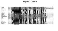
- Figure 2 shows an alignment of the deduced amino acid sequence of KLK15 with members of the kallikrein multi-gene family (SEQ ID NOs. 25-38). Dashes represent gaps to bring the sequences to better alignment.
- the residues of the catalytic triad (H, D, S) are shown in italics. Identical amino acids are highlighted in black and similar residues in grey.
- the 29 invariant serine protease residues are marked by (•) on the bottom, and the cysteine residues by (+) on top of each block.
- the predicted cleavage sites of the signal and activation peptides are indicated by arrows.
- the dotted area represents the kallikrein loop sequence.
- KLK15 has an "E” in this position.
- a unique 8 amino acid sequence, HNEPGTAG (SEQ ID NO. 10), is present at positions 148-155 of the KLK15 gene.
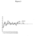
- Figure 3 is a plot of hydrophobicity and hydrophilicity of the KLK15 protein, as compared with the prostate specific antigen (PSA). Note the hydrophobic region at the amino terminus, suggesting presence of a signal peptide.
- PSA prostate specific antigen
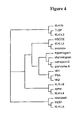
- Figure 4 is a dendrogram of the predicted phylogenetic tree for 15 kallikreins and a few other serine proteases.
- the neighbor-joining method was used to align KLK15 with other serine proteases and members of the kallikrein gene family.
- the tree grouped the classical kallikreins (hK1 hK2, and PSA) together and aligned KLK15 in one group with TLSP and KLK-L3 genes. Other serine proteases were aligned in different groups, as shown.
- KLK represents kallikrein
- KLK-L represents kallikrein-like
- TLSP represents trypsin-like serine protease
- NES 1 represents normal epithelial cell-specific gene
- PSA represents prostate specific antigen
- hK1 and hK2 represents human glandular kallikrein 1 and 2, respectively
- HSCCE represents human stratum corneum chymotryptic enzyme.
- Figure 5 is a schematic presentation of the different splice variants of the KLK15 gene. Exons are shown by boxes and introns by the connecting lines. Numbers inside boxes represent the exon lengths in base pairs. The arrowhead points to the common start codon and stars to the stop codon positions. The length of the predicted polypeptide product is indicated beside each variant in amino acids (AA). The alternative splicing and/or exon skips create a frame shift, which leads to a premature termination.
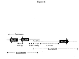
- Figure 6 shows the relative locations of KLK1, KLK15, and KLK3 genes on chromosome 19q13.3-ql3.4. The two overlapping BAC clones are identified, and the overlap region is hatched. Genes are represented by horizontal arrows denoting the direction of the coding sequence. Distances between genes are mentioned in base pairs. Figure is not drawn to scale.
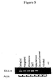
- FIG. 7 shows tissue expression of the KLK15 gene, as determined by RT-PCR.
- KLK15 is primarily expressed in the thyroid gland, and to a lower extent in the prostate, salivary and adrenal glands, colon, testis and kidney.
- M Molecular weight marker.
- PCR was performed with primers KLK15-F2 and KLK15-R1.
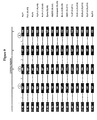
- Figure 8 shows hormonal regulation of the KLK15 gene in the LNCaP prostate cancer cell line.
- Figure 9 is a schematic diagram showing the comparison of the coding regions of the 15 kallikrein genes. Exons are shown by solid bars and introns by the connecting lines. Letters above boxes indicate relative positions of the catalytic triad that was found to be conserved in all genes; H denotes histidine, D aspartic acid and S serine. Roman numbers indicate intron phases.
- the intron phase refers to the location of the intron within the codon; I denotes that the intron occurs after the first nucleotide of the codon, II the intron occurs after the second nucleotide, 0 the intron occurs between codons.
- the intron phases are conserved in all genes. Numbers inside boxes indicate exon lengths in base pairs. Names inside brackets represent the official nomenclature approved by the human gene nomenclature committee. Untranslated 3' and 5' regions and 5' untranslated exons are not shown.
- the invention provides an isolated nucleic acid molecule having a sequence encoding a KLK15 Protein.
- isolated refers to a nucleic acid substantially free of cellular material or culture medium when produced by recombinant DNA techniques, or chemical reactants, or other chemicals when chemically synthesized.
- An "isolated” nucleic acid may also be free of sequences which naturally flank the nucleic acid (i.e., sequences located at the 5' and 3' ends of the nucleic acid molecule) from which the nucleic acid is derived.
- nucleic acid is intended to include DNA and RNA and can be either double stranded or single stranded.
- nucleic acid molecules of the invention encode a protein consisting of an amino acid sequence of SEQ ID NO. 6, 7, 8 or 9.
- nucleic acid molecules if the invention comprise a nucleic acid sequence of one or more of SEQ ID NO. 1 through 5.
- the invention includes nucleic acid sequences complementary to a nucleic acid encoding a protein consisting of an amino acid sequence of SEQ.ID.NO. 6, 7, 8, or 9 preferably the nucleic acid sequences complementary to a full nucleic acid sequence of one or more of SEQ.ID.NO. 1 through 5.
- the invention includes nucleic acid molecules having substantial sequence identity or homology to nucleic acid sequences of the invention or encoding proteins having substantial identity or similarity to the amino acid sequence of SEQ.ID.NO. 6, 7, 8, or 9.
- the nucleic acids have substantial sequence identity for example at least 80%, or 85% nucleic acid identity; more preferably 90% nucleic acid identity; and most preferably at least 95%, 96%, 97%, 98%, or 99% sequence identity.
- Identity as known in the art and used herein, is a relationship between two or more amino acid sequences or two or more nucleic acid sequences, as determined by comparing the sequences.
- the BLAST X program is publicly available from NCBI and other sources (BLAST ManuaL Altschul, S. et al. NCBI NLM MH Bethesda, Md. 20894; Altschul. S. et al. J. Mol. Biol. 215: 403-410,1990).
- nucleic acid molecules encoding a KLK15 Protein and having a sequence which differs from a nucleic acid sequence of the invention due to degeneracy in the genetic code are also within the scope of the invention.
- Such nucleic acids encode functionally equivalent proteins (e.g. a KLK15 Protein) but differ in sequence from the sequence of a KLK15 Protein due to degeneracy in the genetic code.
- DNA sequence polymorphisms within the nucleotide sequence of a KLK15 Protein may result in silent mutations which do not affect the amino acid sequence. Variations in one or more nucleotides may exist among individuals within a population due to natural allelic variation. Any and all such nucleic acid variations are within the scope of the invention.
- DNA sequence polymorphisms may also occur which lead to changes in the amino acid sequence of a KLK15 Protein. These amino acid polymorphisms are also within the scope of the present invention.
- An isolated nucleic acid molecule of the invention which comprises DNA can be isolated by preparing a labelled nucleic acid probe based on all or part of a nucleic acid sequence of the invention.
- the labeled nucleic acid probe is used to screen an appropriate DNA library (e.g. a cDNA or genomic DNA library).
- a cDNA library can be used to isolate a cDNA encoding a KLK15 Protein by screening the library with the labeled probe using standard techniques.
- a genomic DNA library can be similarly screened to isolate a genomic clone encompassing a gene encoding a KLK15 . Protein.
- Nucleic acids isolated by screening of a cDNA or genomic DNA library can be sequenced by standard techniques.
- An isolated nucleic acid molecule of the invention which is DNA can also be isolated by selectively amplifying a nucleic acid encoding a KLK15 Protein using the polymerase chain reaction (PCR) methods and cDNA or genomic DNA. It is possible to design synthetic oligonucleotide primers from the nucleotide sequence of the invention for use in PCR.
- a nucleic acid can be amplified from cDNA of genomic DNA using these oligonucleotide primers and standard PCR amplification techniques. The nucleic acid so amplified can be cloned into an appropriate vector and characterized by DNA sequence analysis.
- cDNA may be prepared from mRNA, by isolating total cellular mRNA by a variety of techniques, for example, by using the guanidinium-thiocyanate extraction procedure of Chirgwin et al., Biochemistry, 18, 5294-5299 (1979). cDNA is then synthesized from the mRNA using reverse transcriptase (for example, Moloney MLV reverse transcriptase available from Gibco/BRL, Bethesda, MD, or AMV reverse transcriptase available from Seikagaku America, Inc., St. Russia, FL).
- reverse transcriptase for example, Moloney MLV reverse transcriptase available from Gibco/BRL, Bethesda, MD, or AMV reverse transcriptase available from Seikagaku America, Inc., St. Russia, FL.
- An isolated nucleic acid molecule of the invention which is RNA can be isolated by cloning a cDNA encoding a KLK15 Protein into an appropriate vector which allows for transcription of the cDNA to produce an RNA molecule which encodes a KLK15 Protein.
- a cDNA can be cloned downstream of a bacteriophage promoter, (e.g. a T7 promoter) in a vector, cDNA can be transcribed in vitro with T7 polymerase, and the resultant RNA can be isolated by conventional techniques.
- Nucleic acid molecules of the invention may be chemically synthesized using standard techniques. Methods of chemically synthesizing polydeoxynucleotides are known, including but not limited to solid-phase synthesis which, like peptide synthesis, has been fully automated in commemially available DNA synthesizers (See e.g., Itakura et aL U.S. Patent No. 4,598,049; Caruthers et al. U.S. Patent No. 4,458,066; and Itakora U.S. Patent Nos. 4,401,796 and 4,373,071).
- Determination of whether a particular nucleic acid molecule encodes a KLK15 Protein can be accomplished by expressing the cDNA in an appropriate host cell by standard techniques, and testing the expressed protein in the methods described herein.
- a cDNA encoding a KLK15 Protein can be sequenced by standard techniques, such as dideoxynucleotide chain termination or Maxam-Gilbert chemical sequencing, to determine the nucleic acid sequence and the predicted amino acid sequence of the encoded protein.
- the initiation codon and untranslated sequences of a KLK15 Protein may be determined using computer software designed for the purpose, such as PC/Gene. (IntelliGenetics Inc., Calif.).
- the intron-exon structure and the transcription regulatory sequences of a gene encoding a KLK15 Protein may be confirmed by using a nucleic acid molecule of the invention encoding a KLK15 Protein to probe a genomic DNA clone library.
- Regulatory elements can be identified using standard techniques.
- the function of the elements can be confirmed by using these elements to express a reporter gene such as the lacZ gene that is operatively linked to the elements.
- These constructs may be introduced into cultured cells using conventional procedures or into non-human transgenic animal models. In addition to identifying regulatory elements in DNA, such constructs may also be used to identify nuclear proteins interacting with the elements, using techniques known in the art.
- the nucleic acid molecules isolated using the methods described herein are mutant KLK15 gene alleles.
- the mutant alleles may be isolated from individuals either known or proposed to have a genotype which contributes to the symptoms of a disorder involving a KLK15
- Mutant alleles and mutant allele products may be used in therapeutic and diagnostic methods described herein.
- a cDNA of a mutant KLK15 gene may be isolated using PCR as described herein, and the DNA sequence of the mutant allele may be compared to the normal allele to ascertain the mutation(s) responsible for the loss or alteration of function of the mutant gene product.
- a genomic library can also be constructed using DNA from an individual suspected of or known to carry a mutant allele, or a cDNA library can be constructed using RNA from tissue known, or suspected to express the mutant allele.
- a nucleic acid encoding a normal KLK15 gene or any suitable fragment thereof may then be labeled and used as a probe to identify the corresponding mutant allele in such libraries.
- Clones containing mutant sequences can be purified and subjected to sequence analysis.
- an expression library can be constructed using cDNA from RNA isolated from a tissue of an individual known or suspected to express a mutant KLK15 allele. Gene products made by the putatively mutant tissue may be expressed and screened, for example using antibodies specific for a KLK15 Protein as described herein. Library clones identified using the antibodies can be purified and subjected to sequence analysis.
- sequence of a nucleic acid molecule of the invention, or a fragment of the molecule may be inverted relative to its normal presentation for transcription to produce an antisense nucleic acid molecule.
- An antisense nucleic acid molecule may be constructed using chemical synthesis and enzymatic ligation reactions using procedures known in the art.
- KLK15 Protein An amino acid sequence of a KLK15 Protein consists of a sequence as shown in SEQ.ID.NO. 6, 7, 8, or 9. The protein is primarily expressed in the thyroid gland, and to a lower extent in the prostate, salivary and adrenal glands, colon, testis, and kidney.
- the invention also contemplates isoforms of the proteins of the invention.
- An isoform contains the same number and kinds of amino acids as a protein of the invention, but the isoform has a different molecular structure.
- Isoforms contemplated by the present invention preferably have the same properties as a protein of the invention as described herein.
- the present invention also includes KLK15 Proteins conjugated with a selected protein, or a marker protein (see below) to produce fusion proteins.
- a KLK15 Protein of the invention may be prepared using recombinant DNA methods. Accordingly, the nucleic acid molecules of the present invention having a sequence which encodes a RLK15
- Protein of the invention may be incorporated in a known manner into an appropriate expression vector which ensures good expression of the protein.
- Possible expression vectors include but are not limited to cosmids, plasmids, or modified viruses (e.g. replication defective retroviruses, adenoviruses and adeno-associated viruses), so long as the vector is compatible with the host cell used.
- the invention therefore contemplates a recombinant expression vector of the invention containing a nucleic acid molecule of the invention, and the necessary regulatory sequences for the transcription and translation of the inserted protein-sequence.
- Suitable regulatory sequences may be derived from a variety of sources, including bacterial, fungal, viral, mammalian, or insect genes [For example, see the regulatory sequences described in Goeddel, Gene Expression Technology: Methods in Enzymology 185, Academic Press, San Diego, CA (1990)]. Selection of appropriate regulatory sequences is dependent on the host cell chosen as discussed below, and may be readily accomplished by one of ordinary skill in the art.
- the necessary regulatory sequences may be supplied by the native KLK15 Protein and/or its flanking regions.
- the invention further provides a recombinant expression vector comprising a DNA nucleic acid molecule of the invention cloned into the expression vector in an antisense orientation. That is the DNA molecule is linked to a regulatory sequence in a manner which allows for expression, by transcription of the DNA molecule, of an RNA molecule which is antisense to the nucleic acid sequence of a protein of the invention or a fragment thereof. Regulatory sequences linked to the antisense nucleic acid can be chosen which direct the continuous expression of the antisense RNA molecule in a variety of cell types, for instance a viral promoter and/or enhancer, or regulatory sequences can be chosen which direct tissue or cell type specific expression of antisense RNA.
- the recombinant expression vectors of the invention may also contain a marker gene which facilitates the selection of host cells transformed or transfected with a recombinant molecule of the invention.
- marker genes are genes encoding a protein such as G418 and hygromycin which confer resistance to certain drugs, ⁇ -galactosidase, chloramphenicol acetyltransfense, firefly luciferase, or an immunoglobulin or portion thereof such as the Fc portion of an immunoglobulin, preferably IgG.
- the markers can be introduced on a separate vector from the nucleic acid of interest.
- the recombinant expression vectors may also contain genes that encode a fusion moiety which provides increased expression of the recombinant protein; increased solubility of the recombinant protein; and aid in the purification of the target recombinant protein by acting as a ligand in affinity purification.
- a proteolytic cleavage site may be added to the target recombinant protein to allow separation of the recombinant protein from the fusion moiety subsequent to purification of the fusion protein.
- Typical fusion expression vectors include pGEX (Amrad Corp., Melbourne, Australia), pMAL (New England Biolabs, Beverly. MA) and pRIT5 (Pharmacia, Piscataway. NJ) which fuse glutathione S-transferase (GST), maltose E binding protein, or protein A, respectively, to the recombinant protein.
- the recombinant expression vectors may be introduced into host cells to produce a transformant host cell.
- Transformant host cells include host cells which have been transformed or transfected with a recombinant expression vector of the invention.
- the terms "transformed with”, “transfected with”, “transformation” and “transfection” encompass the introduction of a nucleic acid (e.g. a vector) into a cell by one of many standard techniques.
- Prokaryotic cells can be transformed with a nucleic acid by, for example, electroporation or calcium-chloride mediated transformation.
- a nucleic acid can be introduced into mammalian cells via conventional techniques such as calcium phosphate or calcium chloride co-precipitation, DEAE-dextran-mediated transfection, lipofectin, electroporation or microinjection. Suitable methods for transforming and transfecting host cells can be found in Sambrook et al. (Molecular Cloning: A Laboratory Manual, 2nd Edition, Cold Spring Harbor Laboratory press (1989)), and other laboratory textbooks.
- Suitable host cells include a wide variety of prokaryotic and eukaryotic host cells.
- the proteins of the invention may be expressed in bacterial cells such as E. coli, insect cells (using baculovirus), yeast cells, or mammalian cells.
- Other suitable host cells can be found in Goeddel, Gene Expression Technology: Methods in Enzymology 185, Academic Press, San Diego, CA (1991).
- a host cell may also be chosen which modulates the expression of an inserted nucleic acid sequence, or modifies (e.g. glycosylation or phosphorylation) and processes (e.g. cleaves) the protein in a desired fashion.
- Host systems or cell lines may be selected which have specific and characteristic mechanisms for post-translational processing and modification of proteins.
- eukaryotic host cells including CHO, VERO, BHK, HeLA, COS, MDCK, 293, 3T3, and WI38 may be used.
- cell lines and host systems which stably express the gene product may be engineered.
- Host cells and in particular cell lines produced using the methods described herein may be particularly useful in screening and evaluating compounds that modulate the activity of a KLK15 Protein.
- the proteins of the invention may also be expressed in non-human transgenic animals including but not limited to mice, rats, rabbits, guinea pigs, micro-pigs, goats, sheep, pigs, non-human primates (e.g. baboons, monkeys, and chimpanzees) [see Hammer et al. (Nature 315:680-683, 1985), Palmiter et al. (Science 222:809-814,1983), Brinster et al. (Proc Natl. Acad. Sci USA 82:44384442, 1985), Palmiter and Brinster (Cell. 41:343-345, 1985) and U.S. Patent No. 4,736,866)].
- non-human transgenic animals including but not limited to mice, rats, rabbits, guinea pigs, micro-pigs, goats, sheep, pigs, non-human primates (e.g. baboons, monkeys, and chimpanzees) [see
- Procedures known in the art may be used to introduce a nucleic acid molecule of the invention encoding a KLK15 Protein into animals to produce the founder lines of transgenic animals. Such procedures include pronuclear microinjection, retrovirus mediated gene transfer into germ lines, gene targeting in embryonic stem cells, electroporation of embryos, and sperm-mediated gene transfer.
- the present invention contemplates a transgenic animal that carries the KLK15 gene in all their cells, and animals which carry the transgene in some but not all their cells.
- the transgene may be integrated as a single transgene or in concatamers.
- the transgene may be selectively introduced into and activated in specific cell types (See for example, Lasko et al, 1992 Proc. Natl. Acad. Sci. USA 89: 6236).
- the transgene may be integrated into the chromosomal site of the endogenous gene by gene targeting.
- the transgene may be selectively introduced into a particular cell type inactivating the endogenous gene in that cell type (See Gu et al Science 265: 103-106).
- the expresrion of a recombinant KLK15, Protein in a transgenic animal may be assayed using standard techniques. Initial screening may be conducted by Southern Blot analysis, or PCR methods to analyze whether the transgene has been integrated. The level of mRNA expression in the tissues of transgenic animals may also be assessed using techniques including Northern blot analysis of tissue samples, in situ hybridization, and RT-PCR. Tissue may also be evaluated immunocytochemically using antibodies against KLK15 Protein.
- Proteins of the invention may also be prepared by chemical synthesis using techniques well known in the chemistry of proteins such as solid phase synthesis (Merrifield. 1964, J. Am. Chem. Assoc. 85:2149-2154) or synthesis in homogenous solution (Houbenweyl, 1987, Methods of Organic Chemistry, ed. E. Wansch, Vol. 15 and II, Thieme, Stuttgart).
- N-terminal or C-terminal fusion proteins comprising a KLK15 Protein of the invention conjugated with other molecules, such as proteins, may be prepared by fusing, through recombinant techniques.
- the N-terminal or C-terminal of a KLK15 Protein and the sequence of a selected protein or marker protein with a desired biological function.
- the resultant fusion proteins contain a KLK15 Protein fused to the selected protein or marker protein as described herein.
- proteins which may be used to prepare fusion proteins include immunoglobulins, glutathione-S-transferase (GST), bemagglutinin (HA), and truncated myc.
- Proteins of the invention can be used to prepare antibodies specific for the proteins.
- Antibodies can be prepared which bind a distinct epitope in an unconserved region of the protein.
- An unconserved region of the protein is one that does not have substantial sequence homology to other proteins.
- a region from a conserved region such as a well-characterized domain can also be used to prepare an antibody to a conserved region of a KLK15 Protein.
- Antibodies having specificity for a KLK15 Protein may also be raised from fusion proteins created by expressing fusion proteins in bacteria as described herein.
- the invention can employ intact monoclonal or polyclonal antibodies, and immunologically active fragments (e.g. a Fab, (Fab) 2 fragment, or Fab expression library fragments and epitope-binding fragments thereof), an antibody heavy chain, and antibody light chain, a genetically engineered single chain Fv molecule (Ladner et al, U.S. Pat. No. 4,946,778), humanized antibody, or a chimeric antibody, for example, an antibody which contains the binding specificity of a murine antibody, but in which the remaining portions are of human origin.
- Antibodies including monoclonal and polyclonal antibodies, fragments and chimeras may be prepared using methods known to those skilled in the art.
- the nucleic acid molecules, KLK15 Proteins, and antibodies of the invention may be used in the prognostic and diagnostic evaluation of disorders involving a KLK15 Protein (e.g. cancer or thyroid disorders), and the identification of subjects with a predisposition to such disorders (Section 4.1.1 and 4.1.2).
- disorders involving a KLK15 Protein e.g. cancer or thyroid disorders
- identification of subjects with a predisposition to such disorders ection 4.1.1 and 4.1.2.
- the present invention provides a test kit for diagnosing a condition associated with a protein of the invention by determining the presence of a nucleic acid molecule or a protein of the invention.
- the test kit may be used a method for detecting the expression of the cancer marker KLK15 in a patient comprising:
- the nucleic acid molecules, KLK15 Proteins, and antibodies of the invention may be used in the diagnosis and staging of cancer, in particular prostate cancer. Increased levels of KLK15 Proteins are associated with more aggressive forms of prostate cancer and may be an indicator of poor prognosis.
- Methods for detecting nucleic acid molecules and KLK15 Proteins of the invention can be used to monitor disorders involving a KLK15 Protein by detecting KLK15 Proteins and nucleic acid molecules encoding KLK15 Proteins.
- the applications of the present invention also include methods for the identification of compounds that modulate the biological activity of KLK15 Proteins (Section 4.2).
- the compounds, antibodies etc. may be used for the treatment of disorders involving a KLK15 Protein (Section 43). It would also be apparent to one skilled in the art that the methods described herein may be used to study the developmental expression of XLX15 Proteins and, accordingly, will provide further insight into the role of KLK15 Proteins.
- a variety of methods can be employed for the diagnostic and prognostic evaluation of disorders involving a KLK15 Protein, and the identification of subjects with a predisposition to such disorders.
- Such methods may, for example, utilize nucleic acid molecules of the invention, and fragments thereof, and antibodies directed against KLK15 Proteins, including peptide fragments.
- the nucleic acids and antibodies may be used, for example, for: (1) the detection of the presence of KLK15 mutations, or the detection of either over- or under-expression of KLK15 mRNA relative to a non-disorder state or the qualitative or quantitative detection of alternatively spliced forms of KLK15 transcripts which may correlate with certain conditions or susceptibility toward such conditions; and (2) the detection of either an over- or an under-abundance of KLK15, Proteins relative to a non- disorder state or the presence of a modified (e.g., less than full length) KLK15 Protein which correlates with a disorder state, or a progression toward a disorder state.
- a modified (e.g., less than full length) KLK15 Protein which correlates with a disorder state, or a progression toward a disorder state.
- the methods described herein may be used to evaluate the probability of the presence of malignant or pre-malignant cells, for example, in a group of cells freshly removed from a host. Such methods can be used to detect tumors, quantitate their growth, and help in the diagnosis and prognosis of disease. The methods can be used to detect the presence of cancer metastasis, as well as confirm the absence or removal of all tumor tissue following surgery, cancer chemotherapy, and/or radiation therapy. They can further be used to monitor cancer chemotherapy and tumor reappearance.
- the methods described herein may be performed by utilizing pre-packaged diagnostic kits comprising at least one specific KLK15 nucleic acid or antibody described herein, which may be conveniently used, e.g., in clinical settings, to screen and diagnose patients and to screen and identify those individuals exhibiting a predisposition to developing a disorder.
- Nucleic acid-based detection techniques are described, below, in Section 4.1.1.
- Peptide detection techniques are described, below, in Section 4.1.2.
- the samples that may be analyzed using the methods of the invention include those which are known or suspected to express KLK15 or contain KLK15 Proteins.
- the samples may be derived from a patient or a cell culture, and include but are not limited to biological fluids, tissue extracts, freshly harvested cells, and lysates of cells which have been incubated in cell cultures.
- Oligonucleotides or longer fragments derived from any of the nucleic acid molecules of the invention may be used as targets in a microarray.
- the microarray can be used to simultaneously monitor the expression levels of large numbers of genes and to identify genetic variants, mutations, and polymorphisms.
- the information from the microarray may be used to determine gene function, to understand the genetic basis of a disorder, to diagnose a disorder, and to develop and monitor the activities of therapeutic agents.
- microarrays are well known to a person skilled in the art.
- nucleic acid molecules of the invention allow those skilled in the art to construct nucleotide probes for use in the detection of nucleic acid sequences of the invention in samples.
- Suitable probes include nucleic acid molecules based on nucleic acid sequences encoding at least 5 sequential amino acids from regions of the KLK15 Protein, preferably they comprise 15 to 30 nucleotides (see SEQ ID Nos. 47-50).
- a nucleotide probe may be labeled with a detectable substance such as a radioactive label which provides for an adequate signal and has sufficient half-life such as 32 P, 3 H, 14 C or the like.
- detectable substances which may be used include antigens that are recognized by a specific labeled antibody, fluorescent compounds, enzymes, antibodies specific for a labeled antigen, and luminescent compounds.
- An appropriate label may be selected having regard to the rate of hybridization and binding of the probe to the nucleotide to be detected and the amount of nucleotide available for hybridization.
- Labeled probes may be hybridized to nucleic acids on solid supports such as nitrocellulose filters or nylon membranes as generally described in Sambrook et al, 1989, Molecular Cloning, A Laboratory Manual (2nd ed.).
- the nucleic acid probes may be used to detect genes, preferably in human cells, that encode KLK15 Proteins.
- the nucleotide probes may also be useful in the diagnosis of disorders involving a KLK15 Protein; in monitoring the progression of such disorders; or monitoring a therapeutic treatment.
- the probe may be used in hybridization techniques to detect genes that encode KLK15 Proteins.
- the technique generally involves contacting and incubating nucleic acids (e.g. recombinant DNA molecules, cloned genes) obtained from a sample from a patient or other cellular source with a probe of the present invention under conditions favorable for the specific annealing of the probes to complementary sequences in the nucleic acids. After incubation, the non-annealed nucleic acids are removed, and the presence of nucleic acids that have hybridized to the probe if any are detected.
- nucleic acids e.g. recombinant DNA molecules, cloned genes
- the detection of nucleic acid molecules of the invention may involve the amplification of specific gene sequences using an amplification method such as PCR, followed by the analysis of the amplified molecules using techniques known to those skilled in the art. Suitable primers can be routinely designed by one of skill in the art.
- Genomic DNA may be used in hybridization or amplification assays of biological samples to detect abnormalities involving KLK15 structure, including point mutations, insertions, deletions, and chromosomal rearrangements.
- direct sequencing single stranded conformational polymorphism analyses, heteroduplex analysis, denaturing gradient gel electrophoresis, chemical mismatch cleavage, and oligonucleotide hybridization may be utilized.
- Genotyping techniques known to one skilled in the art can be used to type polymorphisms that are in close proximity to the mutations in a klk15 gene.
- the polymorphisms may be used to identify individuals in families that are likely to carry mutations. If a polymorphism exhibits linkage disequalibrium with mutations in a KLK15 gene, it can also be used to screen for individuals in the general population likely to carry mutations.
- Polymorphisms which may be used include restriction fragment length polymorphisms (RFLPs), single-base polymorphisms, and simple sequence repeat polymorphisms (SSLPs).
- a probe of the invention may be used to directly identify RFLPs.
- a probe or primer of the invention can additionally be used to isolate genomic clones such as YACs, BACs, PACs, cosmids, phage or plasmids. The DNA in the clones can be screened for SSLPs using hybridization or sequencing procedures.
- Hybridization and amplification techniques described herein may be used to assay qualitative and quantitative aspects of klk15 expression.
- RNA may be isolated from a cell type or tissue known to express klk15 and tested utilizing the hybridization (e.g. standard Northern analyses) or PCR techniques referred to herein.
- the techniques may be used to detect differences in transcript size which may be due to normal or abnormal alternative splicing.
- the techniques may be used to detect quantitative differences between levels of full length and/or alternatively splice transcripts detected in normal individuals relative to those individuals exhibiting symptoms of a disorder involving a KLK15 Protein.
- the primers and probes may be used in the above described methods in situ i.e directly on tissue sections (fixed and/or frozen) of patient tissue obtained from biopsies or resections.
- Antibodies specifically reactive with a KLK15 Protein, or derivatives, such as enzyme conjugates or labeled derivatives, may be used to detect KLK15 Proteins in various samples (e.g. biological materials). They may be used as diagnostic or prognostic reagents and they may be used to detect abnormalities in the level of KLK15 Protein expression, or abnormalities in the structure, and/or temporal, tissue, cellular, or subcellular location of a KLK15 Protein. Antibodies may also be used to screen potentially therapeutic compounds in vitro to determine their effects on disorders involving a KLK15
- In vitro immunoassays may also be used to assess or monitor the efficacy of particular therapies.
- the antibodies of the invention may also be used in vitro to determine the level of KLK15 expression in cells genetically engineered to produce a KLK15 Protein.
- the antibodies may be used in any known immunoassays which rely on the binding interaction between an antigenic determinant of a KLK15 Protein and the antibodies.
- assays are radioimmunoassays, enzyme immunoassays (e.g. ELISA), immunofluorescence, immunoprecipitation, latex agglutination, hemagglutination, and histochemical tests.
- the antibodies may be used to detect and quantify KLK15 Proteins in a sample in order to determine its role in particular cellular events or pathological states, and to diagnose and treat such pathological states.
- the antibodies of the invention may be used in immuno-histochemieal analyses, for example, at the cellular and sub-subcellular level, to detect a KLK15 Protein to localize it to particular cells and tissues, and to specific subcellular locations, and to quantitate the level of expression.
- Cytochemical techniques known in the art for localizing antigens using light and electron microscopy may be used to detect a KLK15 Protein.
- an antibody of the invention may be labeled with a detectable substance and a KLK15 Related Protein may be localised in tissues and cells based upon the presence of the detectable substance.
- detectable substances include, but are not limited to, the following: radioisotopes (e.g., 3 H, 14 C, 35 S, 125 I, 131 I), fluorescent labels (e.g., FITC, rhodamine, lanthanie phosphors), luminescent labels such as luminol; enzymatic labels (e.g., horseradish peroxidase, beta-galactosidase, luciferase, alkaline phosphatase, acetylcholinesterase), biotinyl groups (which can be detected by marked avidin e.g., streptavidin containing a fluorescent marker or enzymatic activity that can be detected by optical or calorimetric methods), predetermined polypeptide epitopes recognized by a secondary reporter (e.g., leucine zipper pair sequences, binding sites for secondary antibodies, metal binding domains, epitope tags).
- labels are attached via spacer arms of various lengths to reduce potential
- the antibody or sample may be immobilized on a carrier or solid support which is capable of immobilizing cells, antibodies etc.
- the carrier or support may be nitrocellulose, or glass, polyacrylamides, gabbros, and magnetite.
- the support material may have any possible configuration including spherical (e.g. bead), cylindrical (e.g. inside surface of a test tube or well, or the external surface of a rod), or flat (e.g. sheet, test strip).
- Indirect methods may also be employed in which the primary antigen-antibody reaction is amplified by the introduction of a second antibody, having specificity for the antibody reactive against KLK15 Protein.
- the antibody having specificity a KLK15 Protein is a rabbit IgG antibody
- the second antibody may be goat anti-rabbit gamma-globulin labeled with a detectable substance as described herein.
- a KLK15 Protein may be localized by radioautography.
- the results of radioautography may be quantitated by determining the density of particles in the radioautographs by various optical methods, or by counting the grains.
- the invention contemplates a test kit for monitoring the progression of cancer (e.g. prostate cancer) in an individual, comprising:
- the amount of complexes may also be compared to a value representative of the amount of the complexes from an individual not at risk of, or afflicted with, cancer (e.g. prostate cancer).
- cancer e.g. prostate cancer
- the methods described herein are designed to identify substances that modulate the biological activity of a KLK15 Protein including substances that bind to KLK15 Proteins, or bind to other proteins that interact with a KLK15 Protein, to compounds that interfere with, or enhance the interacdon of a KLK15 Protein and substances that bind to the KLK15 Protein or other proteins that interact with a KLK15 Protein. Methods are also utilized that identify compounds that bind to KLK15 regulatory sequences.
- the substances and compounds identified using the methods of the invention include but are not limited to peptides such as soluble peptides including Ig-tailed fusion peptides, members of random peptide libraries and combinatorial chemistry-derived molecular libraries made of D- and/or L-configuration amino acids, phosphopeptides (including members of random or partially degenerate, directed phosphopeptide libraries), antibodies [e.g. polyclonal, monoclonal, humanized, anti-idiotypic, chimeric, single chain antibodies, fragments, (e.g. Fab, F(ab)2, and Fab expression library fragments, and epitope-binding fragments thereof)], and small organic or inorganic molecules.
- the substance or compound may be an endogenous physiological compound or it may be a natural or synthetic compound.
- Substances which modulate a ELK15 Protein can be identified based on their ability to bind to a KLK15 Protein. Therefore, the invention also provides methods for identifying subttances which bind to a KLK15 Protein. Substances identified using the methods of the invention may be isolated, cloned and sequenced using conventional techniques. A substance that associates with a polypeptide of the invention may be an agonist or antagonist of the biological or immunological activity of a polypeptide of the invention.
- agonist refers to a molecule that increases the amount of, or prolongs the duration of, the activity of the protein.
- antagonist refers to a molecule which decreases the biological or immunological activity of the protein.
- Agonists and antagonists may include proteins, nucleic acids, carbohydrates, or any other molecules that associate with a protein of the invention.
- Substances which can bind with a KLK15 Protein may be identified by reacting a KLK15 Protein with a test substance which potentially binds to a KLK15 Protein, under conditions which permit the formation of substance-KLK15 Protein complexes and removing and/or detecting the complexes.
- the complexes can be detected by assaying for substance-KLK15 Protein complexes, for free substance, or for non-complexed KLK15 Protein.
- Conditions which permit the formation of substance-KLK15 Protein complexes may be selected having regard to factors such as the nature and amounts of the substance and the protein.
- the sabstance-protein complex, free substance or non-complexed proteins may be isolated by conventional isolation techniques, for example, salting out, chromatography, electrophoresis, gel filtration, fractionation, absorption, polyacrylamide gel electrophoresis, agglutination, or combinations thereof.
- antibody against KLK15 Protein or the substance, or labeled KLK15 Protein, or a labeled substance may be utilized.
- the antibodies, protein or substances may be labeled with a detectable substance as described above.
- a KLK15 Protein, or the substance used in the method of the invention may be insolubilized.
- a KLK15 Protein, or substance may be bound to a suitable carrier such as agarose, cellulose, dextran, Sephadex, Sepharose, carboxymethyl cellulose polystyrene, filter paper, ion-exchange resin, plastic film, plastic tube, glass beads, polyamine-methyl vinyl-ether-maleic acid copolymer, amino acid copolymer, ethylene-maleic acid copolymer, nylon, silk, etc.
- the carrier may be in the shape of, for example, a tube, test plate, beads, disc, sphere etc.
- the insolubilized protein or substance may be prepared by reacting the material with a suitable insoluble carrier using known chemical or physical methods, for example, cyanogen bromide coupling.
- the invention also contemplates a method for evaluating a compound for its ability to modulate the biological activity of a KLK15 protein of the invention, by assaying for an agonist or antagonist (i.e. enhancer or inhibitor) of the binding of a KLK15 Protein with a substance which binds with a KLK15
- an agonist or antagonist i.e. enhancer or inhibitor
- the basic method for evaluating if a compound is an agonist or antagonist of the binding of a KLK15 Protein and a substance that binds to the protein is to prepare a reaction mixture containing the KLK15 Protein and the substance under conditions which permit the formation of substance-KLK15 Protein complexes, in the presence of a test compound.
- the test compound may be initially added to the mixture, or may be added subsequent to the addition of the KLK15 Protein and substance.
- Control reaction mixtures without the test compound or with a placebo are also prepared. The formation of complexes is detected and the formation of complexes in the control reaction but not in the reaction mixture indicates that the test compound interferes with the interaction of the KLK15 Protein and substance.
- the reactions may be carried out in the liquid phase or the KLK15 Protein, substance, or test compound may be immobilized as described herein.
- the ability of a compound to modulate the biological activity of a KLK15 Protein of the invention may be tested by determining the biological effects on cells.
- agonists and antagonists i.e. inhibitors and enhancers that can be assayed using the methods of the invention may act on one or more of the binding sites on the protein or substance including agonist binding sites, competitive antagonist binding sites, non-competitive antagonist binding sites or allosteric sites.
- the invention also makes it possible to screen for antagonists that inhibit the effects of an agonist of the interaction of KLK15 Protein with a substance which is capable of binding to the KLK15 Protein.
- the invention may be used to assay for a compound that competes for the same binding site of a KLK15 Protein.
- the invention also contemplates methods for identifying compounds that bind to proteins that interact with a KLK15 Protein. Protein-protein interactions may be identified using conventional methods such as co-immunoprecipitation, crosslinking and co-purification through gradients or chromatographic Columns. Methods may also be employed that result in the simultaneous identification of genes which encode proteins interacting with a KLK15 Protein. These methods include probing expression libraries with labeled KLK15 Protein.
- Two-hybrid systems may also be used to detect protein interactions in vivo.
- plasmids are constructed that encode two hybrid proteins.
- a first hybrid protein consists of the DNA-binding domain of a transcription activator protein fused to a KLK15 Protein
- the second hybrid protein consists of the transcription activator protein's activator domain fused to an unknown protein encoded by a cDNA which has been recombined into the plasmid as part of a cDNA library.
- the plasmids are transformed into a strain of yeast (e.g. S . cerevisiae) that contains a reporter gene (e.g.
- lacZ lacZ, luciferase, alkaline phosphatase, horsetadish peroxidase
- the hybrid proteins alone cannot activate the transcription of the reporter gene. However, interaction of the two hybrid proteins reconstitutes the functional activator protein and results in expression of the reporter gene, which is detected by an assay for the reporter gene product.
- fusion proteins may be used in the above-described methods.
- KLK15 Proteins fused to a glutathione-S-transferase may be used in the methods.
- Amodulator of a KLK15 Protein of the invention may also be identified based on its ability to inhibit or enhance catalytic activity of the protein.
- the reagents suitable for applying the methods of the invention to evaluate compounds that modulate a KLK15 Protein may be packaged into convenient kits providing the necessary materials packaged into suitable containers.
- the kits may also include suitable supports useful in performing the methods of the invention.
- proteins of the invention substances or compounds identified by the methods described herein, antibodies, and nucleic acid molecules of the invention may be used for modulating the biological activity of a KLK15 Protein, and they may be used in the treatment of conditions such as cancer (particularly thyroid, prostate, colon, kidney, testicular cancer) and thyroid disorders in a patient.
- the substances, antibodies, peptides, and compounds may be formulated into pharmaceutical compositions for administration to subjects in a biologically compatible form suitable for administration in vivo.
- biologically compatible form suitable for administration in vivo is meant a form of the active substance to be administered in which any toxic effects are outweighed by the therapeutic effects.
- the active substances may be administered to living organisms including humans and animals.
- Administration of a therapeutically active amount of a pharmaceutical composition of the present invention is defined as an amount effective, at dosages and for periods of time necessary to achieve the desired result.
- a therapeutically active amount of a substance may vary according to factors such as the disease state, age, sex, and weight of the individual, and the ability of antibody to elicit a desired response in the individual. Dosage periods may be adjusted to provide the optimum therapeutic response. For example, several divided doses may be administered daily or the dose may be proportionally reduced as indicated by the exigencies of the therapeutic situation.
- the active substance may be administered in a convenient manner such as by injection (subcutaneous, intravenous, etc.), oral administration, inhalation, transdermal application, or rectal administration.
- the active substance may be coated in a material to protect the substance from the action of enzymes, acids and other natural conditions that may inactivate the substance.
- compositions described herein can be prepared by per se known methods for the preparation of pharmaceutically acceptable compositions which can be administered to subjects, such that an effective quantity of the active substance is combined in a mixture with a pharmaceutically acceptable vehicle.
- Suitable vehicles are described, for example, in Remington's Pharmaceutical Sciences (Remington's Pharmaceutical Sciences, Mack Publishing Company, Easton, Pa., USA 1985).
- the compositions include, albeit not exclusively, solutions of the active substances in association with one or more pharmaceutically acceptable vehicles or diluents, and contained in buffered solutions with a suitable pH and iso-osmotic with the physiological fluids.
- compositions are indicated as therapeutic agents either alone or in conjunction with other therapeutic agents or other forms of treatment (e.g. chemotherapy or radiotherapy).
- the compositions may be used in combination with anti-proliferative agents, antimicrobial agents, immunostimulatory agents, or anti-inflammatories.
- the compounds may be used in combination with anti-viral and/or anti-proliferative agents.
- the compositions of the invention may be administered concurrently, separately, or sequentially with other therapeutic agents or therapies.
- Vectors derived from retroviruses, adenovirus, herpes or vaccinia viruses, or from various bacterial plasmids may be used to deliver nucleic acid molecules to a targeted organ, tissue, or cell population. Methods well known to those skilled in the art may be used to construct recombinant vectors which will express antisense nucleic acid molecules of the invention. (See, for example, the techniques described in Sambrook et al (supra) and Ausubel et al (supra)).
- nucleic acid molecules comprising full length cDNA sequences and/or their regulatory elements enable a skilled artisan to use sequences encoding a protein of the invention as an investigative tool in sense (Youssoufian H and H F Lodish 1993 Mol Cell Biol 13:98-104) or antisense (Eguchi et al (1991) Annu Rev Biochem 60:631-652) regulation of gene function.
- sense or antisense oligomers, or larger fragments can be designed from various locations along the coding or control regions.
- Genes encoding a protein of the invention can be turned off by transfecting a cell or tissue with vectors which express high levels of a desired KLK15-encoding fragment. Such constructs can inundate cells with untranslatable sense or antisense sequences. Even in the absence of integration into the DNA, such vectors may continue to transcribe RNA molecules until all copies are disabled by endogenous nucleases.
- Modifications of gene expression can be obtained by designing antisense molecules.
- DNA, RNA or PNA to the regulatory regions of a gene encoding a protein of the invention, i.e., the promoters, enhancers, and introns.
- oligonucleotides are derived from the transcription initiation site, eg, between -10 and +10 regions of the leader sequence.
- the antisense molecules may also be designed so that they block translation of mRNA by preventing the transcript from binding to ribosomes. Inhibition may also be achieved using "triple helix" base-pairing methodology. Triple helix pairing compromises the ability of the double helix to open sufficiently for the binding of polymerases, transcription factors, or regulatory molecules. Therapeutic advances using triplex DNA were reviewed by Gee J E et al (In: Huber B E and B I Carr (1994) Molecular and Immunologic Approaches, Futura Publishing Co, Mt Kisco N.Y.).
- Ribozymes are enzymatic RNA molecules that catalyze the specific cleavage of RNA. Ribozymes act by sequence-specific hybridization of the ribozyme molecule to complementary target RNA, followed by endonucleolytic cleavage.
- the invention therefore contemplates engineered hammerhead motif ribozyme molecules that can specifically and efficiently catalyze endonucleolytic cleavage of sequences encoding a protein of the invention.
- Specific ribozyme cleavage sites within any potential RNA target may initially be identified by scanning the target molecule for ribozyme cleavage sites which include the following sequences, GUA, GUU and GUC. Once the sites are identified, short RNA sequences of between 15 and 20 ribonucleotides corresponding to the region of the target gene containing the cleavage site may be evaluated for secondary structural features which may render the oligonucleotide inoperable. The suitability of candidate targets may also be determined by testing accessibility to hybridization with complementary oligonucleotides using ribonuclease protection assays.
- vectors may be introduced into stem cells obtained from a patient and clonally propagated for autologous transplant into the same patient (See U.S. Pat. Nos. 5,399,493 and 5,437,994). Delivery by transfection and by liposome are well known in the art.
- An antibody against a KLK15 Protein may be conjugated to chemotherapeutic drugs, toxins, immunological response modifiers, hematogenous agents, enzymes, and radioisotopes and used in the prevention and treatment of cancer (e.g. thyroid, prostate, colon, kidney, testicular cancer).
- an antibody against a KLK15 Protein may be conjugated to toxic moieties including but not limited to ricin A, diphtheria toxin, abrin, modeccin, or bacterial toxins from Pseudomonas or Shigella.
- Toxins and their derivatives have been reported to form conjugates with antibodies specific to particular target tissues, such as cancer or tumor cells in order to obtain specifically targeted cellular toxicity (Moolten F.L. et al, Immun. Rev. 62:47-72, 1982, and Bernhard,, M.I. Cancer Res. 43:4420,1983).
- Conjugates can be prepared by standard means known in the art.
- a number of bifonctional linking agents e.g. heterobifanctional linkers such as N-succinimidyl-3-(2-pyrldyldithio)propionate are available commercially from Pierce Chemically Company, Rockford. III.
- Administration of the antibodies or immunotoxins for therapeutic use may be by an intravenous route, although with proper formulation additional routes of administration such as intraperitoneal, oral, or transdermal administration may also be used.
- a KLK15 Protein may be conjugated to chemotherapeutic drugs, toxins, immunological response modifiers, enzymes, and radioisotopes using methods known in the art.
- the invention also provides immunotherapeutic approaches for preventing or reducing the severity of a cancer.
- the clinical signs or symptoms of the cancer in a subject are indicative of a beneficial effect to the patient due to the stimulation of the subject's immune response against the cancer.
- Stimulating an immune response refers to inducing an immune response or enhancing the activity of immunoeffector cells in response to administration of a vaccine preparation of the invention.
- the prevention of a cancer can be indicated by an increased time before the appearance of cancer in a patient that is predisposed to developing cancer due for example to a genetic disposition or exposure to a carcinogenic agent.
- the reduction in the severity of a cancer can be indicated by a decrease in size or growth rate of a tumor.
- Vaccines can be derived from a KLK Protein, peptides derived therefrom, or chemically produced synthetic peptides, or any combination of these molecules, or fusion proteins or peptides thereof.
- the proteins, peptides, etc. can be synthesized or prepared recombinantly or otherwise biologically, to comprise one or more amino acid sequences corresponding to one or more epitopes of a tumor associated protein.
- Epitopes of a tumor associated protein will be understood to include the possibility that in some instances amino acid sequence variations of a naturally occurring protein or polypeptide may be antigenic and confer protective immunity against cancer or anti-tumorigenic effects. Sequence variations may include without limitation, amino acid substitutions, extensions, deletions, truncations, interpolations, and combinations thereof. Such variations fall within the scope of the invention provided the protein containing them is immunogenic and antibodies against such polypeptide cross-react with naturally occurring KLK15 Protein to a sufficient extent to provide protective immunity and/or anti-tumorigenic activity when administered as
- the proteins, peptides etc can be incorporated into vaccines capable of inducing an immune response using methods known in the art.
- Techniques for enhancing the antigenicity of the proteins, peptides, etc. are known in the art and include incorporation into a multimeric structure, binding to a highly immunogenic protein carrier, for example, keyhole limpet hemocyanin (KLH), or diptheria toxoid, and administration in combination with adjuvants or any other enhancer of immune response.
- KLH keyhole limpet hemocyanin
- diptheria toxoid diptheria toxoid
- Vaccines may be combined with physiologically acceptable media, including immunologically acceptable diluents and carriers as well as commonly employed adjuvants such as Freund's Complete Adjuvant, saponin, alum, and the like.
- anti-idiotype antibodies to antibodies to KLK15 Proteins described herein are also useful as vaccines and can be similarly formulated.
- the administration of a vaccine in accordance with the invention is generally applicable to the prevention or treatment of cancers including thyroid, prostate, colon, kidney, and testicular cancer.
- the administration to a patient of a vaccine m accordance with the invention for the prevention and/or treatment of cancer can take place before or after a surgical procedure to remove the cancer, before or after a chemotherapeutic procedure for the treatment of cancer, and before or after radiation therapy for the treatment of cancer and any combination thereof.
- the cancer immunotherapy in accordance with the invention would be a preferred treatment for the prevention and /or treatment of cancer, since the side effects involved are substantially minimal compared with the other available treatments e.g. surgery, chemotherapy, radiation therapy.
- the vaccines have the potential or capability to prevent cancer in subjects without cancer but who are at risk of developing cancer.
- the activity of the proteins, substances, compounds, antibodies, nucleic acid molecules, agents, and compositions of the invention may be confirmed in animal experimental model systems.
- Therapeutic efficacy and toxicity may be determined by standard pharmaceutical procedures in cell cultures or with experimental animals, such as by calculating the ED 50 (the dose therapeutically effective in 50% of the population) or LD 50 (the dose lethal to 50% of the population) statistics.
- the therapeutic index is the dose ratio of therapeutic to toxic effects and it can be expressed as the ED 50 /LD 50 ratio.
- Pharmaceutical compositions which exhibit large therapeutic indices are preferred.
- nucleic acid molecules disclosed herein may also be used in molecular biology techniques that have not yet been developed, provided the new techniques rely on properties of nucleotide sequences that are currently known, including but not limited to such properties as the triplet genetic code and specific base pair interactions.
- cells, tissues, and non-human animals lacking in expression or partially lacking in expression of a nucleic acid molecule or gene of the invention may be developed using recombinant expression vectors of the invention having specific deletion or insertion mutations in the gene.
- a recombinant expression vector may be used to inactivate or alter the endogenous gene by homologous recombination, and thereby create a deficient cell, tissue, or animal.
- Null alleles may be generated in cells, such as embryonic stem cells by deletion mutation.
- a recombinant gene may also be engineered to contain an insertion mutation that inactivates the gene.
- Such a construct may then be introduced into a cell, such as an embryonic stem cell, by a technique such as transfection, electroporation, injection etc.
- Cells lacking an intact gene may then be identified, for example by Southern blotting, Northern Blotting, or by assaying for expression of the encoded polypeptide using the methods described herein.
- Such cells may then be fused to embryonic stem cells to generate transgenic non-human animals deficient in a polypeptide of the invention.
- Germline transmission of the mutation may be achieved, for example, by aggregating the embryonic stem cells with early stage embryos, such as 8 cell embryos, in vitro; transferring the resulting blastocysts into recipient females and; generating germline transmission of the resulting aggregation chimeras.
- Such a mutant animal may be used to define specific cell populations, developmental patterns and in vivo processes, normally dependent on gene expression.
- a transgenic non-human mammal may be developed all of whose germ cells and somatic cells contain a recombinant expression vector that inactivates or alters a gene encoding a KLK15 Protein.
- the transgenic non-human mammal (all of whose germ cells and somatic cells contain a recombinant expression vector that inactivates or alters a gene encoding a KLK15 Protein) may result in a KLK15 Protein associated pathology.
- a transgenic non-human mammal which does not express or has altered (e.g. reduced) expression of a KLK15 Protein of the invention may result in a KLK15 Protein associated pathology.
- a KLK15 Protein pathology refers to a phenotype observed for a KLK15 Protein homozygous or heterozygous mutant.
- a transgenic non-human animal may for example be a mouse, rat, rabbit, sheep, hamster, dog, cat, goat or monkey.
- a transgenic non-human animal assay system may provide a model system for testing for an agent that reduces or inhibits a pathology associated with a KLK15 Protein, such as a KLK15 Protein associated pathology, comprising:
- the agent may be useful in the treatment and prophylaxis of conditions such as cancer as discussed herein.
- the agents may also be incorporated in a pharmaceutical composition as described herein.
- a contiguous map for the human kallikrein gene locus extending from the KLK1 gene (centromere) to the KLK14 gene (telomere) (7,8,11,12,27) was constructed. Overlapping bacterial artificial chromosome (BAC) clones spanning this area were identified by screening of a human BAC library using different radiolabeled gene-specific probes. An area of - 300 kb of genomic sequence was established using different techniques, as previously described (11,27). By performing an EcoR1 restriction analysis, the kallikrein locus was oriented along the EcoR1 restriction map of chromosome 19q13 available from the Lawrence Livermore National Laboratory (LLNL).
- LLNL Lawrence Livermore National Laboratory
- a BAC clone that extends more centromerically (BC 781134) was then identified. Contigs of linear genomic sequences from this clone are available from the LLNL. Initially, these contig sequences were used to predict the presence of novel genes, using bioinformatic approaches, as previously described (8,12), and a putative new serine protease was identified. The sequence of the putative gene was then verified by different approaches including sequencing, EST database search, PCR screening of tissues, as described below.
- the predicted exons of the putative new gene were subjected to homology search using the BLASTN algorithm (28) on the National Center for Biotechnology Information web server (http://www ncbi.nlm.nih.gov/BLAST/) against the human EST database (dbEST).
- Clones with > 95% homology were obtained from the I.M.A.G.E. consortium (29) through Research Genetics Inc, Huntsville, AL.
- the clones were propagated, purified as described elsewhere (30) and sequenced from both directions with an automated sequencer, using insert-flanking vector primers.
- the LNCaP prostate cancer cell line was purchased from the American Type Culture Collection (ATCC), Rockville, MD. Cells were cultured in RPMI media (Gibco BRL, Gaithersburg, MD) supplemented with glutamine (200 mmol/L), bovine insulin (10 mg/L), fetal bovine serum (10%), antibiotics and antimycotics, in plastic flasks, to near confluency. The cells were then aliquoted into 24-well tissue culture plates and cultured to 50% confluency. 24 hours before the experiments, the culture media were changed into phenol red-free media containing 10% charcoal-stripped fetal bovine serum. For stimulation experiments, various steroid hormones dissolved in 100% ethanol were added into the culture media at a final concentration of 10 -8 M. Cells stimulated with 100% ethanol were included as controls. The cells were cultured for 24 hours, then harvested for mRNA extraction.
- RT-PCR Reverse transcriptase polymerase chain reaction
- PCR products Equal amounts of PCR products were electrophoresed on 2% agarose gels and visualized by ethidium bromide staining. All primers for RT-PCR spanned at least 2 exons to avoid contamination by genomic DNA. To verify the identity of the PCR products, they were cloned into the pCR 2.1-TOPO vector (Invitrogen, Carlsbad, CA, USA) according to the manufacturer's instructions. The inserts were sequenced from both directions using vector-specific primers, with an automated DNA sequencer.
- RNA isolated from 26 different human tissues was purchased from Clontech, Palo Alto, CA.
- cDNA was prepared as described above for the tissue culture experiments and used for PCR reactions.
- Tissue cDNAs were amplified at various dilutions using two gene-specific primers (KLKI5-F2-SEQ ID NO. 49 and KLK15-R1 - SEQ ID NO. 48) (Table 1). Due to the high degree of homology between kallikreins, and to exclude non-specific amplification, PCR products were cloned and sequenced.
- Prostate tissue samples were obtained from 29 patients who had undergone radical retropubic prostatectomy for prostatic adenocarcinoma at the Charite University Hospital, Berlin, Germany. The patients did not receive any hormonal therapy before surgery. The use of these tissues for research purposes was approved by the Ethics Committee of the Charite Hospital. Fresh prostate tissue samples were obtained from the cancerous and non-cancerous parts of the same prostates that had been removed. Small pieces of tissue were dissected immediately after removal of the prostate and stored in liquid nitrogen until analysis. Histological analysis from all the tissue pieces was performed as previously described (31), to ensure that the tissue was either malignant or benign. The tissues were pulverized with a hammer under liquid nitrogen and RNA was extracted as described above, using Trizol reagent.
- a putative new serine protease was predicted from the sequence of this clone by computer programs as previously described (12). This clone was digested, blotted on a membrane and hybridized with gene-specific primers for the putative KLK15 gene (according to the predicted sequence), and positive fragments were subcloned and sequenced to verify the structure of the putative gene. This putative gene sequence was then blasted against the human EST database and two EST clones were identified (GenBank accession #AW274270 and # AW205420).
- PCR reactions were performed using primers located in different computer-predicted exons, using a panel of 26 human tissue cDNAs as templates. PCR products were sequenced. Two of these primers (K1K15-F1 - SEQ ID NO. 47, and KLK15-R1 - SEQ ID NO. 48)) (Table 1) were able to amplify the full coding region of the gene from different tissues. Comparing the mRNA with the genomic structure indicated the presence of a gene formed of five coding exons with 4 intervening introns. Translation of the mRNA sequence in all possible reading frames revealed the presence of only one frame that gives an uninterrupted polypeptide chain, that also contains the highly conserved structural motifs of the kallikreins, as discussed below.
- the KLK15 gene is formed of 5 coding exons and 4 intervening introns, although, as with other kallikrein genes, the presence of further upstream untranslated exon(s) could not be ruled out (17,32,33). All of the exon /intron splice sites conform to the consensus sequence for eukaryotic splice sites (34). The gene further follows strictly the common structural features of other members of the human kallikrein multigene family, as described below.
- the predicted protein-coding region of the gene is formed of 771 bp, encoding a deduced 256 amino acid polypeptide with a predicted molecular weight of 28.1 kDa.
- KLK15 does not have the consensus G nucleotide at position (+4).
- Nucleotides 7764-7769 (ATTAAA) (SEQ ID NO. 40) closely resemble a consensus polyadenylation signal (37) and are followed, after 17 nucleotides, by the poly A tail. No other potential polyadenylation signals were discernable in the 3' untranslated region, suggesting that the above sequence is the actual polyadenylation signal.
- AATAAA SEQ ID NO. 40
- ATTAAA SEQ ID NO. 40
- sequence is reported to occur as a natural polyadenylation variant in 12% of vertebrate mRNA sequences (38).
- the presence of glutamic acid (E) at position 203 suggests that KLK15 will likely possess a unique substrate specificity.
- PSA has a serine (S) residue in the corresponding position and has chymotryptic-like activity.
- Many other kallikreins usually have aspartate (D) in this position indicating a trypsin-like activity (Figure 2) (6).
- KLK15 protein sequence is unique, comparative analysis revealed that it has a -considerable degree of homology with other members of the kallikrein multigene family.
- KLK15 shows 51 % protein identity and 66% similarity with the trypsin like serine protease (TLSP) and 49%, 48% identity with the neuropsin and KLK-L3 proteins, respectively.
- Hydrophobicity analysis revealed that the amino-terminal region is quite hydrophobic ( Figure 3), consistent with the possibility that this region may harbor a signal sequence, analogous to other serine proteases.
- Computer analysis of the KLK15 protein sequence predicted a cleavage site between amino acids 16 and 17 (TAA-QD).
- the dotted region in Figure 2 indicates an 11-amino acid loop characteristic of the classical kallikreins (PSA, KLK1, and KLK2) but not found in KLK15 or other members of the kallikrein multi-gene family (10,11,13,14).
- KLK15 has a unique 8 amino acid loop (HNEPGTAG) (SEQ ID NO. 10) at positions 148-155, not found in any other kallikrein ( Figure 2).
- Twenty nine "invariant" amino acids surrounding the active site of serine proteases have been described (40). Of these, twenty eight are conserved in KLK15.
- KLK15 was grouped with the KLK-L3 and TLSP ( Figure 4) and the classical kallikreins (hK1, hK2, and PSA) are grouped together in all trees, suggesting that the separation between classical kallikreins and the kallikrein-like genes occurred early during evolution, consistent with suggestions of previous studies (13).
- KLK15-F2 -SEQ ID NO. 49 and KLK15-R2 - SEQ ID NO. 50 revealed the presence of 3 bands in most of the tissue cDNAs examined ( Figure 6). These bands were gel purified, cloned and sequenced. The upper band represents the classical form of the gene, and the lower band is splice variant 3 ( Figure 7). The middle band represents two other splice variants.
- splice variant 1 has exon 4 (137 bp) but is missing 118 bp from exon 3, while splice variant 2 has an additional 118 bp of exon 3 but missing exon 4. All splice variants are expected to encode for truncated protein products (Figure 5).
- KLK1 is the most centromeric and its direction of transcription is from telomere to centromere, followed by KLK15, which is more telomeric and transcribes in the same direction.
- the distance between the two genes is 1501 bp in length.
- the KLK3 gene is more telomeric, located at a distance of 23,335 from the KLK15, and is transcribed in the opposite direction ( Figure 6).
- the KLK15 gene is expressed at highest levels in the thyroid gland. Lower levels of expression are also seen in the prostate, salivary and adrenal glands, colon, testis and kidney. In order to verify the RT-PCR specificity, representative PCR products were cloned and sequenced.
- Figure 8 shows that the KLK15 gene is up-regulated by steroid hormones in the human LNCaP prostate cancer cell line.
- the expression of the KLK15 gene in normal and cancerous prostatic tissues was examined by RT-PCR. Actin was included as a control gene to ensure the quality and amount of the cDNA used.
- Actin was included as a control gene to ensure the quality and amount of the cDNA used.
- KLK15 expression-negative or very low
- Kallikreins are a subgroup of serine proteases.
- the term 'kallikrein' is usually utilized to describe an enzyme that acts upon a precursor molecule (kininogen) for release of a bioactive peptide (kinin)(3,42).
- kininogen a precursor molecule
- kinin a bioactive peptide
- This term is now used to describe a group of enzymes with highly conserved gene and protein structure which also co-localize in the same chromosomal locus.
- KLK1 encodes for a protein with potent kininogenase activity.
- the enzymes encoded by KLK2 and KLK3 genes have very weak kininogenase activity.
- the already cloned 14 members of the human kallikrein gene family have a number of similarities (7,11) as show below:
- FIGS 2 and 8 show that the newly identified KLK15 gene shares all the above similarities and is thus a new member of the human kallikrein multigene family. This gene was named KLK15.
- KLK1 Many kallikrein genes are related to the pathogenesis of human diseases, depending on the tissue of their primary expression.
- the KLK1 gene is involved in many disease processes, including inflammation (3), hypertension (44), renal nephritis and diabetic renal disease (45,46),
- HSCCE HSCCE
- the KLK15 gene is up-regulated, at the mRNA level, in a subset of prostate cancers.
- PSA is the best marker for prostate cancer so far (20).
- hK2 encoded by the KLK2 gene
- NES1 appears to be a novel tumor suppressor gene (23).
- the zyme (KLK6) gene was shown to be differentially expressed in primary breast and ovarian tumors (24), and the human stratum corneum chymotryptic enzyme (HSCCE, KLK7) has been shown to be expressed at abnormally high levels in ovarian cancer (25).
- HSCCE human stratum corneum chymotryptic enzyme
- KLK8 tumor-associated differentially expressed gene-14
- TADG-14 tumor-associated differentially expressed gene-14
- KLK8 tumor-associated differentially expressed gene-14
- TADG-14 tumor-associated differentially expressed gene-14
- KLK8 tumor-associated differentially expressed gene-14
- KLK4 another newly discovered kallikrein-like gene
- KLK13 Two newly discovered kallikreins, KLK-L4 (KLK13) and KLK-LS (KLK12), were also found to be downregulated in breast cancer (10).
- KLK15 a new member of the human kallikrein gene family, KLK15, has been characterized which maps to the human kallikrein locus (chromosome 19q13.3-q13.4). This gene has three related splice forms in addition to the classical form. KLK15 is expressed in a variety of tissues but predominantly in the thyroid, it appears to be up-regulated in more aggressive forms of prostate cancer and its expression is influenced by steroid hormones. Since a few other kallikreins are already used as valuable tumor markers, KLK15 may also find similar clinical applications. Table 1. Primers used for genomic PCR amplification.
- KLK15 expression in 29 pairs of cancerous and non-cancerous prostatic tissues KLK15 Expression Number of patients P value * Higher in cancer vs. normal 13 Lower in cancer vs. normal 3 High expression but approx. equal in both tissues 8 Low (or no) expression but approx. equal in both tissues 5 0.021 * P value was calculated by the McNemar test using the binomial distribution.
- Table 3 Relationship between KLK15 expression and other clinicopathological variables in 29 patients with primary prostate cancer No.
Abstract
Description
- The invention relates to nucleic acid molecules, proteins encoded by such nucleic acid molecules; and use of the proteins and nucleic acid molecules.
- Kallikreins are a group of serine proteases that are found in diverse tissues and biological fluids. The term "kallikrein" was first introduced by Werle and colleagues who found high levels of their original isolates in the pancreas (in Greek, the "kallikreas") (1,2). Kallikreins are divided into two main groups; the plasma kallikrein, which is a single gene (3), and the tissue kallikreins, which are encoded by a large multi-gene family in rodents (4,5). Until recently, the human kallikrein gene family was thought to consist of only three members (6). However, 11 new members of the kallikrein gene family have been identified (7-18). The progress in this area of investigation has recently been reviewed (7).
- Prostate specific antigen (PSA), currently the most useful tumor marker for prostate cancer diagnosis and monitoring, is a member of the human kallikrein gene family of serine proteases (19,20). In addition to PSA, human glandular kallikrein 2 (hK2, encoded by the KLK2 gene) has been proposed as an adjuvant diagnostic marker for prostate cancer (21,22). Moreover, accumulating evidence indicates that other members of the expanded kallikrein gene family may be associated with malignancy (7). The normal epithelial cell-specific 1 gene (NES 1) (KLK10, according to the approved human tissue kallikrein gene nomenclature) was found to be a novel tumor suppressor, which is down-regulated during breast cancer progression (23). Other gene family members, including zyme (KLK6), neuropsin (KLK8), and human stratum corneum chymotyrptic enzyme (HSCCE; KLK7) were also found to be differentially expressed in certain types of malignancies (24-26).
- The present inventors identified a nucleic acid molecule encoding a novel kallikrein. The nucleic acid molecule maps to chromosome 19q13.3-q13.4 and is located between the klk1 and klk3 genes. The novel nucleic acid molecule designated "klk15" has three alternatively spliced forms and is primarily expressed in the thyroid gland, and to a lower extent in the prostate, salivary and adrenal glands, colon, testis, and kidney. The expression of the nucleic acid is up-regulatedin prostate cancer and it is under steroid hormone regulation in the LNCaP prostate cancer cell line. Higher expression of klk15 is associated with more aggressive (higher stage and higher grade) prostate tumors.
- The novel kallikrein protein described herein is referred to as "Kallikrein 15", "KLK15", or "KLK15 Protein". The gene encoding the protein is referred to as "klk15".
- The present invention relates to an isolated protein consisting of an amino acid sequence of SEQ. ID. NO. 6, 7, 8, or 9.
- The present invention also relates to an isolated nucleic acid molecule encoding such a protein, or a nucleic acid sequence complementary thereto, or a nucleic acid molecule having at least 80%, 85%, 90%, 95% or 99% identity thereto.
- The present invention also relates to an isolated nucleic acid molecule having a nucleic acid sequence of SEQ.ID.NO. 1, 2, 3, 4, or 5 wherein T can also be U, or a nucleic acid sequence complementary thereto, or a nucleic acid molecule having at least 80%, 85%, 90%, 95% or 99% identity thereto.
- The nucleic acid molecules of the invention may be inserted into an appropriate expression vector, ie. a vector that contains the necessary elements for the transcription and translation of the inserted coding sequence. Accordingly, recombinant expression vectors adapted for transformation of a host cell may be constructed which comprise a nucleic acid molecule of the invention and one or more transcription and translation elements linked to the nucleic acid molecule.
- The recombinant expression vector can be used to prepare transformed host cells expressing KLK15 Proteins. Therefore, the invention further provides host cells containing a recombinant molecule of the invention. The invention also contemplates transgenic non-human mammals whose germ cells and somatic cells contain a recombinant molecule comprising a nucleic molecule of the invention, in particular one which encodes an analog of the KLK15 Protein, or a truncation of the KLK15 Protein.
- The invention further provides a method for preparing KLK15 Proteins utilizing the purified and isolated nucleic acid molecules of the invention. In one embodiment a method for preparing a KLK15 Protein is provided comprising (a) transferring a recombinant expression vector of the invention into a host cell; (b) selecting transformed host cells from untransformed host cells; (c) culturing a selected transformed host cell under conditions which allow expression of the KLK15 Protein; and (d) isolating the KLK15 Protein.
- The KLK15 Proteins of the invention may be conjugated with other molecules, such as proteins, to prepare fusion proteins. This may be accomplished, for example, by the synthesis of N-terminal or C-terminal fusion proteins.
- The invention further contemplates antibodies which specifically bind to a KLK15 Protein of the invention. Antibodies may be labeled with a detectable substance and used to detect proteins of the invention in tissues and cells. Antibodies may have particular use in therapeutic applications, for example to react with tumor cells, and in conjugated and immunotoxins as target selective carriers of various agents which have antitumor effects including chemotherapeutic drugs, toxins, immunological response modifiers, enzymes, and radioisotopes.
- The invention also permits the construction of nucleotide probes that are unique to the nucleic acid molecules of the invention and/or to proteins of the invention. Therefore, the invention also relates to a probe comprising a nucleic acid sequence of the invention, or a nucleic acid sequence encoding a protein of the invention, or a part thereof. The probe may be labeled, for example, with a detectable substance and it may be used to select from a mixture of nucleotide sequences a nucleic acid molecule of the invention including nucleic acid molecules coding for a protein which displays one or more of the properties of a protein of the invention. A probe may be used to mark tumors.
- The invention also provides antisense nucleic acid molecules e.g. by production of a mRNA or DNA strand in the reverse orientation to a sense molecule. An antisense nucleic acid molecule may be used to suppress the growth of a KLK15 expressing (e.g. cancerous) cell.
- The invention still further provides a method for identifying a substance that binds to a protein of the invention comprising reacting the protein with at least one substance which potentially can bind with the protein, under conditions which permit the formation of complexes between the substance and protein and detecting binding. Binding may be detected by assaying for complexes, for free substance, or for non-complexed protein. The invention also contemplates methods for identifying substances that bind to other intracellular proteins that interact with a KLK15 Protein. Methods can also be utilized which identify compounds which bind to KLK15 gene regulatory sequences (e.g. promoter sequences).
- Still further the invention provides a method for evaluating a compound for its ability to modulate the biological activity of a KLK15 Protein of the invention. For example a substance which inhibits or enhances the interaction of the protein and a substance which binds to the protein may be evaluated. In an embodiment, the method comprises providing a known concentration of a KLK15 Protein, with a substance which binds to the protein and a test compound under conditions which permit the formation of complexes between the substance and protein, and removing and/or detecting complexes.
- Compounds which modulate the biological activity of a protein of the invention may also be identified using the methods of the invention by comparing the pattern and level of expression of the protein of the invention in tissues and cells, in the presence, and in the absence of the compounds.
- The proteins of the invention, antibodies, antisense nucleic acid molecules, and substances and compounds identified using the methods of the invention, and peptides of the invention may be used to modulate the biological activity of a KLK15 Protein of the invention, and they may be used in the treatment of conditions such as cancer (particularly prostate, colon, kidney, and testicular cancer) and thyroid disorders in a subject. Accordingly, the substances and compounds may be formulated into compositions for administration to individuals suffering from disorders such as cancer (particularly particularly prostate, colon, kidney, and testicular cancer) and thyroid disorders in a subject. In particular, the antibodies, antisense nucleic acid molecules, substances and compounds may be used to treat patients who have a KLK15 Protein in, or on, their cancer cells.
- Therefore, the present invention also relates to a composition comprising one or more of a nucleic acid molecule or a protein of the invention, and a pharmaceutically acceptable carrier, excipient or diluent. There is also provided the use of a nucleic acid molecule or protein of the invention in the manufacture of a medicament for use in a method for treating or preventing a disorder such as cancer (particularly prostate, thyroid, colon, kidney, and testicular cancer) and thyroid disorders in a subject.
- Another aspect of the invention is the use of a KLK15 Protein, or its encoding nucleic acid molecule in the preparation of vaccines to prevent cancer and/or to treat cancer, in particular to prevent and/or treat cancer in patients who have a KLK15 Protein detected on their cells. These vaccine preparations may also be used to prevent patients from having tumors prior to their occurrence.
- The invention broadly contemplates vaccines for stimulating or enhancing in a subject to whom the vaccine is administered production of antibodies directed against a KLK15 Protein.
- The invention also provides the use of a protein of the invention in the manufacture of a vaccine for stimulating or enhancing in a subject production of antibodies directed against a KLK15 Protein. The method comprises administering to the subject a vaccine of the invention in a dose effective for stimulating or enhancing production of the antibodies.
- The vaccine may be used in methods for treating, preventing, or delaying recurrence of cancer. The methods comprise administering to the subject a vaccine of the invention in a dose effective for treating, preventing, or delaying recurrence of cancer.
- In other embodiments, the invention provides a method for identifying inhibitors of a KLK 15 Protein interaction, comprising
- (a) providing a reaction mixture including the KLK15 Protein and a substance that binds to the KLK15 Protein, or at least a portion of each which interact;
- (b) contacting the reaction mixture with one or more test compounds;
- (c) identifying compounds which inhibit the interaction of the KLK15 Protein and substance.
- In certain preferred embodiments, the reaction mixture is a whole cell. In other embodiments, the reaction mixture is a cell lysate or purified protein composition. The subject method can be carried out using libraries of test compounds. Such agents can be proteins, peptides, nucleic acids, carbohydrates, small organic molecules, and natural product extract libraries, such as those isolated from animals, plants, fungus and/or microbes. Still another aspect of the present invention provides a method of conducting drug discovery comprising:
- (a) providing one or more assay systems for identifying agents by their ability to inhibit or potentiate the interaction of a KLK15 Protein and a substance that binds to the protein;
- (b) conducting therapeutic profiling of agents identified in step (a), or further analogs thereof, for efficacy and toxicity in animals; and
- (c) determining a formulation for a pharmaceutical preparation including one or more agents identified in step (b) as having an acceptable therapeutic profile.
- Other objects, features and advantages of the present invention will become apparent from the following detailed description. It should be understood, however, that the detailed description and the specific examples while indicating preferred embodiments of the invention are given by way of illustration only.
- The invention will now be described in relation to the drawings in which:
- Figure 1 shows the genomic organization and partial genomic sequence of the KLK15 gene. Intronic sequences are not shown except for the splice junction areas. Introns are shown with lower case letters and exons with capital letters. The coding nucleotides are shown in bold and the 3' untranslated region follows the TGA stop codon (encircled). The translated amino acids of the coding region are shown underneath by a single letter abbreviation. The start and stop codons are encircled and the exon -intron junctions are underlined. The catalytic residues are boxed. The putative polyadenylation signal is underlined. The exact start of the first coding exon was not determined.
- Figure 2 shows an alignment of the deduced amino acid sequence of KLK15 with members of the kallikrein multi-gene family (SEQ ID NOs. 25-38). Dashes represent gaps to bring the sequences to better alignment. The residues of the catalytic triad (H, D, S) are shown in italics. Identical amino acids are highlighted in black and similar residues in grey. The 29 invariant serine protease residues are marked by (•) on the bottom, and the cysteine residues by (+) on top of each block. The predicted cleavage sites of the signal and activation peptides are indicated by arrows. The dotted area represents the kallikrein loop sequence. The trypsin-like cleavage pattern predicted by the presence of the "D" residue is indicated by (*). KLK15 has an "E" in this position. A unique 8 amino acid sequence, HNEPGTAG (SEQ ID NO. 10), is present at positions 148-155 of the KLK15 gene.
- Figure 3 is a plot of hydrophobicity and hydrophilicity of the KLK15 protein, as compared with the prostate specific antigen (PSA). Note the hydrophobic region at the amino terminus, suggesting presence of a signal peptide.
- Figure 4 is a dendrogram of the predicted phylogenetic tree for 15 kallikreins and a few other serine proteases. The neighbor-joining method was used to align KLK15 with other serine proteases and members of the kallikrein gene family. The tree grouped the classical kallikreins (hK1 hK2, and PSA) together and aligned KLK15 in one group with TLSP and KLK-L3 genes. Other serine proteases were aligned in different groups, as shown. KLK represents kallikrein; KLK-L represents kallikrein-like; TLSP represents trypsin-like serine protease;
NES 1 represents normal epithelial cell-specific gene; PSA represents prostate specific antigen; hK1 and hK2 represents humanglandular kallikrein - Figure 5 is a schematic presentation of the different splice variants of the KLK15 gene. Exons are shown by boxes and introns by the connecting lines. Numbers inside boxes represent the exon lengths in base pairs. The arrowhead points to the common start codon and stars to the stop codon positions. The length of the predicted polypeptide product is indicated beside each variant in amino acids (AA). The alternative splicing and/or exon skips create a frame shift, which leads to a premature termination.
- Figure 6 shows the relative locations of KLK1, KLK15, and KLK3 genes on chromosome 19q13.3-ql3.4. The two overlapping BAC clones are identified, and the overlap region is hatched. Genes are represented by horizontal arrows denoting the direction of the coding sequence. Distances between genes are mentioned in base pairs. Figure is not drawn to scale.
- Figure 7 shows tissue expression of the KLK15 gene, as determined by RT-PCR. KLK15 is primarily expressed in the thyroid gland, and to a lower extent in the prostate, salivary and adrenal glands, colon, testis and kidney. M= Molecular weight marker. For explanation of the multiple PCR bands (alternatively spliced forms) see the Example. PCR was performed with primers KLK15-F2 and KLK15-R1.
- Figure 8 shows hormonal regulation of the KLK15 gene in the LNCaP prostate cancer cell line. DHT = dihydrotestosterone. Steroids were added at 10-8 M final concentrations, (-ve) = negative control. Actin was used as a control gene.
- Figure 9 is a schematic diagram showing the comparison of the coding regions of the 15 kallikrein genes. Exons are shown by solid bars and introns by the connecting lines. Letters above boxes indicate relative positions of the catalytic triad that was found to be conserved in all genes; H denotes histidine, D aspartic acid and S serine. Roman numbers indicate intron phases. The intron phase refers to the location of the intron within the codon; I denotes that the intron occurs after the first nucleotide of the codon, II the intron occurs after the second nucleotide, 0 the intron occurs between codons. The intron phases are conserved in all genes. Numbers inside boxes indicate exon lengths in base pairs. Names inside brackets represent the official nomenclature approved by the human gene nomenclature committee. Untranslated 3' and 5' regions and 5' untranslated exons are not shown.
- In accordance with the present invention there may be employed conventional molecular biology, microbiology, and recombinant DNA techniques within the skill of the art. Such techniques are explained fully in the literature. See for example, Sambrook, Fritsch, & Maniatis, Molecular Cloning: A Laboratory Manual, Second Edition (1989) Cold Spring Harbor Laboratory Press, Cold Spring Harbor, N.Y); DNA Cloning: A Practical Approach, Volumes I and II (D.N. Glover ed. 1985); Oligonucleotide Synthesis (M.J. Gait ed. 1984); Nucleic Acid Hybridization B.D. Hames & S.J. Higgins eds. (1985); Transcription and Translation B.D. Hames & SJ. Higgins eds (1984); Animal Cell Culture R.I. Freshney, ed. (1986); Immobilized Cells and enzymes IRL Press, (1986); and B. Perbal, A Practical Guide to Molecular Cloning (1984).
- As hereinbefore mentioned, the invention provides an isolated nucleic acid molecule having a sequence encoding a KLK15 Protein. The term "isolated" refers to a nucleic acid substantially free of cellular material or culture medium when produced by recombinant DNA techniques, or chemical reactants, or other chemicals when chemically synthesized. An "isolated" nucleic acid may also be free of sequences which naturally flank the nucleic acid (i.e., sequences located at the 5' and 3' ends of the nucleic acid molecule) from which the nucleic acid is derived. The term "nucleic acid" is intended to include DNA and RNA and can be either double stranded or single stranded. The nucleic acid molecules of the invention encode a protein consisting of an amino acid sequence of SEQ ID NO. 6, 7, 8 or 9. Preferably the nucleic acid molecules if the invention comprise a nucleic acid sequence of one or more of SEQ ID NO. 1 through 5.
- The invention includes nucleic acid sequences complementary to a nucleic acid encoding a protein consisting of an amino acid sequence of SEQ.ID.NO. 6, 7, 8, or 9 preferably the nucleic acid sequences complementary to a full nucleic acid sequence of one or more of SEQ.ID.NO. 1 through 5.
- The invention includes nucleic acid molecules having substantial sequence identity or homology to nucleic acid sequences of the invention or encoding proteins having substantial identity or similarity to the amino acid sequence of SEQ.ID.NO. 6, 7, 8, or 9. Preferably, the nucleic acids have substantial sequence identity for example at least 80%, or 85% nucleic acid identity; more preferably 90% nucleic acid identity; and most preferably at least 95%, 96%, 97%, 98%, or 99% sequence identity. "Identity" as known in the art and used herein, is a relationship between two or more amino acid sequences or two or more nucleic acid sequences, as determined by comparing the sequences. It also refers to the degree of sequence relatedness between amino acid or nucleic acid sequences, as the case may be, as determined by the match between strings of such sequences. Identity and similarity are well known terms to skilled artisans and they can be calculated by conventional methods (for example see Computational Molecular Biology, Lesk, A.M. ed., Oxford University Press, New York, 1988; Biocomputing: Informatics and Genome Projects. Smith, D.W. ed., Academic Press, New York, 1993; Computer Analysis of Sequence Data, Part I. Griffin. A. M. and Griffin. H.G. eds., Humana Press, New Jersey, 1994; Sequence Analysis in Molecular Biology, von Heinje, G. Acadmeic Press, 1987; and Sequence Analysis Primer, Gribskov, M. and Devereux, J. eds. M. Stockton Press, New York, 1991. Carillo, H. and Lipman, D., SIAM J. Applied Math. 48:1073, 1988). Methods which are designed to give the largest match between the sequences are generally preferred. Methods to determine identity and similarity are codified in publicly available computer programs including the GCG program package (Devereux J. et al., Nucleic Acids Research 12(1): 387,1984); BLASTP, BLASTN, and FASTA (AtschuL S.F. et aL J. Molec. Biol. 215: 403-410. 1990). The BLAST X program is publicly available from NCBI and other sources (BLAST ManuaL Altschul, S. et al. NCBI NLM MH Bethesda, Md. 20894; Altschul. S. et al. J. Mol. Biol. 215: 403-410,1990).
- Isolated nucleic acid molecules encoding a KLK15 Protein, and having a sequence which differs from a nucleic acid sequence of the invention due to degeneracy in the genetic code are also within the scope of the invention. Such nucleic acids encode functionally equivalent proteins (e.g. a KLK15 Protein) but differ in sequence from the sequence of a KLK15 Protein due to degeneracy in the genetic code. As one example, DNA sequence polymorphisms within the nucleotide sequence of a KLK15 Protein may result in silent mutations which do not affect the amino acid sequence. Variations in one or more nucleotides may exist among individuals within a population due to natural allelic variation. Any and all such nucleic acid variations are within the scope of the invention. DNA sequence polymorphisms may also occur which lead to changes in the amino acid sequence of a KLK15 Protein. These amino acid polymorphisms are also within the scope of the present invention.
- An isolated nucleic acid molecule of the invention which comprises DNA can be isolated by preparing a labelled nucleic acid probe based on all or part of a nucleic acid sequence of the invention. The labeled nucleic acid probe is used to screen an appropriate DNA library (e.g. a cDNA or genomic DNA library). For example, a cDNA library can be used to isolate a cDNA encoding a KLK15 Protein by screening the library with the labeled probe using standard techniques. Alternatively, a genomic DNA library can be similarly screened to isolate a genomic clone encompassing a gene encoding a KLK15 . Protein. Nucleic acids isolated by screening of a cDNA or genomic DNA library can be sequenced by standard techniques.
- An isolated nucleic acid molecule of the invention which is DNA can also be isolated by selectively amplifying a nucleic acid encoding a KLK15 Protein using the polymerase chain reaction (PCR) methods and cDNA or genomic DNA. It is possible to design synthetic oligonucleotide primers from the nucleotide sequence of the invention for use in PCR. A nucleic acid can be amplified from cDNA of genomic DNA using these oligonucleotide primers and standard PCR amplification techniques. The nucleic acid so amplified can be cloned into an appropriate vector and characterized by DNA sequence analysis. cDNA may be prepared from mRNA, by isolating total cellular mRNA by a variety of techniques, for example, by using the guanidinium-thiocyanate extraction procedure of Chirgwin et al., Biochemistry, 18, 5294-5299 (1979). cDNA is then synthesized from the mRNA using reverse transcriptase (for example, Moloney MLV reverse transcriptase available from Gibco/BRL, Bethesda, MD, or AMV reverse transcriptase available from Seikagaku America, Inc., St. Petersburg, FL).
- An isolated nucleic acid molecule of the invention which is RNA can be isolated by cloning a cDNA encoding a KLK15 Protein into an appropriate vector which allows for transcription of the cDNA to produce an RNA molecule which encodes a KLK15 Protein. For example, a cDNA can be cloned downstream of a bacteriophage promoter, (e.g. a T7 promoter) in a vector, cDNA can be transcribed in vitro with T7 polymerase, and the resultant RNA can be isolated by conventional techniques.
- Nucleic acid molecules of the invention may be chemically synthesized using standard techniques. Methods of chemically synthesizing polydeoxynucleotides are known, including but not limited to solid-phase synthesis which, like peptide synthesis, has been fully automated in commemially available DNA synthesizers (See e.g., Itakura et aL U.S. Patent No. 4,598,049; Caruthers et al. U.S. Patent No. 4,458,066; and Itakora U.S. Patent Nos. 4,401,796 and 4,373,071).
- Determination of whether a particular nucleic acid molecule encodes a KLK15. Protein can be accomplished by expressing the cDNA in an appropriate host cell by standard techniques, and testing the expressed protein in the methods described herein. A cDNA encoding a KLK15 Protein can be sequenced by standard techniques, such as dideoxynucleotide chain termination or Maxam-Gilbert chemical sequencing, to determine the nucleic acid sequence and the predicted amino acid sequence of the encoded protein.
- The initiation codon and untranslated sequences of a KLK15 Protein may be determined using computer software designed for the purpose, such as PC/Gene. (IntelliGenetics Inc., Calif.). The intron-exon structure and the transcription regulatory sequences of a gene encoding a KLK15 Protein may be confirmed by using a nucleic acid molecule of the invention encoding a KLK15 Protein to probe a genomic DNA clone library. Regulatory elements can be identified using standard techniques. The function of the elements can be confirmed by using these elements to express a reporter gene such as the lacZ gene that is operatively linked to the elements. These constructs may be introduced into cultured cells using conventional procedures or into non-human transgenic animal models. In addition to identifying regulatory elements in DNA, such constructs may also be used to identify nuclear proteins interacting with the elements, using techniques known in the art.
- In a particular embodiment of the invention, the nucleic acid molecules isolated using the methods described herein are mutant KLK15 gene alleles. The mutant alleles may be isolated from individuals either known or proposed to have a genotype which contributes to the symptoms of a disorder involving a KLK15
- Protein. Mutant alleles and mutant allele products may be used in therapeutic and diagnostic methods described herein. For example, a cDNA of a mutant KLK15 gene may be isolated using PCR as described herein, and the DNA sequence of the mutant allele may be compared to the normal allele to ascertain the mutation(s) responsible for the loss or alteration of function of the mutant gene product. A genomic library can also be constructed using DNA from an individual suspected of or known to carry a mutant allele, or a cDNA library can be constructed using RNA from tissue known, or suspected to express the mutant allele. A nucleic acid encoding a normal KLK15 gene or any suitable fragment thereof, may then be labeled and used as a probe to identify the corresponding mutant allele in such libraries. Clones containing mutant sequences can be purified and subjected to sequence analysis. In addition, an expression library can be constructed using cDNA from RNA isolated from a tissue of an individual known or suspected to express a mutant KLK15 allele. Gene products made by the putatively mutant tissue may be expressed and screened, for example using antibodies specific for a KLK15 Protein as described herein. Library clones identified using the antibodies can be purified and subjected to sequence analysis.
- The sequence of a nucleic acid molecule of the invention, or a fragment of the molecule, may be inverted relative to its normal presentation for transcription to produce an antisense nucleic acid molecule. An antisense nucleic acid molecule may be constructed using chemical synthesis and enzymatic ligation reactions using procedures known in the art.
- An amino acid sequence of a KLK15 Protein consists of a sequence as shown in SEQ.ID.NO. 6, 7, 8, or 9. The protein is primarily expressed in the thyroid gland, and to a lower extent in the prostate, salivary and adrenal glands, colon, testis, and kidney.
- The invention also contemplates isoforms of the proteins of the invention. An isoform contains the same number and kinds of amino acids as a protein of the invention, but the isoform has a different molecular structure. Isoforms contemplated by the present invention preferably have the same properties as a protein of the invention as described herein.
- The present invention also includes KLK15 Proteins conjugated with a selected protein, or a marker protein (see below) to produce fusion proteins.
- A KLK15 Protein of the invention may be prepared using recombinant DNA methods. Accordingly, the nucleic acid molecules of the present invention having a sequence which encodes a RLK15
- Protein of the invention may be incorporated in a known manner into an appropriate expression vector which ensures good expression of the protein. Possible expression vectors include but are not limited to cosmids, plasmids, or modified viruses (e.g. replication defective retroviruses, adenoviruses and adeno-associated viruses), so long as the vector is compatible with the host cell used.
- The invention therefore contemplates a recombinant expression vector of the invention containing a nucleic acid molecule of the invention, and the necessary regulatory sequences for the transcription and translation of the inserted protein-sequence. Suitable regulatory sequences may be derived from a variety of sources, including bacterial, fungal, viral, mammalian, or insect genes [For example, see the regulatory sequences described in Goeddel, Gene Expression Technology: Methods in Enzymology 185, Academic Press, San Diego, CA (1990)]. Selection of appropriate regulatory sequences is dependent on the host cell chosen as discussed below, and may be readily accomplished by one of ordinary skill in the art. The necessary regulatory sequences may be supplied by the native KLK15 Protein and/or its flanking regions.
- The invention further provides a recombinant expression vector comprising a DNA nucleic acid molecule of the invention cloned into the expression vector in an antisense orientation. That is the DNA molecule is linked to a regulatory sequence in a manner which allows for expression, by transcription of the DNA molecule, of an RNA molecule which is antisense to the nucleic acid sequence of a protein of the invention or a fragment thereof. Regulatory sequences linked to the antisense nucleic acid can be chosen which direct the continuous expression of the antisense RNA molecule in a variety of cell types, for instance a viral promoter and/or enhancer, or regulatory sequences can be chosen which direct tissue or cell type specific expression of antisense RNA.
- The recombinant expression vectors of the invention may also contain a marker gene which facilitates the selection of host cells transformed or transfected with a recombinant molecule of the invention. Examples of marker genes are genes encoding a protein such as G418 and hygromycin which confer resistance to certain drugs, β-galactosidase, chloramphenicol acetyltransfense, firefly luciferase, or an immunoglobulin or portion thereof such as the Fc portion of an immunoglobulin, preferably IgG. The markers can be introduced on a separate vector from the nucleic acid of interest.
- The recombinant expression vectors may also contain genes that encode a fusion moiety which provides increased expression of the recombinant protein; increased solubility of the recombinant protein; and aid in the purification of the target recombinant protein by acting as a ligand in affinity purification. For example, a proteolytic cleavage site may be added to the target recombinant protein to allow separation of the recombinant protein from the fusion moiety subsequent to purification of the fusion protein. Typical fusion expression vectors include pGEX (Amrad Corp., Melbourne, Australia), pMAL (New England Biolabs, Beverly. MA) and pRIT5 (Pharmacia, Piscataway. NJ) which fuse glutathione S-transferase (GST), maltose E binding protein, or protein A, respectively, to the recombinant protein.
- The recombinant expression vectors may be introduced into host cells to produce a transformant host cell. "Transformant host cells" include host cells which have been transformed or transfected with a recombinant expression vector of the invention. The terms "transformed with", "transfected with", "transformation" and "transfection" encompass the introduction of a nucleic acid (e.g. a vector) into a cell by one of many standard techniques. Prokaryotic cells can be transformed with a nucleic acid by, for example, electroporation or calcium-chloride mediated transformation. A nucleic acid can be introduced into mammalian cells via conventional techniques such as calcium phosphate or calcium chloride co-precipitation, DEAE-dextran-mediated transfection, lipofectin, electroporation or microinjection. Suitable methods for transforming and transfecting host cells can be found in Sambrook et al. (Molecular Cloning: A Laboratory Manual, 2nd Edition, Cold Spring Harbor Laboratory press (1989)), and other laboratory textbooks.
- Suitable host cells include a wide variety of prokaryotic and eukaryotic host cells. For example, the proteins of the invention may be expressed in bacterial cells such as E. coli, insect cells (using baculovirus), yeast cells, or mammalian cells. Other suitable host cells can be found in Goeddel, Gene Expression Technology: Methods in Enzymology 185, Academic Press, San Diego, CA (1991).
- A host cell may also be chosen which modulates the expression of an inserted nucleic acid sequence, or modifies (e.g. glycosylation or phosphorylation) and processes (e.g. cleaves) the protein in a desired fashion. Host systems or cell lines may be selected which have specific and characteristic mechanisms for post-translational processing and modification of proteins. For example, eukaryotic host cells including CHO, VERO, BHK, HeLA, COS, MDCK, 293, 3T3, and WI38 may be used. For long-term high-yield stable expression of the protein, cell lines and host systems which stably express the gene product may be engineered.
- Host cells and in particular cell lines produced using the methods described herein may be particularly useful in screening and evaluating compounds that modulate the activity of a KLK15 Protein.
- The proteins of the invention may also be expressed in non-human transgenic animals including but not limited to mice, rats, rabbits, guinea pigs, micro-pigs, goats, sheep, pigs, non-human primates (e.g. baboons, monkeys, and chimpanzees) [see Hammer et al. (Nature 315:680-683, 1985), Palmiter et al. (Science 222:809-814,1983), Brinster et al. (Proc Natl. Acad. Sci USA 82:44384442, 1985), Palmiter and Brinster (Cell. 41:343-345, 1985) and U.S. Patent No. 4,736,866)]. Procedures known in the art may be used to introduce a nucleic acid molecule of the invention encoding a KLK15 Protein into animals to produce the founder lines of transgenic animals. Such procedures include pronuclear microinjection, retrovirus mediated gene transfer into germ lines, gene targeting in embryonic stem cells, electroporation of embryos, and sperm-mediated gene transfer.
- The present invention contemplates a transgenic animal that carries the KLK15 gene in all their cells, and animals which carry the transgene in some but not all their cells. The transgene may be integrated as a single transgene or in concatamers. The transgene may be selectively introduced into and activated in specific cell types (See for example, Lasko et al, 1992 Proc. Natl. Acad. Sci. USA 89: 6236). The transgene may be integrated into the chromosomal site of the endogenous gene by gene targeting. The transgene may be selectively introduced into a particular cell type inactivating the endogenous gene in that cell type (See Gu et al Science 265: 103-106).
- The expresrion of a recombinant KLK15, Protein in a transgenic animal may be assayed using standard techniques. Initial screening may be conducted by Southern Blot analysis, or PCR methods to analyze whether the transgene has been integrated. The level of mRNA expression in the tissues of transgenic animals may also be assessed using techniques including Northern blot analysis of tissue samples, in situ hybridization, and RT-PCR. Tissue may also be evaluated immunocytochemically using antibodies against KLK15 Protein.
- Proteins of the invention may also be prepared by chemical synthesis using techniques well known in the chemistry of proteins such as solid phase synthesis (Merrifield. 1964, J. Am. Chem. Assoc. 85:2149-2154) or synthesis in homogenous solution (Houbenweyl, 1987, Methods of Organic Chemistry, ed. E. Wansch, Vol. 15 and II, Thieme, Stuttgart).
- N-terminal or C-terminal fusion proteins comprising a KLK15 Protein of the invention conjugated with other molecules, such as proteins, may be prepared by fusing, through recombinant techniques. the N-terminal or C-terminal of a KLK15 Protein, and the sequence of a selected protein or marker protein with a desired biological function. The resultant fusion proteins contain a KLK15 Protein fused to the selected protein or marker protein as described herein. Examples of proteins which may be used to prepare fusion proteins include immunoglobulins, glutathione-S-transferase (GST), bemagglutinin (HA), and truncated myc.
- KLK15, Proteins of the invention can be used to prepare antibodies specific for the proteins. Antibodies can be prepared which bind a distinct epitope in an unconserved region of the protein. An unconserved region of the protein is one that does not have substantial sequence homology to other proteins. A region from a conserved region such as a well-characterized domain can also be used to prepare an antibody to a conserved region of a KLK15 Protein. Antibodies having specificity for a KLK15 Protein may also be raised from fusion proteins created by expressing fusion proteins in bacteria as described herein.
- The invention can employ intact monoclonal or polyclonal antibodies, and immunologically active fragments (e.g. a Fab, (Fab)2 fragment, or Fab expression library fragments and epitope-binding fragments thereof), an antibody heavy chain, and antibody light chain, a genetically engineered single chain Fv molecule (Ladner et al, U.S. Pat. No. 4,946,778), humanized antibody, or a chimeric antibody, for example, an antibody which contains the binding specificity of a murine antibody, but in which the remaining portions are of human origin. Antibodies including monoclonal and polyclonal antibodies, fragments and chimeras, may be prepared using methods known to those skilled in the art.
- The nucleic acid molecules, KLK15 Proteins, and antibodies of the invention may be used in the prognostic and diagnostic evaluation of disorders involving a KLK15 Protein (e.g. cancer or thyroid disorders), and the identification of subjects with a predisposition to such disorders (Section 4.1.1 and 4.1.2).
- The present invention provides a test kit for diagnosing a condition associated with a protein of the invention by determining the presence of a nucleic acid molecule or a protein of the invention.
- The test kit may be used a method for detecting the expression of the cancer marker KLK15 in a patient comprising:
- (a) taking a sample derived from a patient; and
- (b) detecting in the sample a nucleic acid sequence encoding KLK15 or a protein product encoded by a KLK15 nucleic acid sequence.
- The nucleic acid molecules, KLK15 Proteins, and antibodies of the invention may be used in the diagnosis and staging of cancer, in particular prostate cancer. Increased levels of KLK15 Proteins are associated with more aggressive forms of prostate cancer and may be an indicator of poor prognosis.
- Methods for detecting nucleic acid molecules and KLK15 Proteins of the invention, can be used to monitor disorders involving a KLK15 Protein by detecting KLK15 Proteins and nucleic acid molecules encoding KLK15 Proteins. The applications of the present invention also include methods for the identification of compounds that modulate the biological activity of KLK15 Proteins (Section 4.2). The compounds, antibodies etc. may be used for the treatment of disorders involving a KLK15 Protein (Section 43). It would also be apparent to one skilled in the art that the methods described herein may be used to study the developmental expression of XLX15 Proteins and, accordingly, will provide further insight into the role of KLK15 Proteins.
- A variety of methods can be employed for the diagnostic and prognostic evaluation of disorders involving a KLK15 Protein, and the identification of subjects with a predisposition to such disorders. Such methods may, for example, utilize nucleic acid molecules of the invention, and fragments thereof, and antibodies directed against KLK15 Proteins, including peptide fragments. In particular, the nucleic acids and antibodies may be used, for example, for: (1) the detection of the presence of KLK15 mutations, or the detection of either over- or under-expression of KLK15 mRNA relative to a non-disorder state or the qualitative or quantitative detection of alternatively spliced forms of KLK15 transcripts which may correlate with certain conditions or susceptibility toward such conditions; and (2) the detection of either an over- or an under-abundance of KLK15, Proteins relative to a non- disorder state or the presence of a modified (e.g., less than full length) KLK15 Protein which correlates with a disorder state, or a progression toward a disorder state.
- The methods described herein may be used to evaluate the probability of the presence of malignant or pre-malignant cells, for example, in a group of cells freshly removed from a host. Such methods can be used to detect tumors, quantitate their growth, and help in the diagnosis and prognosis of disease. The methods can be used to detect the presence of cancer metastasis, as well as confirm the absence or removal of all tumor tissue following surgery, cancer chemotherapy, and/or radiation therapy. They can further be used to monitor cancer chemotherapy and tumor reappearance.
- The methods described herein may be performed by utilizing pre-packaged diagnostic kits comprising at least one specific KLK15 nucleic acid or antibody described herein, which may be conveniently used, e.g., in clinical settings, to screen and diagnose patients and to screen and identify those individuals exhibiting a predisposition to developing a disorder.
- Nucleic acid-based detection techniques are described, below, in Section 4.1.1. Peptide detection techniques are described, below, in Section 4.1.2. The samples that may be analyzed using the methods of the invention include those which are known or suspected to express KLK15 or contain KLK15 Proteins. The samples may be derived from a patient or a cell culture, and include but are not limited to biological fluids, tissue extracts, freshly harvested cells, and lysates of cells which have been incubated in cell cultures.
- Oligonucleotides or longer fragments derived from any of the nucleic acid molecules of the invention may be used as targets in a microarray. The microarray can be used to simultaneously monitor the expression levels of large numbers of genes and to identify genetic variants, mutations, and polymorphisms. The information from the microarray may be used to determine gene function, to understand the genetic basis of a disorder, to diagnose a disorder, and to develop and monitor the activities of therapeutic agents.
- The preparation, use, and analysis of microarrays are well known to a person skilled in the art. (See, for example, Brerman, T. M. et al. (1995) U.S. Pat. No. 5,474,796; Schena, et al. (1996) Proc. Natl. Acad. Sci. 93:10614-10619; Baldeschweiler et aL (1995), PCT Application WO95/251116; Shalon, D. et al. (1995) PCT application WO95/35505; Heller, R. A. et al. (1997) Proc. Natl. Acad. Sci. 94:2150-2155; and Heller, M. J. et al. (1997) U.S. Pat. No. 5,605,662.)
- The nucleic acid molecules of the invention allow those skilled in the art to construct nucleotide probes for use in the detection of nucleic acid sequences of the invention in samples. Suitable probes include nucleic acid molecules based on nucleic acid sequences encoding at least 5 sequential amino acids from regions of the KLK15 Protein, preferably they comprise 15 to 30 nucleotides (see SEQ ID Nos. 47-50). A nucleotide probe may be labeled with a detectable substance such as a radioactive label which provides for an adequate signal and has sufficient half-life such as 32P, 3H, 14C or the like. Other detectable substances which may be used include antigens that are recognized by a specific labeled antibody, fluorescent compounds, enzymes, antibodies specific for a labeled antigen, and luminescent compounds. An appropriate label may be selected having regard to the rate of hybridization and binding of the probe to the nucleotide to be detected and the amount of nucleotide available for hybridization. Labeled probes may be hybridized to nucleic acids on solid supports such as nitrocellulose filters or nylon membranes as generally described in Sambrook et al, 1989, Molecular Cloning, A Laboratory Manual (2nd ed.). The nucleic acid probes may be used to detect genes, preferably in human cells, that encode KLK15 Proteins. The nucleotide probes may also be useful in the diagnosis of disorders involving a KLK15 Protein; in monitoring the progression of such disorders; or monitoring a therapeutic treatment.
- The probe may be used in hybridization techniques to detect genes that encode KLK15 Proteins. The technique generally involves contacting and incubating nucleic acids (e.g. recombinant DNA molecules, cloned genes) obtained from a sample from a patient or other cellular source with a probe of the present invention under conditions favorable for the specific annealing of the probes to complementary sequences in the nucleic acids. After incubation, the non-annealed nucleic acids are removed, and the presence of nucleic acids that have hybridized to the probe if any are detected.
- The detection of nucleic acid molecules of the invention may involve the amplification of specific gene sequences using an amplification method such as PCR, followed by the analysis of the amplified molecules using techniques known to those skilled in the art. Suitable primers can be routinely designed by one of skill in the art.
- Genomic DNA may be used in hybridization or amplification assays of biological samples to detect abnormalities involving KLK15 structure, including point mutations, insertions, deletions, and chromosomal rearrangements. For example, direct sequencing, single stranded conformational polymorphism analyses, heteroduplex analysis, denaturing gradient gel electrophoresis, chemical mismatch cleavage, and oligonucleotide hybridization may be utilized.
- Genotyping techniques known to one skilled in the art can be used to type polymorphisms that are in close proximity to the mutations in a klk15 gene. The polymorphisms may be used to identify individuals in families that are likely to carry mutations. If a polymorphism exhibits linkage disequalibrium with mutations in a KLK15 gene, it can also be used to screen for individuals in the general population likely to carry mutations. Polymorphisms which may be used include restriction fragment length polymorphisms (RFLPs), single-base polymorphisms, and simple sequence repeat polymorphisms (SSLPs).
- A probe of the invention may be used to directly identify RFLPs. A probe or primer of the invention can additionally be used to isolate genomic clones such as YACs, BACs, PACs, cosmids, phage or plasmids. The DNA in the clones can be screened for SSLPs using hybridization or sequencing procedures.
- Hybridization and amplification techniques described herein may be used to assay qualitative and quantitative aspects of klk15 expression. For example, RNA may be isolated from a cell type or tissue known to express klk15 and tested utilizing the hybridization (e.g. standard Northern analyses) or PCR techniques referred to herein. The techniques may be used to detect differences in transcript size which may be due to normal or abnormal alternative splicing. The techniques may be used to detect quantitative differences between levels of full length and/or alternatively splice transcripts detected in normal individuals relative to those individuals exhibiting symptoms of a disorder involving a KLK15 Protein.
- The primers and probes may be used in the above described methods in situ i.e directly on tissue sections (fixed and/or frozen) of patient tissue obtained from biopsies or resections.
- Antibodies specifically reactive with a KLK15 Protein, or derivatives, such as enzyme conjugates or labeled derivatives, may be used to detect KLK15 Proteins in various samples (e.g. biological materials). They may be used as diagnostic or prognostic reagents and they may be used to detect abnormalities in the level of KLK15 Protein expression, or abnormalities in the structure, and/or temporal, tissue, cellular, or subcellular location of a KLK15 Protein. Antibodies may also be used to screen potentially therapeutic compounds in vitro to determine their effects on disorders involving a KLK15
- Protein, and other conditions. In vitro immunoassays may also be used to assess or monitor the efficacy of particular therapies. The antibodies of the invention may also be used in vitro to determine the level of KLK15 expression in cells genetically engineered to produce a KLK15 Protein.
- The antibodies may be used in any known immunoassays which rely on the binding interaction between an antigenic determinant of a KLK15 Protein and the antibodies. Examples of such assays are radioimmunoassays, enzyme immunoassays (e.g. ELISA), immunofluorescence, immunoprecipitation, latex agglutination, hemagglutination, and histochemical tests. The antibodies may be used to detect and quantify KLK15 Proteins in a sample in order to determine its role in particular cellular events or pathological states, and to diagnose and treat such pathological states.
- In particular, the antibodies of the invention may be used in immuno-histochemieal analyses, for example, at the cellular and sub-subcellular level, to detect a KLK15 Protein to localize it to particular cells and tissues, and to specific subcellular locations, and to quantitate the level of expression.
- Cytochemical techniques known in the art for localizing antigens using light and electron microscopy may be used to detect a KLK15 Protein. Generally, an antibody of the invention may be labeled with a detectable substance and a KLK15 Related Protein may be localised in tissues and cells based upon the presence of the detectable substance. Examples of detectable substances include, but are not limited to, the following: radioisotopes (e.g., 3H, 14C, 35S, 125I, 131I), fluorescent labels (e.g., FITC, rhodamine, lanthanie phosphors), luminescent labels such as luminol; enzymatic labels (e.g., horseradish peroxidase, beta-galactosidase, luciferase, alkaline phosphatase, acetylcholinesterase), biotinyl groups (which can be detected by marked avidin e.g., streptavidin containing a fluorescent marker or enzymatic activity that can be detected by optical or calorimetric methods), predetermined polypeptide epitopes recognized by a secondary reporter (e.g., leucine zipper pair sequences, binding sites for secondary antibodies, metal binding domains, epitope tags). In some embodiments, labels are attached via spacer arms of various lengths to reduce potential steric hindrance. Antibodies may also be coupled to electron dense substances, such as ferritin or colloidal gold, which are readily visualised by electron microscopy.
- The antibody or sample may be immobilized on a carrier or solid support which is capable of immobilizing cells, antibodies etc. For example, the carrier or support may be nitrocellulose, or glass, polyacrylamides, gabbros, and magnetite. The support material may have any possible configuration including spherical (e.g. bead), cylindrical (e.g. inside surface of a test tube or well, or the external surface of a rod), or flat (e.g. sheet, test strip). Indirect methods may also be employed in which the primary antigen-antibody reaction is amplified by the introduction of a second antibody, having specificity for the antibody reactive against KLK15 Protein. By way of example, if the antibody having specificity a KLK15 Protein is a rabbit IgG antibody, the second antibody may be goat anti-rabbit gamma-globulin labeled with a detectable substance as described herein.
- Where a radioactive label is used as a detectable substance, a KLK15 Protein may be localized by radioautography. The results of radioautography may be quantitated by determining the density of particles in the radioautographs by various optical methods, or by counting the grains.
- In an embodiment, the invention contemplates a test kit for monitoring the progression of cancer (e.g. prostate cancer) in an individual, comprising:
- (a) contacting an amount of an antibody which binds to a KLK15 Protein, with a sample from the individual so as to form a binary complex comprising the antibody and KLK15 Protein in the sample;
- (b) determining or detecting the presence or amount of complex formation in the sample;
- (c) repeating steps (a) and (b) at a point later in time; and
- (d) comparing the result of step (b) with the result of step (c), wherein a difference in the amount of complex formation is indicative of the progression of the cancer in said individual.
- The amount of complexes may also be compared to a value representative of the amount of the complexes from an individual not at risk of, or afflicted with, cancer (e.g. prostate cancer).
- The methods described herein are designed to identify substances that modulate the biological activity of a KLK15 Protein including substances that bind to KLK15 Proteins, or bind to other proteins that interact with a KLK15 Protein, to compounds that interfere with, or enhance the interacdon of a KLK15 Protein and substances that bind to the KLK15 Protein or other proteins that interact with a KLK15 Protein. Methods are also utilized that identify compounds that bind to KLK15 regulatory sequences.
- The substances and compounds identified using the methods of the invention include but are not limited to peptides such as soluble peptides including Ig-tailed fusion peptides, members of random peptide libraries and combinatorial chemistry-derived molecular libraries made of D- and/or L-configuration amino acids, phosphopeptides (including members of random or partially degenerate, directed phosphopeptide libraries), antibodies [e.g. polyclonal, monoclonal, humanized, anti-idiotypic, chimeric, single chain antibodies, fragments, (e.g. Fab, F(ab)2, and Fab expression library fragments, and epitope-binding fragments thereof)], and small organic or inorganic molecules. The substance or compound may be an endogenous physiological compound or it may be a natural or synthetic compound.
- Substances which modulate a ELK15 Protein can be identified based on their ability to bind to a KLK15 Protein. Therefore, the invention also provides methods for identifying subttances which bind to a KLK15 Protein. Substances identified using the methods of the invention may be isolated, cloned and sequenced using conventional techniques. A substance that associates with a polypeptide of the invention may be an agonist or antagonist of the biological or immunological activity of a polypeptide of the invention.
- The term "agonist", refers to a molecule that increases the amount of, or prolongs the duration of, the activity of the protein. The term "antagonist" refers to a molecule which decreases the biological or immunological activity of the protein. Agonists and antagonists may include proteins, nucleic acids, carbohydrates, or any other molecules that associate with a protein of the invention.
- Substances which can bind with a KLK15 Protein may be identified by reacting a KLK15 Protein with a test substance which potentially binds to a KLK15 Protein, under conditions which permit the formation of substance-KLK15 Protein complexes and removing and/or detecting the complexes. The complexes can be detected by assaying for substance-KLK15 Protein complexes, for free substance, or for non-complexed KLK15 Protein. Conditions which permit the formation of substance-KLK15 Protein complexes may be selected having regard to factors such as the nature and amounts of the substance and the protein.
- The sabstance-protein complex, free substance or non-complexed proteins may be isolated by conventional isolation techniques, for example, salting out, chromatography, electrophoresis, gel filtration, fractionation, absorption, polyacrylamide gel electrophoresis, agglutination, or combinations thereof. To facilitate the assay of the components, antibody against KLK15 Protein or the substance, or labeled KLK15 Protein, or a labeled substance may be utilized. The antibodies, protein or substances may be labeled with a detectable substance as described above.
- A KLK15 Protein, or the substance used in the method of the invention may be insolubilized. For example, a KLK15 Protein, or substance may be bound to a suitable carrier such as agarose, cellulose, dextran, Sephadex, Sepharose, carboxymethyl cellulose polystyrene, filter paper, ion-exchange resin, plastic film, plastic tube, glass beads, polyamine-methyl vinyl-ether-maleic acid copolymer, amino acid copolymer, ethylene-maleic acid copolymer, nylon, silk, etc. The carrier may be in the shape of, for example, a tube, test plate, beads, disc, sphere etc. The insolubilized protein or substance may be prepared by reacting the material with a suitable insoluble carrier using known chemical or physical methods, for example, cyanogen bromide coupling.
- The invention also contemplates a method for evaluating a compound for its ability to modulate the biological activity of a KLK15 protein of the invention, by assaying for an agonist or antagonist (i.e. enhancer or inhibitor) of the binding of a KLK15 Protein with a substance which binds with a KLK15
- Protein. The basic method for evaluating if a compound is an agonist or antagonist of the binding of a KLK15 Protein and a substance that binds to the protein, is to prepare a reaction mixture containing the KLK15 Protein and the substance under conditions which permit the formation of substance-KLK15 Protein complexes, in the presence of a test compound. The test compound may be initially added to the mixture, or may be added subsequent to the addition of the KLK15 Protein and substance. Control reaction mixtures without the test compound or with a placebo are also prepared. The formation of complexes is detected and the formation of complexes in the control reaction but not in the reaction mixture indicates that the test compound interferes with the interaction of the KLK15 Protein and substance. The reactions may be carried out in the liquid phase or the KLK15 Protein, substance, or test compound may be immobilized as described herein. The ability of a compound to modulate the biological activity of a KLK15 Protein of the invention may be tested by determining the biological effects on cells.
- It will be understood that the agonists and antagonists i.e. inhibitors and enhancers that can be assayed using the methods of the invention may act on one or more of the binding sites on the protein or substance including agonist binding sites, competitive antagonist binding sites, non-competitive antagonist binding sites or allosteric sites.
- The invention also makes it possible to screen for antagonists that inhibit the effects of an agonist of the interaction of KLK15 Protein with a substance which is capable of binding to the KLK15 Protein. Thus, the invention may be used to assay for a compound that competes for the same binding site of a KLK15 Protein.
- The invention also contemplates methods for identifying compounds that bind to proteins that interact with a KLK15 Protein. Protein-protein interactions may be identified using conventional methods such as co-immunoprecipitation, crosslinking and co-purification through gradients or chromatographic Columns. Methods may also be employed that result in the simultaneous identification of genes which encode proteins interacting with a KLK15 Protein. These methods include probing expression libraries with labeled KLK15 Protein.
- Two-hybrid systems may also be used to detect protein interactions in vivo. Generally, plasmids are constructed that encode two hybrid proteins. A first hybrid protein consists of the DNA-binding domain of a transcription activator protein fused to a KLK15 Protein, and the second hybrid protein consists of the transcription activator protein's activator domain fused to an unknown protein encoded by a cDNA which has been recombined into the plasmid as part of a cDNA library. The plasmids are transformed into a strain of yeast (e.g. S. cerevisiae) that contains a reporter gene (e.g. lacZ, luciferase, alkaline phosphatase, horsetadish peroxidase) whose regulatory region contains the transcription activator's binding site. The hybrid proteins alone cannot activate the transcription of the reporter gene. However, interaction of the two hybrid proteins reconstitutes the functional activator protein and results in expression of the reporter gene, which is detected by an assay for the reporter gene product.
- It will be appreciated that fusion proteins may be used in the above-described methods. In particular, KLK15 Proteins fused to a glutathione-S-transferase may be used in the methods.
- Amodulator of a KLK15 Protein of the invention may also be identified based on its ability to inhibit or enhance catalytic activity of the protein.
- The reagents suitable for applying the methods of the invention to evaluate compounds that modulate a KLK15 Protein may be packaged into convenient kits providing the necessary materials packaged into suitable containers. The kits may also include suitable supports useful in performing the methods of the invention.
- The proteins of the invention, substances or compounds identified by the methods described herein, antibodies, and nucleic acid molecules of the invention may be used for modulating the biological activity of a KLK15 Protein, and they may be used in the treatment of conditions such as cancer (particularly thyroid, prostate, colon, kidney, testicular cancer) and thyroid disorders in a patient.
- Accordingly, the substances, antibodies, peptides, and compounds may be formulated into pharmaceutical compositions for administration to subjects in a biologically compatible form suitable for administration in vivo. By "biologically compatible form suitable for administration in vivo" is meant a form of the active substance to be administered in which any toxic effects are outweighed by the therapeutic effects. The active substances may be administered to living organisms including humans and animals. Administration of a therapeutically active amount of a pharmaceutical composition of the present invention is defined as an amount effective, at dosages and for periods of time necessary to achieve the desired result. For example, a therapeutically active amount of a substance may vary according to factors such as the disease state, age, sex, and weight of the individual, and the ability of antibody to elicit a desired response in the individual. Dosage regima may be adjusted to provide the optimum therapeutic response. For example, several divided doses may be administered daily or the dose may be proportionally reduced as indicated by the exigencies of the therapeutic situation.
- The active substance may be administered in a convenient manner such as by injection (subcutaneous, intravenous, etc.), oral administration, inhalation, transdermal application, or rectal administration. Depending on the route of administration, the active substance may be coated in a material to protect the substance from the action of enzymes, acids and other natural conditions that may inactivate the substance.
- The compositions described herein can be prepared by per se known methods for the preparation of pharmaceutically acceptable compositions which can be administered to subjects, such that an effective quantity of the active substance is combined in a mixture with a pharmaceutically acceptable vehicle. Suitable vehicles are described, for example, in Remington's Pharmaceutical Sciences (Remington's Pharmaceutical Sciences, Mack Publishing Company, Easton, Pa., USA 1985). On this basis, the compositions include, albeit not exclusively, solutions of the active substances in association with one or more pharmaceutically acceptable vehicles or diluents, and contained in buffered solutions with a suitable pH and iso-osmotic with the physiological fluids.
- The compositions are indicated as therapeutic agents either alone or in conjunction with other therapeutic agents or other forms of treatment (e.g. chemotherapy or radiotherapy). For example, the compositions may be used in combination with anti-proliferative agents, antimicrobial agents, immunostimulatory agents, or anti-inflammatories. In particular, the compounds may be used in combination with anti-viral and/or anti-proliferative agents. The compositions of the invention may be administered concurrently, separately, or sequentially with other therapeutic agents or therapies.
- Vectors derived from retroviruses, adenovirus, herpes or vaccinia viruses, or from various bacterial plasmids, may be used to deliver nucleic acid molecules to a targeted organ, tissue, or cell population. Methods well known to those skilled in the art may be used to construct recombinant vectors which will express antisense nucleic acid molecules of the invention. (See, for example, the techniques described in Sambrook et al (supra) and Ausubel et al (supra)).
- The nucleic acid molecules comprising full length cDNA sequences and/or their regulatory elements enable a skilled artisan to use sequences encoding a protein of the invention as an investigative tool in sense (Youssoufian H and H F Lodish 1993 Mol Cell Biol 13:98-104) or antisense (Eguchi et al (1991) Annu Rev Biochem 60:631-652) regulation of gene function. Such technology is well known in the art, and sense or antisense oligomers, or larger fragments, can be designed from various locations along the coding or control regions.
- Genes encoding a protein of the invention can be turned off by transfecting a cell or tissue with vectors which express high levels of a desired KLK15-encoding fragment. Such constructs can inundate cells with untranslatable sense or antisense sequences. Even in the absence of integration into the DNA, such vectors may continue to transcribe RNA molecules until all copies are disabled by endogenous nucleases.
- Modifications of gene expression can be obtained by designing antisense molecules. DNA, RNA or PNA, to the regulatory regions of a gene encoding a protein of the invention, i.e., the promoters, enhancers, and introns. Preferably, oligonucleotides are derived from the transcription initiation site, eg, between -10 and +10 regions of the leader sequence. The antisense molecules may also be designed so that they block translation of mRNA by preventing the transcript from binding to ribosomes. Inhibition may also be achieved using "triple helix" base-pairing methodology. Triple helix pairing compromises the ability of the double helix to open sufficiently for the binding of polymerases, transcription factors, or regulatory molecules. Therapeutic advances using triplex DNA were reviewed by Gee J E et al (In: Huber B E and B I Carr (1994) Molecular and Immunologic Approaches, Futura Publishing Co, Mt Kisco N.Y.).
- Ribozymes are enzymatic RNA molecules that catalyze the specific cleavage of RNA. Ribozymes act by sequence-specific hybridization of the ribozyme molecule to complementary target RNA, followed by endonucleolytic cleavage. The invention therefore contemplates engineered hammerhead motif ribozyme molecules that can specifically and efficiently catalyze endonucleolytic cleavage of sequences encoding a protein of the invention.
- Specific ribozyme cleavage sites within any potential RNA target may initially be identified by scanning the target molecule for ribozyme cleavage sites which include the following sequences, GUA, GUU and GUC. Once the sites are identified, short RNA sequences of between 15 and 20 ribonucleotides corresponding to the region of the target gene containing the cleavage site may be evaluated for secondary structural features which may render the oligonucleotide inoperable. The suitability of candidate targets may also be determined by testing accessibility to hybridization with complementary oligonucleotides using ribonuclease protection assays.
- Methods for introducing vectors into cells or tissues include those methods discussed herein and which are suitable for in vivo, in vitro and ex vivo therapy. For ex vivo therapy, vectors may be introduced into stem cells obtained from a patient and clonally propagated for autologous transplant into the same patient (See U.S. Pat. Nos. 5,399,493 and 5,437,994). Delivery by transfection and by liposome are well known in the art.
- An antibody against a KLK15. Protein may be conjugated to chemotherapeutic drugs, toxins, immunological response modifiers, hematogenous agents, enzymes, and radioisotopes and used in the prevention and treatment of cancer (e.g. thyroid, prostate, colon, kidney, testicular cancer). For example, an antibody against a KLK15 Protein may be conjugated to toxic moieties including but not limited to ricin A, diphtheria toxin, abrin, modeccin, or bacterial toxins from Pseudomonas or Shigella. Toxins and their derivatives have been reported to form conjugates with antibodies specific to particular target tissues, such as cancer or tumor cells in order to obtain specifically targeted cellular toxicity (Moolten F.L. et al, Immun. Rev. 62:47-72, 1982, and Bernhard,, M.I. Cancer Res. 43:4420,1983).
- Conjugates can be prepared by standard means known in the art. A number of bifonctional linking agents (e.g. heterobifanctional linkers such as N-succinimidyl-3-(2-pyrldyldithio)propionate) are available commercially from Pierce Chemically Company, Rockford. III.
- Administration of the antibodies or immunotoxins for therapeutic use may be by an intravenous route, although with proper formulation additional routes of administration such as intraperitoneal, oral, or transdermal administration may also be used.
- A KLK15 Protein may be conjugated to chemotherapeutic drugs, toxins, immunological response modifiers, enzymes, and radioisotopes using methods known in the art.
- The invention also provides immunotherapeutic approaches for preventing or reducing the severity of a cancer. The clinical signs or symptoms of the cancer in a subject are indicative of a beneficial effect to the patient due to the stimulation of the subject's immune response against the cancer. Stimulating an immune response refers to inducing an immune response or enhancing the activity of immunoeffector cells in response to administration of a vaccine preparation of the invention. The prevention of a cancer can be indicated by an increased time before the appearance of cancer in a patient that is predisposed to developing cancer due for example to a genetic disposition or exposure to a carcinogenic agent. The reduction in the severity of a cancer can be indicated by a decrease in size or growth rate of a tumor.
- Vaccines can be derived from a KLK Protein, peptides derived therefrom, or chemically produced synthetic peptides, or any combination of these molecules, or fusion proteins or peptides thereof. The proteins, peptides, etc. can be synthesized or prepared recombinantly or otherwise biologically, to comprise one or more amino acid sequences corresponding to one or more epitopes of a tumor associated protein. Epitopes of a tumor associated protein will be understood to include the possibility that in some instances amino acid sequence variations of a naturally occurring protein or polypeptide may be antigenic and confer protective immunity against cancer or anti-tumorigenic effects. Sequence variations may include without limitation, amino acid substitutions, extensions, deletions, truncations, interpolations, and combinations thereof. Such variations fall within the scope of the invention provided the protein containing them is immunogenic and antibodies against such polypeptide cross-react with naturally occurring KLK15 Protein to a sufficient extent to provide protective immunity and/or anti-tumorigenic activity when administered as a vaccine.
- The proteins, peptides etc, can be incorporated into vaccines capable of inducing an immune response using methods known in the art. Techniques for enhancing the antigenicity of the proteins, peptides, etc. are known in the art and include incorporation into a multimeric structure, binding to a highly immunogenic protein carrier, for example, keyhole limpet hemocyanin (KLH), or diptheria toxoid, and administration in combination with adjuvants or any other enhancer of immune response.
- Vaccines may be combined with physiologically acceptable media, including immunologically acceptable diluents and carriers as well as commonly employed adjuvants such as Freund's Complete Adjuvant, saponin, alum, and the like.
- It will be further appreciated that anti-idiotype antibodies to antibodies to KLK15 Proteins described herein are also useful as vaccines and can be similarly formulated.
- The administration of a vaccine in accordance with the invention, is generally applicable to the prevention or treatment of cancers including thyroid, prostate, colon, kidney, and testicular cancer.
- The administration to a patient of a vaccine m accordance with the invention for the prevention and/or treatment of cancer can take place before or after a surgical procedure to remove the cancer, before or after a chemotherapeutic procedure for the treatment of cancer, and before or after radiation therapy for the treatment of cancer and any combination thereof. The cancer immunotherapy in accordance with the invention would be a preferred treatment for the prevention and /or treatment of cancer, since the side effects involved are substantially minimal compared with the other available treatments e.g. surgery, chemotherapy, radiation therapy. The vaccines have the potential or capability to prevent cancer in subjects without cancer but who are at risk of developing cancer.
- The activity of the proteins, substances, compounds, antibodies, nucleic acid molecules, agents, and compositions of the invention may be confirmed in animal experimental model systems. Therapeutic efficacy and toxicity may be determined by standard pharmaceutical procedures in cell cultures or with experimental animals, such as by calculating the ED50 (the dose therapeutically effective in 50% of the population) or LD50 (the dose lethal to 50% of the population) statistics. The therapeutic index is the dose ratio of therapeutic to toxic effects and it can be expressed as the ED50/LD50 ratio. Pharmaceutical compositions which exhibit large therapeutic indices are preferred.
- The nucleic acid molecules disclosed herein may also be used in molecular biology techniques that have not yet been developed, provided the new techniques rely on properties of nucleotide sequences that are currently known, including but not limited to such properties as the triplet genetic code and specific base pair interactions.
- In order to study the function of a polypeptide of the invention, cells, tissues, and non-human animals lacking in expression or partially lacking in expression of a nucleic acid molecule or gene of the invention may be developed using recombinant expression vectors of the invention having specific deletion or insertion mutations in the gene. A recombinant expression vector may be used to inactivate or alter the endogenous gene by homologous recombination, and thereby create a deficient cell, tissue, or animal.
- Null alleles may be generated in cells, such as embryonic stem cells by deletion mutation. A recombinant gene may also be engineered to contain an insertion mutation that inactivates the gene. Such a construct may then be introduced into a cell, such as an embryonic stem cell, by a technique such as transfection, electroporation, injection etc. Cells lacking an intact gene may then be identified, for example by Southern blotting, Northern Blotting, or by assaying for expression of the encoded polypeptide using the methods described herein. Such cells may then be fused to embryonic stem cells to generate transgenic non-human animals deficient in a polypeptide of the invention. Germline transmission of the mutation may be achieved, for example, by aggregating the embryonic stem cells with early stage embryos, such as 8 cell embryos, in vitro; transferring the resulting blastocysts into recipient females and; generating germline transmission of the resulting aggregation chimeras. Such a mutant animal may be used to define specific cell populations, developmental patterns and in vivo processes, normally dependent on gene expression.
- A transgenic non-human mammal may be developed all of whose germ cells and somatic cells contain a recombinant expression vector that inactivates or alters a gene encoding a KLK15 Protein. The transgenic non-human mammal (all of whose germ cells and somatic cells contain a recombinant expression vector that inactivates or alters a gene encoding a KLK15 Protein) may result in a KLK15 Protein associated pathology. A transgenic non-human mammal which does not express or has altered (e.g. reduced) expression of a KLK15 Protein of the invention may result in a KLK15 Protein associated pathology. A KLK15 Protein pathology refers to a phenotype observed for a KLK15 Protein homozygous or heterozygous mutant.
- A transgenic non-human animal may for example be a mouse, rat, rabbit, sheep, hamster, dog, cat, goat or monkey.
- A transgenic non-human animal assay system may provide a model system for testing for an agent that reduces or inhibits a pathology associated with a KLK15 Protein, such as a KLK15 Protein associated pathology, comprising:
- (a) administering the agent to a transgenic non-human animal; and
- (b) determining whether said agent reduces or inhibits the pathology (e.g. KLK15 Protein associated pathology) in the transgenic non-human animal relative to a transgenic non-human animal of step (a) which has not been administered the agent.
- The agent may be useful in the treatment and prophylaxis of conditions such as cancer as discussed herein. The agents may also be incorporated in a pharmaceutical composition as described herein.
- The following non-limiting example is illustrative of the present invention:
- A contiguous map for the human kallikrein gene locus extending from the KLK1 gene (centromere) to the KLK14 gene (telomere) (7,8,11,12,27) was constructed. Overlapping bacterial artificial chromosome (BAC) clones spanning this area were identified by screening of a human BAC library using different radiolabeled gene-specific probes. An area of - 300 kb of genomic sequence was established using different techniques, as previously described (11,27). By performing an EcoR1 restriction analysis, the kallikrein locus was oriented along the EcoR1 restriction map of chromosome 19q13 available from the Lawrence Livermore National Laboratory (LLNL). A BAC clone that extends more centromerically (BC 781134) was then identified. Contigs of linear genomic sequences from this clone are available from the LLNL. Initially, these contig sequences were used to predict the presence of novel genes, using bioinformatic approaches, as previously described (8,12), and a putative new serine protease was identified. The sequence of the putative gene was then verified by different approaches including sequencing, EST database search, PCR screening of tissues, as described below.
- The predicted exons of the putative new gene were subjected to homology search using the BLASTN algorithm (28) on the National Center for Biotechnology Information web server (http://www ncbi.nlm.nih.gov/BLAST/) against the human EST database (dbEST). Clones with > 95% homology were obtained from the I.M.A.G.E. consortium (29) through Research Genetics Inc, Huntsville, AL. The clones were propagated, purified as described elsewhere (30) and sequenced from both directions with an automated sequencer, using insert-flanking vector primers.
- The LNCaP prostate cancer cell line was purchased from the American Type Culture Collection (ATCC), Rockville, MD. Cells were cultured in RPMI media (Gibco BRL, Gaithersburg, MD) supplemented with glutamine (200 mmol/L), bovine insulin (10 mg/L), fetal bovine serum (10%), antibiotics and antimycotics, in plastic flasks, to near confluency. The cells were then aliquoted into 24-well tissue culture plates and cultured to 50% confluency. 24 hours before the experiments, the culture media were changed into phenol red-free media containing 10% charcoal-stripped fetal bovine serum. For stimulation experiments, various steroid hormones dissolved in 100% ethanol were added into the culture media at a final concentration of 10-8 M. Cells stimulated with 100% ethanol were included as controls. The cells were cultured for 24 hours, then harvested for mRNA extraction.
- Total RNA was extracted from the LNCaP cell line or from prostate tissues using Trizol reagent (Gibco BRL) following the manufacturer's instructions. RNA concentration was determined spectrophotometrically. 2 µg of total RNA was reverse-transcribed into first strand cDNA using the Superscript™ preamplification system (Gibco BRL). The final volume was 20 µl. Based on the combined information obtained from the predicted genomic structure of the new gene and the EST sequences (see below), two gene-specific primers were designed (KLK15-F1 - SEQ ID NO. 47 and KLK15-R1- SEQ ID NO. 48) (Table 1) and PCR was carried out in a reaction mixture containing 1 µl of cDNA, 10 mM Tris-HCl (pH 8.3), 50 mM KCl, 1.5 mM MgCl2, 200 µM dNTPs (deoxynucleoside triphosphates), 150 ng of primers and 2.5 units of HotSta™ DNA polymerase (Qiagen Inc., Valencia, CA) on a Perkin-Elmer 9600 thermal cycler. The cycling conditions were 95°C for 15 minutes to activate the Taq DNA polymerase, followed by 35 cycles of 94°C for 30 s, 64°C for 30 s, 72°C for 1 min and a final extension step at 72°C for 10 min. Equal amounts of PCR products were electrophoresed on 2% agarose gels and visualized by ethidium bromide staining. All primers for RT-PCR spanned at least 2 exons to avoid contamination by genomic DNA. To verify the identity of the PCR products, they were cloned into the pCR 2.1-TOPO vector (Invitrogen, Carlsbad, CA, USA) according to the manufacturer's instructions. The inserts were sequenced from both directions using vector-specific primers, with an automated DNA sequencer.
- Total RNA isolated from 26 different human tissues was purchased from Clontech, Palo Alto, CA. cDNA was prepared as described above for the tissue culture experiments and used for PCR reactions. Tissue cDNAs were amplified at various dilutions using two gene-specific primers (KLKI5-F2-SEQ ID NO. 49 and KLK15-R1 - SEQ ID NO. 48) (Table 1). Due to the high degree of homology between kallikreins, and to exclude non-specific amplification, PCR products were cloned and sequenced.
- Prostate tissue samples were obtained from 29 patients who had undergone radical retropubic prostatectomy for prostatic adenocarcinoma at the Charite University Hospital, Berlin, Germany. The patients did not receive any hormonal therapy before surgery. The use of these tissues for research purposes was approved by the Ethics Committee of the Charite Hospital. Fresh prostate tissue samples were obtained from the cancerous and non-cancerous parts of the same prostates that had been removed. Small pieces of tissue were dissected immediately after removal of the prostate and stored in liquid nitrogen until analysis. Histological analysis from all the tissue pieces was performed as previously described (31), to ensure that the tissue was either malignant or benign. The tissues were pulverized with a hammer under liquid nitrogen and RNA was extracted as described above, using Trizol reagent.
- Statistical analysis was performed with SAS software (SAS Institute, Cary, NC). The analysis of differences between KLK15 expression in non-cancerous versus cancerous tissues from the same patient was performed with the non-parametric McNemar test. The binomial distribution was used to compute the significance level. Prostate tumor KLK15 mRNA levels were qualitatively classified into two categories (KLK15 - low and KLK15 - high groups) and associations between KLK15 status and other variables were analyzed using the Fisher's exact test.
- Multiple alignment was performed using the "Clustal X" software package and the multiple alignment program available from the Baylor College of Medicine, Houston, TX, USA. Phylogenetic studies were performed using the "Phylip" software package. Distance matrix analysis was performed using the "Neighbor-Joining/UPGMA" program and parsimony analysis was done using the "Protpars" program. Hydrophobicity study was performed using the Baylor College of Medicine search launcher. Signal peptide was predicted using the "SignalP" server. Protein structure analysis was performed by "SAPS" (structural analysis of protein sequence) program.
- A contiguous map for the human kallikrein gene locus extending from the KLK1 gene (centromere) to the KLK14 gene (telomere) was previously established (7,8,11,12,27). In order to investigate the presence of other kallikrein-like genes centromeric to KLK1, a BAC clone (BC 781134) was obtained as described in materials and methods. According to the published genomic sequence of prostate specific antigen (PSA), and human renal kallikrein (KLK1) genes, gene-specific primers were designed for each of these genes (Table 1, SEQ ID NOs. 47-50) and polymerase chain reaction (PCR)-based amplification protocols were developed which allowed generation of specific PCR products with genomic DNA as a template. PCR screening of the BAC clone by these gene-specific primers indicated that this clone is positive for KLK1 but negative to PSA, thus, confirming its location to be centromeric to PSA.
- A putative new serine protease was predicted from the sequence of this clone by computer programs as previously described (12). This clone was digested, blotted on a membrane and hybridized with gene-specific primers for the putative KLK15 gene (according to the predicted sequence), and positive fragments were subcloned and sequenced to verify the structure of the putative gene. This putative gene sequence was then blasted against the human EST database and two EST clones were identified (GenBank accession #AW274270 and # AW205420). These two clones were 99% identical to the last exon and the 3'untranslated region of the gene and the second EST ends with a stretch of 17 adenine (A) nucleotides that were not found in the genomic sequence, thus verifying the 3' end of the gene and the position of the poly A tail.
- To identify the full mRNA structure of the gene and to determine the exon/intron boundaries, PCR reactions were performed using primers located in different computer-predicted exons, using a panel of 26 human tissue cDNAs as templates. PCR products were sequenced. Two of these primers (K1K15-F1 - SEQ ID NO. 47, and KLK15-R1 - SEQ ID NO. 48)) (Table 1) were able to amplify the full coding region of the gene from different tissues. Comparing the mRNA with the genomic structure indicated the presence of a gene formed of five coding exons with 4 intervening introns. Translation of the mRNA sequence in all possible reading frames revealed the presence of only one frame that gives an uninterrupted polypeptide chain, that also contains the highly conserved structural motifs of the kallikreins, as discussed below.
- As shown in Figure 1, the KLK15 gene is formed of 5 coding exons and 4 intervening introns, although, as with other kallikrein genes, the presence of further upstream untranslated exon(s) could not be ruled out (17,32,33). All of the exon /intron splice sites conform to the consensus sequence for eukaryotic splice sites (34). The gene further follows strictly the common structural features of other members of the human kallikrein multigene family, as described below. The predicted protein-coding region of the gene is formed of 771 bp, encoding a deduced 256 amino acid polypeptide with a predicted molecular weight of 28.1 kDa. The potential translation initiation codon matches the consensus Kozak sequence (35), moreover, there is a purine at position (-3) which occurs in 97% of vertebrate mRNAs (36). It should also be noted, that like most other kallikrein-like genes, KLK15 does not have the consensus G nucleotide at position (+4).
- Nucleotides 7764-7769 (ATTAAA) (SEQ ID NO. 40) closely resemble a consensus polyadenylation signal (37) and are followed, after 17 nucleotides, by the poly A tail. No other potential polyadenylation signals were discernable in the 3' untranslated region, suggesting that the above sequence is the actual polyadenylation signal. Although AATAAA (SEQ ID NO. 40) is highly conserved, natural variants do occur, and the ATTAAA (SEQ ID NO. 40) sequence is reported to occur as a natural polyadenylation variant in 12% of vertebrate mRNA sequences (38). The presence of glutamic acid (E) at position 203 suggests that KLK15 will likely possess a unique substrate specificity. PSA has a serine (S) residue in the corresponding position and has chymotryptic-like activity. Many other kallikreins usually have aspartate (D) in this position indicating a trypsin-like activity (Figure 2) (6).
- Although the KLK15 protein sequence is unique, comparative analysis revealed that it has a -considerable degree of homology with other members of the kallikrein multigene family. KLK15 shows 51 % protein identity and 66% similarity with the trypsin like serine protease (TLSP) and 49%, 48% identity with the neuropsin and KLK-L3 proteins, respectively. Hydrophobicity analysis revealed that the amino-terminal region is quite hydrophobic (Figure 3), consistent with the possibility that this region may harbor a signal sequence, analogous to other serine proteases. Computer analysis of the KLK15 protein sequence predicted a cleavage site between amino acids 16 and 17 (TAA-QD). Sequence alignment (Figure 2) also revealed another potential cleavage site (Lys21), at a site homologous to the activation site of other serine proteases [lysine (K) or arginine (R) is present in most cases] (39). Several evenly distributed hydrophobic regions throughout the KLK15 polypeptide are consistent with a globular protein, similar to other kallikreins and serine proteases. Thus, as is the case with other kallikreins, KLK15 is presumably translated as an inactive 256 amino acid preproenzyme precursor. Prepro-KLK15 has 21 additional residues which constitute the pre-region (the signal peptide formed of 16 residues), and the propeptide (5 residues).
- The dotted region in Figure 2 indicates an 11-amino acid loop characteristic of the classical kallikreins (PSA, KLK1, and KLK2) but not found in KLK15 or other members of the kallikrein multi-gene family (10,11,13,14). However, KLK15 has a unique 8 amino acid loop (HNEPGTAG) (SEQ ID NO. 10) at positions 148-155, not found in any other kallikrein (Figure 2). Twenty nine "invariant" amino acids surrounding the active site of serine proteases have been described (40). Of these, twenty eight are conserved in KLK15. One of the unconserved amino acids (Ser173 instead of Pro) is also found in prostase, KLK-L2 and KLK-L5 proteins, and represents a conserved evolutionary change to a protein of the same group, according to protein evolution studies (41). Twelve cysteine residues are present in the putative mature KLK15 protein; ten of them are conserved in all kallikreins, and would be expected to form disulphide bridges. The other two (C131 and C243) are not found in PSA, KLK1, KLK2 or KLK-L4, however, they are found in similar positions in all other kallikrein genes and are expected to form an additional disulphide bond.
- To predict the phylogenetic relatedness of the KLK15 protein with other serine proteases, the amino acid sequences were aligned together using the Unweighted Pair Group Method with Arithmetic mean (UPGMA) and the Neighbor-Joining distance matrix methods, and the "Protpars" parsimony method. All phylogenetic trees obtained agreed that other serine proteases (non-kallikreins) can be grouped together as a separate group, indicating that kallikreins represent a separate step in the evolution of serine proteases. KLK15 was grouped with the KLK-L3 and TLSP (Figure 4) and the classical kallikreins (hK1, hK2, and PSA) are grouped together in all trees, suggesting that the separation between classical kallikreins and the kallikrein-like genes occurred early during evolution, consistent with suggestions of previous studies (13).
- PCR screening for KLK15 transcripts using gene-specific primers (KLK15-F2 -SEQ ID NO. 49 and KLK15-R2 - SEQ ID NO. 50) (Table 1) revealed the presence of 3 bands in most of the tissue cDNAs examined (Figure 6). These bands were gel purified, cloned and sequenced. The upper band represents the classical form of the gene, and the lower band is splice variant 3 (Figure 7). The middle band represents two other splice variants. Restriction digestion of the PCR product of the middle band with Stu I, followed by gel separation, purification, and sequencing revealed that it is composed of
splice variants variant 1 has exon 4 (137 bp) but is missing 118 bp fromexon 3, whilesplice variant 2 has an additional 118 bp ofexon 3 but missingexon 4. All splice variants are expected to encode for truncated protein products (Figure 5). - Restriction analysis study of a number of overlapping BAC clones spanning the human kallikrein locus followed by comparison with the
EcoR 1 restriction map of the area (available from the LLNL web site) enabled identification of a BAC clone (BC 25479) that is telomerically adjacent to BC 781134 (which harbors the KLK15 gene). Blasting the sequences of the two clones showed that the ends of these clones are overlapping. By identifying the position of the KLK1, KLK3 and KLK15 genes along these clones, the relative location and the direction of transcription of these three genes were precisely defined. KLK1 is the most centromeric and its direction of transcription is from telomere to centromere, followed by KLK15, which is more telomeric and transcribes in the same direction. The distance between the two genes is 1501 bp in length. The KLK3 gene is more telomeric, located at a distance of 23,335 from the KLK15, and is transcribed in the opposite direction (Figure 6). These results are consistent with previous reports where the distance between KLK3 and KLK1 was roughly estimated to be - 31 Kb (6,27). - As shown in Figure 7, the KLK15 gene is expressed at highest levels in the thyroid gland. Lower levels of expression are also seen in the prostate, salivary and adrenal glands, colon, testis and kidney. In order to verify the RT-PCR specificity, representative PCR products were cloned and sequenced. Figure 8 shows that the KLK15 gene is up-regulated by steroid hormones in the human LNCaP prostate cancer cell line.
- The expression of the KLK15 gene in normal and cancerous prostatic tissues was examined by RT-PCR. Actin was included as a control gene to ensure the quality and amount of the cDNA used. In order to examine the relative expression of the KLK15 gene in normal compared with malignant tissues, 29 pairs of prostatic tissues were examined. Each pair represented normal and cancerous tissue obtained from the same patient. The results are summarized in Table 2. Thirteen out of 29 patients had significantly higher KLK15 expression in the cancer tissue and only three had the expression of KLK15 higher in non-cancer than to cancer tissues. Analysis by the McNemar test indicated that the differences between normal and cancerous tissues are statistically significant (P=0.021). Because of the small number of cases, the binomial distribution was used to compute the significance level. The prostate cancer patients were further classified into two groups: (a) KLK15 expression-positive (N= 21) and (b) KLK15 expression-negative (or very low) (N= 8). When the association of KLK15 expression was compared with clinicopathological prognostic variables higher KLK15 expression was found to be more frequent in patients with late stage disease and tumours of higher grade (Table 3).
- Kallikreins are a subgroup of serine proteases. The term 'kallikrein' is usually utilized to describe an enzyme that acts upon a precursor molecule (kininogen) for release of a bioactive peptide (kinin)(3,42). However, the generic term 'tissue kallikrein' is not restricted to the functional definition of the enzyme. This term is now used to describe a group of enzymes with highly conserved gene and protein structure which also co-localize in the same chromosomal locus. Among the three classical human kallikrein genes, only KLK1 encodes for a protein with potent kininogenase activity. The enzymes encoded by KLK2 and KLK3 genes have very weak kininogenase activity. The already cloned 14 members of the human kallikrein gene family have a number of similarities (7,11) as show below:
- All genes localize to the same chromosomal region (19q13.3-q13.4)
- All genes encode for putative serine proteases with a conserved catalytic triad in the appropriate positions, i.e., histidine near the end of the second coding exon, aspartic acid in the middle of the third exon, and serine at the beginning of the fifth (last) exon.
- All genes have five coding exons (some members contain one or more 5/- untranslated exons).
- Coding exon sizes are similar or identical.
- Intron phases are fully conserved.
- All genes have significant sequence homologies at the DNA and amino acid levels (30-80%).
- Many of these genes are regulated by steroid hormones.
- Figures 2 and 8 show that the newly identified KLK15 gene shares all the above similarities and is thus a new member of the human kallikrein multigene family. This gene was named KLK15.
- Many kallikrein genes are related to the pathogenesis of human diseases, depending on the tissue of their primary expression. The KLK1 gene is involved in many disease processes, including inflammation (3), hypertension (44), renal nephritis and diabetic renal disease (45,46), The connections of HSCCE (KLK7) with skin diseases, including pathological keratinization and psoriasis, have already been reported (47,48). Little et al. suggested that zyme (KLK6) may be amyloidogenic and may play a role in the development of Alzheimer's disease (14). There are other reports describing connection of neuropsin (KLK8) expression with diseases of the central nervous system, including epilepsy (49,50). Being primarily expressed in the thyroid, KLK15 may play an important role in the normal physiology and pathophysiology of this gland. Among all other discovered kallikreins, many are expressed in the thyroid but none at highest levels in this tissue (7,11)
- The KLK15 gene is up-regulated, at the mRNA level, in a subset of prostate cancers. The distributions of KLK15 qualitative expression status (high or low) between subgroups of patients differing by disease stage, tumor grade and Gleason score indicated that high KLK15 expression was found more frequently in
Grade 3 tumors as well as in stage III and Gleason score >6 patients. These findings indicate that overexpression of KLK15 is associated with more aggressive forms of the disease and may be an indicator of poor prognosis (Table 3). - There is now growing evidence that many kallikreins and kallikrein-like genes are related to malignancy. PSA is the best marker for prostate cancer so far (20). Recent reports suggest that hK2 (encoded by the KLK2 gene) could be another useful diagnostic marker for prostate cancer (21,51). NES1 (KLK10) appears to be a novel tumor suppressor gene (23). The zyme (KLK6) gene was shown to be differentially expressed in primary breast and ovarian tumors (24), and the human stratum corneum chymotryptic enzyme (HSCCE, KLK7) has been shown to be expressed at abnormally high levels in ovarian cancer (25). Another recently identified kallikrein-like gene, tentatively named the tumor-associated differentially expressed gene-14 (TADG-14)/neuropsin) (KLK8) was found to be overexpressed in about 60% of ovarian cancer tissues (26). Pprostase/KLK-L1/ (KLK4), another newly discovered kallikrein-like gene, is speculated to be linked to prostate cancer (13). Two newly discovered kallikreins, KLK-L4 (KLK13) and KLK-LS (KLK12), were also found to be downregulated in breast cancer (10). Thus, extensive new literature suggests multiple connections of various kallikrein genes to many forms of human cancer.
- The existence of multiple alternatively spliced mRNA forms is frequent among the kallikreins. Distinct RNA species are transcribed from the PSA gene, in addition to the major 1.6 kb transcript (19,52,53). Also, Reigman et al reported the identification of two alternatively spliced forms of the human glandular kallikrein 2 (KLK2) gene (54). A novel transcript of the tissue kallikrein gene (KLK1) was also isolated from the colon (55). Neuropsin, a recently identified kallkrein-like gene, was found to have two alternatively spliced forms, in addition to the major form (26,56). KLK-L4 was also found to have different alternatively spliced forms (10). Because the splice variants of KLK15 have an identical 5' sequence required for translation, secretion and activation, it is possible to assume that they encode for a secreted protein (53).
- In conclusion, a new member of the human kallikrein gene family, KLK15, has been characterized which maps to the human kallikrein locus (chromosome 19q13.3-q13.4). This gene has three related splice forms in addition to the classical form. KLK15 is expressed in a variety of tissues but predominantly in the thyroid, it appears to be up-regulated in more aggressive forms of prostate cancer and its expression is influenced by steroid hormones. Since a few other kallikreins are already used as valuable tumor markers, KLK15 may also find similar clinical applications.
Table 1. Primers used for genomic PCR amplification. Gene Primer name and sequence GenBank accession # KLK1 KLK1-A: ATC CCT CCA TTC CCA TCT TT L10038 KLK1-B CAC ATA CAA TTC TCT GGT TC KLK2 KLK2-A: AGT GAC ACT GTC TCA GAA TT M18157 KLK2-B: CCC CAA TCT CAC GAG TGC AC PSA E5-A: GTC GGC TCT GGA GAC ATT TC M27274 E5-B: AAC TGG GGA GGC TTG AGT C KLK15 KLK15-F1 CTC CTT CCT GCT GGC ATC CA AF242195 KLK15-R1 ATC ACA CGG GTG GTC ATG TG KLK15-F2 CAA GTG GCT CTC TAC GAG CG KLK15-R2 GAC ACC AGG CTT GGT GGT GT * all primers are presented in 5'→ 3' direction. Table 2. KLK15 expression in 29 pairs of cancerous and non-cancerous prostatic tissues. KLK15 Expression Number of patients P value * Higher in cancer vs. normal 13 Lower in cancer vs. normal 3 High expression but approx. equal in both tissues 8 Low (or no) expression but approx. equal in both tissues 5 0.021 * P value was calculated by the McNemar test using the binomial distribution. Table 3. Relationship between KLK15 expression and other clinicopathological variables in 29 patients with primary prostate cancer No. of patients (%) Variable Patients KLK15 negative KLK15 positive P value* Stage I/ II 20 8 (40) 12 (60) III 9 0 (0) 9 (100) 0.033 Grade G1/2 23 8 (34.8) 15 (65.2) G3 6 0 (0) 6(100) 0.15 Gleason score ≤ 6 22 7 (31.8) 15 (62.2) >6 6 0 (0) 6 (100) 0.14 Unknown 1 *Fisher's Exact Test. -
- 1. Kraut, H., K., F. E., and Werle, E. (1930) Physiol. Chem. 189,97-106
- 2. Werle, E. (1934)
Biochem. Z 269, 415-434 - 3. Clements, J. (1997) in The Kinin System (Farmer, S., ed), pp. 71-97, Academic Press, New York
- -4. Evans, B. A., Drinkwater, C. C., and Richards, R. 1. (1987) J. BioL Chem. 262, 8027-34
- 5. Ashley, P. L., and MacDonald, R. J. (1985)
Biochemistry 24, 4520-7 - 6. Riegman, P. H., Vlietstra, R. J., Suurmeijer, L., Cleutjens, C. B., and Trapman, J. (1992) Genomics 14,6-11
- 7. Diamandis, E. P., Yousef, G. M., Luo, L. Y., Magklara, A., and Obiezu, C. V. (2000) Trends Endocrinol. Metab. 11, 54-60
- 8. Yousef, G. M., Obiezu, C. V., Luo, L. Y., Black, M. H., and Diamandis, E. P. (1999) Cancer Res. 59, 4252-6
- 9. Yousef, G. M., and Diamandis, E. P. (1999) J. Biol. Chem. 274, 37511-6
- 10. Yousef, G. M., Chang, A., and Diamandis, E. P. (2000)
J Biol Chem 275 , 11891-8. - 11. Yousef, G. M., and Diamandis, E. P. (2000) Genomics 65, 184-194
- 12. Yousef, G. M., Luo, L. Y., and Diamandis, E.P. (1999) Anticancer Res. 19, 2843-52
- 13. Nelson, P. S., Gan, L., Ferguson, C., Moss, P., Gelinas, R., Hood, L., and Wang, K. (1999) Proc. Natl. Acad. Sci. U. S. A. 96,3114-9
- 14. Little, S. P., Dixon, E. P., Norris, F., Buckley, W., Becker, G. W., Johnson, M., Dobbins, J. R., Wyrick, T., Miller, J. R., MacKellar, W., Hepburn, D., Corvalan, J., McClure, D., Liu, X., Stephenson, D., Clemens, J., and Johnstone, E. M. (1997) J. Biol. Chem. 272, 25135-42
- 15. Liu, X. L., Wazer, D. E., Watanabe, K., and Band, V. (1996) Cancer Res. 56, 3371-9
- 16. Hansson, L., Stromqvist, M., Backman, A., Wallbrandt, P., Carlstein, A., and Egelrud, T. (1994) J. Biol. Chem. 269, 19420-6
- 17. Yoshida, S., Taniguchi, M., Hirata, A., and Shiosaka, S. (1998) Gene 213(1-2), 9-16
- 18. Stephenson, S. A., Verity, K., Ashworth, L. K., and Clements, J. A. (1999) J. Biol. Chem. 274,23210-4
- 19. Riegman, P. H., Vlietstra, R. J., van der Korput, J. A., Romijn, J. C., and Trapman, J. (1989) Biochem. Biophys. Res. Commun. 159, 95-102
- 20. Diamandis, E. P. (1998) Trends Endocrinol. Metab. 9, 310-316
- 21. Stenman, U. H. (1999) Clin. Chem. 45, 753-4.
- 22. Partin, A. W., Catalona, W. J., Finlay, J. A., Darte, C., Tindall, D. J., Young, C. Y., Klee, G. G., Chan, D. W., Rittenhouse, H. G., Wolfert, R. L., and Woodrum, D. L. (1999) Urology 54, 839-45
- 23. Goyal, J., Smith, K. M., Cowan, J. M., Wazer, D. E., Lee, S. W., and Band, V. (1998) Cancer Res. 58, 4782-6
- 24. Anisowicz, A., Sotiropoulou, G., Stenman, G., Mok, S. C., and Sager, R. (1996) Mol. Med. 2, 624-36
- 25. Tanimoto, H., Underwood, L. J., Shigemasa, K., Yan Yan, M. S., Clarke, J., Parmley, T. H., and O'Brien, T. J. (1999)
Cancer 86, 2074-82 - 26. Underwood, L. J., Tanimoto, H., Wang, Y., Shigemasa, K., Parmley, T. H., and O'Brien, T. J. (1999) Cancer Res. 59(17), 4435-9
- 27. Yousef, G. M., Chang, A., and Diamandis, E. P. (2000) submitted
- 28. Altschul, S. F., Madden, T. L., Schaffer, A. A., Zhang, J., Zhang, Z., Miller, W., and Lipman, D. J. (1997) Nucleic Acids Res. 25(17), 3389-402
- 29. Lennon, G.. Auffray, C., Polymeropoulos, M., and Soares, M. B. (1996)
Genomics 33, 151-2 - 30. Sambrook, J., Fritsch. E. F., and Maniatis, T. (1989) Molecular Cloning: A Laboratory Manual, Second Edition Ed.. Cold Sping Harbor Laboratory, NY
- 31. Meyer, A., Jung. K., Lein, M., Rudolph, B., Schnorr, D., and Loening, S. A. (1997) Int. J. Cancer 74, 630-6
- 32. Luo, L., Herbrick. J. A. Scherer. S. W., Beatty, B., Squire, J., and Diamandis, E. P. (1998) Biochem. Biophys. Res. Commun. 247(3), 580-6
- 33. Yousef, G. M., Luo. L Y., Scherer, S. W., Sotiropoulou, G., and Diamandis, E. P. (1999) Genomics 62(2), 251-9
- 34. Iida, Y. (1990) J. Theor. Biol. 145(4), 523-33
- 35. Kozak, M. (1991) J. Cell Biol. 115(4), 887-903
- 36. Kozak, M. (1987) Nucleic Acids Res. 15(20), 8125-48
- 37. Proudfoot, N. J., and Brownlee, G. G. (1976)
Nature 263, 211-4 - 38. Sheets, M. D., Ogg, S. C., and Wickens, M. P. (1990) Nucleic Acids Res. 18(19), 5799-805
- 39. Keil, B. (1971) in The enzymes (P.D. Boyer, E., ed) Vol. 3, 3rd Ed., pp. 249-275, Academic Press, New York
- 40. Dayhoff, M. O. (1978) Natl. Biomed. Res. Found. 5, 79-81
- 41. Miyata, T., Miyazawa, S., and Yasunaga, T. (1979) J. Mol. Evol. 12(3), 219-36
- 42. Bhoola, K. D., Figueroa, C. D., and Worthy, K. (1992) Pharmacol. Rev. 44(1), 1-80
- 43. Diamandis, E. P., Yousef, G., Clements, J. et al., (2000) Clin. Chem. In press
- 44. Margolius, H. S., Horwitz, D., Pisano, J. J., and Keiser, H. R. (1974) Circ. Res. 35(6), 820-5
- 45. Jaffa, A. A., Chai, K. X., Chao, J., Chao, L., and Mayfield, R. K. (1992) Kidney Int. 41(4), 789-95
- 46. Cumming, A. D., Walsh, T., Wojtacha, D., Fleming, S., Thomson, D., and Jenkins, D. A. (1994) Clin. Sci. 87(1), 5-11
- 47. Sondell, B., Dyberg, P., Anneroth, G. K., Ostman, P. O., and Egelrud, T. (1996) Acta. Derm. Venereol. 76(3), 177-81
- 48. Ekholm, E., and Egelrud, T. (1999) Arch. Dermatol. Res. 291(4), 195-200
- 49. Momota, Y., Yoshida, S., Ito, J., Shibata, M., Kato, K., Sakurai, K., Matsumoto, K., and Shiosaka, S. (1998) Eur. J. Neurosci. 10(2), 760-4
- 50. Kishi, T., Kato, M., Shimizu, T., Kato, K., Matsumoto, K., Yoshida, S., Shiosaka, S., and Hakoshima, T. (1999) J. Biol. Chem. 274, 4220-4
- 51. Black, M. H., Magldara, A., Obiezu, C. V., Melegos, D. N., and Diamandis, E. P. (1999) Clin. Chem. 45(6 Pt 1), 790-9
- 52. Riegman, P. H., Klaassen, P., van der Korput, J. A., Romijn, J. C., and Trapman, J. (1988) Biochem. Biophys. Res. Commun. 155(1), 181-8
- 53. Heuze, N., Olayat, S., Gutman, N., Zani, M. L., and Courty, Y. (1999) Cancer Res. 59 , 2820-4
- 54. Riegman, P. H., Vlietstra, R. J., van der Korput, H. A., Romijn, J. C., and Trapman, J. (1991) Mol. Cell Endocrinol. 76(1-3), 181-90
- 55. Chen, L. M., Murray, S. R., Chai, K. X., Chao, L., and Chao, J. (1994) Braz. J. Med. Biol. Res. 27(8), 1829-38
- 56. Mitsui, S., Tsuruoka, N., Yamashiro, K., Nakazato, H., and Yamaguchi, N. (1999) Eur. J. Biochem. 260(3), 627-34
-
-
SEQ ID NO 1
KLK15 full genomic structure


- SEQ ID NO 10
HNEPGTAG
- SEQ ID NO 12
Exon 1
1581-1623
ATGTGGCTTCTCCTCACTCTCTCCTTCCTGCTGGCATCCACAG


- SEQ ID NO 39
CACAACGAGCCTGGGACCGCTGGG -
SEQ ID NO 40
ATTAAA - SEQ ID NO 41
Table 1 KLK1-A
ATCCCTCCATTCCCATCTTT - SEQ ID NO 42
Table 1 KLK1-B
CACATACAATTCTCTGGTTC -
SEQ ID NO 43
Table 1 KLK2-A
AGTGACACTGTCTCAGAATT - SEQ ID NO 44
Table 1 KLK2-B
CCCCAATCTCACGAGTGCAC - SEQ ID NO 45
Table 1 E5-A
GTCGGCTCTGGAGACATTTC -
SEQ ID NO 46
Table 1 E5-B
AACTGGGGAGGCTTGAGTC - SEQ ID NO 47
Table 1 KLK15-F1
CTCCTTCCTGCTGOCATCCA - SEQ ID NO 48
Table 1 KLK15--R1
ATCACACGGGTGGTCATGTG - SEQ ID NO 49
Table 1 KLK15-F2
CAAGTGGCTCTCTACGAGCG - SEQ ID NO 50
Table 1 KLK15-R2
GACACCAGGCTTGGTGGTGT -
- <110> Mount Sinai Hospital
Yousef, George, M.
Diamandis, Eleftherios, P. - <120> Novel Kallikrein Gene
- <130> 3153-256
- <140> PCT/CA01/01141
<141> 2001-08-10 - <150> US 60/224,853
<151> 2000-08-11 - <160> 50
- <170> Patent In version 3.1
- <210> 1
<211> 8735
<212> DNA
<213> Homo sapiens - <400> 1


- <210> 2
<211> 1278
<212> DNA
<213> Homo sapiens - <400> 2


- <210> 3
<211> 1124
<212> DNA
<213> Homo sapiens - <400> 3

- <210> 4
<211> 1105
<212> DNA
<213> Homo sapiens - <400> 4

- <210> 5
<211> 987
<212> DNA
<213> Homo sapiens - <400> 5


- <210> 6
<211> 256
<212> PRT
<213> Homo sapiens - <400> 6


- <210> 7
<211> 121
<212> PRT
<213> Homo sapiens - <400> 7

- <210> 8
<211> 161
<212> PRT
<213> Homo sapiens - <400> 8

- <210> 9
<211> 171
<212> PRT
<213> Homo sapiens - <400> 9


- <210> 10
<211> 8
<212> PRT
<213> Artificial Sequence - <220>
<223> peptide/amino acid segment derived from human sequence
<400> 10
- <210> 11
<211> 1580
<212> DNA
<213> Homo sapiens - <400> 11


- <210> 12
<211> 43
<212> DNA
<213> Homo sapiens - <400> 12
atgtggcttc tcctcactct ctccttcctg ctggcatccacag 43 - <210> 13
<211> 3635
<212> DNA
<213> Homo sapiens - <400> 13


- <210> 14
<211> 154
<212> DNA
<213> Homo sapiens - <400> 14

- <210> 15
<211> 500
<212> DNA
<213> Homo sapiens - <400> 15


- <210> 16
<211> 284
<212> DNA
<213> Homo sapiens - <400> 16

- <210> 17
<211> 119
<212> DNA
<213> Homo sapiens - <400> 17

- <210> 18
<211> 284
<212> DNA
<213> Homo sapiens - <400> 18

- <210> 19
<211> 120
<212> DNA
<213> Homo sapiens - <400> 19

- <210> 20
<211> 137
<212> DNA
<213> Homo sapiens - <400> 20

- <210> 21
<211> 673
<212> DNA
<213> Homo sapiens - <400> 21

- <210> 22
<211> 1046
<212> DNA
<213> Homo sapiens - <400> 22

- <210> 23
<211> 660
<212> DNA
<213> Homo sapiens - <400> 23

- <210> 24
<211> 153
<212> DNA
<213> Homo sapiens - <400> 24

- <210> 25
<211> 244
<212> PRT
<213> Homo sapiens - <400> 25


- <210> 26
<211> 277
<212> PRT
<213> Homo sapiens - <400> 26


- <210> 27
<211> 251
<212> PRT
<213> Homo sapiens - <400> 27


- <210> 28
<211> 277
<212> PRT
<213> Homo sapiens - <400> 28


- <210> 29
<211> 250
<212> PRT
<213> Homo sapiens - <400> 29


- <210> 30
<211> 276
<212> PRT
<213> Homo sapiens - <400> 30


- <210> 31
<211> 248
<212> PRT
<213> Homo sapiens - <400> 31


- <210> 32
<211> 260
<212> PRT
<213> Homo sapiens - <400> 32


- <210> 33
<211> 261
<212> PRT
<213> Homo sapiens - <400> 33


- <210> 34
<211> 261
<212> PRT
<213> Homo sapiens - <400> 34


- <210> 35
<211> 262
<212> PRT
<213> Homo sapiens - <400> 35


- <210> 36
<211> 293
<212> PRT
<213> Homo sapiens - <400> 36


- <210> 37
<211> 253
<212> PRT
<213> Homo sapiens - <400> 37


- <210> 38
<211> 257
<212> PRT
<213> Homo sapiens - <400> 38


- <210> 39
<211> 24
<212> DNA
<213> Artificial Sequence - <220>
<223> segments/peptides derived from human sequence
<400> 39
cacaacgagc ctgggaccgc tggg 24 - <210> 40
<211> 6
<212> DNA
<213> Artificial Sequence - <220>
<223> segments/peptides derived from human sequence
<400> 40
attaaa 6 - <210> 41
<211> 20
<212> DNA
<213> Artificial Sequence - <220>
<223> segments/peptides derived from human sequence
<400> 41
atccctccat tcccatcttt 20 - <210> 42
<211> 20
<212> DNA
<213> Artificial Sequence - <220>
<223> segments/peptides derived from human sequence - <400> 42 20
cacatacaat tctctggttc 20 - <210> 43
<211> 20
<212> DNA
<213> Artificial Sequence - <220>
<223> segments/peptides derived from human sequence - <400> 43 20
agtgacactg tctcagaatt 20 - <210> 44
<211> 20
<212> DNA
<213> Artificial Sequence - <220>
<223> segments/peptides derived from human sequence - <400> 44
ccccaatctc acgagtgcac 20 - <210> 45
<211> 20
<212> DNA
<213> Artificial Sequence - <220>
<223> segments/peptides derived from human sequence - <400> 45
gtcggctctg gagacatttc 20 - <210> 46
<211> 19
<212> DNA
<213> Artificial Sequence - <220>
<223> segments/peptides derived from human sequence - <400> 46
aactggggag gcttgagtc 19 - <210> 47
<211> 20
<212> DNA
<213> Artificial Sequence - <220>
<223> segments/peptides derived from human sequence - <400> 47
ctccttcctg ctggcatcca 20 - <210> 48
<211> 20
<212> DNA
<213> Artificial Sequence - <220>
<223> segments/peptides derived from human sequence - <400> 48
atcacacggg tggtcatgtg 20 - <210> 49
<211> 20
<212> DNA
<213> Artificial Sequence - <220>
<223> segments/peptides derived from human sequence - <400> 49
caagtggctc tctacgagcg 20 - <210> 50
<211> 20
<212> DNA
<213> Artificial Sequence - <220>
<223> segments/peptides derived from human sequence - <400> 50
gacaccaggc ttggtggtgt 20
Claims (24)
- An isolated protein consisting of an amino acid sequence of SEQ. ID. NO. 6, 7, 8, or 9.
- An isolated nucleic acid molecule encoding a protein according to claim 1, or a nucleic acid sequence complementary thereto, or a nucleic acid molecule having at least 80%, 85%, 90%, 95% or 99% identity thereto.
- An isolated nucleic acid molecule according to claim 2 having a nucleic acid sequence of SEQ.ID.NO. 1, 2, 3, 4, or 5 wherein T can also be U, or a nucleic acid sequence complementary thereto, or a nucleic acid molecule having at least 80%, 85%, 90%, 95% or 99% identity thereto.
- A vector comprising a nucleic acid molecule of claim 2 or 3.
- A host cell comprising a nucleic acid molecule of claim 2 or 3.
- A method for preparing a protein as claimed in claim 1 comprising:(a) transferring a vector as claimed in claim 4 into a host cell;(b) selecting transformed host cells from untransformed host cells;(c) culturing a selected transformed host cell under conditions which allow expression of the protein; and(d) isolating the protein.
- An antibody having specificity against an epitope of a protein as claimed in claim 1.
- An antibody as claimed in claim 7 labeled with a detectable substance and used to detect the polypeptide in biological samples, tissues, and cells.
- A probe comprising a sequence encoding a protein as claimed in claim 1.
- A test kit for diagnosing a condition associated with a protein as claimed in claim 1 by determining the presence of a nucleic acid molecule as claimed in any of the preceding claims or a protein as claimed in any of the preceding claims.
- A test kit of claim 10 wherein the condition is cancer.
- A test kit of claim 11 wherein the cancer is prostate cancer.
- A method for identifying a substance which associates with a protein as claimed in claim 1 comprising(a) reacting the protein with at least one substance which potentially can associate with the protein, under conditions which permit the association between the substance and protein, and(b) removing or detecting protein associated with the substance, wherein detection of associated protein and substance indicates the substance associates with the protein.
- A method for evaluating a compound for its ability to modulate the biological activity of a protein as claimed in claim 1 comprising providing the protein with a substance which associates with the protein and a test compound under conditions which permit the formation of complexes between the substance and protein, and removing and/or detecting complexes.
- A method for identifying inhibitors of the interaction of a protein as claimed in claim 1 and a substance that interacts with the protein, comprising(a) providing a reaction mixture including the protein and a substance that binds to the protein, or at least a portion of each which interact;(b) contacting the reaction mixture with one or more test compounds;(c) identifying compounds which inhibit the interaction of the protein and substance.
- A method for detecting a nucleic acid molecule encoding a protein according to claim 1 in a biological sample comprising the steps of:(a) hybridizing a nucleic acid molecule of claim 2 or 3 to nucleic acids of the biological sample, thereby forming a hybridization complex; and(b) detecting the hybridization complex wherein the presence of the hybridization complex correlates with the presence of a nucleic acid molecule encoding the protein in the biological sample.
- A method as claimed in claim 16 wherein nucleic acids of the biological sample are amplified by the polymerase chain reaction prior to the hybridizing step.
- An in vitro method for monitoring the progression of prostate cancer in an individual, comprising the following steps:(a) contacting an amount of an antibody which binds to a protein as claimed in claim 1, with a sample from the individual so as to form a binary complex comprising the antibody and protein in the sample;(b) determining or detecting the presence or amount of complex formation in the sample;(c) repeating steps (a) and (b) at a point later in time; and(d) comparing the result of step (b) with the result of step (c), wherein a difference in the amount of complex formation is indicative of the progression of cancer in said individual.
- Use of a protein as claimed in claim 1 in the manufacture of a medicament for the treatment of prostate cancer.
- A pharmaceutical composition comprising one or more of a nucleic acid molecule or protein claimed in any of the preceding claims, and a pharmaceutically acceptable carrier, excipient or diluent.
- Use of one or more of a nucleic acid molecule or protein claimed in any of the preceding claims in the manufacture of a pharmaceutical composition for treating prostate cancer.
- A method of conducting drug discovery comprising:(a) providing agents identified by their ability to inhibit or potentiate the interaction of a protein as claimed in claim 1 and a substance that binds to the protein;(b) conducting therapeutic profiling of agents identified in step (a), or further analogs thereof, for efficacy and toxicity in animals; and(c) determining a formulation for a pharmaceutical preparation including one or more agents identified in step (b) as having an acceptable therapeutic profile.
- A vaccine for stimulating or enhancing in a subject to whom the vaccine is administered production of antibodies directed against a protein as claimed in claim 1.
- Use of a protein as claimed in claim 1 in the manufacture of a vaccine for stimulating or enhancing in a subject production of antibodies directed against the protein.
Applications Claiming Priority (3)
| Application Number | Priority Date | Filing Date | Title |
|---|---|---|---|
| US22485300P | 2000-08-11 | 2000-08-11 | |
| US224853P | 2000-08-11 | ||
| PCT/CA2001/001141 WO2002014485A2 (en) | 2000-08-11 | 2001-08-10 | Kallikrein gene |
Publications (2)
| Publication Number | Publication Date |
|---|---|
| EP1309703A2 EP1309703A2 (en) | 2003-05-14 |
| EP1309703B1 true EP1309703B1 (en) | 2006-06-14 |
Family
ID=22842508
Family Applications (1)
| Application Number | Title | Priority Date | Filing Date |
|---|---|---|---|
| EP01966837A Expired - Lifetime EP1309703B1 (en) | 2000-08-11 | 2001-08-10 | Kallikrein gene |
Country Status (9)
| Country | Link |
|---|---|
| US (2) | US7199229B2 (en) |
| EP (1) | EP1309703B1 (en) |
| JP (1) | JP2004505649A (en) |
| AT (1) | ATE330017T1 (en) |
| AU (2) | AU8738401A (en) |
| CA (1) | CA2418422C (en) |
| DE (1) | DE60120712T2 (en) |
| ES (1) | ES2266245T3 (en) |
| WO (1) | WO2002014485A2 (en) |
Families Citing this family (23)
| Publication number | Priority date | Publication date | Assignee | Title |
|---|---|---|---|---|
| US7022497B1 (en) * | 1999-03-11 | 2006-04-04 | Mt. Sinai Hospital | Human kallikrein-like genes |
| US9012141B2 (en) | 2000-03-27 | 2015-04-21 | Advaxis, Inc. | Compositions and methods comprising KLK3 of FOLH1 antigen |
| EP1309703B1 (en) * | 2000-08-11 | 2006-06-14 | Mount Sinai Hospital | Kallikrein gene |
| WO2002035232A2 (en) * | 2000-10-27 | 2002-05-02 | Mount Sinai Hospital | Method for detecting ovarian cancer based on human kallikrein 6 (hk6) |
| US7741019B2 (en) * | 2000-11-01 | 2010-06-22 | Mount Sinai Hospital | Detection of ovarian cancer |
| JP2004530884A (en) * | 2001-05-25 | 2004-10-07 | マウント・シナイ・ホスピタル | Methods for regulating the kinase domain of EPHB2 |
| US20050176002A1 (en) * | 2001-10-16 | 2005-08-11 | Mount Sinai Hospital | Methods for detecting ovarian cancer |
| US20060073525A1 (en) * | 2002-08-28 | 2006-04-06 | Mount Sinai Hospital | Methods for detecting breast and ovarian cancer |
| AU2003266038A1 (en) * | 2002-08-28 | 2004-03-19 | Mount Sinai Hospital | Methods for detecting endocrine cancer using kallikrein 13 (klk13) as a biomarker |
| AU2003271456A1 (en) * | 2002-09-26 | 2004-04-19 | Mount Sinai Hospital | Methods for detecting endocrine cancer |
| CA2516591A1 (en) * | 2003-02-26 | 2004-09-10 | Mount Sinai Hospital | Multiple marker assay for detection of ovarian cancer |
| WO2004077060A2 (en) * | 2003-02-27 | 2004-09-10 | Mount Sinai Hospital | Assay for detection of renal cell carcinoma |
| CA2468651A1 (en) * | 2003-06-13 | 2004-12-13 | Eleftherios P. Diamandis | Detection of neurodegenerative diseases |
| US20060269971A1 (en) * | 2003-09-26 | 2006-11-30 | Mount Sinai Hospital | Methods for detecting prostate cancer |
| EP1711616A1 (en) * | 2004-01-28 | 2006-10-18 | Bayer HealthCare AG | Diagnostics and therapeutics for diseases associated with kallikrein 15 (klk15) |
| US9650639B2 (en) | 2008-05-19 | 2017-05-16 | Advaxis, Inc. | Dual delivery system for heterologous antigens |
| EP2288379A4 (en) | 2008-05-19 | 2012-08-08 | Advaxis | Dual delivery system for heterologous antigens |
| US9017660B2 (en) | 2009-11-11 | 2015-04-28 | Advaxis, Inc. | Compositions and methods for prevention of escape mutation in the treatment of Her2/neu over-expressing tumors |
| US10016617B2 (en) | 2009-11-11 | 2018-07-10 | The Trustees Of The University Of Pennsylvania | Combination immuno therapy and radiotherapy for the treatment of Her-2-positive cancers |
| EP2621527A4 (en) | 2010-10-01 | 2015-12-09 | Univ Pennsylvania | The use of listeria vaccine vectors to reverse vaccine unresponsiveness in parasitically infected individuals |
| AU2012229218B2 (en) | 2011-03-11 | 2017-03-02 | Advaxis, Inc. | Listeria-based adjuvants |
| BR112014022662A2 (en) | 2012-03-12 | 2017-10-03 | Advaxis Inc | INHIBITION OF SUPPRESSOR CELL FUNCTION FOLLOWING LISTERIA VACCINE TREATMENT |
| GB201513921D0 (en) * | 2015-08-05 | 2015-09-23 | Immatics Biotechnologies Gmbh | Novel peptides and combination of peptides for use in immunotherapy against prostate cancer and other cancers |
Family Cites Families (17)
| Publication number | Priority date | Publication date | Assignee | Title |
|---|---|---|---|---|
| US7022497B1 (en) * | 1999-03-11 | 2006-04-04 | Mt. Sinai Hospital | Human kallikrein-like genes |
| AU2001271427A1 (en) * | 2000-06-26 | 2002-01-08 | Sugen, Inc. | Novel proteases |
| WO2002008396A2 (en) | 2000-07-21 | 2002-01-31 | Incyte Genomics, Inc. | Human proteases |
| EP1309703B1 (en) * | 2000-08-11 | 2006-06-14 | Mount Sinai Hospital | Kallikrein gene |
| WO2002035232A2 (en) * | 2000-10-27 | 2002-05-02 | Mount Sinai Hospital | Method for detecting ovarian cancer based on human kallikrein 6 (hk6) |
| CA2360219A1 (en) * | 2000-10-27 | 2002-04-27 | Mount Sinai Hospital | Method for detecting alzheimer's disease |
| US7741019B2 (en) * | 2000-11-01 | 2010-06-22 | Mount Sinai Hospital | Detection of ovarian cancer |
| JP2004530884A (en) * | 2001-05-25 | 2004-10-07 | マウント・シナイ・ホスピタル | Methods for regulating the kinase domain of EPHB2 |
| US20050176002A1 (en) * | 2001-10-16 | 2005-08-11 | Mount Sinai Hospital | Methods for detecting ovarian cancer |
| EP1490687A1 (en) | 2002-04-04 | 2004-12-29 | Mount Sinai Hospital Corporation | Methods for detecting ovarian cancer |
| US20060073525A1 (en) | 2002-08-28 | 2006-04-06 | Mount Sinai Hospital | Methods for detecting breast and ovarian cancer |
| AU2003266038A1 (en) | 2002-08-28 | 2004-03-19 | Mount Sinai Hospital | Methods for detecting endocrine cancer using kallikrein 13 (klk13) as a biomarker |
| AU2003271456A1 (en) | 2002-09-26 | 2004-04-19 | Mount Sinai Hospital | Methods for detecting endocrine cancer |
| CA2516591A1 (en) | 2003-02-26 | 2004-09-10 | Mount Sinai Hospital | Multiple marker assay for detection of ovarian cancer |
| WO2004077060A2 (en) | 2003-02-27 | 2004-09-10 | Mount Sinai Hospital | Assay for detection of renal cell carcinoma |
| CA2468651A1 (en) | 2003-06-13 | 2004-12-13 | Eleftherios P. Diamandis | Detection of neurodegenerative diseases |
| US20060269971A1 (en) | 2003-09-26 | 2006-11-30 | Mount Sinai Hospital | Methods for detecting prostate cancer |
-
2001
- 2001-08-10 EP EP01966837A patent/EP1309703B1/en not_active Expired - Lifetime
- 2001-08-10 AU AU8738401A patent/AU8738401A/en active Pending
- 2001-08-10 DE DE60120712T patent/DE60120712T2/en not_active Expired - Lifetime
- 2001-08-10 AU AU2001287384A patent/AU2001287384B2/en not_active Ceased
- 2001-08-10 US US10/344,394 patent/US7199229B2/en not_active Expired - Fee Related
- 2001-08-10 JP JP2002519613A patent/JP2004505649A/en active Pending
- 2001-08-10 WO PCT/CA2001/001141 patent/WO2002014485A2/en active IP Right Grant
- 2001-08-10 ES ES01966837T patent/ES2266245T3/en not_active Expired - Lifetime
- 2001-08-10 AT AT01966837T patent/ATE330017T1/en not_active IP Right Cessation
- 2001-08-10 CA CA2418422A patent/CA2418422C/en not_active Expired - Fee Related
-
2007
- 2007-03-29 US US11/729,560 patent/US7507403B2/en not_active Expired - Fee Related
Also Published As
| Publication number | Publication date |
|---|---|
| DE60120712D1 (en) | 2006-07-27 |
| WO2002014485A2 (en) | 2002-02-21 |
| US7199229B2 (en) | 2007-04-03 |
| WO2002014485A3 (en) | 2003-01-09 |
| CA2418422A1 (en) | 2002-02-21 |
| AU2001287384B2 (en) | 2007-03-29 |
| US20070178114A1 (en) | 2007-08-02 |
| JP2004505649A (en) | 2004-02-26 |
| US20040058342A1 (en) | 2004-03-25 |
| ES2266245T3 (en) | 2007-03-01 |
| US7507403B2 (en) | 2009-03-24 |
| DE60120712T2 (en) | 2007-07-19 |
| CA2418422C (en) | 2011-06-07 |
| ATE330017T1 (en) | 2006-07-15 |
| EP1309703A2 (en) | 2003-05-14 |
| AU8738401A (en) | 2002-02-25 |
Similar Documents
| Publication | Publication Date | Title |
|---|---|---|
| US7507403B2 (en) | Kallikrein gene | |
| AU2001287384A1 (en) | Kallikrein gene | |
| US20060134114A1 (en) | Novel human kallikrein-like genes | |
| CA2362830C (en) | Transmembrane serine protease overexpressed in ovarian carcinoma and uses thereof | |
| US7022821B1 (en) | Antibody kit for the detection of TADG-15 protein | |
| US6942978B1 (en) | Transmembrane serine protease overexpressed in ovarian carcinoma and uses thereof | |
| US7981624B2 (en) | Methods to detect tumors using 20P1F12/TMPRSS2 expression | |
| EP1159431A2 (en) | Human kallikrein-like genes | |
| CA2463920A1 (en) | Methods for detecting ovarian cancer | |
| JP2009148264A (en) | Tumor antigen useful in diagnosis and therapy of bladder cancer, ovary cancer, lung cancer and kidney cancer | |
| ES2256957T3 (en) | EXTRACELLULAR SERINE-PROTEASE. | |
| WO2002063013A2 (en) | Human acid phosphatase gene | |
| EP1724351A2 (en) | Human Kallikrein-like genes | |
| US20020110862A1 (en) | Novel Siglec-like gene | |
| US6642013B1 (en) | Extracellular serine protease | |
| US20030049651A1 (en) | Tumor associated proteins | |
| US20020037581A1 (en) | Extracellular serine protease | |
| AU2007209794A1 (en) | Tumor antigen useful in diagnosis and therapy of prostate and colon cancer |
Legal Events
| Date | Code | Title | Description |
|---|---|---|---|
| PUAI | Public reference made under article 153(3) epc to a published international application that has entered the european phase |
Free format text: ORIGINAL CODE: 0009012 |
|
| 17P | Request for examination filed |
Effective date: 20030310 |
|
| AK | Designated contracting states |
Designated state(s): AT BE CH CY DE DK ES FI FR GB GR IE IT LI LU MC NL PT SE TR |
|
| AX | Request for extension of the european patent |
Extension state: AL LT LV MK RO SI |
|
| 17Q | First examination report despatched |
Effective date: 20040120 |
|
| GRAP | Despatch of communication of intention to grant a patent |
Free format text: ORIGINAL CODE: EPIDOSNIGR1 |
|
| GRAS | Grant fee paid |
Free format text: ORIGINAL CODE: EPIDOSNIGR3 |
|
| GRAA | (expected) grant |
Free format text: ORIGINAL CODE: 0009210 |
|
| AK | Designated contracting states |
Kind code of ref document: B1 Designated state(s): AT BE CH CY DE DK ES FI FR GB GR IE IT LI LU MC NL PT SE TR |
|
| PG25 | Lapsed in a contracting state [announced via postgrant information from national office to epo] |
Ref country code: NL Free format text: LAPSE BECAUSE OF FAILURE TO SUBMIT A TRANSLATION OF THE DESCRIPTION OR TO PAY THE FEE WITHIN THE PRESCRIBED TIME-LIMIT Effective date: 20060614 Ref country code: BE Free format text: LAPSE BECAUSE OF FAILURE TO SUBMIT A TRANSLATION OF THE DESCRIPTION OR TO PAY THE FEE WITHIN THE PRESCRIBED TIME-LIMIT Effective date: 20060614 Ref country code: FI Free format text: LAPSE BECAUSE OF FAILURE TO SUBMIT A TRANSLATION OF THE DESCRIPTION OR TO PAY THE FEE WITHIN THE PRESCRIBED TIME-LIMIT Effective date: 20060614 Ref country code: AT Free format text: LAPSE BECAUSE OF FAILURE TO SUBMIT A TRANSLATION OF THE DESCRIPTION OR TO PAY THE FEE WITHIN THE PRESCRIBED TIME-LIMIT Effective date: 20060614 |
|
| REG | Reference to a national code |
Ref country code: GB Ref legal event code: FG4D |
|
| REG | Reference to a national code |
Ref country code: CH Ref legal event code: EP |
|
| REG | Reference to a national code |
Ref country code: IE Ref legal event code: FG4D |
|
| REF | Corresponds to: |
Ref document number: 60120712 Country of ref document: DE Date of ref document: 20060727 Kind code of ref document: P |
|
| PG25 | Lapsed in a contracting state [announced via postgrant information from national office to epo] |
Ref country code: IE Free format text: LAPSE BECAUSE OF NON-PAYMENT OF DUE FEES Effective date: 20060810 |
|
| PG25 | Lapsed in a contracting state [announced via postgrant information from national office to epo] |
Ref country code: MC Free format text: LAPSE BECAUSE OF NON-PAYMENT OF DUE FEES Effective date: 20060831 |
|
| PG25 | Lapsed in a contracting state [announced via postgrant information from national office to epo] |
Ref country code: SE Free format text: LAPSE BECAUSE OF FAILURE TO SUBMIT A TRANSLATION OF THE DESCRIPTION OR TO PAY THE FEE WITHIN THE PRESCRIBED TIME-LIMIT Effective date: 20060914 Ref country code: DK Free format text: LAPSE BECAUSE OF FAILURE TO SUBMIT A TRANSLATION OF THE DESCRIPTION OR TO PAY THE FEE WITHIN THE PRESCRIBED TIME-LIMIT Effective date: 20060914 |
|
| REG | Reference to a national code |
Ref country code: CH Ref legal event code: NV Representative=s name: MICHELI & CIE INGENIEURS-CONSEILS |
|
| PG25 | Lapsed in a contracting state [announced via postgrant information from national office to epo] |
Ref country code: PT Free format text: LAPSE BECAUSE OF FAILURE TO SUBMIT A TRANSLATION OF THE DESCRIPTION OR TO PAY THE FEE WITHIN THE PRESCRIBED TIME-LIMIT Effective date: 20061114 |
|
| NLV1 | Nl: lapsed or annulled due to failure to fulfill the requirements of art. 29p and 29m of the patents act | ||
| ET | Fr: translation filed | ||
| REG | Reference to a national code |
Ref country code: ES Ref legal event code: FG2A Ref document number: 2266245 Country of ref document: ES Kind code of ref document: T3 |
|
| PLBE | No opposition filed within time limit |
Free format text: ORIGINAL CODE: 0009261 |
|
| STAA | Information on the status of an ep patent application or granted ep patent |
Free format text: STATUS: NO OPPOSITION FILED WITHIN TIME LIMIT |
|
| 26N | No opposition filed |
Effective date: 20070315 |
|
| PG25 | Lapsed in a contracting state [announced via postgrant information from national office to epo] |
Ref country code: GR Free format text: LAPSE BECAUSE OF FAILURE TO SUBMIT A TRANSLATION OF THE DESCRIPTION OR TO PAY THE FEE WITHIN THE PRESCRIBED TIME-LIMIT Effective date: 20060915 |
|
| PG25 | Lapsed in a contracting state [announced via postgrant information from national office to epo] |
Ref country code: LU Free format text: LAPSE BECAUSE OF NON-PAYMENT OF DUE FEES Effective date: 20060810 Ref country code: TR Free format text: LAPSE BECAUSE OF FAILURE TO SUBMIT A TRANSLATION OF THE DESCRIPTION OR TO PAY THE FEE WITHIN THE PRESCRIBED TIME-LIMIT Effective date: 20060614 |
|
| PG25 | Lapsed in a contracting state [announced via postgrant information from national office to epo] |
Ref country code: CY Free format text: LAPSE BECAUSE OF FAILURE TO SUBMIT A TRANSLATION OF THE DESCRIPTION OR TO PAY THE FEE WITHIN THE PRESCRIBED TIME-LIMIT Effective date: 20060614 |
|
| PGFP | Annual fee paid to national office [announced via postgrant information from national office to epo] |
Ref country code: CH Payment date: 20110812 Year of fee payment: 11 |
|
| PGFP | Annual fee paid to national office [announced via postgrant information from national office to epo] |
Ref country code: GB Payment date: 20110815 Year of fee payment: 11 Ref country code: ES Payment date: 20110915 Year of fee payment: 11 Ref country code: DE Payment date: 20110803 Year of fee payment: 11 Ref country code: FR Payment date: 20110818 Year of fee payment: 11 |
|
| PGFP | Annual fee paid to national office [announced via postgrant information from national office to epo] |
Ref country code: IT Payment date: 20110822 Year of fee payment: 11 |
|
| REG | Reference to a national code |
Ref country code: CH Ref legal event code: PL |
|
| GBPC | Gb: european patent ceased through non-payment of renewal fee |
Effective date: 20120810 |
|
| PG25 | Lapsed in a contracting state [announced via postgrant information from national office to epo] |
Ref country code: LI Free format text: LAPSE BECAUSE OF NON-PAYMENT OF DUE FEES Effective date: 20120831 Ref country code: CH Free format text: LAPSE BECAUSE OF NON-PAYMENT OF DUE FEES Effective date: 20120831 |
|
| REG | Reference to a national code |
Ref country code: FR Ref legal event code: ST Effective date: 20130430 |
|
| PG25 | Lapsed in a contracting state [announced via postgrant information from national office to epo] |
Ref country code: IT Free format text: LAPSE BECAUSE OF NON-PAYMENT OF DUE FEES Effective date: 20120810 |
|
| PG25 | Lapsed in a contracting state [announced via postgrant information from national office to epo] |
Ref country code: DE Free format text: LAPSE BECAUSE OF NON-PAYMENT OF DUE FEES Effective date: 20130301 Ref country code: GB Free format text: LAPSE BECAUSE OF NON-PAYMENT OF DUE FEES Effective date: 20120810 |
|
| PG25 | Lapsed in a contracting state [announced via postgrant information from national office to epo] |
Ref country code: FR Free format text: LAPSE BECAUSE OF NON-PAYMENT OF DUE FEES Effective date: 20120831 |
|
| REG | Reference to a national code |
Ref country code: DE Ref legal event code: R119 Ref document number: 60120712 Country of ref document: DE Effective date: 20130301 |
|
| REG | Reference to a national code |
Ref country code: ES Ref legal event code: FD2A Effective date: 20131022 |
|
| PG25 | Lapsed in a contracting state [announced via postgrant information from national office to epo] |
Ref country code: ES Free format text: LAPSE BECAUSE OF NON-PAYMENT OF DUE FEES Effective date: 20120811 |