EP1304573A1 - Method for screening ligand having biological activity - Google Patents
Method for screening ligand having biological activity Download PDFInfo
- Publication number
- EP1304573A1 EP1304573A1 EP01950013A EP01950013A EP1304573A1 EP 1304573 A1 EP1304573 A1 EP 1304573A1 EP 01950013 A EP01950013 A EP 01950013A EP 01950013 A EP01950013 A EP 01950013A EP 1304573 A1 EP1304573 A1 EP 1304573A1
- Authority
- EP
- European Patent Office
- Prior art keywords
- receptor
- domain
- cell
- human
- signal
- Prior art date
- Legal status (The legal status is an assumption and is not a legal conclusion. Google has not performed a legal analysis and makes no representation as to the accuracy of the status listed.)
- Ceased
Links
Images







Classifications
-
- G—PHYSICS
- G01—MEASURING; TESTING
- G01N—INVESTIGATING OR ANALYSING MATERIALS BY DETERMINING THEIR CHEMICAL OR PHYSICAL PROPERTIES
- G01N33/00—Investigating or analysing materials by specific methods not covered by groups G01N1/00 - G01N31/00
- G01N33/48—Biological material, e.g. blood, urine; Haemocytometers
- G01N33/50—Chemical analysis of biological material, e.g. blood, urine; Testing involving biospecific ligand binding methods; Immunological testing
- G01N33/53—Immunoassay; Biospecific binding assay; Materials therefor
- G01N33/566—Immunoassay; Biospecific binding assay; Materials therefor using specific carrier or receptor proteins as ligand binding reagents where possible specific carrier or receptor proteins are classified with their target compounds
-
- A—HUMAN NECESSITIES
- A61—MEDICAL OR VETERINARY SCIENCE; HYGIENE
- A61P—SPECIFIC THERAPEUTIC ACTIVITY OF CHEMICAL COMPOUNDS OR MEDICINAL PREPARATIONS
- A61P17/00—Drugs for dermatological disorders
- A61P17/02—Drugs for dermatological disorders for treating wounds, ulcers, burns, scars, keloids, or the like
-
- A—HUMAN NECESSITIES
- A61—MEDICAL OR VETERINARY SCIENCE; HYGIENE
- A61P—SPECIFIC THERAPEUTIC ACTIVITY OF CHEMICAL COMPOUNDS OR MEDICINAL PREPARATIONS
- A61P19/00—Drugs for skeletal disorders
-
- A—HUMAN NECESSITIES
- A61—MEDICAL OR VETERINARY SCIENCE; HYGIENE
- A61P—SPECIFIC THERAPEUTIC ACTIVITY OF CHEMICAL COMPOUNDS OR MEDICINAL PREPARATIONS
- A61P3/00—Drugs for disorders of the metabolism
- A61P3/04—Anorexiants; Antiobesity agents
-
- A—HUMAN NECESSITIES
- A61—MEDICAL OR VETERINARY SCIENCE; HYGIENE
- A61P—SPECIFIC THERAPEUTIC ACTIVITY OF CHEMICAL COMPOUNDS OR MEDICINAL PREPARATIONS
- A61P3/00—Drugs for disorders of the metabolism
- A61P3/08—Drugs for disorders of the metabolism for glucose homeostasis
- A61P3/10—Drugs for disorders of the metabolism for glucose homeostasis for hyperglycaemia, e.g. antidiabetics
-
- A—HUMAN NECESSITIES
- A61—MEDICAL OR VETERINARY SCIENCE; HYGIENE
- A61P—SPECIFIC THERAPEUTIC ACTIVITY OF CHEMICAL COMPOUNDS OR MEDICINAL PREPARATIONS
- A61P35/00—Antineoplastic agents
- A61P35/02—Antineoplastic agents specific for leukemia
-
- A—HUMAN NECESSITIES
- A61—MEDICAL OR VETERINARY SCIENCE; HYGIENE
- A61P—SPECIFIC THERAPEUTIC ACTIVITY OF CHEMICAL COMPOUNDS OR MEDICINAL PREPARATIONS
- A61P37/00—Drugs for immunological or allergic disorders
- A61P37/02—Immunomodulators
- A61P37/04—Immunostimulants
-
- A—HUMAN NECESSITIES
- A61—MEDICAL OR VETERINARY SCIENCE; HYGIENE
- A61P—SPECIFIC THERAPEUTIC ACTIVITY OF CHEMICAL COMPOUNDS OR MEDICINAL PREPARATIONS
- A61P37/00—Drugs for immunological or allergic disorders
- A61P37/02—Immunomodulators
- A61P37/06—Immunosuppressants, e.g. drugs for graft rejection
-
- A—HUMAN NECESSITIES
- A61—MEDICAL OR VETERINARY SCIENCE; HYGIENE
- A61P—SPECIFIC THERAPEUTIC ACTIVITY OF CHEMICAL COMPOUNDS OR MEDICINAL PREPARATIONS
- A61P7/00—Drugs for disorders of the blood or the extracellular fluid
- A61P7/04—Antihaemorrhagics; Procoagulants; Haemostatic agents; Antifibrinolytic agents
-
- A—HUMAN NECESSITIES
- A61—MEDICAL OR VETERINARY SCIENCE; HYGIENE
- A61P—SPECIFIC THERAPEUTIC ACTIVITY OF CHEMICAL COMPOUNDS OR MEDICINAL PREPARATIONS
- A61P7/00—Drugs for disorders of the blood or the extracellular fluid
- A61P7/06—Antianaemics
-
- G—PHYSICS
- G01—MEASURING; TESTING
- G01N—INVESTIGATING OR ANALYSING MATERIALS BY DETERMINING THEIR CHEMICAL OR PHYSICAL PROPERTIES
- G01N2500/00—Screening for compounds of potential therapeutic value
Definitions
- This invention relates to efficient screening methods for biologically active substances that bind receptors.
- Hematopoietic factors represented by erythropoietin (EPO) and granulocyte-colony stimulating factor (G-CSF) have already been developed as pharmaceutical agents, and have been utilized for the treatment of various diseases.
- Cytokines such as interferon, and hormones, including insulin and growth hormone, are also commercially available as pharmaceutical agents.
- Most of the biologically active proteins used as pharmaceutical agents have been produced by genetic engineering techniques.
- Low molecular weight compounds are being screened as the next-generation drugs replacing biologically active proteins. For instance, if it was possible to find low molecular weight compounds having the same activities as biologically active proteins, such compounds will be useful as pharmaceutical agents, since they may be orally administered. Therefore, there is a need to develop efficient methods for screening low molecular weight compounds that have biological activities similar to naturally existing ligands.
- the present invention provides an efficient method of screening for substances that bind biologically active receptors.
- the present invention provides:
- the present invention converted a method that detected activities using different markers into a test method that detects activities using a predetermined detection marker. Thereby, the present invention developed a method that simultaneously detects two or more distinct activities having a common detection marker.
- the two or more activities are not restricted to any particular activities if the activities include several, distinct activities, but are preferably biological activities.
- "Biological activities” mean activities that can influence or cause quantitative and/or qualitative changes in a living body, tissue, cell, protein, DNA, RNA, etc.
- the two or more different biological activities can be any combinations of any biological activities if the activities can be detected by using a common detection marker.
- Biological activities include, cytokine activity, enzyme activity, transcription activity, membrane transport activity, and binding activity, etc.
- enzyme activities are, proteolytic activity, phosphorylation/dephosphorylation activity, redox activity, transfer activity, nucleic acid degradation activity, and dehydration activity. Furthermore, antigen-antibody reaction, and binding and/or activation of cell adhesion factors are examples of binding activities. It is preferable to use two or more biological activities of the same category, since it is relatively easy to design a detection method using a common marker.
- the two or more activities can be measured by using different markers for each of the activities.
- the predetermined detection marker used in the present invention is one of the distinct markers used to assay the two or more activities, or is a marker that is different from those markers.
- detection markers used for the methods of the present invention any markers can be utilized if quantitative and/or qualitative changes can be measured.
- markers for cell-free or cell-based assays, tissue-specific markers, and organism-specific markers can be used.
- markers for cell-free systems enzyme reactions and quantitative and/or qualitative changes in proteins, DNA and RNA can be used.
- an enzyme reaction for instance, an amino acid transfer reaction, a glycosyl transfer reaction, a dehydration reaction, a dehydrogenation reaction, and a substrate-cleaving reaction can be used.
- protein phosphorylation, dephosphorylation, dimerization, multimerization, degradation, and dissociation, and such, and DNA or RNA amplification, digestion and elongation can also be used.
- downstream protein phosphorylation in signal transduction pathways can be used as a detection marker.
- markers for cell-based systems changes in cellular phenotype, for example, quantitative and/or qualitative changes in cellular products, cell proliferation activity changes, morphological changes, changes in cellular characteristics, and such can be used.
- cellular products secretory proteins, cell-surface antigens, intracellular proteins, mRNA, and such can be used.
- cellular morphological changes changes in: the formation and/or number of cellular protrusions; the degree of cell flatness; cellular elongation or the ratio between cellular length and width; cell-size; intracellular structure; heterogeneity or homogeneity in the cell population; cell density; and such can be used. These morphological changes can be recognized by microscopic observations.
- changes in cellular characteristics changes in anchorage dependency, cytokine dependency, hormone dependency, drug resistance, cell motility, cell migration activity, pulsatility, intracellular substance, and such can be used. Cell motility can be assayed by measuring cell invasion and migration activities.
- a change in cell growth activity induced by the stimulation of the receptor can be used as a marker.
- tissue functional changes in respective tissues can be used as detection markers.
- markers for living organisms changes in tissue weight, changes in the blood system, for example, changes in blood cell counts, protein contents, enzyme activities, electrolyte amounts, as well as changes in the circulatory system (for example, changes in blood pressure and heart rate, etc.) can be used.
- detection markers are not restricted, and luminescence, coloring, fluorescence, radio activity, fluorescence polarity, surface plasmon resonance signal, time-resolved fluorescence, mass, absorption spectra, light scattering, and fluorescence resonance energy transfer, and such can be used.
- luminescence, coloring, fluorescence, radio activity, fluorescence polarity, surface plasmon resonance signal, time-resolved fluorescence, mass, absorption spectra, light scattering, and fluorescence resonance energy transfer and such can be used.
- absorption spectra, luminescence, and fluorescence can be measured by using generally used photometers and plate readers, luminometers, and fluorometers, respectively.
- Mass can be measured by using a mass spectrometer.
- Radiation can be measured by using an instrument such as a gamma counter depending on the type of radioactive ray; fluorescence polarity can be measured by using BEACON (Takara Shuzo Co., Ltd); surface plasmon resonance signals can be measured by using BIACORE; time-resolved fluorescence, fluorescence resonance energy transfer, and such can be measured by using ARVO, etc.
- Flow cytometers and such can also be used for the measurement.
- Each of these methods can be used to assay two or more detection markers, and may also be used for the simultaneous and/or continuous assay of two or more different markers if such an assay would be convenient.
- a fluorometer can simultaneously measure fluorescence and fluorescence resonance energy transfer.
- One embodiment of the methods according to the present invention is a screening method for the selection of compounds having at least one of two or more distinct biological activities.
- the method can be used for the selection of a ligand that can bind one of two or more different receptors of interest.
- ligand means a substance that has an activity to bind a receptor, and that is able to induce biological activities through the binding to the receptor.
- a substance that is produced by a living organism and that has a biological activity in an organism is called a natural ligand.
- test samples any sample whose biological activity is to be detected can be used as a test sample.
- test samples are cell extracts, cell culture supernatants, microorganism fermentation products, marine organism extracts, plant extracts, purified or crude proteins, peptides, non-peptide compounds, synthetic low-molecular-weight compounds, and natural compounds; but are not limited to these examples.
- any kind of receptor molecule can be used in the methods of the present invention, if upon ligand binding, the receptor can induce changes in a characteristic of a detection marker.
- a detection marker for example, cell membrane receptors, nuclear receptors, and intracellular receptors can be used.
- Cell membrane receptors are receptors expressed on the cell surface, and upon ligand binding to the extracellular domain of the receptor, can transmit a signal into the cell and induce some biological change.
- receptor molecules belonging to the following families can be used; the hematopoietic factor receptor family, cytokine receptor family, tyrosine kinase-type receptor family, serine/threonine kinase-type receptor family, TNF receptor family, G protein-coupled receptor family, GPI-anchored receptor family, tyrosine phosphatase-type receptor family, cell adhesion receptor family, and hormone receptor family, etc.
- the following molecules can preferably be used in the present invention: for example, human or mouse erythropoietin (EPO) receptor, human or mouse granulocyte-colony stimulating factor (G-CSF) receptor, human or mouse thrombopoietin (TPO) receptor, human or mouse insulin receptor, human or mouse Flt-3 ligand receptor, human or mouse platelet-derived growth factor (PDGF) receptor, human or mouse interferon (IFN)- ⁇ or - ⁇ receptor, human or mouse leptin receptor, human or mouse growth hormone (GH) receptor, human or mouse interleukin (IL)-10 receptor, human or mouse insulin-like growth factor (IGF)-I receptor, human or mouse leukemia inhibitory factor (LIF) receptor, and human or mouse ciliary neurotrophic factor (CNTF) receptor.
- EPO erythropoietin
- G-CSF granulocyte-colony stimulating factor
- TPO thrombopoietin
- human or mouse insulin receptor human or
- hInsR Ullrich, A. et al. (1985) Nature 313, 756-761.
- hFlt-3 Small, D. et al. (1994) Proc. Natl. Acad. Sci. USA. 91, 459-463.
- hPDGFR Gronwald, RGK. et al. (1988) Proc. Natl. Acad. Sci. USA. 85, 3435-3439.
- hIFN ⁇ / ⁇ R Uze, G. et al. (1990) Cell 60, 225-234.
- Nuclear receptors are receptors that can bind specific DNA sequences following ligand binding, and regulate the transcription activity of mRNA. Examples are, the steroid and retinoid X receptor families, and such.
- the steroid receptor family includes the glucocorticoid receptor, mineral corticoid receptor, progesterone receptor, androgen receptor, and estrogen receptor.
- the retinoid X receptor family includes retinoic acid receptor, thyroid hormone receptor, and vitamin D3 receptor.
- Intracellular receptors are receptors that exist inside the cell, and induce biological activities in the cell, upon binding to various ligands.
- a common functional domain of the receptors can be used for developing a method for detecting the common marker.
- a functional domain is a receptor domain required for the induction of a biological activity, and is usually a domain distinct from the ligand-binding domain.
- receptors are cell membrane receptors capable of binding to secretory proteins such as cytokines or hematopoietic factors
- an intracellular domain of the receptor preferably the signal-transducing domain, can be used as the functional domain. Therefore, a method utilizing a common detection marker can be developed by constructing chimeric receptors having a common signaling domain with the same amino acid sequence, and different extracellular domains.
- the formation of multimers, preferably dimers, of the receptors can be used.
- cytokine receptors it is known that signal transduction is induced by ligand-induced dimerization or multimerization of the receptors.
- receptor dimerization or multimerization can be used as a detection marker.
- detection can be done by directly immobilizing a receptor onto an immunoplate, or by immobilizing a biotinylated receptor onto an avidin-immobilized plate, mixing a test sample and a radio-labeled receptor, and detecting a test sample that promotes the binding of the radio-labeled receptor to the immobilized receptor by scintillitant proximal assay (SPA) (WO99-53313; or Qureshi, SA, et al., Proc. Natl. Acad. Sci. USA, (1999) 96, p12156-12161). Phosphorylation, dephosphorylation, degradation, and such of receptor molecules can also be appropriately used as detection markers.
- SPA scintillitant proximal assay
- Phosphorylation or dephosphorylation reaction can be measured by a usual method well known to those skilled in the art or by using a commercially available kit. Changes in cell growth activity can be used as the detection marker for a cell culture system. Changes in cell growth activity can be measured by the MTT method or a method using tritium-labeled thymidine. Quantitative and/or qualitative changes in cell surface antigens can be detected by detecting the bound amount of a fluorescence-labeled specific antibody, using a flow cytometer or fluorescence microscope. Furthermore, depending on downstream signaling, phosphorylation of substrate proteins, and changes in the concentration of second messengers such as cAMP and Ca 2+ can also be measured as detection markers.
- Reporter gene assays using activity of, for example, luciferase, chloramphenicol acetyltransferase, and ⁇ -galactosidase can also be utilized for the detection of downstream gene expression regulated by the chimeric receptors.
- screening by simultaneous and/or continuous measurement of these multiple distinct markers is one of the efficient and simple screening methods provided by the present invention. Due to the ease in detection, cell-based detection markers are preferable, and especially preferable is a method that uses cell growth activity as a detection marker.
- the signal-transducing domain derived from one of the two or more receptors of interest can be used as it is.
- the other receptor can be chimeric comprising the above-mentioned signal-transducing domain and a different extracellular domain.
- Chimeric receptors may have partial sequences of two or more distinct receptors. Alternatively, they may comprise the whole receptor of one of two or more distinct receptors, plus a whole completely different receptor, or a portion thereof.
- chimeric receptors can have the extracellular domain of a cell membrane receptor and the intracellular domain of another cell membrane receptor, and can also have the ligand-binding domain of a nuclear receptor and the DNA-binding domain of another nuclear receptor.
- chimeric receptors having a common DNA-binding domain and a different ligand-binding domain By using multiple chimeric receptors having a common DNA-binding domain and a different ligand-binding domain, the effect on the multiple receptors can be simultaneously detected. Furthermore, chimeric receptors having the whole molecule or intracellular domain of a cell membrane receptor and the whole molecule or ligand-binding domain of an intracellular receptor can also be used. In this case, the intracellular domain of this chimeric receptor derives from a cell membrane receptor and the extracellular domain derives from an intracellular receptor. Therefore, a ligand inducing multimerization, preferably dimerization, by binding to the intracellular receptor, can be detected by using a detection marker induced by the multimerization or dimerization of the cell membrane receptor used.
- the extracellular domain used for the chimeric receptor of a cell membrane receptor may be any domain other than the signal-transducing domain of the receptor, and may be the whole extracellular domain, or a portion thereof.
- the whole extracellular domain may preferably be used as it may properly reflect the biological activity.
- the ligand-binding domain, or a membrane-proximal domain having 20 or more amino acid residues, preferably 50 or more residues, or more preferably 100 or more residues can be used for the construction of the chimeric receptor.
- any partial sequence can be used, as long as the multimerization, preferably, dimerization, of the signal-transducing domain is induced, and changes in the detection marker are also induced. In this case, there may a substitution, deletion, insertion, or addition to the amino acids constituting the extracellular domain.
- One method well known to those skilled in the art for preparing functionally equivalent proteins is to introduce mutations into proteins. For example, those skilled in the art can construct such a protein by using site-directed mutagenesis (Hashimoto-Gotoh, T. et al. (1995) Gene 152, 271-275; Zoller, MJ, and Smith, M. (1983) Methods Enzymol.
- the number of amino acid residues mutated in such a mutant protein is usually 50 or less, preferably 30 or less, more preferably 20 or less, even more preferably 10 or less, further more preferably 5 or less, or even further more preferably 3 or less.
- amino acid side chains include: hydrophobic amino acids (A, I, L, M, F, P, W, Y, V), hydrophilic amino acids (R, D, N, C, E, Q, G, H, K, S, T), and amino acids comprising the following side chains: aliphatic side-chains (G, A, V, L, I, P); hydroxyl group-containing side-chains (S, T, Y); sulfur atom-containing side-chains (C, M) ; carboxylic acid- and amide-containing side-chains (D, N, E, Q); base-containing side-chain (R, K, H); and aromatic-containing side-chains (H, F, Y, W) (The letters within parentheses indicate the one-letter codes of amino acids).
- the transmembrane domain utilized for the construction of a chimeric receptor can derive from the receptor from which the extracellular or intracellular domain derived. Furthermore, the transmembrane domain can derive from the both these receptor molecules utilized for the construction of the chimeric receptor, or can also derive from cell membrane receptors other than those utilized for the construction. Since multiple chimeric receptors can easily be made, the transmembrane domain of chimeric receptors may preferably derive from receptors used as the intracellular domain for the construction of the chimeric receptor.
- the intracellular domains of chimeric receptors used in the present invention are not restricted, as long as ligand-induced changes in phenotypes can be detected.
- the intracellular domain of G-CSF, EPO, EGF, or TPO receptor can be used, utilizing as a marker the cell growth activity induced by stimulation of these receptors.
- a chimeric receptor having the extracellular domain of the growth hormone receptor and the intracellular domain of the G-CSF receptor has been shown to induce cell growth depending on growth hormone stimulation (Fuh, G. Science (1992) 256, 1677-1680).
- mouse G-CSF receptor for the construction of chimeric receptors, as the structure and function of the receptor have been studied in detail.
- the mouse G-CSF receptor consists of 813 amino acid residues, and a single transmembrane domain separates the extracellular and intracellular domains (Fukunaga, R. Cell (1990) 61, 341-350). It has also been shown that when the G-CSF receptor is expressed in FDC-P1, a myelocyte precursor cell line, and Ba/F3, a pro-B cell line, the expressed receptor can transmit a growth-stimulating signal, and a G-CSF-dependent proliferative activity is seen.
- the intracellular 76 amino acid residues of the G-CSF receptor are essential for the transmission of the G-CSF growth stimulation (Fukunaga, R. EMBO J. (1991) 10, 2855-2865).
- a chimeric receptor containing the 76 amino acid residues as the signal-transducing domain and expressing the chimeric receptor in Ba/F3 cells it is possible to use cell growth activity as a detection marker.
- Any cell line can be used to express a chimeric receptor, if the cell line can respond to the ligand only when the chimeric receptor is expressed, and if the ligand does not cause phenotypic changes to the cell lines in which the chimeric receptor is not expressed.
- cell growth activity is utilized as a detection marker, it is preferable to use cell lines that die in the absence of the ligand in order to increase detection sensitivity.
- cytokine-dependent cell lines are useful, since they can easily be passaged. For example, IL-2-dependent CTLL-2 cells, IL-3-dependent 32D cells, FDC-P1 cells and Ba/F3 cells can be used.
- FDC-P1 or Ba/F3 cells expressing a chimeric receptor comprising the intracellular domain of mouse G-CSF receptor.
- hosts used in the methods of the present invention not only animal cell lines but also yeast and Escherichia coli can be used.
- ligand-mediated receptor dimerization is used for the detection marker, for example, the two-hybrid system utilizing chimeric receptors can be used.
- a gene the encodes a chimeric receptor comprising the activation domain of yeast GAL4 and a receptor, or a chimeric receptor comprising the ligand-binding domain of GAL4 and a receptor, and expressing in yeast
- the expression of a reporter gene in the yeast can be monitored in culture in the presence of test samples.
- the activities of many different substances on multiple cell membrane receptors can simultaneously be assayed by constructing multiple chimeric receptors from different receptors.
- the cell lines of the present invention can be modified to improve the screening sensitivity.
- the sensitivity of the cells can be increased, for example, by expressing chimeric receptor molecules using an appropriate regulatory domain and a polyadenylation signal so that the expression of chimeric receptor molecules will increase, or by replacing the mRNA instability signals with stable ones.
- Chimeric receptor genes in which the domain flanking the initiation codon has been modified into the Kozak's consensus sequence (CCACC) may also be used.
- CCACC Kozak's consensus sequence
- DHFR dihydrofolate reductase
- methotrexate is used to inhibit DHFR to obtain a cell line that highly expresses an objective gene.
- GFP green fluorescence protein
- Modification of the receptor's metabolic process may also provide a highly sensitive detection system. For example, it is known that mouse G-CSF receptor lacking its C-terminus internalizes less efficiently, resulting in the enhanced expression. Since proteins having high contents of proline, glutamic acid, serine, and threonine generally are thought to degrade fast, mutating amino acid sequences to reduce such residues may also be useful to enhance expression.
- cell lines expressing several different chimeric receptors with different extracellular domains can be used if the intracellular domain of the receptors is the same, or if the chimeric receptors induce the same phenotypic change upon ligand binding.
- the cell lines can induce changes in their phenotypes upon binding to multiple different types of ligands.
- changes in the cell's phenotypes for example, cell growth rate, can be detected.
- phenotypic changes in the cultured cell indicate that the test sample is a ligand for at least one of the receptors having multiple extracellular domains.
- the test samples may be mixed together and it is also possible to simultaneously detect a mixture of natural substances.
- the methods of the present invention can be performed by preparing chimeric receptors comprising a common intracellular domain for each of the objective two or more different types of receptors, and preparing cells that separately express each of the chimeric receptors, and mixing these cells when performing the detection.
- the number of cells mixed is not especially critical as long as the cell number is sufficient to appropriately observe the reactivity to a ligand.
- the suitability of the assay system can be evaluated using the natural ligand.
- the number of cells used is preferably 10 or more per well, more preferably 100 or more per well, even more preferably 1000 or more per well.
- the cell concentration used is preferably 1 x 10 7 /mL or less, and more preferably 1 x 10 6 /mL or less.
- the assay of the cell growth rate can be usually carried out in 24-well or 96-well plates well known to those skilled in the art, the assay is not restricted by the number of wells of plates used. It is also possible to use plates having 384 wells. In that case, using approximately 1/4 of the number of cells used for a 96-well plate is recommended.
- the detection is possible after one day or more, but preferably 2 to 4 days, more preferably 3 days.
- the number of cell line types is not critical as long as the phenotypic changes can be detected upon ligand binding.
- the number of types is preferably two or more, more preferably three or more, even more preferably five or more, and most preferably ten or more.
- the specificity of ligand samples can be determined by using ligand-dependent cells or by detecting their reactivity to individually cultured cells expressing a single chimeric receptor, based on the number of receptors of interest.
- the receptor DNA of the present invention can be prepared using methods known in the art.
- a cDNA library can be constructed from cells expressing a protein of the present invention and hybridization can be conducted using a part of the DNA sequence of interest, which can be found in literature, as a probe.
- the cDNA library may be prepared, for example, according to the method described by Sambrook J. et al. (Molecular Cloning, Cold Spring Harbor Laboratory Press (1989)), or instead, commercially available DNA libraries may be used.
- a DNA of the present invention can be obtained by preparing RNA from cells expressing a protein of the present invention, synthesizing cDNA therefrom using a reverse transcriptase, synthesizing oligo-DNA based on a DNA sequence of interest, and amplifying the cDNA encoding a receptor by PCR using the oligo-DNA as primers.
- a desired DNA fragment is prepared from the obtained PCR products and linked to a vector DNA.
- the recombinant vector is used to transform E. coli and such, and the desired recombinant vector is prepared from a selected colony.
- Vector DNA well known to those skilled in the art (pUC19, pBluescript, etc.) can be used for harboring the DNA fragment.
- Escherichia coli strains well known to those skilled in the art (DH5 ⁇ , JM109, etc.) can be used.
- the nucleotide sequence of the desired DNA can be verified by conventional methods, such as dideoxynucleotide chain termination (Sambrook, J. et al., Molecular Cloning, Cold Spring Harbor Laboratory Press (1989)).
- the nucleotide sequence of the desired DNA can be determined by using automated DNA sequencers such as DNA Sequencer PRISM 377 and DNA Sequencer PRISM 310 (Perkin-Elmer), etc.
- a DNA of the invention may be designed to have a sequence that is expressed more efficiently by taking into account the frequency of codon usage in the host used for expression (Grantham, R. et al., Nucleic Acids Res. 9:r43-74 (1981)).
- the DNA of the present invention may be altered by a commercially available kit or a conventional method. For instance, the DNA may be altered by digestion with restriction enzymes, insertion of a synthetic oligonucleotide or an appropriate DNA fragment, addition of a linker, or insertion of the initiation codon (ATG) and/or the stop codon (TAA, TGA, or TAG), etc.
- an expression vector which comprises DNA encoding the chimeric receptor under the control of regulatory sequences such as enhancers/promoters, is constructed. Then, the expression vector is used to simultaneously transform host cells and the chimeric receptor is expressed in the cells. For receptors forming a heterodimer, simultaneous transformation with expression vectors encoding each of the subunits can be done, or an expression vector encoding multiple subunits can be prepared and used for the transformation.
- HEF-1 ⁇ human polypeptide chain elongation factor-1 ⁇
- pEF-BOS Mammalian cell promoters and promoters of viruses, including cytomegalovirus, retrovirus, polyomavirus, adenovirus, simian virus 40 (SV40), and such can be used for the methods of the present invention.
- the SV40 promoter can be easily utilized by the method of Mulligan et al. (Nature (1990) 277, 108).
- expression vectors can contain the following selection marker genes; phosphotransferase APH(3') II or I (neo) gene, thymidine kinase gene, Escherichia coli xanthine-guanine phosphoribosyl transferase (Ecogpt) gene, dihydrofolate reductase (DHFR) gene, etc.
- a further step wherein biological activity is detected by contacting a test sample with one of the two or more receptors used for the screening, may be included to detect the specificity of a test sample towards a receptor.
- kits for the above screening contain (a) two or more kinds of receptors comprising (i) a common signal-transducing domain, and (ii) a domain other than the signal-transducing domain, wherein said domain derives from the same receptor from which the common domain of (i) is derived and/or a different receptor; (b) DNA encoding two or more kinds of receptors comprising (i) a common signal-transducing domain, and (ii) a domain other than the signal-transducing domain, wherein said domain derives from the same receptor from which the common domain of (i) is derived and/or a different receptor; (c) a cell expressing two or more kinds of receptors comprising (i) a common signal-transducing domain, and (ii) a domain other than the signal-transducing domain, wherein said domain derives from the same receptor from which the common domain of (i) is derived and/or a different receptor.
- substances (containing ligands) isolated by the screenings of the present invention can be used as pharmaceutical agents for the treatment and prevention of various diseases, depending on their biological activity.
- a pharmaceutical agent for the treatment of anemia if it is a ligand for the EPO receptor, for the treatment of neutropenia if it is a ligand for the G-CSF receptor, for the treatment of thrombocytopenia if it is a ligand for the TPO receptor, for the treatment of diabetes if it is a ligand for the insulin receptor, for immunostimulation if it is a ligand for the Flt-3 ligand receptor, for stimulation of wound healing if it is a ligand for the PDGF receptor, for the treatment of viral diseases if it is a ligand for the IFN- ⁇ / ⁇ receptors, for the treatment of obesity if it is a ligand for the leptin receptor, for the treatment of short statue if it is
- Such a substance or ligand can be as a pharmaceutical agent for humans and other mammals, such as mice, rats, guinea-pigs, rabbits, chicken, cats, dogs, sheep, pigs, cattle, monkeys, baboons, and chimpanzees. Specifically, it can either itself be directly administered to subjects or it can be formulated using known pharmaceutical preparation methods for administration.
- the substances or ligands can be taken orally, as sugar coated tablets, capsules, elixirs, and microcapsules; or non-orally, in the form of injections of sterile solutions or suspensions with water or any other pharmaceutically acceptable liquid.
- the substances or ligands can be formulated by mixing appropriately with pharmacologically acceptable carriers or medium, such as, sterilized water, physiological saline, plant-oil, emulsifiers, suspending agents, surfactants, stabilizers, flavoring agents, excipients, vehicles, preservatives, and binders, in a unit dose form required for generally accepted drug implementation.
- pharmacologically acceptable carriers or medium such as, sterilized water, physiological saline, plant-oil, emulsifiers, suspending agents, surfactants, stabilizers, flavoring agents, excipients, vehicles, preservatives, and binders
- additives that can be mixed for tablets and capsules are, binders such as gelatin, corn starch, tragacanth gum, and arabic gum; excipients such as crystalline cellulose; swelling agents such as corn starch, gelatin, and alginic acid; lubricants such as magnesium stearate; sweeteners such as sucrose, lactose, or saccharin; flavoring agents such as peppermint, Gaultheria adenothrix oil, and cherry.
- a liquid carrier such as oil, can also be included in the above ingredients.
- Sterile composites for injections can be formulated following normal drug implementations using vehicles such as distilled water used for injections.
- Physiological saline, glucose, and other isotonic liquids including adjuvants can be used as aqueous solutions for injections.
- adjuvants such as D-sorbitol, D-mannnose, D-mannitol, and sodium chloride
- Suitable solubilizers such as alcohol, specifically ethanol, polyalcohols such as propylene glycol and polyethylene glycol, non-ionic surfactants, such as Polysorbate 80 (TM) and HCO-50.
- Sesame oil or Soy-bean oil can be used as a oleaginous liquid and may be used in conjunction with benzyl benzoate or benzyl alcohol as a solubilizer or may be formulated with a buffer such as phosphate buffer and sodium acetate buffer, a pain-killer such as procaine hydrochloride, a stabilizer such as benzyl alcohol, phenol, or an anti-oxidant.
- the prepared injection is generally filled into a suitable ampule.
- Methods well known to one skilled in the art may be used to administer a pharmaceutical compound to patients, for example as intraarterial, intravenous, subcutaneous injections and also as intranasal, transbronchial, intramuscular, percutaneous, or oral administrations.
- the dosage varies according to the body-weight and age of a patient, and the administration method; however, one skilled in the art can suitably select the dosage.
- the dose may be generally in the range of about 0.1 mg to about 500 mg, preferably about 1.0 mg to about 100 mg, and more preferably about 1.0 mg to about 20 mg per day for adults (body weight: 60 kg) in the case of an oral administration.
- a single dose of a compound for parenteral administration is advantageous, for example, when administered intravenously to normal adults (60 kg body weight) in the form of injection, in the range of about 0.01 mg to about 30 mg, preferably about 0.1 mg to about 20 mg, and more preferably about 0.1 mg to about 10 mg per day. Doses converted to 60 kg body weight or per body surface area can be administered to other animals.
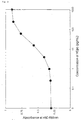
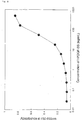
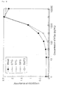
- Fig. 18 depicts responses of cells to human EPO in individual and mixed cell culture systems. Open symbols indicate the responses of the individually cultured established cell lines (4 x 10 3 cells/well), and closed circles those of the mixed culture of the four cell lines (each cell line at 4 x 10 3 cells/well, 16 x 10 3 cells/well in total). In comparison to the individual cultures (open upward triangle), a slight enhancement in the growth activity was observed for the mixed culture (closed circle) in the absence of human EPO. However, the response to human EPO was observed to be similar between those at the concentration 1 to 10 pg/mL, where the response was observed to start, and at maximum stimulation.
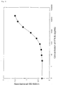
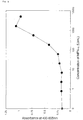
- Fig. 19 depicts the responses of cells to human TPO in individual and mixed culture systems.
- Open symbols indicate the responses of the individually cultured established cell lines (4 x 10 3 cells/well), and closed circles those of the mixed culture of the four cell lines (each cell line at 4 x 10 3 cells/well, 16 x 10 3 cells/well in total).
- a slight enhancement in the growth activity was observed for the mixed culture (closed circle) in the absence of human TPO.
- the response to human TPO was observed to be similar between those at the concentration 100 to 300 pg/mL, where the response was observed to start, and at maximum stimulation.
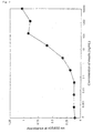
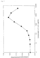
- Fig. 20 depicts the responses of cells to human G-CSF in individual and mixed culture systems.
- Open symbols indicate the responses of the individually cultured established cell lines (4 x 10 3 cells/well), and closed circles those of the mixed culture of the four cell lines (each cell line at 4 x 10 3 cells/well, 16 x 10 3 cells/well in total).
- a slight enhancement in the growth activity was observed for the mixed culture (closed circle) in the absence of human G-CSF.
- the response to human G-CSF was observed to be similar between those at the concentration 2 to 5 pg/mL, where the response was observed to start, and at maximum stimulation.
- Mammalian cell plasmid expression vector pCOS-G was constructed by replacing the polyadenylation signal of pCOS1 (see International Publication No. WO98/13388, "Antibody against human parathormone related peptides" with that of the human G-CSF gene.
- the polyadenylation signal of human G-CSF gene was obtained by digesting pEF-BOS (Mizushima S. et al. (1990) Nuc. Acid Res. 18, 5322) with Xho I and Pvu II.
- the polyadenylation signal fragment was replaced into the Xho I/ Aor 51H I fragment of pCOS1, and the resulting construct was dubbed pCOS-G.
- Mammalian cell plasmid expression vector pCOS2 was constructed by replacing the Bam HI/ Aor 51H I fragment of pCOS1 with the Bam HI/ Aor 51HI fragment of pEGFP-N1 (CLONTECH).
- Mammalian cell plasmid expression vector pCV was constructed by replacing the polyadenylation signal of pCOS1 with that of the human G-CSF gene.
- the polyadenylation signal of human G-CSF gene was obtained by digesting pEF-BOS with Eco RI and Xba I. The 3' end of the fragment was blunted, and a Bam HI site was attached to the 5' end of the fragment. The resulting fragment was replaced as the poly (A) addition signal into the Bam HI/ Aor 51H I site of pCOS1, and was used as pCV.
- EPO Erythropoietin
- a chimeric receptor cDNA was constructed by linking cDNA fragments encoding the extracellular domain (from the 1st to the 249th amino acid residues; Jones, SS. et al. (1990) 76, 31-35) of human EPO receptor, and the transmembrane and intracellular domains (from the 602nd to the 813th amino acid residues; Fukunaga, R. et al. (1990) Cell 61, 341-350) of mouse G-CSF receptor.
- the cDNA was inserted downstream of the EF1 ⁇ promoter of the mammalian expression vector pCOS-G to construct chimeric receptor-expression vector EG/pCOS-G.
- EG/pCOS-G was linearized using Pvu I (Takara Shuzo), extracted with phenol and chloroform, and purified by ethanol precipitation.
- the linearized expression vector was introduced into mouse FDC-P1 cells (ATCC No. CRL-12103) using an electroporation apparatus (Gene Pulser: Bio Rad).
- the FDC-P1 cells were washed twice with Dulbecco's PBS (hereinafter referred to as PBS), and were suspended in PBS to give a cell density of about 1 x 10 7 cells/mL.
- 10 ⁇ g of the linearized expression vector DNA was added to 0.8 mL of the suspension, transferred into a cuvette for electroporation (Bio Rad), and pulsed with a capacitance of 250 ⁇ F at 0.35 kV.
- the cells treated by electroporation were suspended in Media A that was supplemented with 1 ng/mL human EPO (prepared from genetic recombinant CHO cells), and then were seeded on a 96-well microtiter plate (flat bottom, Falcon) at 100 ⁇ L/well. After culturing in a CO 2 incubator (CO 2 concentration, 5%) for about six hours, 100 ⁇ L/well of Media A supplemented with 1 ng/mL EPO and 1 mg/mL GENETICIN (GIBCO) was added to the plate, and was cultured in a CO 2 incubator (CO 2 concentration, 5%).
- CO 2 incubator CO 2 concentration, 5%
- RPMI1640 (GIBCO) supplemented with 10% fetal bovine serum (GIBCO), 100 U/mL penicillin, and 0.1 mg/mL streptomycin (GIBCO) was used as Media A. About one week after the start of the culture, cells were observed under a microscope, and cells were collected from wells with a single colony. The collected cells were subcultured in Media A containing 1 ng/mL human EPO. The titer of human EPO used in this Example was 270,000 IU/mg.
- the cells were washed twice with Media A, and then suspended in Media A to give a cell density of 2 x 10 5 cells/mL.
- 10 ⁇ l/well WST-8 reagent Cell Counting Kit-8; DOJINDO LABORATORIES
- the linearized expression gene vector was introduced into mouse Ba/F3 cells (purchased from RIKEN; Cell No.: RCB0805) using an electroporation apparatus (Gene Pulser, BIO Rad).
- the Ba/F3 cells were washed twice with Dulbecco's PBS (hereinafter referred to as PBS), and then were suspended in PBS to give a cell density of about 1 x 10 7 cells/mL.
- 10 ⁇ g of the linearized expression vector DNA was added to 0.8 mL of this suspension, transferred into a cuvette (Bio Rad) for electroporation, and pulsed with a capacitance of 960 ⁇ F at 0.33 kV.
- the cells treated by electroporation were suspended in Media A containing 1 ng/mL human EPO, and then were seeded on a 96-well microtiter plate (flat bottom, Falcon) at 100 ⁇ l/well. After culturing the cells for about five hours in a CO 2 incubator (CO 2 concentration, 5%), 100 ⁇ L/well Media A that contained 1 ng/mL human EPO and 1 mg/mL GENETICIN (GIBCO) was added, and the cells were cultured in a CO 2 incubator (CO 2 concentration, 5%).
- CO 2 incubator CO 2 concentration, 5%
- RPMI1640 (GIBCO) supplemented with 10% fetal bovine serum (GIBCO), 100 U/mL penicillin, and 0.1 mg/mL streptomycin (GIBCO) was used as Media A.
- GBCO fetal bovine serum
- penicillin 100 U/mL penicillin
- streptomycin 0.1 mg/mL streptomycin
- cell line B#20 that had a high sensitivity to human EPO was selected, and was used as chimeric EPO receptor-expressing cell line EPG (Fig. 3). Similar growth responses to human EPO were observed by using FDC-P1 and Ba/F3 as host cells.
- TPO Thrombopoietin
- a chimeric receptor cDNA was constructed by linking cDNA fragments encoding the extracellular domain (from the 1st to the 491st amino acid residues; Vigon, I. et al. (1992) Proc. Natl. Acad. Sci. USA 89, 5640-5644) of human TPO receptor, and the transmembrane and intracellular domains (from the 602nd to the 813th amino acid residues; Fukunaga, R. et al. (1990) Cell 61, 341-350) of mouse G-CSF receptor.
- the cDNA was inserted downstream of the EF1 ⁇ promoter of the mammalian expression vector pCOS-G to construct chimeric receptor-expression vector TG/pCOS-G.
- TG/pCOS-G was linearized with Pvu I (Takara Shuzo), extracted with phenol and chloroform, and purified by ethanol precipitation.
- the linearized expression gene vector was introduced into mouse Ba/F3 cells (purchased from RIKEN; Cell No.: RCB0805) using an electroporation apparatus (Gene Pulser: Bio Rad).
- the Ba/F3 cells were washed twice with Dulbecco's PBS (hereinafter referred to as PBS), and then were suspended in PBS to give a cell density of about 1 x 10 7 cells/mL.
- 10 ⁇ g of the linearized expression vector DNA was added to 0.8 mL of this suspension, transferred into a cuvette (Bio Rad) for electroporation, and was pulsed with a capacitance of 960 ⁇ F at 0.33 kV.
- the cells treated by electroporation were suspended in Media A, and were seeded on a 96-well microtiter plate (flat bottom, Falcon) at 100 ⁇ L/well.
- 100 ⁇ L/well Media A that contained 2 ng/mL human TPO (R&D Systems) and 1 mg/mL GENETICIN (GIBCO) was added, and the cells were cultured in a CO 2 incubator (CO 2 concentration, 5%).
- RPMI1640 (GIBCO) supplemented with 10% fetal bovine serum (GIBCO), 100 U/mL penicillin, and 0.1 mg/mL streptomycin (GIBCO) was used as Media A.
- the cells were observed under a microscope, cells were collected from wells with a single colony and were subcultured in Media A containing 1 ng/mL human TPO.
- the cells were washed twice with Media A, and were suspended in Media A to give a cell density of 5 x 10 4 cells/mL.
- 50 ⁇ l/well cell suspension and 50 ⁇ l/well of human TPO (R&D Systems) appropriately diluted with Media A were dispensed into the wells of a 96-well microtiter plate (flat bottom, Falcon), and were cultured for 72 hours in a CO 2 incubator (CO 2 concentration, 5%). After the culture, 10 ⁇ l/well WST-8 reagent (Cell Count Reagent SF; Nacalai Tesque) was added, and the plate was incubated for two hours in a CO 2 incubator (CO 2 concentration, 5%).
- G-CSF Granulocyte-colony stimulating factor
- a chimeric receptor cDNA was constructed by linking cDNA fragments encoding the extracellular domain (from the -23rd to the 604th amino acid residues; Fukunaga, R. et al. (1990) Proc. Natl. Acad. Sci. USA 87, 8702-8706) of human G-CSF receptor, and the transmembrane and intracellular domains (from the 602nd to the 813th amino acid residues; Fukunaga, R. et al. (1990) Cell 61, 341-350) of mouse G-CSF receptor.
- the cDNA was inserted downstream of the HEF1 ⁇ promoter of the mammalian expression vector pCOS-G to construct chimeric receptor-expression vector GG/pCOS-G.
- GG/pCOS-G was linearized with Pvu I (Takara Shuzo), extracted with phenol and chloroform, and purified by ethanol precipitation.
- the linearized expression gene vector was introduced into mouse Ba/F3 cells using an electroporation apparatus (Gene Pulser: Bio Rad).
- the Ba/F3 cells were washed twice with Dulbecco's PBS (hereinafter referred to as PBS), and then were suspended in PBS to give a cell density of about 1 x 10 7 cells/mL.
- 10 ⁇ g of linearized expression vector DNA was added to 0.8 mL of this suspension, transferred into a cuvette (Bio Rad) for electroporation, and was pulsed with a capacitance of 960 ⁇ F at 0.33 kV.
- the cells treated by electroporation were suspended in Media A, and were seeded on a 96-well microtiter plate (flat bottom, Falcon) at 100 ⁇ L/well. 100 ⁇ L/well Media A containing 2 ng/mL human G-CSF was added, and the cells were cultured in a CO 2 incubator (CO 2 concentration, 5%).
- RPMI1640 (GIBCO) supplemented with 10% fetal bovine serum (Hyclone), 100 U/mL penicillin, and 0.1 mg/mL streptomycin (GIBCO) was used as Media A.
- the human G-CSF used in this Example was prepared from recombinant CHO cells, and the titer of human G-CSF was 1.2 x 10 8 IU/mg.
- the cells were washed twice with Media A, and were suspended in Media A to give a cell density of 5 x 10 4 cells/mL.
- 50 ⁇ l/well cell suspension and 50 ⁇ l/well of human G-CSF (prepared from recombinant CHO cells) appropriately diluted in Media A were dispensed into wells of a 96-well microtiter plate (flat bottom, Falcon), and were cultured for 70 hours in a CO 2 incubator (CO 2 concentration, 5%).
- cell line GFG#342 that had a high sensitivity to human G-CSF was selected and used as chimeric G-CSF receptor-expressing cell line GFG (Fig. 5).
- Flt-3 ligand (1-5) Flt-3/Flk-2 ligand (hereinafter referred to as Flt-3 ligand) receptor
- a chimeric receptor cDNA was constructed by linking cDNA fragments encoding the extracellular domain (from the 1st to the 541st amino acid residues; Small, D. et al. (1994) 91, 459-463) of human Flt-3, and the transmembrane and intracellular domains (from the 602nd to the 813th amino acid residues; Fukunaga, R. et al. (1990) Cell 61, 341-350) of mouse G-CSF receptor.
- the cDNA was inserted downstream of the EF1 ⁇ promoter of the mammalian expression vector pCOS2 to construct chimeric receptor-expression vector FLG/pCOS2.
- the FLG/pCOS2 was linearized with Hpa I (Takara Shuzo), extracted with phenol and chloroform, and purified by ethanol precipitation.
- the linearized expression gene vector was introduced into mouse Ba/F3 cells using an electroporation apparatus (Gene Pulser: Bio Rad).
- the Ba/F3 cells were washed twice with Dulbecco's PBS (hereinafter referred to as PBS), and then were suspended in PBS to give a cell density of about 1 x 10 7 cells/mL.
- 10 ⁇ g of the linearized expression vector DNA was added to 0.8 mL of this suspension, transferred into a cuvette (Bio Rad) for electroporation, and pulsed with a capacitance of 960 ⁇ F at 0.33 kV.
- the cells treated by electroporation were suspended in Media A, and were seeded on a 96-well microtiter plate (flat bottom, Falcon) at 100 ⁇ L/well.
- 100 ⁇ L/well Media A that contained 10 ng/mL human Flt-3 ligand (Genzyme) was added, and the cells were cultured in a CO 2 incubator (CO 2 concentration, 5%).
- RPMI1640 (GIBCO) supplemented with 10% fetal bovine serum (Hyclone), 100 U/mL penicillin, and 0.1 mg/mL streptomycin (GIBCO) was used as Media A.
- the cells were observed under a microscope, cells were collected from wells with a single colony and subcultured in Media A containing 5 ng/mL human Flt-3 ligand.
- the cells were washed twice with Media A, and were suspended in Media A to give a cell density of 5 x 10 4 cells/mL.
- 50 ⁇ l/well cell suspension and 50 ⁇ l/well of human Flt-3 ligand appropriately diluted in Media A were dispensed into wells of a 96-well microtiter plate (flat bottom, Falcon), and were cultured for 74 hours in a CO 2 incubator (CO 2 concentration, 5%).
- 10 ⁇ l/well WST-8 reagent Cell Count Reagent SF; Nacalai Tesque
- pre-reaction absorbance at a measurement wavelength of 450 nm and a control wavelength of 655 nm was measured using microplate reader (Model 3550, Bio Rad).
- cell line FLG#102 that had a high sensitivity to human Flt-3 ligand was selected and used as chimeric Flt-3 ligand receptor-expressing cell line FLG (Fig. 6).
- a chimeric receptor cDNA was constructed by linking cDNA fragments encoding the extracellular domain (from the -27th to the 917th amino acid residues; Ullrich, A. et al. (1985) Nature 313, 756-761) of human insulin receptor, and the transmembrane and intracellular domains (from the 602nd to the 813th amino acid residues,; Fukunaga, R. et al. (1990) Cell 61, 341-350) of mouse G-CSF receptor.
- the cDNA was inserted downstream of the HEF1 ⁇ promoter of the mammalian expression vector pCV to construct chimeric receptor-expression vector ING/pCV.
- ING/pCV was linearized with Pvu I (Takara Shuzo), extracted with phenol and chloroform, and purified by ethanol precipitation.
- the linearized expression gene vector was introduced into mouse Ba/F3 cells using an electroporation apparatus (Gene Pulser: Bio Rad).
- the Ba/F3 cells were washed twice with Dulbecco's PBS (hereinafter referred to as PBS), and then were suspended in PBS to give a cell density of about 1 x 10 7 cells/mL.
- 10 ⁇ g of the linearized expression vector DNA was added to 0.8 mL of this suspension, transferred into a cuvette (Bio Rad) for electroporation, and pulsed with a capacitance of 960 ⁇ F at 0.33 kV.
- the cells treated by electroporation were suspended in Media A, and were seeded on a 96-well microtiter plate (flat bottom, Falcon) at 100 ⁇ L/well.
- 100 ⁇ L/well Media A containing 10 ⁇ g/mL human insulin (SIGMA) was added, and the cells were cultured in a CO 2 incubator (CO 2 concentration, 5%).
- RPMI1640 (GIBCO) supplemented with 10% fetal bovine serum (Hyclone), 100 U/mL penicillin, and 0.1 mg/mL streptomycin (GIBCO) was used as Media A.
- the cells were observed under a microscope, cells were collected from wells with a single colony and subcultured in Media A containing 10 ⁇ g/mL human insulin.
- the cells were washed twice with media without human insulin (hereinafter referred to as Media B), and were suspended in Media B to give a cell density of 5 x 10 4 cells/mL.
- CHO-S-SFM II medium (GIBCO) prepared without the addition of human insulin was used as Media B.
- 100 ⁇ l/well of the cell suspension, 80 ⁇ L/well of Media B, and 20 ⁇ l/well of human insulin appropriately diluted in 10 mM HCl solution that contained 0.1% bovine serum albumin (SIGMA) were dispensed into the wells of a 96-well microtiter plate (flat bottom, Falcon), and were cultured for 72 hours in a CO 2 incubator (CO 2 concentration, 5%).
- SIGMA bovine serum albumin
- cell line ING#139 that had a high sensitivity to human insulin was selected and used as chimeric insulin receptor-expressing cell line ING (Fig. 7).
- PDGF Platelet-derived growth factor
- a chimeric receptor cDNA was constructed by linking cDNA fragments encoding the ⁇ chain (from the 1st to the 531st amino acid residues; Gronwald, RGK. et al. (1988) Proc. Natl. Acad. Sci. USA. 85, 3435-3439) of human PDGF receptor, and the transmembrane and intracellular domains (from the 602nd to the 813th amino acid residues; Fukunaga, R. et al. (1990) Cell 61, 341-350) of mouse G-CSF receptor.
- the cDNA was inserted downstream of the HEF1 ⁇ promoter of the mammalian expression vector pCV to construct chimeric receptor-expression vector pCV-cPDGFR.
- pCV-cPDGFR was linearized with Pvu I (Takara Shuzo), extracted with phenol and chloroform, and purified by ethanol precipitation.
- the linearized expression gene vector was introduced into mouse Ba/F3 cells using an electroporation apparatus (Gene Pulser: Bio Rad).
- the Ba/F3 cells were washed twice with Dulbecco's PBS (hereinafter referred to as PBS), and then were suspended in PBS to give a cell density of about 1 x 10 7 cells/mL.
- 20 ⁇ g of the linearized expression vector DNA was added to 0.8 mL of this suspension, transferred into a cuvette (Bio Rad) for electroporation, and pulsed with a capacitance of 960 ⁇ F at 0.33 kV.
- the cells treated by electroporation were suspended in Media A, and were seeded on a 96-well microtiter plate (flat bottom, Falcon) at 100 ⁇ L/well.
- 100 ⁇ L/well Media A containing 40 ng/mL human PDGF-BB (Genzyme) was added, and the cells were cultured in a CO 2 incubator (CO 2 concentration, 5%).
- RPMI1640 (GIBCO) supplemented with 10% fetal bovine serum (Hyclone), 100 U/mL penicillin, and 0.1 mg/mL streptomycin (GIBCO) was used as Media A.
- the cells were observed under a microscope, cells were collected from wells with a single colony and subcultured in Media A that contained 20 ng/mL human PDGF-BB.
- the cells were washed twice with Media A, and were suspended in Media A to give a cell density of 5 x 10 4 cells/mL.
- 50 ⁇ l/well of the cell suspension and 50 ⁇ L/well human PDGF-BB appropriately diluted in Media A were dispensed into the wells of a 96-well microtiter plate (flat bottom, Falcon), and were cultured for 70 hours in a CO 2 incubator (CO 2 concentration, 5%).
- 10 ⁇ L/well WST-8 reagent Cell Count Reagent SF; Nacalai Tesque
- pre-reaction absorbance at a measurement wavelength of 450 nm and a control wavelength of 655 nm was measured using microplate reader (Model 3550, Bio Rad).
- cell line PDG#35 that had a high sensitivity to human PDGF-BB was selected and used as chimeric PDGF receptor-expressing cell line PDG (Fig. 8).
- IFN Interferon
- Chimeric receptor cDNAs were constructed by linking cDNA fragments encoding extracellular domain of the ⁇ chain (AR1) (from the 1st to the 436th amino acid residues; Uze, G. et al. (1990) Cell 60, 225-234) and the ⁇ / ⁇ chain (AR2; ⁇ ) (from the 1st to the 243rd amino acid residues; Novick, D. et al. (1994) Cell 77, 391-400) of human IFN ⁇ / ⁇ receptor with the transmembrane and intracellular domains (from the 602nd to the 813th amino acid residues; Fukunaga, R. et al. (1990) Cell 61, 341-350) of mouse G-CSF receptor, respectively.
- AR1 from the 1st to the 436th amino acid residues
- AR2; ⁇ from the 1st to the 243rd amino acid residues
- Novick, D. et al. (1994) Cell 77, 391-400 of human IFN ⁇
- IFG ⁇ /pCV and IFG ⁇ /pCV chimeric receptor-expression vectors
- IFG ⁇ /pCV and IFG ⁇ /pCV were linearized with Pvu I (Takara Shuzo), respectively, extracted with phenol and chloroform, and purified by ethanol precipitation.
- the linearized expression gene vectors were introduced into mouse Ba/F3 cells using an electroporation apparatus (Gene Pulser: Bio Rad).
- the Ba/F3 cells were washed twice with Dulbecco's PBS (hereinafter referred to as PBS), and then were suspended in PBS to give a cell density of about 1 x 10 7 cells/mL.
- 10 ⁇ g each of linearized IFG ⁇ /pCV and IFG ⁇ /pCV was added to 0.8 mL of the suspension, transferred into a cuvette (Bio Rad) for electroporation, and pulsed with a capacitance of 960 ⁇ F at 0.33 kV.
- the cells were suspended in Media A, and were seeded on a 96-well microtiter plate (flat bottom, Falcon) at 100 ⁇ L/well. 100 ⁇ L/well Media A containing 1,000 U/mL human IFN ⁇ (CARBIOCHEM) was added, and the cells were cultured in a CO 2 incubator (CO 2 concentration, 5%). RPMI1640 (GIBCO) supplemented with 10% fetal bovine serum (Hyclone), 100 U/mL penicillin, and 0.1 mg/mL streptomycin (GIBCO) was used as Media A. About one week after the start of the culture, the cells were observed under a microscope, cells were collected from wells with a single colony and subcultured in Media A containing 1,000 U/mL IFN ⁇ .
- the cells were washed twice with Media A, and were suspended in Media A to give a cell density of 5 x 10 4 cells/mL.
- 50 ⁇ l/well cell suspension and 50 ⁇ L/well of human IFN ⁇ appropriately diluted in Media A were dispensed into the wells of a 96-well microtiter plate (flat bottom, Falcon), and were cultured for 72 hours in a CO 2 incubator (CO 2 concentration, 5%).
- 10 ⁇ L/well WST-8 reagent Cell Count Reagent SF; Nacalai Tesque
- pre-reaction absorbance at a measurement wavelength of 450 nm and a control wavelength of 655 nm was measured using microplate reader (Model 3550, Bio Rad).
- the plate was incubated for two hours in a CO 2 incubator (CO 2 concentration, 5%), and post-reaction absorbance was measured as above. Based on the cell growth activity determined by the number of viable cells by plotting the amount of changes in absorbance measured after the two-hour incubation on the vertical axis and the concentrations of human IFN ⁇ on the horizontal axis, cell line IFG#A01 that had a high sensitivity to human IFN ⁇ was selected and used as chimeric IFN ⁇ receptor-expressing cell line IFG.
- the IFN ⁇ / ⁇ receptor has been known to form a heterodimer, not a homodimer, to transmit the signal. According to the present experiment, even if the intracellular domain of a heterodimer forming receptor was replaced with that of a homodimer forming receptor (represented by the G-CSF receptor), the chimeric receptor induced signal transduction and the response of ligands could be detected as the cell growing activity (Fig. 9).
- a chimeric receptor cDNA was constructed by linking cDNA fragments encoding the extracellular domain (from the 1st to the 839th amino acid residues; Tartaglia, LA. et al. (1995) Cell, 83, 1263-1271) of human leptin receptor, and the transmembrane and intracellular domains (from the 602nd to the 813th amino acid residues; Fukunaga, R. et al. (1990) Cell 61, 341-350) of mouse G-CSF receptor.
- This cDNA was inserted downstream of the HEF1 ⁇ promoter of the mammalian expression vector pCV to construct chimeric receptor expression vector pCV-cLepR.
- pCV-cLepR was linearized with Pvu I (Takara Shuzo), extracted with phenol and chloroform, and purified by ethanol precipitation.
- the linearized expression gene vector was introduced into mouse Ba/F3 cells (purchased from RIKEN; Cell No.: RCB0805) using an electroporation apparatus (Gene Pulser: Bio Rad).
- the Ba/F3 cells were washed twice with Dulbecco's PBS (hereinafter referred to as PBS), and then were suspended in PBS to give a cell density of about 1 x 10 7 cells/mL.
- 10 ⁇ g of the linearized expression vector DNA was added to 0.8 mL of this suspension, transferred into a cuvette (Bio Rad) for electroporation, and pulsed with a capacitance of 960 ⁇ F at 0.33 kV.
- the cells treated by electroporation were suspended in 50 mL Media A, and were seeded on five 96-well microtiter plates (flat bottom, Falcon) at 100 ⁇ L/well.
- 100 ⁇ L/well Media A containing 10 ng/mL human leptin (Genzyme) was added, and the cells were cultured in a CO 2 incubator (CO 2 concentration, 5%).
- RPMI1640 (GIBCO) supplemented with 10% fetal bovine serum (Hyclone), 100 U/mL penicillin, and 0.1 mg/mL streptomycin (GIBCO) was used as Media A.
- the cells were observed under a microscope, cells were collected from wells of a single colony and subcultured in Media A containing 10 ng/mL human leptin.
- the cells were washed twice with Media A, and were suspended in Media A to give a cell density of 5 x 10 4 cells/mL.
- 50 ⁇ l/well of the cell suspension and 50 ⁇ L/well of human leptin appropriately diluted in Media A were dispensed into the wells of a 96-well microtiter plate (flat bottom, Falcon), and were cultured for 72 hours in a CO 2 incubator (CO 2 concentration, 5%).
- 10 ⁇ L/well WST-8 reagent Cell Count Reagent SF; Nacalai Tesque
- pre-reaction absorbance at a measurement wavelength of 450 nm and a control wavelength of 655 nm was measured using microplate reader (Model 3550, Bio Rad).
- GH Growth hormone
- a chimeric receptor cDNA was constructed by linking cDNA fragments encoding the extracellular domain (from the 18th to the 246th amino acid residues; Leung, DW. et al. (1987) Nature 330, 537-543) of human GH receptor, and the transmembrane and intracellular domains (from the 602nd to the 813th amino acid residues; Fukunaga, R. et al. (1990) Cell 61, 341-350) of mouse G-CSF receptor.
- This cDNA was inserted downstream of the EF1 ⁇ promoter of the mammalian expression vector pCV to construct chimeric receptor-expression vector GHG/pCV.
- GHG/pCV was linearized with P vu I (Takara Shuzo), extracted with phenol and chloroform, and purified by ethanol precipitation.
- the linearized expression gene vector was introduced into mouse Ba/F3 cells (purchased from RIKEN; Cell No.: RCB0805) using an electroporation apparatus (Gene Pulser: Bio Rad).
- the Ba/F3 cells were washed twice with Dulbecco's PBS (hereinafter referred to as PBS), and then were suspended in PBS to give a cell density of about 1 x 10 7 cells/mL.
- 10 ⁇ g of the linearized expression vector DNA was added to 0.8 mL of this suspension, transferred into a cuvette (Bio Rad) for electroporation, and pulsed with a capacitance of 960 ⁇ F at 0.33 kV.
- the cells treated by electroporation were suspended in 50 mL Media A, and were seeded on five 96-well microtiter plates (flat bottom, Falcon) at 100 ⁇ L/well.
- 100 ⁇ L/well Media A that contained 200 ⁇ IU/mL human GH (Genotropin(R): Pharmacia & Upjohn) was added, and the cells were cultured in a CO 2 incubator (CO 2 concentration, 5%).
- RPMI1640 (GIBCO) supplemented with 10% fetal bovine serum (HyClone), 100 U/mL penicillin, and 0.1 mg/mL streptomycin (GIBCO) was used as Media A.
- the cells were observed under a microscope, cells were collected from wells of a single colony and subcultured in Media A containing 500 ⁇ IU/mL human GH.
- the cells were washed twice with Media A, and were suspended in Media A to give a cell density of 5 x 10 4 cells/mL.
- 50 ⁇ l/well cell suspension and 50 ⁇ L/well human GH appropriately diluted in Media A were dispensed into the wells of a 96-well microtiter plate (flat bottom, Falcon), and were cultured for 72 hours in a CO 2 incubator (CO 2 concentration, 5%).
- 10 ⁇ L/well WST-8 reagent Cell Count Reagent SF; Nacalai Tesque
- pre-reaction absorbance at a measurement wavelength of 450 nm and a control wavelength of 655 nm was measured using microplate reader (Model 3550, Bio Rad).
- the plate was incubated for two hours in a CO 2 incubator (CO 2 concentration, 5%), and post-reaction absorbance was measured as above. Based on the cell growth activity determined by the number of viable cells by plotting the amount of change in the absorbance measured after the two-hour incubation on the vertical axis and the concentrations of human GH on the horizontal axis, cell line GHG#11 that had a high sensitivity to human GH was selected, and was used as chimeric GH receptor-expressing cell line GHG. This cell line was subcultured in the presence of 20 ⁇ IU/mL human GH. At a concentration of 0.3 ⁇ IU/mL or more, an obvious growth reaction against human GH was observed (Fig. 12).
- Interleukin 10 (hereinafter referred to as IL-10) receptor
- Chimeric receptor cDNAs were constructed by linking cDNA fragments encoding the ⁇ chain (IL-10R ⁇ ) (from the 1st to the 235th amino acid residue; Liu, Y. et al. (1994) 152, 1821-1829) and the hIL-10R subunit (CRFB4; IL-10R ⁇ ) extracellular domain (from the 1st to the 220th amino acid residues; Lutfalla, G. et al. (1993) Genomics 16, 366-373) of human IL-10 receptor with the transmembrane and intracellular domains (from the 602nd to the 813th amino acid residues; Fukunaga, R. et al.
- the linearized expression gene vectors were introduced into mouse Ba/F3 cells, respectively, using an electroporation apparatus (Gene Pulser: Bio Rad).
- the Ba/F3 cells were washed twice with Dulbecco's PBS (hereinafter referred to as PBS), and then were suspended in PBS to give a cell density of about 1 x 10 7 cells/mL.
- 10 ⁇ g each of linearized expression vector DNA, pCV-cIL10R ⁇ and pCV-cIL10R ⁇ was added to 0.8 mL of this suspension, transferred into a cuvette (Bio Rad) for electroporation, and pulsed with a capacitance of 960 ⁇ F at 0.33 kV.
- the cells treated by electroporation were suspended in 50 mL Media A, and were seeded on five 96-well microtiter plates (flat bottom, Falcon) at 100 ⁇ L/well. 100 ⁇ L/well Media A that contained 10 ng/mL of human IL-10 (Genzyme) was added, and the cells were cultured in a CO 2 incubator (CO 2 concentration, 5%). RPMI1640 (GIBCO) supplemented with 10% fetal bovine serum (Hyclone), 100 U/mL penicillin, and 0.1 mg/mL streptomycin (GIBCO) was used as Media A. About one week after the start of the culture, the cells were observed under a microscope, cells were collected from wells with a single colony and subcultured in Media A containing 5 ng/mL human IL-10.
- the cells were washed twice with Media A, and were suspended in Media A to give a cell density of 5 x 10 4 cells/mL.
- 50 ⁇ l/well cell suspension and 50 ⁇ L/well human IL-10 appropriately diluted in Media A were dispensed into wells of a 96-well microtiter plate (flat bottom, Falcon), and were cultured for 72 hours in a CO 2 incubator (CO 2 concentration, 5%).
- 10 ⁇ L/well WST-8 reagent Cell Count Reagent SF; Nacalai Tesque
- pre-reaction absorbance at a measurement wavelength of 450 nm and a control wavelength of 655 nm was measured using microplate reader (Model 3550, Bio Rad).
- cell line 10G#10 that had a high sensitivity to human IL-10 was selected and used as chimeric IL-10 receptor-expressing cell line 10G. This cell line was subcuitured in the presence of 1 ng/mL human IL-10. At a concentration of 0.5 ng/mL or more, an obvious growth reaction against human IL-10 was observed (Fig. 13).
- IGF-I Insulin-like growth factor I
- a chimeric receptor cDNA was constructed by linking cDNA fragments encoding the extracellular domain (from the 1st to the 931st amino acid residues; Ullrich, A. et al. (1986) EMBO J., 5, 2503-2512) of human IGF-I receptor, and the transmembrane and intracellular domains (from the 602nd to the 813th amino acid residues; Fukunaga, R. et al. (1990) Cell 61, 341-350) of mouse G-CSF receptor.
- This construct was inserted downstream of the HEF1 ⁇ promoter of the mammalian expression vector pCV to construct chimeric receptor-expression vector pCV-cIGF1R.
- pCV-cIGF1R was linearized with Pvu I (Takara Shuzo), extracted with phenol and chloroform, and purified by ethanol precipitation.
- the linearized expression vector was introduced into mouse Ba/F3 cells using an electroporation apparatus (Gene Pulser: Bio Rad).
- the Ba/F3 cells were washed twice with Dulbecco's PBS (hereinafter referred to as PBS), and then were suspended in PBS to give a cell density of about 1 x 10 7 cells/mL.
- 20 ⁇ g linearized expression vector DNA was added to 0.8 mL of this suspension, transferred into a cuvette (Bio Rad) for electroporation, and pulsed with a capacitance of 960 ⁇ F at 0.33 kV.
- the cells treated by electroporation were suspended in 40 mL Media A, and were seeded on four 96-well microtiter plates (flat bottom, Falcon) at 100 ⁇ L/well.
- 100 ⁇ L/well Media A that contained 40 ng/mL human IGF-I (Genzyme) was added, and the cells were cultured in a CO 2 incubator (CO 2 concentration, 5%).
- RPMI1640 (GIBCO) supplemented with 10% fetal bovine serum (Hyclone), 100 U/mL penicillin, and 0.1 mg/mL streptomycin (GIBCO) was used as Media A.
- the cells were observed under a microscope, cells were collected from wells of a single colony and subcultured in Media A containing 20 to 50 ng/mL human IGF-I.
- the cells were washed twice with media without human insulin (hereinafter referred to as Media B), and were suspended in Media B to give a cell density of 5 x 10 4 cells/mL.
- CHO-S-SFM II medium (GIBCO) without the addition of human insulin was used as Media B.
- 50 ⁇ l/well cell suspension and 50 ⁇ L/well human IGF-I appropriately diluted with Media B were dispensed into the wells of a 96-well microtiter plate (flat bottom, Falcon), and were cultured for 72 hours in a CO 2 incubator (CO 2 concentration, 5%).
- cell line IGG#06 that had a high sensitivity to human IGF-I was selected and used as chimeric insulin receptor-expressing cell line IGG. This cell line was subcultured in the presence of 50 ng/mL human IGF-I. At a concentration of 10 ng/mL or more, an obvious growth reaction to human IGF-I was observed (Fig. 14).
- LIF Leukemia inhibitory factor
- Chimeric receptor DNAs were constructed by linking cDNA fragments encoding human LIF receptor (from the 1st to the 833rd amino acid residues; Gearing, DP. et al. (1988) EMBO J., 10, 2839-2848) and human gp130 (from the 1st to the 619th amino acid residues; Hibi, M. et al. (1990) Cell, 63, 1149-1157) with the transmembrane and intracellular domains (from the 602nd to the 813th amino acid residues; Fukunaga, R. et al. (1990) Cell 61, 341-350) of mouse G-CSF receptor, respectively.
- constructs were inserted downstream of the HEF1 ⁇ promoter of the mammalian expression vector pCV to construct chimeric receptors expression vectors, pCV-cLIFR and pCV-cgp130, respectively.
- pCV-cLIFR and pCV-cgp130 were linearized with Pvu I (Takara Shuzo), respectively, extracted with phenol and chloroform, and purified by ethanol precipitation.
- the linearized expression vectors were introduced into mouse Ba/F3 cells using an electroporation apparatus (Gene Pulser: Bio Rad).
- the Ba/F3 cells were washed twice with Dulbecco's PBS (hereinafter referred to as PBS), and then were suspended in PBS to give a cell density of about 1 x 10 7 cells/mL.
- PBS Dulbecco's PBS
- 20 ⁇ g each of the linearized expression vector DNA, pCV-cLIFR and pCV-cgp130 was added to 0.8 mL of this suspension, transferred into a cuvette (Bio Rad) for electroporation, and pulsed with a capacitance of 960 ⁇ F at 0.33 kV.
- the cells treated by electroporation were suspended in 40 mL of Media A, and were seeded on four 96-well microtiter plates (flat bottom, Falcon) at 100 ⁇ L/well.
- 100 ⁇ L/well Media A that contained 10 ng/mL human LIF (Genzyme) was added, and the cells were cultured in a CO 2 incubator (CO 2 concentration, 5%).
- RPMI1640 (GIBCO) supplemented with 10% fetal bovine serum (Hyclone), 100 U/mL penicillin, and 0.1 mg/mL streptomycin (GIBCO) was used as Media A.
- the cells were observed under a microscope, cells were collected from wells with a single colony and subcultured in Media A containing 5 ng/mL human LIF.
- the cells were washed twice with Media A, and were suspended in Media A to give a cell density of 5 x 10 4 cells/mL.
- cell line LIG#47 that had a high sensitivity to human LIF was selected and used as chimeric LIF receptor-expressing cell line LIG. This cell line was subcultured in the presence of 1 ng/mL human LIF. At a concentration of 0.4 ng/mL or more, an obvious growth reaction to human LIF could be observed (Fig. 15).
- Ciliary neurotrophic factor (hereinafter referred to as CNTF) receptor (1-14) Ciliary neurotrophic factor (hereinafter referred to as CNTF) receptor
- Human CNTF receptor (from the 1st to the 372nd amino acid residues; Davis, S. et al. (1991) Science, 253, 59-63) cDNA was cloned downstream of the HEF1 ⁇ promoter of a mammalian expression vector, pCV, to construct human CNTF receptor (CNTFR) expression vector, pCV-CNTFR.
- CNTFR human CNTF receptor
- pCV-CNTFR was linearized with Pvu I (Takara Shuzo), extracted with phenol and chloroform, and purified by ethanol precipitation.
- the linearized expression gene vector was introduced into the chimeric LIF receptor-expressing cell line LIG (see section 1-13) using an electroporation apparatus (Gene Pulser: Bio Rad).
- the LIG cells were washed twice with Dulbecco's PBS (hereinafter referred to as PBS), and then were suspended in PBS to give a cell density of about 1 x 10 7 cells/mL.
- 10 ⁇ g of linearized pCV-CNTFR was added to 0.8 mL of this suspension, transferred into a cuvette (Bio Rad) for electroporation, and pulsed with a capacitance of 960 ⁇ F at 0.33 kV.
- the cells treated by electroporation were suspended in 40 mL of Media A, and were seeded on four 96-well microtiter plates (flat bottom, Falcon) at 100 ⁇ L/well.
- 100 ⁇ L/well Media A that contained 1 ng/mL human CNTF (Genzyme) was added, and the cells were cultured in a CO 2 incubator (CO 2 concentration, 5%).
- RPMI1640 (GIBCO) supplemented with 10% fetal bovine serum (Hyclone), 100 U/mL penicillin, and 0.1 mg/mL streptomycin (GIBCO) was used as Media A.
- the cells were observed under a microscope, cells were collected from wells with a single colony and subcultured in Media A containing 0.5 to 1 ng/mL CNTF.
- the cells were washed twice with Media A, and were suspended in Media A to give a cell density of 5 x 10 4 cells/mL.
- 50 ⁇ l/well cell suspension and 50 ⁇ L/well human CNTF appropriately diluted in Media A were dispensed into the wells of a 96-well microtiter plate (flat bottom, Falcon), and were cultured for 72 hours in a CO 2 incubator (CO 2 concentration, 5%).
- 10 ⁇ L/well WST-8 reagent Cell Count Reagent SF; Nacalai Tesque
- pre-reaction absorbance at a measurement wavelength of 450 nm and a control wavelength of 655 nm was measured using microplate reader (Model 3550, Bio Rad).
- cell line CNG#203 that had a high sensitivity to human CNTF was selected and used as chimeric CNTF receptor-expressing cell line CNG. This cell line was subcultured in the presence of 1 ng/mL human CNTF. At a concentration of 0.3 ng/mL or more, an obvious growth reaction to human CNTF was observed (Fig. 16).
- CNTF receptor has been known to form a heterotrimer, not a homodimer, to transmit signal. According to the present experiment, a chimeric receptor of such a heterotrimeric receptor, even if the intracellular domain of the receptor was replaced with that of a homodimeric receptor (represented by the G-CSF receptor), could induce signal transduction and the reactivity of the ligand could be detected as the cell growth activity.
- a homodimeric receptor represented by the G-CSF receptor
- PDGF Platelet-derived growth factor
- a chimeric receptor cDNA was constructed by linking cDNA fragments encoding the ⁇ chain (from the 1st to the 531st amino acid residues; Gronwald, RGK. et al. (1988) Proc. Natl. Acad. Sci. USA. 85, 3435-3439) of human PDGF receptor and the transmembrane and intracellular domains (from the 602nd to the 813th amino acid residues; Fukunaga, R. et al. (1990) Cell 61, 341-350) of mouse G-CSF receptor.
- the cDNA was cloned downstream of the EF1 ⁇ promoter of the mammalian expression vector pCV to construct chimeric receptor-expression vector pCV-cPDGFR.
- pCV-cPDGFR was linearized with Pvu I (Takara Shuzo), extracted with phenol and chloroform, and purified by ethanol precipitation.
- the linearized expression vector was introduced into mouse Ba/F3 cells using an electroporation apparatus (Gene Pulser: Bio Rad).
- the Ba/F3 cells were washed twice with Dulbecco's PBS (hereinafter referred to as PBS), and then were suspended in PBS to give a cell density of about 1 x 10 7 cells/mL.
- 20 ⁇ g of the linearized expression vector DNA was added to 0.8 mL of this suspension, transferred into a cuvette (Bio Rad) for electroporation, and pulsed with a capacitance of 960 ⁇ F at 0.33 kV.
- the cells treated by electroporation were suspended in Media A, and were seeded on a 96-well microtiter plate (flat bottom, Falcon) at 100 ⁇ L/well.
- 100 ⁇ L/well Media A that contained 20 ng/mL human PDGF-BB (Genzyme) was added, and the cells were cultured in a CO 2 incubator (CO 2 concentration, 5%).
- RPMI1640 (GIBCO) supplemented with 10 vol% fetal bovine serum (Hyclone), 100 U/mL penicillin, and 0.1 mg/mL streptomycin (GIBCO) was used as Media A.
- the cells were observed under a microscope, cells were collected from wells with a single colony and subcultured in Media A containing 5 ng/mL human PDGF-BB.
- the cells were washed twice with Media A, and were suspended in Media A to give a cell density of 5 x 10 4 cells/mL.
- 50 ⁇ l/well cell suspension and 50 ⁇ L/well human PDGF-BB appropriately diluted in Media A were dispensed into the wells of a 96-well microtiter plate (flat bottom, Falcon), and were cultured for 72 hours in a CO 2 incubator (CO 2 concentration, 5%).
- 10 ⁇ L/well WST-8 reagent Cell Count Reagent SF; Nacalai Tesque
- pre-reaction absorbance at a measurement wavelength of 450 nm and a control wavelength of 655 nm was measured using microplate reader (Model 3550, Bio Rad).
- cell line PDG#10 that had a high sensitivity to human PDGF-BB was selected and used as chimeric PDGF receptor-expressing cell line PDG (Fig. 17).
- EPG, GFG, TPG, and the non-recombinant cell Ba/F3 were subcultured in the presence of 1 ng/mL human EPO, 10 ng/mL human G-CSF, 1 ng/mL human TPO (R & D Systems), and 1 ng/mL mouse IL-3 (R & D Systems), respectively.
- the used human EPO and G-CSF were prepared from recombinant CHO cells, and their titer was 270,000 IU/mg and 1.2 x 10 3 IU/mg, respectively.
- the cells were washed twice with RPMI1640 media containing 2% fetal bovine serum, and then were suspended in RPMI1640 containing 10% fetal bovine serum.
- the four cell lines were mixed by adjusting each of the cell lines to 4 x 10 4 cells/mL (16 x 10 4 cells/mL in total) for the mixed culture, and, for individual cultures, cells were prepared at 4 x 10 4 cells/mL. 100 ⁇ L each of the cells were independently seeded on a 96-well microtiter plate (flat bottom, Falcon). After adding 100 ⁇ L/well each of appropriately diluted cytokines, the cultures were cultured for 72 hours in a CO 2 incubator (5% CO 2 concentration).
- the responses of the mixed culture against cytokines began at similar concentration (2 pg/mL of hEPO, 200 pg/mL of hTPO, and 2 pg/mL of hG-CSF) and a similar growth activity was detected as in the individual cultures.
- the growth of the chimeric receptor-expressing cells was specifically induced by corresponding ligands.
- the parental cell line did not respond to any cytokine, and no growth activity was observed.
- the methods of the present invention enables an efficient and rapid screening for a vast number of test samples to isolate substances having biological activities of interest.
- the methods of the present invention are particularly suited for the screening of ligands that bind to certain receptors, and serves as an important basic technique for the development of novel pharmaceuticals.
Landscapes
- Health & Medical Sciences (AREA)
- Life Sciences & Earth Sciences (AREA)
- Engineering & Computer Science (AREA)
- Chemical & Material Sciences (AREA)
- Immunology (AREA)
- Medicinal Chemistry (AREA)
- General Health & Medical Sciences (AREA)
- Chemical Kinetics & Catalysis (AREA)
- General Chemical & Material Sciences (AREA)
- Bioinformatics & Cheminformatics (AREA)
- Organic Chemistry (AREA)
- Nuclear Medicine, Radiotherapy & Molecular Imaging (AREA)
- Hematology (AREA)
- Animal Behavior & Ethology (AREA)
- Pharmacology & Pharmacy (AREA)
- Public Health (AREA)
- Veterinary Medicine (AREA)
- Diabetes (AREA)
- Molecular Biology (AREA)
- Biomedical Technology (AREA)
- Urology & Nephrology (AREA)
- Analytical Chemistry (AREA)
- Obesity (AREA)
- Pathology (AREA)
- General Physics & Mathematics (AREA)
- Biochemistry (AREA)
- Physics & Mathematics (AREA)
- Biotechnology (AREA)
- Cell Biology (AREA)
- Microbiology (AREA)
- Food Science & Technology (AREA)
- Transplantation (AREA)
- Endocrinology (AREA)
- Emergency Medicine (AREA)
- Child & Adolescent Psychology (AREA)
- Dermatology (AREA)
- Physical Education & Sports Medicine (AREA)
- Oncology (AREA)
- Measuring Or Testing Involving Enzymes Or Micro-Organisms (AREA)
- Peptides Or Proteins (AREA)
Abstract
Description
Claims (27)
- A method for simultaneously detecting two or more distinct activities, wherein said method comprises the steps of:(i) determining in advance, a detection marker common to said activities,(ii) preparing a detection method that can detect each of said two or more distinct activities using said common detection marker, and(iii) simultaneously detecting said activities using said detection method.
- A method for selecting a test sample that has at least one of two or more distinct activities, wherein said method comprises the steps of:(i) determining in advance, a detection marker common to said activities,(ii) preparing a detection method that can detect each of said two or more distinct biological activities using said common detection marker, and(iii) simultaneously detecting at least one of the biological activities of a test sample using said detection method.
- A method of screening for a ligand that can bind to at least one of two or more kinds of receptors, wherein said method comprises the steps of:(i) obtaining two or more kinds of receptors comprising (a) a common signal-transducing domain, and (b) a domain other than the signal-transducing domain, wherein said domain derives from the same receptor from which the common domain of (a) is derived and/or a different receptor,(ii) contacting a test sample with said two or more kinds of receptors, and(iii) detecting the biological activity of said test sample.
- The method according to claim 3, wherein said step (i) obtains a cell expressing two or more kinds of receptors comprising (a) a common signal-transducing domain, and (b) a domain other than the signal-transducing domain, wherein said domain derives from the same receptor from which the common domain of (a) is derived and/or a different receptor.
- The method according to claim 3, wherein said step (i) obtains two or more kinds of cells that express a receptor comprising (a) a common signal-transducing domain, and (b) a domain other than the signal-transducing domain, wherein said domain derives from the same receptor from which the common domain of (a) is derived and/or a different receptor.
- A method according to any one of claims 3 to 5, wherein said signal-transducing domain and/or said domain other than the signal-transducing domain is derived from a cell membrane receptor.
- A method according to any one of claims 3 to 5, wherein said signal-transducing domain and/or said domain other than the signal-transducing domain is derived from a nuclear receptor.
- The method according to claim 6, wherein said domain other than the signal-transducing domain is the extracellular domain, of a cell membrane receptor, or a portion thereof.
- The method according to claim 6, wherein said domain other than the signal-transducing domain is a ligand-binding domain of a cell membrane receptor.
- The method according to claim 6, wherein said signal-transducing domain and/or said domain other than the signal-transducing domain is derived from a receptor belonging to a receptor family selected from the group consisting of the hematopoietic factor receptor family, cytokine receptor family, tyrosine kinase-type receptor family, serine/threonine kinase-type receptor family, TNF receptor family, G protein-coupled receptor family, GPI-anchored receptor family, tyrosine phosphatase-type receptor family, cell adhesion receptor family, and hormone receptor family.
- The method according to claim 6, wherein said signal-transducing domain and/or said domain other than the signal-transducing domain is derived from the following receptors; human or mouse erythropoietin (EPO) receptor, human or mouse granulocyte-colony stimulating factor (G-CSF) receptor, human or mouse thrombopoietin (TPO) receptor, human or mouse insulin receptor, human or mouse Flt-3 receptor, human or mouse platelet-derived growth factor (PDGF) receptor, human or mouse interferon (IFN)-α or -β receptor, human or mouse leptin receptor, human or mouse growth hormone (GH) receptor, human or mouse interleukin (IL)-10 receptor, human or mouse insulin-like growth factor (IGF)-I receptor, human or mouse leukemia inhibitory factor (LIF) receptor, or human or mouse ciliary neurotrophic factor (CNTF) receptor.
- The method according to claim 11, wherein said signal-transducing domain derives from mouse G-CSF receptor.
- The method according to claim 3, wherein in step (ii), two or more kinds of cells expressing said receptors are mixed and contacted with the test sample.
- The method according to any one of claims 4 to 13, wherein said cell(s) is a transformed cell(s).
- The method according to claim 14, wherein said cell(s) is derived from a cytokine-dependent cell.
- The method according to claim 15, wherein said transformed cell(s) is derived from Ba/F3 cells or FDC-P1 cells.
- A method according to claim 2 or 3, wherein in step (iii), an agonist or antagonist activity of the test sample is detected.
- The method according to claim 2 or 3, wherein in step (iii), the biological activity of the test sample is detected by using a cell-free detection system.
- The method according to claim 2 or 3, wherein in step (iii), the biological activity of the test sample is measured by using a cell-based detection system.
- The method according to claim 19, wherein in step (iii), the biological activity of the test sample is measured using a phenotypic change in the cell.
- The method according to claim 20, wherein said phenotypic change is a quantitative and/or qualitative change of a cell surface antigen.
- The method according to claim 21, wherein said phenotypic change is a change in proliferation activity.
- The method according to claim 3, which method further comprises the step of measuring the biological activity of a test sample by contacting it with one of said two or more receptors obtained in (i) in order to determine the specificity of the test sample towards the receptor.
- A cell that expresses two or more kinds of receptors comprising (a) a common signal-transducing domain, and (b) a domain other than the signal-transducing domain, wherein said domain derives from the same receptor from which the common domain of (a) is derived and/or a different receptor.
- A kit for screening a substance or ligand that binds to at least one of two or more kinds of receptors, wherein said kit comprises one of (a) to (c):(a) two or more kinds of receptors comprising (i) a common signal-transducing domain, and (ii) a domain other than the signal-transducing domain, wherein said domain derives from the same receptor from which the common domain of (i) is derived and/or a different receptor;(b) DNA encoding two or more kinds of receptors comprising (i) a common signal-transducing domain, and (ii) a domain other than the signal-transducing domain, wherein said domain derives from the same receptor from which the common domain of (i) is derived and/or a different receptor;(c) a cell expressing two or more kinds of receptors comprising (i) a common signal-transducing domain, and (ii) a domain other than the signal-transducing domain, wherein said domain derives from the same receptor from which the common domain of (i) is derived and/or a different receptor.
- A substance or ligand isolated by the screening according to claim 2 or 3.
- A pharmaceutical composition comprising the substance or ligand according to claim 26.
Applications Claiming Priority (5)
| Application Number | Priority Date | Filing Date | Title |
|---|---|---|---|
| JP2000221070 | 2000-07-17 | ||
| JP2000221070 | 2000-07-17 | ||
| JP2001159032 | 2001-05-28 | ||
| JP2001159032 | 2001-05-28 | ||
| PCT/JP2001/006170 WO2002006838A1 (en) | 2000-07-17 | 2001-07-17 | Method for screening ligand having biological activity |
Publications (2)
| Publication Number | Publication Date |
|---|---|
| EP1304573A1 true EP1304573A1 (en) | 2003-04-23 |
| EP1304573A4 EP1304573A4 (en) | 2006-06-07 |
Family
ID=26596454
Family Applications (1)
| Application Number | Title | Priority Date | Filing Date |
|---|---|---|---|
| EP01950013A Ceased EP1304573A4 (en) | 2000-07-17 | 2001-07-17 | METHOD FOR SCREENING A LIGAND HAVING BIOLOGICAL ACTIVITY |
Country Status (5)
| Country | Link |
|---|---|
| US (1) | US7732133B2 (en) |
| EP (1) | EP1304573A4 (en) |
| JP (1) | JP4908721B2 (en) |
| AU (1) | AU2001271066A1 (en) |
| WO (1) | WO2002006838A1 (en) |
Families Citing this family (25)
| Publication number | Priority date | Publication date | Assignee | Title |
|---|---|---|---|---|
| JP2006180705A (en) * | 2003-03-06 | 2006-07-13 | Chugai Pharmaceut Co Ltd | Chimeric receptor and screening method for ligand or inhibitor of the receptor |
| AU2003271186A1 (en) | 2003-10-14 | 2005-04-27 | Chugai Seiyaku Kabushiki Kaisha | Double specific antibodies substituting for functional protein |
| DK1814590T4 (en) | 2004-11-01 | 2014-02-24 | Amylin Pharmaceuticals Llc | Treatment of obesity and related diseases. |
| KR101374454B1 (en) | 2005-03-31 | 2014-03-17 | 추가이 세이야쿠 가부시키가이샤 | Methods for producing polypeptides by regulating polypeptide association |
| WO2007115230A2 (en) * | 2006-03-30 | 2007-10-11 | University Of Medicine And Dentistry Of New Jersey | A strategy for homo- or hetero-dimerization of various proteins in solutions and in cells |
| EP4793360A2 (en) | 2006-03-31 | 2026-08-19 | Chugai Seiyaku Kabushiki Kaisha | Methods for controlling blood pharmacokinetics of antibodies |
| JP5144499B2 (en) | 2006-03-31 | 2013-02-13 | 中外製薬株式会社 | Antibody modification method for purifying bispecific antibodies |
| JP2008102117A (en) | 2006-09-21 | 2008-05-01 | Fujifilm Corp | Surface plasmon enhanced fluorescence sensor and fluorescence detection method |
| EP3127921A1 (en) | 2007-09-26 | 2017-02-08 | Chugai Seiyaku Kabushiki Kaisha | Method of modifying isoelectric point of antibody via amino acid substition in cdr |
| CN105859889B (en) | 2010-11-17 | 2020-01-07 | 中外制药株式会社 | Multispecific antigen binding molecules with functions that replace the functions of coagulation factor VIII |
| CA2925256C (en) | 2013-09-27 | 2023-08-15 | Chugai Seiyaku Kabushiki Kaisha | Method for producing polypeptide heteromultimer |
| TWI700300B (en) | 2014-09-26 | 2020-08-01 | 日商中外製藥股份有限公司 | Antibodies that neutralize substances with the function of FVIII coagulation factor (FVIII) |
| TWI701435B (en) | 2014-09-26 | 2020-08-11 | 日商中外製藥股份有限公司 | Method to determine the reactivity of FVIII |
| JP7082484B2 (en) | 2015-04-01 | 2022-06-08 | 中外製薬株式会社 | Method for Producing Polypeptide Heterogeneous Multimer |
| GB201522097D0 (en) * | 2015-12-15 | 2016-01-27 | Cellular Therapeutics Ltd | Cells |
| US20200270363A1 (en) | 2015-12-25 | 2020-08-27 | Chugai Seiyaku Kabushiki Kaisha | Antibody having enhanced activity, and method for modifying same |
| SG11201803989WA (en) | 2015-12-28 | 2018-06-28 | Chugai Pharmaceutical Co Ltd | Method for promoting efficiency of purification of fc region-containing polypeptide |
| AU2017255077B2 (en) | 2016-04-28 | 2024-05-16 | Chugai Seiyaku Kabushiki Kaisha | Antibody-containing preparation |
| CA3027018A1 (en) | 2016-07-29 | 2018-02-01 | Chugai Seiyaku Kabushiki Kaisha | Bispecific antibody exhibiting increased alternative fviii-cofactor-function activity |
| US11352438B2 (en) | 2016-09-06 | 2022-06-07 | Chugai Seiyaku Kabushiki Kaisha | Methods of using a bispecific antibody that recognizes coagulation factor IX and/or activated coagulation factor IX and coagulation factor X and/or activated coagulation factor X |
| JP7209298B2 (en) | 2017-03-31 | 2023-01-20 | 公立大学法人奈良県立医科大学 | A pharmaceutical composition used for the prevention and/or treatment of blood coagulation factor IX disorders, containing a multispecific antigen-binding molecule that substitutes for the function of blood coagulation factor VIII |
| JP2020517259A (en) | 2017-04-19 | 2020-06-18 | ボード・オブ・リージエンツ,ザ・ユニバーシテイ・オブ・テキサス・システム | Immune cells expressing engineered antigen receptors |
| CA3071236A1 (en) | 2017-09-29 | 2019-04-04 | Chugai Seiyaku Kabushiki Kaisha | Multispecific antigen-binding molecule having blood coagulation factor viii (fviii) cofactor function-substituting activity, and pharmaceutical formulation containing said moleculeas active ingredient |
| KR20260090008A (en) | 2017-11-01 | 2026-06-16 | 추가이 세이야쿠 가부시키가이샤 | Antibody variant and isoform with lowered biological activity |
| WO2019165121A1 (en) | 2018-02-21 | 2019-08-29 | Board Of Regents, The University Of Texas System | Methods for activation and expansion of natural killer cells and uses therof |
Family Cites Families (8)
| Publication number | Priority date | Publication date | Assignee | Title |
|---|---|---|---|---|
| DE3514272A1 (en) * | 1985-04-17 | 1986-10-23 | Schering AG, 1000 Berlin und 4709 Bergkamen | DERMATICA |
| US4859609A (en) * | 1986-04-30 | 1989-08-22 | Genentech, Inc. | Novel receptors for efficient determination of ligands and their antagonists or agonists |
| WO1991014776A1 (en) * | 1990-03-23 | 1991-10-03 | Osaka Bioscience Institute | Dna coding for granulocytic colony stimulating factor receptor |
| DE69233586T2 (en) | 1991-08-23 | 2006-09-28 | NPS Pharmaceuticals, Inc., Salt Lake City | Calcium receptor active compounds |
| JPH08508889A (en) | 1993-06-07 | 1996-09-24 | アムジエン・インコーポレーテツド | Hybrid receptor molecule |
| CA2220715C (en) * | 1995-06-07 | 2004-11-02 | Praecis Pharmaceuticals Incorporated | Functional bioassay for g-protein coupled receptor agonists and antagonists |
| US5866341A (en) * | 1996-04-03 | 1999-02-02 | Chugai Pharmaceutical Co., Ltd. | Compositions and methods for screening drug libraries |
| WO2001025797A2 (en) * | 1999-10-06 | 2001-04-12 | Sumitomo Chemical Company, Limited | Methods of evaluating whether a test agent is an agent affecting a leptin receptor |
-
2001
- 2001-07-17 US US10/333,103 patent/US7732133B2/en not_active Expired - Fee Related
- 2001-07-17 AU AU2001271066A patent/AU2001271066A1/en not_active Abandoned
- 2001-07-17 JP JP2002512695A patent/JP4908721B2/en not_active Expired - Fee Related
- 2001-07-17 EP EP01950013A patent/EP1304573A4/en not_active Ceased
- 2001-07-17 WO PCT/JP2001/006170 patent/WO2002006838A1/en not_active Ceased
Also Published As
| Publication number | Publication date |
|---|---|
| US20040091876A1 (en) | 2004-05-13 |
| EP1304573A4 (en) | 2006-06-07 |
| WO2002006838A1 (en) | 2002-01-24 |
| JP4908721B2 (en) | 2012-04-04 |
| US7732133B2 (en) | 2010-06-08 |
| AU2001271066A1 (en) | 2002-01-30 |
Similar Documents
| Publication | Publication Date | Title |
|---|---|---|
| US7732133B2 (en) | Screening methods for biologically active ligands | |
| JPWO2002006838A1 (en) | Screening method for physiologically active ligands | |
| Wei et al. | Receptor activator of nuclear factor-κB ligand activates nuclear factor-κB in osteoclast precursors | |
| KR102362176B1 (en) | Devices and Methods for Cell Secretion Assays | |
| US6893827B1 (en) | Receptor function assay for G-protein coupled receptors and orphan receptors by reporter enzyme mutant complementation | |
| CN108351344B (en) | Genome-scale T cell activity arrays and methods of using the same | |
| Lei et al. | Development and application of potency assays based on genetically modified cells for biological products | |
| US20080132427A1 (en) | Cell systems and methods for detecting proliferation acitvity | |
| RU2139936C1 (en) | Method of determining modulation effect of substance on interleukin-5 receptor-linked signal-transmission route in human or animal cell | |
| Tsang et al. | Cytokine assays and their limitations. | |
| Chen et al. | Cell surface protein–protein interaction profiling for biological network analysis and novel target discovery | |
| Kirken et al. | Involvement of JAK-family tyrosine kinases in hematopoietin receptor signal transduction | |
| Montoye et al. | A systematic scan of interactions with tyrosine motifs in the erythropoietin receptor using a mammalian 2-hybrid approach | |
| EP1257818A1 (en) | Enzyme-based gprotein-coupled receptor assay | |
| CN111500668B (en) | Method for determining biological activity of human IL-36/IL36R/IL1RAcP pathway inhibitor | |
| US5733735A (en) | Assay system to determine receptor antagonist target site | |
| US7348151B1 (en) | Method for the cellular high-throughput-detection of nuclear receptor ligand interactions | |
| Ricketts et al. | Pertussis toxin-sensitive and-insensitive thrombin stimulation of Shc phosphorylation and mitogenesis are mediated through distinct pathways | |
| TWI338046B (en) | Human g protein-coupled receptor and modulators thereof for the treatment of inflammatory disorders | |
| Miller | Discovery of cytokine mimics using cell-based systems | |
| JP2006180705A (en) | Chimeric receptor and screening method for ligand or inhibitor of the receptor | |
| WO2005026728A1 (en) | Screening method | |
| Carrithers et al. | A melanophore-based screening assay for erythropoietin receptors | |
| CN1401000A (en) | Method for detecting ligand or ligand-like low molecular weight compounds | |
| RU2810503C1 (en) | Test system and methods of determining biological activity of human thrombopoetin receptor agonists |
Legal Events
| Date | Code | Title | Description |
|---|---|---|---|
| PUAI | Public reference made under article 153(3) epc to a published international application that has entered the european phase |
Free format text: ORIGINAL CODE: 0009012 |
|
| 17P | Request for examination filed |
Effective date: 20030217 |
|
| AK | Designated contracting states |
Designated state(s): AT BE CH CY DE DK ES FI FR GB GR IE IT LI LU MC NL PT SE TR |
|
| AX | Request for extension of the european patent |
Extension state: AL LT LV MK RO SI |
|
| A4 | Supplementary search report drawn up and despatched |
Effective date: 20060503 |
|
| 17Q | First examination report despatched |
Effective date: 20081020 |
|
| RTI1 | Title (correction) |
Free format text: SCREENING METHODS FOR BIOLOGICALLY ACTIVE LIGANDS |
|
| REG | Reference to a national code |
Ref country code: DE Ref legal event code: R003 |
|
| STAA | Information on the status of an ep patent application or granted ep patent |
Free format text: STATUS: THE APPLICATION HAS BEEN REFUSED |
|
| 18R | Application refused |
Effective date: 20170210 |