EP1295940A1 - Polypeptides, use thereof and process for producing the same - Google Patents
Polypeptides, use thereof and process for producing the same Download PDFInfo
- Publication number
- EP1295940A1 EP1295940A1 EP01943820A EP01943820A EP1295940A1 EP 1295940 A1 EP1295940 A1 EP 1295940A1 EP 01943820 A EP01943820 A EP 01943820A EP 01943820 A EP01943820 A EP 01943820A EP 1295940 A1 EP1295940 A1 EP 1295940A1
- Authority
- EP
- European Patent Office
- Prior art keywords
- polypeptide
- liver
- seq
- dna
- mrna
- Prior art date
- Legal status (The legal status is an assumption and is not a legal conclusion. Google has not performed a legal analysis and makes no representation as to the accuracy of the status listed.)
- Withdrawn
Links
- 108090000765 processed proteins & peptides Proteins 0.000 title claims abstract description 152
- 229920001184 polypeptide Polymers 0.000 title claims abstract description 150
- 102000004196 processed proteins & peptides Human genes 0.000 title claims abstract description 150
- 238000000034 method Methods 0.000 title claims description 40
- 230000008569 process Effects 0.000 title description 3
- 210000004185 liver Anatomy 0.000 claims abstract description 77
- 208000006454 hepatitis Diseases 0.000 claims abstract description 70
- 108020004999 messenger RNA Proteins 0.000 claims abstract description 70
- 231100000283 hepatitis Toxicity 0.000 claims abstract description 65
- 238000004519 manufacturing process Methods 0.000 claims abstract description 55
- 239000002299 complementary DNA Substances 0.000 claims description 69
- 108090000623 proteins and genes Proteins 0.000 claims description 45
- 239000013598 vector Substances 0.000 claims description 25
- 108091034117 Oligonucleotide Proteins 0.000 claims description 20
- 238000012408 PCR amplification Methods 0.000 claims description 15
- 238000009007 Diagnostic Kit Methods 0.000 claims description 11
- 238000003199 nucleic acid amplification method Methods 0.000 claims description 11
- 230000003321 amplification Effects 0.000 claims description 10
- 238000012258 culturing Methods 0.000 claims description 2
- 125000003275 alpha amino acid group Chemical group 0.000 claims 3
- 108020004414 DNA Proteins 0.000 description 71
- 238000006243 chemical reaction Methods 0.000 description 62
- 239000000243 solution Substances 0.000 description 46
- 150000001413 amino acids Chemical group 0.000 description 34
- 239000002773 nucleotide Substances 0.000 description 33
- 125000003729 nucleotide group Chemical group 0.000 description 33
- 108091008146 restriction endonucleases Proteins 0.000 description 32
- 239000013612 plasmid Substances 0.000 description 28
- 238000003776 cleavage reaction Methods 0.000 description 23
- 108091028732 Concatemer Proteins 0.000 description 21
- 230000014509 gene expression Effects 0.000 description 21
- 239000000203 mixture Substances 0.000 description 20
- 230000007017 scission Effects 0.000 description 20
- 239000012634 fragment Substances 0.000 description 19
- 238000002360 preparation method Methods 0.000 description 19
- 239000000047 product Substances 0.000 description 18
- 239000000427 antigen Substances 0.000 description 17
- 102000036639 antigens Human genes 0.000 description 17
- 108091007433 antigens Proteins 0.000 description 17
- 238000012163 sequencing technique Methods 0.000 description 17
- 210000004027 cell Anatomy 0.000 description 14
- 239000000872 buffer Substances 0.000 description 13
- HEDRZPFGACZZDS-UHFFFAOYSA-N Chloroform Chemical compound ClC(Cl)Cl HEDRZPFGACZZDS-UHFFFAOYSA-N 0.000 description 12
- 238000011084 recovery Methods 0.000 description 12
- 108020004705 Codon Proteins 0.000 description 11
- 235000018102 proteins Nutrition 0.000 description 10
- 102000004169 proteins and genes Human genes 0.000 description 10
- LFQSCWFLJHTTHZ-UHFFFAOYSA-N Ethanol Chemical compound CCO LFQSCWFLJHTTHZ-UHFFFAOYSA-N 0.000 description 9
- 101500006448 Mycobacterium bovis (strain ATCC BAA-935 / AF2122/97) Endonuclease PI-MboI Proteins 0.000 description 9
- 241000700605 Viruses Species 0.000 description 9
- 239000003153 chemical reaction reagent Substances 0.000 description 9
- 239000013604 expression vector Substances 0.000 description 9
- 108091028043 Nucleic acid sequence Proteins 0.000 description 8
- 238000001574 biopsy Methods 0.000 description 8
- 239000007795 chemical reaction product Substances 0.000 description 8
- 239000000523 sample Substances 0.000 description 8
- 238000013519 translation Methods 0.000 description 8
- 241000588724 Escherichia coli Species 0.000 description 7
- 238000002405 diagnostic procedure Methods 0.000 description 7
- 239000007790 solid phase Substances 0.000 description 7
- HCHKCACWOHOZIP-UHFFFAOYSA-N Zinc Chemical compound [Zn] HCHKCACWOHOZIP-UHFFFAOYSA-N 0.000 description 6
- 238000004458 analytical method Methods 0.000 description 6
- 239000013599 cloning vector Substances 0.000 description 6
- 150000001875 compounds Chemical class 0.000 description 6
- 230000003247 decreasing effect Effects 0.000 description 6
- 239000000032 diagnostic agent Substances 0.000 description 6
- 229940039227 diagnostic agent Drugs 0.000 description 6
- UCSJYZPVAKXKNQ-HZYVHMACSA-N streptomycin Chemical compound CN[C@H]1[C@H](O)[C@@H](O)[C@H](CO)O[C@H]1O[C@@H]1[C@](C=O)(O)[C@H](C)O[C@H]1O[C@@H]1[C@@H](NC(N)=N)[C@H](O)[C@@H](NC(N)=N)[C@H](O)[C@H]1O UCSJYZPVAKXKNQ-HZYVHMACSA-N 0.000 description 6
- 238000011144 upstream manufacturing Methods 0.000 description 6
- 229910052725 zinc Inorganic materials 0.000 description 6
- 108020004635 Complementary DNA Proteins 0.000 description 5
- HVLSXIKZNLPZJJ-TXZCQADKSA-N HA peptide Chemical compound C([C@@H](C(=O)N[C@@H](CC(O)=O)C(=O)N[C@@H](C(C)C)C(=O)N1[C@@H](CCC1)C(=O)N[C@@H](CC(O)=O)C(=O)N[C@@H](CC=1C=CC(O)=CC=1)C(=O)N[C@@H](C)C(O)=O)NC(=O)[C@H]1N(CCC1)C(=O)[C@@H](N)CC=1C=CC(O)=CC=1)C1=CC=C(O)C=C1 HVLSXIKZNLPZJJ-TXZCQADKSA-N 0.000 description 5
- 238000010367 cloning Methods 0.000 description 5
- 230000000694 effects Effects 0.000 description 5
- 238000012317 liver biopsy Methods 0.000 description 5
- 239000002609 medium Substances 0.000 description 5
- 230000036961 partial effect Effects 0.000 description 5
- 239000000725 suspension Substances 0.000 description 5
- 230000009466 transformation Effects 0.000 description 5
- 239000011701 zinc Substances 0.000 description 5
- 101150111197 76 gene Proteins 0.000 description 4
- 102000012410 DNA Ligases Human genes 0.000 description 4
- 108010061982 DNA Ligases Proteins 0.000 description 4
- 102000015636 Oligopeptides Human genes 0.000 description 4
- 108010038807 Oligopeptides Proteins 0.000 description 4
- 108010006785 Taq Polymerase Proteins 0.000 description 4
- 239000007984 Tris EDTA buffer Substances 0.000 description 4
- 230000007423 decrease Effects 0.000 description 4
- 230000001965 increasing effect Effects 0.000 description 4
- 238000010561 standard procedure Methods 0.000 description 4
- 210000001519 tissue Anatomy 0.000 description 4
- 238000013518 transcription Methods 0.000 description 4
- 230000035897 transcription Effects 0.000 description 4
- 108700034663 BCL2-associated athanogene 1 Proteins 0.000 description 3
- 108091026890 Coding region Proteins 0.000 description 3
- 108010014303 DNA-directed DNA polymerase Proteins 0.000 description 3
- 102000016928 DNA-directed DNA polymerase Human genes 0.000 description 3
- 102100036263 Glutamyl-tRNA(Gln) amidotransferase subunit C, mitochondrial Human genes 0.000 description 3
- PEDCQBHIVMGVHV-UHFFFAOYSA-N Glycerine Chemical compound OCC(O)CO PEDCQBHIVMGVHV-UHFFFAOYSA-N 0.000 description 3
- 101001001786 Homo sapiens Glutamyl-tRNA(Gln) amidotransferase subunit C, mitochondrial Proteins 0.000 description 3
- ISWSIDIOOBJBQZ-UHFFFAOYSA-N Phenol Chemical compound OC1=CC=CC=C1 ISWSIDIOOBJBQZ-UHFFFAOYSA-N 0.000 description 3
- 108091081024 Start codon Proteins 0.000 description 3
- 231100000354 acute hepatitis Toxicity 0.000 description 3
- 230000015572 biosynthetic process Effects 0.000 description 3
- 238000003745 diagnosis Methods 0.000 description 3
- 230000002708 enhancing effect Effects 0.000 description 3
- 238000002474 experimental method Methods 0.000 description 3
- PCHJSUWPFVWCPO-UHFFFAOYSA-N gold Chemical compound [Au] PCHJSUWPFVWCPO-UHFFFAOYSA-N 0.000 description 3
- 238000002955 isolation Methods 0.000 description 3
- 210000005228 liver tissue Anatomy 0.000 description 3
- 238000005259 measurement Methods 0.000 description 3
- 210000004940 nucleus Anatomy 0.000 description 3
- 238000001556 precipitation Methods 0.000 description 3
- 230000002441 reversible effect Effects 0.000 description 3
- 229960005322 streptomycin Drugs 0.000 description 3
- 238000003786 synthesis reaction Methods 0.000 description 3
- YBJHBAHKTGYVGT-ZKWXMUAHSA-N (+)-Biotin Chemical compound N1C(=O)N[C@@H]2[C@H](CCCCC(=O)O)SC[C@@H]21 YBJHBAHKTGYVGT-ZKWXMUAHSA-N 0.000 description 2
- 241000894006 Bacteria Species 0.000 description 2
- 108010067770 Endopeptidase K Proteins 0.000 description 2
- 102000004190 Enzymes Human genes 0.000 description 2
- 108090000790 Enzymes Proteins 0.000 description 2
- 108091060211 Expressed sequence tag Proteins 0.000 description 2
- 229920002527 Glycogen Polymers 0.000 description 2
- 102000008394 Immunoglobulin Fragments Human genes 0.000 description 2
- 108010021625 Immunoglobulin Fragments Proteins 0.000 description 2
- 206010061218 Inflammation Diseases 0.000 description 2
- 239000006142 Luria-Bertani Agar Substances 0.000 description 2
- 241000699666 Mus <mouse, genus> Species 0.000 description 2
- 241000283973 Oryctolagus cuniculus Species 0.000 description 2
- 238000012300 Sequence Analysis Methods 0.000 description 2
- 230000010632 Transcription Factor Activity Effects 0.000 description 2
- 108091023040 Transcription factor Proteins 0.000 description 2
- 102000040945 Transcription factor Human genes 0.000 description 2
- 229920004890 Triton X-100 Polymers 0.000 description 2
- 239000013504 Triton X-100 Substances 0.000 description 2
- JLCPHMBAVCMARE-UHFFFAOYSA-N [3-[[3-[[3-[[3-[[3-[[3-[[3-[[3-[[3-[[3-[[3-[[5-(2-amino-6-oxo-1H-purin-9-yl)-3-[[3-[[3-[[3-[[3-[[3-[[5-(2-amino-6-oxo-1H-purin-9-yl)-3-[[5-(2-amino-6-oxo-1H-purin-9-yl)-3-hydroxyoxolan-2-yl]methoxy-hydroxyphosphoryl]oxyoxolan-2-yl]methoxy-hydroxyphosphoryl]oxy-5-(5-methyl-2,4-dioxopyrimidin-1-yl)oxolan-2-yl]methoxy-hydroxyphosphoryl]oxy-5-(6-aminopurin-9-yl)oxolan-2-yl]methoxy-hydroxyphosphoryl]oxy-5-(6-aminopurin-9-yl)oxolan-2-yl]methoxy-hydroxyphosphoryl]oxy-5-(6-aminopurin-9-yl)oxolan-2-yl]methoxy-hydroxyphosphoryl]oxy-5-(6-aminopurin-9-yl)oxolan-2-yl]methoxy-hydroxyphosphoryl]oxyoxolan-2-yl]methoxy-hydroxyphosphoryl]oxy-5-(5-methyl-2,4-dioxopyrimidin-1-yl)oxolan-2-yl]methoxy-hydroxyphosphoryl]oxy-5-(4-amino-2-oxopyrimidin-1-yl)oxolan-2-yl]methoxy-hydroxyphosphoryl]oxy-5-(5-methyl-2,4-dioxopyrimidin-1-yl)oxolan-2-yl]methoxy-hydroxyphosphoryl]oxy-5-(5-methyl-2,4-dioxopyrimidin-1-yl)oxolan-2-yl]methoxy-hydroxyphosphoryl]oxy-5-(6-aminopurin-9-yl)oxolan-2-yl]methoxy-hydroxyphosphoryl]oxy-5-(6-aminopurin-9-yl)oxolan-2-yl]methoxy-hydroxyphosphoryl]oxy-5-(4-amino-2-oxopyrimidin-1-yl)oxolan-2-yl]methoxy-hydroxyphosphoryl]oxy-5-(4-amino-2-oxopyrimidin-1-yl)oxolan-2-yl]methoxy-hydroxyphosphoryl]oxy-5-(4-amino-2-oxopyrimidin-1-yl)oxolan-2-yl]methoxy-hydroxyphosphoryl]oxy-5-(6-aminopurin-9-yl)oxolan-2-yl]methoxy-hydroxyphosphoryl]oxy-5-(4-amino-2-oxopyrimidin-1-yl)oxolan-2-yl]methyl [5-(6-aminopurin-9-yl)-2-(hydroxymethyl)oxolan-3-yl] hydrogen phosphate Polymers Cc1cn(C2CC(OP(O)(=O)OCC3OC(CC3OP(O)(=O)OCC3OC(CC3O)n3cnc4c3nc(N)[nH]c4=O)n3cnc4c3nc(N)[nH]c4=O)C(COP(O)(=O)OC3CC(OC3COP(O)(=O)OC3CC(OC3COP(O)(=O)OC3CC(OC3COP(O)(=O)OC3CC(OC3COP(O)(=O)OC3CC(OC3COP(O)(=O)OC3CC(OC3COP(O)(=O)OC3CC(OC3COP(O)(=O)OC3CC(OC3COP(O)(=O)OC3CC(OC3COP(O)(=O)OC3CC(OC3COP(O)(=O)OC3CC(OC3COP(O)(=O)OC3CC(OC3COP(O)(=O)OC3CC(OC3COP(O)(=O)OC3CC(OC3COP(O)(=O)OC3CC(OC3COP(O)(=O)OC3CC(OC3COP(O)(=O)OC3CC(OC3CO)n3cnc4c(N)ncnc34)n3ccc(N)nc3=O)n3cnc4c(N)ncnc34)n3ccc(N)nc3=O)n3ccc(N)nc3=O)n3ccc(N)nc3=O)n3cnc4c(N)ncnc34)n3cnc4c(N)ncnc34)n3cc(C)c(=O)[nH]c3=O)n3cc(C)c(=O)[nH]c3=O)n3ccc(N)nc3=O)n3cc(C)c(=O)[nH]c3=O)n3cnc4c3nc(N)[nH]c4=O)n3cnc4c(N)ncnc34)n3cnc4c(N)ncnc34)n3cnc4c(N)ncnc34)n3cnc4c(N)ncnc34)O2)c(=O)[nH]c1=O JLCPHMBAVCMARE-UHFFFAOYSA-N 0.000 description 2
- 235000001014 amino acid Nutrition 0.000 description 2
- 238000010171 animal model Methods 0.000 description 2
- 230000027455 binding Effects 0.000 description 2
- 239000000969 carrier Substances 0.000 description 2
- 230000030570 cellular localization Effects 0.000 description 2
- 239000000084 colloidal system Substances 0.000 description 2
- 238000012875 competitive assay Methods 0.000 description 2
- 230000000295 complement effect Effects 0.000 description 2
- FFYPMLJYZAEMQB-UHFFFAOYSA-N diethyl pyrocarbonate Chemical compound CCOC(=O)OC(=O)OCC FFYPMLJYZAEMQB-UHFFFAOYSA-N 0.000 description 2
- 238000010790 dilution Methods 0.000 description 2
- 239000012895 dilution Substances 0.000 description 2
- 229940079593 drug Drugs 0.000 description 2
- 239000003814 drug Substances 0.000 description 2
- 238000004520 electroporation Methods 0.000 description 2
- 238000000605 extraction Methods 0.000 description 2
- 239000000499 gel Substances 0.000 description 2
- 229940096919 glycogen Drugs 0.000 description 2
- 239000010931 gold Substances 0.000 description 2
- 229910052737 gold Inorganic materials 0.000 description 2
- 238000009396 hybridization Methods 0.000 description 2
- 238000003018 immunoassay Methods 0.000 description 2
- 239000012678 infectious agent Substances 0.000 description 2
- 208000015181 infectious disease Diseases 0.000 description 2
- 230000004054 inflammatory process Effects 0.000 description 2
- 238000003780 insertion Methods 0.000 description 2
- 230000037431 insertion Effects 0.000 description 2
- 210000005229 liver cell Anatomy 0.000 description 2
- 210000004962 mammalian cell Anatomy 0.000 description 2
- 239000000463 material Substances 0.000 description 2
- 238000010369 molecular cloning Methods 0.000 description 2
- 239000013641 positive control Substances 0.000 description 2
- 239000011535 reaction buffer Substances 0.000 description 2
- 230000001105 regulatory effect Effects 0.000 description 2
- 238000010186 staining Methods 0.000 description 2
- 230000002194 synthesizing effect Effects 0.000 description 2
- 238000001890 transfection Methods 0.000 description 2
- 238000012795 verification Methods 0.000 description 2
- 108091032973 (ribonucleotides)n+m Proteins 0.000 description 1
- FWMNVWWHGCHHJJ-SKKKGAJSSA-N 4-amino-1-[(2r)-6-amino-2-[[(2r)-2-[[(2r)-2-[[(2r)-2-amino-3-phenylpropanoyl]amino]-3-phenylpropanoyl]amino]-4-methylpentanoyl]amino]hexanoyl]piperidine-4-carboxylic acid Chemical compound C([C@H](C(=O)N[C@H](CC(C)C)C(=O)N[C@H](CCCCN)C(=O)N1CCC(N)(CC1)C(O)=O)NC(=O)[C@H](N)CC=1C=CC=CC=1)C1=CC=CC=C1 FWMNVWWHGCHHJJ-SKKKGAJSSA-N 0.000 description 1
- HRPVXLWXLXDGHG-UHFFFAOYSA-N Acrylamide Chemical compound NC(=O)C=C HRPVXLWXLXDGHG-UHFFFAOYSA-N 0.000 description 1
- 102100027211 Albumin Human genes 0.000 description 1
- 108010088751 Albumins Proteins 0.000 description 1
- 102000002260 Alkaline Phosphatase Human genes 0.000 description 1
- 108020004774 Alkaline Phosphatase Proteins 0.000 description 1
- 206010003827 Autoimmune hepatitis Diseases 0.000 description 1
- 108090001008 Avidin Proteins 0.000 description 1
- 101150033765 BAG1 gene Proteins 0.000 description 1
- 241000589562 Brucella Species 0.000 description 1
- 101000583086 Bunodosoma granuliferum Delta-actitoxin-Bgr2b Proteins 0.000 description 1
- 241000283707 Capra Species 0.000 description 1
- 206010008909 Chronic Hepatitis Diseases 0.000 description 1
- 101100007328 Cocos nucifera COS-1 gene Proteins 0.000 description 1
- 241000701022 Cytomegalovirus Species 0.000 description 1
- 102000053602 DNA Human genes 0.000 description 1
- 102000052510 DNA-Binding Proteins Human genes 0.000 description 1
- 108700020911 DNA-Binding Proteins Proteins 0.000 description 1
- 230000004568 DNA-binding Effects 0.000 description 1
- 101710096438 DNA-binding protein Proteins 0.000 description 1
- 108090000626 DNA-directed RNA polymerases Proteins 0.000 description 1
- 102000004163 DNA-directed RNA polymerases Human genes 0.000 description 1
- 241000238557 Decapoda Species 0.000 description 1
- 241000224431 Entamoeba Species 0.000 description 1
- 108010007577 Exodeoxyribonuclease I Proteins 0.000 description 1
- 102100029075 Exonuclease 1 Human genes 0.000 description 1
- 101710154606 Hemagglutinin Proteins 0.000 description 1
- 206010073069 Hepatic cancer Diseases 0.000 description 1
- 208000037319 Hepatitis infectious Diseases 0.000 description 1
- 241000238631 Hexapoda Species 0.000 description 1
- 241000701044 Human gammaherpesvirus 4 Species 0.000 description 1
- 102100034343 Integrase Human genes 0.000 description 1
- 101710203526 Integrase Proteins 0.000 description 1
- 206010024238 Leptospirosis Diseases 0.000 description 1
- 208000001940 Massive Hepatic Necrosis Diseases 0.000 description 1
- 241000699670 Mus sp. Species 0.000 description 1
- 206010028980 Neoplasm Diseases 0.000 description 1
- 238000000636 Northern blotting Methods 0.000 description 1
- 101710093908 Outer capsid protein VP4 Proteins 0.000 description 1
- 101710135467 Outer capsid protein sigma-1 Proteins 0.000 description 1
- 108010021757 Polynucleotide 5'-Hydroxyl-Kinase Proteins 0.000 description 1
- 102000008422 Polynucleotide 5'-hydroxyl-kinase Human genes 0.000 description 1
- 101710176177 Protein A56 Proteins 0.000 description 1
- 108010078067 RNA Polymerase III Proteins 0.000 description 1
- 102000014450 RNA Polymerase III Human genes 0.000 description 1
- MEFKEPWMEQBLKI-AIRLBKTGSA-N S-adenosyl-L-methioninate Chemical compound O[C@@H]1[C@H](O)[C@@H](C[S+](CC[C@H](N)C([O-])=O)C)O[C@H]1N1C2=NC=NC(N)=C2N=C1 MEFKEPWMEQBLKI-AIRLBKTGSA-N 0.000 description 1
- 240000004808 Saccharomyces cerevisiae Species 0.000 description 1
- 241000293871 Salmonella enterica subsp. enterica serovar Typhi Species 0.000 description 1
- BUGBHKTXTAQXES-UHFFFAOYSA-N Selenium Chemical compound [Se] BUGBHKTXTAQXES-UHFFFAOYSA-N 0.000 description 1
- 229920005654 Sephadex Polymers 0.000 description 1
- 239000012507 Sephadex™ Substances 0.000 description 1
- 101710137500 T7 RNA polymerase Proteins 0.000 description 1
- 108010068068 Transcription Factor TFIIIA Proteins 0.000 description 1
- 102100028509 Transcription factor IIIA Human genes 0.000 description 1
- 102000004357 Transferases Human genes 0.000 description 1
- 108090000992 Transferases Proteins 0.000 description 1
- 108091023045 Untranslated Region Proteins 0.000 description 1
- 208000028207 Weil disease Diseases 0.000 description 1
- DZBUGLKDJFMEHC-UHFFFAOYSA-N acridine Chemical class C1=CC=CC2=CC3=CC=CC=C3N=C21 DZBUGLKDJFMEHC-UHFFFAOYSA-N 0.000 description 1
- 229960001570 ademetionine Drugs 0.000 description 1
- 238000001261 affinity purification Methods 0.000 description 1
- 239000011543 agarose gel Substances 0.000 description 1
- 238000000246 agarose gel electrophoresis Methods 0.000 description 1
- 125000000539 amino acid group Chemical group 0.000 description 1
- 229960000723 ampicillin Drugs 0.000 description 1
- AVKUERGKIZMTKX-NJBDSQKTSA-N ampicillin Chemical compound C1([C@@H](N)C(=O)N[C@H]2[C@H]3SC([C@@H](N3C2=O)C(O)=O)(C)C)=CC=CC=C1 AVKUERGKIZMTKX-NJBDSQKTSA-N 0.000 description 1
- 210000004102 animal cell Anatomy 0.000 description 1
- 238000000137 annealing Methods 0.000 description 1
- 230000000692 anti-sense effect Effects 0.000 description 1
- 230000000890 antigenic effect Effects 0.000 description 1
- 238000003556 assay Methods 0.000 description 1
- 230000001363 autoimmune Effects 0.000 description 1
- 210000003719 b-lymphocyte Anatomy 0.000 description 1
- 108010028263 bacteriophage T3 RNA polymerase Proteins 0.000 description 1
- 230000008901 benefit Effects 0.000 description 1
- 229960002685 biotin Drugs 0.000 description 1
- 235000020958 biotin Nutrition 0.000 description 1
- 239000011616 biotin Substances 0.000 description 1
- 238000010805 cDNA synthesis kit Methods 0.000 description 1
- 201000011510 cancer Diseases 0.000 description 1
- 210000003855 cell nucleus Anatomy 0.000 description 1
- 230000008859 change Effects 0.000 description 1
- 229960005091 chloramphenicol Drugs 0.000 description 1
- WIIZWVCIJKGZOK-RKDXNWHRSA-N chloramphenicol Chemical compound ClC(Cl)C(=O)N[C@H](CO)[C@H](O)C1=CC=C([N+]([O-])=O)C=C1 WIIZWVCIJKGZOK-RKDXNWHRSA-N 0.000 description 1
- 210000000349 chromosome Anatomy 0.000 description 1
- 238000004440 column chromatography Methods 0.000 description 1
- 238000010276 construction Methods 0.000 description 1
- 230000001276 controlling effect Effects 0.000 description 1
- 210000004748 cultured cell Anatomy 0.000 description 1
- 235000018417 cysteine Nutrition 0.000 description 1
- XUJNEKJLAYXESH-UHFFFAOYSA-N cysteine Natural products SCC(N)C(O)=O XUJNEKJLAYXESH-UHFFFAOYSA-N 0.000 description 1
- 238000001514 detection method Methods 0.000 description 1
- 238000011161 development Methods 0.000 description 1
- 201000010099 disease Diseases 0.000 description 1
- 208000037265 diseases, disorders, signs and symptoms Diseases 0.000 description 1
- 238000009826 distribution Methods 0.000 description 1
- 239000000975 dye Substances 0.000 description 1
- 238000001962 electrophoresis Methods 0.000 description 1
- 239000003623 enhancer Substances 0.000 description 1
- 238000006911 enzymatic reaction Methods 0.000 description 1
- ZMMJGEGLRURXTF-UHFFFAOYSA-N ethidium bromide Chemical compound [Br-].C12=CC(N)=CC=C2C2=CC=C(N)C=C2[N+](CC)=C1C1=CC=CC=C1 ZMMJGEGLRURXTF-UHFFFAOYSA-N 0.000 description 1
- 229960005542 ethidium bromide Drugs 0.000 description 1
- PVCRZXZVBSCCHH-UHFFFAOYSA-N ethyl n-[4-[benzyl(2-phenylethyl)amino]-2-(4-phenoxyphenyl)-1h-imidazo[4,5-c]pyridin-6-yl]carbamate Chemical compound N=1C(NC(=O)OCC)=CC=2NC(C=3C=CC(OC=4C=CC=CC=4)=CC=3)=NC=2C=1N(CC=1C=CC=CC=1)CCC1=CC=CC=C1 PVCRZXZVBSCCHH-UHFFFAOYSA-N 0.000 description 1
- MHMNJMPURVTYEJ-UHFFFAOYSA-N fluorescein-5-isothiocyanate Chemical compound O1C(=O)C2=CC(N=C=S)=CC=C2C21C1=CC=C(O)C=C1OC1=CC(O)=CC=C21 MHMNJMPURVTYEJ-UHFFFAOYSA-N 0.000 description 1
- 239000007850 fluorescent dye Substances 0.000 description 1
- 230000006870 function Effects 0.000 description 1
- 239000001963 growth medium Substances 0.000 description 1
- 238000010438 heat treatment Methods 0.000 description 1
- 239000000185 hemagglutinin Substances 0.000 description 1
- 208000005252 hepatitis A Diseases 0.000 description 1
- HNDVDQJCIGZPNO-UHFFFAOYSA-N histidine Natural products OC(=O)C(N)CC1=CN=CN1 HNDVDQJCIGZPNO-UHFFFAOYSA-N 0.000 description 1
- 230000003100 immobilizing effect Effects 0.000 description 1
- 239000000411 inducer Substances 0.000 description 1
- 239000007788 liquid Substances 0.000 description 1
- 201000007270 liver cancer Diseases 0.000 description 1
- 230000003908 liver function Effects 0.000 description 1
- 208000014018 liver neoplasm Diseases 0.000 description 1
- 238000013507 mapping Methods 0.000 description 1
- 239000003550 marker Substances 0.000 description 1
- 230000007246 mechanism Effects 0.000 description 1
- 238000012737 microarray-based gene expression Methods 0.000 description 1
- 239000011325 microbead Substances 0.000 description 1
- 238000012243 multiplex automated genomic engineering Methods 0.000 description 1
- 230000036963 noncompetitive effect Effects 0.000 description 1
- 108020004707 nucleic acids Proteins 0.000 description 1
- 102000039446 nucleic acids Human genes 0.000 description 1
- 150000007523 nucleic acids Chemical class 0.000 description 1
- 230000001717 pathogenic effect Effects 0.000 description 1
- 230000007170 pathology Effects 0.000 description 1
- 230000002085 persistent effect Effects 0.000 description 1
- 238000002203 pretreatment Methods 0.000 description 1
- 230000003362 replicative effect Effects 0.000 description 1
- 238000010839 reverse transcription Methods 0.000 description 1
- 239000003161 ribonuclease inhibitor Substances 0.000 description 1
- 101150098466 rpsL gene Proteins 0.000 description 1
- 150000003839 salts Chemical class 0.000 description 1
- 239000011669 selenium Substances 0.000 description 1
- 229910052711 selenium Inorganic materials 0.000 description 1
- 230000035945 sensitivity Effects 0.000 description 1
- 239000013605 shuttle vector Substances 0.000 description 1
- 210000000952 spleen Anatomy 0.000 description 1
- 238000001356 surgical procedure Methods 0.000 description 1
- 238000012360 testing method Methods 0.000 description 1
- 239000012096 transfection reagent Substances 0.000 description 1
- 238000001665 trituration Methods 0.000 description 1
- 210000004881 tumor cell Anatomy 0.000 description 1
- 108700026220 vif Genes Proteins 0.000 description 1
- XLYOFNOQVPJJNP-UHFFFAOYSA-N water Substances O XLYOFNOQVPJJNP-UHFFFAOYSA-N 0.000 description 1
Images


Classifications
-
- C—CHEMISTRY; METALLURGY
- C07—ORGANIC CHEMISTRY
- C07K—PEPTIDES
- C07K14/00—Peptides having more than 20 amino acids; Gastrins; Somatostatins; Melanotropins; Derivatives thereof
- C07K14/435—Peptides having more than 20 amino acids; Gastrins; Somatostatins; Melanotropins; Derivatives thereof from animals; from humans
- C07K14/46—Peptides having more than 20 amino acids; Gastrins; Somatostatins; Melanotropins; Derivatives thereof from animals; from humans from vertebrates
- C07K14/47—Peptides having more than 20 amino acids; Gastrins; Somatostatins; Melanotropins; Derivatives thereof from animals; from humans from vertebrates from mammals
-
- Y—GENERAL TAGGING OF NEW TECHNOLOGICAL DEVELOPMENTS; GENERAL TAGGING OF CROSS-SECTIONAL TECHNOLOGIES SPANNING OVER SEVERAL SECTIONS OF THE IPC; TECHNICAL SUBJECTS COVERED BY FORMER USPC CROSS-REFERENCE ART COLLECTIONS [XRACs] AND DIGESTS
- Y10—TECHNICAL SUBJECTS COVERED BY FORMER USPC
- Y10S—TECHNICAL SUBJECTS COVERED BY FORMER USPC CROSS-REFERENCE ART COLLECTIONS [XRACs] AND DIGESTS
- Y10S435/00—Chemistry: molecular biology and microbiology
- Y10S435/975—Kit
Definitions
- the present invention relates to polypeptides having a production level specifically lowered in human livers having inflammation ("diseased livers") as well as their preparation methods and uses (i.e., methods for diagnosing hepatitis by determining the production level of said polypeptides in the liver and diagnostic kits for diagnosing hepatitis by determining the production level of said polypeptides in the liver).
- Hepatitis shows significantly varying clinical pictures or histopathological features depending on the cause, severity and stage. Thus, diagnosis/treatment of hepatitis requires a comprehensive judgment based on clinical pictures, histopathological observation and hepatic function tests, etc.
- a novel diagnostic means for such hepatitis would be effective for improving a basic understanding of hepatitis or for diagnosis/treatment of hepatitis.
- an object of the present invention is to provide a novel diagnostic means for hepatitis.
- the object described above was achieved by finding polypeptides having a production level specifically lowered in diseased livers and further studies based on this finding.
- an aspect of the present invention is a polypeptide having the amino acid sequence shown in SEQ ID NO: 11 or SEQ ID NO: 12 and having a production level specifically lowered in human diseased livers.
- Another aspect of the present invention is a diagnostic method for hepatitis comprising determining the production level of said polypeptide in the liver.
- a specific embodiment of this diagnostic method comprises determining the production level of mRNA encoding said polypeptide (or its cDNA) in the liver as the production level of said polypeptide in the liver.
- Another specific embodiment of this diagnostic method comprises using an antibody against said polypeptide.
- kits for diagnosing hepatitis by determining the production level of said polypeptide in the liver comprising a container receiving said polypeptide, a container receiving an antibody against said polypeptide, and a container receiving an oligonucleotide specifically hybridizing to an mRNA encoding said polypeptide (or its cDNA), respectively.
- An aspect of the present invention is a DNA containing a gene encoding said polypeptide.
- Another aspect of the present invention is a method for preparing said polypeptide comprising culturing a host cell transformed with said DNA.
- the "hepatitis” means to include any hepatitis irrespective of the cause. Namely, it includes infectious hepatitis caused by virus or other infectious agents, hepatitis induced by drugs such as alcohol or other drugs, and autoimmune hepatitis induced by autoimmune mechanism. Hepatitis viruses are well known as viruses causing hepatitis, in addition to which infection with Epstein-Barr virus or cytomegalovirus may also develop hepatitis. Infections with other infectious agents than viruses such as Salmonella typhi, Brucella, Entamoeba dysenteriae, pathogenic leptospire (Weil's disease) may also develop hepatitis. Many hepatitis viruses have been isolated such as type A, type B and type C hepatitis viruses and non-A non-B non-C hepatitis viruses, and the presence of many other viruses is predicted.
- Hepatitis is classified by the severity or stage into acute hepatitis, fulminant hepatitis, subacute hepatitis, persistent hepatitis, chronic hepatitis, etc.
- hepatitis means to include any hepatitis of these classes.
- the "production level” means the amount of a polypeptide of the present invention accumulated in liver tissues or the amount of mRNA encoding a polypeptide of the present invention (or its cDNA) at a point of time.
- the average of the amounts of a polypeptide of the present invention accumulated in liver tissues or the amounts of mRNA encoding a polypeptide of the present invention (or its cDNA) at two or more different points of time is also included in the definition of the "production level" as used herein.
- the polypeptide of the present invention has a production level specifically lowered in diseased livers as described in the examples below. Thus, the presence of hepatitis can be diagnosed by determining the production level of said polypeptide in the liver.
- the production level of the polypeptide in the liver is closely and specifically related to hepatitis, suggesting that diseases associated with an increase or decrease of the activity of the polypeptide can be treated by controlling the activity of the polypeptide in the liver (i.e., increasing or decreasing the amount of the polypeptide or enhancing or suppressing the activity of the polypeptide).
- the polypeptide can be externally supplied or a means for enhancing the expression of the gene encoding the polypeptide can be taken.
- the expression of the gene encoding the polypeptide can be suppressed.
- the activity of the polypeptide can be suppressed by supplying a compound that inhibits the activity of the polypeptide.
- a possible means for enhancing/suppressing the expression of the gene encoding the polypeptide is to administer a sense gene or an antisense gene. It is also possible to administer an antibody against the polypeptide, e.g., a humanized antibody.
- the relation between the production level of the polypeptide in the liver and pathology can be clarified by allowing the gene encoding the polypeptide to be specifically expressed in the liver (e.g., by the use of the albumin promoter/enhancer described in Pinkert, C.A., et al., Genes Dev., 1, 268-276 (1987)).
- Polypeptides of the present invention have the amino acid sequence shown in SEQ ID NO: 11 or SEQ ID NO:12.
- a zinc finger motif of C 2 H 2 class (having 2 cysteine molecules and 2 histidine molecules) recurs five times at the C-terminus.
- the zinc finger motif is a steric structure characteristic of a DNA-binding domain of a DNA-binding protein, and proteins having a zinc finger domain are known to have transcription factor activity of RNA polymerase III (TFIIIA). Therefore, polypeptides of the present invention are thought to have transcription factor activity.
- Intracellular localization of a polypeptide having the amino acid sequence shown in SEQ ID NO: 11 was verified by designing an expression vector expressing a fused peptide having a hemagglutinin-tag (HA-tag) at the N-terminus of the polypeptide and observing cells transfected with the vector under a fluorescent microscope.
- HA-tag hemagglutinin-tag
- the nuclear body is a place where DNA is transcribed into RNA very actively, suggesting that the polypeptide of SEQ ID NO: 11 having a zinc finger motif should be a transcription factor that binds to DNA under active transcription to regulate the transcription.
- the expression levels of the genes encoding SEQ ID NO: 11 and SEQ ID NO: 12 in the liver of a plurality of hepatitis patients were examined using a TaqMan kit from PE Biosystems to show that the genes were weakly expressed at the stage of severe condition of hepatitis while the expression level increased after recovery.
- the production level of the polypeptide of the present invention in the liver decreases only when it has hepatitis (irrespective of other factors, i.e., "specifically"), as described above.
- diagnosis of hepatitis according to the present invention can be performed by determining the production level of the polypeptide in a liver and comparing it with the production level of the polypeptide in a liver not having hepatitis.
- the liver is diagnosed as having hepatitis or not, depending on whether the measurement result of the production level of the polypeptide in the liver is lower than or comparable to the production level of the polypeptide in the liver not having hepatitis.
- liver biopsies are used as samples. Methods for collecting biopsies are well known. Pretreatments of collected biopsies before analysis depend on the subsequent analytic method of the polypeptide, as well known to those skilled in the art.
- the production level of the polypeptide of the present invention in the liver may be determined by taking advantage of the structural characteristics of the polypeptide, but it is preferably determined by the following two methods.
- a first method uses an antibody.
- Suitable antibodies may be polyclonal, but especially preferred are monoclonal antibodies. Both monoclonal and polyclonal antibodies may be used in one diagnostic method (or one diagnostic kit). Antibodies here also include humanized antibodies and antibody fragments retaining the ability to specifically bind the antigen.
- Antibodies against polypeptides of the present invention can be obtained by ordinary methods for obtaining antibodies against proteins. That is, goats, rabbits or mice or the like may be immunized with a polypeptide of the present invention to give an antiserum or spleen B cells for obtaining monoclonal antibodies. Methods for preparing humanized antibodies or antibody fragments are also well known.
- Antigens i.e., polypeptides of the present invention
- Polypeptides of the present invention used as antigens may be a polypeptide having all the amino acids of the amino acid sequence of SEQ ID NO: 11 or SEQ ID NO: 12 or a part thereof, especially a part thereof containing an antigenic determinant (epitope).
- Fused proteins of a polypeptide of the present invention fused to another polypeptide may also be used as antigens.
- immunoassays Various methods for determining the amount of a protein using an antibody (immunoassays) have been proposed, and any of them can be applied to the measurement of the production level of the polypeptide.
- a preferred method for increasing the accuracy of measurement is to label the antibody or antigen (a polypeptide of the present invention), as well known. If the antigen is labeled (competitive assay), the labeled antigen and the antigen in a sample (a polypeptide of the present invention) compete for the reaction with their antibodies. Thus, the amount of the antigen in the sample can be known by measuring the amount of the reaction product between the antibody and the labeled antigen (labeled antigen-antibody reaction product). If the antibody is labeled (non-competitive assay), the amount of the antigen in a sample can be known from the labeled antigen-antibody reaction product resulting from the reaction between the antigen in the sample and the labeled antibody.
- Suitable labels include enzymes (involved in color development reaction), radioisotopes, stained colloids (gold colloid, selenium colloid, etc.), fluorescent and chemiluminescent compounds (acridinium salts, etc.).
- the antibody or antigen may not be always labeled, but a compound specifically reacting with the antibody or antigen [e.g., an antibody against a mouse antibody (i.e., an antibody against a polypeptide of the present invention)] may also be labeled.
- the antigen-antibody reaction product can be separated from unreacted antigen and/or antibody by the homogeneous reaction method involving an antigen-antibody reaction in a homogeneous system or the heterogeneous reaction method involving immobilizing the antibody to a solid phase when the antigen is to be assayed.
- the heterogeneous reaction method using a labeled antibody is called sandwich method.
- chromatographic carriers carriers with capillaries
- Container walls such as wells may also be used as solid phases.
- immobilized antibodies antibodies may not always be immobilized but a compound specifically binding an antibody against a polypeptide of the present invention (e.g., an antibody against a mouse antibody) or a compound specifically binding a conjugate of a polypeptide of the present invention with another compound (e.g., biotin or avidin) may be immobilized to a solid phase.
- a labeled antibody as described above can also be used to measure the amount of the polypeptide of the present invention in an experimental animal tissue manipulated to produce the human polypeptide of the present invention or in a human biopsy.
- a second method for determining the production level of a polypeptide of the present invention in the liver is based on the positive correlation between the production level of mRNA encoding the polypeptide of the present invention and the production level of said polypeptide. That is, the second method comprises determining the production level of mRNA encoding a polypeptide of the present invention as the production level of the polypeptide of the present invention.
- the determination of the production level of the mRNA involves measuring the amount of the mRNA or measuring the amount of cDNA of the mRNA,
- a typical method for measuring this mRNA or cDNA comprises using an oligonucleotide specifically hybridizing to the mRNA or cDNA.
- a preferable such oligonucleotide can be readily selected on the basis of the amino acid sequence (SEQ ED NO: 11 and SEQ ID NO: 12) of polypeptides of the present invention disclosed herein. That is, a nucleotide sequence specific to the mRNA or cDNA in the mRNA or cDNA can be found without difficulty by using information from data banks, if desired. Furthermore, an oligonucleotide having a nucleotide length as close as possible to the total nucleotide length of the mRNA or cDNA can be used. In addition, this oligonucleotide may not have a complementary nucleotide sequence completely homologous to the specific nucleotide sequence in the mRNA or cDNA.
- an oligonucleotide not having a completely homologous complementary nucleotide sequence can hybridize to the mRNA or cDNA by appropriately selecting hybridization conditions (stringency). It is well known that stringency can be especially controlled by the reaction temperature and the salt concentration of the reaction solution.
- the oligonucleotide has a length of 10 bases or more, preferably 15 bases or more (e.g., 25, 30, 40).
- This oligonucleotide is typically labeled for assaying its hybrid to the mRNA or cDNA.
- the detection sensitivity of the mRNA or cDNA can be sometimes improved by using two or more labeled oligonucleotides hybridizing to different sites of the mRNA or cDNA or hybridizing to concomitant nucleic acids at different degrees. In this case, two or more different labels are preferably used.
- the assay of the mRNA or cDNA can also. be combined with a nucleic acid amplification method such as PCR.
- the amount of the mRNA or cDNA is calculated on the basis of the amplification speed of the amplification product.
- the amplification speed can be determined by measuring the increasing speed of the fluorescence amount in a PCR reaction using an oligonucleotide (DNA) probe designed to emit fluorescence as the PCR reaction proceeds.
- DNA oligonucleotide
- a distinct synthetic DNA from that for other cDNAs can be added to the cDNAof the mRNA (the nucleotide sequence of the cDNA encoding this polypeptide is disclosed herein) and PCR primers for this synthetic DNA can be used for PCR amplification to determine the amount of the PCR amplification product on the basis of the difference of the molecular weight or nucleotide sequence of the synthetic DNA.
- a specific method for assaying the mRNA to determine the amount of a polypeptide of the present invention is a northern blotting method using a labeled oligopeptide as described above.
- a labeled oligopeptide as described above can be used to determine the distribution or amount in an experimental animal tissue manipulated to produce mRNA encoding the human polypeptide of the present invention or in a human tissue biopsy.
- Polypeptides of the present invention may be both or either one of a polypeptide having the amino acid sequence of SEQ ID NO: 11 and a polypeptide having the amino acid sequence of SEQ ID NO: 12, but this will be no obstacle to determining the production level of said polypeptide in the liver using an antibody against said polypeptide or an oligopeptide specifically hybridizing to mRNA encoding said polypeptide or its cDNA.
- the amino acid sequence of SEQ ID NO: 11 and the amino acid sequence of SEQ ID NO: 12 have a common nucleotide sequence region and a common amino acid sequence region at the C-terminus (the amino acid sequence of SEQ ID NO: 12 contains a further amino acid sequence added to the N-terminus of the amino acid sequence of SEQ ID NO: 11).
- the antibody or oligopeptide used to determine the production level of the polypeptide in the liver can be the same whether the polypeptide of the present invention is both or either one of a polypeptide having the amino acid sequence of SEQ ID NO: 11 and a polypeptide having the amino acid sequence of SEQ ID NO: 12.
- the "diagnostic kit” comprises a container receiving at least one reagent used in a diagnostic method of the present invention. Therefore, a container receiving a diagnostic agent used for a diagnostic method of the present invention in a laboratory is also included in the diagnostic kit of the present invention.
- the diagnostic agent received in the container in the diagnostic kit of the present invention may be a polypeptide of the present invention.
- the polypeptide of the present invention will be used as a control reagent (positive control).
- the polypeptide may be immobilized to a solid phase or labeled.
- the diagnostic agent may be an antibody that specifically reacts with a polypeptide of the present invention.
- the antibody may be labeled or not.
- the antibody may be immobilized or not to a solid phase.
- the diagnostic agent may be an oligonucleotide that specifically hybridizes to an mRNA encoding a polypeptide of the present invention or its cDNA.
- This oligonucleotide may be immobilized or not to a solid phase. It may be labeled or not.
- the diagnostic agent may be the cDNA of an mRNA encoding a polypeptide of the present invention.
- the cDNA is used as a positive control to determine the amount of cDNA by measuring the amplification speed in PCR amplification or to determine the amount of cDNA by hybridization with a labeled oligonucleotide.
- the diagnostic kit of the present invention may comprise a container receiving other reagents necessary for a diagnostic method of the present invention.
- reagents widely vary such as color developing reagents in immunoassays, buffers, necessary reagents for separating the oligonucleotide bound to mRNA if it should be separated, necessary reagents for separating the double strand of a hybrid between mRNA (or cDNA) and an oligonucleotide if it should be separated, necessary reagents for PCR amplification, etc.
- the DNA containing a gene encoding a polypeptide of the present invention comprises a DNA encoding the polypeptide of the present invention alone or in combination with other DNA fragments.
- Such other DNA fragments include vector DNA (including expression and cloning vectors). They also include expression regulating regions such as transcription regulating elements added to the 5' end of the gene or untranslated regions added to the 3' end. They also include a DNA encoding another protein or protein fragment added to the 5' end, for example. (In this case, a fused protein is produced. Such a fused protein is also included in polypeptides of the present invention. Such a fused protein included in polypeptides of the present invention is used to prepare an antibody against a polypeptide of the present invention, for example.)
- a gene encoding a polypeptide of the present invention can be readily obtained on the basis of information such as the amino acid sequences of polypeptides encoded thereby (SEQ ID NO: 11 and SEQ ID NO: 12) disclosed herein or the nucleotide sequences of the coding regions (SEQ ID NO: 10 and SEQ ID NO: 17) disclosed herein. A necessary method for this is described in T. Maniatis et al., "Molecular Cloning: A Laboratory Manual", Cold Spring Harbor Laboratory (1982).
- PCR primers are prepared on the basis of the nucleotide sequences corresponding to the amino acid sequences of polypeptides of the present invention described herein to give PCR product DNA from polyA + mRNA in human liver (normally after conversion into a cDNA library), and the resulting PCR product DNA is cloned (using a cDNA cloning vector such as pIBI24/25 (IBI, New Haven, CT), M13mp18/19, pGEM4, pGEM3, pGEM7Z and pSP72), and then the cloned PCR product DNA is characterized by sequencing and restriction endonuclease restriction mapping.
- a cDNA cloning vector such as pIBI24/25 (IBI, New Haven, CT), M13mp18/19, pGEM4, pGEM3, pGEM7Z and pSP72
- nucleotide sequences of the cloned and characterized PCR product DNA fragments have overlapping nucleotide sequence regions (overlapping clones), these fragments are assembled to generate a contig with the overlapping nucleotide sequence regions being aligned.
- a cDNA library is prepared from mRNA in human liver (or may be commercially available), and a probe is prepared on the basis of the amino acid sequence of a polypeptide of the present invention disclosed herein (SEQ ID NO: 11 or SEQ ID NO: 12) or the nucleotide sequence corresponding to the amino acid sequence (SEQ ID NO: 10 or SEQ ID NO: 17) and said probe is used to select the target cDNA from the cDNA library.
- Verification of thus obtained DNA as DNA of the present invention is performed by identifying the translation start codon and the translation stop codon in the nucleotide sequence of the DNA to identify the coding region.
- the DNA obtained has a nucleotide sequence capable of encoding the amino acid sequence of a polypeptide of the present invention.
- suitable host cells include bacteria, yeast, mammalian cells or insect cells, for example.
- suitable mammalian cells include CV-1 (ATCC CL 70), COS-1 (ATCC CRL 1650), COS-7 (ATCC CRL 1651), CHO-K1 (ATCC CCL 61), 3T3 (ATCC CRL 92), NIH/3T3 (ATCC CRL 1658), HeLa (ATCC CCL 2), C127I (ATCC CRL 1616), BS-C1 (ATCC CCL 26) and MRC-5 (ATCC CCL 171).
- the DNA of the present invention may exist as an extrachromosomal episome in host cells or may be integrated into a chromosome.
- Vectors used for construction of expression vectors should be of course compatible for the host used. They may be shuttle vectors capable of replicating in two hosts (e.g., bacteria - animal cells). Expression vectors often comprise a self-replication start point, a selection marker gene, a promoter sequence, etc. A DNA necessary for an expression vector and having a desired function is inserted into a vector, if it is not contained. Vectors are based on a plasmid, or a virus or phage, or both.
- Examples of commercially available expression vectors functioning in mammalian hosts include PMClneo (Stratagene), pXT1 (Stratagene), pSG5 (Stratagene), EBO-pSV2-neo (ATCC 37593), pBPV-1 (8-2) (ATCC 37110), pdBPV-MMTneo (342-12) (ATCC 37224), pRSVgpt ATCC 37199), pRSVneo (ATCC 37198), pSV-dhfr (ATCC 37146), pUTCag (ATCC 37460) and XZD35 (ATCC 37565).
- Known examples of methods for transferring a DNA of the present invention into host cells include transformation, transfection, bombardment, electroporation, etc.
- the host in which DNA of the present invention has been introduced to produce a polypeptide of the present invention, the host can be cultured under culture conditions suitable for the growth of the host. If necessary, an expression inducer may be added to the culture medium.
- any special method is not required to isolate/purify the polypeptide of the present invention accumulated in the medium. That is, the polypeptide chain is refolded and purified by column chromatography or other means, if necessary. Affinity purification with an antibody may also be used, if necessary.
- step [A] illustrates the process by which polypeptides having specifically decreased production in the liver of hepatitis patients were found. This process consists of step [A] and step [B] below.
- a gene expression profile in the liver of hepatitis patients was prepared as follows to identify a gene whose expression is specifically decreased in the liver of hepatitis patients.
- Preparation of the gene expression profile in human liver comprises preparing tags representing mRNAs, classifying the tags on the basis of their nucleotide sequences and counting the number of tags belonging to each class.
- Preparation and sequencing of tags comprise the following substeps (Fig. 1).
- step [A] a gene having the nucleotide sequence of this tag is identified and the polypeptide encoded by the gene is identified.
- the mRNA was obtained from liver biopsies of hepatitis patients and liver biopsies of these patients after recovery.
- the resulting gene expression profile was not influenced by the individual difference between hepatitis and non-hepatitis patients because the hepatitis patients and non-hepatitis patients (not having hepatitis) were the same individuals.
- Liver tissue biopsies were collected from patients hospitalized by acute hepatitis (Japanese women) at a severe stage immediately after hospitalization (hereinafter referred to as "onset stage") and immediately before leaving the hospital 17 days after hospitalization (hereinafter referred to as "recovery stage”).
- the collected amount of each biopsy was about 0.8 mm x 0.8 mm x 7 mm in size. These patients had acute hepatitis type B.
- a commercially available mRNA preparing kit (Invitrogen, FastTrack 2.0) was used to prepare mRNA from the collected biopsies (stored at -80 °C) according to the protocol attached to the kit in an amount of about 100 ng each from liver biopsies at onset stage and liver biopsies at recovery stage.
- a vector was prepared that can be used for the preparation of a cDNA library, a BodyMap library and a tag (Fig. 2B-A, Fig. 2B-C and Fig. 2D-I).
- This vector contains a restriction site (BstXI in Fig. 2B-A) into which can be inserted a polyT adapter (Fig.
- polyT for inserting single-stranded polyT (having a sufficient length to be attached to polyA of mRNA) to be attached to polyA of mRNA
- a first restriction site used for the preparation of a tag
- a second restriction site used for the preparation of a BodyMap library
- a third restriction site used for the preparation of the BodyMap library and the tag
- a fourth and a fifth restriction sites used for the preparation of the tag
- a type ITS restriction endonuclease cleavage site (BsgI here) (used for the preparation of the tag) in the order shown in Fig. 2B-D, Fig. 2C-E, Fig. 2D-I and Fig. 2D-J.
- This vector was prepared from MAGE/pUC19 (Fig. 2A and Fig. 2B-A) containing a cloning vector pUC19 and a Z fragment (SEQ ID NO: 1) between the EcoRI site and HindIII site thereof. That is, the BstXI site and PstI site in the Z fragment were cleaved and the resulting small fragment was removed (Fig. 2B-B). Then, a polyT adapter was ligated to the BstXI cleavage site (Fig. 2B-D).
- cDNA libraries of the liver at onset stage and recovery stage were obtained as follows.
- the polyA of mRNA (50 ng) and polyT of the vector (18 ng) described above were annealed to insert mRNA into the vector. Then, the inserted mRNA was performed to reverce transcriptase into cDNA using polyT sequence as a primer (first strand cDNA in Fig. 2C-E).
- said first strand cDNA was used as a template together with DNA polymerase to synthesize a complementary strand thereto (synthesis of a second strand in Fig. 2C-E), whereby the single-stranded cDNA was converted into a double stranded cDNA (Fig. 2C-E and F).
- Reverse transcription (synthesis of a first strand) cDNA Synthesis Kit (Ref. # 6120) from Takara was used.
- Vector primer 2.0 ⁇ ] (containing 18 ng) mRNA (prepared from specimens) 3.0 ⁇ l (containing 50 ng) DEPC (diethyl pyrocarbonate)-treated dH 2 O 0.12 ⁇ l 5 x first strand synthesizing buffer 2.2 ⁇ l RNase inhibitor (20 U/ ⁇ l) 1.1 ⁇ l Reverse transferase (RAV-2) 1.48 ⁇ l (containing 2.2 U).
- the first four elements of the above reaction solution composition were premixed, and then heated at 65 °C for 5 minutes and kept on ice. This premix was combined with the remaining three elements, and the mixture was kept at room temperature for 10 minutes, then 42 °C for 1 hour.
- reaction solution 5 x second strand synthesizing buffer 11 ⁇ l DEPC (diethyl pyrocarbonate)-treated dH 2 O 22.55 ⁇ l E. coli DNA polymerase (3.5 U/ ⁇ l) 7.15 ⁇ l E. coli RNase H/DNA ligase mixture 1.1 ⁇ l
- the cDNA library of the liver at onset stage and the cDNA library of the liver at recovery stage described above were treated with a second restriction endonuclease BglII and a third restriction endonuclease (MboI here, Fig. 2C-G) that produces the same end (Fig. 2C-H) as produced by cleavage with the second restriction endonuclease (Fig. 2C-G).
- MboI restriction endonuclease
- Fig. 2C-G a third restriction endonuclease
- Circularized DNA was used to transform E. coli competent cells JM109 (Takara, Ref. # 9052) (the colony incidence was approximately equal to that obtained by transformation with plasmid pBR922 used as a control), and target DNA was isolated from the transformants.
- the BodyMap library was cleaved with a first restriction endonuclease Pmel, then a type IIS restriction endonuclease BsgI (Fig. 2D-I, J), and the resulting DNA fragment containing a site corresponding to polyA was removed (Fig. 2D-J).
- the resulting linear plasmid was circularized to give a circular tag-containing plasmid (Fig.2D-K).
- the tag sequence in the plasmid obtained here ranges from the first restriction endonuclease cleavage site (i.e., the third restriction endonuclease cleavage site) to the type TTS restriction endonuclease cleavage site in the direction of polyA end (Fig.
- the BsgI cleavage end must be blunted because the Bsgl cleavage end projects by 2 bases toward the 5' side while the Pmel cleavage end is blunt (Fig. 2D-J).
- DNA Blunting Kit (Takara, Ret'. #6025) was used. The linear plasmid was heated after condition of 2 ⁇ l of 10 x buffer and 16 ⁇ l of dH 2 O at 70 °C for 5 minutes, and then left at 37 °C for 5 minutes with 2 ⁇ l of T4 DNA polymerase.
- reaction solution was combined with 180 ⁇ l of TE buffer and DNA was extracted from the reaction solution with phenol/chloroform, then with chloroform, and the reaction solution was precipitated with ethanol in the presence of 1 ⁇ l of a glycogen solution as a precipitation aid (Boehringer Mannheim (currently Boehringer Ingelheim), 20 mg/ml solution, Ref. # 901393).
- a glycogen solution as a precipitation aid
- the resulting DNA was combined with 20 ⁇ l of dH 2 O and 20 ⁇ l of solution I in a ligation reaction kit (Takara, DNA Ligation Kit Ver. 2, Ref. # 6022) for self-ligation at 16 °C overnight.
- a ligation reaction kit (Takara, DNA Ligation Kit Ver. 2, Ref. # 6022) for self-ligation at 16 °C overnight.
- the resulting circularized DNA was used to transform E. coli competent cells JM109 (Takara, Ref. # 9052) (as compared with the same host transformed with the plasmid pBR322 used as a control), and the transformant colonies were isolated to give plasmid DNA.
- the number of plasmid DNA molecules estimated per ⁇ l of the reaction solution was 1.3 x 10 7 molecules/ ⁇ l for circularized DNA obtained from the cDNA library of the liver at onset stage and the cDNA library of the liver at recovery stage.
- the plasmid library containing a tag sequence described above was used as a template to amplify the tag sequence by PCR.
- the primers used were oligonucleotides having the sequences of the vector regions at both ends of the tag sequence of SEQ ID NO: 2 and SEQ ID NO: 3 (Fig. 2D-K)
- the resulting PCR amplification product is a DNA fragment of 110 bases in length having the tag sequence at the center and sequences derived from the vector at both ends (Fig. 2D-L).
- PCR reaction Tag sequence-containing plasmid library 8 ⁇ l 10 x PCR buffer (Amersham Pharmacia Biotech) 160 ⁇ l dNTP mix (2.5 mM each) (premix of equivalent amounts of 100 mM dNTP set from GIBCO BRL (Ref. #10297-018) in a 9-fold excess of dH 2 O) 128 ⁇ l Primer solutions (10 ⁇ l) ("UP(-48) 26 mer” and "ZZ-makeMbol" having the sequences shown in Fig. 2D-K were used.) 160 ⁇ l each Taq polymerase (PERKIN ELMER, AmpliTaq Gold, Ref. # N808-0-240) 8 ⁇ l dH 2 O 976 ⁇ l.
- the above composition was used for PCR reaction under the conditions of 95 °C for 9 minutes -> 65 cycles of (95°C for 15 seconds ⁇ 72 °C for 90 seconds) ⁇ 72 °C for 10 minutes ⁇ hold at 4 °C.
- proteinase K was added to the reaction solution at a final concentration of 500 ⁇ g/mg and the mixture was kept at 55 °C.
- a DNA fragment containing the tag sequence described above convenient for ligation is excised from the PCR amplification product containing the tag sequence, and concatenated into a "concatemer". That is, PCR primers were designed in such a manner that the same recognition site as the third restriction endonuclease cleavage site is formed at both ends when the PCR amplification product is cleaved with the third restriction endonuclease. PCR primers here were designed to give a DNA fragment having the same terminal structure at 5'-cleavage site (the fourth restriction endonuclease cleavage site shown in Fig. 2C-E) and 3'-cleavage site (the fifth restriction endonuclease cleavage site shown in Fig. 2D-L) (upper line in Fig. 2E-M) because the PCR amplification product was cleaved with Mbol.
- a primer for PCR reaction from a sequence existing in this adapter can be designed (lower line in Fig. 2E-M). If ligation is performed using an adapter having a different form at the other end or using an adapter in smaller amounts as compared with the tag sequence, a concatemer containing many tag sequences can be formed.
- an adapter obtained by annealing an oligo DNA having the nucleotide sequence of SEQ ID NO: 4 and an oligo DNA having the nucleotide sequence of SEQ ID NO: 5 (hereinafter referred to as "GATC-BgIII") was used to have one end as described for excision with MboI (and BgIII) and the other end blunted.
- the above reaction solution was kept at 37 °C for 5 hours.
- the reaction product was dissolved in an appropriate amount of TE buffer, and electrophoresed on a 20 % acrylamide gel, and the region corresponding to the size of the tag sequence region (about 20 bases) in ethidium bromide staining was cut out with a cutter. The cut-out region was triturated and the tag sequence region was extracted and recovered with TE buffer. To increase the recovery efficiency, recovery with TE buffer was repeated ten times after trituration. DNA was precipitated from the reaction solution with ethanol combined with a glycogen solution as a precipitation aid (Boehringer Mannheim (currently Boehringer Ingelheim), 20 mg/ml solution, Ref. # 901393).
- a glycogen solution as a precipitation aid
- the above reaction solution was mixed and then kept at 37 °C for 2 hours.
- the resulting concatemer may be directly sequenced, but the concatemer was here amplified by PCR using PCR primers derived from the above tag ligation adapter. That is, an oligonucleotide having the nucleotide sequence of SEQ ID NO: 6 (hereinafter referred to as "makeNotI”) was used as a PCR primer.
- PCR reaction Ligation reaction solution as above 5 ⁇ l 10 x PCR buffer (Amersham Pharmacia Biotech) 100 ⁇ l dNTP mix (2.5 mM each) 80 ⁇ l makeNotI primer (20 ⁇ M) 200 ⁇ l dH 2 O 610 ⁇ l Taq polymerase (PERKIN ELMER, AmpliTaq Gold, Ref. # N808-0240). 5 ⁇ l
- the above composition was used for PCR reaction under the conditions of 95 °C for 9 minutes ⁇ 10 cycles of (95°C for 20 seconds ⁇ 40 °C for 3 minutes ⁇ [temperature rise for 1.5 minutes] ⁇ 72 °C for 1 minutes) ⁇ 60 cycles of (95 °C for 20 seconds ⁇ 72 °C for 2 minutes) ⁇ 72 °C for 15 seconds ⁇ hold at 4 °C.
- proteinase K was added at a final concentration of 500 ⁇ g/ml for enzyme treatment at 55 °C.
- the concatemer amplified by PCR was cloned before sequencing.
- the cloning vector used for sequencing was pKF3 (Enforcement Cloning Vector pKF3, Ref. # 3100 available from Takara), and NotI site was used as an insertion site.
- Reaction solution Concatemer PCR amplification product Universal Buffer Kit 10 x H for restriction endonuclease treatment 100 ⁇ l 0.1 % BSA solution (ditto) 100 ⁇ l 0.1 % Triton X-100 (ditto) 100 ⁇ l Notl (Takara, Ref. # 1166A, 10 U/ ⁇ l) 50 ⁇ l dH 2 O 650 ⁇ l.
- Said ligation reaction solution was kept at 16 °C overnight to prepare a circular plasmid in which the concatemer has been integrated.
- the host used was E. coli TH2 competent cells (available from Takara as "Enforcement Cloning System pKF3", Ref. # 6086 in combination with pKF3).
- E. coli TH2 competent cells available from Takara as "Enforcement Cloning System pKF3", Ref. # 6086 in combination with pKF3).
- a streptomycin-containing medium was used, non-transformants could not grow while only strains transformed with foreign genes could grow because the streptomycin-sensitive ribosome protein rpsL gene is inserted into the pKF3 vector.
- the circular plasmid integrated with the concatemer was added to 100 ⁇ l of E. coli TH2 on ice and gently blended and allowed to stand for 30 minutes. Then, the mixture was incubated at 42 °C for 45 seconds and allowed to stand on ice for 2 minutes, and SOC medium preliminarily kept at a temperature of 37 °C was added to a volume of 1 ml. The mixture was shaken at 37 °C for 1 hour. Then, an appropriate amount of the mixture was plated on an LB agar plate containing 12 ⁇ g/ml of chloramphenicol and 50 ⁇ g/ml of streptomycin and incubated overnight at 37 °C, and the colonies produced were isolated.
- the cloned concatemer was first amplified by PCR using the sequence of the cloning vector as a primer.
- PCR amplification Plasmid DNA from isolated colonies (PCR template) 10 x PCR buffer (Amersham Pharmacia Biotech) 1.0 ⁇ l/tube dNTP mix (10 mM each) 0.2 ⁇ l/tube 20 ⁇ M pKF3 primer F3 (Takara, Ref. # 3892) 0.25 ⁇ l/tube 20 ⁇ M pKF3 primer R2 (Takara, Ref. # 3894) 0.25 ⁇ l/tube Taq polymerase (Amersham Pharmacia Biotech) 0.05 ⁇ l/tube dH 2 O 8.25 ⁇ l/tube
- Reaction conditions 94 °C for 1 minute ⁇ 45 cycles of (94 °C for 20 seconds ⁇ 50°C for 1 minute ⁇ 72°C for 30 seconds) ⁇ 72°C for 5 minutes ⁇ hold at 4°C].
- sequence flanked by the third restriction endonuclease recognition site (“GATC” in Fig. 2E-M) and the sequence derived from the primers for PCR (“AAACG” in Fig. 2E-M) was read out from the nucleotide sequence of the concatemer because it corresponds to a tag sequence.
- the orientation of said sequence from the primers was checked to see if it was forward (the tag sequence followed by "AAACG” in Fig. 2E-M) or reverse (the tag sequence preceded by "CGTTT” (Fig. 2E-M)), and if it was reverse, the sequence was read out as if it were forward.
- the molecular types of the resulting tag sequences were classified on the basis of the sequences and the number of tag sequences belonging to each class was counted. That is, the sequences flanked by "GATC” and “AAACGGATC” in the nucleotide sequences of the resulting concatemer were picked up and recorded. The sequences flanked by "GATCCGTTT” and "GATC” were recorded as complementary strands thereto.
- a gene having the nucleotide sequence of SEQ ID NO: 7 was searched through data bases to give a human expression gene tagged EST having the nucleotide sequence completely identical with SEQ ID NO: 7 (GenBank accession number AI828136) (SEQ ID NO: 13).
- the cDNA library used to isolate the target gene was a human cDNA library series human liver available from Takara (plasmid type, Ref. # 9505).
- the cDNA library plasmid was subjected to a PCR reaction using the PCR primer pair shown above.
- the PCR reaction product was isolated by electrophoresis on 4 % agarose gel.
- E. coli suspension for transformation (ElectroMax DH12S Cell, GIBCO-BRL) to perform transformation by electroporation according to a standard method. Then, the suspension was shaken with 1 ml of SOC at 37 °C for 55 minutes, and thoroughly mixed with 6.5 ml of SOC and 2.5 ml of filter-sterilized 80 % glycerol solution by 0.45 ⁇ m filter. This suspension contained about 50000 transformants per ⁇ l.
- This suspension was mixed with 300 ml of 2 x YT medium per 20 ⁇ l of the suspension to a density of several thousands of transformants per well, and a 1 ml aliquot was dispensed in each well of a 96-deep well plate and cultured with shaking at 37 °C overnight. Then, plasmids were extracted from each well as conventionally.
- reaction conditions reaction at 94 °C for 1 minute ⁇ 45 cycles of (94 °C for 20 seconds ⁇ 50 °C for 1 minute ⁇ 72 °C for 30 seconds) ⁇ 72 °C for 5 minutes ⁇ hold at 4°C.
- Plasmid samples containing the target gene obtained here still contained much other genes than the target, so that the amount of transformants per well was adjusted to several hundreds per well and the same operation as described above was performed to select and identify wells containing clones carrying the target gene again.
- One ⁇ l of the plasmids obtained were transformed as described above, and the transformants were appropriately diluted in such a manner that the transformants form colonies consisting of single clones, and such a dilution was plated on an LB agar plate containing 100 ⁇ g/ml ampicillin and incubated at 37 °C overnight.
- Each colony obtained was subjected to PCR reaction using the PCR primers shown above, and clones carrying plasmids showing amplification products (clone #76) were selected as those containing the target gene.
- Clone #76 was inoculated in 100 ml of 2 x YT liquid medium and cultured with shaking at 37 °C overnight, and the cultured cells were used to prepare a plasmid carrying clone # 76 according to a standard method.
- This plasmid DNA was dissolved at about 05 ⁇ g/ ⁇ l and sequenced by using a PCR primer pair of the sequences shown in SEQ ID NO: 8 and SEQ ID NO: 9 and promoter sequences of T7 and T3 RNA polymerases in this library as sequencing primers.
- Sequencing template DNA (0.5 ⁇ g/ ⁇ l) 1 ⁇ l 1.6 pM sequencing primer solution 2 ⁇ l BigDye (PE Applied Biosystems) 8 ⁇ l dH 2 O 9 ⁇ l
- reaction conditions reaction at 96 °C for 2 minutes ⁇ 25 cycles of (96 °C for 15 seconds ⁇ 50 °C for 5 seconds ⁇ 60 °C for 4 minutes) ⁇ hold at 4 °C.
- amino acid sequence encoded by this nucleotide sequence of SEQ ID NO: 10 was determined using a computer software ORFinder that converts gene sequences into amino acids codes (accessible on the internet; hltp://www.ncbi.nlm.nih.gov/gorf/gorf.html ).
- a translation stop codon "TGA(1)” and a translation start codon "ATG” were found in this nucleotide sequence to show the presence of a region encoding a polypeptide having the amino acid sequence of SEQ ID NO: 11.
- This motif is a structure characteristic of a transcription factor involved in the transcription of DNA via zinc atom bound to this region.
- upstream sequences were searched through BLAST to reveal identity in ESTs of GenBank accession Nos. AI091320 (SEQ ID NO: 14), AI089062 (SEQ ID NO: 15) and AW089826 (SEQ ID NO: 16).
- the nucleotide sequences of SEQ ID NO: 14, SEQ IE NO: 15 and SEQ ID NO: 16 were assembled with the sequence of clone #76 to give an extended sequence of clone #76 (SEQ ID NO: 17).
- BAG-1 gene belonging to Bc1- family encodes a protein called BAG-1L translated from "CTG” upstream of "ATG” in addition to the molecule translated from "ATG” and that this BAG-1L is seldom produced in normal tissue cells but a long chain, BAG-1L is produced in tumor cell lines via translation started from "CTG” codon.
- CTG tumor cell lines via translation started from "CTG” codon.
- the gene of SEQ ID NO:17 also seems to be translated from this "CTG” codon.
- the polypeptide produced via translation from the "CTG” codon has the amino acid sequence of SEQ ID NO: 12.
- An expression vector that expresses a fused peptide having HA-Tag (hemagglutinin) attached to the N-terminus of SEQ ID NO: 11 was designed and constructed as follows.
- the cDNA encoding the polypeptide of SEQ ID NO: 11 was amplified by PCR. During then, HA-tag sequence was integrated into the primer sequence corresponding to the 5' end, i.e., the N-terminus of the polypeptide (HA-#76-5'-end primer) (sequence: ctctggtaccatgtatccttatgatgtgcctgattatgcttctctctgatgccaggcatggtgcccgg containing a region encoding HA-tag, KpnI site as a restriction endonuclease recognition site for cloning and the N-terminal cDNA sequence of SEQ ID NO: 11). After amplification by PCR, the amplification product was integrated into a plasmid expression vector, and the constructed plasmid expression vector DNA was prepared in large quantities. This expression vector was transfected into HepG2 cells or Chang liver cells.
- the expression level of #76 gene in specimens obtained by biopsy from the liver of 6 hepatitis patients was examined. A non-cancerous portion of the liver extracted by hepatic cancer surgery was used as non-hepatitis control.
- the expression level was determined with a TaqMan kit from PE Biosystems. Primer sequences were 76MboIcut-3-440F: atgttccactgcccatacgag and 76MboI-3-532R: ctttcctttccgatgcacaag.
- the sequence of TaqMan probe was 76MboIcut-3-485T: ctcagcagcttccagaaccacgtcaa. Experimental procedures were performed following the protocol of the manufacturer.
- the expression level of #76 gene in the control was 0.415 in contrast to the expression level in the liver of hepatitis patients of 0.107-0.167, which was much lower than the control by 25 % - 40 %.
- polypeptide showing a production level specifically lowered in diseased livers is both or either one of a polypeptide having the amino acid sequence of SEQ ID NO: 11 and a polypeptide having the amino acid sequence of SEQ ID NO: 12.
- polypeptide having the amino acid sequence shown in SEQ ID NO: 11 or SEQ ID NO: 12 was found to have a production level specifically lowered in diseased livers.
- a novel diagnostic means can be provided by finding a polypeptide showing a production level specifically lowered in the liver having inflammation.
Landscapes
- Chemical & Material Sciences (AREA)
- Health & Medical Sciences (AREA)
- Life Sciences & Earth Sciences (AREA)
- Organic Chemistry (AREA)
- General Health & Medical Sciences (AREA)
- Gastroenterology & Hepatology (AREA)
- Biochemistry (AREA)
- Biophysics (AREA)
- Zoology (AREA)
- Genetics & Genomics (AREA)
- Medicinal Chemistry (AREA)
- Molecular Biology (AREA)
- Proteomics, Peptides & Aminoacids (AREA)
- Toxicology (AREA)
- Measuring Or Testing Involving Enzymes Or Micro-Organisms (AREA)
- Peptides Or Proteins (AREA)
- Preparation Of Compounds By Using Micro-Organisms (AREA)
Abstract
Description
- The present invention relates to polypeptides having a production level specifically lowered in human livers having inflammation ("diseased livers") as well as their preparation methods and uses (i.e., methods for diagnosing hepatitis by determining the production level of said polypeptides in the liver and diagnostic kits for diagnosing hepatitis by determining the production level of said polypeptides in the liver).
- Hepatitis shows significantly varying clinical pictures or histopathological features depending on the cause, severity and stage. Thus, diagnosis/treatment of hepatitis requires a comprehensive judgment based on clinical pictures, histopathological observation and hepatic function tests, etc.
- A novel diagnostic means for such hepatitis would be effective for improving a basic understanding of hepatitis or for diagnosis/treatment of hepatitis.
- Therefore, an object of the present invention is to provide a novel diagnostic means for hepatitis.
-
- Fig. 1 is a schematic view showing a summarized process for preparing and counting tags representing mRNA.
- Fig. 2A is a (partial) schematic view showing the process for preparing and counting tags representing mRNA.
- Fig. 2B is a (partial) schematic view showing the process for preparing and counting tags representing mRNA.
- Fig. 2C is a (partial) schematic view showing the process for preparing and counting tags representing mRNA.
- Fig. 2D is a (partial) schematic view showing the process for preparing and counting tags representing mRNA.
- Fig. 2E is a (partial) schematic view showing the process for preparing and counting tags representing mRNA.
- Fig. 3 is a schematic view showing cDNAs encoding polypeptides of the present invention.
-
- The object described above was achieved by finding polypeptides having a production level specifically lowered in diseased livers and further studies based on this finding.
- Accordingly, an aspect of the present invention is a polypeptide having the amino acid sequence shown in SEQ ID NO: 11 or SEQ ID NO: 12 and having a production level specifically lowered in human diseased livers.
- Another aspect of the present invention is a diagnostic method for hepatitis comprising determining the production level of said polypeptide in the liver. A specific embodiment of this diagnostic method comprises determining the production level of mRNA encoding said polypeptide (or its cDNA) in the liver as the production level of said polypeptide in the liver. Another specific embodiment of this diagnostic method comprises using an antibody against said polypeptide.
- Further aspects of the present invention are diagnostic kits for diagnosing hepatitis by determining the production level of said polypeptide in the liver, said kits comprising a container receiving said polypeptide, a container receiving an antibody against said polypeptide, and a container receiving an oligonucleotide specifically hybridizing to an mRNA encoding said polypeptide (or its cDNA), respectively.
- An aspect of the present invention is a DNA containing a gene encoding said polypeptide.
- Another aspect of the present invention is a method for preparing said polypeptide comprising culturing a host cell transformed with said DNA.
- Further aspects of the present invention will be clear from the disclosure of the invention herein.
- As used herein, the "hepatitis" means to include any hepatitis irrespective of the cause. Namely, it includes infectious hepatitis caused by virus or other infectious agents, hepatitis induced by drugs such as alcohol or other drugs, and autoimmune hepatitis induced by autoimmune mechanism. Hepatitis viruses are well known as viruses causing hepatitis, in addition to which infection with Epstein-Barr virus or cytomegalovirus may also develop hepatitis. Infections with other infectious agents than viruses such as Salmonella typhi, Brucella, Entamoeba dysenteriae, pathogenic leptospire (Weil's disease) may also develop hepatitis. Many hepatitis viruses have been isolated such as type A, type B and type C hepatitis viruses and non-A non-B non-C hepatitis viruses, and the presence of many other viruses is predicted.
- Hepatitis is classified by the severity or stage into acute hepatitis, fulminant hepatitis, subacute hepatitis, persistent hepatitis, chronic hepatitis, etc. As used herein, the "hepatitis" means to include any hepatitis of these classes.
- As used herein, the "production level" means the amount of a polypeptide of the present invention accumulated in liver tissues or the amount of mRNA encoding a polypeptide of the present invention (or its cDNA) at a point of time. Thus, the average of the amounts of a polypeptide of the present invention accumulated in liver tissues or the amounts of mRNA encoding a polypeptide of the present invention (or its cDNA) at two or more different points of time is also included in the definition of the "production level" as used herein.
- The polypeptide of the present invention has a production level specifically lowered in diseased livers as described in the examples below. Thus, the presence of hepatitis can be diagnosed by determining the production level of said polypeptide in the liver.
- The production level of the polypeptide in the liver is closely and specifically related to hepatitis, suggesting that diseases associated with an increase or decrease of the activity of the polypeptide can be treated by controlling the activity of the polypeptide in the liver (i.e., increasing or decreasing the amount of the polypeptide or enhancing or suppressing the activity of the polypeptide). In order to increase the amount of the polypeptide, the polypeptide can be externally supplied or a means for enhancing the expression of the gene encoding the polypeptide can be taken. In order to decrease the amount of the polypeptide, the expression of the gene encoding the polypeptide can be suppressed. The activity of the polypeptide can be suppressed by supplying a compound that inhibits the activity of the polypeptide. A possible means for enhancing/suppressing the expression of the gene encoding the polypeptide is to administer a sense gene or an antisense gene. It is also possible to administer an antibody against the polypeptide, e.g., a humanized antibody. The relation between the production level of the polypeptide in the liver and pathology can be clarified by allowing the gene encoding the polypeptide to be specifically expressed in the liver (e.g., by the use of the albumin promoter/enhancer described in Pinkert, C.A., et al., Genes Dev., 1, 268-276 (1987)).
- Polypeptides of the present invention have the amino acid sequence shown in SEQ ID NO: 11 or SEQ ID NO:12.
- Analysis of the amino acid sequence of SEQ ID NO: 11 or SEQ ID NO: 12 showed that a zinc finger motif of C2H2 class (having 2 cysteine molecules and 2 histidine molecules) recurs five times at the C-terminus. The zinc finger motif is a steric structure characteristic of a DNA-binding domain of a DNA-binding protein, and proteins having a zinc finger domain are known to have transcription factor activity of RNA polymerase III (TFIIIA). Therefore, polypeptides of the present invention are thought to have transcription factor activity.
- Intracellular localization of a polypeptide having the amino acid sequence shown in SEQ ID NO: 11 was verified by designing an expression vector expressing a fused peptide having a hemagglutinin-tag (HA-tag) at the N-terminus of the polypeptide and observing cells transfected with the vector under a fluorescent microscope.
- The result showed that the polypeptide is localized at the nucleus, especially at the nuclear body-like structure. The nuclear body is a place where DNA is transcribed into RNA very actively, suggesting that the polypeptide of SEQ ID NO: 11 having a zinc finger motif should be a transcription factor that binds to DNA under active transcription to regulate the transcription.
- The expression levels of the genes encoding SEQ ID NO: 11 and SEQ ID NO: 12 in the liver of a plurality of hepatitis patients were examined using a TaqMan kit from PE Biosystems to show that the genes were weakly expressed at the stage of severe condition of hepatitis while the expression level increased after recovery.
- The production level of the polypeptide of the present invention in the liver decreases only when it has hepatitis (irrespective of other factors, i.e., "specifically"), as described above. Thus, the diagnosis of hepatitis according to the present invention can be performed by determining the production level of the polypeptide in a liver and comparing it with the production level of the polypeptide in a liver not having hepatitis. The liver is diagnosed as having hepatitis or not, depending on whether the measurement result of the production level of the polypeptide in the liver is lower than or comparable to the production level of the polypeptide in the liver not having hepatitis.
- In order to determine the production level of the polypeptide of the present invention in the liver, liver biopsies are used as samples. Methods for collecting biopsies are well known. Pretreatments of collected biopsies before analysis depend on the subsequent analytic method of the polypeptide, as well known to those skilled in the art.
- The production level of the polypeptide of the present invention in the liver may be determined by taking advantage of the structural characteristics of the polypeptide, but it is preferably determined by the following two methods.
- A first method uses an antibody.
- Suitable antibodies may be polyclonal, but especially preferred are monoclonal antibodies. Both monoclonal and polyclonal antibodies may be used in one diagnostic method (or one diagnostic kit). Antibodies here also include humanized antibodies and antibody fragments retaining the ability to specifically bind the antigen.
- Antibodies against polypeptides of the present invention can be obtained by ordinary methods for obtaining antibodies against proteins. That is, goats, rabbits or mice or the like may be immunized with a polypeptide of the present invention to give an antiserum or spleen B cells for obtaining monoclonal antibodies. Methods for preparing humanized antibodies or antibody fragments are also well known.
- Antigens (i.e., polypeptides of the present invention) used to obtain said antibodies are preferably highly purified. Polypeptides of the present invention used as antigens may be a polypeptide having all the amino acids of the amino acid sequence of SEQ ID NO: 11 or SEQ ID NO: 12 or a part thereof, especially a part thereof containing an antigenic determinant (epitope). Fused proteins of a polypeptide of the present invention fused to another polypeptide may also be used as antigens.
- Various methods for determining the amount of a protein using an antibody (immunoassays) have been proposed, and any of them can be applied to the measurement of the production level of the polypeptide.
- Briefly, a preferred method for increasing the accuracy of measurement is to label the antibody or antigen (a polypeptide of the present invention), as well known. If the antigen is labeled (competitive assay), the labeled antigen and the antigen in a sample (a polypeptide of the present invention) compete for the reaction with their antibodies. Thus, the amount of the antigen in the sample can be known by measuring the amount of the reaction product between the antibody and the labeled antigen (labeled antigen-antibody reaction product). If the antibody is labeled (non-competitive assay), the amount of the antigen in a sample can be known from the labeled antigen-antibody reaction product resulting from the reaction between the antigen in the sample and the labeled antibody.
- Typical examples of suitable labels include enzymes (involved in color development reaction), radioisotopes, stained colloids (gold colloid, selenium colloid, etc.), fluorescent and chemiluminescent compounds (acridinium salts, etc.). The antibody or antigen may not be always labeled, but a compound specifically reacting with the antibody or antigen [e.g., an antibody against a mouse antibody (i.e., an antibody against a polypeptide of the present invention)] may also be labeled.
- The antigen-antibody reaction product can be separated from unreacted antigen and/or antibody by the homogeneous reaction method involving an antigen-antibody reaction in a homogeneous system or the heterogeneous reaction method involving immobilizing the antibody to a solid phase when the antigen is to be assayed. When the antigen is to be assayed, the heterogeneous reaction method using a labeled antibody is called sandwich method.
- Widely used solid phases in the heterogeneous reaction system include chromatographic carriers (carriers with capillaries) and microbeads of various materials. Container walls such as wells may also be used as solid phases. As for immobilized antibodies, antibodies may not always be immobilized but a compound specifically binding an antibody against a polypeptide of the present invention (e.g., an antibody against a mouse antibody) or a compound specifically binding a conjugate of a polypeptide of the present invention with another compound (e.g., biotin or avidin) may be immobilized to a solid phase.
- According to a specific method for determining the amount of a polypeptide of the present invention, a labeled antibody as described above can also be used to measure the amount of the polypeptide of the present invention in an experimental animal tissue manipulated to produce the human polypeptide of the present invention or in a human biopsy.
- A second method for determining the production level of a polypeptide of the present invention in the liver is based on the positive correlation between the production level of mRNA encoding the polypeptide of the present invention and the production level of said polypeptide. That is, the second method comprises determining the production level of mRNA encoding a polypeptide of the present invention as the production level of the polypeptide of the present invention.
- The determination of the production level of the mRNA involves measuring the amount of the mRNA or measuring the amount of cDNA of the mRNA,
- A typical method for measuring this mRNA or cDNA comprises using an oligonucleotide specifically hybridizing to the mRNA or cDNA.
- A preferable such oligonucleotide can be readily selected on the basis of the amino acid sequence (SEQ ED NO: 11 and SEQ ID NO: 12) of polypeptides of the present invention disclosed herein. That is, a nucleotide sequence specific to the mRNA or cDNA in the mRNA or cDNA can be found without difficulty by using information from data banks, if desired. Furthermore, an oligonucleotide having a nucleotide length as close as possible to the total nucleotide length of the mRNA or cDNA can be used. In addition, this oligonucleotide may not have a complementary nucleotide sequence completely homologous to the specific nucleotide sequence in the mRNA or cDNA. Even an oligonucleotide not having a completely homologous complementary nucleotide sequence can hybridize to the mRNA or cDNA by appropriately selecting hybridization conditions (stringency). It is well known that stringency can be especially controlled by the reaction temperature and the salt concentration of the reaction solution.
- The oligonucleotide has a length of 10 bases or more, preferably 15 bases or more (e.g., 25, 30, 40).
- This oligonucleotide is typically labeled for assaying its hybrid to the mRNA or cDNA. The detection sensitivity of the mRNA or cDNA can be sometimes improved by using two or more labeled oligonucleotides hybridizing to different sites of the mRNA or cDNA or hybridizing to concomitant nucleic acids at different degrees. In this case, two or more different labels are preferably used.
- Many methods for separating the oligonucleotide portions having hybridized and not to the mRNA or cDNA are also known. In the present invention, any of these methods can be applied.
- The assay of the mRNA or cDNA can also. be combined with a nucleic acid amplification method such as PCR. In this case, the amount of the mRNA or cDNA is calculated on the basis of the amplification speed of the amplification product.
- The amplification speed can be determined by measuring the increasing speed of the fluorescence amount in a PCR reaction using an oligonucleotide (DNA) probe designed to emit fluorescence as the PCR reaction proceeds. Alternatively, a distinct synthetic DNA from that for other cDNAs (with the molecular weight or nucleotide sequence being distinct from those of the synthetic DNA added to the other cDNAs) can be added to the cDNAof the mRNA (the nucleotide sequence of the cDNA encoding this polypeptide is disclosed herein) and PCR primers for this synthetic DNA can be used for PCR amplification to determine the amount of the PCR amplification product on the basis of the difference of the molecular weight or nucleotide sequence of the synthetic DNA.
- A specific method for assaying the mRNA to determine the amount of a polypeptide of the present invention is a northern blotting method using a labeled oligopeptide as described above. According to another specific method for assaying the mRNA to determine the amount of a polypeptide of the present invention, a labeled oligopeptide as described above can be used to determine the distribution or amount in an experimental animal tissue manipulated to produce mRNA encoding the human polypeptide of the present invention or in a human tissue biopsy.
- Polypeptides of the present invention may be both or either one of a polypeptide having the amino acid sequence of SEQ ID NO: 11 and a polypeptide having the amino acid sequence of SEQ ID NO: 12, but this will be no obstacle to determining the production level of said polypeptide in the liver using an antibody against said polypeptide or an oligopeptide specifically hybridizing to mRNA encoding said polypeptide or its cDNA. This is because the amino acid sequence of SEQ ID NO: 11 and the amino acid sequence of SEQ ID NO: 12 have a common nucleotide sequence region and a common amino acid sequence region at the C-terminus (the amino acid sequence of SEQ ID NO: 12 contains a further amino acid sequence added to the N-terminus of the amino acid sequence of SEQ ID NO: 11). Thus, the antibody or oligopeptide used to determine the production level of the polypeptide in the liver can be the same whether the polypeptide of the present invention is both or either one of a polypeptide having the amino acid sequence of SEQ ID NO: 11 and a polypeptide having the amino acid sequence of SEQ ID NO: 12.
- As used herein, the "diagnostic kit" comprises a container receiving at least one reagent used in a diagnostic method of the present invention. Therefore, a container receiving a diagnostic agent used for a diagnostic method of the present invention in a laboratory is also included in the diagnostic kit of the present invention.
- In a first case, the diagnostic agent received in the container in the diagnostic kit of the present invention may be a polypeptide of the present invention. In this case, the polypeptide of the present invention will be used as a control reagent (positive control). If desired, the polypeptide may be immobilized to a solid phase or labeled. In a second case, the diagnostic agent may be an antibody that specifically reacts with a polypeptide of the present invention. The antibody may be labeled or not. The antibody may be immobilized or not to a solid phase. In a third case, the diagnostic agent may be an oligonucleotide that specifically hybridizes to an mRNA encoding a polypeptide of the present invention or its cDNA. This oligonucleotide may be immobilized or not to a solid phase. It may be labeled or not. In a fourth case, the diagnostic agent may be the cDNA of an mRNA encoding a polypeptide of the present invention. The cDNA is used as a positive control to determine the amount of cDNA by measuring the amplification speed in PCR amplification or to determine the amount of cDNA by hybridization with a labeled oligonucleotide.
- In addition to said diagnostic agent, the diagnostic kit of the present invention may comprise a container receiving other reagents necessary for a diagnostic method of the present invention. Those other reagents widely vary such as color developing reagents in immunoassays, buffers, necessary reagents for separating the oligonucleotide bound to mRNA if it should be separated, necessary reagents for separating the double strand of a hybrid between mRNA (or cDNA) and an oligonucleotide if it should be separated, necessary reagents for PCR amplification, etc.
- The DNA containing a gene encoding a polypeptide of the present invention comprises a DNA encoding the polypeptide of the present invention alone or in combination with other DNA fragments. Such other DNA fragments include vector DNA (including expression and cloning vectors). They also include expression regulating regions such as transcription regulating elements added to the 5' end of the gene or untranslated regions added to the 3' end. They also include a DNA encoding another protein or protein fragment added to the 5' end, for example. (In this case, a fused protein is produced. Such a fused protein is also included in polypeptides of the present invention. Such a fused protein included in polypeptides of the present invention is used to prepare an antibody against a polypeptide of the present invention, for example.)
- A gene encoding a polypeptide of the present invention can be readily obtained on the basis of information such as the amino acid sequences of polypeptides encoded thereby (SEQ ID NO: 11 and SEQ ID NO: 12) disclosed herein or the nucleotide sequences of the coding regions (SEQ ID NO: 10 and SEQ ID NO: 17) disclosed herein. A necessary method for this is described in T. Maniatis et al., "Molecular Cloning: A Laboratory Manual", Cold Spring Harbor Laboratory (1982).
- A typical method for this is briefly described below. PCR primers are prepared on the basis of the nucleotide sequences corresponding to the amino acid sequences of polypeptides of the present invention described herein to give PCR product DNA from polyA+mRNA in human liver (normally after conversion into a cDNA library), and the resulting PCR product DNA is cloned (using a cDNA cloning vector such as pIBI24/25 (IBI, New Haven, CT), M13mp18/19, pGEM4, pGEM3, pGEM7Z and pSP72), and then the cloned PCR product DNA is characterized by sequencing and restriction endonuclease restriction mapping. When the nucleotide sequences of the cloned and characterized PCR product DNA fragments have overlapping nucleotide sequence regions (overlapping clones), these fragments are assembled to generate a contig with the overlapping nucleotide sequence regions being aligned.
- According to another typical method, a cDNA library is prepared from mRNA in human liver (or may be commercially available), and a probe is prepared on the basis of the amino acid sequence of a polypeptide of the present invention disclosed herein (SEQ ID NO: 11 or SEQ ID NO: 12) or the nucleotide sequence corresponding to the amino acid sequence (SEQ ID NO: 10 or SEQ ID NO: 17) and said probe is used to select the target cDNA from the cDNA library.
- Verification of thus obtained DNA as DNA of the present invention (encoding a polypeptide of the present invention) is performed by identifying the translation start codon and the translation stop codon in the nucleotide sequence of the DNA to identify the coding region. Alternatively, it may be possible to directly read from the nucleotide sequence whether or not thus obtained DNA encodes the amino acid sequence of a polypeptide of the present invention. In any way, it may be confirmed that the DNA obtained has a nucleotide sequence capable of encoding the amino acid sequence of a polypeptide of the present invention.
- Methods for transferring DNA of the present invention into a host to give cells having the ability to produce a polypeptide of the present invention are well known. Briefly, suitable host cells include bacteria, yeast, mammalian cells or insect cells, for example. Commercially available mammalian cells include CV-1 (ATCC CL 70), COS-1 (ATCC CRL 1650), COS-7 (ATCC CRL 1651), CHO-K1 (ATCC CCL 61), 3T3 (ATCC CRL 92), NIH/3T3 (ATCC CRL 1658), HeLa (ATCC CCL 2), C127I (ATCC CRL 1616), BS-C1 (ATCC CCL 26) and MRC-5 (ATCC CCL 171).
- When a DNA of the present invention is transferred into a host to prepare a polypeptide of the present invention, the DNA of the present invention may exist as an extrachromosomal episome in host cells or may be integrated into a chromosome.
- Vectors used for construction of expression vectors should be of course compatible for the host used. They may be shuttle vectors capable of replicating in two hosts (e.g., bacteria - animal cells). Expression vectors often comprise a self-replication start point, a selection marker gene, a promoter sequence, etc. A DNA necessary for an expression vector and having a desired function is inserted into a vector, if it is not contained. Vectors are based on a plasmid, or a virus or phage, or both.
- Many of these vectors are commercially available. Examples of commercially available expression vectors functioning in mammalian hosts include PMClneo (Stratagene), pXT1 (Stratagene), pSG5 (Stratagene), EBO-pSV2-neo (ATCC 37593), pBPV-1 (8-2) (ATCC 37110), pdBPV-MMTneo (342-12) (ATCC 37224), pRSVgpt ATCC 37199), pRSVneo (ATCC 37198), pSV-dhfr (ATCC 37146), pUTCag (ATCC 37460) and XZD35 (ATCC 37565).
- Known examples of methods for transferring a DNA of the present invention into host cells include transformation, transfection, bombardment, electroporation, etc.
- In order to culture the host in which DNA of the present invention has been introduced to produce a polypeptide of the present invention, the host can be cultured under culture conditions suitable for the growth of the host. If necessary, an expression inducer may be added to the culture medium.
- Any special method is not required to isolate/purify the polypeptide of the present invention accumulated in the medium. That is, the polypeptide chain is refolded and purified by column chromatography or other means, if necessary. Affinity purification with an antibody may also be used, if necessary.
- The following examples further illustrate the present invention, but are not intended to limit the invention thereto.
- The following examples illustrate the process by which polypeptides having specifically decreased production in the liver of hepatitis patients were found. This process consists of step [A] and step [B] below.
- A gene expression profile in the liver of hepatitis patients was prepared as follows to identify a gene whose expression is specifically decreased in the liver of hepatitis patients. Preparation of the gene expression profile in human liver comprises preparing tags representing mRNAs, classifying the tags on the basis of their nucleotide sequences and counting the number of tags belonging to each class. Preparation and sequencing of tags comprise the following substeps (Fig. 1).
- (A-1) A BodyMap type cDNA library ("BodyMap library" [Gene,174,151-158 (1996)]) is prepared from liver mRNAs of hepatitis and non-hepatitis patients. Preparation of the BodyMap library is summarized as follows. A linearized vector having polyT at one end and an Mbol site near the other end is used and polyA of mRNA is attached to polyT of the vector to prepare a cDNA using polyT as a primer. Thus obtained cDNA library is cleaved with MboI and the vector fragment is circularized into a BodyMap library (Fig.1-A). The MboI site will occur every 300 bases in average in DNA, so that cDNA up to an average of 300 bases upstream of the site corresponding to polyA will remain in the BodyMap library.
- (A-2) A tag representing each mRNA is obtained by using the BodyMap library. The BodyMap library is cleaved with type IIS restriction endonuclease (cleaving a certain length of bases apart from the recognition site) and the vector fragment is circularized (Fig. 1-B). The resulting library contains a tag consisting of a specific length of cDNA (normally several tens of bases) determined by the type IIS restriction endonuclease used at the 5' end of said region of an average of 300 bases because the vector preliminarily includes a type IIS restriction endonuclease recognition site adjacent to said Mbol site (Fig. 1-B). Then, the tag-containing region is amplified by PCR to give tag-containing DNA fragments (Fig. 1-C).
- (A-3) Thus obtained tags are sequenced. Sequencing is performed on a concatemer prepared by concatenating the resulting tag-containing DNA fragments (mixture) (Fig. 1-D).
- (A-4) The tags existing in the concatemer are classified into molecular types on the basis of the nucleotide sequence of the concatemer and the number of tags belonging to each class is determined (Fig.1-E). The number of tags belonging to each class represents the number of members of each mRNA. The increase/decrease of the mRNA in the liver of hepatitis-patients as compared with the mRNA in the liver of non-hepatitis patients can be known by determining the number of the mRNA members in the liver of hepatitis patients and the number of the mRNA members in the liver of non-hepatitis patients.
-
- Once the tag specifically decreased in the liver of hepatitis patients could be identified by step [A], a gene having the nucleotide sequence of this tag is identified and the polypeptide encoded by the gene is identified.
- (B-1) A gene having the nucleotide sequence of the tag is identified. Identification of the gene is performed by searching through tagged ESTs on the basis of the nucleotide sequence of the tag, preparing PCR primers on the basis of the information obtained and using the PCR primers for PCR on a human liver cDNA library.
- (B-2) The identified gene is sequenced and its coding region and the amino acid sequence of the polypeptide encoded by said region are determined.
-
- The foregoing experimental procedure is explained in detail below. Experimental methods for the preparation of cDNA libraries, cloning of cDNA, sequencing, etc. can be generally performed by applying standard methods as described in J. Sambrook et al., Molecular Cloning: A Laboratory Manual, second edition, Cold Springer Harbor Laboratory Press (1989); Ausubel et al., Current Protocols in Molecular Biology, Wiley-Interscience, New York (1987); and Davis et al., Basic Methods in Molecular Biology, Elesevier Science Publishing Co., New York, (1986), etc.
- The mRNA was obtained from liver biopsies of hepatitis patients and liver biopsies of these patients after recovery. The resulting gene expression profile was not influenced by the individual difference between hepatitis and non-hepatitis patients because the hepatitis patients and non-hepatitis patients (not having hepatitis) were the same individuals.
- Liver tissue biopsies were collected from patients hospitalized by acute hepatitis (Japanese women) at a severe stage immediately after hospitalization (hereinafter referred to as "onset stage") and immediately before leaving the
hospital 17 days after hospitalization (hereinafter referred to as "recovery stage"). The collected amount of each biopsy was about 0.8 mm x 0.8 mm x 7 mm in size. These patients had acute hepatitis type B. - A commercially available mRNA preparing kit (Invitrogen, FastTrack 2.0) was used to prepare mRNA from the collected biopsies (stored at -80 °C) according to the protocol attached to the kit in an amount of about 100 ng each from liver biopsies at onset stage and liver biopsies at recovery stage.
- A vector was prepared that can be used for the preparation of a cDNA library, a BodyMap library and a tag (Fig. 2B-A, Fig. 2B-C and Fig. 2D-I). This vector contains a restriction site (BstXI in Fig. 2B-A) into which can be inserted a polyT adapter (Fig. 2B-C) having polyT (for inserting single-stranded polyT (having a sufficient length to be attached to polyA of mRNA) to be attached to polyA of mRNA), a first restriction site (used for the preparation of a tag), a second restriction site (used for the preparation of a BodyMap library), a third restriction site (used for the preparation of the BodyMap library and the tag), a fourth and a fifth restriction sites (used for the preparation of the tag) and a type ITS restriction endonuclease cleavage site (BsgI here) (used for the preparation of the tag) in the order shown in Fig. 2B-D, Fig. 2C-E, Fig. 2D-I and Fig. 2D-J.
- This vector was prepared from MAGE/pUC19 (Fig. 2A and Fig. 2B-A) containing a cloning vector pUC19 and a Z fragment (SEQ ID NO: 1) between the EcoRI site and HindIII site thereof. That is, the BstXI site and PstI site in the Z fragment were cleaved and the resulting small fragment was removed (Fig. 2B-B). Then, a polyT adapter was ligated to the BstXI cleavage site (Fig. 2B-D).
- cDNA libraries of the liver at onset stage and recovery stage were obtained as follows.
- Isolation of DNA from reaction solutions was performed in each case by extraction of DNA with phenol/chloroform, then with chloroform from the reaction solution and precipitation with ethanol according to a standard method.
- The polyA of mRNA (50 ng) and polyT of the vector (18 ng) described above were annealed to insert mRNA into the vector. Then, the inserted mRNA was performed to reverce transcriptase into cDNA using polyT sequence as a primer (first strand cDNA in Fig. 2C-E).
- Then, said first strand cDNA was used as a template together with DNA polymerase to synthesize a complementary strand thereto (synthesis of a second strand in Fig. 2C-E), whereby the single-stranded cDNA was converted into a double stranded cDNA (Fig. 2C-E and F).
Reverse transcription (synthesis of a first strand) cDNA Synthesis Kit (Ref. # 6120) from Takara was used.Vector primer 2.0 µ] (containing 18 ng) mRNA (prepared from specimens) 3.0 µl (containing 50 ng) DEPC (diethyl pyrocarbonate)-treated dH2O 0.12 µl 5 x first strand synthesizing buffer 2.2 µl RNase inhibitor (20 U/µl) 1.1 µl Reverse transferase (RAV-2) 1.48 µl (containing 2.2 U). - The first four elements of the above reaction solution composition were premixed, and then heated at 65 °C for 5 minutes and kept on ice. This premix was combined with the remaining three elements, and the mixture was kept at room temperature for 10 minutes, then 42 °C for 1 hour.
- Said reaction solution was combined with a reagent having the composition below, and the mixture was kept at 12 °C for 1 hour, then 22 °C for 1 hour and then 70 °C for 10 minutes.
Reaction solution 5 x second strand synthesizing buffer 11 µl DEPC (diethyl pyrocarbonate)-treated dH2O 22.55 µl E. coli DNA polymerase (3.5 U/µl) 7.15 µl E. coli RNase H/DNA ligase mixture 1.1 µl - Then, this was combined with 2.2 µl of T4DNA polymerase (containing 2.2 U), and the mixture was kept for further 10 minutes.
- The cDNA library of the liver at onset stage and the cDNA library of the liver at recovery stage described above were treated with a second restriction endonuclease BglII and a third restriction endonuclease (MboI here, Fig. 2C-G) that produces the same end (Fig. 2C-H) as produced by cleavage with the second restriction endonuclease (Fig. 2C-G). Of the DNA fragments produced, an upstream fragment on the 5' end of cDNA was removed (Fig. 2C-G) and then the DNA strand carrying the vector was circularized (Fig. 2C-H). The resulting BodyMap library has a sequence ranging from the MboI site of cDNA to a site corresponding to mRNA polyA (Fig. 2C-H).
- 37 °C, overnight (complete cleavage)
Reaction solution cDNA library Universal buffer kit 10 x K for restriction endonuclease treatment
(Takara, supplied with restriction endonuclease)10 µl Mbol ( Takara, Ref. # 1069A, 10 U/µl) 10 µl dH2O 80 µl - 37 °C, overnight (complete cleavage)
Reaction solution cDNA cleaved with MboI Restriction endonuclease reaction buffer NEBuffer 3
(New England Biolabs. Ref. # 007-3)25 µl BglII (New England Biolabs. Ref. # 144S (10 U/µl)) 25 µl dH2O 200 µl - To 60 µl of DNA redissolved in dH2O was added 60 µl of solution I in a ligation reaction kit (Takara, DNA Ligation Kit Ver. 2, Ref. # 6022), and this mixture was left at 16 °C overnight.
- Circularized DNA was used to transform E. coli competent cells JM109 (Takara, Ref. # 9052) (the colony incidence was approximately equal to that obtained by transformation with plasmid pBR922 used as a control), and target DNA was isolated from the transformants.
- The BodyMap library was cleaved with a first restriction endonuclease Pmel, then a type IIS restriction endonuclease BsgI (Fig. 2D-I, J), and the resulting DNA fragment containing a site corresponding to polyA was removed (Fig. 2D-J). The resulting linear plasmid was circularized to give a circular tag-containing plasmid (Fig.2D-K). The tag sequence in the plasmid obtained here ranges from the first restriction endonuclease cleavage site (i.e., the third restriction endonuclease cleavage site) to the type TTS restriction endonuclease cleavage site in the direction of polyA end (Fig. 2D-J and K). Here, MboI was used as the third restriction endonuclease and BsgI was used as type IIS restriction endonuclease to give a tag sequence of 15-16 bases in length.
Cleavage of BodyMap library with BsgI BodyMap library dH2O
(in which the library has been preliminarily dissolved)100 µl Restriction endonuclease reaction buffer NEBuffer 4 (New England Biolabs. Ref. # 007-4) 20 µl Bsgl (2 U/µl) 5 µl 0.8 mM S-adenosylmethionine solution
(New England Biolabs. (32 mM, Ref. # 007-SAM) preliminarily diluted 40-fold in dH2O)5 µl dH2O
Reaction temperature and period: 37 °C for 8 hours.55 µl - Said Bsgl cleavage reaction solution was combined with 2 µl of purified BSA solution (New England Biolabs, Reference #007-BSA (10 mg/ml)) and 4 µl of PmeI (New England Biolabs, Reference #560S), and the mixture was left at 37 °C overnight. The enzymatic reaction was stopped by heating the reaction solution at 65 °C for 20 minutes.
- To circularize the linear plasmid, the BsgI cleavage end must be blunted because the Bsgl cleavage end projects by 2 bases toward the 5' side while the Pmel cleavage end is blunt (Fig. 2D-J). For blunting, DNA Blunting Kit (Takara, Ret'. #6025) was used. The linear plasmid was heated after condition of 2 µl of 10 x buffer and 16 µl of dH2O at 70 °C for 5 minutes, and then left at 37 °C for 5 minutes with 2 µl of T4 DNA polymerase. The reaction solution was combined with 180 µl of TE buffer and DNA was extracted from the reaction solution with phenol/chloroform, then with chloroform, and the reaction solution was precipitated with ethanol in the presence of 1 µl of a glycogen solution as a precipitation aid (Boehringer Mannheim (currently Boehringer Ingelheim), 20 mg/ml solution, Ref. # 901393).
- The resulting DNA was combined with 20 µl of dH2O and 20 µl of solution I in a ligation reaction kit (Takara, DNA Ligation Kit Ver. 2, Ref. # 6022) for self-ligation at 16 °C overnight.
- The resulting circularized DNA was used to transform E. coli competent cells JM109 (Takara, Ref. # 9052) (as compared with the same host transformed with the plasmid pBR322 used as a control), and the transformant colonies were isolated to give plasmid DNA. The number of plasmid DNA molecules estimated per µl of the reaction solution was 1.3 x 10⁷ molecules/µl for circularized DNA obtained from the cDNA library of the liver at onset stage and the cDNA library of the liver at recovery stage.
- The plasmid library containing a tag sequence described above was used as a template to amplify the tag sequence by PCR. The primers used were oligonucleotides having the sequences of the vector regions at both ends of the tag sequence of SEQ ID NO: 2 and SEQ ID NO: 3 (Fig. 2D-K)


- The resulting PCR amplification product is a DNA fragment of 110 bases in length having the tag sequence at the center and sequences derived from the vector at both ends (Fig. 2D-L).
PCR reaction Tag sequence-containing plasmid library 8 µl 10 x PCR buffer
(Amersham Pharmacia Biotech)160 µl dNTP mix (2.5 mM each)
(premix of equivalent amounts of 100 mM dNTP set from GIBCO BRL (Ref. #10297-018) in a 9-fold excess of dH2O)128 µl Primer solutions (10 µl)
("UP(-48) 26 mer" and "ZZ-makeMbol" having the sequences shown in Fig. 2D-K were used.)160 µl each Taq polymerase
(PERKIN ELMER, AmpliTaq Gold, Ref. # N808-0-240)8 µl dH2O 976 µl. - The above composition was used for PCR reaction under the conditions of 95 °C for 9 minutes -> 65 cycles of (95°C for 15 seconds → 72 °C for 90 seconds) → 72 °C for 10 minutes → hold at 4 °C. After completion of the reaction, proteinase K was added to the reaction solution at a final concentration of 500 µg/mg and the mixture was kept at 55 °C.
- A DNA fragment containing the tag sequence described above convenient for ligation is excised from the PCR amplification product containing the tag sequence, and concatenated into a "concatemer". That is, PCR primers were designed in such a manner that the same recognition site as the third restriction endonuclease cleavage site is formed at both ends when the PCR amplification product is cleaved with the third restriction endonuclease. PCR primers here were designed to give a DNA fragment having the same terminal structure at 5'-cleavage site (the fourth restriction endonuclease cleavage site shown in Fig. 2C-E) and 3'-cleavage site (the fifth restriction endonuclease cleavage site shown in Fig. 2D-L) (upper line in Fig. 2E-M) because the PCR amplification product was cleaved with Mbol.
- If an adapter having the same form as that of the restriction endonuclease used to excise the above tag sequence at one end exists for ligating this tag sequence, a primer for PCR reaction from a sequence existing in this adapter can be designed (lower line in Fig. 2E-M). If ligation is performed using an adapter having a different form at the other end or using an adapter in smaller amounts as compared with the tag sequence, a concatemer containing many tag sequences can be formed. Here, an adapter obtained by annealing an oligo DNA having the nucleotide sequence of SEQ ID NO: 4 and an oligo DNA having the nucleotide sequence of SEQ ID NO: 5 (hereinafter referred to as "GATC-BgIII") was used to have one end as described for excision with MboI (and BgIII) and the other end blunted.
 Excision of a DNA fragment containing a tag sequence
Reaction solution PCR amplification product Universal Buffer Kit 10 x K for restriction endonuclease treatment
(Takara, supplied with restriction endonuclease)90 µl Mbol (Takara, Ref. # 1069A, 10 U/µl) 60 µl dH2O 750 µl - The above reaction solution was kept at 37 °C for 5 hours.
- Then, the reaction product was dissolved in an appropriate amount of TE buffer, and electrophoresed on a 20 % acrylamide gel, and the region corresponding to the size of the tag sequence region (about 20 bases) in ethidium bromide staining was cut out with a cutter. The cut-out region was triturated and the tag sequence region was extracted and recovered with TE buffer. To increase the recovery efficiency, recovery with TE buffer was repeated ten times after trituration. DNA was precipitated from the reaction solution with ethanol combined with a glycogen solution as a precipitation aid (Boehringer Mannheim (currently Boehringer Ingelheim), 20 mg/ml solution, Ref. # 901393).
Insertion of a ligation adapter Tag sequence extraction solution 5 µl 0.23 M GATC- BglII adapter 3 µl 10 x T4 DNA ligase buffer
(Takara, supplied with T4 DNA ligase below)1.5 µl 50 mM ATP 1.5 µl dH2O 3.1 µl T4 DNA ligase (Takara, Ref. # 2011A) 0.3 µl T4 polynucleotide kinase 0.6 µl. - The above reaction solution was mixed and then kept at 37 °C for 2 hours.
- The resulting concatemer may be directly sequenced, but the concatemer was here amplified by PCR using PCR primers derived from the above tag ligation adapter. That is, an oligonucleotide having the nucleotide sequence of SEQ ID NO: 6 (hereinafter referred to as "makeNotI") was used as a PCR primer.

PCR reaction Ligation reaction solution as above 5 µl 10 x PCR buffer
(Amersham Pharmacia Biotech)100 µl dNTP mix (2.5 mM each) 80 µl makeNotI primer (20 µM) 200 µl dH2O 610 µl Taq polymerase
(PERKIN ELMER, AmpliTaq Gold, Ref. # N808-0240).5 µl - The above composition was used for PCR reaction under the conditions of 95 °C for 9 minutes → 10 cycles of (95°C for 20 seconds → 40 °C for 3 minutes → [temperature rise for 1.5 minutes] → 72 °C for 1 minutes) → 60 cycles of (95 °C for 20 seconds → 72 °C for 2 minutes) → 72 °C for 15 seconds → hold at 4 °C. After completion of the reaction, proteinase K was added at a final concentration of 500 µg/ml for enzyme treatment at 55 °C.
- The concatemer amplified by PCR was cloned before sequencing.
- The cloning vector used for sequencing was pKF3 (Enforcement Cloning Vector pKF3, Ref. # 3100 available from Takara), and NotI site was used as an insertion site.
-
Reaction solution Concatemer PCR amplification product Universal Buffer Kit 10 x H for restriction endonuclease treatment
(Takara, supplied with restriction endonuclease)100 µl 0.1 % BSA solution (ditto) 100 µl 0.1 % Triton X-100 (ditto) 100 µl Notl (Takara, Ref. # 1166A, 10 U/µl) 50 µl dH2O 650 µl. - The above reaction solution was kept at 37 °C overnight, and DNA was extracted from the reaction solution with phenol/chloroform, then chloroform and precipitated with ethanol.
Cleavage of pKF3 DNA with NotI pKF3 DNA (Takara, 500 µg/ml) 2 µl Universal Buffer Kit 10 x H for restriction endonuclease treatment
(Takara, supplied with restriction endonuclease)2 µl 0.1 % BSA solution (ditto) 2 µl 0.1 % Triton X-100 (ditto) 2 µl NotI (Takara, Ref. # 1166A, 10 U/µl) 1 µl. dH2O 11 µl. - Ligation of the concatemer PCR amplification product with the cloning vector
Ligation reaction solution
PCR amplification product/NotI digestpKF3/NotI digest 4 µl Ligation Reaction kit, solution I
(Takara, included in DNA Ligation Kit Ver. 2, Ref. # 6
022).4 µl - Said ligation reaction solution was kept at 16 °C overnight to prepare a circular plasmid in which the concatemer has been integrated.
- The host used was E. coli TH2 competent cells (available from Takara as "Enforcement Cloning System pKF3", Ref. # 6086 in combination with pKF3). When a streptomycin-containing medium was used, non-transformants could not grow while only strains transformed with foreign genes could grow because the streptomycin-sensitive ribosome protein rpsL gene is inserted into the pKF3 vector.
- The circular plasmid integrated with the concatemer was added to 100 µl of E. coli TH2 on ice and gently blended and allowed to stand for 30 minutes. Then, the mixture was incubated at 42 °C for 45 seconds and allowed to stand on ice for 2 minutes, and SOC medium preliminarily kept at a temperature of 37 °C was added to a volume of 1 ml. The mixture was shaken at 37 °C for 1 hour. Then, an appropriate amount of the mixture was plated on an LB agar plate containing 12 µg/ml of chloramphenicol and 50 µg/ml of streptomycin and incubated overnight at 37 °C, and the colonies produced were isolated.
- Thus cloned concatemer was sequenced.
- The cloned concatemer was first amplified by PCR using the sequence of the cloning vector as a primer.
PCR amplification Plasmid DNA from isolated colonies (PCR template) 10 x PCR buffer (Amersham Pharmacia Biotech) 1.0 µl/tube dNTP mix (10 mM each) 0.2 µl/tube 20 µM pKF3 primer F3 (Takara, Ref. # 3892) 0.25 µl/tube 20 µM pKF3 primer R2 (Takara, Ref. # 3894) 0.25 µl/tube Taq polymerase (Amersham Pharmacia Biotech) 0.05 µl/tube dH2O 8.25 µl/tube - Reaction conditions: 94 °C for 1 minute → 45 cycles of (94 °C for 20 seconds → 50°C for 1 minute → 72°C for 30 seconds) → 72°C for 5 minutes → hold at 4°C].
- To 2 µl of a 5-fold dilution of the above PCR reaction solution in dH2O were added 0.3 µl each of Exonuclease I and Alkaline Phosphatase from shrimp (available from USB as PCR product pre-sequencing kit, Ref. # US70995) and 2.4 µl of dH2O, and the mixture was kept at 37 °C for 1 hour. To 5 µl of the reaction solution were added 1 µl of pKF3-R2 (1.6 pM) as a sequencing primer and 4 µl of a mixed sequencing solution (Big Dye from PE Applied Biosystems) to perform a sequencing reaction under reaction conditions as follows: 96 °C for 2 minutes → 25 cycles of (96 °C for 15 seconds → 50°C for 5 seconds → 60 °C for 4 minutes) → hold at 4 °C.
- After completion of the reaction, unreacted materials were removed from the reaction solution, and dried by heat. The resulting DNA was dissolved in 7.5 µl of dH2O and the DNA solution was heated at 95 °C for 4 minutes and then transferred to a capillary sequencer 3700 (PE applied Biosystems).
- Among the sequences obtained by sequencer analysis, the sequence flanked by the third restriction endonuclease recognition site ("GATC" in Fig. 2E-M) and the sequence derived from the primers for PCR ("AAACG" in Fig. 2E-M) was read out from the nucleotide sequence of the concatemer because it corresponds to a tag sequence. During then, the orientation of said sequence from the primers was checked to see if it was forward (the tag sequence followed by "AAACG" in Fig. 2E-M) or reverse (the tag sequence preceded by "CGTTT" (Fig. 2E-M)), and if it was reverse, the sequence was read out as if it were forward. The molecular types of the resulting tag sequences were classified on the basis of the sequences and the number of tag sequences belonging to each class was counted. That is, the sequences flanked by "GATC" and "AAACGGATC" in the nucleotide sequences of the resulting concatemer were picked up and recorded. The sequences flanked by "GATCCGTTT" and "GATC" were recorded as complementary strands thereto.
- The procedure described above gave a total of 4085 types of tag sequences consisting of 3609 sequences from onset stage livers (a total of 31177 members) and 1229 sequences from recovery stage livers (a total of 12220 members).
- Of the 4085 tag sequences, those showing a significant difference in expression frequency between onset stage and recovery stage livers were picked up. The results showed that the tags having the nucleotide sequence of SEQ ID NO: 7 accounted for 0.24 % of the total of tags from recovery stage livers as compared with only 0.003 % of the total of tags from onset stage livers. This means that mRNA having the nucleotide sequence of SEQ ID NO: 7, therefore the polypeptide encoded by the mRNA showed a production level specifically lowered in diseased livers.

- A gene having the nucleotide sequence of SEQ ID NO: 7 was searched through data bases to give a human expression gene tagged EST having the nucleotide sequence completely identical with SEQ ID NO: 7 (GenBank accession number AI828136) (SEQ ID NO: 13).
- On the basis of the sequence of thus obtained tagged EST, PCR primers having the nucleotide sequences of SEQ ID NO: 8 and SEQ ID NO: 9 were designed, respectively.


- The cDNA library used to isolate the target gene was a human cDNA library series human liver available from Takara (plasmid type, Ref. # 9505).
- The cDNA library plasmid was subjected to a PCR reaction using the PCR primer pair shown above. The PCR reaction product was isolated by electrophoresis on 4 % agarose gel.
- To 2 µl of said cDNA library was added 50 µl of an E. coli suspension for transformation (ElectroMax DH12S Cell, GIBCO-BRL) to perform transformation by electroporation according to a standard method. Then, the suspension was shaken with 1 ml of SOC at 37 °C for 55 minutes, and thoroughly mixed with 6.5 ml of SOC and 2.5 ml of filter-sterilized 80 % glycerol solution by 0.45 µm filter. This suspension contained about 50000 transformants per µl. This suspension was mixed with 300 ml of 2 x YT medium per 20 µl of the suspension to a density of several thousands of transformants per well, and a 1 ml aliquot was dispensed in each well of a 96-deep well plate and cultured with shaking at 37 °C overnight. Then, plasmids were extracted from each well as conventionally.
PCR reaction 10 x PCR buffer (Amersham Pharmacia Biotech) 1.0 µl/ tube 10 mM dNTP mix 0.2 µl/ tube 10 µM PCR primer solution 1.0 µl/tube Taq polymerase (Amersham Pharmacia Biotech) 0.05 µl/tube template DNA (2 ng/µl, plasmid prepared as above) 1.0 µl/tube dH2O 6.75 µl/tube - Reaction conditions: reaction at 94 °C for 1 minute → 45 cycles of (94 °C for 20 seconds → 50 °C for 1 minute → 72 °C for 30 seconds) → 72 °C for 5 minutes → hold at 4°C.
- Plasmid samples containing the target gene obtained here still contained much other genes than the target, so that the amount of transformants per well was adjusted to several hundreds per well and the same operation as described above was performed to select and identify wells containing clones carrying the target gene again.
- One µl of the plasmids obtained were transformed as described above, and the transformants were appropriately diluted in such a manner that the transformants form colonies consisting of single clones, and such a dilution was plated on an LB agar plate containing 100 µg/ml ampicillin and incubated at 37 °C overnight. Each colony obtained was subjected to PCR reaction using the PCR primers shown above, and clones carrying plasmids showing amplification products (clone #76) were selected as those containing the target gene.
-
Clone # 76 was inoculated in 100 ml of 2 x YT liquid medium and cultured with shaking at 37 °C overnight, and the cultured cells were used to prepare a plasmid carryingclone # 76 according to a standard method. This plasmid DNA was dissolved at about 05 µg/µl and sequenced by using a PCR primer pair of the sequences shown in SEQ ID NO: 8 and SEQ ID NO: 9 and promoter sequences of T7 and T3 RNA polymerases in this library as sequencing primers.Sequencing template DNA (0.5 µg/µl) 1 µl 1.6 pM sequencing primer solution 2 µl BigDye (PE Applied Biosystems) 8 µl dH2O 9 µl - Reaction conditions: reaction at 96 °C for 2 minutes → 25 cycles of (96 °C for 15 seconds → 50 °C for 5 seconds → 60 °C for 4 minutes) → hold at 4 °C.
- Unreacted dye was removed from the PCR reaction product by gel permeation with Sephadex G-50, and recovered portions were dried at 80 °C and combined with 7.5 µl of water and then heated at 90 °C for 2 minutes. Then, this PCR reaction product was subjected to DNA sequence analysis on a DNA sequencer (ABI 3700 capillary sequencer, PE Applied Biosystems) to give a sequence of about 500 bp in each case. The length of the insert existing in the plasmid was separately preexamined by agarose gel electrophoresis for this sequence of about 500 bp to show that it was shorter than the insert, suggesting the existence of an unsequenced region. Thus, a primer for sequence analysis was separately redesigned from the end of the above sequence of about 500 bp and sequencing reaction was repeated to determine the gene sequence of a total of 1770 bases (SEQ ID NO: 10) (Fig. 3).
- The amino acid sequence encoded by this nucleotide sequence of SEQ ID NO: 10 was determined using a computer software ORFinder that converts gene sequences into amino acids codes (accessible on the internet; hltp://www.ncbi.nlm.nih.gov/gorf/gorf.html). A translation stop codon "TGA(1)" and a translation start codon "ATG" were found in this nucleotide sequence to show the presence of a region encoding a polypeptide having the amino acid sequence of SEQ ID NO: 11.
- It was found that a structural feature of a zinc finger motif having 2 Cys molecules and 2 His molecules recurs five times in about 12 amino acid residues at the C-terminus in this amino acid sequence. This motif is a structure characteristic of a transcription factor involved in the transcription of DNA via zinc atom bound to this region.
- To further analyze the upstream of cDNA of SEQ ID NO: 10 (5' side), upstream sequences were searched through BLAST to reveal identity in ESTs of GenBank accession Nos. AI091320 (SEQ ID NO: 14), AI089062 (SEQ ID NO: 15) and AW089826 (SEQ ID NO: 16). The nucleotide sequences of SEQ ID NO: 14, SEQ IE NO: 15 and SEQ ID NO: 16 were assembled with the sequence of
clone # 76 to give an extended sequence of clone #76 (SEQ ID NO: 17). Analysis of the extended sequence ofclone # 76 showed the presence of a translation stop codon "TGA(2)" spanning nucleotides 102-104 of the nucleotide sequence of SEQ ID NO: 17 in addition to and upstream of the "TGA(1)" shown above. This indicated that the amino acid sequence of the polypeptide encoded by this gene begins downstream of "TGA(2)". Thus, the nearest translation start codon to this stop codon was searched, but any "ATG" codon was not found before the "ATG" codon above. However, a "CTG" codon was found spanning nucleotides 114-116 of the nucleotide sequence of SEQ ID NO: 17 downstream of "TGA(2)". In connection with this "CTG" codon, Takayama S. et al. (Takayama S., et al.: Cancer Res. 58 (14) 3116 (1998)) describes that BAG-1 gene belonging to Bc1- family encodes a protein called BAG-1L translated from "CTG" upstream of "ATG" in addition to the molecule translated from "ATG" and that this BAG-1L is seldom produced in normal tissue cells but a long chain, BAG-1L is produced in tumor cell lines via translation started from "CTG" codon. Thus, the gene of SEQ ID NO:17 also seems to be translated from this "CTG" codon. The polypeptide produced via translation from the "CTG" codon has the amino acid sequence of SEQ ID NO: 12. - An expression vector that expresses a fused peptide having HA-Tag (hemagglutinin) attached to the N-terminus of SEQ ID NO: 11 was designed and constructed as follows.
- First, the cDNA encoding the polypeptide of SEQ ID NO: 11 was amplified by PCR. During then, HA-tag sequence was integrated into the primer sequence corresponding to the 5' end, i.e., the N-terminus of the polypeptide (HA-#76-5'-end primer) (sequence: ctctggtaccatgtatccttatgatgtgcctgattatgcttctctgatgccaggcatggtgcccgg containing a region encoding HA-tag, KpnI site as a restriction endonuclease recognition site for cloning and the N-terminal cDNA sequence of SEQ ID NO: 11). After amplification by PCR, the amplification product was integrated into a plasmid expression vector, and the constructed plasmid expression vector DNA was prepared in large quantities. This expression vector was transfected into HepG2 cells or Chang liver cells.
- Transfection was performed with FuGENE6 Transfection Reagent from Roche (Code 1815075) according to the protocol of the manufacturer. After 48 hours, cells were fixed and treated with rabbit IgG-anti-HA-Tag antibody as a primary antibody and stained with FITC labeled anti-rabbit IgG antibody as a secondary antibody, and observed under a fluorescent microscope.
- As a result, fluorescent staining was observed in cell nuclei of both HepG2 and Chang liver cells. Staining of nuclei was not homogeneous, but especially strong at the nuclear body-like structure in nuclei.
- The expression level of #76 gene in specimens obtained by biopsy from the liver of 6 hepatitis patients was examined. A non-cancerous portion of the liver extracted by hepatic cancer surgery was used as non-hepatitis control. The expression level was determined with a TaqMan kit from PE Biosystems. Primer sequences were 76MboIcut-3-440F: atgttccactgcccatacgag and 76MboI-3-532R: ctttcctttccgatgcacaag. The sequence of TaqMan probe was 76MboIcut-3-485T: ctcagcagcttccagaaccacgtcaa. Experimental procedures were performed following the protocol of the manufacturer.
- As a result, the expression level of #76 gene in the control was 0.415 in contrast to the expression level in the liver of hepatitis patients of 0.107-0.167, which was much lower than the control by 25 % - 40 %.
- This result shows that #76 gene is generally less expressed in hepatitis patients. Therefore, it can be concluded that the polypeptides of SEQ ID NOS: 11 and 12 encoded by #76 gene are universally decreased in hepatitis patients.
- The foregoing results showed that the polypeptide showing a production level specifically lowered in diseased livers is both or either one of a polypeptide having the amino acid sequence of SEQ ID NO: 11 and a polypeptide having the amino acid sequence of SEQ ID NO: 12.
- Thus, the polypeptide having the amino acid sequence shown in SEQ ID NO: 11 or SEQ ID NO: 12 was found to have a production level specifically lowered in diseased livers.
- A novel diagnostic means can be provided by finding a polypeptide showing a production level specifically lowered in the liver having inflammation.


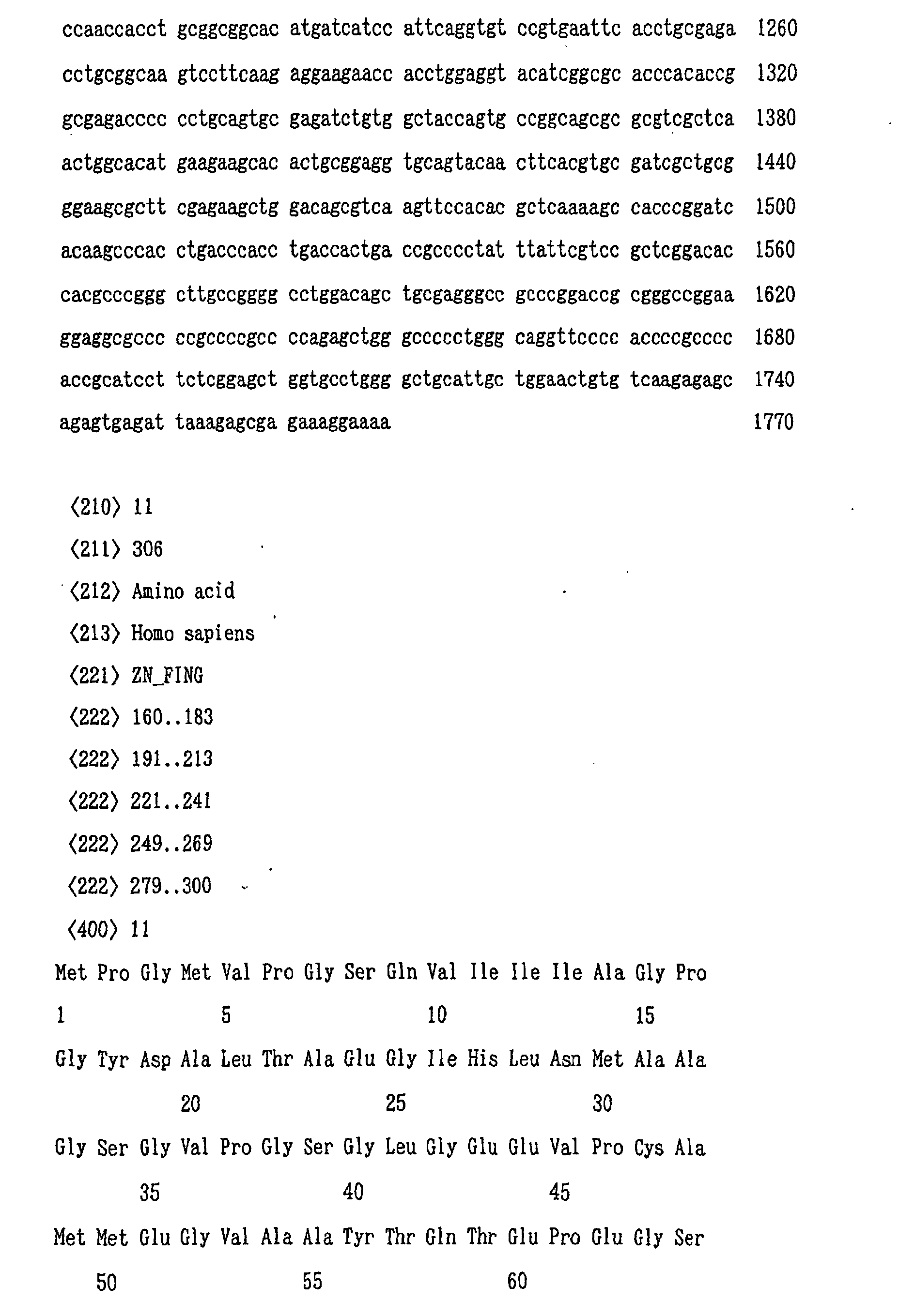


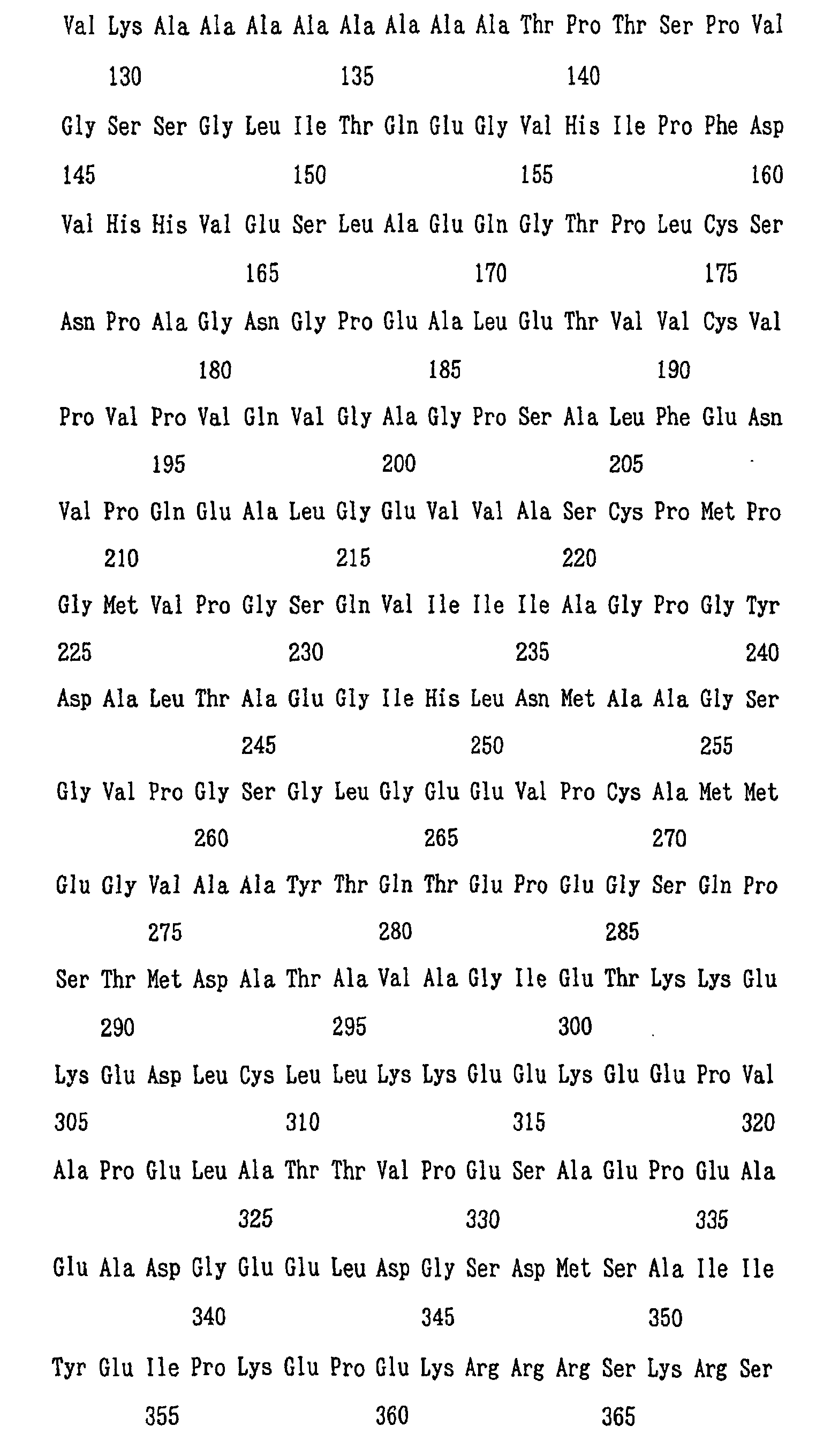
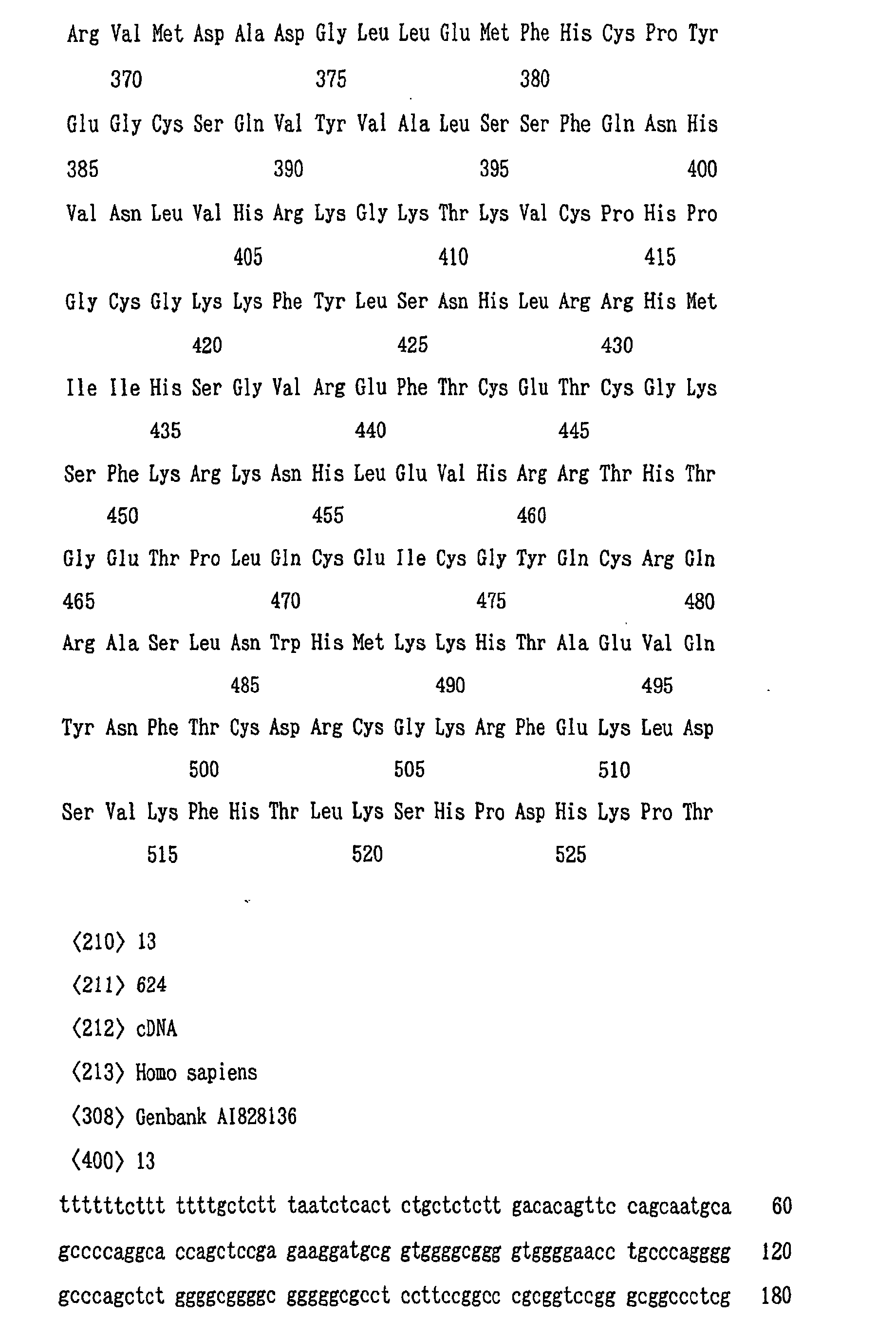

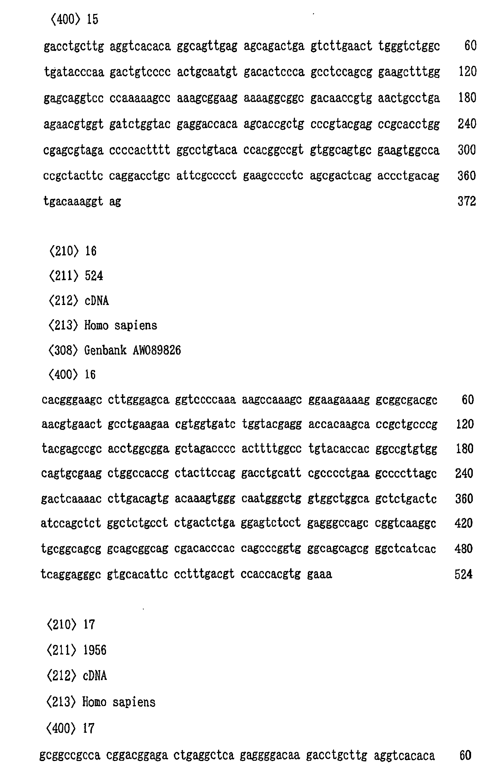
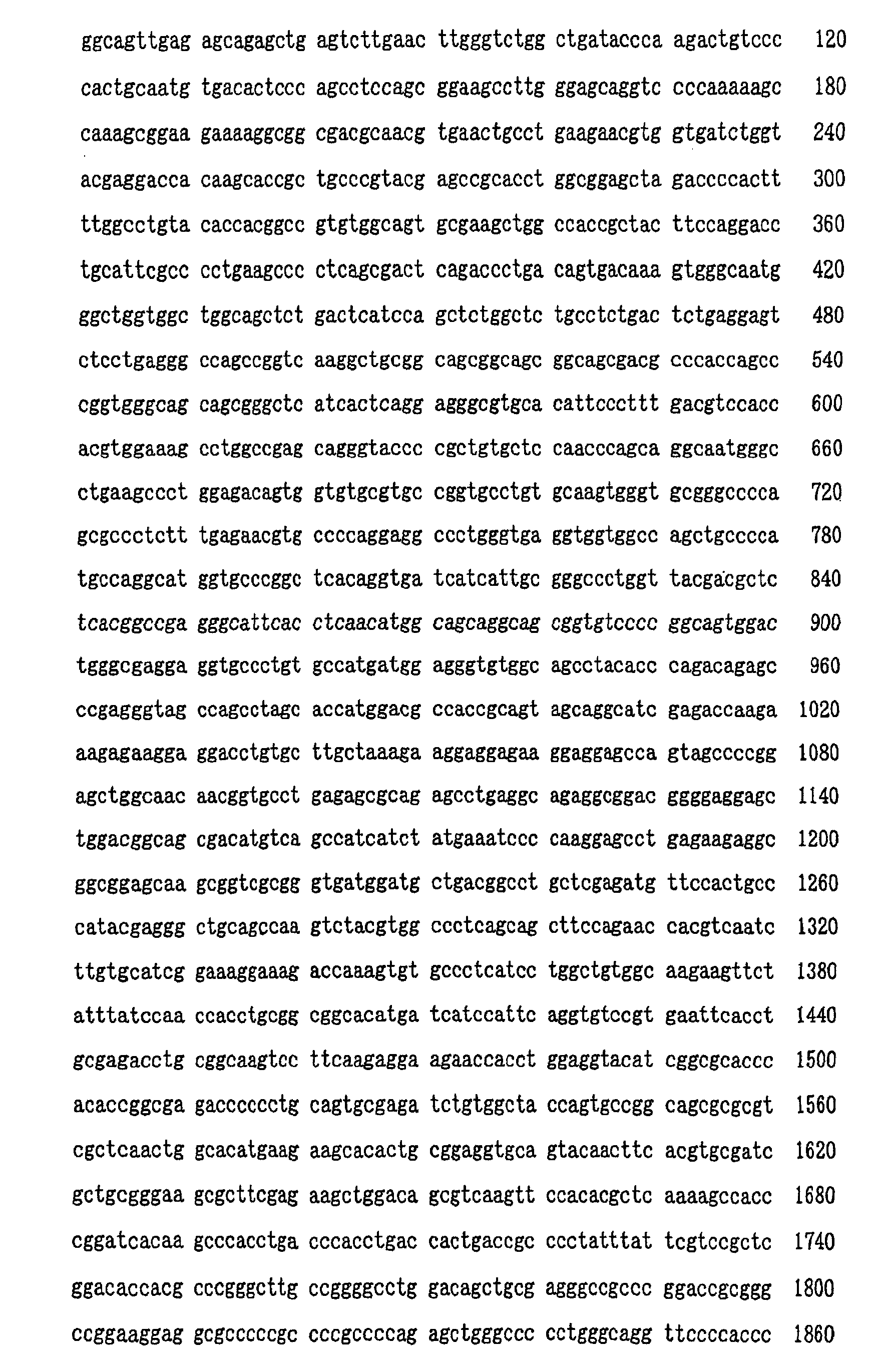
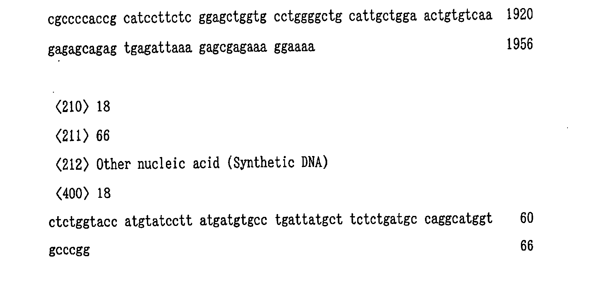
Claims (14)
- A polypeptide having the amino acid sequence shown in SEQ ID NO: 11 or SEQ ID NO: 12.
- A method for diagnosing hepatitis, comprising determining the production level of the polypeptide of claim 1 in the liver.
- The method of claim 2, comprising determining the amount of an mRNA encoding said polypeptide in the liver as the production level of said polypeptide in the liver.
- The method of claim 3, comprising determining the amount of mRNA as the amount of its cDNA.
- The method of claim 3 or 4 wherein said step of determining the amount of mRNA or its cDNA comprises using an oligonucleotide specifically hybridizing to said mRNA or its cDNA.
- The method of claim 4 wherein the amount of the cDNA is determined on the basis of the amplification speed of the PCR amplification product.
- The method of claim 2 comprising using an antibody against said polypeptide.
- A diagnostic kit for diagnosing hepatitis by determining the production level of the polypeptide of claim 1 in the liver, said kit comprising a container receiving said polypeptide.
- A diagnostic kit for diagnosing hepatitis by determining the production level of the polypeptide of claim 1 in the liver, said kit comprising a container receiving an antibody against said polypeptide.
- A diagnostic kit for diagnosing hepatitis by determining the production level of mRNA in the liver as the production level of the polypeptide of claim 1 in the liver, said kit comprising a container receiving an oligonucleotide specifically hybridizing to an mRNA encoding said polypeptide or its cDNA.
- A diagnostic kit for diagnosing hepatitis by determining the production level of mRNA in the liver as the production level of the polypeptide of claim 1 in the liver, said kit comprising a container receiving the cDNA of an mRNA encoding said polypeptide.
- A DNA containing a gene encoding a polypeptide having the amino acid sequence shown in SEQ ID NO: 11 or SEQ ID NO: 12.
- A vector DNA having the DNA of claim 12.
- A method for preparing a polypeptide having the amino acid sequence shown in SEQ ED NO: 11 or SEQ ID NO: 12, comprising culturing a host cell carrying the DNA of claim 12 or the vector DNA of claim 13.
Applications Claiming Priority (3)
| Application Number | Priority Date | Filing Date | Title |
|---|---|---|---|
| JP2000191379 | 2000-06-26 | ||
| JP2000191379 | 2000-06-26 | ||
| PCT/JP2001/005415 WO2002000874A1 (en) | 2000-06-26 | 2001-06-25 | Polypeptides, use thereof and process for producing the same |
Publications (2)
| Publication Number | Publication Date |
|---|---|
| EP1295940A1 true EP1295940A1 (en) | 2003-03-26 |
| EP1295940A4 EP1295940A4 (en) | 2005-01-12 |
Family
ID=18690688
Family Applications (1)
| Application Number | Title | Priority Date | Filing Date |
|---|---|---|---|
| EP01943820A Withdrawn EP1295940A4 (en) | 2000-06-26 | 2001-06-25 | Polypeptides, use thereof and process for producing the same |
Country Status (6)
| Country | Link |
|---|---|
| US (1) | US7189810B2 (en) |
| EP (1) | EP1295940A4 (en) |
| CN (1) | CN1281749C (en) |
| AU (1) | AU2001266326A1 (en) |
| CA (1) | CA2412570A1 (en) |
| WO (1) | WO2002000874A1 (en) |
Families Citing this family (1)
| Publication number | Priority date | Publication date | Assignee | Title |
|---|---|---|---|---|
| EP1718765A2 (en) * | 2004-01-26 | 2006-11-08 | Isis Innovation Limited | Molecular analysis |
Family Cites Families (4)
| Publication number | Priority date | Publication date | Assignee | Title |
|---|---|---|---|---|
| WO1992018532A1 (en) * | 1991-04-17 | 1992-10-29 | Eisai Co., Ltd. | Rna, dna and virus antigen protein of non-a non-b hepatitis virus |
| US5474905A (en) * | 1993-11-24 | 1995-12-12 | Research Corporation Technologies | Antibodies specific for streptococcus pneumoniae hemin/hemoglobin-binding antigens |
| JP2004507202A (en) * | 1999-03-31 | 2004-03-11 | キュラジェン コーポレイション | Nucleic acid containing an open reading frame encoding a polypeptide; "ORFX" |
| EP1407018A2 (en) * | 2000-10-26 | 2004-04-14 | Curagen Corporation | Human proteins, polynucleotides encoding them and methods of using the same |
-
2001
- 2001-06-25 WO PCT/JP2001/005415 patent/WO2002000874A1/en not_active Ceased
- 2001-06-25 CA CA002412570A patent/CA2412570A1/en not_active Abandoned
- 2001-06-25 EP EP01943820A patent/EP1295940A4/en not_active Withdrawn
- 2001-06-25 AU AU2001266326A patent/AU2001266326A1/en not_active Abandoned
- 2001-06-25 CN CNB018118380A patent/CN1281749C/en not_active Expired - Fee Related
-
2002
- 2002-12-26 US US10/327,995 patent/US7189810B2/en not_active Expired - Fee Related
Non-Patent Citations (3)
| Title |
|---|
| OZAKI I ET AL: "Differential expression of laminin receptors in human hepatocellular carcinoma" GUT, vol. 43, no. 6, December 1998 (1998-12), pages 837-842, XP002305542 ISSN: 0017-5749 * |
| See also references of WO0200874A1 * |
| SHIN EUI-CHEOL ET AL: "Expression of fas-related genes in human hepatocellular carcinomas" CANCER LETTERS, vol. 134, no. 2, 25 December 1998 (1998-12-25), pages 155-162, XP002305541 ISSN: 0304-3835 * |
Also Published As
| Publication number | Publication date |
|---|---|
| AU2001266326A1 (en) | 2002-01-08 |
| US7189810B2 (en) | 2007-03-13 |
| US20030187185A1 (en) | 2003-10-02 |
| WO2002000874A1 (en) | 2002-01-03 |
| CA2412570A1 (en) | 2002-12-12 |
| CN1439052A (en) | 2003-08-27 |
| CN1281749C (en) | 2006-10-25 |
| EP1295940A4 (en) | 2005-01-12 |
Similar Documents
| Publication | Publication Date | Title |
|---|---|---|
| US5773579A (en) | Lung cancer marker | |
| Meyers et al. | The t (8; 21) fusion protein interferes with AML-1B-dependent transcriptional activation | |
| CA2257821C (en) | Genes from 20q13 amplicon and their uses | |
| EP0578909B1 (en) | Human prohibitin and DNA coding for the same | |
| EP0951541A1 (en) | Biomarkers and targets for diagnosis, prognosis and management of prostate disease | |
| US7811986B2 (en) | Genes from the 20Q13 amplicon and their uses | |
| EP0889959B1 (en) | Partial intron sequence of von hippel-lindau (vhl) disease gene and its use in diagnosis of disease | |
| WO1995021943A1 (en) | Dna encoding atp-sensitive potassium channel proteins and uses thereof | |
| US7416731B2 (en) | Human hairless antibodies | |
| EP0495910B1 (en) | Production and use of human nm23-h2 protein and antibodies therefor | |
| US7189810B2 (en) | Polypeptides, use thereof and process for producing the same | |
| JPH0956380A (en) | Asialoglycoprotein receptor derivative and use thereof | |
| EP2113566B1 (en) | Genes from 20q13 amplicon and their uses | |
| CA2367468C (en) | Rheumatoid arthritis gene and method for diagnosing rheumatoid arthritis | |
| CA2387576C (en) | Immuno-interactive fragments of the .alpha.c subunit of inhibin | |
| JPWO2002000874A1 (en) | Polypeptides, their uses, and methods for producing them | |
| AU4808601A (en) | Genes from 20q13 amplicon and their uses | |
| JP2001292787A (en) | Human erythroid differentiation-related factors | |
| WO1993005151A1 (en) | Cloned human cenp-c antigen | |
| JP2000152792A (en) | Novel G protein-coupled receptor protein, DNA and use thereof | |
| JP2004049043A (en) | GPCR gene isolation method and novel GPCR gene | |
| JP2003009871A (en) | Test method for therapeutic or prophylactic agent for osteoporosis | |
| JP2001037482A (en) | Human protein and cDNA [1] | |
| JP2000236882A (en) | Mouse nurse cell receptor gene | |
| JPH07250684A (en) | Nucleic acid fragment and plasmid vector for expressing the fragment |
Legal Events
| Date | Code | Title | Description |
|---|---|---|---|
| PUAI | Public reference made under article 153(3) epc to a published international application that has entered the european phase |
Free format text: ORIGINAL CODE: 0009012 |
|
| 17P | Request for examination filed |
Effective date: 20030114 |
|
| AK | Designated contracting states |
Kind code of ref document: A1 Designated state(s): AT BE CH CY DE DK ES FI FR GB GR IE IT LI LU MC NL PT SE TR Designated state(s): AT BE CH CY DE DK ES FI FR GB GR IE IT LI LU MC NL PT SE TR |
|
| AX | Request for extension of the european patent |
Extension state: AL LT LV MK RO SI |
|
| RBV | Designated contracting states (corrected) |
Designated state(s): DE FR GB IT |
|
| A4 | Supplementary search report drawn up and despatched |
Effective date: 20041129 |
|
| 17Q | First examination report despatched |
Effective date: 20050223 |
|
| 17Q | First examination report despatched |
Effective date: 20050223 |
|
| STAA | Information on the status of an ep patent application or granted ep patent |
Free format text: STATUS: THE APPLICATION IS DEEMED TO BE WITHDRAWN |
|
| 18D | Application deemed to be withdrawn |
Effective date: 20080103 |