EP1295896A1 - Method for identifying or isolating a molecule and molecules identified thereby - Google Patents
Method for identifying or isolating a molecule and molecules identified thereby Download PDFInfo
- Publication number
- EP1295896A1 EP1295896A1 EP02021101A EP02021101A EP1295896A1 EP 1295896 A1 EP1295896 A1 EP 1295896A1 EP 02021101 A EP02021101 A EP 02021101A EP 02021101 A EP02021101 A EP 02021101A EP 1295896 A1 EP1295896 A1 EP 1295896A1
- Authority
- EP
- European Patent Office
- Prior art keywords
- cells
- cdna
- nucleic acid
- seq
- sample
- Prior art date
- Legal status (The legal status is an assumption and is not a legal conclusion. Google has not performed a legal analysis and makes no representation as to the accuracy of the status listed.)
- Withdrawn
Links
Images


Classifications
-
- G—PHYSICS
- G01—MEASURING; TESTING
- G01N—INVESTIGATING OR ANALYSING MATERIALS BY DETERMINING THEIR CHEMICAL OR PHYSICAL PROPERTIES
- G01N33/00—Investigating or analysing materials by specific methods not covered by groups G01N1/00 - G01N31/00
- G01N33/48—Biological material, e.g. blood, urine; Haemocytometers
- G01N33/50—Chemical analysis of biological material, e.g. blood, urine; Testing involving biospecific ligand binding methods; Immunological testing
- G01N33/53—Immunoassay; Biospecific binding assay; Materials therefor
- G01N33/574—Immunoassay; Biospecific binding assay; Materials therefor for cancer
- G01N33/57484—Immunoassay; Biospecific binding assay; Materials therefor for cancer involving compounds serving as markers for tumor, cancer, neoplasia, e.g. cellular determinants, receptors, heat shock/stress proteins, A-protein, oligosaccharides, metabolites
-
- C—CHEMISTRY; METALLURGY
- C07—ORGANIC CHEMISTRY
- C07K—PEPTIDES
- C07K14/00—Peptides having more than 20 amino acids; Gastrins; Somatostatins; Melanotropins; Derivatives thereof
- C07K14/435—Peptides having more than 20 amino acids; Gastrins; Somatostatins; Melanotropins; Derivatives thereof from animals; from humans
- C07K14/46—Peptides having more than 20 amino acids; Gastrins; Somatostatins; Melanotropins; Derivatives thereof from animals; from humans from vertebrates
- C07K14/47—Peptides having more than 20 amino acids; Gastrins; Somatostatins; Melanotropins; Derivatives thereof from animals; from humans from vertebrates from mammals
- C07K14/4701—Peptides having more than 20 amino acids; Gastrins; Somatostatins; Melanotropins; Derivatives thereof from animals; from humans from vertebrates from mammals not used
- C07K14/4748—Tumour specific antigens; Tumour rejection antigen precursors [TRAP], e.g. MAGE
-
- C—CHEMISTRY; METALLURGY
- C12—BIOCHEMISTRY; BEER; SPIRITS; WINE; VINEGAR; MICROBIOLOGY; ENZYMOLOGY; MUTATION OR GENETIC ENGINEERING
- C12Q—MEASURING OR TESTING PROCESSES INVOLVING ENZYMES, NUCLEIC ACIDS OR MICROORGANISMS; COMPOSITIONS OR TEST PAPERS THEREFOR; PROCESSES OF PREPARING SUCH COMPOSITIONS; CONDITION-RESPONSIVE CONTROL IN MICROBIOLOGICAL OR ENZYMOLOGICAL PROCESSES
- C12Q1/00—Measuring or testing processes involving enzymes, nucleic acids or microorganisms; Compositions therefor; Processes of preparing such compositions
- C12Q1/68—Measuring or testing processes involving enzymes, nucleic acids or microorganisms; Compositions therefor; Processes of preparing such compositions involving nucleic acids
- C12Q1/6811—Selection methods for production or design of target specific oligonucleotides or binding molecules
-
- C—CHEMISTRY; METALLURGY
- C12—BIOCHEMISTRY; BEER; SPIRITS; WINE; VINEGAR; MICROBIOLOGY; ENZYMOLOGY; MUTATION OR GENETIC ENGINEERING
- C12Q—MEASURING OR TESTING PROCESSES INVOLVING ENZYMES, NUCLEIC ACIDS OR MICROORGANISMS; COMPOSITIONS OR TEST PAPERS THEREFOR; PROCESSES OF PREPARING SUCH COMPOSITIONS; CONDITION-RESPONSIVE CONTROL IN MICROBIOLOGICAL OR ENZYMOLOGICAL PROCESSES
- C12Q1/00—Measuring or testing processes involving enzymes, nucleic acids or microorganisms; Compositions therefor; Processes of preparing such compositions
- C12Q1/68—Measuring or testing processes involving enzymes, nucleic acids or microorganisms; Compositions therefor; Processes of preparing such compositions involving nucleic acids
- C12Q1/6876—Nucleic acid products used in the analysis of nucleic acids, e.g. primers or probes
- C12Q1/6883—Nucleic acid products used in the analysis of nucleic acids, e.g. primers or probes for diseases caused by alterations of genetic material
- C12Q1/6886—Nucleic acid products used in the analysis of nucleic acids, e.g. primers or probes for diseases caused by alterations of genetic material for cancer
-
- G—PHYSICS
- G01—MEASURING; TESTING
- G01N—INVESTIGATING OR ANALYSING MATERIALS BY DETERMINING THEIR CHEMICAL OR PHYSICAL PROPERTIES
- G01N33/00—Investigating or analysing materials by specific methods not covered by groups G01N1/00 - G01N31/00
- G01N33/48—Biological material, e.g. blood, urine; Haemocytometers
- G01N33/50—Chemical analysis of biological material, e.g. blood, urine; Testing involving biospecific ligand binding methods; Immunological testing
- G01N33/53—Immunoassay; Biospecific binding assay; Materials therefor
- G01N33/569—Immunoassay; Biospecific binding assay; Materials therefor for microorganisms, e.g. protozoa, bacteria, viruses
- G01N33/56966—Animal cells
- G01N33/56977—HLA or MHC typing
-
- G—PHYSICS
- G01—MEASURING; TESTING
- G01N—INVESTIGATING OR ANALYSING MATERIALS BY DETERMINING THEIR CHEMICAL OR PHYSICAL PROPERTIES
- G01N33/00—Investigating or analysing materials by specific methods not covered by groups G01N1/00 - G01N31/00
- G01N33/48—Biological material, e.g. blood, urine; Haemocytometers
- G01N33/50—Chemical analysis of biological material, e.g. blood, urine; Testing involving biospecific ligand binding methods; Immunological testing
- G01N33/53—Immunoassay; Biospecific binding assay; Materials therefor
- G01N33/574—Immunoassay; Biospecific binding assay; Materials therefor for cancer
- G01N33/57407—Specifically defined cancers
- G01N33/57438—Specifically defined cancers of liver, pancreas or kidney
-
- A—HUMAN NECESSITIES
- A61—MEDICAL OR VETERINARY SCIENCE; HYGIENE
- A61K—PREPARATIONS FOR MEDICAL, DENTAL OR TOILETRY PURPOSES
- A61K38/00—Medicinal preparations containing peptides
Definitions
- a second stripping step is then carried out.
- the previously stripped sample is now reacted with a sample of the same host cell as was described supra, this time having been transfected or transformed with the carrier vector lacking cDNA from the subject.
- the reason for this second stripping step is an observation made by one of the present inventors and not reported in the literature previously.
- the materials used as vectors, such as phages, viruses, etc. are useful because they naturally infect cells.
- E . coli which inhabit the lower intestine of humans, are infected with lambda phage. It had not been considered, previously, that the immune response to E . coli includes a response to these infectious agents.
- lysed, transfected cells carrying the cDNA and expressing heterologous protein are contacted with the twice stripped sample.
- This sample should only contain immune components specific for the heterologous protein, and should bind thereto. This binding is facilitated if the cell lysates have been immobilized via contact to, e.g., activated filter paper, a solid phase column, etc., but this solid phase binding is not necessary, as the art will surely recognize that many, varied forms of assays are available for identifying a molecule of interest.
- the blotting showed a product with a molecular weight of about 24 kD by SDS-PAGE, which is consistent with a calculated molecular weight of 21.6 kD, based upon predicted amino acid sequence.
- the invention relates to a method for determining or isolating an immunoreactive substance.
- Immunoreactive substance refers to any material which provokes some form of immune response in the subject which produces it. This response may be based upon either a B cell or a T cell response.
- immunoreactive substances include proteins, peptides, glycoproteins, lipoproteins, peptide containing complexes (e.g., MHC/peptide complexes), antibodies, and so forth.
- a cDNA library is prepared from cells of a subject, using well known, standard methods.
- Expression vector comprising the isolated nucleic acid molecule of paragraph 28, operably linked to a promoter.
Abstract
Description
- This application is a continuation-in-part of Serial No. 08/580,980, filed on January 3, 1996, which is a continuation-in-part of application Serial No. 08/479,328, filed on June 7, 1995, both of which are pending, and are incorporated by reference.
- This invention relates to methodologies for identifying molecules of interest. In particularly preferred embodiments, the invention relates to the identification of molecules associated with pathological conditions such as cancer, (melanoma or renal cancer, e.g.), Hodgkin's Disease, autoimmune diseases and so forth. Also a part of the invention are the isolated molecules found as a result of the inventive method, such as presented peptides. These molecules include, inter alia, protein-containing molecules, isolated nucleic acid molecules encoding these, and antibodies which specifically bind to the protein-containing molecules. For convenience, the method described herein will be referred to as "serological fishing".
- It is fairly well established that many pathological conditions, such as infections, cancer, autoimmune disorders, etc., are characterized by the inappropriate expression of certain molecules. These molecules thus serve as "markers" for a particular pathological or abnormal condition. Apart from their use as diagnostic "targets", i.e., materials to be identified to diagnose these abnormal conditions, the molecules serve as reagents which can be used to generate diagnostic and/or therapeutic agents. A by no means limiting example of this is the use of cancer markers to produce antibodies specific to a particular marker. Yet another nonlimiting example is the use of a peptide which complexes with an MHC molecule, to generate cytolytic T cells agains abnormal cells.
- Preparation of such materials, of course, presupposes a source of the reagents used to generate these. Purification from cells is one laborious, far from certain method of doing so. Another preferred method is the isolation of nucleic acid molecules which encode a particular marker, followed by the use of the isolated encoding molecule to express the desired molecule.
- To date, two strategies have been employed for the detection of such antigens in, e.g., human tumors. These will be referred to as the genetic approach and the biochemical approach. The genetic approach is exemplified by, e.g., DePlaen et al., Proc. Natl. Acad. Sci. USA 85: 2275 (1988), incorporated by reference. In this approach, several hundred pools of plasmids of a cDNA library obtained from a tumor are transfected into recipient cells, such as COS cells, or into antigen-negative variants of tumor cell lines which are tested for the expression of the specific antigen. The biochemical approach, exemplified by, e.g., Falk et al., Nature 351: 290 (1991), and Kawakami et al., Nature 369: 69 (1994) both of which are incorporated by reference, is based on acidic elution of peptides which are bound to MHC-I molecules of tumor cells, followed by reversed-phase high performance liquid chromotogaphy (R-HPLC). Antigenic peptides are identified after they bind to empty KHC-I molecules of mutant cell lines which are defective in antigen processing, and induction of specific reactions in cytolytic T-lymphocytes. These reactions include CTL proliferation, TNF release, and lysis of target cells, measurable in an MTT assay, or a 51Cr release assay.
- These two approaches to the molecular definition of antigens have the following disadvantages: first, they are enormously cumbersome, time-consuming and expensive; second, they depend on the establishment of cytolytic T cell lines (CTLs) with predefined specificity; third, their relevance in vivo for the course of the pathology or disease in question has not been proven, as the respective CTLs can be obtained not only from patients with the respective disease, but also from healthy individuals, depending on their T cell repertoire.
- The problems inherent to the two known approaches for the identification and molecular definition of antigens is best demonstrated by the fact that both methods have, so far, succeeded in defining only very few new antigens in human tumors. See, e.g., van der Bruggen et al., Science 254: 1643-1647 (1991); Brichard et al., J. Exp. Med. 178: 489-495 (1993); Coulie, et al., J. Exp. Med. 180: 35-42 (1994), Kawakami et al., Proc. Natl. Acad. Sci. USA 91: 3515-3519 (1994).
- It would be desirable to have available a method which can be used not only for detection of tumor-associated antigens, but to determine molecules associated with any abnormal or pathological condition. Such a method would also facilitate the identification of such molecules, thereby enabling their use on the generation of, e.g., antibodies, cytolytic T cells, and so forth.
- It is therefore the purpose of the present invention to develop methods and reagents for the simple detection and molecular characterization of antigens in human tissues, especially in tumor cells, which are useful in the molecular diagnosis of diseases and/or for immunotherapy and gene therapy of infectious, autoimmune and malignant diseases. The invention is delineated in the disclosure which follows.
-
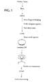
- Figure 1 shows the principles of the approach of the invention.
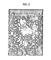
- Figure 2 shows a nitrocellulose membrane with a positive clone derived from the cDNA of a renal cell clear carcinoma that -reacts with a 1:100 dilution of the patient's serum.
- Figure 3 shows, in bar graph form, the Northern Blot analysis of clone HOM-RCC-313 in renal cell carcinoma, normal kidney and other human tissues.
- Figure 4 shows the translated region of the gene coding for HOM-RCC-313.
-
- The following disclosure describes a methodology referred to as serological fishing. In it, a cell sample is taken from a subject afflicted with a pathological condition. The cells preferably are exemplary of the pathology. For example, if the subject has melanoma, the cells are melanoma cells. If the subject is suffering from a neural disorder, e.g., then the cells are preferably a sample of the afflicted cells. This approach is warranted because the afflicted cells are most probably the best source of protein-containing molecules of interest, i.e., such molecules which are specifically associated with the pathological condition of interest.
- Note that cells representative of pathological conditions are not the only cells which may be used in the inventive method. It is very important, e.g., to ascertain those cellular "markers" associated with differentiation and maturation of cells, for example. The example of hematopoietic stem cells comes to mind. Similarly, the invention contemplates the isolation of, e.g., receptor molecules for specific ligands. In effect, one can assay for the presence of any molecule of interest using this methodology.
- The cells chosen are then used to prepare a library of complementary DNA (i.e., "cDNA"). This methodology is well known to the skilled artisan, and need not be reiterated here. It is, of course, based upon the established fact that if proteins are expressed by the cells, then messenger RNA (mRNA) must be present. These mRNA molecules are not long lived, and are unstable, so they are not practical to work with. The stability brought to the molecules when cDNA is used is very helpful to the method.
- Once the cDNA is made, it is used to construct a vector library. In short, carrier vectors are treated, such as by cutting and splicing, to receive molecules of cDNA. The choice of vector may vary, as the skilled artisan is well familiar with many such examples.
- Especially preferred are virus based vectors. In the case of eukaryotic cells, retrovirus or adenovirus based vectors are preferred. Such vectors contain all or a part of a viral genome, such as long term repeats ("LTRs"), promoters (e.g., CMV promoter, SV40 promoter, RSV promoter), enhancers, and so forth. When the host cell is a prokaryote, then bacterial viruses, i.e., phages, are preferred. Exemplary of such vectors are vectors based upon, e.g., lambda phage. In any case, the vector may comprise elements of more than one virus.
- The resulting vectors are transfected or transformed into a host cell, which may be eukaryotic or prokaryotic.
- Any cell normally used for transfection or transformation may be used in the protocol. Preferred materials include strains of E. coli, CHO cells such as CHO-1, COS cells such as COS-7, and so forth. Similarly, yeast cells, e.g., strains of Saccharomyces, strains of Pseudomonas, such as Pseudomonas aeruginosa, Bacillus bacteria, well known insect host cell Spodoptera frugiperda, and so forth, may all be used.
- Once the recipient cells receive the vectors, they are cultivated so as to express foreign, protein containing molecules. "Protein-containing" is used herein because, while prokaryotes express only proteins, eukaryotic cells are well known for their ability to post-translationally modify proteins, so as to produce glycoproteins, lipoproteins, e.g. It must also be borne in mind that "protein containing" as used herein, also encompasses peptides, such as the peptides presented by MHC molecules.
- The processes now described below take place independently of the process described above, and no chronological relationship between the two facets of the invention is intended.
- In pathological conditions such as cancer and, e.g., autoimmune diseases, there is some immune reaction to molecules associated with the pathology. This reaction can include an antibody response, B cell proliferation, proliferation of specific T cell subpopulations, increases in cytokine production, and so forth. The molecules and cells associated with the response may be found in body fluids of a subject, such as his or her serum. The immune responders will react with the molecule of interest whether it is produced recombinantly or autologously. The problem is to find them. As the examples show, this is done in a unique way. First, the body fluid, or other sample of interest, is reacted with a sample of the same host cells used for transfection or transformation. In this first step, the host cells are not transfected or transformed. The effect of this is to strip any immunogenic binding partners specific for the host cell rather than the targeted molecule. This step is necessary because, as was pointed out, supra, the host cell may be one against which the subject has developed an immune response at some point. This first stripping removes these immune components.
- A second stripping step is then carried out. In this step, the previously stripped sample is now reacted with a sample of the same host cell as was described supra, this time having been transfected or transformed with the carrier vector lacking cDNA from the subject. The reason for this second stripping step is an observation made by one of the present inventors and not reported in the literature previously. The materials used as vectors, such as phages, viruses, etc., are useful because they naturally infect cells. Thus, E. coli, which inhabit the lower intestine of humans, are infected with lambda phage. It had not been considered, previously, that the immune response to E. coli includes a response to these infectious agents. Thus, applicants have surprisingly, achieved an ability to remove interfering immune components to an unprecedented degree by carrying out the two stripping steps. As noted, the first is against untransfected or untransformed host cells. The second is against host cells transfected or transformed with a vector which does not carry cDNA, wherein the vector is immunologically equivalent to the vector used to carry cDNA, as described supra.
- It is especially preferred to carry out each of these stripping steps using a plurality of similar, but different procedures. The experiments which follow, for example, show absorption on a solid phase column, and then absorption on nitrocellulose paper. Applicants do not wish to be bound by any theory as to why the use of two similar but different protocols produces the results described herein. It is to be borne in mind, hereafter, that whenever "contacting a sample" is used herein, it is not to be limited to one contact step only, but may refer to more than one, preferably different, contact protocols designed to remove interfering binding partners from a sample under scrutiny.
- It should be understood that these stripping steps may be done completely independently of the steps used to prepare the cDNA library. For example, if the test for an antigen is to be done at day "O", the stripping of sample may be done the day before, a week before, and so forth. One can also "bank" stripped sample from a donor or subject for future use.
- The sample used is preferably serum, but need not be. Any sample which contains immunogenic binding partners may be so used.
- In the next step of the method, lysed, transfected cells carrying the cDNA and expressing heterologous protein are contacted with the twice stripped sample. This sample should only contain immune components specific for the heterologous protein, and should bind thereto. This binding is facilitated if the cell lysates have been immobilized via contact to, e.g., activated filter paper, a solid phase column, etc., but this solid phase binding is not necessary, as the art will surely recognize that many, varied forms of assays are available for identifying a molecule of interest.
- Once the immune component binds to the target molecule, a further step is desirably, but not necessarily, carried out. This additional step involves the use of some binding partner for the first immune component, such as anti-IgG, carrying an identifiable label. The label may be a dye, an enzyme, a gold particle, a radiolabel, or any of the standard labels used in immunoassays.
- Once identification is carried out, the immune components are removed, leaving the target molecule. The target molecule is then studied, using any of the standard methodologies in the art.
- The artisan will note that the methodology also results in isolation of immune components which bind to the molecule of interest. Thus, in another aspect of the invention one can isolate antibodies, e.g., which are specific binding partners for the molecule of interest.
- Yet another immune component which may be identified and isolated following the invention is a cytolytic T cell ("CTL" hereafter), specific for complexes of peptides derived from ( the identified molecule and MHC molecules to which these peptides bind, forming a complex. It is fairly well accepted that a CTL response involves the identification of complexes of MHC molecules and peptides, generally about 8-12 amino acids in length, but most preferably 9 or 10 amino acids in length, by T cell receptors ("TCRs") on the surface of circulating T cells. The TCRs react by binding to these complexes, "setting in motion," as it were, a series of reactions including the proliferation of CTLs specific for these complexes. One can produce and/or isolate such CTLs using the method of the invention, plus further steps.
- As is pointed out in the examples which follow as well as the disclosure in general, one can easily identify cDNA encoding an antigen of interest. Once the cDNA is identified, one uses it to transfect host cells which either already present desired MHC molecules on their surface, or which have been transfected with DNA encoding these MHC molecules. The cDNA for the molecule of interest is expressed, and the molecule is processed to antigenic peptides which are presented by MHC molecules, such as HLA molecules. CTLs directed against the complexes are obtained from lymphocytes, such as autologous lymphocytes. From responder cell populations, long-term CTL clones are then obtained by the well known technique of limiting dilution. Once a positive CTL response is observed, the specific peptides presented to the CTLs are identified using established methods for example, screening the specific of previously identified CTL clones. Alternatively, the more recently described method of studying the sequence of the molecule of interest to identify potential MHC-binding motifs then analyzing these peptides, first for binding to the relevant MHC molecule and then, if positive for MHC-binding, for their ability to generate CTLs recognizing the peptide HHC complex may be used. Of course the peptides can also be eluted from the cells and sequenced, using well known techniques.
- It will also be noted by the skilled artisan that one can correlate the expression of the molecule of interest back to a particular host cell or cells which expressed it. In so doing, one can remove the cDNA which expressed the molecule of interest, sequence it, and so forth. This aspect of the method is another feature of the invention.
- Specific embodiments of the invention will be seen in the examples which follow. Figure 1 depicts the method generally.
- For the establishment of a cDNA library from human tissue total RNA was obtained from 0.5 ug of a renal clear cell carcinoma and established according to the method of Chomzynski, J. Analyt. Biochem. 162: 156-159 (1987), incorporated by reference. The mRNA was extracted from total RNA with oligo-dT-cellulose. The synthesis of the first strand cDNA was accomplished by the method described by Gubler and Hoffmann, Gene 25: 263 (1983) using RNase H and DNA polymerase I. For adaptation of the cDNA Klenow enzyme, adaptors with EcoRI restriction enzyme sites were ligated to the cDNA ends using T4 DNA ligase (Ferretti L and Sgamerella V, Nucl. Acids Res. 9: 3695 (1981)). Following restriction enzymatic digestion with the enzyme XhoI, cDNA molecules of different length were separated using Sephacryl 400 and transfected into λZAPII phage vectors (Short et al., Nucleic Acids Res. 16: 7583 (1988)). The recombinant phage DNA was packed into phages after ligation with packing extracts and used for the transfection of E. coli bacteria. The titration of the library resulted in 1.8 x 10⁶ recombinant primary clones. The total cDNA library was transfected in E. coli and amplified. The titer of the cDNA library after amplification was 1011 plaque forming units per ml (pfu/ml). These transfected cells were used in experiments which follow.
- In accordance with the invention as described, supra, identification of immunogenic material was achieved by using human sera which have been completely depleted of antibodies directed against antigens derived from native and lytic λ phage-transfected E. coli bacteria. To this end, the serum was "stripped" via absorption, as now described.
- E. coli bacteria of the strain XL1-Blue were cultured in 50 ml LB medium overnight. After achieving an optical density of OD600 = 1.0, the bacteria were pelleted by centrifugation, resuspended in 5 ml phosphate buffered saline (PBS), and sonicated by ultrasound to form a lysate. The bacterial lysate was bound onto a matrix of activated Sepharose, which was then put into a column and used for the absorption of the human serum. The serum was run over this
column 10 times. - A culture of E. coli XL1-Blue bacteria in the exponential growth phase was pelleted by centrifugation, transfected in 0.01 M magnesium sulfate with 106 λZAPII phages without a recombinant insert and incubated in 5 ml LB medium for four hours. The lysate of the transfected bacteria was used in the same manner as the untransfected bacteria, with the human serum described supra being passed through the column an additional ten times.
- To complete the depletion of the serum, interfering antibodies from lytically transfected E. coli bacteria were cultured on agar plates (10 hours, 37°C) and their proteins were blotted onto nitrocellulose membranes after this culturing step. Following this, the serum which had been preabsorbed according to the above steps was transferred to the blotted nitrocellulose membrane, and the absorption procedure was repeated five times. The serum, which was processed in accordance with the invention, was totally depleted of antibodies directed against antigens derived from E. coli and phages.
- In these experiments, a renal cancer-specific antigen was identified via the following steps. Bacteria of the strain E. coli XL1-Blue were transfected with recombinant phages derived from the described cDNA library and plated at a density of 4-5x10³ plaque forming units (PFUs) per plate in LB-medium with isopropylthiogalactopyranoside ("IPTG"). After 12 hours of incubation at 37°C, nitrocellulose membranes were put on top of the cultures, and the culture plates were incubated for another four hours. This was followed by incubation of the nitrocellulose-membrane for one hour in Tris-buffered saline (TBS) with 5% milk powder. After washing the nitrocellulose membranes three times in TBS, the stripped human serum secured following Example 2 was diluted 1:1000 in TBS/0.5% milk powder (w/v) and incubated overnight with gentle shaking. After the incubation with the nitrocellulose membrane the serum was removed and kept for additional testing. Following incubation with serum, the nitrocellulose membranes were washed three times in TBS, and incubated with polyclonal alkaline phosphatase-conjugated goat anti-human IgG for one hour. Following this, the nitrocellulose membranes were washed repeatedly with TBS/0.01% Tween 20 (v/v)). The reaction was developed using nitroblue tetrazolium chloride and bromochloro-indoyl-phosphate in TBS. The binding of human antibodies to the expressed protein became visible by a blue, ring-formed color deposit on the nitrocellulose membrane. The efficient preabsorption of the serum made it possible to develop the membrane at 37°C over several hours without compromising the quality of the test because of background reactivity caused by antibodies against E. coli and phage antigens.
- Positive clones were localized on the agar plates, transferred into transfection buffer, and used for a second round of transfection and subcloning. A total of 1.8x10⁶ recombinant clones were subjected to screening and five different positive-reacting clones were identified.
- Positive clones secured following Example 3, i.e., those which had bound antibodies derived from the processed human serum, were subcloned to monoclonality by repeated rounds of transfection and testing of reactivity with the processed human serum. P-bluescript phagemids with the respective cDNA inserts were cloned by in vivo excision (Hay and Short, Strategies 5: 16-19, 1992) from the λZAPII phage vectors and used for transfection of E. coli SOLR bacteria. Plasmids were isolated from the bacteria after alkaline lysis with NaOH in a modification of the method of Birnboim and Doly, J. Nucl. Acids Res. 7: 1513 (1979). The recombinant plasmid DNA was sequenced according to the classic method of Sanger (Proc. Natl. Acad. Sci. USA 74: 5463 (1977)) using M13-forward and K13-reverse oligonucleotides. The DNA sequence obtained and the resulting amino acid sequence were checked for in nucleic acid and protein data banks (Gene Bank, EMBL, Swiss Prot). The sequencing of the cDNA inserts was continued using internal oligonucleotides. Analysis showed no homology with any sequences deposited in the data banks. The full length cDNA clone referred to as SK313, which had been cloned with the RACE method (Frohman MA, Dush MK, Martin GR, Proc. Natl. Acad Sci. USA 85: 8998 (1988)), had a carbonic anhydrase domain at the 5' end. The nucleic acid sequence of this molecule is presented in SEQ ID NO: 1. Figure 2 shows a nitrocellulose membrane with a positive clone from these experiments.
- As a follow up to these experiments, RNA was isolated from a spectrum of malignant and normal human tissues according to the method of Chomzynski and Sacchi, Analyt Biochem. 162: 156 (1987). After denaturation, the total isolated RNA was separated on an agarose gel containing 1% formaldehyde by electrophoresis (Goldberg, Proc. Natl. Acad. Sci. USA 77: 5794 (1980)) and then blotted onto a nylon membrane according to a known method (Seed, Nucl. Acids Res. 10: 1799 (1982)) Radiolabeled cDNA inserts of the identified clones were used for hybridization. The hybridization was carried out according to a known method (Geoffrey and Berger, Enzymol. 152: 419 (1987)). The presence of the respective RNA was demonstrated using autoradiography and X-ray films. The analysis demonstrated that the mRNA of clone HOM-RCC-313 was overexpressed in 4 out of 19 renal cell carcinomas compared to normal kidneys. Very weak expression was found only in colonic mucosal tissue and in normal kidney. Expression in other tissues could not be demonstrated.
- To determine the incidence of antibodies against antigens which are identified in accordance with the invention, sera from healthy individuals and tumor patients were analyzed. To this end, the sera were processed as described, supra, and depleted of antibodies against antigens derived from E. coli and phages. For the detection of antigen-specific antibodies, phages derived from reactive clones were mixed with non-reactive phages derived from the same cDNA library at a ratio of 1:10 and tested as described supra., for reactivity with antibodies in the human test serum. The serum which had been used for the identification of the antigen was used as a positive clone. The non-reactive phages served as a negative control. A serum sample was positive for antigen reactive antibodies, if the expected percentage of the phage plaques showed a positive reaction. In the case of the renal cell carcinoma antigen represented by clone HOM-RCC-313, the analysis of a spectrum of human sera showed that only sera from renal cell carcinoma patients contained reactive antibodies. Sera from healthy controls and patients with other tumors did not contain such antibodies.
- The cDNA for clone HOM-RCC-313 was excised from the plasmid DNA by digestion with the restriction enzyme EcoRI, and separated by agarose gel electrophoresis, followed by extraction from the gel. This was then used to create a vector which expresses a fusion protein with the bacterial protein anthranilate synthetase. A relevant fragment in the exact open reading frame was cloned into pATH plasmid vectors (Koerner, et al, Meth. Enzymol. 194: 477 (1991). Induction of protein expression was obtained after transformation of the plasmids into E. coli of strain BL21 as described (Spindler, et al, J. Virol. 49: 132 (1984)). Expressed, fusion proteins were separated by SDS gel electrophoresis, excised from the gel, eluted and freeze dried. Rabbits were immunized by subcutaneous injection with 100 µg of the lyophilisate dissolved in Freund's adjuvant. Immunization was repeated three times at two-week intervals using incomplete Freund's adjuvant. The rabbit was bled and antiserum was obtained. The obtained antiserum was depleted from antibodies reactive with E. coli and phages in the manner described supra and tested for reactivity against the renal carcinoma antigen as described for the human serum. Reactivity was detected at dilutions of 1: > 100,000.
- The protocols set forth in the preceding examples were followed, using biopsied tissue taken from different subjects suffering from (i) malignant melanoma, (ii) astrocytoma, and (iii) Hodgkin's Disease. Table 1, which follows, summarizes the results, including those obtained with the renal cancer study, set out in detail in Examples 1-6, supra. Table 1. Antibody reactivity of autologous sera with recombinant clones derived from human tumor cDNA. cDNA libraries were screened with autologous patient serum. Positive clones were subcloned to monoclonality. Inserts from each clone were amplified with plasmid primers and separated by agarose gel electrophoresis. Southern blots were performed by cross hybridization with the respective inserts.
tumor clones tested positive clones different inserts malignant melanoma 1.0 x 10⁶ 40 10 renal cell carcinoma 1.8 x 10⁶ 7 5 astrocytoma 1.2 x 10⁶ 49 5 Hodgkin's disease 1.0 x 10⁶ 14 4 - Analysis of the different inserts showed that the melanoma cells expressed the known tumor rejection antigen precursor MAGE-1 (see van der Bruggen et al., Science 254: 1643-7 (1991), incorporated by reference), as well as a new antigen. A portion of the cDNA sequence of this antigen is set forth in SEQ ID NO: 2.
- When the astrocytoma study was completed, the observed insert appeared to correspond to the previously described Tegt gene (Old, Canc. Res. 41: 361-375 (1981), incorporated by reference).
- When the Hodgkin's Disease study was completed, a previously unknown antigen was isolated, and cDNA encoding it was identified in the library, using standard methods. The antigen is a newly observed, lectin-like structure, a portion of the cDNA for which is set forth in SEQ ID NO: 3. Also observed were antibodies against restin, described by Bilbe, et al, EMBO J 11: 2103-13 (1992). This is an intermediate filament associated protein, expression of which has been shown to be restricted to Hodgkin and Reed-Sternberg cells, as well as cultured monocytes.
- A further study of occurrence of antibodies against the antigens described in Examples 1-7 was carried out. Table 2 summarizes these assays. In these studies, phages from positive clones were mixed with non-reactive phage (ratio:1:10), and then used to transfect bacteria (e. coli). Dilutions of patient sera (1:200), were used, in an enzyme linked immunosorbent assay (ELISA), as described supra. "HOM-MEL-40" refers to the new melanoma antigen (SEQ ID NO: 2), while "HOM-HEL-55" refers to MAGE-1 (van der Bruggen et al., supra). "HOM-RCC 3.1.3" is the renal cancer antigen of SEQ ID NO: 1. "HOM-GLO-30.2.1" refers to the previously identified astrocytoma associated antigen, "HOM-HD-21" refers to the new, lectin-like antigen of SEQ ID NO: 3, and "HOH-HD-397" is the previously identified restin antigen.The fact that antibodies against the tumor antigens, excepting only restin, were detected, albeit at varying rates, only in the sera of patients diseased with the same type of tumor suggests that tumor growth is essential for the development of a humoral response against tumor antigens.

- The reason for the presence of restin in healthy controls is not clear. One may speculate that tolerance against respective antigens might be circumvented, because the antigen may have similar sequences to another antigen, the donor may have premalignant cells, or the antigen may be activated in normal cells under non-malignant conditions, such as viral infections, or other inflammatory processes.
- In order to determine the expression pattern of the newly identified antigens described herein, Northern blot analysis was carried out, using a variety of human tissues.
- RNA was extracted from tissue samples (tumor and normal) using the well known guanidium isothiocyanate/phenol/chloroform method of Chomzynski, et al., supra. The RNA integrity was checked via electrophoresis in formalin/MOPS gels. Then, gels containing 40 ug of RNA per lane were blotted onto nylon membranes. These Northern blots were then probed with the cDNA of SEQ ID NO: 1, 2 or 3. Hybridization was with 32P labelled probes at 42°C, with formamide. The filters were washed at 65°C, at 1xSSC, 0.2% SDS, and exposed for 16 hours. These are "stringent conditions" as defined hereafter. After exposure, filters were stripped and rehybridized with GAPDH.
- Table 3 summarizes these results.

- As will be seen, the new melanoma associated antigen is strongly expressed in melanoma, but not other tissues. Carbonic-anhydrase-like antigen was strongly expressed in about 20% of renal cell carcinomas, and only weakly in normal renal tissue. Tegt was overexpressed on 8/12 astrocytoma tissues compared to normal brain tissue. The mRNA for the lectin like molecule associated with Hodgkin's disease was increased about ten fold in diseased tonsils as compared to normal tonsils, suggesting that overexpression may be a frequent characteristic of proteins which elicit autologous B cell responses.
- Further studies were carried out on the HOM-MEL-40 sequence. Using standard genetic analysis techniques, the 5' region of the mRNA for HOM-MEL 40 was shown to have a tyrosine kinase binding domain. This suggests that HOM-MEL-40 may function as a receptor. The 3' portion of the RNA is identical with an RNA molecule for "SSX," a molecule known to be involved in the SYT-SSX translocation in synovial tumors.
- Additional experiments were also carried out to study HOM-MEL 40. Standard Northern blotting showed that, with the exception of testis, HOM-MEL 40 was not expressed in normal tissues. In contrast, it was expressed in 50% of melanomas, 20% of prostate cancers, 20% of gastric cancers, 26% of colorectal cancers, 12% of the lung cancers and 20% of breast and hepatacellular cancer. It was also found in 1/10 gastric, and 1/5 thyroid carcinomas.
- Additional Western blotting work was carried out, showing that antibodies against HOM-MEL 40 were present in 10 of 89 melanoma patients tested, but only 3 out of 49 healthy male subjects.
- In yet further studies, it was observed that HLA-A2 positive tumor cells presented a nonamer derived from HOM-MEL. This suggests that HOM-MEL 40 specific vaccines, useful in inducing CTLs, are possible.
- The phage assay described in the prior examples is not appropriate for screening large numbers of serum samples. In order to do so, a modification of the standard Western Blot was developed. This variation is based upon His-tagged, recombinant HOM-MEL 40, as is herein described.
- HOM-MEL-40 was amplified, over 20 cycles, using pfu polymerase, on plasmid cDNA prepared from melanoma. tissue. The oligonucleotide primers used were:

- Polymerase chain reaction (PCR) was carried out at 95°C/1 minute; 60°C/1 minute; and 72°C/1 minute, followed by a final extension at 72°C for 10 minutes. The amplification product was gel purified, using art recognized techniques, and then ligated in frame to SmaI digested, dephosphorylated and gel purified pQE32 vector. This results in production of a fusion protein having a "tail" of 6 histidine molecules at the N-terminus.
- The construct was then transformed into E. coli SG13009 (pREP4) strain, followed by selection on plates containing kanamycin and ampicillin. Individual colonies were picked, and expressed on a small scale by inducing these with 2 mM isopropyl thiogalactoside (IPTG). This permits checking for protein expression. Small -scale purification over Ni-KTA columns was then performed, for each clone.
- One clone was identified as expressing a protein of expected length. This clone was isolated and sequenced using well known techniques. It was verified as HOM-MEL-40. Following the identification, large scale induction of recombinant protein was carried out. Specifically, cells were induced with 2 mM IPTG, and harvested five hours later. Cells were lysed, by combining with a buffer of 8 M urea, 100 mM Na2PO4; 10 mM Tris·HCl (pH8); 0.01% Triton X, overnight. Any cellular debris was spun down, and supernatant was loaded onto pre-equilibrated Ni-NTA resin. Washes were performed with two volumes of the aforementioned buffer, at pH8, and then at least 10 volumes of the buffer, at pH6.3. Protein was then eluted, using the buffer described herein, plus 250 mM imidazole. Yields ranged from 15 to 40 mg of His-tagged protein per liter of bacterial culture.
- Western Blotting was then carried out, using the Hislabeled protein. In these assays, 5 ug of recombinant His-tagged proteins, which served as internal negative controls, were mixed with 2xSDS sample buffer (0.1 M Tris-HCl, pH 6.8, 0.2M dithiothreitol, 4% SDS, 0.2% bromophenol blue, 20% glycerol), and were then electrophoresed in 12% SDS-PAGE, followed by blotting to nylon membranes using semi-dry transfer.
- After blocking of any unspecified binding with 5% low fat milk in PBS (1 hour), membranes were incubated with 1:100 diluted sera from tumor patients or healthy controls. Blots were then incubated for one hour with alkaline phosphatase conjugated mouse anti-human IgG. The membrane was then incubated, consecutively, with rabbit anti-mouse IgG (30 minutes), anti-alkaline phosphatase, and then 0.25 mg/ml of alkaline phosphatase. After each incubation step, the membranes were washed extensively with TBS and 0.1% Tween. Visualization was performed by staining with 5-bromo-4-chloro-3-indolyl-phosphate (BCIP), and nitroblue tetrazolium. The sera were analyzed in random order (healthy/melanoma positive), with the observer blind to the status of the sample. All analyses were carried out in duplicate.
- The blotting showed a product with a molecular weight of about 24 kD by SDS-PAGE, which is consistent with a calculated molecular weight of 21.6 kD, based upon predicted amino acid sequence.
- The immunoblotting described supra, was carried out on 89 melanoma samples, six ovarian cancer samples, and ten renal carcinoma samples. Of these, 11 melanoma samples, one ovarian cancer sample, and three renal cell carcinoma samples were positive. Sera from subjects with colorectal, lung, breast, gastric, or pancreatic cancer were negative. A total of 41 healthy controls were also analyzed, of which three were positive. Any sera which were reactive in the Western blot, as well as twenty negative serum samples were then reassessed, using the phage assay, described supra. The reactivity was confirmed for 10 of the 11 melanoma patients and the positive ovarian cancer patient. The renal cell cancer patient and the healthy controls were negative, and were all samples negative in the Western blot.
- There were 16 melanoma patients who provided both serum and tumor specimens. It was found that HOM-MEL-40 expression by the tumor could be compared with antibody reactivity in the sample. As will be seen in the table which follows, eight of the sixteen patients had HOM-MEL-40 positive tumors, but only three had antibodies against the antigen in their serum. No antibodies were detected in the serum of patients with HOM-MEL-40 negative tumors.
- Rammensee, et al., Immunogenetics 41: 178-226 (1995), incorporated by reference, disclose many peptides which bind to HLA-2.1, and some which also provoke
CTL proliferation. Some of these are of formula:
 These peptides were synthesized, using well known techniques and Fmoc protected amino acids. The peptides were then purified, using Sephadex G25, followed by reverse phase HPLC on a C-18 column. T2 cells, which are deficient in transporters associated with antigen presentation (DeMars, et al., Proc. Natl. Acad. Sci. USA 82: 8183-8187 (1985); Slater, et al., Immunogenetics 21: 235-241 (1990)), were used in peptide binding assays, as follows. A sample of 5x10⁵ T2 cells were incubated for 4 hours at 37°C, in the presence or absence of 100 uM of the HOM-MEL-40 peptides. Positive controls were an EBV LMP2 derived peptide:
 and an HIV reverse transcriptase derived peptide:
 Upregulation of HLA-A2.1 on T2 cells was measured by labelling with an anti-HLA-A2.1 specific monoclonal antibody (i.e., BB7.2), followed by incubation with an FITC conjugated goat anti-mouse antibody. The samples were analyzed by flow cytometry, with upregulation of HLA-A2.1 being given by the ratio:
- As the foregoing shows, the invention relates to a method for determining or isolating an immunoreactive substance. "Immunoreactive substance" as used herein refers to any material which provokes some form of immune response in the subject which produces it. This response may be based upon either a B cell or a T cell response. Such immunoreactive substances include proteins, peptides, glycoproteins, lipoproteins, peptide containing complexes (e.g., MHC/peptide complexes), antibodies, and so forth. To determine such substances, a cDNA library is prepared from cells of a subject, using well known, standard methods. The cDNA is then inserted into an appropriate vector, such as a eukaryotic cell specific virus or a phage (i.e., a bacterial virus), to form a transfecting/transforming library, which is then incorporated into a host cell. The host cells are treated so that they express the library component (cloned cDNA) they receive. The host cells are then lysed, so that the expressed material is available for further treatment.
- The lysed material is then contacted with a "stripped" sample believed to contain an immunogenic binding partner for the immunoreactive substance. "Immunogenic binding partner" as used herein refers to any immune system associated material which binds to the target, i.e., the immunoreactive substance. Such binding partners include, but are not limited to, antibodies, T cells, cytokines, ligands, receptors, and so forth, as well as truncated portions of these molecules, complementary nucleic acid molecules, and so forth. Note that for some of these components, such as T cells, further steps including those recited herein are required.
- The stripped sample, as indicated supra, has been treated by contact with both (i) non-transfected or transformed host cells, and (ii) host cells transfected or transformed with vectors which do not contain the pertinent cDNA.
- The stripped sample is useful for identifying binding partners for the expressed material because many of the immune components which would otherwise interfere with the specific immunological reaction desired have been removed via the absorption steps described herein.
- The identification of the expressed material may be followed by isolation of the cDNA encoding it. One can punch holes through a membrane such as a nitrocellulose membrane, placed on top of Petri dishes containing colonies of host cells, then use the immune reaction to give position on the solid phase. Each colony is based upon limited cDNA transfection, thereby facilitating isolation and identification of relevant cDNA.
- The invention also relates to the isolated nucleic acid molecules of SEQ ID NO: 1, 2 or 3, which encode for molecules which are associated with particular conditions. In addition to their role as coding materials, these molecules can also be used as probes to identify cells expressing the relevant antigens, as it has been shown that these cDNA molecules (SEQ ID NO: 1, 2 and 3) are based upon mRNA which translated to the antigen.
- Also a part of the invention are isolated nucleic acid molecules, the complementary sequences of which hybridize to one of SEQ ID NO: 1, 2 or 3, and which encode a protein equivalent to those encoded by SEQ ID NO: 1, 2 or 3. "Stringent conditions" as used herein, refers to conditions at least as stringent as hybridization at 50 µl/cm2 of 3.5xSSc, lxDenhardt's solution, 25 mM sodium phosphate buffer (pH 7.0), using a 32P-labelled probe, for 18 hours at 65°C, followed by four washes (one hour, each wash, at 65°C, 2xSSC, 0.1% SDS), and a final wash for 30 minutes at 1.0xSSC 0.2% SDS. The final wash can be changed to 0.5xSSC to 0.2xSSC, or even 0.1xSSC, and SDS can be lowered to 0.1% to increase stringency, if desired.
- The invention also includes those peptides associated with tumor antigens, such as those of SEQ ID NOS: 7, 8 and 9, which bind to HLA-A2.1 molecules, thereby provoking lysis by cytolytic T cells. Also a part of the invention are peptides of formulawherein the sixth amino acid residue is Ser, Lys or Phe, and the ninth amino acid residue is Val or Ile. These molecules can also serve, very simply, as markers for HLA-A2.1 cells, as it is well known that peptide/MCH complex formation is quite specific.

- Other features of the invention will be clear to the skilled artisan-and need not be reiterated here.
- The terms and expressions which have been employed are used as terms of description and not of limitation, and there is no intention in the use of such terms and expressions of excluding any equivalents of the features shown and described or portions thereof, it being recognized that various modifications are possible within the scope of the invention.


- The Isolated nonapeptide which binds to an HLA-A2.1 molecule, and has fomrulawherein the sixth amino acid residue is Ser, Lys or Phe, and the ninth amino acid residue is Val or Ile.

- The isolated nonapeptide of which 1, selected from the group consisting of:
- SEQ ID NO: 7,
- SEQ ID NO: 8, and
- SEQ ID NO: 9.
-
- The isolated nonapeptide of
paragarph 1 or 2 which is SEQ ID NO: 7. - The isolated nonapeptide of
paragraph 1 or 2 which is SEQ ID NO: 8. - The isolated nonapeptide of
paragraph 1 or 2 which is SEQ ID NO: 9. - The Isolated protein encoded by the nucleic acid molecule of SEQ ID NO: 1.
- A Method for screening for possible presence of a tumor associated protein, comprising contacting a sample with at least one of SEQ ID NO: 4 and SEQ ID NO: 5, and determining hybridization of SEQ ID NO: 4 or SEQ ID NO: 5 to a target, as a determination of said tumor associated antigen in said sample.
- The method of paragraph 7 comprising contacting said sample with both SEQ ID NO: 4 and SEQ ID NO: 5.
- The method of
paragraph 8 comprising polymerase chain reaction. - A Method for determining an immunoreactive substance produced by a subject, comprising:
- (a) producing a cDNA library of a cell taken from said subject,
- (b) inserting said cDNA library into a vector,
- (c) transfecting said vector into a host cell to produce a transfected host cell,
- (d) culturing said transfected host cell to express said immunoreactive substance,
- (e) lysing said transfected host cell to form a lysate,
- (f) contacting a sample containing an immunogenic binding partner for said immunoreactive substance with a sample of lysed, non-transfected host cells, to remove any immunogenic binding partner from said sample which is specific for said non-transfected host cell, to produce a stripped sample,
- (g) contacting said stripped sample to a sample of lysed host cells transfected with the same vector into which said cDNA has been inserted wherein said vector does not contain any cDNA, to remove any immunoreactive binding partners specific for said vector, thereby producing a twice stripped sample,
- (h) contacting said twice stripped sample to the lysate of (f), whereby any immunoreactive binding partners specific for said immunoreactive binding partner bind thereto, and
- (i) determining binding in (h) to determine said immunoreactive substance.
-
- The method of paragraph10 further comprising identifying the transfected host cell which expressed said immunoreactive substance, and isolating the cDNA contained therein.
- The method of paragraph 11 further comprising removing any immunoreactive binding partner to isolate a specific immunoreactive binding partner for said substance.
- The method of
paragraph 11 or 12 wherein said host cell is E. coli. - The method of
paragraph - The method of paragraph 14 wherein said viral vector is a eukaryotic virus based vector.
- The method of paragraph 15 wherein said viral vector is a phage vector.
- The method of paragraph 16 wherein said phage is lambda phase.
- The method of
paragraph 10 wherein said host cell is a prokaryote. - The method of
paragraph 10 wherein said host cell is a eukaryote. - The method of
paragraph 10 further comprising immobilizing said lysate to a solid phase prior to contact with said twice stripped sample. - The method of paragraph10 wherein said sample is serum.
- The method of paragraph21 wherein said serum is autologous serum.
- The method of
paragraph 10 wherein said immunogenic binding partner is an antibody - The method of
paragraph 10, wherein said immunoreactive substance is an antigen associated with cancer. - The method of
paragraph 10 wherein said immunoreactive substance is an autoimmune associated antigen. - The method of
paragraph 10 wherein said immunoreactive substance is produced by a pathogen. - Isolated nucleic acid molecule which codes for a renal cell carcinoma-specific antigen, the complementary sequence of which hybridizes to SEQ ID NO: 1, under stringent conditions.
- Isolated nucleic acid molecule which codes for a Hodgkin's Disease-specific antigen, the complementary sequence of which hybridizes to SEQ ID NO: 2 under stringent conditions.
- Isolated nucleic acid molecule which codes for a melanoma specific antigen, the complementary sequence of which hybridizes to SEQ ID NO: 3 under stringent conditions.
- Isolated nucleic acid molecule of paragraph 27, consisting of SEQ ID NO: 1.
- Isolated nucleic acid molecule of paragraph 28 consisting of SEQ ID NO: 2.
- Isolated nucleic acid molecule of paragraph29 consisting of SEQ ID NO: 3.
- Expression vector comprising the isolated nucleic acid molecule of paragraph 27, operably linked to a promoter.
- Expression vector comprising the isolated nucleic acid molecule of paragraph 28, operably linked to a promoter.
- Expression vector comprising the isolated nucleic acid molecule of paragraph 29, operably linked to a promoter.
- Cell line transformed or transfected with the isolated nucleic acid molecule of paragraph 27.
- Cell line transformed or transfected with the isolated nucleic acid molecule of paragraph 28.
- Cell line transformed or transfected with the isolated nucleic acid molecule of paragraph 29.
- Cell line transformed or transfected with the expression vector of paragraph 33.
- Cell line transformed or transfected with the expression vector of paragraph 34.
- Cell line transformed or transfected with the expression vector of paragraph 35.
Claims (4)
- An isolated nucleic acid molecule encoding a renal specific antigen the complementary sequence of which hybridizes to SEQ ID NO: 1, under stringent conditions.
- An expression vector comprising the isolated nucleic acid molecule according to claim 1, operably linked to a promoter.
- A cell line transformed or transfected with the isolated nucleic acid molecule according to claim 1 or 2.
- A method to detect the expression of SEQ ID NO: comprising:i) extracting RNA from a cell sample;ii) incubating said nucleic acid under conditions suitable for hybridisation of a nucleic acid probe to a target comprising SEQ ID NO:1; andiii) detecting the presence of the hybridised probe to the target nucleic acid.
Applications Claiming Priority (8)
| Application Number | Priority Date | Filing Date | Title |
|---|---|---|---|
| US479328 | 1983-11-15 | ||
| US08/479,328 US5698396A (en) | 1995-06-07 | 1995-06-07 | Method for identifying auto-immunoreactive substances from a subject |
| US08/580,980 US6025191A (en) | 1995-06-07 | 1996-01-03 | Isolated nucleic acid molecules which encode a melanoma specific antigen and uses thereof |
| US580980 | 1996-01-03 | ||
| US08/644,116 US6140464A (en) | 1995-06-07 | 1996-05-10 | Nonapeptides that bind a HLA-A2.1 molecule |
| US644116 | 1996-05-10 | ||
| EP00117699A EP1108432B1 (en) | 1995-06-07 | 1996-06-07 | Method for identifying or isolating a molecule |
| EP96919266A EP0831879B1 (en) | 1995-06-07 | 1996-06-07 | Method for identifying or isolating a molecule and molecules identified thereby |
Related Parent Applications (1)
| Application Number | Title | Priority Date | Filing Date |
|---|---|---|---|
| EP00117699A Division EP1108432B1 (en) | 1995-06-07 | 1996-06-07 | Method for identifying or isolating a molecule |
Publications (1)
| Publication Number | Publication Date |
|---|---|
| EP1295896A1 true EP1295896A1 (en) | 2003-03-26 |
Family
ID=27413479
Family Applications (2)
| Application Number | Title | Priority Date | Filing Date |
|---|---|---|---|
| EP02021101A Withdrawn EP1295896A1 (en) | 1995-06-07 | 1996-06-07 | Method for identifying or isolating a molecule and molecules identified thereby |
| EP00117699A Expired - Lifetime EP1108432B1 (en) | 1995-06-07 | 1996-06-07 | Method for identifying or isolating a molecule |
Family Applications After (1)
| Application Number | Title | Priority Date | Filing Date |
|---|---|---|---|
| EP00117699A Expired - Lifetime EP1108432B1 (en) | 1995-06-07 | 1996-06-07 | Method for identifying or isolating a molecule |
Country Status (2)
| Country | Link |
|---|---|
| US (1) | US6140464A (en) |
| EP (2) | EP1295896A1 (en) |
Families Citing this family (6)
| Publication number | Priority date | Publication date | Assignee | Title |
|---|---|---|---|---|
| US6548064B1 (en) * | 1997-05-05 | 2003-04-15 | Ludwig Institute For Cancer Research | Isolated peptides consisting of amino acid sequences found in SSX or NY-ESO-1 molecules, which bind to HLA molecules |
| US7919467B2 (en) * | 2000-12-04 | 2011-04-05 | Immunotope, Inc. | Cytotoxic T-lymphocyte-inducing immunogens for prevention, treatment, and diagnosis of cancer |
| GB0102947D0 (en) * | 2001-02-06 | 2001-03-21 | Oxford Biomedica Ltd | Method |
| US20040255862A1 (en) * | 2001-02-26 | 2004-12-23 | Lee Chung J. | Reactor for producing reactive intermediates for low dielectric constant polymer thin films |
| US20070248628A1 (en) * | 2005-12-06 | 2007-10-25 | Keller Lorraine H | Immunogens in cancer stem cells |
| US20080107668A1 (en) * | 2006-08-30 | 2008-05-08 | Immunotope, Inc. | Cytotoxic t-lymphocyte-inducing immunogens for prevention, treatment, and diagnosis of cancer |
Citations (2)
| Publication number | Priority date | Publication date | Assignee | Title |
|---|---|---|---|---|
| US5342774A (en) * | 1991-05-23 | 1994-08-30 | Ludwig Institute For Cancer Research | Nucleotide sequence encoding the tumor rejection antigen precursor, MAGE-1 |
| WO1996002552A1 (en) * | 1994-07-19 | 1996-02-01 | Cytoclonal Pharmaceutics, Inc. | Lung cancer marker |
Family Cites Families (4)
| Publication number | Priority date | Publication date | Assignee | Title |
|---|---|---|---|---|
| EP0204922A3 (en) * | 1985-05-28 | 1987-09-30 | Abbott Laboratories | Immunohistochemical assay for detection of a tumor-associated marker (p53) |
| EP0285123A3 (en) * | 1987-04-03 | 1989-02-01 | Stabra AG | A method for complete mutagenesis of nucleic acids |
| EP0703783B1 (en) * | 1993-03-05 | 2010-05-05 | Epimmune Inc. | Methods of making immunogenic hla-a2.1 binding peptides |
| CA2153480A1 (en) * | 1993-11-12 | 1995-06-01 | Kenichi Matsubara | Gene signature |
-
1996
- 1996-05-10 US US08/644,116 patent/US6140464A/en not_active Expired - Lifetime
- 1996-06-07 EP EP02021101A patent/EP1295896A1/en not_active Withdrawn
- 1996-06-07 EP EP00117699A patent/EP1108432B1/en not_active Expired - Lifetime
Patent Citations (2)
| Publication number | Priority date | Publication date | Assignee | Title |
|---|---|---|---|---|
| US5342774A (en) * | 1991-05-23 | 1994-08-30 | Ludwig Institute For Cancer Research | Nucleotide sequence encoding the tumor rejection antigen precursor, MAGE-1 |
| WO1996002552A1 (en) * | 1994-07-19 | 1996-02-01 | Cytoclonal Pharmaceutics, Inc. | Lung cancer marker |
Non-Patent Citations (2)
| Title |
|---|
| SAHIN UGUR ET AL: "Human neoplasms elicit multiple specific immune responses in the autologous host", PROCEEDINGS OF THE NATIONAL ACADEMY OF SCIENCES OF USA, NATIONAL ACADEMY OF SCIENCE. WASHINGTON, US, vol. 92, no. 25, December 1995 (1995-12-01), pages 11810 - 11813, XP002193381, ISSN: 0027-8424 * |
| SLY W S ET AL: "HUMAN CARBONIC ANHYDRASES AND CARBONIC ANHYDRASE DEFICIENCIES", ANNUAL REVIEW OF BIOCHEMISTRY, PALTO ALTO, CA, US, vol. 64, 1995, pages 375 - 401, XP000943172, ISSN: 0066-4154 * |
Also Published As
| Publication number | Publication date |
|---|---|
| EP1108432B1 (en) | 2003-03-26 |
| US6140464A (en) | 2000-10-31 |
| EP1108432A1 (en) | 2001-06-20 |
Similar Documents
| Publication | Publication Date | Title |
|---|---|---|
| EP0831879B1 (en) | Method for identifying or isolating a molecule and molecules identified thereby | |
| US6103873A (en) | Isolated renal cancer specific antigen | |
| EP0845032B1 (en) | Isolated nucleic acid molecules, peptides which form complexes with mhc molecule hla-a2 and uses thereof | |
| US9404925B2 (en) | Cancer antigen and use thereof | |
| KR100384402B1 (en) | Methods for diagnosis and treating cancers, and methods for identifying pathogenic markers in a sample of normal cells | |
| NZ551214A (en) | Compositions and methods for the therapy and diagnosis of ovarian cancer | |
| JP2002538074A (en) | Isolated peptide corresponding to the amino acid sequence of NY-ESO-1 and binding to MHC class I molecules and MHC class II molecules, and method of using the same | |
| WO2000050595A2 (en) | Nucleic acid molecules associated with melanoma and thyroid tumors | |
| EP1108432B1 (en) | Method for identifying or isolating a molecule | |
| AU739680B2 (en) | Isolated nucleic acid molecule coding for tumor rejection antigen precursors MAGE-C1 and MAGE-C2 and uses thereof | |
| US5840568A (en) | Hodgkin's disease associated molecules and uses thereof | |
| JP2002531088A (en) | Major rejection antigen | |
| AU704109C (en) | Method for identifying or isolating a molecule and molecules identified thereby | |
| KR20030090655A (en) | Polynucleotide Useful for Modulating Cancer Cell Proliferation | |
| WO2003011902A1 (en) | Identification of specific tumour antigens by selection of cdna libraries with sera | |
| US9469678B2 (en) | NY-ESO-1 peptides which bind to class II molecules and uses thereof | |
| EP1403283A1 (en) | Tumor antigen | |
| US20020176865A1 (en) | Isolated nucleic acid molecules coding for tumor rejection antigen precursors of members of the MAGE-C and MAGE-B families and uses thereof | |
| JPH0998786A (en) | Antibody cdna | |
| WO2003104259A2 (en) | PEPTIDES PRESENTED BY HLA-B18 AND Cw16 MOLECULES, AND USES THEREOF |
Legal Events
| Date | Code | Title | Description |
|---|---|---|---|
| PUAI | Public reference made under article 153(3) epc to a published international application that has entered the european phase |
Free format text: ORIGINAL CODE: 0009012 |
|
| 17P | Request for examination filed |
Effective date: 20020923 |
|
| AC | Divisional application: reference to earlier application |
Ref document number: 0831879 Country of ref document: EP Kind code of ref document: P Ref document number: 1108432 Country of ref document: EP Kind code of ref document: P |
|
| AK | Designated contracting states |
Kind code of ref document: A1 Designated state(s): AT BE CH DE DK ES FI FR GB GR IE IT LI LU MC NL PT SE Designated state(s): AT BE CH DE DK ES FI FR GB GR IE IT LI LU MC NL PT SE |
|
| AKX | Designation fees paid |
Designated state(s): AT BE CH DE DK ES FI FR GB GR IE IT LI LU MC NL PT SE |
|
| STAA | Information on the status of an ep patent application or granted ep patent |
Free format text: STATUS: THE APPLICATION IS DEEMED TO BE WITHDRAWN |
|
| 18D | Application deemed to be withdrawn |
Effective date: 20041231 |