EP1292332B1 - ADJUVANTS AND PROCESSES TO INDUCE A Th2 IMMUNE RESPONSE - Google Patents
ADJUVANTS AND PROCESSES TO INDUCE A Th2 IMMUNE RESPONSE Download PDFInfo
- Publication number
- EP1292332B1 EP1292332B1 EP01944590A EP01944590A EP1292332B1 EP 1292332 B1 EP1292332 B1 EP 1292332B1 EP 01944590 A EP01944590 A EP 01944590A EP 01944590 A EP01944590 A EP 01944590A EP 1292332 B1 EP1292332 B1 EP 1292332B1
- Authority
- EP
- European Patent Office
- Prior art keywords
- gingivalis
- lps
- lipid
- detoxified
- immune response
- Prior art date
- Legal status (The legal status is an assumption and is not a legal conclusion. Google has not performed a legal analysis and makes no representation as to the accuracy of the status listed.)
- Expired - Lifetime
Links
Images


Classifications
-
- A—HUMAN NECESSITIES
- A61—MEDICAL OR VETERINARY SCIENCE; HYGIENE
- A61K—PREPARATIONS FOR MEDICAL, DENTAL OR TOILETRY PURPOSES
- A61K31/00—Medicinal preparations containing organic active ingredients
- A61K31/70—Carbohydrates; Sugars; Derivatives thereof
- A61K31/715—Polysaccharides, i.e. having more than five saccharide radicals attached to each other by glycosidic linkages; Derivatives thereof, e.g. ethers, esters
- A61K31/739—Lipopolysaccharides
-
- A—HUMAN NECESSITIES
- A61—MEDICAL OR VETERINARY SCIENCE; HYGIENE
- A61K—PREPARATIONS FOR MEDICAL, DENTAL OR TOILETRY PURPOSES
- A61K39/00—Medicinal preparations containing antigens or antibodies
- A61K39/39—Medicinal preparations containing antigens or antibodies characterised by the immunostimulating additives, e.g. chemical adjuvants
-
- A—HUMAN NECESSITIES
- A61—MEDICAL OR VETERINARY SCIENCE; HYGIENE
- A61P—SPECIFIC THERAPEUTIC ACTIVITY OF CHEMICAL COMPOUNDS OR MEDICINAL PREPARATIONS
- A61P37/00—Drugs for immunological or allergic disorders
-
- A—HUMAN NECESSITIES
- A61—MEDICAL OR VETERINARY SCIENCE; HYGIENE
- A61P—SPECIFIC THERAPEUTIC ACTIVITY OF CHEMICAL COMPOUNDS OR MEDICINAL PREPARATIONS
- A61P37/00—Drugs for immunological or allergic disorders
- A61P37/02—Immunomodulators
- A61P37/04—Immunostimulants
-
- A—HUMAN NECESSITIES
- A61—MEDICAL OR VETERINARY SCIENCE; HYGIENE
- A61K—PREPARATIONS FOR MEDICAL, DENTAL OR TOILETRY PURPOSES
- A61K39/00—Medicinal preparations containing antigens or antibodies
- A61K2039/555—Medicinal preparations containing antigens or antibodies characterised by a specific combination antigen/adjuvant
- A61K2039/55511—Organic adjuvants
- A61K2039/55572—Lipopolysaccharides; Lipid A; Monophosphoryl lipid A
-
- A—HUMAN NECESSITIES
- A61—MEDICAL OR VETERINARY SCIENCE; HYGIENE
- A61K—PREPARATIONS FOR MEDICAL, DENTAL OR TOILETRY PURPOSES
- A61K39/00—Medicinal preparations containing antigens or antibodies
- A61K2039/57—Medicinal preparations containing antigens or antibodies characterised by the type of response, e.g. Th1, Th2
Definitions
- the present invention relates to the field of using adjuvants to promote a specific type of immunological response.
- the immune system has evolved two different types of adaptive immunity, each specialized for the elimination of a particular class of pathogens.
- CD4+ T-helper (Th) cells differentiate into Th1 cells, which produce interferon ⁇ (IFN ⁇ ) and interleukin (IL)-1, which, in turn, enhance cell-mediated immunity and inhibit the humoral immune responses.
- helminths induce differentiation of CD4+ T-helper (Th) cells into Th2 cells, which produce cytokines (principally IL-4, IL-5, and IL-10) to induce immunoglobulin E (IgE) and eosinophil-mediated destruction of pathogens.
- Th2 immune response inhibits cell-mediated immunity and enhances humoral immunity.
- the mechanism by which a given pathogen induces a Th1 or Th2 type of immune response is unknown.
- DCs dendritic cells
- the putative lymphoid-related CD8 ⁇ + DCs in spleens induce Th1 immune responses (Shortman, K.D., et al. 1998. "The linkage between T-cell and dendritic cell development in the mouse thymus," Immune Rev 165:39-46).
- Th2 immune responses are induced by the CD8 ⁇ -myeloid DCs (Maldonado-Lopez, R., et al. 1999.
- CD8 ⁇ + and CD8 ⁇ - subclasses of dendritic cells direct the development of distinct T helper cells in vivo," J Exp Med 189:587-592; Pulendran, B., et al. 1999. "Distinct dendritic cell subsets differentially regulate the class of immune response in vivo," Proc Natl Acad Sci USA 96:1036-1041; Rissoan, M.C., et al. 1999. “Reciprocal control of T helper cell and dendritic cell differentiation,” Science 283:1183-1186). Different patterns of immunity can be elicited by activating distinct DC subsets.
- Escherichia coli lipopolysaccharide is reported to signal through the Toll-like receptor 4 (TLR4) complex (Qureshi, S.T., et al. 1999. "Endotoxin-tolerant mice have mutations in Toll-like receptor 4 (Tlr4)," J Exp Med 189:615-625; published erratum appears in J Exp Med 189:1518) and promote a Th1 immune response in vivo (Khoruts, A., A., et al. 1998. "A natural immunological adjuvant enhances T cell clonal expansion through a CD28-dependent, interleukin (IL)-2-independent mechanism,” J Exp Med 187:225-236).
- IL interleukin
- Porphyromonas gingivalis LPS is reported to signal through a TLR4-independent mechanism (Tanamoto, K., S., et al. 1997. "The lipid A moiety of Porphyromonas gingivalis lipopolysaccharide specifically mediates the activation of C3H/HeJ mice," J Immunol 158:4430-4436). It has now been found that although the LPS from these two different bacterial sources induce potent clonal expansion of antigen-specific CD4+ and CD8+ T cells in mice, they elicit strikingly different T cell cytokine profiles through differential cytokine expression by the CD8 ⁇ + and CD8 ⁇ - DCs.
- FIG. 1 depicts the experimental design utilized herein.
- B6.PL.THY1 a (B6.PL) mice or C57BL/6 mice reconstituted with OT-2 cells were injected with either soluble OVA, soluble OVA + E. coli LPS, soluble OVA + P. gingivalis LPS, E. coli LPS alone, or P. gingivalis LPS alone intraperitoneally or in the footpad.
- the spleens Intraperitoneal route
- draining lymph nodes footpad route
- phenotypic and functional analyses including clonal expansion of OVA-specific CD4+ T cells, in vitro proliferation of OVA-specific CD4+ T cells and cytokine production by the OVA-specific CD4+ T cells.
- FIGS 2A-2G depict E. coli LPS and P. gingivalis LPS enhancing antigen-specific T-helper responses in vivo.
- B6.PL.THY1 a (B6.PL) mice reconstituted with OT-2 transgenic T cells were immunized with soluble OVA, E. coli LPS alone, P. gingivalis LPS alone, OVA + E. coli LPS, or OVA + P. gingivalis LPS, either subcutaneously in the footpad ( Figures 2B, 2D and 2F) or intraperitoneally ( Figures 2C, 2E and 2G).
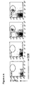
- FIG. 2A depicts flow cytometry profiles from Day 4 of the response in the popliteal lymph nodes (subcutaneous route), from a representative experiment.
- Figures 2B and 2C depict the percentage expansion of OVA-specific CD4+ T cells (Thy1.2+, CD4+) in the draining lymph nodes and the spleens, respectively, at Day 4.
- E. coli LPS and P. gingivalis LPS significantly enhanced the clonal expansion, regardless of the route of injection.
- Figures 2D and 2E depict the absolute numbers of Thy1.2+ CD4+ cells per popliteal lymph node or per spleen, respectively, at Day 4.
- Figures 2F and 2G depict in vitro restimulation of OVA-specific T cells expanded in vivo, by footpad injections or intraperitoneal injections, respectively.
- Four days after priming single cell suspensions from the draining popliteal lymph nodes (Figure 2F) or spleens ( Figure 2G) were restimulated with varying concentrations of OVA for 72 hours, and pulsed with [ 3 H] for 12 hours. Injections of E. coli LPS alone, or P. gingivalis LPS alone did not result in significant clonal expansion or in vitro proliferation.
- the data presented in Figures 2A-2G are representative of ten independent experiments.
- Figures 3A-3H depict E. coli LPS and P. gingivalis LPS inducing distinct types of antigen-specific T-helper responses in vivo.
- Culture supernatants from the cultures described in Figure 2F (subcutaneous injection) and Figure 2G (intraperitoneal injection) were assayed for IL-2 ( Figures 3A and 3E), IFN ⁇ ( Figures 3B and 3F), IL-10 ( Figures 3C and 3G), IL-4 (data not shown), and IL-5 ( Figure 3D and 3H) with ELISA. Injections of E. coli LPS alone or P. gingivalis LPS alone did not result in significant cytokine production.
- the data presented in Figures 3A-3H are representative of ten independent experiments.
- FIGS 4A-4G depict E. coli LPS and P. gingivalis LPS enhancing antigen-specific CD8+ T-cell responses in vivo.
- C57BL/6 mice, or B6.PL.THY1 a (B6.PL) mice reconstituted with OT-1 transgenic T cells were immunized with soluble OVA, OVA + E. coli LPS, or OVA + P. gingivalis LPS, either subcutaneously in the footpad ( Figures 4B, 4D and 4F) or intraperitoneally ( Figure 4C, 4E and 4G).
- FIG. 4A depicts flow cytometry profiles from Day 4 of the response in the popliteal lymph nodes from a representative experiment.
- Figures 4B and 4C depict the percentage expansion of OVA-specific CD8+ T cells (CD8+ Thy1.2+) in the draining lymph nodes and the spleens, respectively, at Day 4. Both E. coli LPS and P.
- FIGS 4D and 4E depict the absolute numbers of OVA-specific CD8+ T cells per popliteal lymph node or per spleen, respectively, at Day 4.
- Figures 4F and 4G depict in vitro restimulation of OVA-specific CD8+T cells expanded in vivo, by footpad injections or intraperitoneal injections, respectively.
- Four days after priming single cell suspensions from the draining popliteal lymph nodes (Figure 4F) or spleens ( Figure 4G) were restimulated with varying concentrations of OVA for 72 hours, and pulsed with [ 3 H] for 12 hours. Injections of E. coli LPS alone or P. gingivalis LPS alone did not result in significant clonal expansion, or in vitro proliferation (data not shown).
- the data presented in Figures 4A-4G are representative of three independent experiments.
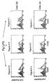
- Figures 5A-5H depict E. coli LPS and P. gingivalis LPS inducing distinct types of antigen-specific CD8+ T-cell responses in vivo.
- Culture supernatants from the cultures described in Figures 3E and 3F were assayed for IL-2 ( Figures 5A and 5B), IFN ⁇ ( Figures 5C and 5D), IL-10 ( Figures 5E and 5F), IL-4 (data not shown), and IL-5 ( Figures 5G and 5H) with ELISA. Injections of E. coli LPS alone or P. gingivalis LPS alone did not result in significant cytokine production (data not shown).
- the data presented in Figures 5A-5H are representative of three independent experiments.
- Figures 6A-6L depict both E. coli LPS and P. gingivalis LPS activating CD8 ⁇ + and CD8 ⁇ - DC subsets in vivo.
- C57BL/6 mice were injected with PBS (light histograms), or with E. coli LPS or P.
- gingivalis LPS (heavy, open histograms), either intravenously or intraperitoneally, and 6 hours later, the expression of CD80 ( Figures 6A, 6D, 6G, and 6J), CD86 ( Figures 6B, 6E, 6H, and 6K), and CD40 ( Figures 6C, 6F, 6I, and 6L) assessed on gated, splenic CD11c+CD8 ⁇ + and CD11c+CD8 ⁇ - DC subsets, by flow cytometry. Isotype controls are filled histograms. The data presented in Figures 6A-6L are representative of three experiments.
- Figures 7A-7C depict E. coli LPS, but not P. gingivalis LPS inducing IL-12 in CD8 ⁇ + DCs.
- Splenic CD11c+CD8 ⁇ + and CD11c+CD8 ⁇ - DCs were isolated from C57BL/6 mice by microbead enrichment, followed by flow cytometry and stimulated in vitro with 10 ⁇ g/mL of E. coli LPS or P. gingivalis LPS. Culture supernatants were assayed for IL-12 (Figure 7A), IL-6 ( Figure 7B), or TNF ⁇ (Figure 7C) 24 hours later.
- the data presented in Figures 7A-7C are representative of ten independent experiments.
- FIGS 8A and 8B demonstrate that TLR4 is the dominant receptor for signaling mediated by E. coli LPS, but not for P. gingivalis LPS.
- Splenocytes from C3H/HeJ mice and C3H/HeN mice were cultured with varying concentrations of E. coli LPS (Figure 8A) or P. gingivalis LPS ( Figure 8B) for 72 hours. The cultures were pulsed with [ 3 H] during the last 12 hours of culture.
- the data presented in Figures 8A-8B are representative of three independent experiments.
- Figures 9A-9D depict IL-6 production by splenocytes stimulated with E. coli LPS or P. gingivalis LPS in C3H/HeN and C3H/HeJ mice.
- Splenocytes from C3H/HeJ mice and C3H/HeN mice were cultured with varying concentrations of E. coli LPS for 12 hours or 48 hours ( Figures 9A and Figure 9C, respectively) or P. gingivalis LPS for 12 hours or 48 hours ( Figures 9B and 9D, respectively).
- IL-6 was measured by cytokine ELISA.
- the data presented in Figures 9A-9B are representative of three independent experiments.
- FIG 10 depicts P. gingivalis LPS inducing much greater levels of IL-13 than E. coli LPS.
- B6.PL.THY1 a (B6.PL) mice reconstituted with OT-2 transgenic T cells were immunized with soluble OVA, E. coli LPS alone, P. gingivalis LPS alone, OVA + E. coli LPS, or OVA + P. gingivalis LPS, either subcutaneously in the footpad or intraperitoneally.
- the invention is an adjuvant isolated lipid moiety selected from the group consisting of P. gingivalis LPS, detoxified P. gingivalis LPS, P. gingivalis Lipid A and detoxified P. gingivalis Lipid A and its use for the preparation of a pharmaceutical composition.
- the adjuvant is for eliciting a Th2 response in a mammal. In another aspect, the adjuvant is for modulating the balance between Th1 and Th2 immune response in a mammal. In another aspect, the adjuvant is for modulating the Th2 immune response in a mammal.
- the invention is a use of an adjuvant lipid moiety selected from the group consisting of P. gingivalis LPS, detoxified P. gingivalis LPS, P. gingivalis Lipid A and detoxified P. gingivalis Lipid A for the preparation of a pharmaceutical composition for enhancing antibody harvest in a laboratory animal through elicited Th2 immune response.
- an adjuvant lipid moiety selected from the group consisting of P. gingivalis LPS, detoxified P. gingivalis LPS, P. gingivalis Lipid A and detoxified P. gingivalis Lipid A for the preparation of a pharmaceutical composition for enhancing antibody harvest in a laboratory animal through elicited Th2 immune response.
- the invention is a use of an adjuvant lipid moiety selected from the group consisting of P. gingivalis LPS, detoxified P. gingivalis LPS, P. gingivalis Lipid A and detoxified P. gingivalis Lipid A for the preparation of a pharmaceutical composition for treating an autoimmune disease in a mammal.
- the pharmaceutical composition further comprises disease-specific antigens.
- the pharmaceutical composition further comprises a co-adjuvant which elicits a Th1 immune response.
- the invention is a use of an adjuvant lipid moiety selected from the group consisting of P. gingivalis LPS, detoxified P. gingivalis LPS, P. gingivalis Lipid A and detoxified P. gingivalis Lipid A for the preparation of a pharmaceutical composition for treating an infectious disease in a mammal.
- the invention is a use of an adjuvant isolated lipid moiety selected from the group consisting of P. gingivalis LPS, detoxified P. gingivalis LPS, P. gingivalis Lipid A and detoxified P. gingivalis Lipid A for the preparation of a pharmaceutical composition for modulating the Th2 immune response in a laboratory animal.
- the invention is a use of an adjuvant lipid moiety selected from the group consisting of P. gingivalis LPS, detoxified P. gingivalis LPS P. gingivalis Lipid A and detoxified P. gingivalis Lipid A for the preparation of a pharmaceutical composition for stimulating IL-5 production in a mammal.
- the invention is a use of an adjuvant lipid moiety selected from the group consisting of P. gingivalis LPS, detoxified P. gingivalis LPS, P. gingivalis Lipid A and detoxified P. gingivalis Lipid A for the preparation of a pharmaceutical composition for dampening IFN ⁇ production in a mammal.
- an adjuvant lipid moiety selected from the group consisting of P. gingivalis LPS, detoxified P. gingivalis LPS, P. gingivalis Lipid A and detoxified P. gingivalis Lipid A for the preparation of a pharmaceutical composition for dampening IFN ⁇ production in a mammal.
- the invention is a pharmaceutical composition
- a pharmaceutical composition comprising at least one isolated lipid moiety selected from the group consisting of P. gingivalis LPS, detoxified P. gingivalis LPS, P. gingivalis Lipid A and detoxified P. gingivalis Lipid A.
- the pharmaceutical composition further comprises disease-specific antigens.
- the pharmaceutical composition further comprises a co-adjuvant which elicits a Th1 immune response.
- the invention is a method of enhancing antibody harvest in a laboratory animal through elicited Th2 immune response comprising administering to the animal an adjuvant lipid moiety selected from the group consisting of P. gingivalis LPS, detoxified P. gingivalis LPS, P. gingivalis Lipid A and detoxified P. gingivalis Lipid A.
- the method further comprises co-administering to the mammal disease-specific antigens.
- the method further comprises co-administering to the mammal a co-adjuvant which elicits a Th1 immune response.
- the adjuvant, disease-specific antigens and, optionally, the co-adjuvant can be administered concurrently or sequentially.
- P. gingivalis lipopolysaccharide can be extracted from any isolated strain of Porphyromonas gingivalis.
- P. gingivalis 33277, P. gingivalis 49417, and P. gingivalis 53978 from the American Type Culture Collection (Manassas, VA) can be utilized.
- a clinical P. gingivalis isolate is another acceptable source for LPS extraction.
- P. gingivalis LPS may be toxic from de novo preparations, it can be detoxified with existing technology without compromising its adjuvant activity (Rietschel, E.T., et al. 1994. "Bacterial endotoxin: molecular relationships of structure to activity and function," FASEB Journal 8:217-825; Johnson, A.G., et al. 1987. "Characterization of a nontoxic monophosphoryl lipid A,” Rev Infectious Diseases 9 (Suppl 5):S512-S516).
- P. gingivalis LPS, detoxified P. gingivalis LPS, P. gingivalis Lipid A, detoxified P. gingivalis Lipid A, or any combination thereof can be used to induce a Th2 immune response in humans and animals for clinical benefit, experimental purposes, or industrial applications.
- the invention disclosed herein is the elicitation of a Th2 immune response through the administration of an adjuvant comprising one or more of the following: P. gingivalis LPS, detoxified P. gingivalis LPS, P. gingivalis Lipid A or detoxified P. gingivalis Lipid A.
- P. gingivalis LPS, detoxified P. gingivalis LPS, P. gingivalis Lipid A and detoxified P. gingivalis Lipid A used singly or in any combination, are considered suitable for use as an adjuvant to produce a Th2 immune response in a human or animal.
- P. gingivalis LPS, detoxified P. gingivalis LPS, P. gingivalis Lipid A or detoxified P. gingivalis Lipid A can be used in combination with other adjuvants known in the art to modulate and induce immune responses in humans and animals.
- Th2 immune response The selective elicitation of the Th2 immune response is highly desirable for treatment or prophylactic vaccination of humans or animals against various autoimmune diseases and graft-versus-host disease. Many autoimmune diseases are characterized by pathogenic Th1 immune responses. Currently, there are intensive efforts to discover adjuvants that can redirect a pathogenic Th1 immune response towards a benign Th2 immune response. Activating a Th2 immune response in a human or animal suffering from an autoimmune disease may decrease the pathogenic Th1 immune response, thereby decreasing the debilitating inflammation characteristic of such diseases. According to the instant disclosure, this effect of activating a Th2 immune response in a human or animal can be achieved through administration of a therapeutically effective composition comprising P. gingivalis LPS, detoxified P.
- gingivalis LPS P. gingivalis Lipid A
- detoxified P. gingivalis Lipid A or any combination thereof by methods well known in the art (Pulendran, B., et al. 2001. “Modulating the immune response with dendritic cells and their growth factors,” Trends in Immunology 22(1):41-47).
- Th2 immune responses Therapeutic immunity against many tumors or infectious diseases or in transplantation requires Th2 immune responses.
- these diseases can be treated by administration of an adjuvant comprising P. gingivalis LPS, detoxified P. gingivalis LPS, P. gingivalis Lipid A, detoxified P. gingivalis Lipid A, or any combination thereof to induce a Th2 immune response.
- P. gingivalis LPS, detoxified P. gingivalis LPS, P. gingivalis Lipid A, detoxified P. gingivalis Lipid A, or any combination thereof can be co-administered concurrently or sequentially with an adjuvant causing a Th1 immune response when a mixed response is required in the prevention or cure of diseases affecting humans or animals.
- This effect can be achieved through the introduction of a therapeutically effective amount of P. gingivalis LPS, detoxified P. gingivalis LPS, derivatives of P. gingivalis LPS, derivatives of detoxified P. gingivalis LPS, P.
- gingivalis Lipid A detoxified P. gingivalis Lipid A, derivatives of P. gingivalis Lipid A, derivatives of detoxified P. gingivalis Lipid A, mimetics thereof, or any combination thereof by methods well known in the art (Pulendran, B., et al. 2001. “Modulating the immune response with dendritic cells and their growth factors," Trends in Immunology 22(1):41-47).
- an adjuvant comprising P. gingivalis LPS, detoxified P. gingivalis LPS, P. gingivalis Lipid A, detoxified P. gingivalis Lipid A, or any combination thereof is preferably administered intravenously, intra-arterially, intra-muscularly, intra-dermally, and local (e.g., intra-tumoral or at the vicinity of a tumor site). Regardless of administration route, P.
- gingivalis LPS detoxified P. gingivalis LPS, P. gingivalis Lipid A, detoxified P. gingivalis Lipid A, or any combination thereof can be administered with or without additional adjuvants and antigens.
- An effective amount of P. gingivalis LPS, detoxified P. gingivalis LPS, P. gingivalis Lipid A, detoxified P. gingivalis Lipid A, or any combination thereof will elicit a Th2 immune response in a human or animal.
- gingivalis Lipid A maintains the solubility of the compound.
- Formulating an effective amount of P. gingivalis LPS, detoxified P. gingivalis LPS, P. gingivalis Lipid A, detoxified P. gingivalis Lipid A, or any combination thereof to elicit a Th2 immune response in a human or animal for oral administration is also contemplated.
- the adjuvant of the instant invention can be administered concurrently or sequentially with a vaccine to enhance immunity by eliciting a Th2 immune response.
- Sequential administration indicates that the adjuvant and vaccine may be injected separately, in any order.
- Preferred administration routes include intravenous, intra-arterial, intra-muscular, intra-dermal, and local (e.g., intra-tumoral or at the vicinity of a tumor site).
- Methods for co-administering an adjuvant with a vaccine to increase immunoreactivity are well known in the art (Pulendran, B., et al. 2001. "Modulating the immune response with dendritic cells and their growth factors," Trends in Immunology 22(1):41-47).
- the instant invention includes use of adjuvants comprising P. gingivalis LPS, detoxified P. gingivalis LPS, P. gingivalis Lipid A, detoxified P. gingivalis Lipid A, or any combination thereof as research tools to study the immune system in laboratory animals.
- P. gingivalis LPS, detoxified P. gingivalis LPS, P. gingivalis Lipid A, detoxified P. gingivalis Lipid A, or any combination thereof can also be used in conjunction with an antigen for enhancing the production and harvest of antibodies in animals by methods well known in the art.
- OT-2 TCR transgenic mice (strain 426-6), generated by Dr. W. Heath (Walter & Eliza Hall Institute, Melbourne, Australia) and Dr. F. Carbone (Monash University, Melbourne, Australia) were obtained from Dr. J. Kapp (Emory University, Atlanta).
- OT-1 TCR transgenic mice were purchased from Jackson Laboratory (Bar Harbor, ME).
- C57BL/6 mice, B6.PL.THY1 a (B6.PL) mice, and C3H/HeJ mice were purchased from Jackson Laboratory (Bar Harbor, ME).
- C3H/HeN mice were purchased from Harlan Sprague Dawley (Indianapolis, IN). All mice were kept in microisolator cages in a specific-pathogen free facility.
- age matched, male C57BL/6 or B6.PL.THY1 a recipients were given 2.5 x 10 6 of either OT-2 cells or OT-1 TCR transgenic T cells intravenously.
- P. gingivalis strain A7436 Hirschfeld, M., et al. 2001. "Signaling by toll-like receptor 2 and 4 agonists results in differential gene expression in murine macrophages," Infect Immun 69:1477-1482) and E. coli strain 25922 (American Type Culture Collection, Manassas, VA) were cultured under identical conditions and LPS purified as previously described (Cutler, C.W., et al. 1996. "Hemin-induced modifications of the antigenicity and hemin-binding capacity of Porphyromonas gingivalis lipopolysaccharide,” Infect Immun 64:2282-2287; Westphal, O., and K. Jann. 1965.
- the dialyzed LPS preparation was then subjected to cesium chloride isopycnic density gradient centrifugation (in 0.5837 g CsCl 2 per 4.4 mL of the LPS preparation) at 42,000 rpm for 72 hours in a Beckman L-60 Ultracentrifuge (Palo Alto, CA).
- the refractive indices of the gradient fractions were determined with a refractometer (Milton Roy, Rochester, NY), and values were converted to density (grams per milliliter).
- Fractions containing LPS density fractions between 1.42 and 1.52 g/mL were pooled, dialyzed against distilled water for 3 days, lyophilized and stored at room temperature.
- LPS was analyzed for protein by the BCA protein assay (Pierce Chemical Company, Rockford, IL). LPS samples were also separated by sodium dodecyl-sulfate-polyacrylamide gel electrophoresis (SDS-PAGE) and stained for protein with Coomasie blue (Pierce Chemical Company, Rockford, IL). Selected samples were also subjected to proteinase K digestion and nuclease treatment and reanalyzed by SDS-PAGE to confirm the purity of the LPS moieties (Pierce Chemical Company, Rockford, IL).
- Chicken OVALBUMIN (OVA) (Sigma Chemical Co., St. Louis, MO) was freshly prepared in phosphate buffered saline (PBS) at a concentration of 20 mg/mL, and depleted of the endotoxin activity (measured by LAL QCL-1000 kit from Bio Whittaker, Walkersville, MD, using manufacturer's protocol dated 2000), with the Detoxi-Gel Affinity Pack Columns (Pierce Chemical Company, Rockford, IL). After depletion, the endotoxin level was below the limit of detection of the LAL QCL-1000 kit ( ⁇ 0.1 EU).
- mice 3-5 per group were injected either intraperitoneally, or in the footpad, with either 2 mg OVA in saline, 2 mg OVA+25 ⁇ g E. coli LPS, or 2mg OVA+25 ⁇ g P. gingivalis LPS. Endotoxin activity in saline was measured by LAL QCL-1000 kit, and observed to be below the detection limit. Prior to mixing with OVA, LPS was sonicated extensively, to ensure uniform mixing of micelles. Footpad injections were given in a volume of 25 ⁇ l. Intraperitoneal injections were given in a volume of 100 ⁇ l.
- cell suspensions were prepared from the draining popliteal lymph nodes or spleens, and incubated on ice with PE-labeled anti-Thy1.2 (Pharmingen, San Diego, CA), FITC-labeled V ⁇ 2 (Pharmingen), Cy-Chrome labeled CD4 (Pharmingen) and Biotin-labeled V ⁇ 5 (Pharmingen), followed by streptavidin allophycocyanin (APC) (Pharmingen).
- PE-labeled anti-Thy1.2 Pharmingen, San Diego, CA
- FITC-labeled V ⁇ 2 Pharmingen
- Cy-Chrome labeled CD4 conjugated immunoglobulfen
- Biotin-labeled V ⁇ 5 Pharmingen
- streptavidin allophycocyanin APC
- DCs were stained with FITC-labeled CD11c (Pharmingen), in combination with PE-labeled CD11b (Pharmingen), or biotin-labeled CD8 ⁇ (Pharmingen), followed by streptavidin allophycocyanin (Pharmingen) using a FACSvantage flow cytometer (Becton Dickinson), equipped with Enterprise II laser (Coherent Radiation, Palo Alto, CA).
- Proliferative responses were assessed after 72 hours of culture in a humidified atmosphere of 5% CO 2 in air. Cultures were pulsed with 1.0 ⁇ Ci [ 3 H] thymidine for 12 hours, and incorporation of the radionucleotide was measured by ⁇ -scintillation spectroscopy (Pulendran, B., et al. 1999. "Distinct dendritic cell subsets differentially regulate the class of immune response in vivo," Proc Natl Acad Sci USA 96:1036-1041).
- cytokine assays aliquots of culture supernatants were removed after 72 hours, pooled, and assayed for the presence of IFN ⁇ , IL-2, IL-4, IL-5, IL-10, and IL-13 by ELISA.
- IFN ⁇ , IL-2, IL-10, IL-4, IL-5, IL-6, IL-12, IL-13 and TNF ⁇ were quantified by ELISA kits from Pharmingen (San Diego, CA) using manufacturer's instructions dated 2000.
- Splenic sections were rehydrated with PBS, and blocked with PBS/5% bovine serum albumin (BSA)/1% goat serum for 20 minutes, and stained with FITC-conjugated anti-CD11c (Pharmingen, San Diego, CA) and PE-conjugated anti-CD4 (Pharmingen, San Diego, CA) for 1 hour.
- the sections were washed and coverslips mounted onto glass slides with Fluoromount (Southern Biotechnology Associates, Birmingham, AL). Confocal microscopy was performed using a TCS SP microscope equipped with argon and krypton ion lasers and a 10X HC PL-APO objective (Leica Microsystem, Heidelberg, Germany).
- CD11c+CD8 ⁇ + and CD11c+CD8 ⁇ - DC subsets were purified from spleens as follows. Spleens of C57BL/6 mice were dissected, cut into small fragments and then digested with collegenase D (0.5 mg/mL; Boehringer-Mannheim, Mannheim, Germany) and Dnase I (40 mg/mL, Boehringer-Mannheim) in RPMI 1640 medium supplemented with 5% fetal calf serum (PCS) for 10 minutes at 37°C. Digested fragments were washed twice in PBS/5% FCS.
- PCS fetal calf serum
- CD11c+ DCs were enriched using CD11c+ microbeads (Miltenyi Biotech, San Diego, CA).
- the enriched DCs were stained with FITC-conjugated CD11c (Pharmingen) and PE-conjugated CD8 ⁇ + (Pharmingen) and sorted into the CD11c+ CD8 ⁇ + and CD11c+ CD8 ⁇ - subsets, using a FACSvantage flow cytometer (Becton Dickinson), equipped with Enterprise II laser (Coherent Radiation, Palo Alto, CA).
- CD 11c+ CD8 ⁇ + and CD 11c+ CD8 ⁇ - were isolated by flow cytometry and cultured in RPMI complete medium supplemented with 5% FBS and with either 10 ⁇ g/mL E. coli LPS or 10 ⁇ g/mL P. gingivalis LPS for 24 hours or 48 hours.
- EXAMPLE 1 E. coli LPS and P. gingivalis LPS enhance antigen-specific T-helper responses in vivo
- TCR transgenic T cells were adoptively transferred into Thy-1 congenic B6.PL.THY1 a (B6.PL) mice, such that they constituted a small but detectable proportion of all T cells (Kearney, E.R., et al. 1995, "Antigen-dependent clonal expansion of a trace population of antigen-specific CD4+ T cells in vivo is dependent on CD28 costimulation and inhibited by CTLA-4," J Immunol 155:1032-1036). In this system, the fate of OVA-specifc, transgenic T cells was followed using the Thy1.2 antibody, which stains only the transferred cells. T cells with the phenotype Thy1.2+ CD4+ V ⁇ 2+ V ⁇ 5+ was considered OVA-specific CD4+ T cells.
- mice were injected with one of the following compositions: soluble endotoxin-free OVA, E. coli LPS alone, P. gingivalis LPS alone, OVA + E. coli LPS, or OVA + P. gingivalis LPS. Injections were either intraperitoneal or in a footpad ( Figure 1). Prior to the injection, the OVA was depleted of endotoxin contamination using methods described above. The CD4+ OVA-specific T cell response in either the draining lymph nodes or in the spleen was monitored by flow cytometry ( Figure 2A).
- EXAMPLE 2 E. coli LPS and P. gingivalis LPS induce distinct types of antigen-specific T-helper responses in vivo
- Cytokine production by antigen-specific T cells was measured by assaying the culture supernatants from the single cell suspensions of the draining lymph nodes described above for IL-2, IFN ⁇ , IL-4, IL-10 and IL-5. Assessment of cytokine production in these cultures revealed significant differences between mice injected with OVA, OVA + E. coli LPS, or OVA + P. gingivalis LPS ( Figures 3A-3H). In cultures from mice injected with OVA alone, there was little, if any, IL-2, IFN ⁇ , IL-10, IL-4, or IL-5 produced. In contrast, in cultures from mice injected with OVA + E. coli LPS, there was significant IL-2 and IL-10 and very high levels of IFN ⁇ produced by the antigen-specific T cells. Neither IL-4 nor IL-5 could be detected in cells from OVA + E. coli LPS injected mice.
- EXAMPLE 3 E. coli LPS and P. gingivalis LPS enhance antigen-specific CD8+ T-cell responses in vivo
- EXAMPLE 4 E. coli LPS and P. gingivalis LPS induce distinct types of antigen-specific CD8+ T-cell responses in vivo
- gingivalis LPS produced much lower levels of IFN ⁇ ( Figures 5B and 5F), but significant levels of IL-10 ( Figures 5C and 5G) and IL-5 ( Figures 5D and 5H), consistent with the cytokine patterns observed with CD4+ OT-2 cells ( Figures 3A-3H). No significant levels of IL-4 were detected in any condition.
- EXAMPLE 5 Both E. coli LPS and P. gingivalis LPS activate CD8 ⁇ + and CD8 ⁇ - DC subsets in vivo
- EXAMPLE 6 E. coli LPS, but not P. gingivalis LPS induces IL-12 in CD8 ⁇ + DCs
- IL-10 and IL-13 may be Th2-inducing cytokines, significant levels of either IL-10 or IL-13 could not be consistently detected in these cultures.
- TLR4 is the dominant receptor for signaling mediated by E. coli LPS, but not for P. gingivalis LPS
- TLR4 Toll-like receptor 4
- P. gingivalis LPS may signal through a TLR4-independent mechanism.
- C3H/HeJ mice C3H/HeJ mice
- C3H/HeN mice C3H/HeN mice
- C3H/HeJ splenocytes cultured with E. coli LPS were greatly impaired in their proliferative capacity, compared to the C3H/HeN controls ( Figure 8A).
- gingivalis LPS were only modestly impaired in their proliferative capacity, compared to the C3H/HeN controls ( Figure 8B). Consistent with this, production of IL-6 induced by E. coli LPS was greatly impaired in C3H/HeJ splenocytes, compared to C3H/HeN splenocytes ( Figures 9A and 9C). However, production of IL-6 induced by P. gingivalis LPS was not impaired in C3H/HeJ mice ( Figures 9B and 9D). Therefore, as reported previously, while E. coli LPS signaling is largely dependent on TLR4, P. gingivalis LPS appears to signal mainly through a TLR4-independent mechanism. Therefore, as reported previously, E. coli LPS signaling is largely dependent on TLR4, whereas P. gingivalis LPS appears to signal mainly through a TLR4-independent mechanism.
- EXAMPLE 8 P. gingivalis LPS, but not E. coli LPS, stimulates IL-13 production
- Examples 1-8 have demonstrated that different microbial products may induce distinct types of immune responses via differential activation of DC subsets.
- E. coli LPS induced Th1 and Tc1 responses with high levels of IFN ⁇ , but no IL-4 or IL-5.
- P. gingivalis LPS induced Th2 and Tc2 immune responses characterized by significant levels ofIL-10, IL-13, and IL-5, but very little or no IFN ⁇ .
- an adjuvant which selectively induces the Th2 immune response.
- the subject of Examples 9-12 is the application of the ability to selectively activate the Th2 immune response rather than the Th1 immune response.
- the subject matter of Examples 9-12 is meant to be illustrative and in no way limit the application of using P.
- the invention contemplates a method to elicit a Th2 or Th2-like immune response in a subject who suffers from a disease state that can be alleviated at least in part with an appropriate Th2 or Th2-like immune response.
- the next step may be to determine disease specific antigens.
- the subsequent step may be to co-administer the disease specific antigens with P. gingivalis LPS, detoxified P. gingivalis LPS, derivatives of P. gingivalis LPS, derivatives of detoxified P. gingivalis LPS, P. gingivalis Lipid A, detoxified P. gingivalis Lipid A, derivatives of P.
- gingivalis Lipid A derivatives of detoxified P. gingivalis Lipid A, mimetics thereof, or any combination thereof, to induce a Th2 immune response.
- Co-administration of disease specific antigens and adjuvants of the present invention that elicit a Th2 or Th2-like immune response can be sequentially or concurrently delivered intravenously, intra-arterially, intra-muscularly, intra-dermally, intra-tumorally, or orally. Any pharmaceutical carrier or diluent that maintains the solubility of the components can be used.
- EXAMPLE 10 Modulating the balance of Th1 and Th2 immune responses for treating disease states
- the invention contemplates a method to elicit a Th2 or Th2-like immune response in a subject who suffers from a disease state in which an inappropriate Th1 immune response is associated.
- the method can be implemented to alleviate at least in part an inappropriate Th1 immune response by shifting the response away from the Th1 to a Th2 or Th2-like immune response.
- the next step may be to determine disease specific antigens.
- the subsequent step may be to co-administer the disease specific antigens with P. gingivalis LPS, detoxified P. gingivalis LPS, derivatives of P. gingivalis LPS, derivatives of detoxified P. gingivalis LPS, P.
- a Th2 immune response to induce a Th2 immune response.
- Co-administration of disease specific antigens and adjuvants of the present invention that elicit a Th2 or Th2-like immune response can be sequentially or concurrently delivered intravenously, intra-arterially, intra-muscularly, intra-dermally, intra-tumorally, or orally. Any pharmaceutical carrier or diluent that maintains the solubility of the components can be used.
- EXAMPLE 11 Using P. gingivalis LPS in conjunction with other adjuvants to elicit a combined Th1/Th2 immune response
- the invention contemplates a method to elicit both a Th1 and a Th2 or Th2-like immune response in a subject who suffers from a disease state that can be alleviated at least in part with a combined Th1 and Th2 or Th2-like immune response.
- the next step may be to determine disease specific antigens.
- the subsequent step may be to co-administer the disease specific antigens with P. gingivalis LPS, detoxified P. gingivalis LPS, derivatives of P. gingivalis LPS, derivatives of detoxified P. gingivalis LPS, P. gingivalis Lipid A, detoxified P.
- gingivalis Lipid A derivatives of P. gingivalis Lipid A, derivatives of detoxified P. gingivalis Lipid A, mimetics thereof, or any combination thereof, and at least one adjuvant known to induce a Th1 immune response, to induce a combined Th1/Th2 immune response.
- Co-administration of disease specific antigens, adjuvants for a Th1 immune response, and adjuvants of the present invention that elicit a Th2 or Th2-like immune response can be sequentially or concurrently delivered intravenously, intra-arterially, intra-muscularly, intra-dermally, intra-tumorally, or orally. Any pharmaceutical carrier or diluent that maintains the solubility of the components can be used.
- EXAMPLE 12 Using P. gingivalis LPS as a vaccine adjuvant
- the invention contemplates a method to elicit a Th2 or Th2-like immune response in a subject to prevent disease onset.
- the next step may be to determine disease specific antigens.
- the next step may be to co-administer the disease specific antigens with P. gingivalis LPS, detoxified P. gingivalis LPS, derivatives of P. gingivalis LPS, derivatives of detoxified P. gingivalis LPS, P. gingivalis Lipid A, detoxified P. gingivalis Lipid A, derivatives of P. gingivalis Lipid A, derivatives of detoxified P.
- a Th2 immune response to induce a Th2 immune response.
- Co-administration of disease specific antigens and adjuvants of the present invention that elicit a Th2 or Th2-like immune response can be sequentially or concurrently delivered intravenously, intra-arterially, intra-muscularly, intra-dermally, or orally. Any pharmaceutical carrier or diluent that maintains the solubility of the components can be used.
Landscapes
- Health & Medical Sciences (AREA)
- Life Sciences & Earth Sciences (AREA)
- Chemical & Material Sciences (AREA)
- Veterinary Medicine (AREA)
- General Health & Medical Sciences (AREA)
- Medicinal Chemistry (AREA)
- Immunology (AREA)
- Pharmacology & Pharmacy (AREA)
- Public Health (AREA)
- Animal Behavior & Ethology (AREA)
- Chemical Kinetics & Catalysis (AREA)
- General Chemical & Material Sciences (AREA)
- Epidemiology (AREA)
- Nuclear Medicine, Radiotherapy & Molecular Imaging (AREA)
- Engineering & Computer Science (AREA)
- Organic Chemistry (AREA)
- Microbiology (AREA)
- Mycology (AREA)
- Bioinformatics & Cheminformatics (AREA)
- Molecular Biology (AREA)
- Medicines Containing Antibodies Or Antigens For Use As Internal Diagnostic Agents (AREA)
- Steroid Compounds (AREA)
- Medicines That Contain Protein Lipid Enzymes And Other Medicines (AREA)
Abstract
Description
- The present invention relates to the field of using adjuvants to promote a specific type of immunological response.
- The immune system has evolved two different types of adaptive immunity, each specialized for the elimination of a particular class of pathogens. In response to intracellular microbes, CD4+ T-helper (Th) cells differentiate into Th1 cells, which produce interferon γ (IFNγ) and interleukin (IL)-1, which, in turn, enhance cell-mediated immunity and inhibit the humoral immune responses. In contrast, helminths induce differentiation of CD4+ T-helper (Th) cells into Th2 cells, which produce cytokines (principally IL-4, IL-5, and IL-10) to induce immunoglobulin E (IgE) and eosinophil-mediated destruction of pathogens. The Th2 immune response inhibits cell-mediated immunity and enhances humoral immunity. The mechanism by which a given pathogen induces a Th1 or Th2 type of immune response is unknown.
- Distinct subsets of dendritic cells (DCs) differentially induce Th1 and Th2 immune responses. In mice, the putative lymphoid-related CD8α+ DCs in spleens induce Th1 immune responses (Shortman, K.D., et al. 1998. "The linkage between T-cell and dendritic cell development in the mouse thymus," Immune Rev 165:39-46). In contrast, Th2 immune responses are induced by the CD8α-myeloid DCs (Maldonado-Lopez, R., et al. 1999. "CD8α+ and CD8α- subclasses of dendritic cells direct the development of distinct T helper cells in vivo," J Exp Med 189:587-592; Pulendran, B., et al. 1999. "Distinct dendritic cell subsets differentially regulate the class of immune response in vivo," Proc Natl Acad Sci USA 96:1036-1041; Rissoan, M.C., et al. 1999. "Reciprocal control of T helper cell and dendritic cell differentiation," Science 283:1183-1186). Different patterns of immunity can be elicited by activating distinct DC subsets.
- Escherichia coli lipopolysaccharide (LPS) is reported to signal through the Toll-like receptor 4 (TLR4) complex (Qureshi, S.T., et al. 1999. "Endotoxin-tolerant mice have mutations in Toll-like receptor 4 (Tlr4)," J Exp Med 189:615-625; published erratum appears in J Exp Med 189:1518) and promote a Th1 immune response in vivo (Khoruts, A., A., et al. 1998. "A natural immunological adjuvant enhances T cell clonal expansion through a CD28-dependent, interleukin (IL)-2-independent mechanism," J Exp Med 187:225-236). In contrast, Porphyromonas gingivalis LPS is reported to signal through a TLR4-independent mechanism (Tanamoto, K., S., et al. 1997. "The lipid A moiety of Porphyromonas gingivalis lipopolysaccharide specifically mediates the activation of C3H/HeJ mice," J Immunol 158:4430-4436). It has now been found that although the LPS from these two different bacterial sources induce potent clonal expansion of antigen-specific CD4+ and CD8+ T cells in mice, they elicit strikingly different T cell cytokine profiles through differential cytokine expression by the CD8α+ and CD8α- DCs.
- Although the use of adjuvants with antigen delivery to boost immunity is well known in the prior art, the adjuvants of the prior art reportedly elicit only a Th1 immune response. Currently, there is no known way to elicit a selective Th2 immune response with an adjuvant. A means of eliciting Th2 immune responses using P. gingivalis LPS has now been found, the application of the same to prevent and treat disease in humans and animals (mammals), increase antibody production in industrial practice, and to provide a method for studying the immune response in laboratory animals are presented herein.
- Figure 1 depicts the experimental design utilized herein. B6.PL.THY1a (B6.PL) mice or C57BL/6 mice reconstituted with OT-2 cells were injected with either soluble OVA, soluble OVA + E. coli LPS, soluble OVA + P. gingivalis LPS, E. coli LPS alone, or P. gingivalis LPS alone intraperitoneally or in the footpad. Four days later, the spleens (intraperitoneal route) or draining lymph nodes (footpad route) were removed for phenotypic and functional analyses, including clonal expansion of OVA-specific CD4+ T cells, in vitro proliferation of OVA-specific CD4+ T cells and cytokine production by the OVA-specific CD4+ T cells.
- Figures 2A-2G depict E. coli LPS and P. gingivalis LPS enhancing antigen-specific T-helper responses in vivo. B6.PL.THY1a (B6.PL) mice reconstituted with OT-2 transgenic T cells were immunized with soluble OVA, E. coli LPS alone, P. gingivalis LPS alone, OVA + E. coli LPS, or OVA + P. gingivalis LPS, either subcutaneously in the footpad (Figures 2B, 2D and 2F) or intraperitoneally (Figures 2C, 2E and 2G). Four days later, the draining popliteal lymph nodes (subcutaneous route), or spleens (intraperitoneal route) were removed, and the clonal expansion of OVA-specific CD4+ T cells assessed by flow cytometry, by staining with Thy1.2 versus CD4. Figure 2A depicts flow cytometry profiles from
Day 4 of the response in the popliteal lymph nodes (subcutaneous route), from a representative experiment. Figures 2B and 2C depict the percentage expansion of OVA-specific CD4+ T cells (Thy1.2+, CD4+) in the draining lymph nodes and the spleens, respectively, atDay 4. Both E. coli LPS and P. gingivalis LPS significantly enhanced the clonal expansion, regardless of the route of injection. Figures 2D and 2E depict the absolute numbers of Thy1.2+ CD4+ cells per popliteal lymph node or per spleen, respectively, atDay 4. Figures 2F and 2G depict in vitro restimulation of OVA-specific T cells expanded in vivo, by footpad injections or intraperitoneal injections, respectively. Four days after priming, single cell suspensions from the draining popliteal lymph nodes (Figure 2F) or spleens (Figure 2G) were restimulated with varying concentrations of OVA for 72 hours, and pulsed with [3H] for 12 hours. Injections of E. coli LPS alone, or P. gingivalis LPS alone did not result in significant clonal expansion or in vitro proliferation. The data presented in Figures 2A-2G are representative of ten independent experiments. - Figures 3A-3H depict E. coli LPS and P. gingivalis LPS inducing distinct types of antigen-specific T-helper responses in vivo. Culture supernatants from the cultures described in Figure 2F (subcutaneous injection) and Figure 2G (intraperitoneal injection) were assayed for IL-2 (Figures 3A and 3E), IFNγ (Figures 3B and 3F), IL-10 (Figures 3C and 3G), IL-4 (data not shown), and IL-5 (Figure 3D and 3H) with ELISA. Injections of E. coli LPS alone or P. gingivalis LPS alone did not result in significant cytokine production. The data presented in Figures 3A-3H are representative of ten independent experiments.
- Figures 4A-4G depict E. coli LPS and P. gingivalis LPS enhancing antigen-specific CD8+ T-cell responses in vivo. C57BL/6 mice, or B6.PL.THY1a (B6.PL) mice reconstituted with OT-1 transgenic T cells were immunized with soluble OVA, OVA + E. coli LPS, or OVA + P. gingivalis LPS, either subcutaneously in the footpad (Figures 4B, 4D and 4F) or intraperitoneally (Figure 4C, 4E and 4G). Four days later, the draining popliteal lymph nodes (footpad injections) or spleens (intraperitoneal route) were removed, and the clonal expansion of OVA-specific CD8+ T cells assessed by flow cytometry, by staining with CD8 versus Vα2, or CD8 versus Thy1.2 versus Vα2. Figure 4A depicts flow cytometry profiles from
Day 4 of the response in the popliteal lymph nodes from a representative experiment. Figures 4B and 4C depict the percentage expansion of OVA-specific CD8+ T cells (CD8+ Thy1.2+) in the draining lymph nodes and the spleens, respectively, atDay 4. Both E. coli LPS and P. gingivalis LPS significantly enhance the clonal expansion, regardless of the route of injection. Figures 4D and 4E depict the absolute numbers of OVA-specific CD8+ T cells per popliteal lymph node or per spleen, respectively, atDay 4. Figures 4F and 4G depict in vitro restimulation of OVA-specific CD8+T cells expanded in vivo, by footpad injections or intraperitoneal injections, respectively. Four days after priming, single cell suspensions from the draining popliteal lymph nodes (Figure 4F) or spleens (Figure 4G) were restimulated with varying concentrations of OVA for 72 hours, and pulsed with [3H] for 12 hours. Injections of E. coli LPS alone or P. gingivalis LPS alone did not result in significant clonal expansion, or in vitro proliferation (data not shown). The data presented in Figures 4A-4G are representative of three independent experiments. - Figures 5A-5H depict E. coli LPS and P. gingivalis LPS inducing distinct types of antigen-specific CD8+ T-cell responses in vivo. Culture supernatants from the cultures described in Figures 3E and 3F were assayed for IL-2 (Figures 5A and 5B), IFNγ (Figures 5C and 5D), IL-10 (Figures 5E and 5F), IL-4 (data not shown), and IL-5 (Figures 5G and 5H) with ELISA. Injections of E. coli LPS alone or P. gingivalis LPS alone did not result in significant cytokine production (data not shown). The data presented in Figures 5A-5H are representative of three independent experiments.
- Figures 6A-6L depict both E. coli LPS and P. gingivalis LPS activating CD8α+ and CD8α- DC subsets in vivo. C57BL/6 mice were injected with PBS (light histograms), or with E. coli LPS or P. gingivalis LPS (heavy, open histograms), either intravenously or intraperitoneally, and 6 hours later, the expression of CD80 (Figures 6A, 6D, 6G, and 6J), CD86 (Figures 6B, 6E, 6H, and 6K), and CD40 (Figures 6C, 6F, 6I, and 6L) assessed on gated, splenic CD11c+CD8α+ and CD11c+CD8α- DC subsets, by flow cytometry. Isotype controls are filled histograms. The data presented in Figures 6A-6L are representative of three experiments.
- Figures 7A-7C depict E. coli LPS, but not P. gingivalis LPS inducing IL-12 in CD8α+ DCs. Splenic CD11c+CD8α+ and CD11c+CD8α- DCs were isolated from C57BL/6 mice by microbead enrichment, followed by flow cytometry and stimulated in vitro with 10 µg/mL of E. coli LPS or P. gingivalis LPS. Culture supernatants were assayed for IL-12 (Figure 7A), IL-6 (Figure 7B), or TNFα (Figure 7C) 24 hours later. The data presented in Figures 7A-7C are representative of ten independent experiments.
- Figures 8A and 8B demonstrate that TLR4 is the dominant receptor for signaling mediated by E. coli LPS, but not for P. gingivalis LPS. Splenocytes from C3H/HeJ mice and C3H/HeN mice were cultured with varying concentrations of E. coli LPS (Figure 8A) or P. gingivalis LPS (Figure 8B) for 72 hours. The cultures were pulsed with [3H] during the last 12 hours of culture. The data presented in Figures 8A-8B are representative of three independent experiments.
- Figures 9A-9D depict IL-6 production by splenocytes stimulated with E. coli LPS or P. gingivalis LPS in C3H/HeN and C3H/HeJ mice. Splenocytes from C3H/HeJ mice and C3H/HeN mice were cultured with varying concentrations of E. coli LPS for 12 hours or 48 hours (Figures 9A and Figure 9C, respectively) or P. gingivalis LPS for 12 hours or 48 hours (Figures 9B and 9D, respectively). IL-6 was measured by cytokine ELISA. The data presented in Figures 9A-9B are representative of three independent experiments.
- Figure 10 depicts P. gingivalis LPS inducing much greater levels of IL-13 than E. coli LPS. B6.PL.THY1a (B6.PL) mice reconstituted with OT-2 transgenic T cells were immunized with soluble OVA, E. coli LPS alone, P. gingivalis LPS alone, OVA + E. coli LPS, or OVA + P. gingivalis LPS, either subcutaneously in the footpad or intraperitoneally. Four days later, the draining popliteal lymph nodes (subcutaneous route), or spleens (intraperitoneal route) were removed, and single cell suspensions were restimulated with varying concentrations of OVA (0-500 µg/mL) for 72 hours, and pulsed with [3H] for 12 hours. The cell populations were then assayed for IL-13 with ELISA. The data presented in Figure 10 are representative of two independent experiments.
- In one aspect, the invention is an adjuvant isolated lipid moiety selected from the group consisting of P. gingivalis LPS, detoxified P. gingivalis LPS, P. gingivalis Lipid A and detoxified P. gingivalis Lipid A and its use for the preparation of a pharmaceutical composition.
- In one aspect, the adjuvant is for eliciting a Th2 response in a mammal. In another aspect, the adjuvant is for modulating the balance between Th1 and Th2 immune response in a mammal. In another aspect, the adjuvant is for modulating the Th2 immune response in a mammal.
- In another aspect, the invention is a use of an adjuvant lipid moiety selected from the group consisting of P. gingivalis LPS, detoxified P. gingivalis LPS, P. gingivalis Lipid A and detoxified P. gingivalis Lipid A for the preparation of a pharmaceutical composition for enhancing antibody harvest in a laboratory animal through elicited Th2 immune response.
- In another aspect, the invention is a use of an adjuvant lipid moiety selected from the group consisting of P. gingivalis LPS, detoxified P. gingivalis LPS, P. gingivalis Lipid A and detoxified P. gingivalis Lipid A for the preparation of a pharmaceutical composition for treating an autoimmune disease in a mammal. In one embodiment, the pharmaceutical composition further comprises disease-specific antigens. In another embodiment, the pharmaceutical composition further comprises a co-adjuvant which elicits a Th1 immune response.
- In another aspect, the invention is a use of an adjuvant lipid moiety selected from the group consisting of P. gingivalis LPS, detoxified P. gingivalis LPS, P. gingivalis Lipid A and detoxified P. gingivalis Lipid A for the preparation of a pharmaceutical composition for treating an infectious disease in a mammal.
- In another aspect, the invention is a use of an adjuvant isolated lipid moiety selected from the group consisting of P. gingivalis LPS, detoxified P. gingivalis LPS, P. gingivalis Lipid A and detoxified P. gingivalis Lipid A for the preparation of a pharmaceutical composition for modulating the Th2 immune response in a laboratory animal.
- In another aspect, the invention is a use of an adjuvant lipid moiety selected from the group consisting of P. gingivalis LPS, detoxified P. gingivalis LPS P. gingivalis Lipid A and detoxified P. gingivalis Lipid A for the preparation of a pharmaceutical composition for stimulating IL-5 production in a mammal.
- In another aspect, the invention is a use of an adjuvant lipid moiety selected from the group consisting of P. gingivalis LPS, detoxified P. gingivalis LPS, P. gingivalis Lipid A and detoxified P. gingivalis Lipid A for the preparation of a pharmaceutical composition for dampening IFNγ production in a mammal.
- In another aspect, the invention is a pharmaceutical composition comprising at least one isolated lipid moiety selected from the group consisting of P. gingivalis LPS, detoxified P. gingivalis LPS, P. gingivalis Lipid A and detoxified P. gingivalis Lipid A. In one embodiment, the pharmaceutical composition further comprises disease-specific antigens. In another embodiment, the pharmaceutical composition further comprises a co-adjuvant which elicits a Th1 immune response.
- In another aspect, the invention is a method of enhancing antibody harvest in a laboratory animal through elicited Th2 immune response comprising administering to the animal an adjuvant lipid moiety selected from the group consisting of P. gingivalis LPS, detoxified P. gingivalis LPS, P. gingivalis Lipid A and detoxified P. gingivalis Lipid A. In one embodiment, the method further comprises co-administering to the mammal disease-specific antigens. In another embodiment, the method further comprises co-administering to the mammal a co-adjuvant which elicits a Th1 immune response. The adjuvant, disease-specific antigens and, optionally, the co-adjuvant can be administered concurrently or sequentially.
- According to the present invention, P. gingivalis lipopolysaccharide (LPS) can be extracted from any isolated strain of Porphyromonas gingivalis. For example, P. gingivalis 33277, P. gingivalis 49417, and P. gingivalis 53978 from the American Type Culture Collection (Manassas, VA) can be utilized. A clinical P. gingivalis isolate is another acceptable source for LPS extraction.
- Although P. gingivalis LPS may be toxic from de novo preparations, it can be detoxified with existing technology without compromising its adjuvant activity (Rietschel, E.T., et al. 1994. "Bacterial endotoxin: molecular relationships of structure to activity and function," FASEB Journal 8:217-825; Johnson, A.G., et al. 1987. "Characterization of a nontoxic monophosphoryl lipid A," Rev Infectious Diseases 9 (Suppl 5):S512-S516). P. gingivalis LPS, detoxified P. gingivalis LPS, P. gingivalis Lipid A, detoxified P. gingivalis Lipid A, or any combination thereof can be used to induce a Th2 immune response in humans and animals for clinical benefit, experimental purposes, or industrial applications.
- In one aspect, the invention disclosed herein is the elicitation of a Th2 immune response through the administration of an adjuvant comprising one or more of the following: P. gingivalis LPS, detoxified P. gingivalis LPS, P. gingivalis Lipid A or detoxified P. gingivalis Lipid A. According to the instant invention, P. gingivalis LPS, detoxified P. gingivalis LPS, P. gingivalis Lipid A and detoxified P. gingivalis Lipid A, used singly or in any combination, are considered suitable for use as an adjuvant to produce a Th2 immune response in a human or animal. Moreover, P. gingivalis LPS, detoxified P. gingivalis LPS, P. gingivalis Lipid A or detoxified P. gingivalis Lipid A, can be used in combination with other adjuvants known in the art to modulate and induce immune responses in humans and animals.
- The selective elicitation of the Th2 immune response is highly desirable for treatment or prophylactic vaccination of humans or animals against various autoimmune diseases and graft-versus-host disease. Many autoimmune diseases are characterized by pathogenic Th1 immune responses. Currently, there are intensive efforts to discover adjuvants that can redirect a pathogenic Th1 immune response towards a benign Th2 immune response. Activating a Th2 immune response in a human or animal suffering from an autoimmune disease may decrease the pathogenic Th1 immune response, thereby decreasing the debilitating inflammation characteristic of such diseases. According to the instant disclosure, this effect of activating a Th2 immune response in a human or animal can be achieved through administration of a therapeutically effective composition comprising P. gingivalis LPS, detoxified P. gingivalis LPS, P. gingivalis Lipid A, detoxified P. gingivalis Lipid A, or any combination thereof by methods well known in the art (Pulendran, B., et al. 2001. "Modulating the immune response with dendritic cells and their growth factors," Trends in Immunology 22(1):41-47).
- Therapeutic immunity against many tumors or infectious diseases or in transplantation requires Th2 immune responses. According to the instant disclosure, these diseases can be treated by administration of an adjuvant comprising P. gingivalis LPS, detoxified P. gingivalis LPS, P. gingivalis Lipid A, detoxified P. gingivalis Lipid A, or any combination thereof to induce a Th2 immune response.
- Furthermore, therapeutic immunity against many tumors or infectious diseases often require both Th1 and Th2 immune responses simultaneously. According to the instant disclosure, P. gingivalis LPS, detoxified P. gingivalis LPS, P. gingivalis Lipid A, detoxified P. gingivalis Lipid A, or any combination thereof can be co-administered concurrently or sequentially with an adjuvant causing a Th1 immune response when a mixed response is required in the prevention or cure of diseases affecting humans or animals. This effect can be achieved through the introduction of a therapeutically effective amount of P. gingivalis LPS, detoxified P. gingivalis LPS, derivatives of P. gingivalis LPS, derivatives of detoxified P. gingivalis LPS, P. gingivalis Lipid A, detoxified P. gingivalis Lipid A, derivatives of P. gingivalis Lipid A, derivatives of detoxified P. gingivalis Lipid A, mimetics thereof, or any combination thereof by methods well known in the art (Pulendran, B., et al. 2001. "Modulating the immune response with dendritic cells and their growth factors," Trends in Immunology 22(1):41-47).
- Methods for administering an adjuvant for the purpose of modulating an immune response are well known in the art (Pulendran, B., et al. 2001. "Modulating the immune response with dendritic cells and their growth factors," Trends in Immunology 22(1):41-47). To elicit a Th2 immune response, an adjuvant comprising P. gingivalis LPS, detoxified P. gingivalis LPS, P. gingivalis Lipid A, detoxified P. gingivalis Lipid A, or any combination thereof is preferably administered intravenously, intra-arterially, intra-muscularly, intra-dermally, and local (e.g., intra-tumoral or at the vicinity of a tumor site). Regardless of administration route, P. gingivalis LPS, detoxified P. gingivalis LPS, P. gingivalis Lipid A, detoxified P. gingivalis Lipid A, or any combination thereof can be administered with or without additional adjuvants and antigens. An effective amount of P. gingivalis LPS, detoxified P. gingivalis LPS, P. gingivalis Lipid A, detoxified P. gingivalis Lipid A, or any combination thereof will elicit a Th2 immune response in a human or animal. A suitable pharmaceutical carrier or diluent for administering an effective amount of P. gingivalis LPS, detoxified P. gingivalis LPS, P. gingivalis Lipid A, detoxified P. gingivalis Lipid A, or any combination thereof maintains the solubility of the compound. Formulating an effective amount of P. gingivalis LPS, detoxified P. gingivalis LPS, P. gingivalis Lipid A, detoxified P. gingivalis Lipid A, or any combination thereof to elicit a Th2 immune response in a human or animal for oral administration is also contemplated.
- The adjuvant of the instant invention can be administered concurrently or sequentially with a vaccine to enhance immunity by eliciting a Th2 immune response. Sequential administration indicates that the adjuvant and vaccine may be injected separately, in any order. Preferred administration routes include intravenous, intra-arterial, intra-muscular, intra-dermal, and local (e.g., intra-tumoral or at the vicinity of a tumor site). Methods for co-administering an adjuvant with a vaccine to increase immunoreactivity are well known in the art (Pulendran, B., et al. 2001. "Modulating the immune response with dendritic cells and their growth factors," Trends in Immunology 22(1):41-47).
- In addition to compositions for treating diseases, the instant invention includes use of adjuvants comprising P. gingivalis LPS, detoxified P. gingivalis LPS, P. gingivalis Lipid A, detoxified P. gingivalis Lipid A, or any combination thereof as research tools to study the immune system in laboratory animals. P. gingivalis LPS, detoxified P. gingivalis LPS, P. gingivalis Lipid A, detoxified P. gingivalis Lipid A, or any combination thereof can also be used in conjunction with an antigen for enhancing the production and harvest of antibodies in animals by methods well known in the art.
- OT-2 TCR transgenic mice (strain 426-6), generated by Dr. W. Heath (Walter & Eliza Hall Institute, Melbourne, Australia) and Dr. F. Carbone (Monash University, Melbourne, Australia) were obtained from Dr. J. Kapp (Emory University, Atlanta). OT-1 TCR transgenic mice were purchased from Jackson Laboratory (Bar Harbor, ME). C57BL/6 mice, B6.PL.THY1a (B6.PL) mice, and C3H/HeJ mice were purchased from Jackson Laboratory (Bar Harbor, ME). C3H/HeN mice were purchased from Harlan Sprague Dawley (Indianapolis, IN). All mice were kept in microisolator cages in a specific-pathogen free facility. For adoptive transfers, age matched, male C57BL/6 or B6.PL.THY1a recipients were given 2.5 x 10⁶ of either OT-2 cells or OT-1 TCR transgenic T cells intravenously.
- P. gingivalis strain A7436 (Hirschfeld, M., et al. 2001. "Signaling by toll-
like receptor - Chicken OVALBUMIN (OVA) (Sigma Chemical Co., St. Louis, MO) was freshly prepared in phosphate buffered saline (PBS) at a concentration of 20 mg/mL, and depleted of the endotoxin activity (measured by LAL QCL-1000 kit from Bio Whittaker, Walkersville, MD, using manufacturer's protocol dated 2000), with the Detoxi-Gel Affinity Pack Columns (Pierce Chemical Company, Rockford, IL). After depletion, the endotoxin level was below the limit of detection of the LAL QCL-1000 kit (<0.1 EU).
- Reconstituted mice (3-5 per group) were injected either intraperitoneally, or in the footpad, with either 2 mg OVA in saline, 2 mg OVA+25 µg E. coli LPS, or 2mg OVA+25 µg P. gingivalis LPS. Endotoxin activity in saline was measured by LAL QCL-1000 kit, and observed to be below the detection limit. Prior to mixing with OVA, LPS was sonicated extensively, to ensure uniform mixing of micelles. Footpad injections were given in a volume of 25 µl. Intraperitoneal injections were given in a volume of 100µl.
- For analyses of OT-2 cells from mouse strain 426-6, cell suspensions were prepared from the draining popliteal lymph nodes or spleens, and incubated on ice with PE-labeled anti-Thy1.2 (Pharmingen, San Diego, CA), FITC-labeled Vα2 (Pharmingen), Cy-Chrome labeled CD4 (Pharmingen) and Biotin-labeled Vβ5 (Pharmingen), followed by streptavidin allophycocyanin (APC) (Pharmingen). Alternatively, we used antibodies against Thy1.2 and CD4. For analyses of OT-1 cells from OT-1 TCR transgenic mice, cell suspensions of draining popliteal lymph nodes or spleens were stained with PE-labeled anti-Thy1.2 (Pharmingen, San Diego, CA), FITC-labeled Vα2 (Pharmingen), and Biotin labeled CD8 (Pharmingen), followed by streptavidin allophycocyanin (Pharmingen). Alternatively, we simply used antibodies against Vα2 and CD8. DCs were stained with FITC-labeled CD11c (Pharmingen), in combination with PE-labeled CD11b (Pharmingen), or biotin-labeled CD8α (Pharmingen), followed by streptavidin allophycocyanin (Pharmingen) using a FACSvantage flow cytometer (Becton Dickinson), equipped with Enterprise II laser (Coherent Radiation, Palo Alto, CA).
- Four days after injecting with OVA or OVA + LPS, 2.5 x 10⁵ popliteal lymph node cells (footpad injections) or splenocytes (intraperitoneal injections) were plated in triplicate in 96-well flat bottomed plates (Costar, Cambridge, MA) in 200 µl of RPMI complete medium (GIBCO BRL, Grand Island, NY, US) supplemented with 5% fetal bovine serum (FBS), together with different concentrations (0-500 µg/mL of OVA, or OVA peptide (SIINFEKL, SEQ ID NO: 1) (New England Peptide Incorporated, Fitchburg, MA). Proliferative responses were assessed after 72 hours of culture in a humidified atmosphere of 5% CO2 in air. Cultures were pulsed with 1.0 µCi [3H] thymidine for 12 hours, and incorporation of the radionucleotide was measured by β-scintillation spectroscopy (Pulendran, B., et al. 1999. "Distinct dendritic cell subsets differentially regulate the class of immune response in vivo," Proc Natl Acad Sci USA 96:1036-1041). For cytokine assays, aliquots of culture supernatants were removed after 72 hours, pooled, and assayed for the presence of IFNγ, IL-2, IL-4, IL-5, IL-10, and IL-13 by ELISA.
- IFNγ, IL-2, IL-10, IL-4, IL-5, IL-6, IL-12, IL-13 and TNFα were quantified by ELISA kits from Pharmingen (San Diego, CA) using manufacturer's instructions dated 2000.
- Cohorts of C57BL/6 mice were injected with either PBS, E. coli LPS (50 µg), or P. gingivalis LPS (50 µg), either intravenously or intraperitoneally. Spleens were removed 6 hours later and embedded in Tissue-Tek OCT compound (Miles, Elkhart, IN) by flash freezing in 2-methyl butane (Mallinckrodt, Paris, KY) cooled with liquid nitrogen. The frozen tissue was stored at -70°C. Six-micrometer sections were cut on a cryostat (Reichert Jung, Cambridge Instruments GmbH, Germany) and mounted onto poly-L-lysine-coated slides. Sections were air dried for 10 minutes, fixed in ice-cold acetone (Baxter Diagnostics, Deerfield, IL) for 10 minutes, air dried and stored.
- Splenic sections were rehydrated with PBS, and blocked with PBS/5% bovine serum albumin (BSA)/1% goat serum for 20 minutes, and stained with FITC-conjugated anti-CD11c (Pharmingen, San Diego, CA) and PE-conjugated anti-CD4 (Pharmingen, San Diego, CA) for 1 hour. The sections were washed and coverslips mounted onto glass slides with Fluoromount (Southern Biotechnology Associates, Birmingham, AL). Confocal microscopy was performed using a TCS SP microscope equipped with argon and krypton ion lasers and a 10X HC PL-APO objective (Leica Microsystem, Heidelberg, Germany).
- CD11c+CD8α+ and CD11c+CD8α- DC subsets were purified from spleens as follows. Spleens of C57BL/6 mice were dissected, cut into small fragments and then digested with collegenase D (0.5 mg/mL; Boehringer-Mannheim, Mannheim, Germany) and Dnase I (40 mg/mL, Boehringer-Mannheim) in RPMI 1640 medium supplemented with 5% fetal calf serum (PCS) for 10 minutes at 37°C. Digested fragments were washed twice in PBS/5% FCS. Then, CD11c+ DCs were enriched using CD11c+ microbeads (Miltenyi Biotech, San Diego, CA). The enriched DCs were stained with FITC-conjugated CD11c (Pharmingen) and PE-conjugated CD8α+ (Pharmingen) and sorted into the CD11c+ CD8α+ and CD11c+ CD8α- subsets, using a FACSvantage flow cytometer (Becton Dickinson), equipped with Enterprise II laser (Coherent Radiation, Palo Alto, CA).
- CD 11c+ CD8α+ and CD 11c+ CD8α- were isolated by flow cytometry and cultured in RPMI complete medium supplemented with 5% FBS and with either 10 µg/mL E. coli LPS or 10 µg/mL P. gingivalis LPS for 24 hours or 48 hours.
- We demonstrated that LPS from E. coli and P. gingivalis could enhance antigen-specific T-helper responses against a soluble protein using OVA as an example. We utilized OVA-specific, MHC class II-restricted (I-Ab), αβ T cell receptor (TCR) transgenic mice (OT-2 mice) because the CD4+ OVA-specific T cells express Vα2 and Vβ5 (Barnden, M.J., et al. 1998. "Defective TCR expression in transgenic mice constructed using cDNA-based alpha- and beta-chain genes under the control of heterologous regulatory elements," Immunol Cell Biol 76:34-40). TCR transgenic T cells were adoptively transferred into Thy-1 congenic B6.PL.THY1a (B6.PL) mice, such that they constituted a small but detectable proportion of all T cells (Kearney, E.R., et al. 1995, "Antigen-dependent clonal expansion of a trace population of antigen-specific CD4+ T cells in vivo is dependent on CD28 costimulation and inhibited by CTLA-4," J Immunol 155:1032-1036). In this system, the fate of OVA-specifc, transgenic T cells was followed using the Thy1.2 antibody, which stains only the transferred cells. T cells with the phenotype Thy1.2+ CD4+ Vα2+ Vβ5+ was considered OVA-specific CD4+ T cells.
- The reconstituted mice were injected with one of the following compositions: soluble endotoxin-free OVA, E. coli LPS alone, P. gingivalis LPS alone, OVA + E. coli LPS, or OVA + P. gingivalis LPS. Injections were either intraperitoneal or in a footpad (Figure 1). Prior to the injection, the OVA was depleted of endotoxin contamination using methods described above. The CD4+ OVA-specific T cell response in either the draining lymph nodes or in the spleen was monitored by flow cytometry (Figure 2A). Injection of OVA elicited a significant clonal expansion of the Thy 1.2+ CD4+ T cells in the draining lymph nodes from mice injected in a footpad Figures 2A, 2B and 2D or intraperitoneally Figures 2C and 2E. However, P. gingivalis LPS was marginally more effective than E. coli LPS in enhancing the clonal expansion when the antigen was delivered subcutaneously (7.0 % OVA + E. coli LPS versus 9.4 % OVA + P. gingivalis LPS; Figures 2B and 2D).
- We next demonstrated the in vitro proliferative capacity of the OVA-specific T cells from the cohorts of mice as well as the effect of the adjuvant in eliciting an immune response. Single cell suspensions of the draining lymph nodes were cultured with varying concentrations of OVA as described above. As shown in Figures 2F and 2G, mice that received an injection of OVA + E. coli LPS or OVA + P. gingivalis LPS had a greatly enhanced proliferative response compared with the mice that received OVA alone.
- Cytokine production by antigen-specific T cells was measured by assaying the culture supernatants from the single cell suspensions of the draining lymph nodes described above for IL-2, IFNγ, IL-4, IL-10 and IL-5. Assessment of cytokine production in these cultures revealed significant differences between mice injected with OVA, OVA + E. coli LPS, or OVA + P. gingivalis LPS (Figures 3A-3H). In cultures from mice injected with OVA alone, there was little, if any, IL-2, IFNγ, IL-10, IL-4, or IL-5 produced. In contrast, in cultures from mice injected with OVA + E. coli LPS, there was significant IL-2 and IL-10 and very high levels of IFNγ produced by the antigen-specific T cells. Neither IL-4 nor IL-5 could be detected in cells from OVA + E. coli LPS injected mice.
- In cultures from mice injected with OVA + P. gingivalis LPS, there was a striking diminution of IFNγ production, despite significant production of IL-2 and Th2 cytokines IL-10 and IL-5 (Figures 3A-3H). In fact, the level of IFNγ was as low as that observed with OVA alone. Therefore, while both types of LPS elicit potent clonal expansion of antigen-specific CD4+ T cells in vivo, E. coli LPS induces a Thl-like response, characterized by high levels of IFNγ. In contrast, P. gingivalis LPS induces a response that is essentially devoid of IFNγ and characterized by significant levels of IL-10 and IL-5. It should be noted that the route of injection did not affect the pattern of cytokine production (Figures 3A-3H). No significant levels of IL-4 could be detected in any of the conditions, which may reflect the Th1 bias of the C57BL/6 strain utilized.
- The dramatically different immune responses induced by E. coli LPS and P. gingivalis LPS suggested that there may be differences in antigen-specific CD8+ T cell responses. Using OT-1 mice (H12Kb restricted, OVA-specific TCR-transgenic mice), we demonstrated that the distinct immune responses elicited by the different LPS molecules are due to the differences in antigen-specific CD8+ T cell responses (Hogquist, K.A., et al. 1994. "T cell receptor antagonist peptides induce positive selection," Cell 76:17-27; Martin, S. and M.J. Bevan 1997. "Antigen-specific and nonspecific deletion of immature cortical thymocytes caused by antigen injection," Eur J Immunol 27:2726-2736). Spleen cells (5 x 10⁶) from OT-1 mice (B6.PL, Thy1.2) were adoptively transferred into B6.PL (Thy 1.1) hosts. Cohorts of host mice were injected with one of the following compositions: OVA, OVA + E. coli LPS, or OVA + P. gingivalis LPS. Clonal expansion of OVA-specific CD8+ T cells Thy1.2+, Vα2+ CD8+ was assessed by flow cytometry (Figures 4A-4E). Both E. coli LPS and P. gingivalis LPS adjuvants enhanced the clonal expansion of OVA-specific CD8+T cells.
- Next, we demonstrated the in vitro proliferative capacity of the OVA-specific CD8+ T cells from the cohorts of mice by culturing single cell suspensions of the draining lymph nodes (subcutaneous route), or spleen (intraperitoneal route) with varying concentrations of OVA. As shown in Figures 4F and 4G, mice that received an injection of either OVA + E. coli LPS or OVA + P. gingivalis LPS had greatly enhanced proliferative responses, compared to cells from mice who received OVA alone.
- We next examined the cytokines produced in CD8+ T-cell cultures by ELISA. As observed with the CD4+ OT-2 cells, CD8+ OT-1 cells stimulated with OVA alone did not secrete significant levels of IL-2, IFNγ, IL-10, or IL-5 (Figures 5A-5H). Cells from mice injected with OVA + E. coli LPS produced very high levels of IFNγ (Figures 5B and 5F) and significant levels of IL-10 (Figures 5C and 5G), but no IL-5 (Figures 5D and 5H). In contrast, cells from mice injected with OVA + P. gingivalis LPS produced much lower levels of IFNγ (Figures 5B and 5F), but significant levels of IL-10 (Figures 5C and 5G) and IL-5 (Figures 5D and 5H), consistent with the cytokine patterns observed with CD4+ OT-2 cells (Figures 3A-3H). No significant levels of IL-4 were detected in any condition.
- All known adjuvants in the prior art are known to nonspecifically activate DCs, thereby enhancing T-cell immunity (Reis e Sousa, C. and R.N. Germain. 1999. "Analysis of adjuvant function by direct visualization of antigen presentation in vivo: endotoxin promotes accumulation of antigen-bearing dendritic cells in the T cell areas of lymphoid tissue," J Immunol 162:6552-6561). We demonstrated that LPS originating from both E. coli and P. gingivalis were capable of activating DC subsets in vivo. C57BL/6 mice were injected with 25 µg of E. coli LPS or P. gingivalis LPS, either subcutaneously, intraperitoneally, or intravenously, and sacrificed 6 hours later. Spleens and lymph nodes were collected, and the expression of activation or maturation markers (CD80, CD86 and CD40) on DCs were determined. CD8α+ and myeloid CD8α- DCs from the spleens of PBS treated control mice express significant levels of CD80, CD86 and CD40 as reported previously (Pulendran, B., J., et al. 1997 "Developmental pathways of dendritic cells in vivo: distinct function, phenotype, and localization of dendritic cell subsets in FLT3 ligand-treated mice" J Immunol 159:2222-2231). In addition to this observation, upon injection of either type of LPS, there was a significant up-regulation of CD80, CD86 and CD40 on both DC subsets (Figures 6A-6L). Therefore, both types of LPS appear to activate the CD8a+ and CD8α- DC subsets in vivo.
- We demonstrated that IL-12 production in CD8α+ DCs only occurred with LPS from E. coli. DCs were enriched from the spleens of C57BL/6 mice as described previously. The CD11c+ CD8α+ and CD11c+ CD8α- DC subsets were isolated by flow cytometry, and cultured for 24 hours without additional material or with either E. coli LPS or P. gingivalis LPS. The supernatants were then assayed for the presence of IL-12, IL-6, and TNFα by ELISA. Both types of LPS induced IL-6, and TNFα in both DC subsets, but only E. coli LPS induced IL-12 in the CD8α+ DC subset (Figure 7A). Therefore, while both types of LPS could activate both DC subsets, only the E. coli LPS could elicit the Th1 inducing cytokine IL-12. Although IL-10 and IL-13 may be Th2-inducing cytokines, significant levels of either IL-10 or IL-13 could not be consistently detected in these cultures.
- Although E. coli LPS mediates its effects by signaling through Toll-like receptor 4 (TLR4), P. gingivalis LPS may signal through a TLR4-independent mechanism. We determined the effect of either type of LPS on proliferation of splenocytes from TLR4 deficient mice (C3H/HeJ mice) and wild type (C3H/HeN) mice. C3H/HeJ splenocytes cultured with E. coli LPS were greatly impaired in their proliferative capacity, compared to the C3H/HeN controls (Figure 8A). In contrast, C3H/HeJ splenocytes cultured with P. gingivalis LPS were only modestly impaired in their proliferative capacity, compared to the C3H/HeN controls (Figure 8B). Consistent with this, production of IL-6 induced by E. coli LPS was greatly impaired in C3H/HeJ splenocytes, compared to C3H/HeN splenocytes (Figures 9A and 9C). However, production of IL-6 induced by P. gingivalis LPS was not impaired in C3H/HeJ mice (Figures 9B and 9D). Therefore, as reported previously, while E. coli LPS signaling is largely dependent on TLR4, P. gingivalis LPS appears to signal mainly through a TLR4-independent mechanism. Therefore, as reported previously, E. coli LPS signaling is largely dependent on TLR4, whereas P. gingivalis LPS appears to signal mainly through a TLR4-independent mechanism.
- We next demonstrated that IL-13 production is stimulated by the adjuvant P. gingivalis LPS. Cytokine production by antigen-specific T cells was measured by assaying the culture supernatants from the single cell suspensions of the draining lymph nodes cultured with varying concentrations of OVA (0-500µg/mL). Assessment of cytokine production in these cultures revealed significant differences between B6.PL.THY1a (B6.PL) mice injected with OVA, OVA + E. coli LPS, or OVA + P. gingivalis LPS and the route of administration (Figure 10). In cultures from mice injected with OVA alone intraperitoneally or subcutaneously, there was little, if any, IL-13, produced. In contrast, in cultures from mice injected intraperitoneally with OVA + E. coli LPS, there was significant IL-13 produced by the antigen-specific T cells. Cultures from mice injected subcutaneously with OVA + E. coli LPS did not greatly increase IL-13 production.
- In cultures from mice injected with OVA + P. gingivalis LPS, there was a striking increase of IL-13 production (Figure 10). Mice injected subcutaneously with OVA + P. gingivalis LPS produced much greater IL-13 than those mice injected intraperitoneally. However, even the intraperitoneal injection of OVA + P. gingivalis LPS produced much greater IL-13 than those mice injected with OVA + E. coli LPS. The production of IL-13, a specific cytokine for the Th2 immune response, in response to P. gingivalis LPS injection and not E. coli LPS injection, further demonstrates the selective Th2 immune response elicitation of P. gingivalis LPS.
- Examples 1-8 have demonstrated that different microbial products may induce distinct types of immune responses via differential activation of DC subsets. E. coli LPS induced Th1 and Tc1 responses, with high levels of IFNγ, but no IL-4 or IL-5. In contrast, P. gingivalis LPS induced Th2 and Tc2 immune responses characterized by significant levels ofIL-10, IL-13, and IL-5, but very little or no IFNγ. We have shown, for the first time, an adjuvant which selectively induces the Th2 immune response. The subject of Examples 9-12 is the application of the ability to selectively activate the Th2 immune response rather than the Th1 immune response. The subject matter of Examples 9-12 is meant to be illustrative and in no way limit the application of using P. gingivalis LPS, detoxified P. gingivalis LPS, derivatives of P. gingivalis LPS, derivatives of detoxified P. gingivalis LPS, P. gingivalis Lipid A, detoxified P. gingivalis Lipid A, derivatives of P. gingivalis Lipid A, derivatives of detoxified P. gingivalis Lipid A, mimetics thereof, or any combination thereof, to induce a Th2 immune response. It should also be understood to those skilled in the art that the applications described herein are not limited to humans.
- The invention contemplates a method to elicit a Th2 or Th2-like immune response in a subject who suffers from a disease state that can be alleviated at least in part with an appropriate Th2 or Th2-like immune response. After identifying a disease state that may be alleviated at least in part with a Th2 or Th2-like immune response, the next step may be to determine disease specific antigens. The subsequent step may be to co-administer the disease specific antigens with P. gingivalis LPS, detoxified P. gingivalis LPS, derivatives of P. gingivalis LPS, derivatives of detoxified P. gingivalis LPS, P. gingivalis Lipid A, detoxified P. gingivalis Lipid A, derivatives of P. gingivalis Lipid A, derivatives of detoxified P. gingivalis Lipid A, mimetics thereof, or any combination thereof, to induce a Th2 immune response. Co-administration of disease specific antigens and adjuvants of the present invention that elicit a Th2 or Th2-like immune response can be sequentially or concurrently delivered intravenously, intra-arterially, intra-muscularly, intra-dermally, intra-tumorally, or orally. Any pharmaceutical carrier or diluent that maintains the solubility of the components can be used.
- The invention contemplates a method to elicit a Th2 or Th2-like immune response in a subject who suffers from a disease state in which an inappropriate Th1 immune response is associated. The method can be implemented to alleviate at least in part an inappropriate Th1 immune response by shifting the response away from the Th1 to a Th2 or Th2-like immune response. After identifying a disease state that may be alleviated at least in part with a Th2 or Th2-like immune response, the next step may be to determine disease specific antigens. The subsequent step may be to co-administer the disease specific antigens with P. gingivalis LPS, detoxified P. gingivalis LPS, derivatives of P. gingivalis LPS, derivatives of detoxified P. gingivalis LPS, P. gingivalis Lipid A, detoxified P. gingivalis Lipid A, derivatives of P. gingivalis Lipid A, derivatives of detoxified P. gingivalis Lipid A, mimetics thereof, or any combination thereof, to induce a Th2 immune response. Co-administration of disease specific antigens and adjuvants of the present invention that elicit a Th2 or Th2-like immune response can be sequentially or concurrently delivered intravenously, intra-arterially, intra-muscularly, intra-dermally, intra-tumorally, or orally. Any pharmaceutical carrier or diluent that maintains the solubility of the components can be used.
- The invention contemplates a method to elicit both a Th1 and a Th2 or Th2-like immune response in a subject who suffers from a disease state that can be alleviated at least in part with a combined Th1 and Th2 or Th2-like immune response. After identifying a disease state that may be alleviated at least in part with a combined Th1 and Th2 or Th2-like immune response, the next step may be to determine disease specific antigens. The subsequent step may be to co-administer the disease specific antigens with P. gingivalis LPS, detoxified P. gingivalis LPS, derivatives of P. gingivalis LPS, derivatives of detoxified P. gingivalis LPS, P. gingivalis Lipid A, detoxified P. gingivalis Lipid A, derivatives of P. gingivalis Lipid A, derivatives of detoxified P. gingivalis Lipid A, mimetics thereof, or any combination thereof, and at least one adjuvant known to induce a Th1 immune response, to induce a combined Th1/Th2 immune response. Co-administration of disease specific antigens, adjuvants for a Th1 immune response, and adjuvants of the present invention that elicit a Th2 or Th2-like immune response can be sequentially or concurrently delivered intravenously, intra-arterially, intra-muscularly, intra-dermally, intra-tumorally, or orally. Any pharmaceutical carrier or diluent that maintains the solubility of the components can be used.
- The invention contemplates a method to elicit a Th2 or Th2-like immune response in a subject to prevent disease onset. After identifying a disease state that may be prevented with a Th2 or Th2-like immune response, the next step may be to determine disease specific antigens. The next step may be to co-administer the disease specific antigens with P. gingivalis LPS, detoxified P. gingivalis LPS, derivatives of P. gingivalis LPS, derivatives of detoxified P. gingivalis LPS, P. gingivalis Lipid A, detoxified P. gingivalis Lipid A, derivatives of P. gingivalis Lipid A, derivatives of detoxified P. gingivalis Lipid A, mimetics thereof, or any combination thereof, to induce a Th2 immune response. Co-administration of disease specific antigens and adjuvants of the present invention that elicit a Th2 or Th2-like immune response can be sequentially or concurrently delivered intravenously, intra-arterially, intra-muscularly, intra-dermally, or orally. Any pharmaceutical carrier or diluent that maintains the solubility of the components can be used.
-
- <110> Baylor Research Institute
Pulendran, Bali
Banchereau, Jacques
Cutler, Christopher - <120> ADJUVANTS AND PROCESSES TO INDUCE A SPECIFIC TYPE OF IMMUNE RESPONSE
- <130> 10347/21802
- <150>
US 60/121,182
<151> 2001-06-16 - <160> 1
- <170> PatentIn version 3.0
- <210> 1
<211> 8
<212> PRT
<213> Chicken - <220>
<221> PEPTIDE
<222> (1)..(8) - <400> 1

Claims (29)
- A lipid moiety selected from the group consisting of Porphyromonas gingivalis (P. gingivalis) LPS, detoxified P. gingivalis LPS, P. gingivalis Lipid A, and detoxified P. gingivalis Lipid A for use as an adjuvant in a pharmaceutical composition.
- The lipid moiety of Claim 1, wherein the pharmaceutical composition is for eliciting a Th2 response in a mammal.
- The lipid moiety of Claim 1, wherein the pharmaceutical composition is for modulating the balance between Th1 and Th2 immune response in a mammal.
- The lipid moiety of Claim 1, wherein the pharmaceutical composition is for treating an autoimmune disease in a mammal.
- The lipid moiety of Claim 1, wherein the pharmaceutical composition is for treating an infectious disease in a mammal.
- The lipid moiety of Claim 1, wherein the pharmaceutical composition is for modulating the Th2 immune response in a laboratory animal.
- The lipid moiety of Claim 1, wherein the pharmaceutical composition is for modulating the Th2 immune response in a mammal.
- The lipid moiety of Claim 1, wherein the pharmaceutical composition is for stimulating IL-5 production in a mammal.
- The lipid moiety of Claim 1, wherein the pharmaceutical composition is for dampening IFNγ production in a mammal.
- A pharmaceutical composition comprising at least one isolated lipid moiety selected from the group consisting of P. gingivalis LPS, detoxified P. gingivalis LPS, P. gingivalis Lipid A, and detoxified P. gingivalis Lipid A.
- The pharmaceutical composition of Claim 10, further comprising disease-specific antigens.
- The pharmaceutical composition of Claim 10 or 11, further comprising a co-adjuvant which elicits a Th1 immune response.
- A method of enhancing antibody harvest in a laboratory animal through elicited Th2 immune response comprising administering to said animal an adjuvant comprising at least one lipid moiety selected from the group consisting of P. gingivalis LPS, detoxified P. gingivalis LPS, P. gingivalis Lipid A, and detoxified P. gingivalis Lipid A.
- The method of Claim 13, further comprising co-administering to said mammal disease-specific antigens.
- The method of Claim 13 or 14, further comprising co-administering to said mammal a co-adjuvant which elicits a Th1 immune response.
- The method of Claim 14 or 15, wherein said adjuvant, disease-specific antigens and, optionally, said co-adjuvant are administered concurrently.
- The method of Claim 14 or 15, wherein said adjuvant, said disease-specific antigens and, optionally, said co-adjuvant are administered sequentially.
- The method of Claim 13, 14, 15, 16 or 17, wherein said lipid moiety is P. gingivalis LPS.
- The method of Claim 13, 14, 15, 16 or 17, wherein said lipid moiety is detoxified P. gingivalis LPS.
- The method of Claim 13, 14, 15, 16 or 17, wherein said lipid moiety is P. gingivalis Lipid A.
- The method of Claim 13, 14, 15, 16 or 17, wherein said lipid moiety is detoxified P. gingivalis Lipid A.
- Use of an effective amount of an adjuvant comprising at least one isolated lipid moiety selected from the group consisting of P. gingivalis LPS, detoxified P. gingivalis LPS, P. gingivalis Lipid A, and detoxified P. gingivalis Lipid A for the preparation of a pharmaceutical composition for enhancing antibody harvest in a laboratory animal through elicited Th2 immune response.
- Use of an effective amount of an adjuvant comprising at least one isolated lipid moiety selected from the group consisting of P. gingivalis LPS, detoxified P. gingivalis LPS, P. gingivalis Lipid A, and detoxified P. gingivalis Lipid A for the preparation of a pharmaceutical composition for treating an autoimmune disease in a mammal.
- The use of Claim 23, wherein said pharmaceutical composition further comprises disease-specific antigens.
- The use of Claim 23 or 24, wherein said pharmaceutical composition further comprises a co-adjuvant which elicits a Th1 immune response.
- The use of Claim 22, 23, 24 or 25, wherein said lipid moiety is P. gingivalis LPS.
- The use of Claim 22, 23, 24 or 25, wherein said lipid moiety is detoxified P. gingivalis LPS.
- The use of Claim 22, 23, 24 or 25, wherein said lipid moiety is P. gingivalis Lipid A.
- The use of Claim 22, 23, 24 or 25, wherein said lipid moiety is detoxified P. gingivalis Lipid A.
Applications Claiming Priority (3)
| Application Number | Priority Date | Filing Date | Title |
|---|---|---|---|
| US21218200P | 2000-06-16 | 2000-06-16 | |
| US212182P | 2000-06-16 | ||
| PCT/US2001/019411 WO2001097838A2 (en) | 2000-06-16 | 2001-06-18 | Adjuvants and processes to induce a specific type of immune response |
Publications (2)
| Publication Number | Publication Date |
|---|---|
| EP1292332A2 EP1292332A2 (en) | 2003-03-19 |
| EP1292332B1 true EP1292332B1 (en) | 2007-02-21 |
Family
ID=22789909
Family Applications (1)
| Application Number | Title | Priority Date | Filing Date |
|---|---|---|---|
| EP01944590A Expired - Lifetime EP1292332B1 (en) | 2000-06-16 | 2001-06-18 | ADJUVANTS AND PROCESSES TO INDUCE A Th2 IMMUNE RESPONSE |
Country Status (6)
| Country | Link |
|---|---|
| US (1) | US6818221B2 (en) |
| EP (1) | EP1292332B1 (en) |
| AT (1) | ATE354371T1 (en) |
| AU (1) | AU2001266983A1 (en) |
| DE (1) | DE60126768T2 (en) |
| WO (1) | WO2001097838A2 (en) |
Families Citing this family (3)
| Publication number | Priority date | Publication date | Assignee | Title |
|---|---|---|---|---|
| US20040259790A1 (en) * | 2003-01-30 | 2004-12-23 | Bali Pulendran | Methods for identifying and administering agents that bias the immune response via dendritic cells |
| AU2004266162A1 (en) * | 2003-08-25 | 2005-03-03 | 3M Innovative Properties Company | Immunostimulatory combinations and treatments |
| US7622128B2 (en) * | 2005-12-13 | 2009-11-24 | University Of Washington | Porphyromonas gingivalis 1435/1449 LPS as an immune modulator |
Family Cites Families (2)
| Publication number | Priority date | Publication date | Assignee | Title |
|---|---|---|---|---|
| JP2000507245A (en) * | 1996-03-22 | 2000-06-13 | ユニバーシティー オブ ジョージア リサーチ ファウンデーション,インコーポレイテッド | Immunogenic compositions and methods comprising Porphyromonas gingivalis peptides |
| AUPN901296A0 (en) * | 1996-03-29 | 1996-04-26 | University Of Melbourne, The | Porphyromonas gingivalis antigens for the diagnosis and treatment of periodontitis |
-
2001
- 2001-06-18 WO PCT/US2001/019411 patent/WO2001097838A2/en active IP Right Grant
- 2001-06-18 EP EP01944590A patent/EP1292332B1/en not_active Expired - Lifetime
- 2001-06-18 AU AU2001266983A patent/AU2001266983A1/en not_active Abandoned
- 2001-06-18 AT AT01944590T patent/ATE354371T1/en not_active IP Right Cessation
- 2001-06-18 DE DE60126768T patent/DE60126768T2/en not_active Expired - Lifetime
- 2001-06-18 US US10/311,123 patent/US6818221B2/en not_active Expired - Fee Related
Also Published As
| Publication number | Publication date |
|---|---|
| DE60126768D1 (en) | 2007-04-05 |
| US20040082538A1 (en) | 2004-04-29 |
| US6818221B2 (en) | 2004-11-16 |
| EP1292332A2 (en) | 2003-03-19 |
| AU2001266983A1 (en) | 2002-01-02 |
| ATE354371T1 (en) | 2007-03-15 |
| WO2001097838A3 (en) | 2002-06-27 |
| DE60126768T2 (en) | 2007-10-25 |
| WO2001097838A2 (en) | 2001-12-27 |
Similar Documents
| Publication | Publication Date | Title |
|---|---|---|
| US7129037B2 (en) | Methods for use of apoptotic cells to deliver antigen to dendritic cells for induction or tolerization of T cells | |
| Hao et al. | Dendritic cell-derived exosomes stimulate stronger CD8+ CTL responses and antitumor immunity than tumor cell-derived exosomes | |
| AU2013271375B2 (en) | Compositions and methods for cancer immunotherapy | |
| KR101374091B1 (en) | Defective ribosomal products in blebs(DRibbles) and methods of use to stimulate an immune response | |
| US9855330B2 (en) | Granulysin in immunotherapy | |
| US20090175890A1 (en) | Use of immature dendritic cells to silence antigen specific cd8+ t cell function | |
| WO2004053095A2 (en) | In situ maturation of dendritic cells | |
| EP1292332B1 (en) | ADJUVANTS AND PROCESSES TO INDUCE A Th2 IMMUNE RESPONSE | |
| Hao et al. | Intradermal vaccination of dendritic cell–derived exosomes is superior to a subcutaneous one in the induction of antitumor immunity | |
| US20070243159A1 (en) | Therapeutic Compositions and Vaccines By Glycosyl-Phosphatidylinositol (Gpi)-Anchored Cytokines and Immunostimulatory Molecules | |
| Ahmed et al. | Acquired pMHC I complexes greatly enhance CD4+ Th cell's stimulatory effect on CD8+ T cell-mediated diabetes in Transgenic RIP-mOVA mice | |
| EP2393505B1 (en) | Use of specific peptides in the preparation of a medicament for the treatment of monoclonal gammopathy of undetermined significance (mgus) or of smoldering multiple myeloma (smm) | |
| EP1712634A1 (en) | Selection of highly efficient antigen presenting cells for regulating immunity and uses thereof | |
| Sofra | Microsomal membranes from antigen presenting cells present antigenic peptides to T cells: a novel approach in vaccine development. | |
| Cotter et al. | Adhesion molecules | |
| Frleta | Regulation of dendritic cell biology in vitro and in vivo with biological response modifiers | |
| Shen | The role of dendritic cells (DC) in Friend retrovirus-induced immunosuppression and immunotherapeutic applications of functionalized nanoparticles | |
| Coffman et al. | Session 01: Dendritic cell: regulatory T-cell interactions | |
| Hilf | More than a Danger Signal-The Versatility of the Heat Shock Protein Gp96 in Innate and Adaptive Immunity |
Legal Events
| Date | Code | Title | Description |
|---|---|---|---|
| PUAI | Public reference made under article 153(3) epc to a published international application that has entered the european phase |
Free format text: ORIGINAL CODE: 0009012 |
|
| 17P | Request for examination filed |
Effective date: 20030107 |
|
| AK | Designated contracting states |
Kind code of ref document: A2 Designated state(s): AT BE CH CY DE DK ES FI FR GB GR IE IT LI LU MC NL PT SE TR |
|
| AX | Request for extension of the european patent |
Extension state: AL LT LV MK RO SI |
|
| RIN1 | Information on inventor provided before grant (corrected) |
Inventor name: PULENDRAN, BALI Inventor name: BANCHEREAU, JACQUES, F. Inventor name: CUTLER, CHRISTOPHER, W. |
|
| 17Q | First examination report despatched |
Effective date: 20040416 |
|
| GRAP | Despatch of communication of intention to grant a patent |
Free format text: ORIGINAL CODE: EPIDOSNIGR1 |
|
| RTI1 | Title (correction) |
Free format text: ADJUVANTS AND PROCESSES TO INDUCE A TH2 IMMUNE RESPONSE |
|
| GRAS | Grant fee paid |
Free format text: ORIGINAL CODE: EPIDOSNIGR3 |
|
| GRAA | (expected) grant |
Free format text: ORIGINAL CODE: 0009210 |
|
| AK | Designated contracting states |
Kind code of ref document: B1 Designated state(s): AT BE CH CY DE DK ES FI FR GB GR IE IT LI LU MC NL PT SE TR |
|
| PG25 | Lapsed in a contracting state [announced via postgrant information from national office to epo] |
Ref country code: FI Free format text: LAPSE BECAUSE OF FAILURE TO SUBMIT A TRANSLATION OF THE DESCRIPTION OR TO PAY THE FEE WITHIN THE PRESCRIBED TIME-LIMIT Effective date: 20070221 Ref country code: LI Free format text: LAPSE BECAUSE OF FAILURE TO SUBMIT A TRANSLATION OF THE DESCRIPTION OR TO PAY THE FEE WITHIN THE PRESCRIBED TIME-LIMIT Effective date: 20070221 Ref country code: AT Free format text: LAPSE BECAUSE OF FAILURE TO SUBMIT A TRANSLATION OF THE DESCRIPTION OR TO PAY THE FEE WITHIN THE PRESCRIBED TIME-LIMIT Effective date: 20070221 Ref country code: CH Free format text: LAPSE BECAUSE OF FAILURE TO SUBMIT A TRANSLATION OF THE DESCRIPTION OR TO PAY THE FEE WITHIN THE PRESCRIBED TIME-LIMIT Effective date: 20070221 Ref country code: NL Free format text: LAPSE BECAUSE OF FAILURE TO SUBMIT A TRANSLATION OF THE DESCRIPTION OR TO PAY THE FEE WITHIN THE PRESCRIBED TIME-LIMIT Effective date: 20070221 Ref country code: DK Free format text: LAPSE BECAUSE OF FAILURE TO SUBMIT A TRANSLATION OF THE DESCRIPTION OR TO PAY THE FEE WITHIN THE PRESCRIBED TIME-LIMIT Effective date: 20070221 Ref country code: BE Free format text: LAPSE BECAUSE OF FAILURE TO SUBMIT A TRANSLATION OF THE DESCRIPTION OR TO PAY THE FEE WITHIN THE PRESCRIBED TIME-LIMIT Effective date: 20070221 |
|
| REG | Reference to a national code |
Ref country code: GB Ref legal event code: FG4D |
|
| REG | Reference to a national code |
Ref country code: CH Ref legal event code: EP |
|
| REF | Corresponds to: |
Ref document number: 60126768 Country of ref document: DE Date of ref document: 20070405 Kind code of ref document: P |
|
| REG | Reference to a national code |
Ref country code: IE Ref legal event code: FG4D |
|
| PG25 | Lapsed in a contracting state [announced via postgrant information from national office to epo] |
Ref country code: SE Free format text: LAPSE BECAUSE OF FAILURE TO SUBMIT A TRANSLATION OF THE DESCRIPTION OR TO PAY THE FEE WITHIN THE PRESCRIBED TIME-LIMIT Effective date: 20070521 |
|
| PG25 | Lapsed in a contracting state [announced via postgrant information from national office to epo] |
Ref country code: ES Free format text: LAPSE BECAUSE OF FAILURE TO SUBMIT A TRANSLATION OF THE DESCRIPTION OR TO PAY THE FEE WITHIN THE PRESCRIBED TIME-LIMIT Effective date: 20070601 |
|
| ET | Fr: translation filed | ||
| PG25 | Lapsed in a contracting state [announced via postgrant information from national office to epo] |
Ref country code: PT Free format text: LAPSE BECAUSE OF FAILURE TO SUBMIT A TRANSLATION OF THE DESCRIPTION OR TO PAY THE FEE WITHIN THE PRESCRIBED TIME-LIMIT Effective date: 20070723 |
|
| NLV1 | Nl: lapsed or annulled due to failure to fulfill the requirements of art. 29p and 29m of the patents act | ||
| REG | Reference to a national code |
Ref country code: CH Ref legal event code: PL |
|
| PLBE | No opposition filed within time limit |
Free format text: ORIGINAL CODE: 0009261 |
|
| STAA | Information on the status of an ep patent application or granted ep patent |
Free format text: STATUS: NO OPPOSITION FILED WITHIN TIME LIMIT |
|
| 26N | No opposition filed |
Effective date: 20071122 |
|
| PG25 | Lapsed in a contracting state [announced via postgrant information from national office to epo] |
Ref country code: MC Free format text: LAPSE BECAUSE OF NON-PAYMENT OF DUE FEES Effective date: 20070630 |
|
| PG25 | Lapsed in a contracting state [announced via postgrant information from national office to epo] |
Ref country code: GR Free format text: LAPSE BECAUSE OF FAILURE TO SUBMIT A TRANSLATION OF THE DESCRIPTION OR TO PAY THE FEE WITHIN THE PRESCRIBED TIME-LIMIT Effective date: 20070522 Ref country code: IT Free format text: LAPSE BECAUSE OF FAILURE TO SUBMIT A TRANSLATION OF THE DESCRIPTION OR TO PAY THE FEE WITHIN THE PRESCRIBED TIME-LIMIT Effective date: 20070221 |
|
| PG25 | Lapsed in a contracting state [announced via postgrant information from national office to epo] |
Ref country code: CY Free format text: LAPSE BECAUSE OF FAILURE TO SUBMIT A TRANSLATION OF THE DESCRIPTION OR TO PAY THE FEE WITHIN THE PRESCRIBED TIME-LIMIT Effective date: 20070221 |
|
| PG25 | Lapsed in a contracting state [announced via postgrant information from national office to epo] |
Ref country code: LU Free format text: LAPSE BECAUSE OF NON-PAYMENT OF DUE FEES Effective date: 20070618 |
|
| PG25 | Lapsed in a contracting state [announced via postgrant information from national office to epo] |
Ref country code: TR Free format text: LAPSE BECAUSE OF FAILURE TO SUBMIT A TRANSLATION OF THE DESCRIPTION OR TO PAY THE FEE WITHIN THE PRESCRIBED TIME-LIMIT Effective date: 20070221 |
|
| PGFP | Annual fee paid to national office [announced via postgrant information from national office to epo] |
Ref country code: IE Payment date: 20120612 Year of fee payment: 12 Ref country code: DE Payment date: 20120613 Year of fee payment: 12 |
|
| PGFP | Annual fee paid to national office [announced via postgrant information from national office to epo] |
Ref country code: FR Payment date: 20120619 Year of fee payment: 12 Ref country code: GB Payment date: 20120613 Year of fee payment: 12 |
|
| GBPC | Gb: european patent ceased through non-payment of renewal fee |
Effective date: 20130618 |
|
| REG | Reference to a national code |
Ref country code: IE Ref legal event code: MM4A |
|
| REG | Reference to a national code |
Ref country code: FR Ref legal event code: ST Effective date: 20140228 |
|
| REG | Reference to a national code |
Ref country code: DE Ref legal event code: R119 Ref document number: 60126768 Country of ref document: DE Effective date: 20140101 |
|
| PG25 | Lapsed in a contracting state [announced via postgrant information from national office to epo] |
Ref country code: DE Free format text: LAPSE BECAUSE OF NON-PAYMENT OF DUE FEES Effective date: 20140101 Ref country code: GB Free format text: LAPSE BECAUSE OF NON-PAYMENT OF DUE FEES Effective date: 20130618 Ref country code: IE Free format text: LAPSE BECAUSE OF NON-PAYMENT OF DUE FEES Effective date: 20130618 |
|
| PG25 | Lapsed in a contracting state [announced via postgrant information from national office to epo] |
Ref country code: FR Free format text: LAPSE BECAUSE OF NON-PAYMENT OF DUE FEES Effective date: 20130701 |