TECHNICAL FIELD
-
The present invention relates to an extracorporeal
diagnostic for use in a dry chemistry test method, and a
diagnostic method. More particularly, the present
invention relates to an extracorporeal diagnostic whose
precision is maintained at a level no less than a predetermined
level even after long-term storage, and a diagnostic method.
BACKGROUND ART
-
Recently, a variety of test methods are utilized in
clinical tests. One of the test methods is a dry chemistry
test method. Dry chemistry is a method for measuring a
substance to be tested in a sample, comprising spotting a
liquid sample to be tested onto a reagent retained in dry
form on a solid-phase matrix, such as film or litmus paper.
Examples of the form of the extracorporeal diagnostic used
in dry chemistry include a monolayer form in which a reagent
is retained on a filter paper, a multilayer form in which
a development layer, a reaction layer, a reagent layer, etc.
are stacked on a layer, and the like. An exemplary
characteristic feature of the dry chemistry test method is
such that since a reagent is already retained on the
solid-phase matrix, it is not necessary to prepare the reagent,
the reagent can be stored in a space-saving manner, and only
a small amount of sample to be tested is required.
-
A typical dry chemistry test method is, for example,
immunochromatography. Immunochromatography is a test
method utilizing an antigen-antibody reaction and capillary
action. In an extracorporeal diagnostic used in this method,
immobilized antibodies (or antigens) and antibodies (or
antigens) sensitized to a detection reagent are each retained
in dry form on a carrier, such as a membrane filter or the
like. In testing, a sample to be tested containing antigens
(or antibodies) is added onto the extracorporeal diagnostic,
and is developed by capillary action, causing a sandwich-type
antigen-antibody reaction at a reaction site. Thereafter,
a color is caused to be developed at the reaction site so
as to identify an antigen (or an antibody) in a sample to
be tested, or determine the presence or absence or the amount
of the antigen (or the antibody). The form of the
antigen-antibody reaction includes a competition-type
reaction in addition to the sandwich-type reaction.
Immunochromatography may utilize the competition-type
reaction. In this case, the structure of the device and the
test method are similar to those in sandwich-type
immunochromatography. An immunochromatographic device
utilizing such an immunochromatography principle is used
mainly for a qualitative determination to determine a
positive or negative result depending on the presence or
absence of a coloration at a reaction site, as exemplified
by a pregnancy test device.
-
The advantages of a test method utilizing
immunochromatography include ease of handling, quick
determination, and low cost, in addition to the
above-described advantages of the dry chemistry. Therefore,
the test method utilizing immunochromatography is not limited
to clinical tests and is applicable to point of care
(hereinafter abbreviated as POC) which has recently received
attention. Particularly, in the scene of medical diagnosis
based on the concept of POC, handling of the reagent is an
important matter. With respect to this point, an
immunochromatographic device for use in qualitative
determination which can be stored at room temperature is
easy for the user to handle, and handling the device does
not require expert knowledge or techniques for the reagent
retained on the device, such as an antigen, an immobilized
antibody, a labeled antibody, or the like.
-
Moreover, recently, immunochromatography has been
utilized for semi-quantitative or quantitative
determination by measuring the density of a coloration at
a reaction site. Means for semi-quantification or
quantification include a method for measuring absorbance
at a reaction site typically using a reflection-absorbance
method. Quantitative determination can clarify a variety
of medical matters which cannot be revealed by qualitative
determination. Therefore, attention has been focused on the
usefulness of quantitative determination.
-
However, differently from qualitative determination
based on the presence or absence of a coloration,
semi-quantitative or quantitative determination is
performed based not only on the presence or absence of a
coloration but also on the density of the coloration.
Therefore, the semi-quantitative or quantitative
determination requires a higher level of storage stability.
Conventional quantitative immunochromatographic devices
can provide high-precision quantitative determination
immediately after production thereof, but have a drawback
that the high-precision quantification capability cannot
be maintained after long-term storage. The maintenance of
the high-precision quantification capability requires that
development of a sample into a carrier is uniform over time
and dissolution of a labeled antibody is uniform over time.
-
As a specific strategy for maintaining the stability
of immunochromatographic devices, bovine serum albumin
(hereafter abbreviated as BSA) and dextrin have been added
as stabilizers for the purpose of preventing reduction in
the affinity of an immobilized antibody which is an important
factor of the device, as disclosed in Japanese Patent
No. 1849714. However, this method did not provide a
sufficient level of storage stability to allow quantification
determination.
-
Moreover, among dry chemistry-based extracorporeal
diagnostics other than immunochromatographic devices, there
are high-precision quantification devices, but such devices
have poor storage stability and a difficulty in using for
POC.
-
In view of the above-described problems, the object
of the present invention is to provide an extracorporeal
diagnostic having excellent storage stability in
quantitative determination as well.
DISCLOSURE OF THE INVENTION
-
In order to solve the above-described problems, the
present invention provides an extracorporeal diagnostic for
measuring a substance to be tested in a specimen. The
extracorporeal diagnostic is characterized by comprising
a reagent which specifically binds to the substance to be
tested, and a hydrophilic material (e.g., a sugar or a sugar
derivative).
-
The present invention provides the following.
- (1) An extracorporeal diagnostic for measuring a
substance to be tested in a specimen, comprising:
- 1) a reagent which specifically binds to the
substance to be tested; and
- 2) a compound comprising at least one hydroxyl group
and at least one aldehyde group or ketone group.
- (2) An extracorporeal diagnostic according to (1),
wherein the reagent is an antibody or an antigen, or a
derivative thereof.
- (3) An extracorporeal diagnostic according to (1),
wherein the compound is a sugar or a sugar derivative.
- (4) An extracorporeal diagnostic according to (1),
further comprising bovine serum albumin, casein, surfactant,
or skim milk.
- (5) An extracorporeal diagnostic according to (1),
wherein the compound comprises sugar alcohol or a derivative
thereof.
- (6) An extracorporeal diagnostic according to (1),
wherein the compound comprises sucrose or sorbitol.
- (7) An extracorporeal diagnostic according to (1),
wherein the compound comprises sucrose and sorbitol.
- (8) An extracorporeal diagnostic according to (1),
wherein the compound is present at a concentration of no
less than about 3 w/v%.
- (9) An extracorporeal diagnostic according to (1),
wherein the compound is present at a concentration of about
3 w/v% to about 10 w/v%.
- (10) An extracorporeal diagnostic according to (1).
wherein the extracorporeal diagnostic comprises a plurality
of reagents which specifically bind to the protein, the
plurality of reagents comprise a first antibody or antigen,
or a derivative thereof, and a second antibody or antigen,
or a derivative thereof.
- (11) An extracorporeal diagnostic according to (1),
wherein the reagent is labeled with a colloidal particle,
a latex particle, a pigment, a micelle, an enzyme, a
fluorescent material, or a phosphorescent material.
- (12) An extracorporeal diagnostic according to (1),
wherein the reagent and the compound are retained on a support.
- (13) An extracorporeal diagnostic according to (12),
wherein the reagent and the compound are retained on separate
regions of the support.
- (14) An extracorporeal diagnostic according to (12),
wherein the support is a solid-phase matrix.
- (15) An extracorporeal diagnostic according to (12),
wherein the support comprises a porous material.
- (16) An extracorporeal diagnostic according to (12),
wherein the support comprises a labeling region, a
determination region, and a sample introduction region,
- 1) the labeling region comprises a first antibody
or antigen which binds to the substance to be tested,
- 2) the determination region comprises a second
antibody or antigen which binds to the substance to be tested,
and is disposed in fluid communication with the labeling
region,
- 3) the sample introduction region is disposed in
fluid communication with the labeling region.
- (17) An extracorporeal diagnostic according to (16),
wherein the compound is contained in at least one region
selected from the group consisting of the labeling region
and the determination region.
- (18) An extracorporeal diagnostic according to (16),
wherein the compound is contained in the labeling region.
- (19) A method for producing an extracorporeal
diagnostic for measuring a substance to be tested in a specimen,
the method comprising the steps of:
- 1) providing a reagent which specifically binds to
the substance to be tested; and
- 2) providing a compound comprising at least one
hydroxyl group and at least one aldehyde group or ketone
group.
- (20) A method for detecting a substance to be tested
in a specimen, the method comprising the steps of:
- A) providing an extracorporeal diagnostic, the
extracorporeal diagnostic comprising:
- 1) a reagent which specifically reacts with the
substance to be tested; and
- 2) a compound comprising at least one hydroxyl
group and at least one aldehyde group or ketone group;
- B) providing the specimen to the extracorporeal
diagnostic;
- C) disposing the extracorporeal diagnostic under a
condition that the reagent specifically reacts with the
specimen; and
- D) detecting a signal caused by a specific reaction
of the reagent with the specimen.
- (21) Use of an extracorporeal diagnostic for
measuring a substance to be tested in a specimen, the
extracorporeal diagnostic comprising:
- 1) a reagent which specifically binds to the
substance to be tested; and
- 2) a compound comprising at least one hydroxyl group
and at least one aldehyde group or ketone group.
-
BRIEF DESCRIPTION OF THE DRAWINGS
-
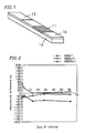
- Figure 1 is a perspective view showing an
immunochromatographic device according to an embodiment of
the present invention.
- Figure 2 is a graph showing the storage stability
at 4°C of the immunochromatographic device of an embodiment
of the present invention.
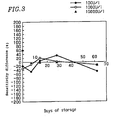
- Figure 3 is a graph showing the storage stability
at 4°C of the immunochromatographic device of a comparative
example.
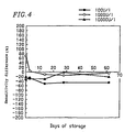
- Figure 4 is a graph showing the storage stability
at 25°C of the immunochromatographic device of an embodiment
of the present invention.
- Figure 5 is a graph showing the storage stability
at 25°C of the immunochromatographic device of the comparative
example.
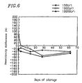
- Figure 6 is a graph showing the storage stability
at 40°C of the immunochromatographic device of an embodiment
of the present invention.
- Figure 7 is a graph showing the storage stability
at 40°C of the immunochromatographic device of a comparative
example.
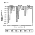
- Figure 8 is a graph showing degradation of the
intensity of a coloration for various sucrose concentrations
due to storage at 40°C for one month.
- Figure 9 is a diagram showing the storage stability
at 25°C of the sensitivity of an immunochromatographic device
according to Example 3.
- Figure 10 is a diagram showing the precision (CV
value) of the storage stability at 25°C of the
immunochromatographic device of Example 3.
- Figure 11 is a diagram showing the storage stability
at 25°C of an immunochromatographic device of a comparative
example.
- Figure 12 is a diagram showing the precision (CV
value) of the storage stability at 25°C of the
immunochromatographic device of a comparative example.
- Figure 13 is a diagram showing stability indexes
representing the degradation rates of the example of the
present invention (Figure 9), where measured values at day
14 are used as a reference.
- Figure 14 is a diagram showing stability indexes
representing the degradation rates of the comparative example
(Figure 11), where measured values at day 14 are used as
a reference.
-
(Description of Reference Numerals)
-
- 10 Sample introduction region
- 11 Labeling region
- 12 Determination region
- 13 Substrate
-
BEST MODE FOR CARRYING OUT THE INVENTION
-
It should be understood throughout the present
specification that articles for a singular form (e.g., "a",
"an", "the", etc. in English; "ein", "der", "das", "die".
etc. and their inflections in German; "un", "une", "le",
"la", etc. in French; and articles, adjectives, etc. in other
languages) include the concept of their plurality unless
otherwise mentioned. It should be also understood that the
terms as used herein have definitions typically used in the
art unless otherwise mentioned.
-
The present invention provides an extracorporeal
diagnostic for measuring a substance tobe tested in a specimen.
The extracorporeal diagnostic comprises:
- 1) a reagent which specifically reacts with the
substance to be tested; and
- 2) a hydrophilic material.
-
-
"Extracorporeal diagnostic" as used herein refers
to a product which can be monitored from the outside of the
body of a subject on at least one specific biological parameter.
The extracorporeal diagnostic may be in any form depending
on the situation, e.g., may be in the form of a composition
or a device.
-
"Hydrophilic material" as used herein refers to a
material comprising an atom group having a strong affinity
for water molecules. It is herein necessary for the
hydrophilic material not to destroy the three-dimensional
structure of proteins. Examples of such a hydrophilic
material include a side chain of an amino acid, such as lysine,
arginine, glutamic acid, aspartic acid, and the like; the
phosphate group of a nucleic acid; a side chain of an amino
acid, such as serine, threonine, and the like; the hydroxyl
group of a sugar or a sugar derivative; and the like.
-
In one embodiment, the above-described hydrophilic
material comprises a compound comprising at least one
hydroxyl group and at least one aldehyde group or ketone
group. Therefore, in a preferred embodiment, the present
invention provides an extracorporeal diagnostic for
measuring a substance to be tested in a specimen. The
extracorporeal diagnostic comprises:
- 1) a reagent which specifically reacts with the
substance to be tested; and
- 2) a compound comprising at least one hydroxyl group
and at least one aldehyde group or ketone group.
-
-
In one preferred embodiment, the above-described
hydrophilic material may be a sugar or a sugar derivative.
-
Sugar as used herein refers to polyhydroxyaldehyde
or polyhydroxyketone comprising at least one hydroxyl group
and at least one aldehyde group or ketone group. As used
herein, sugar also refers to carbohydrate, and both are
interchangeably used.
-
Sugar used for the extracorporeal diagnostic of the
present invention may be any sugar that can be dissolved
in liquid. Examples of such a sugar include monosaccharides,
such as glucose, mannose, galactose, fructose, and the like,
and oligosaccharides, such as maltose, isomaltose,
cellobiose, lactose, sucrose, and the like. Further, the
sugar may include polysaccharides in which monosaccharides
or oligosaccharides are chemically linked together. The
sugar has stereoisomers. All of the stereoisomers can be
applied to the extracorporeal diagnostic of the present
invention. Among the sugars, sucrose is preferable.
-
A sugar derivative as used herein refers to a sugar
whose substituents are substituted with other substituents,
and a sugar variant obtained by an oxidation-reduction
reaction of a sugar. Here, the substituent includes, but
is not limited to, alkyl, substituted alkyl, cycloalkyl,
substituted cycloalkyl, alkenyl, substituted alkenyl,
cycloalkenyl, substituted cycloalkenyl, alkynyl,
substituted alkynyl, alkoxy, substituted alkoxy,
carbocyclic group, substituted carbocyclic group,
heterocyclic group, substituted heterocyclic group, halogen,
hydroxy, substituted hydroxy, thiol, substituted thiol,
ciano, nitro, amino, substitutedamino, carboxy, substituted
carboxy, acyl, substituted acyl, thiocarboxy, substituted
thiocarboxy, amide, substituted amide, substituted carbonyl1
substituted thiocarbonyl, substituted sulfonyl and
substituted sulfinyl.
-
A sugar derivative used for the extracorporeal
diagnostic of the present invention may be any sugar
derivative which can be dissolved in liquid. Examples of
the sugar derivative include: components of organisms, such
as proteins, lipids, nucleic acids, and the like, modified
with monosaccharides, oligosaccharides or polysaccharides;
sugar alcohol; inositol; uronic acid; ascorbic acid; amino
sugar; sugar phosphate ester; naturally occurring
glycoprotein; and the like. Among these sugar derivatives,
sugar alcohol is preferable.
-
Sugars or sugar derivatives have hygroscopicity, a
low level of drying property, and an excellent level of
moisture retention property against temperature changes.
In this case, the moisture content of the extracorporeal
diagnostic can be moderately retained. Therefore, it is
possible to obtain an extracorporeal diagnostic having
storage stability with respect to high-precision
quantitative determination.
-
A sugar alcohol, which can be used in the
extracorporeal diagnostic of the present invention, includes
chain polyhydric alcohol obtained by reducing the carbonyl
group of aldose or ketose, or stereoisomers thereof,
including, for example, glycerol, erythritol, threitol,
ribitol, arabinitol, xylitol, allitol, sorbitol, mannitol,
iditol, dulcitol and talitol, or, the stereoisomers of
glycerol, erythritol, threitol, ribitol, arabinitol,
xylitol, allitol, sorbitol, mannitol, iditol, dulcitol and
talitol. Further, the sugar alcohol may include a compound
in which at least two of the above-described chain polyhydric
alcohols or stereoisomers thereof, are chemically linked
together, including, for example, a compound in which two
of glycerol, erythritol, threitol, ribitol, arabinitol,
xylitol, allitol, sorbitol, mannitol, iditol, dulcitol or
talitol, or D- and L-stereoisomers of glycerol, erythritol,
threitol, ribitol, arabinitol, xylitol, allitol, sorbitol,
mannitol, iditol, dulcitol or talitol, are chemically linked
together. Furthermore, the sugar alcohol may include a
compound in which the above-described chain polyhydric
alcohols or stereoisomers thereof, including, for example,
glycerol, erythritol, threitol, ribitol, arabinitol,
xylitol, allitol, sorbitol, mannitol, iditol, dulcitol or
talitol, or stereoisomers of glycerol, erythritol, threitol,
ribitol, arabinitol, xylitol, allitol, sorbitol, mannitol,
iditol, dulcitol or talitol, are partially or entirely linked
with naturally occurring sugar or non-naturally occurring
and artificially synthesized sugar, or stereoisomers of
naturally occurring sugar or non-naturally occurring and
artificially synthesized sugar via an alcohol group. Among
them, sugar alcohol is preferably sorbitol.
-
Therefore, preferably, the above-described material
may comprise sugar alcohol or derivatives thereof. More
preferably, the above-described material may comprise
sucrose or sorbitol. It may be preferable that the
above-described material comprises sucrose and sorbitol.
-
In one embodiment, the above-described material may
be present at a concentration of more than about 1 w/v%.
More preferably, the above-described material is present
at a concentration of at least about 3 w/v%. Even more
preferably, the above-described material is present at a
concentration of about 3 w/v% to about 10 w/v%. The content
concentration of the above-described material is not limited
to these ranges, and includes, for example, about 1 w/v%,
about 1.5 w/v%, about 2 w/v%, about 2.5 w/v%, about 3 w/v%,
about 4 w/v%, about 5 w/v%, about 6 w/v%, about 7 w/v%, about
8 w/v%, about 9 w/v%, about 10 w/v%, about 15 w/v%, and the
like as a lower limit. The upper limit of the content
concentration of the above-described material includes, but
is not limited to, for example, about 1.5 w/v%, about 2 w/v%,
about 2.5 w/v%, about 3 w/v%, about 4 w/v%, about 5 w/v%,
about 6 w/v%, about w/v%, about 8 w/v%, about 9 w/v%, about
10 w/v%, about 12.5 w/v%, about 15 w/v%, about 17.5 w/v%,
about 20 w/v%, about 30 w/v%, about 40 w/v%, about 50 w/v%,
and the like. The above-described range may be any
combination of the above-described lower and upper limits.
-
A reagent which can be used in the present invention,
is any reagent which can specifically react with a substance
to be tested, including, for example, antibodies, antigens,
avidin, biotin, nucleic acids, and the like. Among them,
antigens or antibodies are preferable. Therefore, in
another aspect, the reagent used in the present invention
may be an antibody or antigen, or a derivative thereof.
"Specifically react with" refers to an interaction with an
intended object which is stronger than an interaction with
other objects.
-
"Antigen" as used herein refers to any substance which
raises an antibody. Such an antigen includes haptens,
proteins, bacteria, viruses, anti-viral antibodies, and the
like. Here, examples of the haptens include low molecular
weight compounds, such as dioxin, amphetamine,
methamphetamine, estradiol, and the like. The proteins
include hemoglobin, albumin, hemoglobin Alc
(glycohemoglobin), HDL (high density lipoprotein), LDL (low
density lipoprotein), HCV antibody (hepatitis C virus
antibody), HIV antibody (human immunodeficiency virus
antibody), CEA (carcinoembryonic antigen), AFP
(α-fetoprotein), CRP (C responsive protein), SAA (serum
amyloid A) , hCG (human chrionic gonadotropin), and the like.
The bacteria include bacteria belonging to the genus E. coli,
the genus Salmonella, the genus Staphylococcus aureus, or
the genus Vibrio. The viruses include HIV, HBs, and the like.
-
"Antibody" as used herein comprises an antibody
raised by the above-described antigens. The antibody is also
herein intended to include the entirety of the antibody,
and an immunogenic derivative and fragment thereof. The
immunogenic fragment of the antibody refers to any fragment
of the antibody having immunogenicity. Examples of the
immunogenic fragment of the antibody include, but are not
limited to, variable regions, such as Fab, F(ab)2' and the
like. Therefore, the immunogenic fragment of the antibody
may be any fragment as long as it can raise ah immune reaction.
-
In another embodiment, the extracorporeal
diagnostic of the present invention may further comprise
bovine serum albumin, casein, surfactant, or skim milk.
-
In the extracorporeal diagnostic of the present
invention in one aspect, the extracorporeal diagnostic
comprises a plurality of the above-described reagents. The
at least two reagents may comprise a first antibody or antigen,
or a derivative thereof, and a second antibody or antigen,
or a derivative thereof.
-
In a preferred embodiment, the above-described
reagent may be labeled with a colloidal particle, a latex
particle, a pigment, a micelle, an enzyme, a fluorescent
material or a phosphorescent material.
-
In one embodiment, the above-described reagent and
the above-described material may be retained on a support.
Preferably, the above-described reagent and the
above-described compound may be retained on other regions
of the above-described support.
-
In a preferred embodiment, the above-described
support may be a solid-phase matrix. The matrix may be in
the form of a layer (solid-phase matrix). Preferably, the
above-described support may comprise a porous material.
Here, preferably, the reagent and a sugar or a sugar derivative
are retained on the solid-phase matrix.
-
In general, the solid-phase matrix is any solid-phase
matrix which can absorb, retain and develop sample solution,
and can adsorb a biological material physically or chemically,
including, for example, glass fiber filter paper, membrane
filter, and the like.
-
The extracorporeal diagnostic of the present
invention comprises a sample introduction region, a labeling
region containing a first antibody or antigen which binds
to an antigen or antibody which is a substance to be tested,
and a determination region containing a second antibody or
antigen which binds to the antigen or antibody which is a
substance to be tested, as a solid-phase matrix. The
extracorporeal diagnostic is characterized in that: the
sample introduction region, the labeling region and the
determination region are arranged so that the sample solution
is introduced into the sample introduction region and is
then transferred via the labeling region to the determination
region; the sample introduction region, the labeling region
or the determination region contains a sugar or a sugar
derivative; and an antigen or antibody in the sample solution,
which is a substance to be tested, is measured based on a
specific binding reaction between antigen and antibody.
-
Thereby, the moisture content of at least the sample
introduction region, the labeling region or the determination
region is appropriately retained, so that development of
the sample solution to the carrier is uniform over time.
Therefore, it is possible to obtain an extracorporeal
diagnostic having storage stability with respect to
high-precision quantitative determination.
-
In a preferred embodiment, in the extracorporeal
diagnostic of the present invention, the above-described
support comprises a labeling region, a determination region
and a sample introduction region,
- 1) the labeling region contains the first antibody
or antigen which binds to the substance to be tested,
- 2) the determination region contains the second
antibody or antigen which binds to the substance to be tested,
and is disposed in fluid communication with the labeling
region,
- 3) the sample introduction region is disposed in
fluid communication with the labeling region.
-
-
In a preferred embodiment, the extracorporeal
diagnostic of the present invention may be, but is not limited
to, in the form of an extracorporeal diagnostic device.
Therefore, the extracorporeal diagnostic may also be in the
form of a composition.
-
Here, the labeling region preferably contains a sugar
or a sugar derivative. In this case, adsorption of the first
antibody (or antigen) into the solid-phase matrix is relaxed,
whereby dissolution of the first antibody (or antigen) is
uniform over time, and an extracorporeal diagnostic having
a higher level of storage stability can be obtained.
-
In one embodiment, the first antibody (or antigen)
may be labeled with a colloidal particle, a latex particle,
a pigment, or a micelle, and at least the labeling region
preferably contains a sugar or a sugar derivative.
-
A material for the sample introduction region may
be any material which can develop the sample solution at
an appropriate speed, including, for example, nitrocellulose
and glass filter paper.
-
Moreover, a material for the labeling region and the
determination region may be any material which can retain
the first and second antibodies (or antigens) and develop
the sample solution at an appropriate speed, including, for
example, nitrocellulose and glass filter paper.
-
In one embodiment of the present invention, for
example, an extracorporeal diagnostic is provided, which
is required when a substance to be tested is measured using
an automatic analyzer and which is prepared by adding a sugar.
or a sugar derivative to a liquid reagent contained in a
cuvette, an Eppendorf tube, or the like, followed by drying.
A variety of applications are expected when such an
extracorporeal diagnostic is used in measurement. For
example, a buffer solution or the like is poured into the
above-described container containing the lyophilized
product to dissolve the lyophilized product and thereafter,
the resultant solution is transferred to a cuvette for the
analyzer so that the solution can be measured. Alternatively,
the container containing the solution can be directly placed
in the analyzer and the analyzer can automatically pour a
buffer solution or the like into the container so that
measurement can be immediately started. With the dried
reagent-containing container, the user does not have to
prepare a reagent. Further, since the reagent is in a solid
state, the reagent is easy to handle unlike when it is in
a liquid state. If the container is disposable, washing the
container is not required, thereby expecting that handling
the container is easier. Furthermore, the container can be
stored at room temperature, thereby expecting that handling
the container is even easier.
-
In another embodiment of the present invention, an
extracorporeal diagnostic is provided, which is used in
utilizing an immune serum test method, such as
immunonephelometry, nephelometry, latex agglutination test,
radioimmunosorbent test, enzyme-linked immunosorbent assay,
immunofluorescence assay, chemiluminescence immunoassay,
particle coefficient immunoassay, and the like, and which
is prepared by adding a sugar or a sugar derivative to an
antibody or antigen solution contained in a cuvette, an
Eppendorf tube, or the like, followed by lyophilization.
The extracorporeal diagnostic can be handled in a manner
similar to the above-described usage of the automatic
analyzer when a reagent is handled and measured.
-
In one embodiment, the above-described compound may
be contained in at least one region selected from the group
consisting of the labeling region and the determination
region. Preferably, the compound may be contained in at least
the labeling region, and may be contained in all of the regions.
-
In one aspect, the present invention provides a method
for producing an extracorporeal diagnostic for measuring
a substance to be tested in a specimen. The method comprises:
- 1) providing a reagent which specifically reacts
with the substance to be tested; and
- 2) providing a compound comprising at least one
hydroxyl group and at least one aldehyde group or ketone
group.
-
-
In one embodiment, the extracorporeal diagnostic of
the present invention can be prepared by mixing a solution
of a reagent which specifically reacts with a substance to
be tested with a sugar or a sugar derivative, and air drying
or lyophilizing the solution.
-
When a reagent and a sugar or a sugar derivative are
retained on a solid-phase matrix, a solution of a reagent
which specifically reacts with a substance to be tested is
mixed with a sugar or a sugar derivative, and the resultant
solution is caused to permeate or be immobilized to the
solid-phasematrix, followed by air drying or lyophilization.
It should be noted that "permeate" herein indicates that
the reagent is retained in such a manner that the reagent
can be dissolved, and "immobilized" herein indicates that
the reagent is retained in such a manner that the reagent
cannot be dissolved.
-
Further, the extracorporeal diagnostic of the
present invention can be prepared by mixing a solution
containing a first antibody (or antigen) with a sugar or
a sugar derivative, causing the resultant solution to
permeate the labeling region, or/and mixing a solution
containing a second antibody (or antigen) with a sugar or
a sugar derivative, and causing the resultant solution to
be immobilized in the determination region, followed by air
drying or lyophilization.
-
In another aspect, the present invention provides
a method for detecting a substance to be tested in a specimen.
The method comprises the steps of:
- A) providing an extracorporeal diagnostic, the
extracorporeal diagnostic comprising:
- 1) a reagent which specifically reacts with the
substance to be tested; and
- 2) a compound comprising at least one hydroxyl
group and at least one aldehyde group or ketone group;
- B) providing the specimen to the extracorporeal
diagnostic;
- C) disposing the extracorporeal diagnostic under a
condition that the reagent specifically reacts with the
specimen; and
- D) detecting a signal caused by a specific reaction
of the reagent with the specimen.
-
-
In another aspect, the present invention relates to
use of the extracorporeal diagnostic of the present invention
for measuring the substance to be tested in the specimen.
-
With the diagnostic method or detection method of
the present invention, it is possible to detect, for example,
components in blood (e.g., HbAlc (glycohemoglobin), HDL (high
density lipoprotein), LDL (low density lipoprotein), HCV
antibody (hepatitis C virus antibody), HIV antibody (human
immunodeficiency virus antibody), CEA (carcinoembryonic
antigen), AFP (α-fetoprotein), CRP (C responsive protein),
SAA (serum amyloid A), etc.). To this end, for example, blood
is spotted onto an immunochromatographic device which is
used to detect or immobilize an antibody against the
above-described proteins, and the intensity of a coloration
after a predetermined time is determined by measuring
reflection absorbance, thereby making it possible to quantify
the concentration of the proteins.
-
The effects of the present invention include that
high-precision diagnosis and detection can be achieved after
long-term storage (e.g., one month). "Stability index" is
herein used as an index for evaluating precision after a
predetermined time. "Stability index" as used herein refers
to the sensitivity of an extracorporeal diagnostic relative
to a reference where the sensitivity of the extracorporeal
diagnostic 14 days after production thereof is the reference
(0). Therefore, when a 14-day sensitivity is -10 and a 30-day
sensitivity is 20, a corresponding stability index is +30.
It is desirable that the stability index is close to 0.
Further, it is also preferable that a variation in the
stability index at each measurement time point is small,
i.e., the stability index does not vary much with time. In
the present invention, the value of the stability index may
be preferably within ±20. More preferably, the stability
index may be within ±10. If the variation is within ±20,
since variations in the device have a larger influence, the
variation of the stability index cannot be simply said to
be caused by degradation. Such a range may be tolerable.
In another aspect, in the present invention, it is preferable
that the difference between the stability indexes at
measurement time points is within 20. More preferably, such
a difference may be within 10. When the storage stability
is evaluated, it is important to predict a result of a
subsequent measurement concerning measured data. In a
storage stability test, it is also important to predict a
result of measurement after a final measurement time point.
Therefore, it is desirable that the stability index does
not vary much with time, and particularly does not
continuously vary to a large extent. Conversely, when the
stability index for a certain concentration is beyond the
range within ±20 and the absolute value of the stability
index tends to increase with an increase in time, it can
be said that the possibility that subsequent measurement
produces unreliable data is high. Moreover, it is expected
that the absolute value of the stability index is beyond
20 with respect to other concentrations. Actually, a similar
tendency was observed in comparative examples herein
described.
-
Therefore, the present invention can provide
high-precision diagnosis and detection after long-term
storage including, but not limited to, for example, one month,
two months, three months, six months, one year, and the like.
-
Hereinafter, the present invention will be described
in more detail by way of examples. The examples below are
provided only for illustration purposes. Therefore, the
scope of the present invention is limited only by the claims,
but not by the examples.
EXAMPLES
-
Reagents, instruments, and systems used in the
examples below are those commonly used in the art. It should
be noted that an extracorporeal diagnostic will be described,
exemplifying an immunochromatographic device. The present
invention is not limited to the examples below.
(Example 1)
-
An immunochromatographic device for quantitatively
measuring hCG in human urine was prepared, in which human
urine and hCG were used as a sample solution and an antigen,
i.e., a substance to be tested, respectively, and sorbitol,
which is a sugar alcohol, was contained in a labeling region.
The storage stability of the device was assessed. Figure 1
shows a structure of an immunochromatographic device
according to Example 1.
(Preparation of nitrocellulose membrane)
-
The concentration of anti-hCG-β antibody as a second
antibody was adjusted using phosphate buffer solution
(pH 7.4) and thereafter, the resultant anti-hCG-β antibody
solution was applied using a solution discharging apparatus
onto a central region of a 150-µm thick nitrocellulose
membrane (manufactured by Millipore: high flow membrane)
into line, followed by drying. Thus, a determination
region 12 was prepared. Next, nitrocellulose membrane was
immersed in Tris(hydroxymethyl)aminomethane (hereinafter
referred to as Tris) - HCl buffer solution (pH 8.2) containing
1% skim milk for 30 minutes while shaking and thereafter,
was immersed in another Tris-HCl buffer solution (pH 8.2)
for 10 minutes while shaking. The thus-obtained
nitrocellulose membrane was dried at room temperature.
(Preparation of gold colloid sensitized anti-hCG-α
antibody solution)
-
Initially, 4 ml of 1% citric acid solution was quickly
added to 200 ml of refluxing 0.01% chloroauric acid solution,
thereby producing gold colloid. The gold colloid solution
was cooled at room temperature and was thereafter adjusted
to pH 9.0 by adding 0.2 M potassium carbonate solution. Next,
500 µl of PBS buffer solution containing 5 mg/ml anti-hCG-α
antibody was added as a first antibody, followed by shaking
for several minutes. Further, 20 ml of 10% bovine serum
albumin (hereinafter referred to as BSA) solution (pH 9.0)
was added. The thus-obtained reaction mixture containing
the gold colloid sensitized anti-hCG-α antibody was
centrifuged twice so as to remove unreacted anti-hCG-α
antibody and BSA. The thus-purified gold colloid sensitized
anti-hCG-α antibody was suspended in phosphate buffer
solution (pH 7.4) containing 10% sorbitol and passed through
a 0.8-µm filter (manufactured by ADVANTEC: DISMIC-25cs),
and stored at 4°C.
(Preparation of immunochromatographic device)
-
The prepared gold colloid sensitized anti-hCG-α
antibody solution was applied using a solution discharging
apparatus onto a portion of a position away from the
determination region 12 of the above-described
nitrocellulose membrane, followed by drying, thereby
producing a labeling region 11 on the nitrocellulose membrane.
Further, a portion of the nitrocellulose membrane, onto which
the gold colloid sensitized anti-hCG-α antibody solution
was not applied, was referred to as a sample introduction
region 10.
-
Finally, the above-described nitrocellulose
membrane was attached to a substrate 13 of white PET having
a thickness of 0.5 mm, and cut into 5 mm × 50 mm strips,
thereby producing immunochromatographic devices.
-
An immunochromatographic device, in which sorbitol
was not contained in a labeling region, was produced in the
same manner as that in Example 1, except that sorbitol was
not used in the step of preparing a gold colloid sensitized
anti-hCG-α antibody solution.
(Preparation of sample solution)
-
hCG solution having a known concentration was added
to human urine to adjust the concentration thereof to 100 U/l,
1000 U/l and 10000 U/l.
(Quantitative determination of hCG concentration)
-
Quantitative determination of hCG concentration in
sample solution was performed immediately after production
of an immunochromatographic device and 3 days, 14 days,
28 days and 62 days after the production for the
immunochromatographic device of Example 1, and immediately
after production of an immunochromatographic device and
7 days, 14 days, 28 days and 62 days after the production
for the immunochromatographic device of Comparative
Example 1. For this period, the immunochromatographic
devices were stored at 4°C, 25°C and 40°C for both Example 1
and Comparative Example 1.
-
In measurement, 40 µl of 100 U/l, 1000 U/l or
10000 U/l hCG solution was added to the sample introduction
region 10 of the immunochromatographic device prepared and
was allowed to develop on the membrane. Quantification of
the hCG concentration of a sample solution was performed
after 5 minutes by measuring a coloration in the
determination region 12 by reflectance absorbance at 520 nm
using a reflection absorption spectrophotometer
(manufactured by Shimadzu: CS9300).
-
Figure 2 shows the storage stability at 4°C of the
immunochromatographic device of Example 1. Figure 3 shows
the storage stability at 4°C of the immunochromatographic
device of Comparative Example 1. Figure 4 shows the storage
stability at 25°C of the immunochromatographic device of
Example 1. Figure 5 shows the storage stability at 25°C of
the immunochromatographic device of Comparative Example 1.
Figure 6 shows the storage stability at 40°C of the
immunochromatographic device of Example 1. Figure 7 shows
the storage stability at 40°C of the immunochromatographic
device of Comparative Example 1. In Figures 2 to 7, the
horizontal axis represents the number of days of storage
after production of an immunochromatographic device until
performance of measurement, while the vertical axis
represents the sensitivity difference between hCG
concentration obtained from reflectance absorbance at each
measurement and the actual hCG concentration of the sample
solution based on a hCG concentration-reflectance absorbance
calibration curve prepared immediately after the production
of the device. Filled circles indicate data for the sample
solution having a concentration of 100 U/l, open circles
indicate data for the sample solution having a concentration
of 1000 U/l, and triangles indicate data for the sample
solution having a concentration of 10000 U/l.
-
As can be seen from the figures, in the
immunochromatographic device of Comparative Example 1, the
greater the number of days of storage, the lower the
sensitivity. Such tendency is more significant as the
storage temperature is increased. In contrast to this, the
immunochromatographic device of Example 1 showed changes
in its sensitivity at any of the storage temperatures from
the day of the production until day 14, and substantially
stable sensitivity after day 14 until day 62. In the
immunochromatographic device of Example 1, the storage
stability at 40°C was maintained for one month from day 14
in a storage stability acceleration test shown at 40°C in
Figure 6. Therefore, about 1 to 1.5-year storage stability
can be guaranteed where the above-described period is
converted to storage stability at room temperature.
Moreover, precision can be guaranteed in terms of a variation
in CV value.
(Example 2) Optimization of sucrose concentration
-
An immunochromatographic device was produced in the
same manner as that of Example 1, except that in the step
of preparing a gold colloid sensitized anti-hCG-α antibody
solution, 0, 1, 3, 5, 7 or 10% sucrose was added to the gold
colloid sensitized anti-hCG-α antibody solution under
respective conditions.
(Preparation of sample solution)
-
hCG solution having a known concentration was added
to serum to adjust the concentration thereof to 0.01, 0.1,
and 1 mg/dl.
(Quantitative determination of hCG concentration)
-
The immunochromatographic devices carrying
respective sucrose concentrations in Example 2 were stored
at 40°C for 30 days. The measurement results of the
immunochromatographic devices were assessed immediately
after the production of the devices and 30 days after
production so as to determine an optimum sucrose
concentration. According to the results of Example 1
(Figures 2 to 7), if a degradation in sensitivity after
storage at 40°C for one month can be suppressed within around
-20%, 1.5-year storage stability at 4°C and 25°C can be
guaranteed.
-
In measurement, 40 µl of 0.01, 0.1 or 1 mg/dl hCG
solution was added to the sample introduction region 10 of
the prepared immunochromatographic device and was allowed
to develop on the membrane. Quantification of the hCG
concentration of a sample solution was performed after
5 minutes by measuring a coloration in the determination
region 12 by reflectance absorbance at 520 nm using a
reflection absorption spectrophotometer (manufactured by
Shimadzu: CS9300).
-
Figure 8 shows the degradation rate of the
immunochromatographic device of Example 2 after storage at
40°C for one month where the intensity of a coloration
immediately after the production of the device is used as
a reference. According to the above-described results, the
device having a sucrose concentration of no less than 3%
could achieve a coloration whose intensity could be
suppressed by only around -20% despite harsh conditions,
i.e., storage at 40°C.
-
In Example 2, since the specimen is serum, it has
a certain level of viscosity which is expected to have an
adverse influence on the precision of quantitative
determination. Therefore, it is preferable that sugar
concentration is as low as possible within a range in which
the degradation of the intensity of a coloration can be
suppressed.
(Example 3) Performance of storage stability test
-
Next, immunochromatographic devices for measuring
hCG, some having a sugar concentration of 5% and the others
containing no sugar, were prepared and subjected to a
real-time storage stability test. The test is shown in
Example 3.
-
The immunochromatographic devices of Example 3 were
produced in the same manner as that of Example 2, except
that in the step of adjusting the gold colloid sensitized
anti-hCG-α antibody solution, sucrose was added to the
solution to 5% or no sucrose was added. One of the
thus-produced devices which did not contain sucrose in its
labeling region was used as a Comparative Example, while
another which contained 5% w/v sucrose was used as an Example.
These devices will be described in detail below.
(Quantitative determination of hCG concentration)
-
Quantitative determination of hCG concentration in
a sample was performed immediately after production of the
immunochromatographic device of Example 3 and after 3 days,
7 days, 14 days, 25 days, 52 days, and 80 days of storage
after the production. As for the immunochromatographic
device of the Comparative Example, quantitative
determination was performed immediately after the production
of the device and after 3 days, 7 days, 14 days, 26 days,
and 54 days of storage after the production. The storage
temperature was 25°C.
-
In measurement, 40 µl of 0.01, 0.1 or 1 mg/dl hCG
solution was added to the sample introduction region 10 of
the prepared immunochromatographic device and was allowed
to develop on the membrane. Quantification of the hCG
concentration of a sample solution was performed after
5 minutes by measuring a coloration in the determination
region 12 by reflectance absorbance at 520 nm using a
reflection absorption spectrophotometer (manufactured by
Shimadzu: CS9300).
-
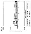
Figure 9 shows the storage stability at 25°C of the
immunochromatographic device of Example 3. Figure 10 shows
the precision (CV value) of the storage stability at 25°C
of the immunochromatographic device of Example 3.
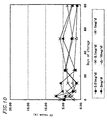
Figure 11 shows the storage stability at 25°C of the
immunochromatographic device of the Comparative Example.
Figure 12 shows the precision (CV value) of the storage
stability at 25°C of the immunochromatographic device of the
Comparative Example. In Figures 9 and 10, the horizontal
axis represents the number of days of storage after production
of an immunochromatographic device until performance of
measurement, while the vertical axis represents a degradation
rate with respect to the intensity of a coloration immediately
after the production of the device. In Figures 11 and 12,
the horizontal axis represents the number of days of storage
after production of an immunochromatographic device until
performance of measurement, while the vertical axis
represents CV value which is an index of the precision of
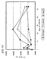
quantification. Further, Figures 13 and 14 show stability
indexes representing the degradation rates of Example 3 and
the Comparative Example, respectively, where measured values
at day 14 were used as a reference.
-
As can be seen from the figures, in the
immunochromatographic device of the Comparative Example,
the greater the number of days of storage, the lower the
sensitivity. In contrast to this, the
immunochromatographic device of Example 3 showed a
degradation in its sensitivity from the day of the production
until day 14, and substantially stable sensitivity after
day 14. Concerning the CV value, whereas the Comparative
Example showed a degradation in performance, the
immunochromatographic device of Example 3 had a degradation
rate within around 5% and the quantification precision was
guaranteed.
-
Particularly, the immunochromatographic device will
be discussed regarding the stability index. In Example 3
(Figure 13), the stability index was maintained within ±20,
and there was little variation in sensitivity between
measurement results at three time points ( days 25, 52 and
80). The possibility that degradation will not occur can
be said to be high for subsequent measurements. On the other
hand, in the Comparative Example (Figure 14), although the
stability index was maintained within ±20 at the most
concentrations, a variation (reduction) of up to 70% at the
maximum (0.01 mg/dl) was observed between day 26 and day
54. A similar variation (reduction) tendency is seen for
the other concentrations. Therefore, it is predicted that
stability index will reach an undesirable range in subsequent
measurements. As described above, it was demonstrated that
the extracorporeal diagnostic of the present invention
provides precise results for a considerably long term.
INDUSTRIAL APPLICABILITY
-
According to the present invention, the storage
stability of an extracorporeal diagnostic can be improved
by adding a sugar or a sugar derivative, even when quantitative
determination is performed. The extracorporeal diagnostic
of the present invention provides high reliability and ease
of handling to the user, and can be utilized not only in
clinical tests but also in POC.