EP1283878B1 - Receptor-based interaction trap - Google Patents
Receptor-based interaction trap Download PDFInfo
- Publication number
- EP1283878B1 EP1283878B1 EP01960237A EP01960237A EP1283878B1 EP 1283878 B1 EP1283878 B1 EP 1283878B1 EP 01960237 A EP01960237 A EP 01960237A EP 01960237 A EP01960237 A EP 01960237A EP 1283878 B1 EP1283878 B1 EP 1283878B1
- Authority
- EP
- European Patent Office
- Prior art keywords
- receptor
- binding
- lepr
- polypeptide
- psv
- Prior art date
- Legal status (The legal status is an assumption and is not a legal conclusion. Google has not performed a legal analysis and makes no representation as to the accuracy of the status listed.)
- Expired - Lifetime
Links
- 230000003993 interaction Effects 0.000 title description 60
- 108090000765 processed proteins & peptides Proteins 0.000 claims abstract description 121
- 102000004196 processed proteins & peptides Human genes 0.000 claims abstract description 119
- 229920001184 polypeptide Polymers 0.000 claims abstract description 117
- 230000027455 binding Effects 0.000 claims abstract description 74
- 230000001086 cytosolic effect Effects 0.000 claims abstract description 62
- 238000000034 method Methods 0.000 claims abstract description 40
- 239000003446 ligand Substances 0.000 claims abstract description 31
- 108020001756 ligand binding domains Proteins 0.000 claims abstract description 30
- 210000004027 cell Anatomy 0.000 claims description 157
- 102000005962 receptors Human genes 0.000 claims description 130
- 108020003175 receptors Proteins 0.000 claims description 130
- 239000013598 vector Substances 0.000 claims description 106
- 108010019813 leptin receptors Proteins 0.000 claims description 75
- 230000004913 activation Effects 0.000 claims description 58
- 239000012634 fragment Substances 0.000 claims description 58
- 230000026731 phosphorylation Effects 0.000 claims description 41
- 238000006366 phosphorylation reaction Methods 0.000 claims description 41
- 230000004048 modification Effects 0.000 claims description 28
- 238000012986 modification Methods 0.000 claims description 28
- 230000001419 dependent effect Effects 0.000 claims description 27
- 238000010276 construction Methods 0.000 claims description 17
- NRYBAZVQPHGZNS-ZSOCWYAHSA-N leptin Chemical class O=C([C@H](CO)NC(=O)[C@H](CC(C)C)NC(=O)[C@H](CC(O)=O)NC(=O)[C@H](CC(C)C)NC(=O)[C@H](CCC(N)=O)NC(=O)[C@H](CC=1C2=CC=CC=C2NC=1)NC(=O)[C@H](CC(C)C)NC(=O)[C@@H](NC(=O)[C@H](CC(O)=O)NC(=O)[C@H](CCC(N)=O)NC(=O)[C@H](CC(C)C)NC(=O)[C@H](CO)NC(=O)CNC(=O)[C@H](CCC(N)=O)NC(=O)[C@@H](N)CC(C)C)CCSC)N1CCC[C@H]1C(=O)NCC(=O)N[C@@H](CS)C(O)=O NRYBAZVQPHGZNS-ZSOCWYAHSA-N 0.000 claims description 15
- 230000004927 fusion Effects 0.000 claims description 13
- 230000004850 protein–protein interaction Effects 0.000 claims description 12
- OUYCCCASQSFEME-QMMMGPOBSA-N L-tyrosine Chemical compound OC(=O)[C@@H](N)CC1=CC=C(O)C=C1 OUYCCCASQSFEME-QMMMGPOBSA-N 0.000 claims description 10
- 210000003527 eukaryotic cell Anatomy 0.000 claims description 9
- OUYCCCASQSFEME-UHFFFAOYSA-N tyrosine Natural products OC(=O)C(N)CC1=CC=C(O)C=C1 OUYCCCASQSFEME-UHFFFAOYSA-N 0.000 claims description 9
- 230000008859 change Effects 0.000 claims description 7
- 102000037865 fusion proteins Human genes 0.000 claims description 6
- 108020001507 fusion proteins Proteins 0.000 claims description 6
- 230000021736 acetylation Effects 0.000 claims description 4
- 238000006640 acetylation reaction Methods 0.000 claims description 4
- 230000010933 acylation Effects 0.000 claims description 4
- 238000005917 acylation reaction Methods 0.000 claims description 4
- 239000013599 cloning vector Substances 0.000 claims description 4
- 230000013595 glycosylation Effects 0.000 claims description 4
- 238000006206 glycosylation reaction Methods 0.000 claims description 4
- 230000011987 methylation Effects 0.000 claims description 4
- 238000007069 methylation reaction Methods 0.000 claims description 4
- 102000005861 leptin receptors Human genes 0.000 claims description 3
- 210000004962 mammalian cell Anatomy 0.000 claims description 3
- 230000001404 mediated effect Effects 0.000 claims description 3
- 230000002538 fungal effect Effects 0.000 claims description 2
- 230000006337 proteolytic cleavage Effects 0.000 claims description 2
- 238000000338 in vitro Methods 0.000 claims 1
- 102100031775 Leptin receptor Human genes 0.000 description 72
- 108020004414 DNA Proteins 0.000 description 62
- 230000002441 reversible effect Effects 0.000 description 47
- 230000000694 effects Effects 0.000 description 42
- OHCQJHSOBUTRHG-KGGHGJDLSA-N FORSKOLIN Chemical compound O=C([C@@]12O)C[C@](C)(C=C)O[C@]1(C)[C@@H](OC(=O)C)[C@@H](O)[C@@H]1[C@]2(C)[C@@H](O)CCC1(C)C OHCQJHSOBUTRHG-KGGHGJDLSA-N 0.000 description 40
- RXWNCPJZOCPEPQ-NVWDDTSBSA-N puromycin Chemical compound C1=CC(OC)=CC=C1C[C@H](N)C(=O)N[C@H]1[C@@H](O)[C@H](N2C3=NC=NC(=C3N=C2)N(C)C)O[C@@H]1CO RXWNCPJZOCPEPQ-NVWDDTSBSA-N 0.000 description 40
- 238000001890 transfection Methods 0.000 description 40
- 230000006698 induction Effects 0.000 description 37
- 102000004190 Enzymes Human genes 0.000 description 32
- 108090000790 Enzymes Proteins 0.000 description 32
- 108060001084 Luciferase Proteins 0.000 description 25
- 239000005089 Luciferase Substances 0.000 description 25
- 108090000623 proteins and genes Proteins 0.000 description 25
- 150000001875 compounds Chemical class 0.000 description 24
- 235000001014 amino acid Nutrition 0.000 description 21
- 150000001413 amino acids Chemical class 0.000 description 21
- 239000002299 complementary DNA Substances 0.000 description 21
- SUZLHDUTVMZSEV-UHFFFAOYSA-N Deoxycoleonol Natural products C12C(=O)CC(C)(C=C)OC2(C)C(OC(=O)C)C(O)C2C1(C)C(O)CCC2(C)C SUZLHDUTVMZSEV-UHFFFAOYSA-N 0.000 description 20
- OHCQJHSOBUTRHG-UHFFFAOYSA-N colforsin Natural products OC12C(=O)CC(C)(C=C)OC1(C)C(OC(=O)C)C(O)C1C2(C)C(O)CCC1(C)C OHCQJHSOBUTRHG-UHFFFAOYSA-N 0.000 description 20
- 229950010131 puromycin Drugs 0.000 description 20
- 230000000638 stimulation Effects 0.000 description 20
- 230000014509 gene expression Effects 0.000 description 19
- 101710152369 Interleukin-6 receptor subunit beta Proteins 0.000 description 18
- 230000019491 signal transduction Effects 0.000 description 18
- 239000002609 medium Substances 0.000 description 17
- 230000001177 retroviral effect Effects 0.000 description 17
- 102000003951 Erythropoietin Human genes 0.000 description 16
- 108090000394 Erythropoietin Proteins 0.000 description 16
- 229940105423 erythropoietin Drugs 0.000 description 16
- 238000002474 experimental method Methods 0.000 description 16
- 239000013612 plasmid Substances 0.000 description 16
- OXCMYAYHXIHQOA-UHFFFAOYSA-N potassium;[2-butyl-5-chloro-3-[[4-[2-(1,2,4-triaza-3-azanidacyclopenta-1,4-dien-5-yl)phenyl]phenyl]methyl]imidazol-4-yl]methanol Chemical compound [K+].CCCCC1=NC(Cl)=C(CO)N1CC1=CC=C(C=2C(=CC=CC=2)C2=N[N-]N=N2)C=C1 OXCMYAYHXIHQOA-UHFFFAOYSA-N 0.000 description 16
- 239000000203 mixture Substances 0.000 description 15
- 238000002703 mutagenesis Methods 0.000 description 15
- 231100000350 mutagenesis Toxicity 0.000 description 15
- 108010070955 beta C receptor Proteins 0.000 description 14
- 238000003780 insertion Methods 0.000 description 14
- 230000037431 insertion Effects 0.000 description 14
- 238000003757 reverse transcription PCR Methods 0.000 description 14
- 102000016267 Leptin Human genes 0.000 description 13
- 108010092277 Leptin Proteins 0.000 description 13
- 230000029087 digestion Effects 0.000 description 13
- 229940039781 leptin Drugs 0.000 description 13
- 239000013642 negative control Substances 0.000 description 13
- 238000003752 polymerase chain reaction Methods 0.000 description 13
- 102100024784 Suppressor of cytokine signaling 2 Human genes 0.000 description 12
- 101710137422 Suppressor of cytokine signaling 2 Proteins 0.000 description 12
- 239000006144 Dulbecco’s modified Eagle's medium Substances 0.000 description 11
- 238000005516 engineering process Methods 0.000 description 10
- 235000018102 proteins Nutrition 0.000 description 10
- 102000004169 proteins and genes Human genes 0.000 description 10
- 108700010039 chimeric receptor Proteins 0.000 description 9
- 231100000673 dose–response relationship Toxicity 0.000 description 9
- 238000001556 precipitation Methods 0.000 description 9
- 238000012216 screening Methods 0.000 description 9
- 235000002374 tyrosine Nutrition 0.000 description 9
- 108091093088 Amplicon Proteins 0.000 description 8
- 108020004705 Codon Proteins 0.000 description 8
- 238000006243 chemical reaction Methods 0.000 description 8
- 230000003834 intracellular effect Effects 0.000 description 8
- 108091000080 Phosphotransferase Proteins 0.000 description 7
- 108010017324 STAT3 Transcription Factor Proteins 0.000 description 7
- 102100024040 Signal transducer and activator of transcription 3 Human genes 0.000 description 7
- 230000008901 benefit Effects 0.000 description 7
- 230000002779 inactivation Effects 0.000 description 7
- 230000035772 mutation Effects 0.000 description 7
- 102000020233 phosphotransferase Human genes 0.000 description 7
- 230000007115 recruitment Effects 0.000 description 7
- 101001114111 Homo sapiens Protease-associated domain-containing protein 1 Proteins 0.000 description 6
- 102000004058 Leukemia inhibitory factor Human genes 0.000 description 6
- 108090000581 Leukemia inhibitory factor Proteins 0.000 description 6
- 102100023223 Protease-associated domain-containing protein 1 Human genes 0.000 description 6
- FAPWRFPIFSIZLT-UHFFFAOYSA-M Sodium chloride Chemical compound [Na+].[Cl-] FAPWRFPIFSIZLT-UHFFFAOYSA-M 0.000 description 6
- 238000013459 approach Methods 0.000 description 6
- 239000003795 chemical substances by application Substances 0.000 description 6
- 238000010494 dissociation reaction Methods 0.000 description 6
- 230000005593 dissociations Effects 0.000 description 6
- 208000015181 infectious disease Diseases 0.000 description 6
- 244000062804 prey Species 0.000 description 6
- 108091026890 Coding region Proteins 0.000 description 5
- YQYJSBFKSSDGFO-UHFFFAOYSA-N Epihygromycin Natural products OC1C(O)C(C(=O)C)OC1OC(C(=C1)O)=CC=C1C=C(C)C(=O)NC1C(O)C(O)C2OCOC2C1O YQYJSBFKSSDGFO-UHFFFAOYSA-N 0.000 description 5
- 238000012413 Fluorescence activated cell sorting analysis Methods 0.000 description 5
- 108010017213 Granulocyte-Macrophage Colony-Stimulating Factor Proteins 0.000 description 5
- 102100039620 Granulocyte-macrophage colony-stimulating factor Human genes 0.000 description 5
- 102100025087 Insulin receptor substrate 1 Human genes 0.000 description 5
- 108010002747 Pfu DNA polymerase Proteins 0.000 description 5
- 230000003321 amplification Effects 0.000 description 5
- 229930189065 blasticidin Natural products 0.000 description 5
- 239000006285 cell suspension Substances 0.000 description 5
- 239000013078 crystal Substances 0.000 description 5
- 230000037430 deletion Effects 0.000 description 5
- 238000012217 deletion Methods 0.000 description 5
- 230000010354 integration Effects 0.000 description 5
- 238000003199 nucleic acid amplification method Methods 0.000 description 5
- 238000000746 purification Methods 0.000 description 5
- 238000002741 site-directed mutagenesis Methods 0.000 description 5
- 238000010396 two-hybrid screening Methods 0.000 description 5
- 241001430294 unidentified retrovirus Species 0.000 description 5
- 101001077604 Homo sapiens Insulin receptor substrate 1 Proteins 0.000 description 4
- 238000012408 PCR amplification Methods 0.000 description 4
- 230000015572 biosynthetic process Effects 0.000 description 4
- 239000000872 buffer Substances 0.000 description 4
- 239000001506 calcium phosphate Substances 0.000 description 4
- 229910000389 calcium phosphate Inorganic materials 0.000 description 4
- 235000011010 calcium phosphates Nutrition 0.000 description 4
- 238000010367 cloning Methods 0.000 description 4
- 238000010790 dilution Methods 0.000 description 4
- 239000012895 dilution Substances 0.000 description 4
- 230000008030 elimination Effects 0.000 description 4
- 238000003379 elimination reaction Methods 0.000 description 4
- 230000000670 limiting effect Effects 0.000 description 4
- 239000006166 lysate Substances 0.000 description 4
- 239000003550 marker Substances 0.000 description 4
- 150000007523 nucleic acids Chemical group 0.000 description 4
- 238000002823 phage display Methods 0.000 description 4
- 150000003384 small molecules Chemical class 0.000 description 4
- QORWJWZARLRLPR-UHFFFAOYSA-H tricalcium bis(phosphate) Chemical compound [Ca+2].[Ca+2].[Ca+2].[O-]P([O-])([O-])=O.[O-]P([O-])([O-])=O QORWJWZARLRLPR-UHFFFAOYSA-H 0.000 description 4
- QKNYBSVHEMOAJP-UHFFFAOYSA-N 2-amino-2-(hydroxymethyl)propane-1,3-diol;hydron;chloride Chemical compound Cl.OCC(N)(CO)CO QKNYBSVHEMOAJP-UHFFFAOYSA-N 0.000 description 3
- 101100297347 Caenorhabditis elegans pgl-3 gene Proteins 0.000 description 3
- 102000014914 Carrier Proteins Human genes 0.000 description 3
- 102100025064 Cellular tumor antigen p53 Human genes 0.000 description 3
- KCXVZYZYPLLWCC-UHFFFAOYSA-N EDTA Chemical compound OC(=O)CN(CC(O)=O)CCN(CC(O)=O)CC(O)=O KCXVZYZYPLLWCC-UHFFFAOYSA-N 0.000 description 3
- COLNVLDHVKWLRT-QMMMGPOBSA-N L-phenylalanine Chemical compound OC(=O)[C@@H](N)CC1=CC=CC=C1 COLNVLDHVKWLRT-QMMMGPOBSA-N 0.000 description 3
- 108091028043 Nucleic acid sequence Proteins 0.000 description 3
- 102100027913 Peptidyl-prolyl cis-trans isomerase FKBP1A Human genes 0.000 description 3
- 238000012300 Sequence Analysis Methods 0.000 description 3
- 108010006877 Tacrolimus Binding Protein 1A Proteins 0.000 description 3
- 108020005038 Terminator Codon Proteins 0.000 description 3
- 241000700605 Viruses Species 0.000 description 3
- 238000003556 assay Methods 0.000 description 3
- 239000011324 bead Substances 0.000 description 3
- 108091008324 binding proteins Proteins 0.000 description 3
- 101150102092 ccdB gene Proteins 0.000 description 3
- 230000002255 enzymatic effect Effects 0.000 description 3
- 239000012145 high-salt buffer Substances 0.000 description 3
- 238000011534 incubation Methods 0.000 description 3
- 230000005764 inhibitory process Effects 0.000 description 3
- 238000006386 neutralization reaction Methods 0.000 description 3
- 238000010899 nucleation Methods 0.000 description 3
- 238000011084 recovery Methods 0.000 description 3
- 108091008146 restriction endonucleases Proteins 0.000 description 3
- 238000012163 sequencing technique Methods 0.000 description 3
- 230000011664 signaling Effects 0.000 description 3
- 239000011780 sodium chloride Substances 0.000 description 3
- 238000010561 standard procedure Methods 0.000 description 3
- HBZBAMXERPYTFS-SECBINFHSA-N (4S)-2-(6,7-dihydro-5H-pyrrolo[3,2-f][1,3]benzothiazol-2-yl)-4,5-dihydro-1,3-thiazole-4-carboxylic acid Chemical compound OC(=O)[C@H]1CSC(=N1)c1nc2cc3CCNc3cc2s1 HBZBAMXERPYTFS-SECBINFHSA-N 0.000 description 2
- 108091032973 (ribonucleotides)n+m Proteins 0.000 description 2
- 101710176122 28 kDa heat- and acid-stable phosphoprotein Proteins 0.000 description 2
- 102100039377 28 kDa heat- and acid-stable phosphoprotein Human genes 0.000 description 2
- 102000002260 Alkaline Phosphatase Human genes 0.000 description 2
- 108020004774 Alkaline Phosphatase Proteins 0.000 description 2
- 241000283707 Capra Species 0.000 description 2
- 102000004127 Cytokines Human genes 0.000 description 2
- 108090000695 Cytokines Proteins 0.000 description 2
- IGXWBGJHJZYPQS-SSDOTTSWSA-N D-Luciferin Chemical compound OC(=O)[C@H]1CSC(C=2SC3=CC=C(O)C=C3N=2)=N1 IGXWBGJHJZYPQS-SSDOTTSWSA-N 0.000 description 2
- 108010017826 DNA Polymerase I Proteins 0.000 description 2
- 102000004594 DNA Polymerase I Human genes 0.000 description 2
- 238000001712 DNA sequencing Methods 0.000 description 2
- CYCGRDQQIOGCKX-UHFFFAOYSA-N Dehydro-luciferin Natural products OC(=O)C1=CSC(C=2SC3=CC(O)=CC=C3N=2)=N1 CYCGRDQQIOGCKX-UHFFFAOYSA-N 0.000 description 2
- 108010046276 FLP recombinase Proteins 0.000 description 2
- BJGNCJDXODQBOB-UHFFFAOYSA-N Fivefly Luciferin Natural products OC(=O)C1CSC(C=2SC3=CC(O)=CC=C3N=2)=N1 BJGNCJDXODQBOB-UHFFFAOYSA-N 0.000 description 2
- 102000002464 Galactosidases Human genes 0.000 description 2
- 108010093031 Galactosidases Proteins 0.000 description 2
- YWAQATDNEKZFFK-BYPYZUCNSA-N Gly-Gly-Ser Chemical compound NCC(=O)NCC(=O)N[C@@H](CO)C(O)=O YWAQATDNEKZFFK-BYPYZUCNSA-N 0.000 description 2
- DHCLVCXQIBBOPH-UHFFFAOYSA-N Glycerol 2-phosphate Chemical compound OCC(CO)OP(O)(O)=O DHCLVCXQIBBOPH-UHFFFAOYSA-N 0.000 description 2
- 102100029217 High affinity cationic amino acid transporter 1 Human genes 0.000 description 2
- 101000987586 Homo sapiens Eosinophil peroxidase Proteins 0.000 description 2
- 101000920686 Homo sapiens Erythropoietin Proteins 0.000 description 2
- 101000871017 Homo sapiens Growth factor receptor-bound protein 2 Proteins 0.000 description 2
- 101000633751 Homo sapiens High affinity cationic amino acid transporter 1 Proteins 0.000 description 2
- 101000599056 Homo sapiens Interleukin-6 receptor subunit beta Proteins 0.000 description 2
- 102100034343 Integrase Human genes 0.000 description 2
- 108010002386 Interleukin-3 Proteins 0.000 description 2
- 102000000646 Interleukin-3 Human genes 0.000 description 2
- 108010002616 Interleukin-5 Proteins 0.000 description 2
- 102100039897 Interleukin-5 Human genes 0.000 description 2
- AZLASBBHHSLQDB-GUBZILKMSA-N Leu-Ile Chemical group CC[C@H](C)[C@@H](C(O)=O)NC(=O)[C@@H](N)CC(C)C AZLASBBHHSLQDB-GUBZILKMSA-N 0.000 description 2
- DDWFXDSYGUXRAY-UHFFFAOYSA-N Luciferin Natural products CCc1c(C)c(CC2NC(=O)C(=C2C=C)C)[nH]c1Cc3[nH]c4C(=C5/NC(CC(=O)O)C(C)C5CC(=O)O)CC(=O)c4c3C DDWFXDSYGUXRAY-UHFFFAOYSA-N 0.000 description 2
- TWRXJAOTZQYOKJ-UHFFFAOYSA-L Magnesium chloride Chemical compound [Mg+2].[Cl-].[Cl-] TWRXJAOTZQYOKJ-UHFFFAOYSA-L 0.000 description 2
- 101710143111 Mothers against decapentaplegic homolog 3 Proteins 0.000 description 2
- 101710143112 Mothers against decapentaplegic homolog 4 Proteins 0.000 description 2
- 241001529936 Murinae Species 0.000 description 2
- 229930193140 Neomycin Natural products 0.000 description 2
- 108091034117 Oligonucleotide Proteins 0.000 description 2
- 102000004160 Phosphoric Monoester Hydrolases Human genes 0.000 description 2
- 108090000608 Phosphoric Monoester Hydrolases Proteins 0.000 description 2
- 108010092799 RNA-directed DNA polymerase Proteins 0.000 description 2
- 238000010240 RT-PCR analysis Methods 0.000 description 2
- 102000018120 Recombinases Human genes 0.000 description 2
- 108010091086 Recombinases Proteins 0.000 description 2
- 108700008625 Reporter Genes Proteins 0.000 description 2
- 102000014400 SH2 domains Human genes 0.000 description 2
- 108050003452 SH2 domains Proteins 0.000 description 2
- 102000049939 Smad3 Human genes 0.000 description 2
- 102000049937 Smad4 Human genes 0.000 description 2
- 239000004098 Tetracycline Substances 0.000 description 2
- 238000004458 analytical method Methods 0.000 description 2
- 210000004899 c-terminal region Anatomy 0.000 description 2
- 238000010805 cDNA synthesis kit Methods 0.000 description 2
- 238000012761 co-transfection Methods 0.000 description 2
- 230000009089 cytolysis Effects 0.000 description 2
- 210000000805 cytoplasm Anatomy 0.000 description 2
- 238000001514 detection method Methods 0.000 description 2
- 238000002825 functional assay Methods 0.000 description 2
- 230000002068 genetic effect Effects 0.000 description 2
- BRZYSWJRSDMWLG-CAXSIQPQSA-N geneticin Chemical compound O1C[C@@](O)(C)[C@H](NC)[C@@H](O)[C@H]1O[C@@H]1[C@@H](O)[C@H](O[C@@H]2[C@@H]([C@@H](O)[C@H](O)[C@@H](C(C)O)O2)N)[C@@H](N)C[C@H]1N BRZYSWJRSDMWLG-CAXSIQPQSA-N 0.000 description 2
- 230000012010 growth Effects 0.000 description 2
- 102000044890 human EPO Human genes 0.000 description 2
- 102000054653 human GRB2 Human genes 0.000 description 2
- 238000001114 immunoprecipitation Methods 0.000 description 2
- 230000006872 improvement Effects 0.000 description 2
- 230000001939 inductive effect Effects 0.000 description 2
- 230000002401 inhibitory effect Effects 0.000 description 2
- 230000004807 localization Effects 0.000 description 2
- 238000005259 measurement Methods 0.000 description 2
- 108020004999 messenger RNA Proteins 0.000 description 2
- 229960004927 neomycin Drugs 0.000 description 2
- 102000039446 nucleic acids Human genes 0.000 description 2
- 108020004707 nucleic acids Proteins 0.000 description 2
- 230000036961 partial effect Effects 0.000 description 2
- 230000037361 pathway Effects 0.000 description 2
- COLNVLDHVKWLRT-UHFFFAOYSA-N phenylalanine Natural products OC(=O)C(N)CC1=CC=CC=C1 COLNVLDHVKWLRT-UHFFFAOYSA-N 0.000 description 2
- 230000004962 physiological condition Effects 0.000 description 2
- 125000002924 primary amino group Chemical group [H]N([H])* 0.000 description 2
- 239000000047 product Substances 0.000 description 2
- 230000009467 reduction Effects 0.000 description 2
- 230000008521 reorganization Effects 0.000 description 2
- 150000003839 salts Chemical class 0.000 description 2
- 230000035945 sensitivity Effects 0.000 description 2
- 239000007787 solid Substances 0.000 description 2
- 238000012421 spiking Methods 0.000 description 2
- 238000003786 synthesis reaction Methods 0.000 description 2
- 230000008685 targeting Effects 0.000 description 2
- 229960002180 tetracycline Drugs 0.000 description 2
- 229930101283 tetracycline Natural products 0.000 description 2
- 235000019364 tetracycline Nutrition 0.000 description 2
- 150000003522 tetracyclines Chemical class 0.000 description 2
- 238000013518 transcription Methods 0.000 description 2
- 230000035897 transcription Effects 0.000 description 2
- 238000010361 transduction Methods 0.000 description 2
- 230000026683 transduction Effects 0.000 description 2
- 238000012546 transfer Methods 0.000 description 2
- 230000009466 transformation Effects 0.000 description 2
- 238000005406 washing Methods 0.000 description 2
- XLYOFNOQVPJJNP-UHFFFAOYSA-N water Substances O XLYOFNOQVPJJNP-UHFFFAOYSA-N 0.000 description 2
- 238000001086 yeast two-hybrid system Methods 0.000 description 2
- CXNPLSGKWMLZPZ-GIFSMMMISA-N (2r,3r,6s)-3-[[(3s)-3-amino-5-[carbamimidoyl(methyl)amino]pentanoyl]amino]-6-(4-amino-2-oxopyrimidin-1-yl)-3,6-dihydro-2h-pyran-2-carboxylic acid Chemical compound O1[C@@H](C(O)=O)[C@H](NC(=O)C[C@@H](N)CCN(C)C(N)=N)C=C[C@H]1N1C(=O)N=C(N)C=C1 CXNPLSGKWMLZPZ-GIFSMMMISA-N 0.000 description 1
- WHTVZRBIWZFKQO-AWEZNQCLSA-N (S)-chloroquine Chemical compound ClC1=CC=C2C(N[C@@H](C)CCCN(CC)CC)=CC=NC2=C1 WHTVZRBIWZFKQO-AWEZNQCLSA-N 0.000 description 1
- 102100034134 Activin receptor type-1B Human genes 0.000 description 1
- 108010045123 Blasticidin-S deaminase Proteins 0.000 description 1
- 108091035707 Consensus sequence Proteins 0.000 description 1
- 102000008130 Cyclic AMP-Dependent Protein Kinases Human genes 0.000 description 1
- 108010049894 Cyclic AMP-Dependent Protein Kinases Proteins 0.000 description 1
- 102000001493 Cyclophilins Human genes 0.000 description 1
- 108010068682 Cyclophilins Proteins 0.000 description 1
- 229930105110 Cyclosporin A Natural products 0.000 description 1
- PMATZTZNYRCHOR-CGLBZJNRSA-N Cyclosporin A Chemical compound CC[C@@H]1NC(=O)[C@H]([C@H](O)[C@H](C)C\C=C\C)N(C)C(=O)[C@H](C(C)C)N(C)C(=O)[C@H](CC(C)C)N(C)C(=O)[C@H](CC(C)C)N(C)C(=O)[C@@H](C)NC(=O)[C@H](C)NC(=O)[C@H](CC(C)C)N(C)C(=O)[C@H](C(C)C)NC(=O)[C@H](CC(C)C)N(C)C(=O)CN(C)C1=O PMATZTZNYRCHOR-CGLBZJNRSA-N 0.000 description 1
- 108010036949 Cyclosporine Proteins 0.000 description 1
- 102100032218 Cytokine-inducible SH2-containing protein Human genes 0.000 description 1
- 101710132484 Cytokine-inducible SH2-containing protein Proteins 0.000 description 1
- 241000283074 Equus asinus Species 0.000 description 1
- 241000588724 Escherichia coli Species 0.000 description 1
- 241000724791 Filamentous phage Species 0.000 description 1
- 102000016285 Guanine Nucleotide Exchange Factors Human genes 0.000 description 1
- 108010067218 Guanine Nucleotide Exchange Factors Proteins 0.000 description 1
- 101710088172 HTH-type transcriptional regulator RipA Proteins 0.000 description 1
- 229920000209 Hexadimethrine bromide Polymers 0.000 description 1
- 241000238631 Hexapoda Species 0.000 description 1
- 101000799189 Homo sapiens Activin receptor type-1B Proteins 0.000 description 1
- 101000852145 Homo sapiens Erythropoietin receptor Proteins 0.000 description 1
- 101001063991 Homo sapiens Leptin Proteins 0.000 description 1
- 101000942967 Homo sapiens Leukemia inhibitory factor Proteins 0.000 description 1
- 101000835893 Homo sapiens Mothers against decapentaplegic homolog 4 Proteins 0.000 description 1
- 101100356345 Homo sapiens RETREG2 gene Proteins 0.000 description 1
- 101000997832 Homo sapiens Tyrosine-protein kinase JAK2 Proteins 0.000 description 1
- 108010034219 Insulin Receptor Substrate Proteins Proteins 0.000 description 1
- 101710201824 Insulin receptor substrate 1 Proteins 0.000 description 1
- 108090001005 Interleukin-6 Proteins 0.000 description 1
- 101150009428 MAG2 gene Proteins 0.000 description 1
- 241000829100 Macaca mulatta polyomavirus 1 Species 0.000 description 1
- 108090000157 Metallothionein Proteins 0.000 description 1
- 241000713869 Moloney murine leukemia virus Species 0.000 description 1
- 101001063890 Mus musculus Leptin Proteins 0.000 description 1
- 101001129925 Mus musculus Leptin receptor Proteins 0.000 description 1
- 229910020700 Na3VO4 Inorganic materials 0.000 description 1
- 238000000636 Northern blotting Methods 0.000 description 1
- 102000007999 Nuclear Proteins Human genes 0.000 description 1
- 108010089610 Nuclear Proteins Proteins 0.000 description 1
- 241001494479 Pecora Species 0.000 description 1
- 229940124158 Protease/peptidase inhibitor Drugs 0.000 description 1
- 102000001253 Protein Kinase Human genes 0.000 description 1
- 239000012083 RIPA buffer Substances 0.000 description 1
- 101001035657 Rattus norvegicus 28 kDa heat- and acid-stable phosphoprotein Proteins 0.000 description 1
- 101000702488 Rattus norvegicus High affinity cationic amino acid transporter 1 Proteins 0.000 description 1
- 102100024733 Reticulophagy regulator 2 Human genes 0.000 description 1
- 102000000395 SH3 domains Human genes 0.000 description 1
- 108050008861 SH3 domains Proteins 0.000 description 1
- 108010044012 STAT1 Transcription Factor Proteins 0.000 description 1
- 229920002684 Sepharose Polymers 0.000 description 1
- 102100029904 Signal transducer and activator of transcription 1-alpha/beta Human genes 0.000 description 1
- 108010085012 Steroid Receptors Proteins 0.000 description 1
- 108010090804 Streptavidin Proteins 0.000 description 1
- 108010075383 Suppressor of Cytokine Signaling Proteins Proteins 0.000 description 1
- 102000008036 Suppressor of Cytokine Signaling Proteins Human genes 0.000 description 1
- 108010006785 Taq Polymerase Proteins 0.000 description 1
- 101710120037 Toxin CcdB Proteins 0.000 description 1
- 102100033444 Tyrosine-protein kinase JAK2 Human genes 0.000 description 1
- 102100033019 Tyrosine-protein phosphatase non-receptor type 11 Human genes 0.000 description 1
- 101710116241 Tyrosine-protein phosphatase non-receptor type 11 Proteins 0.000 description 1
- 102100021657 Tyrosine-protein phosphatase non-receptor type 6 Human genes 0.000 description 1
- 101710128901 Tyrosine-protein phosphatase non-receptor type 6 Proteins 0.000 description 1
- JLCPHMBAVCMARE-UHFFFAOYSA-N [3-[[3-[[3-[[3-[[3-[[3-[[3-[[3-[[3-[[3-[[3-[[5-(2-amino-6-oxo-1H-purin-9-yl)-3-[[3-[[3-[[3-[[3-[[3-[[5-(2-amino-6-oxo-1H-purin-9-yl)-3-[[5-(2-amino-6-oxo-1H-purin-9-yl)-3-hydroxyoxolan-2-yl]methoxy-hydroxyphosphoryl]oxyoxolan-2-yl]methoxy-hydroxyphosphoryl]oxy-5-(5-methyl-2,4-dioxopyrimidin-1-yl)oxolan-2-yl]methoxy-hydroxyphosphoryl]oxy-5-(6-aminopurin-9-yl)oxolan-2-yl]methoxy-hydroxyphosphoryl]oxy-5-(6-aminopurin-9-yl)oxolan-2-yl]methoxy-hydroxyphosphoryl]oxy-5-(6-aminopurin-9-yl)oxolan-2-yl]methoxy-hydroxyphosphoryl]oxy-5-(6-aminopurin-9-yl)oxolan-2-yl]methoxy-hydroxyphosphoryl]oxyoxolan-2-yl]methoxy-hydroxyphosphoryl]oxy-5-(5-methyl-2,4-dioxopyrimidin-1-yl)oxolan-2-yl]methoxy-hydroxyphosphoryl]oxy-5-(4-amino-2-oxopyrimidin-1-yl)oxolan-2-yl]methoxy-hydroxyphosphoryl]oxy-5-(5-methyl-2,4-dioxopyrimidin-1-yl)oxolan-2-yl]methoxy-hydroxyphosphoryl]oxy-5-(5-methyl-2,4-dioxopyrimidin-1-yl)oxolan-2-yl]methoxy-hydroxyphosphoryl]oxy-5-(6-aminopurin-9-yl)oxolan-2-yl]methoxy-hydroxyphosphoryl]oxy-5-(6-aminopurin-9-yl)oxolan-2-yl]methoxy-hydroxyphosphoryl]oxy-5-(4-amino-2-oxopyrimidin-1-yl)oxolan-2-yl]methoxy-hydroxyphosphoryl]oxy-5-(4-amino-2-oxopyrimidin-1-yl)oxolan-2-yl]methoxy-hydroxyphosphoryl]oxy-5-(4-amino-2-oxopyrimidin-1-yl)oxolan-2-yl]methoxy-hydroxyphosphoryl]oxy-5-(6-aminopurin-9-yl)oxolan-2-yl]methoxy-hydroxyphosphoryl]oxy-5-(4-amino-2-oxopyrimidin-1-yl)oxolan-2-yl]methyl [5-(6-aminopurin-9-yl)-2-(hydroxymethyl)oxolan-3-yl] hydrogen phosphate Polymers Cc1cn(C2CC(OP(O)(=O)OCC3OC(CC3OP(O)(=O)OCC3OC(CC3O)n3cnc4c3nc(N)[nH]c4=O)n3cnc4c3nc(N)[nH]c4=O)C(COP(O)(=O)OC3CC(OC3COP(O)(=O)OC3CC(OC3COP(O)(=O)OC3CC(OC3COP(O)(=O)OC3CC(OC3COP(O)(=O)OC3CC(OC3COP(O)(=O)OC3CC(OC3COP(O)(=O)OC3CC(OC3COP(O)(=O)OC3CC(OC3COP(O)(=O)OC3CC(OC3COP(O)(=O)OC3CC(OC3COP(O)(=O)OC3CC(OC3COP(O)(=O)OC3CC(OC3COP(O)(=O)OC3CC(OC3COP(O)(=O)OC3CC(OC3COP(O)(=O)OC3CC(OC3COP(O)(=O)OC3CC(OC3COP(O)(=O)OC3CC(OC3CO)n3cnc4c(N)ncnc34)n3ccc(N)nc3=O)n3cnc4c(N)ncnc34)n3ccc(N)nc3=O)n3ccc(N)nc3=O)n3ccc(N)nc3=O)n3cnc4c(N)ncnc34)n3cnc4c(N)ncnc34)n3cc(C)c(=O)[nH]c3=O)n3cc(C)c(=O)[nH]c3=O)n3ccc(N)nc3=O)n3cc(C)c(=O)[nH]c3=O)n3cnc4c3nc(N)[nH]c4=O)n3cnc4c(N)ncnc34)n3cnc4c(N)ncnc34)n3cnc4c(N)ncnc34)n3cnc4c(N)ncnc34)O2)c(=O)[nH]c1=O JLCPHMBAVCMARE-UHFFFAOYSA-N 0.000 description 1
- 239000012190 activator Substances 0.000 description 1
- 102000030621 adenylate cyclase Human genes 0.000 description 1
- 108060000200 adenylate cyclase Proteins 0.000 description 1
- 238000000246 agarose gel electrophoresis Methods 0.000 description 1
- 230000019552 anatomical structure morphogenesis Effects 0.000 description 1
- 239000003242 anti bacterial agent Substances 0.000 description 1
- 230000009830 antibody antigen interaction Effects 0.000 description 1
- 239000000427 antigen Substances 0.000 description 1
- 102000036639 antigens Human genes 0.000 description 1
- 108091007433 antigens Proteins 0.000 description 1
- 238000003149 assay kit Methods 0.000 description 1
- 102000005936 beta-Galactosidase Human genes 0.000 description 1
- 108010005774 beta-Galactosidase Proteins 0.000 description 1
- 239000011230 binding agent Substances 0.000 description 1
- 230000003115 biocidal effect Effects 0.000 description 1
- 230000004071 biological effect Effects 0.000 description 1
- 230000031018 biological processes and functions Effects 0.000 description 1
- CXNPLSGKWMLZPZ-UHFFFAOYSA-N blasticidin-S Natural products O1C(C(O)=O)C(NC(=O)CC(N)CCN(C)C(N)=N)C=CC1N1C(=O)N=C(N)C=C1 CXNPLSGKWMLZPZ-UHFFFAOYSA-N 0.000 description 1
- 235000014633 carbohydrates Nutrition 0.000 description 1
- 150000001720 carbohydrates Chemical class 0.000 description 1
- 230000030833 cell death Effects 0.000 description 1
- 239000013592 cell lysate Substances 0.000 description 1
- 210000000170 cell membrane Anatomy 0.000 description 1
- 239000002458 cell surface marker Substances 0.000 description 1
- 230000001413 cellular effect Effects 0.000 description 1
- 230000005754 cellular signaling Effects 0.000 description 1
- 239000013043 chemical agent Substances 0.000 description 1
- 229960003677 chloroquine Drugs 0.000 description 1
- WHTVZRBIWZFKQO-UHFFFAOYSA-N chloroquine Natural products ClC1=CC=C2C(NC(C)CCCN(CC)CC)=CC=NC2=C1 WHTVZRBIWZFKQO-UHFFFAOYSA-N 0.000 description 1
- 239000013611 chromosomal DNA Substances 0.000 description 1
- 210000000349 chromosome Anatomy 0.000 description 1
- 229960001265 ciclosporin Drugs 0.000 description 1
- 238000003776 cleavage reaction Methods 0.000 description 1
- 230000004186 co-expression Effects 0.000 description 1
- 238000000749 co-immunoprecipitation Methods 0.000 description 1
- 230000005757 colony formation Effects 0.000 description 1
- 230000008878 coupling Effects 0.000 description 1
- 238000010168 coupling process Methods 0.000 description 1
- 238000005859 coupling reaction Methods 0.000 description 1
- 108010012154 cytokine inducible SH2-containing protein Proteins 0.000 description 1
- 210000005220 cytoplasmic tail Anatomy 0.000 description 1
- 210000000172 cytosol Anatomy 0.000 description 1
- 238000013461 design Methods 0.000 description 1
- 238000006471 dimerization reaction Methods 0.000 description 1
- 108010030074 endodeoxyribonuclease MluI Proteins 0.000 description 1
- 108010048367 enhanced green fluorescent protein Proteins 0.000 description 1
- 230000010856 establishment of protein localization Effects 0.000 description 1
- 239000013604 expression vector Substances 0.000 description 1
- GNBHRKFJIUUOQI-UHFFFAOYSA-N fluorescein Chemical compound O1C(=O)C2=CC=CC=C2C21C1=CC=C(O)C=C1OC1=CC(O)=CC=C21 GNBHRKFJIUUOQI-UHFFFAOYSA-N 0.000 description 1
- 238000001943 fluorescence-activated cell sorting Methods 0.000 description 1
- 238000010230 functional analysis Methods 0.000 description 1
- 239000000499 gel Substances 0.000 description 1
- 238000009650 gentamicin protection assay Methods 0.000 description 1
- 102000035122 glycosylated proteins Human genes 0.000 description 1
- 108091005608 glycosylated proteins Proteins 0.000 description 1
- 238000013537 high throughput screening Methods 0.000 description 1
- 102000047742 human IRS1 Human genes 0.000 description 1
- 102000049953 human LEP Human genes 0.000 description 1
- 102000046645 human LIF Human genes 0.000 description 1
- 102000045603 human SMAD4 Human genes 0.000 description 1
- 238000001727 in vivo Methods 0.000 description 1
- 230000002458 infectious effect Effects 0.000 description 1
- 239000003112 inhibitor Substances 0.000 description 1
- 230000000977 initiatory effect Effects 0.000 description 1
- 108040006856 interleukin-3 receptor activity proteins Proteins 0.000 description 1
- 108040006859 interleukin-5 receptor activity proteins Proteins 0.000 description 1
- 238000002955 isolation Methods 0.000 description 1
- 101150066555 lacZ gene Proteins 0.000 description 1
- 238000002961 luciferase induction Methods 0.000 description 1
- 229910001629 magnesium chloride Inorganic materials 0.000 description 1
- JQAACYUZYRBHGG-QHTZZOMLSA-L magnesium;(2s)-5-oxopyrrolidine-2-carboxylate Chemical compound [Mg+2].[O-]C(=O)[C@@H]1CCC(=O)N1.[O-]C(=O)[C@@H]1CCC(=O)N1 JQAACYUZYRBHGG-QHTZZOMLSA-L 0.000 description 1
- 238000004519 manufacturing process Methods 0.000 description 1
- 239000000463 material Substances 0.000 description 1
- 230000007246 mechanism Effects 0.000 description 1
- 239000003068 molecular probe Substances 0.000 description 1
- 238000012544 monitoring process Methods 0.000 description 1
- 231100000219 mutagenic Toxicity 0.000 description 1
- 230000003505 mutagenic effect Effects 0.000 description 1
- 230000030147 nuclear export Effects 0.000 description 1
- 230000005937 nuclear translocation Effects 0.000 description 1
- 239000002773 nucleotide Substances 0.000 description 1
- 125000003729 nucleotide group Chemical group 0.000 description 1
- 210000004940 nucleus Anatomy 0.000 description 1
- 238000004806 packaging method and process Methods 0.000 description 1
- 239000002245 particle Substances 0.000 description 1
- 239000000137 peptide hydrolase inhibitor Substances 0.000 description 1
- 239000000816 peptidomimetic Substances 0.000 description 1
- 208000028591 pheochromocytoma Diseases 0.000 description 1
- 108091005981 phosphorylated proteins Proteins 0.000 description 1
- DCWXELXMIBXGTH-UHFFFAOYSA-N phosphotyrosine Chemical compound OC(=O)C(N)CC1=CC=C(OP(O)(O)=O)C=C1 DCWXELXMIBXGTH-UHFFFAOYSA-N 0.000 description 1
- 239000013600 plasmid vector Substances 0.000 description 1
- 230000004983 pleiotropic effect Effects 0.000 description 1
- 238000005498 polishing Methods 0.000 description 1
- 238000002264 polyacrylamide gel electrophoresis Methods 0.000 description 1
- 239000002244 precipitate Substances 0.000 description 1
- 238000012545 processing Methods 0.000 description 1
- 238000000159 protein binding assay Methods 0.000 description 1
- 108060006633 protein kinase Proteins 0.000 description 1
- 230000002797 proteolythic effect Effects 0.000 description 1
- 108010045647 puromycin N-acetyltransferase Proteins 0.000 description 1
- 238000010814 radioimmunoprecipitation assay Methods 0.000 description 1
- 239000011541 reaction mixture Substances 0.000 description 1
- 230000006798 recombination Effects 0.000 description 1
- 238000005215 recombination Methods 0.000 description 1
- 230000001105 regulatory effect Effects 0.000 description 1
- 230000010076 replication Effects 0.000 description 1
- 238000010839 reverse transcription Methods 0.000 description 1
- 210000003705 ribosome Anatomy 0.000 description 1
- 239000000523 sample Substances 0.000 description 1
- 230000007017 scission Effects 0.000 description 1
- 239000006152 selective media Substances 0.000 description 1
- 238000013207 serial dilution Methods 0.000 description 1
- 230000009870 specific binding Effects 0.000 description 1
- 102000005969 steroid hormone receptors Human genes 0.000 description 1
- 150000003431 steroids Chemical class 0.000 description 1
- 230000004936 stimulating effect Effects 0.000 description 1
- 239000000126 substance Substances 0.000 description 1
- 239000006228 supernatant Substances 0.000 description 1
- 230000004083 survival effect Effects 0.000 description 1
- 239000000725 suspension Substances 0.000 description 1
- 238000012360 testing method Methods 0.000 description 1
- 210000001519 tissue Anatomy 0.000 description 1
- 231100000167 toxic agent Toxicity 0.000 description 1
- 239000003440 toxic substance Substances 0.000 description 1
- 230000001052 transient effect Effects 0.000 description 1
- 102000027257 transmembrane receptors Human genes 0.000 description 1
- 108091008578 transmembrane receptors Proteins 0.000 description 1
- IHIXIJGXTJIKRB-UHFFFAOYSA-N trisodium vanadate Chemical compound [Na+].[Na+].[Na+].[O-][V]([O-])([O-])=O IHIXIJGXTJIKRB-UHFFFAOYSA-N 0.000 description 1
- 150000003668 tyrosines Chemical class 0.000 description 1
- 125000001493 tyrosinyl group Chemical group [H]OC1=C([H])C([H])=C(C([H])=C1[H])C([H])([H])C([H])(N([H])[H])C(*)=O 0.000 description 1
- 230000035899 viability Effects 0.000 description 1
- 239000013603 viral vector Substances 0.000 description 1
- 238000001262 western blot Methods 0.000 description 1
- 210000005253 yeast cell Anatomy 0.000 description 1
Images


Classifications
-
- C—CHEMISTRY; METALLURGY
- C12—BIOCHEMISTRY; BEER; SPIRITS; WINE; VINEGAR; MICROBIOLOGY; ENZYMOLOGY; MUTATION OR GENETIC ENGINEERING
- C12N—MICROORGANISMS OR ENZYMES; COMPOSITIONS THEREOF; PROPAGATING, PRESERVING, OR MAINTAINING MICROORGANISMS; MUTATION OR GENETIC ENGINEERING; CULTURE MEDIA
- C12N15/00—Mutation or genetic engineering; DNA or RNA concerning genetic engineering, vectors, e.g. plasmids, or their isolation, preparation or purification; Use of hosts therefor
- C12N15/09—Recombinant DNA-technology
- C12N15/10—Processes for the isolation, preparation or purification of DNA or RNA
- C12N15/1034—Isolating an individual clone by screening libraries
- C12N15/1055—Protein x Protein interaction, e.g. two hybrid selection
-
- C—CHEMISTRY; METALLURGY
- C07—ORGANIC CHEMISTRY
- C07K—PEPTIDES
- C07K14/00—Peptides having more than 20 amino acids; Gastrins; Somatostatins; Melanotropins; Derivatives thereof
- C07K14/435—Peptides having more than 20 amino acids; Gastrins; Somatostatins; Melanotropins; Derivatives thereof from animals; from humans
- C07K14/705—Receptors; Cell surface antigens; Cell surface determinants
- C07K14/71—Receptors; Cell surface antigens; Cell surface determinants for growth factors; for growth regulators
-
- C—CHEMISTRY; METALLURGY
- C07—ORGANIC CHEMISTRY
- C07K—PEPTIDES
- C07K14/00—Peptides having more than 20 amino acids; Gastrins; Somatostatins; Melanotropins; Derivatives thereof
- C07K14/435—Peptides having more than 20 amino acids; Gastrins; Somatostatins; Melanotropins; Derivatives thereof from animals; from humans
- C07K14/705—Receptors; Cell surface antigens; Cell surface determinants
- C07K14/715—Receptors; Cell surface antigens; Cell surface determinants for cytokines; for lymphokines; for interferons
-
- C—CHEMISTRY; METALLURGY
- C07—ORGANIC CHEMISTRY
- C07K—PEPTIDES
- C07K2319/00—Fusion polypeptide
-
- C—CHEMISTRY; METALLURGY
- C07—ORGANIC CHEMISTRY
- C07K—PEPTIDES
- C07K2319/00—Fusion polypeptide
- C07K2319/01—Fusion polypeptide containing a localisation/targetting motif
- C07K2319/02—Fusion polypeptide containing a localisation/targetting motif containing a signal sequence
-
- C—CHEMISTRY; METALLURGY
- C07—ORGANIC CHEMISTRY
- C07K—PEPTIDES
- C07K2319/00—Fusion polypeptide
- C07K2319/20—Fusion polypeptide containing a tag with affinity for a non-protein ligand
-
- C—CHEMISTRY; METALLURGY
- C07—ORGANIC CHEMISTRY
- C07K—PEPTIDES
- C07K2319/00—Fusion polypeptide
- C07K2319/70—Fusion polypeptide containing domain for protein-protein interaction
-
- C—CHEMISTRY; METALLURGY
- C07—ORGANIC CHEMISTRY
- C07K—PEPTIDES
- C07K2319/00—Fusion polypeptide
- C07K2319/90—Fusion polypeptide containing a motif for post-translational modification
-
- C—CHEMISTRY; METALLURGY
- C12—BIOCHEMISTRY; BEER; SPIRITS; WINE; VINEGAR; MICROBIOLOGY; ENZYMOLOGY; MUTATION OR GENETIC ENGINEERING
- C12N—MICROORGANISMS OR ENZYMES; COMPOSITIONS THEREOF; PROPAGATING, PRESERVING, OR MAINTAINING MICROORGANISMS; MUTATION OR GENETIC ENGINEERING; CULTURE MEDIA
- C12N2799/00—Uses of viruses
- C12N2799/02—Uses of viruses as vector
- C12N2799/021—Uses of viruses as vector for the expression of a heterologous nucleic acid
- C12N2799/027—Uses of viruses as vector for the expression of a heterologous nucleic acid where the vector is derived from a retrovirus
-
- C—CHEMISTRY; METALLURGY
- C12—BIOCHEMISTRY; BEER; SPIRITS; WINE; VINEGAR; MICROBIOLOGY; ENZYMOLOGY; MUTATION OR GENETIC ENGINEERING
- C12N—MICROORGANISMS OR ENZYMES; COMPOSITIONS THEREOF; PROPAGATING, PRESERVING, OR MAINTAINING MICROORGANISMS; MUTATION OR GENETIC ENGINEERING; CULTURE MEDIA
- C12N2799/00—Uses of viruses
- C12N2799/02—Uses of viruses as vector
- C12N2799/06—Uses of viruses as vector in vitro
Definitions
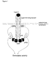
- the present invention relates to a recombinant receptor, comprising an extracellular ligand-binding domain and a cytoplasmic domain that comprises a heterologous bait polypeptide, which receptor is activated by binding of a ligand to said ligand binding domain and by binding of a prey polypeptide to said heterologous bait peptide.
- the present invention also relates to a method to detect compound-compound-binding using said recombinant receptor.
- Protein-protein interactions are an essential key in all biological processes, from the replication and expression of genes to the morphogenesis of organisms. Protein-protein interactions govem amongst others ligand-receptor interaction and the subsequent signaling pathway; they are important in assembly of enzyme subunits, in the formation of biological supramolecular structures such as ribosomes, filaments and virus particles and in antigen-antibody interactions.
- the fusion proteins need to be translocated to the nucleus, which is not always evident. Proteins with intrinsic transcription activation properties may cause false positives. Moreover, interactions that are dependent upon secondary modifications of the protein such as phosphorylation cannot be easily detected.
- WO9002809 describes how a binding protein can be displayed on the surface of a genetic package, such as a filamentous phage, whereby the gene encoding the binding protein is packaged inside the phage. Phages, which bear the binding protein that recognizes the target molecule are isolated and amplified.
- Several improvements of the phage display approach have been proposed, as described e.g. in WO9220791, WO9710330 and W09732017.
- US5637463 describes an improvement of the yeast two-hybrid system, whereby can be screened for modification dependent protein-protein interactions.
- this method relies on the co-expression of the modifying enzyme, which will exert its activity in the cytoplasm and may modify other enzymes than the one involved in the protein-protein interaction, which may on its tum affect the viability of the host organism.
- the present invention satisfies this need and provides additional advantages as well.
- the first interacting polypeptide is called “bait” or “bait polypeptide”
- the second interacting polypeptide is called “prey” or "prey polypeptide”.
- the recombinant receptor can be a chimeric receptor, in which the ligand binding domain and the cytoplasmic domain are derived from two different receptors.
- the receptor is a multimerizing receptor, this can be a homomultimerizing receptor as well as a heteromultimerizing receptor.
- the cytoplasmic domain of the recombinant receptor comprises a heterologous bait polypeptide, which can be fused to the carboxyterminal end, or can replace a part of this carboxyterminal end or can be situated in the cytoplasmic domain itself, as an insertion or a replacement of an endogenous intemal fragment.
- the chains need to comprise the bait, but it is sufficient if one of the composing chains does comprise the bait in its cytoplasmic domain.
- At least one of the activation sites in the cytoplasmic domain of the receptor has been inactivated, so that the receptor is not activated and there is no active signaling pathway if only a ligand is binding to the ligand-binding domain of said recombinant receptor.
- Such inactivation can be obtained in several ways, such as by replacement of the amino acid, which can be activated, by another amino acid, by changing the amino acid context of the activation site or by deleting the activation site.
- Insertion of the heterologous bait polypeptide and inactivation of the activation sites may result in one or more deletions of the original cytoplasmic domain.
- the only limiting factor for the changes in the cytoplasmic domain is that said cytoplasmic domain should retain, directly or indirectly, its inherent modifying enzyme activity activity, either by retaining a modifying enzyme activity binding site such as a Jak binding site, or by incorporating an active modifying enzyme activity in the cytoplasmic domain itself.
- Activation of the receptor and of the signaling pathway is achieved by binding of a ligand to the ligand-binding domain and by binding of a prey polypeptide to the heterologous bait polypeptide comprised in the cytoplasmic domain of the receptor.
- the gene, encoding the recombinant receptor comprising the bait polypeptide may be placed downstream either a constitutive or an inducible promoter.
- the latter construction may have some advantages in cases where there is a competition for the binding site between prey polypeptides and endogenous polypeptides. Induction of the recombinant receptor comprising the bait polypeptide in presence of the prey polypeptides may facilitate the binding and avoid saturation of the binding sites with endogenous polypeptides
- One preferred embodiment is a recombinant receptor according to the invention whereby the activation site is a phosphorylation site and the modifying enzyme activity is a kinase.
- Another preferred embodiment of the invention is a homomultimerizing recombinant leptin receptor, with a heterologous bait polypeptide fused into, or, preferentially, at the carboxyterminal end of its cytoplasmic domain.
- Said heterologous bait polypeptide may replace part of said cytopmasmic domain.
- the three conserved tyrosine phosphorylation sites of the cytoplasmic domain are inactivated, more preferentially by a replacement of tyrosine by phenylalanine.
- Another preferred embodiment is a homomultimerizing recombinant receptor in which an inactivated cytoplasmic domain of the leptin receptor, comprising a heterologous bait polypeptide, as described above, is fused to the ligand binding domain of the erythropoietin (EPO) receptor.
- EPO erythropoietin
- Still another embodiment is a heteromultimerizing recombinant receptor in which the inactivated cytoplasmic domain of the leptin receptor, comprising a heterologous bait polypeptide is fused to the Interleukine-5 receptor ⁇ -chain ligand-binding domain for one subunit, and to the interleukine-5 receptor ⁇ -chain for another subunit.
- Still another embodiment is a heteromultimerizing recombinant receptor in which the inactivated cytoplasmic domain of the leptin receptor, comprising a heterologous bait polypeptide is fused to the GM-CSF ⁇ -chain ligand-binding domain for one subunit, and to the interleukine-5 receptor ⁇ -chain for another subunit.
- the bait is modified by the bait-modifying-enzyme activity which can be, but is not necessarily identical to the modifying enzyme activity which is modifying the activation site.
- the recombinant receptor can be a chimeric receptor, in which the ligand binding domain and the cytoplasmic domain are derived from two different receptors. Preferentially, the receptor is a multimerizing receptor.
- the cytoplasmic domain of the recombinant receptor comprises a heterologous bait polypeptide, which can be fused to the carboxyterminal end, or can replace a part of this carboxyterminal end or can be situated in the cytoplasmic domain itself, as an insertion or a replacement of an endogenous intemal fragment.
- a heteromultimerizing receptor not all the chains need to comprise the bait, but it is sufficient if one of the composing chains does comprise the bait in its cytoplasmic domain.
- At least one of the activation sites in the cytoplasmic domain of the receptor has been inactivated, so that the receptor is not activated and there is no active signaling pathway if only a ligand is binding to the ligand-binding domain of said recombinant receptor.
- Such inactivation can be obtained in several ways, such as by replacement of the amino acid, which can be activated, by another amino acid, or by changing the amino acid context of the activation site or by deleting the activation site. Insertion of the heterologous bait polypeptide and inactivation of the activation sites may result in one or more deletions of the original cytoplasmic domain.
- cytoplasmic domain should retain, directly or indirectly, its inherent modifying enzyme activity, either by retaining a modifying enzyme binding site, or by incorporating an active modifying enzyme activity in the cytoplasmic domain itself.
- the activation site is a phosphorylation site
- the modifying enzyme activity is a kinase activity.
- the modification of the bait may be either in cis or in trans, i.e. by an enzymatic activity that is situated on the same cytoplasmic domain, or by an enzymatic activity that comes from elsewhere.
- the modification of the bait is induced by binding of a ligand to the ligand-binding domain.
- One preferred embodiment is a homodimerizing receptor in which the bait is phosphorylated by the inherent kinase activity of the cytoplasmic domain, preferentially a Jak kinase that is binding to said cytoplasmic domain.
- Another preferred embodiment is a heteromultimerizing receptor where the cytoplasmic domain of one chain comprises a bait to be modified, and the cytoplasmic domain of another chain comprises the bait-modifying enzyme activity.
- Activation of the receptor and of the signaling pathway is achieved by binding of a ligand to the ligand-binding domain and by binding of a prey polypeptide to the heterologous bait polypeptide situated in the cytoplasmic domain of the receptor.
- Binding of said prey polypeptide is dependent upon the modification state of said heterologous bait polypeptide, it means that binding occurs only in case the bait is modified or only in case the bait is not modified.
- prey polypeptide is a fusion protein comprising a polypeptide that can interact directly or indirectly with a bait polypeptide and another polypeptide that comprises at least one activation site of a receptor .
- Said activation site is preferentially a phosphorylation site, more preferentially a tyrosine phosphorylation site.
- said tyrosine phosphorylation site is part of a Signal Transducer and Activator of Transcription (STAT) binding site, most preferentially part of a STAT1 and/or STAT3 binding site.
- STAT Signal Transducer and Activator of Transcription
- Direct interaction means that there is a direct protein-protein contact between the heterologous bait polypeptide and the prey polypeptide; indirect interaction means that the heterologous bait polypeptide interacts with one or more other polypeptides to form a complex that interacts with said prey polypeptide or vice versa.
- the prey polypeptide may interact either with only one or with several polypeptides from the complex.
- the binding of the prey polypeptide to the bait polypeptide may be dependent upon the modification state of said bait polypeptide and/or of proteins within the binding complex.
- the prey polypeptide may comprise a Nuclear Export Sequence (NES), to ensure that it is available in the cytosol.
- NES Nuclear Export Sequence
- the NES signal amino acids 37-466 of the heat-stable inhibitor of the cAMP-dependent protein kinase has been shown to override a strong nuclear localisation signal (Wiley et al ., 1999). This NES will keep the prey polypeptide in the cytoplasm even if it has a strong nuclear localisation signal, facilitating the interaction with the bait.
- One preferred embodiment is a prey polypeptide according to the invention, whereby said prey polypeptide interacts with the heterologous bait polypeptide of a recombinant receptor according to the invention.
- the activation site of the prey polypeptide can be modified by the modifying enzyme activity inherent to the cytoplasmic domain of the receptor. The modification of the activation site will activate the signaling pathway.
- said activation site is a phosphorylation site and the modifying enzyme activity is a kinase activity. More preferentially, this activation comprises binding of a STAT polypeptide to the phosphorylated phosphorylation site, followed by phosphorylation of said STAT polypeptide and subsequent dimerization of two phosphorylated STAT molecules.
- Another aspect of the invention is a vector, encoding a recombinant receptor according to the invention and/or a vector, encoding a prey polypeptide according to the invention.
- Said recombinant receptor and said prey polypeptide may be situated on one or on separated vectors.
- the vector can be any vector, know to the person skilled in the art, including but not limited to episomal vectors, integrative vectors and viral vectors.
- a preferred embodiment is a bait vector whereby the bait may be integrated in the chromosome by a recombinase-assisted integration such as cre-lox or flp-frt, and/or a retroviral prey vector that allows retroviral integration in the genome.
- Another aspect of the invention is an eukaryotic cell comprising a recombinant receptor according to the invention.
- the eukaryotic cell is obtained by transformation or transfection with one or more vectors according to the invention.
- Said eukaryotic cell comprises, but is not limited to yeast cells, fungal cells, plant cells, insect cells and mammalian cells.
- the eukaryotic cell is a mammalian cell.
- a preferred embodiment is an eukaryotic cell line expression the mouse retroviral receptor, allowing safe retroviral work using retroviral cDNA libraries.
- kits comprising one or more cloning vectors allowing the construction of one or more vectors according to the invention. It is clear for the people skilled in the art that a cloning vector, encoding a recombinant receptor in which the part, encoding for the cytoplasmic domain comprises one or more restriction sites allowing an "in frame" fusion of a nucleic acid fragment encoding a polypeptide can easily be used to construct a vector encoding a recombinant receptor according to the invention.
- a cloning vector encoding a first polypeptide comprising at least one activation site, comprising one or more restriction sites allowing an "in frame" fusion of a nucleic acid encoding a second polypeptide with said first polypeptide can easily be used to construct a vector encoding a prey polypeptide according to the invention.
- cloning strategies known to the person skilled in the art may be used.
- Still another aspect of the invention is a method to detect compound-compound binding using a recombinant receptor and/or a prey polypeptide according to the invention.
- an eukaryotic cell, carrying a recombinant receptor according to the invention is transformed or transfected with a vector library encoding prey polypeptides according to the invention. Bait-prey binding will result in an activation of the signaling pathway and can be detected by the use of a reporter system.
- the use of a chimeric receptor may represent an additional advantage for this method.
- a first advantage of the use of a chimeric receptor in this method is that it allows the elimination of a non bait-specific background.
- a difference can be made between bait-specific and non bait-specific binding.
- This can be realized by the use of a host cell carrying at least two receptors, a first receptor, comprising a first ligand binding domain and a cytoplasmic domain that does not comprise an activation site neither a heterologous bait polypeptide and a second receptor, comprising the same inactivated cytoplasmic domain, however with a heterologous bait polypeptide now, and a second ligand binding domain.
- a positive signal can only be detected when there is a non bait-specific interaction of a prey polypeptide fused to a polypeptide comprising an activation site with the cytoplasmic domain of the receptor; these cells can be selected and/or eliminated.
- the second ligand can be added to the medium.
- a positive signal will only be detected upon specific bait-prey interaction, as the preys binding to the cytoplasmic domain have been removed.
- One specific embodiment of the method to detect compound-compound binding is a method whereby said binding is a protein-protein interaction.
- Another specific embodiment is a method to detect protein-protein interaction, whereby said interaction is modification state dependent.
- Still another specific embodiment is a method to detect compound-compound binding, whereby said binding is mediated by three or more partners. In this case, one or more partners may not be or not completely be of proteineous nature.
- a recombinant receptor, according to the invention may, as a non-limiting example, bind to a small molecule.
- the prey polypeptide, according to the invention may also bind to the small molecule, so that bait and prey are linked together by said small molecule.
- Said small molecule may be present in the host cell, as a compound produced by the cell itself, or as a compound that is taken up from the medium.
- said method to detect compound-compound binding comprises the construction of an eukaryotic cell comprising a recombinant receptor according to the invention, followed by transformation or transfection of said cell by a library of prey polypeptide vectors according to the invention.
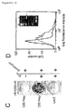
- the compound-compound binding is detected by the activation of the receptor, leading to an active signaling pathway, resulting in the induction of a reporter system.
- a reporter system can be any system that allows the detection and/or the selection of the cells carrying a recombinant receptor according to the invention. It is clear for the person skilled in the art that several reporter systems can be used.
- a luciferase gene, an antibiotic resistance gene or a cell surface marker gene can be placed after a promoter that is induced by the signaling pathway.
- reporter systems may be used that are based on the change in characteristics of compounds of the signaling pathway, when said pathway is active, such as the phosphorylation and/or dimerisation of such compounds.
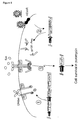
- Receptor does not necessarily indicate a single polypeptide, but may indicate a receptor complex, consisting of two or more polypeptides, and comprising a ligand binding domain and a cytoplasmic domain.
- Recombinant receptor means that at least one of said polypeptides is recombinant.
- the polypeptide comprising the cytoplasmic domain is recombinant.
- Activation site of a receptor is the site that, in the wild type receptor, is modified after binding of a ligand to the ligand binding domain, leading to a reorganization of the receptor and subsequent activation of the modifying enzyme activity, and to which a compound of the signaling pathway can bind after modification, or any site that can fulfill a similar function.
- the activation site is not necessarily located on the same polypeptide as in the wild type receptor, but may be situated on another polypeptide of the receptor complex.
- Modifying enzyme activity means the enzymatic activity, associated to or incorporated in the cytoplasmic domain of the receptor that is normally induced upon binding of the ligand to the ligand binding domain and subsequent reorganization of the receptor (e.g. by a conformational change), and may modify the activation site.
- the activation site is a phosphorylation site and the modifying enzyme activity is a kinase activity.
- the bait-modifying enzyme activity means the activity which modifies the bait. It can be, but is not necessarily identical to the modifying enzyme activity.
- Activation of a receptor means that the receptor is inducing a signaling pathway, by binding of a compound of the signaling pathway to the modified activation site, whereby said activation normally results in the induction or repression of one or more genes.
- Said gene is preferentially a reporter gene, which allows monitoring the activation of the receptor.
- An activated receptor is a receptor where the binding of a compound to the activation site has been enabled by modification of said site. A receptor in which the modifying enzyme activity has been induced, without modification of an activation site is not considered as activated.
- Multimerizing receptor as used here means that the activated receptor comprises several polypeptides. It does not necessarily imply that the multimerization is induced by ligand binding: the receptor can exist as a preformed complex of which the conformation is changed upon ligand binding.
- Polypeptide as used here means any proteineous structure, independent of the length and includes molecules such as peptides, phosphorylated proteins and glycosylated proteins. Polypeptide as used herein is not necessarily indicating an independent compound but can also be used to indicate a part of a bigger compound, such as a domain of a protein.
- Heterologous bait polypeptide, as comprised in the cytoplasmic domain of a receptor means that within the cytoplasmic domain, or fused to the cytoplasmic domain, there is a polypeptide that is not present in the cytoplasmic domain of the non-recombinant receptor. Said heterologous bait polypeptide may replace a part of said cytoplasmic domain. Bait herein means that this polypeptide can interact with other polypeptides, not belonging to the normal receptor complex.
- Prey polypeptide as used here means a fusion protein comprising a polypeptide that can bind with the heterologous bait polypeptide and a polypeptide that comprises at least one activation site.
- Ligand means every compound that can bind to the extracellular domain of a receptor and that is able to initiate the signaling pathway by binding to said extracellular domain. Initiating as used here means starting the events that normally directly follow the binding of the ligand to the extracellular domain of a receptor, e.g. multimerization for a multimerizing receptor, but it does not imply activation of the receptor and/or accomplishing of the signaling pathway.
- Compound means any chemical or biological compound, including simple or complex organic or inorganic molecules, peptides, peptido-mimetics, proteins, antibodies, carbohydrates, nucleic acids or derivatives thereof.
- Bind(ing) means any interaction, be it direct or indirect.
- a direct interaction implies a contact between the binding partners.
- An indirect interaction means any interaction whereby the interaction partners interact in a complex of more than two compounds. This interaction can be completely indirect, with the help of one or more bridging compounds, or partly indirect, where there is still a direct contact that is stabilized by the interaction of one or more compounds.
- Functional fragment of the inactivated leptin receptor cytoplasmic domain means a fragment of the leptin receptor cytoplasmic domain that still allows binding of the Jak kinases.
- Inactivation of an activation site means any change, mutation or deletion that is inhibiting a modification at the position of the potentially modified residue in the polypeptide.
- inactivation of a tyrosine phosphorylation site means any change, mutation or deletion that is inhibiting a phosphorylation at the position of the potentially phosphorylated tyrosine residue in the polypeptide.
- it is a mutation at this position; more preferentially, it is a change of tyrosine into phenylalanine.
- Cloning vector is a vector that is generally considered as an intermediate step for the construction of another vector. It is intended to insert one or more nucleic acid fragments, in order to obtain one or more new vectors that will be used to transform or transfect the host cell of interest, or as cloning vectors themselves.
- Recombinant mouse leptin, recombinant human leukemia inhibitory factor (LIF) and recombinant human erythropoietin (Epo) were all purchased from R&D Systems. Typical stimulation conditions were 100ng/ml leptin, 1 ng/ml LIF and 50 ng/ml Epo.
- ⁇ NX-Eco cells were seeded at a density of 6x10 6 cells/petridish the day prior to transfection.
- Cells were transfected with 50 ⁇ g of the retroviral vector pBG1-CIS according to the calcium phosphate procedure. 25 ⁇ M chloroquine was added 5 min. before transfection.
- Medium was harvested 24 and 48 hours post transfection, filtered over a 0.22 ⁇ m GV filter (Millipore) and stored at -80°C.
- Packaging of the HEK cDNA library was performed as described above with the exception that 1.6x10 7 cells in a 175cm 2 falcon were transfected with 87 ⁇ g pBG1-HEK293cDNA.
- 10% pMFG-EGFP gift from Dr. Mulligan, Cambridge, MA
- the virus titer was approx. 5x10 6 infectious units/ml as determined by FACS analysis of EGFP expressing cells.
- target cells were seeded at a density of 2x10 4 cells/well in a 24-well plate, and 10 6 in 75cm 2 culture flasks. The day after, cells were incubated for 24-48 hours with supernatant containing virus, diluted in medium as indicated. Polybrene (Sigma) was added at a final concentration of 2.5 ⁇ g/ml. After infection, cells were stimulated with Epo (50 ng/ml) for 24-48 hours, followed by puromycin (1-2 ⁇ g/ml as indicated; Sigma) selection for 10 days.
- Epo 50 ng/ml
- puromycin 1-2 ⁇ g/ml as indicated; Sigma
- PCR polymerase chain reactions
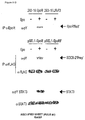
- Primer design resulted in the insertion of an Asn between the PacI generated Leu-Ile sequence and the extracellular Gly.
- the amplicon was gel-purified and ligated in the pCR®-Blunt vector (Invitrogen). PacI-SacI digestion on this pCR®-Blunt construct results, after gel-purification, in the desired LepR fragment.
- RT-PCR was performed as follows: 2 ⁇ l (2 ⁇ g) of oligodT (12-18 mer; Pharmacia) was added and incubated at 70°C for 10 min., the reaction mixture was chilled on ice for 1 min., cDNA was prepared by adding 4 ⁇ l of 10x RT buffer (Life Sciences), 1 ⁇ l 20 mM dNTP's (Pharmacia), 2 ⁇ l 0,1M DTT, and 1 ⁇ l of MMLV reverse transcriptase (200U; Superscript RT; Life Technologies) to an end volume of 20 ⁇ l. Incubations were as follows: RT for 10 min., 42°C for 50 min., 90°C for 5 min., and 0°C for 10 min.
- RnaseH 2U ; Life Technologies
- RnaseH 2U ; Life Technologies
- PCR on this cDNA was performed using Pfu enzyme (5 U; Stratagene).
- Forward primer (MBU-O-167) and reverse primer (MBU-O-308) were designed to amplify the extracellular part of the EpoR (amino acids 1-249) between a Kpnl and PacI site.
- a band of correct size was purified and the DNA was digested with Kpnl and PacI and was inserted into the KpnI-PacI opened pSV-SPORT-IL-5R ⁇ /IFNaR2-2 vector.
- This vector contains a chimeric receptor that has the extracellular domain of the IL-5R ⁇ receptor, fused to the transmembrane and intracellular domains of IFNaR2-2.
- a PacI site was added to the fusion point by means of the QuikchangeTM site-directed mutagenesis kit (Stratagene, La Jolla) which resulted in the insertion of two amino acids (Leu-Ile) before the most membrane-proximal, extracellular amino acid (Lys) of IFNaR2-2.
- the extracellular domain of IL-5R ⁇ could be exchanged by the one of EpoR, as described above.
- the LepR fragment generated by PacI-SacI digestion was ligated in the PacI-SacI digested and gel-purified pSV-SPORT-EpoR/IFNaR2-2 vector, resulting in pSV-SPORT-EpoR/LepR.
- the pSV-SPORT-IL-5R ⁇ /IFNaR2-2 and pSV-SPORT- ⁇ c /IFNaR1 vectors express an IL-5R ⁇ /IFNaR2-2 and a ⁇ c /IFNaR1 chimera, respectively, composed of the extracellular portion of the IL-5R ⁇ or ⁇ c chain, and the transmembrane and intracellular parts of the IFNaR2-2 or IFNaR1.
- a PacI site was used to generate the fusion site just preceding the transmembrane segment.
- the IFNaR2-2 or IFNaR1 parts in these vectors were replaced by the same segments of the LepR, using the PacI site and an Xbal site which is located just after the IFNaR2-2 or IFNaR1 stop codon. Therefore, the LepR fragment was generated by a PacI-XbaI digest of the pSV-SPORT-EpoR/LepR vector (see example 1), and was inserted into the PacI-XbaI opened and gel-purified pSV-SPORT-IL-5R ⁇ /IFNaR2-2 and pSV-SPORT- ⁇ c /IFNaR1 vectors, resulting in the vectors pSV-SPORT-IL-5R ⁇ /LepR and pSV-SPORT- ⁇ c /LepR.
- the pSV-SPORT-IL-3R ⁇ /LepR and pSV-SPORT-GM-CSFR ⁇ /LepR vectors were constructed as follows: the extracellular portion of the IL-3R ⁇ and GM-CSFR ⁇ chains were amplified using standard RT-PCR procedures with Pfu polymerase. 2 ⁇ l TF-1 cDNA was used as input. Forward primers were MBU-O-752 (IL-3R ⁇ ) and MBU-O-754 (GM-CSFR ⁇ ), and generated a Kpnl site. Reverse primers MBU-O-753 (IL-3R ⁇ ) and MBU-O-755 (GM-CSFR ⁇ ), contain a PacI site allowing in frame fusion to the LepR.
- the KpnI-PacI excised extracellular fragments were ligated into the KpnI-PacI opened pSV-SPORT-IL-5R ⁇ /LepR.
- pSV-SPORT-IL-5R ⁇ /LepR For the GM-CSFR ⁇ construction, a partial KpnI digest was applied since the extracellular portion contained an intemal KpnI site.
- the resulting vectors, pSV-SPORT-IL-3R ⁇ /LepR and pSV-SPORT-GM-CSFR ⁇ /LepR contain chimeric receptors composed of the extracellular portion of the IL-3R ⁇ or GM-CSFR ⁇ chain fused to the transmembrane and cytoplasmatic tail of the LepR.
- mutant leptin receptors (Eyckerman et al ., 1999) Y985-1077F and Y985-1077-1138F (LepR-F3; previously called F-all) were generated using the QuikchangeTM site-directed mutagenesis procedure using Pfu polymerase (Stratagene) on the pMET7-LepR template.
- Mutagenic oligonucleotides were MBU-O-157, MBU-O-158, MBU-O-159, MBU-O-160, MBU-O-161 and MBU-O-162. Each single mutation was coupled to a change in restriction cleavage and was confirmed by restriction and DNA sequence analysis. The double and triple mutants were created using a sequential approach.
- the pMET7mcs vector is a modified version of pMET7 containing an expanded MCS by insertion of the extra unique BglII, EcoRV, BstEII, AgeI and Xhol restriction sites.
- PCR amplification on the pSVL-gp130 template using the forward primer MBU-O-586 and the reverse primer MBU-O-443 generated a DNA fragment encoding a 158 amino acid-long intracellular fragment of the human gp130 chain, which contains 4 STAT-3 association motifs (amino acids 761-918, the stopcodon was not co-amplified).
- the forward primer contains from 5' to 3' an Apal restriction site, a Kozak consensus sequence, a flag-tag encoding sequence (Met-Asp-Tyr-Lys-Asp-Asp-Asp-Asp-Lys-Ile), and a BglII restriction site.
- the reverse primer encodes an additional hinge sequence (Gly-Gly-Ser) and contains an EcoRI recognition site. Apal and EcoRI digestion of the PCR product (after subcloning in pCR®-Blunt) and of pMET7-mcsA, allowed us to ligate the gp130 fragment into the pMET7 vector, generating the pMET7-flag-gp130 construct.
- SV40 largeT antigen was amplified using a vector from the HybriZAP-2.1 Two-Hybrid cDNA synthesis kit (Stratagene, pSV40) as template. Primers MBU-O-445 and MBU-O-446 were used to generate a DNA fragment encoding 448 amino acid between residues 261 and 708. The N-terminal deletion eliminates the nuclear targeting signal in SVT.
- the forward primer contains an EcoRI recognition site that allows in-frame ligation to the gp130-hinge sequence.
- the reverse primer contains additional Nrul, Xhol, BglII, NotI and Xbal restriction sites and also encodes the stop codon after the SVT coding sequence.
- a DNA fragment encompassing murine p53 was amplified with MBU-O-450 and MBU-O-451 using the p53 control plasmid from the HybriZAP-2.1 Two-Hybrid cDNA synthesis kit (Stratagene) as template.
- the forward primer contains a SalI restriction site that allows in-frame coupling to the EpoR/LepR-F3 hinge construct.
- the reverse primer contains a STOP codon and a Xbal restriction site.
- the 243 amino acid-long p53 fragment (amino acids 73-315) contains the interaction site with SVT, but lacks the nuclear targeting signal and the oligomerisation domain.
- Amplification using MBU-O-695 and MBU-O-696 as forward and reverse primer respectively, and using pMG1-SVT as template resulted in a SVT fragment (amino acids 261-708 + stopcodon) between BglII and Xbal recognition sites.
- the BglII-XbaI digested and purified PCR fragment was ligated into a BglII-XbaI cut and gel-purified pMG1-SVT vector, resulting in pMET7-SVT.
- RNA was prepared from 5x10 6 TF-1 cells using the RNeasy kit (Qiagen), and eluted in 50 ⁇ l water from which 10 ⁇ l was used as input for RT-PCR.
- RT-PCR was performed using standard reaction conditions as described in section I .1.1.
- An intracellular fragment of the human EpoR (amino acids 370-453) was amplified from 4 ⁇ l TF1 cDNA using MBU-O-675 and MBU-O-676 as forward and reverse primer respectively, with two consecutive PCR reactions and an intermediate gel-purifrcation. Sacl and Xbal recognition sites are present in the forward and reverse primers respectively.
- the reverse primer also encodes a stop codon.
- the fragment was subcloned in pCR®-Blunt, digested with SstI (which has the same recognition site as SacI) and Xbal, and ligated into SstI-XbaI digested and gel-purified pSEL1 vector, resulting in the pSEL1-EpoR construct.
- the complete coding region for mouse Cytokine Inducible SH2-containing protein CIS was amplified using MBU-O-677 and MBU-O-678 as forward and reverse primer respectively.
- the forward primer contains an EcoRl recognition site and the reverse primer contains a Xbal recognition site and the stop codon.
- the amplified and gel-purified fragment was subcloned into the pCR®-Blunt vector.
- the insert was recovered by EcoRI and Xbal digestion and gel-purification and was cloned into an EcoRI-XbaI digested and gel-purified pMG1-SVT vector, leading to the pMG1-CIS vector.
- the SH2 domain of GRB2 (aa 60-158) was amplified in the same way using primers MBU-O-469 and MBU-O-470 as forward and reverse respectively.
- the forward primer contains an extra EcoRI recognition site which allows in frame fusion to the gp130 chain
- the reverse primer contains an extra stop codon and a Xbal enzyine recognition site.
- a fragment of human GRB2 comprising the C-terminal SH3 domain (aa 159-217) was amplified using the pMG1-GRB2 construct as template and MBU-O-770 and MBU-O-468 as forward and reverse primer respectively.
- MBU-O-770 allows in frame fusion to the flag tag by a BglII site
- MBU-O-468 is described above.
- the GRB2 fragment was inserted in the pMG1 vector by a BglII-XbaI based exchange, resulting in the pMET7-GRB2SH3 vector.
- VavS aa 259-789
- VavS human TF1 cell line by standard RT-PCR techniques.
- Primers were MBU-O-737 and MBU-O-738 as forward and reverse respectively.
- MBU-O-737 contains an extra EcoRI allowing in frame fusion to gp130, and MBU-O-738 contains a Stop codon and a Xhol enzyme recognition site.
- the amplified fragment was subcloned in the pCR®-Blunt vector and ligated in the pMG1 vector through an EcoRI-Xhol based exchange.
- the VavS fragment was also amplified using forward primer MBU-O-771, reverse primer MBU-O-741 and pMG1-VavS as template.
- the forward primer contains a BamHI site, which allows in frame fusion to the flag tag, and the reverse primer contains a stop codon and a Xbal restriction site.
- the amplicon was subcloned in the pCR®-Blunt vector, and excised with BamHI and Xbal.
- the purified fragment was cloned in a BglII-XbaI cut pMG1 vector, resulting in the pMET7-VavS construct.
- Genomic DNA was isolated from the rat pheochromocytoma PC12 cell line using the DNAzoI procedure (Gibco BRL).
- Optimal primers for PCR amplification of the rat PAP1 promoter were selected using the "Oligo Primer 3" program (http://www-genome.wi.mit.cdu/cgi-bin/primer3.cgi).
- Forward and reverse primers were MBU-O-222 and MBU-O-223 respectively.
- Amplification was performed using Taq polymerase in 30 cycles: 2' at 94°C, 2' at 57°C, 2' at 72°C, followed by a 10' filling-in reaction at 72°C.
- Optimal MgCl 2 concentration was determined to be 6mM.
- the promoter fragment was cloned after polishing with Klenow polymerase in the pCR®-Blunt vector.
- the promoter fragment was cut from this plasmid construct using a successive PstI digest, Klenow treatment to polish this end, and BamHI digest, resulting in a blunt-sticky fragment.
- the gel-purified fragment was cloned into a Smal-BglII opened and gel-purified pGL3 control vector (Promega). Digestion with SstI-SpeI restriction enzymes and gel-purification resulted in a fragment that was cloned into a Sstl-Nhel and purified pGL3 basic vector, resulting in the pGL3- rPAP1-luci construct.
- DNA sequencing revealed 10 nucleotides differing from the published sequence, but without affecting the leptin-dependent induction of this promoter segment.
- the full-length rPAP1 promoter fragment was excised from pGL3 rPAP1-luci using partial digestion with Kpnl and Xhol and ligated into the Kpnl-Xhol opened pXP2d2 vector (gift from Prof. S. Nordeen), resulting in pXP2d2-rPAPl-luci.
- the coding sequence for puromycin was amplified using primers MBU-O-719 and MBU-O-720 on the pIRESpuro2 (Clontech) template.
- Combined XhoI-XbaI digestion allowed insertion in the pXP2d2-rPAP1-luci construct, resulting in pXP2d2-rPAP1-puro R .
- Insertion of the EpoR-LR-F3-EpoR into the pcDNA5/FRT vector was obtained by re-amplifying the complete chimeric construct using MBU-O-167 and MBU-O-769 on the pSEL1-EpoR template, followed by subcloning using Kpnl and NotI sites. This construct was named pcDNA5/FRT-EpoR.
- the SVT fragment was re-amplified from pMG1-SVT using forward primer MBU-O-766 and MBU-O-446.
- BamHI-NotI digestion allowed insertion in the pBMN-Z retroviral vector (gift from G. Nolan), resulting in vector pBG1-SVT.
- EcoRI-NotI based exchange of the SVT fragment for CIS resulted in the pBG1-CIS vector.
- the E. coli control of cell death gene (ccdB) was amplified using primers MBU-O-835 and MBU-O-83 and template pENTRY11 (Life Technologies), and cloned in the pBG1-CIS vector by an EcoRI-NotI restriction-based insertion, resulting in the pBG1-ccdB vector.
- the LepR fragment in pSV-SPORT-GM-CSFR ⁇ /LepR and pSV-SPORT- ⁇ c /LepR was replaced by the LepR-F3 fragment of pSEL1 (pSV-SPORT-EpoR/LepR-F3) using the PacI and NotI site.
- the LepR-F3 fragment was generated by a PacI-NotI digest of the pSEL1 construct, gel-purified and inserted into the PacI-NotI opened and gel-purified pSV-SPORT-GM-CSFR ⁇ or pSV-SPORT- ⁇ c vectors, resulting in the vectors pSV-SPORT-GM-CSFR ⁇ /LepR-F3 or pSV-SPORT- ⁇ c /LepR-F3.
- the pSV-SPORT-GM-CSFR ⁇ /LepR-F3 and pSV-SPORT- ⁇ c /LepR-F3 vectors were digested with Sall, gel-purified and the ends were polished by Klenow fragment (Boehringer Mannheim). These blunt ended vectors were incubated with Alkaline Phosphatase (Boehringer Mannheim) to dephosphorylate the blunt ends.
- a construct containing the mouse cytoplasmic tail of ALK4 with mutation T206D, resulting in a constitutive active kinase, in the vector pGBT9 was obtained from Prof. D. Huylebroeck.
- the mutated cytoplasmic tail of ALK4 was removed from the construct by an EcoRI-BamHI digestion, gel-purified and incubated with Klenow fragment to polish the ends.
- This insert was ligated in the opened pSV-SPORT-GM-CSFR ⁇ /LepR-F3 and pSV-SPORT- ⁇ c /LepR-F3 vectors resulting in pSV-SPORT-GM-CSFR ⁇ /LepR-F3-ALK4CA and pSV-SPORT- ⁇ c /LepR-F3-ALK4CA.
- a construct containing a human cDNA encoding the entire Smad3 protein in vector pcdef was a kind gift of Prof. D. Huylebroeck.
- the Smad3 insert was removed with an EcoRI-XhoI digestion, gel-purified and the ends were polished by Klenow fragment (Boehringer Mannheim).
- This Smad3 insert was ligated into the opened pSV-SPORT-GM-CSFR ⁇ /LepR-F3 and pSV-SPORT- ⁇ c /LepR-F3 vectors (described above) resulting in pSV-SPORT-GM-CSFR ⁇ /LepR-F3-Smad3 and pSV-SPORT- ⁇ c /LepR-F3-Smad3.
- the pMG1-SVT vector was digested with EcoRI and NotI, followed by incubation with Klenow fragment (Boehringer Mannheim) to polish the ends. This blunt ended vector was incubated with Alkaline Phosphatase (Boehringer Mannheim) to dephosphorylate the blunt ends. Cassette rfB of the Gateway Vector Conversion System (Life Technologies) was then ligated into the opened vector leading to the pMG1-gateway vector. A PCR reaction using primers MBU-O-1094 and MBU-O-1076 on the pMG1-SVT template was performed, resulting in a fragment that contains gateway recombination sites.
- This fragment also contains a part (amino acids 905 - 918) of the gp130 chain.
- the fragment was then cloned in the pMG1-gateway vector using a two-step gateway reaction (Lifetechnologies), resulting in pMG2-SVT, a prey construct with a total of 6 STAT recruitment sites.
- the pMG2-SVT construct was digested by EcoRI-XhoI and the vector was gel-purified.
- the Smad4 insert was removed by an EcoRI-XhoI digestion, gel-purified and ligated into the opened pMG2 vector.
- the Blasticidin system (Invitrogen) was used to create a stable cell line with an endogenous pSEAP-rPAP1 reporter construct. Sensitivity to the toxic agent Blasticidin (Invitrogen) of Hek293T cells was estimated to be 3 ⁇ g/ml. 10 6 cells were seeded in a petridish and transfected the day after seeding using the Calcium Phosphate Transfection System (Life Technologies) according to the manufacturers instructions. A total of 20 ⁇ g DNA was transfected (18 ⁇ g of pSEAP-rPAP1seap and 2 ⁇ g of pcDNA6/V5-HisA, which contains a Blasticidin resistance gene).
- the transfected cells were seeded in 96 well plates at 10 cells per well. After 24 hours, Blasticidin was added at a concentration of 3 ⁇ g/ml and cells were maintained under selective conditions for 3-4 weeks. The resulting single cell clones were screened by stimulation for 24 hours with 20 ng/ml hyper-IL6 (fusion protein of IL-6 with its specific receptor IL6-R ⁇ ; Fischer et al ., 1997). Hek293T PAP21 was selected as the best responsive cell line.
- hyper-IL6 fusion protein of IL-6 with its specific receptor IL6-R ⁇ ; Fischer et al ., 1997.
- Flp-In-293 cells were stably transfected with a plasmid containing expression cassettes for the mouse ecotropic retroviral receptor (mEcoR) and for neomycin resistance.
- the pool of neomycin resistant cells (resistant to 400 ⁇ g/ml geneticin, Life Technologies) were supertransfected, at a ratio of 5:1 respectively, with the following two plasmids: i) a plasmid carrying the cDNA encoding the puromycin resistance marker (puromycin-N-acetyl-transferase) under control of promoter sequences of rPAP1 (pXP2d2-rPAP1-puro R ), ii) a plasmid carrying the cDNA for the blasticidin resistance marker (blasticidin S deaminase), under the control of the EM7 promoter (pcDNA6/V5-His, Invitrogen).
- mEcoR mouse ecotropic retroviral receptor
- cells were lysed in 100 ⁇ l RLT buffer (RNeasy® method, Qiagen) and chromosomal DNA was sheared using Qiashredder columns (Qiagen).
- Beads were pre-treated according to the manufacturers' instructions (Dynabeads M-280 Streptavidin, Dynal). Briefly, Dynabeads were washed twice in a high salt buffer (1M NaCl, 10mM Tris HCl pH7,5 and 1mM EDTA), and were incubated with 200 pmoles of biotinylated oligonucleotide directed against the gp130 chain (5' GGGCTGGGTAGACTCGGATCTTGAGAAGAC).
- beads were washed three times in the above mentioned high salt buffer and resuspended in a low salt buffer (0,15M NaCl, 10mM Tris HCl pH7,5 and 1mM EDTA) to a concentration of 10 ⁇ g/ ⁇ l. 5 ⁇ l of this suspension was added to 100 ⁇ l total lysate diluted 1/5 in the high salt buffer. 15' minutes after gentle rotation at room temperature, beads were washed three times with low salt buffer and eluted in 30 ⁇ l water for 2' at 65°C. 15 ⁇ l of this sample was used as input for a standard RT-PCR reaction with the Qiagen OneStep RT-PCR Kit.
- Primers 5' GGCATGGAGGCTGCGACTG and 5' TCGTCGACCACT GTGCTGGC were used for amplification of the "prey" fragment.
- efficient amplification was obtained with lysate from less than 10 3 cells.
- Luciferase was measured after lysis of the cells and addition of luciferase substrate (Luciferin, Duchefa). Light emission was measured using a TopCount chemiluminescence counter (Canberra Packard). All luciferase measurements were normalized using an expression construct constitutively expressing ⁇ -galactosidase (pUT651), which was measured in triplicate for every transfection using the GalactoStar kit (TROPIX).
- Puromycin-resistant cell colonies were stained with crystal violet using a standard procedure.
- HEK293 library was performed using standard procedures. Briefly, 5 ⁇ g of HEK293 polyA+mRNA was used as input for both oligo-dT and random primed first strand synthesis with Superscript II reverse transcriptase (Life Technologies). Both the oligo-dT and random primers contain a Notl site. After second strand synthesis, adaptors containing an EcoRI site were ligated. The cDNA was analyzed using agarose gel electrophoresis and fragments between 0.5 and 2.5 Kbp were cloned unidirectionally in the pBG1-ccdB vector opened with EcoRI-NotI.
- a total of 6.10 7 "bait" expressing cells were seeded at a density of 2.10 6 per 175 cm 2 tissue culture flasks. 24 hours after seeding, cells were infected with the retroviral HEK293"prey" cDNA library (complexity of 2.10 6 ) for another 24 hours. After infection, cells were stimulated for 6,5 hours with 50 ng/ml Epo, and puromycin was added at a final concentration of 2 ⁇ g/ml for 20 days. Single cell colonies were picked and analysed using a functional assay and RT-PCR sequencing.
- STAT3 phosphorylation was demonstrated using the Phospho-STAT3 (Tyr705) Antibody (Cell Signaling) according to the manufacturers instructions. STAT3 expression was verified on the same blots using anti-STAT3 antibody (Transduction Laboratories).
- Transfection was performed according to the calcium phosphate method (Graham and van der Eb, 1973). A precipitate was formed using 3,4 ⁇ g as total DNA input (0.4 ⁇ g for pUT651, 1 ⁇ g for each of the others) into a 300 ⁇ l total mixture. 200 ⁇ l of this mixture was added to 4x10 5 Hek293T PAP21 cells seeded the day before transfection into a six well plate (Falcon). 6 hours after adding the mixture, the cells were washed once with Dulbecco's PBS (Life Technologies) and new DMEM medium (Life Technologies) was added. Two days after transfection, medium was removed and cells were resuspended using 200 ⁇ l Cell Dissociation Agent (Life Technologies).
- DMEM medium After neutralization with 1200 ⁇ l DMEM medium, 50 ⁇ l of the cell suspension was seeded into a 96 well plate (Costar) in triplicate for every condition; and stimulated by adding recombinant human leptin (R&D systems) to a final concentration of 100 ng/ml, or recombinant human erythropoietin (R&D systems) to a final concentration of 0.5 units/ml, or the combination of leptin (same concentration as above) plus forskolin (Sigma, 10 ⁇ M final concentration), or the combination of erythropoietin (same concentration as above) plus forskolin (same concentration as above).
- R&D systems recombinant human leptin
- R&D systems recombinant human erythropoietin
- Forskolin is a chemical agent that activates the adenylate cyclase that is present within the cells, which leads to heightened levels of the second messenger cAMP.
- Treatment of transfected cells with forskolin alone did not result in a significant induction of luciferase activity.
- cAMP elevation leads to strong co-stimulation with the leptin signal on PAP1 induction (Eyckerman et al., 1999). 24 hours after stimulation, the cells were lysed in the wells and luciferase substrate (Luciferin, Duchefa) was added.
- EpoR/LepR and LepR Y985/1077F The difference in induction between EpoR/LepR and LepR Y985/1077F is very likely due to the elimination in the latter receptor construct of tyrosines involved in the recruitment of tyrosine phosphatases and SOCS proteins to the complex, leading to an enhanced signal (Eyckerman et al ., 1999).
- a 300 ⁇ l precipitation mixture was prepared which contained 3.1 ⁇ g DNA (0.1 ⁇ g of pUT651, 1 ⁇ g of each of the others). 200 ⁇ l was added to the cells for 6 hours after which they were washed once with Dulbecoo's PBS. After washing, DMEM medium was added to the cells. After 24 hours cells were resuspended using 200 ⁇ l Cell Dissociation Agent which was neutralized with 2200 ⁇ l DMEM medium. Of this cell suspension 40 ⁇ l was brought into a 96 well plate for each transfection and stimulation was performed in triplicate.
- DMEM 60 ⁇ l DMEM was added to an end volume of 100 ⁇ l and 24 hours later, cells were stimulated for 24 hours with erythropoietin or erythropoietin plus forskolin (same concentrations as described above). A non-stimulated negative control was also included in the experiment. Luciferase measurements are shown in figure 3.
- Transfected cells from transfections a, b and c showed no significant induction of the reporter construct under all conditions tested.
- a 9,4 fold induction and a 14,6 fold induction was detected in transfected cells from transfection d, after stimulation with erythropoietin and erythropoietin plus forskolin respectively, implying an interaction-dependent signal.
- No signal was detected in transfection e and f. This implies a specific interaction, which leads to gp130-dependent STAT-3 activation. All results were normalized using the intemal transfection control vector pUT651 and the GalactoStar kit (See above).
- EpoR-CIS phosphorylation-dependent interaction trap To determine the functionality of the EpoR-CIS phosphorylation-dependent interaction trap, the following plasmid combinations were transfected in 4x10 5 Hek293T cells, which were seeded the day before transfection:
- a precipitation mixture of 300 ⁇ l was prepared which contained 3,1 ⁇ g DNA (0,1 ⁇ g of pUT651, 1 ⁇ g of each of the others). 200 ⁇ l of this mixture was applied to the cells.
- DMEM medium was added. After 48 hours the cells were resuspended using 250 ⁇ l Cell Dissociation Agent. After neutralization with 2200 ⁇ l DMEM medium, 100 ⁇ l of this cell suspension was brought into a 96 well plate (Costar). The cells were stimulated with erythropoietin or erythropoietin plus forskolin (for final concentrations, see above). A non-stimulated negative control was also included in the experiment. Luciferase expression was measured 24 hours after stimulation using a TopCount chemiluminescense counter (Canberra Packard).
- Transfected cells from transfections a, b, c, e, f and g showed no significant induction of luciferase activity.
- Transfected cells from transfection d showed a 6,2 fold and a 10,5 fold induction with erythropoietin or erythropoietin plus forskolin, respectively. This indicates an erythropoietin-dependent phosphorylation of the EpoR bait, resulting in interaction between the CIS protein and EpoR. Interaction leads to gp130 phosphorylation, STAT activation and thus signalling toward the rPAP1 promoter, leading to luciferase activity ( Figure 4).
- a 300 ⁇ l precipitation mixture was prepared containing 3,05 ⁇ g DNA (0,05 ⁇ g for pUT651, 1 ⁇ g for others). 200 ⁇ l of this mixture was added to the cells for 16 hours. After transfection the cells were washed once with Dulbecco's PBS and DMEM (both from Gibco BRL) was added. After 48 hours the cells were resuspended using 200 ⁇ l of Cell Dissociation Agent (Gibco BRL) which was neutralized by 1,8 ml DMEM medium.
- Gibco BRL Cell Dissociation Agent
- Transfection experiment f shows a clear induction of luciferase activity with leptin (5,2 fold), which is more pronounced when co-stimulated with forskolin (12,0 fold). This indicates an interaction between IRS1 and Vav, which is probably mediated through endogenous GRB2.
- the 300 ⁇ l precipitation mixture contained also 0,05 ⁇ g of pUT651 DNA and 1 ⁇ g of pMET7 LepR-IRS1 and pGL3-rPAP1-luci DNA. From the normalized results ( Figure 7) we can conclude that gp130 in the gp130-VavS fusion construct is essential for PAP1 promoter induction since dose dependent competition with uncoupled VavS leads to a significant reduction in luciferase activity.
- a HEK293 cell clone was used (i) containing a FRT integration cassette in a transcriptionally active locus (Flp-In-293 cell line, Invitrogen), (ii) stably expressing the murine ecotropic retroviral receptor EcoR, and (iii) with a stably integrated pXP2d2-rPAP1-puro R selection cassette that directs STAT-regulated expression of the puromycin resistance gene.
- Hygromycin-resistant cells were subsequently infected with CIS"prey"-expressing retrovirus for 24-48 hours. Retroviral gene transfer was chosen to attain expression from single integrants (Kitamura et al ., 1995; Kojima and Kitamura, 1999).
- Cells were treated with Epo (50 ng/ml) for another 24-48 hours prior to puromycin selection. As shown in Figure 9C, colony formation was only observed in Epo-stimulated HEK293-16 cells co-expressing EpoR-"bait" and gp130-CIS"prey” proteins. FACS analysis with anti-FLAG antibody of permeabilized, puromycin resistant cells confirmed expression of the "prey" polypeptide ( Figure 9D).
- Figure 9E shows the results of a "spiking" experiment where a complex retroviral HEK293 cDNA library was mixed with a dilution series of retrovirus expressing the gp130-CIS "prey". A dose-dependent recovery of cell clones was observed, only in the presence of ligand. RT-PCR cycle sequencing in a parallel experiment allowed identification of gp130-CIS"prey" expression in 19 out of 21 clones analysed.
- Example 7 the use of MAPPIT with heterodimeric receptors: Functionality of IL3R-, IL5R- and GM-CSFR-LepR chimeras in the Hek 293T cell line
- a 300 ⁇ l precipitation mixture was prepared as described before, and which contained 0.05 ⁇ g of pUT651, and 1 ⁇ g of each of the others vectors. 200 ⁇ l of this mixture was applied to the cells. After 6 hours, the cells were washed once with Dulbecco's PBS. After 2 days, cells were resuspended using Cell Dissociation Agent, transferred to a 96 well plate (Costar). Transfections a - g were stimulated with 10000, 1000, 100, 10 pg/ml of the respective cytokine.
- IL-5R ⁇ /LepR and ⁇ c /LepR The biological activity of IL-5 on cells transfected with the chimera IL-5R ⁇ /LepR and ⁇ c /LepR is less than these for IL-3 or Epo.
- Cells transfected with chimeras of the GM-CSFR and the LepR are much more sensitive to stimulation, with a clear 7.7 fold induction at a concentration as low as 10 pg/ml.
- a precipitation mixture of 300 ⁇ l was prepared which contained 2.92 ⁇ g (0.02 ⁇ g for pUT651, 0.2 ⁇ g for pXP2d2-rPAP1-luci + 0.9 ⁇ g of each of the others). 200 ⁇ l of this mixture was applied to the cells. After 6 hours, the cells were washed once with Dulbecco's PBS, and DMEM medium was added. After 48 hours the cells were resuspended using 200 ⁇ l Cell Dissociation Agent. After neutralization with 2200 ⁇ l DMEM medium, 40 ⁇ l of this cell suspension was brought into a 96 well plate (Costar).
- the cells were stimulated in a final volume of 100 ⁇ l with final concentrations of 1 ng/ml GM-CSF, 1 ng/ml GM-CSF plus 10 ⁇ M forskolin, 10 ⁇ M forskolin or were left unstimulated. 24 hours after stimulation, luciferase and galactosidase activity assays were performed as described above. From these results ( Figure 11) we can conclude that the cells from transfection c, d, e, f, g, h, i, and j showed no significant induction of luciferase activity. Cells from transfection a show a slight induction with GM-CSF (3 fold) which is increased to 37 fold when co-stimulation with forskolin.
- Transfection experiment b shows a clear induction of the rPAP1 promoter with GM-CSF (9 fold), which is again more pronounced when co-stimulated with forskolin (71 fold). This indicates a bait-modifying enzyme activity ALK4CA-dependent phosphorylation of the Smad3 bait, resulting in an interaction between Smad4 and phosphorylated Smad3. Interaction leads to gp130 phosphorylation, STAT activation and thus signalling towards the rPAP1 promoter, leading to luciferase activity.
- Example 9 the use op MAPPIT to screen compound-compound interactions comprising non-polypeptide compounds
- FK506 is chemically linked to cyclosporin A (e.g. WO 94/18317).
- bivalent compounds are obtained by fusing FK506 to tetracycline or to a steroid ligand.
- Such hybrid compounds have the capacity to interact with both protein partners: FKBP12 and cyclophilin (or tetracycline/steroid receptors).
- Cells stably expressing the receptor/FKBP12 chimera are treated with the membrane-permeable divalent compound, and, after washing of excess compound, are infected or transfected to enforce "prey" expression. Altematively, both receptor/FKBP12 and "prey" chimeras are expressed simultaneously prior to addition of the compound. Careful compound dose-response experiments are performed; given the dimerizer effect of the hybrid compounds, bell-shaped dose-response curves are obtained. Addition of excess monovalent compound is used as specificity control and reduces significantly signal output.
Landscapes
- Health & Medical Sciences (AREA)
- Life Sciences & Earth Sciences (AREA)
- Chemical & Material Sciences (AREA)
- Organic Chemistry (AREA)
- Genetics & Genomics (AREA)
- Zoology (AREA)
- Engineering & Computer Science (AREA)
- Molecular Biology (AREA)
- Biochemistry (AREA)
- Biophysics (AREA)
- General Health & Medical Sciences (AREA)
- Bioinformatics & Cheminformatics (AREA)
- General Engineering & Computer Science (AREA)
- Gastroenterology & Hepatology (AREA)
- Proteomics, Peptides & Aminoacids (AREA)
- Toxicology (AREA)
- Wood Science & Technology (AREA)
- Medicinal Chemistry (AREA)
- Cell Biology (AREA)
- Immunology (AREA)
- Biomedical Technology (AREA)
- Biotechnology (AREA)
- Crystallography & Structural Chemistry (AREA)
- Plant Pathology (AREA)
- Physics & Mathematics (AREA)
- Microbiology (AREA)
- Bioinformatics & Computational Biology (AREA)
- Micro-Organisms Or Cultivation Processes Thereof (AREA)
- Measuring Or Testing Involving Enzymes Or Micro-Organisms (AREA)
- Peptides Or Proteins (AREA)
- Preparation Of Compounds By Using Micro-Organisms (AREA)
- Investigating Or Analysing Biological Materials (AREA)
- Pharmaceuticals Containing Other Organic And Inorganic Compounds (AREA)
- Saccharide Compounds (AREA)
- Coloring Foods And Improving Nutritive Qualities (AREA)
Abstract
Description
- The present invention relates to a recombinant receptor, comprising an extracellular ligand-binding domain and a cytoplasmic domain that comprises a heterologous bait polypeptide, which receptor is activated by binding of a ligand to said ligand binding domain and by binding of a prey polypeptide to said heterologous bait peptide. The present invention also relates to a method to detect compound-compound-binding using said recombinant receptor.
- Protein-protein interactions are an essential key in all biological processes, from the replication and expression of genes to the morphogenesis of organisms. Protein-protein interactions govem amongst others ligand-receptor interaction and the subsequent signaling pathway; they are important in assembly of enzyme subunits, in the formation of biological supramolecular structures such as ribosomes, filaments and virus particles and in antigen-antibody interactions.
- Researchers have developed several approaches in attempts to identify protein-protein interactions. Co-punfication of proteins and co-immunoprecipitation were amongst the first techniques used. However, these methods are tedious and do not allow high throughput screening. Moreover, they require lysis corrupting the normal cellular context. A major breakthrough was obtained by the introduction of the genetic approaches, of which the yeast two-hybrid (Fields and Song, 1989) is the most important one. Although this technique became widely used, it has several drawbacks.
- The fusion proteins need to be translocated to the nucleus, which is not always evident. Proteins with intrinsic transcription activation properties may cause false positives. Moreover, interactions that are dependent upon secondary modifications of the protein such as phosphorylation cannot be easily detected.
- Several altemative systems have been developed to solve one or more of these problems.
- Approaches based on phage display do avoid the nuclear translocation. WO9002809 describes how a binding protein can be displayed on the surface of a genetic package, such as a filamentous phage, whereby the gene encoding the binding protein is packaged inside the phage. Phages, which bear the binding protein that recognizes the target molecule are isolated and amplified. Several improvements of the phage display approach have been proposed, as described e.g. in WO9220791, WO9710330 and W09732017.
- However, all these methods suffer from the difficulties that are inherent at the phage display methodology: the proteins need to be exposed at the phage surface and are so exposed to an environment that is not physiological relevant for the in vivo interaction. Moreover, when screening a phage library, there will be a competition between the phages that results in a selection of the high affinity binders. Finally, modification-dependent phage display systems have not been described.
- US5637463 describes an improvement of the yeast two-hybrid system, whereby can be screened for modification dependent protein-protein interactions. However, this method relies on the co-expression of the modifying enzyme, which will exert its activity in the cytoplasm and may modify other enzymes than the one involved in the protein-protein interaction, which may on its tum affect the viability of the host organism.
- An interesting evolution is described in US5776689, by the so-called protein recruitment system. Protein-protein interactions are detected by recruitment of a guanine nucleotide exchange factor (Sos) to the plasma membrane, where Sos activates a Ras reporter molecule. This results in the survival of the cell that otherwise would not survive in the culture conditions used. Although this method has certainly the advantage that the protein-protein interaction takes place under physiological conditions in the submembranary space, it has several drawbacks. Modification-dependent interactions cannot be detected. Moreover, the method is using the pleiotropic Ras pathway, which may cause technical complications.
- There is still a need for a selection system for protein-protein interactions that can study these interactions under physiological conditions, with a low and controllable background and by which modification-dependent protein-protein interactions can be isolated.
- The present invention satisfies this need and provides additional advantages as well.
- A schematic representation of the invention is given in Figure 1.
- It is one aspect of the present invention to provide a recombinant transmembrane receptor, comprising an extracellular ligand binding domain and a cytoplasmic domain derived from a receptor, said cytoplasmic domain comprising a first interacting polypeptide, heterologous to said cytoplasmic domain, whereby said recombinant receptor is activated by binding of a ligand to said ligand binding domain and by binding of a second interacting polypeptide with the first interacting polypeptide. In this application, the first interacting polypeptide is called "bait" or "bait polypeptide", and the second interacting polypeptide is called "prey" or "prey polypeptide". The recombinant receptor can be a chimeric receptor, in which the ligand binding domain and the cytoplasmic domain are derived from two different receptors. Preferentially, the receptor is a multimerizing receptor, this can be a homomultimerizing receptor as well as a heteromultimerizing receptor. The cytoplasmic domain of the recombinant receptor comprises a heterologous bait polypeptide, which can be fused to the carboxyterminal end, or can replace a part of this carboxyterminal end or can be situated in the cytoplasmic domain itself, as an insertion or a replacement of an endogenous intemal fragment. In case of a heteromultimerizing receptor, not all the chains need to comprise the bait, but it is sufficient if one of the composing chains does comprise the bait in its cytoplasmic domain. At least one of the activation sites in the cytoplasmic domain of the receptor has been inactivated, so that the receptor is not activated and there is no active signaling pathway if only a ligand is binding to the ligand-binding domain of said recombinant receptor. Such inactivation can be obtained in several ways, such as by replacement of the amino acid, which can be activated, by another amino acid, by changing the amino acid context of the activation site or by deleting the activation site. Insertion of the heterologous bait polypeptide and inactivation of the activation sites may result in one or more deletions of the original cytoplasmic domain. The only limiting factor for the changes in the cytoplasmic domain is that said cytoplasmic domain should retain, directly or indirectly, its inherent modifying enzyme activity activity, either by retaining a modifying enzyme activity binding site such as a Jak binding site, or by incorporating an active modifying enzyme activity in the cytoplasmic domain itself. Activation of the receptor and of the signaling pathway is achieved by binding of a ligand to the ligand-binding domain and by binding of a prey polypeptide to the heterologous bait polypeptide comprised in the cytoplasmic domain of the receptor.
- The gene, encoding the recombinant receptor comprising the bait polypeptide may be placed downstream either a constitutive or an inducible promoter. The latter construction may have some advantages in cases where there is a competition for the binding site between prey polypeptides and endogenous polypeptides. Induction of the recombinant receptor comprising the bait polypeptide in presence of the prey polypeptides may facilitate the binding and avoid saturation of the binding sites with endogenous polypeptides
- One preferred embodiment is a recombinant receptor according to the invention whereby the activation site is a phosphorylation site and the modifying enzyme activity is a kinase.
- Another preferred embodiment of the invention is a homomultimerizing recombinant leptin receptor, with a heterologous bait polypeptide fused into, or, preferentially, at the carboxyterminal end of its cytoplasmic domain. Said heterologous bait polypeptide may replace part of said cytopmasmic domain. Preferentially, the three conserved tyrosine phosphorylation sites of the cytoplasmic domain are inactivated, more preferentially by a replacement of tyrosine by phenylalanine. Another preferred embodiment is a homomultimerizing recombinant receptor in which an inactivated cytoplasmic domain of the leptin receptor, comprising a heterologous bait polypeptide, as described above, is fused to the ligand binding domain of the erythropoietin (EPO) receptor. Still another embodiment is a heteromultimerizing recombinant receptor in which the inactivated cytoplasmic domain of the leptin receptor, comprising a heterologous bait polypeptide is fused to the Interleukine-5 receptor α-chain ligand-binding domain for one subunit, and to the interleukine-5 receptor β-chain for another subunit. Still another embodiment is a heteromultimerizing recombinant receptor in which the inactivated cytoplasmic domain of the leptin receptor, comprising a heterologous bait polypeptide is fused to the GM-CSFα-chain ligand-binding domain for one subunit, and to the interleukine-5 receptor β-chain for another subunit.
- It is another aspect of the invention to provide a recombinant receptor, comprising a ligand binding domain and a cytoplasmic domain that comprises a heterologous bait polypeptide which can be modified by modifications such as, but not limited to phosphorylation, acetylation, acylation, methylation, ubiquitinilation, glycosylation or proteolytic processing, whereby said recombinant receptor is activated by binding of a ligand to said ligand binding domain and by binding of a prey polypeptide to said heterologous bait polypeptide and whereby said binding of the prey polypeptide to the heterologous bait polypeptide is dependent upon the modification state of the heterologous bait polypeptide, i.e. either there is only binding with modification, or there is only binding without modification. Said modification state can be, but is not limited to presence or absence of phosphorylation, acetylation, acylation, methylation, ubiquitinilation or glycosylation, or occurrence of proteolytic cleavage or not. The bait is modified by the bait-modifying-enzyme activity which can be, but is not necessarily identical to the modifying enzyme activity which is modifying the activation site. The recombinant receptor can be a chimeric receptor, in which the ligand binding domain and the cytoplasmic domain are derived from two different receptors. Preferentially, the receptor is a multimerizing receptor. As described above, the cytoplasmic domain of the recombinant receptor comprises a heterologous bait polypeptide, which can be fused to the carboxyterminal end, or can replace a part of this carboxyterminal end or can be situated in the cytoplasmic domain itself, as an insertion or a replacement of an endogenous intemal fragment. In case of a heteromultimerizing receptor, not all the chains need to comprise the bait, but it is sufficient if one of the composing chains does comprise the bait in its cytoplasmic domain. At least one of the activation sites in the cytoplasmic domain of the receptor has been inactivated, so that the receptor is not activated and there is no active signaling pathway if only a ligand is binding to the ligand-binding domain of said recombinant receptor. Such inactivation can be obtained in several ways, such as by replacement of the amino acid, which can be activated, by another amino acid, or by changing the amino acid context of the activation site or by deleting the activation site. Insertion of the heterologous bait polypeptide and inactivation of the activation sites may result in one or more deletions of the original cytoplasmic domain. The only limiting factor for the changes in the cytoplasmic domain is that said cytoplasmic domain should retain, directly or indirectly, its inherent modifying enzyme activity, either by retaining a modifying enzyme binding site, or by incorporating an active modifying enzyme activity in the cytoplasmic domain itself. Preferentially, the activation site is a phosphorylation site, and the modifying enzyme activity is a kinase activity.
- The modification of the bait may be either in cis or in trans, i.e. by an enzymatic activity that is situated on the same cytoplasmic domain, or by an enzymatic activity that comes from elsewhere. Preferentially, the modification of the bait is induced by binding of a ligand to the ligand-binding domain. One preferred embodiment is a homodimerizing receptor in which the bait is phosphorylated by the inherent kinase activity of the cytoplasmic domain, preferentially a Jak kinase that is binding to said cytoplasmic domain. Another preferred embodiment is a heteromultimerizing receptor where the cytoplasmic domain of one chain comprises a bait to be modified, and the cytoplasmic domain of another chain comprises the bait-modifying enzyme activity.
- Activation of the receptor and of the signaling pathway is achieved by binding of a ligand to the ligand-binding domain and by binding of a prey polypeptide to the heterologous bait polypeptide situated in the cytoplasmic domain of the receptor.
- Binding of said prey polypeptide is dependent upon the modification state of said heterologous bait polypeptide, it means that binding occurs only in case the bait is modified or only in case the bait is not modified.
- It is another aspect of the invention to provide a prey polypeptide; whereby said prey polypeptide is a fusion protein comprising a polypeptide that can interact directly or indirectly with a bait polypeptide and another polypeptide that comprises at least one activation site of a receptor. Said activation site is preferentially a phosphorylation site, more preferentially a tyrosine phosphorylation site. Even more preferentially, said tyrosine phosphorylation site is part of a Signal Transducer and Activator of Transcription (STAT) binding site, most preferentially part of a STAT1 and/or STAT3 binding site. Direct interaction means that there is a direct protein-protein contact between the heterologous bait polypeptide and the prey polypeptide; indirect interaction means that the heterologous bait polypeptide interacts with one or more other polypeptides to form a complex that interacts with said prey polypeptide or vice versa. In the latter case, the prey polypeptide may interact either with only one or with several polypeptides from the complex. The binding of the prey polypeptide to the bait polypeptide may be dependent upon the modification state of said bait polypeptide and/or of proteins within the binding complex.
- In case that interactions of nuclear proteins are studied, the prey polypeptide may comprise a Nuclear Export Sequence (NES), to ensure that it is available in the cytosol. The NES signal (amino acids 37-46) of the heat-stable inhibitor of the cAMP-dependent protein kinase has been shown to override a strong nuclear localisation signal (Wiley et al., 1999). This NES will keep the prey polypeptide in the cytoplasm even if it has a strong nuclear localisation signal, facilitating the interaction with the bait.
- One preferred embodiment is a prey polypeptide according to the invention, whereby said prey polypeptide interacts with the heterologous bait polypeptide of a recombinant receptor according to the invention. Upon binding of a ligand to the ligand binding domain of the recombinant receptor and upon direct or indirect interaction of said heterologous bait polypeptide with said prey polypeptide, the activation site of the prey polypeptide can be modified by the modifying enzyme activity inherent to the cytoplasmic domain of the receptor. The modification of the activation site will activate the signaling pathway. Preferentially, said activation site is a phosphorylation site and the modifying enzyme activity is a kinase activity. More preferentially, this activation comprises binding of a STAT polypeptide to the phosphorylated phosphorylation site, followed by phosphorylation of said STAT polypeptide and subsequent dimerization of two phosphorylated STAT molecules.
- Another aspect of the invention is a vector, encoding a recombinant receptor according to the invention and/or a vector, encoding a prey polypeptide according to the invention. Said recombinant receptor and said prey polypeptide may be situated on one or on separated vectors. The vector can be any vector, know to the person skilled in the art, including but not limited to episomal vectors, integrative vectors and viral vectors. A preferred embodiment is a bait vector whereby the bait may be integrated in the chromosome by a recombinase-assisted integration such as cre-lox or flp-frt, and/or a retroviral prey vector that allows retroviral integration in the genome.
- Another aspect of the invention is an eukaryotic cell comprising a recombinant receptor according to the invention. Preferentially, the eukaryotic cell is obtained by transformation or transfection with one or more vectors according to the invention. Said eukaryotic cell comprises, but is not limited to yeast cells, fungal cells, plant cells, insect cells and mammalian cells. Preferentially, the eukaryotic cell is a mammalian cell. A preferred embodiment is an eukaryotic cell line expression the mouse retroviral receptor, allowing safe retroviral work using retroviral cDNA libraries.
- Still another aspect of the invention is a kit, comprising one or more cloning vectors allowing the construction of one or more vectors according to the invention. It is clear for the people skilled in the art that a cloning vector, encoding a recombinant receptor in which the part, encoding for the cytoplasmic domain comprises one or more restriction sites allowing an "in frame" fusion of a nucleic acid fragment encoding a polypeptide can easily be used to construct a vector encoding a recombinant receptor according to the invention. In a similar way, a cloning vector encoding a first polypeptide comprising at least one activation site, comprising one or more restriction sites allowing an "in frame" fusion of a nucleic acid encoding a second polypeptide with said first polypeptide can easily be used to construct a vector encoding a prey polypeptide according to the invention. Altematively, both for the construction of the vector encoding the recombinant receptor and for the vector encoding the prey polypeptide, other cloning strategies known to the person skilled in the art may be used.
- Still another aspect of the invention is a method to detect compound-compound binding using a recombinant receptor and/or a prey polypeptide according to the invention. In a preferred embodiment, an eukaryotic cell, carrying a recombinant receptor according to the invention is transformed or transfected with a vector library encoding prey polypeptides according to the invention. Bait-prey binding will result in an activation of the signaling pathway and can be detected by the use of a reporter system. Although it is not an essential feature, the use of a chimeric receptor may represent an additional advantage for this method. A first advantage of the use of a chimeric receptor in this method is that it allows the elimination of a non bait-specific background. Indeed, by the use of two different receptors, a non bait-comprising and a bait-comprising receptor, a difference can be made between bait-specific and non bait-specific binding. This can be realized by the use of a host cell carrying at least two receptors, a first receptor, comprising a first ligand binding domain and a cytoplasmic domain that does not comprise an activation site neither a heterologous bait polypeptide and a second receptor, comprising the same inactivated cytoplasmic domain, however with a heterologous bait polypeptide now, and a second ligand binding domain. Upon exogenous addition of the first ligand to the medium and binding of the first ligand to the receptor, a positive signal can only be detected when there is a non bait-specific interaction of a prey polypeptide fused to a polypeptide comprising an activation site with the cytoplasmic domain of the receptor; these cells can be selected and/or eliminated. After selection and/or elimination of the non bait-specific interacting preys, the second ligand can be added to the medium. Upon binding of the second ligand to its ligand-binding domain, a positive signal will only be detected upon specific bait-prey interaction, as the preys binding to the cytoplasmic domain have been removed. Another advantage of the use of a chimeric receptor is that, in a similar way, a subtractive selection can be made for preys binding to closely related but different baits.
- One specific embodiment of the method to detect compound-compound binding is a method whereby said binding is a protein-protein interaction. Another specific embodiment is a method to detect protein-protein interaction, whereby said interaction is modification state dependent. Still another specific embodiment is a method to detect compound-compound binding, whereby said binding is mediated by three or more partners. In this case, one or more partners may not be or not completely be of proteineous nature. It is clear for a person skilled in the art that a recombinant receptor, according to the invention may, as a non-limiting example, bind to a small molecule. On the other hand, the prey polypeptide, according to the invention may also bind to the small molecule, so that bait and prey are linked together by said small molecule. Said small molecule may be present in the host cell, as a compound produced by the cell itself, or as a compound that is taken up from the medium.
- Preferably, said method to detect compound-compound binding comprises the construction of an eukaryotic cell comprising a recombinant receptor according to the invention, followed by transformation or transfection of said cell by a library of prey polypeptide vectors according to the invention. The compound-compound binding is detected by the activation of the receptor, leading to an active signaling pathway, resulting in the induction of a reporter system. A reporter system can be any system that allows the detection and/or the selection of the cells carrying a recombinant receptor according to the invention. It is clear for the person skilled in the art that several reporter systems can be used. As a non-limiting example, a luciferase gene, an antibiotic resistance gene or a cell surface marker gene can be placed after a promoter that is induced by the signaling pathway. Alternatively, reporter systems may be used that are based on the change in characteristics of compounds of the signaling pathway, when said pathway is active, such as the phosphorylation and/or dimerisation of such compounds.
- The following definitions are set forth to illustrate and define the meaning and scope of various terms used to describe the invention herein.
- Receptor as used here does not necessarily indicate a single polypeptide, but may indicate a receptor complex, consisting of two or more polypeptides, and comprising a ligand binding domain and a cytoplasmic domain. Recombinant receptor means that at least one of said polypeptides is recombinant. Preferably the polypeptide comprising the cytoplasmic domain is recombinant.
- Activation site of a receptor is the site that, in the wild type receptor, is modified after binding of a ligand to the ligand binding domain, leading to a reorganization of the receptor and subsequent activation of the modifying enzyme activity, and to which a compound of the signaling pathway can bind after modification, or any site that can fulfill a similar function.
- In the latter case, the activation site is not necessarily located on the same polypeptide as in the wild type receptor, but may be situated on another polypeptide of the receptor complex.
- Modifying enzyme activity as used here means the enzymatic activity, associated to or incorporated in the cytoplasmic domain of the receptor that is normally induced upon binding of the ligand to the ligand binding domain and subsequent reorganization of the receptor (e.g. by a conformational change), and may modify the activation site. Preferably, the activation site is a phosphorylation site and the modifying enzyme activity is a kinase activity. The bait-modifying enzyme activity means the activity which modifies the bait. It can be, but is not necessarily identical to the modifying enzyme activity.
- Activation of a receptor as used here means that the receptor is inducing a signaling pathway, by binding of a compound of the signaling pathway to the modified activation site, whereby said activation normally results in the induction or repression of one or more genes. Said gene is preferentially a reporter gene, which allows monitoring the activation of the receptor. An activated receptor is a receptor where the binding of a compound to the activation site has been enabled by modification of said site. A receptor in which the modifying enzyme activity has been induced, without modification of an activation site is not considered as activated.
- Multimerizing receptor as used here means that the activated receptor comprises several polypeptides. It does not necessarily imply that the multimerization is induced by ligand binding: the receptor can exist as a preformed complex of which the conformation is changed upon ligand binding.
- Polypeptide as used here means any proteineous structure, independent of the length and includes molecules such as peptides, phosphorylated proteins and glycosylated proteins. Polypeptide as used herein is not necessarily indicating an independent compound but can also be used to indicate a part of a bigger compound, such as a domain of a protein.
- Heterologous bait polypeptide, as comprised in the cytoplasmic domain of a receptor means that within the cytoplasmic domain, or fused to the cytoplasmic domain, there is a polypeptide that is not present in the cytoplasmic domain of the non-recombinant receptor. Said heterologous bait polypeptide may replace a part of said cytoplasmic domain. Bait herein means that this polypeptide can interact with other polypeptides, not belonging to the normal receptor complex.
- Prey polypeptide as used here means a fusion protein comprising a polypeptide that can bind with the heterologous bait polypeptide and a polypeptide that comprises at least one activation site.
- Ligand means every compound that can bind to the extracellular domain of a receptor and that is able to initiate the signaling pathway by binding to said extracellular domain. Initiating as used here means starting the events that normally directly follow the binding of the ligand to the extracellular domain of a receptor, e.g. multimerization for a multimerizing receptor, but it does not imply activation of the receptor and/or accomplishing of the signaling pathway.
- Compound means any chemical or biological compound, including simple or complex organic or inorganic molecules, peptides, peptido-mimetics, proteins, antibodies, carbohydrates, nucleic acids or derivatives thereof.
- Bind(ing) means any interaction, be it direct or indirect. A direct interaction implies a contact between the binding partners. An indirect interaction means any interaction whereby the interaction partners interact in a complex of more than two compounds. This interaction can be completely indirect, with the help of one or more bridging compounds, or partly indirect, where there is still a direct contact that is stabilized by the interaction of one or more compounds.
- Functional fragment of the inactivated leptin receptor cytoplasmic domain means a fragment of the leptin receptor cytoplasmic domain that still allows binding of the Jak kinases.
- Inactivation of an activation site means any change, mutation or deletion that is inhibiting a modification at the position of the potentially modified residue in the polypeptide. In particular, inactivation of a tyrosine phosphorylation site means any change, mutation or deletion that is inhibiting a phosphorylation at the position of the potentially phosphorylated tyrosine residue in the polypeptide. Preferentially, it is a mutation at this position; more preferentially, it is a change of tyrosine into phenylalanine.
- Cloning vector is a vector that is generally considered as an intermediate step for the construction of another vector. It is intended to insert one or more nucleic acid fragments, in order to obtain one or more new vectors that will be used to transform or transfect the host cell of interest, or as cloning vectors themselves.
-
- Figure 1: Principle of the receptor-based interaction trap. Ligand binding leads to activation of a modifying enzyme activity (MA). Due to the inactivation of the normal receptor activation site (inactivated activation site, iAS), the activation of modifying enzyme activity does not result in an activation of the signaling pathway, unless the heterologous bait in the cytoplasmic domain of the recombinant receptor (indicated as 'bait') is binding to a prey polypeptide (indicated as 'prey') which is fused to a polypeptide comprising an activation site (AS). The modifying enzyme activity can now modify this activation site; modification (x) of this activation site results in activation of the signaling pathway and induction of a reporter system (indicated as 'detectable activity').
- Figure 2: Functionality of EpoR-LepR chimera in the Hek293T PAP21 cell line, as
measured by luciferase light emission, measured in a chemiluminescence counter
(counts per second, cps). The cells were transfected with:
- a. pSV-SPORT + pMET7mcs + pGL3-rPAP1-luci + pUT651
- b. pSV-SPORT EpoR/LepR + pMET7mcs + pGL3-rPAP1-luci + pUT651
- c. pMET7 LepRY985/1077F + pMET7mcs + pGL3-rPAP1-luci +pUT651 NC: non-stimulated negative control. Stimulations were carried out as described in the examples.
- Figure 3: Functionality of p53-SV40 LargeT interaction trap, as measured by
luciferase light emission, measured in a chemiluminescence counter (cps).
The cells were transfected with: - a. pSV-SPORT + pMG1-SVT + pGL3-rPAP1-luci + pUT651
- b. pSV-SPORT + pMG1-CIS + pGL3-rPAPl-luci + pUT651
- c. pSV-SPORT + pMET7-SVT + pGL3-rPAP1-luci + pUT651
- d. pSEL1-p53 + pMG1-SVT + pGL3-rPAP1-luci + pUT651
- e. pSEL1-p53 + pMG1-CIS + pGL3-rPAP1-luci + pUT651
- f. pSEL1-p53 + pMET7-SVT + pGL3-rPAP1-luci + pUT651 NC: non-stimulated negative control. Stimulations were carried out as described in the examples.
- Figure 4: Functionality of the EpoR-CIS phosphorylation-dependent interaction trap,
as measured by luciferase light emission, measured in a chemiluminescence counter
(cps).
The cells were transfected with: - a. pSV-SPORT + pMG1-CIS + pGL3-rPAP1-luci + pUT651
- b. pSV-SPORT + pMG1-SVT + pGL3-rPAP1-luci + pUT651
- c. pSV-SPORT + pEF-FLAG-I/mCIS + pGL3-rPAP1-luci + pUT651
- d. pSEL1-EpoR + pMG1-CIS + pGL3-rPAP1-luci + pUT651
- e. pSEL1-EpoR + pEF-FLAG-I/mCIS + pGL3-rPAP1-luci + pUT651
- f. pSEL1-EpoRY-F + pMG1-CIS + pGL3-rPAP1-luci + pUT651
- g. pSEL1-EpoR + pMG1-SVT + pGL3-rPAP1-luci + pUT651 NC: non-stimulated negative control. Stimulations were carried out as described in the examples.
- Figure 5: Functionality of the IRS1-GRB2-Vav indirect interaction trap, as measured
by luciferase light emission, measured in a chemiluminescence counter (cps).
The cells were transfected with: - a. pMET7mcs + pMG1-CIS + pGL3-rPAP1-luci + pUT651
- b. pMET7mcs + pMG1-GRB2S + pGL3-rPAP1-luci + pUT651
- c. pMET7mcs + pMG1-VavS + pGL3-rPAPl-luci + pUT651
- d. pMET7 LepR-IRS1 + pMG1-CIS + pGL3-rPAP1-luci + pUT651
- e. pMET7 LepR-IRS1 + pMG1-GRB2S + pGL3-rPAP1-luci + pUT651
- f. pMET7 LepR-IRS1 + pMG1-VavS + pGL3-rPAP1-luci + pUT651 NC: non-stimulated negative control. Stimulations were carried out as described in the examples.
- Figure 6: Functionality of the IRS1-GRB2-Vav indirect interaction trap, as measured
by luciferase light emission, measured in a chemiluminescence counter (cps): GRB2
dose dependent inhibition of the signal.
The cells were transfected with: - a. pMET7 LepR-IRS1 + 200 ng pMG1-VavS + pGL3-rPAP1-luci + pUT651
- b. pMET7 LepR-IRS1 + 200 ng pMG1-VavS + 200 ng pMET7 GRB2SH3 + pGL3-rPAP1-luci + pUT651
- c. pMET7 LepR-IRS1 + 200 ng pMG1-VavS + 1000 ng pMET7 GRB2SH3 + pGL3-rPAP1-luci + pUT651 NC: non-stimulated negative control. Stimulations were carried out as described in the examples.
- Figure 7: Functionality of the IRS1-GRB2-Vav indirect interaction trap, as measured
by luciferase light emission, measured in a chemiluminescence counter (cps): VavS
dose dependent inhibition of the signal.
The cells were transfected with: - a. pMET7 LepR-IRS1 + 200 ng pMG1-VavS + pGL3-rPAP1-luci + pUT651
- b. pMET7 LepR-IRS1 + 200 ng pMG1-VavS + 200 ng pMET7 VavS + pGL3-rPAP1-luci + pUT651
- c. pMET7 LepR-IRS1 + 200 ng pMG1-VavS + 1000 ng pMET7 VavS + pGL3-rPAP1-luci + pUT651 NC: non-stimulated negative control. Stimulations were carried out as described in the examples.
- Figure 8: Layout of an optimised MAPPIT-based two-hybrid screening method
The procedure encompasses three successive steps, indicated with encircled numbers. First, cells expressing the chimeric receptor with the C-terminal "bait" (CR-Bait) are generated upon recombinase-assisted genomic integration, followed by hygromycin selection. Next, gp130-"prey" chimeras are expressed upon retroviral gene transfer. Finally, if cognate "bait"-"prey" interaction occurs, ligand binding induces a signalling cascade leading to induction of the puromycin resistance marker and concomitant formation of cell colonies in selective medium. Direct RT-PCR amplification of "prey" encoding transcripts from lysed cell colonies allows rapid "prey" identification. - Figure 9: the MAPPIT procedure for two-hybrid screening
- (A) The HEK293-16 cell line shows ligand-induced puromycin resistance. HEK293-16 cells were seeded in a 24 well plate, and were left untreated (top well), or were subject to puromycin selection (1 µg/ml) with (middle well) or without (bottom well) prior activation of gp130 using LIF for 48 hours. After 1 week, surviving cells were stained with crystal violet.
- (B) Selection of isogenic HEK293-16 cells expressing the EpoR"bait". HEK293-16 cells were co-transfected with the pcDNA5/FRT-EpoR"bait" and the Flp recombinase expression vectors and selected for hygromycin resistance (100 µg/ml) for 10 days. Cells were stained using polyclonal antiserum recognising the extracellular domain of the EpoR and Alexa488-labelled secondary antibody. Solid and dotted lines show parental or hygromycin-selected HEK293-16 cells, respectively.
- (C) Selection of cells based on a cognate "prey"-"bait" interaction. Hygromycin-resistant cells from (B) were seeded in a 24 well plate, infected with CIS"prey" expressing retrovirus (1/30 dilution of retroviral stock) for 48 hours, and were either left untreated (top well) or were stimulated with Epo for another 48hours (middle well), prior to treatment with puromycin (1 µg/ml). The bottom well shows Epo-stimulated cells selected as above, but expressing irrelevant lacZ protein (1/3 dilution of retroviral stock). Surviving cells were stained after 7 days with crystal violet.
- (D) Puromycin-resistant cells express the "prey" chimera. Parental HEK293-16 cells or puromycin-resistant cells from (C) were permeabilized, sequentially treated with anti-FLAG antibody and FITC-labelled secondary antibody and subjected to FACS analysis. Solid and dotted lines show parental or puromycin-selected HEK293-16 cells, respectively. (INSET) RT-PCR detection of transcripts encoding the "prey" chimera. Cells from (C) were lysed and the "prey"-encoding transcript was amplified by RT-PCR. The arrow indicates the CIS-specific amplicon, which was verified by DNA sequencing. A negative control on parental cells was also performed (middle lane). M: marker lane.
- (E) Dose-dependent recovery of gp130-CIS"prey" expressing cell clones. EpoR"bait"
expressing cells were seeded in 75 cm2 culture flasks and infected with CIS"prey"
expressing retrovirus, 1/10 serially diluted in a complex retroviral HEK293 cDNA
library. After selection, puromycin-resistant colonies were stained using crystal violet.
Panel 1 shows cells infected with a 1/10 dilution of CIS-"prey" but without Epo stimulation. Panels 2-5 show the 1/10 to 1/10,000 serial dilutions, whilepanel 6 shows the outcome of cells infected with the retroviral cDNA library in the absence of CIS"prey". In a parallel "spiking" experiment, 19 out of 21 analysed clones contained gp130-CIS"prey" transcripts. - (F) Functional analysis of the EpoR"bait" - SOCS-2"prey" interaction. MAPPIT screening of a HEK293 cDNA library using the EpoR"bait" resulted in the isolation of a cell clone expressing a SOCS-2"prey" construct. This selected clone was transiently transfected with the pXP2d2-rPAP1-luci reporter alone, or in combination with the vector encoding the LR-F3 variant, and was stimulated with Epo or leptin for 24 hours, respectively, or left unstimulated. The upper panel shows the corresponding luciferase inductions with a diagrammatic presentation of the activated receptors on top. In the lower panel, a graphic representation of the SOCS-2"preyr" chimera and of intact SOCS-2 and CIS is shown.
- (G) The EpoR"bait"- SOCS-2"prey" interaction is phosphorylation dependent. (Upper
panel) HEK293-16 cells expressing chimeric receptors with (293-16 EpoR) or without
(293-16 LR-F3) the EpoR"bait" were stimulated with Epo or were left untreated.
Ligand-dependent phosphorylation of the EpoR"bait" is observed, in contrast to lack of
phosphorylation of the LR-F3 chimera.
(Middle and lower panels) HEK293T cells were transiently transfected with expression constructs for the EpoR"bait" (pSEL1-EpoR) and for the SOCS-2"prey". The middle panel and lower panel respectively show ligand-dependent "prey" and STAT3 phosphorylation which is only observed upon transfection with pSEL1-EpoR but not with pSEL1-EpoRF. Expression controls for the SOCS-2"prey" and for STAT3 are also shown. - Figure 10: Functionality of IL3R-, IL5R- and GM-CSFR-LepR chimera in the Hek
293T cell line, as as measured by luciferase light emission, measured in a
chemiluminescence counter (cps).
The cells were transfected with: - a. pSV-SPORT-EpoR/LepR + pGL3-rPAP1-luci + pUT651
- b. pSV-SPORT-IL-3Rα/LepR + pSV-SPORT-βc/LepR + pGL3-rPAP1-tuci + pUT651
- c. pSV-SPORT-IL-5Rα/LepR + pSV-SPORT-βc/LepR + pGL3-rPAP1-luci + pUT651
- d. pSV-SPORT-GM-CSFRα/LepR + pSV-SPORT-βc/LepR + pGL3-rPAP1-luci + pUT651
- e. pSV-SPORT-IL-3Rα/LepR + pGL3-rPAP1-luci + pUT651
- f. pSV-SPORT-IL-5Rα/LepR + pGL3-rPAP1-luci + pUT651
- g. pSV-SPORT-GM-CSFRα/LepR + pGL3-rPAP1-luci + pUT651
- h. pSV-SPORT-βc/LepR + pGL3-rPAP1-luci + pUT651 NC: non-stimulated negative control. Stimulation was as described in the examples.
- Figure 11: Functionality of the Smad3-Smad4 phosphorylation-dependent interaction
trap, as measured by luciferase light emission, measured in a chemiluminescence
counter (cps).
The cells were transfected with: - a. pSV-SPORT-βc/LepR-F3-ALK4CA + pSV-SPORT-GM-CSFRα/LepR-F3-Smad3 + pMG2-Smad4 + pXP2d2-rPAP1luci + pUT651.
- b. pSV-SPORT-βc/LepR-F3-Smad3 + pSV-SPORT-GM-CSFRα/LepR-F3-ALK4CA + pMG2-Smad4 + pXP2d2-rPAP1luci + pUT651.
- c. pSV-SPORT-βc/LepR-F3 + pSV-SPORT-GM-CSFRα/LepR-F3-Smad3 + pMG2-Smad4 + pXP2d2-rPAP1luci + pUT651.
- d. pSV-SPORT-βc/LepR-F3-Smad3 + pSV-SPORT-GM-CSFRα/LepR-F3 + pMG2-Smad4 + pXP2d2-rPAP1luci + pUT651.
- e. pSV-SPORT-βc/LepR-F3-ALK4CA + pSV-SPORT-GM-CSFRα/LepR-F3 + pMG2-Smad4 + pXP2d2-rPAP1luci + pUT651.
- f. pSV-SPORT-βc/LepR-F3 + pSV-SPORT-GM-CSFRα/LepR-F3-ALK4CA + pMG2-Smad4 + pXP2d2-rPAP1luci + pUT651.
- g. pSV-SPORT-βc/LepR-F3-ALK4CA + pSV-SPORT-GM-CSFRα/LepR-F3-Smad3 + pMG2 + pXP2d2-rPAP1luci + pUT651.
- h. pSV-SPORT-βc/LepR-F3-Smad3 + pSV-SPORT-GM-CSFRα/LepR-F3-ALK4CA + pMG2 + pXP2d2-rPAP1luci + pUT651.
- i. pSV-SPORT-βc/LepR-F3-ALK4CA + pSV-SPORT-GM-CSFRα/LepR-F3-Smad3 + pMET7-Smad4 + pXP2d2-rPAP1luci + pUT651.
- j. pSV-SPORT-βc/LepR-F3-Smad3 + pSV-SPORT-GM-CSFRα/LepR-F3-ALK4CA + pMET7-Smad4 + pXP2d2-rPAP1luci + pUT651.
-
- Transfections were performed according to the calcium phosphate method (Graham and van der Eb, 1973).
- Recombinant mouse leptin, recombinant human leukemia inhibitory factor (LIF) and recombinant human erythropoietin (Epo) were all purchased from R&D Systems. Typical stimulation conditions were 100ng/ml leptin, 1 ng/ml LIF and 50 ng/ml Epo.
- For production of ecotropic retrovirus harbouring the gp130-CIS or LacZ coding sequence, NX-Eco cells were seeded at a density of 6x10⁶ cells/petridish the day prior to transfection. Cells were transfected with 50 µg of the retroviral vector pBG1-CIS according to the calcium phosphate procedure. 25 µM chloroquine was added 5 min. before transfection. Medium was harvested 24 and 48 hours post transfection, filtered over a 0.22 µm GV filter (Millipore) and stored at -80°C. Packaging of the HEK cDNA library was performed as described above with the exception that 1.6x10⁷ cells in a 175cm2 falcon were transfected with 87 µg pBG1-HEK293cDNA. For titer determination, 10% pMFG-EGFP (gift from Dr. Mulligan, Cambridge, MA) was included in the DNA in a parallel experiment. The virus titer was approx. 5x10⁶ infectious units/ml as determined by FACS analysis of EGFP expressing cells.
- For infection with CIS"prey", target cells were seeded at a density of 2x10⁴ cells/well in a 24-well plate, and 106 in 75cm2 culture flasks. The day after, cells were incubated for 24-48 hours with supernatant containing virus, diluted in medium as indicated. Polybrene (Sigma) was added at a final concentration of 2.5 µg/ml. After infection, cells were stimulated with Epo (50 ng/ml) for 24-48 hours, followed by puromycin (1-2 µg/ml as indicated; Sigma) selection for 10 days.
- All polymerase chain reactions (PCR) were performed using Pfu polymerase (Stratagene, typically 2,5-5U Pfu were used per reaction). The mouse leptin receptor (LepR) transmembrane and intracellular parts (amino acids 839-1162) were amplified by PCR using forward primer MBU-O-447 that contains a PacI restriction enzyme recognition site and the reverse primer MBU-O-448 that contains both a linker sequence (Gly-Gly-Ser) and a multi cloning site (MCS) with SalI, SacI, Spel, NotI and Xbal recognition sites. Only one amino acid from the extracellular part of the LepR was included in the fragment (Gly). Primer design resulted in the insertion of an Asn between the PacI generated Leu-Ile sequence and the extracellular Gly. The amplicon was gel-purified and ligated in the pCR®-Blunt vector (Invitrogen). PacI-SacI digestion on this pCR®-Blunt construct results, after gel-purification, in the desired LepR fragment.
- The pSV-SPORT-EpoR/IFNaR2-2 (Pattyn et al, 1999). vector expresses an EpoR/IFNaR2-2 receptor chimera and was constructed as follows: RNA was isolated from 5x10⁶ TF-1 cells using the RNeasy kit (Qiagen). RT-PCR was performed as follows: 2 µl (2µg) of oligodT (12-18 mer; Pharmacia) was added and incubated at 70°C for 10 min., the reaction mixture was chilled on ice for 1 min., cDNA was prepared by adding 4 µl of 10x RT buffer (Life Sciences), 1µl 20 mM dNTP's (Pharmacia),
2µl 0,1M DTT, and 1µl of MMLV reverse transcriptase (200U; Superscript RT; Life Technologies) to an end volume of 20µl. Incubations were as follows: RT for 10 min., 42°C for 50 min., 90°C for 5 min., and 0°C for 10 min. Following this, 0,5 µl RnaseH (2U ; Life Technologies) was added and the mixture was incubated at 37°C for 20 min., followed by chilling on ice. PCR on this cDNA was performed using Pfu enzyme (5 U; Stratagene). Forward primer (MBU-O-167) and reverse primer (MBU-O-308) were designed to amplify the extracellular part of the EpoR (amino acids 1-249) between a Kpnl and PacI site. A band of correct size was purified and the DNA was digested with Kpnl and PacI and was inserted into the KpnI-PacI opened pSV-SPORT-IL-5Rα/IFNaR2-2 vector. This vector contains a chimeric receptor that has the extracellular domain of the IL-5Rα receptor, fused to the transmembrane and intracellular domains of IFNaR2-2. By site-specific mutagenesis, a PacI site was added to the fusion point by means of the Quikchange™ site-directed mutagenesis kit (Stratagene, La Jolla) which resulted in the insertion of two amino acids (Leu-Ile) before the most membrane-proximal, extracellular amino acid (Lys) of IFNaR2-2. Hence, using the KpnI site that precedes the coding sequence and the created PacI site on the extracellular/transmembrane domain fusion site, the extracellular domain of IL-5Rα could be exchanged by the one of EpoR, as described above. - The LepR fragment generated by PacI-SacI digestion was ligated in the PacI-SacI digested and gel-purified pSV-SPORT-EpoR/IFNaR2-2 vector, resulting in pSV-SPORT-EpoR/LepR.
- The pSV-SPORT-IL-5Rα/IFNaR2-2 and pSV-SPORT-βc/IFNaR1 vectors express an IL-5Rα/IFNaR2-2 and a βc/IFNaR1 chimera, respectively, composed of the extracellular portion of the IL-5Rα or βc chain, and the transmembrane and intracellular parts of the IFNaR2-2 or IFNaR1. A PacI site was used to generate the fusion site just preceding the transmembrane segment. The IFNaR2-2 or IFNaR1 parts in these vectors were replaced by the same segments of the LepR, using the PacI site and an Xbal site which is located just after the IFNaR2-2 or IFNaR1 stop codon. Therefore, the LepR fragment was generated by a PacI-XbaI digest of the pSV-SPORT-EpoR/LepR vector (see example 1), and was inserted into the PacI-XbaI opened and gel-purified pSV-SPORT-IL-5Rα/IFNaR2-2 and pSV-SPORT-βc/IFNaR1 vectors, resulting in the vectors pSV-SPORT-IL-5Rα/LepR and pSV-SPORT-βc/LepR.
- The pSV-SPORT-IL-3Rα/LepR and pSV-SPORT-GM-CSFRα/LepR vectors were constructed as follows: the extracellular portion of the IL-3Rα and GM-CSFRα chains were amplified using standard RT-PCR procedures with Pfu polymerase. 2 µl TF-1 cDNA was used as input. Forward primers were MBU-O-752 (IL-3Rα) and MBU-O-754 (GM-CSFRα), and generated a Kpnl site. Reverse primers MBU-O-753 (IL-3Rα) and MBU-O-755 (GM-CSFRα), contain a PacI site allowing in frame fusion to the LepR.
- After subcloning in the pCR®-Blunt vector, the KpnI-PacI excised extracellular fragments were ligated into the KpnI-PacI opened pSV-SPORT-IL-5Rα/LepR. For the GM-CSFRα construction, a partial KpnI digest was applied since the extracellular portion contained an intemal KpnI site. The resulting vectors, pSV-SPORT-IL-3Rα/LepR and pSV-SPORT-GM-CSFRα/LepR, contain chimeric receptors composed of the extracellular portion of the IL-3Rα or GM-CSFRα chain fused to the transmembrane and cytoplasmatic tail of the LepR.
- The mutant leptin receptors (Eyckerman et al., 1999) Y985-1077F and Y985-1077-1138F (LepR-F3; previously called F-all) were generated using the Quikchange™ site-directed mutagenesis procedure using Pfu polymerase (Stratagene) on the pMET7-LepR template. Mutagenic oligonucleotides were MBU-O-157, MBU-O-158, MBU-O-159, MBU-O-160, MBU-O-161 and MBU-O-162. Each single mutation was coupled to a change in restriction cleavage and was confirmed by restriction and DNA sequence analysis. The double and triple mutants were created using a sequential approach. Signalling properties of the generated mutants were investigated at the gene induction level using the rPAP1-luci reporter construct (see below) and Northern blot analysis of induction of Metallothionein II gene transcripts. The double Y985-1077F mutant showed a higher stimulation of the relevant genes compared to the wild type LepR, which is probably due to loss of recruitment of a SH2-module containing tyrosine phosphatase such as SHP-1 or SHP-2. The triple Y985-1077-1138F (LepR-F3) showed almost complete loss of induction due to elimination of the Box3 or STAT-3 association motif. This results in a receptor which still allows phosphorylation and activation of the associated JAK2 kinase but which cannot deliver a stimulatory signal to the studied genes.
- PCR amplification on this pMET7-LepR-F3 vector template using MBU-O-447 and MBU-O-448 as forward and reverse primers, respectively, resulted in a LepR-F3 amplicon spanning the transmembrane and intracellular domains of LepR-F3 (+1 extra Gly of the extracellular part, see above), which was subcloned in the pCR®-Blunt vector. PacI-SacI digestion of the resulting plasmid yielded a DNA fragment containing the LepR-F3 sequence, which was ligated into PacI-SacI digested and gel-purified pSV-SPORT-EpoR/IFNaR2-2 vector (see above). This resulted in the pSV-SPORT-EpoR/LepR-F3, which was renamed to pSEL1.
- Prey constructs were generated in the pMET7 vector, which contains a strong constitutive hybrid SRα promoter (Takebe et al., 1988).
- Through site-directed mutagenesis (Quikchange™, Stratagene) a unique Apal site was introduced after the pMET7 promoter and before the unique EcoRI site in the pMET7mcs vector resulting in pMET7mcsA (primers MBU-O-567 and MBU-O-568).
- The pMET7mcs vector is a modified version of pMET7 containing an expanded MCS by insertion of the extra unique BglII, EcoRV, BstEII, AgeI and Xhol restriction sites. PCR amplification on the pSVL-gp130 template using the forward primer MBU-O-586 and the reverse primer MBU-O-443 generated a DNA fragment encoding a 158 amino acid-long intracellular fragment of the human gp130 chain, which contains 4 STAT-3 association motifs (amino acids 761-918, the stopcodon was not co-amplified). The forward primer contains from 5' to 3' an Apal restriction site, a Kozak consensus sequence, a flag-tag encoding sequence (Met-Asp-Tyr-Lys-Asp-Asp-Asp-Asp-Lys-Ile), and a BglII restriction site. The reverse primer encodes an additional hinge sequence (Gly-Gly-Ser) and contains an EcoRI recognition site. Apal and EcoRI digestion of the PCR product (after subcloning in pCR®-Blunt) and of pMET7-mcsA, allowed us to ligate the gp130 fragment into the pMET7 vector, generating the pMET7-flag-gp130 construct.
- SV40 largeT antigen (SVT) was amplified using a vector from the HybriZAP-2.1 Two-Hybrid cDNA synthesis kit (Stratagene, pSV40) as template. Primers MBU-O-445 and MBU-O-446 were used to generate a DNA fragment encoding 448 amino acid between residues 261 and 708. The N-terminal deletion eliminates the nuclear targeting signal in SVT. The forward primer contains an EcoRI recognition site that allows in-frame ligation to the gp130-hinge sequence. The reverse primer contains additional Nrul, Xhol, BglII, NotI and Xbal restriction sites and also encodes the stop codon after the SVT coding sequence. Subcloning in pCR®-Blunt, followed by recovery of the cleaved amplicon with EcoRI and Xbal, allowed ligation in the EcoRI-XbaI opened pMET7-flag-gp130 vector, yielding pMET7-flag-gp130-SVT, which was renamed to pMG1-SVT. Digestion with EcoRI-XhoI or EcoRI-NotI allows the insertion of model preys or of cDNA libraries into this vector. In these cases, the SVT fragment acts as a 'stuffer'.
- A DNA fragment encompassing murine p53 was amplified with MBU-O-450 and MBU-O-451 using the p53 control plasmid from the HybriZAP-2.1 Two-Hybrid cDNA synthesis kit (Stratagene) as template. The forward primer contains a SalI restriction site that allows in-frame coupling to the EpoR/LepR-F3 hinge construct. The reverse primer contains a STOP codon and a Xbal restriction site. The 243 amino acid-long p53 fragment (amino acids 73-315) contains the interaction site with SVT, but lacks the nuclear targeting signal and the oligomerisation domain. Subcloning over pCR®-Blunt, digestion with Sall-Xbal and gel-purification yielded a fragment that was ligated into Sall-Xbal cut and gel-purified pSEL1 vector, resulting in pSEL1-p53.
- Generation of the pMG1-SVT vector is described above.
- Amplification using MBU-O-695 and MBU-O-696 as forward and reverse primer respectively, and using pMG1-SVT as template resulted in a SVT fragment (amino acids 261-708 + stopcodon) between BglII and Xbal recognition sites. The BglII-XbaI digested and purified PCR fragment was ligated into a BglII-XbaI cut and gel-purified pMG1-SVT vector, resulting in pMET7-SVT.
- RNA was prepared from 5x10⁶ TF-1 cells using the RNeasy kit (Qiagen), and eluted in 50 µl water from which 10 µl was used as input for RT-PCR. RT-PCR was performed using standard reaction conditions as described in section I .1.1. An intracellular fragment of the human EpoR (amino acids 370-453) was amplified from 4µl TF1 cDNA using MBU-O-675 and MBU-O-676 as forward and reverse primer respectively, with two consecutive PCR reactions and an intermediate gel-purifrcation. Sacl and Xbal recognition sites are present in the forward and reverse primers respectively. The reverse primer also encodes a stop codon. After gel-purification of the PCR amplicon band of the correct size, the fragment was subcloned in pCR®-Blunt, digested with SstI (which has the same recognition site as SacI) and Xbal, and ligated into SstI-XbaI digested and gel-purified pSEL1 vector, resulting in the pSEL1-EpoR construct.
- Through site directed mutagenesis (Quikchange™, Stratagene) a Tyr to Phe mutation on position 426 in the human EpoR was introduced in the pSEL1-EpoR construct resulting in an inactive EpoR fragment. Forward primer MBU-O-717 and reverse primer MBU-O-718 were used for the PCR-based mutagenesis which also resulted in the insertion of an EcoRl enzyme recognition site. Introduction of the mutation was confirmed by restriction and DNA sequence analysis. The construct was named pSEL1-EpoRY-F.
- The complete coding region for mouse Cytokine Inducible SH2-containing protein CIS (amino acids 2-257) was amplified using MBU-O-677 and MBU-O-678 as forward and reverse primer respectively. The forward primer contains an EcoRl recognition site and the reverse primer contains a Xbal recognition site and the stop codon. The amplified and gel-purified fragment was subcloned into the pCR®-Blunt vector. The insert was recovered by EcoRI and Xbal digestion and gel-purification and was cloned into an EcoRI-XbaI digested and gel-purified pMG1-SVT vector, leading to the pMG1-CIS vector.
- Through a mutagenesis approach (Quikchange™, Stratagene) 4 amino acids (P1137-Y-M-P1140) within the mutant leptin receptor Y985-1077F (pMET7 LepR Y985-1077F) were exchanged for a phosphotyrosine encoding region from human IRS1 (
Insulin Receptor Substrate 1 ; S892-P-G-E-Y-V-N-t-E-F901). This removes the functional STAT3 association motif within the leptin receptor. Primers MBU-O-515 and MBU-O-516 were used for the PCR-based mutagenesis. This construct was named pMET7 LepR-IRS1. - Using standard RT-PCR procedures with Pfu polymerase full size human GRB2 (Growth Receptor Bound 2; aa 1-217) was amplified using 2 µl HepG2 cDNA as input, and using MBU-O-467 and MBU-O-468 as forward and reverse primer respectively. The forward primer contains an extra EcoRI site which allows in frame fusion to the gp130 chain in the pMG1 vector and the reverse primer contains an extra Xbal site after the stopcodon. After subcloning in the pCR®-Blunt vector, the EcoRI-XbaI excised fragment was ligated into the EcoRI-XbaI cut pMG1 vector, resulting in pMG1-GRB2. The SH2 domain of GRB2 (aa 60-158) was amplified in the same way using primers MBU-O-469 and MBU-O-470 as forward and reverse respectively. The forward primer contains an extra EcoRI recognition site which allows in frame fusion to the gp130 chain, and the reverse primer contains an extra stop codon and a Xbal enzyine recognition site. After subcloning of the PCR fragment in the pCR®-Blunt vector, the EcoRI-XbaI generated fragment was ligated in the EcoRI-XbaI cut pMG1 vector, resulting in the pMG1-GRB2S vector.
- A fragment of human GRB2 comprising the C-terminal SH3 domain (aa 159-217) was amplified using the pMG1-GRB2 construct as template and MBU-O-770 and MBU-O-468 as forward and reverse primer respectively. MBU-O-770 allows in frame fusion to the flag tag by a BglII site, MBU-O-468 is described above. After subcloning this PCR fragment in the pCR®-Blunt vector, the GRB2 fragment was inserted in the pMG1 vector by a BglII-XbaI based exchange, resulting in the pMET7-GRB2SH3 vector.
- A fragment of human Vav (VavS: aa 259-789) was amplified using Pfu polymerase from mRNA of the human TF1 cell line by standard RT-PCR techniques. Primers were MBU-O-737 and MBU-O-738 as forward and reverse respectively. MBU-O-737 contains an extra EcoRI allowing in frame fusion to gp130, and MBU-O-738 contains a Stop codon and a Xhol enzyme recognition site. The amplified fragment was subcloned in the pCR®-Blunt vector and ligated in the pMG1 vector through an EcoRI-Xhol based exchange. The VavS fragment was also amplified using forward primer MBU-O-771, reverse primer MBU-O-741 and pMG1-VavS as template. The forward primer contains a BamHI site, which allows in frame fusion to the flag tag, and the reverse primer contains a stop codon and a Xbal restriction site. The amplicon was subcloned in the pCR®-Blunt vector, and excised with BamHI and Xbal. The purified fragment was cloned in a BglII-XbaI cut pMG1 vector, resulting in the pMET7-VavS construct.
- Genomic DNA was isolated from the rat pheochromocytoma PC12 cell line using the DNAzoI procedure (Gibco BRL). Optimal primers for PCR amplification of the rat PAP1 promoter were selected using the "
Oligo Primer 3" program (http://www-genome.wi.mit.cdu/cgi-bin/primer3.cgi). Forward and reverse primers were MBU-O-222 and MBU-O-223 respectively. Amplification was performed using Taq polymerase in 30 cycles: 2' at 94°C, 2' at 57°C, 2' at 72°C, followed by a 10' filling-in reaction at 72°C. Optimal MgCl2 concentration was determined to be 6mM. The promoter fragment was cloned after polishing with Klenow polymerase in the pCR®-Blunt vector. - The promoter fragment was cut from this plasmid construct using a successive PstI digest, Klenow treatment to polish this end, and BamHI digest, resulting in a blunt-sticky fragment. The gel-purified fragment was cloned into a Smal-BglII opened and gel-purified pGL3 control vector (Promega). Digestion with SstI-SpeI restriction enzymes and gel-purification resulted in a fragment that was cloned into a Sstl-Nhel and purified pGL3 basic vector, resulting in the pGL3- rPAP1-luci construct. DNA sequencing revealed 10 nucleotides differing from the published sequence, but without affecting the leptin-dependent induction of this promoter segment.
- The full-length rPAP1 promoter fragment was excised from pGL3 rPAP1-luci using partial digestion with Kpnl and Xhol and ligated into the Kpnl-Xhol opened pXP2d2 vector (gift from Prof. S. Nordeen), resulting in pXP2d2-rPAPl-luci. The coding sequence for puromycin was amplified using primers MBU-O-719 and MBU-O-720 on the pIRESpuro2 (Clontech) template. Combined XhoI-XbaI digestion allowed insertion in the pXP2d2-rPAP1-luci construct, resulting in pXP2d2-rPAP1-puroR.
- Digestion of the pGL3-rPAPl-luci construct using MluI and Xhol restriction enzymes resulted in a fragment spanning the full size rPAP1 promoter. This fragment was gel-purified and ligated into a MluI-XhoI cut and gel-purified pSEAP vector (TROPIX, Perkin Elmer) resulting in the pSEAP-rPAP1 construct. Functionality of this construct was assayed by transient co-transfection of pMET7 LepR and pSEAP-rPAP1 in PC12 cells using the Phospha-Light™ reporter assay kit from TROPIX (Perkin Elmer).
- Insertion of the EpoR-LR-F3-EpoR into the pcDNA5/FRT vector was obtained by re-amplifying the complete chimeric construct using MBU-O-167 and MBU-O-769 on the pSEL1-EpoR template, followed by subcloning using Kpnl and NotI sites. This construct was named pcDNA5/FRT-EpoR.
- The SVT fragment was re-amplified from pMG1-SVT using forward primer MBU-O-766 and MBU-O-446. BamHI-NotI digestion allowed insertion in the pBMN-Z retroviral vector (gift from G. Nolan), resulting in vector pBG1-SVT. EcoRI-NotI based exchange of the SVT fragment for CIS (pMG1-CIS) resulted in the pBG1-CIS vector.
- To allow counter-selection for vector self-ligation in case of insertion of cDNA libraries in the pBG1 vector, the E. coli control of cell death gene (ccdB) was amplified using primers MBU-O-835 and MBU-O-83 and template pENTRY11 (Life Technologies), and cloned in the pBG1-CIS vector by an EcoRI-NotI restriction-based insertion, resulting in the pBG1-ccdB vector.
- The LepR fragment in pSV-SPORT-GM-CSFRα/LepR and pSV-SPORT-βc/LepR was replaced by the LepR-F3 fragment of pSEL1 (pSV-SPORT-EpoR/LepR-F3) using the PacI and NotI site. Therefore, the LepR-F3 fragment was generated by a PacI-NotI digest of the pSEL1 construct, gel-purified and inserted into the PacI-NotI opened and gel-purified pSV-SPORT-GM-CSFRα or pSV-SPORT-βc vectors, resulting in the vectors pSV-SPORT-GM-CSFRα/LepR-F3 or pSV-SPORT-βc/LepR-F3.
- The pSV-SPORT-GM-CSFRα/LepR-F3 and pSV-SPORT-βc/LepR-F3 vectors were digested with Sall, gel-purified and the ends were polished by Klenow fragment (Boehringer Mannheim). These blunt ended vectors were incubated with Alkaline Phosphatase (Boehringer Mannheim) to dephosphorylate the blunt ends. For the modifying enzyme a construct containing the mouse cytoplasmic tail of ALK4 with mutation T206D, resulting in a constitutive active kinase, in the vector pGBT9 was obtained from Prof. D. Huylebroeck. The mutated cytoplasmic tail of ALK4 was removed from the construct by an EcoRI-BamHI digestion, gel-purified and incubated with Klenow fragment to polish the ends. This insert was ligated in the opened pSV-SPORT-GM-CSFRα/LepR-F3 and pSV-SPORT-βc/LepR-F3 vectors resulting in pSV-SPORT-GM-CSFRα/LepR-F3-ALK4CA and pSV-SPORT-βc/LepR-F3-ALK4CA.
- For the bait a construct containing a human cDNA encoding the entire Smad3 protein in vector pcdef was a kind gift of Prof. D. Huylebroeck. The Smad3 insert was removed with an EcoRI-XhoI digestion, gel-purified and the ends were polished by Klenow fragment (Boehringer Mannheim). This Smad3 insert was ligated into the opened pSV-SPORT-GM-CSFRα/LepR-F3 and pSV-SPORT-βc/LepR-F3 vectors (described above) resulting in pSV-SPORT-GM-CSFRα/LepR-F3-Smad3 and pSV-SPORT-βc/LepR-F3-Smad3.
- For construction of the pMG2 vector, the pMG1-SVT vector was digested with EcoRI and NotI, followed by incubation with Klenow fragment (Boehringer Mannheim) to polish the ends. This blunt ended vector was incubated with Alkaline Phosphatase (Boehringer Mannheim) to dephosphorylate the blunt ends. Cassette rfB of the Gateway Vector Conversion System (Life Technologies) was then ligated into the opened vector leading to the pMG1-gateway vector. A PCR reaction using primers MBU-O-1094 and MBU-O-1076 on the pMG1-SVT template was performed, resulting in a fragment that contains gateway recombination sites. This fragment also contains a part (amino acids 905 - 918) of the gp130 chain. The fragment was then cloned in the pMG1-gateway vector using a two-step gateway reaction (Lifetechnologies), resulting in pMG2-SVT, a prey construct with a total of 6 STAT recruitment sites. The pMG2-SVT construct was digested by EcoRI-XhoI and the vector was gel-purified. For the prey we obtained a construct from Prof. D. Huylebroeck containing a cDNA encoding almost the entire human Smad4 protein, only lacking the first 3 amino acids. The Smad4 insert was removed by an EcoRI-XhoI digestion, gel-purified and ligated into the opened pMG2 vector.
- The Blasticidin system (Invitrogen) was used to create a stable cell line with an endogenous pSEAP-rPAP1 reporter construct. Sensitivity to the toxic agent Blasticidin (Invitrogen) of Hek293T cells was estimated to be 3 µg/ml. 106 cells were seeded in a petridish and transfected the day after seeding using the Calcium Phosphate Transfection System (Life Technologies) according to the manufacturers instructions. A total of 20 µg DNA was transfected (18 µg of pSEAP-rPAP1seap and 2 µg of pcDNA6/V5-HisA, which contains a Blasticidin resistance gene). After 48 hours the transfected cells were seeded in 96 well plates at 10 cells per well. After 24 hours, Blasticidin was added at a concentration of 3 µg/ml and cells were maintained under selective conditions for 3-4 weeks. The resulting single cell clones were screened by stimulation for 24 hours with 20 ng/ml hyper-IL6 (fusion protein of IL-6 with its specific receptor IL6-Rα; Fischer et al., 1997). Hek293T PAP21 was selected as the best responsive cell line.
- Flp-In-293 cells (Invitrogen) were stably transfected with a plasmid containing expression cassettes for the mouse ecotropic retroviral receptor (mEcoR) and for neomycin resistance. The pool of neomycin resistant cells (resistant to 400 µg/ml geneticin, Life Technologies) were supertransfected, at a ratio of 5:1 respectively, with the following two plasmids: i) a plasmid carrying the cDNA encoding the puromycin resistance marker (puromycin-N-acetyl-transferase) under control of promoter sequences of rPAP1 (pXP2d2-rPAP1-puroR), ii) a plasmid carrying the cDNA for the blasticidin resistance marker (blasticidin S deaminase), under the control of the EM7 promoter (pcDNA6/V5-His, Invitrogen). After selection in blasticidin S (5 µg/ml, Invitrogen), single colonies were picked and seeded in 24 well plates. Puromycin resistance (1 µg/ml, Sigma; added 48 hours after seeding) was monitored in the absence or presence of LIF (1ng/ml). After another 5 days, surviving cells were stained with crystal violet using a standard procedure.
- Unless otherwise stated, cells were lysed in 100 µl RLT buffer (RNeasy® method, Qiagen) and chromosomal DNA was sheared using Qiashredder columns (Qiagen). Beads were pre-treated according to the manufacturers' instructions (Dynabeads M-280 Streptavidin, Dynal). Briefly, Dynabeads were washed twice in a high salt buffer (1M NaCl, 10mM Tris HCl pH7,5 and 1mM EDTA), and were incubated with 200 pmoles of biotinylated oligonucleotide directed against the gp130 chain (5' GGGCTGGGTAGACTCGGATCTTGAGAAGAC). Next, beads were washed three times in the above mentioned high salt buffer and resuspended in a low salt buffer (0,15M NaCl, 10mM Tris HCl pH7,5 and 1mM EDTA) to a concentration of 10 µg/µl. 5 µl of this suspension was added to 100 µl total lysate diluted 1/5 in the high salt buffer. 15' minutes after gentle rotation at room temperature, beads were washed three times with low salt buffer and eluted in 30 µl water for 2' at 65°C. 15 µl of this sample was used as input for a standard RT-PCR reaction with the Qiagen OneStep RT-PCR Kit. Primers 5' GGCATGGAGGCTGCGACTG and 5' TCGTCGACCACT GTGCTGGC were used for amplification of the "prey" fragment. In a pilot experiment using CIS as "prey" template, efficient amplification was obtained with lysate from less than 103 cells.
- Luciferase was measured after lysis of the cells and addition of luciferase substrate (Luciferin, Duchefa). Light emission was measured using a TopCount chemiluminescence counter (Canberra Packard). All luciferase measurements were normalized using an expression construct constitutively expressing β-galactosidase (pUT651), which was measured in triplicate for every transfection using the GalactoStar kit (TROPIX).
- Puromycin-resistant cell colonies were stained with crystal violet using a standard procedure.
- Human erythropoietin receptor expression was monitored using goat anti-human EpoR polyclonal IgG (R&D Systems) at 2 µg/ml and Alexa488-conjugated donkey anti-goat IgG (Molecular Probes) at 4 µg/ml. For demonstration of the expression of the FLAG-tagged "prey" construct, cells were fixed and permeabilized with. Starfiqs™ according to the manufacturers' protocol (Immuno Quality Products) and were stained with an anti-FLAG mouse mAb (Sigma) at 8 µg/ml and fluorescein-conjugated sheep antimouse IgG (Amersharn), 1/50 diluted. All FACS analyses were performed on a FACSCalibur (Becton Dickinson).
- Construction of the HEK293 library was performed using standard procedures. Briefly, 5 µg of HEK293 polyA+mRNA was used as input for both oligo-dT and random primed first strand synthesis with Superscript II reverse transcriptase (Life Technologies). Both the oligo-dT and random primers contain a Notl site. After second strand synthesis, adaptors containing an EcoRI site were ligated. The cDNA was analyzed using agarose gel electrophoresis and fragments between 0.5 and 2.5 Kbp were cloned unidirectionally in the pBG1-ccdB vector opened with EcoRI-NotI.
- For screening, a total of 6.107 "bait" expressing cells were seeded at a density of 2.106 per 175 cm2 tissue culture flasks. 24 hours after seeding, cells were infected with the retroviral HEK293"prey" cDNA library (complexity of 2.106) for another 24 hours. After infection, cells were stimulated for 6,5 hours with 50 ng/ml Epo, and puromycin was added at a final concentration of 2 µg/ml for 20 days. Single cell colonies were picked and analysed using a functional assay and RT-PCR sequencing.
- For demonstration of EpoR"bait" phosphorylation, we transiently transfected approximately 3.106 HEK293T cells with plasmids encoding the EpoR"bait" or the EpoR"bait" Y402F mutant, and the CIS/SOCS-2"preys". Cleared lysates (in modified RIPA buffer: 50 mM TrisHCl pH8.0; 200 mM NaCl; 1% NP40; 0.5 % DOC; 0.05% SDS; 2 mM EDTA; 1mM Na3VO4; 1mM NaF; 20 mM β-glycerophosphate; Complete™ protease inhibitor cocktail [Roche]) of stimulated and unstimulated cells were incubated with 2 µg goat anti-human EpoR polyclonal IgG and Protein G Sepharose (Amersham Pharmacia Biotech). After precipitation, polyacrylamide gel electrophoresis and blotting, phosphorylation was revealed using PY20 antibody (Transduction Laboratories).
- Immunoprecipitation of "preys" was performed after transfection of EpoR/EpoRF"baits" and the SOCS-2"prey"in HEK293T cells. Incubation of cleared lysates (modified RIPA) with anti-FLAG M2 affinity gel, allowed SOCS-2"prey" precipitation. Phosphorylation of the "prey" was revealed using the PY20 antibody. "Prey" expression levels were verified after stripping the blots and reprobing with anti-FLAG antibody.
- STAT3 phosphorylation was demonstrated using the Phospho-STAT3 (Tyr705) Antibody (Cell Signaling) according to the manufacturers instructions. STAT3 expression was verified on the same blots using anti-STAT3 antibody (Transduction Laboratories).
- To determine the functionality of the EpoR/LepR chimera, 3 combinations of plasmids were transfected into Hek293T PAP21 cells:
- a. pSV-SPORT + pMET7mcs + pGL3-rPAP1-luci + pUT651
- b. pSV-SPORT EpoR/LepR + pMET7mcs + pGL3-rPAP1-luci + pUT651
- c. pMET7 LepRY985/1077F + pMET7mcs + pGL3-rPAP1-luci +pUT651
-
- Transfection was performed according to the calcium phosphate method (Graham and van der Eb, 1973). A precipitate was formed using 3,4 µg as total DNA input (0.4 µg for pUT651, 1 µg for each of the others) into a 300 µl total mixture. 200 µl of this mixture was added to 4x10⁵ Hek293T PAP21 cells seeded the day before transfection into a six well plate (Falcon). 6 hours after adding the mixture, the cells were washed once with Dulbecco's PBS (Life Technologies) and new DMEM medium (Life Technologies) was added. Two days after transfection, medium was removed and cells were resuspended using 200 µl Cell Dissociation Agent (Life Technologies). After neutralization with 1200 µl DMEM medium, 50 µl of the cell suspension was seeded into a 96 well plate (Costar) in triplicate for every condition; and stimulated by adding recombinant human leptin (R&D systems) to a final concentration of 100 ng/ml, or recombinant human erythropoietin (R&D systems) to a final concentration of 0.5 units/ml, or the combination of leptin (same concentration as above) plus forskolin (Sigma, 10 µM final concentration), or the combination of erythropoietin (same concentration as above) plus forskolin (same concentration as above). A non-stimulated negative control was also incorporated in the experiment. Forskolin is a chemical agent that activates the adenylate cyclase that is present within the cells, which leads to heightened levels of the second messenger cAMP. Treatment of transfected cells with forskolin alone did not result in a significant induction of luciferase activity. Through an undefined mechanism, cAMP elevation leads to strong co-stimulation with the leptin signal on PAP1 induction (Eyckerman et al., 1999). 24 hours after stimulation, the cells were lysed in the wells and luciferase substrate (Luciferin, Duchefa) was added. Light emission was measured using a TopCount chemiluminescence counter (Canberra Packard). The mock control transfection (transfection a) showed no signal in all cases. Results are shown in figure 2. The transfection with the EpoR/LepR chimera resulted in a 3,7 fold induction with erythropoietin, and a 6,5 fold induction with erythropoietin and forskolin. No significant signal was detected when stimulated with leptin, nor with leptin + forskolin. In the cells transfected with the LepR Y985/1077F mutant a 33,2 fold induction was detected when stimulated with leptin, and a 37,6 fold induction was detected when co-stimulated with forskolin. No signal was detected in both erythropoietin and erythropoietin + forskolin stimulated cells. All results were normalized using the intemal transfection control vector pUT651 and the GalactoStar kit (See above).
- The difference in induction between EpoR/LepR and LepR Y985/1077F is very likely due to the elimination in the latter receptor construct of tyrosines involved in the recruitment of tyrosine phosphatases and SOCS proteins to the complex, leading to an enhanced signal (Eyckerman et al., 1999).
- To investigate the functionality of this modification-independent interaction, the following plasmid combinations were transfected into 4x10⁵ Hek293T cells seeded in a 6 well plate the day before transfection:
- a. pSV-SPORT + pMG1-SVT + pGL3-rPAP1-luci + pUT651
- b. pSV-SPORT + pMG1-CIS + pGL3-rPAP1-luci + pUT651
- c. pSV-SPORT + pMET7-SVT + pGL3-rPAP1-luci + pUT651
- d. pSEL1-p53 + pMG1-SVT + pGL3-rPAP1-luci + pUT651
- e. pSEL1-p53 + pMG1-CIS + pGL3-rPAP1-luci + pUT651
- f. pSEL1-p53 + pMET7-SVT + pGL3-rPAP1-luci + pUT651
-
- A 300 µl precipitation mixture was prepared which contained 3.1 µg DNA (0.1 µg of pUT651, 1 µg of each of the others). 200 µl was added to the cells for 6 hours after which they were washed once with Dulbecoo's PBS. After washing, DMEM medium was added to the cells. After 24 hours cells were resuspended using 200 µl Cell Dissociation Agent which was neutralized with 2200 µl DMEM medium. Of this
cell suspension 40 µl was brought into a 96 well plate for each transfection and stimulation was performed in triplicate. 60 µl DMEM was added to an end volume of 100 µl and 24 hours later, cells were stimulated for 24 hours with erythropoietin or erythropoietin plus forskolin (same concentrations as described above). A non-stimulated negative control was also included in the experiment. Luciferase measurements are shown in figure 3. - Transfected cells from transfections a, b and c showed no significant induction of the reporter construct under all conditions tested. A 9,4 fold induction and a 14,6 fold induction was detected in transfected cells from transfection d, after stimulation with erythropoietin and erythropoietin plus forskolin respectively, implying an interaction-dependent signal. No signal was detected in transfection e and f. This implies a specific interaction, which leads to gp130-dependent STAT-3 activation. All results were normalized using the intemal transfection control vector pUT651 and the GalactoStar kit (See above).
- To determine the functionality of the EpoR-CIS phosphorylation-dependent interaction trap, the following plasmid combinations were transfected in 4x10⁵ Hek293T cells, which were seeded the day before transfection:
- a. pSV-SPORT + pMG1-CIS + pGL3-rPAP1-luci + pUT651
- b. pSV-SPORT + pMG1-SVT + pGL3-rPAP1-luci + pUT651
- c. pSV-SPORT + pEF-FLAG-I/mCIS + pGL3-rPAP1-luci + pUT651
- d. pSEL1-EpoR + pMG1-CIS + pGL3-rPAP1-luci + pUT651
- e. pSEL1-EpoR + pEF-FLAG-I/mCIS + pGL3-rPAP1-luci + pUT651
- f. pSEL1-EpoRY-F + pMG1-CIS + pGL3-rPAP1-luci + pUT651
- g. pSEL1-EpoR + pMG1-SVT + pGL3-rPAP1-luci + pUT651
-
- A precipitation mixture of 300 µl was prepared which contained 3,1 µg DNA (0,1 µg of pUT651, 1 µg of each of the others). 200 µl of this mixture was applied to the cells.
- After 6 hours, the cells were washed once with Dulbecco's PBS, and DMEM medium was added. After 48 hours the cells were resuspended using 250 µl Cell Dissociation Agent. After neutralization with 2200 µl DMEM medium, 100 µl of this cell suspension was brought into a 96 well plate (Costar). The cells were stimulated with erythropoietin or erythropoietin plus forskolin (for final concentrations, see above). A non-stimulated negative control was also included in the experiment. Luciferase expression was measured 24 hours after stimulation using a TopCount chemiluminescense counter (Canberra Packard). Transfected cells from transfections a, b, c, e, f and g showed no significant induction of luciferase activity. Transfected cells from transfection d showed a 6,2 fold and a 10,5 fold induction with erythropoietin or erythropoietin plus forskolin, respectively. This indicates an erythropoietin-dependent phosphorylation of the EpoR bait, resulting in interaction between the CIS protein and EpoR. Interaction leads to gp130 phosphorylation, STAT activation and thus signalling toward the rPAP1 promoter, leading to luciferase activity (Figure 4).
- In order to investigate the IRS1-GRB2-Vav indirect interaction trap, following combinations of plasmids were transfected in 4x10⁵ Hek293T cells, seeded the day before transfection:
- a. pMET7mcs + pMG1-CIS + pGL3-rPAP1-luci + pUT651
- b. pMET7mcs + pMG1-GRB2S + pGL3-rPAP1-luci + pUT651
- c. pMET7mcs + pMG1-VavS + pGL3-rPAP1-luci + pUT651
- d. pMET7 LepR-IRS1 + pMG1-CIS + pGL3-rPAP1-luci + pUT651
- e. pMET7 LepR-IRS1 + pMG1-GRB2S + pGL3-rPAP1-luci + pUT651
- f. pMET7 LepR-IRS1 + pMG1-VavS + pGL3-rPAP1-luci + pUT651
-
- A 300 µl precipitation mixture was prepared containing 3,05 µg DNA (0,05 µg for pUT651, 1 µg for others). 200 µl of this mixture was added to the cells for 16 hours. After transfection the cells were washed once with Dulbecco's PBS and DMEM (both from Gibco BRL) was added. After 48 hours the cells were resuspended using 200 µl of Cell Dissociation Agent (Gibco BRL) which was neutralized by 1,8 ml DMEM medium. 100 µl of cell suspension was seeded in a Costar 96 well plate and stimulated in a final volume of 200µl with final concentrations of 100 ng/ml leptin, 100 ng/ml leptin plus 10 µM forskolin, 10 µM forskolin, or were left unstimulated. 24 hours after stimulation, luciferase and galactosidase activity assays were performed as described above. From these results (Figure 5) we can conclude that the cells from transfections a, b, c, and d show no significant induction of the rPAP1 promoter. Cells from transfection e show a slight induction with leptin and a moderate induction when co-stimulated with forskolin (2,5 fold), suggesting a direct interaction between IRS-1 and GRB2S. Transfection experiment f shows a clear induction of luciferase activity with leptin (5,2 fold), which is more pronounced when co-stimulated with forskolin (12,0 fold). This indicates an interaction between IRS1 and Vav, which is probably mediated through endogenous GRB2.
- To investigate the specificity and the involvement of GRB2 in the interaction, and to test if the signal is generated by recruitment of the gp130 chain, a number of control experiments were performed.
- The following combinations of plasmids were tested in the same way as described above:
- a. pMET7 LepR-IRS1 + 200 ng pMG1-VavS + pGL3-rPAP1-luci + pUT651
- b. pMET7 LepR-IRS1 + 200 ng pMG1-VavS + 200 ng pMET7 GRB2SH3 + pGL3-rPAP1-luci + pUT651
- c. pMET7 LepR-IRS1 + 200 ng pMG1-VavS + 1000 ng pMET7 GRB2SH3 + pGL3-rPAP1-luci + pUT651
-
- 0,05 µg of pUT651 DNA and 1 µg of pMET7 LepR-IRS1 and pGL3-rPAP1-luci were added to the 300 µl precipitation mixture. The results (Figure 6) are shown in fold induction. The results clearly show a dose dependent inhibition of rPAP1 induction when GRB2SH3 is overexpressed. Due to competition with endogenous GRB2 for Vav binding, recruitment of the gp130-VavS fusion protein to the complex is blocked, resulting in a dose dependent reduction of rPAP1 promoter activation. From this we can conclude that the specific GRB2-VavS interaction is required for induction of luciferase activity.
- In order to investigate the critical role of gp130 recruitment in rPAP1 promoter induction, following combinations of plasmids were transfected as described above :
- a. pMET7 LepR-IRS1 + 200 ng pMG1-VavS + pGL3-rPAP1-luci + pUT651
- b. pMET7 LepR-IRS1 + 200 ng pMG1-VavS + 200 ng pMET7 VavS + pGL3-rPAP1-luci + pUT651
- c. pMET7 LepR-IRS1 + 200 ng pMG1-VavS + 1000 ng pMET7 VavS + pGL3-rPAP1-luci + pUT651
-
- The 300 µl precipitation mixture contained also 0,05 µg of pUT651 DNA and 1 µg of pMET7 LepR-IRS1 and pGL3-rPAP1-luci DNA. From the normalized results (Figure 7) we can conclude that gp130 in the gp130-VavS fusion construct is essential for PAP1 promoter induction since dose dependent competition with uncoupled VavS leads to a significant reduction in luciferase activity.
- To permit easy interaction-dependent cDNA library screening, a selection system was developed as outlined in Figure 8. A HEK293 cell clone was used (i) containing a FRT integration cassette in a transcriptionally active locus (Flp-In-293 cell line, Invitrogen), (ii) stably expressing the murine ecotropic retroviral receptor EcoR, and (iii) with a stably integrated pXP2d2-rPAP1-puroR selection cassette that directs STAT-regulated expression of the puromycin resistance gene. Clone HEK293-16 showed high sensitivity to puromycin (1 µg/ml), but acquired puromycin resistance upon LIF (Leukemia Inhibitory Factor)-induced activation of endogenous gp130 (Figure 9A). A model screening experiment involved the following successive steps: (i) EpoR"bait" expression was obtained after Flp recombinase-assisted integration of the pcDNAS/FRT-EpoR"bait" vector. Isogenic cells were selected by growth in hygromycin-containing medium (100 µg/ml, 10days), and FACS analysis using anti-EpoR antibodies indicated that almost the whole cell population showed homogeneous expression of the chimeric "bait" receptor (Figure 9B). (ii) Hygromycin-resistant cells were subsequently infected with CIS"prey"-expressing retrovirus for 24-48 hours. Retroviral gene transfer was chosen to attain expression from single integrants (Kitamura et al., 1995; Kojima and Kitamura, 1999). (iii) Cells were treated with Epo (50 ng/ml) for another 24-48 hours prior to puromycin selection. As shown in Figure 9C, colony formation was only observed in Epo-stimulated HEK293-16 cells co-expressing EpoR-"bait" and gp130-CIS"prey" proteins. FACS analysis with anti-FLAG antibody of permeabilized, puromycin resistant cells confirmed expression of the "prey" polypeptide (Figure 9D). Rapid identification of expressed "prey" transcripts was performed using a RT-PCR procedure. Taking advantage of the fact that all "prey" polypeptides are fused to human gp130, a biotinylated, gp130-specific primer was used to select "prey" transcripts directly from cell lysates using streptavidin-magnetobeads. After reverse transcription, selective PCR amplification of the "prey" insert was obtained using a gp130/3'LTR primer pair. DNA sequence analysis of such amplicons recovered from Epo-stimulated, puromycin-resistant cells showed that transcripts encoding the gp130-CIS "prey" were expressed as expected (Figure 9D, inset). Figure 9E shows the results of a "spiking" experiment where a complex retroviral HEK293 cDNA library was mixed with a dilution series of retrovirus expressing the gp130-CIS "prey". A dose-dependent recovery of cell clones was observed, only in the presence of ligand. RT-PCR cycle sequencing in a parallel experiment allowed identification of gp130-CIS"prey" expression in 19 out of 21 clones analysed.
- A screening experiment with the EpoR"bait" using a retroviral HEK293 cDNA library (2.106 independent clones) was performed. To favour single integrants, 6.107 HEK293-16 cells expressing the EpoR"bait" were infected with an estimated infection efficiency of 4 %. Three weeks after Epo stimulation and selection in medium containing 2 µg/ml puromycin, 33 colonies were picked and analysed in a functional assay (Figure 9F). Since all clones stably co-express "bait" and "prey", we were able to rapidly demonstrate specific interaction with the EpoR"bait" by co-transfection with the LR-F3, lacking the "bait", and the rPAP1-luciferase constructs. Three clones showed induction by Epo but not by leptin, indicating that the interaction occurred specifically with the Y402 EpoR motif. In one of these clones, RT-PCR analysis showed the presence of a specific 1700 bases amplicon and cycle sequencing revealed this fragment to encode SOCS-2, another member of the SOCS family. The latter was fused in frame within its pre-SH2 domain to gp130 (figure 9F). This again underscores the low background observed with this two-hybrid procedure. After subcloning in a plasmid vector, ligand-dependent phosphorylation of SOCS-2"prey" and of STAT3 was shown to depend on the phosphorylation of the Y402 EpoR motif, (Fig 9G). This demonstrates each of the phosphorylation (and interaction) steps preceding reporter gene activation.
- In order to compare the functionality of the Epo, IL-3R, IL-5R and GM-CSFR LepR-chimera, following combinations of plasmids were transfected into 4x10⁵ Hek293T cells, seeded the day before transfection:
- a. pSV-SPORT-EpoR/LepR + pGL3-rPAP1-luci + pUT651
- b. pSV-SPORT-IL-3Rα/LepR + pSV-SPORT-βc/LepR + pGL3-rPAP1-luci + pUT651
- c. pSV-SPORT-IL-5Rα/LepR + pSV-SPORT-βc/LepR + pGL3-rPAP1-luci + pUT651
- d. pSV-SPORT-GM-CSFRα/LepR + pSV-SPORT-βc/LepR + pGL3-rPAP1-luci + pUT651
- e. pSV-SPORT-IL-3Rα/LepR + pGL3-rPAP1-luci + pUT651
- f. pSV-SPORT-IL-5Rα/LepR + pGL3-rPAP1-tuci + pUT651
- g. pSV-SPORT-GM-CSFRα/LepR + pGL3-rPAP1-luci + pUT651
- h. pSV-SPORT-βc/LepR + pGL3-rPAP1-luci + pUT651
-
- A 300 µl precipitation mixture was prepared as described before, and which contained 0.05 µg of pUT651, and 1 µg of each of the others vectors. 200 µl of this mixture was applied to the cells. After 6 hours, the cells were washed once with Dulbecco's PBS. After 2 days, cells were resuspended using Cell Dissociation Agent, transferred to a 96 well plate (Costar). Transfections a - g were stimulated with 10000, 1000, 100, 10 pg/ml of the respective cytokine. Cells of transfection h, expressing only the pSV-SPORT-βc/LepR, were treated with either 10 ng/ml Epo, IL-3, IL-5 or GM-CSF. A non-stimulated negative control was also included in the experiment. Luciferase expression was measured 24 hours after stimulation. Results are given in figure 10. The EpoR/LepR chimera and a combination of IL-3Rα/LepR with βc/LepR have similar fold inductions. A signal above background is observed at cytokine concentrations of 1 ng/ml. The biological activity of IL-5 on cells transfected with the chimera IL-5Rα/LepR and βc/LepR is less than these for IL-3 or Epo. Cells transfected with chimeras of the GM-CSFR and the LepR are much more sensitive to stimulation, with a clear 7.7 fold induction at a concentration as low as 10 pg/ml. The cells from the negative controls, transfections e, f, g and h, showed no significant induction of luciferase activity.
- To determine the functionality of the Smad3-Smad4 phosphorylation-dependent interaction trap, the following plasmid combinations were transfected in 4x10⁵ Hek293T cells, which were seeded the day before transfection:
- a. pSV-SPORT-βc/LepR-F3-ALK4CA + pSV-SPORT-GM-CSFRα/LepR-F3-Smad3 + pMG2-Smad4 + pXP2d2-rPAP1-luci + pUT651.
- b. pSV-SPORT-βc/LepR-F3-Smad3 + pSV-SPORT-GM-CSFRα/LepR-F3-ALK4CA + pMG2-Smad4 + pXP2d2-rPAP1-luci + pUT651.
- c. pSV-SPORT-βc/LepR-F3 + pSV-SPORT-GM-CSFRα/LepR-F3-Smad3 + pMG2-Smad4 + pXP2d2-rPAP1-luci + pUT651.
- d. pSV-SPORT-βc/LepR-F3-Smad3 + pSV-SPORT-GM-CSFRα/LepR-F3 + pMG2-Smad4 + pXP2d2-rPAP1-luci + pUT651.
- e. pSV-SPORT-βc/LepR-F3-ALK4CA + pSV-SPORT-GM-CSFRα/LepR-F3 + pMG2-Smad4 + pXP2d2-rPAP1-luci + pUT651.
- f. pSV-SPORT-βc/LepR-F3 + pSV-SPORT-GM-CSFRα/LepR-F3-ALK4CA + pMG2-Smad4 + pXP2d2-rPAP1-luci + pUT651.
- g. pSV-SPORT-βc/LepR-F3-ALK4CA + pSV-SPORT-GM-CSFRα/LepR-F3-Smad3 + pMG2 + pXP2d2-rPAP1-luci + pUT651.
- h. pSV-SPORT-βc/LepR-F3-Smad3 + pSV-SPORT-GM-CSFRα/LepR-F3-ALK4CA + pMG2 + pXP2d2-rPAP1-luci + pUT651.
- i. pSV-SPORT-βc/LepR-F3-ALK4CA + pSV-SPORT-GM-CSFRα/LepR-F3-Smad3 + pMET7-Smad4 + pXP2d2-rPAP1-luci + pUT651.
- j. pSV-SPORT-βc/LepR-F3-Smad3 + pSV-SPORT-GM-CSFRα/LepR-F3-ALK4CA + pMET7-Smad4 + pXP2d2-rPAP1-tuci + pUT651.
-
- A precipitation mixture of 300 µl was prepared which contained 2.92µg (0.02 µg for pUT651, 0.2 µg for pXP2d2-rPAP1-luci + 0.9 µg of each of the others). 200 µl of this mixture was applied to the cells. After 6 hours, the cells were washed once with Dulbecco's PBS, and DMEM medium was added. After 48 hours the cells were resuspended using 200 µl Cell Dissociation Agent. After neutralization with 2200µl DMEM medium, 40 µl of this cell suspension was brought into a 96 well plate (Costar).
- The cells were stimulated in a final volume of 100 µl with final concentrations of 1 ng/ml GM-CSF, 1 ng/ml GM-CSF plus 10 µM forskolin, 10 µM forskolin or were left unstimulated. 24 hours after stimulation, luciferase and galactosidase activity assays were performed as described above. From these results (Figure 11) we can conclude that the cells from transfection c, d, e, f, g, h, i, and j showed no significant induction of luciferase activity. Cells from transfection a show a slight induction with GM-CSF (3 fold) which is increased to 37 fold when co-stimulation with forskolin. Transfection experiment b shows a clear induction of the rPAP1 promoter with GM-CSF (9 fold), which is again more pronounced when co-stimulated with forskolin (71 fold). This indicates a bait-modifying enzyme activity ALK4CA-dependent phosphorylation of the Smad3 bait, resulting in an interaction between Smad4 and phosphorylated Smad3. Interaction leads to gp130 phosphorylation, STAT activation and thus signalling towards the rPAP1 promoter, leading to luciferase activity.
- An experiment is performed with Fujisporin, whereby FK506 is chemically linked to cyclosporin A (e.g. WO 94/18317). Altematively, bivalent compounds are obtained by fusing FK506 to tetracycline or to a steroid ligand. Such hybrid compounds have the capacity to interact with both protein partners: FKBP12 and cyclophilin (or tetracycline/steroid receptors).
- Cells stably expressing the receptor/FKBP12 chimera are treated with the membrane-permeable divalent compound, and, after washing of excess compound, are infected or transfected to enforce "prey" expression. Altematively, both receptor/FKBP12 and "prey" chimeras are expressed simultaneously prior to addition of the compound. Careful compound dose-response experiments are performed; given the dimerizer effect of the hybrid compounds, bell-shaped dose-response curves are obtained. Addition of excess monovalent compound is used as specificity control and reduces significantly signal output.


-
- Eyckerman, S., Waelput, W., Verhee, A., Broekaert, D., Vandekerckhove, J. And Tavemier, J. (1999). Analysis of Tyr to Phe and fa/fa leptin receptor mutations in the PC12 cell line. Eur. Cytokine Netw., 10, 549 - 556.
- Fields, S. and Song, O.K. (1989). A novel genetic system to detect protein-protein interactions. Nature, 340, 245 - 246.
- Fisher, M., Goldschinitt, J., Peschel, C., Kallen, K.J., Brakenhoff, J.P.J., Wollmer, A., Grötzinger, J. and Rose-John, S. (1997). A designer cytokine with high activity on human hemapoietic progenitor cells. Nat. Biotechnol., 15, 142 - 145.
- Graham,F.L. and van der Eb, A.J. (1973). Transformation of rat cells by DNA of
human adenovirus 5. Virology, 54, 536 - 539. - Pattyn, E., Van Ostade, X., Schauvliege, L., Verhee, A., Kalai, M., Vandekerckhove, J. And Tavemier, J. (1999). Dimerization of the interferon type I receptor IFNaR2-2 is suffieicent for induction of interferon effector genes but not for full antiviral activity. J. Biol. Chem., 274, 34838 - 34845.
- Kitamura, T., Onishi, M., Kinoshita, S., Shibuya, A., Miyajima, A. and Nolan, G.P. (1995) Efficient screening of retroviral cDNA expression libraries. Proc. Natl. Acad. Sci. USA, 14, 9146 - 9150.
- Kojima, T and Kitamura, T. (1999). A signal sequence trap based on a constituvely active cytokine receptor. Nat. Biotechnol., 17, 487 - 490.
- Takebe, Y., Seiki, M., Fujisawa, J., Hoy, P., Yokota, K., Arai, K., Yoshida, M. and
Arai, N. (1988) SR alph promoter: an efficient and versatile mammalian cDNA
expression system composed of the
simian virus 40 early promoter and the R-U5 segment of human T-cellleukemia virus type 1 log terminal repeat. Mol.Cell.Biol., 8, 466 - 472. - Wiley, J.C., Wailes, L.A., Idzerda, R.L. and McKnight, G.S. (1999). Role of regulatory subunits and protein kinase inhibitor (PKI) in determining nuclear localization and activity of the catalytic subunit of protein kinase A. J. Biol. Chem., 274, 6381 - 6387.
-
- <110> Vlaams Interuniversitair Instituut voor Biotechnol
- <120> RECEPTOR-BASED INTERACTION TRAP
- <130> JT/MAG2/V055
- <140>
<141> - <150> 00201771.3
<151> 2000-05-22 - <160> 47
- <170> PatentIn Ver. 2.1
- <210> 1
<211> 41
<212> DNA
<213> Artificial Sequence - <220>
<223> Description of Artificial Sequence: forward
primer: MBU-O-157; Y985F mutagenesis in mLepR - <400> 1

- <210> 2
<211> 41
<212> DNA
<213> Artificial Sequence - <220>
<223> Description of Artificial Sequence: reverse
primer: MBU-O-158; Y985F mutagenesis in mLepR - <400> 2

- <210> 3
<211> 41
<212> DNA
<213> Artificial Sequence - <220>
<223> Description of Artificial Sequence: forward
primer: MBU-O-159; Y1077F mutagenesis in mLepR - <400> 3

- <210> 4
<211> 41
<212> DNA
<213> Artificial Sequence - <220>
<223> Description of Artificial Sequence: reverse
primer: MBU-O-160; Y1077F mutagenesis in mLepR - <400> 4

- <210> 5
<211> 43
<212> DNA
<213> Artificial Sequence - <220>
<223> Description of Artificial Sequence: forward
primer: MBU-O-161; Y1138F mutagenesis in mLepR - <400> 5

- <210> 6
<211> 43
<212> DNA
<213> Artificial Sequence - <220>
<223> Description of Artificial Sequence: reverse
primer: MBU-O-162: Y1138F mutagenesis in mLepR - <400> 6

- <210> 7
<211> 30
<212> DNA
<213> Artificial Sequence - <220>
<223> Description of Artificial Sequence: forward
primer: MBU-O-167; hEpoR primer - <400> 7

- <210> 8
<211> 22
<212> DNA
<213> Artificial Sequence - <220>
<223> Description of Artificial Sequence: forward
primer: MBU-O-222; rPAP1 promoter primer - <400> 8

- <210> 9
<211> 20
<212> DNA
<213> Artificial Sequence - <220>
<223> Description of Artificial Sequence: reverse
primer: MBU-O-223; rPAP1 promoter primer - <400> 9

- <210> 10
<211> 32
<212> DNA
<213> Artificial Sequence - <220>
<223> Description of Artificial Sequence: reverse
primer: MBU-O-308; hEpoR primer - <400> 10

- <210> 11
<211> 35
<212> DNA
<213> Artificial Sequence - <220>
<223> Description of Artificial Sequence: reverse
primer: MBU-O-493; hgp130 primer - <400> 11

- <210> 12
<211> 32
<212> DNA
<213> Artificial Sequence - <220>
<223> Description of Artificial Sequence: forward
primer: MBU-O-445; SV40LargeT primer - <400> 12

- <210> 13
<211> 55
<212> DNA
<213> Artificial Sequence - <220>
<223> Description of Artificial Sequence: reverse
primer: MBU-O-446; SV40LargeT primer - <400> 13

- <210> 14
<211> 31
<212> DNA
<213> Artificial Sequence - <220>
<223> Description of Artificial Sequence: forward
primer: MBU-O-947; mLepR intracellular and transmembrane fragment - <400> 14

- <210> 15
<211> 63
<212> DNA
<213> Artificial Sequence - <220>
<223> Description of Artificial Sequence: reverse
primer: MBU-O-448; mLepR intracellular and transmembrane fragment - <400> 15

- <210> 16
<211> 27
<212> DNA
<213> Artificial Sequence - <220>
<223> Description of Artificial Sequence: forward
primer: MBU-O-450; mp53 primer - <400> 16

- <210> 17
<211> 28
<212> DNA
<213> Artificial Sequence - <220>
<223> Description of Artificial Sequence: reverse
primer: MBU-O-451; mp53 primer - <400> 17

- <210> 18
<211> 27
<212> DNA
<213> Artificial Sequence - <220>
<223> Description of Artificial Sequence: forward
primer: MBU-O-967; hGRB2 primer - <400> 18

- <210> 19
<211> 28
<212> DNA
<213> Artificial Sequence - <220>
<223> Description of Artificial Sequence: reverse
primer: MBU-O-468; hGRB2 primer - <400> 19

- <210> 20
<211> 27
<212> DNA
<213> Artificial Sequence - <220>
<223> Description of Artificial Sequence: forward
primer: MBU-O-469; hGRB2 SH2 primer - <400> 20

- <210> 21
<211> 28
<212> DNA
<213> Artificial Sequence - <220>
<223> Description of Artificial Sequence: reverse
primer: MBU-O-470; hGRB2 SH2 primer - <400> 21

- <210> 22
<211> 59
<212> DNA
<213> Artificial Sequence - <220>
<223> Description of Artificial Sequence: forward
primer: MBU-O-515; mutagenesis IRS1 primer - <400> 22

- <210> 23
<211> 59
<212> DNA
<213> Artificial Sequence - <220>
<223> Description of Artificial Sequence: reverse
primer: MBU-O-516; mutagenesis IRS1 primer - <400> 23

- <210> 24
<211> 38
<212> DNA
<213> Artificial Sequence - <220>
<223> Description of Artificial Sequence: forward
primer: MBU-O-567; mutagenesis ApaI site primer - <400> 24

- <210> 25
<211> 38
<212> DNA
<213> Artificial Sequence - <220>
<223> Description of Artificial Sequence: reverse
primer: MBU-O-568; mutagenesis ApaI site primer - <400> 25

- <210> 26
<211> 66
<212> DNA
<213> Artificial Sequence - <220>
<223> Description of Artificial Sequence: forward
primer: hgp130 primer - <400> 26

- <210> 27
<211> 29
<212> DNA
<213> Artificial Sequence - <220>
<223> Description of Artificial Sequence: forward
primer: MBU-O-675; hEpoR intr fragment primer - <400> 27

- <210> 28
<211> 32
<212> DNA
<213> Artificial Sequence - <220>
<223> Description of Artificial Sequence: reverse
primer: MBU-O-676; hEpoR intr. fragment primer - <400> 28

- <210> 29
<211> 29
<212> DNA
<213> Artificial Sequence - <220>
<223> Description of Artificial Sequence: forward
primer: MBU-O-677; mCIS primer - <400> 29

- <210> 30
<211> 30
<212> DNA
<213> Artificial Sequence - <220>
<223> Description of Artificial Sequence: reverse
primer: MBU-O-678; mCIS primer - <400> 30

- <210> 31
<211> 35
<212> DNA
<213> Artificial Sequence - <220>
<223> Description of Artificial Sequence: forward
primer: MBU-O-695; SV40LargeT primer - <400> 31

- <210> 32
<211> 32
<212> DNA
<213> Artificial Sequence - <220>
<223> Description of Artificial Sequence: reverse
primer: MBU-O-696; SV40LargeT primer - <400> 32

- <210> 33
<211> 29
<212> DNA
<213> Artificial Sequence - <220>
<223> Description of Artificial Sequence: forward
primer: MBU-O-717; hEpoR Y426F mutagenesis - <400> 33

- <210> 34
<211> 29
<212> DNA
<213> Artificial Sequence - <220>
<223> Description of Artificial Sequence: reverse
primer: MBU-O-718; hEpoR Y426F mutagenesis - <400> 34

- <210> 35
<211> 29
<212> DNA
<213> Artificial Sequence - <220>
<223> Description of Artificial Sequence: forward
primer: MBU-O-737; hVavS primer - <400> 35

- <210> 36
<211> 32
<212> DNA
<213> Artificial Sequence - <220>
<223> Description of Artificial Sequence: reverse
primer: MBU-O-738; hVavS primer - <400> 36

- <210> 37
<211> 32
<212> DNA
<213> Artificial Sequence - <220>
<223> Description of Artificial Sequence: reverse
primer: MBU-O-741; hVavS primer - <400> 37

- <210> 38
<211> 35
<212> DNA
<213> Artificial Sequence - <220>
<223> Description of Artificial Sequence: forward
primer: MBU-O-770; hGRB2 SH3 primer - <400> 38

- <210> 39
<211> 31
<212> DNA
<213> Artificial Sequence - <220>
<223> Description of Artificial Sequence: forward
primer: MBU-O-771; hVavS primer - <400> 39

- <210> 40
<211> 35
<212> DNA
<213> Artificial Sequence - <220>
<223> Description of Artificial Sequence: forward
primer: MBU-O-719; PuroR primer - <400> 40

- <210> 41
<211> 27
<212> DNA
<213> Artificial Sequence - <220>
<223> Description of Artificial Sequence: reverse
primer: MBU-O-720; PuroR primer - <400> 41

- <210> 42
<211> 27
<212> DNA
<213> Artificial Sequence - <220>
<223> Description of Artificial Sequence: forward
primer: MBU-O-766; gp130 primer - <400> 42

- <210> 43
<211> 33
<212> DNA
<213> Artificial Sequence - <220>
<223> Description of Artificial Sequence: reverse
primer: MBU-O-769; hEpoR primer - <400> 43

- <210> 44
<211> 24
<212> DNA
<213> Artificial Sequence - <220>
<223> Description of Artificial Sequence: forward
primer: MBU-O-835; ccdB primer - <400> 44

- <210> 45
<211> 32
<212> DNA
<213> Artificial Sequence - <220>
<223> Description of Artificial Sequence: reverse
primer: MBU-O-836; ccdB primer - <400> 45

- <210> 46
<211> 50
<212> DNA
<213> Artificial Sequence - <220>
<223> Description of Artificial Sequence: forward
primer: MBU-1094; Gateway-primer - <400> 46

- <210> 47
<211> 48
<212> DNA
<213> Artificial Sequence - <220>
<223> Description of Artificial Sequence: reverse
primer: MBU-O-1076; Gateway-primer - <400> 47

-
Claims (19)
- A recombinant receptor, comprising an extracellular ligand binding domain and a cytoplasmic domain derived from a receptor, said cytoplasmic domain comprising a first interacting polypeptide, heterologous to said cytoplasmic domain, whereby said recombinant receptor is activated by binding of a ligand to said ligand binding domain and by binding of a second interacting polypeptide with the first interacting polypeptide.
- A recombinant receptor according to claim 1 whereby said recombinant receptor is a homomultimerizing receptor.
- A recombinant receptor according to claim 1 whereby said recombinant receptor is a heteromultimerizing receptor.
- A recombinant receptor according to any of the claims 1-3 whereby the binding of said second interacting polypeptide is depending upon the modification state of said heterologous first interacting polypeptide.
- A recombinant receptor according to claim 4 whereby said modification state is presence or absence of phosphorylation, acetylation, acylation, methylation, ubiquitinilation or glycosylation.
- A recombinant receptor according to claim 4 whereby said modification state is occurrence of proteolytic cleavage or not.
- A recombinant receptor according to any of the claims 4-6 whereby the change of said modification state is dependent upon binding of a ligand to the ligand-binding domain.
- A recombinant receptor according to any of the claims 1-7 whereby said second interacting polypeptide is a fusion protein comprising a polypeptide which comprises at least one activation site of a receptor.
- A recombinant receptor according to any of the preceding claims, whereby the cytoplasmic domain comprises the leptin receptor cytoplasmic domain of which at least one of the tyrosine phosphorylation sites has been inactivated, or a functional fragment of said inactivated leptin receptor cytoplasmic domain.
- A vector encoding a recombinant receptor according to any of the claims 1-9.
- An eukaryotic cell comprising a recombinant receptor according to any of the claims 1-9.
- An eukaryotic cell according to claim 11, where said cell is a mammalian cell, a fungal cell or a plant cell.
- A kit, comprising a cloning vector allowing the construction of a vector according to claim 10.
- In vitro method to detect compound-compound binding using a recombinant receptor according to any of the claims 1-9.
- Method to detect compound-compound binding according to claim 14, whereby said binding is modification state dependent.
- Method to detect compound-compound binding according to claim 15, whereby said modification is phosphorylation, acetylation, acylation, methylation, ubiquitinilation or glycosylation.
- Method to detect compound-compound binding according to any of the claims 14-16, whereby said binding is mediated by three or more partners.
- Method to detect compound-compound binding according to claim 17, whereby one or more of the partners is not or not completely of proteineous nature.
- A system to screen protein-protein interactions, comprising 1) a recombinant receptor, comprising an extracellular ligand binding domain and a cytoplasmic domain derived from a receptor, said cytoplasmic domain comprising a first interacting polypeptide heterologous to said cytoplasmic domain, 2) a fusion polypeptide, comprising a second interacting polypeptide and a polypeptide comprising at least one activation site of a receptor, and whereby said recombinant receptor is activated by binding of a ligand to said ligand binding domain and by binding of the first interacting polypeptide to the second interacting polypeptide.
Priority Applications (2)
| Application Number | Priority Date | Filing Date | Title |
|---|---|---|---|
| EP01960237A EP1283878B1 (en) | 2000-05-22 | 2001-05-22 | Receptor-based interaction trap |
| EP05016201A EP1612221A3 (en) | 2000-05-22 | 2001-05-22 | Receptor-based interaction trap |
Applications Claiming Priority (4)
| Application Number | Priority Date | Filing Date | Title |
|---|---|---|---|
| EP00201771 | 2000-05-22 | ||
| EP00201771 | 2000-05-22 | ||
| EP01960237A EP1283878B1 (en) | 2000-05-22 | 2001-05-22 | Receptor-based interaction trap |
| PCT/EP2001/005916 WO2001090188A2 (en) | 2000-05-22 | 2001-05-22 | Receptor-based interaction trap |
Related Child Applications (1)
| Application Number | Title | Priority Date | Filing Date |
|---|---|---|---|
| EP05016201A Division EP1612221A3 (en) | 2000-05-22 | 2001-05-22 | Receptor-based interaction trap |
Publications (2)
| Publication Number | Publication Date |
|---|---|
| EP1283878A2 EP1283878A2 (en) | 2003-02-19 |
| EP1283878B1 true EP1283878B1 (en) | 2005-07-27 |
Family
ID=8171515
Family Applications (2)
| Application Number | Title | Priority Date | Filing Date |
|---|---|---|---|
| EP01960237A Expired - Lifetime EP1283878B1 (en) | 2000-05-22 | 2001-05-22 | Receptor-based interaction trap |
| EP05016201A Withdrawn EP1612221A3 (en) | 2000-05-22 | 2001-05-22 | Receptor-based interaction trap |
Family Applications After (1)
| Application Number | Title | Priority Date | Filing Date |
|---|---|---|---|
| EP05016201A Withdrawn EP1612221A3 (en) | 2000-05-22 | 2001-05-22 | Receptor-based interaction trap |
Country Status (8)
| Country | Link |
|---|---|
| US (2) | US7855270B2 (en) |
| EP (2) | EP1283878B1 (en) |
| JP (1) | JP4756813B2 (en) |
| AT (1) | ATE300611T1 (en) |
| AU (2) | AU8178401A (en) |
| CA (1) | CA2407872C (en) |
| DE (1) | DE60112282T2 (en) |
| WO (1) | WO2001090188A2 (en) |
Families Citing this family (11)
| Publication number | Priority date | Publication date | Assignee | Title |
|---|---|---|---|---|
| ES2360205T3 (en) | 2001-03-02 | 2011-06-01 | Agennix Ag | THREE HYBRID TEST SYSTEM. |
| WO2003004643A2 (en) | 2001-07-03 | 2003-01-16 | Vlaams Interuniversitair Instituut Voor Biotechnologie Vzw | Reversed mammalian protein-protein interaction trap |
| GB201103453D0 (en) | 2011-03-01 | 2011-04-13 | Vib Vzw | Kinase substrate sensor |
| EP2707714B1 (en) * | 2011-05-09 | 2019-10-09 | Whitehead Institute For Biomedical Research | Chaperone interaction assays and uses thereof |
| WO2013131957A1 (en) | 2012-03-06 | 2013-09-12 | Vib Vzw | Membrane span-kinase fusion protein and the uses thereof |
| EP2855510B1 (en) | 2012-05-24 | 2023-06-28 | Vib Vzw | Virus-like particle based protein-protein interaction trap |
| GB201504859D0 (en) | 2015-03-23 | 2015-05-06 | Vib Vzw | Viral particle based small molecule-protein interaction trap |
| GB201909491D0 (en) * | 2019-07-01 | 2019-08-14 | Governing Council Of The Univ Of Toronto | Detection of protein to protein interactions |
| EP4077399A4 (en) * | 2019-12-17 | 2024-03-06 | Orionis Biosciences, Inc. | Detection of molecular interactions |
| CN115210381A (en) * | 2019-12-17 | 2022-10-18 | 奥里尼斯生物科学股份有限公司 | Detection of novel degradation-related interactions |
| EP4405383A1 (en) * | 2021-09-22 | 2024-07-31 | Sonoma Biotherapeutics, Inc. | Il5ra cell surface markers |
Family Cites Families (34)
| Publication number | Priority date | Publication date | Assignee | Title |
|---|---|---|---|---|
| DE768377T1 (en) | 1988-09-02 | 1998-01-02 | Dyax Corp | Production and selection of recombinant proteins with different binding sites |
| WO1992020791A1 (en) | 1990-07-10 | 1992-11-26 | Cambridge Antibody Technology Limited | Methods for producing members of specific binding pairs |
| EP1978095A1 (en) | 1993-02-12 | 2008-10-08 | The Board Of Trustees Of The Leland Stanford Junior University | Chimeric receptor proteins for controlling signal transduction and ligands binding thereto |
| US5521066A (en) * | 1993-09-13 | 1996-05-28 | Bristol-Myers Squibb Company | Periplasmic membrane-bound system for detecting protein-protein interactions |
| US5935797A (en) * | 1994-06-16 | 1999-08-10 | Stanford University | Interaction of MHC Class II proteins with members of the PCNA family of proteins |
| US5716622A (en) * | 1995-01-06 | 1998-02-10 | The Rockefeller University | Functionally active regions of signal transducer and activators of transcription |
| US5712149A (en) * | 1995-02-03 | 1998-01-27 | Cell Genesys, Inc. | Chimeric receptor molecules for delivery of co-stimulatory signals |
| US5637463A (en) | 1995-05-04 | 1997-06-10 | Hoffmann-La Roche Inc. | Method to detect protein-protein interactions |
| GB9511935D0 (en) | 1995-06-13 | 1995-08-09 | Smithkline Beecham Plc | Novel compound |
| SE9503151D0 (en) | 1995-09-13 | 1995-09-13 | Bioinvent Internatioal Ab | Combined ligand and receptor display |
| US5972621A (en) * | 1995-11-27 | 1999-10-26 | Millennium Pharmaceuticals, Inc. | Methods of identifying compounds that modulate body weight using the OB receptor |
| WO1997020933A2 (en) | 1995-12-06 | 1997-06-12 | Schering Corporation | MUTATIONAL VARIANTS OF MAMMALIAN Ob GENE PROTEINS |
| US7063958B1 (en) | 1996-01-16 | 2006-06-20 | The Rockefeller University | Nucleic acids db, the receptor for leptin |
| AU2053197A (en) * | 1996-02-23 | 1997-09-10 | Ariad Pharmaceuticals, Inc. | Cell-based assay |
| WO1997032017A1 (en) | 1996-02-26 | 1997-09-04 | Morphosys Gesellschaft Für Proteinoptimierung Mbh | Novel method for the identification of nucleic acid sequences encoding two or more interacting (poly)peptides |
| PL330361A1 (en) | 1996-06-06 | 1999-05-10 | Smithkline Beecham Plc | Leptin fragments (ob proteins) |
| US6001816A (en) * | 1996-06-20 | 1999-12-14 | Merck & Co., Inc. | Gene therapy for leptin deficiency |
| US5843697A (en) * | 1996-07-17 | 1998-12-01 | University Of Medicine And Dentistry Of New Jersey | Cells expressing IL-10 receptor and the CRFB4 gene product, an IL-10 receptor accessory protein |
| US5776689A (en) | 1996-07-19 | 1998-07-07 | The Regents Of The University Of California | Protein recruitment system |
| RU2201249C2 (en) | 1996-09-20 | 2003-03-27 | Хехст Акциенгезелльшафт | Application of leptin antagonists for treatment of insulin resistance in diabetes mellitus type ii |
| EP0951564A1 (en) | 1996-11-01 | 1999-10-27 | Smithkline Beecham Plc | Method for the detection of compounds that modulate the effects of the obese (ob) protein |
| CA2196496A1 (en) * | 1997-01-31 | 1998-07-31 | Stephen William Watson Michnick | Protein fragment complementation assay for the detection of protein-protein interactions |
| US6294330B1 (en) * | 1997-01-31 | 2001-09-25 | Odyssey Pharmaceuticals Inc. | Protein fragment complementation assays for the detection of biological or drug interactions |
| US6342345B1 (en) * | 1997-04-02 | 2002-01-29 | The Board Of Trustees Of The Leland Stanford Junior University | Detection of molecular interactions by reporter subunit complementation |
| WO1999003974A1 (en) * | 1997-07-15 | 1999-01-28 | The Regents Of The University Of California | Compositions and methods for identifying modulators of transducisomes, a new class of therapeutic targets |
| US5885779A (en) * | 1997-09-09 | 1999-03-23 | University Of British Columbia | Repressed trans-activator system for characterization of protein-protein interactions |
| JP2002506004A (en) | 1998-02-11 | 2002-02-26 | ベス イスラエル ディーコネス メディカル センター | Methods and compositions for modulating leptin activity |
| EP1101112B1 (en) | 1998-07-28 | 2004-10-06 | Vlaams Interuniversitair Instituut voor Biotechnologie vzw. | Leptin-mediated gene-induction |
| CA2337086A1 (en) * | 1998-07-28 | 2000-02-10 | Vlaams Interuniversitair Instituut Voor Biotechnologie Vzw | Eukaryotic cell-based gene interaction cloning |
| EP1027608B1 (en) * | 1998-07-30 | 2010-01-20 | Odyssey Thera, Inc. | Protein fragment complementation assays |
| EP1294903B1 (en) * | 2000-06-13 | 2008-05-07 | Bayer HealthCare AG | Regulation of human transmembrane serine protease |
| ES2294048T3 (en) | 2000-11-14 | 2008-04-01 | Vlaams Interuniversitair Instituut Voor Biotechnologie Vzw. | FUNCTIONAL LEPTINE RECEIVER FRAGMENT. |
| MXPA03006989A (en) | 2001-02-06 | 2003-11-18 | Merck Patent Gmbh | Modified leptin with reduced immunogenicity. |
| WO2006053883A1 (en) * | 2004-11-18 | 2006-05-26 | Vib Vzw | Fibronectin iii domain as leptin receptor antagonists |
-
2001
- 2001-05-22 AU AU8178401A patent/AU8178401A/en active Pending
- 2001-05-22 JP JP2001586999A patent/JP4756813B2/en not_active Expired - Lifetime
- 2001-05-22 AT AT01960237T patent/ATE300611T1/en active
- 2001-05-22 EP EP01960237A patent/EP1283878B1/en not_active Expired - Lifetime
- 2001-05-22 EP EP05016201A patent/EP1612221A3/en not_active Withdrawn
- 2001-05-22 AU AU2001281784A patent/AU2001281784B2/en not_active Expired
- 2001-05-22 DE DE60112282T patent/DE60112282T2/en not_active Expired - Lifetime
- 2001-05-22 WO PCT/EP2001/005916 patent/WO2001090188A2/en active IP Right Grant
- 2001-05-22 CA CA2407872A patent/CA2407872C/en not_active Expired - Lifetime
-
2002
- 2002-11-22 US US10/303,157 patent/US7855270B2/en not_active Expired - Lifetime
-
2009
- 2009-12-14 US US12/653,484 patent/US8003757B2/en not_active Expired - Fee Related
Also Published As
| Publication number | Publication date |
|---|---|
| WO2001090188A2 (en) | 2001-11-29 |
| EP1612221A2 (en) | 2006-01-04 |
| AU2001281784B2 (en) | 2005-09-15 |
| EP1612221A3 (en) | 2008-07-23 |
| US8003757B2 (en) | 2011-08-23 |
| US20030100021A1 (en) | 2003-05-29 |
| US20100173408A1 (en) | 2010-07-08 |
| ATE300611T1 (en) | 2005-08-15 |
| JP2003533998A (en) | 2003-11-18 |
| WO2001090188A9 (en) | 2002-09-19 |
| CA2407872C (en) | 2013-11-12 |
| US7855270B2 (en) | 2010-12-21 |
| CA2407872A1 (en) | 2001-11-29 |
| DE60112282T2 (en) | 2006-04-20 |
| WO2001090188A3 (en) | 2002-06-13 |
| DE60112282D1 (en) | 2005-09-01 |
| EP1283878A2 (en) | 2003-02-19 |
| AU8178401A (en) | 2001-12-03 |
| JP4756813B2 (en) | 2011-08-24 |
Similar Documents
| Publication | Publication Date | Title |
|---|---|---|
| US8003757B2 (en) | Receptor-based interaction trap | |
| Eyckerman et al. | Design and application of a cytokine-receptor-based interaction trap | |
| US7041467B2 (en) | Methods and compositions for screening for modulators of IgE synthesis, secretion and switch rearrangement | |
| EP1009827B1 (en) | Chimeric binding peptide library screening method | |
| AU2001281784A1 (en) | Receptor-based interaction trap | |
| US8048986B2 (en) | Reversed mammalian protein-protein interaction trap | |
| CA2866097C (en) | Membrane span-kinase fusion protein and the uses thereof | |
| EP1261705B1 (en) | Methods and compositions for screening using diphtheria toxin constructs | |
| US7566685B2 (en) | Methods and compositions for screening using diphtheria toxin constructs | |
| US20010023062A1 (en) | Eukaryotic cell-based gene interaction cloning | |
| CA2311678C (en) | Signal sequence trapping method | |
| CN115636881A (en) | Recombinant chimeric membrane protein based on Luciferase reporter gene system and its application | |
| EP1062368A1 (en) | Protein interaction and transcription factor trap | |
| Bhattacharya | NF-κB-mediated regulation of immunoglobulin isotype switching and apoptosis | |
| Urech | Screening for extracellular protein-protein interactions in a novel yeast growth selection system |
Legal Events
| Date | Code | Title | Description |
|---|---|---|---|
| PUAI | Public reference made under article 153(3) epc to a published international application that has entered the european phase |
Free format text: ORIGINAL CODE: 0009012 |
|
| 17P | Request for examination filed |
Effective date: 20021203 |
|
| AK | Designated contracting states |
Designated state(s): AT BE CH CY DE DK ES FI FR GB GR IE IT LI LU MC NL PT SE TR |
|
| AX | Request for extension of the european patent |
Extension state: AL LT LV MK RO SI |
|
| RIN1 | Information on inventor provided before grant (corrected) |
Inventor name: EYCKERMAN, SVEN Inventor name: VERHEE, ANNICK Inventor name: VANDEKERCKHOVE, JOEL Inventor name: VAN OSTADE, XAVEER Inventor name: TAVERNIER, JAN |
|
| 17Q | First examination report despatched |
Effective date: 20030506 |
|
| GRAP | Despatch of communication of intention to grant a patent |
Free format text: ORIGINAL CODE: EPIDOSNIGR1 |
|
| GRAS | Grant fee paid |
Free format text: ORIGINAL CODE: EPIDOSNIGR3 |
|
| GRAA | (expected) grant |
Free format text: ORIGINAL CODE: 0009210 |
|
| AK | Designated contracting states |
Kind code of ref document: B1 Designated state(s): AT BE CH CY DE DK ES FI FR GB GR IE IT LI LU MC NL PT SE TR |
|
| PG25 | Lapsed in a contracting state [announced via postgrant information from national office to epo] |
Ref country code: IT Free format text: LAPSE BECAUSE OF FAILURE TO SUBMIT A TRANSLATION OF THE DESCRIPTION OR TO PAY THE FEE WITHIN THE PRESCRIBED TIME-LIMIT;WARNING: LAPSES OF ITALIAN PATENTS WITH EFFECTIVE DATE BEFORE 2007 MAY HAVE OCCURRED AT ANY TIME BEFORE 2007. THE CORRECT EFFECTIVE DATE MAY BE DIFFERENT FROM THE ONE RECORDED. Effective date: 20050727 Ref country code: TR Free format text: LAPSE BECAUSE OF FAILURE TO SUBMIT A TRANSLATION OF THE DESCRIPTION OR TO PAY THE FEE WITHIN THE PRESCRIBED TIME-LIMIT Effective date: 20050727 Ref country code: FI Free format text: LAPSE BECAUSE OF FAILURE TO SUBMIT A TRANSLATION OF THE DESCRIPTION OR TO PAY THE FEE WITHIN THE PRESCRIBED TIME-LIMIT Effective date: 20050727 |
|
| REG | Reference to a national code |
Ref country code: GB Ref legal event code: FG4D |
|
| REG | Reference to a national code |
Ref country code: CH Ref legal event code: EP |
|
| REG | Reference to a national code |
Ref country code: IE Ref legal event code: FG4D |
|
| REF | Corresponds to: |
Ref document number: 60112282 Country of ref document: DE Date of ref document: 20050901 Kind code of ref document: P |
|
| PG25 | Lapsed in a contracting state [announced via postgrant information from national office to epo] |
Ref country code: SE Free format text: LAPSE BECAUSE OF FAILURE TO SUBMIT A TRANSLATION OF THE DESCRIPTION OR TO PAY THE FEE WITHIN THE PRESCRIBED TIME-LIMIT Effective date: 20051027 Ref country code: DK Free format text: LAPSE BECAUSE OF FAILURE TO SUBMIT A TRANSLATION OF THE DESCRIPTION OR TO PAY THE FEE WITHIN THE PRESCRIBED TIME-LIMIT Effective date: 20051027 Ref country code: GR Free format text: LAPSE BECAUSE OF FAILURE TO SUBMIT A TRANSLATION OF THE DESCRIPTION OR TO PAY THE FEE WITHIN THE PRESCRIBED TIME-LIMIT Effective date: 20051027 |
|
| REG | Reference to a national code |
Ref country code: CH Ref legal event code: NV Representative=s name: ISLER & PEDRAZZINI AG |
|
| PG25 | Lapsed in a contracting state [announced via postgrant information from national office to epo] |
Ref country code: ES Free format text: LAPSE BECAUSE OF FAILURE TO SUBMIT A TRANSLATION OF THE DESCRIPTION OR TO PAY THE FEE WITHIN THE PRESCRIBED TIME-LIMIT Effective date: 20051107 |
|
| PG25 | Lapsed in a contracting state [announced via postgrant information from national office to epo] |
Ref country code: PT Free format text: LAPSE BECAUSE OF FAILURE TO SUBMIT A TRANSLATION OF THE DESCRIPTION OR TO PAY THE FEE WITHIN THE PRESCRIBED TIME-LIMIT Effective date: 20051227 |
|
| ET | Fr: translation filed | ||
| PG25 | Lapsed in a contracting state [announced via postgrant information from national office to epo] |
Ref country code: MC Free format text: LAPSE BECAUSE OF NON-PAYMENT OF DUE FEES Effective date: 20060531 |
|
| PLBE | No opposition filed within time limit |
Free format text: ORIGINAL CODE: 0009261 |
|
| STAA | Information on the status of an ep patent application or granted ep patent |
Free format text: STATUS: NO OPPOSITION FILED WITHIN TIME LIMIT |
|
| 26N | No opposition filed |
Effective date: 20060428 |
|
| REG | Reference to a national code |
Ref country code: CH Ref legal event code: PCAR Free format text: ISLER & PEDRAZZINI AG;POSTFACH 1772;8027 ZUERICH (CH) |
|
| PG25 | Lapsed in a contracting state [announced via postgrant information from national office to epo] |
Ref country code: CY Free format text: LAPSE BECAUSE OF FAILURE TO SUBMIT A TRANSLATION OF THE DESCRIPTION OR TO PAY THE FEE WITHIN THE PRESCRIBED TIME-LIMIT Effective date: 20050727 |
|
| REG | Reference to a national code |
Ref country code: FR Ref legal event code: PLFP Year of fee payment: 15 |
|
| REG | Reference to a national code |
Ref country code: FR Ref legal event code: PLFP Year of fee payment: 16 |
|
| REG | Reference to a national code |
Ref country code: FR Ref legal event code: PLFP Year of fee payment: 17 |
|
| REG | Reference to a national code |
Ref country code: FR Ref legal event code: PLFP Year of fee payment: 18 |
|
| PGFP | Annual fee paid to national office [announced via postgrant information from national office to epo] |
Ref country code: CH Payment date: 20200520 Year of fee payment: 20 Ref country code: IE Payment date: 20200527 Year of fee payment: 20 Ref country code: DE Payment date: 20200520 Year of fee payment: 20 Ref country code: NL Payment date: 20200527 Year of fee payment: 20 Ref country code: FR Payment date: 20200522 Year of fee payment: 20 Ref country code: LU Payment date: 20200520 Year of fee payment: 20 |
|
| PGFP | Annual fee paid to national office [announced via postgrant information from national office to epo] |
Ref country code: GB Payment date: 20200527 Year of fee payment: 20 Ref country code: BE Payment date: 20200527 Year of fee payment: 20 |
|
| PGFP | Annual fee paid to national office [announced via postgrant information from national office to epo] |
Ref country code: AT Payment date: 20200522 Year of fee payment: 20 |
|
| REG | Reference to a national code |
Ref country code: DE Ref legal event code: R071 Ref document number: 60112282 Country of ref document: DE |
|
| REG | Reference to a national code |
Ref country code: NL Ref legal event code: MK Effective date: 20210521 |
|
| REG | Reference to a national code |
Ref country code: CH Ref legal event code: PL |
|
| REG | Reference to a national code |
Ref country code: GB Ref legal event code: PE20 Expiry date: 20210521 |
|
| REG | Reference to a national code |
Ref country code: IE Ref legal event code: MK9A |
|
| REG | Reference to a national code |
Ref country code: BE Ref legal event code: MK Effective date: 20210522 |
|
| REG | Reference to a national code |
Ref country code: AT Ref legal event code: MK07 Ref document number: 300611 Country of ref document: AT Kind code of ref document: T Effective date: 20210522 |
|
| PG25 | Lapsed in a contracting state [announced via postgrant information from national office to epo] |
Ref country code: GB Free format text: LAPSE BECAUSE OF EXPIRATION OF PROTECTION Effective date: 20210521 Ref country code: IE Free format text: LAPSE BECAUSE OF EXPIRATION OF PROTECTION Effective date: 20210522 |