EP1273917A2 - Acridinium ester labels having hydrophilic modifiers - Google Patents
Acridinium ester labels having hydrophilic modifiers Download PDFInfo
- Publication number
- EP1273917A2 EP1273917A2 EP02013902A EP02013902A EP1273917A2 EP 1273917 A2 EP1273917 A2 EP 1273917A2 EP 02013902 A EP02013902 A EP 02013902A EP 02013902 A EP02013902 A EP 02013902A EP 1273917 A2 EP1273917 A2 EP 1273917A2
- Authority
- EP
- European Patent Office
- Prior art keywords
- dmae
- nsp
- folate
- theophylline
- assay
- Prior art date
- Legal status (The legal status is an assumption and is not a legal conclusion. Google has not performed a legal analysis and makes no representation as to the accuracy of the status listed.)
- Granted
Links
- YKIGKZVBFIUYDN-XAGOSGLRSA-N C[C@H](CC=CC1C)/C1=C\C(C)=C Chemical compound C[C@H](CC=CC1C)/C1=C\C(C)=C YKIGKZVBFIUYDN-XAGOSGLRSA-N 0.000 description 1
- 0 Cc1cc(*=O)cc(C)c1C Chemical compound Cc1cc(*=O)cc(C)c1C 0.000 description 1
Images


Classifications
-
- G—PHYSICS
- G01—MEASURING; TESTING
- G01N—INVESTIGATING OR ANALYSING MATERIALS BY DETERMINING THEIR CHEMICAL OR PHYSICAL PROPERTIES
- G01N33/00—Investigating or analysing materials by specific methods not covered by groups G01N1/00 - G01N31/00
- G01N33/48—Biological material, e.g. blood, urine; Haemocytometers
- G01N33/50—Chemical analysis of biological material, e.g. blood, urine; Testing involving biospecific ligand binding methods; Immunological testing
- G01N33/53—Immunoassay; Biospecific binding assay; Materials therefor
- G01N33/531—Production of immunochemical test materials
- G01N33/532—Production of labelled immunochemicals
- G01N33/533—Production of labelled immunochemicals with fluorescent label
Definitions
- the present invention is useful in bioanalytical applications and is generally directed to detectable chemiluminescent acridinium ester labels having hydrophilic modifiers; to compositions, complexes and/or conjugates which include such labels; and to processes for performing bioanalytical assays for target analytes which use such labels.
- Acridinium esters are extremely useful chemiluminescent labels that have been extensively used in the field of immunoassays as well as nucleic acid assays.
- Each of the following patent documents is both (a) incorporated herein by reference in its respective entirety; and (b) directed to varying aspects of bioanalytical applications of acridinium ester compounds.
- the present invention is widely directed to detectable chemiluminescent acridinium ester labels having hydrophilic modifiers.
- inventive labels we have incorporated two (2) types of structural elements in NSP-DMAE and we have found that these modifications allow for the preparation of unique hapten tracers which show enhanced performance in immunoassays.
- three (3) different clinically relevant analytes namely- (the vitamin folate, the asthma drug theophylline, and the aminoglycoside antibiotic tobramycin) we have demonstrated the generality of our findings.
- a typical architecture for such a competitive assay consists of three components -namely- a tracer, a sample containing the analyte of interest, and a method for the separation of bound and unbound analyte. (Note that in homogenous immunoassays, however, no separation is performed).
- immobilization of folic acid binding protein on a solid support such as paramagnetic particles (hereinafter referred to as "PMP") provides a means for achieving such a separation (magnetic) of free and bound analyte (which analyte in this case would be folic acid).
- PMP paramagnetic particles
- the tracer When the tracer is included in the assay, it competes with the analyte from the sample for binding to the immobilized protein. Increased levels of analyte in the assay result in less tracer being bound to the immobilized protein.
- PEG Polyethylene glycol
- PEG conjugates of drugs exhibit improved solubility and are longer-lived in the bloodstream.
- PEG modification of proteins and peptides improves solubility, confers resistance to proteolysis, and reduces immunogenicity.
- PEG modification of oligonucleotides increases solubility and confers nuclease stability.
- PEG modification of lipids permits the preparation of PEG-grafted liposomes that are sterically stabilized and display improved blood circulation times.
- An excellent review of the prior art in the uses of PEG is described by S. Zalipsky in Bioconjugate Chemistry, 1995 , 6, 150-165 (which is incorporated herein by reference in its entirety).
- the use of PEG to modify the properties of fluorescent dyes is also described in the prior art.
- PEG-modified fluorescent porphyrin and phthalocyanine dyes have been shown to exhibit decreased aggregation behavior in aqueous solution as well as diminished non-specific binding to components of human serum such as HSA (Human Serum Albumin).
- conjugates also show extended fluorescence decay times (PCT/US91/03424 and PCT/US91/03426).
- Applications of such conjugates in fluorescence immunoassays and in vivo imaging and in vivo tumor therapy were proposed by the same authors.
- the vitamin folic acid is a clinically important analyte which is commonly measured using immunoassay techniques.
- pteroic acid is a simplified, structural variant of folic acid which lacks the glutamate moiety normally found in folic acid.
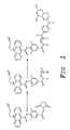
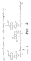
- NSP-DMAE conjugates of pteroic acid one containing a hydrophobic aliphatic (hexamethylene) spacer while the other contained a hydrophilic hexaethylene glycol spacer (see Figures 1 & 2).
- the synthesis of the first tracer was accomplished by reacting NSP-DMAE-NHS with 1,6-hexanediamine (hereinafter, referred to as "HD").
- N 10 -trifluoroacetyl pteroic acid was then condensed with N 10 -trifluoroacetyl pteroic acid followed by removal of the trifluoroacetyl protecting group from the conjugate.
- N 10 -trifluoroacetyl pteroic acid was then condensed with N 10 -trifluoroacetyl pteroic acid followed by removal of the trifluoroacetyl protecting group from the conjugate.
- N 10 -trifluoroacetyl pteroic acid was then condensed with N 10 -trifluoroacetyl pteroic acid followed by removal of the trifluoroacetyl protecting group from the conjugate.
- N 10 -trifluoroacetyl pteroic acid was then condensed with N 10 -trifluoroacetyl pteroic acid followed by removal of the trifluoroacetyl protecting group from the conjugate.
- NSP-DMAE-HEG acridinium ester derivative
- NSP-DMAE-HD-pteroate which does not have a hydrophilic modifier, had the highest nonspecific binding.
- the analogous HEG containing tracer, which does have a hydrophilic spacer was >2-fold lower in nonspecific binding.
- the two (2) folate tracers by virtue of having the hydrophilic HEG spacer had lower nonspecific binding as well.
- the increased binding combined with the lower nonspecific binding of the HEG containing tracers also increased the dynamic range of the folate assay and improved assay sensitivity (Table 3).
- the second tracer was prepared by converting NSP-DMAE-HEG to the glutarate derivative by condensation with glutaric anhydride.
- the carboxylic acid in the resulting adduct (hereinafter referred to as "NSP-DMAE-HEG-glutarate”) was then converted to the NHS ester followed by coupling with one equivalent tobramycin to furnish the tracer.
- This invention thus discloses tracers of the following generic structures A-B-C where A is an analyte of interest such as pteroic acid, folic acid, steroids, therapeutic drugs such as theophylline, phenytoin, digoxin, aminoglycosides such as tobramycin phenobarbital etc.; and where B is either (a) polyethylene glycol of molecular weight 150-5000 or (b) is a polyionic spacer derived from spermine or any polyamine where the internal, but not necessarily all, amines have been modified by hydrophilic molecules such as sultones, anhydrides etc.; and where C is a chemiluminescent or fluorescent label.
- A is an analyte of interest such as pteroic acid, folic acid, steroids, therapeutic drugs such as theophylline, phenytoin, digoxin, aminoglycosides such as tobramycin phenobarbital etc.
- B is either (a) polyethylene
- Preferred acridinium ester conjugates of hydrophilic modifiers include compounds of the following formula: wherein
- M is an acridinium or benzacridinium ester derivative as defined earlier, except R 6 is optionally an alkyl, alkenyl, alkynyl, aryl, aralkyl, having up to 24 carbons and upto 20 heteroatoms selected from the group consisting of nitrogen, oxygen, phosphorus and sulfur; and where L is an amide, ether, thioether, ester or carbamate linkage; and where S is a spacer as defined earlier.
- tobramycin and theophylline tracers are illustrated by the following structures. where S, L and M are as defined earlier.
- NSP-DMAE-HD-pteroate was accomplished as follows ( Figure 1).
- N 10 -Trifluoroacetylpteroic acid (2.5 mg, 6.13 umoles) in DMF (400 uL) was treated with ethyl chloroformate ( 3 uL, 5 equivalents) and diisopropylethyl amine (5.4 uL, 5 equivalents).
- the reaction was stirred at room temperature for 1 hour.
- the solvent was then removed under reduced pressure and the residue was treated with NSP-DMAE-HD (1.4 mg, 2.37 umoles) and diisopropylethylamine (2 uL, 11.3 umoles) .
- the resulting reaction was stirred at room temperature for 4 hours and then concentrated.
- hexaethylene glycol dimethanesulfonate was accomplished as follows. A solution of hexaethyleneglycol ( 1g, 3.54 mmol) in chloroform (10 mL) was cooled in an icebath under nitrogen and treated with methanesulfonyl chloride (603 uL, 2.2 equivalents) and diisopropylethylamine (1.56 mL, 2.5 equivalents). The reaction was warmed to room temperature and stirred under nitrogen. After 2 hours, additional methanesulfonyl chloride (274 uL, 1.0 equivalent) and disopropylethylamine (749 uL, 1.2 equivalents) ' was added.
- diaminoHEG diamino hexaethylene glycol
- NSP-DMAE-HEG NSP-DMAE-HEG
- a solution of diaminoHEG (33 mg, 0.12 mmol) in 2 mL of 1:1, DMF and 0.1 M carbonate pH 9 was treated with NSP-DMAE-NHS (10 mg, 17 umoles).
- the reaction was stirred at room temperature for 16 hours.
- NSP-DMAE-HEG-pteroate was accomplished as follows (see step (III) of Figure 2).
- N 10 -Trifluoroacetyl pteroic acid (5.4 mg, 13.2 umoles) in DMF (0.5 mL) was treated with NHS (7.6 mg, 5 equivalents) and DCC (13.6 mg, 5 equivalents) .
- the reaction was stirred at room temperature under a nitrogen atmosphere. After 2 hours, the reaction was treated with a solution of NSP-DMAE-PEG (3.5 mg, 4.6 umoles) in DMF (400 uL) along with diisopropylethyl amine ( 2 uL, 11.3 umoles).
- NSP-DMAE-HEG-gamma-folate conjugate was accomplished as follows. N-tert-Butoxycarbonyl-L-glutamic acid alpha-tert-butyl ester (25 mg, 0.082 mmol) was dissolved in MeCN (2 mL) and treated with NHS (14.2 mg, 1.5 equivalents) and DCC (25.5 mg, 1.5 equivalents). The reaction was stirred at room temperature for 1.5 hours. . This solution (0.54 mL) was added to a solution of NSP-DMAE-PEG (14 mg, 18.54 umol) in DMF (500 uL) containing diisopropylethylamine (5 ul, 1.5 equivalents).
- NSP-DMAE-HEG-gamma-folate was accomplished as follows. N 10 -Trifluoroacetyl pteroic acid (5 mg, 12.25 umoles) in DMF (1 mL) was treated with isobutylchloroformate (4.7 uL, 3 equivalents) and diisopropylethylamine (8 uL, 4 equivalents). The reaction was stirred at room temperature for 1 hour and was then concentrated under reduced pressure.
- NSP-DMAE-HEG-alpha-folate conjugate was accomplished as follows. N-tert-Butoxycarbonyl-L-glutamic acid g-tert-butyl ester (20 mg, 0.065 mmol) was dissolved in MeCN ( ⁇ 2 mL) and cooled in ice under a nitrogen atmosphere. N-Hydroxysuccinimide (11.4 mg, 1.5 equivalents) and dicyclohexylcarbodiimide (20.3 mg, 0.0985 mmol) were added and the reaction was warmed to room temperature and stirred for one hour.
- NSP-DMAE-HEG 14 mg, 0.0185 mmol
- DMF 0.5 mL
- diisopropylethylamine 7 uL, ⁇ 2 equivalents
- the resulting solution was stirred at room temperature under nitrogen for 24 hours.
- the reaction was then concentrated under reduced pressure.
- the residue was treated with 2 mL of 30% HBr in acetic acid.
- ether was added to precipitate the product which was collected by filtration, rinsed with addtional ether and air dried.
- the crude product (28 mg) was subjected to preparative HPLC as described earlier.
- NSP-DMAE-HEG-alpha-folate was accomplished as follows.
- N 10 -Trifluoroacetylpteroic acid (5 mg, 12.25 umol) in DMF (1 mL) was treated with isobutylchloroformate (4.7 uL, 3 equivalents) and diisopropylethylamine (8 uL, 4 equivalents).
- the reaction was stirred at romm temperature for 1 hour and then concentrated under reduced pressure. The residue was dissolved in DMF (0.5 mL) and evaporated to dryness again.
- ASSAY SENSITIVITY The projected minimum detectable analyte concentration, hereby refered to as sensitivity, was determined as the predicted analyte concentration at two standard deviations from the zero dose response.
- Fractional nonspecific binding (fNSB) in competitive assay is calculated as the quotient of the projected, assymptotic lower limit of y at infinite dose, y ⁇ , and the total chemiluminescent signal input T.
- Fractional NSB is a measure of the binding interaction of the conjugate for the solid phase that does not involve the specifically preferred binding association between the conjugate and the binding protein or antibody on the solid phase.
- Elevated fNSB is undesirable and may result from one or more of a number of different factors; hydrophobic interaction, exacerbated by the excessive hydrobicity of a conjugate; ionic or polar interactions promoted through the charge density or polarity of the conjugate; and/or a specific but undesirable biological binding interaction. If the assay precision remains unaffected while there is a significant increase in NSB, the apparent slope of the dose response curve will decrease more rapidly as the B 0 exceeds the detector's linearity limit.
- reaction mixture containing 150 ⁇ l of folate standard, 50 ⁇ l of DTT Reagent and 75 ⁇ l of Releasing Agent, was incubated for 2.5 min. at 37°C.
- the solid phase was collected on an array of permanent magnets and washed with deionized water to remove unbound tracer.
- the chemiluminescent reaction was initiated, as described previously. Chemiluminescence data were collected as photons detected by the ACS:180 and expressed in relative light units (RLUs).
- NSP-DMAE-HEG-gamma-folate conjugate The best folate assay sensitivity was attained with NSP-DMAE-HEG-gamma-folate conjugate.
- the projected minimum detectable analyte concentration was determined from both the folate concentration at two standard deviations from the 0.00 nM folate dose-response, B 0 -2sigma (n-1) .
- the NSP-DMAE-HEG-gamma-folate conjugate issued the lowest detectable folate concentration, which was followed by the NSP-DMAE-HEG-alpha-folate and NSP-DMAE-HEG-pteroate conjugates in that order.
- the NSP-DMAE-HD-pteroate tracer was the least sensitive conjugate as a result of the comparatively low B 0 , curtailed dynamic range and elevated fractional NSB (fNSB).
- the tracer structural differences may be ranked as follows in accordance with their degree of influence on sensitivity.
- the folate substitution for pteroate in the tracer structure resulted in an increase in assay sensitivity of at least 2.7-fold when the results of the NSP-DMAE-HEG-alpha-folate tracer were compared with those of the NSP-DMAE-HEG-pteroate tracer. This reflects the relative importance of tracer and analyte structural similarity with regards to folate assay sensitivity.
- NSP-DMAE-FOLATE OR PTEROATE CONJUGATE FRACTIONAL NONSPECIFIC BINDING Fractional NSB (hereinafter referred to as "fNSB") was significantly reduced with the incorporation of the hydrophilic HEG-spacer into the conjugate structure.
- the fNSB of the NSP-DMAE-HD-pteroate conjugate was at least 2.2-fold higher than that of the other pteroate or folate based conjugates.
- the hydrophobic HD spacer increased the nonspecific hydrophobic interaction of the NSP-DMAE-HD-pteroate conjugate with the solid phase.
- hydrophilic HEG-spacer and the correct orientation of the entire folate moiety are important structural properties for increasing the %B 0 /T.
- the %B 0 /T for NSPDMAE-HEG-gamma-folate conjugate was 3.5-fold higher that that of the NSPDMAE-HD-pteroate conjugate, indicating that both the incorporation of the hydrophilic HEG-spacer and linkage via the gamma-glutamate carboxyl are required for higher binding values.
- NSP-DMAE-tobramycin conjugate was accomplished as follows. Tobramycin (1.45 mg, 3.3 umoles) was dissolved in 1:1, DMF/0.1 M carbonate pH 9 (1 mL) and treated with a solution of NSP-DMAE-NHS ester (2 mg, 3.3 umoles) in DMF (0.2 mL) added periodically at five minute intervals. The reaction was stirred at room temperature for 2 hours and then at 4oC for an additional 24-36 hours. The product was purified by preparative HPLC using a C18 column (7.8 mm x 30 cm) and a 40 min. gradient of 10-->60% MeCN/0.1 M TEAA pH 5 at a flow rate of 2.3 mL/min.
- tracers acridinium ester-tobramycin conjugates
- tobramycin from tobramycin-containing standards Bayer Diagnostics, Walpole, MA
- Tobramycin standards contained tobramycin at concentrations of 0.00, 1.07, 2.14, 4.28, 8.56, 17.1, 25.7 and 34.2 ⁇ M.
- the reaction is initiated by mixing 50 ⁇ l tobramycin standard, 400 ⁇ l of solid phase and 100 ⁇ l of tracer. The reaction mixture was incubated for 7.5 minutes at 37°C. The solid phase was collected on an array of permanent magnets and washed with deionized water to remove any unbound tracer. The chemiluminescent reaction was initiated, as described previously. Data were collected as photons detected by the ACS:180 and expressed as RLU. A non-linear, inverse relationship exists between the tobramycin concentration present in the standard and the RLUs detected by the ACS:180. The acquired data was processed as previously described for the folate assay data treatment.
- TOBRAMYCIN ASSAY PRECISION Within run precision was excellent for both tobramycin tracers, with % C.Vs. being well below 10 % over the entire standard curve. There was no difference in overall precision between the two conjugates.
- A1 NSP-DMAE-tobramycin conjugate
- A2 NSP-DMAE-HEG-glutarate-tobramycin conjugate
- TOBRAMYCIN ASSAY ACCURACY Accuracy evinced as percent error with predicted 4PL values was acceptable for both tobramycin tracers, being for the most part within ⁇ 5 % over the entire standard curve. There was no difference in overall accuracy for these conjugates.
- Tobramycin Assay Accuracy % S [tobramycin] in microM A1 A2 0.00 0.01 0.00 1.07 -0.11 -0.43 2.14 0.05 1.91 4.28 1.21 -1.89 8.56 -2.69 -2.61 17.1 -0.74 -0.74 25.7 0.80 2.54 34.2 2.90 5.54
- A1 NSP-DMAE-tobramycin conjugate
- A2 NSP-DMAE-HEG-glutarate-tobramycin conjugate
- TOBRAMYCIN ASSAY SENSITIVITY The best tobramycin assay sensitivity was achieved using the NSP-DMAE-HEG-glut-tobramycin conjugate.
- the predicted sensitivity from assay results using the NSP-DMAE-HEG-glut-tobramycin conjugate was 1.7-fold lower than that using NSP-DMAE-tobramycin conjugate.
- the hydrophilic HEG-glut-spacer must be integrated into the tobramycin conjugate structure in order to attain improved tobramycin assay sensitivity.
- the increase in sensitivity resulted from the steeper incline of the slope when the HEG-glut-spacer was incorporated into the conjugate.
- A1 NSP-DMAE-tobramycin conjugate
- A2 NSP-DMAE-HEG-glutarate-tobramycin conjugate
- NSP-DMAE-HD-theophylline conjugate was accomplished as follows. 8-Carboxypropyltheophylline (10 mg, 0.038 mmol) in DMF (3 mL) was treated with N-hydroxysuccinimide (21.6 mg, 0.188 mmol) and dicyclohexylcarbodiimide (38.8 mg, 0.188 mmol). The resulting solution was stirred at room temperature for 16 hours. HPLC analysis on a C18 column (4.6 mm x 300 mm) using a gradient of 10-->60% MeCN/water (each containing 0.05% TFA) over 40 minutes at a flow of 1 mL/min.
- spermine disulfonate (SPDS) was accomplished as follows. Bis(phthalimido)spermine disulfonate (0.53 g) was dissolved in methanol (15-20 mL) and treated with hydrazine (0.5 mL). The resulting solution was stirred at room temperature for 24 hours and then concentrated under reduced pressure. The residue was dissolved in ⁇ 5 mL of 20% ammonium hydroxide, 80% methanol and evaporated to dryness. This process was repeated once. Finally, the residue was dissolved in a mixture of methanol (1 mL), water (1.5 mL) and triethylamine (1.5 mL) and the solution was evaporated to dryness again.
- SPDS spermine disulfonate
- NSP-DMAE-SPDS was accomplished as follows.
- Spermine disulfonate 25 mg, 0.056 mmol
- the reaction was stirred at room temperature for 16 hours.
- spermine dicarboxylate was accomplished as follows.
- reaction mixture was then treated with hydrazine (0.45 mL, ⁇ 10 eq.) and methanol (10 mL). The reaction was stirred at room temperature. After 1-2 hours, a crystalline precipitate appeared in the reaction mixture. After 3-4 hours, total reaction time, the reaction was concentrated under reduced pressure. The residue was suspended in acetone and filtered. The precipitate was rinsed with acetone and dissolved in water (50 mL) with triethyl amine (1.5 mL). This was concentrated under reduced pressure to afford a white powder. MALDI-TOF MS 403.7 obs. (402.49).
- NSP-DMAE-SPDC was accomplished as follows.
- Spermine dicarboxylate 45 mg, 0.112 mmol
- 2 mL of 0.1 M carbonate pH 9 (adjusted with 5N NaOH)
- NSP-DMAE-NHS ester 10.5 mg, 0.0178 mmol
- DMF 2 mL
- the reaction was stirred at room temperature for 16 hours.
- the product was isolated by preparative HPLC on a C18 column (20 x 300 mm) using a 40 min. gradient of 0-->40% MeCN/water (each containing 0.05% TFA) at a flow rate of 16 mL/min.
- NSP-DMAE-SPDC-theophylline conjugate was accomplished as follows. NSP-DMAE-SPDC (1 mg, 0.00114 mmol) was dissolved in 0.1 mL DMF and 8-carboxypropyltheophylline (1 mg, 0.00262 mmol) was added along with diisopropylethylamine (2 uL, 2 eq.). The reaction was stirred at room temperature for 16 hours and was then purified directly by HPLC on a C18 column ( 20 x 300 mm) using a gradient of 10-->60% MeCN/water (each containing 0.05% TFA) over 40 min. at a flow rate of 16 mL/min.
- tracers acridinium ester-theophylline conjugates
- theophylline from theophylline-containing standards (Bayer Diagnostics, Walpole, MA) compete for a limited amount murine IgG, monoclonal anti-theophylline antibody which was covalently coupled to a paramagnetic particle solid phase.
- Theophylline standards contained theophylline in concentrations of 0.00, 6.94, 13.9, 27.7, 55.5, 111 and 222 ⁇ M.
- the solid phase was collected on an array of permanent magnets and washed twice with deionized water to remove unbound tracer.
- the chemiluminescent reaction was initiated, as described previously.
- Data were collected as photons detected by the ACS:180 and expressed as RLU.
- the acquired data was processed as previously described for the folate assay data treatment.
- NSP-DMAE-HEG-theophylline and NSP-DMAE-SPDS-theophylline gave minimal detectable doses that were higher than that of the NSP-DMAE-HD-theophylline.
- the decreased sensitivity in the case of NSP-DMAE-HEG-theophylline and NSP-DMAE-SPDS-theophylline may result from slightly elevated imprecision for the zero dose.
- Theophylline Assay Sensitivity & Binding Date least detectable dose at B 0 -2sigma (n-1) in microM A1 A2 A3 A4 0.272 0.497 0.311 0.089 [theophylline] in microM Relative Light Units 0.0 715,394 539,268 769,617 648,559 6.94 511,553 351,686 442,911 317,389 13.0 392,914 261,193 313,980 214,678 27.7 268,471 173,922 201,019 133,152 55.5 165,325 105,453 118,487 77,581 111 96,836 60,938 66,996 44,253 222 57,387 35,158 37,668 25586 dynamic range 658,007 504,110 731,949 622,973 fNSB 1.27 ⁇ 10 -2 6.40 ⁇ 10 -3 3.47 ⁇
- NSP-DMAE-THEOPHYLLINE CONJUGATE FRACTIONAL NONSPECIFIC BINDING Fractional NSB was significantly reduced with the incorporation of the hydrophilic spacers into the conjugate structures.
- the fNSB of the NSP-DMAE-SPDS-theophylline was the lowest overall being 3.7-fold lower than that of the NSP-DMAE-HD-theophylline conjugate.
- the NSP-DMAE-SPDC-theophylline and NSP-DMAE-HEG-theophylline conjugates had fNSBs that were 3.1-and 2.0-fold lower, respectively. The more highly polar or charged spacers confer lower fNSBs upon their respective conjugates.
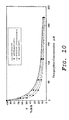
- the dose response curves of %B/B 0 vs. theophylline concentration indicate that the increased hydrophilicity of the spacer increases the initial slope of the dose response curve, thereby increasing sensitivity of the assay assuming an equivalence in precision.
- the NSP-DMAE-SPDC-theophylline conjugate elicited the steepest decline in the initial slope followed by 1 st NSP-DMAE-SPDS-theophylline; 2 nd NSP-DMAE-HEG-theophylline; & 3 rd NSP-DMAE-HD-theophylline (in that order).
Landscapes
- Health & Medical Sciences (AREA)
- Immunology (AREA)
- Life Sciences & Earth Sciences (AREA)
- Engineering & Computer Science (AREA)
- Molecular Biology (AREA)
- Biomedical Technology (AREA)
- Chemical & Material Sciences (AREA)
- Hematology (AREA)
- Urology & Nephrology (AREA)
- Food Science & Technology (AREA)
- Biochemistry (AREA)
- Cell Biology (AREA)
- Biotechnology (AREA)
- Medicinal Chemistry (AREA)
- Physics & Mathematics (AREA)
- Analytical Chemistry (AREA)
- Microbiology (AREA)
- General Health & Medical Sciences (AREA)
- General Physics & Mathematics (AREA)
- Pathology (AREA)
- Peptides Or Proteins (AREA)
- Investigating Or Analysing Materials By The Use Of Chemical Reactions (AREA)
- Investigating Or Analysing Biological Materials (AREA)
- Measuring Or Testing Involving Enzymes Or Micro-Organisms (AREA)
Abstract
Description
- The present invention is useful in bioanalytical applications and is generally directed to detectable chemiluminescent acridinium ester labels having hydrophilic modifiers; to compositions, complexes and/or conjugates which include such labels; and to processes for performing bioanalytical assays for target analytes which use such labels.
- Acridinium esters are extremely useful chemiluminescent labels that have been extensively used in the field of immunoassays as well as nucleic acid assays. Each of the following patent documents is both (a) incorporated herein by reference in its respective entirety; and (b) directed to varying aspects of bioanalytical applications of acridinium ester compounds. EP0263657; US4745181;EP0353971; EP0361817; US4918192; US5110932; US5227489; US5241070; EP0617288; WO9421823; US5395752; EP0661270; US5449556; WO9527702; US5538901; US5595875; EP0754178; US5656426; US5656500; US5663074; US5702887; WO9854574; US5879894; WO9911813; WO0009487; EP0982298; EP0988551; WO0031543; EP1009852; US6080591; EP1049933; US6165800; WO0109372 & EP1104405.
- Certain particular detectable chemiluminescent acridinium ester labels lacking hydrophilic modifiers are well-known in the art - e.g., 2',6'-dimethyl-4'-[N-succinimidyloxycarbonyl]phenyl-10-methyl-9-acridine carboxylate and 2',6'-dimethyl-4'-[N-succinimidyloxycarbonyl]phenyl-10-sulfopropyl-9-acridine carboxylate {each label being hereinafter referred to as, respectively, "DMAE-NHS" and "NSP-DMAE-NHS"} - and are being commercialized for immunoassay instrument systems available from Bayer Corporation, Business Group Diagnostics, 511 Benedict Avenue, Tarrytown, New York 10591-5097. For the reader's convenience, the structure of each of these compounds is depicted below.

- As previously stated, the present invention is widely directed to detectable chemiluminescent acridinium ester labels having hydrophilic modifiers. In several preferred embodiments of the inventive labels, we have incorporated two (2) types of structural elements in NSP-DMAE and we have found that these modifications allow for the preparation of unique hapten tracers which show enhanced performance in immunoassays. By employing three (3) different clinically relevant analytes -namely- (the vitamin folate, the asthma drug theophylline, and the aminoglycoside antibiotic tobramycin) we have demonstrated the generality of our findings.
- Before examining in depth the present inventive labels, a brief overview of assay formats is presented below. Competitive immunoassays commonly: (a) employ a format where a conjugate of a fluorescent or chemiluminescent label to an analyte of interest is used as a tracer in the assay; and (b) utilizes a solid support. A typical architecture for such a competitive assay consists of three components -namely- a tracer, a sample containing the analyte of interest, and a method for the separation of bound and unbound analyte. (Note that in homogenous immunoassays, however, no separation is performed). For example, immobilization of folic acid binding protein on a solid support such as paramagnetic particles (hereinafter referred to as "PMP") provides a means for achieving such a separation (magnetic) of free and bound analyte (which analyte in this case would be folic acid). When the tracer is included in the assay, it competes with the analyte from the sample for binding to the immobilized protein. Increased levels of analyte in the assay result in less tracer being bound to the immobilized protein.
- As described in detail below (and as further exemplified later) two (2) types of spacers which are particularly useful for the preparation of hapten tracers - (a) nonionic polyethylene glycol; and (b) polyionic spermine disulfonate and polyionic spermine dicarboxylate - have been developed.
- Polyethylene glycol (hereinafter referred to as "PEG") is a well known polymer. It is biocompatible, soluble in both aqueous and organic solvents, nontoxic, and nonimmunogenic. In the prior art, it has been extensively used as a modifier of a variety of molecules ranging from small molecular weight drugs to large proteins as well as large aggregates such as liposomes. PEG conjugates of drugs exhibit improved solubility and are longer-lived in the bloodstream. PEG modification of proteins and peptides improves solubility, confers resistance to proteolysis, and reduces immunogenicity. PEG modification of oligonucleotides increases solubility and confers nuclease stability. PEG modification of lipids permits the preparation of PEG-grafted liposomes that are sterically stabilized and display improved blood circulation times. An excellent review of the prior art in the uses of PEG is described by S. Zalipsky in Bioconjugate Chemistry, 1995, 6, 150-165 (which is incorporated herein by reference in its entirety). The use of PEG to modify the properties of fluorescent dyes is also described in the prior art. PEG-modified fluorescent porphyrin and phthalocyanine dyes have been shown to exhibit decreased aggregation behavior in aqueous solution as well as diminished non-specific binding to components of human serum such as HSA (Human Serum Albumin). These conjugates also show extended fluorescence decay times (PCT/US91/03424 and PCT/US91/03426). Applications of such conjugates in fluorescence immunoassays and in vivo imaging and in vivo tumor therapy were proposed by the same authors.
- Notwithstanding the above uses of PEG, modification of acridinium esters with polyethylene glycol has not been described previously. Likewise, polyionic spacers devised using the polyamine spermine as a scaffold for introducing ionic functional groups have also not been reported. We find that these latter molecules are also extremely useful for modifying acridinium esters. The synthesis and applications of these modified acridinium esters follow.
- As mentioned earlier, the vitamin folic acid is a clinically important analyte which is commonly measured using immunoassay techniques. As a closely-related compound, pteroic acid is a simplified, structural variant of folic acid which lacks the glutamate moiety normally found in folic acid. We prepared two (2) different NSP-DMAE conjugates of pteroic acid, one containing a hydrophobic aliphatic (hexamethylene) spacer while the other contained a hydrophilic hexaethylene glycol spacer (see Figures 1 & 2). The synthesis of the first tracer was accomplished by reacting NSP-DMAE-NHS with 1,6-hexanediamine (hereinafter, referred to as "HD"). The resulting acridinium ester derivative (hereinafter referred to as "NSP-DMAE-HD") was then condensed with N10-trifluoroacetyl pteroic acid followed by removal of the trifluoroacetyl protecting group from the conjugate. To prepare an analogous tracer with a PEG spacer, a short diaminohexaethylene glycol was synthesized from commercially available, hexaethylene glycol. The two hydroxyl groups in hexaethylene glycol were converted to methane sulfonate esters which were subsequently displaced with azide. Reduction of the diazidohexaethylene glycol afforded diaminohexaethylene glcyol which then was condensed with NSP-DMAE-NHS. The resulting acridinium ester derivative (hereinafter referred to as "NSP-DMAE-HEG") was coupled to pteroic acid as discussed above.
- The assay performance of these two (2) different NSP-DMAE-pteroate conjugates was then evaluated in a folate immunoassay (Example 5, Tables 1-3 & Figure 3). In this assay format, the folate binding protein is immobilized onto PMP and the two (2) tracers compete with the analyte folic acid. The dose-response curves are shown in Figure 3. The methodology used for generating assay data and the definitions of various assay parameters are explained in Example 5. Tables 1-3 summarize data relating to assay precision, assay accuracy, and assay sensitivity. Incorporation of the hexaethylene glycol spacer between the acridinium ester and pteroate moieties increased tracer binding more than two-fold; thus the fraction of bound tracer defined as B/T increased from 0.53% to 1.15% for the tracer with the PEG spacer (Table 3). Clearly, the polyethylene glycol spacer alleviates any steric interference to binding in the PEG-containing tracer. We next synthesized a NSP-DMAE-folate conjugate also containing the hexaethylene glycol spacer (Figure 4). Since it is known in the prior art that the alpha-carboxylate in folic acid must remain free for good binding to folate binding proteins (Wang, S. et al. Bioconjugate Chem. 1996, 7, 56-62), we first synthesized a specific gamma-linked folate tracer. Specifically, this was accomplished by condensing N-tert-butoxycarbonyl glutamic acid alpha-tert-butyl ester with NSP-DMAE-HEG (Figure 4). Removal of the protecting groups from the resulting conjugate, coupling with N10-trifluoroacetyl pteroic acid followed by removal of the trifluoroacetyl group afforded the gamma-linked folate-NSP-DMAE tracer incorporating the short, polyethylene glycol spacer. Evaluation of this tracer in the folate immunoassay, indeed showed even better binding (B/T 1.88%) than the pteroate tracer as would be anticipated. We also prepared a specific alpha-linked folate tracer starting with N-tert-butoxycarbonyl glutamic acid gamma-tert-butyl ester and following the same sequence of reactions described above (Figure 5). The resulting alpha-linked folate tracer when evaluated in the folate immunoassay exhibited diminished binding owing to the lack of a free alpha-carboxyl group. While there were no discernible differences in assay precision or assay accuracy among the various tracers, the HEG containing tracers did exhibit lower nonspecific binding (Table 3). Thus, NSP-DMAE-HD-pteroate, which does not have a hydrophilic modifier, had the highest nonspecific binding. The analogous HEG containing tracer, which does have a hydrophilic spacer, was >2-fold lower in nonspecific binding. The two (2) folate tracers by virtue of having the hydrophilic HEG spacer had lower nonspecific binding as well. The increased binding combined with the lower nonspecific binding of the HEG containing tracers also increased the dynamic range of the folate assay and improved assay sensitivity (Table 3).
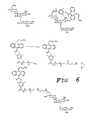
- In an immunoassay for the aminoglycoside antibiotic tobramycin, we again compared the assay performance of two tobramycin-NSP-DMAE tracers, one of which contained the (hydrophilic) hexaethylene glycol spacer while the other did not. Procedures for generating antibodies for tobramycin as well as for site-specific modification of tobramycin with other small molecules have been described previously (Singh, P. et al. Can. J. Chem., 1984, 62, 2471-2477). In tobramycin, the 6'-amine is the most reactive (Figure 6). Thus, treatment of the aminoglycoside with one equivalent of NSP-DMAE-NHS furnished a 1:1 tobramycin-NSP-DMAE tracer. The second tracer was prepared by converting NSP-DMAE-HEG to the glutarate derivative by condensation with glutaric anhydride. The carboxylic acid in the resulting adduct (hereinafter referred to as "NSP-DMAE-HEG-glutarate") was then converted to the NHS ester followed by coupling with one equivalent tobramycin to furnish the tracer.
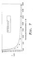
- Examination of the two (2) tracers in an immunoassay for tobramycin revealed that while overall binding of the two tracers to a tobramycin antibody on PMP was similar, the nonspecific binding of the hydrophilic PEG-containing tracer was more than 2.5-fold lower than the conventional tracer (Tables 4-6, Figure 7 in Example 8). Since increased nonspecific binding is most often related to hydrophobicity, it is remarkable that even for a highly, water-soluble analyte such as tobramycin, introduction of the polyethylene glycol modifier in the tracer is so beneficial. While assay precision and assay accuracy was similar for the two (2) tracers, tobramycin assay sensitivity was significantly better (1.7x lower, Table 6) for the HEG containing tracer.
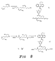
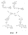
- In the case of the asthma drug analyte theophylline, in addition to two (2) NSP-DMAE-theophylline tracers incorporating a six-carbon spacer and the hexaethylene glycol spacer (Figures 8 & 9) we prepared two (2) new tracers which contain polyionic spacers. The first two (2) tracers were simply prepared by condensing the NHS ester of 8-carboxypropyltheophylline with either NSP-DMAE-HD or NSP-DMAE-HEG. The polyionic spacers were derived from the polyamine, spermine and were prepared by first converting the two primary amines to phthalimido groups. The resulting compound, bis(phthalimido)spermine was either alkylated at the two secondary amines by heating in neat 1,3-propane sultone or acylated with succinic anhydride (Figure 8). Removal of the phthalimido protecting groups with hydrazine afforded spermine disulfonate (hereinafter referred to as "SPDS") and spermine dicarboxylate (hereinafter referred to as "SPDC"). These new spacers were coupled to NSP-DMAE-NHS and the resulting NSP-DMAE derivatives were coupled to 8-carboxytheophylline (Figure 9). All four (4) theophylline tracers were then evaluated in a theophylline immunoassay (Example 13, Tables 7-9, Figure 10). The theophylline tracer containing the hydrophilic PEG spacer showed lower nonspecific binding (2-fold) when compared to the tracer with the nonhydrophilic six-carbon spacer. Tracers containing the SPDS spacer and the SPDC spacer had even lower nonspecific binding (3.7 and 3.1-fold lower than the hydrophobic HD spacer respectively). While binding, assay precision, and assay accuracy was similar for the four (4) tracers, the tracer containing the polycarboxylate spacer SPDC increased assay sensitivity >3-fold. The other hydrophilic spacers did not show such improvement in this specific assay even though both tracers did show lower nonspecific binding.
- The above set of results clearly demonstrate the utility of hydrophilic spacers in acridinium ester-hapten conjugates. No one spacer is beneficial in all assays, but by selection, a hydrophilic spacer is easily identified that confers maximal benefits on the tracer in terms of assay performance. We have disclosed two types (nonionic and polyionic) of spacers that are useful in this regard. It is also evident that from the methodology provided by the current invention, one with ordinary skill in the art could apply the same methodology for the preparation of a variety of tracers using different analytes and different labels. This invention thus discloses tracers of the following generic structures
A-B-C
where A is an analyte of interest such as pteroic acid, folic acid, steroids, therapeutic drugs such as theophylline, phenytoin, digoxin, aminoglycosides such as tobramycin phenobarbital etc.; and
where B is either (a) polyethylene glycol of molecular weight 150-5000 or (b) is a polyionic spacer derived from spermine or any polyamine where the internal, but not necessarily all, amines have been modified by hydrophilic molecules such as sultones, anhydrides etc.; and
where C is a chemiluminescent or fluorescent label. - Preferred acridinium ester conjugates of hydrophilic modifiers include compounds of the following formula:wherein

- R1 is alkyl, alkyenyl, alkynyl, aryl, sulfoethyl, sulfopropyl, sulfobutyl, or aralkyl having up to 24 carbons and up to 20 heteroatoms selected from the group consisting of nitrogen, oxygen, phosphorus and sulfur; and
- R2, R3, R5, R7 are hydrogen, amino, hydroxyl, halide, nitro, -CN, -SO3H, -SCN, -OR, NHCOR, -COR, -COOR, or -CONHR wherein R is alkyl, alkenyl, alkynyl, aryl, aralkyl, having up to 24 carbons and upto 20 heteroatoms selected from the group consisting of nitrogen, oxygen, phosphorus and sulfur; and
- R4 and R8 are alkyl, alkenyl, alkynyl, aralkyl or alkoxyl having upto 8 carbons with no branching wherein the side chain groups have more than 2 carbons;
- R6 represents the following substitutions: R6 = R-L-S-R10 where R is optionally alkyl, alkenyl, alkynyl, aryl, aralkyl, having up to 24 carbons and upto 20 heteroatoms selected from the group consisting of nitrogen, oxygen, phosphorus and sulfur, L is one of the following linkages: ether, thioether, amide, ester or carbamate;
- S is polyethylene glycol from 300 to 5000 molecular
weight; or the following structures

- R6 can be attached, alternatively, at a position of the phenoxy ring, which is meta to the ester linkage (in this case R5 or R7 is attached para to the ester linkage); and
- R10 is an electrophile , a leaving group , or a nucleophile.
-
- Preferred embodiments of a pteroate tracer and a folate tracer are illustrated by the following structures.
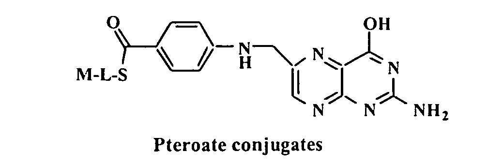 where M is an acridinium or benzacridinium ester derivative as defined earlier, except R6 is optionally an alkyl, alkenyl, alkynyl, aryl, aralkyl, having up to 24 carbons and upto 20 heteroatoms selected from the group consisting of nitrogen, oxygen, phosphorus and sulfur; and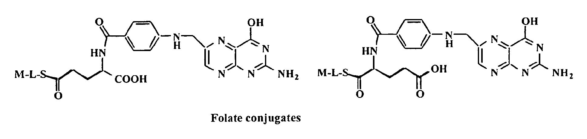
where L is an amide, ether, thioether, ester or carbamate linkage; and
where S is a spacer as defined earlier. - Preferred embodiments of tobramycin and theophylline tracers are illustrated by the following structures.where S, L and M are as defined earlier.

- The following nonlimiting, representative examples are presented for the purposes of illustration only and are not intened to limit the scope of the patent protection to which the instant invention is entitled and to which protection is defined only by the appended claims.
- The synthesis of NSP-DMAE-HD was accomplished as follows (Figure 1). 1,6-Diaminohexane (49 mg, 0.42 mmol) in DMF (1 mL) and 0.1 M carbonate pH 9 (1 mL) was treated with NSP-DMAE-NHS (25 mg, 0.042 mmol) in DMF (1 mL). The reaction was stirred at room temperature for 16 hours and was then purified directly by preparative HPLC using a C18 column (20 x 250 mm) and a 40 min. gradient of 10-->60% MeCN/water each containing 0.05% trifluoroacetic acid (TFA). The product eluted at ∼21 minutes. The HPLC fraction containing the product was concentrated under reduced pressure and then lyophilized to dryness to afford a yellow solid. Yield = 25 mg (quant.), MALDI-TOF MS 591 obs., (591.73 calc.).
- Next, the synthesis of NSP-DMAE-HD-pteroate was accomplished as follows (Figure 1). Next, N10-Trifluoroacetylpteroic acid (2.5 mg, 6.13 umoles) in DMF (400 uL) was treated with ethyl chloroformate ( 3 uL, 5 equivalents) and diisopropylethyl amine (5.4 uL, 5 equivalents). The reaction was stirred at room temperature for 1 hour. The solvent was then removed under reduced pressure and the residue was treated with NSP-DMAE-HD (1.4 mg, 2.37 umoles) and diisopropylethylamine (2 uL, 11.3 umoles) . The resulting reaction was stirred at room temperature for 4 hours and then concentrated. The residue was dissolved in methanol and filtered. The filtrate was purified by HPLC on a C18 column (7.8 mm x 25 cm) using a 40 min. gradient of 0-->60% MeCN/water each containing 0.05 % TFA; Rt (product) = ∼27 minutes. The HPLC fraction containing the product was lyophilized to dryness to afford a yellow solid. Yield = 0.6 mg (26%); MALDI-TOF MS 983.47 obs. (982.01 calc.).
- The above material was stirred in 0.1 M piperidine (500 uL) at room temperature for 4 hours and then the product was purified directly by HPLC as described above; Rt (product) =∼26 minutes. The HPLC fraction containing the product was lyophilized to yield a yellow solid. Yield = ∼0.2 mg, MALDI-TOF MS 889.48 obs., (886 calc.).
- The synthesis of hexaethylene glycol dimethanesulfonate was accomplished as follows. A solution of hexaethyleneglycol ( 1g, 3.54 mmol) in chloroform (10 mL) was cooled in an icebath under nitrogen and treated with methanesulfonyl chloride (603 uL, 2.2 equivalents) and diisopropylethylamine (1.56 mL, 2.5 equivalents). The reaction was warmed to room temperature and stirred under nitrogen. After 2 hours, additional methanesulfonyl chloride (274 uL, 1.0 equivalent) and disopropylethylamine (749 uL, 1.2 equivalents) ' was added. After two more hours at room temperature, the reaction was diluted with chloroform and the resulting solution was washed twice with aqueous ammonium chloride followed by brine. The chloroform solution was then dried over magnesium sulfate, filtered and concentrated under reduced pressure. A light yellow oil was obtained. Yield = 1.38 g (89%). TLC (10% methanol, 90% chloroform) Rf (product) = 0.64; Rf (starting material) = 0.42.
- Next, the synthesis of diazido hexaethylene glycol was accomplished as follows. A solution of hexaethylene glycol dimethanesulfonate (0.5 g, 1.14 mmol) in DMF (5 mL) was treated with sodium azide ( 0.31 g, 4.76 mmol). The reaction was heated in an oil-bath at 110°C under a nitrogen atmosphere for 8 hours. The reaction was then cooled to room temperature and stirred for an additional 16 hours. The DMF was then removed under reduced pressure and the residue was partitioned between chloroform and brine. The chloroform layer was separated, dried over magnesium sulfate, filtered and concentrated under reduced pressure to afford an oil. Yield = 0.442 g (quant.); TLC (5% methanol, 95% chloroform) Rf (product) = 0.59, Rf (starting material) = 0.35.
- Next, the synthesis of diamino hexaethylene glycol (hereinafter referred to as "diaminoHEG") was accomplished as follows (Figure 2). Next, a solution of diazido hexaethylene glycol (0.44 g1.32 mmol) in ethyl acetate ( 15 mL) was treated with 10% Pd on activated carbon ( 95 mg) and the black reaction mixture was hydrogenated at room temperature. After 16 hours at room temperature, the reaction was filtered and the filtrate was concentrated under reduced pressure to afford an oil. Yield = 0.26 g (70%), MALDI-TOF MS 280 obs. 280 calc., TLC (45% methanol, 50% chloroform, 5% ammonium hydroxide) Rf = 0.29.
- Next, the synthesis of NSP-DMAE-HEG was accomplished as follows (Figure 2). Next, a solution of diaminoHEG (33 mg, 0.12 mmol) in 2 mL of 1:1, DMF and 0.1 M
carbonate pH 9 was treated with NSP-DMAE-NHS (10 mg, 17 umoles). The reaction was stirred at room temperature for 16 hours. The product was purified directly by preparative HPLC on a C18 column (20 mm x 300 cm) using a 40 min. gradient of 0-->60% MeCN/water each containing 0.05%; Rt (product) =∼21 minutes. The HPLC fraction containing the product was lyophilized to dryness to afford a yellow solid. Yield = 10. 6 mg (83%), MALDI-TOF MS 757.39 obs. , (755.89 calc.). - Next, the synthesis of NSP-DMAE-HEG-pteroate was accomplished as follows (see step (III) of Figure 2). Next, N10-Trifluoroacetyl pteroic acid (5.4 mg, 13.2 umoles) in DMF (0.5 mL) was treated with NHS (7.6 mg, 5 equivalents) and DCC (13.6 mg, 5 equivalents) . The reaction was stirred at room temperature under a nitrogen atmosphere. After 2 hours, the reaction was treated with a solution of NSP-DMAE-PEG (3.5 mg, 4.6 umoles) in DMF (400 uL) along with diisopropylethyl amine ( 2 uL, 11.3 umoles). The resulting solution was stirred at room temperature under a nitrogen atmosphere for 16 hours. The reaction mixture was then purified directly by preparative HPLC on a C18 column (7.8 mm x 300 cm) as described earlier; Rt (product) = -24 minutes; MALDI-TOF MS 1148.71.92 obs., (1146.17 calc.).
- Next, the above conjugate was stirred in 400 uL of 0.1 M piperidine at room temperature for 1 hour. The reaction was then lyophilized to dryness to afford a yellow solid. HPLC Rt = -21 minutes, MALDI-TOF MS 1051.92 obs., (1050.16 calc.).
- The synthesis of NSP-DMAE-HEG-gamma-folate conjugate was accomplished as follows. N-tert-Butoxycarbonyl-L-glutamic acid alpha-tert-butyl ester (25 mg, 0.082 mmol) was dissolved in MeCN (2 mL) and treated with NHS (14.2 mg, 1.5 equivalents) and DCC (25.5 mg, 1.5 equivalents). The reaction was stirred at room temperature for 1.5 hours. . This solution (0.54 mL) was added to a solution of NSP-DMAE-PEG (14 mg, 18.54 umol) in DMF (500 uL) containing diisopropylethylamine (5 ul, 1.5 equivalents). After 2-3 hours additional diisopropylethylamine (2.5 ul) was added along with an additional 540 uL of the active ester solution from above. The resulting reaction was stirred at room temperature for 16 hours. The solvent was then removed under reduced pressure and the residue was dissolved in 2 mL MeCN. This was filtered through glass wool and the filtrate was concentrated under reduced pressure. The crude product was deblocked by stirring in 1 mL of 30% HBr in acetic acid for 2 hours. The product was precipitated with the addition of ether (10 mL). The ether was decanted and the residue was purified directly by HPLC using the same solvent system described earlier, Rt (product) = -20.5 minutes. The HPLC fraction containing the product was lyophilized to dryness to afford 2.8 mg (20%) of the product as a yellow solid. MALDI-TOF MS 888.67 obs. (885.0 calc.).
- Next, the synthesis of NSP-DMAE-HEG-gamma-folate was accomplished as follows. N10-Trifluoroacetyl pteroic acid (5 mg, 12.25 umoles) in DMF (1 mL) was treated with isobutylchloroformate (4.7 uL, 3 equivalents) and diisopropylethylamine (8 uL, 4 equivalents). The reaction was stirred at room temperature for 1 hour and was then concentrated under reduced pressure. The residue was dissolved in DMF (0.5 mL) and 170 uL of this solution was added to NSP-DMAE-PEG-gamma-glutamate (1.8 mg, 2.03 umoles) along with diisopropylethylamine (1 uL). The reaction was stirred at room temperature for 16 hours and then concentrated under reduced pressure. The residue was dissolved in DMF (1 mL) and purified by HPLC using the same conditions described earlier, Rt (product) = ∼25.5 minutes. The HPLC fraction containing the product was lyophilized to dryness to afford a yellow solid. MALDI-TOF MS 1276.34 obs., (1275.28 calc.).
- Next, the trifluoroacetyl group in the conjugate was removed by stirring in a mixture of 0.1 M piperidine (400 uL) in water and DMF (200 uL) at room temperature. After 6 hours, the product was purified directly by HPLC using the same conditions described earlier, Rt (product) = ∼22.5 minutes. The HPLC fraction containing the product was lyophilized to give a yellow solid. MALDI-TOF MS 1182.95 obs. (1179.27).
- The synthesis of NSP-DMAE-HEG-alpha-folate conjugate was accomplished as follows. N-tert-Butoxycarbonyl-L-glutamic acid g-tert-butyl ester (20 mg, 0.065 mmol) was dissolved in MeCN (∼2 mL) and cooled in ice under a nitrogen atmosphere. N-Hydroxysuccinimide (11.4 mg, 1.5 equivalents) and dicyclohexylcarbodiimide (20.3 mg, 0.0985 mmol) were added and the reaction was warmed to room temperature and stirred for one hour. NSP-DMAE-HEG (14 mg, 0.0185 mmol) in DMF (0.5 mL) was treated with diisopropylethylamine (7 uL, ∼ 2 equivalents) followed by 1.2 mL of the above MeCN solution. The resulting solution was stirred at room temperature under nitrogen for 24 hours. The reaction was then concentrated under reduced pressure. The residue was treated with 2 mL of 30% HBr in acetic acid. After stirring for 3hours at room temperature, ether was added to precipitate the product which was collected by filtration, rinsed with addtional ether and air dried. The crude product (28 mg) was subjected to preparative HPLC as described earlier. The HPLC fraction containing the product (Rt = ∼18 min.) was lyophilized to dryness. Yield = 4.7 mg (29%). MALDI-TOF MS 910.14 ( M + Na+) obs. (885 calc.).
- Next, the synthesis of NSP-DMAE-HEG-alpha-folate was accomplished as follows. Next, N10-Trifluoroacetylpteroic acid (5 mg, 12.25 umol) in DMF (1 mL) was treated with isobutylchloroformate (4.7 uL, 3 equivalents) and diisopropylethylamine (8 uL, 4 equivalents). The reaction was stirred at romm temperature for 1 hour and then concentrated under reduced pressure. The residue was dissolved in DMF (0.5 mL) and evaporated to dryness again. The compound thus recovered was dissolved in DMF (0.5 mL) and a portion (0.2 mL) of this solution was mixed with NSP-DMAE-HEG-alpha-glutamate (2 mg, 0.0023 mmol). The reaction was stirred at room temperature for 16 hours and then purified directly by preparative HPLC as described earlier (Rt = ∼26 min.). The HPLC fraction containing the product was lyophilized to dryness to afford a yellow solid. MALDI-TOF MS 1277.47 obs. (1275.28 calc.).
- Next, the HPLC purified compound was dissolved in DMF (0.1 mL) and treated with 0.1 M piperidine in water (0.2 mL). The reaction was stirred at room temperature for 3 hours and then purified directly by HPLC as described previously (Rt = ∼22 min.). The HPLC fraction containing the product was lyophilized to dryness to afford the conjugate. MALDI-TOF MS 1181.42 obs. (1179.27 calc.).
- Several competitive assay parameters were examined for the comparative evaluation of conjugate (tracer) binding functionality. Specifically, these measures included assay precision, assay accuracy, assay sensitivity, fractional nonspecific binding, binding affinity and standard curve shape.
- Next, arithmetic means for RLUs (Relative Light Units, defined later) resulting from a specific analyte concentration, represented here as µ, were calculated from three replicates. Non-tracer assay reagents also contribute a small though sometimes significant number of RLUs. Hence, a control reaction, containing all assay reagents except tracer, was run in parallel to determine non-tracer reagent background, represented here as n. Mean RLUs, µ, were corrected to represent RLUs obtained from the tracer only, represented here as B, where B = µ - n. Where the analyte concentration was 0.00, the corrected arithmetic mean RLU value was denoted as B0. A non-linear, inverse relationship exists between the analyte concentration present in the standard and the detected RLUs. Consequently, the same antithetical, correlation also relates the analyte concentration to the resultant %B/B0 and can be represented empirically as
- Four (4) parameters, namely the regression constant, b, the regression coefficient, m, the projected, asymptotic nonspecific binding (NSB) at infinite dose (analyte concentration), y∞, and the asymptotic zero dose response in the absence of analyte, y0, were calculated directly using the iterative, unweighted, four-parameter logistic (4PL) analysis function of the DOSECALC.EXE Rev.1.73 program (Bayer Diagnostics Corp., Walpole, MA).
- ASSAY PRECISION Precision was was determined from the standard deviation, sigma(n-1), as the percent coefficient of variation, %C.V., where %C.V. = 100×sigma(n-1)/µ. Values of less than 10 % are desirable (Feldkamp, Carolyn S.; Smith, Stuart W.; Immunoassay: A Practical Guide; (1987); Chan, Daniel W.; Perlstein, Marie T. (Eds.); Academic Press, Inc., San Diego, California; p 49 - 95.)
- ASSAY ACCURACY Accuracy, manifest as percent error (%S) in relation to the 4PL model, was calculated as %S = 100×(B - y)/y. Values between ±5 % are acceptable (Feldkamp, Carolyn S.; Smith, Stuart W.; Immunoassay: A Practical Guide; (1987); Chan, Daniel W.; Perlstein, Marie T. (Eds.); Academic Press, Inc., San Diego, California; p 49 - 95).
- ASSAY SENSITIVITY The projected minimum detectable analyte concentration, hereby refered to as sensitivity, was determined as the predicted analyte concentration at two standard deviations from the zero dose response.
- FRACTIONAL NONSPECIFIC BINDING Fractional nonspecific binding (fNSB) in competitive assay is calculated as the quotient of the projected, assymptotic lower limit of y at infinite dose, y∞, and the total chemiluminescent signal input T. Fractional NSB is a measure of the binding interaction of the conjugate for the solid phase that does not involve the specifically preferred binding association between the conjugate and the binding protein or antibody on the solid phase. Elevated fNSB is undesirable and may result from one or more of a number of different factors; hydrophobic interaction, exacerbated by the excessive hydrobicity of a conjugate; ionic or polar interactions promoted through the charge density or polarity of the conjugate; and/or a specific but undesirable biological binding interaction. If the assay precision remains unaffected while there is a significant increase in NSB, the apparent slope of the dose response curve will decrease more rapidly as the B0 exceeds the detector's linearity limit.
- CONJUGATE BINDING AFFINITY Competitive assay %B0/T was examined for a comparative evaluation of conjugate binding functionality. Comparison of the resulting quotients is indicative of the relative binding affinity each conjugate has for analyte-binding protein or antibody.
- FOLATE ASSAY - ASSESSMENT OF ACRIDINIUM ESTER-FOLATE AND -PTEROATE CONJUGATE BINDING FUNCTIONALITY IN A FOLATE BINDING ASSAY In this assay the acridinium ester-folate conjugates (henceforth referred to as tracers) and folate from folate-containing standards (Bayer Diagnostics Corp., Walpole, MA) compete for a limited quantity of bovine folate-binding protein, covalently coupled to a paramagnetic particle solid phase. Folate standards contained folate in concentrations of 0.00, 2.66, 6.52, 12.8, 24.7, 52.7 nM. A reaction mixture, containing 150 µl of folate standard, 50 µl of DTT Reagent and 75 µl of Releasing Agent, was incubated for 2.5 min. at 37°C. To each
reaction 200 µl of solid phase was added and incubated for 2.5 min. at 37°C. Finally 100 µl (280 fmoles) of tracer was added and incubated for 2.5 min. at 37°C. The solid phase was collected on an array of permanent magnets and washed with deionized water to remove unbound tracer. The chemiluminescent reaction was initiated, as described previously. Chemiluminescence data were collected as photons detected by the ACS:180 and expressed in relative light units (RLUs). - FOLATE ASSAY PRECISION Within run precision was satisfactory for all the folate conjugates, with % C.Vs. being less than 10 % over the entire dose response curve. There was no significant difference in overall precision among the conjugates.
Folate Assay Precision % C.V. [Folate]
in nMA1 A2 A3 A4 0.00 1.79 1.86 0.66 0.28 2.66 2.32 1.12 4.16 1.67 6.51 1.03 1.69 1.11 1.56 12.8 3.41 0.95 4.30 1.94 24.7 1.56 2.08 4.32 2.55 52.7 1.34 1.11 3.57 1.02 A1 = NSP-DMAE-HD-pteroate A2 = NSP-DMAE-HEG-pteroate A3 = NSP-DMAE-alpha-folate A4 = NSP-DMAE-gamma-folate - FOLATE ASSAY ACCURACY Accuracy manifest as percent error (%S) with predicted 4PL values was acceptable for all four folate conjugates, being within ±5 % over the entire dose response curve. There was no difference in overall accuracy among these conjugates.
Folate Assay Accuracy % S [Folate]
in nMA1 A2 A3 A4 0.00 0.10 -0.18 -0.32 -0.07 2.66 -0.46 0.59 1.31 0.26 6.51 0.87 -0.28 -2.01 -0.25 12.8 -0.72 -1.66 0.82 -0.28 24.7 0.16 3.61 1.72 0.82 52.7 0.09 -2.36 -1.96 -0.59 A1 = NSP-DMAE-HD-pteroate A2 = NSP-DMAE-HEG-pteroate A3 = NSP-DMAE-alpha-folate A4 = NSP-DMAE-gamma-folate - FOLATE ASSAY SENSITIVITY The best folate assay sensitivity was attained with NSP-DMAE-HEG-gamma-folate conjugate. The projected minimum detectable analyte concentration was determined from both the folate concentration at two standard deviations from the 0.00 nM folate dose-response, B0-2sigma(n-1). The NSP-DMAE-HEG-gamma-folate conjugate issued the lowest detectable folate concentration, which was followed by the NSP-DMAE-HEG-alpha-folate and NSP-DMAE-HEG-pteroate conjugates in that order. The NSP-DMAE-HD-pteroate tracer was the least sensitive conjugate as a result of the comparatively low B0, curtailed dynamic range and elevated fractional NSB (fNSB). The tracer structural differences may be ranked as follows in accordance with their degree of influence on sensitivity. The folate substitution for pteroate in the tracer structure resulted in an increase in assay sensitivity of at least 2.7-fold when the results of the NSP-DMAE-HEG-alpha-folate tracer were compared with those of the NSP-DMAE-HEG-pteroate tracer. This reflects the relative importance of tracer and analyte structural similarity with regards to folate assay sensitivity. Linking NSP-DMAE-HEG-amine to folate through the glutamate gamma-carboxylate was preferable to conjugation through the alpha-carboxylate, since the gamma-carboxylate union conferred an increase in assay sensitivity of 2.2-fold relative to the alpha-carboxylate isomer. Similarly, the substitution of the hydrophilic HEG-spacer arm for the hydrophobic HD-spacer arm in the pteroate tracer enhanced folate assay sensitivity by 1.4-fold.
Folate Assay Sensitivity & Binding Data A1 A2 A3 A4 least detectable dose at
B0-2sigma(n-1)
in nM0.900 0.641 0.240 0.110 Relative Light Units [folate]
in nMA1 A2 A3 A4 0.00 64,815 151,551 253,776 483,273 2.66 57,652 127,445 213,252 413,729 6.51 47,744 102,239 173,866 336,448 12.8 38,470 76,349 121,856 252,297 24.7 28,321 47,877 77,193 167,239 52.7 19,703 28,391 42,840 94,811 dynamic range 45,112 123,160 210,936 388,463 fNSB 8.6× 10⁻⁴ 3.2× 10⁻⁴ 3.9× 10⁻⁴ 3.9× 10⁻⁴ % B0/T 0.53 1.15 1.50 1.88 A1 = NSP-DMAE-HD-pteroate A2 = NSP-DMAE-HEG-pteroate A3 = NSP-DMAE-alpha-folate A4 = NSP-DMAE-gamma-folate - NSP-DMAE-FOLATE OR PTEROATE CONJUGATE FRACTIONAL NONSPECIFIC BINDING Fractional NSB (hereinafter referred to as "fNSB") was significantly reduced with the incorporation of the hydrophilic HEG-spacer into the conjugate structure. The fNSB of the NSP-DMAE-HD-pteroate conjugate was at least 2.2-fold higher than that of the other pteroate or folate based conjugates. The hydrophobic HD spacer increased the nonspecific hydrophobic interaction of the NSP-DMAE-HD-pteroate conjugate with the solid phase. Introduction of the hydrophilic HEG-spacer into the conjugate structure reduced the fNSB as evidenced with the NSPDMAE-HEG-pteroate, NSPDMAE-HEG-alpha-folate and NSPDMAE-HEG-gamma-folate. The slight increase in the fNSB of the latter two folate-based conjugates may reflect a slight increase in the hydrophobicity as introduced with the glutamate moiety.
- CONJUGATE BINDING AFFINITY FOR PTEROATE AND FOLATE-BASED CONJUGATES The hydrophilic HEG-spacer and the correct orientation of the entire folate moiety are important structural properties for increasing the %B0/T. The %B0/T for NSPDMAE-HEG-gamma-folate conjugate was 3.5-fold higher that that of the NSPDMAE-HD-pteroate conjugate, indicating that both the incorporation of the hydrophilic HEG-spacer and linkage via the gamma-glutamate carboxyl are required for higher binding values. A comparison of the NSPDMAE-HD-pteroate and NSPDMAE-HEG-pteroate binding values indicated that the HEG-spacer conferred 2.2-fold of the overall 3.5-fold increase relative to the HD-spacer. An additional 1.6-fold increase in binding resulted from the incorporation of the gamma-glutamate carboxyl linked folate. A small additional increase of 1.2-fold was noted for the substitution of the alpha-glutamate carboxyl linkage with the gamma-glutamate carboxyl linkage.
- FOLATE DOSE RESPONSE CURVE SHAPE The dose response curves of %B/B0 vs. folate concentration indicate that the increased hydrophilicity of the HEG-spacer is important in improving assay sensitivity by increasing the initial slope of the dose response curve. High end dose response is also improved for the same reason, since the high end %B/B0 of the NSPDMAE-HD-pteroate conjugate is at least 10 percentage points higher than the other compared conjugates.
- The synthesis of NSP-DMAE-tobramycin conjugate was accomplished as follows. Tobramycin (1.45 mg, 3.3 umoles) was dissolved in 1:1, DMF/0.1 M carbonate pH 9 (1 mL) and treated with a solution of NSP-DMAE-NHS ester (2 mg, 3.3 umoles) in DMF (0.2 mL) added periodically at five minute intervals. The reaction was stirred at room temperature for 2 hours and then at 4oC for an additional 24-36 hours. The product was purified by preparative HPLC using a C18 column (7.8 mm x 30 cm) and a 40 min. gradient of 10-->60% MeCN/0.1 M TEAA pH 5 at a flow rate of 2.3 mL/min. and UV-detection at 260 nm. The conjugate eluted at 17-18 minutes. The HPLC fraction containing the conjugate was lyophilized to dryness to afford a white, amorphous solid. ES MS 943.7 obs. (943 calc.)
- The synthesis of NSP-DMAE-HEG glutarate NHS ester was accomplished as follows. NSP-DMAE-HEG (20 mg, 23 umoles) in DMF (1-2 mL) was treated glutaric anhydride (4.2 mg, 1.5 equivalents) and diisopropylethylamine (12 uL, 3 equivalents). The reaction was stirred at room temperature. After ∼6 hours additional glutaric anhydride (3.2 mg) was added and the reaction was continued overnight. The product was purified by preparative HPLC on a C18 column (20 x 250 mm) and a 40 min. gradient of 10-->60% MeCN/water each containing 0.05% TFA at a flow rate of 16 mL/min and UV detection at 260 nm. The HPLC fraction containing the product (Rt =∼20-21 min.) was lyophilized to dryness to yield a yellow solid. Yield = 7.3 mg (32%). MALDI-TOF MS 873.5 obs. (870 calc.).
- This compound (7.3 mg, 8.4 umoles) in DMF (1 mL) was treated with N-hydroxysuccinimide (4.8 mg, 5 eq.) and dicyclohexylcarbodiimide (8.7 mg, 5 eq.). The reaction was stirred at room temperature under nitrogen. After ∼16 hours, the reaction was filtered through glass wool and the product was isolated by HPLC as described above (Rt = ∼23-24 min.). The HPLC fraction containing the product was lyophilized to dryness to give a yellow solid. Yield = 2.3 mg (28%). MALDI-TOF MS 970.82 obs. (967.1 calc.).
- Next, the synthesis of NSP-DMAE-HEG-glutarate-tobramycin conjugate was accomplished as follows. Tobramycin (1 mg, 2.14 umoles) in 0.1 M carbonate pH 8.5 (0.3 mL) was treated with NSP-DMAE-HEG-glutarate-NHS ester (0.5 mg, 0.52 umol) in DMF (0.15 mL), added in 25 uL aliquots every minute. The reaction was stirred at room temperature for 16 hours and then purified directly by HPLC (Rt = ∼18 min.) as described previously for the NSP-DMAE-tobramycin conjugate. Yield = ∼0.1 mg. MALDI-TOF MS 1323.38 obs. (1320.49 calc.).
- TOBRAMYCIN ASSAY - ASSESSMENT OF ACRIDINIUM ESTER-TOBRAMYCIN CONJUGATE BINDING FUNCTIONALITY IN A TOBRAMYCIN BINDING ASSAY In this assay the acridinium ester-tobramycin conjugates (henceforth referred to as tracers) and tobramycin from tobramycin-containing standards (Bayer Diagnostics, Walpole, MA) compete for a limited amount of murine IgG, monoclonal antibody covalently coupled to a paramagnetic solid phase. Tobramycin standards contained tobramycin at concentrations of 0.00, 1.07, 2.14, 4.28, 8.56, 17.1, 25.7 and 34.2 µM. The reaction is initiated by mixing 50 µl tobramycin standard, 400 µl of solid phase and 100 µl of tracer. The reaction mixture was incubated for 7.5 minutes at 37°C. The solid phase was collected on an array of permanent magnets and washed with deionized water to remove any unbound tracer. The chemiluminescent reaction was initiated, as described previously. Data were collected as photons detected by the ACS:180 and expressed as RLU. A non-linear, inverse relationship exists between the tobramycin concentration present in the standard and the RLUs detected by the ACS:180. The acquired data was processed as previously described for the folate assay data treatment.
- TOBRAMYCIN ASSAY PRECISION Within run precision was excellent for both tobramycin tracers, with % C.Vs. being well below 10 % over the entire standard curve. There was no difference in overall precision between the two conjugates.
Tobramycin Assay Precision % C.V. [tobramycin]
in microMA1 A2 0.00 0.39 0.46 1.07 0.71 1.17 2.14 1.81 2.34 4.28 1.67 0.79 8.56 2.98 3.18 17.1 1.31 2.51 25.7 2.19 3.26 34.2 1.60 2.93 A1 = NSP-DMAE-tobramycin conjugate A2 = NSP-DMAE-HEG-glutarate-tobramycin conjugate - TOBRAMYCIN ASSAY ACCURACY Accuracy evinced as percent error with predicted 4PL values was acceptable for both tobramycin tracers, being for the most part within ±5 % over the entire standard curve. There was no difference in overall accuracy for these conjugates.
Tobramycin Assay Accuracy % S [tobramycin] in microM A1 A2 0.00 0.01 0.00 1.07 -0.11 -0.43 2.14 0.05 1.91 4.28 1.21 -1.89 8.56 -2.69 -2.61 17.1 -0.74 -0.74 25.7 0.80 2.54 34.2 2.90 5.54 A1 = NSP-DMAE-tobramycin conjugate A2 = NSP-DMAE-HEG-glutarate-tobramycin conjugate - TOBRAMYCIN ASSAY SENSITIVITY The best tobramycin assay sensitivity was achieved using the NSP-DMAE-HEG-glut-tobramycin conjugate. The predicted sensitivity from assay results using the NSP-DMAE-HEG-glut-tobramycin conjugate was 1.7-fold lower than that using NSP-DMAE-tobramycin conjugate. We conclude therefore, that the hydrophilic HEG-glut-spacer must be integrated into the tobramycin conjugate structure in order to attain improved tobramycin assay sensitivity. The increase in sensitivity resulted from the steeper incline of the slope when the HEG-glut-spacer was incorporated into the conjugate.
Tobramycin Assay Sensitivity & Binding Data least detectable dose
at B0-2sigma(n-1)
in microMA1 A2 10.19 5.98 [tobramycin] in microM Relative Lights Units 0.00 1,442,713 1,152,351 1.070 738,611 408,291 2.14 495,195 248,234 4.28 302,218 140,911 8.56 175,943 78,004 17.1 103,452 44,041 25.7 77,712 32,297 34.2 64,766 26,443 dynamic range 1,377,947 1,125,908 fNSB 1.15 × 10⁻² 4.31 × 10⁻³ % B0/T 65.1 56.6 A1 = NSP-DMAE-tobramycin conjugate A2 = NSP-DMAE-HEG-glutarate-tobramycin conjugate - NSP-DMAE-TOBRAMYCIN CONJUGATE FRACTIONAL NONSPECIFIC BINDING Fractional NSB was significantly reduced with the incorporation of the hydrophilic HEG-glut-spacer into the conjugate structure. Fractional NSB of the NSP-DMAE-HEG-glut-tobramycin conjugate was 2.7-fold lower than that of the NSP-DMAE-tobramycin conjugate.
- CONJUGATE BINDING AFFINITY FOR TOBRAMYCIN-BASED CONJUGATES Incorporation of the hydrophilic HEG-glut-spacer lowered the %B0/T of the tobramycin conjugate by 8.9 percentage points.
- TOBRAMYCIN DOSE RESPONSE CURVE SHAPE The dose response curves of %B/B0 vs. tobramycin concentration indicate that the increased hydrophilicity of the HEG-glut-spacer steepened the initial slope of the dose response curve, thereby increasing assay sensitivity.
- The synthesis of NSP-DMAE-HD-theophylline conjugate was accomplished as follows. 8-Carboxypropyltheophylline (10 mg, 0.038 mmol) in DMF (3 mL) was treated with N-hydroxysuccinimide (21.6 mg, 0.188 mmol) and dicyclohexylcarbodiimide (38.8 mg, 0.188 mmol). The resulting solution was stirred at room temperature for 16 hours. HPLC analysis on a C18 column (4.6 mm x 300 mm) using a gradient of 10-->60% MeCN/water (each containing 0.05% TFA) over 40 minutes at a flow of 1 mL/min. and UV-detection at 260 nm showed ∼50% conversion; Rt (starting material) = 10 min., Rt (product) = 14 min.. This material was used as such without purification for subsequent coupling reactions. Next, NSP-DMAE-HD (3.3 mg, 0.00564 mmol) in methanol (0.2 mL) was treated with disopropylethylamine (2.95 uL, 0.0169 mmol) and 0.9 mL of the above DMF solution of 8-carboxypropyltheophyl-line NHS ester (1.5 mg, 1 eq.). The reaction was stirred at room temperature for 16 hours and was then purified by HPLC on a C18 column (20 x 300 mm) using a 40 min. gradient of 10-->60% MeCN/water (each containing 0.05% TFA) at a flow rate of 16 mL/min. and UV detection at 260 nm. Rt (conjugate) = ∼23 min. The HPLC fraction containing the product was lyophilized to dryness to afford a yellow solid. Yield = 4.3 mg (91%); MALDI-TOF MS 840.39 obs. (839.97 calc.).
- The synthesis of NSP-DMAE-SPDS-theophylline conjugate was accomplished as follows. NSP-DMAE-HEG (6.5 mg, 0.0086 mmol) was dissolved in methanol (0.2 mL) and treated with diisopropylethylamine (3.93 uL, 3 eq.) followed by the NHS ester of carboxytheophylline (2 mg, 1 eq.) in DMF (1.2 mL). The resulting reaction was stirred at room temperature for 16 hours. The reaction was then filtered through glass wool and purified directly by HPLC as described previously (Rt = 22 min.). The HPLC fraction containing the product was lyophilized to dryness to afford a yellow solid. Yield = 3.6 mg (42%); MALDI-TOF MS 1004.36 obs. (1002.11 calc.).
- The synthesis of bis(pohthalimido)spermine was accomplished as follows. Spermine (275 mg, 0.00138 mol) in chloroform (5 mL) was treated with N-carbethoxyphthalimide (0.608 g, 0.00278 mol). The reaction was stirred at romm temperature for 40 minutes by which time TLC analysis (5% ammonium hydroxide, 95% methanol) showed complete conversion (Rf = 0.42). The reaction mixture was then evaporated to dryness and the crude material was used a such for the next reaction. MALDI-TOF MS 463.8 obs. (462.55 calc.)
- Next, the synthesis of bis(phthalimido)spermine disulfonate was accomplished. Bis(phthalimido)spermine (0.4 g) was mixed with 1,3-propane sultone (4 g) in a sealed tube and the mixture was heated in an oil-bath at 140oC for 16 hours. The reaction mixture was then cooled to room temperature and the residue was partitioned between water and ethyl acetate. The cloudy aqueous layer was separated and extracted twice with ethyl acetate. The ethyl acetate extracts were discarded. The aqueous layer was concentrated under reduced pressure to afford a sticky solid. Yield = 0.53 g (87%). MALDI-TOF MS 708.61 obs. (706.84 calc.).
- Next, the synthesis of spermine disulfonate (SPDS) was accomplished as follows. Bis(phthalimido)spermine disulfonate (0.53 g) was dissolved in methanol (15-20 mL) and treated with hydrazine (0.5 mL). The resulting solution was stirred at room temperature for 24 hours and then concentrated under reduced pressure. The residue was dissolved in ∼5 mL of 20% ammonium hydroxide, 80% methanol and evaporated to dryness. This process was repeated once. Finally, the residue was dissolved in a mixture of methanol (1 mL), water (1.5 mL) and triethylamine (1.5 mL) and the solution was evaporated to dryness again. The crude product obtained after this was purified by preparative TLC on silica gel using 10
% ammonium hydroxide 90% methanol as eluent. The compound was extracted from the TLC plates using 25-30% ammonium hydroxide in methanol and evaporated to dryness. The residue was evaporated to dryness once more from a solution of methanol (5), water (5) and triethylamine (1). This process was repeated twice. In the end, a white solid was obtained. Yield = 0.2 g (57%). MALDI-TOF MS 470.36 (M + Na+) obs. (446.63). - Next, the synthesis of NSP-DMAE-SPDS was accomplished as follows. Spermine disulfonate (25 mg, 0.056 mmol) was dissolved in 2.0 mL of water/0.2 M sodium bicarbonate pH 8.5 (1:4) and treated with NSP-DMAE-NHS (4.7 mg, 1/7 eq.) followed by 0.5 mL DMF. The reaction was stirred at room temperature for 16 hours. HPLC analysis using a C18 column (3.9 x 300 mm) and a 40 min. gradient of 10-->60% MeCN/water (each containing 0.05% TFA) at a flow rate of 1 mL/min. and UV detection at 260 nm showed product at Rt = 14.5 min. This was isolated by preparative HPLC using a 25 x 300 mm column and the same gradient . The HPLC fraction containing the product was lyophilized to dryness to afford a yellow solid. Yield = 2.4 mg (33%). MALDI-TOF MS 926.9 obs. (924.17 calc.).
- Next, the synthesis of NSP-DMAE-SPDS-theophylline conjugate was accomplished as follows. NSP-DMAE-SPDS (5.2 mg, 0.00564 mmol) was dissolved in a mxiture of DMF (0.16 mL) and 0.1 M phosphate pH 8 (40 uL) and treated with a solution of 8-carboxypropyltheophylline NHS ester (1.5 mg, 1 eq.) in DMF (0.9 mL). The reaction was stirred at room temperature for for 16 hours. The conjugate was isolated by preparative HPLC on a C18 column as described above; Rt(conjugate) = 15 min. The HPLC fraction containing the product was lyophilized to dryness. Yield = 5.6 mg (85%); MALDI-TOF MS 1171.89 obs. (1172.41 calc.).
- The synthesis of spermine dicarboxylate was accomplished as follows. Spermine (296 mg, 0.00146 mol) in chloroform (10 mL) was treated with N-carbethoxyphthalimide (658 mg, 2.05 eq.). The reaction was stirred at room temperature under nitrogen. After 1.5 hours, succinic anhydride (0.440 g, 2 eq.) was added along with pyridine (353 uL, 3 eq.) and diisopropylethylamine (774 uL, 3 eq.). The reaction was stirred at room temperature for 16 hours. TLC analysis (90% chloroform, 9% methanol, 1% acetic acid) showed clean conversion to a major product (Rf = 0.43). The reaction mixture was then treated with hydrazine (0.45 mL, ∼10 eq.) and methanol (10 mL). The reaction was stirred at room temperature. After 1-2 hours, a crystalline precipitate appeared in the reaction mixture. After 3-4 hours, total reaction time, the reaction was concentrated under reduced pressure. The residue was suspended in acetone and filtered. The precipitate was rinsed with acetone and dissolved in water (50 mL) with triethyl amine (1.5 mL). This was concentrated under reduced pressure to afford a white powder. MALDI-TOF MS 403.7 obs. (402.49).
- Next, the synthesis of NSP-DMAE-SPDC was accomplished as follows. Spermine dicarboxylate (45 mg, 0.112 mmol) was dissolved in 2 mL of 0.1 M carbonate pH 9 (adjusted with 5N NaOH) and treated with a solution of NSP-DMAE-NHS ester (10.5 mg, 0.0178 mmol) in DMF (2 mL). The reaction was stirred at room temperature for 16 hours. The product was isolated by preparative HPLC on a C18 column (20 x 300 mm) using a 40 min. gradient of 0-->40% MeCN/water (each containing 0.05% TFA) at a flow rate of 16 mL/min. and UV detection at 260 nm; Rt (product) = 18 min. The HPLC fraction containing the product was lyophilized to dryness to afford a yellow solid. Yield = 6.7 mg (43%); MALDI-TOF MS 877.53 obs. (878.01 calc.).
- Next, the synthesis of NSP-DMAE-SPDC-theophylline conjugate was accomplished as follows. NSP-DMAE-SPDC (1 mg, 0.00114 mmol) was dissolved in 0.1 mL DMF and 8-carboxypropyltheophylline (1 mg, 0.00262 mmol) was added along with diisopropylethylamine (2 uL, 2 eq.). The reaction was stirred at room temperature for 16 hours and was then purified directly by HPLC on a C18 column ( 20 x 300 mm) using a gradient of 10-->60% MeCN/water (each containing 0.05% TFA) over 40 min. at a flow rate of 16 mL/min. and UV detection at 260 nm; Rt (conjugate) = 18 min. The HPLC fraction containing the product was lyophilized to dryness. Yield = 1.9 mg (quant.); MALDI-TOF MS 1127.24 obs. (1126.25 calc.)
- In this assay the acridinium ester-theophylline conjugates (henceforth referred to as tracers) and theophylline from theophylline-containing standards (Bayer Diagnostics, Walpole, MA) compete for a limited amount murine IgG, monoclonal anti-theophylline antibody which was covalently coupled to a paramagnetic particle solid phase. A reaction mixture containing 20 microL of theophylline standard, 450 microL of solid phase and 100 microL (59 fmoles) of tracer was incubated at 37°C for 7.5 min. Theophylline standards contained theophylline in concentrations of 0.00, 6.94, 13.9, 27.7, 55.5, 111 and 222 µM. The solid phase was collected on an array of permanent magnets and washed twice with deionized water to remove unbound tracer. The chemiluminescent reaction was initiated, as described previously. Data were collected as photons detected by the ACS:180 and expressed as RLU. A non-linear, inverse relationship exists between the theophylline concentration present in the standard and the RLUs detected by the ACS:180. The acquired data was processed as previously described for the folate assay data treatment.
- THEOPHYLLINE ASSAY PRECISION Within run precision was satisfactory for all the theophylline tracers, with % C.Vs. being less than 10 % over the entire standard curve.
Theophylline Assay Precision % C.V. [theophylline]
in microMA1 A2 A3 A4 0.00 0.67 1.83 1.70 0.79 6.94 2.57 2.00 2.94 2.52 13.9 1.86 5.00 1.64 0.33 27.8 1.19 0.36 2.13 5.82 55.5 5.81 2.01 2.33 3.51 111 0.97 2.04 1.59 1.59 222 2.94 1.37 4.34 7.44 A1 = NSP-DMAE-HD-theophylline A2 = NSP-DMAE-PEG-theophylline A3 = NSP-DMAE-SPDS-theophylline A4 = NSP-DMAE-SPDC-theophylline - THEOPHYLLINE ASSAY ACCURACY Accuracy specified as percent error with predicted 4PL values was satisfactory for all of the theophylline conjugates, being well within ±5 % over the entire standard curve. There was no difference in overall accuracy among these conjugates.
Theophylline Assay Accuracy % Error [theophylline]
in microMA1 A2 A3 A4 0.00 -0.04 0.00 0.01 -0.01 6.94 0.49 0.09 -0.26 0.22 13.9 -1.20 -0.43 1.10 -0.58 27.8 1.18 0.79 -2.07 -0.08 55.5 0.17 -0.02 1.74 2.08 111 -1.21 -2.26 0.73 -0.95 222 0.54 2.52 -1.51 -1.71 A1 = NSP-DMAE-HD-theophylline A2 = NSP-DMAE-PEG-theophylline A3 = NSP-DMAE-SPDS-theophylline A4 = NSP-DMAE-SPDC-theophylline - THEOPHYLLINE ASSAY SENSITIVITY The best theophylline assay sensitivity was effected using the NSP-DMAE-SPDC-theophylline conjugate. NSP-DMAE-HEG-theophylline and NSP-DMAE-SPDS-theophylline gave minimal detectable doses that were higher than that of the NSP-DMAE-HD-theophylline. The decreased sensitivity in the case of NSP-DMAE-HEG-theophylline and NSP-DMAE-SPDS-theophylline may result from slightly elevated imprecision for the zero dose.
Theophylline Assay Sensitivity & Binding Date least
detectable
dose at
B0-2sigma(n-1)
in microMA1 A2 A3 A4 0.272 0.497 0.311 0.089 [theophylline] in microM Relative Light Units 0.0 715,394 539,268 769,617 648,559 6.94 511,553 351,686 442,911 317,389 13.0 392,914 261,193 313,980 214,678 27.7 268,471 173,922 201,019 133,152 55.5 165,325 105,453 118,487 77,581 111 96,836 60,938 66,996 44,253 222 57,387 35,158 37,668 25586 dynamic range 658,007 504,110 731,949 622,973 fNSB 1.27 ×
10-26.40 ×
10-33.47 ×
10-34.04 ×
10-3% B0/T 56.8 52.0 54.7 56.9 A1 = NSP-DMAE-HD-theophylline A2 = NSP-DMAE-PEG-theophylline A3 = NSP-DMAE-SPDS-theophylline A4 = NSP-DMAE-SPDC-theophylline - NSP-DMAE-THEOPHYLLINE CONJUGATE FRACTIONAL NONSPECIFIC BINDING Fractional NSB was significantly reduced with the incorporation of the hydrophilic spacers into the conjugate structures. The fNSB of the NSP-DMAE-SPDS-theophylline was the lowest overall being 3.7-fold lower than that of the NSP-DMAE-HD-theophylline conjugate. The NSP-DMAE-SPDC-theophylline and NSP-DMAE-HEG-theophylline conjugates had fNSBs that were 3.1-and 2.0-fold lower, respectively. The more highly polar or charged spacers confer lower fNSBs upon their respective conjugates.
- CONJUGATE BINDING AFFINITY FOR THEOPHYLLINE-BASED CONJUGATES No appreciable difference could be seen in the %B0/Ts of the various conjugates.
- THEOPHYLLINE DOSE RESPONSE CURVE SHAPE The dose response curves of %B/B0 vs. theophylline concentration indicate that the increased hydrophilicity of the spacer increases the initial slope of the dose response curve, thereby increasing sensitivity of the assay assuming an equivalence in precision. The NSP-DMAE-SPDC-theophylline conjugate elicited the steepest decline in the initial slope followed by 1st NSP-DMAE-SPDS-theophylline; 2nd NSP-DMAE-HEG-theophylline; & 3rd NSP-DMAE-HD-theophylline (in that order).
Claims (9)
- A detectable chemiluminescent acridinium ester label, comprising:(a) an acridinium ester with a hydrophilic modifier.
- A label as defined in Claim 1, wherein:(a) the hydrophilic modifier is a modifer selcted from the group consisting of nonionic polyethylene glycol, polyionic spermine disulfonate and polyionic spermine dicarboxylate.
- A label as defined in Claim 2, wherein:(a) the modifer covalently links the acridium ester with a competitive moiety.
- A label as defined in Claim 3, wherein:(a) the competitive moiety is selected from the group consisting of a target analyte and a derivative or analogue of an target analyte.
- A detectable chemiluminescent acridinium label adapted for and capable of perforing assays for folate.
- A detectable chemiluminescent acridinium label adapted for and capable of perforing assays for theophylline.
- A detectable chemiluminescent acridinium label adapted for and capable of perforing assays for tobramycin.
- A complex, comprising:(a) a label as defined in Claim 3;(b) a binding partner of the competitive moiety; and(c) wherein the competitive moiety within the label is bound to the binding partner.
- A process for performing an assay comprising,(a) exposing a sample suspected of containing a target analyte to a label as defined in Claim 4 and to a corresponding binding partner for the target analte;(b) determining the extent to which the label is competitively prevented and/or displaced by target analyte from the sample from forming a binding interaction with the corresponding binding partner;(c) correlating the determination made in step (b) above with the presence or amount of target analyte from the sample.
Applications Claiming Priority (2)
| Application Number | Priority Date | Filing Date | Title |
|---|---|---|---|
| US898381 | 2001-07-03 | ||
| US09/898,381 US6664043B2 (en) | 2001-07-03 | 2001-07-03 | Acridinium ester labels having hydrophilic modifiers |
Publications (3)
| Publication Number | Publication Date |
|---|---|
| EP1273917A2 true EP1273917A2 (en) | 2003-01-08 |
| EP1273917A3 EP1273917A3 (en) | 2003-09-17 |
| EP1273917B1 EP1273917B1 (en) | 2014-05-07 |
Family
ID=25409366
Family Applications (1)
| Application Number | Title | Priority Date | Filing Date |
|---|---|---|---|
| EP02013902.8A Expired - Lifetime EP1273917B1 (en) | 2001-07-03 | 2002-06-21 | Acridinium ester labels having hydrophilic modifiers |
Country Status (6)
| Country | Link |
|---|---|
| US (1) | US6664043B2 (en) |
| EP (1) | EP1273917B1 (en) |
| JP (2) | JP4467869B2 (en) |
| AU (1) | AU782559B2 (en) |
| CA (1) | CA2387466A1 (en) |
| ES (1) | ES2486815T3 (en) |
Cited By (4)
| Publication number | Priority date | Publication date | Assignee | Title |
|---|---|---|---|---|
| EP1946108A4 (en) * | 2005-06-02 | 2010-08-25 | Siemens Healthcare Diagnostics | QUANTICALLY HIGH PERFORMANCE ACRIDINE-BASED COMPOUNDS AND THEIR USE IN ENHANCING DOSAGE SENSITIVITY |
| WO2014102511A1 (en) * | 2012-12-27 | 2014-07-03 | bioMérieux | Folate derivatives, useful in particular in the context of the folate assay |
| CN104411688A (en) * | 2012-07-10 | 2015-03-11 | 西门子医疗保健诊断公司 | Synthesis of Acridinium Compounds by N-Alkylation of 9,10-Dihydroacridine |
| CN109535062A (en) * | 2019-01-10 | 2019-03-29 | 中北大学 | A kind of norborneol alkenes zwitterionic monomer and preparation method thereof |
Families Citing this family (19)
| Publication number | Priority date | Publication date | Assignee | Title |
|---|---|---|---|---|
| US6653456B2 (en) * | 2001-07-31 | 2003-11-25 | Roche Diagnostics Corporation | Site-specific aminoglycoside derivatives and their use in immunodiagnostic assays |
| EP1539702B1 (en) * | 2002-08-20 | 2012-04-11 | Quest Diagnostics Investments Incorporated | Hydrophilic chemiluminescent acridinium labeling reagents |
| JP2009525946A (en) | 2005-12-20 | 2009-07-16 | デューク・ユニヴァーシティ | Methods and compositions for delivering active agents with enhanced pharmacological properties |
| CA2676820A1 (en) * | 2007-01-31 | 2008-08-07 | Affymax, Inc. | Nitrogen-based linkers for attaching modifying groups to polypeptides and other macromolecules |
| JP4887319B2 (en) | 2008-03-17 | 2012-02-29 | 株式会社日立ハイテクノロジーズ | Analysis system using automatic analyzer and photomultiplier tube |
| US20110058984A1 (en) | 2008-05-08 | 2011-03-10 | Hitachi High-Technologies Corporation | Automatic analyzer |
| JP5124498B2 (en) | 2009-01-30 | 2013-01-23 | 株式会社日立ハイテクノロジーズ | Automatic analyzer |
| JP5852578B2 (en) * | 2009-11-16 | 2016-02-03 | シーメンス・ヘルスケア・ダイアグノスティックス・インコーポレーテッドSiemens Healthcare Diagnostics Inc. | Zwitterion-containing acridinium compounds |
| JP6219308B2 (en) * | 2012-11-30 | 2017-10-25 | 株式会社糖鎖工学研究所 | Glycosylation linker, compound containing glycosylation linker and physiologically active substance or salt thereof, and production method thereof |
| US10073039B2 (en) | 2013-05-14 | 2018-09-11 | Siemens Healthcare Diagnostics, Inc. | Degradable cationic surfactants and use thereof in enhancing chemiluminescence |
| WO2015006174A1 (en) | 2013-07-08 | 2015-01-15 | Siemens Healthcare Diagnostics Inc. | Hydrophilic high quantum yield acridinium esters with improved stability and fast light emission |
| ES2906117T3 (en) * | 2017-02-23 | 2022-04-13 | Siemens Healthcare Diagnostics Inc | Chemiluminescent androstenedione conjugates |
| EP4541789A3 (en) | 2018-06-25 | 2025-07-16 | Siemens Healthcare Diagnostics, Inc. | N-alkylation of acridans |
| CN110850077A (en) * | 2019-11-06 | 2020-02-28 | 迪瑞医疗科技股份有限公司 | Tobramycin chemiluminescence detection kit and preparation method thereof |
| CN112920399A (en) * | 2019-12-05 | 2021-06-08 | 广东菲鹏生物有限公司 | Acridine compound labeled steroid hormone derivative and preparation method and application thereof |
| CN112684163B (en) * | 2021-01-29 | 2022-08-09 | 安邦(厦门)生物科技有限公司 | Acridine compound marking raw material working solution and preparation method thereof |
| US20240353412A1 (en) | 2021-07-29 | 2024-10-24 | Euroimmun Medizinische Labordiagnostika Ag | A method and reagents for enhancing the chemiluminescent signal |
| CN116284978B (en) * | 2022-12-16 | 2024-09-20 | 厦门为正生物科技股份有限公司 | Fluorescent microsphere and preparation method and application thereof |
| US20260008819A1 (en) * | 2022-12-21 | 2026-01-08 | Siemens Healthcare Diagnostics Inc. | Cyclosporine-acridinium esters and methods of production and use thereof |
Family Cites Families (12)
| Publication number | Priority date | Publication date | Assignee | Title |
|---|---|---|---|---|
| ATE40572T1 (en) * | 1985-01-10 | 1989-02-15 | Molecular Diagnostics Inc | PHOTOCHEMICAL METHOD FOR LABELING NUCLEIC ACIDS FOR DETECTION IN HYBRIDISATION SAMPLES. |
| US5656426A (en) * | 1988-08-01 | 1997-08-12 | Chiron Diagnostics Corporation | Functionaized hydrophilic acridinium esters |
| JPH0286567A (en) * | 1988-09-21 | 1990-03-27 | Ricoh Co Ltd | Sheet post-processing control device |
| US5663074A (en) * | 1988-09-26 | 1997-09-02 | Chiron Diagnostics Corporation | Nucleophilic polysubstituted aryl acridinium ester conjugates and syntheses thereof |
| CA1339491C (en) * | 1988-09-26 | 1997-10-07 | Say-Jong Law | Nucleophilic polysubstituted aryl acridinium ester and uses thereof |
| DE4004296A1 (en) * | 1990-02-13 | 1991-08-14 | Hoechst Ag | IMPROVED MARKED HAPTENE, METHOD FOR THE PRODUCTION THEREOF AND USE OF THIS MARKED HAPTENE IN IMMUNOASSAYS |
| DE4444002A1 (en) * | 1994-12-10 | 1996-06-13 | Behringwerke Ag | Immunoassays for haptens and haptic tracer-antibody complexes which can be used for this purpose, and methods for producing the same |
| US6165800A (en) * | 1997-05-30 | 2000-12-26 | Bayer Corporation | Chemiluminescent energy transfer conjugates and their uses as labels in binding assays |
| DE69927240T2 (en) * | 1998-11-25 | 2006-06-22 | Bayer Corp. | MEASUREMENT OF HYDRIDES WITH THE HELP OF CHEMILUMINESCENT ACRIDINIUM COMPOUNDS |
| US6187566B1 (en) * | 1999-03-09 | 2001-02-13 | Applied Gene Technologies, Inc. | Method of labeling a nucleic acid amplicon with simultaneous contamination prevention |
| AU6381900A (en) * | 1999-07-30 | 2001-02-19 | Bayer Corporation | Chemiluminescent substrates of hydrolytic enzymes such as phosphatases |
| US6242188B1 (en) * | 1999-07-30 | 2001-06-05 | Applied Gene Technologies, Inc. | Sample processing to release nucleic acids for direct detection |
-
2001
- 2001-07-03 US US09/898,381 patent/US6664043B2/en not_active Expired - Lifetime
-
2002
- 2002-05-24 CA CA002387466A patent/CA2387466A1/en not_active Abandoned
- 2002-05-31 AU AU45736/02A patent/AU782559B2/en not_active Ceased
- 2002-06-21 EP EP02013902.8A patent/EP1273917B1/en not_active Expired - Lifetime
- 2002-06-21 ES ES02013902.8T patent/ES2486815T3/en not_active Expired - Lifetime
- 2002-06-26 JP JP2002185836A patent/JP4467869B2/en not_active Expired - Lifetime
-
2009
- 2009-10-16 JP JP2009239298A patent/JP4913195B2/en not_active Expired - Lifetime
Non-Patent Citations (1)
| Title |
|---|
| None |
Cited By (13)
| Publication number | Priority date | Publication date | Assignee | Title |
|---|---|---|---|---|
| EP1946108A4 (en) * | 2005-06-02 | 2010-08-25 | Siemens Healthcare Diagnostics | QUANTICALLY HIGH PERFORMANCE ACRIDINE-BASED COMPOUNDS AND THEIR USE IN ENHANCING DOSAGE SENSITIVITY |
| CN104411688A (en) * | 2012-07-10 | 2015-03-11 | 西门子医疗保健诊断公司 | Synthesis of Acridinium Compounds by N-Alkylation of 9,10-Dihydroacridine |
| CN104411688B (en) * | 2012-07-10 | 2019-01-08 | 西门子医疗保健诊断公司 | Synthesis of acridinium compounds from N-alkylation of 9,10-dihydroacridine |
| CN105008930B (en) * | 2012-12-27 | 2018-05-29 | 生物梅里埃公司 | The particularly useful folic acid derivatives in the situation for measuring folic acid |
| CN105008930A (en) * | 2012-12-27 | 2015-10-28 | 生物梅里埃公司 | Folate derivatives, useful in particular in the context of the folate assay |
| US9926323B2 (en) | 2012-12-27 | 2018-03-27 | Biomerieux | Folate derivatives, useful in particular in the context of the folate assay |
| FR3000551A1 (en) * | 2012-12-27 | 2014-07-04 | Biomerieux Sa | FOLATE DERIVATIVES, PARTICULARLY USEFUL IN FOLATE ASSAY (S) |
| US20180170934A1 (en) * | 2012-12-27 | 2018-06-21 | Biomerieux | Folate derivatives, useful in particular in the context of the folate assay |
| WO2014102511A1 (en) * | 2012-12-27 | 2014-07-03 | bioMérieux | Folate derivatives, useful in particular in the context of the folate assay |
| US10640505B2 (en) | 2012-12-27 | 2020-05-05 | bioMérieux | Folate derivatives, useful in particular in the context of the folate assay |
| US11535620B2 (en) | 2012-12-27 | 2022-12-27 | bioMérieux | Folate derivatives, useful in particular in the context of the folate assay |
| CN109535062A (en) * | 2019-01-10 | 2019-03-29 | 中北大学 | A kind of norborneol alkenes zwitterionic monomer and preparation method thereof |
| CN109535062B (en) * | 2019-01-10 | 2021-03-23 | 中北大学 | Norbornene zwitterionic monomer and preparation method thereof |
Also Published As
| Publication number | Publication date |
|---|---|
| AU4573602A (en) | 2003-01-09 |
| EP1273917A3 (en) | 2003-09-17 |
| AU782559B2 (en) | 2005-08-11 |
| US20030045716A1 (en) | 2003-03-06 |
| JP4913195B2 (en) | 2012-04-11 |
| JP4467869B2 (en) | 2010-05-26 |
| US6664043B2 (en) | 2003-12-16 |
| EP1273917B1 (en) | 2014-05-07 |
| JP2010019854A (en) | 2010-01-28 |
| ES2486815T3 (en) | 2014-08-19 |
| CA2387466A1 (en) | 2003-01-03 |
| JP2003050204A (en) | 2003-02-21 |
Similar Documents
| Publication | Publication Date | Title |
|---|---|---|
| EP1273917B1 (en) | Acridinium ester labels having hydrophilic modifiers | |
| EP0399184B1 (en) | Reagents, methods and kits for an amphetamine-class fluorescence polarization immunoassay | |
| EP1019722A1 (en) | Polymeric fluorophores enhanced by moieties providing a hydrophobic and conformationally restrictive microenvironment | |
| JPS60252684A (en) | Fluorescent assembly | |
| EP4183773A1 (en) | Zwitterionic reagents | |
| JP2750003B2 (en) | Photoactivatable biotin derivatives and their use to reduce interference in immunoassays | |
| JP4068137B2 (en) | Biotinylated chemiluminescent label, conjugate, assay and assay kit | |
| AU1141697A (en) | Topiramate immunoassay, as well as analogs and antibodies | |
| CN107365342B (en) | Aldosterone derivative, immunogene and synthetic method, specific antibody and detection reagent and preparation method, kit | |
| EP0028332A1 (en) | Beta-galactosyl-umbelliferone-labeled protein and polypeptide conjugates, and N-substituted 7-beta-galactosylcumarin-3-carboxamides | |
| JPH07502045A (en) | Novel biotinylation reagent | |
| AU662627B2 (en) | Novel compounds and conjugates | |
| JP4990785B2 (en) | Immunoassay for topiramate | |
| JP2953501B2 (en) | Interference elimination reagent for measuring analytes using luminescent metal complexes | |
| EP0144695A1 (en) | Immunogens, antibodies, labeled conjugates, and related derivatives for lidocaine and analogs thereof | |
| US6262265B1 (en) | Non-hydrolyzable analogs of heroin metabolites suitable for use in immunoassay | |
| US20070072242A1 (en) | Immunoassay method and kit to leucomalachite green and malachite green | |
| EP1810029A1 (en) | Topiramate analogs | |
| JP3248628B2 (en) | Chemiluminescent compound and assay method using the same | |
| EP0297763A2 (en) | Cyclic anhydride derivatives of chromophors | |
| JP3325370B2 (en) | Chemiluminescent compound and assay method using the same | |
| CN116930478A (en) | Thyroxine fluorescent conjugate and preparation method and application thereof | |
| CA2244768C (en) | Polymeric fluorophores enhanced by moieties providing a hydrophobic and conformationally restrictive microenvironment | |
| JP3220000B2 (en) | SH group labeling reagent, method for producing the same, and labeling method using the same | |
| WO1993003344A2 (en) | Haptens, tracers, immunogens and antibodies for immunoassays for propoxyphene |
Legal Events
| Date | Code | Title | Description |
|---|---|---|---|
| PUAI | Public reference made under article 153(3) epc to a published international application that has entered the european phase |
Free format text: ORIGINAL CODE: 0009012 |
|
| AK | Designated contracting states |
Kind code of ref document: A2 Designated state(s): AT BE CH CY DE DK ES FI FR GB GR IE IT LI LU MC NL PT SE TR |
|
| AX | Request for extension of the european patent |
Free format text: AL;LT;LV;MK;RO;SI |
|
| PUAL | Search report despatched |
Free format text: ORIGINAL CODE: 0009013 |
|
| AK | Designated contracting states |
Kind code of ref document: A3 Designated state(s): AT BE CH CY DE DK ES FI FR GB GR IE IT LI LU MC NL PT SE TR |
|
| AX | Request for extension of the european patent |
Extension state: AL LT LV MK RO SI |
|
| 17P | Request for examination filed |
Effective date: 20040317 |
|
| AKX | Designation fees paid |
Designated state(s): AT BE CH CY DE DK ES FI FR GB GR IE IT LI LU MC NL PT SE TR |
|
| RAP1 | Party data changed (applicant data changed or rights of an application transferred) |
Owner name: SIEMENS HEALTHCARE DIAGNOSTICS INC. |
|
| 17Q | First examination report despatched |
Effective date: 20090326 |
|
| GRAP | Despatch of communication of intention to grant a patent |
Free format text: ORIGINAL CODE: EPIDOSNIGR1 |
|
| INTG | Intention to grant announced |
Effective date: 20131209 |
|
| RIN1 | Information on inventor provided before grant (corrected) |
Inventor name: SHARPE, DAVID Inventor name: NATRAJAN, ANAND Inventor name: JIANG, QINGPING |
|
| GRAS | Grant fee paid |
Free format text: ORIGINAL CODE: EPIDOSNIGR3 |
|
| GRAA | (expected) grant |
Free format text: ORIGINAL CODE: 0009210 |
|
| AK | Designated contracting states |
Kind code of ref document: B1 Designated state(s): AT BE CH CY DE DK ES FI FR GB GR IE IT LI LU MC NL PT SE TR |
|
| REG | Reference to a national code |
Ref country code: GB Ref legal event code: FG4D |
|
| REG | Reference to a national code |
Ref country code: AT Ref legal event code: REF Ref document number: 667070 Country of ref document: AT Kind code of ref document: T Effective date: 20140515 |
|
| REG | Reference to a national code |
Ref country code: IE Ref legal event code: FG4D |
|
| REG | Reference to a national code |
Ref country code: DE Ref legal event code: R096 Ref document number: 60246251 Country of ref document: DE Effective date: 20140612 |
|
| REG | Reference to a national code |
Ref country code: ES Ref legal event code: FG2A Ref document number: 2486815 Country of ref document: ES Kind code of ref document: T3 Effective date: 20140819 |
|
| REG | Reference to a national code |
Ref country code: AT Ref legal event code: MK05 Ref document number: 667070 Country of ref document: AT Kind code of ref document: T Effective date: 20140507 |
|
| REG | Reference to a national code |
Ref country code: NL Ref legal event code: VDEP Effective date: 20140507 |
|
| PG25 | Lapsed in a contracting state [announced via postgrant information from national office to epo] |
Ref country code: GR Free format text: LAPSE BECAUSE OF FAILURE TO SUBMIT A TRANSLATION OF THE DESCRIPTION OR TO PAY THE FEE WITHIN THE PRESCRIBED TIME-LIMIT Effective date: 20140808 Ref country code: FI Free format text: LAPSE BECAUSE OF FAILURE TO SUBMIT A TRANSLATION OF THE DESCRIPTION OR TO PAY THE FEE WITHIN THE PRESCRIBED TIME-LIMIT Effective date: 20140507 Ref country code: CY Free format text: LAPSE BECAUSE OF FAILURE TO SUBMIT A TRANSLATION OF THE DESCRIPTION OR TO PAY THE FEE WITHIN THE PRESCRIBED TIME-LIMIT Effective date: 20140507 |
|
| PG25 | Lapsed in a contracting state [announced via postgrant information from national office to epo] |
Ref country code: AT Free format text: LAPSE BECAUSE OF FAILURE TO SUBMIT A TRANSLATION OF THE DESCRIPTION OR TO PAY THE FEE WITHIN THE PRESCRIBED TIME-LIMIT Effective date: 20140507 Ref country code: SE Free format text: LAPSE BECAUSE OF FAILURE TO SUBMIT A TRANSLATION OF THE DESCRIPTION OR TO PAY THE FEE WITHIN THE PRESCRIBED TIME-LIMIT Effective date: 20140507 |
|
| PG25 | Lapsed in a contracting state [announced via postgrant information from national office to epo] |
Ref country code: PT Free format text: LAPSE BECAUSE OF FAILURE TO SUBMIT A TRANSLATION OF THE DESCRIPTION OR TO PAY THE FEE WITHIN THE PRESCRIBED TIME-LIMIT Effective date: 20140908 |
|
| PG25 | Lapsed in a contracting state [announced via postgrant information from national office to epo] |
Ref country code: BE Free format text: LAPSE BECAUSE OF FAILURE TO SUBMIT A TRANSLATION OF THE DESCRIPTION OR TO PAY THE FEE WITHIN THE PRESCRIBED TIME-LIMIT Effective date: 20140507 Ref country code: DK Free format text: LAPSE BECAUSE OF FAILURE TO SUBMIT A TRANSLATION OF THE DESCRIPTION OR TO PAY THE FEE WITHIN THE PRESCRIBED TIME-LIMIT Effective date: 20140507 |
|
| REG | Reference to a national code |
Ref country code: CH Ref legal event code: PL |
|
| REG | Reference to a national code |
Ref country code: DE Ref legal event code: R097 Ref document number: 60246251 Country of ref document: DE |
|
| PG25 | Lapsed in a contracting state [announced via postgrant information from national office to epo] |
Ref country code: NL Free format text: LAPSE BECAUSE OF FAILURE TO SUBMIT A TRANSLATION OF THE DESCRIPTION OR TO PAY THE FEE WITHIN THE PRESCRIBED TIME-LIMIT Effective date: 20140507 |
|
| PLBE | No opposition filed within time limit |
Free format text: ORIGINAL CODE: 0009261 |
|
| STAA | Information on the status of an ep patent application or granted ep patent |
Free format text: STATUS: NO OPPOSITION FILED WITHIN TIME LIMIT |
|
| REG | Reference to a national code |
Ref country code: IE Ref legal event code: MM4A |
|
| 26N | No opposition filed |
Effective date: 20150210 |
|
| PG25 | Lapsed in a contracting state [announced via postgrant information from national office to epo] |
Ref country code: LI Free format text: LAPSE BECAUSE OF NON-PAYMENT OF DUE FEES Effective date: 20140630 Ref country code: IE Free format text: LAPSE BECAUSE OF NON-PAYMENT OF DUE FEES Effective date: 20140621 Ref country code: CH Free format text: LAPSE BECAUSE OF NON-PAYMENT OF DUE FEES Effective date: 20140630 Ref country code: IT Free format text: LAPSE BECAUSE OF FAILURE TO SUBMIT A TRANSLATION OF THE DESCRIPTION OR TO PAY THE FEE WITHIN THE PRESCRIBED TIME-LIMIT Effective date: 20140507 |
|
| REG | Reference to a national code |
Ref country code: DE Ref legal event code: R097 Ref document number: 60246251 Country of ref document: DE Effective date: 20150210 |
|
| PG25 | Lapsed in a contracting state [announced via postgrant information from national office to epo] |
Ref country code: MC Free format text: LAPSE BECAUSE OF FAILURE TO SUBMIT A TRANSLATION OF THE DESCRIPTION OR TO PAY THE FEE WITHIN THE PRESCRIBED TIME-LIMIT Effective date: 20140507 |
|
| REG | Reference to a national code |
Ref country code: FR Ref legal event code: PLFP Year of fee payment: 15 |
|
| PG25 | Lapsed in a contracting state [announced via postgrant information from national office to epo] |
Ref country code: LU Free format text: LAPSE BECAUSE OF NON-PAYMENT OF DUE FEES Effective date: 20140621 Ref country code: TR Free format text: LAPSE BECAUSE OF FAILURE TO SUBMIT A TRANSLATION OF THE DESCRIPTION OR TO PAY THE FEE WITHIN THE PRESCRIBED TIME-LIMIT Effective date: 20140507 |
|
| REG | Reference to a national code |
Ref country code: FR Ref legal event code: PLFP Year of fee payment: 16 |
|
| REG | Reference to a national code |
Ref country code: FR Ref legal event code: PLFP Year of fee payment: 17 |
|
| PGFP | Annual fee paid to national office [announced via postgrant information from national office to epo] |
Ref country code: FR Payment date: 20210621 Year of fee payment: 20 |
|
| PGFP | Annual fee paid to national office [announced via postgrant information from national office to epo] |
Ref country code: DE Payment date: 20210819 Year of fee payment: 20 Ref country code: GB Payment date: 20210705 Year of fee payment: 20 Ref country code: ES Payment date: 20210913 Year of fee payment: 20 |
|
| REG | Reference to a national code |
Ref country code: DE Ref legal event code: R071 Ref document number: 60246251 Country of ref document: DE |
|
| REG | Reference to a national code |
Ref country code: GB Ref legal event code: PE20 Expiry date: 20220620 |
|
| PG25 | Lapsed in a contracting state [announced via postgrant information from national office to epo] |
Ref country code: GB Free format text: LAPSE BECAUSE OF EXPIRATION OF PROTECTION Effective date: 20220620 |
|
| REG | Reference to a national code |
Ref country code: ES Ref legal event code: FD2A Effective date: 20220805 |
|
| PG25 | Lapsed in a contracting state [announced via postgrant information from national office to epo] |
Ref country code: ES Free format text: LAPSE BECAUSE OF EXPIRATION OF PROTECTION Effective date: 20220622 |