EP1270044A2 - Wound healing biomarkers - Google Patents
Wound healing biomarkers Download PDFInfo
- Publication number
- EP1270044A2 EP1270044A2 EP02254207A EP02254207A EP1270044A2 EP 1270044 A2 EP1270044 A2 EP 1270044A2 EP 02254207 A EP02254207 A EP 02254207A EP 02254207 A EP02254207 A EP 02254207A EP 1270044 A2 EP1270044 A2 EP 1270044A2
- Authority
- EP
- European Patent Office
- Prior art keywords
- gene
- cluster
- treatment
- accession numbers
- wound healing
- Prior art date
- Legal status (The legal status is an assumption and is not a legal conclusion. Google has not performed a legal analysis and makes no representation as to the accuracy of the status listed.)
- Withdrawn
Links
Images


Classifications
-
- C—CHEMISTRY; METALLURGY
- C12—BIOCHEMISTRY; BEER; SPIRITS; WINE; VINEGAR; MICROBIOLOGY; ENZYMOLOGY; MUTATION OR GENETIC ENGINEERING
- C12Q—MEASURING OR TESTING PROCESSES INVOLVING ENZYMES, NUCLEIC ACIDS OR MICROORGANISMS; COMPOSITIONS OR TEST PAPERS THEREFOR; PROCESSES OF PREPARING SUCH COMPOSITIONS; CONDITION-RESPONSIVE CONTROL IN MICROBIOLOGICAL OR ENZYMOLOGICAL PROCESSES
- C12Q1/00—Measuring or testing processes involving enzymes, nucleic acids or microorganisms; Compositions therefor; Processes of preparing such compositions
- C12Q1/68—Measuring or testing processes involving enzymes, nucleic acids or microorganisms; Compositions therefor; Processes of preparing such compositions involving nucleic acids
- C12Q1/6876—Nucleic acid products used in the analysis of nucleic acids, e.g. primers or probes
- C12Q1/6883—Nucleic acid products used in the analysis of nucleic acids, e.g. primers or probes for diseases caused by alterations of genetic material
-
- A—HUMAN NECESSITIES
- A61—MEDICAL OR VETERINARY SCIENCE; HYGIENE
- A61P—SPECIFIC THERAPEUTIC ACTIVITY OF CHEMICAL COMPOUNDS OR MEDICINAL PREPARATIONS
- A61P1/00—Drugs for disorders of the alimentary tract or the digestive system
- A61P1/04—Drugs for disorders of the alimentary tract or the digestive system for ulcers, gastritis or reflux esophagitis, e.g. antacids, inhibitors of acid secretion, mucosal protectants
-
- A—HUMAN NECESSITIES
- A61—MEDICAL OR VETERINARY SCIENCE; HYGIENE
- A61P—SPECIFIC THERAPEUTIC ACTIVITY OF CHEMICAL COMPOUNDS OR MEDICINAL PREPARATIONS
- A61P11/00—Drugs for disorders of the respiratory system
-
- A—HUMAN NECESSITIES
- A61—MEDICAL OR VETERINARY SCIENCE; HYGIENE
- A61P—SPECIFIC THERAPEUTIC ACTIVITY OF CHEMICAL COMPOUNDS OR MEDICINAL PREPARATIONS
- A61P13/00—Drugs for disorders of the urinary system
- A61P13/08—Drugs for disorders of the urinary system of the prostate
-
- A—HUMAN NECESSITIES
- A61—MEDICAL OR VETERINARY SCIENCE; HYGIENE
- A61P—SPECIFIC THERAPEUTIC ACTIVITY OF CHEMICAL COMPOUNDS OR MEDICINAL PREPARATIONS
- A61P13/00—Drugs for disorders of the urinary system
- A61P13/10—Drugs for disorders of the urinary system of the bladder
-
- A—HUMAN NECESSITIES
- A61—MEDICAL OR VETERINARY SCIENCE; HYGIENE
- A61P—SPECIFIC THERAPEUTIC ACTIVITY OF CHEMICAL COMPOUNDS OR MEDICINAL PREPARATIONS
- A61P17/00—Drugs for dermatological disorders
- A61P17/02—Drugs for dermatological disorders for treating wounds, ulcers, burns, scars, keloids, or the like
-
- A—HUMAN NECESSITIES
- A61—MEDICAL OR VETERINARY SCIENCE; HYGIENE
- A61P—SPECIFIC THERAPEUTIC ACTIVITY OF CHEMICAL COMPOUNDS OR MEDICINAL PREPARATIONS
- A61P17/00—Drugs for dermatological disorders
- A61P17/06—Antipsoriatics
-
- A—HUMAN NECESSITIES
- A61—MEDICAL OR VETERINARY SCIENCE; HYGIENE
- A61P—SPECIFIC THERAPEUTIC ACTIVITY OF CHEMICAL COMPOUNDS OR MEDICINAL PREPARATIONS
- A61P19/00—Drugs for skeletal disorders
- A61P19/02—Drugs for skeletal disorders for joint disorders, e.g. arthritis, arthrosis
-
- A—HUMAN NECESSITIES
- A61—MEDICAL OR VETERINARY SCIENCE; HYGIENE
- A61P—SPECIFIC THERAPEUTIC ACTIVITY OF CHEMICAL COMPOUNDS OR MEDICINAL PREPARATIONS
- A61P25/00—Drugs for disorders of the nervous system
-
- A—HUMAN NECESSITIES
- A61—MEDICAL OR VETERINARY SCIENCE; HYGIENE
- A61P—SPECIFIC THERAPEUTIC ACTIVITY OF CHEMICAL COMPOUNDS OR MEDICINAL PREPARATIONS
- A61P27/00—Drugs for disorders of the senses
- A61P27/02—Ophthalmic agents
- A61P27/06—Antiglaucoma agents or miotics
-
- A—HUMAN NECESSITIES
- A61—MEDICAL OR VETERINARY SCIENCE; HYGIENE
- A61P—SPECIFIC THERAPEUTIC ACTIVITY OF CHEMICAL COMPOUNDS OR MEDICINAL PREPARATIONS
- A61P29/00—Non-central analgesic, antipyretic or antiinflammatory agents, e.g. antirheumatic agents; Non-steroidal antiinflammatory drugs [NSAID]
-
- A—HUMAN NECESSITIES
- A61—MEDICAL OR VETERINARY SCIENCE; HYGIENE
- A61P—SPECIFIC THERAPEUTIC ACTIVITY OF CHEMICAL COMPOUNDS OR MEDICINAL PREPARATIONS
- A61P9/00—Drugs for disorders of the cardiovascular system
-
- C—CHEMISTRY; METALLURGY
- C07—ORGANIC CHEMISTRY
- C07K—PEPTIDES
- C07K14/00—Peptides having more than 20 amino acids; Gastrins; Somatostatins; Melanotropins; Derivatives thereof
- C07K14/435—Peptides having more than 20 amino acids; Gastrins; Somatostatins; Melanotropins; Derivatives thereof from animals; from humans
- C07K14/46—Peptides having more than 20 amino acids; Gastrins; Somatostatins; Melanotropins; Derivatives thereof from animals; from humans from vertebrates
- C07K14/47—Peptides having more than 20 amino acids; Gastrins; Somatostatins; Melanotropins; Derivatives thereof from animals; from humans from vertebrates from mammals
-
- G—PHYSICS
- G01—MEASURING; TESTING
- G01N—INVESTIGATING OR ANALYSING MATERIALS BY DETERMINING THEIR CHEMICAL OR PHYSICAL PROPERTIES
- G01N33/00—Investigating or analysing materials by specific methods not covered by groups G01N1/00 - G01N31/00
- G01N33/48—Biological material, e.g. blood, urine; Haemocytometers
- G01N33/50—Chemical analysis of biological material, e.g. blood, urine; Testing involving biospecific ligand binding methods; Immunological testing
- G01N33/5005—Chemical analysis of biological material, e.g. blood, urine; Testing involving biospecific ligand binding methods; Immunological testing involving human or animal cells
- G01N33/5091—Chemical analysis of biological material, e.g. blood, urine; Testing involving biospecific ligand binding methods; Immunological testing involving human or animal cells for testing the pathological state of an organism
-
- G—PHYSICS
- G01—MEASURING; TESTING
- G01N—INVESTIGATING OR ANALYSING MATERIALS BY DETERMINING THEIR CHEMICAL OR PHYSICAL PROPERTIES
- G01N33/00—Investigating or analysing materials by specific methods not covered by groups G01N1/00 - G01N31/00
- G01N33/48—Biological material, e.g. blood, urine; Haemocytometers
- G01N33/50—Chemical analysis of biological material, e.g. blood, urine; Testing involving biospecific ligand binding methods; Immunological testing
- G01N33/68—Chemical analysis of biological material, e.g. blood, urine; Testing involving biospecific ligand binding methods; Immunological testing involving proteins, peptides or amino acids
- G01N33/6893—Chemical analysis of biological material, e.g. blood, urine; Testing involving biospecific ligand binding methods; Immunological testing involving proteins, peptides or amino acids related to diseases not provided for elsewhere
-
- G—PHYSICS
- G01—MEASURING; TESTING
- G01N—INVESTIGATING OR ANALYSING MATERIALS BY DETERMINING THEIR CHEMICAL OR PHYSICAL PROPERTIES
- G01N33/00—Investigating or analysing materials by specific methods not covered by groups G01N1/00 - G01N31/00
- G01N33/48—Biological material, e.g. blood, urine; Haemocytometers
- G01N33/50—Chemical analysis of biological material, e.g. blood, urine; Testing involving biospecific ligand binding methods; Immunological testing
- G01N33/86—Chemical analysis of biological material, e.g. blood, urine; Testing involving biospecific ligand binding methods; Immunological testing involving blood coagulating time or factors, or their receptors
-
- G—PHYSICS
- G01—MEASURING; TESTING
- G01N—INVESTIGATING OR ANALYSING MATERIALS BY DETERMINING THEIR CHEMICAL OR PHYSICAL PROPERTIES
- G01N33/00—Investigating or analysing materials by specific methods not covered by groups G01N1/00 - G01N31/00
- G01N33/48—Biological material, e.g. blood, urine; Haemocytometers
- G01N33/50—Chemical analysis of biological material, e.g. blood, urine; Testing involving biospecific ligand binding methods; Immunological testing
- G01N33/92—Chemical analysis of biological material, e.g. blood, urine; Testing involving biospecific ligand binding methods; Immunological testing involving lipids, e.g. cholesterol, lipoproteins, or their receptors
-
- C—CHEMISTRY; METALLURGY
- C12—BIOCHEMISTRY; BEER; SPIRITS; WINE; VINEGAR; MICROBIOLOGY; ENZYMOLOGY; MUTATION OR GENETIC ENGINEERING
- C12Q—MEASURING OR TESTING PROCESSES INVOLVING ENZYMES, NUCLEIC ACIDS OR MICROORGANISMS; COMPOSITIONS OR TEST PAPERS THEREFOR; PROCESSES OF PREPARING SUCH COMPOSITIONS; CONDITION-RESPONSIVE CONTROL IN MICROBIOLOGICAL OR ENZYMOLOGICAL PROCESSES
- C12Q2600/00—Oligonucleotides characterized by their use
- C12Q2600/158—Expression markers
Definitions
- This invention relates to the fields of molecular biology and wound healing. More particularly this invention relates to methods and probes for investigating and evaluating the presence of RNA species that are differentially expressed in wound and normal tissue. The invention also relates to use of specific genes and their translation products to monitor wound healing and/or to detect disorders or diseases characterised by impaired or excessive wound healing. The invention also relates to methods for the identification of compounds useful for the treatment of wounds, inflammation and wound healing disorders, compounds identified by such screening methods, the use of such compounds in the manufacture of medicaments or in methods of medical treatment.
- a clot serves as a temporary shield and provides a provisional matrix over and through which cells can migrate during the repair process.
- the clot also serves as a reservoir of cytokines and growth factors, released as the activated platelets degranulate, which provide chemotactic signals for circulating inflammatory cells to migrate to the wound site, initiate the tissue movements of reepithelialisation and connective tissue contraction and stimulate the angiogenic response.
- the inflammatory component of the wound healing response was long considered necessary, because it would help eliminate the damaged tissue and any contaminants that may have entered the wound environment and would allow new skin to form.
- a genetically modified mouse model is available that lacks macrophages and B-cells (McKercher, S.R. et al (1996) EMBO J. 15, 5647-5658).
- the mouse model was generated by a knockout mutation in the gene for transcription factor PU.1, a member of the Ets family of transcription factors, the expression of which appears to be strictly limited to cells of the haematopoietic lineage (Lloberas, J. et al (1999) Immunol. Today 20, 184-189).
- the PU. 1 knockout mouse was used to investigate genes involved in wound healing, and, when results from the knockout mouse are compared with the wildtype mouse, to dissect out the inflammatory components in wound healing. Analysis was carried out to identify genes that are upregulated or downregulated during a wound healing response, occurring with or without the inflammatory response usually occurring in healing wounds.
- One method for this type of analysis is microarray technology. With DNA microarray technology, it becomes possible to monitor large-scale gene expression over time. Prefabricated arrays of large numbers of especially designed oligonucleotide probes, e.g. as manufactured by Affymetrix (CA), enable simultaneous hybridisation-based analysis of the expression of thousands of genes.
- Affymetrix CA
- the present invention is based on the analysis of genes, the expression of which is upregulated or downregulated during a wound healing response, occurring with or without the inflammatory response usually occurring in healing wounds.
- the present invention provides novel targets (genes, or the corresponding RNA or protein products), for therapeutic intervention in diseases or disorders involving impaired wound healing, as well as such targets for therapeutic intervention in diseases or disorders characterised by an excessive wound healing response.
- the invention also provides the use of antagonists or inhibitors of these novel targets for the treatment of wounds or of disorders characterised by excessive wound healing; the invention also provides the use of agonists or activators of these targets for the treatment of wounds or disorders characterised by impaired wound healing.
- antagonists or inhibitors can be compounds that reduce the activity of the target, antibodies specific for the target, as well as antisense oligonucleotides or ribozymes or transcription inhibitors, that have the effect of reducing the amount of the target produced.
- agonists or activators of these targets can comprise compounds that increase the activity of the target itself, as well as compounds that increase the expression of the target. Increased activity of the target could also be achieved by delivering the target itself, either as protein or its coding region in an appropriate vector as gene therapy.
- the present invention also provides markers which are useful in monitoring, for example, the state of healing of a wound; these markers can be used more generally for monitoring diseases or disorders characterised by impaired or by excessive wound healing.
- the invention provides markers for wound inflammation. These markers are useful in the clinical assessment of progress of healing, and can be used to aid in the selection of the appropriate therapeutic intervention.
- Coding sequence or coding region refers to a nucleic acid molecule having sequence information necessary to produce a gene product when the sequence is expressed.
- Antisense oligonucleotide refers to a small nucleic acid molecule, typically between about 10 to 50 nucleotides long, which may be unmodified RNA or DNA or modified RNA or DNA; it is designed to hybridise to the respective transcript, and thereby reduce its translation into protein, e.g. by blocking translation or by facilitating rapid degradation of the respective transcript.
- Ribozymes are RNA molecules capable of catalysing RNA cleavage in a sequence specific manner.
- Antibody as used herein includes polyclonal and monoclonal antibodies, single chain, chimeric and humanised antibodies, as well as antibody fragments, whether produced by recombinant or proteolytic means.
- the term is also meant to include the products of any antibody-derived expression libraries, e.g. single-chain Fv or Fab fragment expression libraries.
- Marker/biomarker refers to any molecule derived from the gene, e.g., a transcript of the gene, a sense (coding) or antisense (non-coding) probe sequence derived from the gene, or a full length or partial length translation product of the gene or an antibody thereto, which can be used to monitor a condition, disorder, disease, or the status in the progression of a process, e.g. a healing process or the progression in a disease.
- Biomarkers may be labelled to assist detection, the choice of label will be directed by the nature of the biomarker, suitable labels include radionuclides, enzymes, fluorescent, chemiluminescent, or chromogenic agents as well as substrates, cofactors, inhibitors, magnetic particles.
- mice Mus muscularis sequences. Mammalian homologues of these mouse genes, particularly human or rat homologues are included in the scope of this invention and, given the mouse sequence information, the corresponding gene in human, rat or other mammals can be readily identified and isolated using techniques known in the art.
- mice used for this experiment were either wildtype or PU.1 knockout (KO) mice, the latter are unable to produce inflammatory cells and therefore cannot mount an inflammatory response.
- KO PU.1 knockout
- the tissue was homogenised using a glass teflon homogeniser.
- the RNA was extracted using RNAzol (Biogenesis), following the manufacturer's protocol.
- the RNA was cleaned up using RNeasy minicolumns from Qiagen, following the manufacturer's protocol.
- Double stranded cDNA was generated from 10 ⁇ g total RNA (produced according to example 2) using Superscript Choice kit (Life Technologies) with a T7-polyT primer. Approximately 1 ⁇ g cDNA was used to generate biotinylated cRNA by in vitro transcription using Bioarray High Yield RNA Transcript Labeling Kit (Enzo).
- 10 ⁇ g fragmented cRNA was hybridised in 100mM MES, 1M Na + , 20mM EDTA, 0.01% Tween 20, 0.1mg/ml herring sperm DNA, 0.5mg/ml acetylated BSA plus 50pM control oligonucleotide and eukaryotic hybridisation controls to MGU74A arrays (Affymetrix) at 45°C for 16 hours.
- Arrays were washed using Affymetrix protocols in non-stringent buffer (6X SSPE, 0.01% Tween 20, 0.005% antifoam) at 25°C and stringent wash buffer (100mM MES, 0.1 M Na + , 0.01% Tween) at 50°C and stained with streptavidin phycoerythrin (10 ⁇ g/ml) including an antibody amplification step.
- Arrays were scanned using a laser confocal scanner to generate fluorescence intensities. The data were analysed using Microarray Analysis Suite version 4.0 (Affymetrix). The data were scaled to a target intensity of 300.
- centroids are the centre point of the profile cluster.
- New centroids are calculated for the resulting cluster and the profiles are reassigned to the closest centroid. These steps are repeated until a steady state has been reached.
- the method used to generate the centroid was evenly spaced profiles, i.e. profiles in this case are evenly distributed between the maximum and minimum value for each variable in the profile set.
- the number of clusters was set to 20, assigning the number of clusters expected should be based on an assessment of the distribution of the data. Similarity to the active profile was calculated using Euclidean distances.
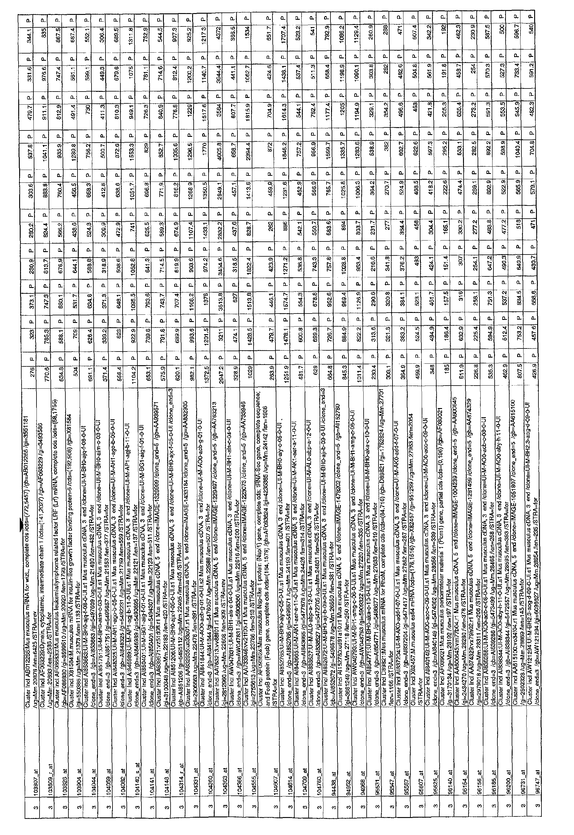
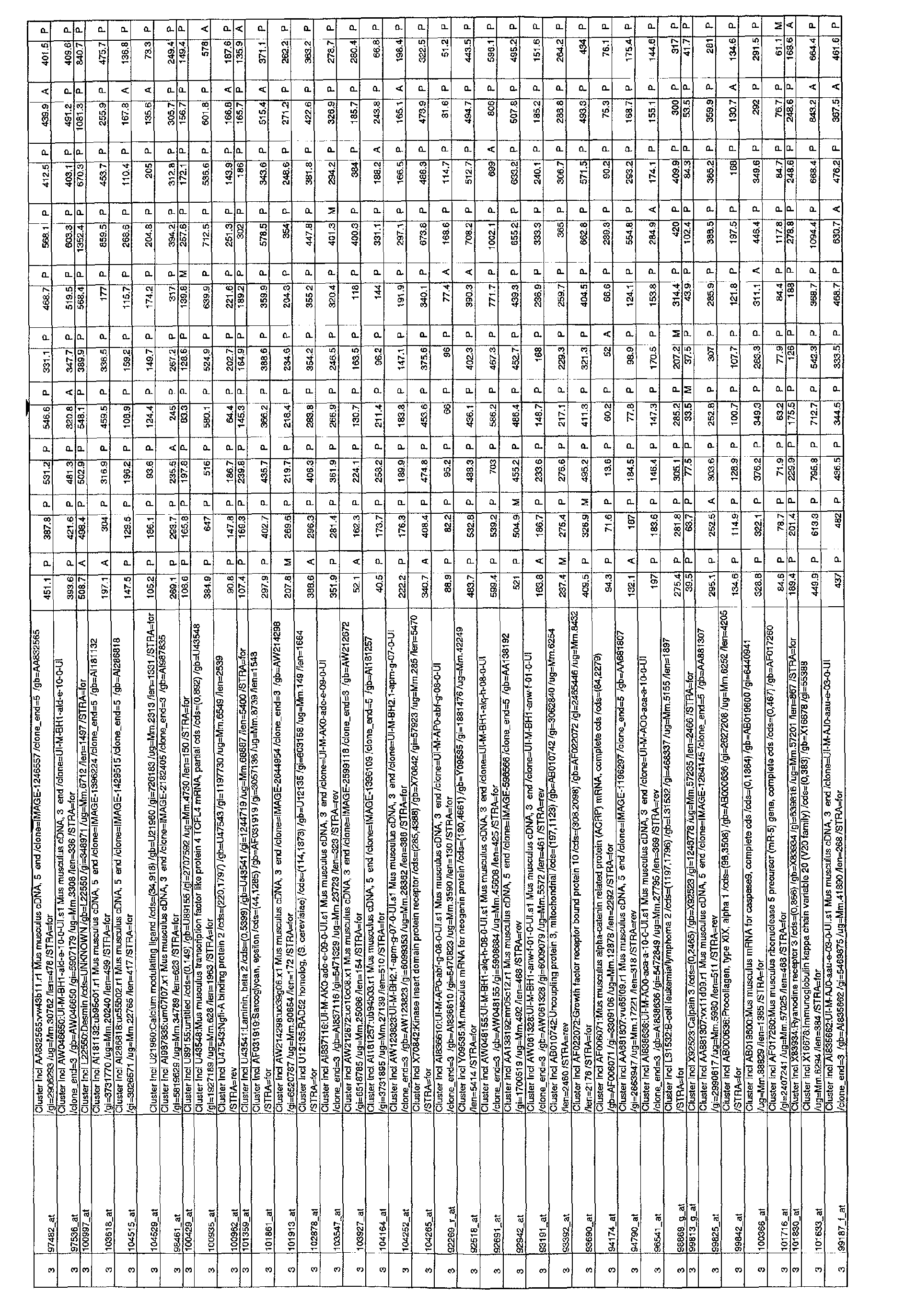
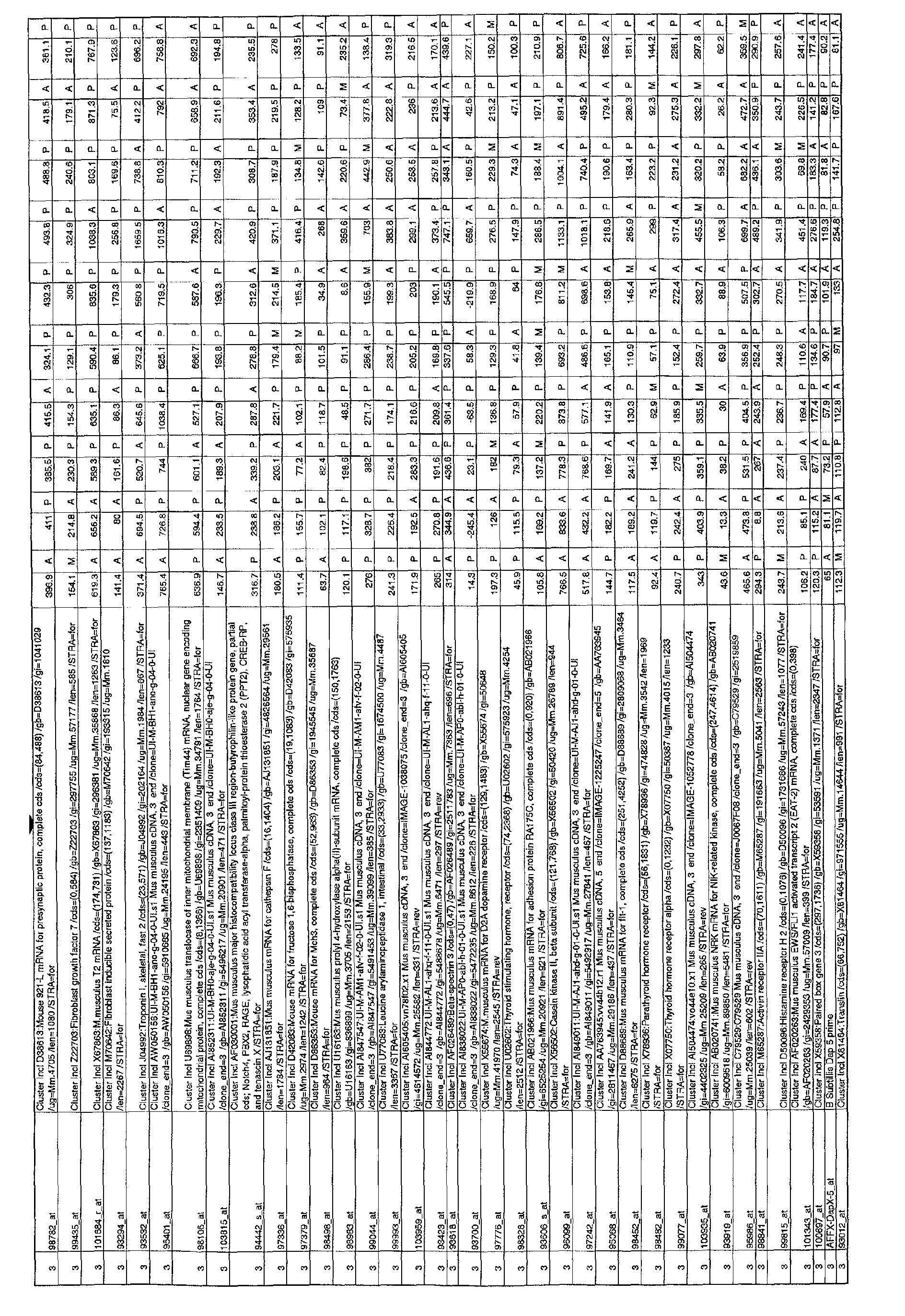
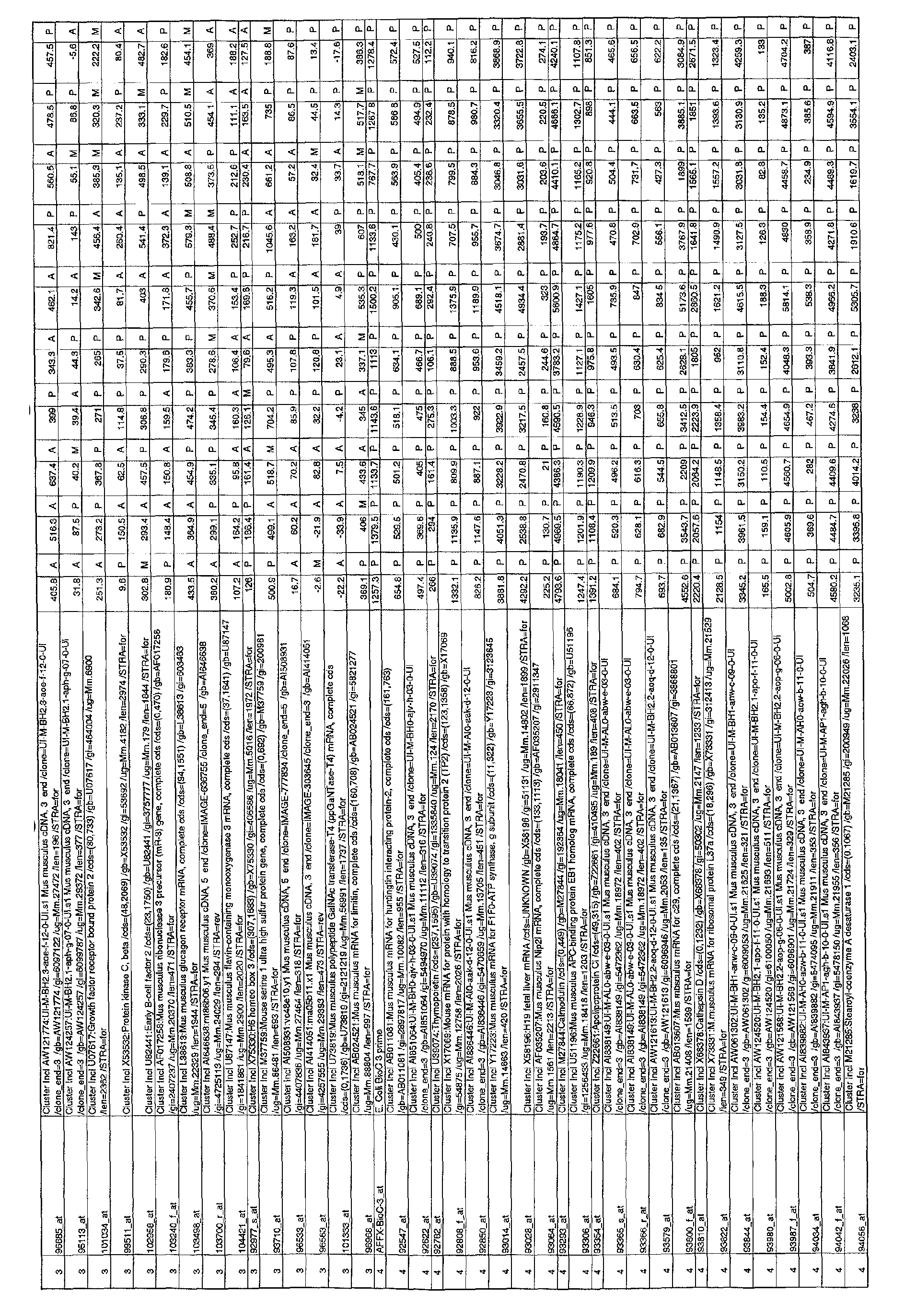
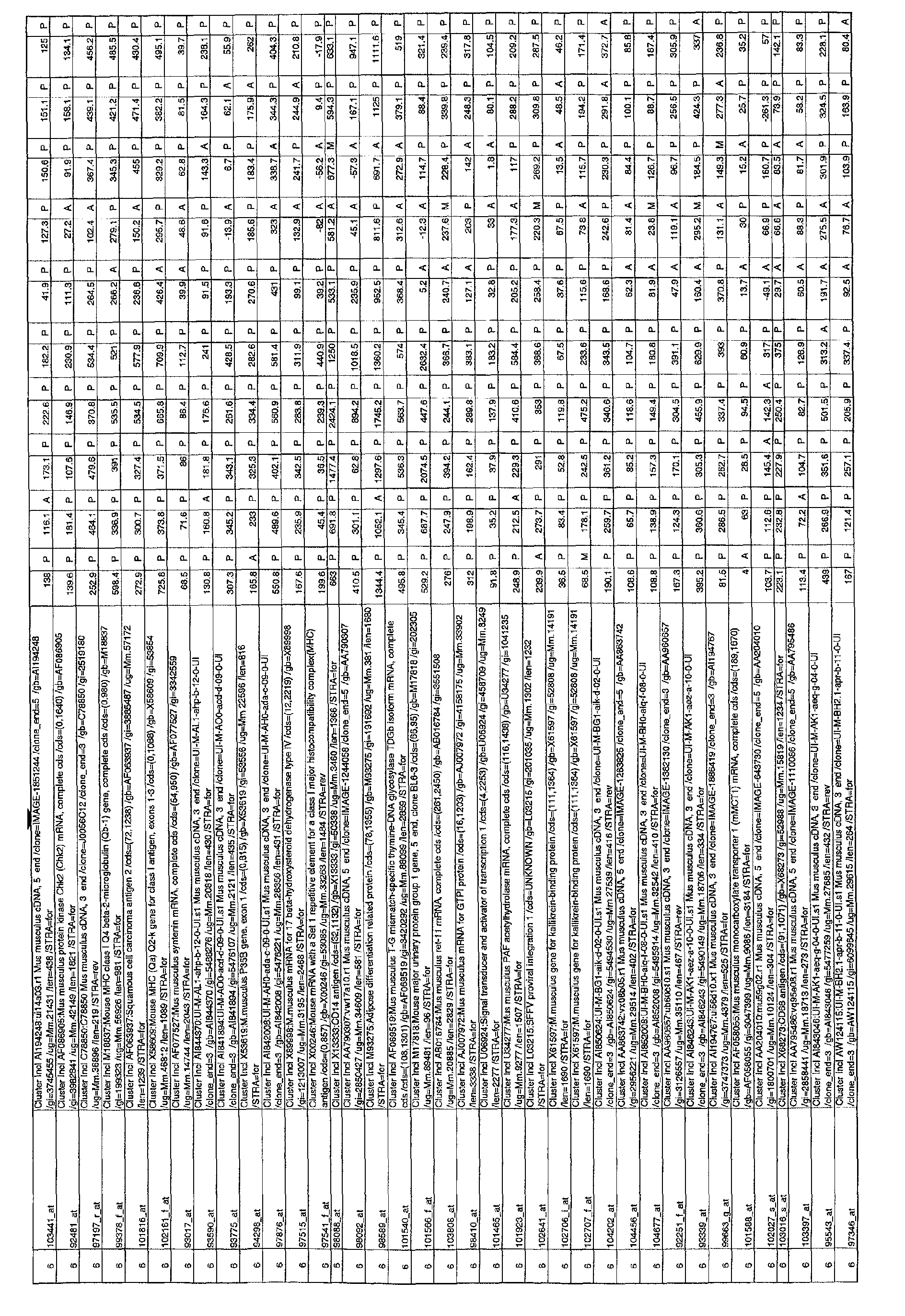
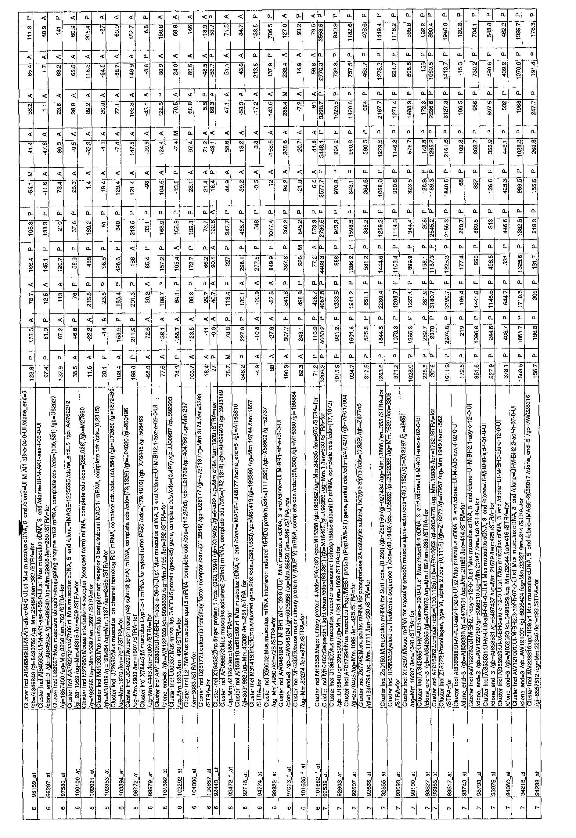
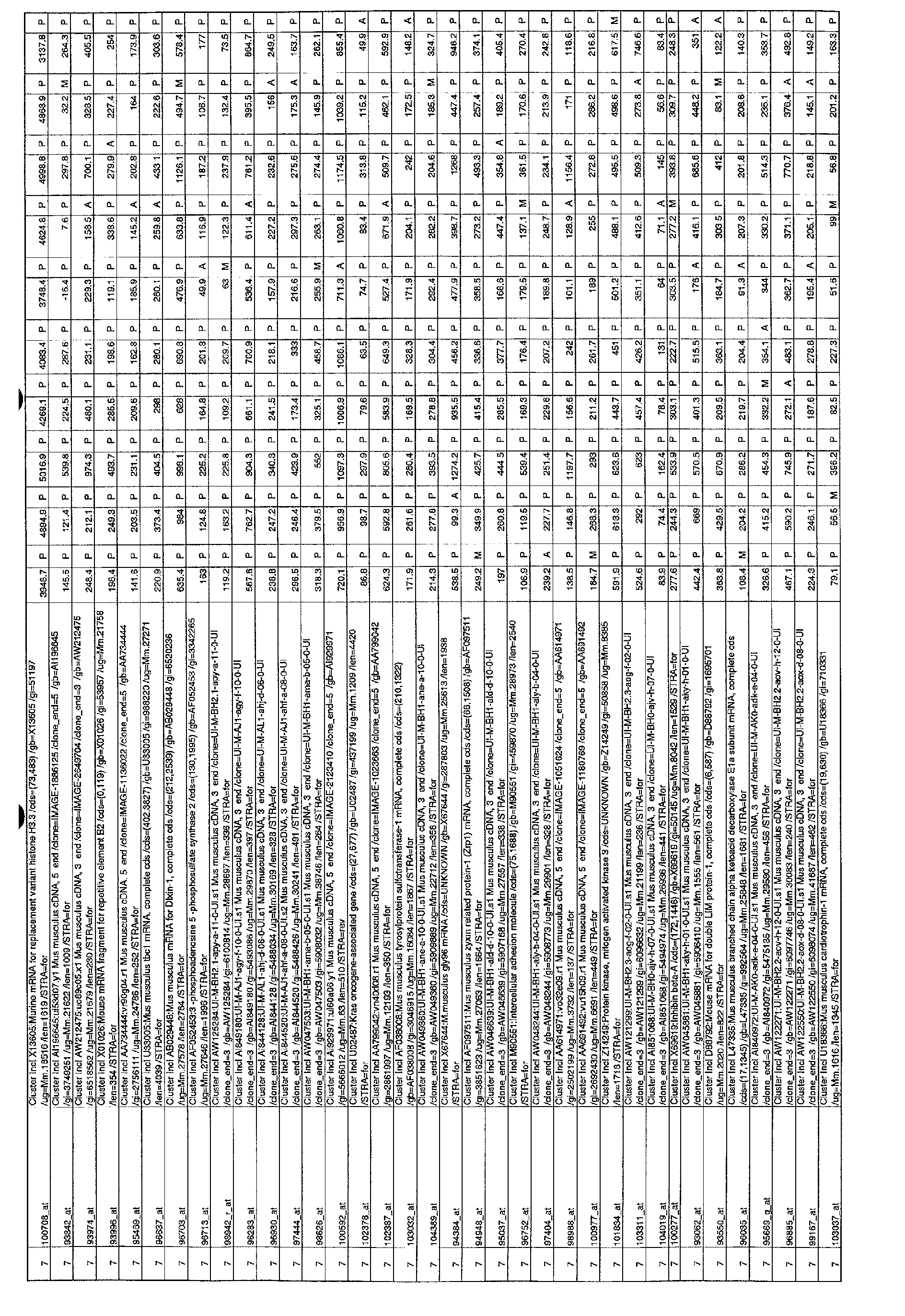
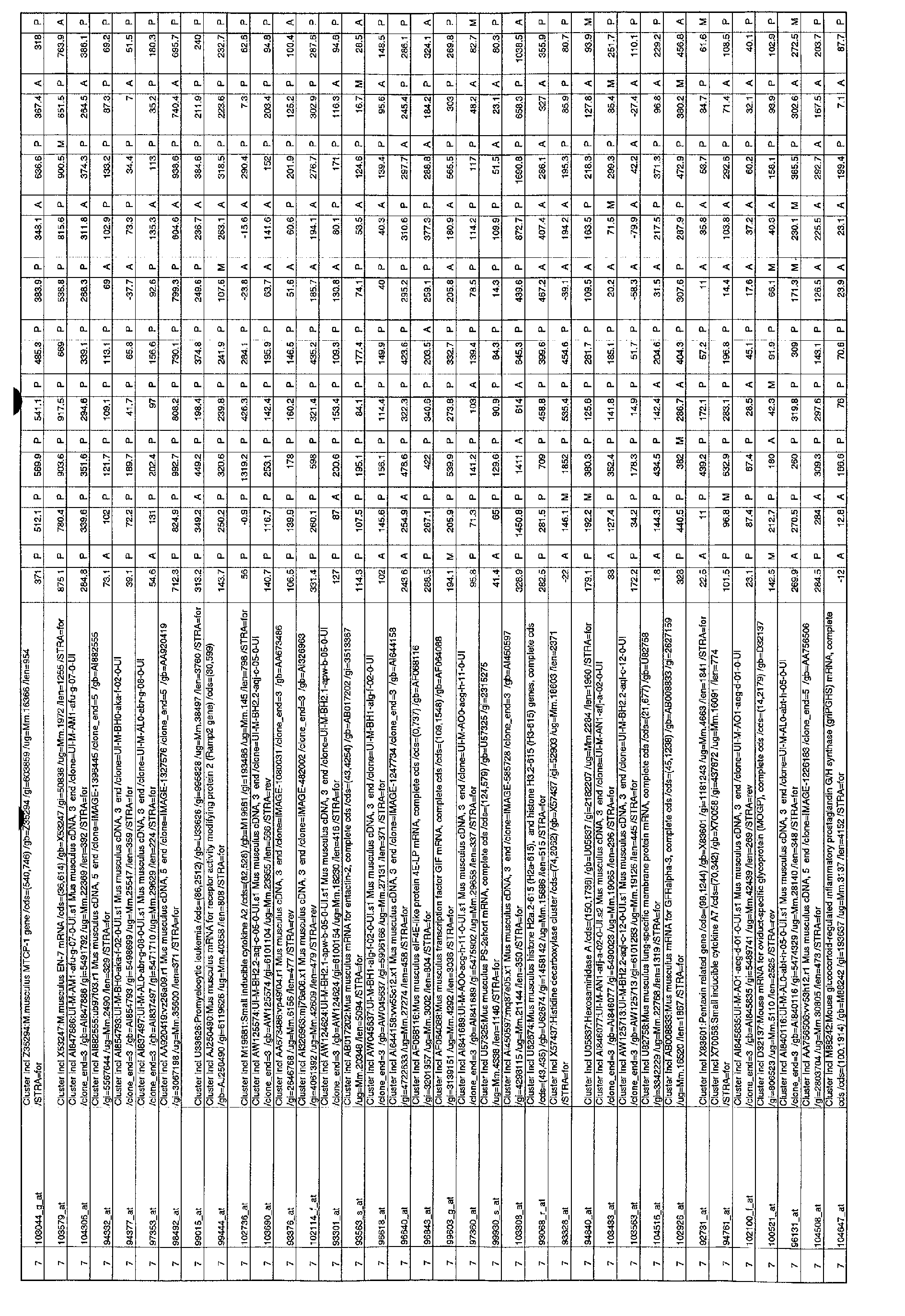
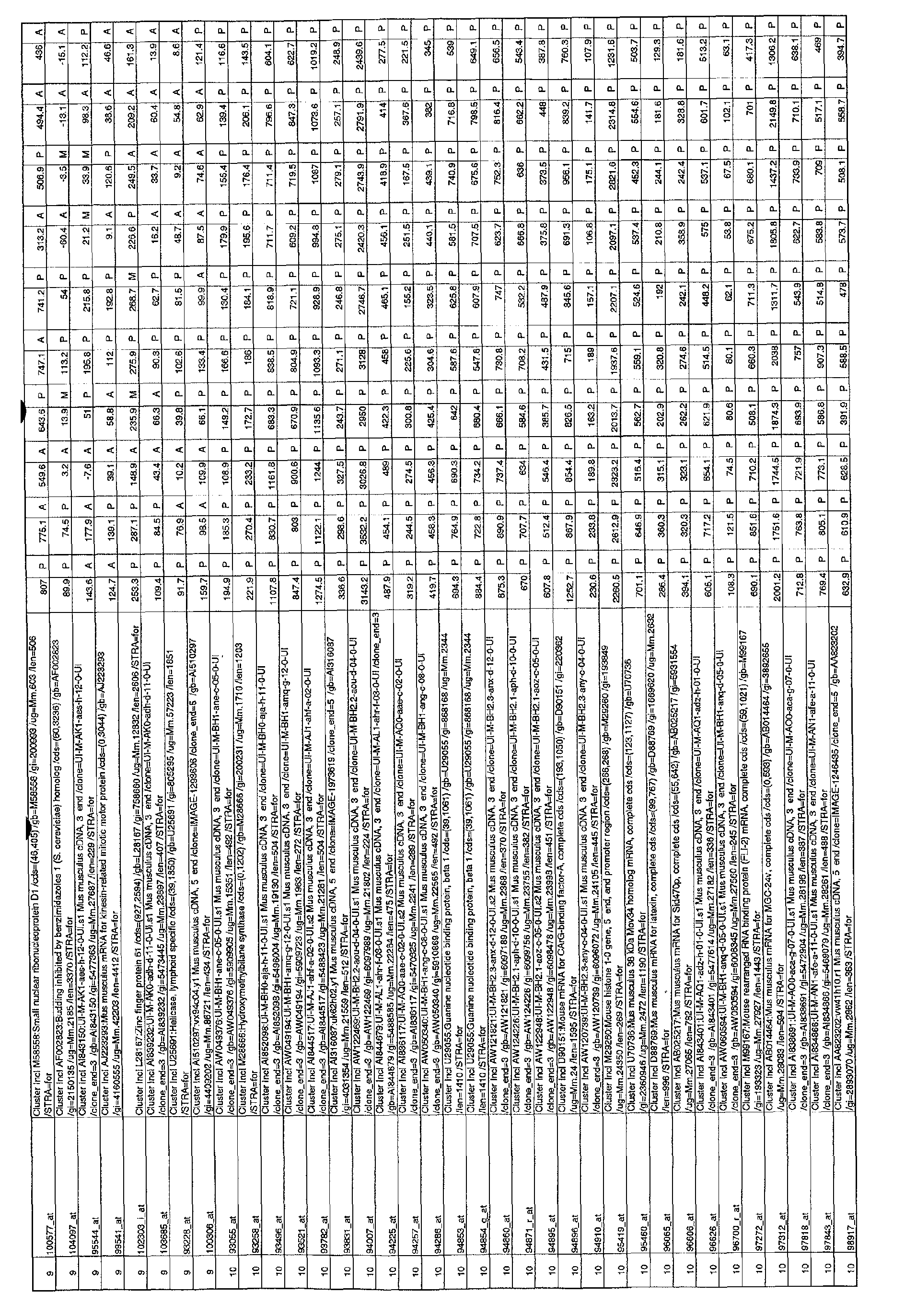
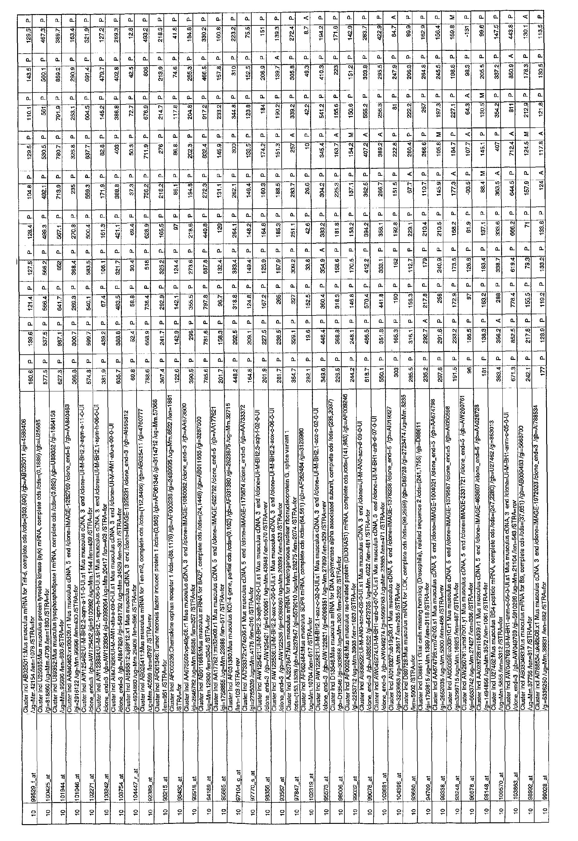
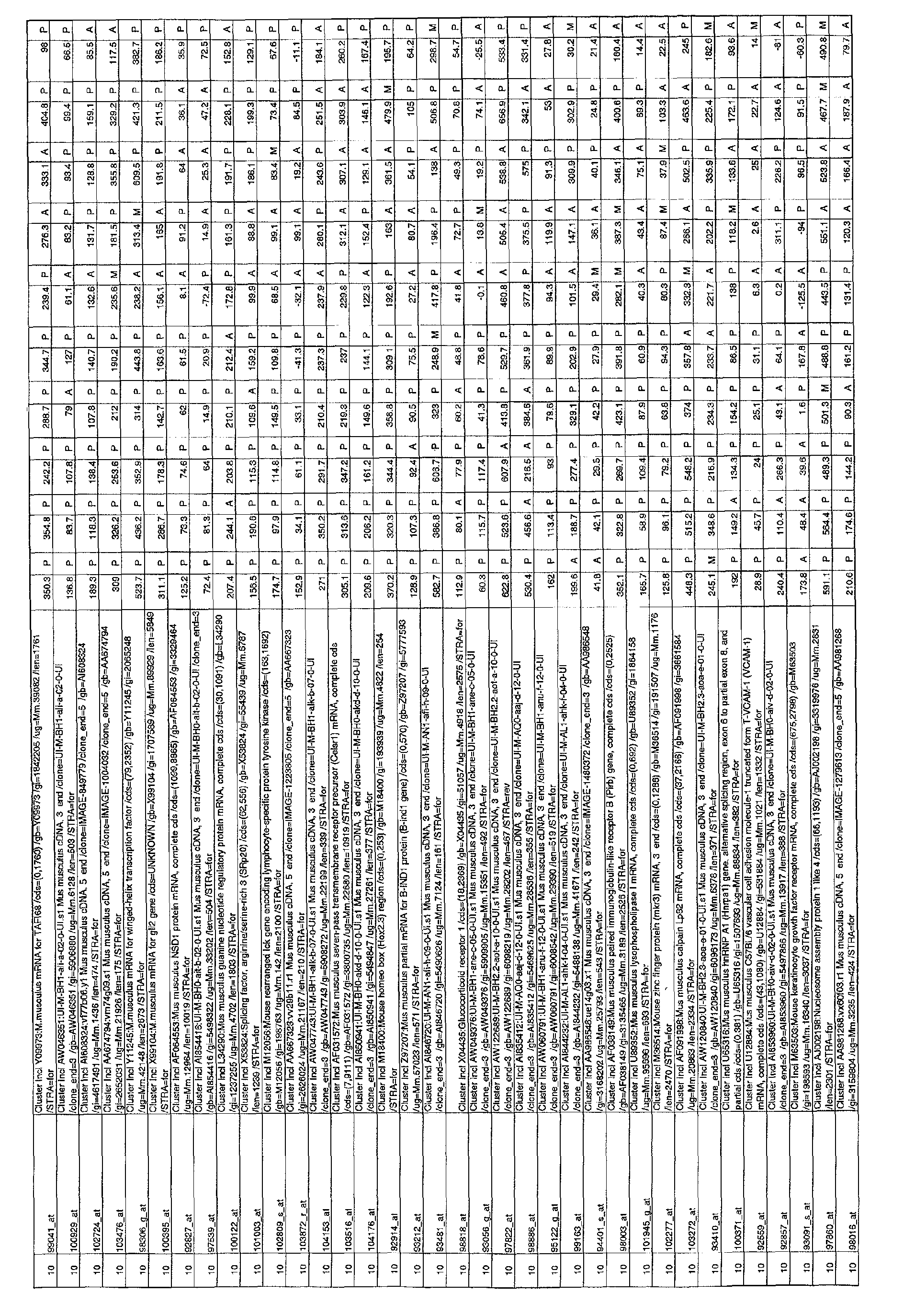
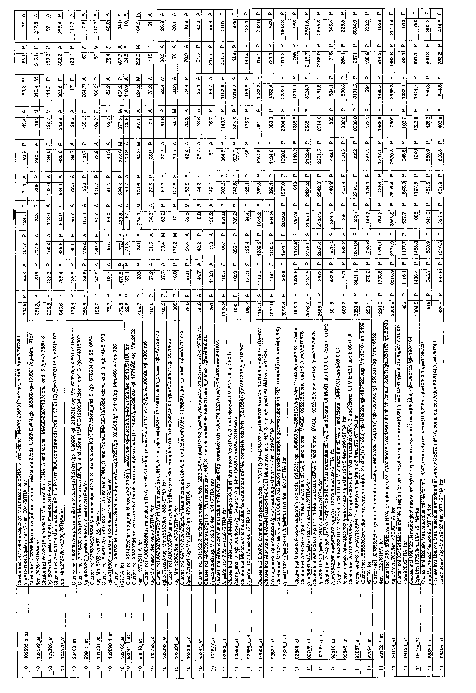
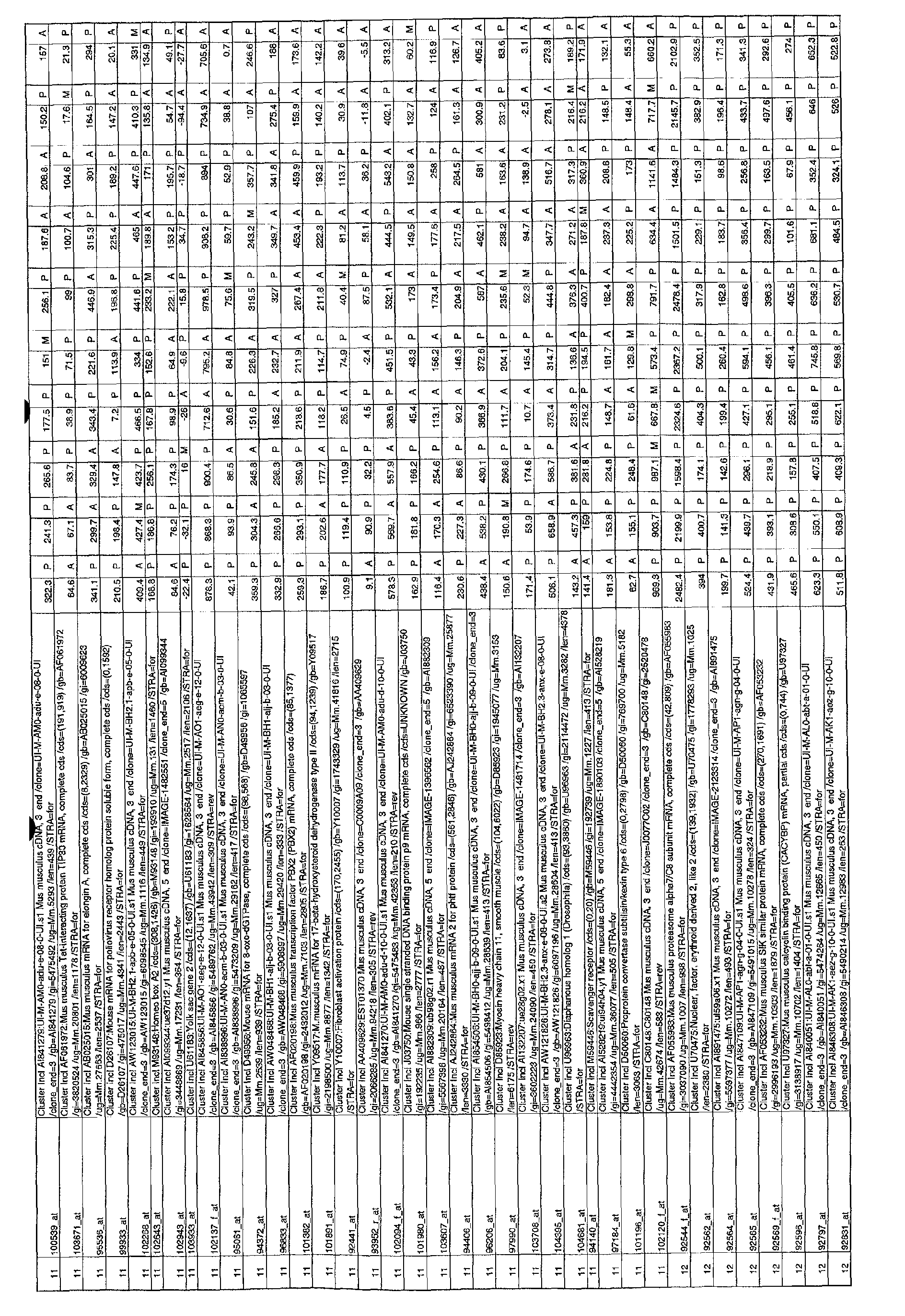
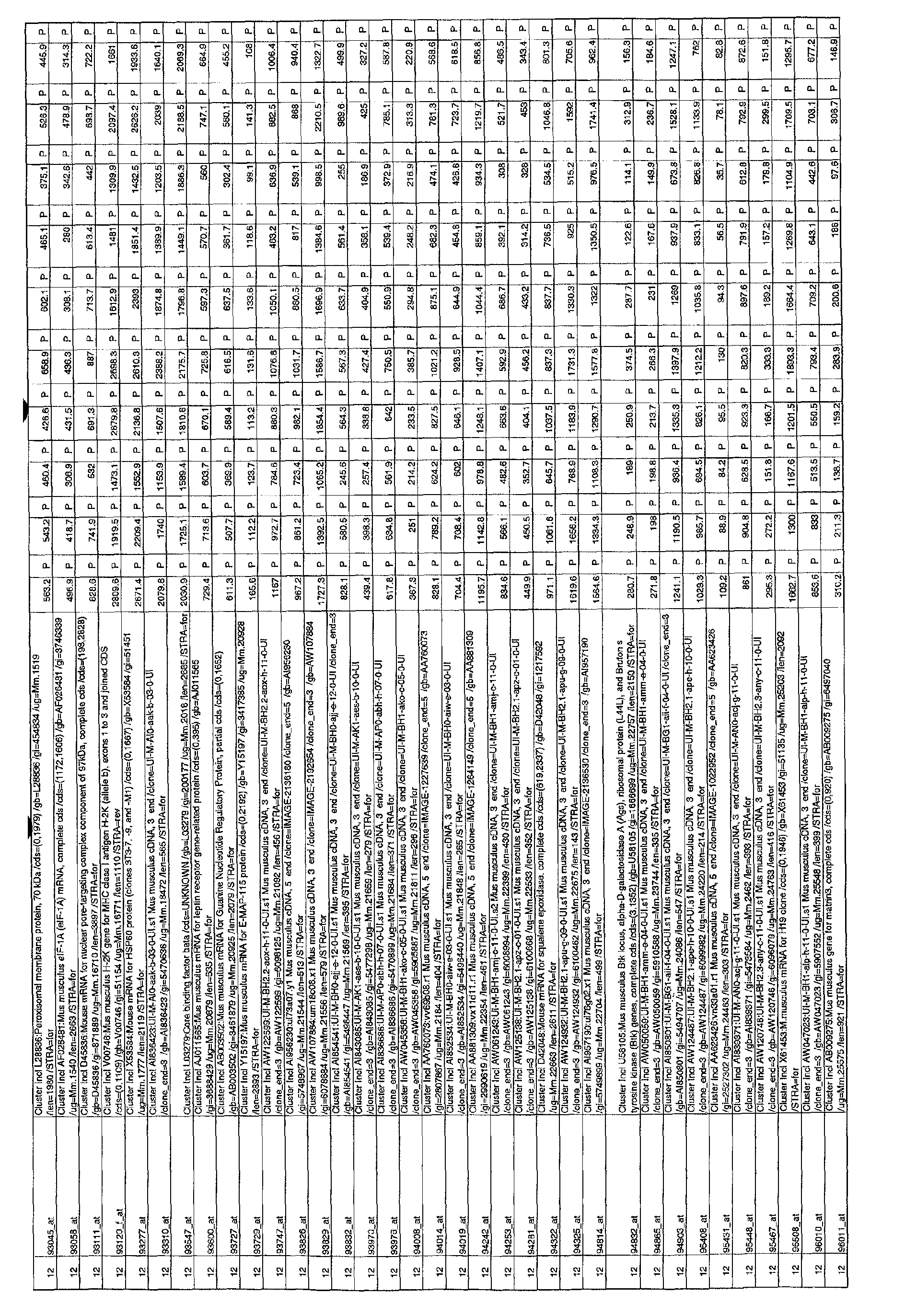
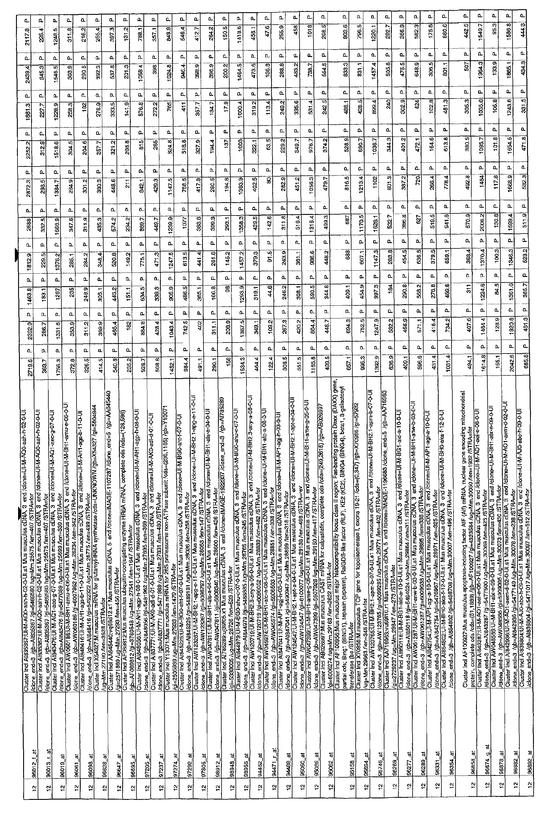
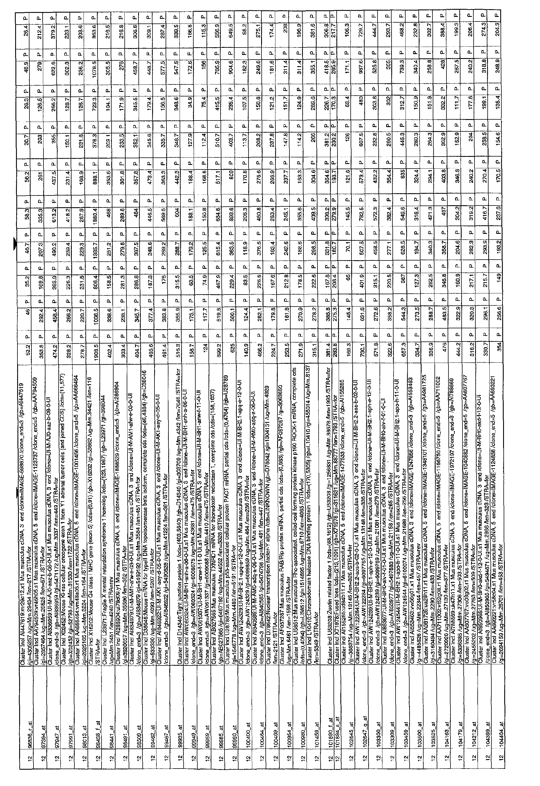
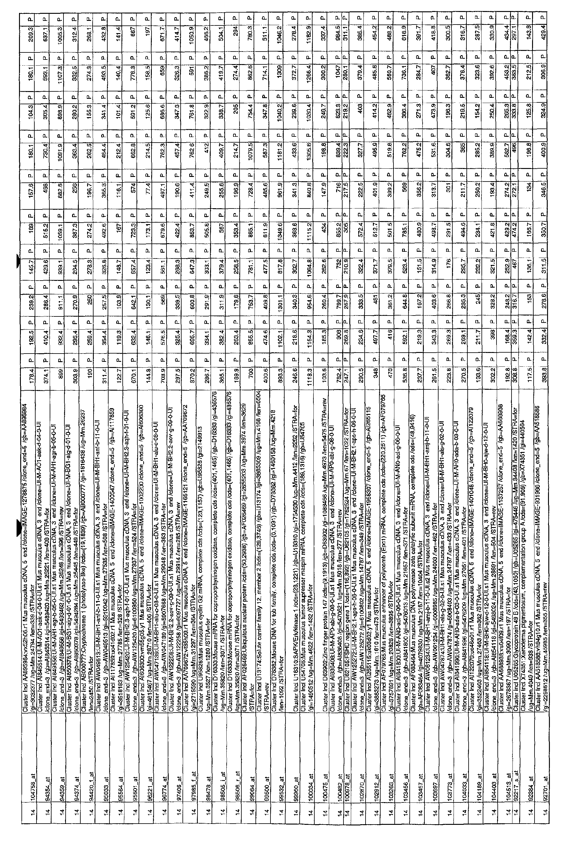
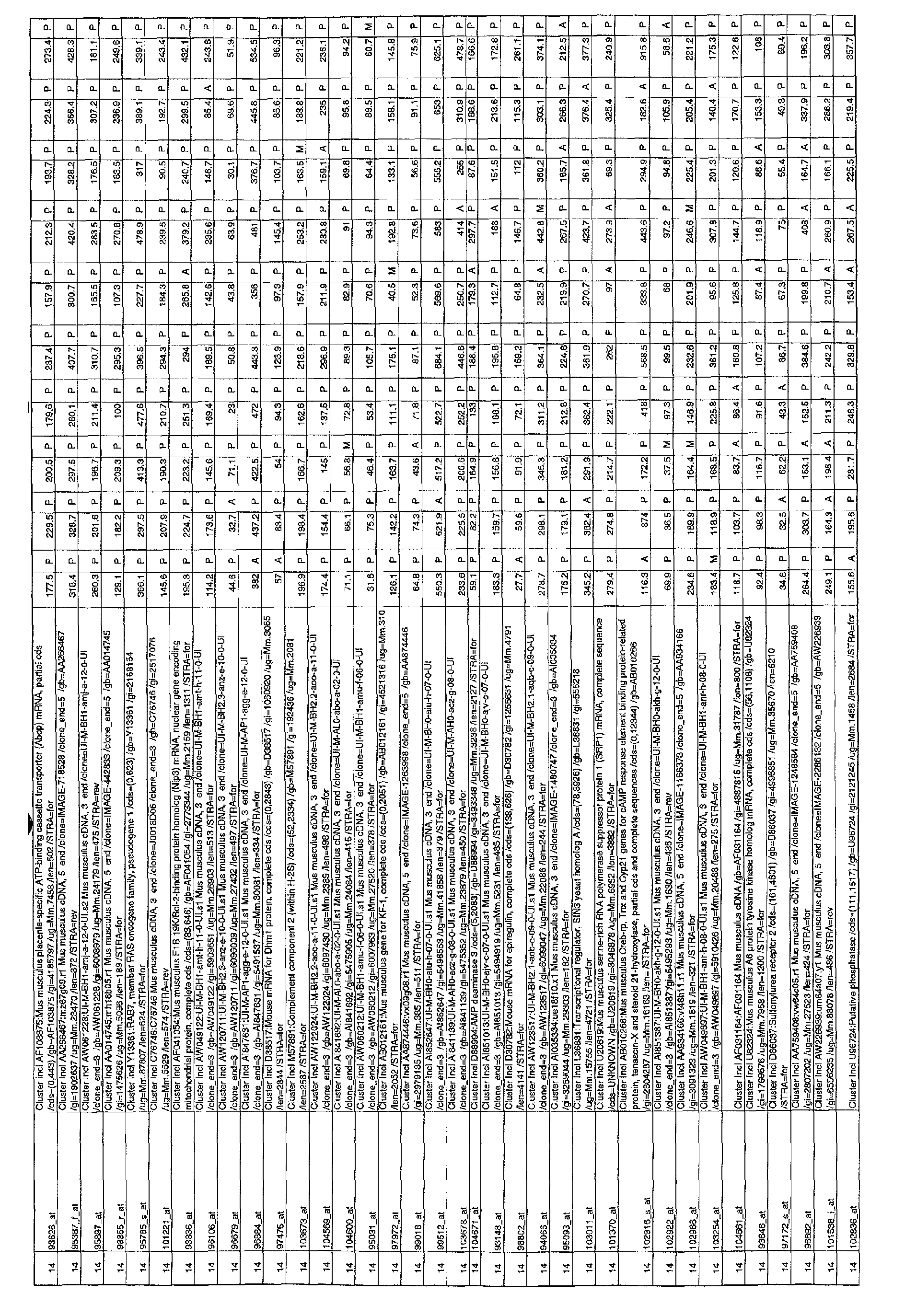
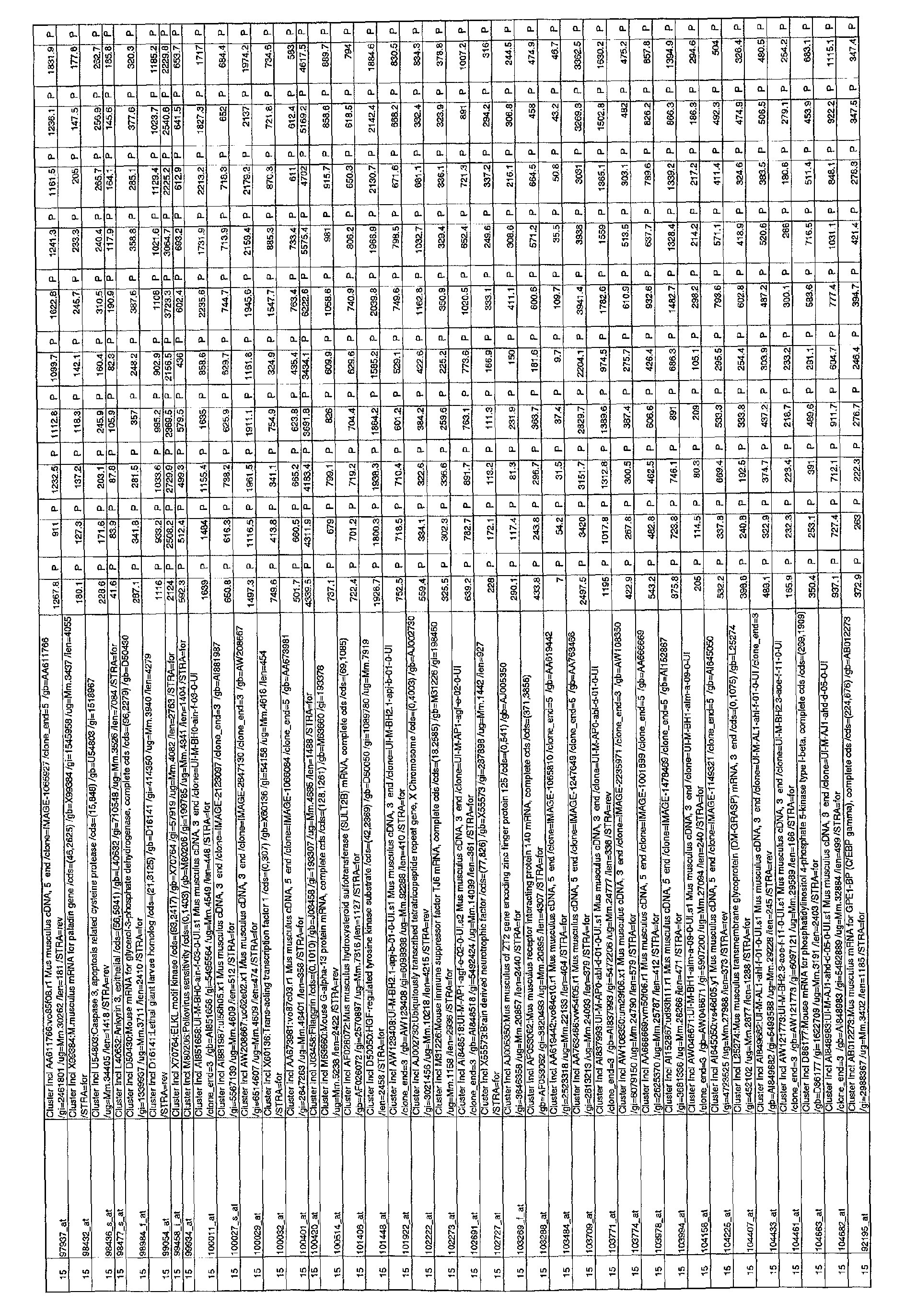
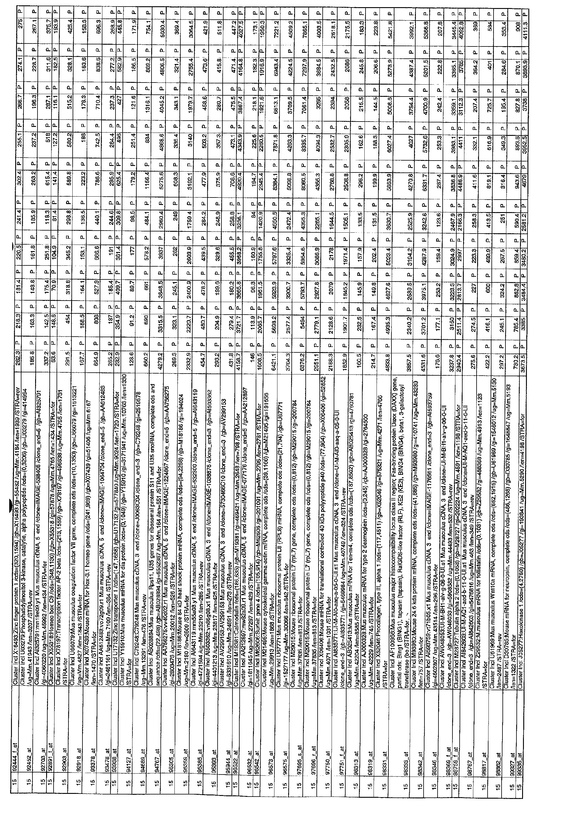
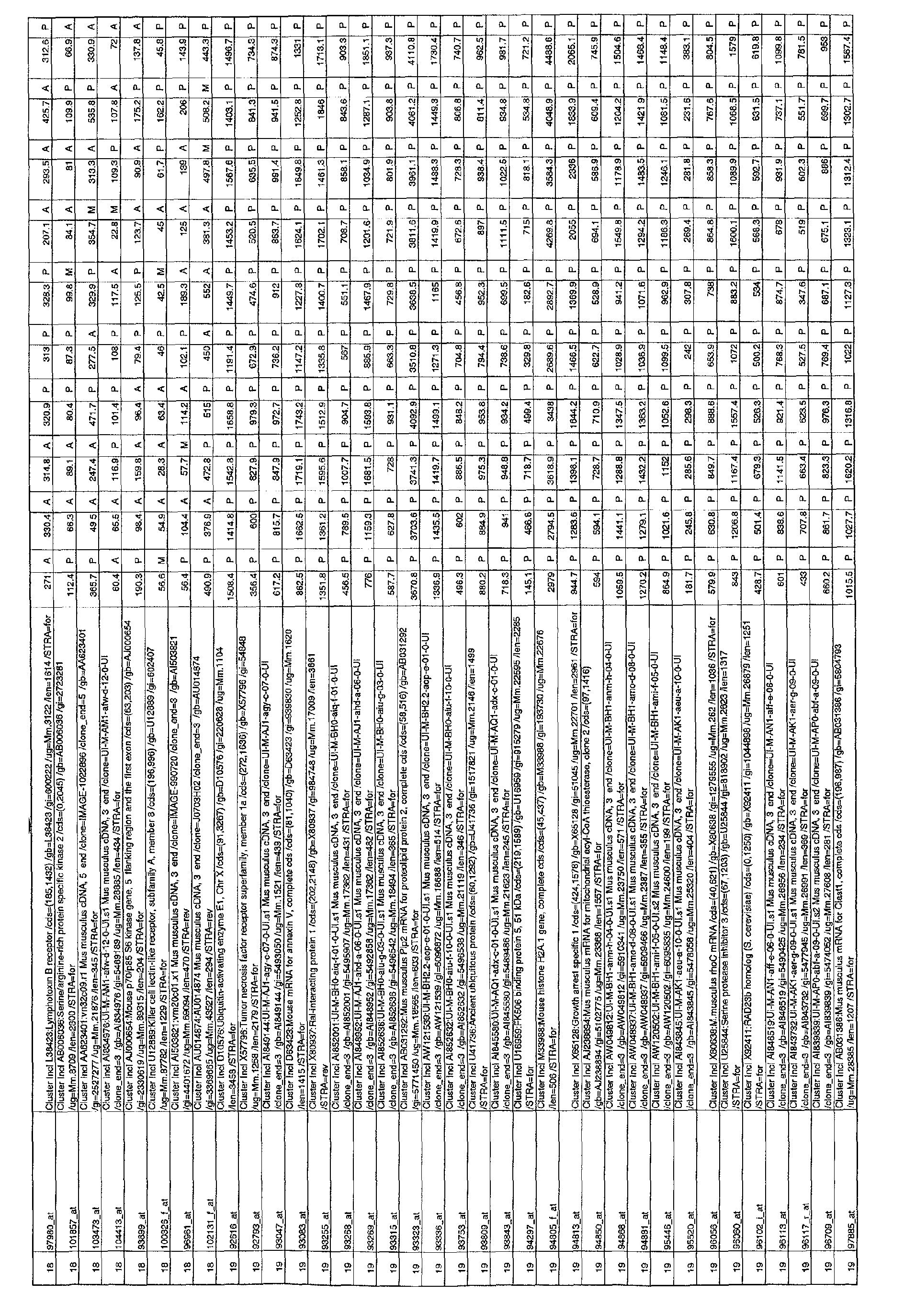
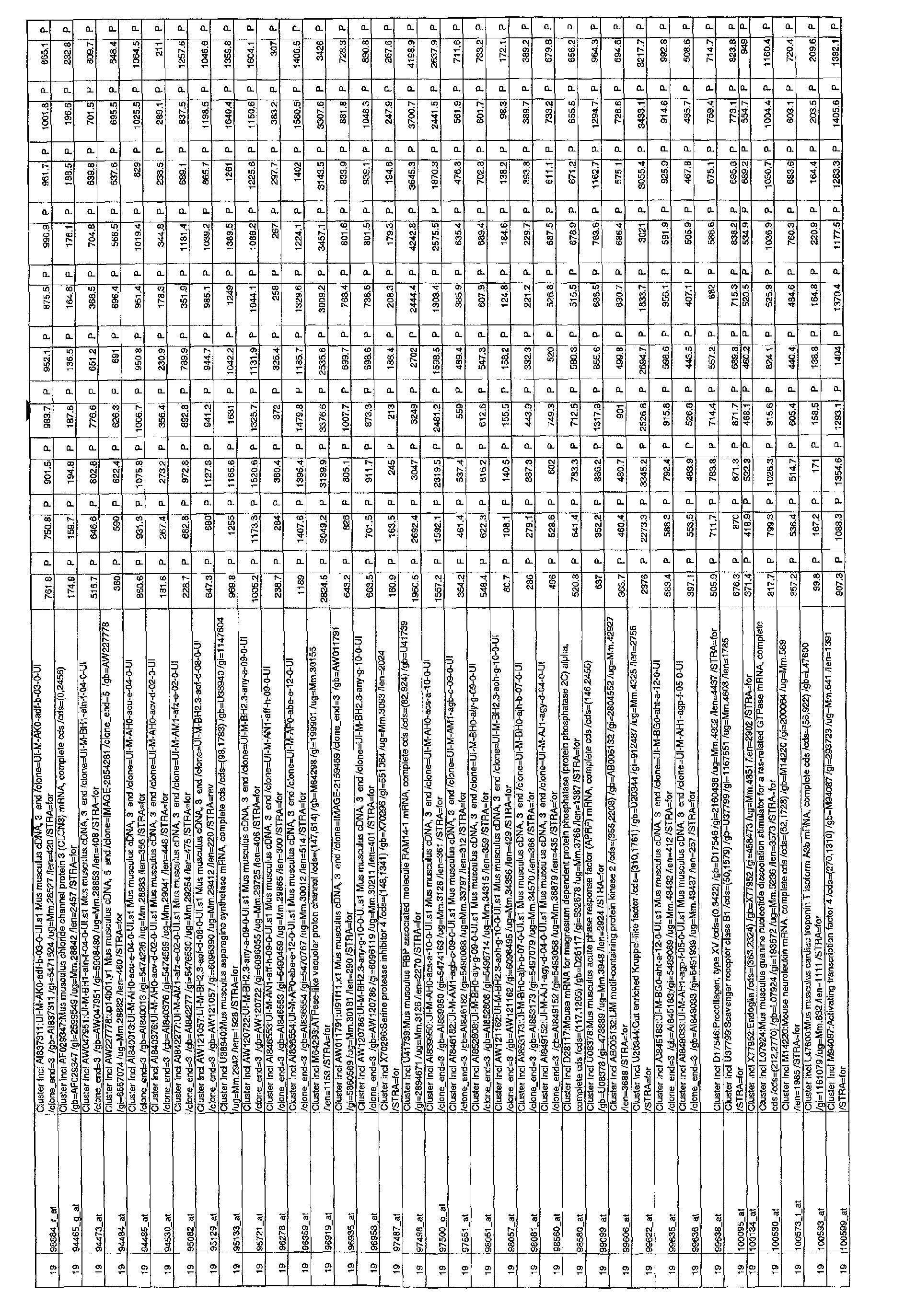
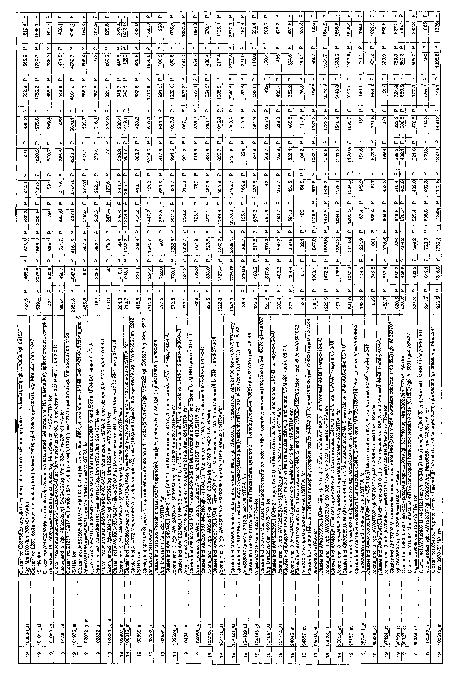
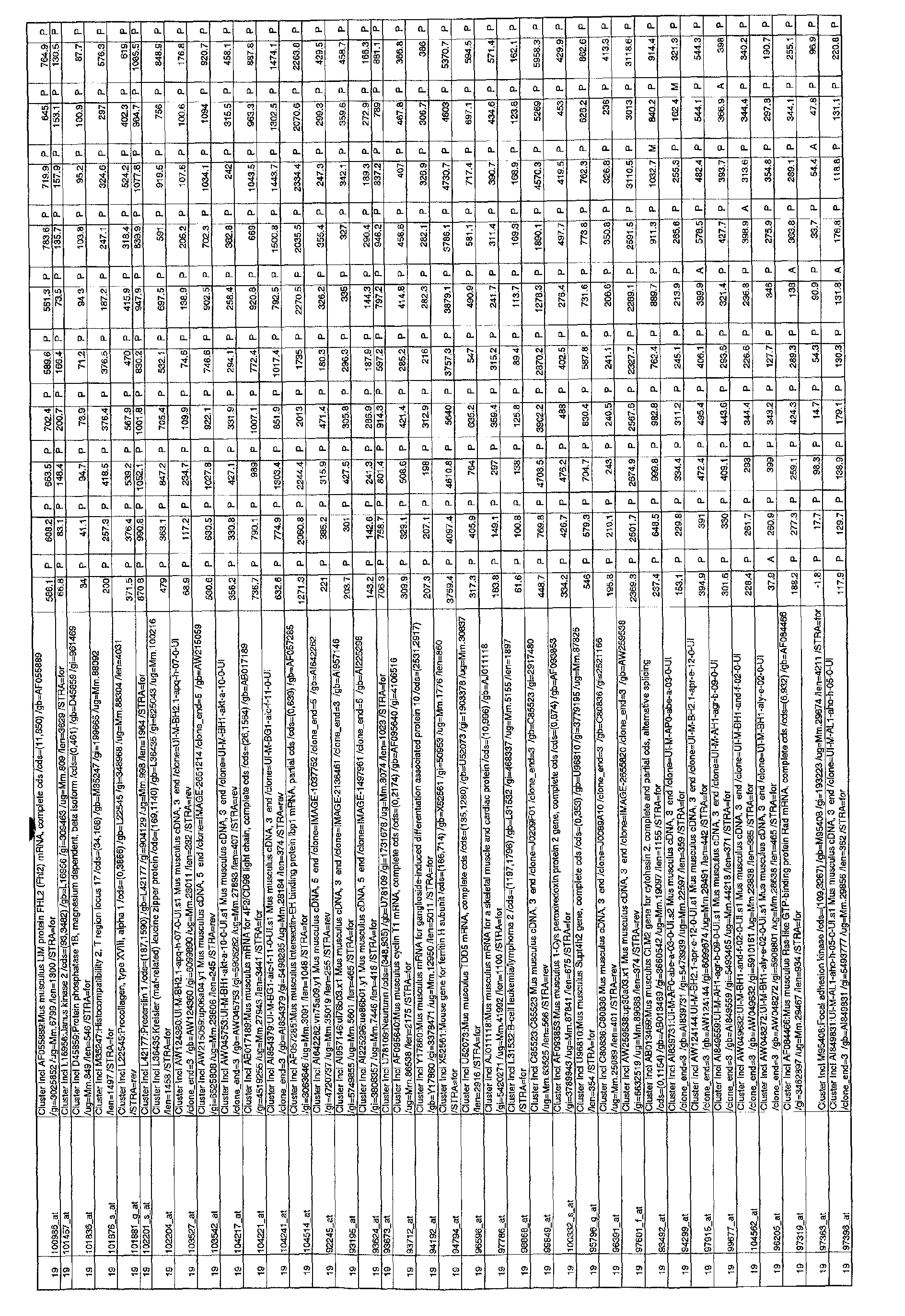
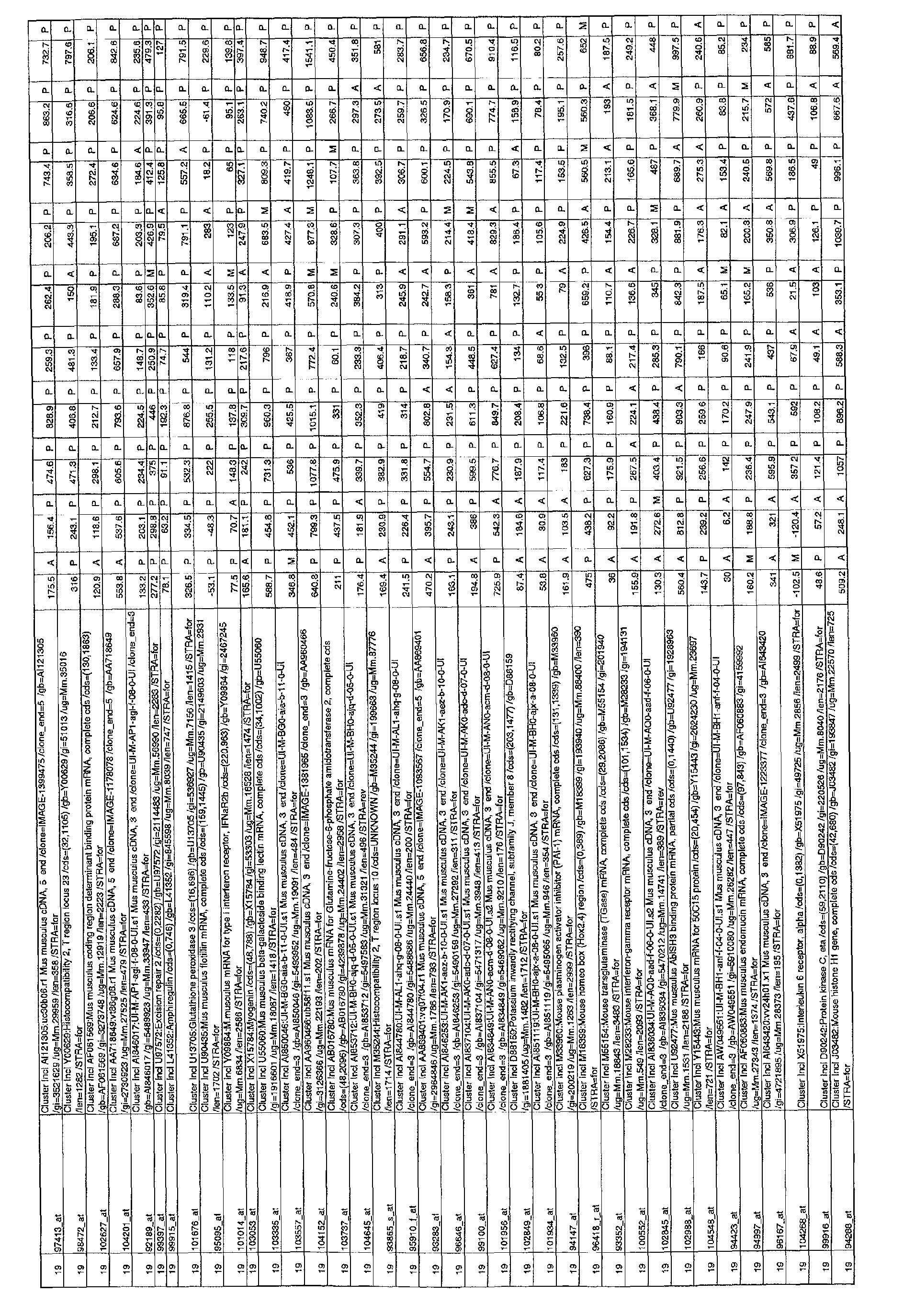
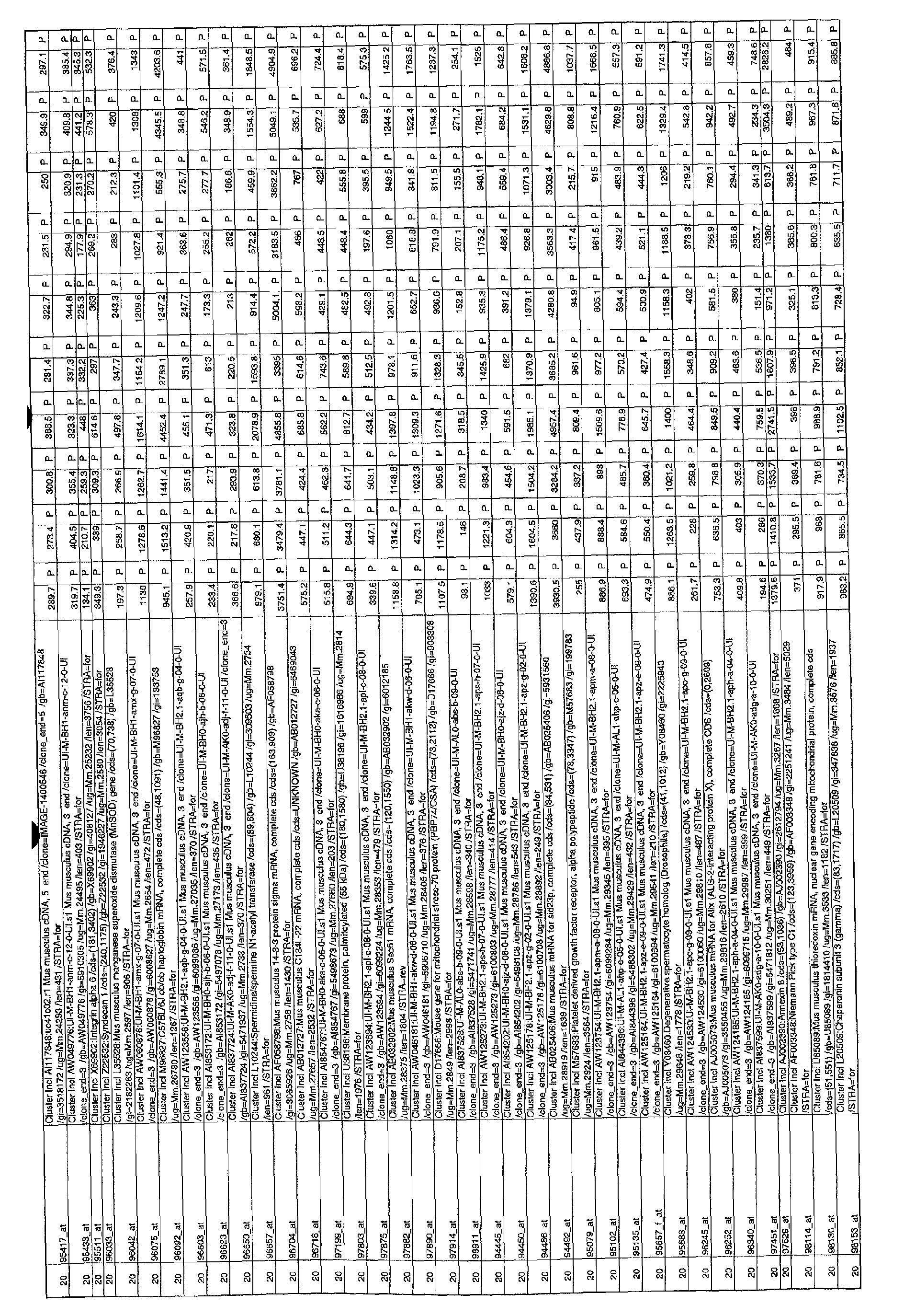
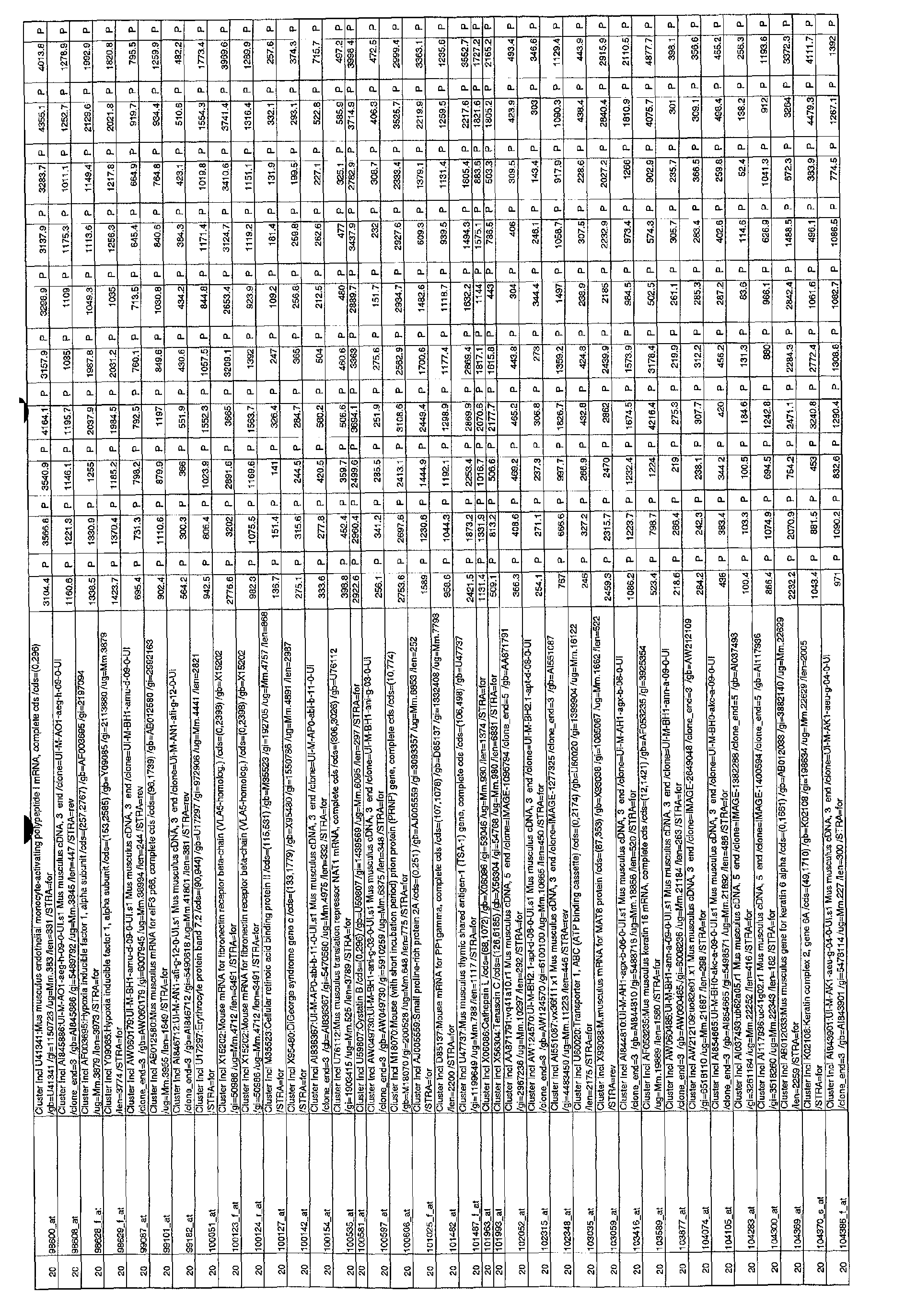
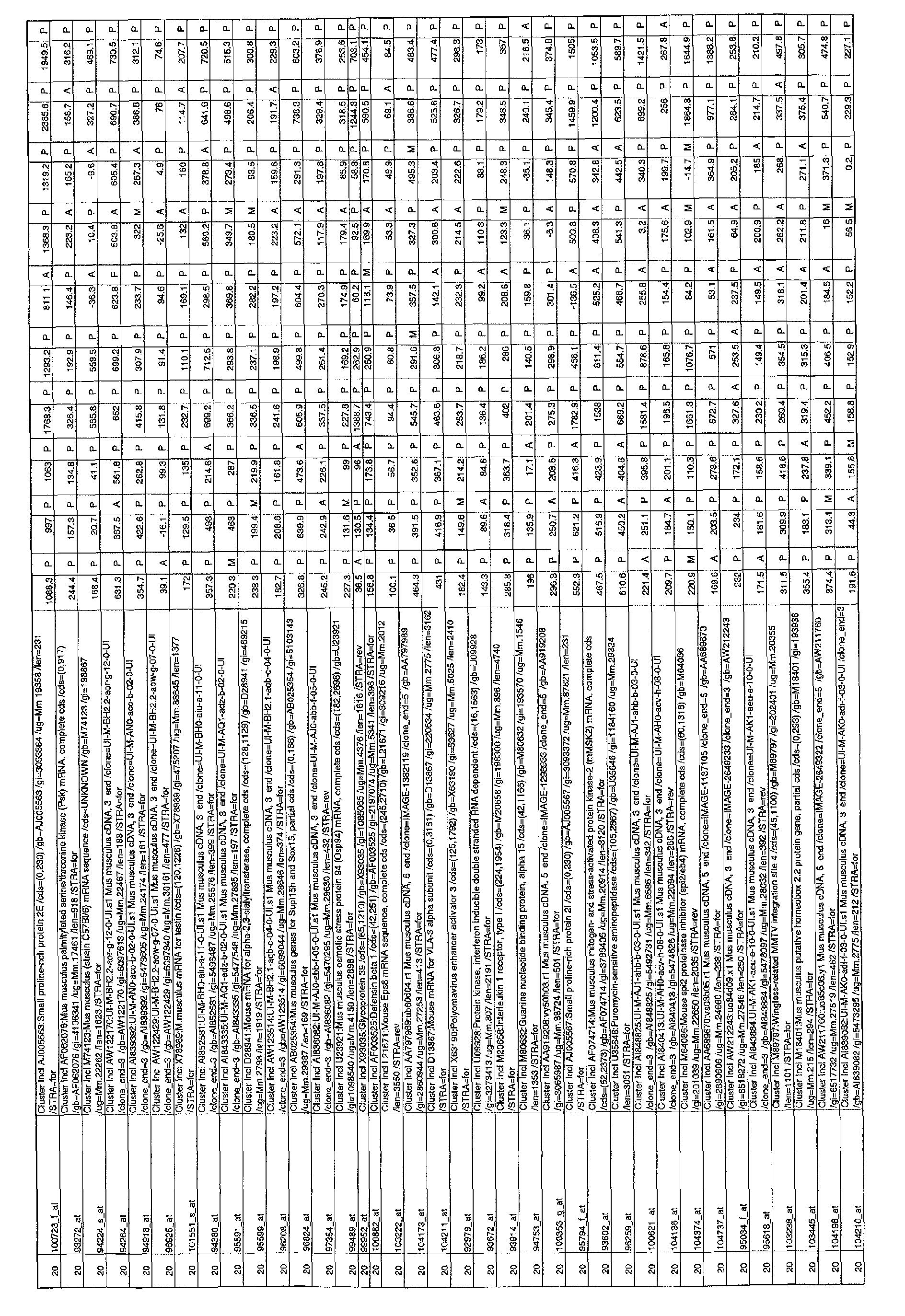
- clusters produced by this method resulted in 20 clusters, with a reasonable number of profiles in each (lowest number of profiles: cluster 10 with 141 profiles, highest number: cluster 3 with 337 profiles).
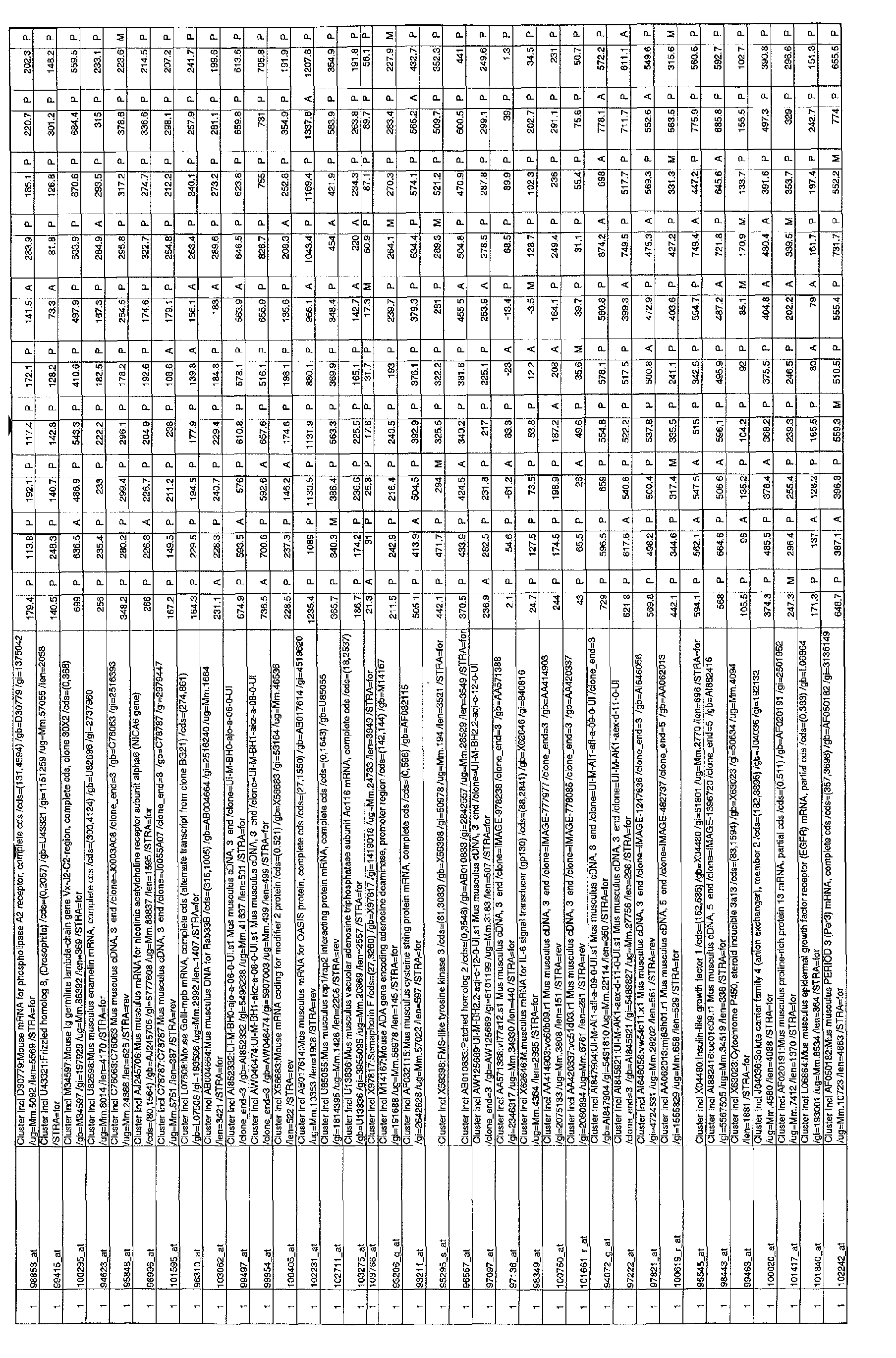
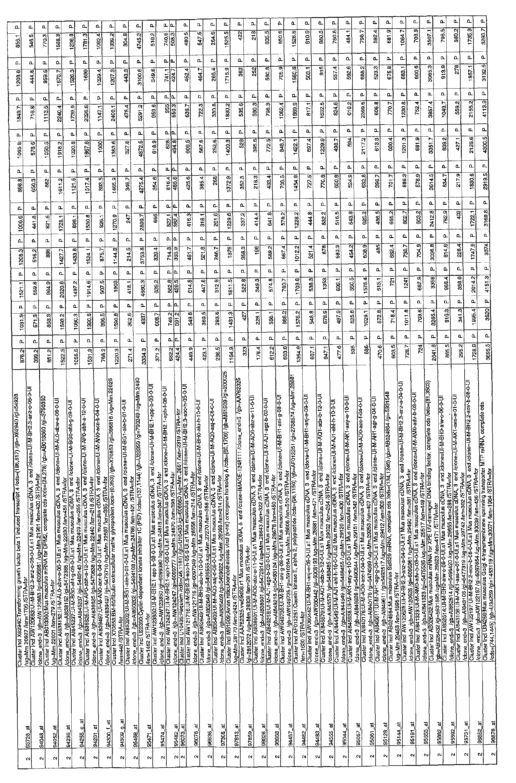
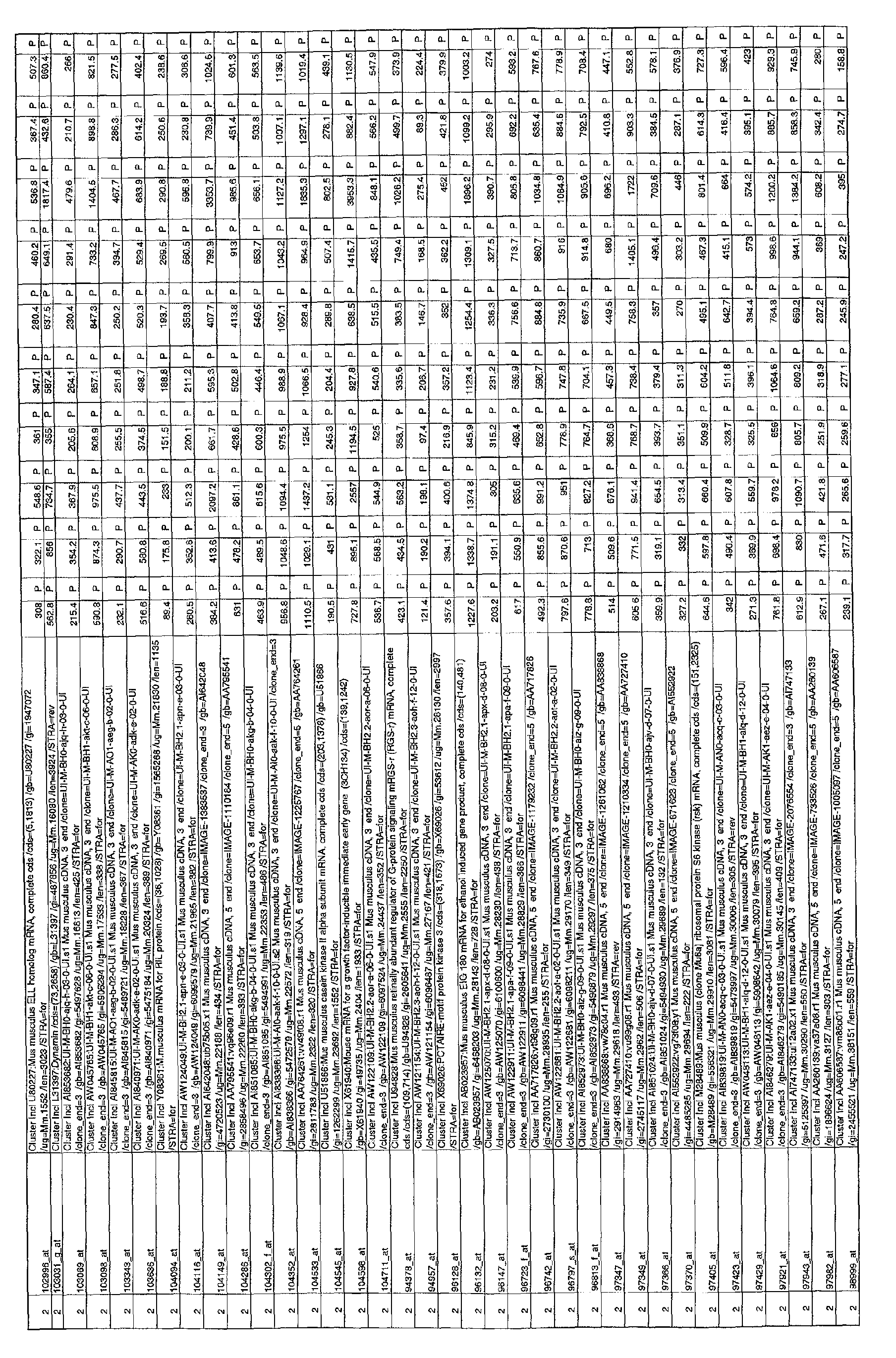
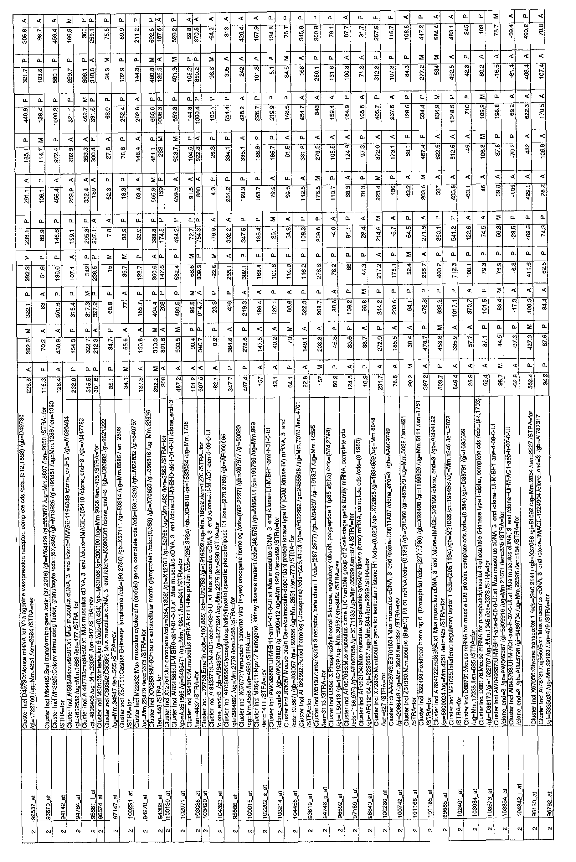
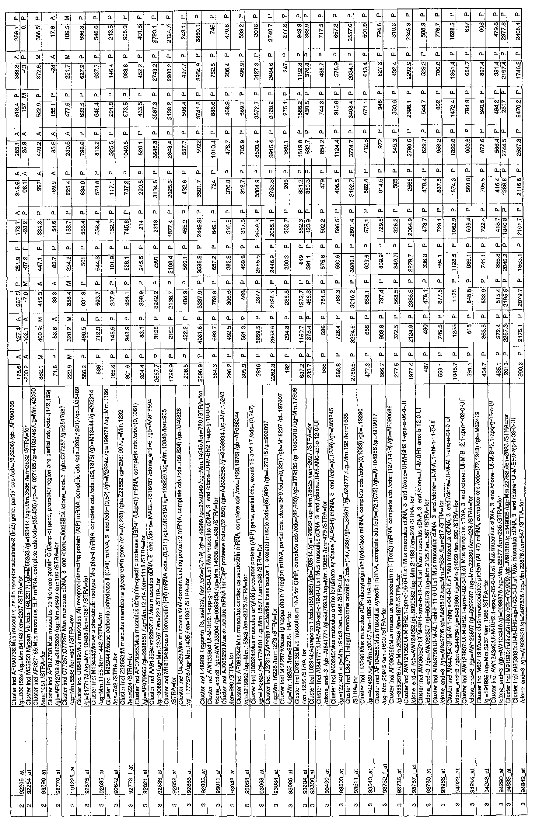
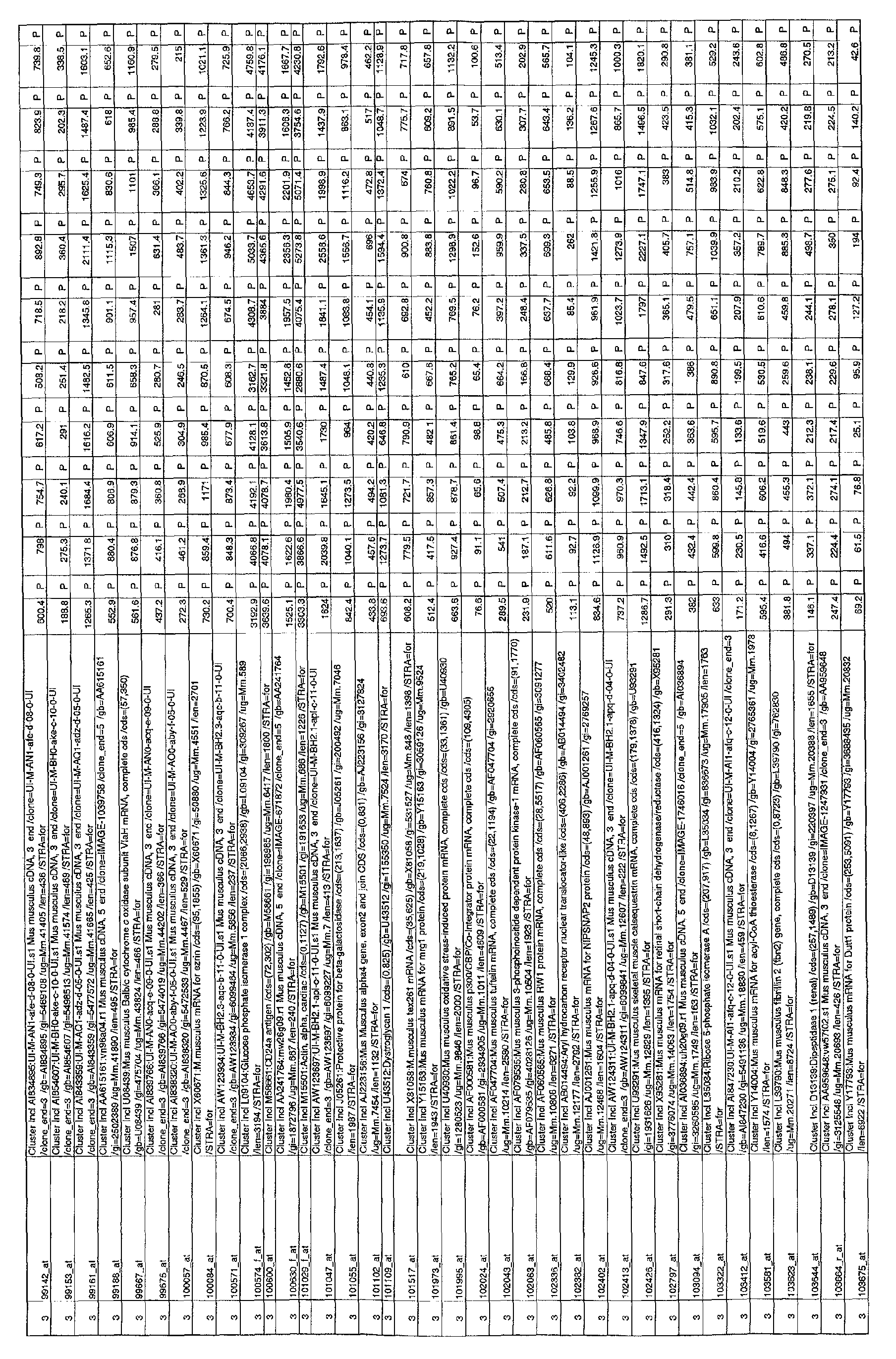
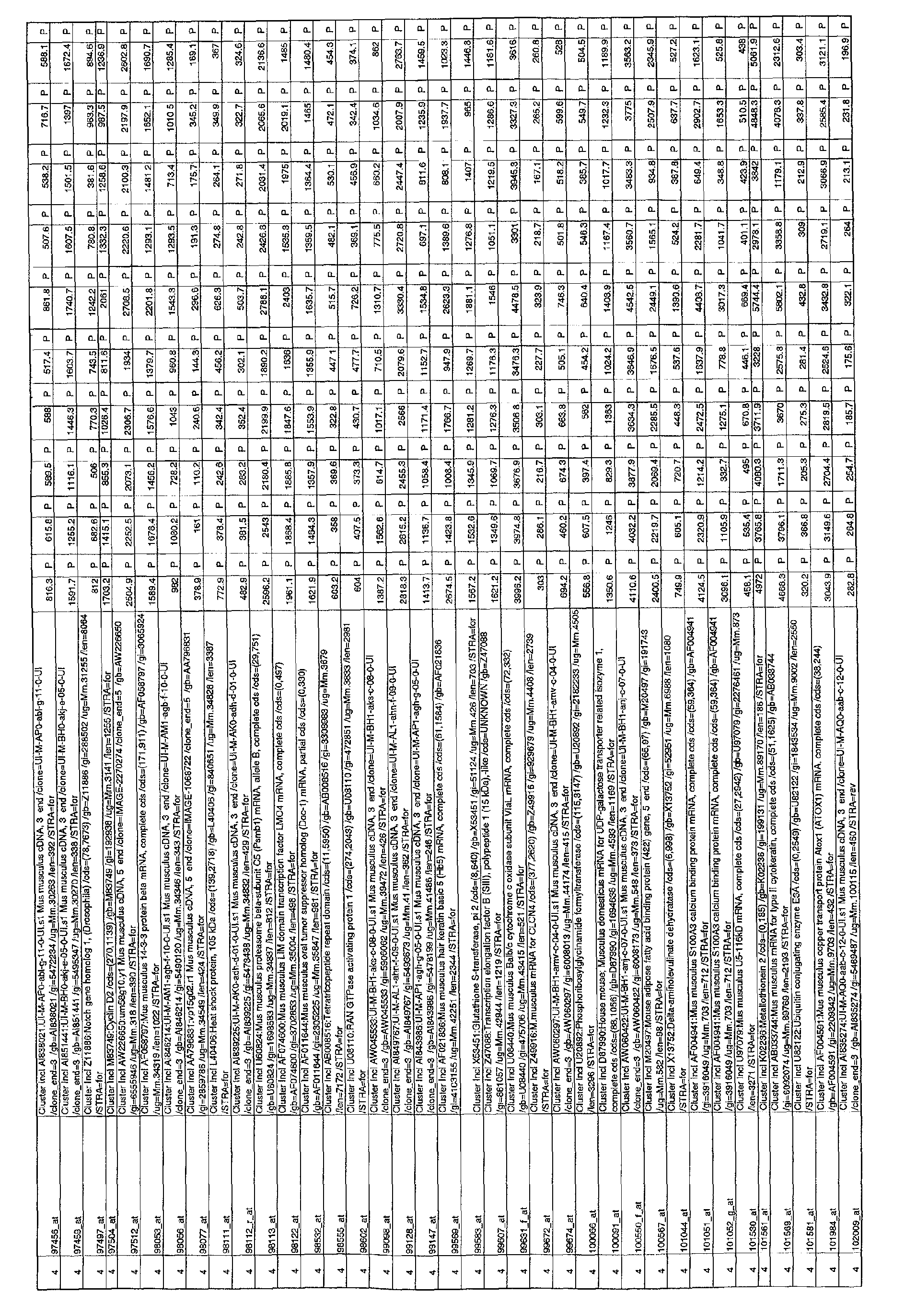
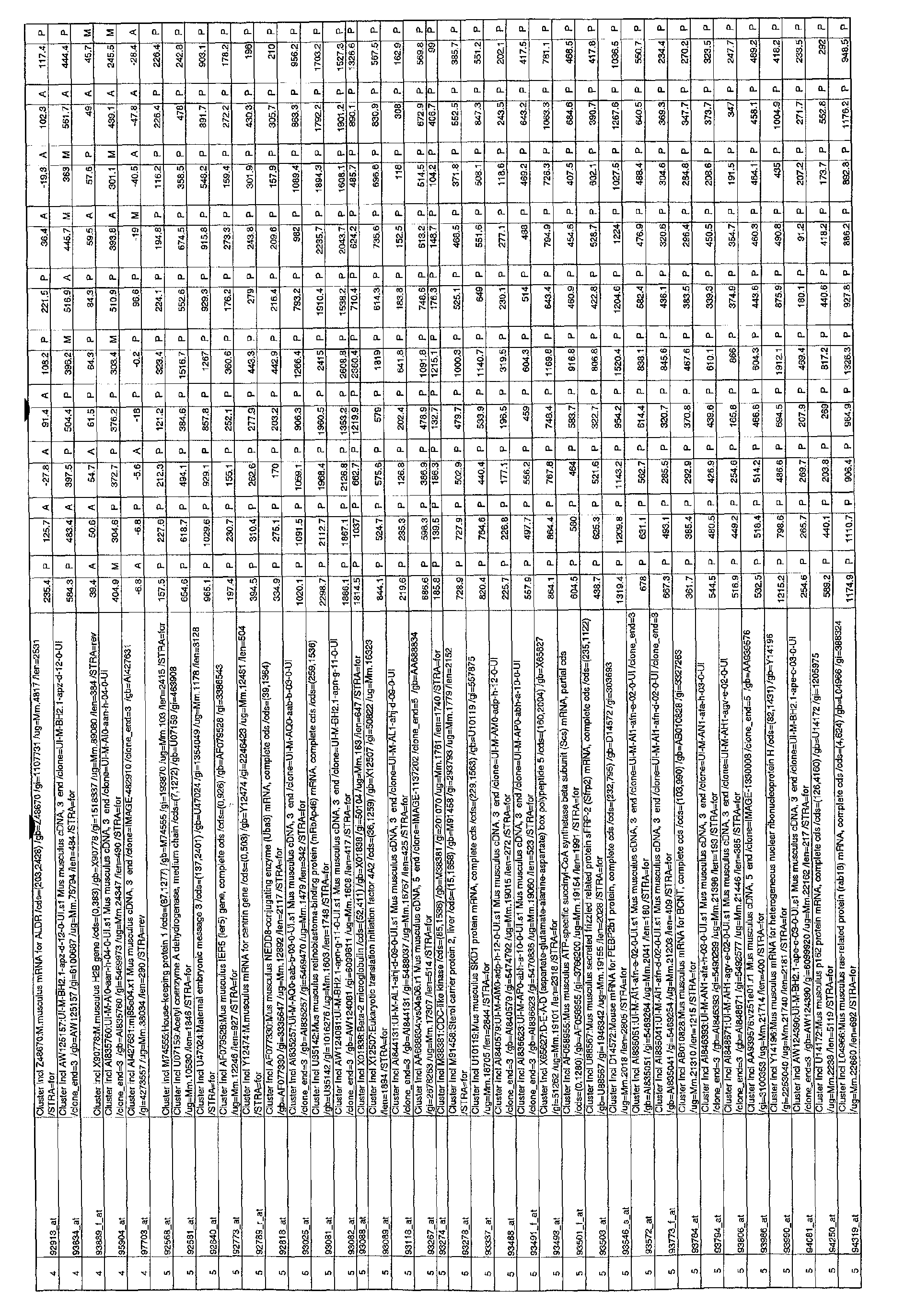
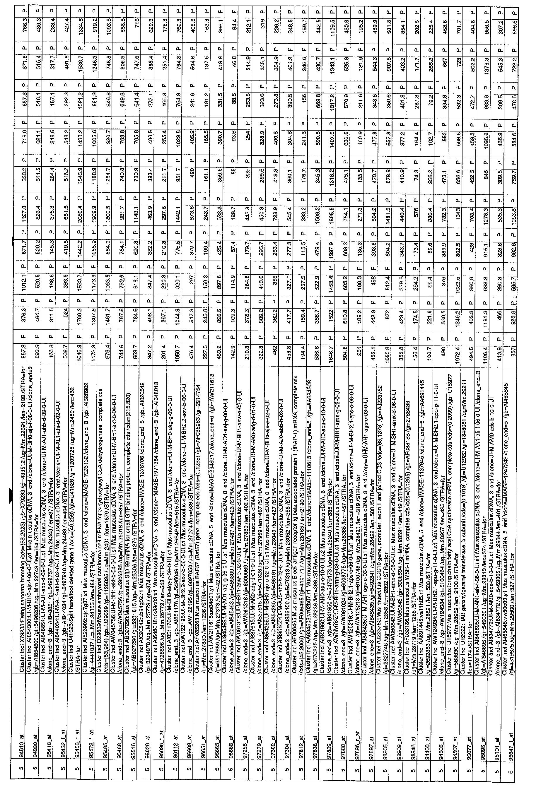
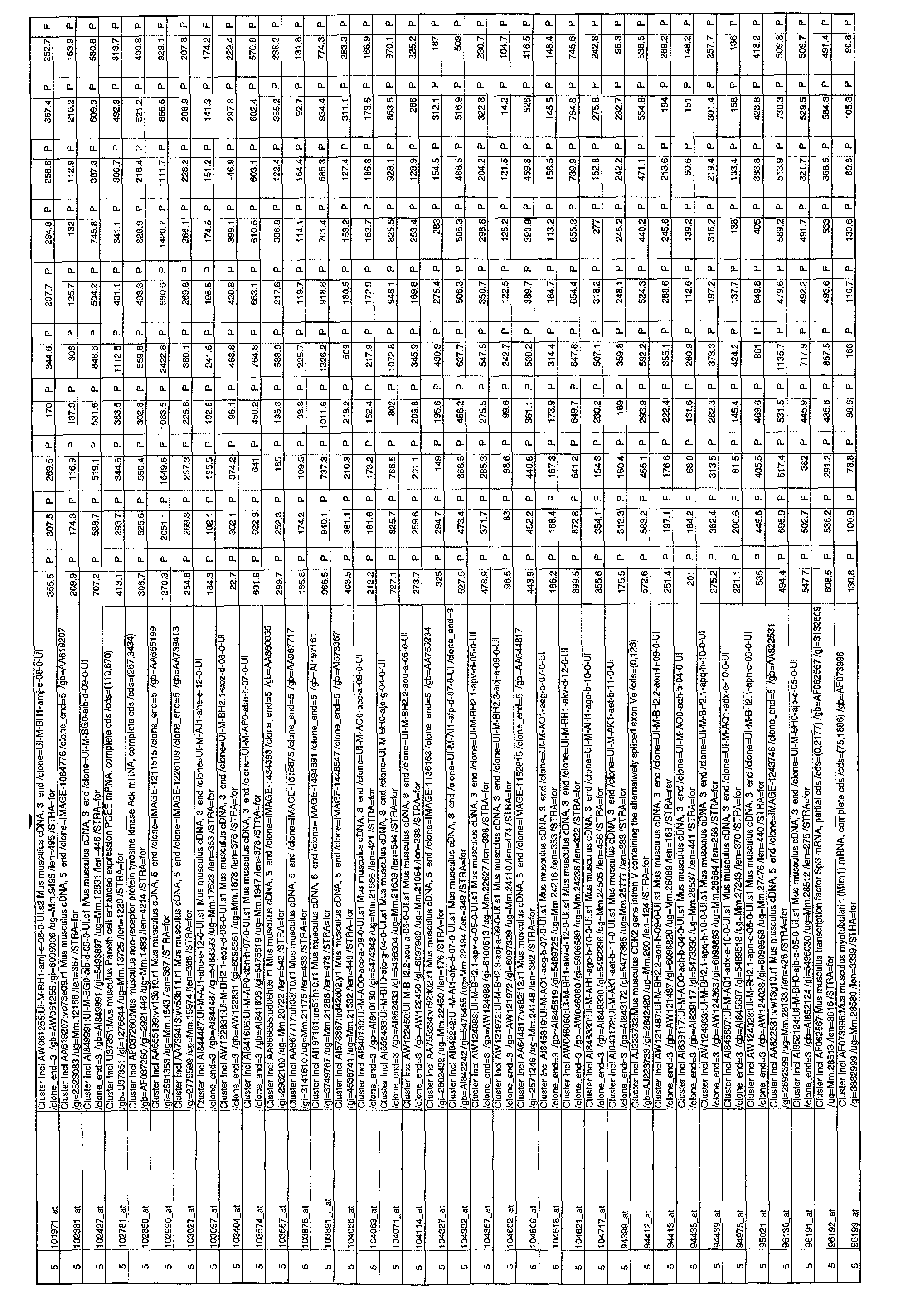
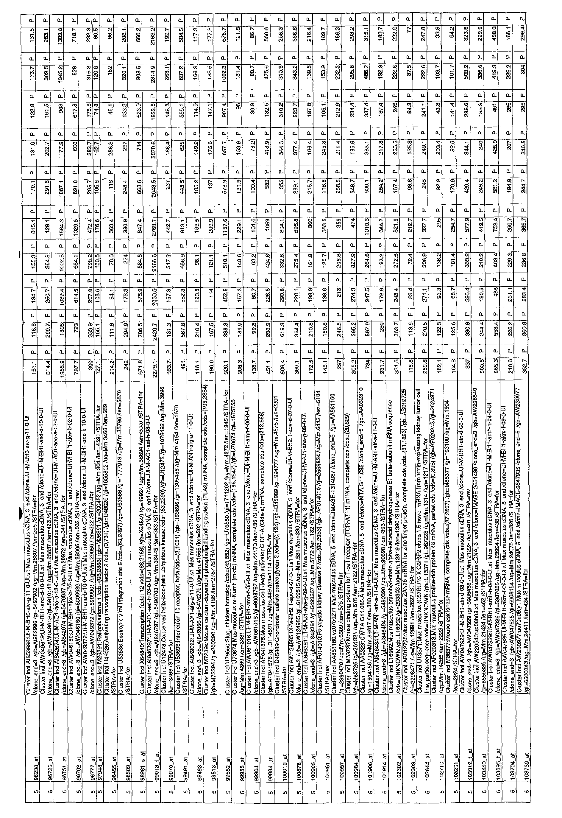
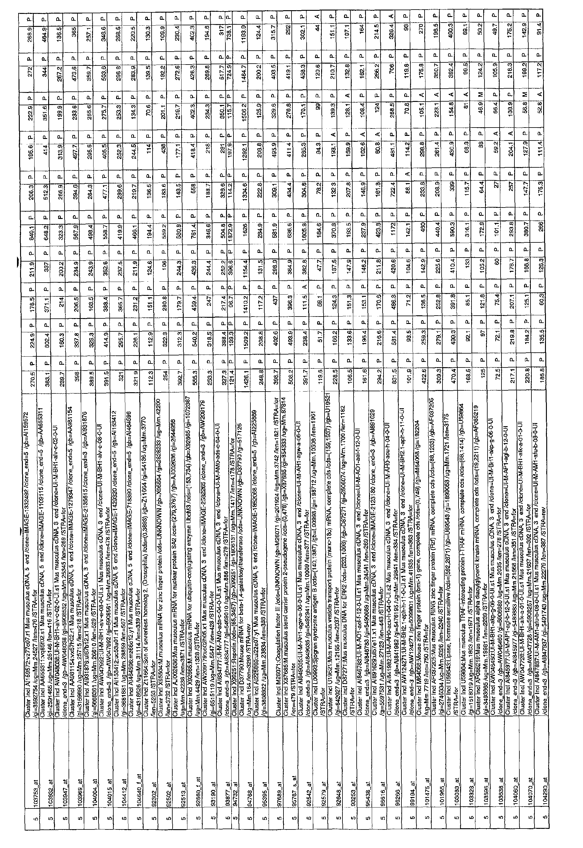
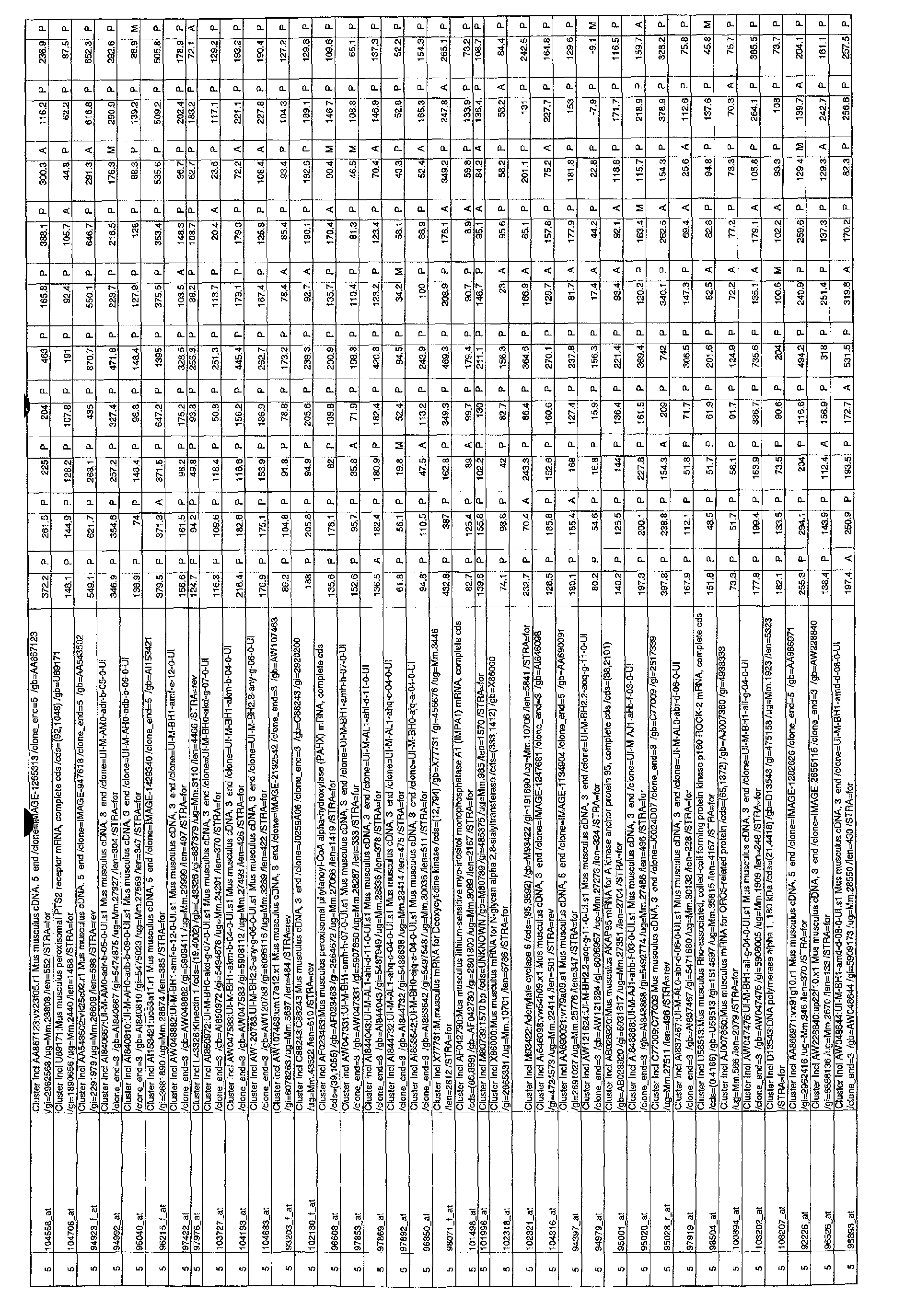
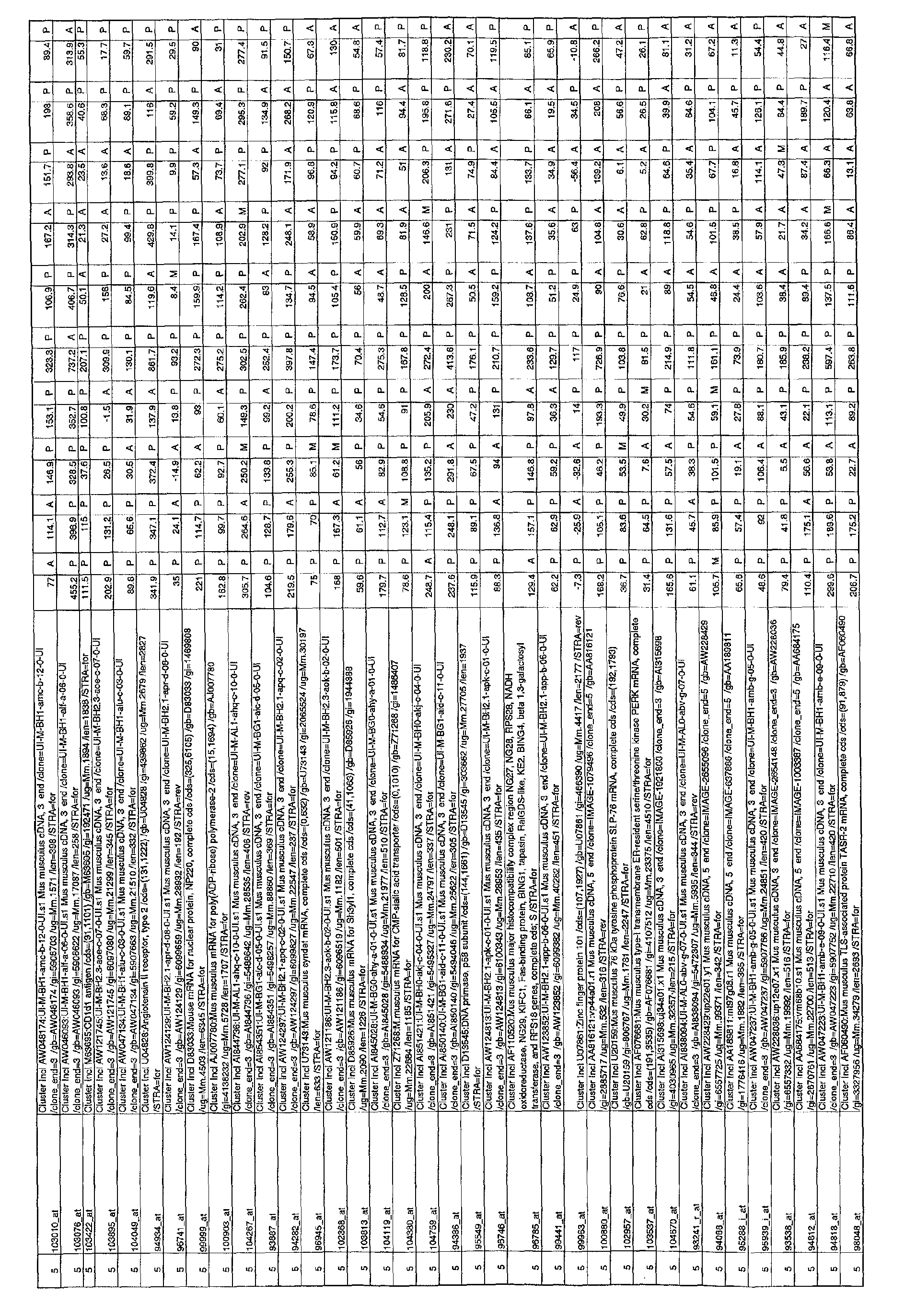
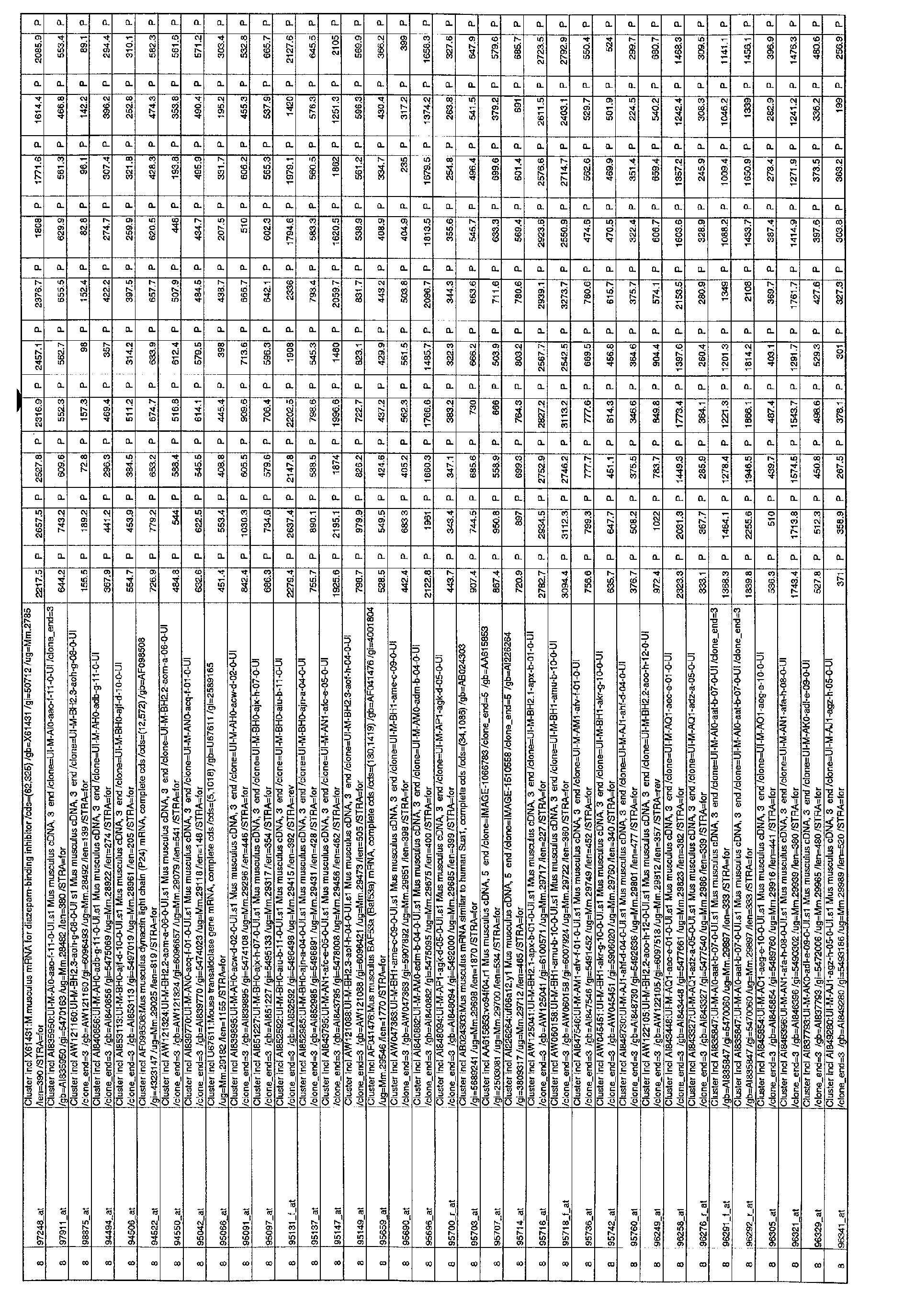
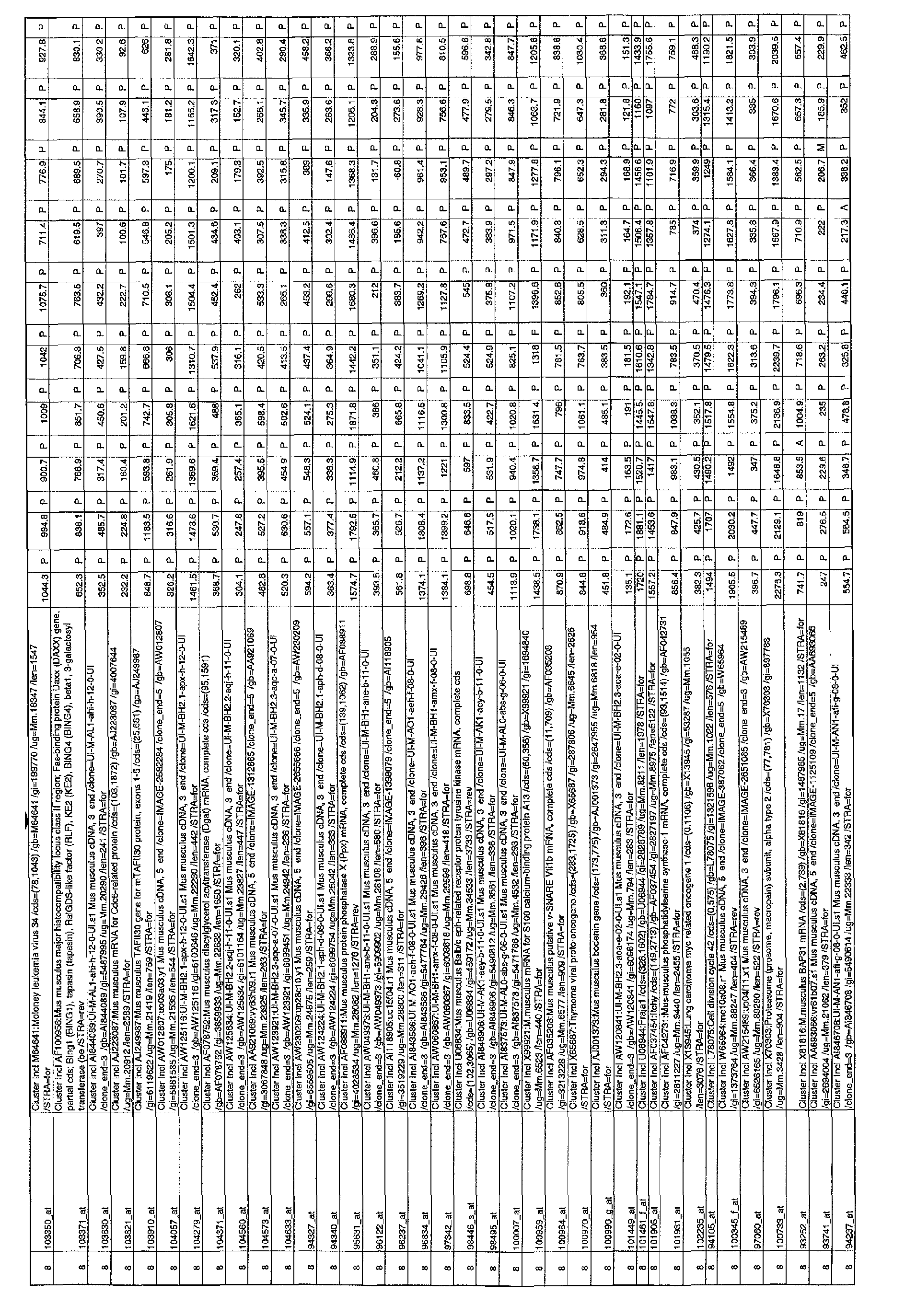
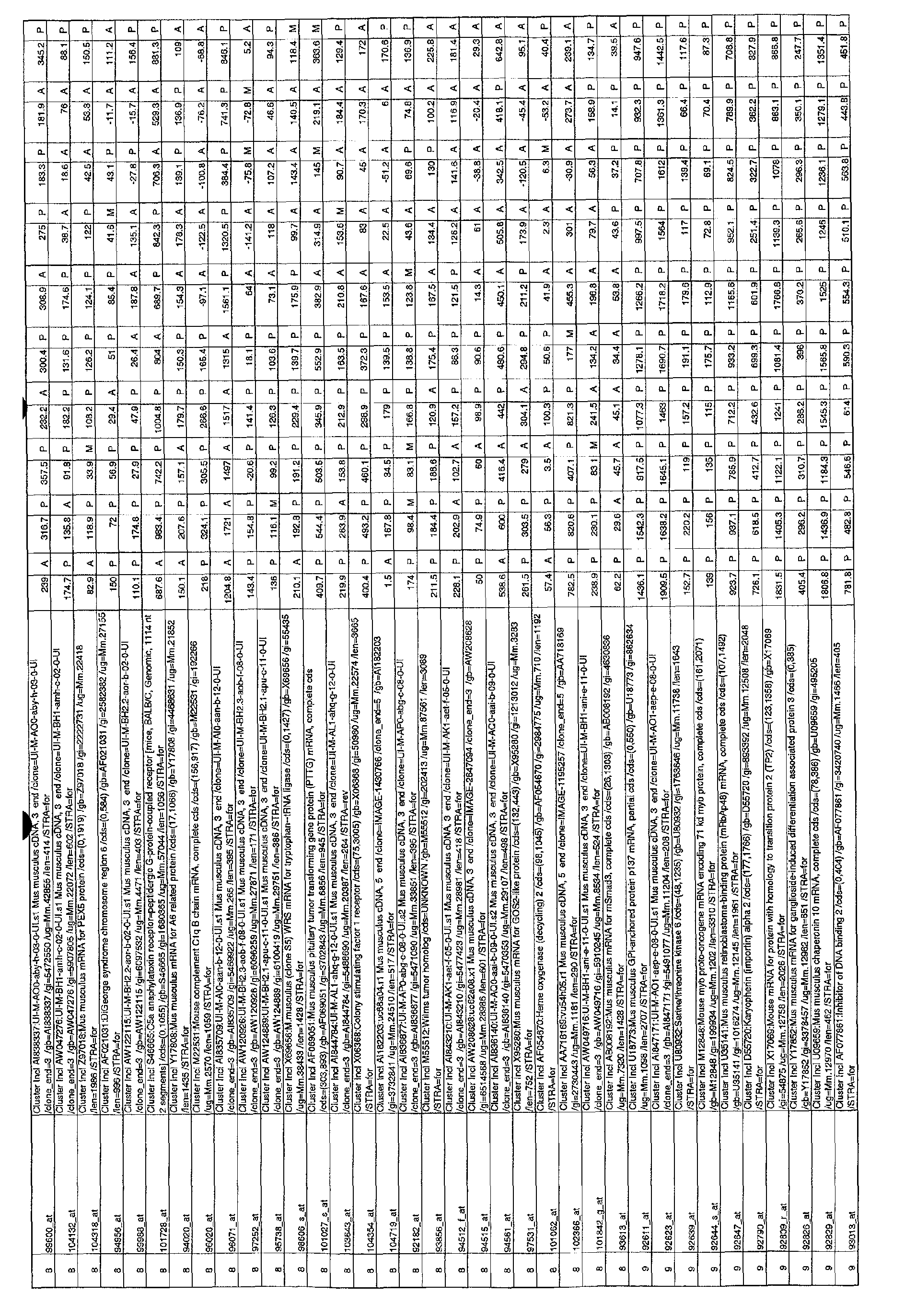
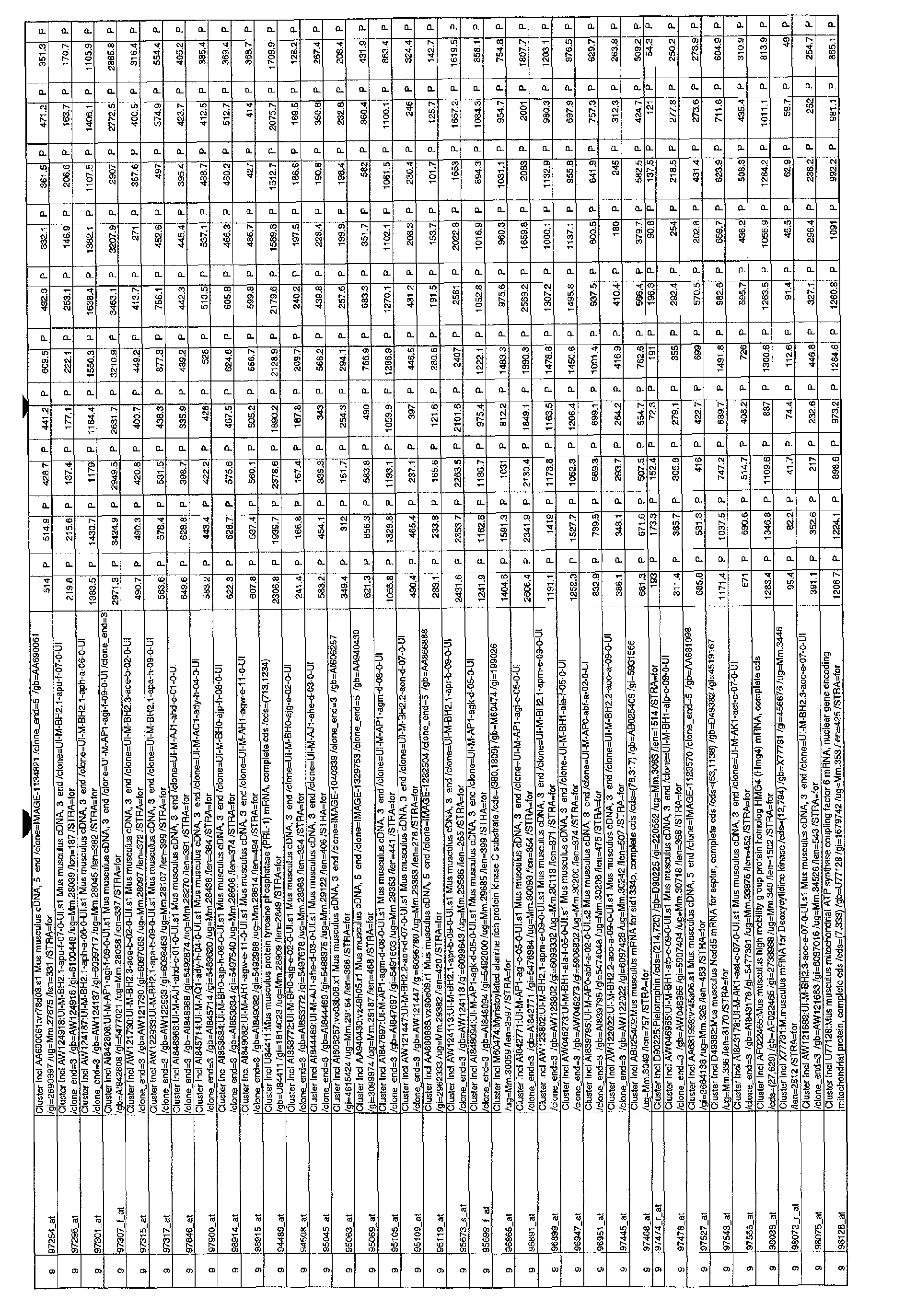
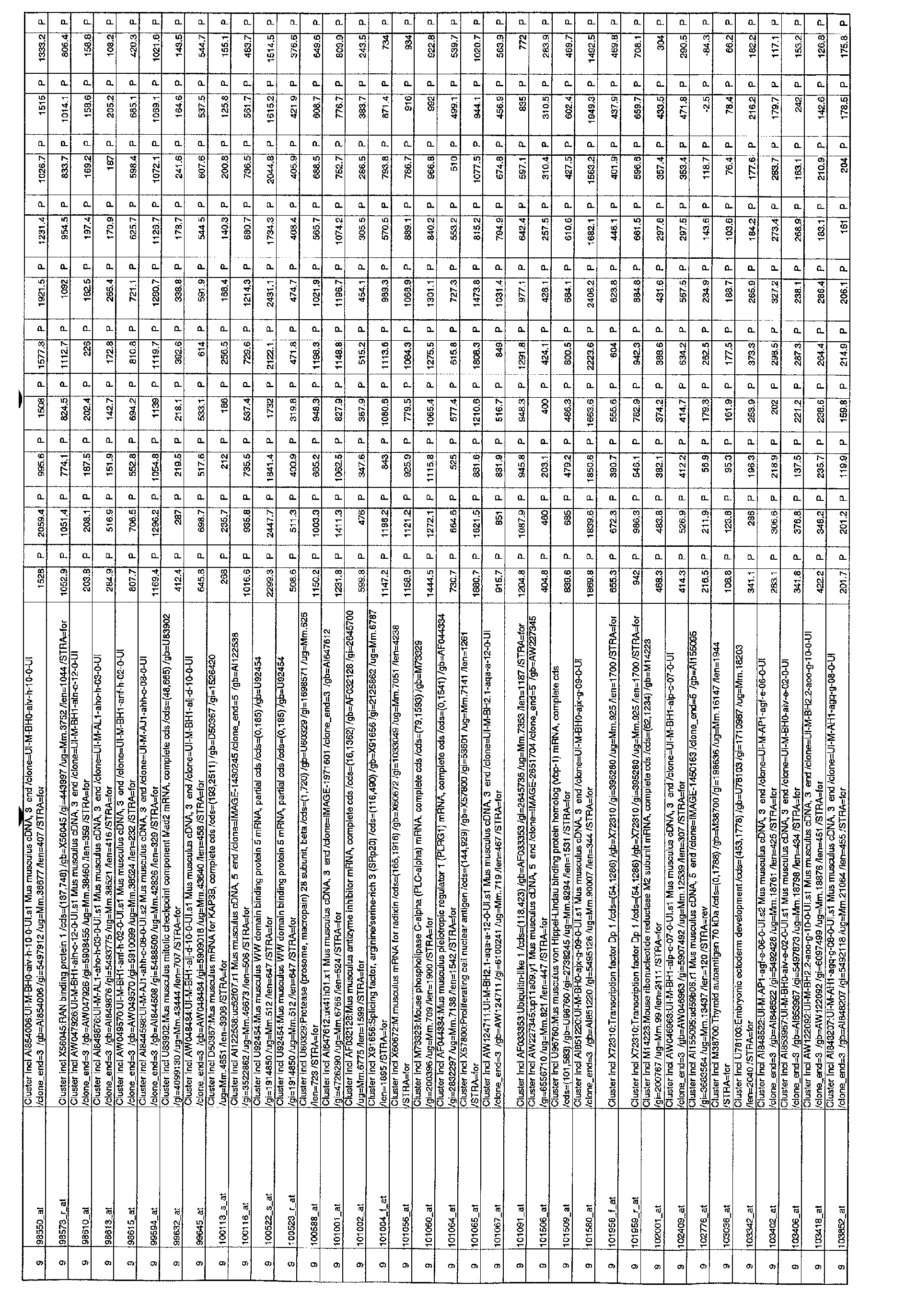
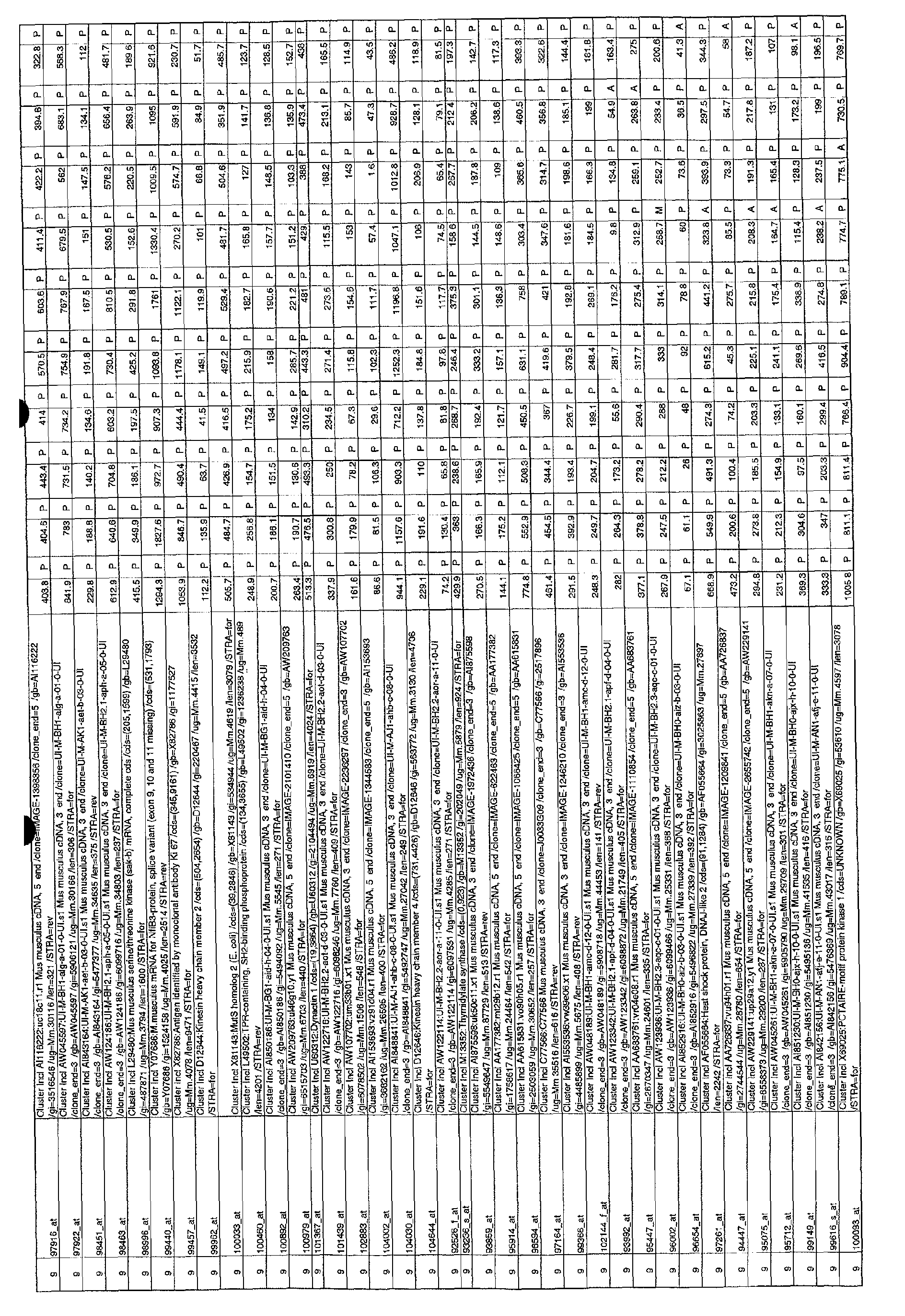
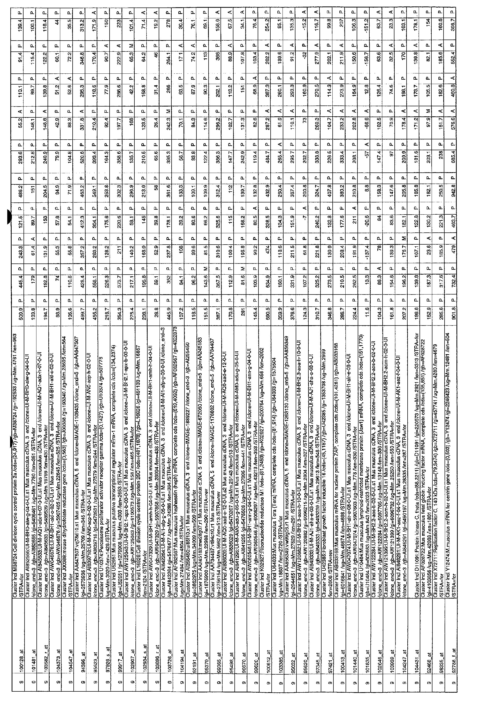
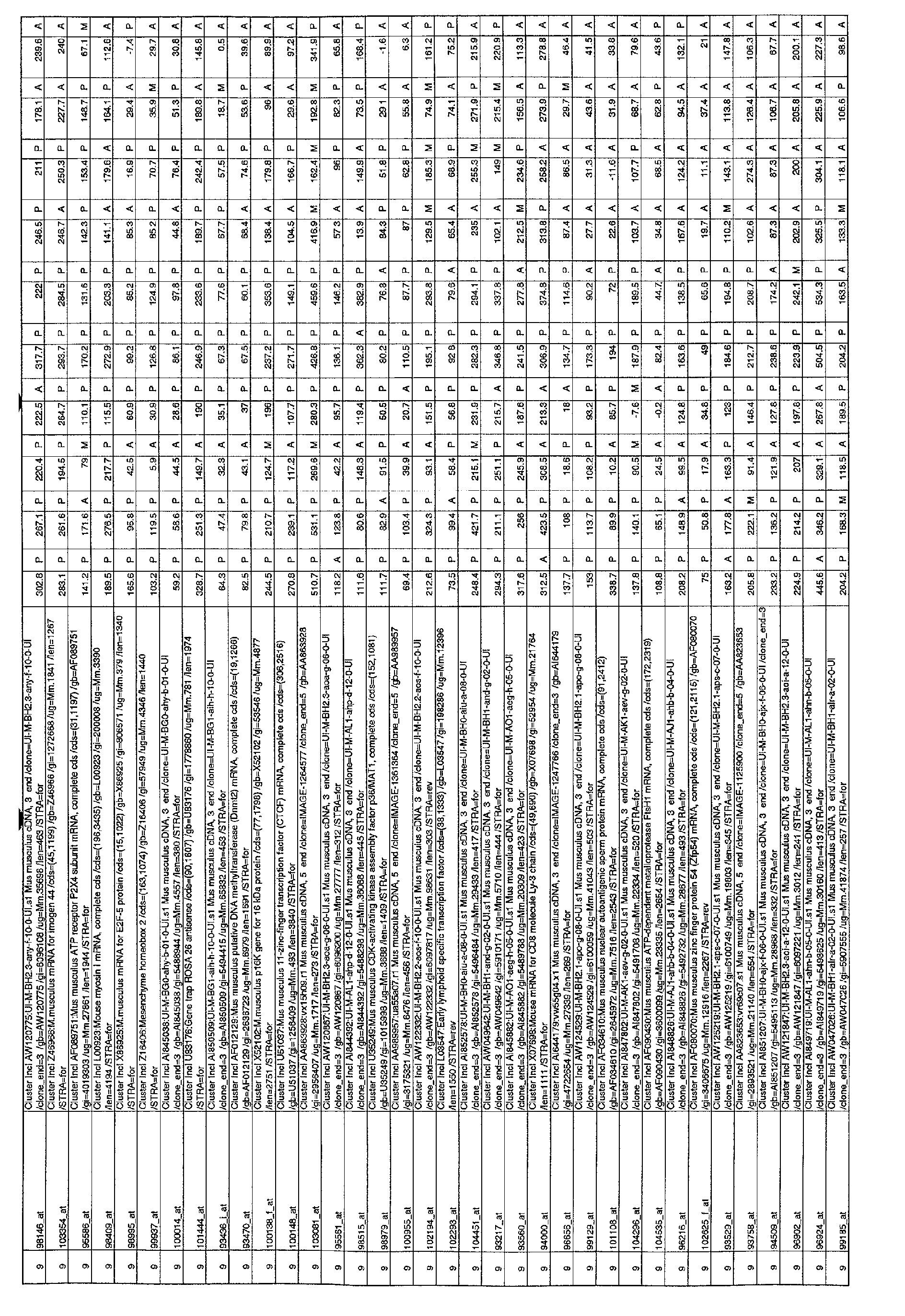
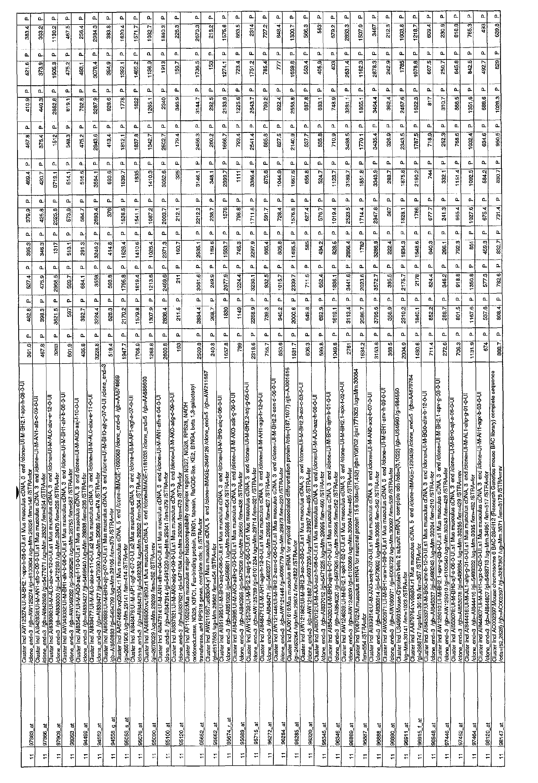
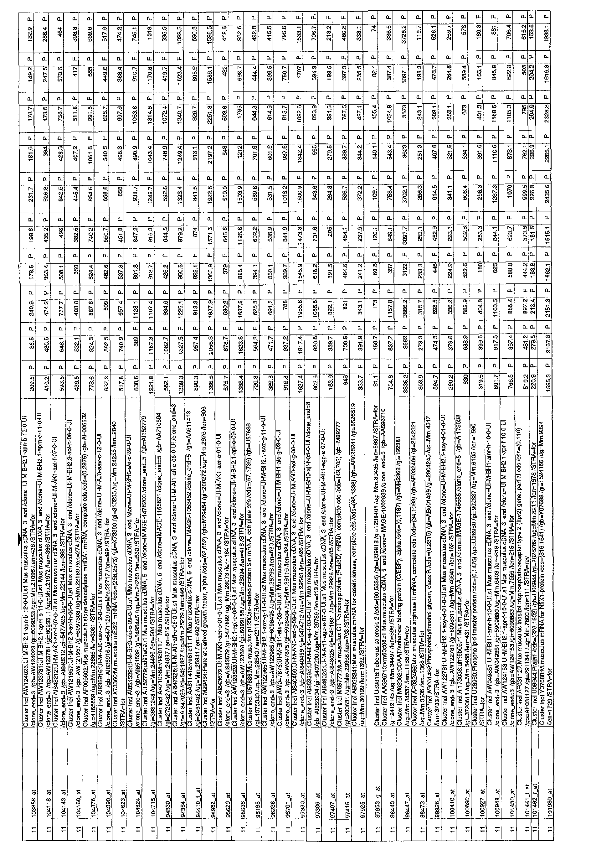
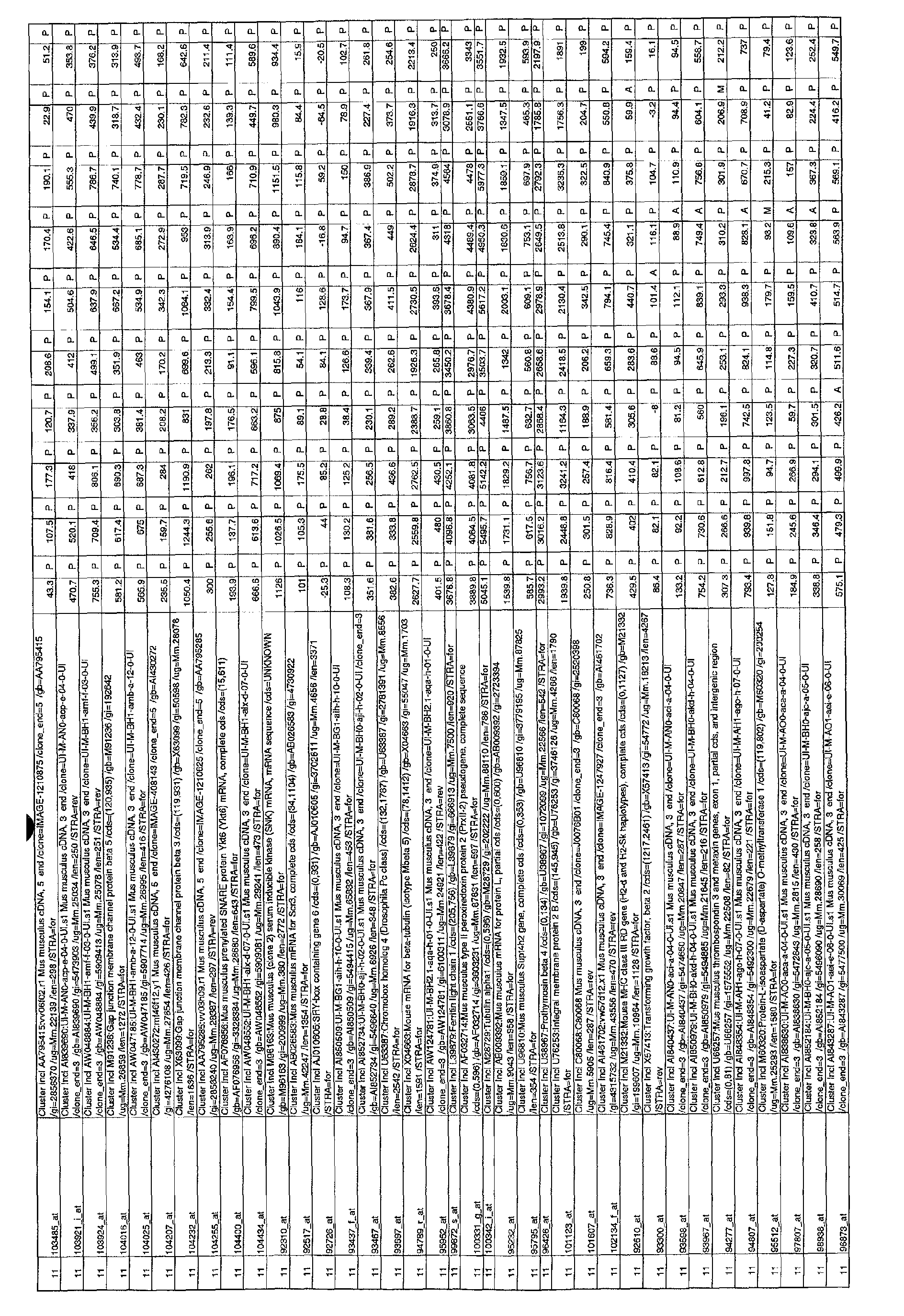
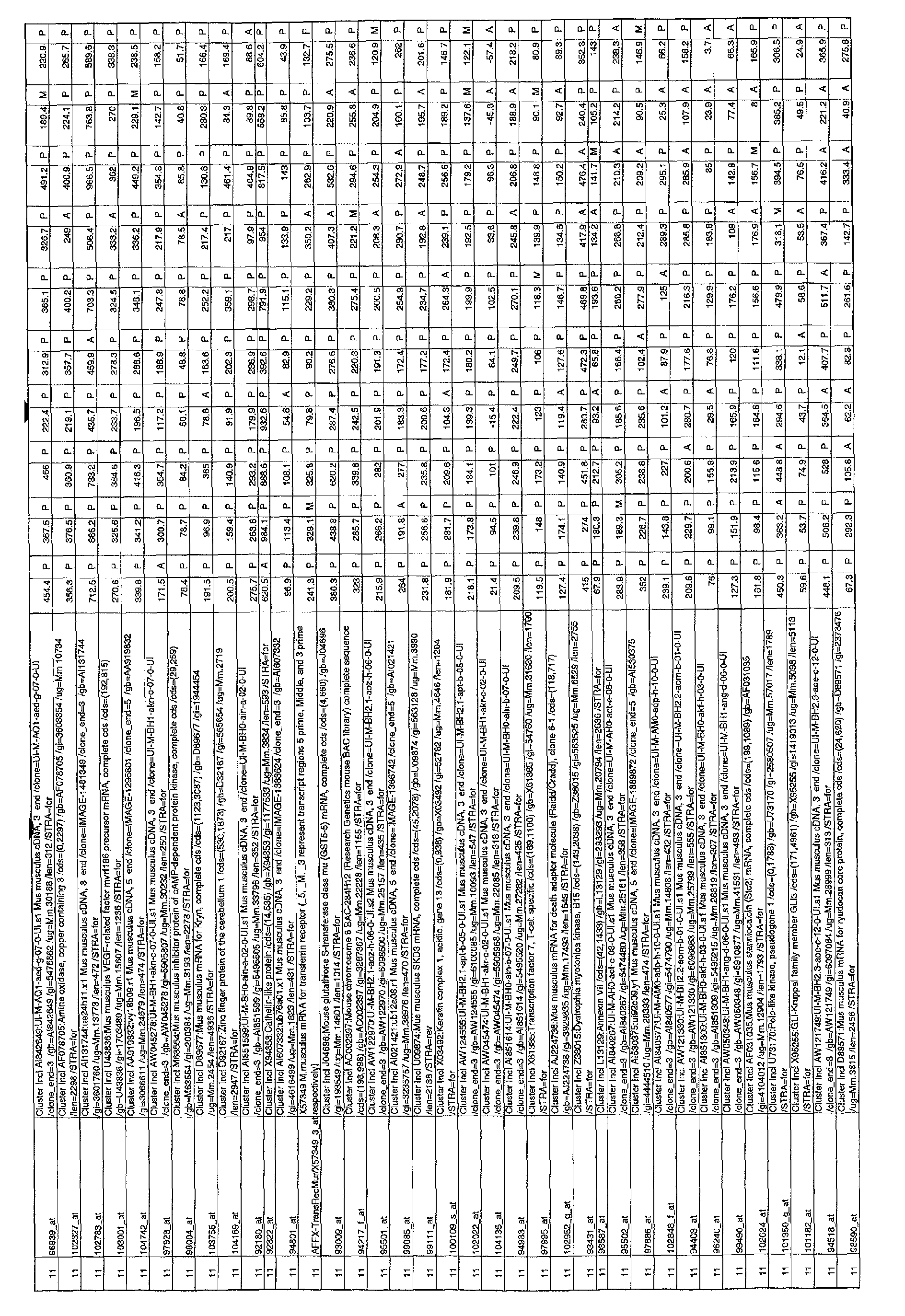
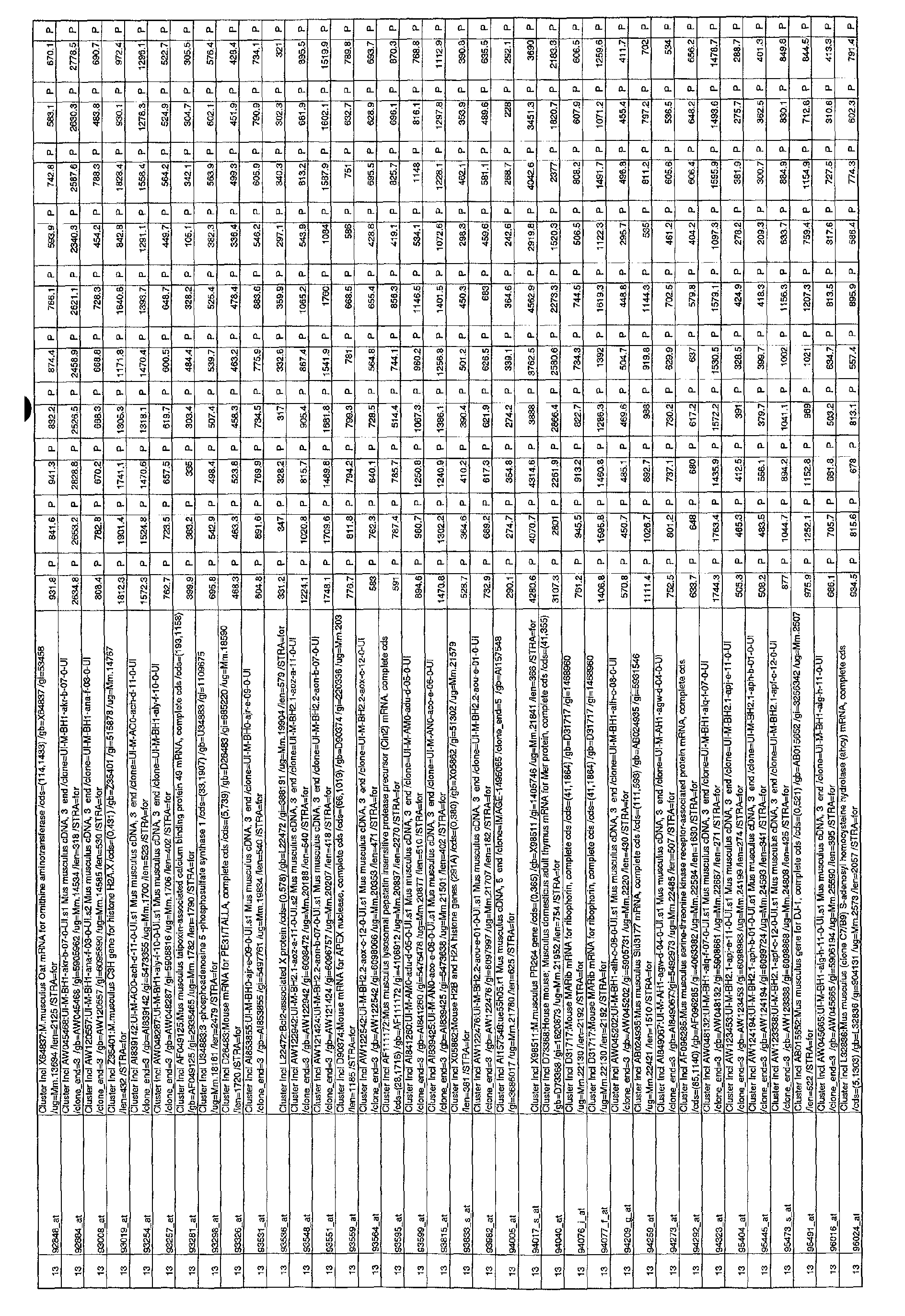
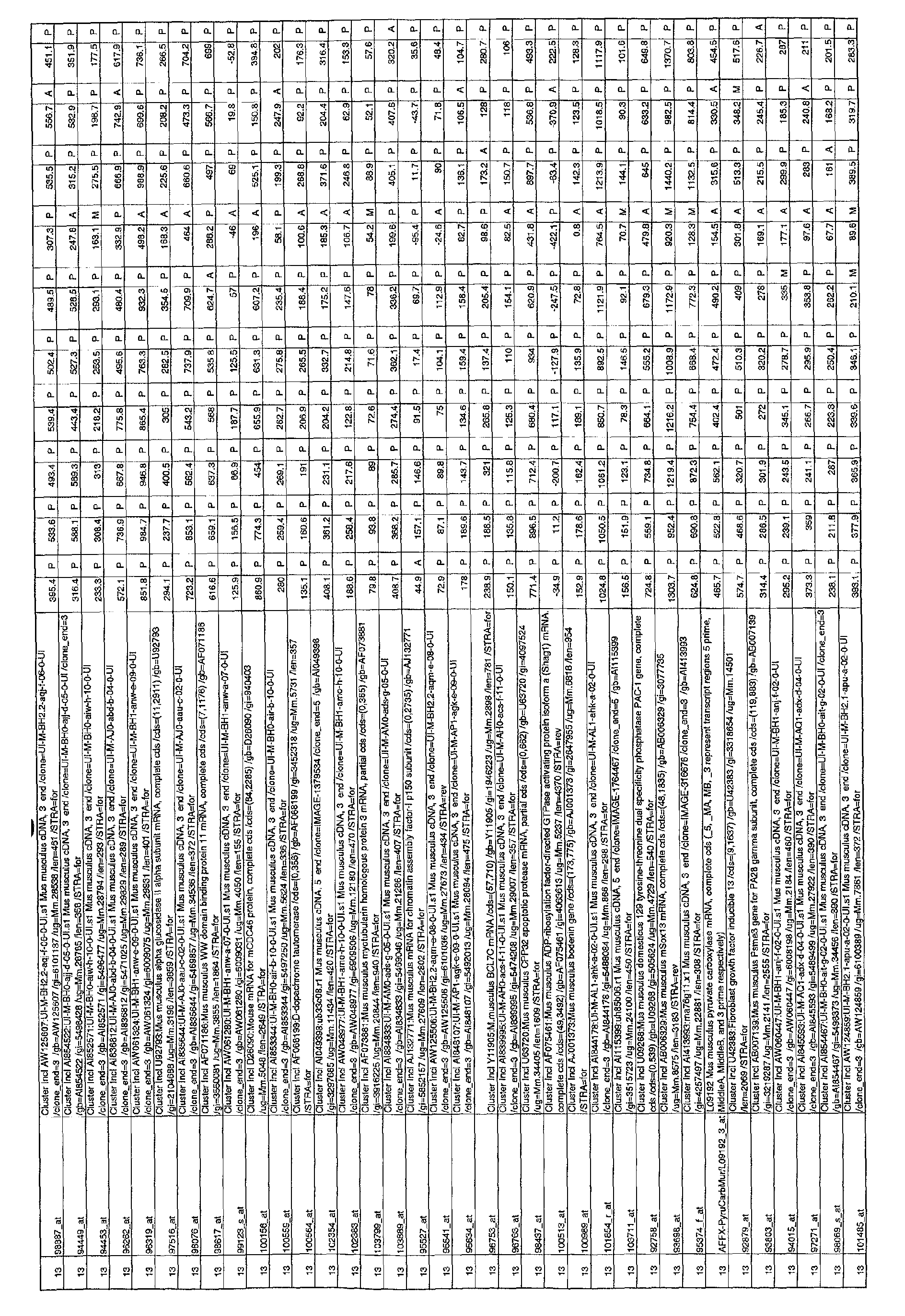
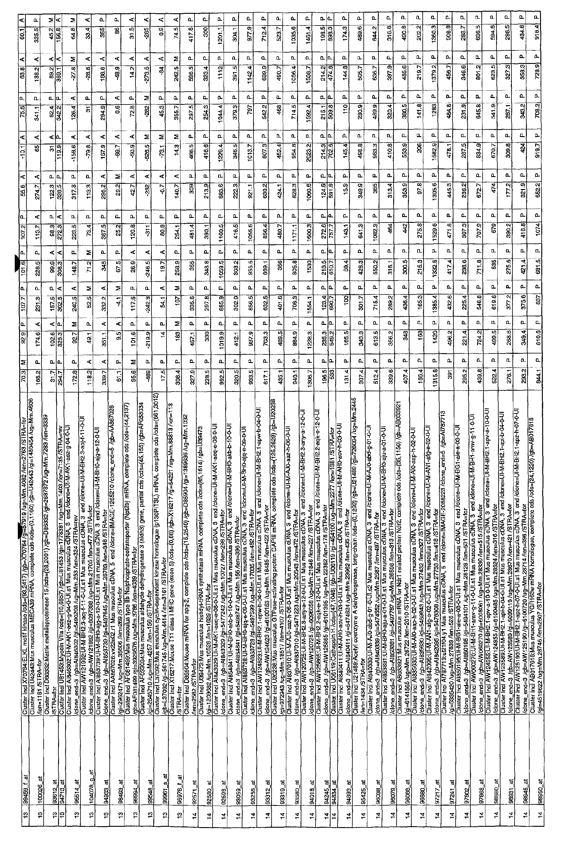
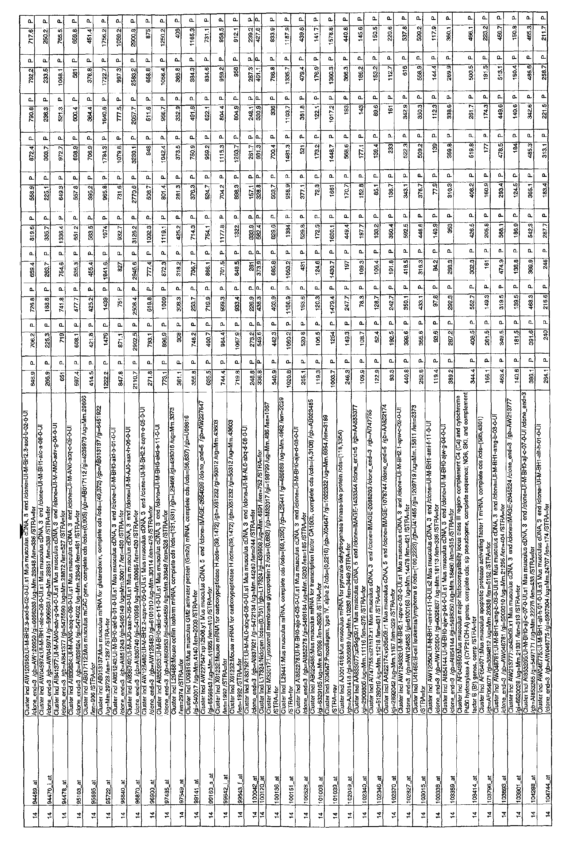
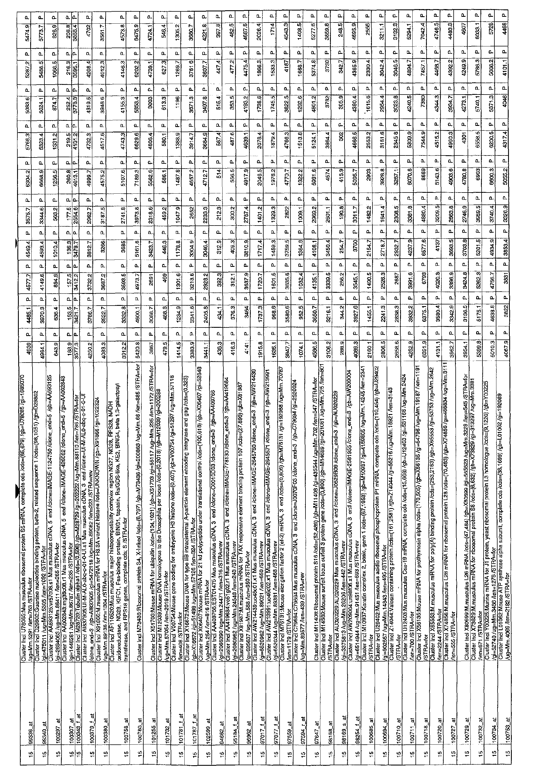
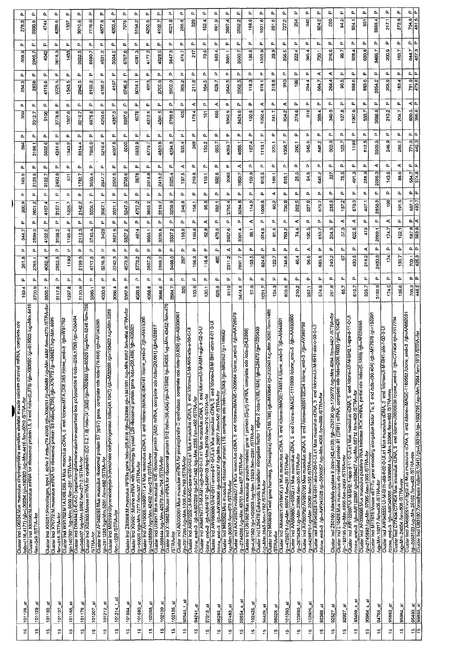
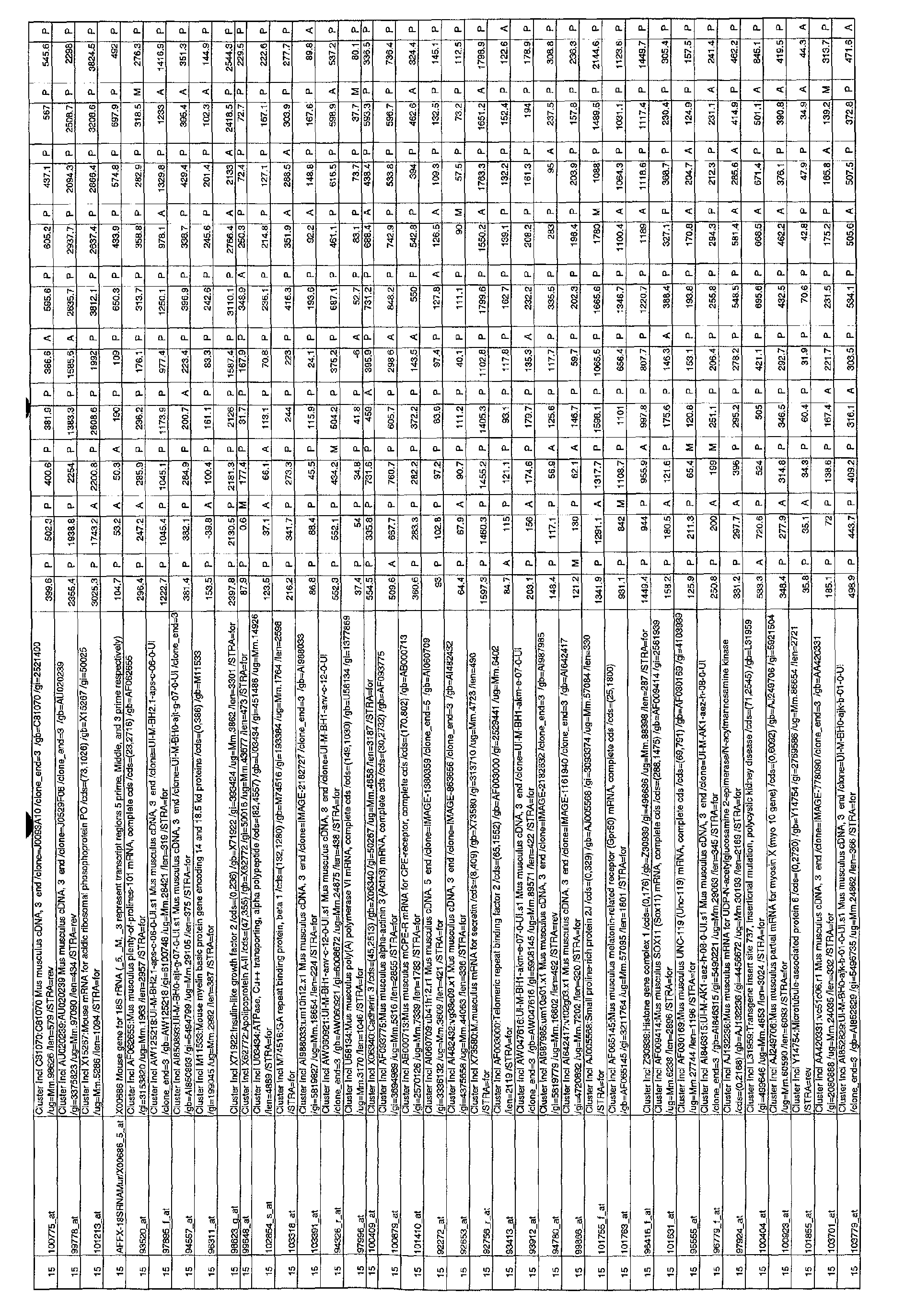
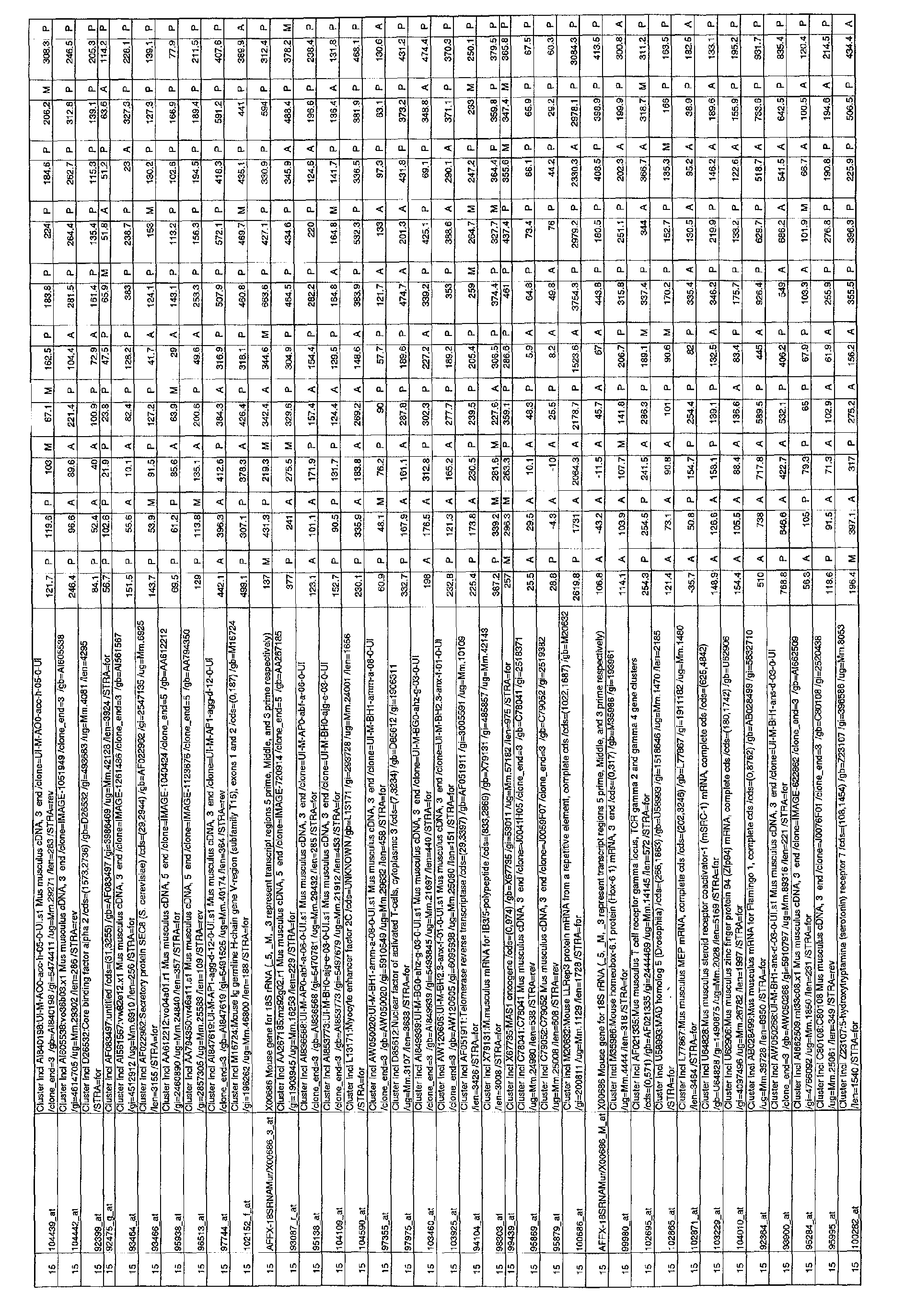
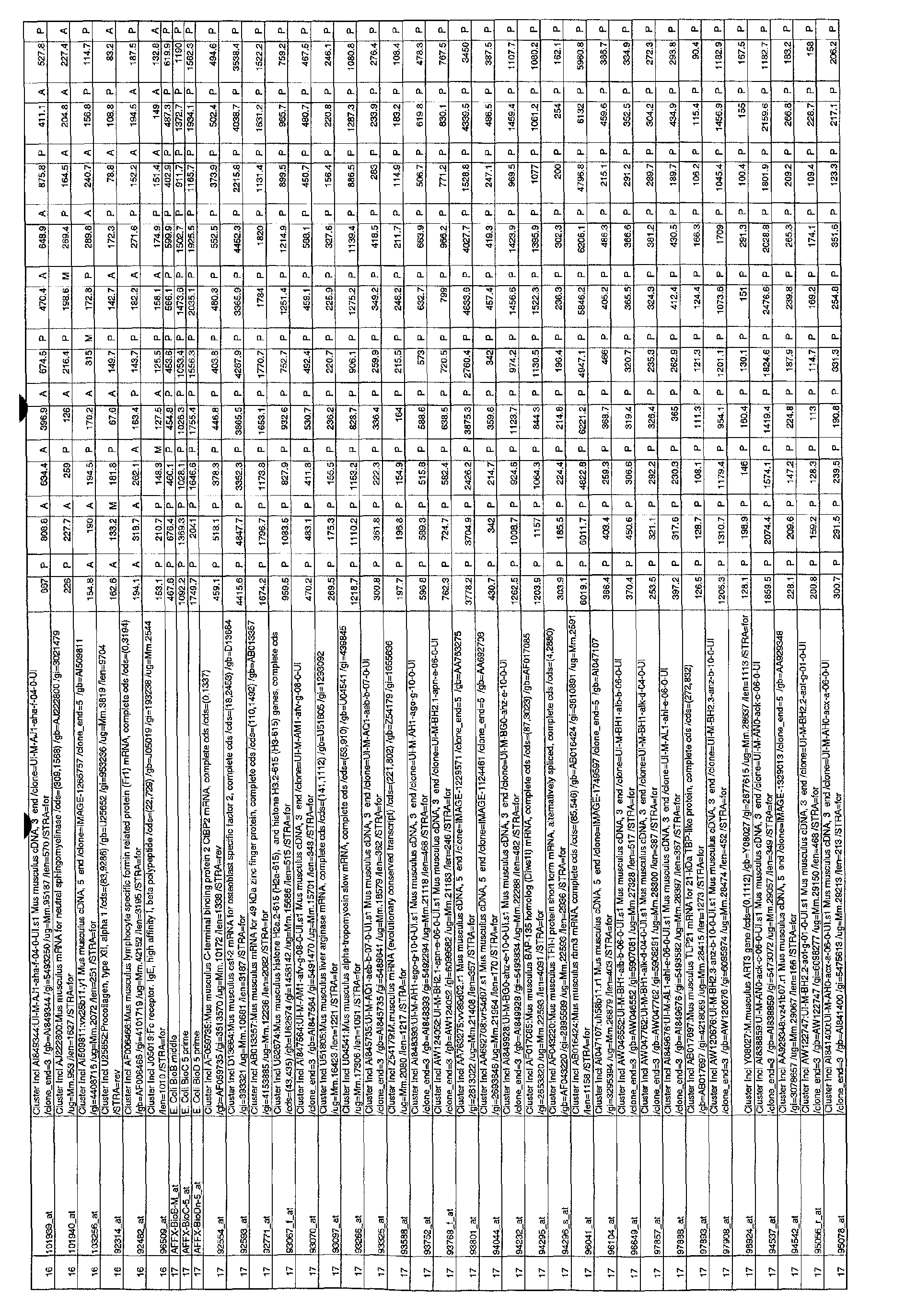
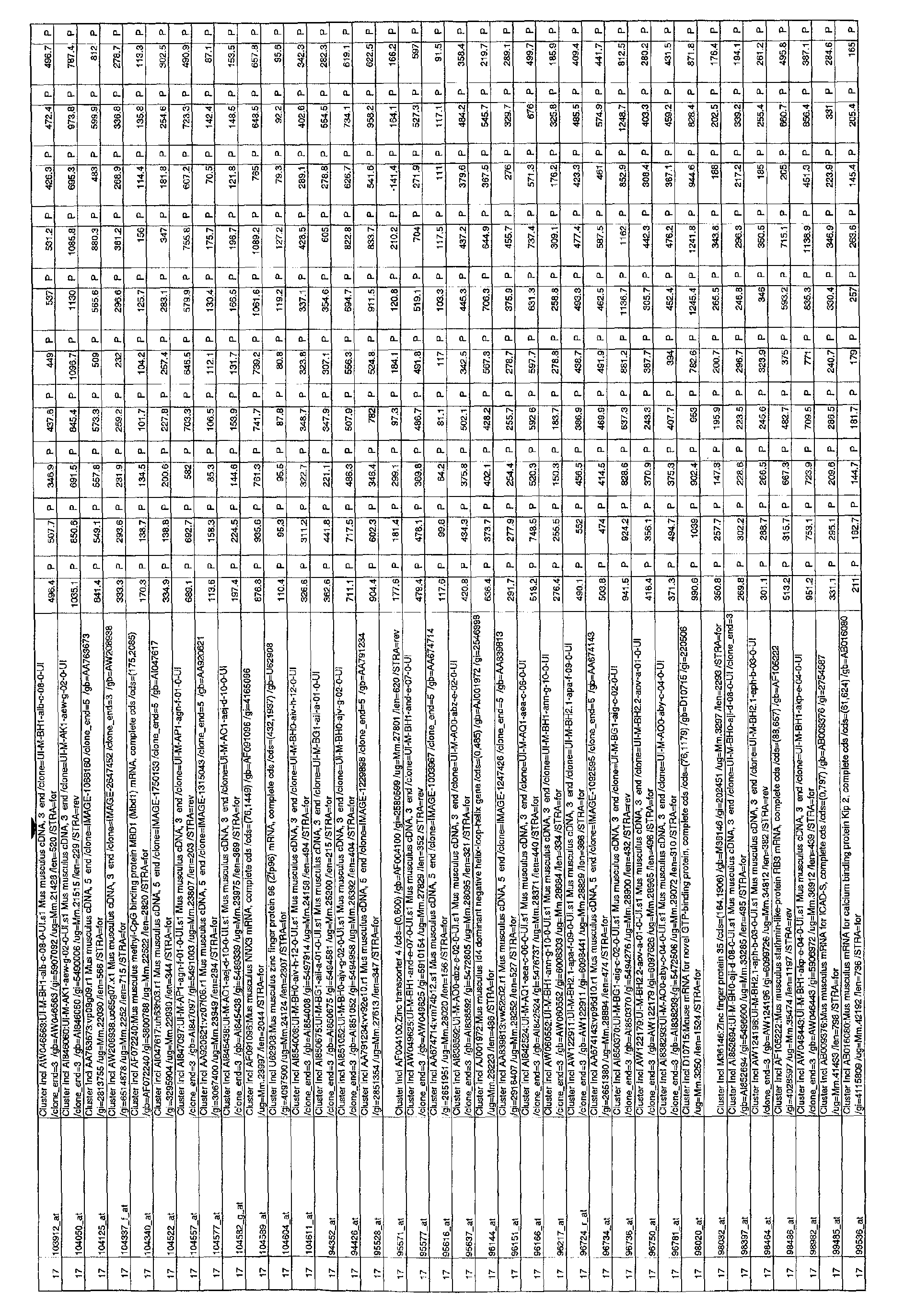
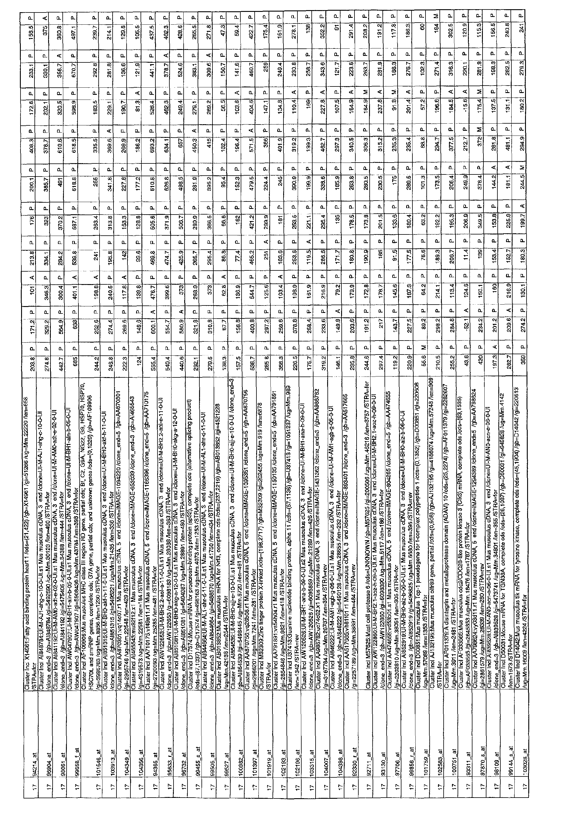
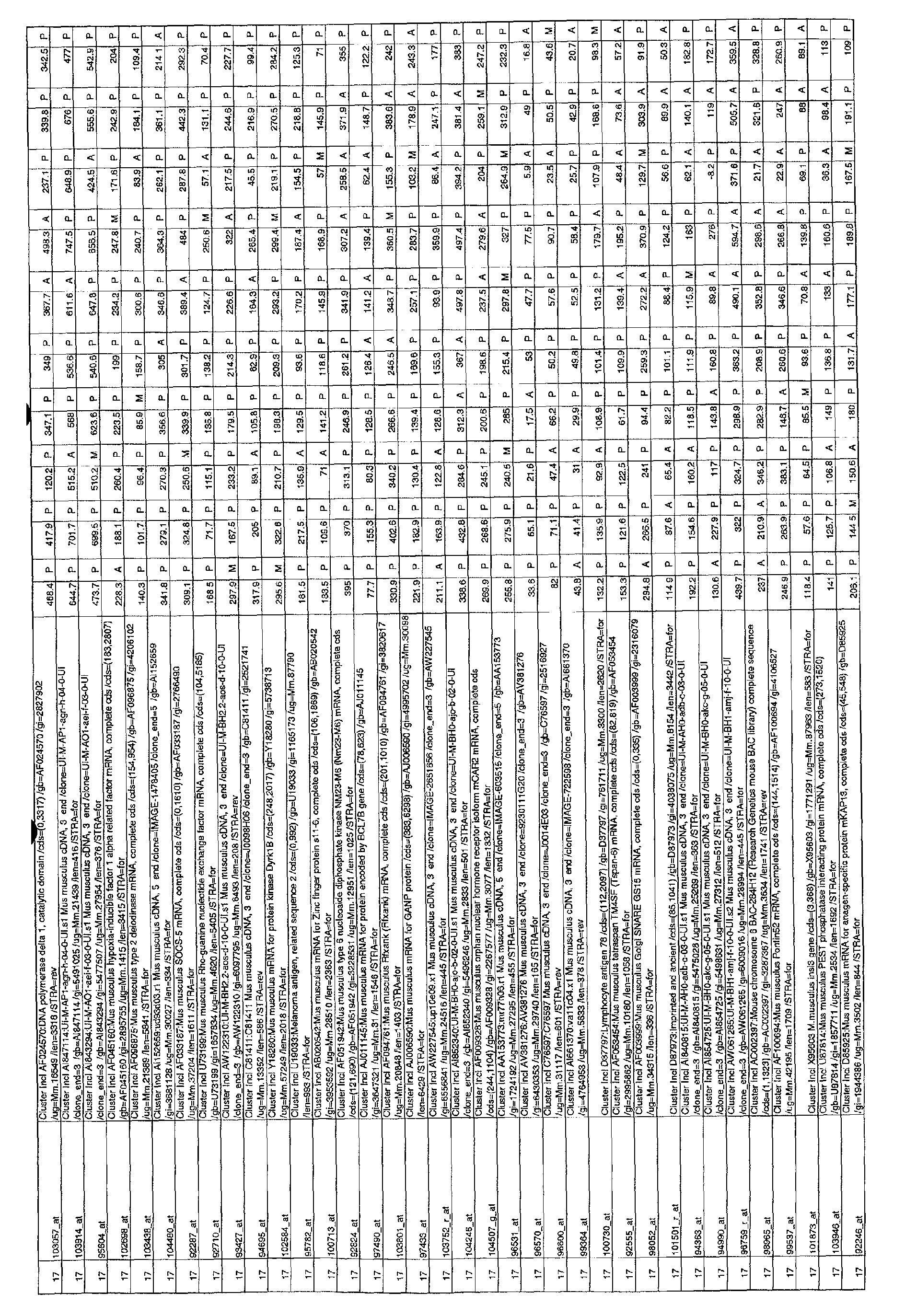
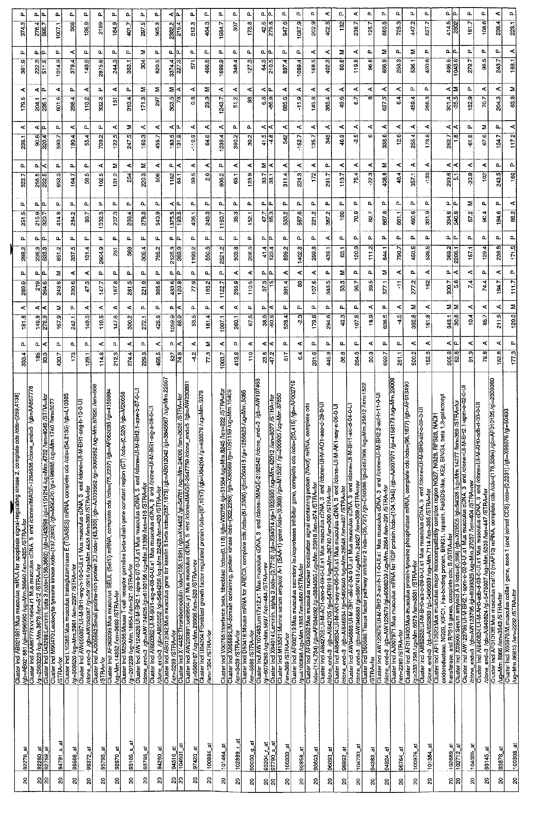
- Table 1 found at the end of the example section
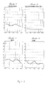
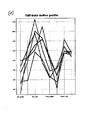
- the summary profile for each cluster is shown in Figure 1.
- the clusters are presented as the time course of expression for the respective group of genes in the order wild type control (taken at 12 hours), wild type 30 minutes after wounding, wild type 3 hours after wounding, wild type 12 hours after wounding, wild type 24 hours after wounding, followed by the same time points for the PU.
- Cluster 2 Cluster 7, Cluster 11, Cluster 16 and Cluster 20.
- Cluster 2 310 profiles
- the profile of this cluster identifies genes whose expression peaks 3hrs after wounding in both the wild type and the PU.1 KO mice.
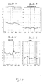
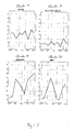
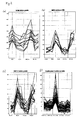
- Examples of prototypical wound healing genes in cluster 2 are transcription factor JunB (immediate early gene, involved in transcriptional regulation) and actin which is required for cell migration. All genes in this cluster are likely to be essential wound healing genes, and/or marker genes indicative of wound repair. Specifically, the nine genes that show a profile most similar to actin, identified using actin as the active profile, are likely to be important (Fig. 2a, Table 2).
- One or more of the proteins encoded by nucleotide sequences comprising the sequences of the accession numbers grouped in clusters 2, 7, 11, 16, and 20 as indicated in Table 1 can be used as a marker, or markers indicative of wound repair.
- An inhibitor or antagonist of a protein encoded by a gene comprising a sequence of one of the accession numbers grouped in clusters 2, 7, 11, 16, and 20 can be used for the treatment of a disease or disorder characterised by an excessive healing response, such as, but not limited to, scarring, fibrosis, restenosis post angioplasty, psoriasis, post-traumatic/surgical adhesions of the peritoneal cavity, joints and ligaments, benign prostatic hyperplasia, glaucoma, and peripheral nerve injury.
- An antisense oligonucleotide, or ribozyme against, or compounds that downregulate transcription of, any of the genes comprising the sequences of the accession numbers grouped in clusters 2, 7, 11, 16, and 20 can be used for the treatment of diseases and disorders characterised by an excessive healing response, such as, but not limited to, scarring, fibrosis, restenosis post angioplasty, psoriasis, post-traumatic/surgical adhesions of the peritoneal cavity, joints and ligaments, benign prostatic hyperplasia, glaucoma, and peripheral nerve injury.
- an excessive healing response such as, but not limited to, scarring, fibrosis, restenosis post angioplasty, psoriasis, post-traumatic/surgical adhesions of the peritoneal cavity, joints and ligaments, benign prostatic hyperplasia, glaucoma, and peripheral nerve injury.
- An increase in activity of a protein encoded by a gene comprising a sequence of one of the accession numbers grouped in clusters 2, 7, 11, 16, and 20 can be achieved by, for example, agonists or stimulators of the protein, gene therapy, application of the protein itself, or substances that upregulate the transcription and/or translation of these genes, means that provide an increase in activity of the protein have application in the treatment of diseases and disorders characterised by impaired healing response such as chronic dermal ulcers, oral mucocystis, emphysema, ulcerative diseases of the gastro-intestinal (GI) tract, cystitis.
- diseases and disorders characterised by impaired healing response such as chronic dermal ulcers, oral mucocystis, emphysema, ulcerative diseases of the gastro-intestinal (GI) tract, cystitis.
- WISP-1 Connective Tissue Growth Factor Gene Family member WISP-1 is in this cluster and therefore identified as an early regulator of wound repair.
- WISP-1, or a viral or plasmid construct comprising the coding sequence of WISP-1 under control of a promoter active in mammalian cells is useful for the treatment of conditions or disorders involving impaired wound repair.
- the profile of this cluster identifies genes whose expression increases during wound repair in both the wildtype and the PU.1 KO mice.
- PAF acetyl hydrolase is present in this cluster and therefore identified as a marker of wound repair.
- inhibitors of PAF find use as wound healing agents.
- PAF antagonists are therefore useful as agents for the treatment of diseases and disorders characterised by impaired healing response.
- Suitable PAF antagonists are disclosed in European Patent EP 0310386, especially modipafant, which is (+)-4-(2-chlorophenyl)-1,4-dihydro-3-ethoxycarbonyl-6-methyl-2-[4-(2-methylimidazo[4,5-c]pyrid-1-yl)phenyl]-5-(N-[2-pyridyl]carbamoyl)pyridine, and is disclosed in European Patent 0310386 [see Example 34(d)].
- cyclooxygenase I is present in this cluster and is a marker of wound repair, particularly early wound repair processes. This enzyme is required to produce bioactive eicosanoids, therefore cyclooxygenase I inhibitors are useful for the treatment of diseases and disorders characterised by excessive healing response.
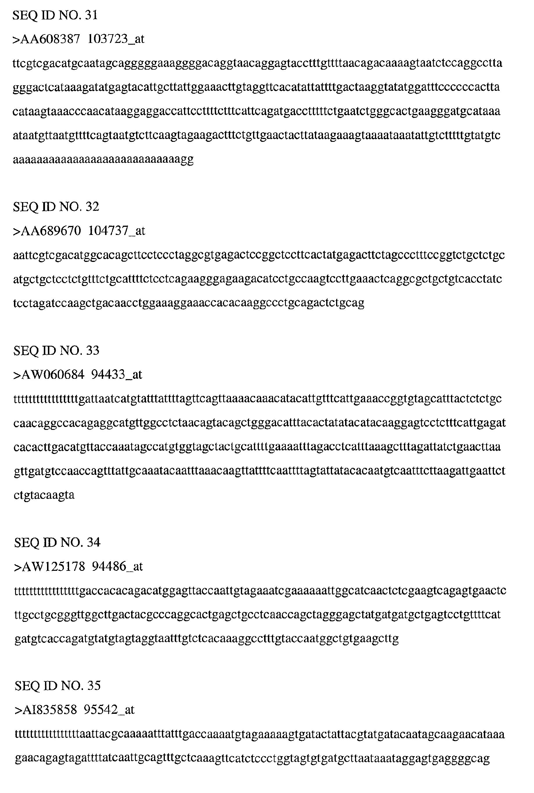
- Genes in this cluster are essential wound healing genes, expressed toward the end of the healing process. These genes are involved in cessation of the healing response, and maturation and remodelling of the repaired tissue, transcription and translation products of these genes are markers for advanced wound repair.
- MRP-8 a cytokine from the S100 family, previously identified as being strongly induced in normal wounds, is a member of this cluster, and can therefore serve as a marker of advanced wound repair. Eight genes were identified using MRP-8 as the active profile (see Figure 2b, Table 3). These eight genes and their transcription or translation products are useful as markers of advanced wound repair.
- EST AA689670 was identified as a keratinocyte marker of advanced wound repair.
- MMP3 The presence of MMP3 in this cluster suggests that inhibitors of MMP-3 will be useful to inhibit wound remodelling. Inhibitors of the sixteen gene products that are identified using MMP3 as the active profile will also find use for inhibition of wound remodelling (Fig. 2c, Table 4).
- Inflammatory genes not required for wound healing are genes that are found upregulated in the wildtype but not in the PU.1 KO mice. They are found in clusters 5 and 6:
- Cluster 5 321 profiles
- Cluster 6 213 profiles
- Genes in these clusters that are not expressed in the PU.1 KO mice represent inflammatory genes not required for wound healing. As excessive inflammation is associated with impaired healing and excessive scarring, these non-essential genes represent targets for the treatment of all types of impaired healing. Antagonists or inhibitors for the products of any of the genes of these clusters are likely to be useful for improving wound healing. Also, genes in these clusters and their transcription and translation products are useful as markers for wound inflammation. Using cathepsin-S as the active profile, 47 genes were found to cluster with cathepsin-S (Fig. 2d, Table 5); these are especially useful as markers for wound inflammation, as well as for screening for inhibitor or antagonist compounds useful for the treatment of impaired healing and excessive scarring. The IMAGE clone number 1429340 is also an example of such an inflammatory marker or potential target gene. Especially inhibitors of ERK2 map kinase will be useful for the treatment of diseases and disorders characterised by impaired healing response.
- Clusters 4 (247 profiles), Cluster 13 (234 profiles) and Cluster 17 (238 profiles). These clusters represent genes that are down regulated during healing, in both wildtype and PU.1 KO mice. This down regulation may be required for repair to occur. Therefore inhibitors of these genes will be useful to promote wound healing and activators of these genes to prevent overhealing.
- Apolipoprotein E is found within this cluster and thus compounds that modulate the Apolipoprotein E expression or activity will be useful as agents for the control of wound repair.
- Cluster 19 187 profiles
- This cluster represents essential wound healing genes that show decreased expression during the late macrophage dependent inflammatory response. As excessive inflammation is associated with impaired healing and excessive scarring, these genes, their transcription and translation products are targets for the treatment of impaired healing and excessive scarring.
- Focal adhesion kinase is found in this cluster and thus inhibitors of focal adhesion kinase can be used to treat treat diseases and disorders of over-repair.
- the endogenous plasminogen activator inhibitor, PAI-1 is found to be down-regulated by inflammation, therefore inhibitors of Plasminogen activators (especially Urokinase-type plasminogen activator) can be used for the treatment of inflammation-impaired wound healing.
- This cluster also contains early wound healing genes that are suppressed by resident inflammatory cells such as mast cells and dendritic cells. Genes in this cluster include fibronectin, which is known to be the earliest scaffold for wound repair. Agents which overcome the resident inflammatory cell suppression of fibronectin synthesis will be useful for the treatment of impaired wound healing. More specifically, agonists of the fibroblast growth factor (FGF) receptor and agonists of the ROR alpha 1 nuclear hormone receptor can be used for the treatment of inflammation impaired wound healing.
- FGF fibroblast growth factor
- TGF- ⁇ is known to be a major factor in governing scarring and fibrosis, using TGF- ⁇ as the active profile, a cluster of six genes was identified (Fig. 2e, Table 6) each of which thus is implicated in scarring and fibrosis.
- Various means to decrease the activity and/or amount of the expression products of these genes can be used for the treatment of diseases and disorders characterised by excessive healing response such as scarring, fibrosis, restenosis post angioplasty, psoriasis, post-traumatic/surgical adhesions of the peritoneal cavity, joints and ligaments, benign prostatic hyperplasia, glaucoma, and peripheral nerve injury.
- Such means include an inhibitor or antagonist of, or antibody to, any one of the proteins encoded by a gene comprising the sequence of one of the accession numbers clustered with TGF- ⁇ ; or an antisense oligonucleotide, or ribozyme against, or a compound that downregulates transcription and or translation of, any of the genes comprising the sequence of one of the accession numbers clustered with TGF- ⁇ .
- E11.5 embryos were obtained by means of timed matings of CD1 mice, with embryos considered to be E0.5 at noon on the day of plug checking.
- E11.5 embryos were dissected from the uterus in Tyrode's saline and prepared for culture as described by P Martin & D Cockcroft, (1999) Culture of post-implantation mouse embryos, in Molecular embryology methods and protocols (editors Sharpe, P and Mason, I). New York; Humana Press p7 - 22. Standard wounding was performed by amputating the left hindlimb at its base using iridectomy scissors as described by McCluskey and Martin (1995) Developmental Biology, 17: 102-114.
- incisional wounds of approximately 500 ⁇ m in length were also generated on the left forelimb, as well as superficial puncture wounds on the flank.
- Wounded embryos were transferred to 50ml Falcon tubes containing 4ml of culture medium (3ml of Tyrode's saline plus 1ml of normal rat serum) and roller-cultured for up to 24 hours in an atmosphere of 95% oxygen/5% carbon dioxide at 30rpm and 37°C (Martin and Cockcroft (1999), as above).
- Sections were hybridised (as described by DG Wilkinson & MA Nieto (1993) Methods Enzymol 225: 361-373) at 55°C overnight with digoxigenin labelled antisense radioprobes specific for the gene of interest. Expression was visualised by BCIP/NBT precipitation (Boehringer Mannheim) and sections were viewed on a Zeiss Axiophot microscope, after mounting in Citifluor (UKC). The labelled antisense radioprobe biomarkers derived from the gene of interest demonstrate expression of the gene of interest.
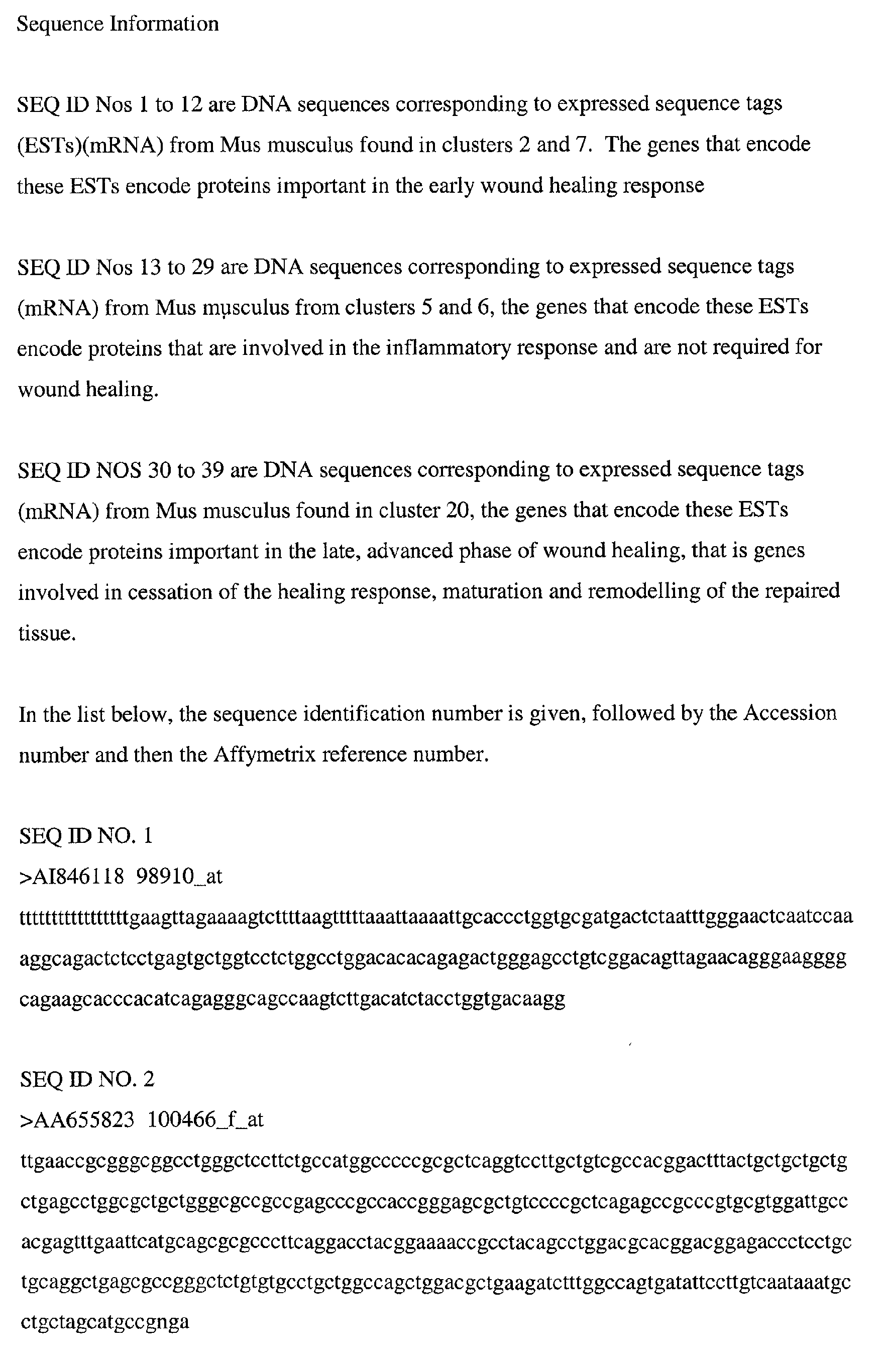
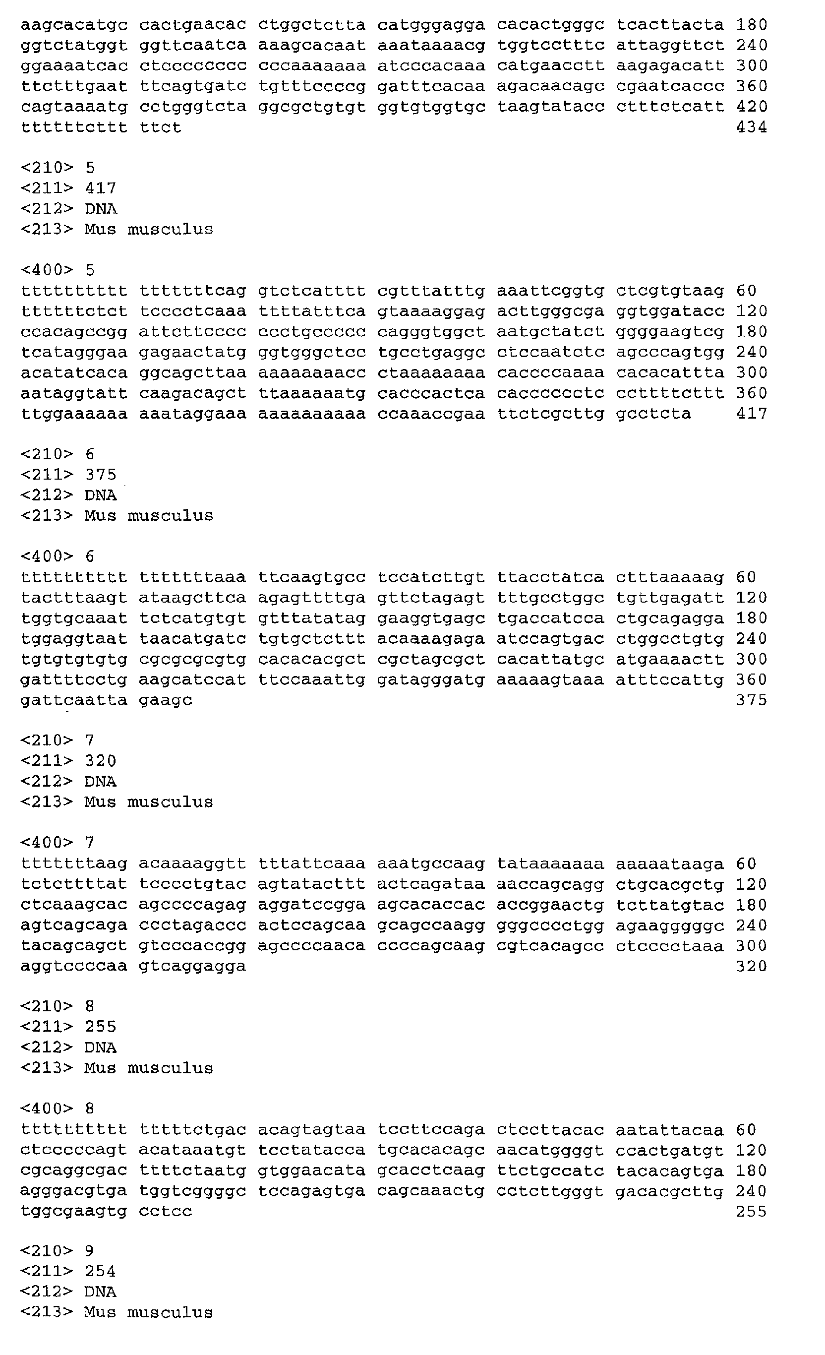
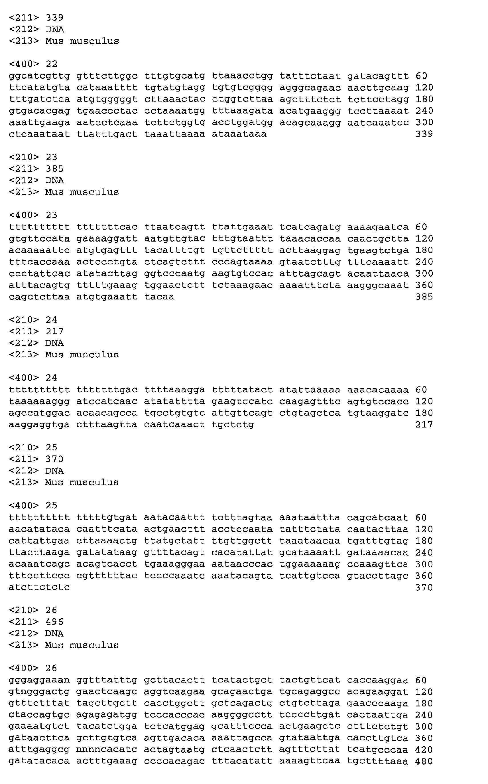
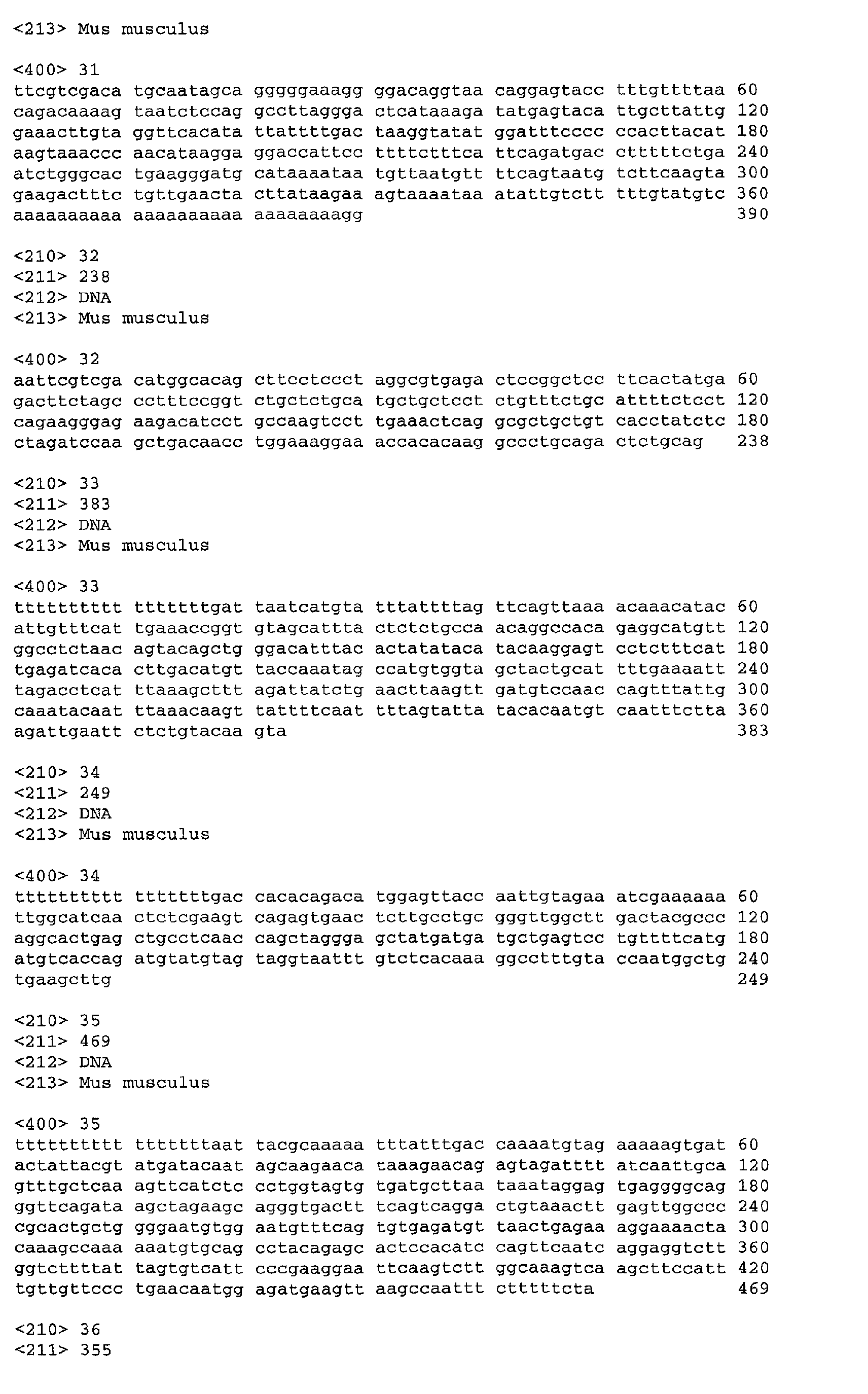
- EST 1 of attachment 1 is identified by accession number AA689670, this sequence corresponds to SEQ ID No. 32; the gene comprising this sequence is found in cluster 20.
- AA689670 (SEQ ID No. 32) is a keratinocyte marker of advanced wound repair. Biomarkers derived from the gene comprising SEQ ID NO. 32 are useful as markers of advanced wound repair. In situ hybridisation on 24 hour frozen wound sections using an antisense radioprobe biomarker derived from the gene shows that this gene is expressed in the basal layers of the epidermis and expression extends back 10 to 12 cell diameters from the wound site.
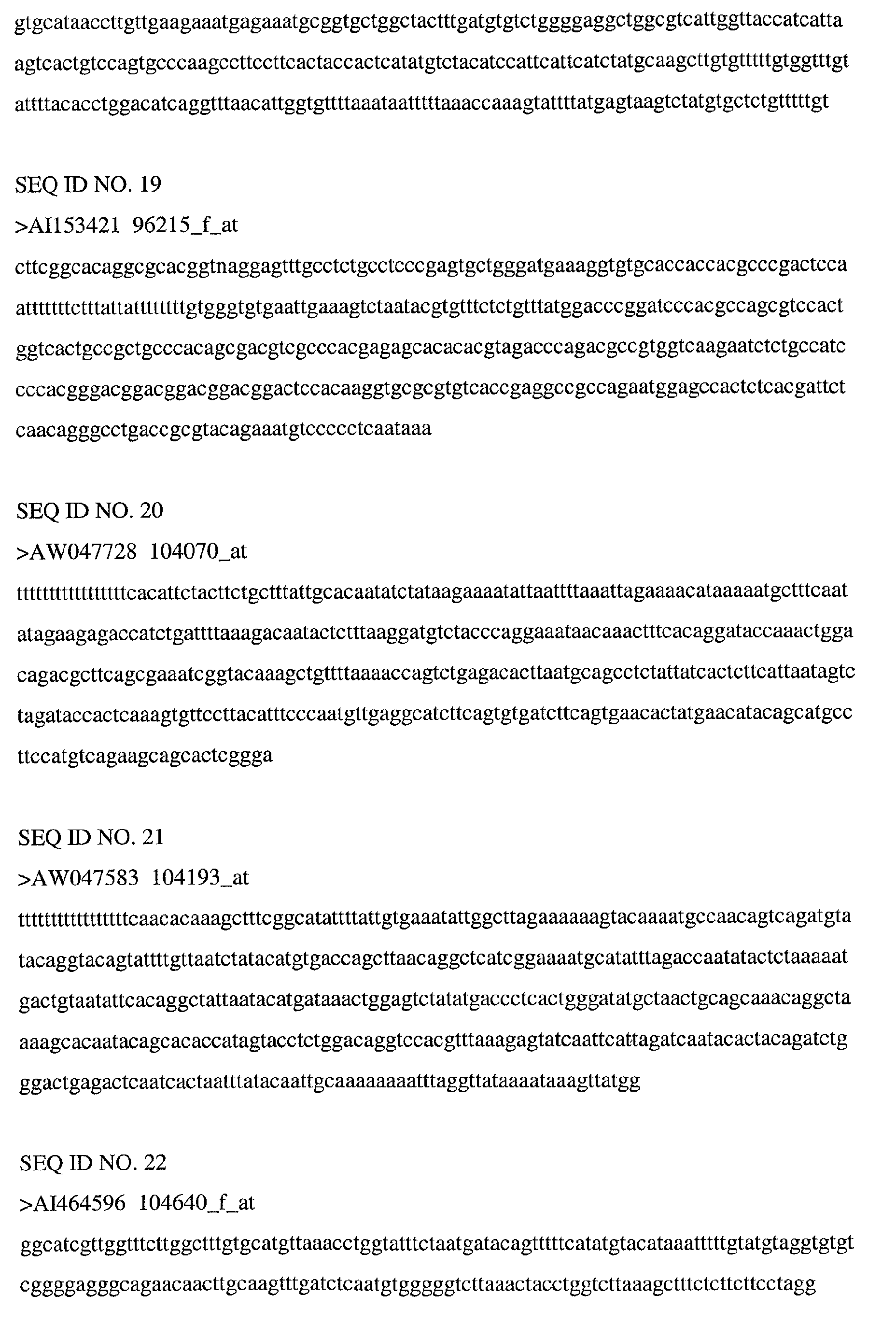
- EST 3 of attachment 1 is identified by accession number AI153421, this sequence corresponds to SEQ ID No. 19; the gene comprising this sequence is found in cluster 5.
- AI153421 (SEQ ID No. 19) is a biomarker for wound inflammation. Biomarkers derived from the gene comprising SEQ ID No. 19 are useful as markers of wound inflammation and/or impaired healing. In situ hybridisation on 24 hour frozen wound tissue sections using an antisense radioprobe biomarker derived from the gene shows that this gene is expressed in all tissue layers by cells as part of the inflammatory response downstream of macrophage infiltration.
Landscapes
- Health & Medical Sciences (AREA)
- Life Sciences & Earth Sciences (AREA)
- Chemical & Material Sciences (AREA)
- Engineering & Computer Science (AREA)
- General Health & Medical Sciences (AREA)
- Molecular Biology (AREA)
- Organic Chemistry (AREA)
- Medicinal Chemistry (AREA)
- Immunology (AREA)
- Hematology (AREA)
- Urology & Nephrology (AREA)
- Biomedical Technology (AREA)
- Bioinformatics & Cheminformatics (AREA)
- Biochemistry (AREA)
- Analytical Chemistry (AREA)
- Animal Behavior & Ethology (AREA)
- Veterinary Medicine (AREA)
- Public Health (AREA)
- General Chemical & Material Sciences (AREA)
- Chemical Kinetics & Catalysis (AREA)
- Pharmacology & Pharmacy (AREA)
- Nuclear Medicine, Radiotherapy & Molecular Imaging (AREA)
- Pathology (AREA)
- Biotechnology (AREA)
- Physics & Mathematics (AREA)
- Microbiology (AREA)
- Proteomics, Peptides & Aminoacids (AREA)
- General Physics & Mathematics (AREA)
- Food Science & Technology (AREA)
- Cell Biology (AREA)
- Zoology (AREA)
- Biophysics (AREA)
- Genetics & Genomics (AREA)
- Wood Science & Technology (AREA)
- Physiology (AREA)
- General Engineering & Computer Science (AREA)
- Tropical Medicine & Parasitology (AREA)
- Endocrinology (AREA)
- Ophthalmology & Optometry (AREA)
- Rheumatology (AREA)
Abstract
Description
characterised by impaired wound healing. It will be appreciated that such antagonists or inhibitors can be compounds that reduce the activity of the target, antibodies specific for the target, as well as antisense oligonucleotides or ribozymes or transcription inhibitors, that have the effect of reducing the amount of the target produced. It will also be appreciated that agonists or activators of these targets can comprise compounds that increase the activity of the target itself, as well as compounds that increase the expression of the target. Increased activity of the target could also be achieved by delivering the target itself, either as protein or its coding region in an appropriate vector as gene therapy.
An increase in activity of a protein encoded by a gene comprising a sequence of one of the accession numbers grouped in clusters 2, 7, 11, 16, and 20 can be achieved by, for example, agonists or stimulators of the protein, gene therapy, application of the protein itself, or substances that upregulate the transcription and/or translation of these genes, means that provide an increase in activity of the protein have application in the treatment of diseases and disorders characterised by impaired healing response such as chronic dermal ulcers, oral mucocystis, emphysema, ulcerative diseases of the gastro-intestinal (GI) tract, cystitis.
Importantly, the Connective Tissue Growth Factor Gene Family member WISP-1 is in this cluster and therefore identified as an early regulator of wound repair. WISP-1, or a viral or plasmid construct comprising the coding sequence of WISP-1 under control of a promoter active in mammalian cells is useful for the treatment of conditions or disorders involving impaired wound repair.
These clusters represent genes that are down regulated during healing, in both wildtype and PU.1 KO mice. This down regulation may be required for repair to occur. Therefore inhibitors of these genes will be useful to promote wound healing and activators of these genes to prevent overhealing. Apolipoprotein E is found within this cluster and thus compounds that modulate the Apolipoprotein E expression or activity will be useful as agents for the control of wound repair.
















































| Genes clustering closely to actin | ||
| Affymetrix Accession | Accession number | Descriptions |
| 92573_at | AB021743 | Mus musculus mRNA for PR65 (type 2A serine/threonine protein phosphatase A subunit), complete cds |
| 92621_at | X97982 | M.musculus mRNA for poly(C)-binding protein, splice variant E |
| 93346_at | M15668 | Mus musculus X chromosome-linked phosphoglycerate kinase (pgk-1) mRNA |
| 95682_at | AB026432 | Mus musculus mRNA for XPE UV-damaged DNA binding factor, complete cds |
| 96876_at | U34259 | Mus musculus Golgi 4-transmembrane spanning transporter MTP mRNA, complete cds |
| 99668_at | U86405 | Mus musculus brain amphiphysin 2 mRNA, complete cds |
| 101028_i_at | M15501 | Actin, alpha, cardiac /cds=(0,1127) /gb=M15501 |
| 101030_at | X99963 | M.musculus rhoB gene /cds=(0,590) /gb=X99963 |
| 101554_at | U57524 | Mus musculus I kappa B alpha gene, exons 2-6, partial cds |
| 104598_at | X61940 | Mouse mRNA for a growth factor-inducible immediate early gene (3CH134) |
| Genes clustering closely to MRP-8 | ||
| Affymetrix Accession | Accession number | Descriptions |
| 96092_at | M96827 | C57BL/6J ob/ob haptoglobin mRNA, complete cds |
| 97529_at | AJ002390 | Annexin 8 |
| 103589_at | AF053235 | Mus musculus keratin 16 mRNA, complete cds |
| 104370_s_at | K02108 | Keratin complex 2, gene 6A |
| 103448_at | M83218 | Mus musculus intracellular calcium-binding protein (MRP8) mRNA |
| 94121_at | AJ005566 | Small proline-rich protein 2H |
| 99701_f_at | AJ005560 | Small proline-rich protein |
| 101024_i_at | AJ005559 | Small proline-rich protein 2A |
| Genes clustering closely to MMP-3 | ||
| Affymetrix Accession | Accession number | Descriptions |
| 100127_at | M35523 | Cellular retinoic acid binding protein II |
| 94391_at | AW123650 | UI-M-BH2.1-api-g-08-0-UI.s1 Mus musculus cDNA, 3 end |
| 97950_at | X75129 | Xanthine dehydrogenase |
| 102255_at | AB015978 | Oncostatin receptor |
| 103723_at | AA608387 | vo42b10.r1 Mus musculus cDNA, 5 end |
| 99159_at | AI842675 | UI-M-AO1-aeo-b-01-0-UI.s1 Mus musculus cDNA, 3 end |
| 98018_at | L39017 | Protein C receptor, endothelial |
| 94224_5_at | M74123 | Mus musculus (strain C57B1/6) mRNA sequence |
| 100882_at | AF003525 | Defensin beta 1 |
| 100884_at | U04204 | Fibroblast growth factor regulated protein |
| 101384_at | AI852933 | UI-M-BHO-aiz-c-09-0-UI.s1 Mus musculus cDNA, 3 end |
| 103080_at | U15635 | Mus musculus IFN-gamma induced (Mg11) mRNA |
| 103634_at | U51992 | Interferon dependent positive acting transcription factor 3 gamma |
| 96123_at | X99347 | M.musculus mRNA for LPS-binding protein |
| 98772_at | U27267 | Small inducible cytokine B subfamily, member 5 |
| 98833_at | X66402 | Matrix metalloproteinase 3 |
| Genes clustering closely to Cathepsin-S | ||
| Affymetrix Accession | Accession number | Descriptions |
| 93118_at | AI844131 | UI-M-AL1-ahj-d-09-0-UI.s1 Mus musculus cDNA, 3 end |
| 93274_at | M38381 | CDC-like kinase |
| 93806_at | AI848671 | UI-M-AHl-agv-e-02-0-UI.s1 Mus musculus cDNA, 3 end |
| 94081_at | AW124390 | UI-M-BH2.1-ape-c-03-0-UI.s1 Mus musculus cDNA, 3 end |
| 98946_at | AF033186 | Mus musculus WSB-1 mRNA, complete cds /cds=(0,1265) / |
| 98087_at | AW048562 | UI-M-BH1-alx-e-05-0-UI.s1 Mus musculus cDNA, 3 end |
| 99577_at | M57647 | Mouse mast cell growth factor (MGF) mRNA, complete cds |
| 102781_at | U37351 | Mus musculus Paneth cell enhanced expression PCEE mRNA, complete cds |
| 103667_at | AA866655 | ud06h05.r1 Mus musculus cDNA, 5 end |
| 104056_at | AI573367 | ud42d02.y1 Mus musculus cDNA, 5 end |
| 94975_at | AI845607 | UI-M-AQ1-adx-e-10-0-UI.s1 Mus musculus cDNA, 3 end / |
| 102302_at | L16992 | Mus musculus branched-chain alpha-ketoacid dehydrogenase E1 beta-subunit mRNA sequence |
| 103753_at | AI159572 | vz77c07.r1 Mus musculus cDNA, 5 end |
| 104640_f_at | AI464596 | mz66c12.y1 Mus musculus cDNA, 5 end |
| 92502_at | X95504 | M.musculus mRNA for zinc finger protein |
| 92513_at | AJ002636 | Mus musculus mRNA for nuclear protein SA2 / |
| 92542_at | AI849035 | UI-M-AH1-agw-a-06-0-UI.s1 Mus musculus cDNA, 3 end |
| 96616_at | AI891629 | u157e11.x1 Mus musculus cDNA, 3 end |
| 104070_at | AW047728 | UI-M-BH1-alk-a-01-0-UI.s1 Mus musculus cDNA, 3 end |
| 104193_at | AW047583 | UI-M-BH1-akm-b-04-0-UI.s1 Mus musculus cDNA, 3 end |
| 93775_at | AI841894 | UI-M-AO0-acd-d-09-0-UI.s1 Mus musculus cDNA, 3 end / |
| 97869_at | AI844043 | UI-M-AL1-ahi-d-11-0-UI.sl Mus musculus cDNA, 3 end |
| 97541_f_at | X00246 | Mouse mRNA with a Set 1 repetitive element for a class I major histocompatibility complex(MHC) antigen |
| 95028_r_at | C77009 | C77009 Mus musculus cDNA, 3 end |
| 97421_at | U42385 | Fibroblast growth factor inducible 16 |
| 101923_at | U34277 | Mus musculus PAF acetylhydrolase mRNA, complete cds |
| 103202_at | AW047476 | UI-M-BH1-all-g-04-0-UI.s1 Mus musculus cDNA, 3 end |
| 92226_at | AA866971 | vx91g10.r1 Mus musculus cDNA, 5 end |
| 98893_at | AW048644 | UI-M-BH1-amd-d-08-0-UI.s1 Mus musculus cDNA, 3 end |
| 103016_5_at | X68273 | CD68 antigen |
| 94934_at | U04828 | Angiotensin II receptor, type 2 |
| 100880_at | AA816121 | vp44a01.r1 Mus musculus cDNA, 5 end |
| 94818_at | AW047223 | UI-M-BH1-amb-e-09-0-UI.s1 Mus musculus cDNA, 3 end |
| 98045_5_at | U18869 | Mus musculus mitogen-responsive 96 kDa phosphoprotein p96 mRNA, alternatively spliced p67 mRNA, and alternatively spliced p93 mRNA, complete cds |
| 98543_at | AJ223208 | Mus musculus mRNA for cathepsin S, partial |
| 98562_at | X58861 | Complement component 1, q subcomponent, alpha polypeptide |
| 100611_at | M21050 | Mouse lysozyme M gene |
| 98465_f_at | M31419 | Interferon activated gene 204 |
| 100397_at | AF024637 | TYRO protein tyrosine kinase binding protein |
| 103226_at | Z11974 | Mannose receptor, C type 1 |
| 95806_f_at | M68899 | Mouse mast cell chymase 2 mRNA, complete cds |
| 102104_f_at | AI504305 | v104g11.x1 Mus musculus cDNA, 3 end |
| 99507_at | M21247 | Uncoupling protein, mitochondrial |
| 92718_at | AI158810 | ud38d09.r1 Mus musculus cDNA, 5 end |
| 94774_at | M31418 | Interferon activated gene 202 |
| 101635_f_at | M16360 | Mouse major urinary protein V (MUP V) mRNA, complete cds |
| 101682_f_at | M16358 | Major urinary protein 4 |
| Genes clustering closely to TGF-beta | ||
| Affymetrix Accession | Accession number | Descriptions |
| 92793_at | X57796 | Tumor necrosis factor receptor superfamily, member 1a |
| 92877_at | L19932 | Transforming growth factor, beta induced, 68 kDa / |
| 99604_at | AI848713 | UI-M-AJ1-ahf-a-10-0-UI.sl Mus musculus cDNA, 3 end |
| 101040_at | D38117 | Mus musculus mRNA for m-calpain large subunit, complete cds |
| 101982_at | X98475 | M.musculus VASP gene |
| 96187_at | AI837021 | UI-M-APO-abj-a-11-0-UI.s2 Mus musculus cDNA, 3 end |










Claims (19)
- A gene comprising a sequence according to any one of SEQ ID Nos. 1 to 39.
- A gene comprising a sequence according to SEQ ID No. 32.
- A gene comprising a sequence according to SEQ ID No. 19.
- An isolated and purified protein encoded by a gene according to any one of claims 1 to 3.
- A human homologue of a gene according to any one of claims 1 to 3 or of a protein according to claim 4.
- A biomarker derived from a gene according to any one of claims 1 to 4.
- A method for identification of a compound useful in the treatment of wounds or of conditions characterised by an excessive or impaired wound healing response, which comprises contacting a compound with a protein encoded by a gene comprising a sequence identified by one of the accession numbers of cluster 2, 7, 11, 16, or 20 or a mammalian homologue thereof and detecting specific binding of the chemical compound to the protein.
- A method for identification of a compound useful for the treatment or reduction of inflammation which comprises contacting a compound with a protein encoded by a gene comprising a sequence identified by one of the accession numbers of cluster 5 or 6 or a mammalian homologue thereof and detecting specific binding of the chemical compound to the protein.
- An antagonist or inhibitor of a protein encoded by a gene comprising a sequence identified by one of the accession numbers of cluster 2, 7, 11, 16, or 20 or a mammalian homologue thereof, for use in the treatment of a disease or disorder characterised by excessive wound healing response, such as scarring, fibrosis, restenosis post angioplasty, psoriasis, post-traumatic/surgical adhesions of the peritoneal cavity, joints and ligaments, benign prostatic hyperplasia, glaucoma, or peripheral nerve injury.
- The use of an antagonist or inhibitor of a protein encoded by a gene comprising a sequence identified by one of the accession numbers of cluster 2, 7, 11, 16, or 20 or a mammalian homologue thereof, in the manufacture of a medicament for the treatment of a disease or disorder characterised by excessive wound healing response, such as scarring, fibrosis, restenosis post angioplasty, psoriasis, post-traumatic/surgical adhesions of the peritoneal cavity, joints and ligaments, benign prostatic hyperplasia, glaucoma, or peripheral nerve injury.
- A method for treatment of a disease or disorder characterised by excessive wound healing response, such as scarring, fibrosis, restenosis post angioplasty, psoriasis, post-traumatic/surgical adhesions of the peritoneal cavity, joints and ligaments, benign prostatic hyperplasia, glaucoma, or peripheral nerve injury, comprising administering to a mammal in need of such treatment a therapeutically effective amount of an antagonist or inhibitor of a protein encoded by a gene comprising a sequence identified by one of the accession numbers of cluster 2, 7, 11, 16, or 20 or a mammalian homologue thereof.
- An agonist or activator of a protein encoded by a gene comprising a sequence identified by one of the accession numbers of cluster 2, 7, 11, 16, or 20 or a mammalian homologue thereof, for use in the treatment of a disease or disorder characterised by an impaired healing response, such as chronic dermal ulcers, oral mucocystis, emphysema, ulcerative diseases of the gastro-intestinal (GI) tract, or cystitis.
- The use of an agonist or activator of a protein encoded by a gene comprising a sequence identified by one of the accession numbers of cluster 2, 7, 11, 16, or 20 or a mammalian homologue thereof, in the manufacture of a medicament for the treatment of a disease or disorder characterised by an impaired healing response, such as chronic dermal ulcers, oral mucocystis, emphysema, ulcerative diseases of the gastro-intestinal (GI) tract, or cystitis.
- A method for treatment of a disease or disorder characterised by an impaired healing response, such as chronic dermal ulcers, oral mucocystis, emphysema, ulcerative diseases of the gastro-intestinal (GI) tract, or cystitis, comprising administering to a mammal in need of such treatment a therapeutically effective amount of an agonist or activator of a protein encoded by a gene comprising a sequence identified by one of the accession numbers of cluster 2, 7, 11, 16, or 20 or a mammalian homologue thereof.
- An antagonist or inhibitor of a protein encoded by a gene comprising a sequence identified by one of the accession numbers of cluster 5 or 6 or a mammalian homologue thereof for the treatment or prevention of inflammation.
- The use of an antagonist or inhibitor of a protein encoded by a gene comprising a sequence identified by one of the accession numbers of cluster 5 or 6 or a mammalian homologue thereof, in the manufacture of a medicament for the treatment or prevention of inflammation.
- A method for treatment or prevention of inflammation comprising administering to a mammal in need of such treatment a therapeutically effective amount of an antagonist or inhibitor of a protein encoded by a gene comprising a sequence identified by one of the accession numbers of cluster 5 or 6 or a mammalian homologue thereof for the treatment or prevention of inflammation.
- A method for investigating wound repair in a mammal comprising: taking a wound tissue sample at one or more time points, isolating mRNA from the sample (or samples), determining for the, or each time point, the level of mRNA expression from one or more of the genes including the sequence of one or more of the accession numbers grouped in clusters 2, 7, 11, 16 and 20 or a homologue thereof; comparing the level of mRNA expression of the, or each, gene from the wound tissue sample at a given time point with the level of mRNA expression of that gene in a control sample of mRNA taken from non wounded tissue and determining whether the level of mRNA expression of the, or each, gene differs in the wound tissue sample relative to a control sample taken from unwounded skin.
- A method for identification of a mammal with an impaired wound healing response comprising: taking a tissue sample from a test subject, isolating mRNA from the sample, determining the level of mRNA expression from one or more of the genes including the sequence of one or more of the accession numbers grouped in clusters 2, 7, 11, 16 and 20 and comparing the mRNA expression level detected in the test subject with the mRNA expression level of the or each gene a control subject with a normal wound healing response; wherein a difference in mRNA expression levels of one or more of said genes indicates that the test subject has an impaired wound healing response.
Applications Claiming Priority (4)
| Application Number | Priority Date | Filing Date | Title |
|---|---|---|---|
| GB0114869 | 2001-06-18 | ||
| GB0114869A GB0114869D0 (en) | 2001-06-18 | 2001-06-18 | Wound healing |
| US30534601P | 2001-07-13 | 2001-07-13 | |
| US305346P | 2001-07-13 |
Publications (2)
| Publication Number | Publication Date |
|---|---|
| EP1270044A2 true EP1270044A2 (en) | 2003-01-02 |
| EP1270044A3 EP1270044A3 (en) | 2003-03-12 |
Family
ID=26246205
Family Applications (1)
| Application Number | Title | Priority Date | Filing Date |
|---|---|---|---|
| EP02254207A Withdrawn EP1270044A3 (en) | 2001-06-18 | 2002-06-17 | Wound healing biomarkers |
Country Status (4)
| Country | Link |
|---|---|
| US (1) | US20040038292A1 (en) |
| EP (1) | EP1270044A3 (en) |
| JP (1) | JP2003102485A (en) |
| CA (1) | CA2388761A1 (en) |
Cited By (2)
| Publication number | Priority date | Publication date | Assignee | Title |
|---|---|---|---|---|
| GB2486533A (en) * | 2010-12-14 | 2012-06-20 | Univ Cardiff | Classification of wounds |
| EP2695947A4 (en) * | 2011-04-01 | 2014-10-29 | Chugai Pharmaceutical Co Ltd | PROCESS FOR PRODUCING RECOMBINANT POLYPEPTIDE |
Families Citing this family (16)
| Publication number | Priority date | Publication date | Assignee | Title |
|---|---|---|---|---|
| AU2002231736A1 (en) | 2000-12-22 | 2002-07-08 | Max-Planck-Gesellschaft Zur Forderung Der Wissenschaften E.V. | Use of repulsive guidance molecule (rgm) and its modulators |
| CN101277974A (en) * | 2005-09-30 | 2008-10-01 | 阿伯特有限及两合公司 | Binding domains of proteins of the Repulsive Guide Molecule (RGM) protein family, functional fragments thereof and their uses |
| EP2033971A1 (en) * | 2007-09-06 | 2009-03-11 | Abbott GmbH & Co. KG | Bone Morphogenetic Protein (BMP) binding domains of proteins of the Repulsive Guidance Molecule (RGM) protein family and functional fragments thereof and their application |
| US8962803B2 (en) * | 2008-02-29 | 2015-02-24 | AbbVie Deutschland GmbH & Co. KG | Antibodies against the RGM A protein and uses thereof |
| US8497122B2 (en) * | 2008-04-11 | 2013-07-30 | Washington University | Biomarkers for Niemann-pick C disease and related disorders |
| US20100203553A1 (en) * | 2009-02-11 | 2010-08-12 | Abdeen Suad M | Histochemical and biomarker for liver fibrosis |
| US8535282B2 (en) * | 2009-07-14 | 2013-09-17 | Southwest Research Institute | Wound healing sensor techniques |
| PH12012501070A1 (en) * | 2009-12-08 | 2017-08-23 | Abbott Gmbh & Co Kg | Monoclonal antibodies against the rgm a protein for use in the treatment of retinal nerve fiber layer degeneration |
| US8591459B2 (en) | 2009-12-21 | 2013-11-26 | Ethicon Endo-Surgery, Inc. | Use of biomarkers and therapeutic agents with surgical devices |
| AU2013211939C1 (en) | 2012-01-27 | 2018-06-07 | AbbVie Deutschland GmbH & Co. KG | Composition and method for diagnosis and treatment of diseases associated with neurite degeneration |
| JP6001192B2 (en) * | 2013-10-31 | 2016-10-05 | 国立大学法人京都大学 | Aldehyde-degrading enzyme activity genotype determination device, squamous cell carcinoma occurrence risk determination device, and program |
| KR101562050B1 (en) | 2014-09-16 | 2015-10-22 | 가톨릭대학교 산학협력단 | Composition for diagnosing psoriasis |
| US12027276B1 (en) * | 2017-10-27 | 2024-07-02 | DecisionQ Corporation | Methods, systems and devices for assessing wound healing |
| CN111979208B (en) * | 2019-05-23 | 2023-01-10 | 弈柯莱生物科技(上海)股份有限公司 | A kind of L-glutamic acid dehydrogenase mutant and its application |
| EP3764103A1 (en) * | 2019-07-12 | 2021-01-13 | Urgo Recherche Innovation Et Developpement | Method of diagnosis or prognosis of a non-healing or chronic wound |
| CN114252608A (en) * | 2020-09-24 | 2022-03-29 | 首都医科大学附属北京世纪坛医院 | Application of urine delta-aminolevulinic acid dehydratase and polypeptide fragment thereof in allergic diseases |
Family Cites Families (4)
| Publication number | Priority date | Publication date | Assignee | Title |
|---|---|---|---|---|
| US5474796A (en) * | 1991-09-04 | 1995-12-12 | Protogene Laboratories, Inc. | Method and apparatus for conducting an array of chemical reactions on a support surface |
| AU3428493A (en) * | 1991-12-31 | 1993-07-28 | Zymogenetics Inc. | Novel human transglutaminases |
| DK0941108T3 (en) * | 1996-12-04 | 2002-11-18 | Renovo Ltd | Wound healing and treatment of fibrosis |
| EP1206233A4 (en) * | 1999-08-03 | 2004-04-14 | Human Genome Sciences Inc | SERINE PROTEASES |
-
2002
- 2002-06-17 EP EP02254207A patent/EP1270044A3/en not_active Withdrawn
- 2002-06-17 CA CA002388761A patent/CA2388761A1/en not_active Abandoned
- 2002-06-18 JP JP2002176686A patent/JP2003102485A/en active Pending
- 2002-06-18 US US10/175,184 patent/US20040038292A1/en not_active Abandoned
Cited By (10)
| Publication number | Priority date | Publication date | Assignee | Title |
|---|---|---|---|---|
| GB2486533A (en) * | 2010-12-14 | 2012-06-20 | Univ Cardiff | Classification of wounds |
| EP2695947A4 (en) * | 2011-04-01 | 2014-10-29 | Chugai Pharmaceutical Co Ltd | PROCESS FOR PRODUCING RECOMBINANT POLYPEPTIDE |
| AU2012239382B2 (en) * | 2011-04-01 | 2016-04-21 | Chugai Seiyaku Kabushiki Kaisha | Recombinant polypeptide production method |
| CN106995831A (en) * | 2011-04-01 | 2017-08-01 | 中外制药株式会社 | The manufacture method of recombinant polypeptide |
| CN108546295A (en) * | 2011-04-01 | 2018-09-18 | 中外制药株式会社 | The manufacturing method of recombinant polypeptide |
| TWI647309B (en) * | 2011-04-01 | 2019-01-11 | Chugai Seiyaku Kabushiki Kaisha | Method for manufacturing recombinant polypeptide |
| TWI674272B (en) * | 2011-04-01 | 2019-10-11 | 中外製藥股份有限公司 | Nucleic acid molecule with APES activity |
| US11028149B2 (en) | 2011-04-01 | 2021-06-08 | Chugai Seiyaku Kabushiki Kaisha | Recombinant polypeptide production method |
| CN108546295B (en) * | 2011-04-01 | 2022-08-09 | 中外制药株式会社 | Method for producing recombinant polypeptide |
| US11905325B2 (en) | 2011-04-01 | 2024-02-20 | Chugai Seiyaku Kabushiki Kaisha | Recombinant polypeptide production method |
Also Published As
| Publication number | Publication date |
|---|---|
| JP2003102485A (en) | 2003-04-08 |
| US20040038292A1 (en) | 2004-02-26 |
| CA2388761A1 (en) | 2002-12-18 |
| EP1270044A3 (en) | 2003-03-12 |
Similar Documents
| Publication | Publication Date | Title |
|---|---|---|
| JP6254125B2 (en) | Role of soluble uPAR in the pathogenesis of proteinuria | |
| Li et al. | Analysis of gene expression in the wound repair/regeneration process | |
| Glocker et al. | Rheumatoid arthritis, a complex multifactorial disease: on the way toward individualized medicine | |
| JP4155561B2 (en) | Allergic disease test method | |
| JP2003102485A (en) | Wound healing biomarker | |
| Zhang et al. | Microarray analysis reveals the involvement of beta-2 microglobulin (B2M) in human osteoarthritis | |
| Foote et al. | Oxidative DNA damage promotes vascular ageing associated with changes in extracellular matrix-regulating proteins | |
| EP2105742A1 (en) | Use of cathepsin C | |
| US20060003338A1 (en) | System and methods for the management and treatment of vascular graft disease | |
| Hage et al. | The role of C-reactive protein polymorphisms in inflammation and cardiovascular risk | |
| US20060019272A1 (en) | Diagnosis of disease and monitoring of therapy using gene expression analysis of peripheral blood cells | |
| WO2000014283A2 (en) | Gene markers for chronic mucosal injury | |
| EP2646574B1 (en) | Gpnmb/osteoactivin as a biomarker in cardiac diseases | |
| US6368794B1 (en) | Detection of altered expression of genes regulating cell proliferation | |
| US20050130171A1 (en) | Genes expressed in Alzheimer's disease | |
| JP5045906B2 (en) | Novel ischemic marker and method for detecting ischemic state using the same | |
| US7052865B1 (en) | Identification of genes having a role in the presentation of diabetic nephropathy | |
| JP6788256B2 (en) | Methods and Reagents for Predicting Coronary Events | |
| JP5360923B2 (en) | Novel ischemic marker and method for detecting ischemic state using the same | |
| Linna-Kuosmanen et al. | Cardiovascular disease causes proinflammatory microvascular changes in the human right atrium | |
| US20080166734A1 (en) | Genes and methods of using the same for diagnosis and for targeting the therapy of cardiovascular disease | |
| JP5360922B2 (en) | Novel ischemic marker and method for detecting ischemic state using the same | |
| US20070184466A1 (en) | Detection, generation and uses of atherosclerosis-protective endothelium | |
| EP1835283A1 (en) | Immune system malfunction disease diagnosis supporting method and diagnosis support information output device | |
| US20070054321A1 (en) | Methods of diagnosing and treating inflammatory diseases using pac-1 (dusp2) |
Legal Events
| Date | Code | Title | Description |
|---|---|---|---|
| PUAI | Public reference made under article 153(3) epc to a published international application that has entered the european phase |
Free format text: ORIGINAL CODE: 0009012 |
|
| 17P | Request for examination filed |
Effective date: 20020624 |
|
| AK | Designated contracting states |
Kind code of ref document: A2 Designated state(s): AT BE CH CY DE DK ES FI FR GB GR IE IT LI LU MC NL PT SE TR |
|
| AX | Request for extension of the european patent |
Free format text: AL;LT;LV;MK;RO;SI |
|
| PUAL | Search report despatched |
Free format text: ORIGINAL CODE: 0009013 |
|
| AK | Designated contracting states |
Kind code of ref document: A3 Designated state(s): AT BE CH CY DE DK ES FI FR GB GR IE IT LI LU MC NL PT SE TR Designated state(s): AT BE CH CY DE DK ES FI FR GB GR IE IT LI LU MC NL PT SE TR |
|
| AX | Request for extension of the european patent |
Extension state: AL LT LV MK RO SI |
|
| AKX | Designation fees paid |
Designated state(s): AT BE CH CY DE DK ES FI FR GB GR IE IT LI LU MC NL PT SE TR |
|
| 17Q | First examination report despatched |
Effective date: 20031028 |
|
| GRAP | Despatch of communication of intention to grant a patent |
Free format text: ORIGINAL CODE: EPIDOSNIGR1 |
|
| GRAS | Grant fee paid |
Free format text: ORIGINAL CODE: EPIDOSNIGR3 |
|
| STAA | Information on the status of an ep patent application or granted ep patent |
Free format text: STATUS: THE APPLICATION IS DEEMED TO BE WITHDRAWN |
|
| 18D | Application deemed to be withdrawn |
Effective date: 20051105 |