EP1257287B1 - Compositions and methods for the early diagnosis of ovarian cancer - Google Patents
Compositions and methods for the early diagnosis of ovarian cancer Download PDFInfo
- Publication number
- EP1257287B1 EP1257287B1 EP01914444A EP01914444A EP1257287B1 EP 1257287 B1 EP1257287 B1 EP 1257287B1 EP 01914444 A EP01914444 A EP 01914444A EP 01914444 A EP01914444 A EP 01914444A EP 1257287 B1 EP1257287 B1 EP 1257287B1
- Authority
- EP
- European Patent Office
- Prior art keywords
- hepsin
- residues
- prt
- homo sapiens
- protein
- Prior art date
- Legal status (The legal status is an assumption and is not a legal conclusion. Google has not performed a legal analysis and makes no representation as to the accuracy of the status listed.)
- Expired - Lifetime
Links
Images




Classifications
-
- C—CHEMISTRY; METALLURGY
- C12—BIOCHEMISTRY; BEER; SPIRITS; WINE; VINEGAR; MICROBIOLOGY; ENZYMOLOGY; MUTATION OR GENETIC ENGINEERING
- C12N—MICROORGANISMS OR ENZYMES; COMPOSITIONS THEREOF; PROPAGATING, PRESERVING, OR MAINTAINING MICROORGANISMS; MUTATION OR GENETIC ENGINEERING; CULTURE MEDIA
- C12N9/00—Enzymes; Proenzymes; Compositions thereof; Processes for preparing, activating, inhibiting, separating or purifying enzymes
- C12N9/14—Hydrolases (3)
- C12N9/48—Hydrolases (3) acting on peptide bonds (3.4)
- C12N9/50—Proteinases, e.g. Endopeptidases (3.4.21-3.4.25)
- C12N9/64—Proteinases, e.g. Endopeptidases (3.4.21-3.4.25) derived from animal tissue
- C12N9/6421—Proteinases, e.g. Endopeptidases (3.4.21-3.4.25) derived from animal tissue from mammals
- C12N9/6424—Serine endopeptidases (3.4.21)
-
- C—CHEMISTRY; METALLURGY
- C07—ORGANIC CHEMISTRY
- C07K—PEPTIDES
- C07K14/00—Peptides having more than 20 amino acids; Gastrins; Somatostatins; Melanotropins; Derivatives thereof
- C07K14/435—Peptides having more than 20 amino acids; Gastrins; Somatostatins; Melanotropins; Derivatives thereof from animals; from humans
-
- C—CHEMISTRY; METALLURGY
- C12—BIOCHEMISTRY; BEER; SPIRITS; WINE; VINEGAR; MICROBIOLOGY; ENZYMOLOGY; MUTATION OR GENETIC ENGINEERING
- C12N—MICROORGANISMS OR ENZYMES; COMPOSITIONS THEREOF; PROPAGATING, PRESERVING, OR MAINTAINING MICROORGANISMS; MUTATION OR GENETIC ENGINEERING; CULTURE MEDIA
- C12N9/00—Enzymes; Proenzymes; Compositions thereof; Processes for preparing, activating, inhibiting, separating or purifying enzymes
- C12N9/14—Hydrolases (3)
- C12N9/48—Hydrolases (3) acting on peptide bonds (3.4)
- C12N9/50—Proteinases, e.g. Endopeptidases (3.4.21-3.4.25)
- C12N9/64—Proteinases, e.g. Endopeptidases (3.4.21-3.4.25) derived from animal tissue
- C12N9/6421—Proteinases, e.g. Endopeptidases (3.4.21-3.4.25) derived from animal tissue from mammals
- C12N9/6472—Cysteine endopeptidases (3.4.22)
-
- C—CHEMISTRY; METALLURGY
- C12—BIOCHEMISTRY; BEER; SPIRITS; WINE; VINEGAR; MICROBIOLOGY; ENZYMOLOGY; MUTATION OR GENETIC ENGINEERING
- C12Q—MEASURING OR TESTING PROCESSES INVOLVING ENZYMES, NUCLEIC ACIDS OR MICROORGANISMS; COMPOSITIONS OR TEST PAPERS THEREFOR; PROCESSES OF PREPARING SUCH COMPOSITIONS; CONDITION-RESPONSIVE CONTROL IN MICROBIOLOGICAL OR ENZYMOLOGICAL PROCESSES
- C12Q1/00—Measuring or testing processes involving enzymes, nucleic acids or microorganisms; Compositions therefor; Processes of preparing such compositions
- C12Q1/34—Measuring or testing processes involving enzymes, nucleic acids or microorganisms; Compositions therefor; Processes of preparing such compositions involving hydrolase
- C12Q1/37—Measuring or testing processes involving enzymes, nucleic acids or microorganisms; Compositions therefor; Processes of preparing such compositions involving hydrolase involving peptidase or proteinase
-
- C—CHEMISTRY; METALLURGY
- C12—BIOCHEMISTRY; BEER; SPIRITS; WINE; VINEGAR; MICROBIOLOGY; ENZYMOLOGY; MUTATION OR GENETIC ENGINEERING
- C12Q—MEASURING OR TESTING PROCESSES INVOLVING ENZYMES, NUCLEIC ACIDS OR MICROORGANISMS; COMPOSITIONS OR TEST PAPERS THEREFOR; PROCESSES OF PREPARING SUCH COMPOSITIONS; CONDITION-RESPONSIVE CONTROL IN MICROBIOLOGICAL OR ENZYMOLOGICAL PROCESSES
- C12Q1/00—Measuring or testing processes involving enzymes, nucleic acids or microorganisms; Compositions therefor; Processes of preparing such compositions
- C12Q1/68—Measuring or testing processes involving enzymes, nucleic acids or microorganisms; Compositions therefor; Processes of preparing such compositions involving nucleic acids
- C12Q1/6876—Nucleic acid products used in the analysis of nucleic acids, e.g. primers or probes
- C12Q1/6883—Nucleic acid products used in the analysis of nucleic acids, e.g. primers or probes for diseases caused by alterations of genetic material
- C12Q1/6886—Nucleic acid products used in the analysis of nucleic acids, e.g. primers or probes for diseases caused by alterations of genetic material for cancer
-
- G—PHYSICS
- G01—MEASURING; TESTING
- G01N—INVESTIGATING OR ANALYSING MATERIALS BY DETERMINING THEIR CHEMICAL OR PHYSICAL PROPERTIES
- G01N33/00—Investigating or analysing materials by specific methods not covered by groups G01N1/00 - G01N31/00
- G01N33/48—Biological material, e.g. blood, urine; Haemocytometers
- G01N33/50—Chemical analysis of biological material, e.g. blood, urine; Testing involving biospecific ligand binding methods; Immunological testing
- G01N33/53—Immunoassay; Biospecific binding assay; Materials therefor
- G01N33/573—Immunoassay; Biospecific binding assay; Materials therefor for enzymes or isoenzymes
-
- G—PHYSICS
- G01—MEASURING; TESTING
- G01N—INVESTIGATING OR ANALYSING MATERIALS BY DETERMINING THEIR CHEMICAL OR PHYSICAL PROPERTIES
- G01N33/00—Investigating or analysing materials by specific methods not covered by groups G01N1/00 - G01N31/00
- G01N33/48—Biological material, e.g. blood, urine; Haemocytometers
- G01N33/50—Chemical analysis of biological material, e.g. blood, urine; Testing involving biospecific ligand binding methods; Immunological testing
- G01N33/53—Immunoassay; Biospecific binding assay; Materials therefor
- G01N33/575—Immunoassay; Biospecific binding assay; Materials therefor for cancer
-
- G—PHYSICS
- G01—MEASURING; TESTING
- G01N—INVESTIGATING OR ANALYSING MATERIALS BY DETERMINING THEIR CHEMICAL OR PHYSICAL PROPERTIES
- G01N33/00—Investigating or analysing materials by specific methods not covered by groups G01N1/00 - G01N31/00
- G01N33/48—Biological material, e.g. blood, urine; Haemocytometers
- G01N33/50—Chemical analysis of biological material, e.g. blood, urine; Testing involving biospecific ligand binding methods; Immunological testing
- G01N33/53—Immunoassay; Biospecific binding assay; Materials therefor
- G01N33/575—Immunoassay; Biospecific binding assay; Materials therefor for cancer
- G01N33/57545—Immunoassay; Biospecific binding assay; Materials therefor for cancer of the ovaries
-
- G—PHYSICS
- G01—MEASURING; TESTING
- G01N—INVESTIGATING OR ANALYSING MATERIALS BY DETERMINING THEIR CHEMICAL OR PHYSICAL PROPERTIES
- G01N33/00—Investigating or analysing materials by specific methods not covered by groups G01N1/00 - G01N31/00
- G01N33/48—Biological material, e.g. blood, urine; Haemocytometers
- G01N33/50—Chemical analysis of biological material, e.g. blood, urine; Testing involving biospecific ligand binding methods; Immunological testing
- G01N33/53—Immunoassay; Biospecific binding assay; Materials therefor
- G01N33/575—Immunoassay; Biospecific binding assay; Materials therefor for cancer
- G01N33/5758—Immunoassay; Biospecific binding assay; Materials therefor for cancer involving compounds serving as markers for tumours, cancers or neoplasias, e.g. cellular determinants, receptors, heat shock/stress proteins, A-protein, oligosaccharides or metabolites
- G01N33/5759—Immunoassay; Biospecific binding assay; Materials therefor for cancer involving compounds serving as markers for tumours, cancers or neoplasias, e.g. cellular determinants, receptors, heat shock/stress proteins, A-protein, oligosaccharides or metabolites involving compounds localised on the membrane of tumour or cancer cells
-
- A—HUMAN NECESSITIES
- A61—MEDICAL OR VETERINARY SCIENCE; HYGIENE
- A61K—PREPARATIONS FOR MEDICAL, DENTAL OR TOILETRY PURPOSES
- A61K39/00—Medicinal preparations containing antigens or antibodies
- A61K2039/51—Medicinal preparations containing antigens or antibodies comprising whole cells, viruses or DNA/RNA
- A61K2039/515—Animal cells
- A61K2039/5154—Antigen presenting cells [APCs], e.g. dendritic cells or macrophages
-
- A—HUMAN NECESSITIES
- A61—MEDICAL OR VETERINARY SCIENCE; HYGIENE
- A61K—PREPARATIONS FOR MEDICAL, DENTAL OR TOILETRY PURPOSES
- A61K39/00—Medicinal preparations containing antigens or antibodies
- A61K2039/51—Medicinal preparations containing antigens or antibodies comprising whole cells, viruses or DNA/RNA
- A61K2039/53—DNA (RNA) vaccination
-
- A—HUMAN NECESSITIES
- A61—MEDICAL OR VETERINARY SCIENCE; HYGIENE
- A61K—PREPARATIONS FOR MEDICAL, DENTAL OR TOILETRY PURPOSES
- A61K38/00—Medicinal preparations containing peptides
-
- A—HUMAN NECESSITIES
- A61—MEDICAL OR VETERINARY SCIENCE; HYGIENE
- A61K—PREPARATIONS FOR MEDICAL, DENTAL OR TOILETRY PURPOSES
- A61K39/00—Medicinal preparations containing antigens or antibodies
-
- A—HUMAN NECESSITIES
- A61—MEDICAL OR VETERINARY SCIENCE; HYGIENE
- A61K—PREPARATIONS FOR MEDICAL, DENTAL OR TOILETRY PURPOSES
- A61K48/00—Medicinal preparations containing genetic material which is inserted into cells of the living body to treat genetic diseases; Gene therapy
-
- C—CHEMISTRY; METALLURGY
- C12—BIOCHEMISTRY; BEER; SPIRITS; WINE; VINEGAR; MICROBIOLOGY; ENZYMOLOGY; MUTATION OR GENETIC ENGINEERING
- C12Q—MEASURING OR TESTING PROCESSES INVOLVING ENZYMES, NUCLEIC ACIDS OR MICROORGANISMS; COMPOSITIONS OR TEST PAPERS THEREFOR; PROCESSES OF PREPARING SUCH COMPOSITIONS; CONDITION-RESPONSIVE CONTROL IN MICROBIOLOGICAL OR ENZYMOLOGICAL PROCESSES
- C12Q1/00—Measuring or testing processes involving enzymes, nucleic acids or microorganisms; Compositions therefor; Processes of preparing such compositions
- C12Q1/68—Measuring or testing processes involving enzymes, nucleic acids or microorganisms; Compositions therefor; Processes of preparing such compositions involving nucleic acids
- C12Q1/6844—Nucleic acid amplification reactions
- C12Q1/686—Polymerase chain reaction [PCR]
-
- C—CHEMISTRY; METALLURGY
- C12—BIOCHEMISTRY; BEER; SPIRITS; WINE; VINEGAR; MICROBIOLOGY; ENZYMOLOGY; MUTATION OR GENETIC ENGINEERING
- C12Q—MEASURING OR TESTING PROCESSES INVOLVING ENZYMES, NUCLEIC ACIDS OR MICROORGANISMS; COMPOSITIONS OR TEST PAPERS THEREFOR; PROCESSES OF PREPARING SUCH COMPOSITIONS; CONDITION-RESPONSIVE CONTROL IN MICROBIOLOGICAL OR ENZYMOLOGICAL PROCESSES
- C12Q2600/00—Oligonucleotides characterized by their use
- C12Q2600/136—Screening for pharmacological compounds
-
- C—CHEMISTRY; METALLURGY
- C12—BIOCHEMISTRY; BEER; SPIRITS; WINE; VINEGAR; MICROBIOLOGY; ENZYMOLOGY; MUTATION OR GENETIC ENGINEERING
- C12Q—MEASURING OR TESTING PROCESSES INVOLVING ENZYMES, NUCLEIC ACIDS OR MICROORGANISMS; COMPOSITIONS OR TEST PAPERS THEREFOR; PROCESSES OF PREPARING SUCH COMPOSITIONS; CONDITION-RESPONSIVE CONTROL IN MICROBIOLOGICAL OR ENZYMOLOGICAL PROCESSES
- C12Q2600/00—Oligonucleotides characterized by their use
- C12Q2600/158—Expression markers
-
- G—PHYSICS
- G01—MEASURING; TESTING
- G01N—INVESTIGATING OR ANALYSING MATERIALS BY DETERMINING THEIR CHEMICAL OR PHYSICAL PROPERTIES
- G01N2333/00—Assays involving biological materials from specific organisms or of a specific nature
- G01N2333/90—Enzymes; Proenzymes
- G01N2333/914—Hydrolases (3)
- G01N2333/948—Hydrolases (3) acting on peptide bonds (3.4)
- G01N2333/95—Proteinases, i.e. endopeptidases (3.4.21-3.4.99)
- G01N2333/964—Proteinases, i.e. endopeptidases (3.4.21-3.4.99) derived from animal tissue
- G01N2333/96425—Proteinases, i.e. endopeptidases (3.4.21-3.4.99) derived from animal tissue from mammals
- G01N2333/96427—Proteinases, i.e. endopeptidases (3.4.21-3.4.99) derived from animal tissue from mammals in general
- G01N2333/9643—Proteinases, i.e. endopeptidases (3.4.21-3.4.99) derived from animal tissue from mammals in general with EC number
- G01N2333/96433—Serine endopeptidases (3.4.21)
-
- G—PHYSICS
- G01—MEASURING; TESTING
- G01N—INVESTIGATING OR ANALYSING MATERIALS BY DETERMINING THEIR CHEMICAL OR PHYSICAL PROPERTIES
- G01N2333/00—Assays involving biological materials from specific organisms or of a specific nature
- G01N2333/90—Enzymes; Proenzymes
- G01N2333/914—Hydrolases (3)
- G01N2333/948—Hydrolases (3) acting on peptide bonds (3.4)
- G01N2333/95—Proteinases, i.e. endopeptidases (3.4.21-3.4.99)
- G01N2333/964—Proteinases, i.e. endopeptidases (3.4.21-3.4.99) derived from animal tissue
- G01N2333/96425—Proteinases, i.e. endopeptidases (3.4.21-3.4.99) derived from animal tissue from mammals
- G01N2333/96427—Proteinases, i.e. endopeptidases (3.4.21-3.4.99) derived from animal tissue from mammals in general
- G01N2333/9643—Proteinases, i.e. endopeptidases (3.4.21-3.4.99) derived from animal tissue from mammals in general with EC number
- G01N2333/96486—Metalloendopeptidases (3.4.24)
-
- G—PHYSICS
- G01—MEASURING; TESTING
- G01N—INVESTIGATING OR ANALYSING MATERIALS BY DETERMINING THEIR CHEMICAL OR PHYSICAL PROPERTIES
- G01N2333/00—Assays involving biological materials from specific organisms or of a specific nature
- G01N2333/90—Enzymes; Proenzymes
- G01N2333/914—Hydrolases (3)
- G01N2333/948—Hydrolases (3) acting on peptide bonds (3.4)
- G01N2333/976—Trypsin; Chymotrypsin
-
- G—PHYSICS
- G01—MEASURING; TESTING
- G01N—INVESTIGATING OR ANALYSING MATERIALS BY DETERMINING THEIR CHEMICAL OR PHYSICAL PROPERTIES
- G01N2500/00—Screening for compounds of potential therapeutic value
- G01N2500/20—Screening for compounds of potential therapeutic value cell-free systems
Definitions
- the present invention relates to the fields of molecular biology and medicine. More specifically, the present invention is in the field of cancer, especially ovarian cancer diagnosis.
- ovarian cancer remains the number one killer of women with gynecologic malignant hyperplasia. Approximately 75% of women diagnosed with such cancers are already at an advanced stage (III and IV) of the disease at their initial diagnosis. During the past 20 years, neither diagnosis nor five year survival rates have greatly improved for these patients. This is substantially due to the high percentage of high-stage initial detections of the disease. Therefore, the challenge remains to develop new markers that improve early diagnosis and thereby reduce the percentage of high-stage initial diagnoses.
- Extracellular proteases have already been implicated in the growth, spread and metastatic progression of many cancers, due to the ability of malignant cells not only to grow in situ, but to dissociate from the primary tumor and to invade new surfaces. The ability to disengage from one tissue and re-engage the surface of another tissue is what provides for the morbidity and mortality associated with this disease. Therefore, extracellular proteases may be good candidates for markers of neoplastic development.
- malignant cells In order for malignant cells to grow, spread or metastasize, they must have the capacity to invade local host tissue, dissociate or shed from the primary tumor, and for metastasis to occur, enter and survive in the bloodstream, implant by invasion into the surface of the target organ and establish an environment conducive for new colony growth (including the induction of angiogenic and growth factors).
- natural tissue barriers have to be degraded, including basement membranes and connective tissue. These barriers include collagen, laminin, proteoglycans and extracellular matrix glycoproteins, including fibronectin. Degradation of these natural barriers, both those surrounding the primary tumor and at the sites of metastatic invasion, is believed to be brought about by the action of a matrix of extracellular proteases.
- proteases have been classified into four families: serine proteases, metallo-proteases, aspartic proteases and cysteine proteases. Many proteases have been shown to be involved in the human disease process and these enzymes are targets for the development of inhibitors as new therapeutic agents. Additionally, certain individual proteases have been shown to be induced and overexpressed in a diverse group of cancers, and as such, are potential candidates for markers of early diagnosis and possible therapeutic intervention. A group of examples are shown in Table 1.
- Metastatic potential of melanoma cells has also been shown to be reduced in a mouse model using a synthetic inhibitor (batimastat) of metallo-proteases.
- batimastat synthetic inhibitor of metallo-proteases.
- Powell et al. [Cancer Research, 53, 417-422 (1993 )] presented evidence to confirm that the expression of extracellular proteases in relatively non-invasive tumor cells enhances their malignant progression using a tumorgenic, but non-metastatic, prostate cell line. Specifically, enhanced metastasis was demonstrated after introducing and expressing the PUMP-1 metallo-protease gene.
- PUMP-1 metallo-protease gene There is also a body of data to support the notion that expression of cell surface proteases on relatively non-metastatic cell types increases the invasive potential of such cells.
- the prior art is deficient in a tumor marker useful as an indicator of early disease, particularly for ovarian cancers.
- the present invention fulfills this longstanding need and desire in the art.
- This invention allows for the detection of cancer, especially ovarian cancer, by screening for hepsin mRNA in tissue, which is indicative of the hepsin protease, which is shown herein to be specifically associated with the surface of 80 percent of ovarian and other tumors. Proteases are considered to be an integral part of tumor growth and metastasis, and therefore, markers indicative of their presence or absence are useful for the diagnosis of cancer. Furthermore, the present invention is useful for treatment ( i.e., by inhibiting hepsin or expression of hepsin), for targeted therapy, for vaccination, etc.
- an immunogenic composition consisting of an immunogenic fragment of hepsin protein of SEQ ID NOS. 31, 88, 89, 108, 109, 128, 129, 148, 149, 150, 151, 152, 153, or 154 and an appropriate adjuvant.
- This invention identifies a hepsin protease on ovarian and other tumor cells which is characteristic of this type of cancer, and in various combinations with other proteases, is characteristic of individual tumor types.
- Such information can provide the basis for diagnostic tests (assays or immunohistochemistry), prognostic evaluation (depending on the display pattern) and therapeutic intervention utilizing either antibodies directed at the protease, antisense vehicles for downregulation or protease inhibitors both from established inhibition data and/or for the design of new drugs.
- diagnostic tests assays or immunohistochemistry
- prognostic evaluation depending on the display pattern
- therapeutic intervention utilizing either antibodies directed at the protease, antisense vehicles for downregulation or protease inhibitors both from established inhibition data and/or for the design of new drugs.
- Long-term treatment of tumor growth, invasion and metastasis has not succeeded with existing chemotherapeutic agents - most tumors become resistant to drugs after multiple cycles of chemotherapy.
- the present invention is directed toward an immunogenic composition, comprising an immunogenic fragment of hepsin protein and an appropriate adjuvant.
- the fragment is a 9-residue fragment up to a 20-residue fragment as shown in SEQ ID Nos. 31, 88, 89, 108, 109, 128, 129, 148, 149, 150, 151, 152, 153 or 154.
- cDNA shall refer to the DNA copy of the mRNA transcript of a gene.
- derived amino acid sequence shall mean the amino acid sequence determined by reading the triplet sequence of nucleotide bases in the cDNA.
- screening a library shall refer to the process of using a labeled probe to check whether, under the appropriate conditions, there is a sequence complementary to the probe present in a particular DNA library.
- screening a library could be performed by PCR.
- PCR refers to the polymerase chain reaction that is the subject of U.S. Patent Nos. 4,683,195 and 4,683,202 to Mullis , as well as other improvements now known in the art.
- amino acid described herein are preferred to be in the "L" isomeric form.
- residues in the "D” isomeric form can be substituted for any L-amino acid residue, as long as the desired functional property of immunoglobulin-binding is retained by the polypeptide.
- NH 2 refers to the free amino group present at the amino terminus of a polypeptide.
- COOH refers to the free carboxy group present at the carboxy terminus of a polypeptide.
- amino-acid residue sequences are represented herein by formulae whose left and right orientation is in the conventional direction of amino-terminus to carboxy-terminus. Furthermore, it should be noted that a dash at the beginning or end of an amino acid residue sequence indicates a peptide bond to a further sequence of one or more amino-acid residues.
- a “replicon” is any genetic element (e.g ., plasmid, chromosome, virus) that functions as an autonomous unit of DNA replication in vivo; i.e., capable of replication under its own control.
- a “vector” is a replicon, such as plasmid, phage or cosmid, to which another DNA segment may be attached so as to bring about the replication of the attached segment.
- a “vector” may further be defined as a replicable nucleic acid construct, e.g ., a plasmid or viral nucleic acid.
- a "DNA molecule” refers to the polymeric form of deoxyribonucleotides (adenine, guanine, thymine, or cytosine) in its either single-stranded form or as a double-stranded helix. This term refers only to the primary and secondary structure of the molecule, and does not limit it to any particular tertiary forms. Thus, this term includes double-stranded DNA found, inter alia, in linear DNA molecules ( e.g ., restriction fragments), viruses, plasmids, and chromosomes. The structure is discussed herein according to the normal convention of giving only the sequence in the 5' to 3' direction along the nontranscribed strand of DNA ( i.e ., the strand having a sequence homologous to the mRNA).
- An expression vector is a replicable construct in which a nucleic acid sequence encoding a polypeptide is operably linked to suitable control sequences capable of effecting expression of the polypeptide in a cell.
- control sequences include a transcriptional promoter and/or enhancer, suitable mRNA ribosomal binding sites and sequences which control the termination of transcription and translation. Methods which are well known to those skilled in the art can be used to construct expression vectors containing appropriate transcriptional and translational control signals. See, for example, techniques described in Sambrook et al., 1989, Molecular Cloning: A Laboratory Manual (2nd Ed.), Cold Spring Harbor Press, N.Y .
- a gene and its transcription control sequences are defined as being "operably linked” if the transcription control sequences effectively control transcription of the gene.
- Vectors of the invention include, but are not limited to, plasmid vectors and viral vectors.
- Preferred viral vectors of the invention are those derived from retroviruses, adenovirus, adeno-associated virus, SV40 virus, or herpes viruses.
- expression vectors contain promoter sequences which facilitate the efficient transcription of the inserted DNA fragment and are used in connection with a specific host.
- the expression vector typically contains an origin of replication, promoter(s), terminator(s), as well as specific genes which are capable of providing phenotypic selection in transformed cells.
- the transformed hosts can be fermented and cultured according to means known in the art to achieve optimal cell growth.
- An "origin of replication” refers to those DNA sequences that participate in DNA synthesis.
- a DNA "coding sequence” is a double-stranded DNA sequence which is transcribed and translated into a polypeptide in vivo when placed under the control of appropriate regulatory sequences.
- the boundaries of the coding sequence are typically determined by a start codon at the 5' (amino) terminus and a translation stop codon at the 3' (carboxyl) terminus.
- a coding sequence can include, but is not limited to, prokaryotic sequences, cDNA from eukaryotic mRNA, genomic DNA sequences from eukaryotic ( e.g ., mammalian) DNA, and even synthetic DNA sequences.
- a polyadenylation signal and transcription termination sequence will usually be located 3' to the coding sequence.
- Transcriptional and translational control sequences are DNA regulatory sequences, such as promoters, enhancers, polyadenylation signals, terminators, and the like, that provide for the expression of a coding sequence in a host cell.
- a “promoter sequence” is a DNA regulatory region capable of binding RNA polymerase in a cell and initiating transcription of a downstream (3' direction) coding sequence.
- the promoter sequence is bounded at its 3' terminus by the transcription initiation site and extends upstream (5' direction) to include the minimum number of bases or elements necessary to initiate transcription at levels detectable above background.
- a transcription initiation site within the promoter sequence will be found a transcription initiation site, as well as protein binding domains (consensus sequences) responsible for the binding of RNA polymerase.
- Eukaryotic promoters often, but not always, contain "TATA" boxes and "CAT” boxes.
- Prokaryotic promoters typically contain Shine-Dalgarno ribosome-binding sequences in addition to the -10 and - 35 consensus sequences.
- An “expression control sequence” is a DNA sequence that controls and regulates the transcription and translation of another DNA sequence.
- a coding sequence is "under the control" of transcriptional and translational control sequences in a cell when RNA polymerase transcribes the coding sequence into mRNA, which is then translated into the protein encoded by the coding sequence.
- a "signal sequence” can be included near the coding sequence. This sequence encodes a signal peptide, N-terminal to the polypeptide, that communicates to the host cell to direct the polypeptide to the cell surface or secrete the polypeptide into the media, and this signal peptide is clipped off by the host cell before the protein leaves the cell. Signal sequences can be found associated with a variety of proteins native to prokaryotes and eukaryotes.
- restriction endonucleases and “restriction enzymes” refer to enzymes, each of which cut double-stranded DNA at or near a specific nucleotide sequence.
- a cell has been "transformed” by exogenous or heterologous DNA when such DNA has been introduced inside the cell.
- the transforming DNA may or may not be integrated (covalently linked) into the genome of the cell.
- the transforming DNA may be maintained on an episomal element such as a plasmid.
- a stably transformed cell is one in which the transforming DNA has become integrated into a chromosome so that it is inherited by daughter cells through chromosome replication. This stability is demonstrated by the ability of the eukaryotic cell to establish cell lines or clones comprised of a population of daughter cells containing the transforming DNA.
- a "clone” is a population of cells derived from a single cell or ancestor by mitosis.
- a "cell line” is a clone of a primary cell that is capable of stable growth in vitro for many generations.
- Two DNA sequences are "substantially homologous" when at least about 75% (preferably at least about 80%, and most preferably at least about 90% or 95%) of the nucleotides match over the defined length of the DNA sequences. Sequences that are substantially homologous can be identified by comparing the sequences using standard software available in sequence data banks, or in a Southern hybridization experiment under, for example, stringent conditions as defined for that particular system. Defining appropriate hybridization conditions is within the skill of the art. See, e.g ., Maniatis et al., supra; DNA Cloning, Vols. I & II, supra; Nucleic Acid Hybridization , supra.
- a "heterologous" region of the DNA construct is an identifiable segment of DNA within a larger DNA molecule that is not found in association with the larger molecule in nature.
- the gene will usually be flanked by DNA that does not flank the mammalian genomic DNA in the genome of the source organism.
- Another example is a construct where the coding sequence itself is not found in nature (e.g ., a cDNA where the genomic coding sequence contains introns, or synthetic sequences having codons different than the native gene). Allelic variations or naturally-occurring mutational events do not give rise to a heterologous region of DNA as defined herein.
- the labels most commonly employed for these studies are radioactive elements, enzymes, chemicals which fluoresce when exposed to ultraviolet light, and others.
- a number of fluorescent materials are known and can be utilized as labels. These include, for example, fluorescein, rhodamine, auramine, Texas Red, AMCA blue and Lucifer Yellow.
- a particular detecting material is anti-rabbit antibody prepared in goats and conjugated with fluorescein through an isothiocyanate. Proteins can also be labeled with a radioactive element or with an enzyme. The radioactive label can be detected by any of the currently available counting procedures.
- the preferred isotope may be selected from 3 H, 14 C, 32 P, 35 S, 36 Cl, 51 Cr, 57 Co, 58 Co, 59 Fe, 90 Y, 125 I, 131 I, and 186 Re.
- Enzyme labels are likewise useful, and can be detected by any of the presently utilized colorimetric, spectrophotometric, fluorospectrophotometric, amperometric or gasometric techniques.
- the enzyme is conjugated to the selected particle by reaction with bridging molecules such as carbodiimides, diisocyanates, glutaraldehyde and the like. Many enzymes which can be used in these procedures are known and can be utilized.
- a particular assay system developed and utilized in the art is known as a receptor assay.
- the material to be assayed is appropriately labeled and then certain cellular test colonies are inoculated with a quantitiy of both the label after which binding studies are conducted to determine the extent to which the labeled material binds to the cell receptors. In this way, differences in affinity between materials can be ascertained.
- an assay useful in the art is known as a "cis/trans” assay. Briefly, this assay employs two genetic constructs, one of which is typically a plasmid that continually expresses a particular receptor of interest when transfected into an appropriate cell line, and the second of which is a plasmid that expresses a reporter such as luciferase, under the control of a receptor/ligand complex.
- one of the plasmids would be a construct that results in expression of the receptor in the chosen cell line, while the second plasmid would possess a promoter linked to the luciferase gene in which the response element to the particular receptor is inserted.
- the compound under test is an agonist for the receptor, the ligand will complex with the receptor, and the resulting complex will bind the response element and initiate transcription of the luciferase gene.
- the resulting chemiluminescence is then measured photometrically, and dose response curves are obtained and compared to those of known ligands.
- the foregoing protocol is described in detail in U.S. Patent No. 4,981,784 .
- the term "host” is meant to include not only prokaryotes but also eukaryotes such as yeast, plant and animal cells.
- a recombinant DNA molecule or gene which encodes a human hepsin protein of the present invention can be used to transform a host using any of the techniques commonly known to those of ordinary skill in the art.
- a vector containing coding sequences for the gene which encodes a human hepsin protein of the present invention for purposes of prokaryote transformation.
- Prokaryotic hosts may include E. coli, S. tymphimurium, Serratia marcescens and Bacillus subtilis.
- Eukaryotic hosts include yeasts such as Pichia pastoris , mammalian cells and insect cells.
- substantially pure DNA means DNA that is not part of a milieu in which the DNA naturally occurs, by virtue of separation (partial or total purification) of some or all of the molecules of that milieu, or by virtue of alteration of sequences that flank the claimed DNA.
- the term therefore includes, for example, a recombinant DNA which is incorporated into a vector, into an autonomously replicating plasmid or virus, or into the genomic DNA of a prokaryote or eukaryote; or which exists as a separate molecule (e.g ., a cDNA or a genomic or cDNA fragment produced by polymerase chain reaction (PCR) or restriction endonuclease digestion) independent of other sequences.
- PCR polymerase chain reaction
- telomere sequence which is part of a hybrid gene encoding additional polypeptide sequence, e.g ., a fusion protein. Also included is a recombinant DNA which includes a portion of the nucleotides listed in SEQ ID No. 188 and which encodes an alternative splice variant of hepsin.
- substantially pure protein is meant a protein which has been separated from at least some of those components which naturally accompany it.
- the protein is substantially pure when it is at least 60% (by weight) free from the proteins and other naturally-occurring organic molecules with which it is naturally associated in vivo .
- the purity of the preparation is at least 75%, more preferably at least 90%, and most preferably at least 99%.
- a substantially pure hepsin protein may be obtained, for example, by extraction from a natural source; by expression of a recombinant nucleic acid encoding a hepsin polypeptide; or by chemically synthesizing the protein.
- Purity can be measured by any appropriate method, e.g ., column chromatography, such as immunoaffinity chromatography using an antibody specific for hepsin, polyacrylamide gel electrophoresis, or HPLC analysis.
- a protein is substantially free of naturally associated components when it is separated from at least some of those contaminants which accompany it in its natural state.
- a protein which is chemically synthesized or produced in a cellular system different from the cell from which it naturally originates will be, by definition, substantially free from its naturally associated components.
- substantially pure proteins include eukaryotic proteins synthesized in E. coli, other prokaryotes, or any other organism in which they do not naturally occur.
- oligonucleotide is defined as a molecule comprised of two or more ribonucleotides, preferably more than three. Its exact size will depend upon many factors, which, in turn, depend upon the ultimate function and use of the oligonucleotide.
- primer refers to an oligonucleotide, whether occurring naturally (as in a purified restriction digest) or produced synthetically, and which is capable of initiating synthesis of a strand complementary to a nucleic acid when placed under appropriate conditions, i.e ., in the presence of nucleotides and an inducing agent, such as a DNA polymerase, and at a suitable temperature and pH.
- the primer may be either single-stranded or double-stranded and must be sufficiently long to prime the synthesis of the desired extension product in the presence of the inducing agent.
- the exact length of the primer will depend upon many factors, including temperature, sequence and/or homology of primer and the method used.
- the oligonucleotide primer typically contains 15-25 or more nucleotides, depending upon the complexity of the target sequence, although it may contain fewer nucleotides.
- the primers herein are selected to be “substantially" complementary to particular target DNA sequences. This means that the primers must be sufficiently complementary to hybridize with their respective strands. Therefore, the primer sequence need not reflect the exact sequence of the template. For example, a non-complementary nucleotide fragment (i.e ., containing a restriction site) may be attached to the 5' end of the primer, with the remainder of the primer sequence being complementary to the strand. Alternatively, non-complementary bases or longer sequences can be interspersed into the primer, provided that the primer sequence has sufficient complementary with the sequence to hybridize therewith and form the template for synthesis of the extension product.
- a non-complementary nucleotide fragment i.e ., containing a restriction site
- the probe to which the DNA of the invention hybridizes preferably consists of a sequence of at least 20 consecutive nucleotides, more preferably 40 nucleotides, even more preferably 50 nucleotides, and most preferably 100 nucleotides or more (up to 100%) of the coding sequence of the nucleotides listed in SEQ ID No. 188 or the complement thereof.
- Such a probe is useful for detecting expression of hepsin in a cell by a method including the steps of (a) contacting mRNA obtained from the cell with a labeled hepsin hybridization probe; and (b) detecting hybridization of the probe with the mRNA.
- high stringency DNA hybridization and wash conditions characterized by high temperature and low salt concentration, e.g. , wash conditions of 65°C at a salt concentration of approximately 0.1X SSC, or the functional equivalent thereof.
- high stringency conditions may include hybridization at about 42°C in the presence of about 50% formamide; a first wash at about 65°C with about 2X SSC containing 1% SDS; followed by a second wash at about 65°C with about 0.1X SSC.
- the DNA may have at least about 70% sequence identity to the coding sequence of the nucleotides listed in SEQ ID No. 188, preferably at least 75% ( e.g ., at least 80%); and most preferably at least 90%.
- the identity between two sequences is a direct function of the number of matching or identical positions.
- a position in both of the two sequences is occupied by the same monomeric subunit, e.g ., if a given position is occupied by an adenine in each of two DNA molecules, then they are identical at that position.
- the length of comparison sequences will generally be at least 50 nucleotides, preferably at least 60 nucleotides, more preferably at least 75 nucleotides, and most preferably 100 nucleotides.
- Sequence identity is typically measured using sequence analysis software (e.g ., Sequence Analysis Software Package of the Genetics Computer Group (GCG), University of Wisconsin Biotechnology Center, 1710 University Avenue, Madison, WI 53705).
- the present invention comprises a vector comprising a DNA sequence which encodes a hepsin protein, wherein said vector is capable of replication in a host, and comprises, in operable linkage: a) an origin of replication; b) a promoter; and c) a DNA sequence coding for said hepsin protein.
- Vectors may be used to amplify and/or express nucleic acid encoding a hepsin protein or fragment thereof.
- the invention also includes fragments (e.g ., antigenic fragments) of the hepsin protein.
- fragment as applied to a polypeptide, will ordinarily be at least 10 residues, more typically at least 20 residues, and preferably at least 30 ( e.g ., 50) residues in length, but less than the entire, intact sequence.
- Fragments of the hepsin protein can be generated by methods known to those skilled in the art, e.g., by enzymatic digestion of naturally occurring or recombinant hepsin protein, by recombinant DNA techniques using an expression vector that encodes a defined fragment of hepsin, or by chemical synthesis.
- hepsin The ability of a candidate fragment to exhibit a characteristic of hepsin (e.g ., binding to an antibody specific for hepsin) can be assessed by methods described herein.
- Purified hepsin or antigenic fragments of hepsin can be used to generate new antibodies or to test existing antibodies ( e.g ., as positive controls in a diagnostic assay) by employing standard protocols known to those skilled in the art. Included in this invention is polyclonal antisera generated by using hepsin or a fragment of hepsin as the immunogen in, e.g ., rabbits. Standard protocols for monoclonal and polyclonal antibody production known to those skilled in this art are employed. The monoclonal antibodies generated by this procedure can be screened for the ability to identify recombinant hepsin cDNA clones, and to distinguish them from other cDNA clones.
- Serine Protease (histidine) S1 tgggtigtiacigcigcica(ct)tg 1
- Serine Protease (aspartic acid) AS1 a(ag)ia(ag)igciatitcitticc 2
- Serine Protease (serine) AS11 a(ag)iggiccicci(cg)(ta)(ag)tcicc 3
- proteases described herein are reflective of surface activities for this type of carcinoma, the most common form of ovarian cancer. Applicant has also shown PCR amplification bands unique to the mucinous tumor type and the clear cell type of similar base pair size. About 20-25% of ovarian cancers are classified as either mucinous, clear cell, or endometrioid.
- Applicant compared the PCR products amplified from normal and carcinoma cDNAs using sense-histidine and antisense-aspartate as well as sense-histidine and antisense-serine.
- the anticipated PCR products of approximately 200 bp and 500 bp for those pairs of primers were observed (aspartate is approximately 50-70 amino acids downstream from histidine, and serine is about 100-150 amino acids toward the carboxy end from histidine).
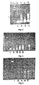
- Figure 1 shows a comparison of PCR products derived from normal and carcinoma cDNA as shown by staining in an agarose gel. Two distinct bands in Lane 2 were present in the primer pair sense-His/antisense ASP (AS1) and multiple bands of about 500 bp are noted in the carcinoma lane for the sense-His/antisense-Ser (AS2) primer pairs in Lane 4.
- AS1 sense-His/antisense ASP
- AS2 sense-His/antisense-Ser
- mRNA overexpression of hepsin was detected and determined using quantitative PCR. Quantitative PCR was performed generally according to the method of Noonan et al. [Proc.Natl.Acad.Sci., USA, 87:7160-7164 (1990 )]. The following oligonucleotide primers were used:
- a tumor tissue bank of fresh frozen tissue of ovarian carcinomas as shown in Table 4 was used for evaluation. Approximately 100 normal ovaries removed for medical reasons other than malignancy were obtained from surgery and were available as controls. TABLE 4 Ovarian cancer tissue bank Total Stage I/11 Stage III/IV No Stage Serous Malignant 166 15 140 8 LMP 16 9 7 0 Benign 12 0 0 12 Mucinous Malignant 26 6 14 6 LMP 28 25 3 0 Benign 3 0 0 3 Endometrioid Malignant 38 17 21 0 LMP 2 2 0 0 Benign 0 0 0 0 Other* Malignant 61 23 29 9 LMP 0 0 0 0 Benign 5 0 0 5 *Other category includes the following tumor types: Brenner's tumor, thecoma, teratoma, fibrothecoma, fibroma, granulosa cell, clear cell, germ cell, mixed mullerian, stromal, undifferentiated, and dysgerminoma.
- carcinomas were evaluated encompassing most histological sub-types of ovarian carcinoma, including borderline or low-malignant potential tumors and overt carcinomas.
- the approach included using mRNA prepared from fresh frozen tissue (both normal and malignant) to compare expression of genes in normal, low malignant potential tumors and overt carcinomas.
- the cDNA prepared from polyA + mRNA was deemed to be genomic DNA-free by checking all preparations with primers that encompassed a known intron-exon splice site using both ⁇ -tubulin and p53 primers.
- Northern panels for examining expression of genes in a multi-tissue normal adult as well as fetal tissue are commercially available (CLONTECH). Such evaluation tools are not only important to confirm the overexpression of individual transcripts in tumor versus normal tissues, but also provides the opportunity to confirm transcript size, and to determine if alternate splicing or other transcript alteration may occur in ovarian carcinoma.
- Northern blot analysis was performed as follows: 10 ⁇ g of mRNA was loaded onto a 1% formaldehyde-agarose gel, electrophoresed and blotted onto a HyBond-N + TM nylon membrane (Amersham). 32 P-labeled cDNA probes were made using Prime-a-Gene Labeling SystemTM (Promega). The PCR products amplified by specific primers were used as probes. Blots were prehybridized for 30 min and then hybridized for 60 min at 68°C with 32 P-labeled cDNA probe in ExpressHybTM Hybridization Solution (CLONTECH). Control hybridization to determine relative gel loading was accomplished using the ⁇ -tubulin probe.
- PCR-amplified cDNA products were cloned and sequenced and the appropriate gene identified based upon nucleotide and amino acid sequences stored in the GCG and EST databases.
- Figures 3, 4 & 5 show the PCR product displays comparing normal and carcinomatous tissues using redundant primers for serine proteases ( Figure 3 ), for cysteine proteases ( Figure 4 ) and for metallo-proteases ( Figure 5 ). Note the differential expression in the carcinoma tissues versus the normal tissues.
- proteases were identified using redundant cDNA primers (see Table 2) directed towards conserved sequences that are associated with intrinsic enzyme activity (for serine proteases, cysteine proteases and metallo-proteases) by comparing mRNA expression in normal, low malignant potential and overt ovarian carcinoma tissues according to Sakanari et al. [Biochemistry 86, 4863-4867 (1989 )].
- Hepsin is a trypsin-like serine protease cloned from hepatoma cells. Hepsin is an extracellular protease (the enzyme includes a secretion signal sequence) which is anchored in the plasma membrane by its amino terminal domain, thereby exposing its catalytic domain to the extracellular matrix. Hepsin has also been shown to be expressed in breast cancer cell lines and peripheral nerve cells. Hepsin has never before been associated with ovarian carcinoma. Specific primers for the hepsin gene were synthesized and the expression of hepsin examined using Northern blots of fetal tissue and ovarian tissue (both normal and ovarian carcinoma).
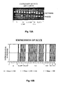
- Figure 10A shows that hepsin was expressed in ovarian carcinomas of different histologic types, but not in normal ovary.
- Figure 10B shows that hepsin was expressed in fetal liver and fetal kidney as anticipated, but at very low levels or not at all in fetal brain and lung.
- Figure 10C shows that hepsin overexpression is not observed in normal adult tissue. Slight expression above the background level is observed in the adult prostate. The mRNA identified in both Northern blots was the appropriate size for the hepsin transcript.
- hepsin The expression of hepsin was examined in 10 normal ovaries and 44 ovarian tumors using specific primers to ⁇ -tubulin and hepsin in a quantitative PCR assay, and found it to be linear over 35 cycles. Expression is presented as the ratio of 32 P-hepsin band to the internal control, the 32 P- ⁇ -tubulin band.
- Hepsin expression was investigated in normal (N), mucinous (M) and serous (S) low malignant potential (LMP) tumors and carcinomas (CA).
- Figure 11A shows quantitative PCR of hepsin and internal control ⁇ -tubulin.
- Figure 11B shows the ratio of hepsin: ⁇ -tubulin expression in normal ovary, LMP tumor, and ovarian carcinoma. It was observed that Hepsin mRNA expression levels were significantly elevated in LMP tumors, ( p ⁇ 0.405) and carcinomas ( p ⁇ 0.0001) compared to levels in normal ovary. All 10 cases of normal ovaries showed a relatively low level of hepsin mRNA expression.
- Hepsin mRNA is highly overexpressed in most histopathologic types of ovarian carcinomas including some low malignant potential tumors (see Figures 11A & 11B ). Most noticeably, hepsin is highly expressed in serous, endometrioid and clear cell tumors tested. It is highly expressed in some mucinous tumors, but it is not overexpressed in the majority of such tumors.
- SCCE Stratum corneum chymotrypsin enzyme
- the PCR product identified was the catalytic domain of the sense-His/antisense-Ser of the stratum corneum chymotrypsin enzyme. This extracellular protease was cloned, sequenced and shown to be expressed on the surface of keratinocytes in the epidermis.
- Stratum corneum chymotrypsin enzyme is a chymotrypsin-like serine protease whose function is suggested to be in the catalytic degradation of intercellular cohesive structures in the stratum corneum layer of the skin. This degradation allows continuous shedding (desquamation) of cells from the skin surface.
- stratum corneum chymotrypsin enzyme The subcellular localization of stratum corneum chymotrypsin enzyme is in the upper granular layer in the stratum corneum of normal non-palmoplantar skin and in the cohesive parts of hypertrophic plantar stratum corneum.
- stratum corneum chymotrypsin enzyme is exclusively associated with the stratum corneum and has not so far been shown to be expressed in any carcinomatous tissues.
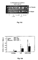
- Figure 13A shows a comparison using quantitative PCR of stratum corneum chymotrypsin enzyme cDNA from normal ovary and ovarian carcinomas.
- Figure 13B shows the ratio of stratum corneum chymotrypsin enzyme to the ⁇ -tubulin internal standard in 10 normal and 44 ovarian carcinoma tissues. Again, it is observed that stratum corneum chymotrypsin enzyme is highly overexpressed in ovarian carcinoma cells. It is also noted that some mucinous tumors overexpress stratum corneum chymotrypsin enzyme, but the majority do not.
- Protease M was identified from subclones of the His--ser primer pair. This protease was first cloned by Anisowicz, et al., [Molecular Medicine, 2, 624-636 (1996 )] and shown to be overexpressed in carcinomas. A preliminary evaluation indicates that this enzyme is overexpressed in ovarian carcinoma ( Figure 14 ).
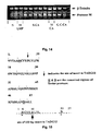
- TADG-12 was identified from the primer pairs, sense-His/antisense-Asp (see Figure 1 , Lanes 1 & 2). Upon subcloning both PCR products in lane 2, the 200 bp product had a unique protease-like sequence not included in GenBank. This 200 bp product contains many of the conserved amino acids common for the His-Asp domain of the family of serine proteins. The second and larger PCR product (300 bp) was shown to have a high degree of homology with TADG-12 (His-Asp sequence), but also contained approximately 100 bp of unique sequence.
- the larger PCR product (present in about 50% of ovarian carcinomas) includes an insert of about 100 bp near the 5' end (and near the histidine) of the sequence.
- This insert may be a retained genomic intron because of the appropriate position of splice sites and the fact that the insert does not contain an open reading frame (see Figure 15 ). This suggests the possibility of a splice site mutation which gives rise to retention of the intron, or a translocation of a sequence into the TADG-12 gene in as many as half of all ovarian carcinomas.
- TADG-13 and -14 are, however, heretofore undocumented genes which the specific primers of the invention allow to be evaluated in normal and tumor cells, and with which the presence or absence of expression of these genes is useful in the diagnosis or treatment selection for specific tumor types.
- PUMP-1 matrix metallo-protease 7
- PUMP-1 is differentially expressed in fetal lung and kidney tissues.
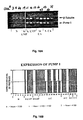
- Figure 17A shows the expression of PUMP-1 in human fetal tissue, while no transcript could be detected in either fetal brain or fetal liver.
- Figure 17B compares PUMP-1 expression in normal ovary and carcinoma subtypes using Northern blot analysis. Notably, PUMP-1 is expressed in ovarian carcinoma tissues, and again, the presence of a transcript in normal tissue was not detected.
- Quantitative PCR comparing normal versus ovarian carcinoma expression of the PUMP-1 mRNA indicates that this gene is highly expressed in serous carcinomas, including most low malignant serous tumors, and is, again, expressed to a lesser extent in Mucinous tumors (see Figures 18A & 18B ). PUMP-1, however, is so far the protease most frequently found overexpressed in mucinous tumors (See Table 7).
- cathepsin-L protease was identified in several serous carcinomas. An initial examination of the expression of cathepsin L in normal and ovarian tumor tissue indicates that transcripts for the cathepsin-L protease are present in both normal and tumor tissues ( Figure 19 ). However, its presence or absence in combination with other proteases of the present invention permits identification of specific tumor types and treatment choices.
- the availability of the instant gene-specific primers coding for the appropriate region of tumor specific proteases allows for the amplification of a specific cDNA probe using Northern and Southern analysis, and their use as markers to detect the presence of the cancer in tissue.
- the probes also allow more extensive evaluation of the expression of the gene in normal ovary versus low malignant potential tumor, as well as both high- and low-stage carcinomas.
- the evaluation of a panel of fresh frozen tissue from all the carcinoma subtypes (Table 4) allowed the determination of whether a protease is expressed predominantly in early stage disease or within specific carcinoma subtypes. It was also determined whether each gene's expression is confined to a particular stage in tumor progression and/or is associated with metastatic lesions. Detection of specific combinations of proteases is an identifying characteristic of the specific tumor types and yields valuable information for diagnoses and treatment selection. Particular tumor types may be more accurately diagnosed by the characteristic expression pattern of each specific tumor.
- hepsin peptides that strongly bind to an HLA allele are putative immunogens, and are used to innoculate an individual against hepsin.
- HLA A0201 1 170 SLGRWPWQV 521.640 28 2 191 SLLSGDWVL 243.051 29 3 229 GLQLGVQAV 159.970 30 4 392 KVSDFREWI 134.154 31 5 308 VLQEARVPI 72.717 32 6 130 RLLEVISVC 71.069 33 7 98 ALTHSELDV 69.552 34 8 211 VLSRWRVFA 46.451 35 9 26 LLLLTAIGA 31.249 36 10 284 ALVDGKICT 30.553 37 11 145 FLAAICQDC 22.853 38 12 192 LLSGDWVLT 21.536 39 13 20 ALTAGTLLL 21.362 40 14 259 ALVHLSSPL 21.362 41 15 277 CLPAAGQAL 21.362 42 16 230 LQLGVQAVV 18.186 43 17 268 PLTEYIQPV 14.429 44 18 31 AIGAASWAI 10.759 45 19 285 LVDGKICTV 9.518 46 20 27 LLLTAIGAA 9.
Landscapes
- Health & Medical Sciences (AREA)
- Life Sciences & Earth Sciences (AREA)
- Chemical & Material Sciences (AREA)
- Engineering & Computer Science (AREA)
- Immunology (AREA)
- Molecular Biology (AREA)
- Biomedical Technology (AREA)
- Organic Chemistry (AREA)
- Zoology (AREA)
- General Health & Medical Sciences (AREA)
- Biochemistry (AREA)
- Urology & Nephrology (AREA)
- Hematology (AREA)
- Genetics & Genomics (AREA)
- Biotechnology (AREA)
- Wood Science & Technology (AREA)
- Microbiology (AREA)
- Analytical Chemistry (AREA)
- Medicinal Chemistry (AREA)
- Physics & Mathematics (AREA)
- Pathology (AREA)
- Bioinformatics & Cheminformatics (AREA)
- Proteomics, Peptides & Aminoacids (AREA)
- General Engineering & Computer Science (AREA)
- Cell Biology (AREA)
- General Physics & Mathematics (AREA)
- Food Science & Technology (AREA)
- Biophysics (AREA)
- Toxicology (AREA)
- Gastroenterology & Hepatology (AREA)
- Hospice & Palliative Care (AREA)
- Oncology (AREA)
- Measuring Or Testing Involving Enzymes Or Micro-Organisms (AREA)
- Medicines That Contain Protein Lipid Enzymes And Other Medicines (AREA)
Description
- Generally, the present invention relates to the fields of molecular biology and medicine. More specifically, the present invention is in the field of cancer, especially ovarian cancer diagnosis.
- To date, ovarian cancer remains the number one killer of women with gynecologic malignant hyperplasia. Approximately 75% of women diagnosed with such cancers are already at an advanced stage (III and IV) of the disease at their initial diagnosis. During the past 20 years, neither diagnosis nor five year survival rates have greatly improved for these patients. This is substantially due to the high percentage of high-stage initial detections of the disease. Therefore, the challenge remains to develop new markers that improve early diagnosis and thereby reduce the percentage of high-stage initial diagnoses.
- Extracellular proteases have already been implicated in the growth, spread and metastatic progression of many cancers, due to the ability of malignant cells not only to grow in situ, but to dissociate from the primary tumor and to invade new surfaces. The ability to disengage from one tissue and re-engage the surface of another tissue is what provides for the morbidity and mortality associated with this disease. Therefore, extracellular proteases may be good candidates for markers of neoplastic development.
- In order for malignant cells to grow, spread or metastasize, they must have the capacity to invade local host tissue, dissociate or shed from the primary tumor, and for metastasis to occur, enter and survive in the bloodstream, implant by invasion into the surface of the target organ and establish an environment conducive for new colony growth (including the induction of angiogenic and growth factors). During this progression, natural tissue barriers have to be degraded, including basement membranes and connective tissue. These barriers include collagen, laminin, proteoglycans and extracellular matrix glycoproteins, including fibronectin. Degradation of these natural barriers, both those surrounding the primary tumor and at the sites of metastatic invasion, is believed to be brought about by the action of a matrix of extracellular proteases.
- Proteases have been classified into four families: serine proteases, metallo-proteases, aspartic proteases and cysteine proteases. Many proteases have been shown to be involved in the human disease process and these enzymes are targets for the development of inhibitors as new therapeutic agents. Additionally, certain individual proteases have been shown to be induced and overexpressed in a diverse group of cancers, and as such, are potential candidates for markers of early diagnosis and possible therapeutic intervention. A group of examples are shown in Table 1.
TABLE 1 Known proteases expressed in various cancers Gastric Brain Breast Ovarian Serine Proteases: uPA uPA NES-1 NES-1 PAI-1 PAI-1 uPA uPA tPA PAI-2 Cysteine Proteases: Cathepsin B Cathepsin L Cathepsin B Cathepsin B Cathepsin L Cathepsin L Cathepsin L Metallo-proteases: Matrilysin* Matrilysin Stromelysin-3 MMP-2 Collagenase* Stromelysin MMP-8 Stromelysin-1* Gelatinase B MMP-9 Gelatinase A uPA, Urokinase-type plasminogen activator; tPA, Tissue-type plasminogen activator; PAI-I, Plasminogen activator 0 inhibitors; PAI-2, Plasminogen activator inhibitors; NES-1, Normal epithelial cell-specific-1; MMP, Matrix P metallo-protease. *Overexpressed in gastrointestinal ulcers. - Significantly, there is a good body of evidence supporting the downregulation or inhibition of individual proteases and the reduction in invasive capacity or malignancy. In work by Clark et al., inhibition of in vitro growth of human small cell lung cancer was demonstrated using a general serine protease inhibitor. More recently, Torres-Rosedo et al., [Proc.Natl.Acad.Sci.USA, 90, 7181-7185 (1993)] demonstrated an inhibition of hepatoma tumor cell growth using specific antisense inhibitors for the serine protease hepsin gene. Metastatic potential of melanoma cells has also been shown to be reduced in a mouse model using a synthetic inhibitor (batimastat) of metallo-proteases. Powell et al. [Cancer Research, 53, 417-422 (1993)] presented evidence to confirm that the expression of extracellular proteases in relatively non-invasive tumor cells enhances their malignant progression using a tumorgenic, but non-metastatic, prostate cell line. Specifically, enhanced metastasis was demonstrated after introducing and expressing the PUMP-1 metallo-protease gene. There is also a body of data to support the notion that expression of cell surface proteases on relatively non-metastatic cell types increases the invasive potential of such cells.
- Thus, the prior art is deficient in a tumor marker useful as an indicator of early disease, particularly for ovarian cancers. The present invention fulfills this longstanding need and desire in the art.
- This invention allows for the detection of cancer, especially ovarian cancer, by screening for hepsin mRNA in tissue, which is indicative of the hepsin protease, which is shown herein to be specifically associated with the surface of 80 percent of ovarian and other tumors. Proteases are considered to be an integral part of tumor growth and metastasis, and therefore, markers indicative of their presence or absence are useful for the diagnosis of cancer. Furthermore, the present invention is useful for treatment (i.e., by inhibiting hepsin or expression of hepsin), for targeted therapy, for vaccination, etc.
- In one embodiment of the present invention, there is provided an immunogenic composition, consisting of an immunogenic fragment of hepsin protein of SEQ ID NOS. 31, 88, 89, 108, 109, 128, 129, 148, 149, 150, 151, 152, 153, or 154 and an appropriate adjuvant.
- Other and further aspects, features, and advantages of the present invention will be apparent from the following description of the presently preferred embodiments of the invention. These embodiments are given for the purpose of disclosure.
- The appended drawings have been included herein so that the above-recited features, advantages and objects of the invention will become clear and can be understood in detail. These drawings form a part of the specification. It is to be noted, however, that the appended drawings illustrate preferred embodiments of the invention and should not be considered to limit the scope of the invention.
-
Figure 1 shows agarose gel comparison of PCR products derived from normal and carcinoma cDNA. -
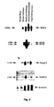
Figure 2 shows Northern blot analysis of ovarian tumors using hepsin, SCCE, PUMP-1, TADG-14 and β-tubulin probes. -
Figure 3 shows amplification with serine protease redundant primers: histidine sense (S1) with aspartic acid antisense (AS1), using normal cDNA (Lane 1) and tumor cDNA (Lane 2); and histidine sense (S1) with serine antisense (AS2), using normal cDNA (Lane 3) and tumor cDNA (Lane 4). -
Figure 4 shows amplification with cysteine protease redundant primers. Normal (Lane 1), low malignant potential (Lane 2), serious carcinoma (Lane 3), mucinous carcinoma (Lane 4), and clear cell carcinoma (Lane 5). -
Figure 5 shows amplification with metallo-protease redundant primers. Normal (Lane 1), low malignant potential (Lane 2), serious carcinoma (Lane 3), mucinous carcinoma (Lane 4), and clear cell carcinoma (Lane 5). -
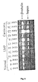
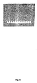
Figure 6 shows amplification with specific primers directed towards the serine protease, hepsin. Expression in normal (Lanes 1-3), low malignant potential tumors (Lanes 4-8), and ovarian carcinomas (Lanes 9-12). -
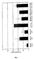
Figure 7 shows hepsin expression levels in normal, low malignant potential tumors, and ovarian carcinomas. S=serous, M=mucinous, LMP=low malignant potential. -
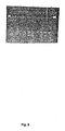
Figure 8 shows serine protease stratum corneum chymotrypsin enzyme (SCCE) expression in normal, low malignant potential tumors, and ovarian carcinomas. -
Figure 9 shows metallo-protease PUMP-1 (MMP-7) gene expression in normal (lanes 1-2) and ovarian carcinomas tissue (Lanes 3-10). -
Figure 10A shows Northern blot analysis of hepsin expression in normal ovary and ovarian carcinomas.Lane 1, normal ovary (case 10);lane 2, serous carcinoma (case 35);lane 3, mucinous carcinoma (case 48);lane 4, endometrioid carcinoma (case 51); and lane 5, clear cell carcinoma (case 54). Incases Figure 10B shows Northern blot analysis of hepsin in normal human fetal.Figure 10C shows Northern blot analysis of hepsin in adult tissues. Significant overexpression of the hepsin transcript is noted in both fetal liver and fetal kidney. Notably, hepsin overexpression is not observed in normal adult tissue. Slight expression above the background level is observed in the adult prostate. -
Figure 11A shows hepsin expression in normal (N), mucinous (M) and serous (S) low malignant potential (LMP) tumors and carcinomas (CA). β-tubulin was used as an internal control.Figure 11B shows the ratio of hepsin:β-tubulin expression in normal ovary, LMP tumor, and ovarian carcinoma. Hepsin mRNA expression levels were significantly elevated in LMP tumors, (p < 0.005) and carcinomas (p < 0.0001) compared to levels in normal ovary. All 10 cases of normal ovaries showed a relatively low level of hepsin mRNA expression. -
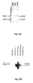
Figure 12A shows northern blot analysis of mRNA expression of the SCCE gene in fetal tissue.Figure 12B shows northern blot analysis of mRNA expression of the SCCE gene in ovarian tissue. -
Figure 13A shows a comparison of quantitative PCR of SCCE cDNA from normal ovary and ovarian carcinomas.Figure 13B shows a bar graph comparing the ratio of SCCE to β-tubulin in 10 normal and 44 ovarian carcinoma tissues. -
Figure 14 shows a comparison by quantitative PCR of normal and ovarian carcinoma expression of mRNA for protease M. -
Figure 15 shows the TADG-12 catalytic domain including an insert near the His 5'-end. -
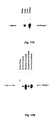
Figure 16A shows northern blot analysis comparing TADG-14 expression in normal and ovarian carcinoma tissues.Figure 16B shows preliminary quantitative PCR amplification of normal and carcinoma cDNAs using specific primers for TADG-14. -
Figure 17A shows northern blot analysis of the PUMP-1 gene in human fetal tissue.Figure 17B shows northern blot analysis of the PUMP-1 gene in normal ovary and ovarian carcinomas. -
Figure 18A shows a comparison of PUMP-1 expression in normal and carcinoma tissues using quantitative PCR with an internal β-tubulin control.Figure 18B shows the ratio of mRNA expression of PUMP-1 compared to the internal control β-tubulin in 10 normal and 44 ovarian carcinomas. -
Figure 19 shows a comparison of PCR amplified products for the hepsin, SCCE, protease M, PUMP-1 and Cathepsin L genes. - This invention identifies a hepsin protease on ovarian and other tumor cells which is characteristic of this type of cancer, and in various combinations with other proteases, is characteristic of individual tumor types. Such information can provide the basis for diagnostic tests (assays or immunohistochemistry), prognostic evaluation (depending on the display pattern) and therapeutic intervention utilizing either antibodies directed at the protease, antisense vehicles for downregulation or protease inhibitors both from established inhibition data and/or for the design of new drugs. Long-term treatment of tumor growth, invasion and metastasis has not succeeded with existing chemotherapeutic agents - most tumors become resistant to drugs after multiple cycles of chemotherapy.
- The present invention is directed toward an immunogenic composition, comprising an immunogenic fragment of hepsin protein and an appropriate adjuvant. The fragment is a 9-residue fragment up to a 20-residue fragment as shown in SEQ ID Nos. 31, 88, 89, 108, 109, 128, 129, 148, 149, 150, 151, 152, 153 or 154.
- In accordance with the present invention there may be employed conventional molecular biology, microbiology, and recombinant DNA techniques within the skill of the art. Such techniques are explained fully in the literature. See, e.g., Maniatis, Fritsch & Sambrook, "Molecular Cloning: A Laboratory Manual (1982); "DNA Cloning: A Practical Approach," Volumes I and II (D.N. Glover ed. 1985); "Oligonucleotide Synthesis" (M.J. Gait ed. 1984); "Nucleic Acid Hybridization" (B.D. Hames & S.J. Higgins eds. 1985); "Transcription and Translation" (B.D. Hames & S.J. Higgins eds. 1984); "Animal Cell Culture" (R.I. Freshney, ed. 1986); "Immobilized Cells And Enzymes" (IRL Press, 1986); B. Perbal, "A Practical Guide To Molecular Cloning" (1984). Therefore, if appearing herein, the following terms shall have the definitions set out below.
- As used herein, the term "cDNA" shall refer to the DNA copy of the mRNA transcript of a gene.
- As used herein, the term "derived amino acid sequence" shall mean the amino acid sequence determined by reading the triplet sequence of nucleotide bases in the cDNA.
- As used herein the term "screening a library" shall refer to the process of using a labeled probe to check whether, under the appropriate conditions, there is a sequence complementary to the probe present in a particular DNA library. In addition, "screening a library" could be performed by PCR.
- As used herein, the term "PCR" refers to the polymerase chain reaction that is the subject of
U.S. Patent Nos. 4,683,195 and4,683,202 to Mullis , as well as other improvements now known in the art. - The amino acid described herein are preferred to be in the "L" isomeric form. However, residues in the "D" isomeric form can be substituted for any L-amino acid residue, as long as the desired functional property of immunoglobulin-binding is retained by the polypeptide. NH2 refers to the free amino group present at the amino terminus of a polypeptide. COOH refers to the free carboxy group present at the carboxy terminus of a polypeptide. In keeping with standard polypeptide nomenclature, J Biol. Chem., 243:3552-59 (1969), abbreviations for amino acid residues may be used.
- It should be noted that all amino-acid residue sequences are represented herein by formulae whose left and right orientation is in the conventional direction of amino-terminus to carboxy-terminus. Furthermore, it should be noted that a dash at the beginning or end of an amino acid residue sequence indicates a peptide bond to a further sequence of one or more amino-acid residues.
- A "replicon" is any genetic element (e.g., plasmid, chromosome, virus) that functions as an autonomous unit of DNA replication in vivo; i.e., capable of replication under its own control.
- A "vector" is a replicon, such as plasmid, phage or cosmid, to which another DNA segment may be attached so as to bring about the replication of the attached segment. A "vector" may further be defined as a replicable nucleic acid construct, e.g., a plasmid or viral nucleic acid.
- A "DNA molecule" refers to the polymeric form of deoxyribonucleotides (adenine, guanine, thymine, or cytosine) in its either single-stranded form or as a double-stranded helix. This term refers only to the primary and secondary structure of the molecule, and does not limit it to any particular tertiary forms. Thus, this term includes double-stranded DNA found, inter alia, in linear DNA molecules (e.g., restriction fragments), viruses, plasmids, and chromosomes. The structure is discussed herein according to the normal convention of giving only the sequence in the 5' to 3' direction along the nontranscribed strand of DNA (i.e., the strand having a sequence homologous to the mRNA).
- An expression vector is a replicable construct in which a nucleic acid sequence encoding a polypeptide is operably linked to suitable control sequences capable of effecting expression of the polypeptide in a cell. The need for such control sequences will vary depending upon the cell selected and the transformation method chosen. Generally, control sequences include a transcriptional promoter and/or enhancer, suitable mRNA ribosomal binding sites and sequences which control the termination of transcription and translation. Methods which are well known to those skilled in the art can be used to construct expression vectors containing appropriate transcriptional and translational control signals. See, for example, techniques described in Sambrook et al., 1989, Molecular Cloning: A Laboratory Manual (2nd Ed.), Cold Spring Harbor Press, N.Y. A gene and its transcription control sequences are defined as being "operably linked" if the transcription control sequences effectively control transcription of the gene. Vectors of the invention include, but are not limited to, plasmid vectors and viral vectors. Preferred viral vectors of the invention are those derived from retroviruses, adenovirus, adeno-associated virus, SV40 virus, or herpes viruses. In general, expression vectors contain promoter sequences which facilitate the efficient transcription of the inserted DNA fragment and are used in connection with a specific host. The expression vector typically contains an origin of replication, promoter(s), terminator(s), as well as specific genes which are capable of providing phenotypic selection in transformed cells. The transformed hosts can be fermented and cultured according to means known in the art to achieve optimal cell growth.
- An "origin of replication" refers to those DNA sequences that participate in DNA synthesis.
- A DNA "coding sequence" is a double-stranded DNA sequence which is transcribed and translated into a polypeptide in vivo when placed under the control of appropriate regulatory sequences. The boundaries of the coding sequence are typically determined by a start codon at the 5' (amino) terminus and a translation stop codon at the 3' (carboxyl) terminus. A coding sequence can include, but is not limited to, prokaryotic sequences, cDNA from eukaryotic mRNA, genomic DNA sequences from eukaryotic (e.g., mammalian) DNA, and even synthetic DNA sequences. A polyadenylation signal and transcription termination sequence will usually be located 3' to the coding sequence.
- Transcriptional and translational control sequences are DNA regulatory sequences, such as promoters, enhancers, polyadenylation signals, terminators, and the like, that provide for the expression of a coding sequence in a host cell.
- A "promoter sequence" is a DNA regulatory region capable of binding RNA polymerase in a cell and initiating transcription of a downstream (3' direction) coding sequence. For purposes of defining the present invention, the promoter sequence is bounded at its 3' terminus by the transcription initiation site and extends upstream (5' direction) to include the minimum number of bases or elements necessary to initiate transcription at levels detectable above background. Within the promoter sequence will be found a transcription initiation site, as well as protein binding domains (consensus sequences) responsible for the binding of RNA polymerase. Eukaryotic promoters often, but not always, contain "TATA" boxes and "CAT" boxes. Prokaryotic promoters typically contain Shine-Dalgarno ribosome-binding sequences in addition to the -10 and - 35 consensus sequences.
- An "expression control sequence" is a DNA sequence that controls and regulates the transcription and translation of another DNA sequence. A coding sequence is "under the control" of transcriptional and translational control sequences in a cell when RNA polymerase transcribes the coding sequence into mRNA, which is then translated into the protein encoded by the coding sequence.
- A "signal sequence" can be included near the coding sequence. This sequence encodes a signal peptide, N-terminal to the polypeptide, that communicates to the host cell to direct the polypeptide to the cell surface or secrete the polypeptide into the media, and this signal peptide is clipped off by the host cell before the protein leaves the cell. Signal sequences can be found associated with a variety of proteins native to prokaryotes and eukaryotes.
- As used herein, the terms "restriction endonucleases" and "restriction enzymes" refer to enzymes, each of which cut double-stranded DNA at or near a specific nucleotide sequence.
- A cell has been "transformed" by exogenous or heterologous DNA when such DNA has been introduced inside the cell. The transforming DNA may or may not be integrated (covalently linked) into the genome of the cell. In prokaryotes, yeast, and mammalian cells for example, the transforming DNA may be maintained on an episomal element such as a plasmid. With respect to eukaryotic cells, a stably transformed cell is one in which the transforming DNA has become integrated into a chromosome so that it is inherited by daughter cells through chromosome replication. This stability is demonstrated by the ability of the eukaryotic cell to establish cell lines or clones comprised of a population of daughter cells containing the transforming DNA. A "clone" is a population of cells derived from a single cell or ancestor by mitosis. A "cell line" is a clone of a primary cell that is capable of stable growth in vitro for many generations.
- Two DNA sequences are "substantially homologous" when at least about 75% (preferably at least about 80%, and most preferably at least about 90% or 95%) of the nucleotides match over the defined length of the DNA sequences. Sequences that are substantially homologous can be identified by comparing the sequences using standard software available in sequence data banks, or in a Southern hybridization experiment under, for example, stringent conditions as defined for that particular system. Defining appropriate hybridization conditions is within the skill of the art. See, e.g., Maniatis et al., supra; DNA Cloning, Vols. I & II, supra; Nucleic Acid Hybridization, supra.
- A "heterologous" region of the DNA construct is an identifiable segment of DNA within a larger DNA molecule that is not found in association with the larger molecule in nature. Thus, when the heterologous region encodes a mammalian gene, the gene will usually be flanked by DNA that does not flank the mammalian genomic DNA in the genome of the source organism. Another example is a construct where the coding sequence itself is not found in nature (e.g., a cDNA where the genomic coding sequence contains introns, or synthetic sequences having codons different than the native gene). Allelic variations or naturally-occurring mutational events do not give rise to a heterologous region of DNA as defined herein.
- The labels most commonly employed for these studies are radioactive elements, enzymes, chemicals which fluoresce when exposed to ultraviolet light, and others. A number of fluorescent materials are known and can be utilized as labels. These include, for example, fluorescein, rhodamine, auramine, Texas Red, AMCA blue and Lucifer Yellow. A particular detecting material is anti-rabbit antibody prepared in goats and conjugated with fluorescein through an isothiocyanate. Proteins can also be labeled with a radioactive element or with an enzyme. The radioactive label can be detected by any of the currently available counting procedures. The preferred isotope may be selected from 3H, 14C, 32P, 35S, 36Cl, 51Cr, 57Co, 58Co, 59Fe, 90Y, 125I, 131I, and 186Re. Enzyme labels are likewise useful, and can be detected by any of the presently utilized colorimetric, spectrophotometric, fluorospectrophotometric, amperometric or gasometric techniques. The enzyme is conjugated to the selected particle by reaction with bridging molecules such as carbodiimides, diisocyanates, glutaraldehyde and the like. Many enzymes which can be used in these procedures are known and can be utilized. The preferred are peroxidase, β-glucuronidase, β-D-glucosidase, β-D-galactosidase, urease, glucose oxidase plus peroxidase and alkaline phosphatase.
U.S. Patent Nos. 3,654,090 ,3,850,752, and4,016,043 are referred to by way of example for their disclosure of alternate labeling material and methods. - A particular assay system developed and utilized in the art is known as a receptor assay. In a receptor assay, the material to be assayed is appropriately labeled and then certain cellular test colonies are inoculated with a quantitiy of both the label after which binding studies are conducted to determine the extent to which the labeled material binds to the cell receptors. In this way, differences in affinity between materials can be ascertained.
- An assay useful in the art is known as a "cis/trans" assay. Briefly, this assay employs two genetic constructs, one of which is typically a plasmid that continually expresses a particular receptor of interest when transfected into an appropriate cell line, and the second of which is a plasmid that expresses a reporter such as luciferase, under the control of a receptor/ligand complex. Thus, for example, if it is desired to evaluate a compound as a ligand for a particular receptor, one of the plasmids would be a construct that results in expression of the receptor in the chosen cell line, while the second plasmid would possess a promoter linked to the luciferase gene in which the response element to the particular receptor is inserted. If the compound under test is an agonist for the receptor, the ligand will complex with the receptor, and the resulting complex will bind the response element and initiate transcription of the luciferase gene. The resulting chemiluminescence is then measured photometrically, and dose response curves are obtained and compared to those of known ligands. The foregoing protocol is described in detail in
U.S. Patent No. 4,981,784 . - As used herein, the term "host" is meant to include not only prokaryotes but also eukaryotes such as yeast, plant and animal cells. A recombinant DNA molecule or gene which encodes a human hepsin protein of the present invention can be used to transform a host using any of the techniques commonly known to those of ordinary skill in the art. Especially preferred is the use of a vector containing coding sequences for the gene which encodes a human hepsin protein of the present invention for purposes of prokaryote transformation. Prokaryotic hosts may include E. coli, S. tymphimurium, Serratia marcescens and Bacillus subtilis. Eukaryotic hosts include yeasts such as Pichia pastoris, mammalian cells and insect cells.
- As used herein, "substantially pure DNA" means DNA that is not part of a milieu in which the DNA naturally occurs, by virtue of separation (partial or total purification) of some or all of the molecules of that milieu, or by virtue of alteration of sequences that flank the claimed DNA. The term therefore includes, for example, a recombinant DNA which is incorporated into a vector, into an autonomously replicating plasmid or virus, or into the genomic DNA of a prokaryote or eukaryote; or which exists as a separate molecule (e.g., a cDNA or a genomic or cDNA fragment produced by polymerase chain reaction (PCR) or restriction endonuclease digestion) independent of other sequences. It also includes a recombinant DNA which is part of a hybrid gene encoding additional polypeptide sequence, e.g., a fusion protein. Also included is a recombinant DNA which includes a portion of the nucleotides listed in SEQ ID No. 188 and which encodes an alternative splice variant of hepsin.
- By a "substantially pure protein" is meant a protein which has been separated from at least some of those components which naturally accompany it. Typically, the protein is substantially pure when it is at least 60% (by weight) free from the proteins and other naturally-occurring organic molecules with which it is naturally associated in vivo. Preferably, the purity of the preparation (by weight) is at least 75%, more preferably at least 90%, and most preferably at least 99%. A substantially pure hepsin protein may be obtained, for example, by extraction from a natural source; by expression of a recombinant nucleic acid encoding a hepsin polypeptide; or by chemically synthesizing the protein. Purity can be measured by any appropriate method, e.g., column chromatography, such as immunoaffinity chromatography using an antibody specific for hepsin, polyacrylamide gel electrophoresis, or HPLC analysis. A protein is substantially free of naturally associated components when it is separated from at least some of those contaminants which accompany it in its natural state. Thus, a protein which is chemically synthesized or produced in a cellular system different from the cell from which it naturally originates will be, by definition, substantially free from its naturally associated components. Accordingly, substantially pure proteins include eukaryotic proteins synthesized in E. coli, other prokaryotes, or any other organism in which they do not naturally occur.
- The term "oligonucleotide", as used herein, is defined as a molecule comprised of two or more ribonucleotides, preferably more than three. Its exact size will depend upon many factors, which, in turn, depend upon the ultimate function and use of the oligonucleotide. The term "primer", as used herein, refers to an oligonucleotide, whether occurring naturally (as in a purified restriction digest) or produced synthetically, and which is capable of initiating synthesis of a strand complementary to a nucleic acid when placed under appropriate conditions, i.e., in the presence of nucleotides and an inducing agent, such as a DNA polymerase, and at a suitable temperature and pH. The primer may be either single-stranded or double-stranded and must be sufficiently long to prime the synthesis of the desired extension product in the presence of the inducing agent. The exact length of the primer will depend upon many factors, including temperature, sequence and/or homology of primer and the method used. For example, in diagnostic applications, the oligonucleotide primer typically contains 15-25 or more nucleotides, depending upon the complexity of the target sequence, although it may contain fewer nucleotides.
- The primers herein are selected to be "substantially" complementary to particular target DNA sequences. This means that the primers must be sufficiently complementary to hybridize with their respective strands. Therefore, the primer sequence need not reflect the exact sequence of the template. For example, a non-complementary nucleotide fragment (i.e., containing a restriction site) may be attached to the 5' end of the primer, with the remainder of the primer sequence being complementary to the strand. Alternatively, non-complementary bases or longer sequences can be interspersed into the primer, provided that the primer sequence has sufficient complementary with the sequence to hybridize therewith and form the template for synthesis of the extension product.
- The probe to which the DNA of the invention hybridizes preferably consists of a sequence of at least 20 consecutive nucleotides, more preferably 40 nucleotides, even more preferably 50 nucleotides, and most preferably 100 nucleotides or more (up to 100%) of the coding sequence of the nucleotides listed in SEQ ID No. 188 or the complement thereof. Such a probe is useful for detecting expression of hepsin in a cell by a method including the steps of (a) contacting mRNA obtained from the cell with a labeled hepsin hybridization probe; and (b) detecting hybridization of the probe with the mRNA.
- By "high stringency" is meant DNA hybridization and wash conditions characterized by high temperature and low salt concentration, e.g., wash conditions of 65°C at a salt concentration of approximately 0.1X SSC, or the functional equivalent thereof. For example, high stringency conditions may include hybridization at about 42°C in the presence of about 50% formamide; a first wash at about 65°C with about 2X SSC containing 1% SDS; followed by a second wash at about 65°C with about 0.1X SSC. The DNA may have at least about 70% sequence identity to the coding sequence of the nucleotides listed in SEQ ID No. 188, preferably at least 75% (e.g., at least 80%); and most preferably at least 90%. The identity between two sequences is a direct function of the number of matching or identical positions. When a position in both of the two sequences is occupied by the same monomeric subunit, e.g., if a given position is occupied by an adenine in each of two DNA molecules, then they are identical at that position. For example, if 7 positions in a
sequence 10 nucleotides in length are identical to the corresponding positions in a second 10-nucleotide sequence, then the two sequences have 70% sequence identity. The length of comparison sequences will generally be at least 50 nucleotides, preferably at least 60 nucleotides, more preferably at least 75 nucleotides, and most preferably 100 nucleotides. Sequence identity is typically measured using sequence analysis software (e.g., Sequence Analysis Software Package of the Genetics Computer Group (GCG), University of Wisconsin Biotechnology Center, 1710 University Avenue, Madison, WI 53705). - The present invention comprises a vector comprising a DNA sequence which encodes a hepsin protein, wherein said vector is capable of replication in a host, and comprises, in operable linkage: a) an origin of replication; b) a promoter; and c) a DNA sequence coding for said hepsin protein. Vectors may be used to amplify and/or express nucleic acid encoding a hepsin protein or fragment thereof.
- In addition to substantially full-length proteins, the invention also includes fragments (e.g., antigenic fragments) of the hepsin protein. As used herein, "fragment," as applied to a polypeptide, will ordinarily be at least 10 residues, more typically at least 20 residues, and preferably at least 30 (e.g., 50) residues in length, but less than the entire, intact sequence. Fragments of the hepsin protein can be generated by methods known to those skilled in the art, e.g., by enzymatic digestion of naturally occurring or recombinant hepsin protein, by recombinant DNA techniques using an expression vector that encodes a defined fragment of hepsin, or by chemical synthesis. The ability of a candidate fragment to exhibit a characteristic of hepsin (e.g., binding to an antibody specific for hepsin) can be assessed by methods described herein. Purified hepsin or antigenic fragments of hepsin can be used to generate new antibodies or to test existing antibodies (e.g., as positive controls in a diagnostic assay) by employing standard protocols known to those skilled in the art. Included in this invention is polyclonal antisera generated by using hepsin or a fragment of hepsin as the immunogen in, e.g., rabbits. Standard protocols for monoclonal and polyclonal antibody production known to those skilled in this art are employed. The monoclonal antibodies generated by this procedure can be screened for the ability to identify recombinant hepsin cDNA clones, and to distinguish them from other cDNA clones.
- The following examples are given for the purpose of illustrating various embodiments of the invention and are not meant to limit the present invention in any fashion:
- Only cDNA preparations deemed free of genomic DNA were used for gene expression analysis. Redundant primers were prepared for serine proteases, metallo-proteases and cysteine protease. The primers were synthesized to consensus sequences of amino acid surrounding the catalytic triad for serine proteases, viz. histidine ... aspartate ... and serine. The sequences of both sense (histidine & aspartate) and antisense (aspartate and serine) redundant primers are shown in Table 2.
TABLE 2 PCR Primers 5'→3' SEQ ID No. Redundant Primers: Serine Protease (histidine) = S1 tgggtigtiacigcigcica(ct)tg 1 Serine Protease (aspartic acid) = AS1 a(ag)ia(ag)igciatitcitticc 2 Serine Protease (serine) = AS11 a(ag)iggiccicci(cg)(ta)(ag)tcicc 3 Cysteine Protease - sense ca(ag)ggica(ag)tg(ct)ggi(ta)(cg)itg(ct)tgg 4 Cysteine Protease - antisense taiccicc(ag)tt(ag)caicc(ct)tc 5 Metallo Protease - sense cci(ac)gitg(tc)ggi(ga)(ta)icciga 6 Metallo Protease - antisense tt(ag)tgicciai(ct)tc(ag)tg 7 Specific Primers: Serine Protease (hepsin) = sense tgtcccgatggcgagtgttt 8 Serine Protease (hepsin) = antisense cctgttggccatagtactgc 9 Serine Protease (SCCE) = sense agatgaatgagtacaccgtg 10 Serine Protease (SCCE) = antisense ccagtaagtccttgtaaacc 11 Serine Protease (Comp B) = sense aagggacacgagagctgtat 12 Serine Protease (Comp B) = antisense aagtggtagttggaggaagc 13 Serine Protease (Protease M)= sense ctgtgatccaccctgactat 20 Serine Protease (Protease M) = antisense caggtggatgtatgcacact 21 Serine Protease (TADG12) = sense (Ser10-s) gcgcactgtgtttatgagat 22 Serine Protease (TADG 12) = antisense (Ser10-as) ctctttggcttgtacttgct 23 Serine Protease (TADG13) = sense tgagggacatcattatgcac 24 Serine Protease (TADG13) = antisense caagttttccccataattgg 25 Serine Protease (TADG14) = sense acagtacgcctgggagacca 26 Serine Protease (TADG14) = antisense ctgagacggtgcaattctgg 27 Cysteine Protease (Cath-L) = sense attggagagagaaaggctac 14 Cysteine Protease (Cath-L) = antisense cttgggattgtacttacagg 15 Metallo Protease (PUMP1) = sense cttccaaagtggtcacctac 16 Metallo Protease (PUMP1) = antisense ctagactgctaccatccgtc 17 - Several protease entities were identified and subcloned from PCR amplification of cDNA derived from serous cystadenocarcinomas. Therefore, the proteases described herein are reflective of surface activities for this type of carcinoma, the most common form of ovarian cancer. Applicant has also shown PCR amplification bands unique to the mucinous tumor type and the clear cell type of similar base pair size. About 20-25% of ovarian cancers are classified as either mucinous, clear cell, or endometrioid.
- To determine the identity of the PCR products, all the appropriate bands were ligated into Promega T-vector plasmid and the ligation product was used to transform JM109 cells (Promega) grown on selective media. After selection and culturing of individual colonies, plasmid DNA was isolated by means of the WIZARD MINIPREP™ DNA purification system (Promega). Inserts were sequenced using a Prism Ready Reaction Dydeoxy Terminators cycle sequencing kit (Applied Biosystems). Residual dye terminators were removed from the completed sequencing reaction using a CENTRISEP SPIN™ column (Princeton Separation), and samples were loaded into an Applied Biosystems Model 373A DNA sequencing system. The results of subcloning and sequencing for the serine protease primers are summarized in Table 3.
TABLE 3 Serine protease candidates Subclone Primer Set Gene Candidate 1 His- Ser Hepsin 2 His- Ser SCCE 3 His- Ser Compliment B 4 His- Asp Cofactor 1 5 His-Asp TADG-12* 6 His-Ser TADG-13* 7 His-Ser TADG-14* 8 His- Ser Protease M 9 His-Ser TADG-15* *indicates novel proteases - Cloning and characterization of new gene candidates was undertaken to expand the panel representative of extracellular proteases specific for ovarian carcinoma subtypes. Sequencing of the PCR products derived from tumor cDNA confirms the potential candidacy of these genes. The three novel genes all have conserved residues within the catalytic triad sequence consistent with their membership in the serine protease family.
- Applicant compared the PCR products amplified from normal and carcinoma cDNAs using sense-histidine and antisense-aspartate as well as sense-histidine and antisense-serine. The anticipated PCR products of approximately 200 bp and 500 bp for those pairs of primers were observed (aspartate is approximately 50-70 amino acids downstream from histidine, and serine is about 100-150 amino acids toward the carboxy end from histidine).
-
Figure 1 shows a comparison of PCR products derived from normal and carcinoma cDNA as shown by staining in an agarose gel. Two distinct bands inLane 2 were present in the primer pair sense-His/antisense ASP (AS1) and multiple bands of about 500 bp are noted in the carcinoma lane for the sense-His/antisense-Ser (AS2) primer pairs inLane 4. - The mRNA overexpression of hepsin was detected and determined using quantitative PCR. Quantitative PCR was performed generally according to the method of Noonan et al. [Proc.Natl.Acad.Sci., USA, 87:7160-7164 (1990)]. The following oligonucleotide primers were used:
- hepsin:
- forward 5'-TGTCCCGATGGCGAGTGTTT-3' (SEQ ID No. 8), and
- reverse 5'-CCTGTTGGCCATAGTACTGC-3' (SEQ ID No. 9);
- and β-tubulin:
- forward 5'- TGCATTGACAACGAGGC -3' (SEQ ID No. 18), and
- reverse 5'- CTGTCTTGA CATTGTTG -3' (SEQ ID No. 19).
- Experiments comparing PCR amplification in normal ovary and ovarian carcinoma suggested overexpression and/or alteration in mRNA transcript in tumor tissues. Northern blot analysis of TADG-14 confirms a transcript size of 1.4 kb and data indicate overexpression in ovarian carcinoma (
Figure 2 ). Isolation and purification using both PCR and a specific 250 bp PCR product to screen positive plaques yielded a 1.2 kb clone of TADG-14. Other proteases were amplified by the same method using the appropriate primers from Table 2. - A tumor tissue bank of fresh frozen tissue of ovarian carcinomas as shown in Table 4 was used for evaluation. Approximately 100 normal ovaries removed for medical reasons other than malignancy were obtained from surgery and were available as controls.
TABLE 4 Ovarian cancer tissue bank Total Stage I/11 Stage III/IV No Stage Serous Malignant 166 15 140 8 LMP 16 9 7 0 Benign 12 0 0 12 Mucinous Malignant 26 6 14 6 LMP 28 25 3 0 Benign 3 0 0 3 Endometrioid Malignant 38 17 21 0 LMP 2 2 0 0 Benign 0 0 0 0 Other* Malignant 61 23 29 9 LMP 0 0 0 0 Benign 5 0 0 5 *Other category includes the following tumor types: Brenner's tumor, thecoma, teratoma, fibrothecoma, fibroma, granulosa cell, clear cell, germ cell, mixed mullerian, stromal, undifferentiated, and dysgerminoma. - From the tumor bank, approximately 100 carcinomas were evaluated encompassing most histological sub-types of ovarian carcinoma, including borderline or low-malignant potential tumors and overt carcinomas. The approach included using mRNA prepared from fresh frozen tissue (both normal and malignant) to compare expression of genes in normal, low malignant potential tumors and overt carcinomas. The cDNA prepared from polyA+ mRNA was deemed to be genomic DNA-free by checking all preparations with primers that encompassed a known intron-exon splice site using both β-tubulin and p53 primers.
- Significant information can be obtained by examining the expression of these candidate genes by Northern blot. Analysis of normal adult multi-tissue blots offers the opportunity to identify normal tissues which may express the protease. Ultimately, if strategies for inhibition of proteases for therapeutic intervention are to be developed, it is essential to appreciate the expression of these genes in normal tissue if and when it occurs.
- Significant information is expected from Northern blot analysis of fetal tissue. Genes overexpressed in carcinomas are often highly expressed in organogenesis. As indicated, the hepsin gene cloned from hepatoma cells and overexpressed in ovarian carcinoma is overtly expressed in fetal liver. Hepsin gene expression was also detected in fetal kidney, and therefore, could be a candidate for expression in renal carcinomas.
- Northern panels for examining expression of genes in a multi-tissue normal adult as well as fetal tissue are commercially available (CLONTECH). Such evaluation tools are not only important to confirm the overexpression of individual transcripts in tumor versus normal tissues, but also provides the opportunity to confirm transcript size, and to determine if alternate splicing or other transcript alteration may occur in ovarian carcinoma.
- Northern blot analysis was performed as follows: 10 µg of mRNA was loaded onto a 1% formaldehyde-agarose gel, electrophoresed and blotted onto a HyBond-N+™ nylon membrane (Amersham). 32P-labeled cDNA probes were made using Prime-a-Gene Labeling System™ (Promega). The PCR products amplified by specific primers were used as probes. Blots were prehybridized for 30 min and then hybridized for 60 min at 68°C with 32P-labeled cDNA probe in ExpressHyb™ Hybridization Solution (CLONTECH). Control hybridization to determine relative gel loading was accomplished using the β-tubulin probe.
- Normal human tissues including spleen, thymus, prostate, testis, ovary, small intestine, colon, peripheral blood leukocyte, heart, brain, placenta, lung, liver, skeletal muscle, kidney, pancreas and normal human fetal tissues; brain, lung, liver and kidney (Human Multiple Tissue Northern Blot; CLONTECH) were all examined using the same hybridization procedure.
- Based on their unique expression in either low malignant potential tumors or carcinomas, PCR-amplified cDNA products were cloned and sequenced and the appropriate gene identified based upon nucleotide and amino acid sequences stored in the GCG and EST databases.
Figures 3, 4 & 5 show the PCR product displays comparing normal and carcinomatous tissues using redundant primers for serine proteases (Figure 3 ), for cysteine proteases (Figure 4 ) and for metallo-proteases (Figure 5 ). Note the differential expression in the carcinoma tissues versus the normal tissues. The proteases were identified using redundant cDNA primers (see Table 2) directed towards conserved sequences that are associated with intrinsic enzyme activity (for serine proteases, cysteine proteases and metallo-proteases) by comparing mRNA expression in normal, low malignant potential and overt ovarian carcinoma tissues according to Sakanari et al. [Biochemistry 86, 4863-4867 (1989)]. - For the serine protease group, using the histidine domain primer sense, S1, in combination with antisense primer AS2, the following proteases were identified:
- (a) Hepsin, a trypsin-like serine protease cloned from hepatoma cells shown to be a cell surface protease essential for the growth of hepatoma cells in culture and highly expressed in hepatoma tumor cells (
Figure 3 , Lane 4); - (b) Complement factor B protease (human factor IX), a protease involved in the coagulation cascade and associated with the production and accumulation of fibrin split products associated with tumor cells (
Figure 3 , Lane 4). Compliment factor B belongs in the family of coagulation factors X (Christmas factor). As part of the intrinsic pathway, compliment factor B catalyzes the proteolytic activation of coagulation factor X in the presence of Ca2+ phospholipid and factor VIIIa e5; and - (c) A stratum corneum chymotryptic enzyme (SCCE) serine protease involved in desquarnation of skin cells from the human stratum corneum (
Figure 3 , Lane 4). SCCE is expressed in keratinocytes of the epidermis and functions to degrade the cohesive structures in the cornified layer to allow continuous skin surface shedding. - In the cysteine protease group, using redundant sense and anti-sense primers for cysteine proteases, one unique PCR product was identified by overexpression in ovarian carcinoma when compared to normal ovarian tissue (
Figure 4 , Lanes 3-5). Cloning and sequencing this PCR product identified a sequence of Cathepsin L, which is a lysomal cysteine protease whose expression and secretion is induced by malignant transformation, growth factors and tumor promoters. Many human tumors (including ovarian) express high levels of Cathepsin L. Cathepsin L cysteine protease belongs in the stromolysin family and has potent elastase and collagenase activities. Published data indicates increased levels in the serum of patients with mucinous cystadenocarcinoma of the ovary. It has not heretofore been shown to be expressed in other ovarian tumors. - Using redundant sense and anti-sense primers for the metallo-protease group, one unique PCR product was detected in the tumor tissue which was absent in normal ovarian tissue (
Figure 5 , Lanes 2-5). Subcloning and sequencing this product indicates it has complete homology in the appropriate region with the so-called PUMP-1 (MMP-7) gene. This zinc-binding metallo-protease is expressed as a proenzyme with a signal sequence and is active in gelatin and collagenase digestion. PUMP-1 has also been shown to be induced and overexpressed in 9 of 10 colorectal carcinomas compared to normal colon tissue, suggesting a role for this substrate in the progression of this disease. - The expression of the serine protease hepsin gene in 8 normal, 11 low malignant potential tumors, and 14 carcinoma (both mucinous and serous type) by quantitative PCR using hepsin-specific primers (see Table 2) was determined (primers directed toward the β-tubulin message were used as an internal standard) (Table 5). These data confirm the overexpression of the hepsin surface protease gene in ovarian carcinoma, including both low malignant potential tumors and overt carcinoma. Expression of hepsin is increased over normal levels in low malignant potential tumors, and high stage tumors (Stage III) of this group have higher expression of hepsin when compared to low stage tumors (Stage 1) (Table 6). In overt carcinoma, serous tumors exhibit the highest levels of hepsin expression, while mucinous tumors express levels of hepsin comparable with the high stage low malignant potential group (
Figures 6 &7 ).TABLE 5 Patient Characteristics and Expression of Hensin Gene Case Histological typea Stage/Grade LN b mRNA expression of hepsinc 1 normal ovary n 2 normal ovary n 3 normal ovary n 4 normal ovary n 5 normal ovary n 6 normal ovary n 7 normal ovary n 8 normal ovary n 9 normal ovary n 10 normal ovary n 11 S adenoma (LMP) 1/1 N 4+ 12 S adenoma (LMP) 1/1 NE 4+ 13 S adenoma (LMP) 1/1 NE n 14 S adenoma (LMP) 1/1 N 2+ 15 S adenoma (LMP) 3/1 P 4+ 16 S adenoma (LMP) 3/1 P 4+ 17 S adenoma (LMP) 3/1 P 4+ 18 M adenoma (LMP) 1/1 NE 4+ 19 M adenoma (LMP) 1/1 N n 20 M adenoma (LMP) 1/1 N n 21 M adenoma (LMP) 1/1 N n 22 M adenoma (LMP) 1/1 NE n 23 S carcinoma 1/2 N 4+ 24 S carcinoma 1/3 N 4+ 25 S carcinoma 3/1 NE 2+ 26 S carcinoma 3/2 NE 4+ 27 S carcinoma 3/2 P 4+ 28 S carcinoma 3/2 NE 2+ 29 S carcinoma 3/3 NE 2+ 30 S carcinoma 3/3 NE 4+ 31 S carcinoma 3/3 NE 4+ 32 S carcinoma 3/3 NE 4+ 33 S carcinoma 3/3 N 4+ 34 S carcinoma 3/3 NE n 35 S carcinoma 3/3 NE 4+ 36 S carcinoma 3/3 NE 4+ 37 S carcinoma 3/3 NE 4+ 38 S carcinoma 3/3 N 4+ 39 S carcinoma 3/2 NE 2+ 40 S carcinoma 3/3 NE 4+ 41 S carcinoma 3/2 NE 4+ 42 M carcinoma 1/2 N n 43 M carcinoma 2/2 NE 4+ 44 M carcinoma 2/2 N 4+ 45 M carcinoma 3/1 NE n 46 M carcinoma 3/2 NE 4+ 47 M carcinoma 3/2 NE n 48 M carcinoma 3/3 NE n 49 E carcinoma 2/3 N 4+ 50 E carcinoma 3/2 NE 4+ 51 E carcinoma 3/3 NE 4+ 52 C carcinoma 1/3 N 4+ 53 C carcinoma 1/1 N 4+ 54 C carcinoma 3/2 P 4+ aS, serous; M, mucinous; E, endometrioid; C, clear cell; bLN, lymph node metastasis; P, positive; N, negative; NE, not examined; cn, normal range = mean ±2SD; 2+, mean ±2SD to ±4SD; 4+, mean ±4SD or greater. TABLE 6 Overexnression of hepsin in normal ovaries and ovarian tumors Type N Hepsin Ratio of Hepsin Overexpression to β- tubulin Normal 10 0 (0%) 0.06 ± 0.05 LMP 12 7 (58.3%) 0.26 ± 0.19 Serous 7 6 (85.7%) 0.34 ± 0.20 Mucinous 5 1 (20.0%) 0.14 ± 0.12 Carcinomous 32 27 (84.4%) 0.46 ± 0.29 Serous 19 18 (94.7%) 0.56 ± 0.32 Mucinous 7 3 (42.9%) 0.26 ± 0.22 Endometrioid 3 3 (100%) 0.34 ± 0.01 Clear Cell 3 3 (100%) 0.45 ± 0.08 - Studies using both SCCE-specific primers (
Figure 8 ) and PUMP-specific primers (Figure 9 ) indicate overexpression of these proteases in ovarian carcinomas. - Most of the proteases described herein were identified from the sense-His/antisense-Ser primer pair, yielding a 500 bp PCR product (
Figure 1 , Lane 4). Some of the enzymes are familiar, a short summary of each follows. - Hepsin is a trypsin-like serine protease cloned from hepatoma cells. Hepsin is an extracellular protease (the enzyme includes a secretion signal sequence) which is anchored in the plasma membrane by its amino terminal domain, thereby exposing its catalytic domain to the extracellular matrix. Hepsin has also been shown to be expressed in breast cancer cell lines and peripheral nerve cells. Hepsin has never before been associated with ovarian carcinoma. Specific primers for the hepsin gene were synthesized and the expression of hepsin examined using Northern blots of fetal tissue and ovarian tissue (both normal and ovarian carcinoma).
-
Figure 10A shows that hepsin was expressed in ovarian carcinomas of different histologic types, but not in normal ovary.Figure 10B shows that hepsin was expressed in fetal liver and fetal kidney as anticipated, but at very low levels or not at all in fetal brain and lung.Figure 10C shows that hepsin overexpression is not observed in normal adult tissue. Slight expression above the background level is observed in the adult prostate. The mRNA identified in both Northern blots was the appropriate size for the hepsin transcript. The expression of hepsin was examined in 10 normal ovaries and 44 ovarian tumors using specific primers to β-tubulin and hepsin in a quantitative PCR assay, and found it to be linear over 35 cycles. Expression is presented as the ratio of 32P-hepsin band to the internal control, the 32P-β-tubulin band. - Hepsin expression was investigated in normal (N), mucinous (M) and serous (S) low malignant potential (LMP) tumors and carcinomas (CA).
Figure 11A shows quantitative PCR of hepsin and internal control β-tubulin.Figure 11B shows the ratio of hepsin:β-tubulin expression in normal ovary, LMP tumor, and ovarian carcinoma. It was observed that Hepsin mRNA expression levels were significantly elevated in LMP tumors, (p < 0.405) and carcinomas (p < 0.0001) compared to levels in normal ovary. All 10 cases of normal ovaries showed a relatively low level of hepsin mRNA expression. - Hepsin mRNA is highly overexpressed in most histopathologic types of ovarian carcinomas including some low malignant potential tumors (see
Figures 11A & 11B ). Most noticeably, hepsin is highly expressed in serous, endometrioid and clear cell tumors tested. It is highly expressed in some mucinous tumors, but it is not overexpressed in the majority of such tumors. - The PCR product identified was the catalytic domain of the sense-His/antisense-Ser of the stratum corneum chymotrypsin enzyme. This extracellular protease was cloned, sequenced and shown to be expressed on the surface of keratinocytes in the epidermis. Stratum corneum chymotrypsin enzyme is a chymotrypsin-like serine protease whose function is suggested to be in the catalytic degradation of intercellular cohesive structures in the stratum corneum layer of the skin. This degradation allows continuous shedding (desquamation) of cells from the skin surface. The subcellular localization of stratum corneum chymotrypsin enzyme is in the upper granular layer in the stratum corneum of normal non-palmoplantar skin and in the cohesive parts of hypertrophic plantar stratum corneum. Stratum corneum chymotrypsin enzyme is exclusively associated with the stratum corneum and has not so far been shown to be expressed in any carcinomatous tissues.
- Northern blots were probed with the PCR product to determine expression of stratum corneum chymotrypsin enzyme in fetal tissue and ovarian carcinoma (
Figures 12A & 12B ). Noticeably, detection of stratum corneum chymotrypsin enzyme messenger RNA on the fetal Northern was almost non-existent (a problem with the probe or the blot was excluded by performing the proper controls). A faint band appeared in fetal kidney. On the other hand, stratum corneum chymotrypsin enzyme mRNA is abundant in the ovarian carcinoma mRNA (Figure 12B ). Two transcripts of the correct size are observed for stratum corneum chymotrypsin enzyme. The same panel of cDNA used for hepsin analysis was used for stratum corneum chymotrypsin enzyme expression. - No stratum corneum chymotrypsin enzyme expression was detected in the normal ovary lane of the Northern blot. A comparison of all candidate genes, including a loading marker (β-tubulin), was shown to confirm that this observation was not a result of a loading bias. Quantitative PCR using stratum corneum chymotrypsin enzyme primers, along with β-tubulin internal control primers, confirmed the overexpression of stratum corneum chymotrypsin enzyme mRNA in carcinoma of the ovary with no expression in normal ovarian tissue (
Figure 13 ). -
Figure 13A shows a comparison using quantitative PCR of stratum corneum chymotrypsin enzyme cDNA from normal ovary and ovarian carcinomas.Figure 13B shows the ratio of stratum corneum chymotrypsin enzyme to the β-tubulin internal standard in 10 normal and 44 ovarian carcinoma tissues. Again, it is observed that stratum corneum chymotrypsin enzyme is highly overexpressed in ovarian carcinoma cells. It is also noted that some mucinous tumors overexpress stratum corneum chymotrypsin enzyme, but the majority do not. - Protease M was identified from subclones of the His--ser primer pair. This protease was first cloned by Anisowicz, et al., [Molecular Medicine, 2, 624-636 (1996)] and shown to be overexpressed in carcinomas. A preliminary evaluation indicates that this enzyme is overexpressed in ovarian carcinoma (
Figure 14 ). - Several serine proteases associated with the coagulation pathway were also subcloned. Examination of normal and ovarian carcinomas by quantitative PCR for expression of these enzymes, it was noticeable that this mRNA was not clearly overexpressed in ovarian carcinomas when compared to normal ovarian tissue. It should be noted that the same panel of tumors was used for the evaluation of each candidate protease.
- TADG-12 was identified from the primer pairs, sense-His/antisense-Asp (see
Figure 1 ,Lanes 1 & 2). Upon subcloning both PCR products inlane 2, the 200 bp product had a unique protease-like sequence not included in GenBank. This 200 bp product contains many of the conserved amino acids common for the His-Asp domain of the family of serine proteins. The second and larger PCR product (300 bp) was shown to have a high degree of homology with TADG-12 (His-Asp sequence), but also contained approximately 100 bp of unique sequence. Synthesis of specific primers and the sequencing of the subsequent PCR products from three different tumors demonstrated that the larger PCR product (present in about 50% of ovarian carcinomas) includes an insert of about 100 bp near the 5' end (and near the histidine) of the sequence. This insert may be a retained genomic intron because of the appropriate position of splice sites and the fact that the insert does not contain an open reading frame (seeFigure 15 ). This suggests the possibility of a splice site mutation which gives rise to retention of the intron, or a translocation of a sequence into the TADG-12 gene in as many as half of all ovarian carcinomas. - Specific primers were synthesized for TADG-13 and TADG-14 to evaluate expression of genes in normal and ovarian carcinoma tissue. Northern blot analysis of ovarian tissues indicates the transcript for the TADG-14 gene is approximately 1.4 kb and is expressed in ovarian carcinoma tissues (
Figure 16A ) with no noticeable transcript presence in normal tissue. In quantitative PCR studies using specific primers, increased expression of TADG-14 in ovarian carcinoma tissues was noted compared to a normal ovary (Figure 16B ). The presence of a specific PCR product for TADG-14 in both an HeLa library and an ovarian carcinoma library was also confirmed. Several candidate sequences corresponding to TADG-14 have been screened and isolated from the HeLa library. - Clearly from sequence homology, these genes fit into the family of serine proteases. TADG-13 and -14 are, however, heretofore undocumented genes which the specific primers of the invention allow to be evaluated in normal and tumor cells, and with which the presence or absence of expression of these genes is useful in the diagnosis or treatment selection for specific tumor types.
- In a similar strategy using redundant primers to metal binding domains and conserved histidine domains, a differentially expressed PCR product identical to matrix metallo-protease 7 (MMP-7) was identified, herein called PUMP-1. Using specific primers for PUMP-1, PCR produced a 250 bp product for Northern blot analysis.
- PUMP-1 is differentially expressed in fetal lung and kidney tissues.
Figure 17A shows the expression of PUMP-1 in human fetal tissue, while no transcript could be detected in either fetal brain or fetal liver.Figure 17B compares PUMP-1 expression in normal ovary and carcinoma subtypes using Northern blot analysis. Notably, PUMP-1 is expressed in ovarian carcinoma tissues, and again, the presence of a transcript in normal tissue was not detected. Quantitative PCR comparing normal versus ovarian carcinoma expression of the PUMP-1 mRNA indicates that this gene is highly expressed in serous carcinomas, including most low malignant serous tumors, and is, again, expressed to a lesser extent in Mucinous tumors (seeFigures 18A & 18B ). PUMP-1, however, is so far the protease most frequently found overexpressed in mucinous tumors (See Table 7). - Using redundant cysteine protease primers to conserved domains surrounding individual cysteine and histidine residues, the cathepsin-L protease was identified in several serous carcinomas. An initial examination of the expression of cathepsin L in normal and ovarian tumor tissue indicates that transcripts for the cathepsin-L protease are present in both normal and tumor tissues (
Figure 19 ). However, its presence or absence in combination with other proteases of the present invention permits identification of specific tumor types and treatment choices. - Redundant primers to conserved domains of serine, metallo-, and cysteine proteases have yielded a set of genes whose mRNAs are overexpressed in ovarian carcinoma. The genes which are clearly overexpressed include the serine proteases hepsin, stratum corneum chymotrypsin enzyme, protease M TADG12, TADG14 and the metallo-protease PUMP-1 (see
Figure 19 and Table 7). Northern blot analysis of normal and ovarian carcinoma tissues, summarized inFigure 14 , indicated overexpression of hepsin, stratum corneum chymotrypsin enzyme, PUMP-1 and TADG-14. A β-tubulin probe to control for loading levels was included.TABLE 7 Overexpression of Proteases in Ovarian Tumors Type N Hepsin SCCE PUMP-1 Protease M Normal 10 0% (0/10) 0% (0/10) 0% (0/10) 0% (0/10) LMP 12 58.3% (7/12) 66.7% (8/12) 75.0% (9/12) 75% (9/12) serous 7 85.7% (6/7) 85.7% (6/7) 85.7% (6/7) 100% (7/7) mucinous 5 20.0% (1/5) 40.0% (2/5) 60% (3/5) 40.0% (2/5) Carcinoma 32 84.4% (27/32) 78.1% (25/32) 81.3% (26/32) 90.6% (29/32) serous 19 94.7% (18/19) 89.5% (17/19) 78.9% (15/19) 94.7% (18/19) mucinous 7 42.9% (3/7) 28.6% (2/7) 71.4% (5/7) 85.7% (6/7) endometr. 3 100% (3/3) 100% (3/3) 100% (3/3) 100% (3/3) clear cell 3 100% (3/3) 100% (3/3) 100% (3/3) 67.7% (2/3) - For the most part, these proteins previously have not been associated with the extracellular matrix of ovarian carcinoma cells. No panel of proteases which might contribute to the growth, shedding, invasion and colony development of metastatic carcinoma has been previously described, including the three new candidate serine proteases which are herein disclosed. The establishment of an extracellular protease panel associated with either malignant growth or malignant potential offers the opportunity for the identification of diagnostic or prognostic markers and for therapeutic intervention through inhibition or down regulation of these proteases.
- The availability of the instant gene-specific primers coding for the appropriate region of tumor specific proteases allows for the amplification of a specific cDNA probe using Northern and Southern analysis, and their use as markers to detect the presence of the cancer in tissue. The probes also allow more extensive evaluation of the expression of the gene in normal ovary versus low malignant potential tumor, as well as both high- and low-stage carcinomas. The evaluation of a panel of fresh frozen tissue from all the carcinoma subtypes (Table 4) allowed the determination of whether a protease is expressed predominantly in early stage disease or within specific carcinoma subtypes. It was also determined whether each gene's expression is confined to a particular stage in tumor progression and/or is associated with metastatic lesions. Detection of specific combinations of proteases is an identifying characteristic of the specific tumor types and yields valuable information for diagnoses and treatment selection. Particular tumor types may be more accurately diagnosed by the characteristic expression pattern of each specific tumor.
- For vaccine or immune stimulation, individual 9-mers to 11-mers of the hepsin protein were examined to rank the binding of individual peptides to the top 8 haplotypes in the general population (Parker et al., (1994)). The computer program used for this analyses can be found at <http://www-bimas.dcrt.nih.gov/molbio/hla_bind>. Table 8 shows the peptide ranking based upon the predicted half-life of each peptide's binding to a particular HLA allele. A larger half-life indicates a stronger association with that peptide and the particular HLA molecule. The hepsin peptides that strongly bind to an HLA allele are putative immunogens, and are used to innoculate an individual against hepsin.
TABLE 8 Hepsin peptide ranking HLA Type Predicted SEQ & Ranking Start Peptide Dissociation 1/2 ID No. HLA A0201 1 170 SLGRWPWQV 521.640 28 2 191 SLLSGDWVL 243.051 29 3 229 GLQLGVQAV 159.970 30 4 392 KVSDFREWI 134.154 31 5 308 VLQEARVPI 72.717 32 6 130 RLLEVISVC 71.069 33 7 98 ALTHSELDV 69.552 34 8 211 VLSRWRVFA 46.451 35 9 26 LLLLTAIGA 31.249 36 10 284 ALVDGKICT 30.553 37 11 145 FLAAICQDC 22.853 38 12 192 LLSGDWVLT 21.536 39 13 20 ALTAGTLLL 21.362 40 14 259 ALVHLSSPL 21.362 41 15 277 CLPAAGQAL 21.362 42 16 230 LQLGVQAVV 18.186 43 17 268 PLTEYIQPV 14.429 44 18 31 AIGAASWAI 10.759 45 19 285 LVDGKICTV 9.518 46 20 27 LLLTAIGAA 9.343 47 HLA A0205 1 191 SLLSGDWVL 25.200 48 2 163 IVGGRDTSL 23.800 49 3 392 KVSDFREWI 18.000 50 4 64 MVFDKTEGT 15.300 51 5 236 AWYHGGYL 14.000 52 6 55 QVSSADARL 14.000 53 7 130 RLLEVISVC 9.000 54 8 230 LQLGVQAW 8.160 55 9 20 ALTAGTLLL 7.000 56 10 259 ALVHLSSPL 7.000 57 11 277 CLPAAGQAL 7.000 58 12 17 KVAALTAGT 6.000 59 13 285 LVDGKICTV 5.440 60 14 308 VLQEARVPI 5.100 61 15 27 LLLTAIGAA 5.100 62 16 229 GLQLGVQAV 4.000 63 17 313 RVPIISNDV 4.000 64 18 88 LSCEEMGFL 3.570 65 19 192 LLSGDWVLT 3.400 66 20 284 ALVDGKICT 3.000 67 HLA A1 1 89 SCEEMGFLR 45.000 68 2 58 SADARLMVF 25.000 69 3 393 VSDFREWIF 7.500 70 4 407 HSEASGMVT 6.750 71 5 137 VCDCPRGRF 5.000 72 6 269 LTEYIQPVC 4.500 73 7 47 DQEPLYPVQ 2.700 74 8 119 CVDEGRLPH 2.500 75 9 68 KTEGTWRLL 2.250 76 10 101 HSELDVRTA 1.350 77 11 250 NSEENSNDI 1.350 78 12 293 VTGWGNTQY 1.250 79 13 231 QLGVQAVVY 1.000 80 14 103 ELDVRTAGA 1.000 81 15 378 GTGCALAQK 1.000 82 16 358 VCEDSISRT 0.900 83 17 264 SSPLPLTEY 0.750 84 18 87 GLSCEEMGF 0.500 85 19 272 YIQPVCLPA 0.500 86 20 345 GIDACQGDS 0.500 87 HLA A24 1 301 YYGQQAGVL 200.000 88 2 238 VYHGGYLPF 100.000 89 3 204 CFPERNRVL 36.000 90 4 117 FFCVDEGRL 20.000 91 5 124 RLPHTQRLL 12.000 92 6 80 RSNARVAGL 12.000 93 7 68 KTEGTWRLL 12.000 94 8 340 GYPEGGIDA 9.000 95 9 242 GYLPFRDPN 9.000 96 10 51 LYPVQVSSA 7.500 97 11 259 ALVHLSSPL 7.200 98 12 277 CLPAAGQAL 7.200 99 13 191 SLLSGDWVL 6.000 100 14 210 RVLSRWRVF 6.000 101 15 222 VAQASPHGL 6.000 102 16 236 AWYHGGYL 6.000 103 17 19 AALTAGTLL 6.000 104 18 36 SWAIVAVLL 5.600 105 19 35 ASWAIVAVL 5.600 106 20 300 QYYGQQAGV 5.600 107 HLA B7 1 363 ISRTPRWRL 90.000 108 2 366 TPRWRLCGI 80.000 109 3 236 AVVYHGGYL 60.000 110 4 13 CSRPKVAAL 40.000 111 5 179 SLRYDGAHL 40.000 112 6 43 LLRSDQEPL 40.000 113 7 19 AALTAGTLL 36.000 114 8 55 QVSSADARL 20.000 115 9 163 IVGGRDTSL 20.000 116 10 140 CPRGRFLAA 20.000 117 11 20 ALTAGTLLL 12.000 118 12 409 EASGMVTQL 12.000 119 13 259 ALVHLSSPL 12.000 120 14 35 ASWAIVAVL 12.000 121 15 184 GAHLCGGSL 12.000 122 16 18 VAALTAGTL 12.000 123 17 222 VAQASPHGL 12.000 124 18 224 QASPHGLQL 12.000 125 19 265 SPLPLTEYI 8.000 126 20 355 GPFVCEDSI 8.00 127 HLA B8 1 13 CSRPKVAAL 80.000 128 2 366 TPRWRLCGI 80.000 129 3 140 CPRGRFLAA 16.000 130 4 152 DCGRRKLPV 4.800 131 5 363 ISRTPRWRL 4.000 132 6 163 IVGGRDTSL 4.000 133 7 331 QIKPKMFCA 4.000 134 8 80 RSNARVAGL 2.000 135 9 179 SLRYDGAHL 1.600 136 10 43 LLRSDQEPL 1.600 137 11 409 EASGMVTQL 1.600 138 12 311 EARVPIISN 0.800 139 13 222 VAQASPHGL 0.800 140 14 19 AALTAGTLL 0.800 141 15 18 VAALTAGTL 0.800 142 16 184 GAHLCGGSL 0.800 143 17 224 QASPHGLQL 0.800 144 18 82 NARVAGLSC 0.800 145 19 204 CFPERNRVL 0.600 146 20 212 LSRWRVFAG 0.400 147 HLA B2702 1 172 GRWPWQVSL 300.000 148 2 44 LRSDQEPLY 200.00 149 3 155 RRKLPVDRI 180.000 150 4 213 SRWRVFAGA 100.000 151 5 166 GRDTSLGRW 100.000 152 6 369 WRLCGIVSW 100.000 153 7 180 LRYDGAHLC 100.000 154 8 96 LRALTHSEL 60.000 155 9 396 FREWIFQAI 60.000 156 10 123 GRLPHTQRL 60.000 157 11 207 ERNRVLSRW 30.000 158 12 209 NRVLSRWRV 20.000 159 13 14 SRPKVAALT 20.000 160 14 106 VRTAGANGT 20.000 161 15 129 QRLLEVISV 20.000 162 16 349 CQGDSGGPF 20.000 163 17 61 ARLMVFDKT 20.000 164 18 215 WRVFAGAVA 20.000 165 19 143 GRFLAAICQ 10.000 166 20 246 FRDPNSEEN 10.000 167 HLA B4403 1 132 LEVISVCDC 36.000 168 2 91 EEMGFLRAL 18.000 169 3 264 SSPLPLTEY 13.500 170 4 310 QEARVPIIS 12.000 171 5 319 NDVCNGADF 10.000 172 6 4 KEGGRTVPC 9.000 173 7 251 SEENSNDIA 8.000 174 8 256 NDIALVHLS 7.500 175 9 294 TGWGNTQYY 6.750 176 10 361 DSISRTPRW 6.750 177 11 235 QAWYHGGY 6.000 178 12 109 AGANGTSGF 6.000 179 13 270 TEYIQPVCL 6.000 180 14 174 WPWQVSLRY 4.500 181 15 293 VTGWGNTQY 4.500 182 16 69 TEGTWRLLC 4.000 183 17 90 CEEMGFLRA 4.000 184 18 252 EENSNDIAL 4.000 185 19 48 QEPLYPVQV 4.000 186 20 102 SELDVRTAG 3.600 187 - Any patents or publications mentioned in this specification are indicative of the levels of those skilled in the art to which the invention pertains.
- One skilled in the art will appreciate readily that the present invention is well adapted to carry out the objects and obtain the ends and advantages mentioned, as well as those objects, ends and advantages inherent herein. The present examples, along with the methods, procedures, treatments, molecules, and specific compounds described herein are presently representative of preferred embodiments, are exemplary, and are not intended as limitations on the scope of the invention. Changes therein and other uses will occur to those skilled in the art which are encompassed within the spirit of the invention as defined by the scope of the claims.
-
- <110> O'Brien, Timothy J.
- <120> Compositions and Methods for the Early Diagnosis of Ovarian Cancer
- <130> D6223CIPA/PCT
- <141> 2001-02-20
<150>09/510,738
<151> 2000-02-22 - <160> 188
- <210> 1
<211> 23
<212> DNA
<213> Artificial sequence - <220>
<221> primer_bind
<222> 6, 9, 12, 15, 18
<223> sense oligonucleotide primer for amplifying serine proteases, n = Inosine - <400> 1
tgggtngtna cngcngcnca ytg 23 - <210> 2
<211> 20
<212> DNA
<213> Artificial sequence - <220>
<221> primer_bind
<222> 3, 6, 9, 12, 15, 18
<223> antisense oligonucleotide primer for amplifying serine proteases, n = Inosine - <400> 2
arnarngcna tntcnttncc 20 - <210> 3
<211> 20
<212> DNA
<213> Artificial sequence - <220>
<221> primer_bind
<222> 3, 6, 9, 12, 18
<223> antisense oligonucleotide primer for amplifying serine proteases, n = Inosine - <400> 3
arnggnccnc cnswrtcncc 20 - <210> 4
<211> 24
<212> DNA
<213> Artificial sequence - <220>
<221> primer_bind
<222> 6, 15, 18
<223> sense oligonucleotide primer for amplifying cysteine proteases, n = Inosine - <400> 4
carggncart gyggnwsntg ytgg 24 - <210> 5
<211> 20
<212> DNA
<213> Artificial sequence - <220>
<221> primer_bind
<222> 3, 6, 15
<223> antisense oligonucleotide primer for amplifying cysteine proteases, n = Inosine - <400> 5
tanccnccrt trcanccytc 20 - <210> 6
<211> 20
<212> DNA
<213> Artificial sequence - <220>
<221> primer_bind
<222> 3, 6, 12, 15, 18
<223> sense oligonucleotide primer for amplifying metallo-proteases, n = Inosine - <400> 6
ccnmgntgyg gnrwnccnga 20 - <210> 7
<211> 17
<212> DNA
<213> Artificial sequence - <220>
<221> primer_bind
<222> 6, 9, 11
<223> antisense oligonucleotide primer for amplifying metallo-proteases, n = Inosine - <400> 7
ttrtgnccna nytcrtg 17 - <210> 8
<211> 20
<212> DNA
<213> Artificial sequence - <220>
<223> sense oligonucleotide primer specific for hepsin - <400> 8
tgtcccgatg gcgagtgttt 20 - <210> 9
<211> 20
<212> DNA
<213> Artificial sequence - <220>
<223> antisense oligonucleotide primer specific for hepsin - <400> 9
cctgttggcc atagtactgc 20 - <210> 10
<211> 20
<212> DNA
<213> Artificial sequence - <220>
<223> sense oligonucleotide primer specific for SCCE - <400> 10
agatgaatga gtacaccgtg 20 - <210> 11
<211> 20
<212> DNA
<213> Artificial sequence - <220>
<223> antisense oligonucleotide primer specific for SCCE - <400> 11
ccagtaagtc cttgtaaacc 20 - <210> 12
<211> 20
<212> DNA
<213> Artificial sequence - <220>
<223> sense oligonucleotide primer specific for CompB - <400> 12
aagggacacg agagctgtat 20 - <210> 13
<211> 20
<212> DNA
<213> Artificial sequence - <220>
<223> antisense oligonucleotide primer specific for CompB - <400> 13
aagtggtagt tggaggaagc 20 - <210> 14
<211> 20
<212> DNA
<213> Artificial sequence - <220>
<223> sense oligonucleotide primer specific for Cath-L - <400> 14
attggagaga gaaaggctac 20 - <210> 15
<211> 20
<212> DNA
<213> Artificial sequence - <220>
<223> antisense oligonucleotide primer specific for Cath-L - <400> 15
cttgggattg tacttacagg 20 - <210> 16
<211> 20
<212> DNA
<213> Artificial sequence - <220>
<223> sense oligonucleotide primer specific for PUMP-1 - <400> 16
cttccaaagt ggtcacctac 20 - <210> 17
<211> 20
<212> DNA
<213> Artificial sequence - <220>
<223> antisense oligonucleotide primer specific for PUMP-1 - <400> 17
ctagactgct accatccgtc 20 - <210> 18
<211> 17
<212> DNA
<213> Artificial sequence - <220>
<223> sense oligonucleotide primer specific for β-tubulin - <400> 18
tgcattgaca acgaggc 17 - <210> 19
<211> 17
<212> DNA
<213> Artificial sequence - <220>
<223> antisense oligonucleotide primer specific for β-tubulin - <400> 19
ctgtcttgac attgttg 17 - <210> 20
<211> 20
<212> DNA
<213> Artificial sequence - <220>
<223> sense oligonucleotide primer specific for Protease M - <400> 20
ctgtgatcca ccctgactat 20 - <210> 21
<211> 20
<212> DNA
<213> Artificial sequence - <220>
<223> antisense oligonucleotide primer specific for Protease M - <400> 21
caggtggatg tatgcacact 20 - <210> 22
<211> 20
<212> DNA
<213> Artificial sequence - <220>
<223> sense oligonucleotide primer specific for TADG-12 - <400> 22
gcgcactgtg tttatgagat 20 - <210> 23
<211> 20
<212> DNA
<213> Artificial sequence - <220>
<223> antisense oligonucleotide primer specific for TADG-12 - <400> 23
ctctttggct tgtacttgct 20 - <210> 24
<211> 20
<212> DNA
<213> Artificial sequence - <220>
<223> sense oligonucleotide primer specific for TADG-13 - <400> 24
tgagggacat cattatgcac 20 - <210> 25
<211> 20
<212> DNA
<213> Artificial sequence - <220>
<223> antisense oligonucleotide primer specific for TADG-13 - <400> 25
caagttttcc ccataattgg 20 - <210> 26
<211> 20
<212> DNA
<213> Artificial sequence - <220>
<223> sense oligonucleotide primer specific for TADG-14 - <400> 26
acagtacgcc tgggagacca 20 - <210> 27
<211> 20
<212> DNA
<213> Artificial sequence - <220>
<223> antisense oligonucleotide primer specific for TADG-14 - <400> 27
ctgagacggt gcaattctgg 20 - <210> 28
<211> 9
<212> PRT
<213> Homo sapiens - <220>
<223> Residues 170-178 of the hepsin protein - <400> 28

- <210> 29
<211> 9
<212> PRT
<213> Homo sapiens - <220>
<223> Residues 191-199 of the hepsin protein - <400> 29

- <210> 30
<211> 9
<212> PRT
<213> Homo sapiens - <220>
<223> Residues 229-237 of the hepsin protein - <400> 30

- <210> 31
<211> 9
<212> PRT
<213> Homo sapiens - <220>
<223> Residues 392-400 of the hepsin protein - <400> 31

- <210> 32
<211> 9
<212> PRT
<213> Homo sapiens - <220>
<223> Residues 308-316 of the hepsin protein - <400> 32

- <210> 33
<211> 9
<212> PRT
<213> Homo sapiens - <220>
<223> Residues 130-138 of the hepsin protein - <400> 33

- <210> 34
<211> 9
<212> PRT
<213> Homo sapiens - <220>
<223> Residues 98-106 of the hepsin protein - <400> 34

- <210> 35
<211> 9
<212> PRT
<213> Homo sapiens - <220>
<223> Residues 211-219 of the hepsin protein - <400> 35

- <210> 36
<211> 9
<212> PRT
<213> Homo sapiens - <220>
<223> Residues 26-34 of the hepsin protein - <400> 36

- <210> 37
<211> 9
<212> PRT
<213> Homo sapiens - <220>
<223> Residues 284-292 of the hepsin protein - <400> 37

- <210> 38
<211> 9
<212> PRT
<213> Homo sapiens - <220>
<223> Residues 145-153 of the hepsin protein - <400> 38

- <210> 39
<211> 9
<212> PRT
<213> Homo sapiens - <220>
<223> Residues 192-200 of the hepsin protein - <400> 39

- <210> 40
<211> 9
<212> PRT
<213> Homo sapiens - <220>
<223> Residues 20-28 of the hepsin protein - <400> 40

- <210> 41
<211> 9
<212> PRT
<213> Homo sapiens - <220>
<223> Residues 259-267 of the hepsin protein - <400> 41

- <210> 42
<211> 9
<212> PRT
<213> Homo sapiens - <220>
<223> Residues 277-285 of the hepsin protein - <400> 42

- <210> 43
<211> 9
<212> PRT
<213> Homo sapiens - <220>
<223> Residues 230-238 of the hepsin protein - <400> 43

- <210> 44
<211> 9
<212> PRT
<213> Homo sapiens - <220>
<223> Residues 268-276 of the hepsin protein - <400> 44

- <210> 45
<211> 9
<212> PRT
<213> Homo sapiens - <220>
<223> Residues 31-39 of the hepsin protein - <400> 45

- <210> 46
<211> 9
<212> PRT
<213> Homo sapiens - <220>
<223> Residues 285-293 of the hepsin protein - <400> 46

- <210> 47
<211> 9
<212> PRT
<213> Homo sapiens - <220>
<223> Residues 27-35 of the hepsin protein - <400> 47

- <210> 48
<211> 9
<212> PRT
<213> Homo sapiens - <220>
<223> Residues 191-199 of the hepsin protein - <400> 48

- <210> 49
<211> 9
<212> PRT
<213> Homo sapiens - <220>
<223> Residues 163-171 of the hepsin protein - <400> 49

- <210> 50
<211> 9
<212> PRT
<213> Homo sapiens - <220>
<223> Residues 392-400 of the hepsin protein - <400> 50

- <210> 51
<211> 9
<212> PRT
<213> Homo sapiens - <220>
<223> Residues 64-72 of the hepsin protein - <400> 51

- <210> 52
<211> 9
<212> PRT
<213> Homo sapiens - <220>
<223> Residues 236-244 of the hepsin protein - <400> 52

- <210> 53
<211> 9
<212> PRT
<213> Homo sapiens - <220>
<223> Residues 55-63 of the hepsin protein - <400> 53

- <210> 54
<211> 9
<212> PRT
<213> Homo sapiens - <220>
<223> Residues 130-138 of the hepsin protein - <400> 54

- <210> 55
<211> 9
<212> PRT
<213> Homo sapiens - <220>
<223> Residues 230-238 of the hepsin protein - <400> 55

- <210> 56
<211> 9
<212> PRT
<213> Homo sapiens - <220>
<223> Residues 20-28 of the hepsin protein - <400> 56

- <210> 57
<211> 9
<212> PRT
<213> Homo sapiens - <220>
<223> Residues 259-267 of the hepsin protein - <400> 57

- <210> 58
<211> 9
<212> PRT
<213> Homo sapiens - <220>
<223> Residues 277-285 of the hepsin protein - <400> 58

- <210> 59
<211> 9
<212> PRT
<213> Homo sapiens - <220>
<223> Residues 17-25 of the hepsin protein - <400> 59

- <210> 60
<211> 9
<212> PRT
<213> Homo sapiens - <220>
<223> Residues 285-293 of the hepsin protein - <400> 60

- <210> 61
<211> 9
<212> PRT
<213> Homo sapiens - <220>
<223> Residues 308-316 of the hepsin protein - <400> 61

- <210> 62
<211> 9
<212> PRT
<213> Homo sapiens - <220>
<223> Residues 27-35 of the hepsin protein - <400> 62

- <210> 63
<211> 9
<212> PRT
<213> Homo sapiens - <220>
<223> Residues 229-237 of the hepsin protein - <400> 63

- <210> 64
<211> 9
<212> PRT
<213> Homo sapiens - <220>
<223> Residues 313-321 of the hepsin protein - <400> 64

- <210> 65
<211> 9
<212> PRT
<213> Homo sapiens - <220>
<223> Residues 88-96 of the hepsin protein - <400> 65

- <210> 66
<211> 9
<212> PRT
<213> Homo sapiens - <220>
<223> Residues 192-200 of the hepsin protein - <400> 66

- <210> 67
<211> 9
<212> PRT
<213> Homo sapiens - <220>
<223> Residues 284-292 of the hepsin protein - <400> 67

- <210> 68
<211> 9
<212> PRT
<213> Homo sapiens - <220>
<223> Residues 89-97 of the hepsin protein - <400> 68

- <210> 69
<211> 9
<212> PRT
<213> Homo sapiens - <220>
<223> Residues 58-66 of the hepsin protein - <400> 69

- <210> 70
<211> 9
<212> PRT
<213> Homo sapiens - <220>
<223> Residues 393-401 of the hepsin protein - <400> 70

- <210> 71
<211> 9
<212> PRT
<213> Homo sapiens - <220>
<223> Residues 407-415 of the hepsin protein - <400> 71

- <210> 72
<211> 9
<212> PRT
<213> Homo sapiens - <220>
<223> Residues 137-145 of the hepsin protein - <400> 72

- <210> 73
<211> 9
<212> PRT
<213> Homo sapiens - <220>
<223> Residues 269-277 of the hepsin protein - <400> 73

- <210> 74
<211> 9
<212> PRT
<213> Homo sapiens - <220>
<223> Residues 47-55 of the hepsin protein - <400> 74

- <210> 75
<211> 9
<212> PRT
<213> Homo sapiens - <220>
<223> Residues 119-127 of the hepsin protein - <400> 75

- <210> 76
<211> 9
<212> PRT
<213> Homo sapiens - <220>
<223> Residues 68-76 of the hepsin protein - <400> 76

- <210> 77
<211> 9
<212> PRT
<213> Homo sapiens - <220>
<223> Residues 101-109 of the hepsin protein - <400> 77

- <210> 78
<211> 9
<212> PRT
<213> Homo sapiens - <220>
<223> Residues 250-258 of the hepsin protein - <400> 78

- <210> 79
<211> 9
<212> PRT
<213> Homo sapiens - <220>
<223> Residues 293-301 of the hepsin protein - <400> 79

- <210> 80
<211> 9
<212> PRT
<213> Homo sapiens - <220>
<223> Residues 231-239 of the hepsin protein - <400> 80

- <210> 81
<211> 9
<212> PRT
<213> Homo sapiens - <220>
<223> Residues 103-111 of the hepsin protein - <400> 81

- <210> 82
<211> 9
<212> PRT
<213> Homo sapiens - <220>
<223> Residues 378-386 of the hepsin protein - <400> 82

- <210> 83
<211> 9
<212> PRT
<213> Homo sapiens - <220>
<223> Residues 358-366 of the hepsin protein - <400> 83

- <210> 84
<211> 9
<212> PRT
<213> Homo sapiens - <220>
<223> Residues 264-272 of the hepsin protein - <400> 84

- <210> 85
<211> 9
<212> PRT
<213> Homo sapiens - <220>
<223> Residues 87-95 of the hepsin protein - <400> 85

- <210> 86
<211> 9
<212> PRT
<213> Homo sapiens - <220>
<223> Residues 272-280 of the hepsin protein - <400> 86

- <210> 87
<211> 9
<212> PRT
<213> Homo sapiens - <220>
<223> Residues 345-353 of the hepsin protein - <400> 87

- <210> 88
<211> 9
<212> PRT
<213> Homo sapiens - <220>
<223> Residues 301-309 of the hepsin protein - <400> 88

- <210> 89
<211> 9
<212> PRT
<213> Homo sapiens - <220>
<223> Residues 238-246 of the hepsin protein - <400> 89

- <210> 90
<211> 9
<212> PRT
<213> Homo sapiens - <220>
<223> Residues 204-212 of the hepsin protein - <400> 90

- <210> 91
<211> 9
<212> PRT
<213> Homo sapiens - <220>
<223> Residues 117-125 of the hepsin protein - <400> 91

- <210> 92
<211> 9
<212> PRT
<213> Homo sapiens - <220>
<223> Residues 124-132 of the hepsin protein - <400> 92

- <210> 93
<211> 9
<212> PRT
<213> Homo sapiens - <220>
<223> Residues 80-88 of the hepsin protein - <400> 93

- <210> 94
<211> 9
<212> PRT
<213> Homo sapiens - <220>
<223> Residues 68-76 of the hepsin protein - <400> 94

- <210> 95
<211> 9
<212> PRT
<213> Homo sapiens - <220>
<223> Residues 340-348 of the hepsin protein - <400> 95

- <210> 96
<211> 9
<212> PRT
<213> Homo sapiens - <220>
<223> Residues 242-250 of the hepsin protein - <400> 96

- <210> 97
<211> 9
<212> PRT
<213> Homo sapiens - <220>
<223> Residues 51-59 of the hepsin protein - <400> 97

- <210> 98
<211> 9
<212> PRT
<213> Homo sapiens - <220>
<223> Residues 259-267 of the hepsin protein - <400> 98

- <210> 99
<211> 9
<212> PRT
<213> Homo sapiens - <220>
<223> Residues 277-285 of the hepsin protein - <400> 99

- <210> 100
<211> 9
<212> PRT
<213> Homo sapiens - <220>
<223> Residues 191-199 of the hepsin protein - <400> 100

- <210> 101
<211> 9
<212> PRT
<213> Homo sapiens - <220>
<223> Residues 210-218 of the hepsin protein - <400> 101

- <210> 102
<211> 9
<212> PRT
<213> Homo sapiens - <220>
<223> Residues 222-230 of the hepsin protein - <400> 102

- <210> 103
<211> 9
<212> PRT
<213> Homo sapiens - <220>
<223> Residues 236-244 of the hepsin protein - <400> 103

- <210> 104
<211> 9
<212> PRT
<213> Homo sapiens - <220>
<223> Residues 19-27 of the hepsin protein - <400> 104

- <210> 105
<211> 9
<212> PRT
<213> Homo sapiens - <220>
<223> Residues 36-44 of the hepsin protein - <400> 105

- <210> 106
<211> 9 .
<212> PRT
<213> Homo sapiens - <220>
<223> Residues 35-43 of the hepsin protein - <400> 106

- <210> 107
<211> 9
<212> PRT
<213> Homo sapiens - <220>
<223> Residues 300-308 of the hepsin protein - <400> 107

- <210> 108
<211> 9
<212> PRT
<213> Homo sapiens - <220>
<223> Residues 363-371 of the hepsin protein - <400> 108

- <210> 109
<211> 9
<212> PRT
<213> Homo sapiens - <220>
<223> Residues 366-374 of the hepsin protein - <400> 109

- <210> 110
<211> 9
<212> PRT
<213> Homo sapiens - <220>
<223> Residues 236-244 of the hepsin protein - <400> 110

- <210> 111
<211> 9
<212> PRT
<213> Homo sapiens - <220>
<223> Residues 13-21 of the hepsin protein - <400> 111

- <210> 112
<211> 9
<212> PRT
<213> Homo sapiens - <220>
<223> Residues 179-187 of the hepsin protein - <400> 112

- <210> 113
<211> 9
<212> PRT
<213> Homo sapiens - <220>
<223> Residues 43-51 of the hepsin protein - <400> 113

- <210> 114
<211> 9
<212> PRT
<213> Homo sapiens - <220>
<223> Residues 19-27 of the hepsin protein - <400> 114

- <210> 115
<211> 9
<212> PRT
<213> Homo sapiens - <220>
<223> Residues 55-63 of the hepsin protein - <400> 115

- <210> 116
<211> 9
<212> PRT
<213> Homo sapiens - <220>
<223> Residues 163-171 of the hepsin protein - <400> 116

- <210> 117
<211> 9
<212> PRT
<213> Homo sapiens - <220>
<223> Residues 140-148 of the hepsin protein - <400> 117

- <210> 118
<211> 9
<212> PRT
<213> Homo sapiens - <220>
<223> Residues 20-28 of the hepsin protein - <400> 118

- <210> 119
<211> 9
<212> PRT
<213> Homo sapiens - <220>
<223> Residues 409-417 of the hepsin protein - <400> 119

- <210> 120
<211> 9
<212> PRT
<213> Homo sapiens - <220>
<223> Residues 259-267 of the hepsin protein - <400> 120


- <210> 121
<211> 9
<212> PRT
<213> Homo sapiens - <220>
<223> Residues 35-43 of the hepsin protein - <400> 121

- <210> 122
<211> 9
<212> PRT
<213> Homo sapiens - <220>
<223> Residues 184-192 of the hepsin protein - <400> 122

- <210> 123
<211> 9
<212> PRT
<213> Homo sapiens - <220>
<223> Residues 18-26 of the hepsin protein - <400> 123

- <210> 124
<211> 9
<212> PRT
<213> Homo sapiens - <220>
<223> Residues 222-230 of the hepsin protein - <400> 124

- <210> 125
<211> 9
<212> PRT
<213> Homo sapiens - <220>
<223> Residues 224-232 of the hepsin protein - <400> 125

- <210> 126
<211> 9
<212> PRT
<213> Homo sapiens - <220>
<223> Residues 265-273 of the hepsin protein - <400> 126

- <210> 127
<211> 9
<212> PRT
<213> Homo sapiens - <220>
<223> Residues 355-363 of the hepsin protein - <400> 127

- <210> 128
<211> 9
<212> PRT
<213> Homo sapiens - <220>
<223> Residues 13-21 of the hepsin protein - <400> 128

- <210> 129
<211> 9
<212> PRT
<213> Homo sapiens - <220>
<223> Residues 366-374 of the hepsin protein - <400> 129

- <210> 130
<211> 9
<212> PRT
<213> Homo sapiens - <220>
<223> Residues 140-148 of the hepsin protein - <400> 130

- <210> 131
<211> 9
<212> PRT
<213> Homo sapiens - <220>
<223> Residues 152-160 of the hepsin protein - <400> 131

- <210> 132
<211> 9
<212> PRT
<213> Homo sapiens - <220>
<223> Residues 363-371 of the hepsin protein - <400> 132

- <210> 133
<211> 9
<212> PRT
<213> Homo sapiens - <220>
<223> Residues 133-141 of the hepsin protein - <400> 133

- <210> 134
<211> 9
<212> PRT
<213> Homo sapiens - <220>
<223> Residues 331-339 of the hepsin protein - <400> 134

- <210> 135
<211> 9
<212> PRT
<213> Homo sapiens - <220>
<223> Residues 80-88 of the hepsin protein - <400> 135

- <210> 136
<211> 9
<212> PRT
<213> Homo sapiens - <220>
<223> Residues 179-187 of the hepsin protein - <400> 136

- <210> 137
<211> 9
<212> PRT
<213> Homo sapiens - <220>
<223> Residues 43-51 of the hepsin protein - <400> 137

- <210> 138
<211> 9
<212> PRT
<213> Homo sapiens - <220>
<223> Residues 409-417 of the hepsin protein - <400> 138

- <210> 139
<211> 9
<212> PRT
<213> Homo sapiens - <220>
<223> Residues 311-319 of the hepsin protein - <400> 139

- <210> 140
<211> 9
<212> PRT
<213> Homo sapiens - <220>
<223> Residues 222-230 of the hepsin protein - <400> 140

- <210> 141
<211> 9
<212> PRT
<213> Homo sapiens - <220>
<223> Residues 19-27 of the hepsin protein - <400> 141

- <210> 142
<211> 9
<212> PRT
<213> Homo sapiens - <220>
<223> Residues 18-26 of the hepsin protein - <400> 142

- <210> 143
<211> 9
<212> PRT
<213> Homo sapiens - <220>
<223> Residues 184-192 of the hepsin protein - <400> 143

- <210> 144
<211> 9
<212> PRT
<213> Homo sapiens - <220>
<223> Residues 224-232 of the hepsin protein - <400> 144

- <210> 145
<211> 9
<212> PRT
<213> Homo sapiens - <220>
<223> Residues 82-90 of the hepsin protein - <400> 145

- <210> 146
<211> 9
<212> PRT
<213> Homo sapiens - <220>
<223> Residues 204-212 of the hepsin protein - <400> 146

- <210> 147
<211> 9
<212> PRT
<213> Homo sapiens - <220>
<223> Residues 212-220 of the hepsin protein - <400> 147

- <210> 148
<211> 9
<212> PRT
<213> Homo sapiens - <220>
<223> Residues 172-180 of the hepsin protein - <400> 148

- <210> 149
<211> 9
<212> PRT
<213> Homo sapiens - <220>
<223> Residues 44-52 of the hepsin protein - <400> 149

- <210> 150
<211> 9
<212> PRT
<213> Homo sapiens - <220>
<223> Residues 155-163 of the hepsin protein - <400> 150

- <210> 151
<211> 9
<212> PRT
<213> Homo sapiens - <220>
<223> Residues 213-221 of the hepsin protein - <400> 151

- <210> 152
<211> 9
<212> PRT
<213> Homo sapiens - <220>
<223> Residues 166-174 of the hepsin protein - <400> 152

- <210> 153
<211> 9
<212> PRT
<213> Homo sapiens - <220>
<223> Residues 369-377 of the hepsin protein - <400> 153

- <210> 154
<211> 9
<212> PRT
<213> Homo sapiens - <220>
<223> Residues 180-188 of the hepsin protein - <400> 154

- <210> 155
<211> 9
<212> PRT
<213> Homo sapiens - <220>
<223> Residues 96-104 of the hepsin protein - <400> 155

- <210> 156
<211> 9
<212> PRT
<213> Homo sapiens - <220>
<223> Residues 396-404 of the hepsin protein - <400> 156

- <210> 157
<211> 9
<212> PRT
<213> Homo sapiens - <220>
<223> Residues 123-131 of the hepsin protein - <400> 157

- <210> 158
<211> 9
<212> PRT
<213> Homo sapiens - <220>
<223> Residues 207-215 of the hepsin protein - <400> 158

- <210> 159
<211> 9
<212> PRT
<213> Homo sapiens - <220>
<223> Residues 209-217 of the hepsin protein - <400> 159

- <210> 160
<211> 9
<212> PRT
<213> Homo sapiens - <220>
<223> Residues 14-22 of the hepsin protein - <400> 160

- <210> 161
<211> 9
<212> PRT
<213> Homo sapiens - <220>
<223> Residues 106-114 of the hepsin protein - <400> 161

- <210> 162
<211> 9
<212> PRT
<213> Homo sapiens - <220>
<223> Residues 129-137 of the hepsin protein - <400> 162

- <210> 163
<211> 9
<212> PRT
<213> Homo sapiens - <220>
<223> Residues 349-357 of the hepsin protein - <400> 163


- <210> 164
<211> 9
<212> PRT
<213> Homo sapiens - <220>
<223> Residues 61-69 of the hepsin protein - <400> 164

- <210> 165
<211> 9
<212> PRT
<213> Homo sapiens - <220>
<223> Residues 215-223 of the hepsin protein - <400> 165

- <210> 166
<211> 9
<212> PRT
<213> Homo sapiens - <220>
<223> Residues 143-151 of the hepsin protein - <400> 166

- <210> 167
<211> 9
<212> PRT
<213> Homo sapiens - <220>
<223> Residues 246-254 of the hepsin protein - <400> 167

- <210> 168
<211> 9
<212> PRT
<213> Homo sapiens - <220>
<223> Residues 132-140 of the hepsin protein - <400> 168

- <210> 169
<211> 9
<212> PRT
<213> Homo sapiens - <220>
<223> Residues 91-99 of the hepsin protein - <400> 169

- <210> 170
<211> 9
<212> PRT
<213> Homo sapiens - <220>
<223> Residues 264-272 of the hepsin protein - <400> 170

- <210> 171
<211> 9
<212> PRT
<213> Homo sapiens - <220>
<223> Residues 310-318 of the hepsin protein - <400> 171

- <210> 172
<211> 9
<212> PRT
<213> Homo sapiens - <220>
<223> Residues 319-327 of the hepsin protein - <400> 172

- <210> 173
<211> 9
<212> PRT
<213> Homo sapiens - <220>
<223> Residues 4-12 of the hepsin protein - <400> 173

- <210> 174
<211> 9
<212> PRT
<213> Homo sapiens - <220>
<223> Residues 251-259 of the hepsin protein - <400> 174

- <210> 175
<211> 9
<212> PRT
<213> Homo sapiens - <220>
<223> Residues 256-264 of the hepsin protein - <400> 175

- <210> 176
<211> 9
<212> PRT
<213> Homo sapiens - <220>
<223> Residues 294-302 of the hepsin protein - <400> 176

- <210> 177
<211> 9
<212> PRT
<213> Homo sapiens - <220>
<223> Residues 361-369 of the hepsin protein - <400> 177

- <210> 178
<211> 9
<212> PRT
<213> Homo sapiens - <220>
<223> Residues 235-243 of the hepsin protein - <400> 178

- <210> 179
<211> 9
<212> PRT
<213> Homo sapiens - <220>
<223> Residues 109-117 of the hepsin protein - <400> 179

- <210> 180
<211> 9
<212> PRT
<213> Homo sapiens - <220>
<223> Residues 270-278 of the hepsin protein - <400> 180

- <210> 181
<211> 9
<212> PRT
<213> Homo sapiens - <220>
<223> Residues 174-182 of the hepsin protein - <400> 181
Trp Pro Trp Gln Val Ser Leu Arg Tyr - <210> 182
<211> 9
<212> PRT
<213> Homo sapiens - <220>
<223> Residues 293-301 of the hepsin protein - <400> 182

- <210> 183
<211> 9
<212> PRT
<213 > Homo sapiens - <220>
<223> Residues 69-77 of the hepsin protein - <400> 183

- <210> 184
<211> 9
<212> PRT
<213> Homo sapiens - <220>
<223> Residues 90-98 of the hepsin protein - <400> 184

- <210> 185
<211> 9
<212> PRT
<213> Homo sapiens - <220>
<223> Residues 252-260 of the hepsin protein - <400> 185

- <210> 186
<211> 9
<212> PRT
<213> Homo sapiens - <220>
<223> Residues 48-56 of the hepsin protein - <400> 186

- <210> 187
<211> 9
<212> PRT
<213> Homo sapiens - <220>
<223> Residues 102-110 of the hepsin protein - <400> 187

- <210> 188
<211> 1783
<212> DNA
<213> Homo sapiens - <220>
<223> full length cDNA of hepsin - <400> 188

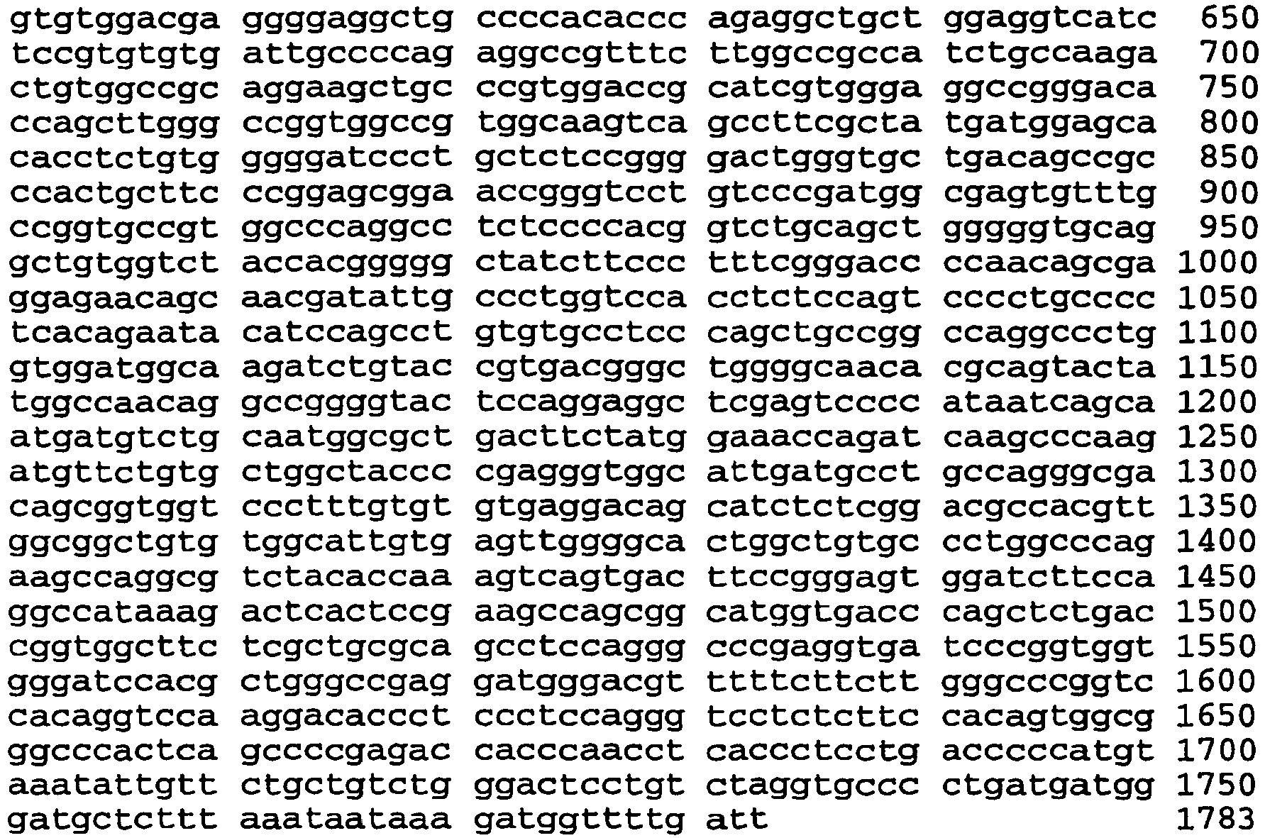
Claims (1)
- An immunogenic composition, consisting of an immunogenic fragment of hepsin protein of SEQ ID NOS. 31, 88, 89, 108, 109, 128, 129, 148, 149, 150, 151, 152, 153, or 154 and an appropriate adjuvant.
Applications Claiming Priority (3)
| Application Number | Priority Date | Filing Date | Title |
|---|---|---|---|
| US09/510,738 US6268165B1 (en) | 1997-03-19 | 2000-02-22 | Methods for the early diagnosis of ovarian cancer |
| US510738 | 2000-02-22 | ||
| PCT/US2001/005703 WO2001062271A1 (en) | 2000-02-22 | 2001-02-20 | Compositions and methods for the early diagnosis of ovarian cancer |
Publications (3)
| Publication Number | Publication Date |
|---|---|
| EP1257287A1 EP1257287A1 (en) | 2002-11-20 |
| EP1257287A4 EP1257287A4 (en) | 2005-10-12 |
| EP1257287B1 true EP1257287B1 (en) | 2012-10-17 |
Family
ID=24031976
Family Applications (1)
| Application Number | Title | Priority Date | Filing Date |
|---|---|---|---|
| EP01914444A Expired - Lifetime EP1257287B1 (en) | 2000-02-22 | 2001-02-20 | Compositions and methods for the early diagnosis of ovarian cancer |
Country Status (7)
| Country | Link |
|---|---|
| US (2) | US6268165B1 (en) |
| EP (1) | EP1257287B1 (en) |
| JP (1) | JP2003523524A (en) |
| AU (2) | AU2001239832B2 (en) |
| CA (1) | CA2400703A1 (en) |
| MX (1) | MXPA02008258A (en) |
| WO (1) | WO2001062271A1 (en) |
Families Citing this family (52)
| Publication number | Priority date | Publication date | Assignee | Title |
|---|---|---|---|---|
| US7795211B2 (en) * | 2000-02-22 | 2010-09-14 | Board Of Trustees Of The University Of Arkansas | Methods for the early diagnosis of ovarian cancer |
| AU2002213704A1 (en) * | 2000-10-27 | 2002-05-06 | Mount Sinai Hospital | Method for detecting ovarian cancer based on human kallikrein 6 (HK6) |
| JP2004526434A (en) * | 2001-01-23 | 2004-09-02 | アイアールエム・リミテッド・ライアビリティ・カンパニー | Genes overexpressed in prostate disorders as diagnostic and therapeutic targets |
| CA2438433A1 (en) * | 2001-02-14 | 2002-08-22 | Tularik Inc. | Methods for the diagnosis and treatment of tumors employing the hepsin gene |
| US7332290B2 (en) | 2001-08-02 | 2008-02-19 | The Regents Of The University Of Michigan | Dectection of AMACR cancer markers in urine |
| US7700293B2 (en) | 2001-08-02 | 2010-04-20 | The Regents Of The University Of Michigan | Expression profile of prostate cancer |
| US7229774B2 (en) * | 2001-08-02 | 2007-06-12 | Regents Of The University Of Michigan | Expression profile of prostate cancer |
| WO2003016484A2 (en) * | 2001-08-17 | 2003-02-27 | Affymetrix, Inc. | Gleason grade 4/5 prostate cancer genes |
| AU2003225540A1 (en) * | 2002-01-31 | 2003-09-02 | Irm, Llc | Hepsin substrates and prodrugs |
| US20040029151A1 (en) * | 2002-04-09 | 2004-02-12 | Affymetrix, Inc. | Molecular genetic profiling of gleason grades 3 and 4/5 prostate cancer |
| US20050272052A1 (en) * | 2002-04-09 | 2005-12-08 | Affymetrix, Inc. | Molecular genetic profiling of gleason grades 3 and 4/5 prostate cancer |
| AU2003260316A1 (en) * | 2002-07-23 | 2004-02-09 | Bayer Healthcare Ag | Regulation of human hepsin |
| GB0307428D0 (en) | 2003-03-31 | 2003-05-07 | Medical Res Council | Compartmentalised combinatorial chemistry |
| US20060078893A1 (en) | 2004-10-12 | 2006-04-13 | Medical Research Council | Compartmentalised combinatorial chemistry by microfluidic control |
| GB0307403D0 (en) | 2003-03-31 | 2003-05-07 | Medical Res Council | Selection by compartmentalised screening |
| US20050221339A1 (en) | 2004-03-31 | 2005-10-06 | Medical Research Council Harvard University | Compartmentalised screening by microfluidic control |
| US7968287B2 (en) | 2004-10-08 | 2011-06-28 | Medical Research Council Harvard University | In vitro evolution in microfluidic systems |
| EP2363205A3 (en) | 2006-01-11 | 2014-06-04 | Raindance Technologies, Inc. | Microfluidic Devices And Methods Of Use In The Formation And Control Of Nanoreactors |
| EP2530168B1 (en) | 2006-05-11 | 2015-09-16 | Raindance Technologies, Inc. | Microfluidic Devices |
| US9562837B2 (en) | 2006-05-11 | 2017-02-07 | Raindance Technologies, Inc. | Systems for handling microfludic droplets |
| ZA200810225B (en) | 2006-06-22 | 2010-03-31 | Genentech Inc | Methods and compositions for targeting hepsin |
| EP2077912B1 (en) | 2006-08-07 | 2019-03-27 | The President and Fellows of Harvard College | Fluorocarbon emulsion stabilizing surfactants |
| US8772046B2 (en) | 2007-02-06 | 2014-07-08 | Brandeis University | Manipulation of fluids and reactions in microfluidic systems |
| WO2008130623A1 (en) | 2007-04-19 | 2008-10-30 | Brandeis University | Manipulation of fluids, fluid components and reactions in microfluidic systems |
| RU2348042C1 (en) * | 2007-10-05 | 2009-02-27 | Автономная некоммерческая организация "Центр медицинских биотехнологий" | Method for diagnostics of prostate oncological diseases |
| WO2010009365A1 (en) | 2008-07-18 | 2010-01-21 | Raindance Technologies, Inc. | Droplet libraries |
| US12038438B2 (en) | 2008-07-18 | 2024-07-16 | Bio-Rad Laboratories, Inc. | Enzyme quantification |
| US8528589B2 (en) | 2009-03-23 | 2013-09-10 | Raindance Technologies, Inc. | Manipulation of microfluidic droplets |
| US10520500B2 (en) | 2009-10-09 | 2019-12-31 | Abdeslam El Harrak | Labelled silica-based nanomaterial with enhanced properties and uses thereof |
| BR112012009412A2 (en) | 2009-10-22 | 2016-11-29 | Genentech Inc | 'isolated anti-hepsin antibody, nucleic acid molecule, host cell, method of making an antibody, immunoconjugate, pharmaceutical formulation, use of the antibody and method of making an anti-serine protease antibody' |
| US8697358B2 (en) | 2009-12-15 | 2014-04-15 | Technische Universitaet Dresden | Non-invasive method for diagnosis of prostate cancer |
| EP2517025B1 (en) | 2009-12-23 | 2019-11-27 | Bio-Rad Laboratories, Inc. | Methods for reducing the exchange of molecules between droplets |
| US9399797B2 (en) | 2010-02-12 | 2016-07-26 | Raindance Technologies, Inc. | Digital analyte analysis |
| US10351905B2 (en) | 2010-02-12 | 2019-07-16 | Bio-Rad Laboratories, Inc. | Digital analyte analysis |
| JP5934657B2 (en) | 2010-02-12 | 2016-06-15 | レインダンス テクノロジーズ, インコーポレイテッド | Digital specimen analysis |
| US9366632B2 (en) | 2010-02-12 | 2016-06-14 | Raindance Technologies, Inc. | Digital analyte analysis |
| WO2011161189A1 (en) | 2010-06-24 | 2011-12-29 | F. Hoffmann-La Roche Ag | Anti-hepsin antibodies and methods of use |
| WO2012045012A2 (en) | 2010-09-30 | 2012-04-05 | Raindance Technologies, Inc. | Sandwich assays in droplets |
| EP3859011A1 (en) | 2011-02-11 | 2021-08-04 | Bio-Rad Laboratories, Inc. | Methods for forming mixed droplets |
| EP2675819B1 (en) | 2011-02-18 | 2020-04-08 | Bio-Rad Laboratories, Inc. | Compositions and methods for molecular labeling |
| DE202012013668U1 (en) | 2011-06-02 | 2019-04-18 | Raindance Technologies, Inc. | enzyme quantification |
| US8841071B2 (en) | 2011-06-02 | 2014-09-23 | Raindance Technologies, Inc. | Sample multiplexing |
| WO2012174569A2 (en) * | 2011-06-17 | 2012-12-20 | The Board Of Trustees Of The University Of Arkansas | New markers for early diagnosis of ovarian cancer, monitoring during therapy, and new therapy options during and after chematherapy |
| US8658430B2 (en) | 2011-07-20 | 2014-02-25 | Raindance Technologies, Inc. | Manipulating droplet size |
| WO2013120089A1 (en) | 2012-02-10 | 2013-08-15 | Raindance Technologies, Inc. | Molecular diagnostic screening assay |
| EP2844768B1 (en) | 2012-04-30 | 2019-03-13 | Raindance Technologies, Inc. | Digital analyte analysis |
| EP2986762B1 (en) | 2013-04-19 | 2019-11-06 | Bio-Rad Laboratories, Inc. | Digital analyte analysis |
| US11901041B2 (en) | 2013-10-04 | 2024-02-13 | Bio-Rad Laboratories, Inc. | Digital analysis of nucleic acid modification |
| US9944977B2 (en) | 2013-12-12 | 2018-04-17 | Raindance Technologies, Inc. | Distinguishing rare variations in a nucleic acid sequence from a sample |
| US11193176B2 (en) | 2013-12-31 | 2021-12-07 | Bio-Rad Laboratories, Inc. | Method for detecting and quantifying latent retroviral RNA species |
| US10647981B1 (en) | 2015-09-08 | 2020-05-12 | Bio-Rad Laboratories, Inc. | Nucleic acid library generation methods and compositions |
| US10998178B2 (en) | 2017-08-28 | 2021-05-04 | Purdue Research Foundation | Systems and methods for sample analysis using swabs |
Family Cites Families (10)
| Publication number | Priority date | Publication date | Assignee | Title |
|---|---|---|---|---|
| US3654090A (en) | 1968-09-24 | 1972-04-04 | Organon | Method for the determination of antigens and antibodies |
| NL154598B (en) | 1970-11-10 | 1977-09-15 | Organon Nv | PROCEDURE FOR DETERMINING AND DETERMINING LOW MOLECULAR COMPOUNDS AND PROTEINS THAT CAN SPECIFICALLY BIND THESE COMPOUNDS AND TEST PACKAGING. |
| US4016043A (en) | 1975-09-04 | 1977-04-05 | Akzona Incorporated | Enzymatic immunological method for the determination of antigens and antibodies |
| US4683195A (en) | 1986-01-30 | 1987-07-28 | Cetus Corporation | Process for amplifying, detecting, and/or-cloning nucleic acid sequences |
| US4683202A (en) | 1985-03-28 | 1987-07-28 | Cetus Corporation | Process for amplifying nucleic acid sequences |
| US4981784A (en) | 1987-12-02 | 1991-01-01 | The Salk Institute For Biological Studies | Retinoic acid receptor method |
| US5484726A (en) | 1990-11-21 | 1996-01-16 | Bristol-Myers Squibb Company | Antibodies specific for human stromelysin-3 and a method for detection of stromelysin-3 |
| US5804410A (en) * | 1994-07-29 | 1998-09-08 | Teijin Limited | Nucleic acid sequence encoding trypsin-like enzyme and process for producing the enzyme |
| US6294344B1 (en) * | 1997-03-19 | 2001-09-25 | The Board Of Trustees Of The University Of Arkansas | Methods for the early diagnosis of ovarian cancer |
| WO1998041656A1 (en) * | 1997-03-19 | 1998-09-24 | The Board Of Trustees Of The University Of Arkansas | Compositions and method for the early diagnosis of ovarian cancer |
-
2000
- 2000-02-22 US US09/510,738 patent/US6268165B1/en not_active Expired - Lifetime
-
2001
- 2001-02-20 MX MXPA02008258A patent/MXPA02008258A/en active IP Right Grant
- 2001-02-20 JP JP2001561336A patent/JP2003523524A/en active Pending
- 2001-02-20 AU AU2001239832A patent/AU2001239832B2/en not_active Ceased
- 2001-02-20 EP EP01914444A patent/EP1257287B1/en not_active Expired - Lifetime
- 2001-02-20 CA CA002400703A patent/CA2400703A1/en not_active Abandoned
- 2001-02-20 AU AU3983201A patent/AU3983201A/en active Pending
- 2001-02-20 WO PCT/US2001/005703 patent/WO2001062271A1/en not_active Ceased
- 2001-05-21 US US09/861,966 patent/US6518028B1/en not_active Expired - Lifetime
Also Published As
| Publication number | Publication date |
|---|---|
| JP2003523524A (en) | 2003-08-05 |
| US6268165B1 (en) | 2001-07-31 |
| MXPA02008258A (en) | 2003-05-23 |
| US6518028B1 (en) | 2003-02-11 |
| AU3983201A (en) | 2001-09-03 |
| EP1257287A4 (en) | 2005-10-12 |
| EP1257287A1 (en) | 2002-11-20 |
| CA2400703A1 (en) | 2001-08-30 |
| AU2001239832B2 (en) | 2004-09-30 |
| WO2001062271A1 (en) | 2001-08-30 |
Similar Documents
| Publication | Publication Date | Title |
|---|---|---|
| US6518028B1 (en) | Methods for the early diagnosis of ovarian and prostate cancer | |
| US6316213B1 (en) | Methods for the early diagnosis of ovarian, breast and lung cancer | |
| CA2402017C (en) | Compositions and methods for the early diagnosis of ovarian cancer | |
| AU2001239832A1 (en) | Compositions and methods for the early diagnosis of ovarian cancer | |
| US6303318B1 (en) | Methods for the early diagnosis of ovarian cancer | |
| US6294663B1 (en) | Transmembrane serine protease overexpressed in ovarian carcinoma and uses thereof | |
| US20110212023A1 (en) | TADG-15: an extracellular serine protease overexpossed in carcinomas | |
| US20100119513A1 (en) | Transmembrane serine protease overexpressed in ovarian carcinoma and uses thereof | |
| JP2003523524A5 (en) | ||
| US20030027144A1 (en) | Tumor antigen-derived gene 16 (TADG-16): a novel extracellular serine protease and uses thereof | |
| EP1887082A2 (en) | TADG-15: An extracellular serine protease overexpressed in carcinomas | |
| US7935531B2 (en) | Methods for the early diagnosis of ovarian cancer | |
| US6875609B2 (en) | Methods for the early diagnosis of ovarian cancer | |
| US6870027B2 (en) | Methods for the early diagnosis of ovarian cancer | |
| US6787354B2 (en) | Methods for the early diagnosis of ovarian cancer | |
| US7795211B2 (en) | Methods for the early diagnosis of ovarian cancer | |
| US7785841B2 (en) | Methods for the early diagnosis of ovarian cancer | |
| AU2001234910B2 (en) | Compositions and methods for the early diagnosis of ovarian cancer | |
| US20110086056A1 (en) | Methods for the early diagnosis of ovarian cancer | |
| US6627403B2 (en) | Methods for the early diagnosis of ovarian cancer | |
| US20110213337A1 (en) | Methods for the early diagnosis of ovarian cancer | |
| AU2001234910A1 (en) | Compositions and methods for the early diagnosis of ovarian cancer |
Legal Events
| Date | Code | Title | Description |
|---|---|---|---|
| PUAI | Public reference made under article 153(3) epc to a published international application that has entered the european phase |
Free format text: ORIGINAL CODE: 0009012 |
|
| 17P | Request for examination filed |
Effective date: 20020823 |
|
| AK | Designated contracting states |
Kind code of ref document: A1 Designated state(s): AT BE CH CY DE DK ES FI FR GB GR IE IT LI LU MC NL PT SE TR |
|
| AX | Request for extension of the european patent |
Free format text: AL;LT;LV;MK;RO;SI |
|
| A4 | Supplementary search report drawn up and despatched |
Effective date: 20050826 |
|
| 17Q | First examination report despatched |
Effective date: 20060828 |
|
| GRAP | Despatch of communication of intention to grant a patent |
Free format text: ORIGINAL CODE: EPIDOSNIGR1 |
|
| GRAS | Grant fee paid |
Free format text: ORIGINAL CODE: EPIDOSNIGR3 |
|
| GRAA | (expected) grant |
Free format text: ORIGINAL CODE: 0009210 |
|
| AK | Designated contracting states |
Kind code of ref document: B1 Designated state(s): AT BE CH CY DE DK ES FI FR GB GR IE IT LI LU MC NL PT SE TR |
|
| REG | Reference to a national code |
Ref country code: GB Ref legal event code: FG4D |
|
| REG | Reference to a national code |
Ref country code: CH Ref legal event code: EP |
|
| REG | Reference to a national code |
Ref country code: IE Ref legal event code: FG4D |
|
| REG | Reference to a national code |
Ref country code: AT Ref legal event code: REF Ref document number: 579577 Country of ref document: AT Kind code of ref document: T Effective date: 20121115 |
|
| REG | Reference to a national code |
Ref country code: DE Ref legal event code: R096 Ref document number: 60147244 Country of ref document: DE Effective date: 20121206 |
|
| REG | Reference to a national code |
Ref country code: AT Ref legal event code: MK05 Ref document number: 579577 Country of ref document: AT Kind code of ref document: T Effective date: 20121017 |
|
| REG | Reference to a national code |
Ref country code: NL Ref legal event code: VDEP Effective date: 20121017 |
|
| PG25 | Lapsed in a contracting state [announced via postgrant information from national office to epo] |
Ref country code: SE Free format text: LAPSE BECAUSE OF FAILURE TO SUBMIT A TRANSLATION OF THE DESCRIPTION OR TO PAY THE FEE WITHIN THE PRESCRIBED TIME-LIMIT Effective date: 20121017 Ref country code: FI Free format text: LAPSE BECAUSE OF FAILURE TO SUBMIT A TRANSLATION OF THE DESCRIPTION OR TO PAY THE FEE WITHIN THE PRESCRIBED TIME-LIMIT Effective date: 20121017 Ref country code: NL Free format text: LAPSE BECAUSE OF FAILURE TO SUBMIT A TRANSLATION OF THE DESCRIPTION OR TO PAY THE FEE WITHIN THE PRESCRIBED TIME-LIMIT Effective date: 20121017 Ref country code: ES Free format text: LAPSE BECAUSE OF FAILURE TO SUBMIT A TRANSLATION OF THE DESCRIPTION OR TO PAY THE FEE WITHIN THE PRESCRIBED TIME-LIMIT Effective date: 20130128 |
|
| PGFP | Annual fee paid to national office [announced via postgrant information from national office to epo] |
Ref country code: GB Payment date: 20130131 Year of fee payment: 13 |
|
| PG25 | Lapsed in a contracting state [announced via postgrant information from national office to epo] |
Ref country code: PT Free format text: LAPSE BECAUSE OF FAILURE TO SUBMIT A TRANSLATION OF THE DESCRIPTION OR TO PAY THE FEE WITHIN THE PRESCRIBED TIME-LIMIT Effective date: 20130218 Ref country code: BE Free format text: LAPSE BECAUSE OF FAILURE TO SUBMIT A TRANSLATION OF THE DESCRIPTION OR TO PAY THE FEE WITHIN THE PRESCRIBED TIME-LIMIT Effective date: 20121017 Ref country code: GR Free format text: LAPSE BECAUSE OF FAILURE TO SUBMIT A TRANSLATION OF THE DESCRIPTION OR TO PAY THE FEE WITHIN THE PRESCRIBED TIME-LIMIT Effective date: 20130118 Ref country code: CY Free format text: LAPSE BECAUSE OF FAILURE TO SUBMIT A TRANSLATION OF THE DESCRIPTION OR TO PAY THE FEE WITHIN THE PRESCRIBED TIME-LIMIT Effective date: 20121017 |
|
| PG25 | Lapsed in a contracting state [announced via postgrant information from national office to epo] |
Ref country code: AT Free format text: LAPSE BECAUSE OF FAILURE TO SUBMIT A TRANSLATION OF THE DESCRIPTION OR TO PAY THE FEE WITHIN THE PRESCRIBED TIME-LIMIT Effective date: 20121017 |
|
| PG25 | Lapsed in a contracting state [announced via postgrant information from national office to epo] |
Ref country code: DK Free format text: LAPSE BECAUSE OF FAILURE TO SUBMIT A TRANSLATION OF THE DESCRIPTION OR TO PAY THE FEE WITHIN THE PRESCRIBED TIME-LIMIT Effective date: 20121017 |
|
| PLBE | No opposition filed within time limit |
Free format text: ORIGINAL CODE: 0009261 |
|
| STAA | Information on the status of an ep patent application or granted ep patent |
Free format text: STATUS: NO OPPOSITION FILED WITHIN TIME LIMIT |
|
| PG25 | Lapsed in a contracting state [announced via postgrant information from national office to epo] |
Ref country code: IT Free format text: LAPSE BECAUSE OF FAILURE TO SUBMIT A TRANSLATION OF THE DESCRIPTION OR TO PAY THE FEE WITHIN THE PRESCRIBED TIME-LIMIT Effective date: 20121017 |
|
| 26N | No opposition filed |
Effective date: 20130718 |
|
| PG25 | Lapsed in a contracting state [announced via postgrant information from national office to epo] |
Ref country code: MC Free format text: LAPSE BECAUSE OF NON-PAYMENT OF DUE FEES Effective date: 20130228 |
|
| REG | Reference to a national code |
Ref country code: CH Ref legal event code: PL |
|
| PG25 | Lapsed in a contracting state [announced via postgrant information from national office to epo] |
Ref country code: CH Free format text: LAPSE BECAUSE OF NON-PAYMENT OF DUE FEES Effective date: 20130228 Ref country code: LI Free format text: LAPSE BECAUSE OF NON-PAYMENT OF DUE FEES Effective date: 20130228 |
|
| REG | Reference to a national code |
Ref country code: DE Ref legal event code: R097 Ref document number: 60147244 Country of ref document: DE Effective date: 20130718 |
|
| REG | Reference to a national code |
Ref country code: FR Ref legal event code: ST Effective date: 20131031 |
|
| REG | Reference to a national code |
Ref country code: IE Ref legal event code: MM4A |
|
| REG | Reference to a national code |
Ref country code: DE Ref legal event code: R119 Ref document number: 60147244 Country of ref document: DE Effective date: 20130903 |
|
| PG25 | Lapsed in a contracting state [announced via postgrant information from national office to epo] |
Ref country code: FR Free format text: LAPSE BECAUSE OF NON-PAYMENT OF DUE FEES Effective date: 20130228 Ref country code: IE Free format text: LAPSE BECAUSE OF NON-PAYMENT OF DUE FEES Effective date: 20130220 Ref country code: DE Free format text: LAPSE BECAUSE OF NON-PAYMENT OF DUE FEES Effective date: 20130903 |
|
| GBPC | Gb: european patent ceased through non-payment of renewal fee |
Effective date: 20140220 |
|
| PG25 | Lapsed in a contracting state [announced via postgrant information from national office to epo] |
Ref country code: GB Free format text: LAPSE BECAUSE OF NON-PAYMENT OF DUE FEES Effective date: 20140220 |
|
| PG25 | Lapsed in a contracting state [announced via postgrant information from national office to epo] |
Ref country code: TR Free format text: LAPSE BECAUSE OF FAILURE TO SUBMIT A TRANSLATION OF THE DESCRIPTION OR TO PAY THE FEE WITHIN THE PRESCRIBED TIME-LIMIT Effective date: 20121017 |
|
| PG25 | Lapsed in a contracting state [announced via postgrant information from national office to epo] |
Ref country code: LU Free format text: LAPSE BECAUSE OF NON-PAYMENT OF DUE FEES Effective date: 20130220 |