EP1254251B1 - Neurosteroids as markers for alzheimer's disease - Google Patents
Neurosteroids as markers for alzheimer's disease Download PDFInfo
- Publication number
- EP1254251B1 EP1254251B1 EP01903310A EP01903310A EP1254251B1 EP 1254251 B1 EP1254251 B1 EP 1254251B1 EP 01903310 A EP01903310 A EP 01903310A EP 01903310 A EP01903310 A EP 01903310A EP 1254251 B1 EP1254251 B1 EP 1254251B1
- Authority
- EP
- European Patent Office
- Prior art keywords
- dhea
- cells
- brain
- levels
- tissue
- Prior art date
- Legal status (The legal status is an assumption and is not a legal conclusion. Google has not performed a legal analysis and makes no representation as to the accuracy of the status listed.)
- Expired - Lifetime
Links
- 239000002858 neurotransmitter agent Substances 0.000 title claims abstract description 19
- 208000024827 Alzheimer disease Diseases 0.000 title claims description 88
- FMGSKLZLMKYGDP-USOAJAOKSA-N dehydroepiandrosterone Chemical compound C1[C@@H](O)CC[C@]2(C)[C@H]3CC[C@](C)(C(CC4)=O)[C@@H]4[C@@H]3CC=C21 FMGSKLZLMKYGDP-USOAJAOKSA-N 0.000 claims abstract description 127
- 238000000034 method Methods 0.000 claims abstract description 53
- 239000002243 precursor Substances 0.000 claims abstract description 46
- 238000011282 treatment Methods 0.000 claims abstract description 45
- 210000002966 serum Anatomy 0.000 claims abstract description 31
- 208000037265 diseases, disorders, signs and symptoms Diseases 0.000 claims abstract description 23
- 230000002503 metabolic effect Effects 0.000 claims abstract description 3
- 210000004027 cell Anatomy 0.000 claims description 153
- FMGSKLZLMKYGDP-UHFFFAOYSA-N Dehydroepiandrosterone Natural products C1C(O)CCC2(C)C3CCC(C)(C(CC4)=O)C4C3CC=C21 FMGSKLZLMKYGDP-UHFFFAOYSA-N 0.000 claims description 119
- 229960002847 prasterone Drugs 0.000 claims description 119
- 230000015572 biosynthetic process Effects 0.000 claims description 91
- BAUYGSIQEAFULO-UHFFFAOYSA-L iron(2+) sulfate (anhydrous) Chemical group [Fe+2].[O-]S([O-])(=O)=O BAUYGSIQEAFULO-UHFFFAOYSA-L 0.000 claims description 54
- 229910000359 iron(II) sulfate Inorganic materials 0.000 claims description 54
- 230000000694 effects Effects 0.000 claims description 42
- 238000003786 synthesis reaction Methods 0.000 claims description 42
- 230000001965 increasing effect Effects 0.000 claims description 38
- 210000001130 astrocyte Anatomy 0.000 claims description 36
- GVJHHUAWPYXKBD-UHFFFAOYSA-N (±)-α-Tocopherol Chemical compound OC1=C(C)C(C)=C2OC(CCCC(C)CCCC(C)CCCC(C)C)(C)CCC2=C1C GVJHHUAWPYXKBD-UHFFFAOYSA-N 0.000 claims description 32
- 210000001320 hippocampus Anatomy 0.000 claims description 30
- 210000004248 oligodendroglia Anatomy 0.000 claims description 29
- 230000036542 oxidative stress Effects 0.000 claims description 28
- 210000001519 tissue Anatomy 0.000 claims description 27
- 210000001175 cerebrospinal fluid Anatomy 0.000 claims description 24
- 239000000523 sample Substances 0.000 claims description 23
- 238000012360 testing method Methods 0.000 claims description 20
- 238000003127 radioimmunoassay Methods 0.000 claims description 18
- 229930003427 Vitamin E Natural products 0.000 claims description 16
- 210000004958 brain cell Anatomy 0.000 claims description 16
- WIGCFUFOHFEKBI-UHFFFAOYSA-N gamma-tocopherol Natural products CC(C)CCCC(C)CCCC(C)CCCC1CCC2C(C)C(O)C(C)C(C)C2O1 WIGCFUFOHFEKBI-UHFFFAOYSA-N 0.000 claims description 16
- 235000019165 vitamin E Nutrition 0.000 claims description 16
- 229940046009 vitamin E Drugs 0.000 claims description 16
- 239000011709 vitamin E Substances 0.000 claims description 16
- 239000003795 chemical substances by application Substances 0.000 claims description 13
- 230000002981 neuropathic effect Effects 0.000 claims description 13
- 230000004044 response Effects 0.000 claims description 11
- DZHSAHHDTRWUTF-SIQRNXPUSA-N amyloid-beta polypeptide 42 Chemical group C([C@@H](C(=O)N[C@@H](C)C(=O)N[C@@H](CCC(O)=O)C(=O)N[C@@H](CC(O)=O)C(=O)N[C@H](C(=O)NCC(=O)N[C@@H](CO)C(=O)N[C@@H](CC(N)=O)C(=O)N[C@@H](CCCCN)C(=O)NCC(=O)N[C@@H](C)C(=O)N[C@H](C(=O)N[C@@H]([C@@H](C)CC)C(=O)NCC(=O)N[C@@H](CC(C)C)C(=O)N[C@@H](CCSC)C(=O)N[C@@H](C(C)C)C(=O)NCC(=O)NCC(=O)N[C@@H](C(C)C)C(=O)N[C@@H](C(C)C)C(=O)N[C@@H]([C@@H](C)CC)C(=O)N[C@@H](C)C(O)=O)[C@@H](C)CC)C(C)C)NC(=O)[C@H](CC=1C=CC=CC=1)NC(=O)[C@@H](NC(=O)[C@H](CC(C)C)NC(=O)[C@H](CCCCN)NC(=O)[C@H](CCC(N)=O)NC(=O)[C@H](CC=1N=CNC=1)NC(=O)[C@H](CC=1N=CNC=1)NC(=O)[C@@H](NC(=O)[C@H](CCC(O)=O)NC(=O)[C@H](CC=1C=CC(O)=CC=1)NC(=O)CNC(=O)[C@H](CO)NC(=O)[C@H](CC(O)=O)NC(=O)[C@H](CC=1N=CNC=1)NC(=O)[C@H](CCCNC(N)=N)NC(=O)[C@H](CC=1C=CC=CC=1)NC(=O)[C@H](CCC(O)=O)NC(=O)[C@H](C)NC(=O)[C@@H](N)CC(O)=O)C(C)C)C(C)C)C1=CC=CC=C1 DZHSAHHDTRWUTF-SIQRNXPUSA-N 0.000 claims description 10
- 210000001124 body fluid Anatomy 0.000 claims description 10
- 210000003169 central nervous system Anatomy 0.000 claims description 10
- 238000011161 development Methods 0.000 claims description 10
- 239000012530 fluid Substances 0.000 claims description 10
- 238000004128 high performance liquid chromatography Methods 0.000 claims description 10
- 230000001404 mediated effect Effects 0.000 claims description 10
- 239000010839 body fluid Substances 0.000 claims description 9
- 230000003247 decreasing effect Effects 0.000 claims description 8
- 230000001590 oxidative effect Effects 0.000 claims description 8
- 239000013068 control sample Substances 0.000 claims description 6
- 239000003963 antioxidant agent Substances 0.000 claims description 5
- 235000006708 antioxidants Nutrition 0.000 claims description 5
- 210000004369 blood Anatomy 0.000 claims description 5
- 239000008280 blood Substances 0.000 claims description 5
- QVGXLLKOCUKJST-UHFFFAOYSA-N atomic oxygen Chemical compound [O] QVGXLLKOCUKJST-UHFFFAOYSA-N 0.000 claims description 4
- 238000000338 in vitro Methods 0.000 claims description 4
- 239000003960 organic solvent Substances 0.000 claims description 4
- 239000001301 oxygen Substances 0.000 claims description 4
- 229910052760 oxygen Inorganic materials 0.000 claims description 4
- 238000004587 chromatography analysis Methods 0.000 claims description 3
- 239000000463 material Substances 0.000 claims description 3
- 239000002207 metabolite Substances 0.000 claims description 3
- 238000004817 gas chromatography Methods 0.000 claims description 2
- 238000004949 mass spectrometry Methods 0.000 claims description 2
- 210000004556 brain Anatomy 0.000 abstract description 82
- 201000010099 disease Diseases 0.000 abstract description 15
- 230000007171 neuropathology Effects 0.000 abstract description 10
- 208000012902 Nervous system disease Diseases 0.000 abstract description 2
- 208000025966 Neurological disease Diseases 0.000 abstract description 2
- 238000003745 diagnosis Methods 0.000 abstract description 2
- 241000282414 Homo sapiens Species 0.000 description 47
- 230000037361 pathway Effects 0.000 description 46
- 239000003642 reactive oxygen metabolite Substances 0.000 description 42
- 150000003431 steroids Chemical class 0.000 description 37
- 210000002569 neuron Anatomy 0.000 description 21
- 108020004999 messenger RNA Proteins 0.000 description 19
- 206010018338 Glioma Diseases 0.000 description 18
- 108010090849 Amyloid beta-Peptides Proteins 0.000 description 17
- 102000013455 Amyloid beta-Peptides Human genes 0.000 description 17
- 208000032612 Glial tumor Diseases 0.000 description 17
- 210000003016 hypothalamus Anatomy 0.000 description 17
- 108090000623 proteins and genes Proteins 0.000 description 17
- RJKFOVLPORLFTN-LEKSSAKUSA-N Progesterone Chemical compound C1CC2=CC(=O)CC[C@]2(C)[C@@H]2[C@@H]1[C@@H]1CC[C@H](C(=O)C)[C@@]1(C)CC2 RJKFOVLPORLFTN-LEKSSAKUSA-N 0.000 description 16
- 102100031274 Translocator protein Human genes 0.000 description 16
- 101710166801 Translocator protein Proteins 0.000 description 16
- 230000002093 peripheral effect Effects 0.000 description 16
- 102000004169 proteins and genes Human genes 0.000 description 15
- HVYWMOMLDIMFJA-DPAQBDIFSA-N cholesterol Chemical compound C1C=C2C[C@@H](O)CC[C@]2(C)[C@@H]2[C@@H]1[C@@H]1CC[C@H]([C@H](C)CCCC(C)C)[C@@]1(C)CC2 HVYWMOMLDIMFJA-DPAQBDIFSA-N 0.000 description 14
- 230000037353 metabolic pathway Effects 0.000 description 14
- 230000007246 mechanism Effects 0.000 description 13
- 241000700159 Rattus Species 0.000 description 12
- 230000007423 decrease Effects 0.000 description 12
- 238000002474 experimental method Methods 0.000 description 12
- RTZKZFJDLAIYFH-UHFFFAOYSA-N Diethyl ether Chemical compound CCOCC RTZKZFJDLAIYFH-UHFFFAOYSA-N 0.000 description 11
- CZWCKYRVOZZJNM-UHFFFAOYSA-N Prasterone sodium sulfate Natural products C1C(OS(O)(=O)=O)CCC2(C)C3CCC(C)(C(CC4)=O)C4C3CC=C21 CZWCKYRVOZZJNM-UHFFFAOYSA-N 0.000 description 11
- 102000004190 Enzymes Human genes 0.000 description 10
- 108090000790 Enzymes Proteins 0.000 description 10
- ORNBQBCIOKFOEO-YQUGOWONSA-N Pregnenolone Natural products O=C(C)[C@@H]1[C@@]2(C)[C@H]([C@H]3[C@@H]([C@]4(C)C(=CC3)C[C@@H](O)CC4)CC2)CC1 ORNBQBCIOKFOEO-YQUGOWONSA-N 0.000 description 10
- 230000032683 aging Effects 0.000 description 10
- 230000001413 cellular effect Effects 0.000 description 10
- CZWCKYRVOZZJNM-USOAJAOKSA-N dehydroepiandrosterone sulfate Chemical compound C1[C@@H](OS(O)(=O)=O)CC[C@]2(C)[C@H]3CC[C@](C)(C(CC4)=O)[C@@H]4[C@@H]3CC=C21 CZWCKYRVOZZJNM-USOAJAOKSA-N 0.000 description 10
- 239000003814 drug Substances 0.000 description 10
- 210000005153 frontal cortex Anatomy 0.000 description 10
- 238000004519 manufacturing process Methods 0.000 description 10
- 229960000249 pregnenolone Drugs 0.000 description 10
- ORNBQBCIOKFOEO-QGVNFLHTSA-N pregnenolone Chemical compound C1C=C2C[C@@H](O)CC[C@]2(C)[C@@H]2[C@@H]1[C@@H]1CC[C@H](C(=O)C)[C@@]1(C)CC2 ORNBQBCIOKFOEO-QGVNFLHTSA-N 0.000 description 10
- XEKOWRVHYACXOJ-UHFFFAOYSA-N Ethyl acetate Chemical compound CCOC(C)=O XEKOWRVHYACXOJ-UHFFFAOYSA-N 0.000 description 9
- OKKJLVBELUTLKV-UHFFFAOYSA-N Methanol Chemical compound OC OKKJLVBELUTLKV-UHFFFAOYSA-N 0.000 description 9
- 230000033228 biological regulation Effects 0.000 description 9
- 239000003112 inhibitor Substances 0.000 description 9
- 238000005259 measurement Methods 0.000 description 9
- 102000053171 Glial Fibrillary Acidic Human genes 0.000 description 8
- 101710193519 Glial fibrillary acidic protein Proteins 0.000 description 8
- 230000018109 developmental process Effects 0.000 description 8
- 238000002405 diagnostic procedure Methods 0.000 description 8
- 210000005046 glial fibrillary acidic protein Anatomy 0.000 description 8
- 210000004498 neuroglial cell Anatomy 0.000 description 8
- 239000000186 progesterone Substances 0.000 description 8
- 229960003387 progesterone Drugs 0.000 description 8
- 238000003757 reverse transcription PCR Methods 0.000 description 8
- 235000012000 cholesterol Nutrition 0.000 description 7
- 150000001875 compounds Chemical class 0.000 description 7
- 239000000284 extract Substances 0.000 description 7
- 150000003254 radicals Chemical class 0.000 description 7
- 230000001105 regulatory effect Effects 0.000 description 7
- 231100000331 toxic Toxicity 0.000 description 7
- 230000002588 toxic effect Effects 0.000 description 7
- 238000003556 assay Methods 0.000 description 6
- 239000003550 marker Substances 0.000 description 6
- 230000008569 process Effects 0.000 description 6
- 238000004809 thin layer chromatography Methods 0.000 description 6
- 108091032973 (ribonucleotides)n+m Proteins 0.000 description 5
- 108091003079 Bovine Serum Albumin Proteins 0.000 description 5
- 206010012289 Dementia Diseases 0.000 description 5
- 241000283984 Rodentia Species 0.000 description 5
- 101100054106 Sus scrofa HSD3B gene Proteins 0.000 description 5
- 229940079593 drug Drugs 0.000 description 5
- 239000012091 fetal bovine serum Substances 0.000 description 5
- 239000012528 membrane Substances 0.000 description 5
- 230000003389 potentiating effect Effects 0.000 description 5
- 230000006950 reactive oxygen species formation Effects 0.000 description 5
- 238000012216 screening Methods 0.000 description 5
- 230000000365 steroidogenetic effect Effects 0.000 description 5
- KVJXBPDAXMEYOA-CXANFOAXSA-N trilostane Chemical compound OC1=C(C#N)C[C@]2(C)[C@H]3CC[C@](C)([C@H](CC4)O)[C@@H]4[C@@H]3CC[C@@]32O[C@@H]31 KVJXBPDAXMEYOA-CXANFOAXSA-N 0.000 description 5
- 229960001670 trilostane Drugs 0.000 description 5
- 238000004458 analytical method Methods 0.000 description 4
- 230000003078 antioxidant effect Effects 0.000 description 4
- 238000006243 chemical reaction Methods 0.000 description 4
- 208000014674 injury Diseases 0.000 description 4
- 230000014759 maintenance of location Effects 0.000 description 4
- 238000001543 one-way ANOVA Methods 0.000 description 4
- 239000000047 product Substances 0.000 description 4
- 239000004017 serum-free culture medium Substances 0.000 description 4
- 208000014644 Brain disease Diseases 0.000 description 3
- 108020004414 DNA Proteins 0.000 description 3
- LFQSCWFLJHTTHZ-UHFFFAOYSA-N Ethanol Chemical compound CCO LFQSCWFLJHTTHZ-UHFFFAOYSA-N 0.000 description 3
- HOKKHZGPKSLGJE-GSVOUGTGSA-N N-Methyl-D-aspartic acid Chemical compound CN[C@@H](C(O)=O)CC(O)=O HOKKHZGPKSLGJE-GSVOUGTGSA-N 0.000 description 3
- 108010029485 Protein Isoforms Proteins 0.000 description 3
- 102000001708 Protein Isoforms Human genes 0.000 description 3
- AJLFOPYRIVGYMJ-UHFFFAOYSA-N SJ000287055 Natural products C12C(OC(=O)C(C)CC)CCC=C2C=CC(C)C1CCC1CC(O)CC(=O)O1 AJLFOPYRIVGYMJ-UHFFFAOYSA-N 0.000 description 3
- 208000027418 Wounds and injury Diseases 0.000 description 3
- 230000001919 adrenal effect Effects 0.000 description 3
- SHGAZHPCJJPHSC-YCNIQYBTSA-N all-trans-retinoic acid Chemical compound OC(=O)\C=C(/C)\C=C\C=C(/C)\C=C\C1=C(C)CCCC1(C)C SHGAZHPCJJPHSC-YCNIQYBTSA-N 0.000 description 3
- 230000004075 alteration Effects 0.000 description 3
- 150000001413 amino acids Chemical class 0.000 description 3
- 230000006378 damage Effects 0.000 description 3
- 238000013461 design Methods 0.000 description 3
- 238000010790 dilution Methods 0.000 description 3
- 239000012895 dilution Substances 0.000 description 3
- 208000035475 disorder Diseases 0.000 description 3
- 230000002255 enzymatic effect Effects 0.000 description 3
- 229940093499 ethyl acetate Drugs 0.000 description 3
- 235000019439 ethyl acetate Nutrition 0.000 description 3
- 238000002290 gas chromatography-mass spectrometry Methods 0.000 description 3
- 230000003834 intracellular effect Effects 0.000 description 3
- AJLFOPYRIVGYMJ-INTXDZFKSA-N mevastatin Chemical compound C([C@H]1[C@@H](C)C=CC2=CCC[C@@H]([C@H]12)OC(=O)[C@@H](C)CC)C[C@@H]1C[C@@H](O)CC(=O)O1 AJLFOPYRIVGYMJ-INTXDZFKSA-N 0.000 description 3
- 229950009116 mevastatin Drugs 0.000 description 3
- BOZILQFLQYBIIY-UHFFFAOYSA-N mevastatin hydroxy acid Natural products C1=CC(C)C(CCC(O)CC(O)CC(O)=O)C2C(OC(=O)C(C)CC)CCC=C21 BOZILQFLQYBIIY-UHFFFAOYSA-N 0.000 description 3
- 230000004770 neurodegeneration Effects 0.000 description 3
- 230000000324 neuroprotective effect Effects 0.000 description 3
- 229950009829 prasterone sulfate Drugs 0.000 description 3
- 230000001681 protective effect Effects 0.000 description 3
- 238000000746 purification Methods 0.000 description 3
- 229930002330 retinoic acid Natural products 0.000 description 3
- 241000894007 species Species 0.000 description 3
- 239000000758 substrate Substances 0.000 description 3
- 239000013589 supplement Substances 0.000 description 3
- 230000002194 synthesizing effect Effects 0.000 description 3
- 229960001727 tretinoin Drugs 0.000 description 3
- 102000000563 2',3'-Cyclic Nucleotide 3'-Phosphodiesterase Human genes 0.000 description 2
- 108010041801 2',3'-Cyclic Nucleotide 3'-Phosphodiesterase Proteins 0.000 description 2
- APKFDSVGJQXUKY-KKGHZKTASA-N Amphotericin-B Natural products O[C@H]1[C@@H](N)[C@H](O)[C@@H](C)O[C@H]1O[C@H]1C=CC=CC=CC=CC=CC=CC=C[C@H](C)[C@@H](O)[C@@H](C)[C@H](C)OC(=O)C[C@H](O)C[C@H](O)CC[C@@H](O)[C@H](O)C[C@H](O)C[C@](O)(C[C@H](O)[C@H]2C(O)=O)O[C@H]2C1 APKFDSVGJQXUKY-KKGHZKTASA-N 0.000 description 2
- IJGRMHOSHXDMSA-UHFFFAOYSA-N Atomic nitrogen Chemical compound N#N IJGRMHOSHXDMSA-UHFFFAOYSA-N 0.000 description 2
- 208000003174 Brain Neoplasms Diseases 0.000 description 2
- 201000010915 Glioblastoma multiforme Diseases 0.000 description 2
- WZUVPPKBWHMQCE-UHFFFAOYSA-N Haematoxylin Chemical compound C12=CC(O)=C(O)C=C2CC2(O)C1C1=CC=C(O)C(O)=C1OC2 WZUVPPKBWHMQCE-UHFFFAOYSA-N 0.000 description 2
- MHAJPDPJQMAIIY-UHFFFAOYSA-N Hydrogen peroxide Chemical compound OO MHAJPDPJQMAIIY-UHFFFAOYSA-N 0.000 description 2
- 102000004286 Hydroxymethylglutaryl CoA Reductases Human genes 0.000 description 2
- 108090000895 Hydroxymethylglutaryl CoA Reductases Proteins 0.000 description 2
- 206010028980 Neoplasm Diseases 0.000 description 2
- 208000018737 Parkinson disease Diseases 0.000 description 2
- 229940123934 Reductase inhibitor Drugs 0.000 description 2
- 108010015330 Steroid 17-alpha-Hydroxylase Proteins 0.000 description 2
- 102000001854 Steroid 17-alpha-Hydroxylase Human genes 0.000 description 2
- 208000030886 Traumatic Brain injury Diseases 0.000 description 2
- 230000009471 action Effects 0.000 description 2
- 230000004913 activation Effects 0.000 description 2
- APKFDSVGJQXUKY-INPOYWNPSA-N amphotericin B Chemical compound O[C@H]1[C@@H](N)[C@H](O)[C@@H](C)O[C@H]1O[C@H]1/C=C/C=C/C=C/C=C/C=C/C=C/C=C/[C@H](C)[C@@H](O)[C@@H](C)[C@H](C)OC(=O)C[C@H](O)C[C@H](O)CC[C@@H](O)[C@H](O)C[C@H](O)C[C@](O)(C[C@H](O)[C@H]2C(O)=O)O[C@H]2C1 APKFDSVGJQXUKY-INPOYWNPSA-N 0.000 description 2
- 229960003942 amphotericin b Drugs 0.000 description 2
- 230000008901 benefit Effects 0.000 description 2
- 230000001851 biosynthetic effect Effects 0.000 description 2
- 230000000903 blocking effect Effects 0.000 description 2
- 210000005013 brain tissue Anatomy 0.000 description 2
- 230000015556 catabolic process Effects 0.000 description 2
- 230000008859 change Effects 0.000 description 2
- 238000012512 characterization method Methods 0.000 description 2
- 239000003153 chemical reaction reagent Substances 0.000 description 2
- 239000003638 chemical reducing agent Substances 0.000 description 2
- 238000011278 co-treatment Methods 0.000 description 2
- 230000001149 cognitive effect Effects 0.000 description 2
- 230000003920 cognitive function Effects 0.000 description 2
- 230000000875 corresponding effect Effects 0.000 description 2
- 230000004069 differentiation Effects 0.000 description 2
- 230000006870 function Effects 0.000 description 2
- 208000005017 glioblastoma Diseases 0.000 description 2
- 210000002149 gonad Anatomy 0.000 description 2
- 239000003102 growth factor Substances 0.000 description 2
- 230000000971 hippocampal effect Effects 0.000 description 2
- 230000013632 homeostatic process Effects 0.000 description 2
- 238000003119 immunoblot Methods 0.000 description 2
- 238000003365 immunocytochemistry Methods 0.000 description 2
- 230000006698 induction Effects 0.000 description 2
- 230000002401 inhibitory effect Effects 0.000 description 2
- NOESYZHRGYRDHS-UHFFFAOYSA-N insulin Chemical compound N1C(=O)C(NC(=O)C(CCC(N)=O)NC(=O)C(CCC(O)=O)NC(=O)C(C(C)C)NC(=O)C(NC(=O)CN)C(C)CC)CSSCC(C(NC(CO)C(=O)NC(CC(C)C)C(=O)NC(CC=2C=CC(O)=CC=2)C(=O)NC(CCC(N)=O)C(=O)NC(CC(C)C)C(=O)NC(CCC(O)=O)C(=O)NC(CC(N)=O)C(=O)NC(CC=2C=CC(O)=CC=2)C(=O)NC(CSSCC(NC(=O)C(C(C)C)NC(=O)C(CC(C)C)NC(=O)C(CC=2C=CC(O)=CC=2)NC(=O)C(CC(C)C)NC(=O)C(C)NC(=O)C(CCC(O)=O)NC(=O)C(C(C)C)NC(=O)C(CC(C)C)NC(=O)C(CC=2NC=NC=2)NC(=O)C(CO)NC(=O)CNC2=O)C(=O)NCC(=O)NC(CCC(O)=O)C(=O)NC(CCCNC(N)=N)C(=O)NCC(=O)NC(CC=3C=CC=CC=3)C(=O)NC(CC=3C=CC=CC=3)C(=O)NC(CC=3C=CC(O)=CC=3)C(=O)NC(C(C)O)C(=O)N3C(CCC3)C(=O)NC(CCCCN)C(=O)NC(C)C(O)=O)C(=O)NC(CC(N)=O)C(O)=O)=O)NC(=O)C(C(C)CC)NC(=O)C(CO)NC(=O)C(C(C)O)NC(=O)C1CSSCC2NC(=O)C(CC(C)C)NC(=O)C(NC(=O)C(CCC(N)=O)NC(=O)C(CC(N)=O)NC(=O)C(NC(=O)C(N)CC=1C=CC=CC=1)C(C)C)CC1=CN=CN1 NOESYZHRGYRDHS-UHFFFAOYSA-N 0.000 description 2
- 239000007788 liquid Substances 0.000 description 2
- 230000004060 metabolic process Effects 0.000 description 2
- 210000003470 mitochondria Anatomy 0.000 description 2
- 230000001537 neural effect Effects 0.000 description 2
- 239000007800 oxidant agent Substances 0.000 description 2
- 239000008194 pharmaceutical composition Substances 0.000 description 2
- 230000007505 plaque formation Effects 0.000 description 2
- 108090000765 processed proteins & peptides Proteins 0.000 description 2
- 238000011160 research Methods 0.000 description 2
- 230000002441 reversible effect Effects 0.000 description 2
- 230000035945 sensitivity Effects 0.000 description 2
- 239000002904 solvent Substances 0.000 description 2
- UCSJYZPVAKXKNQ-HZYVHMACSA-N streptomycin Chemical compound CN[C@H]1[C@H](O)[C@@H](O)[C@H](CO)O[C@H]1O[C@@H]1[C@](C=O)(O)[C@H](C)O[C@H]1O[C@@H]1[C@@H](NC(N)=N)[C@H](O)[C@@H](NC(N)=N)[C@H](O)[C@H]1O UCSJYZPVAKXKNQ-HZYVHMACSA-N 0.000 description 2
- 208000001608 teratocarcinoma Diseases 0.000 description 2
- 230000009529 traumatic brain injury Effects 0.000 description 2
- 210000004881 tumor cell Anatomy 0.000 description 2
- 230000003827 upregulation Effects 0.000 description 2
- VQVUBYASAICPFU-UHFFFAOYSA-N (6'-acetyloxy-2',7'-dichloro-3-oxospiro[2-benzofuran-1,9'-xanthene]-3'-yl) acetate Chemical compound O1C(=O)C2=CC=CC=C2C21C1=CC(Cl)=C(OC(C)=O)C=C1OC1=C2C=C(Cl)C(OC(=O)C)=C1 VQVUBYASAICPFU-UHFFFAOYSA-N 0.000 description 1
- RTHCYVBBDHJXIQ-MRXNPFEDSA-N (R)-fluoxetine Chemical compound O([C@H](CCNC)C=1C=CC=CC=1)C1=CC=C(C(F)(F)F)C=C1 RTHCYVBBDHJXIQ-MRXNPFEDSA-N 0.000 description 1
- GLDQAMYCGOIJDV-UHFFFAOYSA-N 2,3-dihydroxybenzoic acid Chemical compound OC(=O)C1=CC=CC(O)=C1O GLDQAMYCGOIJDV-UHFFFAOYSA-N 0.000 description 1
- FTOAOBMCPZCFFF-UHFFFAOYSA-N 5,5-diethylbarbituric acid Chemical compound CCC1(CC)C(=O)NC(=O)NC1=O FTOAOBMCPZCFFF-UHFFFAOYSA-N 0.000 description 1
- OXEUETBFKVCRNP-UHFFFAOYSA-N 9-ethyl-3-carbazolamine Chemical compound NC1=CC=C2N(CC)C3=CC=CC=C3C2=C1 OXEUETBFKVCRNP-UHFFFAOYSA-N 0.000 description 1
- 208000037259 Amyloid Plaque Diseases 0.000 description 1
- 208000019901 Anxiety disease Diseases 0.000 description 1
- 206010003594 Ataxia telangiectasia Diseases 0.000 description 1
- 206010006187 Breast cancer Diseases 0.000 description 1
- 208000026310 Breast neoplasm Diseases 0.000 description 1
- 102000000905 Cadherin Human genes 0.000 description 1
- 108050007957 Cadherin Proteins 0.000 description 1
- 102000005598 Chondroitin Sulfate Proteoglycans Human genes 0.000 description 1
- 108010059480 Chondroitin Sulfate Proteoglycans Proteins 0.000 description 1
- 238000009007 Diagnostic Kit Methods 0.000 description 1
- MYMOFIZGZYHOMD-UHFFFAOYSA-N Dioxygen Chemical compound O=O MYMOFIZGZYHOMD-UHFFFAOYSA-N 0.000 description 1
- 206010061818 Disease progression Diseases 0.000 description 1
- 102000010834 Extracellular Matrix Proteins Human genes 0.000 description 1
- 108010037362 Extracellular Matrix Proteins Proteins 0.000 description 1
- 201000011240 Frontotemporal dementia Diseases 0.000 description 1
- 108091008681 GABAA receptors Proteins 0.000 description 1
- 102000027484 GABAA receptors Human genes 0.000 description 1
- 229930182566 Gentamicin Natural products 0.000 description 1
- CEAZRRDELHUEMR-URQXQFDESA-N Gentamicin Chemical compound O1[C@H](C(C)NC)CC[C@@H](N)[C@H]1O[C@H]1[C@H](O)[C@@H](O[C@@H]2[C@@H]([C@@H](NC)[C@@](C)(O)CO2)O)[C@H](N)C[C@@H]1N CEAZRRDELHUEMR-URQXQFDESA-N 0.000 description 1
- 208000010055 Globoid Cell Leukodystrophy Diseases 0.000 description 1
- 102000018899 Glutamate Receptors Human genes 0.000 description 1
- 108010027915 Glutamate Receptors Proteins 0.000 description 1
- 241001559547 Hippocampus comes Species 0.000 description 1
- 241000282412 Homo Species 0.000 description 1
- 101500025419 Homo sapiens Epidermal growth factor Proteins 0.000 description 1
- 101000934372 Homo sapiens Macrosialin Proteins 0.000 description 1
- 208000023105 Huntington disease Diseases 0.000 description 1
- DGAQECJNVWCQMB-PUAWFVPOSA-M Ilexoside XXIX Chemical compound C[C@@H]1CC[C@@]2(CC[C@@]3(C(=CC[C@H]4[C@]3(CC[C@@H]5[C@@]4(CC[C@@H](C5(C)C)OS(=O)(=O)[O-])C)C)[C@@H]2[C@]1(C)O)C)C(=O)O[C@H]6[C@@H]([C@H]([C@@H]([C@H](O6)CO)O)O)O.[Na+] DGAQECJNVWCQMB-PUAWFVPOSA-M 0.000 description 1
- 102000004877 Insulin Human genes 0.000 description 1
- 108090001061 Insulin Proteins 0.000 description 1
- 102100034343 Integrase Human genes 0.000 description 1
- 229910021578 Iron(III) chloride Inorganic materials 0.000 description 1
- 208000028226 Krabbe disease Diseases 0.000 description 1
- WHUUTDBJXJRKMK-VKHMYHEASA-N L-glutamic acid Chemical compound OC(=O)[C@@H](N)CCC(O)=O WHUUTDBJXJRKMK-VKHMYHEASA-N 0.000 description 1
- 208000006136 Leigh Disease Diseases 0.000 description 1
- 208000017507 Leigh syndrome Diseases 0.000 description 1
- 102100025136 Macrosialin Human genes 0.000 description 1
- 102000006386 Myelin Proteins Human genes 0.000 description 1
- 108010083674 Myelin Proteins Proteins 0.000 description 1
- 102100026784 Myelin proteolipid protein Human genes 0.000 description 1
- 108090001041 N-Methyl-D-Aspartate Receptors Proteins 0.000 description 1
- 102000004868 N-Methyl-D-Aspartate Receptors Human genes 0.000 description 1
- 108050000637 N-cadherin Proteins 0.000 description 1
- 208000001738 Nervous System Trauma Diseases 0.000 description 1
- 108010069196 Neural Cell Adhesion Molecules Proteins 0.000 description 1
- 102000001068 Neural Cell Adhesion Molecules Human genes 0.000 description 1
- 102000008763 Neurofilament Proteins Human genes 0.000 description 1
- 108010088373 Neurofilament Proteins Proteins 0.000 description 1
- 239000000020 Nitrocellulose Substances 0.000 description 1
- 239000004677 Nylon Substances 0.000 description 1
- 208000017493 Pelizaeus-Merzbacher disease Diseases 0.000 description 1
- 229930182555 Penicillin Natural products 0.000 description 1
- JGSARLDLIJGVTE-MBNYWOFBSA-N Penicillin G Chemical compound N([C@H]1[C@H]2SC([C@@H](N2C1=O)C(O)=O)(C)C)C(=O)CC1=CC=CC=C1 JGSARLDLIJGVTE-MBNYWOFBSA-N 0.000 description 1
- 102000003992 Peroxidases Human genes 0.000 description 1
- 208000000609 Pick Disease of the Brain Diseases 0.000 description 1
- 206010036618 Premenstrual syndrome Diseases 0.000 description 1
- 238000002123 RNA extraction Methods 0.000 description 1
- 108010092799 RNA-directed DNA polymerase Proteins 0.000 description 1
- 101100496572 Rattus norvegicus C6 gene Proteins 0.000 description 1
- BQCADISMDOOEFD-UHFFFAOYSA-N Silver Chemical compound [Ag] BQCADISMDOOEFD-UHFFFAOYSA-N 0.000 description 1
- 238000002105 Southern blotting Methods 0.000 description 1
- 208000006011 Stroke Diseases 0.000 description 1
- 229930006000 Sucrose Natural products 0.000 description 1
- CZMRCDWAGMRECN-UGDNZRGBSA-N Sucrose Chemical compound O[C@H]1[C@H](O)[C@@H](CO)O[C@@]1(CO)O[C@@H]1[C@H](O)[C@@H](O)[C@H](O)[C@@H](CO)O1 CZMRCDWAGMRECN-UGDNZRGBSA-N 0.000 description 1
- 102000004338 Transferrin Human genes 0.000 description 1
- 108090000901 Transferrin Proteins 0.000 description 1
- 201000004810 Vascular dementia Diseases 0.000 description 1
- 206010063661 Vascular encephalopathy Diseases 0.000 description 1
- 238000001790 Welch's t-test Methods 0.000 description 1
- 230000001154 acute effect Effects 0.000 description 1
- 239000003470 adrenal cortex hormone Substances 0.000 description 1
- 210000004100 adrenal gland Anatomy 0.000 description 1
- 239000011543 agarose gel Substances 0.000 description 1
- 230000002776 aggregation Effects 0.000 description 1
- 238000004220 aggregation Methods 0.000 description 1
- 230000007792 alzheimer disease pathology Effects 0.000 description 1
- 230000003321 amplification Effects 0.000 description 1
- 108010064539 amyloid beta-protein (1-42) Proteins 0.000 description 1
- 206010002022 amyloidosis Diseases 0.000 description 1
- 239000003098 androgen Substances 0.000 description 1
- 229940030486 androgens Drugs 0.000 description 1
- 230000036506 anxiety Effects 0.000 description 1
- 230000003140 astrocytic effect Effects 0.000 description 1
- 230000005540 biological transmission Effects 0.000 description 1
- 238000001574 biopsy Methods 0.000 description 1
- 230000006696 biosynthetic metabolic pathway Effects 0.000 description 1
- 229940098773 bovine serum albumin Drugs 0.000 description 1
- 230000003925 brain function Effects 0.000 description 1
- 244000309466 calf Species 0.000 description 1
- 125000002915 carbonyl group Chemical group [*:2]C([*:1])=O 0.000 description 1
- 238000004113 cell culture Methods 0.000 description 1
- 210000004671 cell-free system Anatomy 0.000 description 1
- 230000033077 cellular process Effects 0.000 description 1
- 210000001638 cerebellum Anatomy 0.000 description 1
- 208000013677 cerebrovascular dementia Diseases 0.000 description 1
- YDQXYRCYDMRJGD-UHFFFAOYSA-N chloroform;phenol;thiocyanic acid Chemical compound SC#N.ClC(Cl)Cl.OC1=CC=CC=C1 YDQXYRCYDMRJGD-UHFFFAOYSA-N 0.000 description 1
- 230000003930 cognitive ability Effects 0.000 description 1
- 230000003931 cognitive performance Effects 0.000 description 1
- 230000000052 comparative effect Effects 0.000 description 1
- 239000003636 conditioned culture medium Substances 0.000 description 1
- 230000008094 contradictory effect Effects 0.000 description 1
- 230000002596 correlated effect Effects 0.000 description 1
- 210000000805 cytoplasm Anatomy 0.000 description 1
- 230000003436 cytoskeletal effect Effects 0.000 description 1
- 230000002950 deficient Effects 0.000 description 1
- 238000006731 degradation reaction Methods 0.000 description 1
- 230000008021 deposition Effects 0.000 description 1
- 238000001514 detection method Methods 0.000 description 1
- 230000005750 disease progression Effects 0.000 description 1
- 238000009826 distribution Methods 0.000 description 1
- 239000000975 dye Substances 0.000 description 1
- 230000002124 endocrine Effects 0.000 description 1
- 230000037149 energy metabolism Effects 0.000 description 1
- 238000005516 engineering process Methods 0.000 description 1
- 230000009088 enzymatic function Effects 0.000 description 1
- 230000009483 enzymatic pathway Effects 0.000 description 1
- 239000002532 enzyme inhibitor Substances 0.000 description 1
- 206010015037 epilepsy Diseases 0.000 description 1
- 125000001033 ether group Chemical group 0.000 description 1
- 210000002744 extracellular matrix Anatomy 0.000 description 1
- 239000007850 fluorescent dye Substances 0.000 description 1
- 229960002464 fluoxetine Drugs 0.000 description 1
- 229960004038 fluvoxamine Drugs 0.000 description 1
- CJOFXWAVKWHTFT-XSFVSMFZSA-N fluvoxamine Chemical compound COCCCC\C(=N/OCCN)C1=CC=C(C(F)(F)F)C=C1 CJOFXWAVKWHTFT-XSFVSMFZSA-N 0.000 description 1
- 239000012634 fragment Substances 0.000 description 1
- 229960003692 gamma aminobutyric acid Drugs 0.000 description 1
- BTCSSZJGUNDROE-UHFFFAOYSA-N gamma-aminobutyric acid Chemical compound NCCCC(O)=O BTCSSZJGUNDROE-UHFFFAOYSA-N 0.000 description 1
- 229960002518 gentamicin Drugs 0.000 description 1
- 239000003862 glucocorticoid Substances 0.000 description 1
- 229930195712 glutamate Natural products 0.000 description 1
- PCHJSUWPFVWCPO-UHFFFAOYSA-N gold Chemical compound [Au] PCHJSUWPFVWCPO-UHFFFAOYSA-N 0.000 description 1
- 239000010931 gold Substances 0.000 description 1
- 229910052737 gold Inorganic materials 0.000 description 1
- 239000011544 gradient gel Substances 0.000 description 1
- 239000001963 growth medium Substances 0.000 description 1
- 229940116978 human epidermal growth factor Drugs 0.000 description 1
- 230000001433 inducive effect Effects 0.000 description 1
- 230000005764 inhibitory process Effects 0.000 description 1
- 229940125396 insulin Drugs 0.000 description 1
- 239000000543 intermediate Substances 0.000 description 1
- RBTARNINKXHZNM-UHFFFAOYSA-K iron trichloride Chemical compound Cl[Fe](Cl)Cl RBTARNINKXHZNM-UHFFFAOYSA-K 0.000 description 1
- 238000002372 labelling Methods 0.000 description 1
- 201000010901 lateral sclerosis Diseases 0.000 description 1
- 210000002332 leydig cell Anatomy 0.000 description 1
- 239000003446 ligand Substances 0.000 description 1
- 230000003859 lipid peroxidation Effects 0.000 description 1
- 238000002595 magnetic resonance imaging Methods 0.000 description 1
- 208000024714 major depressive disease Diseases 0.000 description 1
- 230000006386 memory function Effects 0.000 description 1
- 230000003340 mental effect Effects 0.000 description 1
- 230000002025 microglial effect Effects 0.000 description 1
- 208000012268 mitochondrial disease Diseases 0.000 description 1
- 230000025608 mitochondrion localization Effects 0.000 description 1
- 201000004058 mixed glioma Diseases 0.000 description 1
- 230000004048 modification Effects 0.000 description 1
- 238000012986 modification Methods 0.000 description 1
- 239000003068 molecular probe Substances 0.000 description 1
- 208000005264 motor neuron disease Diseases 0.000 description 1
- 210000005012 myelin Anatomy 0.000 description 1
- 239000013642 negative control Substances 0.000 description 1
- GVUGOAYIVIDWIO-UFWWTJHBSA-N nepidermin Chemical compound C([C@@H](C(=O)N[C@@H]([C@@H](C)CC)C(=O)NCC(=O)N[C@@H](CCC(O)=O)C(=O)N[C@@H](CCCNC(N)=N)C(=O)N[C@@H](CS)C(=O)N[C@@H](CCC(N)=O)C(=O)N[C@@H](CC=1C=CC(O)=CC=1)C(=O)N[C@@H](CCCNC(N)=N)C(=O)N[C@@H](CC(O)=O)C(=O)N[C@@H](CC(C)C)C(=O)N[C@@H](CCCCN)C(=O)N[C@@H](CC=1C2=CC=CC=C2NC=1)C(=O)N[C@@H](CC=1C2=CC=CC=C2NC=1)C(=O)N[C@@H](CCC(O)=O)C(=O)N[C@@H](CC(C)C)C(=O)N[C@@H](CCCNC(N)=N)C(O)=O)NC(=O)CNC(=O)[C@@H](NC(=O)[C@@H](NC(=O)[C@H](CS)NC(=O)[C@H](CC(N)=O)NC(=O)[C@H](CS)NC(=O)[C@H](C)NC(=O)[C@H](CC=1C=CC(O)=CC=1)NC(=O)[C@H](CCCCN)NC(=O)[C@H](CC(O)=O)NC(=O)[C@H](CC(C)C)NC(=O)[C@H](C)NC(=O)[C@H](CCC(O)=O)NC(=O)[C@@H](NC(=O)[C@H](CC=1C=CC(O)=CC=1)NC(=O)[C@H](CCSC)NC(=O)[C@H](CS)NC(=O)[C@@H](NC(=O)CNC(=O)[C@H](CC(O)=O)NC(=O)[C@H](CC=1NC=NC=1)NC(=O)[C@H](CC(C)C)NC(=O)[C@H](CS)NC(=O)[C@H](CC=1C=CC(O)=CC=1)NC(=O)CNC(=O)[C@H](CC(O)=O)NC(=O)[C@H](CC=1NC=NC=1)NC(=O)[C@H](CO)NC(=O)[C@H](CC(C)C)NC(=O)[C@H]1N(CCC1)C(=O)[C@H](CS)NC(=O)[C@H](CCC(O)=O)NC(=O)[C@H](CO)NC(=O)[C@H](CC(O)=O)NC(=O)[C@H](CO)NC(=O)[C@@H](N)CC(N)=O)C(C)C)[C@@H](C)CC)C(C)C)C(C)C)C1=CC=C(O)C=C1 GVUGOAYIVIDWIO-UFWWTJHBSA-N 0.000 description 1
- 208000015122 neurodegenerative disease Diseases 0.000 description 1
- 210000002682 neurofibrillary tangle Anatomy 0.000 description 1
- 230000008587 neuronal excitability Effects 0.000 description 1
- 230000003961 neuronal insult Effects 0.000 description 1
- 239000002581 neurotoxin Substances 0.000 description 1
- 231100000618 neurotoxin Toxicity 0.000 description 1
- 239000002547 new drug Substances 0.000 description 1
- 229920001220 nitrocellulos Polymers 0.000 description 1
- 229910052757 nitrogen Inorganic materials 0.000 description 1
- 238000001422 normality test Methods 0.000 description 1
- 238000003199 nucleic acid amplification method Methods 0.000 description 1
- 230000030648 nucleus localization Effects 0.000 description 1
- 229920001778 nylon Polymers 0.000 description 1
- 210000000056 organ Anatomy 0.000 description 1
- 230000008506 pathogenesis Effects 0.000 description 1
- 230000001575 pathological effect Effects 0.000 description 1
- 230000007170 pathology Effects 0.000 description 1
- 239000013610 patient sample Substances 0.000 description 1
- 229940049954 penicillin Drugs 0.000 description 1
- 210000001428 peripheral nervous system Anatomy 0.000 description 1
- 108040007629 peroxidase activity proteins Proteins 0.000 description 1
- 238000005502 peroxidation Methods 0.000 description 1
- 238000002205 phenol-chloroform extraction Methods 0.000 description 1
- 230000008288 physiological mechanism Effects 0.000 description 1
- 210000002826 placenta Anatomy 0.000 description 1
- 239000011148 porous material Substances 0.000 description 1
- 239000013641 positive control Substances 0.000 description 1
- DIJBBUIOWGGQOP-QGVNFLHTSA-N pregnenolone sulfate Chemical compound C1C=C2C[C@@H](OS(O)(=O)=O)CC[C@]2(C)[C@@H]2[C@@H]1[C@@H]1CC[C@H](C(=O)C)[C@@]1(C)CC2 DIJBBUIOWGGQOP-QGVNFLHTSA-N 0.000 description 1
- 230000004952 protein activity Effects 0.000 description 1
- 238000002731 protein assay Methods 0.000 description 1
- 238000000159 protein binding assay Methods 0.000 description 1
- 238000011552 rat model Methods 0.000 description 1
- 239000000376 reactant Substances 0.000 description 1
- 230000009467 reduction Effects 0.000 description 1
- 230000008844 regulatory mechanism Effects 0.000 description 1
- 230000008521 reorganization Effects 0.000 description 1
- 230000011506 response to oxidative stress Effects 0.000 description 1
- 238000012552 review Methods 0.000 description 1
- 230000000698 schizophrenic effect Effects 0.000 description 1
- 238000007423 screening assay Methods 0.000 description 1
- 238000000926 separation method Methods 0.000 description 1
- 108010085082 sigma receptors Proteins 0.000 description 1
- 229910052709 silver Inorganic materials 0.000 description 1
- 239000004332 silver Substances 0.000 description 1
- 229910052708 sodium Inorganic materials 0.000 description 1
- 239000011734 sodium Substances 0.000 description 1
- 238000002415 sodium dodecyl sulfate polyacrylamide gel electrophoresis Methods 0.000 description 1
- 238000004611 spectroscopical analysis Methods 0.000 description 1
- 210000000278 spinal cord Anatomy 0.000 description 1
- 208000020431 spinal cord injury Diseases 0.000 description 1
- 238000007619 statistical method Methods 0.000 description 1
- 230000010009 steroidogenesis Effects 0.000 description 1
- 229960005322 streptomycin Drugs 0.000 description 1
- 239000005720 sucrose Substances 0.000 description 1
- 230000008093 supporting effect Effects 0.000 description 1
- 208000024891 symptom Diseases 0.000 description 1
- 229940037128 systemic glucocorticoids Drugs 0.000 description 1
- 230000002381 testicular Effects 0.000 description 1
- CFMYXEVWODSLAX-QOZOJKKESA-N tetrodotoxin Chemical compound O([C@@]([C@H]1O)(O)O[C@H]2[C@@]3(O)CO)[C@H]3[C@@H](O)[C@]11[C@H]2[C@@H](O)N=C(N)N1 CFMYXEVWODSLAX-QOZOJKKESA-N 0.000 description 1
- 229950010357 tetrodotoxin Drugs 0.000 description 1
- CFMYXEVWODSLAX-UHFFFAOYSA-N tetrodotoxin Natural products C12C(O)NC(=N)NC2(C2O)C(O)C3C(CO)(O)C1OC2(O)O3 CFMYXEVWODSLAX-UHFFFAOYSA-N 0.000 description 1
- 231100000419 toxicity Toxicity 0.000 description 1
- 230000001988 toxicity Effects 0.000 description 1
- 230000026683 transduction Effects 0.000 description 1
- 238000010361 transduction Methods 0.000 description 1
- 239000012581 transferrin Substances 0.000 description 1
- 238000013519 translation Methods 0.000 description 1
- 230000032258 transport Effects 0.000 description 1
- 230000008733 trauma Effects 0.000 description 1
- 230000001960 triggered effect Effects 0.000 description 1
- 238000012762 unpaired Student’s t-test Methods 0.000 description 1
- 238000011870 unpaired t-test Methods 0.000 description 1
- 230000035899 viability Effects 0.000 description 1
Images


Classifications
-
- G—PHYSICS
- G01—MEASURING; TESTING
- G01N—INVESTIGATING OR ANALYSING MATERIALS BY DETERMINING THEIR CHEMICAL OR PHYSICAL PROPERTIES
- G01N33/00—Investigating or analysing materials by specific methods not covered by groups G01N1/00 - G01N31/00
- G01N33/48—Biological material, e.g. blood, urine; Haemocytometers
- G01N33/50—Chemical analysis of biological material, e.g. blood, urine; Testing involving biospecific ligand binding methods; Immunological testing
- G01N33/68—Chemical analysis of biological material, e.g. blood, urine; Testing involving biospecific ligand binding methods; Immunological testing involving proteins, peptides or amino acids
- G01N33/6893—Chemical analysis of biological material, e.g. blood, urine; Testing involving biospecific ligand binding methods; Immunological testing involving proteins, peptides or amino acids related to diseases not provided for elsewhere
- G01N33/6896—Neurological disorders, e.g. Alzheimer's disease
-
- A—HUMAN NECESSITIES
- A61—MEDICAL OR VETERINARY SCIENCE; HYGIENE
- A61P—SPECIFIC THERAPEUTIC ACTIVITY OF CHEMICAL COMPOUNDS OR MEDICINAL PREPARATIONS
- A61P17/00—Drugs for dermatological disorders
- A61P17/02—Drugs for dermatological disorders for treating wounds, ulcers, burns, scars, keloids, or the like
-
- A—HUMAN NECESSITIES
- A61—MEDICAL OR VETERINARY SCIENCE; HYGIENE
- A61P—SPECIFIC THERAPEUTIC ACTIVITY OF CHEMICAL COMPOUNDS OR MEDICINAL PREPARATIONS
- A61P21/00—Drugs for disorders of the muscular or neuromuscular system
-
- A—HUMAN NECESSITIES
- A61—MEDICAL OR VETERINARY SCIENCE; HYGIENE
- A61P—SPECIFIC THERAPEUTIC ACTIVITY OF CHEMICAL COMPOUNDS OR MEDICINAL PREPARATIONS
- A61P25/00—Drugs for disorders of the nervous system
-
- A—HUMAN NECESSITIES
- A61—MEDICAL OR VETERINARY SCIENCE; HYGIENE
- A61P—SPECIFIC THERAPEUTIC ACTIVITY OF CHEMICAL COMPOUNDS OR MEDICINAL PREPARATIONS
- A61P25/00—Drugs for disorders of the nervous system
- A61P25/02—Drugs for disorders of the nervous system for peripheral neuropathies
-
- A—HUMAN NECESSITIES
- A61—MEDICAL OR VETERINARY SCIENCE; HYGIENE
- A61P—SPECIFIC THERAPEUTIC ACTIVITY OF CHEMICAL COMPOUNDS OR MEDICINAL PREPARATIONS
- A61P25/00—Drugs for disorders of the nervous system
- A61P25/14—Drugs for disorders of the nervous system for treating abnormal movements, e.g. chorea, dyskinesia
- A61P25/16—Anti-Parkinson drugs
-
- A—HUMAN NECESSITIES
- A61—MEDICAL OR VETERINARY SCIENCE; HYGIENE
- A61P—SPECIFIC THERAPEUTIC ACTIVITY OF CHEMICAL COMPOUNDS OR MEDICINAL PREPARATIONS
- A61P25/00—Drugs for disorders of the nervous system
- A61P25/28—Drugs for disorders of the nervous system for treating neurodegenerative disorders of the central nervous system, e.g. nootropic agents, cognition enhancers, drugs for treating Alzheimer's disease or other forms of dementia
-
- A—HUMAN NECESSITIES
- A61—MEDICAL OR VETERINARY SCIENCE; HYGIENE
- A61P—SPECIFIC THERAPEUTIC ACTIVITY OF CHEMICAL COMPOUNDS OR MEDICINAL PREPARATIONS
- A61P3/00—Drugs for disorders of the metabolism
-
- A—HUMAN NECESSITIES
- A61—MEDICAL OR VETERINARY SCIENCE; HYGIENE
- A61P—SPECIFIC THERAPEUTIC ACTIVITY OF CHEMICAL COMPOUNDS OR MEDICINAL PREPARATIONS
- A61P35/00—Antineoplastic agents
-
- A—HUMAN NECESSITIES
- A61—MEDICAL OR VETERINARY SCIENCE; HYGIENE
- A61P—SPECIFIC THERAPEUTIC ACTIVITY OF CHEMICAL COMPOUNDS OR MEDICINAL PREPARATIONS
- A61P43/00—Drugs for specific purposes, not provided for in groups A61P1/00-A61P41/00
-
- A—HUMAN NECESSITIES
- A61—MEDICAL OR VETERINARY SCIENCE; HYGIENE
- A61P—SPECIFIC THERAPEUTIC ACTIVITY OF CHEMICAL COMPOUNDS OR MEDICINAL PREPARATIONS
- A61P9/00—Drugs for disorders of the cardiovascular system
-
- G—PHYSICS
- G01—MEASURING; TESTING
- G01N—INVESTIGATING OR ANALYSING MATERIALS BY DETERMINING THEIR CHEMICAL OR PHYSICAL PROPERTIES
- G01N33/00—Investigating or analysing materials by specific methods not covered by groups G01N1/00 - G01N31/00
- G01N33/48—Biological material, e.g. blood, urine; Haemocytometers
- G01N33/50—Chemical analysis of biological material, e.g. blood, urine; Testing involving biospecific ligand binding methods; Immunological testing
- G01N33/74—Chemical analysis of biological material, e.g. blood, urine; Testing involving biospecific ligand binding methods; Immunological testing involving hormones or other non-cytokine intercellular protein regulatory factors such as growth factors, including receptors to hormones and growth factors
- G01N33/743—Steroid hormones
-
- G—PHYSICS
- G01—MEASURING; TESTING
- G01N—INVESTIGATING OR ANALYSING MATERIALS BY DETERMINING THEIR CHEMICAL OR PHYSICAL PROPERTIES
- G01N2800/00—Detection or diagnosis of diseases
- G01N2800/28—Neurological disorders
- G01N2800/2814—Dementia; Cognitive disorders
- G01N2800/2821—Alzheimer
Definitions
- the present invention concerns diagnostic methods for detecting neuropathologies, and particularly Alzheimer's disease, by testing for increased levels of central nervous system dehydroepiandrosterone (DHEA).
- DHEA central nervous system dehydroepiandrosterone
- the present inventors have found that DHEA in the brains of patients with neuropathologic disease is formed by an alternative metabolic pathway than that used for the synthesis of peripheral DHEA, and that a concomitant decrease in a steroid precursor involved in this alternative pathway may also serve as an indicator of neuropathology. Understanding the mechanisms by which neuroactive steroids are synthesized within the human brain and the roles of neurosteroids in normal and pathological brain function will also enable the design of better targeted drug treatments for neuropathologic disorders.
- Aisen and Pasinetti reviewed glucocorticoids in Alzheimer's Disease.
- Bastianetto et al. (Molecular Brain Research, vol 66, issue 1-2, 20 March 1999, pages 35-41 ) investigated the antioxidant effects of DHEA in models of oxidative stress using rat primary hippocampal cells and human hippocampal tissue from Alzheimer's Disease patients and age-matched controls.
- DHEA dehydroepiandrosterone
- DHEA-S DHEA-sulfate
- DHEA Dehydroepiandrosterone
- DHEA-S DHEA sulfate
- DHEA As a neurosteroid is well established (10). However, the mechanism by which DHEA is produced in the brain has been the source of some debate.
- P450c17 active cytochrome P450 17 ⁇ -hydroxylase
- One report has indicated expression in the adult rodent (13), but evidence for either protein or enzymatic activity has not been forthcoming (14, 5).
- Neurosteroids have been implicated in a number of different clinical conditions including memory (9, 26, 85), depression (84), and pre-menstrual syndrome (67). They have also been proposed to be potential drugs for the treatment of anxiety (36, 37, 40, 46) and epilepsy (35, 41, 47) via their ability to potentiate GABA inhibition (65). Although several studies have looked at the regional distribution of neurosteroids in the human brain (30, 31, 38), there is no evidence as to where these steroids were synthesized. In addition, there have been no published studies examining the mechanisms of regulation of neurosteroid biosynthesis in either rodent or human brain, assuming it occurs.
- central nervous system (CNS) DHEA is increased in patients having Alzheimer's disease.
- CNS central nervous system
- the present inventors have also found that, contrary to the focus of prior studies, DHEA is synthesized in the brain via an alternative metabolic pathway than that used in the periphery.
- the methods of the invention involve measuring the amounts of neurosteroids in the central nervous system and serum of patients.
- the methods involve screening for increased levels of DHEA, or for decreased levels of newly identified precursor for DHEA, as indicators of the early onset of neurodegenerative disease.
- Methods of making or increasing the synthesis of DHEA by exposing cells to ⁇ -amyloid protein are also included, given the finding reported herein that ⁇ -amyloid protein and other agents or conditions inducive of oxidative stress may have a regulatory role in the alternative pathway for DHEA synthesis.
- the present inventors have found that patients in the early stages of oxidative stress-related neuropathological diseases can be identified by virtue of increased levels of DHEA in the brain, and that the DHEA in these patients is synthesized in the brain by an alternative metabolic pathway (16).
- This pathway may be regulated by intracellular free radicals and reactive oxygen species through a novel precursor, as the absence or decrease of this precursor parallels the observed increase in DHEA in the CNS of these patients.
- the precise identity of the precursor is not yet known, its presence can be measured by the addition of FeSO 4 and other reducing agents which presumedly cause a classical Fenton reaction and formation of DHEA if the precursor is present (15).
- oxygen-free radicals by the addition of Fe 2+ to cells has been previously described (88, 89).
- Generation of oxygen free radicals has been implicated in neurodegeneration (44), and evidence of oxidative stress has been shown in Alzheimer's disease brain (20, 21, 29), where oxidative stress contributes to the formation of amyloid plaques and neurofibrillary tangles (45, 83).
- ⁇ -amyloid is a component of Alzheimer's disease plaques (18) and can cause increases in reactive oxygen species (ROS) via several mechanisms (55, 58, 59).
- DHEA levels are elevated in the brains of Alzheimer's and the observed role of ⁇ -amyloid on DHEA synthesis by oligodendrocytes together suggest that elevated DHEA levels may be employed as an early indication of neuropathological processes.
- the present invention includes a method of detecting oxidative stress-mediated Dehydroepiandrosterone (DHEA) formation, ie., formation of DHEA by the alternative metabolic pathway, in a test sample of blood, body fluid or tissue by screening for the absence of or a decrease in the normal levels of precursor.
- DHEA oxidative stress-mediated Dehydroepiandrosterone
- the identity of the precursor is still unknown, its presence can be determined by treating cells with an agent that causes the formation of ROS i.e. a ⁇ -amyloid peptide and screening for the subsequent formation of DHEA.
- ROS i.e. a ⁇ -amyloid peptide
- one diagnostic method according to the present invention includes a method of detecting oxidative stress-mediated DHEA formation in a test sample of blood, body fluid or tissue comprising:
- Oxidative stress-mediated DHEA formation refers particularly to DHEA formed by an alternative metabolic pathway, which differs from the classical synthesis pathway used in the periphery that purportedly relies on cytochrome P450 17 ⁇ -hydroxylase (P450c17) to convert pregnenolone to DHEA (11).
- P450c17 cytochrome P450 17 ⁇ -hydroxylase
- the alternative pathway may involve hydroperoxide intermediates, and particularly a C-17 or C-20 oxygenated steroid (28).
- oligodendrocytes and astrocytes can make DHEA via this pathway, but neurons do not.
- a patient in the beginning stages of a brain disorder might be diagnosed by showing decreased levels of precursor in a sample of oligodendrocytes or astrocytes according to the method described above.
- Oligodendricyte cells and astrocytes can be identified by the presence of markers specific to that cell type. For instance, both MBP (myelin baisc protein) and 2',3'-cyclic nucleotide-3'-phosphodiesterase are oligodendrocyte-specific markers, whereas glial fibrillary acidic protein (GFAP) is an astrocyte-specific marker.
- markers specific for these cell types are known in the art, and could be readily used by one of skill in the art to verify the identity of cells employed in the present assay, for instance using RT-PCR or other screening techniques.
- MGM-1 derived from a glioblastoma multiforme tumor (70) also responds to FeSO 4 treatment, as well as to treatment with ⁇ -amyloid, by increased ROS formation and DHEA synthesis.
- MGM-3 cells (70) nor KG-1-C cells (69) respond to FeSO 4 treatment; and might be employed as negative controls.
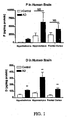
- the inventors have further found that FeSO 4 treatment results in a significant increase in DHEA in the hippocampus and cortex of control patients, indicating the presence of an alternative precursor in these patients. There was no effect observed in the hypothalamus of control patients. In contrast, Alzheimer's patients showed a significant increase in DHEA levels in response to FeSO 4 treatment only in the frontal cortex, but not in the hippocampus or hypothalamus. Given the presence of higher levels of DHEA in the Alzheimer's patient brain before FeSO 4 and the difference in increase in DHEA levels in the hippocampus in Alzheimer's patients versus controls patients following FeSO 4 treatment, this suggests that the precursor in the hippocampus of Alzheimer's patients has already been converted to DHEA. Thus, a patient in the beginning stages of a brain disorder might be diagnosed by showing decreased levels of precursor in a sample of hippocampus tissue according to the method described above.
- CSF cerebrospinal fluid
- serum cerebrospinal fluid
- CSF fluid may be used as a diagnostic tool in the assays of the invention is perhaps logical given the inventors' other findings about oxidative stress-induced DHEA formation in the Alzheimer's disease brain.
- serum can be used in the above method as a tool for diagnosing oxidative stress-induced formation of DHEA in the brain was somewhat surprising in this context. For instance, the DHEA levels in the serum of Alzheimer's disease patients are not significantly greater than the serum levels in control patients.
- the first diagnostic method introduced above may be performed with a variety of body fluids and tissue samples, wherein said samples are selected from the group consisting of cerebrospinal fluid, serum, hippocampus tissue, oligodendrocytes and astrocytes.
- ⁇ -amyloid peptide and other reagents that induce the formation of ROS maybe used to test for the presence of precursor in a body fluid or tissue sample.
- the diagnostic methods arc amenable to the use of not only FeSO 4 , but other reducing agents and/or oxidizing agents as well, particularly other classical Fenton reactants also exemplified by FeCl 3 and H 2 O 2 .
- DHEA quantities are preferably measured by (i) extracting the samples with an organic solvent, (ii) subjecting the extracted material to HPLC chromatography, and (iii) detecting the DHEA using radioimmunoassay.
- a preferred organic solvent is ether:ethylacetate (v/v), but any solvent may be used and many are known in the art.
- Radioimmunoassay kits for measuring DHEA and its sulfated derivative, DHEA-S are commercially available (see for example Diagnostic Systems Laboratories, 445 Medical Centre Blvd., Webster, Texas 77598).
- Antibodies specific for DHEA are also available from ICN Pharmaceuticals (Costa Mesa, CA). Alternatively, quantities of DHEA may be measured by gas chromatography coupled to mass spectrometry. Other studies have optically measured steroid levels in initial extracts, without any further purification steps. The present inventors have found, however, that attempting to measure DHEA and other neurosteroids without previous purification procedures may not provide accurate measurements.
- the methods of the invention may be supplemented with further assays to verify the validity of the results.
- the samples may also be tested for an increase in radical oxygen species after treatment with the regulating agent, e.g., by using 2,7 DCF fluorescence.
- the correlation between the treatment and the effect may also be verified by determining whether the effect is blocked by vitamin E.
- the samples may also be tested directly for the presence of the metabolic precursor for DHEA once it has been identified.
- the diagnostic method described above may also include steps whereby the cells are treated with compounds that optimize the synthesis of DHEA using the alternative pathway.
- SU 10603 is a known inhibitor of P450c17 (64).
- P450c17 is the enzyme responsible for the classical conversion of pregnenolone to DHEA (the non-oxidative stress-induced pathway).
- treating with SU 10603 would clarify results by inhibiting synthesis via the classical pathway such that only the potential for the alternative pathway is measured.
- Trilostane (Stegram Pharmaceuticals, Wales, U.K.), a specific inhibitor of 3 ⁇ -HSD, also inhibits the metabolism of pregnenolone by inhibiting its conversion into progesterone.
- a cell sample With both trilostane and SU 10603, it is possible to maximize pregnenolone production.
- mevastatin a 3-hydroxy-3-methylglutaryl CoA reductase inhibitor, i.e., an inhibitor of cholesterol synthesis, it may be possible to modulate the levels of the unknown substrate.
- DHEA levels are significantly higher in the brains of Alzheimer's patients than in the brains of control patients, and that concomitant to this increase is a decrease in the precursor for the oxidative stress-induced metabolic pathway for DHEA.
- Oxidative stress contributes to a variety of neurological disorders, and it is likely that elevated DHEA levels may be used as an indicator for a variety of such neuropathologies, including dementias in general, Alzheimer's disease, Parkinson's disease, ALS (Amylotropic Lateral Sclerosis), amyloidosis, Ataxia Telangiectasia, Binswanger's disease, brain cancer (which, in some cases, may lead to increased reactive oxygen species), Hallervoden-Spatz disease, Huntington's disease, Krabbe disease, Leigh's disease, mitochondrial disorders, Multi-Infract dementia, Pelizaeus-Merzbacher disease, Pick's disease, stroke, and traumatic brain injury.
- dementias in general Alzheimer's disease, Parkinson's disease, ALS (Amylotropic Lateral Sclerosis), amyloidosis, Ataxia Telangiectasia, Binswanger's disease, brain cancer (which, in some cases, may lead to increased reactive oxygen species), Hallervoden-Spat
- diagnosing traumatic brain injury may particularly benefit from the disclosed diagnostic assays due to the dramatic acute increase of oxidative stress due to the injury.
- Spinal cord injury and other peripheral nervous system injuries may also be diagnostic targets given the ability of the spinal cord Swann cells to make neurosteroids and the increased oxidative stress due to the injury,
- the present invention generally includes a method of diagnosing the existence of a neuropathologic disorder in a patient comprising measuring the quantity of a neurosteroid in the CNS of said patient. Depending on whether one measures DHEA levels or levels of the precursor, either an increased quantity or a decreased quantity of neurosteroid relative to a control will indicate the development of a neuropathologic disorder.
- DHEA may be important in the aging process, particularly in modulating memory formation via its actions on NMDA receptors in the hippocampus. - We have demonstrated that, contrary to current theories, levels of DHEA are much higher in the Alzheimer's disease brain than in the normal brain. The question remains, however, as to whether DHEA synthesis plays a protective role in neuropathology, or is acting to potentiate the disease process.
- DHEA and its sulfated derivative are neuroprotective against excitaory amino acid-induced toxicity in the hippocampus (32).
- DHEA neuroprotective effects
- glia in response to oxidative stress may serve a protective role.
- ⁇ -amyloid peptide has the potential to stimulate the oxidative strees-induced synthesis of DHEA
- astrocytes become reactive to this peptide in Alzheimer's disease pathology and produce extracellular matrix molecules and growth factors that help to potentiate plaque formation (33).
- the increase in DHEA would be a harmful event, helping to protect reactive astrocytes and potentiate neuronal damage. It remains to be seen whether DHEA plays an important role in neuropathology, or whether it is simply an epiphenomenon of the disease process.
- DHEA Regardless of whether DHEA plays an important role in the pathogenesis of Alzheimer's disease and other neuropathologies, it can serve as a useful marker of disease via the diagnostic methods described above. But if DHEA is found to lay a protective role, the novel observation that the alternative metabolic pathway may be stimulated by exposure of cells to ⁇ -amyloid peptide may provide a new means of synthesizing DHEA via cells that are capable of the alternative metabolic pathway, for use in pharmaceutical compositions.
- U.S. Patent No. 4,812,447 herein incorporated by reference, provides a discussion of the use of DHEA and DHEA-S in pharmaceutical compositions for the treatment of Alzheimer's diease.
- the present invention also includes a method of increasing or causing DHEA synthesis by cells in vitro comprising exposing said cells to ⁇ -amyloid peptide, wherein such exposure results in synthesis of DHEA.
- brain cells are the only cells at present demonstrated to facilitate this mode of synthesis
- the preferred cells for the synthesis are brain cells, and particularly astrocytes and oligodendrocytes.
- other cells may be identified that may be used for such a method.
- the human glioma cell line KG-1-C was obtained from the Riken Cell Bank (Tsukuba, Japan).
- the MGM-1 and MGM-3 cell lines were a gift from Drs. Hiroaki Kataoka and Mashi Koono (Miyazaki Medical College, Japan).
- Normal human astrocytes (NHA) were purchased from Clonetics (San Diego, CA), while purified human neurons (hNT-PF) were purchased from Stratagene (La Jolla, CA).
- the P450c17 antibody was a generous gift from Dr. Dale B. Hales at the University of Illinois.
- Trilostane a specific inhibitor of 3 ⁇ -HSD, was a gift from Stegram Pharmaceuticals, Wales, UK.
- KG-1-C is derived from a mixed glioma and has been described as having the characteristics of an oligodendrocytoma (69).
- MGM-1 and MGM-3 are derived from glioblastoma multiforme tumors (70). All three cell lines were grown in Dulbecco's Modification of Eagle's Media (DMEM) with 10% fetal bovine serum (Mediatech Cellgro, Gaithersberg, MD) plus 100 U/ml Penicillin, 100 mg/ml Streptomycin and 1.25 mg/ml Amphotericin B (Sigma, St. Louis, MO) at 37C and 6% CO 2 .
- DMEM Dulbecco's Modification of Eagle's Media
- Mediatech Cellgro Mediatech Cellgro, Gaithersberg, MD
- Penicillin 100 mg/ml Streptomycin
- Amphotericin B Sigma, St. Louis, MO
- NHA cells were grown at 37C and 6% CO 2 in Astrocyte Growth Medium (Clonetics, San Diego, CA) containing 5% fetal bovine serum, 0.02 mg/ml human epidermal growth factor, 25 mg/ml insulin, 0.025 mg/ml progesterone, 50.0 mg/ml transferrin, 50.0 mg/ml Gentamicin, and 0.05 mg/ml Amphotericin B.
- hNT-PF cells are a post-mitotic, differentiated cell derived from the Ntera2/D1 teratocarcinoma cell line (NT2).
- NT2 precursor cells can be induced to differentiate in vitro by six week treatment with retinoic acid (Stratagene, La Jolla, CA).
- hNT-PF cells were plated and maintained at 37C and 6% CO 2 , in hNT-PF neuron conditioned media (Stratagene).
- Probes were synthesized by RT-PCR from either total human adrenal RNA (PBR, P450scc, P450c17 or 3b-HSD) or total human brain RNA (MBP). To confirm specificity, probes were sequenced using the ABI Prism protocol at the Lombardi Cancer Center Core Facility, Georgetown University Medical Center. Probes were labeled by random primed labeling using the kit from Boehringer Mannheim (Indianapolis, IN) and were incubated with the membranes overnight at 42°C. Membranes were washed and exposed to XOMAT-AR film (Eastman Kodak, Rochester, NY) for 2-24 hours at -70°C. Table 1.
- Extracts were run on C 18 TLC plates (Whatman, Clifton, NJ) with a mobile phase of 95% methanol (v/v); radiolabeled standards for P, progesterone, and D were run on each plate, and appropriate steroid fractions were collected for each sample.
- TLC fractions were extracted with 2 ml diethyl ether (Fluka, New York) and evaporated.
- TLC extracts were assayed by specific radioimmunoassay (RIA). In separate experiments, steroids eluted from TLC were further identified by HPLC, RIA and Gas Chromatography-Mass Spectrometry (data not shown).
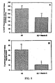
- Tissue Samples For brain samples from Alzheimer's patients and control patients, samples were treated as previously described (17). In brief, aliquots of homogenates were incubated with a final concentration of 30 mM (brain) or 10 mM (serum and CSF) FeSO 4 at 37°C for 2 hours. After 2 hours, samples were extracted and purified as described for the endogenous levels, and assayed by specific radioimmunoassay.
- Serum and CSF Levels of DHEA Serum was obtained from 11 patients, 5 with AD and 6 age-matched controls. Serum samples were split and treated with and without 10 mM FeSO 4 . Serum and CSF samples were extracted as described for the brain tissue samples, and purified by HPLC. DHEA levels were measured by specific radioimmunoassay.
- MGM-1 cells were plated on 96 well ( ⁇ 5,000 cells/well, for ROS measurements) or 24 well ( ⁇ 15,000 cells/well, for D measurements) plates (Corning Costar, Acton, MA). At the same time, 1 mg Ab (Ab (1-42), AnaSpec Inc., San Jose, CA) was dissolved in 0.5 ml serum-free media and incubated at 4C for aggregation. 24 hours later, cells were incubated in DMEM with 10% fetal bovine serum and 50 mM Ab or 50 mM Ab and 10 mM Vitamin E (Sigma, St. Louis, MO). Control samples were incubated in DMEM with 10% FBS. Cells were incubated at 37C and 6% CO 2 for at least 12 hours and processed for ROS generation (see below) or D synthesis as described above for the alternative experiments.
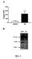
- RIA using antibodies specific for each steroid (ICN Pharmaceuticals, Costa Mesa, CA Mesa) was used to measure P, progesterone and D levels. The data were analyzed using the IBM-PC RIA Data Reduction Program (version 4.1) from Jaffe and Associates (Silver Spring, MD). Levels of ⁇ -amyloid species in AD and control brain samples were analyzed using 4G8 (Senetex PLC, Napa, CA), a monoclonal antibody to ⁇ -amyloid peptide that recognizes amino acids 17-24. Samples in 0.25 M sucrose buffer were run on 4-20% precast gradient gels (Novex, San Diego, CA).
- Microgram amounts of protein were measured using the dye-binding assay of Bradford (1976) (39), using bovine serum albumin (ICN Pharmaceuticals, Costa Mesa, CA) as the standard.
- oligodendrocytes To determine the phenotypes of the glioma cell lines, we performed RT-PCR for MBP, an oligodendrocyte marker, and GFAP, an astrocyte marker. We found the all three glioma cell lines express message for MBP (data not shown), as does total human brain RNA. None of the glioma cell lines express message for GFAP (data not shown). These cells will be collectively referred to as oligodendrocytes.
- NHA cells from Clonetics are shipped immunopositive for GFAP and we confirmed GFAP expression by RT-PCR (data not shown). These cells are also immunonegative for CD68, a microglial marker, and for 2',3'-cyclic nucleotide-3'-phosphodiesterase, an oligodendrocyte marker (Clonetics Certificate of Analysis, Clonetics, San Diego, CA).
- the hNT-PF cells are derived from a Ntera2/D1 teratocarcinoma cell line by 5-6 weeks of differentiation by retinoic acid. These cells are post-mitotic and have been well characterized by other investigators. Once differentiated, they express the three neurofilament proteins, but not GFAP (61).
- hNT-PF cells behave like neurons, with tetrodotoxin-sensitive sodium currents (78). Retinoic acid differentiation also induces the expression of functional glutamate receptors, of both the NMDA and non-NMDA subtypes (86).
- Oligodendrocytes synthesize P de novo, whereas astrocytes and neurons do not
- a steroid-synthesizing cell is defined as a cell that is able to convert endogenous cholesterol to pregnenolone (P).
- P pregnenolone
- Very few cells in the body have this ability - it is limited to steroidogenic tissues such as the adrenal glands, gonads, placenta and brain. However, almost all cells have the ability to metabolize steroids to final steroid products.
- Mevastatin was blocked endogenous cholesterol synthesis with the 3-hydroxy-3-methylglutaryl-CoA reductase inhibitor Mevastatin and incubated the cells with a radiolabeled cholesterol precursor, 3 H-mevalonactone.
- Trilostane and SU 10603 we attempted to maximize P production.
- Human brain cells express mRNA and immunoreactivity for different components of the peripheral steroid biosynthesis machinery
- MGM-3 cells express immunoreactivity for PBR, P450scc and P450c17, but not for 3 ⁇ -HSD. However, MGM-3 does have message for 3 ⁇ -HSD. This may indicate an increased breakdown in 3 ⁇ -HSD mRNA or increased degradation of the protein. As a whole, these data indicate that in human oligodendrocytes, mRNA and protein expression corresponds with the ability of the cells to synthesize neurosteroids de novo.
- Human astrocytes express mRNA for all four components (data not shown). They are immunoreactive for PBR, P450scc and P450c17, although protein expression is much lower then that seen in the glioma cells. Similar to MGM-3 cells, NHA have mRNA but no immunoreactivity for 3 ⁇ -HSD. Again, this may indicate alterations in mRNA or protein stability. NHA cells have the potential to synthesize P and D, but not progesterone. Human neurons express mRNA for PBR, P450c17 and 3 ⁇ -HSD, but not for P450scc, while the NT2 precursor cells have message for all four components (data not shown). This indicates that hNT-PF will not be able to synthesize P de novo, in agreement with the data above, but they may be able to metabolize P to other neuroactive steroids.
- rat glioma cells are able to synthesize neurosteroids, such as DHEA (D), through a P450c17-independent pathway (15).
- D neurosteroids
- Fig. 6B see brief description of drawings for one-way ANOVA F and p values
- FeSO 4 did not significantly affect the KG-1-C and MGM-3 D levels (Fig. 6A, 6C).
- KG-1-C and MGM-3 are the cells that synthesized radiolabeled D de novo in a manner which was not inhibited by SU-10603, again suggesting that endogenous free radicals may be responsible for D formation in these cells.
- FeSO 4 treatment had a significant effect on D formation in NHA cells (Fig. 6D), indicating that this effect is not limited to tumor cell lines. This effect was only seen at the highest dose of FeSO 4 used, indicating that there may be differences between cell types in terms of their sensitivity to FeSO 4 or ROS levels.
- the hNT-PF cells were also incubated with increasing concentrations of FeSO 4 , with and without SU 10603 as described above. In addition, these cells were incubated with 5mM P to enhance D production; recent studies have indicated that providing P as a substrate can enhance the effects of FeSO 4 in rat C6 cells (15). Human neurons were not affected by FeSO 4 treatment (Fig. 6E), suggesting that this alternative pathway is specific to glial cells. hNT-PF cells were also not affected by the SU 10603 treatment, indicating that D in these cells does not come from endogenous activity of the P450c17 enzyme. Neuronal D, therefore, may be completely derived from other sources.
- Treatment with ⁇ -Amyloid increases ROS and D levels in MGM-1 human glioma cells
- oligodendrocytes are the probable source of brain-synthesized P (62), and validates the use of the rat as a model to study neurosteroid biosynthesis.
- astrocytes or neurons have the ability to metabolize P into progesterone and D, or other neuroactive compounds.
- Oligodendrocytes can be designated as steroid synthesizing cells, and astrocytes and neurons as steroid metabolizing cell populations. This means that neuroactive steroids, which may play a role in neuronal excitability, can be produced locally from a number of different cellular sources, as well as coming from the periphery.
- Oligodendrocytes have mRNA and immunoreactivity for PBR, P450scc, P450c17 and 3b-HSD. This confirms the ability of oligodendrocytes to make cholesterol from a precursor, transport cholesterol into the mitochondria and metabolize it to P. Enzymatic activity for P450c17 was not detected, due to the inability of the specific inhibitor SU 10603 to block D formation in these cells (Fig. 6A-C). Astrocytes also have mRNA and immunoreactivity for PBR, P450scc and P450c17, although we were unable to find activity for P450scc.
- Oligodendrocytes and astrocytes can make D by an alternative pathway. There also seems to be a threshold for ROS induction of D synthesis, which may vary between oligodendrocytes and astrocytes. Astrocytes seem to produce more ROS in response to Fe 2+ than glioma cells, which may indicate an increased sensitivity to FeSO 4 and potentially, a higher threshold for ROS regulation of D synthesis in normal human brain cells. These data suggest that the oxidative environment of the brain under different conditions may influence D formation by glia. Neurons do not have the alternative pathway for D synthesis suggesting that the alternative pathway is specific to glia.
- Oligodendrocytes that do not respond to Fe 2+ can synthesize D de novo in a P450c17-independent manner. This is potentially due to high endogenous ROS.
- Vitamin E treatment of the glioma cell lines decreases MGM-3 ROS and D levels to those seen in MGM-1 cells, but KG-1-C cells are less drastically affected by this antioxidant.
- a ⁇ while toxic to neurons, induces astrocytic activation (56) and cytoskeletal reorganization (80), but does not alter astrocyte viability (76). It has been hypothesized that astrocyte activation and subsequent deposition of chondroitin sulfate proteoglycans (33) and growth factors (90) may be involved in plaque formation. It is possible that glial cells may be able to resist the toxic effects of Ab by their ability to produce D, known to exert neuroprotective effects in the hippocampus (32).
- PBR is upregulated in human brain tumors (43, 68), which may indicate a general upregulation of the steroid biosynthetic pathway. Furthermore, PBR binding sites are significantly increased in specific areas from AD brains, as well as brains from patients with Parkinson's disease (66). Upregulation of steroid biosynthetic components may be, like the activity of the alternative pathway, a sign of altered homeostasis, neurodegeneration or trauma in the brain. It may be that an increase in neurosteroid biosynthesis is a hallmark of an altered or toxic cellular environment.
- the alternative pathway may only become a player in brain steroid biosynthesis in situations in which normal homeostasis has been disrupted, as in the MGM-1 cells which have nuclear PBR and therefore cannot synthesize neurosteroids via the classical enzymatic pathway.
- alterations in cellular processes due to injury or disease may allow for the function of the alternative pathway in cells that do not normally use this pathway.
- AD samples In the hippocampus, AD samples have a much higher A ⁇ load (Figure 3B), with molecular weights ranging from 45-80 kDa, while control samples have very little detectable A ⁇ -immunoreactivity. This supports the hypothesis that D in the hippocampus is formed as a result of oxidative stress, induced by the higher levels of A ⁇ present.
- AD patients In the cortex, AD patients have a relatively constant level of A ⁇ immunoreactivity, ranging from 20-50 kDa. Control patients also have detectable A ⁇ -immunoreactivity, but these patients tend to have higher molecular weight species, ranging from about 30-50 kDa.
- D DHEA
- P pregnenolone
- the increased levels of D in the AD brain are contrary to the current thinking on the role of D in aging.
- the decrease in D levels with age has been considered to be a hallmark of the process, potentially playing a role in a number of the problems occurring with aging.
- D replacement has been the focus of a number of clinical studies attempting to prevent age-related cognitive changes and dementia (23-26).
- We now show that the actual CNS levels of D are increased in AD pathology. This is the opposite of what has been seen in the periphery with normal aging. This observation has been used as the basis of the present invention to formulate diagnostic methods and kits to test for the early development of AD.
- AD diagnosis and treatment is the inability of clinicians to determine the onset of the disease. Currently, this is done by a combination of MRI scans, to look for generalized shrinkage of the brain and cognitive tests to determine state of dementia. Typically symptoms do not occur until very late in the disease process. If the changes in D in the CSF are indeed regulated by oxidative conditions within the brain, there may be alterations in CSF D early in the progression of the disease. We have also shown that presence of the alternative precursor can be detected in the serum of control patients, but not of AD patients. Further analysis of the data shown in Table 2 indicates that the serum shows a greater response to FeSO 4 treatment than does any other brain area tested.
Landscapes
- Health & Medical Sciences (AREA)
- Life Sciences & Earth Sciences (AREA)
- Engineering & Computer Science (AREA)
- Chemical & Material Sciences (AREA)
- Medicinal Chemistry (AREA)
- General Health & Medical Sciences (AREA)
- Bioinformatics & Cheminformatics (AREA)
- Biomedical Technology (AREA)
- Organic Chemistry (AREA)
- Public Health (AREA)
- Veterinary Medicine (AREA)
- Pharmacology & Pharmacy (AREA)
- General Chemical & Material Sciences (AREA)
- Animal Behavior & Ethology (AREA)
- Chemical Kinetics & Catalysis (AREA)
- Nuclear Medicine, Radiotherapy & Molecular Imaging (AREA)
- Neurology (AREA)
- Neurosurgery (AREA)
- Hematology (AREA)
- Molecular Biology (AREA)
- Immunology (AREA)
- Urology & Nephrology (AREA)
- Analytical Chemistry (AREA)
- Physics & Mathematics (AREA)
- Pathology (AREA)
- General Physics & Mathematics (AREA)
- Biochemistry (AREA)
- Food Science & Technology (AREA)
- Microbiology (AREA)
- Biotechnology (AREA)
- Cell Biology (AREA)
- Psychology (AREA)
- Proteomics, Peptides & Aminoacids (AREA)
- Physical Education & Sports Medicine (AREA)
- Endocrinology (AREA)
- Obesity (AREA)
- Cardiology (AREA)
- Dermatology (AREA)
- Psychiatry (AREA)
- Hospice & Palliative Care (AREA)
Priority Applications (1)
| Application Number | Priority Date | Filing Date | Title |
|---|---|---|---|
| EP07075646A EP1860442A3 (en) | 2000-01-28 | 2001-01-26 | Neurosteroids as markers for Alzheimer's disease |
Applications Claiming Priority (3)
| Application Number | Priority Date | Filing Date | Title |
|---|---|---|---|
| US17869800P | 2000-01-28 | 2000-01-28 | |
| US178698P | 2000-01-28 | ||
| PCT/US2001/002476 WO2001055692A2 (en) | 2000-01-28 | 2001-01-26 | Neurosteroids as markers for alzheimer's disease |
Related Child Applications (1)
| Application Number | Title | Priority Date | Filing Date |
|---|---|---|---|
| EP07075646A Division EP1860442A3 (en) | 2000-01-28 | 2001-01-26 | Neurosteroids as markers for Alzheimer's disease |
Publications (3)
| Publication Number | Publication Date |
|---|---|
| EP1254251A2 EP1254251A2 (en) | 2002-11-06 |
| EP1254251A4 EP1254251A4 (en) | 2004-07-21 |
| EP1254251B1 true EP1254251B1 (en) | 2007-08-15 |
Family
ID=22653575
Family Applications (1)
| Application Number | Title | Priority Date | Filing Date |
|---|---|---|---|
| EP01903310A Expired - Lifetime EP1254251B1 (en) | 2000-01-28 | 2001-01-26 | Neurosteroids as markers for alzheimer's disease |
Country Status (8)
| Country | Link |
|---|---|
| EP (1) | EP1254251B1 (https=) |
| JP (1) | JP2004507712A (https=) |
| AT (1) | ATE370416T1 (https=) |
| AU (2) | AU785364B2 (https=) |
| CA (1) | CA2398358A1 (https=) |
| DE (1) | DE60129932T2 (https=) |
| ES (1) | ES2290111T3 (https=) |
| WO (1) | WO2001055692A2 (https=) |
Families Citing this family (5)
| Publication number | Priority date | Publication date | Assignee | Title |
|---|---|---|---|---|
| WO2003100092A2 (en) * | 2002-05-28 | 2003-12-04 | Evotec Neurosciences Gmbh | Diagnostic and therapeutic use of cyp11a1 for neurodegenerative diseases |
| FR2847038B1 (fr) * | 2002-11-07 | 2005-01-07 | Conservatoire Nat Arts | Procede de diagnostic de maladies neurodegeneratives |
| US7576073B2 (en) | 2004-05-28 | 2009-08-18 | UNIVERSITé LAVAL | Combined therapy for the treatment of parkinson's disease |
| CN102998464A (zh) * | 2011-09-16 | 2013-03-27 | 武汉优尔生科技股份有限公司 | 脱氢表雄酮酶联免疫吸附测定试剂盒的研制方法 |
| US11977085B1 (en) | 2023-09-05 | 2024-05-07 | Elan Ehrlich | Date rape drug detection device and method of using same |
Family Cites Families (2)
| Publication number | Priority date | Publication date | Assignee | Title |
|---|---|---|---|---|
| US5162198A (en) * | 1991-02-08 | 1992-11-10 | Virginia Commonwealth University | Method of using dehydroepiandrosterone and dehydroepiandrosterone-sulfate as inhibitors of thrombuxane production and platelet aggregation |
| US6146836A (en) * | 1997-05-19 | 2000-11-14 | Bayer Corporation | Immunoassays using anti-allotypic monoclonal antibodies |
-
2001
- 2001-01-26 CA CA002398358A patent/CA2398358A1/en not_active Abandoned
- 2001-01-26 EP EP01903310A patent/EP1254251B1/en not_active Expired - Lifetime
- 2001-01-26 DE DE60129932T patent/DE60129932T2/de not_active Expired - Fee Related
- 2001-01-26 ES ES01903310T patent/ES2290111T3/es not_active Expired - Lifetime
- 2001-01-26 JP JP2001555784A patent/JP2004507712A/ja active Pending
- 2001-01-26 AU AU31142/01A patent/AU785364B2/en not_active Ceased
- 2001-01-26 WO PCT/US2001/002476 patent/WO2001055692A2/en not_active Ceased
- 2001-01-26 AT AT01903310T patent/ATE370416T1/de not_active IP Right Cessation
-
2007
- 2007-04-30 AU AU2007201915A patent/AU2007201915A1/en not_active Abandoned
Also Published As
| Publication number | Publication date |
|---|---|
| WO2001055692A2 (en) | 2001-08-02 |
| AU2007201915A1 (en) | 2007-05-24 |
| CA2398358A1 (en) | 2001-08-02 |
| EP1254251A4 (en) | 2004-07-21 |
| AU3114201A (en) | 2001-08-07 |
| AU785364B2 (en) | 2007-02-01 |
| DE60129932D1 (de) | 2007-09-27 |
| ES2290111T3 (es) | 2008-02-16 |
| DE60129932T2 (de) | 2008-06-19 |
| ATE370416T1 (de) | 2007-09-15 |
| JP2004507712A (ja) | 2004-03-11 |
| EP1254251A2 (en) | 2002-11-06 |
| WO2001055692A3 (en) | 2002-03-07 |
Similar Documents
| Publication | Publication Date | Title |
|---|---|---|
| Brown et al. | Pathways of neurosteroid biosynthesis in cell lines from human brain: regulation of dehydroepiandrosterone formation by oxidative stress and β‐amyloid peptide | |
| Brown et al. | Oxidative stress-mediated DHEA formation in Alzheimer’s disease pathology | |
| Lütjohann et al. | Profile of cholesterol-related sterols in aged amyloid precursor protein transgenic mouse brain | |
| van Broekhoven et al. | Neurosteroids in depression: a review | |
| Pesaresi et al. | Sex differences in neuroactive steroid levels in the nervous system of diabetic and non-diabetic rats | |
| Naylor et al. | Cerebrospinal fluid dehydroepiandrosterone levels are correlated with brain dehydroepiandrosterone levels, elevated in Alzheimer’s disease, and related to neuropathological disease stage | |
| Weill-Engerer et al. | In vitro metabolism of dehydroepiandrosterone (DHEA) to 7α-hydroxy-DHEA and Δ5-androstene-3β, 17β-diol in specific regions of the aging brain from Alzheimer’s and non-demented patients | |
| Chen et al. | Decreased estradiol release from astrocytes contributes to the neurodegeneration in a mouse model of Niemann‐Pick disease type C | |
| He et al. | Intracellular oxidation of allopregnanolone by human brain type 10 17beta-hydroxysteroid dehydrogenase | |
| Gezen-Ak et al. | Molecular basis of vitamin D action in neurodegeneration: the story of a team perspective | |
| Verdile et al. | Luteinizing hormone levels are positively correlated with plasma amyloid-β protein levels in elderly men | |
| Cascio et al. | Pathways of dehydroepiandrosterone formation in rat brain glia | |
| Caruso et al. | Sex‐dimorphic changes in neuroactive steroid levels after chronic experimental autoimmune encephalomyelitis | |
| Makioka et al. | Involvement of endoplasmic reticulum stress defined by activated unfolded protein response in multiple system atrophy | |
| AU2007201915A1 (en) | Neurosteroids as markers for Alzheimer's disease | |
| Peng et al. | A novel potent synthetic steroidal liver X receptor agonist lowers plasma cholesterol and triglycerides and reduces atherosclerosis in LDLR–/–mice | |
| Vaňková et al. | Reduced sulfotransferase SULT2A1 activity in patients with Alzheimer's disease. | |
| Baker et al. | 17β-Estradiol reduces plasma Aβ40 for HRT-naive postmenopausal women with Alzheimer disease: a preliminary study | |
| Jørgensen | Oxidatively generated DNA/RNA damage in psychological stress states | |
| EP1860442A2 (en) | Neurosteroids as markers for Alzheimer's disease | |
| Rammouz et al. | Oxidative stress-mediated brain dehydroepiandrosterone (DHEA) formation in Alzheimer’s disease diagnosis | |
| US20030213746A1 (en) | Neurosteroids as markers for alzheimer's disease | |
| Ernst et al. | Androstenedione changes steroidogenic activity of SGBS cells | |
| Maldonado-Devincci et al. | Reduction of circulating and selective limbic brain levels of (3α, 5α)-3-hydroxy-pregnan-20-one (3α, 5α-THP) following forced swim stress in C57BL/6J mice | |
| Perkins et al. | Neurosteroids in the pathophysiology and treatment of mood and anxiety disorders |
Legal Events
| Date | Code | Title | Description |
|---|---|---|---|
| PUAI | Public reference made under article 153(3) epc to a published international application that has entered the european phase |
Free format text: ORIGINAL CODE: 0009012 |
|
| 17P | Request for examination filed |
Effective date: 20020827 |
|
| AK | Designated contracting states |
Kind code of ref document: A2 Designated state(s): AT BE CH CY DE DK ES FI FR GB GR IE IT LI LU MC NL PT SE TR |
|
| AX | Request for extension of the european patent |
Free format text: AL;LT;LV;MK;RO;SI |
|
| RIC1 | Information provided on ipc code assigned before grant |
Ipc: 7G 01N 33/74 A |
|
| A4 | Supplementary search report drawn up and despatched |
Effective date: 20040609 |
|
| 17Q | First examination report despatched |
Effective date: 20041119 |
|
| RAP1 | Party data changed (applicant data changed or rights of an application transferred) |
Owner name: GEORGETOWN UNIVERSITY MEDICAL CENTER |
|
| RAP1 | Party data changed (applicant data changed or rights of an application transferred) |
Owner name: GEORGETOWN UNIVERSITY |
|
| GRAP | Despatch of communication of intention to grant a patent |
Free format text: ORIGINAL CODE: EPIDOSNIGR1 |
|
| GRAS | Grant fee paid |
Free format text: ORIGINAL CODE: EPIDOSNIGR3 |
|
| GRAA | (expected) grant |
Free format text: ORIGINAL CODE: 0009210 |
|
| AK | Designated contracting states |
Kind code of ref document: B1 Designated state(s): AT BE CH CY DE DK ES FI FR GB GR IE IT LI LU MC NL PT SE TR |
|
| REG | Reference to a national code |
Ref country code: GB Ref legal event code: FG4D |
|
| REG | Reference to a national code |
Ref country code: CH Ref legal event code: EP |
|
| REG | Reference to a national code |
Ref country code: IE Ref legal event code: FG4D |
|
| REF | Corresponds to: |
Ref document number: 60129932 Country of ref document: DE Date of ref document: 20070927 Kind code of ref document: P |
|
| REG | Reference to a national code |
Ref country code: SE Ref legal event code: TRGR |
|
| REG | Reference to a national code |
Ref country code: CH Ref legal event code: NV Representative=s name: KIRKER & CIE S.A. |
|
| PG25 | Lapsed in a contracting state [announced via postgrant information from national office to epo] |
Ref country code: FI Free format text: LAPSE BECAUSE OF FAILURE TO SUBMIT A TRANSLATION OF THE DESCRIPTION OR TO PAY THE FEE WITHIN THE PRESCRIBED TIME-LIMIT Effective date: 20070815 |
|
| REG | Reference to a national code |
Ref country code: ES Ref legal event code: FG2A Ref document number: 2290111 Country of ref document: ES Kind code of ref document: T3 |
|
| ET | Fr: translation filed | ||
| PG25 | Lapsed in a contracting state [announced via postgrant information from national office to epo] |
Ref country code: DK Free format text: LAPSE BECAUSE OF FAILURE TO SUBMIT A TRANSLATION OF THE DESCRIPTION OR TO PAY THE FEE WITHIN THE PRESCRIBED TIME-LIMIT Effective date: 20070815 Ref country code: GR Free format text: LAPSE BECAUSE OF FAILURE TO SUBMIT A TRANSLATION OF THE DESCRIPTION OR TO PAY THE FEE WITHIN THE PRESCRIBED TIME-LIMIT Effective date: 20071116 |
|
| PG25 | Lapsed in a contracting state [announced via postgrant information from national office to epo] |
Ref country code: PT Free format text: LAPSE BECAUSE OF FAILURE TO SUBMIT A TRANSLATION OF THE DESCRIPTION OR TO PAY THE FEE WITHIN THE PRESCRIBED TIME-LIMIT Effective date: 20080115 |
|
| PLBE | No opposition filed within time limit |
Free format text: ORIGINAL CODE: 0009261 |
|
| STAA | Information on the status of an ep patent application or granted ep patent |
Free format text: STATUS: NO OPPOSITION FILED WITHIN TIME LIMIT |
|
| 26N | No opposition filed |
Effective date: 20080516 |
|
| PG25 | Lapsed in a contracting state [announced via postgrant information from national office to epo] |
Ref country code: MC Free format text: LAPSE BECAUSE OF NON-PAYMENT OF DUE FEES Effective date: 20080131 |
|
| PGFP | Annual fee paid to national office [announced via postgrant information from national office to epo] |
Ref country code: AT Payment date: 20090102 Year of fee payment: 9 Ref country code: ES Payment date: 20090126 Year of fee payment: 9 Ref country code: IE Payment date: 20090126 Year of fee payment: 9 |
|
| PGFP | Annual fee paid to national office [announced via postgrant information from national office to epo] |
Ref country code: DE Payment date: 20090302 Year of fee payment: 9 Ref country code: NL Payment date: 20090124 Year of fee payment: 9 |
|
| PGFP | Annual fee paid to national office [announced via postgrant information from national office to epo] |
Ref country code: CH Payment date: 20090126 Year of fee payment: 9 Ref country code: GB Payment date: 20090129 Year of fee payment: 9 |
|
| PG25 | Lapsed in a contracting state [announced via postgrant information from national office to epo] |
Ref country code: CY Free format text: LAPSE BECAUSE OF FAILURE TO SUBMIT A TRANSLATION OF THE DESCRIPTION OR TO PAY THE FEE WITHIN THE PRESCRIBED TIME-LIMIT Effective date: 20070815 |
|
| PGFP | Annual fee paid to national office [announced via postgrant information from national office to epo] |
Ref country code: BE Payment date: 20090219 Year of fee payment: 9 |
|
| PGFP | Annual fee paid to national office [announced via postgrant information from national office to epo] |
Ref country code: IT Payment date: 20090127 Year of fee payment: 9 Ref country code: SE Payment date: 20090128 Year of fee payment: 9 |
|
| PGFP | Annual fee paid to national office [announced via postgrant information from national office to epo] |
Ref country code: FR Payment date: 20090119 Year of fee payment: 9 |
|
| PG25 | Lapsed in a contracting state [announced via postgrant information from national office to epo] |
Ref country code: LU Free format text: LAPSE BECAUSE OF NON-PAYMENT OF DUE FEES Effective date: 20080126 |
|
| BERE | Be: lapsed |
Owner name: GEORGETOWN UNIVERSITY Effective date: 20100131 |
|
| REG | Reference to a national code |
Ref country code: NL Ref legal event code: V1 Effective date: 20100801 |
|
| PG25 | Lapsed in a contracting state [announced via postgrant information from national office to epo] |
Ref country code: TR Free format text: LAPSE BECAUSE OF FAILURE TO SUBMIT A TRANSLATION OF THE DESCRIPTION OR TO PAY THE FEE WITHIN THE PRESCRIBED TIME-LIMIT Effective date: 20070815 |
|
| REG | Reference to a national code |
Ref country code: CH Ref legal event code: PL |
|
| GBPC | Gb: european patent ceased through non-payment of renewal fee |
Effective date: 20100126 |
|
| EUG | Se: european patent has lapsed | ||
| REG | Reference to a national code |
Ref country code: FR Ref legal event code: ST Effective date: 20100930 |
|
| PG25 | Lapsed in a contracting state [announced via postgrant information from national office to epo] |
Ref country code: CH Free format text: LAPSE BECAUSE OF NON-PAYMENT OF DUE FEES Effective date: 20100131 Ref country code: NL Free format text: LAPSE BECAUSE OF NON-PAYMENT OF DUE FEES Effective date: 20100801 Ref country code: LI Free format text: LAPSE BECAUSE OF NON-PAYMENT OF DUE FEES Effective date: 20100131 Ref country code: FR Free format text: LAPSE BECAUSE OF NON-PAYMENT OF DUE FEES Effective date: 20100201 |
|
| PG25 | Lapsed in a contracting state [announced via postgrant information from national office to epo] |
Ref country code: AT Free format text: LAPSE BECAUSE OF NON-PAYMENT OF DUE FEES Effective date: 20100126 Ref country code: DE Free format text: LAPSE BECAUSE OF NON-PAYMENT OF DUE FEES Effective date: 20100803 |
|
| PG25 | Lapsed in a contracting state [announced via postgrant information from national office to epo] |
Ref country code: GB Free format text: LAPSE BECAUSE OF NON-PAYMENT OF DUE FEES Effective date: 20100126 |
|
| PG25 | Lapsed in a contracting state [announced via postgrant information from national office to epo] |
Ref country code: IE Free format text: LAPSE BECAUSE OF NON-PAYMENT OF DUE FEES Effective date: 20100126 |
|
| PG25 | Lapsed in a contracting state [announced via postgrant information from national office to epo] |
Ref country code: BE Free format text: LAPSE BECAUSE OF NON-PAYMENT OF DUE FEES Effective date: 20100131 |
|
| REG | Reference to a national code |
Ref country code: ES Ref legal event code: FD2A Effective date: 20110224 |
|
| PG25 | Lapsed in a contracting state [announced via postgrant information from national office to epo] |
Ref country code: IT Free format text: LAPSE BECAUSE OF NON-PAYMENT OF DUE FEES Effective date: 20100126 |
|
| PG25 | Lapsed in a contracting state [announced via postgrant information from national office to epo] |
Ref country code: ES Free format text: LAPSE BECAUSE OF NON-PAYMENT OF DUE FEES Effective date: 20110223 |
|
| PG25 | Lapsed in a contracting state [announced via postgrant information from national office to epo] |
Ref country code: ES Free format text: LAPSE BECAUSE OF NON-PAYMENT OF DUE FEES Effective date: 20100127 |
|
| PG25 | Lapsed in a contracting state [announced via postgrant information from national office to epo] |
Ref country code: SE Free format text: LAPSE BECAUSE OF NON-PAYMENT OF DUE FEES Effective date: 20100127 |