EP1249246B1 - Use of an histone deacetylase inhibitor for the treatment of diseases associated with an hpv infection - Google Patents
Use of an histone deacetylase inhibitor for the treatment of diseases associated with an hpv infection Download PDFInfo
- Publication number
- EP1249246B1 EP1249246B1 EP01108923A EP01108923A EP1249246B1 EP 1249246 B1 EP1249246 B1 EP 1249246B1 EP 01108923 A EP01108923 A EP 01108923A EP 01108923 A EP01108923 A EP 01108923A EP 1249246 B1 EP1249246 B1 EP 1249246B1
- Authority
- EP
- European Patent Office
- Prior art keywords
- histone deacetylase
- cyclin
- cells
- sodium butyrate
- treatment
- Prior art date
- Legal status (The legal status is an assumption and is not a legal conclusion. Google has not performed a legal analysis and makes no representation as to the accuracy of the status listed.)
- Expired - Lifetime
Links
Images


Classifications
-
- A—HUMAN NECESSITIES
- A61—MEDICAL OR VETERINARY SCIENCE; HYGIENE
- A61K—PREPARATIONS FOR MEDICAL, DENTAL OR TOILETRY PURPOSES
- A61K31/00—Medicinal preparations containing organic active ingredients
- A61K31/185—Acids; Anhydrides, halides or salts thereof, e.g. sulfur acids, imidic, hydrazonic or hydroximic acids
- A61K31/19—Carboxylic acids, e.g. valproic acid
-
- A—HUMAN NECESSITIES
- A61—MEDICAL OR VETERINARY SCIENCE; HYGIENE
- A61K—PREPARATIONS FOR MEDICAL, DENTAL OR TOILETRY PURPOSES
- A61K31/00—Medicinal preparations containing organic active ingredients
-
- A—HUMAN NECESSITIES
- A61—MEDICAL OR VETERINARY SCIENCE; HYGIENE
- A61K—PREPARATIONS FOR MEDICAL, DENTAL OR TOILETRY PURPOSES
- A61K31/00—Medicinal preparations containing organic active ingredients
- A61K31/16—Amides, e.g. hydroxamic acids
- A61K31/165—Amides, e.g. hydroxamic acids having aromatic rings, e.g. colchicine, atenolol, progabide
-
- A—HUMAN NECESSITIES
- A61—MEDICAL OR VETERINARY SCIENCE; HYGIENE
- A61K—PREPARATIONS FOR MEDICAL, DENTAL OR TOILETRY PURPOSES
- A61K31/00—Medicinal preparations containing organic active ingredients
- A61K31/185—Acids; Anhydrides, halides or salts thereof, e.g. sulfur acids, imidic, hydrazonic or hydroximic acids
- A61K31/19—Carboxylic acids, e.g. valproic acid
- A61K31/192—Carboxylic acids, e.g. valproic acid having aromatic groups, e.g. sulindac, 2-aryl-propionic acids, ethacrynic acid
-
- A—HUMAN NECESSITIES
- A61—MEDICAL OR VETERINARY SCIENCE; HYGIENE
- A61P—SPECIFIC THERAPEUTIC ACTIVITY OF CHEMICAL COMPOUNDS OR MEDICINAL PREPARATIONS
- A61P35/00—Antineoplastic agents
Definitions
- the present invention relates to the use of histone deacetylase inhibitors for the treatment of a disease, wherein the disease is cervical cancer, cervical intraepithelial neoplasm, wart, larynx papilloma or condyloma acuminatum associated with a human papilloma virus HPV infection.
- histone deacetylase inhibitors are sodium butyrate, phenylbutyrate and trichostatin A.
- Carcinoma of the uterine cervix (cervical cancer, CC) is the second most common cancer in women worldwide and the first in developing countries. CC develops through premalignant intermediate stages of increasing severity known as cervical intraepithelial neoplasm (CIN) grades 1-3, the latter leading to the development of invasive cancer in about 50% of cases over a period of 1-2 decades. More than 11% of the global cancer incidence in women is due to human papillomavirus (HPV) infections. Infection with HPV-types 16 and 18 has been associated with the development of CIN and CC, with HPV genotype 16 being the most prevalent viral type to infect the cervix.
- CIN cervical intraepithelial neoplasm
- the E6 and E7 proteins encoded by these HPV types are thought to be involved in the pathogenesis of CC by inducing abnormal cell proliferation. Expression of E6 and E7 is consistently detected in tissue and tumor cells from HPV-associated CCs. Furthermore, the E6 and E7 genes from HPV types 16 and 18 are sufficient for transformation of epithelial cells in culture.
- E6 and E7 viral oncogenes encoded by HPV types 16 and 18 may be effective targets for tumor rejection by the host and that a therapy might be based on inactivation of said proteins or inhibition of expression of the corresponding genes.
- a therapy might be achieved by methods like, e.g., the suppression of the expression of viral oncogenes using antisense-approaches or interference of specific protein interactions between cellular proteins and viral oncogenes using aptameres.
- the object of the present invention to provide means allowing the treatment of diseases associated with an HPV infection wherein the disease is cervical cancer, cervical intraepithelial neoplasm, wart, larynx papilloma, or condyloma acuminatum.
- the histone deacetylase inhibitors sodium butyrate and trichostatin A arrest human papillomavirus (HPV)-positive cells in G1 to S transition of the cell cycle, which is paralleled by an up-regulation of the cyclin dependent kinase inhibitors (CKIs) p21 CIP1 and p27 KIP1 . While these CKls normally cannot exert their cdk2-inhibitory function in the presence of the viral oncoprotein E7, co-immunoprecipitation experiments revealed that with binding of p21 CIP1 and p27 KIP1 to the cyclin-cdk2 complex, E7 binding is prevented.
- HPV human papillomavirus
- HPV expression is thought to be required to maintain a proliferative phenotype of cervical carcinoma cells, exclusion of E7 and complete suppression of cdk2 activity is achieved even in the presence of ongoing viral transcription.
- Increase of p27 KIP1 correlates with down-regulation of p45 SKP2 , a component of the ubiquitin-protein ligase SCF SKP2 which controls the half-life of regulatory proteins during the cell cycle.
- cyclin D1 and cyclin A suppression, cyclin E induction inhibition of histone deacetylation also triggered Rb degradation, while the levels of E2F remained unaffected.
- the present invention relates to the use of a histone deacetylase inhibitor for the preparation of a medicament for the treatment of a disease associated with an HPV infection, wherein the disease is cervical cancer, cervical intraepithelial neoplasm, wart, larynx papilloma or condyloma acuminatum.
- histone deacetylase inhibitor relates to any compound which is capable of inhibiting the activity of histone deacetylase.
- the person skilled in the art can select suitable compounds on the basis of the known structures (and amino acid sequences) of histone deacetylases, e.g. histone deacetylases 1, 2, 3, 4, 5, 6, 7, 7A, isoform a, 7B, isoform b and 8; see NCBI-Databases AAH00301, XP004370, AAH00614, NP006028, NP005465, NP006035, AAF63491, NP056216, NP057680 and NP060956.
- WO 98/25146 described further methods for screening libraries of complexes for compounds having a desired property, especially, the capacity to agonize, bind to, or antagonize a polypeptide.
- the complexes in such libraries comprise a compound under test, a tag recording at least one step in synthesis of the compound, and a tether susceptible to modification by a reporter molecule. Modification of the tether is used to signify that a complex contains a compound having a desired property.
- the tag can be decoded to reveal at least one step in the synthesis of such a compound.
- Mimetic analogs of the histone deacetylase can be employed and comprise, for example, mimetic analogs of the histone deacetylase.
- Mimetic analogs or biologically active fragments thereof can be generated by, for example, substituting the amino acids that are expected to be essential for the biological activity with, e.g., stereoisomers, i.e. D-amino acids; see e.g., Tsukida, J. Med. Chem. 40 (1997), 3534-3541.
- pro-mimetic components can be incorporated into a peptide to reestablish at least some of the conformational properties that may have been lost upon removal of part of the original polypeptide; see, e.g., Nachman, Regul. Pept. 57 (1995), 359-370.
- the histone deacetylase can be used to identify synthetic chemical peptide mimetics that bind to or can function as a ligand, substrate or binding partner of the histone deacetylase as effectively as does the natural polypeptide; see, e.g., Engleman, J. Clin. Invest. 99 (1997), 2284-2292.
- folding simulations and computer redesign of structural motifs of the histone deacetylase can be performed using appropriate computer programs (Olszewski, Proteins 25 (1996), 286-299; Hoffman, Comput. Appl. Biosci. 11 (1995), 675-679).
- Computer modeling of protein folding can be used for the conformational and energetic analysis of detailed peptide and protein models (Monge, J. Mol. Biol. 247 (1995), 995-1012; Renouf, Adv. Exp. Med. Biol. 376 (1995), 37-45).
- the appropriate programs can be used for the identification of interactive sites of the histone deacetylase and its ligand or other interacting proteins by computer assistant searches for complementary peptide sequences (Fassina, Immunomethods 5 (1994), 114-120). Further appropriate computer systems for the design of protein and peptides are described in the prior art, for example in Berry, Biochem. Soc. Trans. 22 (1994), 1033-1036; Wodak, Ann. N. Y. Acad. Sci. 501 (1987), 1-13; Pabo, Biochemistry 25 (1986), 5987-5991. The results obtained from the above-described computer analysis can be used for, e.g., the preparation of peptide mimetics of a histone deacetylase.
- pseudopeptide analogues of the natural amino acid sequence of the protein may very efficiently mimic the parent protein (Benkirane, J. Biol. Chem. 271 (1996), 33218-33224).
- incorporation of easily available achiral ⁇ -amino acid residues into a histone deacetylase results in the substitution of amide bonds by polymethylene units of an aliphatic chain, thereby providing a convenient strategy for constructing a peptide mimetic (Banerjee, Biopolymers 39 (1996), 769-777).
- peptide mimetics of histone deacetylases can also be identified by the synthesis of peptide mimetic combinatorial libraries through successive amide alkylation and testing the resulting compounds, e.g., for their binding and immunological properties. Methods for the generation and use of peptidomimetic combinatorial libraries are described in the prior art, for example in Ostresh, Methods in Enzymology 267 (1996), 220-234 and Dorner, Bioorg. Med. Chem. 4 (1996), 709-715.
- a three-dimensional and/or crystallographic structure of a histone deacetylase can be used for the design of peptide mimetic inhibitors (Rose, Biochemistry 35 (1996), 12933-12944; Rutenber, Bioorg. Med. Chem. 4 (1996), 1545-1558).
- Preferred histone deacetylase inhibitors are sodium butyrate, phenylbutyrate and trichostatin A, respectively. Particularly preferred are derivatives of said inhibitors showing increased pharmalogical half-life (Brettman and Chaturvedi, J. Cli. Pharmacol. 36 (1996),617-622).
- the present invention particularly, but not exclusively, relates to the use of a histone deacetylase inhibitor for the preparation of a medicament for the treatment of a disease associated with an infection with an HPV of the HPV-16 and HPV-18 genotypes and wherein the disease is cervical cancer, cervical intraepithelial neoplasm, wart, larynx papilloma or condyloma acuminatum.
- the invention extends to variants of such HPV genotypes and other HPV genotypes, e.g. HPV1 or HPV11, which may have oncogenic or other pathologic potential.
- these histone deacetylase inhibitors are preferably combined with suitable pharmaceutical carriers.
- suitable pharmaceutical carriers include phosphate buffered saline solutions, water, emulsions, such as oil/water emulsions, various types of wetting agents, sterile solutions etc..
- Such carriers can be formulated by conventional methods and can be administered to the subject at a suitable dose.
- Administration of the suitable compositions may be effected by different ways, e.g. by intravenous, intraperetoneal, subcutaneous, intramuscular, topical or intradermal administration.
- the route of administration depends on the nature of the disease and the kind of compound contained in the pharmaceutical composition.
- the dosage regimen will be determined by the attending physician and other clinical factors.
- dosages for any one patient depends on many factors, including the patient's size, body surface area, age, sex, the particular compound to be administered, time and route of administration, the kind of the disease, general health and other drugs being administered concurrently.
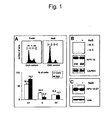
- Figure 1 Sodium butyrate (NaB) inhibits G1 to S phase transition in HeLa cells without affecting viral oncogene expression
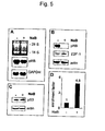
- Figure 3 Regulation of cyclin-dependent kinase inhibitors and suppression of p45 SKP2 by sodium butyrate
- HPV18-positive cervical Carcinoma cells were maintained in Dulbecco's modified Eagle's medium (DMEM), supplemented with 10 % fetal calf serum (Gibco BRL, Rockville, USA), 1% penicillin and streptomycin (Sigma, Deisenhofen, Germany).
- DMEM Dulbecco's modified Eagle's medium
- Primary human keratinocytes, immortalized by E6-, E7- and E6/E7-open reading frames carrying amphotroptic retroviruses were cultivated in "Keratinocyte Medium Kit” (Sigma).
- Trichostatin A (Sigma) was prepared in dimethylsulfoxid (DMSO) (Merck, Darmstadt, Germany).
- Cells were harvested by trypsinisation, washed twice with phosphate-buffered saline (PBS) and fixed overnight with 70 % ethanol. The cells were resuspended in PBS containing 40 ⁇ g/ml of DNase-free RNase A and 50 ⁇ g/ml propidium iodide and cell cycle distribution was measured in a fluorescence-activated cell sorter (FACSort) from Becton Dickinson, San Jose, USA. DNA content was quantified by using the "Cell Quest" software (Becton Dickinson, San Jose, USA).
- FACSort fluorescence-activated cell sorter
- the cDNAs of p21 (el-Deiry et al., Cell 75 (1993), 817-825), p27 (Polyak et al., Cell 78 (1994), 59-66), cyclin D1 (Baldin et al., Genes Dev. 7 (1993), 812-821), cyclin A (Pines and Hunter, Nature 346 (1990), 760-763) and cyclin E (Hinds et al., Cell 70 (1992), 993-1006) were used.
- the cDNA stretch of pRB (nucleotide 379-928) was obtained from M. Tommasino (DKFZ Heidelberg).
- the GAPDH probe (Ercolani et al., J. Biol.
- Chem. 263 (1988, 15335-15341) was obtained from A. Alonso (DKFZ, Heidelberg).
- the unit-length HPV18 genome was cloned in pBR322 (Boshart et al., EMBO J. 3 (1984), 1151-1157).
- cyclin E HE 12
- cyclin D 1 HD 11
- cdk2 M2
- cdk4 C-22
- cdk6 C-21
- p27 KIP1 C-19
- p45 SKP2 N-19
- HPV18-E7 N-19
- E2F-1 KH95
- Cyclin A was kindly provided by M. Pagano (Pagano et al., EMBO J. 11 (1992), 961-971). Equal protein transfer and loading was routinely checked by incubating the filters with a monoclonal actin antibody (ICN Biomedicals, Ohio, USA).
- cdk2 was immunoprecipitated and analyzed by immunoblotting. Beads used for cdk2 kinase assay were washed 3 times with lysis buffer (Blomberg and Hoffmann, 1999), incubated with Laemmli sample buffer and boiled for 5 min. Supernatant was analyzed by SDS-PAGE gels.
- Immunoblotting was carried out with the following antibodies: p21 CIP1 (C24420) and p27 KIP1 (K25020; Transduction Laboratories) or HPV18-E7 (N-19; Santa Cruz, Inc.). Cdk2 specific antibodies or preimmune serum were kindly provided by I. Hoffmann (DKFZ, Heidelberg).
- the rate of apoptosis was determined with the Cell Death Detection kit (ELISA PLUS , Roche Diagnostics, Mannheim, Germany) following the instructions of the manufacturer.
- Histone deacetylase (HDAC) inhibitors induce G1/S phase arrest in HPV18-positive cervical carcinoma cells despite ongoing E6/E7 synthesis
- HAT histone acetylases
- HDAC deacetylases
- HAT activity In higher eukaryotes, particular adaptor molecules such as p300/CBP or p/CAF (termed as p300/CPB-associated factor) possess intrinsic HAT activity. Their association with CREB, c-jun, c-fos or unliganded nuclear receptors provides a functional linkage between transcriptional co-activators and histone acetylators during initiation of gene expression. Conversely, histone deacetylase type 1 (HDAC1) is an inherent component of a general corepressor complex which interacts with YY-1, Mad/Max as well as the retinoblastoma protein pRb, regularly leading to inhibition of gene expression, although exceptions exist (Workman and guitarist, Annu. Rev. Biochemn.
- HDAC1 histone deacetylase type 1
- HPV16 E6 is capable of abrogating the costimulatory function of CBP and p300, resulting in a decreased ability of these factors to trans-activate p53-, NF-KB- and c-jun-responsive promoter elements. While E6 interferes with the CBP/p300 tethering function to other transcription factors and possibly with intrinsic HAT activity, E7 oncoprotein can indirectly bind to the histone deacetylase complex via the bridge-protein Mi2ß.
- E7 to inactivate cellular genes incompatible with the outgrowth of premalignant cells during development of cervical cancer.
- the interferon-regulatory factor-1 (IRF-1) gene whose expression is important for interferon signaling and immunological surveillance of persisting HPV infections, is silenced via an E7-mediated recruitment of HDAC to the respective promoter.
- IRF-1 interferon-regulatory factor-1
- E7 can also act in an opposite way. For example, E7 relieves the repressive effect of pRb and HDAC1 on the cyclin E promoter thereby promoting unscheduled cell cycle progression.
- E6 binds to p53 and promotes its degradation via the ubiquitin/proteosome pathway.
- E7 complexes with the retinoblastoma protein pRb, p107, p130, cyclin A, cyclin E as well as the cyclin-dependent kinase inhibitors p21 CIP1 and p27 KIP1 .
- HPV18-positive cervical carcinoma cell line HeLa as well as primary human foreskin keratinocytes, which were separately immortalized with amphotropic retroviruses carrying the open reading frames of HPV16 E6, E7 or E6/E7 were used in the experiments of the present invention.
- flow cytometric analysis of cellular DNA content was carried out (Fig. 1).
- HDAC inhibitors modulate cyclins but not cdk expression
- Sodium butyrate induces cyclin-dependent kinase inhibitor p21 CIP1 on transcriptional level, while p27 KIP1 is up-regulated post-translationally by concomitant suppression of p45 SKP2 , a component of the ubiquitin-protein ligase SCF SKP2
- CKIs cyclin-dependent kinase inhibitors
- Cdk2 inhibitors such as p21 CIP1 and p27 KIP1 play an important role during immortalization and cellular transformation by potential DNA tumorviruses, because their function can be neutralized, or bypassed after binding of viral oncoproteins such as E7.
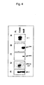
- cyclin-cdk2 complexes were first immunoprecipitated using specific antibodies directed against cdk2, and subsequently assayed in vitro using histone H1 as substrate (Fig. 4A). In comparison with the untreated control, cdk2 kinase activity was completely abolished 16 hours after sodium butyrate addition.
- the most studied G1 specific cyclin/cdk substrate is the retinoblastoma protein pRb, which can recruit HDAC1 to repress cell cycle regulatory proteins such as cyclin E. Since cyclin E was significantly up-regulated by sodium butyrate, it was mandatory to examine the fate of pRb in the experimental cell systems.
- Fig. 5B illustrates that pRB became totally degraded after 16 hours, but histone deacetylase inhibition apparently has no consequences on gene expression and translation of the transcription factor E2F-1. Instead, the absence of pRb could be clearly attributed to a post-translational event, because the steady-state level of the corresponding mRNA was maintained (Fig. 5A).
- the lack of a modulatory effect on p53 (Fig. 5C), whose half-life is controlled by E6, confirmed the previous finding that viral oncoprotein expression is sustained in the presence of sodium butyrate (see Fig. 1 C).
- E2F-1 also did not significantly vary in HPV16-immortalized human keratinocytes, pRb degradation seems to be strictly dependent on the presence of E7. As shown in Fig. 6, pRb completely disappeared exclusively in E7-expressing cells, while it is only seen to be hypophosphorylated in cells containing E6 as viral oncogene. E6/E7-immortalized keratinocytes again revealed pRb degradation, clearly showing that the fate of pRb is determined by E7 in a dominant fashion. Biological availability of pRb can be followed by monitoring the expression level of cyclin E, whose transcription is negatively regulated by pRb. The reason for cyclin E expression in untreated E7-positive cells is consistent with the ability of E7 to overcome the pRb suppressive effect on the cognate promoter by destroying the pRb-HDACI complex.
Abstract
Description
Exponentially growing HeLa cells were treated with trichostatin A as indicated in Fig. 1. 50µg of protein was separated on 12% SDS-PAGE minigels. After electrotransfer, the filters were incubated with antibodies against cyclin E and cyclin A. Equal protein loading was confirmed by reincubating the filters with a monoclonal actin antibody. (Contr.): untreated cells; (DMSO): DMSO control; (TSA): treated with 330 nM trichostatin A for 16 hours.
Western blot analysis. 50 µg of cellular protein was loaded on SDS-PAGE minigels. After electrotransfer, the filters were incubated with p21CIP1, p27KIP1, p16INK4 and p45SKP2. Equal protein loading was confirmed by a monoclonal actin antibody. (Contr.): untreated cells; (DMSO): DMSO control; (TSA): treated with 330 nM trichostatin A for 16 hours.
Autoradiography: cdk2 complexes were immunoprecipitated from HeLa cells and assayed for their activity as in (c). Cells were treated for 3, 6, 9, 12 and 16 hours with 6 mM sodium butyrate (NaB). (Contr.): untreated cells harvested after 3, 6, 9, 12, and 16 hours.
The expression of E6 and E7 was detected after electrophoresis of the reverse transcriptase (RT)-PCR products on 2 % agarose gels. Expected E6, E6* or E7 fragments are indicated. As control GAPDH mRNA was detected by RT-PCR (460 bp). The RT-reaction was performed with the Superscript TMII (Gibco) following the attached instructions of the manufacturer, using 1,5 µg of total RNA. For E6 detection, the following primers were used: upper primer 5' ACT GCA ATG TTT CAG GAC CC 3', lower primer 5' TCA GGA CAC AGT GGC TTT TG 3', for the E7 detection; upper primer 5' CCC AGC TGT AAT CAT GCA TG 3', lower primer 5' TGC CCA TTA ACA GGT CTT CC 3'. For GAPDH detection, the following forward (TGG ATA TTG TTG CCA TCA ATG ACC) and reverse (GAT GGC ATG GAC TGT GGT CAT G) primers were used. Conditions for all sets of primers were: initially denaturation time of 3 min. at 94°C and then 30 sec. at 94°C, 1 min. at 60°C, 1 min. at 72°C (35 cycles) and 10 min. at 72°C. (1): E6 and (2) E6* fragments; (#): E7 fragment; (+): treated with 6 mM sodium butyrate for 16 hours; (-): untreated cells.
Claims (2)
- Use of a histone deacetylase inhibitor for the preparation of a medicament for the treatment of a disease associated with an human papillomavirus (HPV) infection, wherein the disease is cervical cancer, cervical intraepithelial neoplasm, wart, larynx papilloma or condyloma acuminatum.
- Use according to claim 1, wherein the histone deacetylase inhibitor is sodium butyrate, phenylbutyrate or trichostatin A.
Priority Applications (5)
| Application Number | Priority Date | Filing Date | Title |
|---|---|---|---|
| DE60113645T DE60113645T2 (en) | 2001-04-10 | 2001-04-10 | Use of histone deacetylase inhibitors for the treatment of papillomavirus-associated diseases |
| EP01108923A EP1249246B1 (en) | 2001-04-10 | 2001-04-10 | Use of an histone deacetylase inhibitor for the treatment of diseases associated with an hpv infection |
| AT01108923T ATE305297T1 (en) | 2001-04-10 | 2001-04-10 | USE OF HISTONEDEACETYLASE INHIBITORS FOR THE TREATMENT OF PAPILLOMAVIRUS-ASSOCIATED DISEASES |
| PCT/EP2002/004004 WO2002083173A1 (en) | 2001-04-10 | 2002-04-10 | Use of an histone deacetylase inhibitor for the treatment of diseases associated with an hpv infection |
| US10/474,628 US20040132817A1 (en) | 2001-04-10 | 2002-04-10 | Use of an histone deacetylase inhibitor for the treatment of diseases associated with an hpv infection |
Applications Claiming Priority (1)
| Application Number | Priority Date | Filing Date | Title |
|---|---|---|---|
| EP01108923A EP1249246B1 (en) | 2001-04-10 | 2001-04-10 | Use of an histone deacetylase inhibitor for the treatment of diseases associated with an hpv infection |
Publications (2)
| Publication Number | Publication Date |
|---|---|
| EP1249246A1 EP1249246A1 (en) | 2002-10-16 |
| EP1249246B1 true EP1249246B1 (en) | 2005-09-28 |
Family
ID=8177103
Family Applications (1)
| Application Number | Title | Priority Date | Filing Date |
|---|---|---|---|
| EP01108923A Expired - Lifetime EP1249246B1 (en) | 2001-04-10 | 2001-04-10 | Use of an histone deacetylase inhibitor for the treatment of diseases associated with an hpv infection |
Country Status (5)
| Country | Link |
|---|---|
| US (1) | US20040132817A1 (en) |
| EP (1) | EP1249246B1 (en) |
| AT (1) | ATE305297T1 (en) |
| DE (1) | DE60113645T2 (en) |
| WO (1) | WO2002083173A1 (en) |
Cited By (1)
| Publication number | Priority date | Publication date | Assignee | Title |
|---|---|---|---|---|
| US9078864B2 (en) | 2008-01-08 | 2015-07-14 | Akthelia Pharmaceuticals | Agonists for antimicrobial peptide systems |
Families Citing this family (10)
| Publication number | Priority date | Publication date | Assignee | Title |
|---|---|---|---|---|
| EP1291015A1 (en) | 2001-09-10 | 2003-03-12 | Lunamed AG | Dosage forms having prolonged active ingredient release |
| DE60332367D1 (en) * | 2002-08-20 | 2010-06-10 | Astellas Pharma Inc | HEMMER OF THE ABBAUS OF THE EXTRACELLULAR MATRIX OF ARTHRODIA CARTILAGE |
| US20050222013A1 (en) * | 2003-01-16 | 2005-10-06 | Georgetown University | Methods for the use of inhibitors of histone deacetylase as synergistic agents in cancer therapy |
| US20050059682A1 (en) * | 2003-09-12 | 2005-03-17 | Supergen, Inc., A Delaware Corporation | Compositions and methods for treatment of cancer |
| WO2007011626A2 (en) | 2005-07-14 | 2007-01-25 | Takeda San Diego, Inc. | Histone deacetylase inhibitors |
| CA2574531C (en) * | 2007-01-19 | 2016-10-25 | The University Of British Columbia | Hat acetylation promoters and uses of compositions thereof in promoting immunogenicity |
| US20120269818A1 (en) * | 2011-04-25 | 2012-10-25 | Ozbun Michelle A | Methods for treating infection by hpv |
| WO2014138507A1 (en) * | 2013-03-06 | 2014-09-12 | C & C Biopharma, Llc | Treatment of cervical cancer and/or ovarian cancer using a transcription factor modulator |
| US10307406B2 (en) * | 2013-08-31 | 2019-06-04 | The Wistar Institute Of Anatomy And Biology | Methods and compositions for re-activating Epstein-Barr virus and screening compounds therefor |
| US20230233520A1 (en) * | 2020-04-23 | 2023-07-27 | Georgetown University | Combination therapy of artemisinin-related compounds and histone deacetylase inhibitors for treatment of hpv-related benign, premalignant, and malignant diseases |
Family Cites Families (3)
| Publication number | Priority date | Publication date | Assignee | Title |
|---|---|---|---|---|
| JPS61176523A (en) * | 1985-01-30 | 1986-08-08 | Teruhiko Beppu | Carcinostatic agent |
| EP0827742A1 (en) * | 1996-09-04 | 1998-03-11 | Vrije Universiteit Brussel | Use of histone deacetylase inhibitors for treating fribosis or cirrhosis |
| US6124495A (en) * | 1997-03-11 | 2000-09-26 | Beacon Laboratories, Inc. | Unsaturated oxyalkylene esters and uses thereof |
-
2001
- 2001-04-10 EP EP01108923A patent/EP1249246B1/en not_active Expired - Lifetime
- 2001-04-10 AT AT01108923T patent/ATE305297T1/en not_active IP Right Cessation
- 2001-04-10 DE DE60113645T patent/DE60113645T2/en not_active Expired - Fee Related
-
2002
- 2002-04-10 WO PCT/EP2002/004004 patent/WO2002083173A1/en not_active Application Discontinuation
- 2002-04-10 US US10/474,628 patent/US20040132817A1/en not_active Abandoned
Non-Patent Citations (1)
| Title |
|---|
| KIM Y.B. ET AL: "Oxamflatin is a novel antitumour compound that inhibits mammalian histone deacetylase.", ONCOGENE, vol. 18, 15 April 1999 (1999-04-15), pages 2461 - 2470 * |
Cited By (1)
| Publication number | Priority date | Publication date | Assignee | Title |
|---|---|---|---|---|
| US9078864B2 (en) | 2008-01-08 | 2015-07-14 | Akthelia Pharmaceuticals | Agonists for antimicrobial peptide systems |
Also Published As
| Publication number | Publication date |
|---|---|
| ATE305297T1 (en) | 2005-10-15 |
| WO2002083173A1 (en) | 2002-10-24 |
| DE60113645D1 (en) | 2006-02-09 |
| EP1249246A1 (en) | 2002-10-16 |
| DE60113645T2 (en) | 2006-07-06 |
| US20040132817A1 (en) | 2004-07-08 |
Similar Documents
| Publication | Publication Date | Title |
|---|---|---|
| Finzer et al. | Inhibitors of histone deacetylase arrest cell cycle and induce apoptosis in cervical carcinoma cells circumventing human papillomavirus oncogene expression | |
| Corsten et al. | The microRNA-221/-222 cluster balances the antiviral and inflammatory response in viral myocarditis | |
| Herbein et al. | Histone deacetylases in viral infections | |
| Yu et al. | Inhibitory role of peroxisome proliferator‐activated receptor gamma in hepatocarcinogenesis in mice and in vitro | |
| EP1249246B1 (en) | Use of an histone deacetylase inhibitor for the treatment of diseases associated with an hpv infection | |
| Bodily et al. | Human papillomavirus E7 enhances hypoxia-inducible factor 1–mediated transcription by inhibiting binding of histone deacetylases | |
| Gilbert et al. | Histone deacetylase inhibition attenuates diabetes-associated kidney growth: potential role for epigenetic modification of the epidermal growth factor receptor | |
| Grabiec et al. | Targeting histone deacetylase activity in rheumatoid arthritis and asthma as prototypes of inflammatory disease: should we keep our HATs on? | |
| Davies et al. | Spiruchostatin A inhibits proliferation and differentiation of fibroblasts from patients with pulmonary fibrosis | |
| Heller et al. | Tetra-O-methyl nordihydroguaiaretic acid induces G2 arrest in mammalian cells and exhibits tumoricidal activity in vivo | |
| Kusaczuk et al. | Phenylbutyrate—A pan-HDAC inhibitor—Suppresses proliferation of glioblastoma LN-229 cell line | |
| Gomez et al. | Stimulation of primary human endothelial cell proliferation by IFN | |
| O'Shea et al. | Regulation of the RelA (p65) transactivation domain | |
| Darcis et al. | Preclinical shock strategies to reactivate latent HIV-1: an update | |
| Scherer et al. | The human CMV IE1 protein: an offender of PML nuclear bodies | |
| Finzer et al. | HDAC inhibitors trigger apoptosis in HPV-positive cells by inducing the E2F–p73 pathway | |
| Prayson et al. | Cyclooxygenase-2 (COX-2) expression by immunohistochemistry in glioblastoma multiforme | |
| Kang et al. | Effect of α-interferon on P-glycoprotein expression and function and on verapamil modulation of doxorubicin resistance | |
| Nagata et al. | Histone deacetylase inhibitor SAHA treatment prevents the development of heart failure after myocardial infarction via an induction of heat-shock proteins in rats | |
| Yang et al. | CD74 knockout attenuates alcohol intake-induced cardiac dysfunction through AMPK-Skp2-mediated regulation of autophagy | |
| US20110003753A1 (en) | COMPOSITIONS AND METHODS FOR DISRUPTING THE FUNCTION OF THE TRANSCRIPTIONAL REPRESSOR COMPONENT Sin3A-PAH2 DOMAIN TO INDUCE DIFFERENTIATION AND GROWTH INHIBITION IN BREAST CANCER | |
| Ci et al. | Inhibitory effect of Saposhnikovia divaricate polysaccharide on fibroblast-like synoviocytes from rheumatoid arthritis rat in vitro. | |
| Narayanan et al. | The effect of all-trans and 9-cis retinoic acid on the steady state level of HPV16 E6/E7 mRNA and cell cycle in cervical carcinoma cells | |
| WO2013012477A1 (en) | Propolis and caffeic acid phenethyl ester and uses thereof | |
| Hartlapp et al. | Depsipeptide induces cell death in Hodgkin lymphoma-derived cell lines |
Legal Events
| Date | Code | Title | Description |
|---|---|---|---|
| PUAI | Public reference made under article 153(3) epc to a published international application that has entered the european phase |
Free format text: ORIGINAL CODE: 0009012 |
|
| AK | Designated contracting states |
Kind code of ref document: A1 Designated state(s): AT BE CH CY DE DK ES FI FR GB GR IE IT LI LU MC NL PT SE TR |
|
| AX | Request for extension of the european patent |
Free format text: AL;LT;LV;MK;RO;SI |
|
| 16A | New documents despatched to applicant after publication of the search report |
Effective date: 20021011 |
|
| 17P | Request for examination filed |
Effective date: 20021218 |
|
| 17Q | First examination report despatched |
Effective date: 20030210 |
|
| AKX | Designation fees paid |
Designated state(s): AT BE CH CY DE DK ES FI FR GB GR IE IT LI LU MC NL PT SE TR |
|
| GRAP | Despatch of communication of intention to grant a patent |
Free format text: ORIGINAL CODE: EPIDOSNIGR1 |
|
| RIC1 | Information provided on ipc code assigned before grant |
Ipc: 7A 61P 35/00 B Ipc: 7A 61K 31/19 A Ipc: 7A 61K 31/165 B |
|
| GRAS | Grant fee paid |
Free format text: ORIGINAL CODE: EPIDOSNIGR3 |
|
| GRAA | (expected) grant |
Free format text: ORIGINAL CODE: 0009210 |
|
| AK | Designated contracting states |
Kind code of ref document: B1 Designated state(s): AT BE CH CY DE DK ES FI FR GB GR IE IT LI LU MC NL PT SE TR |
|
| PG25 | Lapsed in a contracting state [announced via postgrant information from national office to epo] |
Ref country code: IT Free format text: LAPSE BECAUSE OF FAILURE TO SUBMIT A TRANSLATION OF THE DESCRIPTION OR TO PAY THE FEE WITHIN THE PRESCRIBED TIME-LIMIT;WARNING: LAPSES OF ITALIAN PATENTS WITH EFFECTIVE DATE BEFORE 2007 MAY HAVE OCCURRED AT ANY TIME BEFORE 2007. THE CORRECT EFFECTIVE DATE MAY BE DIFFERENT FROM THE ONE RECORDED. Effective date: 20050928 Ref country code: FI Free format text: LAPSE BECAUSE OF FAILURE TO SUBMIT A TRANSLATION OF THE DESCRIPTION OR TO PAY THE FEE WITHIN THE PRESCRIBED TIME-LIMIT Effective date: 20050928 Ref country code: BE Free format text: LAPSE BECAUSE OF FAILURE TO SUBMIT A TRANSLATION OF THE DESCRIPTION OR TO PAY THE FEE WITHIN THE PRESCRIBED TIME-LIMIT Effective date: 20050928 Ref country code: AT Free format text: LAPSE BECAUSE OF FAILURE TO SUBMIT A TRANSLATION OF THE DESCRIPTION OR TO PAY THE FEE WITHIN THE PRESCRIBED TIME-LIMIT Effective date: 20050928 Ref country code: NL Free format text: LAPSE BECAUSE OF FAILURE TO SUBMIT A TRANSLATION OF THE DESCRIPTION OR TO PAY THE FEE WITHIN THE PRESCRIBED TIME-LIMIT Effective date: 20050928 |
|
| REG | Reference to a national code |
Ref country code: GB Ref legal event code: FG4D |
|
| REG | Reference to a national code |
Ref country code: CH Ref legal event code: EP |
|
| REG | Reference to a national code |
Ref country code: IE Ref legal event code: FG4D |
|
| PG25 | Lapsed in a contracting state [announced via postgrant information from national office to epo] |
Ref country code: SE Free format text: LAPSE BECAUSE OF FAILURE TO SUBMIT A TRANSLATION OF THE DESCRIPTION OR TO PAY THE FEE WITHIN THE PRESCRIBED TIME-LIMIT Effective date: 20051228 Ref country code: DK Free format text: LAPSE BECAUSE OF FAILURE TO SUBMIT A TRANSLATION OF THE DESCRIPTION OR TO PAY THE FEE WITHIN THE PRESCRIBED TIME-LIMIT Effective date: 20051228 Ref country code: GR Free format text: LAPSE BECAUSE OF FAILURE TO SUBMIT A TRANSLATION OF THE DESCRIPTION OR TO PAY THE FEE WITHIN THE PRESCRIBED TIME-LIMIT Effective date: 20051228 |
|
| PG25 | Lapsed in a contracting state [announced via postgrant information from national office to epo] |
Ref country code: ES Free format text: LAPSE BECAUSE OF FAILURE TO SUBMIT A TRANSLATION OF THE DESCRIPTION OR TO PAY THE FEE WITHIN THE PRESCRIBED TIME-LIMIT Effective date: 20060108 |
|
| REF | Corresponds to: |
Ref document number: 60113645 Country of ref document: DE Date of ref document: 20060209 Kind code of ref document: P |
|
| REG | Reference to a national code |
Ref country code: CH Ref legal event code: NV Representative=s name: ING. MARCO ZARDI C/O M. ZARDI & CO. S.A. |
|
| PG25 | Lapsed in a contracting state [announced via postgrant information from national office to epo] |
Ref country code: PT Free format text: LAPSE BECAUSE OF FAILURE TO SUBMIT A TRANSLATION OF THE DESCRIPTION OR TO PAY THE FEE WITHIN THE PRESCRIBED TIME-LIMIT Effective date: 20060228 |
|
| NLV1 | Nl: lapsed or annulled due to failure to fulfill the requirements of art. 29p and 29m of the patents act | ||
| PG25 | Lapsed in a contracting state [announced via postgrant information from national office to epo] |
Ref country code: IE Free format text: LAPSE BECAUSE OF NON-PAYMENT OF DUE FEES Effective date: 20060410 |
|
| PG25 | Lapsed in a contracting state [announced via postgrant information from national office to epo] |
Ref country code: MC Free format text: LAPSE BECAUSE OF NON-PAYMENT OF DUE FEES Effective date: 20060430 |
|
| ET | Fr: translation filed | ||
| PLBE | No opposition filed within time limit |
Free format text: ORIGINAL CODE: 0009261 |
|
| STAA | Information on the status of an ep patent application or granted ep patent |
Free format text: STATUS: NO OPPOSITION FILED WITHIN TIME LIMIT |
|
| 26N | No opposition filed |
Effective date: 20060629 |
|
| REG | Reference to a national code |
Ref country code: IE Ref legal event code: MM4A |
|
| PG25 | Lapsed in a contracting state [announced via postgrant information from national office to epo] |
Ref country code: LU Free format text: LAPSE BECAUSE OF NON-PAYMENT OF DUE FEES Effective date: 20060410 Ref country code: TR Free format text: LAPSE BECAUSE OF FAILURE TO SUBMIT A TRANSLATION OF THE DESCRIPTION OR TO PAY THE FEE WITHIN THE PRESCRIBED TIME-LIMIT Effective date: 20050928 |
|
| PGFP | Annual fee paid to national office [announced via postgrant information from national office to epo] |
Ref country code: CH Payment date: 20080428 Year of fee payment: 8 |
|
| PGFP | Annual fee paid to national office [announced via postgrant information from national office to epo] |
Ref country code: DE Payment date: 20080630 Year of fee payment: 8 |
|
| PG25 | Lapsed in a contracting state [announced via postgrant information from national office to epo] |
Ref country code: CY Free format text: LAPSE BECAUSE OF FAILURE TO SUBMIT A TRANSLATION OF THE DESCRIPTION OR TO PAY THE FEE WITHIN THE PRESCRIBED TIME-LIMIT Effective date: 20050928 |
|
| PGFP | Annual fee paid to national office [announced via postgrant information from national office to epo] |
Ref country code: GB Payment date: 20080430 Year of fee payment: 8 |
|
| REG | Reference to a national code |
Ref country code: CH Ref legal event code: PL |
|
| GBPC | Gb: european patent ceased through non-payment of renewal fee |
Effective date: 20090410 |
|
| REG | Reference to a national code |
Ref country code: FR Ref legal event code: ST Effective date: 20091231 |
|
| PG25 | Lapsed in a contracting state [announced via postgrant information from national office to epo] |
Ref country code: DE Free format text: LAPSE BECAUSE OF NON-PAYMENT OF DUE FEES Effective date: 20091103 Ref country code: CH Free format text: LAPSE BECAUSE OF NON-PAYMENT OF DUE FEES Effective date: 20090430 Ref country code: LI Free format text: LAPSE BECAUSE OF NON-PAYMENT OF DUE FEES Effective date: 20090430 |
|
| PG25 | Lapsed in a contracting state [announced via postgrant information from national office to epo] |
Ref country code: FR Free format text: LAPSE BECAUSE OF NON-PAYMENT OF DUE FEES Effective date: 20091222 Ref country code: GB Free format text: LAPSE BECAUSE OF NON-PAYMENT OF DUE FEES Effective date: 20090410 |
|
| PGFP | Annual fee paid to national office [announced via postgrant information from national office to epo] |
Ref country code: FR Payment date: 20080429 Year of fee payment: 8 |