EP1246597B1 - Polypeptides and polynucleotides for enhancing immune reactivity to her-2 protein - Google Patents
Polypeptides and polynucleotides for enhancing immune reactivity to her-2 protein Download PDFInfo
- Publication number
- EP1246597B1 EP1246597B1 EP00953823.2A EP00953823A EP1246597B1 EP 1246597 B1 EP1246597 B1 EP 1246597B1 EP 00953823 A EP00953823 A EP 00953823A EP 1246597 B1 EP1246597 B1 EP 1246597B1
- Authority
- EP
- European Patent Office
- Prior art keywords
- peptide
- epitope
- sequence
- chimeric peptide
- cell
- Prior art date
- Legal status (The legal status is an assumption and is not a legal conclusion. Google has not performed a legal analysis and makes no representation as to the accuracy of the status listed.)
- Expired - Lifetime
Links
Images


Classifications
-
- A—HUMAN NECESSITIES
- A61—MEDICAL OR VETERINARY SCIENCE; HYGIENE
- A61K—PREPARATIONS FOR MEDICAL, DENTAL OR TOILETRY PURPOSES
- A61K9/00—Medicinal preparations characterised by special physical form
- A61K9/0012—Galenical forms characterised by the site of application
- A61K9/0019—Injectable compositions; Intramuscular, intravenous, arterial, subcutaneous administration; Compositions to be administered through the skin in an invasive manner
-
- A—HUMAN NECESSITIES
- A61—MEDICAL OR VETERINARY SCIENCE; HYGIENE
- A61K—PREPARATIONS FOR MEDICAL, DENTAL OR TOILETRY PURPOSES
- A61K9/00—Medicinal preparations characterised by special physical form
- A61K9/14—Particulate form, e.g. powders, Processes for size reducing of pure drugs or the resulting products, Pure drug nanoparticles
- A61K9/16—Agglomerates; Granulates; Microbeadlets ; Microspheres; Pellets; Solid products obtained by spray drying, spray freeze drying, spray congealing,(multiple) emulsion solvent evaporation or extraction
- A61K9/1605—Excipients; Inactive ingredients
- A61K9/1629—Organic macromolecular compounds
- A61K9/1641—Organic macromolecular compounds obtained otherwise than by reactions only involving carbon-to-carbon unsaturated bonds, e.g. polyethylene glycol, poloxamers
- A61K9/1647—Polyesters, e.g. poly(lactide-co-glycolide)
-
- A—HUMAN NECESSITIES
- A61—MEDICAL OR VETERINARY SCIENCE; HYGIENE
- A61P—SPECIFIC THERAPEUTIC ACTIVITY OF CHEMICAL COMPOUNDS OR MEDICINAL PREPARATIONS
- A61P35/00—Antineoplastic agents
-
- A—HUMAN NECESSITIES
- A61—MEDICAL OR VETERINARY SCIENCE; HYGIENE
- A61P—SPECIFIC THERAPEUTIC ACTIVITY OF CHEMICAL COMPOUNDS OR MEDICINAL PREPARATIONS
- A61P37/00—Drugs for immunological or allergic disorders
- A61P37/02—Immunomodulators
-
- A—HUMAN NECESSITIES
- A61—MEDICAL OR VETERINARY SCIENCE; HYGIENE
- A61P—SPECIFIC THERAPEUTIC ACTIVITY OF CHEMICAL COMPOUNDS OR MEDICINAL PREPARATIONS
- A61P37/00—Drugs for immunological or allergic disorders
- A61P37/02—Immunomodulators
- A61P37/04—Immunostimulants
-
- C—CHEMISTRY; METALLURGY
- C07—ORGANIC CHEMISTRY
- C07K—PEPTIDES
- C07K14/00—Peptides having more than 20 amino acids; Gastrins; Somatostatins; Melanotropins; Derivatives thereof
- C07K14/435—Peptides having more than 20 amino acids; Gastrins; Somatostatins; Melanotropins; Derivatives thereof from animals; from humans
- C07K14/705—Receptors; Cell surface antigens; Cell surface determinants
- C07K14/71—Receptors; Cell surface antigens; Cell surface determinants for growth factors; for growth regulators
-
- A—HUMAN NECESSITIES
- A61—MEDICAL OR VETERINARY SCIENCE; HYGIENE
- A61K—PREPARATIONS FOR MEDICAL, DENTAL OR TOILETRY PURPOSES
- A61K39/00—Medicinal preparations containing antigens or antibodies
- A61K2039/60—Medicinal preparations containing antigens or antibodies characteristics by the carrier linked to the antigen
- A61K2039/6031—Proteins
- A61K2039/6037—Bacterial toxins, e.g. diphteria toxoid [DT], tetanus toxoid [TT]
-
- A—HUMAN NECESSITIES
- A61—MEDICAL OR VETERINARY SCIENCE; HYGIENE
- A61K—PREPARATIONS FOR MEDICAL, DENTAL OR TOILETRY PURPOSES
- A61K39/00—Medicinal preparations containing antigens or antibodies
- A61K2039/64—Medicinal preparations containing antigens or antibodies characterised by the architecture of the carrier-antigen complex, e.g. repetition of carrier-antigen units
Definitions
- HER-2 protein a product of the HER-2 oncogene
- the HER-2 protein is overexpressed in a variety of cancers. It is found in 50%-60% of ductal in situ carcinoma and 20%-40% of all breast cancers, as well as a substantial fraction of adenocarcinomas arising in the ovaries, prostate, colon and lung.
- Overexpression of the HER-2 protein is related to malignant transformation in humans. Overexpression of the HER-2 protein is also intimately associated with the aggressiveness of the malignancy, being found in one-fourth of all invasive breast cancers. Overexpression of HER-2 protein is correlated with a poor prognosis in both breast and ovarian cancer.
- trastuzumab has also been shown to improve survival when combined with cytotoxic chemotherapeutics ( Baselga, J., et al., J. Clin. Oncol. 14:737-44, 1996 ; Pegram, M.D., et al., J. Clin. Oncol., 16:2659-71, 1988 .).
- a number of vaccine approaches targeting a recombinant HER-2 protein, the HER-2 ECD, or the ECD of rat neu, which is the rat homolog of HER-2 have also been evaluated.
- strain NFS mice immunized with a vaccinia virus recombinant that expresses the ECD rat neu developed a protective antibody response against subsequent challenge with neu-transformed NIH 3T3 cells ( Bernards, R., et al., Proc. Natl. Acad. Sci. USA, 84:6854-8, 1987 .).
- Immunization of BDIX rats with the same immunogen did not result in antibody response nor did it inhibit the growth of syngeneic neu-expressing B 104 neuroblastoma cells, suggesting that this strategy was insufficient to induce immune responses in the rat.
- a polysaccharide-oncoprotein complex vaccine consisting of the 147 amino-terminal amino acids of HER-2 ECD complexed with cholesteryl group-bearing mannan and pullulan, induced cellular and humoral immune responses that mediated rejection of HER-2-expressing sarcomas in BALB/c mice ( Gu, X. G., et al., Cancer Res., 58: 3385-90, 1998 .). Partial protection was shown in rat neu transgenic mice destined to develop mammary tumors by immunizing with either a purified rat neu ECD ( Esserman, L. J., Cancer Immunol.
- WO97/38011 describes (i) a non-dendritic peptide for use as a carrier of an immunogenic substance and/or an immune mediator, (ii) a construct of said carrier carrying an immunogenic substance and/or an immune mediator, (iii) a process for the preparation of immunogens with high and predictable immunogenicity which comprise said non-dendritic peptide carrier, (iv) use of such immunogens for the production of vaccines and (v) vaccines comprising an immunogenic substance and/or an immune mediator on the peptide carrier.
- the non-dendritic peptide carrier comprises 10-50 amino acids capable of forming a secondary structure in a benign buffer after liberation from the solid phase.
- WO94/07530 describes a vaccine for fertility control and cancer treatment.
- the vaccine comprises an antigenic conjugate of a protein reproductive hormone, a fragment of such a hormone, or a peptide substantially immunologically equivalent to such a hormone or fragment conjugated with a chemically-modified diphtheria toxoid, and an adjuvant, both dispersed in an aqueous medium and emulsified with a mixture of oils.
- US 5 876 712 A describes methods for the detection, monitoring and treatment of malignancies in which the HER-2/neu oncogene is associated. Also described are methods and compositions, including peptides, for treating such malignancies.
- the authors describe a chimeric immunogen incorporating a promiscuous T-cell epitope to enhance the immunogenicity of an oligopeptide corresponding to a weakly immunogenic substitution.
- GM-CSF granulocyte-macrophage colony- stimulating factor
- GM-CSF was as effective as CFA in generating rat neu specific DTH responses after immunization with a neu peptide based vaccine.
- Soluble GM-CSF is a potent adjuvant for the generation of immune responses to foreign proteins as well as peptides derived from a self tumor antigen.
- HER2-derived peptide HER2p63 (A) (TYLPANASL).
- the HER2-derived peptide is reported to be able to induce Kd-restricted mouse cytotoxic T lymphocytes (CTL) and function as a tumor rejection antigen in an in vivo assay.
- CTL cytotoxic T lymphocytes
- T human peptide HER2p63
- TYLPTNASL autologous antigen presenting cells exposed to this peptide were able to sensitise killer cells in vitro such that they selectively lysed HER2-expressing SKOV3 transfected with HLA-A2402 cDNA.
- MVFMF2 human T-lymphotropic virus type 1
- GPSL four residue turn
- adjuvant N-acetyl-glucosamine-3yl-acetyl-L-alanyl-D-isoglutamine, nor-MDP
- the present invention provides a chimeric peptide for stimulating an immune response to HER-2 protein, wherein said chimeric peptide comprises a HER-2 B cell epitope, a T-helper (Th) epitope, and a linker joining said HER-2 B cell epitope to said Th epitope; wherein the sequence of said HER-2 B cell epitope is
- sequence of said linker is the sequence G-P-S-L, (SEQ ID NO. 20.)
- the invention also provides a composition for stimulating an immune response to HER-2 protein, wherein said composition comprises a first chimeric peptide and a second chimeric peptide; wherein said first chimeric peptide is the chimeric peptide of the invention as described above; wherein said second chimeric peptide comprising a HER-2 B cell epitope, a T-helper (Th) epitope, and a linker joining said HER-2 B cell epitope to said Th epitope; wherein the sequence of the HER-2 B cell epitope of said second chimeric peptide is selected from the group consisting of:
- sequence of the linker of said second chimeric peptide is the sequence G-P-S-L, (SEQ ID NO. 20)
- the invention also provides for the use of the chimeric peptide or the composition of the invention, as described above, in the manufacture of a medicament for the treatment of cancer in a subject.
- the medicament is for the treatment of one of the following cancers, or a predisposition to one of the following cancers in a human: breast cancer, ovarian cancer, lung cancer, prostate cancer, and colon cancer.
- the invention further provides a pharmaceutical composition comprising the chimeric peptide or the composition of the invention, as described above, and a pharmaceutically acceptable vehicle.
- vehicle is biodegradeable and is selected from the group consisting of an emulsion comprising a pharmaceutically acceptable oil/water emulsion and a biodegradeable microsphere or nanosphere comprising a polylactide-polyglycolic acid polymer.
- oil is squalene or squalane.
- the microsphere is from 0.1 to 50 nanometers in diameter and comprises poly (D, I lactide-co-glycide).
- the invention also provides an isolated polynucleotide which encodes the chimeric peptide of the invention, as described above.
- the invention futher provides a composition for stimulating an immune response to HER-2 protein, wherein said composition comprises a first chimeric peptide and a second chimeric peptide, wherein each chimeric peptide comprises a HER-2 B cell epitope, a T-helper (Th) epitope, and a linker joining said HER-2 B cell epitope to said Th epitope; wherein the sequence of the HER-2 B cell epitope of the first chimeric peptide is:
- HER-2 B cell epitopes a peptide that are immunogenic epitopes of the HER-2 protein, referred to hereinafter as HER-2 B cell epitopes and HER-2 CTL epitopes.
- the HER-2 B cell epitopes are capable of invoking a humoral response which results in the production of antibodies that are immunoreactive with the extracellular domain of the HER-2 protein.
- HER-2 protein, and its rat homolog neu are transmembrane proteins with a relative molecular mass of 185 kd that is approximately 1255 amino acids (aa) in length.
- HER-2/neu protein has an extracellular binding domain (ECD) of approximately 645 aa, with 40% homology to epidermal growth factor receptor (EGFR), a highly hydrophobic transmembrane anchor domain (TMD), and a carboxyterminal cytoplasmic domain (CD) of approximately 580 aa with n 80% homology to EGFR.
- ECD extracellular binding domain
- the amino acid sequence of the HER-2 protein and a nucleotide sequence which encodes such amino acid sequence are shown in GenBank Accession No. M11730.
- the HER-2 B cell epitopes encompass peptides having one of the sequences, referred to hereinafter as the "reference sequences", shown in Table I below.
- the reference sequences were selected and scored using computer-aided analysis using six correlates of antigenicity: (a) the profiles of chain flexibility and mobility of individual sequences was calculated according to Karplus and Schultz, Naturwiss 72:212-213, 1985 ; (b) hydropathy profiles were generated over a seven residue span setting and were finally smoothed with a three residue span using the scale of Kyte and Doolittle, J. Moi. Biol. 157:105-132, 1982 ; (c) hydrophilicity profiles were generated over a 6-residue window using the program of Hopp and Woods, Proc.
- Sequences were given a score of 1 to 6 based on their respective index values and were ranked: the highest ranking sequences had the highest individual score for the analyses examined (6/6), and successive candidates had the next highest score (5/6), etc.
- the best scoring epitopes were further ranked by correlation with their secondary structural attributes, e.g .., an amphiphilic ⁇ -helical sequence or a ⁇ -turn loop region are preferred over a random coil fragment.
- Computer programs by Chou and Fasman, Adv. Enzymol. Relat. Subj. Biochem. 47: 45-148, 1978 were used to predict the secondary structure ( ⁇ -helix, ⁇ -strand/sheet, (3-turn/loop, random coil) and helical amphiphilic moment.
- Electrostatic ion pairs and helix dipole interaction in helical segment were also considered (e.g., hydrophobic / hydrophilic balance).
- the hydrophilic/hydrophobic balance is from 2/2 to 4/1.
- the HER-2 B cell epitopes also encompass peptides that are functional equivalents of the peptides shown in Table I below. Such functional equivalents have an altered sequence in which one or more of the amino acids in the corresponding reference sequence is substituted or in which one or more amino acids are deleted from or added to the corresponding reference sequence. For example, cysteine residues may be deleted or replaced with other amino acids to prevent formation of incorrect intramolecular disulfide bridges upon renaturation.
- the HER-2 B cell epitopes are glycosylated.
- substitutions be conservative amino acid substitutions, in which the substituted amino acid has similar structural or chemical properties with the corresponding amino acid in the reference sequence.
- conservative amino acid substitutions involve substitution of one aliphatic or hydrophobic amino acids, e.g., alanine, valine, leucine and isoleucine, with another; substitution of one hydroxyl-containing amino acid, e.g., serine and threonine, with another; substitution of one acidic residue, e.g., glutamic acid or aspartic acid, with another; replacement of one amide-containing residue, e.g., asparagine and glutamine, with another; replacement of one aromatic residue, e.g., phenylalanine and tyrosine, with another; replacement of one basic residue, e.g., lysine, arginine and histidine, with another; and replacement of one small amino acid, e.g., alanine, serine, threonine, methionine, and glycine, with another.
- substitution of one aliphatic or hydrophobic amino acids e.g., alanine, va
- the deletions and additions are located at the amino terminus, the carboxy terminus, or both, of one of the sequences shown above.
- the HER-2 B cell epitope equivalent has an amino acid sequence which is at least 70% identical, preferably at least 80% identical, more preferably at least 90% identical, most preferably, at least 95% identical to the corresponding reference sequences. Sequences which are at least 90% identical have no more than 1 alteration, i.e., any combination of deletions, additions or substitutions, per 10 amino acids of the reference sequence. Percent identity is determined by comparing the amino acid sequence of the variant with the reference sequence using MEGALIGN project in the DNA STAR program.
- the functional equivalent For functional equivalents that are longer than a corresponding reference sequence, it is preferred that the functional equivalent have a sequence which is at least 90% identical to the reference sequence and the sequences which flank the reference sequence in the wild-type HER-2 protein.
- Table 1 Consolidated Human p185 HER-2 predicted B cell epitopes listed in the order of ranking by amino acid residue numbers. Aspargine (N)-linked glycosylation sites are underlined in bold.
- Predictive Ranking Residue Amino Acid Sequence Secondary Structure 7 27 - 45 Tgtdmklrlpaspethldm 25 - 28 ⁇ turn; 29-32 ⁇ helix; 35 - 38 ⁇ turn 8 (DW5) 115-136 AVLDNGDPL NNTT PVTGASPGG 116 - 135 ⁇ turn 9 168-189 LWKDIFHKNNQLALTLIDT NRS 173 - 176 ⁇ turn; 177-181 ⁇ helix 1 182-216 184-212 ⁇ turn/loop 6 270-290 ALVTYNTDTFESMPNPEGRYT 273 - 286 ⁇ turn; 278-280 ⁇ helix 3 316-339 PLHNQEVTAEDGTQR A EKCSKPCA 319-324 ⁇ helix; 324-336 ⁇ turn.
- chimeric HER-2 B cell peptides which comprise HER-2 B cel epitope, a helper T (Th) cell epitope, preferably a promiscuous Th cell epitope, and a linker.
- Th helper T
- the HER-2 B cell epitope is linked to either the amino or the carboxy terminus of the Th cell epitope.
- the location and selection of the Th cell epitope depends on the structural characteristics of the B cell epitope (whether alpha helical or beta-turn or strand.
- Th cell epitopes are described in Kaumaya et al., "DE NOVO” ENGINEERING OF PEPTIDE IMMUNOGENIC AND ANTIGENIC DETERMINANTS AS POTENTIAL VACCINES, in Peptides, Design, Synthesis and Biological Activity (1994).
- a summary of the immune responses elicited a variety of promiscuous T-helper cell epitope containing B-cell epitope chimeras was presented in a review titled " Synthetic Peptides: Dream or Reality" by Kaumaya et al., and published in PEPTIDES IN IMMUNOLOGY, Wiley and Sons, Ltd. (1996 ).
- the chimeric HER-2 B cell peptides are useful immunogens for inducing production of antibodies that interact with and bind to the extracellular domain of the HER-2 protein.
- the chimeric B peptides are also useful as laboratory tools for detecting antibodies to HER-2 protein in patient's sera.
- the chimeric HER-2 B cell peptides MVF-HER-2 (628-647), HER-2 (376-395)-MVF, and HER-2 (410-429)-MVF invoked an antibody response in rabbits and that such antibodies (a) immunopreciptate HER-2 protein, (b) bind to intact HER 2 receptor on ER-2 overexpressing cells in culture, and (c) reduce proliferation of HER-2 overexpressing cells in vitro and in a xconograft mouse model. It has also been determined that immunization of transgenic mice with the chimeric peptide MVF-HER-2 (628-647) retards tumor development within such mice for at least 9 months post the time when control mice developed tumors.
- HER-2 CTL epitopes which have the ability to invoke a cell-mediated responses to HER-2 protein.
- HER-2 CTL epitope encompasses peptides having one of the sequences shown below in Table 2 or a functional equivalent thereof.
- the functional equivalent has an amino acid sequence which is at least 80%, preferably at least 90 % identical to one of the sequences, referred to as the "reference sequence,” shown in Table 2 below.
- the ability of the functional equivalent to invoke a cell-mediated response to HER-2 protein or the extracellular binding domain of HER-2 protein is the same as or greater than the corresponding reference sequence.
- HLA - A3 167-175 (ILWKDIFRK); 714-722 (ILKETELRK); 754-762 (VLRENTSPK HLA - B7: 1159-1167 (AARPAGATL); 35-43 (LPASPETHL); 1101 (LPTHDPSPL) HLA - B27: 7-15 (CRWGLLLAL); 897-905 (RRFTHQSDV); 433-441 (GRILHNGAY) H-2K rd : 63-71 (TYLPTNASL); 553-561 (EYVNARHCL); 440-448 (AYSLTLQGL) HLA - A2: 5-13 (ALCRWGLLL); 48-57 (HLYQGCQV); 141-149 (QLRSLTEIL); 435-443 (ILHNGAYSL); 661-669 (ILLVVVLGV); 789-797 (DLTSTVQLV): 851-859 (VLVKSPNHV): 369-378 (KIFGSLAFL
- chimeric peptides which comprise one of the above described HER-2 CTL epitopes or a functional equivalent thereof.
- the chimeric HER-2 CTL peptides comprises three units.
- the first unit comprises a HER-2 CTL epitope or functional equivalent thereof.
- the second unit is a promiscuous T helper cell epitope.
- the second unit preferably is from about 14 to about 22, more preferably about 15 to 21, most preferably 16 amino acids in length.
- the third unit is a linker which joins the first and second units.
- the linker is an amino acid or, preferably, a peptide which is from about 2 to about 15 amino acids, more preferably from about 2 to about 10 amino acids, most preferably from about 5 to about 6 amino acids in length.
- the most preferred linker comprises the sequences: Gly-Pro-Ser-Leu, SEQ ID NO. 20.
- co-linear chimeric peptide which comprises multiple HER-2 CTL epitopes, which are linked to each other by a linker that is from 1-5 amino acids in length.
- the linker comprises a protcolytic site.
- the linker comprises adjacent basic amino acid residues.
- the co-linear chimeric peptide comprises a HER-2 CTL eptiope from class HLA-A3, a HER-2 CTL epitope from class HLA-B7, a HER-2 CTL epitope from class HLA-A2, and a HER-2 CTL epitope from class HLA-B27.
- the co-liner chimeric peptide further comprises a second unit which is a promiscuous Th epitope of from 14 to 22 amino acids in length. The second unit is linked to the amino terminus or the carboxy terminus of the first unit by the linker.
- the HER-2 B cell epitopes, CTL epitopes, chimeric, and multivalent peptides are synthesized using commercially available peptide synthesizers.
- the chemical methods described in Kaumaya et al., "DE NOVO" ENGINEERING OF PEPTIDE IMMUNOGENIC AND ANTIGENIC DETERMINANTS AS POTENTIAL VACCINES, in Peptides, Design, Synthesis and Biological Activity (1994), pp 133-164 are used.
- the HER-2 B cell epitopes, HER-2 CTL epitopes and chimeric peptides may also be produced using cell-free translation systems and RNA molecules derived from DNA constructs that encode the epitope or peptide.
- the epitopes or chimeric peptides are made by transfecting host cells with expression vectors that comprise a DNA sequence that encodes the respective epitope or chimeric peptide and then inducing expression of the polypeptide in the host cells.
- recombinant constructs comprising one or more of the sequences which encode the epitope, chimeric peptide, or a variant thereof are introduced into host cells by conventional methods such as calcium phosphate transfection, DEAE-dextran mediated transfection, transvection, microinjection, cationic lipid-mediated transfection, electroporation, transduction, scrape lading, bollistic introduction or infection.
- the HER-2 B cell epitope, CTL epitope, and chimeric peptide may be expressed in suitable host cells, such as for example, mammalian cells, yeast, bacteria, insect cells or other cells under the control of appropriate promoters using conventional techniques.
- suitable hosts include, but are not limited to, E. coli, P. pastoris, Cos cells and 293 HEK cells.
- Naturally occurring variants of the HER-2 B epitopes and HER-2 CTL epitopes shown in tables 1 and 2 above may also be isolated by, for example, by screening an appropriate cDNA or genomic library with a DNA sequence encoding the polypeptide.
- the preferred synthetic approaches for preparing the HER-2 multivalent peptides employ a combinatorial Fmoc/tbutyl, Fmoc/benzyl and Boc benzyl strategy as well as a fourth level of differential protecting group (Npys) strategy. Details of such approach are presented in Larimore et al. (1995) Journal of Virology 69:6077-6089 .
- Functional equivalents of the HER-2 B cell epitopes shown above may generally be identified by modifying the sequence of the epitope and then assaying the resulting polypeptide for the ability to stimulate an immune response, e.g., production of antibodies.
- assays may generally be performed by preparing a chimeric peptide which comprises the modified polypeptide and a promiscuous Th cell epitope, injecting the chimeric peptide into a test animal and assaying for antibodies.
- Such antibodies may be found in a variety of body fluids including sera and ascites. Briefly, a body fluid sample is isolated from a warm-blooded animal, such as a human, for whom it is desired to determine whether antibodies specific for HER-2/neu polypeptide are present.
- the body fluid is incubated with HER-2/neu polypeptide under conditions and for a time sufficient to permit immunocomplexes to form between the polypeptide and antibodies specific for the protein and then assayed, preferably using an ELISA technique. In such technique, the colorimetric change is measured at 490 nm.
- Epitopes which induce production of antibodies that exhibit a titer equal to 10,000 or greater for HER-2/neu protein are preferred. As used herein a titer of 10,000 refers to an absorbance value of 0.2 above background.
- Functional equivalents of the HER-2 CTL epitopes shown above in Table 2 are identified by modifying the sequences and then assaying the resulting peptide for the ability to stimulate an immune response, e.g., activation of the Tc cell.
- an immune response e.g., activation of the Tc cell.
- small numbers of immune T cells secrete lymphokines, proliferate and differentiate into effector and memory T cells.
- the primary immune response occurs in vivo but is difficult to detect in vitro.
- Subsequent encounter with the same HER-2 antigen (i.e., the CTL epitope) by the memory T cell leads to a faster and more intense immune response.
- the secondary response occurs either in vivo or in vitro.
- the in vitro response is easily gauged by measuring the degree of proliferation, the degree of cytokine production, or the generation of cytolytic activity of the T cell population re-exposed to the HER-2 antigen.
- Detection of the proliferation of T cells may be accomplished by a variety of known techniques.
- T cell proliferation can be detected by measuring the rate of DNA synthesis.
- T cells which have been stimulated to proliferate exhibit an increased rate of DNA synthesis.
- a typical way to measure the rate of DNA synthesis is, for example, by pulse-labeling cultures of T cells with tritiated thymidine, a nucleoside precursor which is incorporated into newly synthesized DNA.
- the amount of tritiated thymidine incorporated can be determined using a liquid scintillation spectrophotometer.
- Other ways to detect T cell proliferation include measuring increases in interleukin-2 (IL-2) production, Ca. 2+ flux, or dye uptake, such as 3-(4,5-dimethylthiazol-2yl)-2,5-diphenyltetrazolium.
- IL-2 interleukin-2
- dye uptake such as 3-(4,5-dimethylthiazol-2yl)-2,5-diphenyltetrazolium.
- lymphokines such as interferon-gamma
- the relative number of T cells that can respond to intact HER-2/neu protein may be quantified.
- the present invention also provides isolated polynucleotides which encode the B cell epitopes, the CTL epitopes, and the chimeric peptides of the present invention.
- the present polynucleotides also encompass polynucleotides having sequences that are capable of hybridizing to the nucleotide sequences of under stringent conditions, preferably highly stringent conditions. Hybridization conditions are based on the melting temperature TM of the nucleic acid binding complex or probe, as described in Berger and Kimmel (1987) Guide to Molecular Cloning Techniques, Methods in Enzymology, vol 152, Academic Press .
- stringent conditions is the “stringency” which occurs within a range from about Tm-5 (5° below the melting temperature of the probe) to about 20° C below Tm.
- highly stringent conditions employ at least 0.2 x SSC buffer and at least 65° C.
- stringency conditions can be attained by varying a number of factors such as the length and nature, i.e., DNA or RNA, of the probe; the length and nature of the target sequence, the concentration of the salts and other components, such as formamide, dextran sulfate, and polyethylene glycol, of the hybridization solution. All of these factors may be varied to generate conditions of stringency which are equivalent to the conditions listed above.
- Polynucleotides comprising sequences encoding a HER-2 B cell epitope, a HER-2 CTL epitope or a chimeric peptide of the present invention may be synthesized in whole or in part using chemical methods or, preferably, recombinant methods which are known in the art. Polynucleotides which encode a HER-2 B cell epitope or a CTL epitope may be obtained by screening a genomic library or cDNA library with antibodies immunospecific for the HER-2 protein or the CTL protein, respectively, to identify clones containing such polynucleotide.
- the polynucleotides are useful for producing a HER-2 B cell epitope, a CTL epitope or a chimeric peptide.
- an RNA molecule encoding a multivalent chimeric peptide is used in a cell-free translation systems to prepare such polypeptide.
- a DNA molecule encoding a HER-2 B cell epitope, a CTL epitope or a chimeric peptide is introduced into an expression vector and used to transform cells.
- Suitable expression vectors include for example chromosomal, nonchromosomal and synthetic DNA sequences, e.g., derivatives of SV40, bacterial plasmids, phage DNAs; yeast plasmids, vectors derived from combinations of plasmids and phage DNAs, viral DNA such as vaccinia, adenovirus, fowl pox virus, pseudorabies, baculovirus, and retrovirus.
- the DNA sequence is introduced into the expression vector by conventional procedures.
- Suitable constructs include, for example, vectors, such as a plasmid, phagemid, or viral vector, into which a sequence that encodes the HER-2 B cell epitiope, the HER-2 CTL epitope or the chimeric peptide has been inserted.
- vectors such as a plasmid, phagemid, or viral vector
- the DNA sequence which encodes the epitope or chimeric peptide is operatively linked to an expression control sequence, i.e., a promoter, which directs mRNA synthesis.
- promoters include the LTR or SV40 promoter, the E.
- the expression vector preferably, also contains a ribosome binding site for translation initiation and a transcription terminator.
- the recombinant expression vectors also include an origin of replication and a selectable marker, such as for example, the ampicillin resistance gene of E. coli to permit selection of transformed cells, i.e., cells that are expressing the heterologous DNA sequences.
- the polynucleotide sequence encoding the HER-B cell epitope, the HER-2 CTL epitope or the chimeric peptide is incorporated into the vector in frame with translation initiation and termination sequences.
- the polynucleotide further encodes a signal sequence which is operatively linked to the amino terminus of the HER-2 B cell epitope, the HER-2 CTL epitope, or chimeric peptide.
- polynucleotides encoding the HER-2 B cell epitope, the HER-2 CTL epitope or the chimeric peptides comprising such epitopes are used to express recombinant peptide using techniques well known in the art. Such techniques are described in Sambrook, J. et al (1989) Molecular Cloning A Laboratory Manual, Cold Spring Harbor Press, Plainview, N.Y . and Ausubel, F. M. et al. (1989) Cuurent Protocols in Molecular Biology, John Wile & Sons, New York, NY . Polynucleotides encoding the HER-2 B cell epitope, the HER-2 CTL epitope or the chimeric peptides comprising such epitopes are also used to immunize animals.
- compositions which comprise the chimeric and multivalent HER-2 B cell peptides, HER-2 CTL peptides, and the HER-2 B/CTL peptides or the polynucleotides which encode the same are preferably formulated for use as a pharmaceutical composition (e.g., an immunogenic composition or a vaccine).
- a pharmaceutical composition e.g., an immunogenic composition or a vaccine.
- Such compositions generally comprise one or more of the HER-2 chimeric or multivalent peptides or the polynucleotides which encode the same in combination with a pharmaceutically acceptable carrier, excipient or diluent.
- Such carriers will be nontoxic to recipients at the dosages and concentrations employed.
- multivalent peptides, and chimeric peptides (which functions as antigens) or the polynucleotide which encodes the same are, preferably, included in the pharmaceutical composition.
- vehicle for antigen delivery include aluminum salts, water-in-oil emulsions, biodegradable oil vehicles, oil-in-water emulsions, biodegradable microcapsules, and liposomes.
- the preferred vehicle for antigen delivery is a biodegradable microsphere, which preferably is comprised of poly (D, L- lactide-co-glycolide) (PLGA).
- the carrier preferably comprises water, saline, alcohol, a fat, a wax, or a buffer.
- Biodegradable microspheres e.g., polylactic galactide
- the pharmaceutical composition comprises an adjuvant.
- the HER-2 chimeric and multivalent peptides and the polynucleotides which encode the same are useful for enhancing or eliciting, in a subject or a cell line, a humoral response and, preferably, a cellular immune response (e.g., the generation of antigen-specific cytolytic T cells).
- a subject refers to any warm-blooded animal, preferably a human.
- a subject may be afflicted with cancer, such as breast cancer, or may be normal (i.e., free of detectable disease and infection).
- the pharmaceutical composition is particularly useful for treating women who have a family history of breast cancer or who have had breast tumors removed.
- the present invention also provides methods of treating a cancer which is associated with overexpression of the HER-2 protein.
- treating is meant inhibiting or slowing or retarding the growth of the tumor.
- cancers include breast, lung, ovarian, bladder and prostate.
- the method comprises administering a pharmaceutical composition comprising one or more of the chimeric peptides or multivalent peptides of the present invention to a subject.
- Preferred multivalent peptides are those which comprise one or more of the following epitopes: HER-2 (628-647), HER-2 (316-339), and HER-2 (485-503).
- multiple intramuscular injections, at three week intervals are used to administer the pharmaceutical composition.
- Peptide Synthesis and HPLC Purification Peptides were synthesized as previously described (Kaumaya 1994). Briefly, peptides were synthesized on a Milligen/Biosearch 9600 peptide synthesizer, using a 4-methylbenzhydrylamine resin as the solid support (substitution 0.54mm/g). The Fmoc/t-butyl synthetic method was employed using 4-(hydroxymethyl) phenoxyacetic acid as the linker. After the final deprotection step, protecting groups and peptide resin bond were cleaved with 90% TFA, 5% anisole, 3% thioanisole, 2% ethanedithiol.
- CZE Capillary Zone Electrophoresis. CZE was performed on a Beckman P/ACE System 2100 interfaced with an IBM computer. Sample was voltage separated (15kV) in 100mM sodium borate using a 50cm capillary over 20min. Eluant was monitored at 214nm.
- Circular Dichroism and mass spectrometry Measurements were performed on a JASCO J-500 spectropolarimeter interfaced with an IBM computer. The instrument was calibrated in 0.06% (w/v) solution of ammonium-d-10-camphorsulfonate. The CD spectra of the peptides (62.5- 250uM by dilution of peptide stocks in water) were measured at ambient temperature in a 0.1cm path length cylindrical quartz cuvette (Hellma).
- FAB Fast atom bombardment
- Antibody was allowed to precipitate by slowly adding SAS to 35% v/v under stirring in cold room. Samples were centrifuged 14,000 xg 20min and the supemate stored at -20°C. The pellet was dissolved with 0.1M PBS in 1 ⁇ 2 original volume. Fractions were then placed in Slide-a-lyzer cassettes (Pierce) and dialyzed against frequent changes of >200 volumes pH 8, 0.15M NaCl. The saline was brought to pH 8 with a few drops of 0.1M NaOH. IgG concentration was determined by radial immunodiffusion (RID) (The Binding Site, UK). Monoclonal antibodies were purchased from Oncogene Science.
- RID radial immunodiffusion
- SKBR-3 and MCF-7 were obtained from the American Type Culture Collection and was subcultured in McCoy's 5A or DMEM supplemented with 10% FCS and L-glutainine. Cav-1 was maintained in RPMI 1640 with 10% FCS and L-glutamine. Cav-was derived from a fresh colon tumor specimen which was cryopreserved and subsequently cultured; it does not express detectable levels of HER-2/ neu .
- SKBR3 is a breast tumor cell line which overexpresses the HER-2 protein while MCF-7 expresses the normal concentration of protein.
- Cold lysis buffer (150mM NaCl; 50 mM Tris, pH 8; 10mM EDTA, 10mM sodium pyrophosphate, 10mM sodium fluoride; 1% NP-40, 0.1% SDS) containing 3mM Na 3 VO 4 , 10 ⁇ g/ml each aprotinin and leupeptin was added to cells resuspended in 100 ⁇ l HBSS. Lysis was achieved by gentle rotation at 4°C for 20min. After centrifugation (14,000xg, 20min) to remove cell debris, lysates were incubated with 3-5 ⁇ g antibody and 30 ⁇ l Protein A/Protein G (Oncogene Science) overnight. Beads were pelleted by centrifugation (14,000xg 30sec),washed twice in lysis buffer containing 1mM Na 3 VO 4 and boiled in SDS sample buffer 5 min.
- Proteins were resolved by 7.5% SDS-PAGE, transferred to nitrocellulose and probed with antibody. Protein transfer was monitored with prestained molecular mass standards (BioRad). Immunoreactive bands were detected using horse radish peroxidase conjugated goat anti rabbit immunoglobins by enhanced chemiluminescence (Amersham).
- SKBR3 cells or MCF-7 cells were plated at 5,000 cells/well in V-bottom plates (Linbro, McLean VA). The cells were incubated with various concentrations of antibodies. After being washed with Hank's Balanced Salts Solution (HBSS) the cells were incubated for one hour with fluorescein isothiocyanate (FITC)-conjugated goat anti-rabbit or goat anti-mouse antibody and fixed with formalin. A mouse monoclonal Ab (Oncogene Science, Cambridge, MA) was used as the positive control and an anti-CD3 Ab as the negative control.
- FITC fluorescein isothiocyanate
- the cells were analyzed by a Coulter ELITE flow cytometer (Coulter, Hialeah, FL), which has an argon laser for excitation at 488mn, and a 525run band pass filter for FITC fluorescence 5.0 x 10 3 cells were counted for each sample and final processing was performed. Debris, cell clusters and dead cells were gated out by light scatted assessment before single parameter histograms were drawn.
- Coulter ELITE flow cytometer Coulter, Hialeah, FL
- SKBR3, MCF7 and CAVI cells were plated 5,000 cells/well in V-bottom plates along with various concentrations of Ab on day zero. On day 3, cells were pulsed with [3H] thymidine (1 ⁇ Ci/well) at which time they were placed in a 20°C freezer for 1h. After thawing at room temperature cells were harvested an a PHD cell harvester (Cambridge Tech, Inc.). Samples were incubated in 5ml Ready Safe liquid scintillation cocktail (Beckman) and radioactivity determined by beta counter. Results are expressed as the mean CPM +/- the standard deviation (SD).
- SD standard deviation
- CTL Assay In vitro stimulation. Inguinal and periaortic lymph nodes (LN) are removed 7-10 days after immunization. LN cells (4 x 10 6 - 5x 10 6 ) are then stimulated in vitro by coculturing with 1.5 x 10 5 irradiated (10 000 rad) P815 cells prepulsed for 1h with 1 ⁇ M of the appropriate CTL peptide.
- the culture medium used is cDMEM (DMEM supplemented with 10% FCS). Supernatant containing 30 U/ml (final) of IL-2, 2mM L-glutamine, 10mM Hepes and 5 x 10 5 M-2-mercaptoethanol).
- P815 cells (10 6 ) are labeled with 150 ⁇ Ci sodium [ 51 Cr] chromate for 1h at 37°C in the presence or absence of the appropriate peptide (1 ⁇ M) and washed three times.
- Labeled targets (2 x 10 3 ) are co-incubated with stimulated LN cells at predetermined ratios in 200 ⁇ l volumes in V-bottom 96 well plates. After a 4h incubation at 37°C, the supernatants (100 ⁇ l) are harvested for ⁇ -counting. The % specific lysis is calculated as 100 x [(experimental-spontaneous release)/(total-spontaneous release)] (Valmori, et al. 1994).
- HER2 cells (3 x 10 6 ) were suspended in 250ul PBS, mixed with 250 ⁇ l MATRIGEL (Beckton Dickinson) on ice and injected subcutaneously into mice. Polyclonal antibodies to a total concentration of 2mg/mouse, were injected i.p. on days 9 and 11. Tumor volume was measured twice weekly with calipers and calculated by the formula (length x width x height).
- the epitope comprises amino acids 376-395 of the HER-2 protein linked to the promiscuous Th cell epitope MVF.
- DW1 is predicted to be ⁇ -helical with a slight turn propensity.
- the 20 amino acid HER-2 sequence was attached to the N-terminus of MVF 288-302 by the four amino acid linked sequence Gly-Pro-Ser-Leu SEQ ID NO. 20.
- the resulting peptide was named DW1MVF indicating DW1 placement at the N-terminus as opposed to MVFDW1 which would represent C-terminal position.
- the first amino acid was joined manually and completion monitored by Kaiser ninhydrin test. Subsequent couplings were performed on the Milligen/Biosearch 9600 peptide synthesizer. After final deblocking the peptide was cleaved from the resin with Reagent PU.
- MVFDW4 comprises an altered sequence of the peptide extending from amino acid 628 through 647 0f the HER-2 protein.
- the native sequence contains 3 cysteine residues whose disulfide bonding pairs are unknown. Since the cysteines at position 634 and 642 had the potential to form a bridge, Cys 630 was substituted with Gly. Substituting glycine for cysteine is one way to preserve the relative size of the R group at that position. Synthesis proceeded by first making the DW4 (628-647) peptide attached to the linker then extending the sequence N-terminally by addition of the MVF (288-302) T helper cell sequence. This produced the MVFDW4 peptide.
- the tBut protecting group was cleaved giving the free thiol form.
- the mercuric acetate/2-mercaptoethanol procedure reduces production of disulfide bonded multimers.
- Analytical HPLCs of the crude product and samples were compared. In the crude sample, two sharp peaks are immediately followed by a broad ill-defined shoulder. The treated sample showed a reduction in the size of the leading peak and a broader second peak. The correct fraction was later identified by mass spectrometry which confirmed the molecular weight of the peptide as 4612.
- the profile of the DTT treated product resembled the starting material. These profiles reveal that the starting material is a mixture of reduced and oxidized peptide. Mercuric acetate treatment shifts the concentration in favor of the reduced species.
- MVF 288-302 plus the four residue amino acid linker was connected to the resin as described above in example 1 and the sequence continued with amino acids 115-136 of the HER-2 protein. This produced the peptide DW5MVF.
- the sequence is predicted to be a ⁇ -turn with high aggregation potential. This necessitated double coupling critical residues A115, V116, T127, V129 and S133.
- DW5MVF was cleaved and extracted with ether and water. Extraction was quite difficult as the peptide formed dense, sticky aggregates which were only minimally soluble by addition of acetic acid.
- Analytical HPLC of the crude sample showed one predominant peak with a minor doublet. Semipreparative HPLC was used to separate the doublet. The lyophilized sample was readily dissolved in dilute acetic acid for analytical HPLC. A sample was subjected to time of flight mass spectrometry and yielded a molecule of the correct molecular weight 4431. The peptide elutes as a single peak at 15.5min.
- Residues 410-429 of HER-2 represents a potential immunogenic epitopes from the same region as DW1MVF. Due to the success of DW1MVF in early FACS experiments we wished to raise additional antibodies to this region. Residues 410-429 were synthesized as previously described by N-terminal addition to the MVF/4-residue-linker sequence. The final product was DW6MVF. It is predicted to be a ⁇ -turn with moderate to high aggregation potential at its C-terminus.
- Derivatized amino acids were analyzed as their phenylthiohydanton derivatives, (observed (theory): Asp (2.99(3)), Glu (4.32 (4)), Ser (4.77(5)), Gly (3.53 (4)), His (1.62(2)), Arg (1.08 (1)), Thr (.02 (0)), Ala (0.97(1)), Pro (3.02 (3)), Tyr (0.95(1)), Val (3.86(4)), Met (0.09 (0)), Cys (0(0)), Ile (2.87(3)), Leu (9.34 (9)), Phe (0.93(1)), Lys (2.28 (2)) and Trp (0(0)).
- DW2 comprises amino acids 391-399 of the HER-2 protein.
- DW3 comprises amino acids 376-399 of the HER-2 protien.
- Chimeric peptides prepared as described above in example 1-6 are highly immunogenic as evidenced by high antibody titers as early as the third week post immunization. Sera obtained weekly was assayed for their ability to recognize and bind to the peptide sequence.
- DW5MVF showed a steady rise in antibody titers. Titers for DW5MVF were higher in one rabbit than the other. Rabbit 1 showed an immediate, vigorous response to peptide immunogen while Rabbit 2 gave a slow but steady rise in antibody titer.
- MVFDW4 produced the most immediate and vigorous response.
- Peptide DW6MVF gave the lowest titers of the four antibodies but responses were stable and comparable between rabbits.
- the polyclonal IgG sera did not cross react with the MVF T-cell sequence. All results were obtained in an outbred population indicating the broad immunogenicity of the peptides in rabbits.
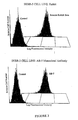
- FACS and Immunoprecipitatio Flow cytometric analysis determined that DW1MVF anti-peptide antibody directly targeted the HER-2 receptor.
- a commercially available mouse Mab to HER-2/neu was used as a control in SKBR3 cells. Negative control sera showed no binding to the receptor while an increase in fluorescence was seen with the immune sera. Fluorescence intensity of the polyclonal antipeptide serum was comparable to the monoclonal antibody. Therefore polyclonal serum can mimic the specificity and affinity of monoclonal antibodies.
- Immune serum was serially diluted in Hank's Balanced Salts Solution and the mean cell fluorescence determined. Serum from the 'tertiary ⁇ 3 weeks' bleed gave a fluorescence value of 121.4 (diluted 1:320) while the MAb gave a value of 132. The following is a list of the mean cell fluorescence values at various serum dilutions. Dilution Mean Cell Fluorescence (DWLMVF) Control 0.647 1:80 39.3 1:160 83.5 1:320 121.4 1:640 69.4 1:1280 34.4 1:2560 17.4
- DWLMVF Dilution Mean Cell Fluorescence
- peptide antibodies MVFDW4, DW5MVF and DW6MVF did not give the same intensity of fluorescence as DW1MVF. Therefore, immunoprecipitation was used to verify specificity for HER-2.
- SKBR3 cells were immunoprecipitated with Protein A/G purified antipeptide antibodies. Antibodies are shown to be HER-2 reactive. Identical bands are evident in the Mab sample and the anti-peptide antibodies.
- CTL Cytotoxic T cell
- Peptide DW1MVF (376-395) has an overlapping T cell epitope 391-399. We wished to test the efficacy of this epitope for incorporation into a HER-2 vaccine. Results showed that a specific CTL response could be raised to a HER-2 derived peptide. Cytotoxic T cells primed with HER-2 391-399 were able to specifically lyse autologous targets in a dose dependent manner. However, two in vitro re-stimulations were necessary to produce this result. In this study IL-2 was added to the culture medium. It is expected that immunization with a peptide containing both T helper and cytotoxic T cell sequences will boost the results.
- a transgenic mouse model (designated N202) that expresses mammary tumors similar to human breast cancer, developed by Muller and co-workers, Guy, C. T., et al, Proc.Natl. Acad. Sci. USA, 89: 10578-82, 1992 was employed to test in vivo anti-tumor effects of the chimeric peptides.
- Focal mammary tumors arise in at least 50% of the female transgenic mice around 28 weeks of age due to overexpression of the rat neu gene under murine mammary tumor virus 3' long terminal repeat.
- HER-2 peptide sequences 376-395, 410-429 and 628-647) have greater than 80% homology to the analogous regions in rat neu.
- MVF HER-2 (628-647) elicited high titered antibody responses against the immunogen of over 50,000 as early as two weeks after the second booster, the antibody titers reached more than 250,000 after the third booster.
- Antibodies against MVF HER 2 (628-647) also reacted with recombinant HER-2 ECD with titers over 10,000 and the intact-HER-2 and rat neu receptors of cells.
- the transgenic mice did not mount appreciable antibody responses against HER-2 immunogens, 115 - 136 MVF and 410-429 MVF.
- a chimeric peptide comprising amino acids 27-45 of the HER-2 protein, the promiscuous helper T cell epitope from measles virus fusion protein (amino acids 288-302) at the N-terminus, and a GPSL linker (MVFDN1) was synthesized using procedures as described above in example 1.
- a chimeric peptide comprising a modified sequence 316-339 of the HER-2 protein, the promiscuous helper T cell epitope from measles virus fusion protein (amino acids 288-302) at the N-terminus, and a GPSL linker (MVFND2) was synthesized using procedures as described in example 1 above.
- HER-2 sequence 316-339 contains three cysteines at positions 331, 334 and 338.
- residues 334 & 338 form energetically most stable cysteine-cysteine bond pair.
- Cysteine 331 with Alanine to prevent interference with secondary structure formation and aggregation post synthesis. Table 3.
- the HER-2 (316-339) epitope was linked to the Th epitopes comprising sequences 580-589 and 947- 967 tetanus toxin, designated TT and TT1, respectively and sequence 288-302 of the measles fusion protein, designated MVF to provide three different chimeric HER-2 B cell peptides.
- a chimeric peptide comprising a modified sequence 495-508 of the HER-2 protein, the promiscuous helper T cell epitope from measles virus fusion protein (amino acids 288-302) at the N-terminus, and a GPSL linker (MVFND3) was synthesized using procedures as described in example 1 above.
- HER-2 sequence 495-508 is four residues short of an optimal B-cell epitope of approximately 17-18 residues spanning the 750 Angstrom 2 antigen binding site of an antibody.
- Sequence 485-499 was assigned the highest score of all sequences analyzed in the HER-2 extracellular domain on Welling's antigenicity scale, this scale indicates the probability of a 5 residue sequence to form an antigenic epitope.
- This sequence also harbors a defined ⁇ -turn (Residues 488-491) and an ⁇ -helix (Residues 491-495). Therefore, we extended the originally proposed epitope 495-508 to include 485-499. Furthermore, we shortened the sequence 495-508 by five residues at the C-terminus to exclude the single cysteine at position 504 that could lead to aggregation of the peptide and make it difficult for purification and characterization. The four residues following cysteine do not form any defined secondary structure.
- a chimeric peptide comprising amino acids 605-622 of the HER-2 protein, the promiscuous helper T cell epitope from measles virus fusion protein (amino acids 288-302) at the N-terminus, and a GPSL linker(MVFDN4) was synthesized using procedures as described above in example 1.
- the chimeric HER-2 peptides of examples 7-10 elicited variable antibody responses in pairs of immunized out bred rabbits.
- the most immediate and high titered antibodies were elicited by MVF HER-2 (316-339). Lower antibody responses were mounted against the other three peptide constructs.
- the antibodies produced in response to immunization with each of the chimeric peptides were able to recognize the HER-2 receptor both by flow cytometry and immunprecipitation.
- the effect of combinations of chimeric peptides on antibody production in out bred rabbits was also determined.
- These rabbits were first immunized with individual epitopes and two weeks after the fourth booster the same rabbits were cross-immunized first with one of the three epitopes and later with all three epitopes.
- the antibody response to the cross immunogen was as good or better than they were administered as single immunogens.
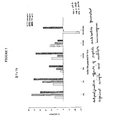
- MVF HER-2 (485-503) antibodies inhibited the growth of SKBR-3 cells by 22% and BT-474 by 11%.
- Cells of the breast tumor cell line SK-BR3 were also treated with mixtures of various HER-2 peptide antibodies, each at a concentration of 0.1 mg/ml. Treatment with a plurality of the antibodies resulted in an additive growth inhibitory effect which was greater than the inhibitory effect of a single antibody. A mixture of three different growth inhibitory peptide antibodies prevented greater than 90% of the proliferation of SK-BR-3 cell relative to untreated cells.
- Cells of the breast tumor cell line BT474 were also treated with mixtures of various HER-2 peptide antibodies at concentrations of 266 ⁇ g/ml, 528 ⁇ g/ml, and 792 ⁇ g/ml.
- MVFDW4 prepared as described above in example is loaded into microspheres as described below.
- Microsphere preparations utilized 75/25 poly(DL-lactide-co-glycolide)(PLGA) with an inherent viscosity of 0.58 dL/g at 120 mg/ml.
- the peptide/polymer solution was emulsified in mineral oil/cottonseed oil with Span 85(sorbitan trioleate) as an emulsifier at 40°C. After one hour of stirring at 750 rpm, the oil-in-oil emulsion was filtered through a 0.45 ⁇ m membrane and washed several times with petroleum ether. The filter was placed in a conical tube, frozen in liquid nitrogen, and lyophilized for at least three days. Peptide loading was determined by amino acid analysis.
- Microsphere morphology, surface characteristics, and size were observed using a Philips Electronics XL-30 FEG scanning electron microscope. Dry samples were sprayed onto a carbon conductive tabbed specimen mount (3.2mm pin diameter, 12.7mm table diameter) which was sputter-coated for 110 seconds in an argon atmosphere. The images were obtained using a secondary electron detector and an accelerating potential of 5 kV at a working distance of 10 mm. At least one hundred particles for each preparation were sized from electron micrographs obtained using a Philips Electronics XL-30 Field Emission Gun Scanning, Electron Microscope.
- Peptide loading was determined by extracting the peptide from the microspheres using ethyl acetate. Microspheres were immersed in excess ethyl acetate and vortexed vigorously. The undissolved polymer and released peptide was spun down by 5 minutes of centrifugation. The supernatant was removed and the process repeated three more times. Excess ethyl acetate was removed by evaporation. Samples were reconstituted in 2% acetic acid and submitted for amino acid analysis. The peptide was hydrolyzed in 6 N HCl and the subsequent amino acids derivatized and analyzed as their phenylthiohydantoins using a Waters PicoTag System.
- mice 100 ⁇ g of peptide and 100 ⁇ g of nor MDP in PBS is emulsified in 4:1 squalene:Arlacel and injected subcutaneously in multiple sites of five mice/peptide. Three weeks later, mice are boosted using the same protocol. Two weeks after the second immunization, spleens are recovered surgically.
- MVFDW4 peptides and Nor-MDP adjuvant were individually encapsulated into microspheres 5.2% loading (MVFDW4), and 5% loading Nor-MDP.
- Microspheres that contain 100 ⁇ g peptide/mouse and 100 ⁇ g Nor-MDP /mouse were mixed with 200 ⁇ l of 4:1 squalene:Arlacel A and subcutaneously injected into mice.
- splenocytes were taken from each mouse. Single cell suspension was prepared, part of which was used in INF- ⁇ detection by Elispot, the rest for chromium-release assay.
- Elispot plate (PolyFiltronics) was coated with Anti-mouse-INF- ⁇ (clone R4-6A2, Pharmingen) diluted in sterile PBS with no azide at 4 ⁇ g/ml. The plate was then incubated overnight at 4°C in a humidity chamber. On day 2, the plate was washed with PBS four times and then blocked with 1% BSA in DMEM with no additives at 200 ⁇ l/well for 1 hour at room temperature.
- In vitro re-stimulation was performed using syngeneic spleen cell stimulators, which were taken from the same batch of single cell suspension and pulsed for 1h with the p63 synthetic peptide.
- Responder cells stimulator cells (3:1) were mixed in 24-well late and incubated at 37°C for a week and then those responder splenocytes was used as the effector cells in the following CTL assay.
- CTL assay was performed using standard Chromium release assay.
- peptide-pulsed targets were prepared by incubating the SVBalb cells(H-2 d ) or MCS7 cells(H-2 b , as control) with or without 10 ⁇ g/ml of peptide p63 and 500 ⁇ Ci/ml of 51 Cr sodium chromate for 1 h at 37°C. Labeled target cells (10 4 cells/well) and various numbers of effector cells were plated in a final volume of 0.2 ml in 96-well plate.
- HER-2 is extensively glycosylated at seven potential sites in the extracellular domain alone.
- Three constructs comprising B-cell epitope encoding sequences suitably mutated to improve glycosylation and their wild type counterparts HER-2 (115-136); (182-216); and (630-650) and T helper cell epitope are subcloned into a baculovirus mammalian shuttle expression vector. This vector allows easy baculovirus virus mediated transfection of gene of interest into mammalian cells, but cannot replicate in the cells allowing efficient expression and glycosylation of the epitopes.
- the expressed chimeric glycosylated epitopes are characterized by capillary electrophoresis for the type of sugars and glycosylation efficiency. Rabbits are immunized with efficiently glycosylated epitopes and peptide antibodies generated.
Description
- Currently, the most common forms of treating breast cancer involve surgery, chemical intervention, and/or radiotherapy. Unless the cancer is restricted to a defined area, surgery alone cannot eliminate the cancer. Accordingly, radiation treatment is often given after surgery to destroy cancer cells that are near the surgical site and that have evaded surgery. The side effects of such treatment include skin sensitivity or itchiness, interference with the immune system, sometimes queasiness and, rarely, radiation fibrosis where an affected portion of the lung becomes fibrous. Chemotherapy may also be employed following surgery. Chemotherapy utilizes drugs that are toxic to cancer cells. Since this is not a perfectly selective system, normal cells are affected as well. Negative side effects include nausea, tiredness, loss of appetite, hair loss and diarrhea.
- In view of the disadvantages of the present therapies, attempts have been made to find additional approaches for treating breast cancer. One such approach is immunotherapy. One of the targets for an immunotherapeutic approach is the HER-2 protein. The HER-2 protein, a product of the HER-2 oncogene, is overexpressed in a variety of cancers. It is found in 50%-60% of ductal in situ carcinoma and 20%-40% of all breast cancers, as well as a substantial fraction of adenocarcinomas arising in the ovaries, prostate, colon and lung. Overexpression of the HER-2 protein is related to malignant transformation in humans. Overexpression of the HER-2 protein is also intimately associated with the aggressiveness of the malignancy, being found in one-fourth of all invasive breast cancers. Overexpression of HER-2 protein is correlated with a poor prognosis in both breast and ovarian cancer.
- In recent studies, antibodies directed against the extracellular binding domain (ECD) of HER-2 have been shown to confer inhibitory effects on tumor growth in vitro and in animal models (Hudziak, R.M., et al., Mol. Cell. Biol., 9:11-65-72, 1989; Tagliabue, E., et al., Int. J. Cancer 47:933-7, 1991; Drebin, J.A., et al., Proc. Natil. Acad. Scie. USA 83:9129-33, 1986; Drebin, J.A., et al., Oncogene, 2:273-7, 1988; Drebin, J.A., et al., Oncogene, 2:387-94, 1988; and Katsumata, M., et al., Nat. Med. 1:644-8. 1995.) In addition, Phase II and III clinical trials of a recombinant humanized anti-HER-2 monoclonal antibody, Trastuzumab, in patients with metastatic, HER-2-overexpressing breast cancers produced an overall response rate of 15% as a single agent. Trastuzumab has also been shown to improve survival when combined with cytotoxic chemotherapeutics (Baselga, J., et al., J. Clin. Oncol. 14:737-44, 1996; Pegram, M.D., et al., J. Clin. Oncol., 16:2659-71, 1988.). A number of vaccine approaches targeting a recombinant HER-2 protein, the HER-2 ECD, or the ECD of rat neu, which is the rat homolog of HER-2 have also been evaluated. For example, strain NFS mice immunized with a vaccinia virus recombinant that expresses the ECD rat neu developed a protective antibody response against subsequent challenge with neu-transformed NIH 3T3 cells (Bernards, R., et al., Proc. Natl. Acad. Sci. USA, 84:6854-8, 1987.). Immunization of BDIX rats with the same immunogen, however, did not result in antibody response nor did it inhibit the growth of syngeneic neu-expressing B 104 neuroblastoma cells, suggesting that this strategy was insufficient to induce immune responses in the rat. A polysaccharide-oncoprotein complex vaccine, consisting of the 147 amino-terminal amino acids of HER-2 ECD complexed with cholesteryl group-bearing mannan and pullulan, induced cellular and humoral immune responses that mediated rejection of HER-2-expressing sarcomas in BALB/c mice (Gu, X. G., et al., Cancer Res., 58: 3385-90, 1998.). Partial protection was shown in rat neu transgenic mice destined to develop mammary tumors by immunizing with either a purified rat neu ECD (Esserman, L. J., Cancer Immunol. Immunother., 47:337-42, 1999.) or neu-transfected allogeneic mouse fibroblasts (Cefai, D., et al.. Int. J. Cancer, 83:393-400, 1999.)
-
WO97/38011 -
WO94/07530 -
US 5 876 712 A describes methods for the detection, monitoring and treatment of malignancies in which the HER-2/neu oncogene is associated. Also described are methods and compositions, including peptides, for treating such malignancies. - Kaumaya P T P et al., J. Mol. Recog., vol. 6, 1993, pages 81-94, describes two peptides to encompass the sequences from the universally immunogenic tetanus toxoid (TT) epitope and the contraceptive vaccine candidate lactate dehydrogenase C4 (LDH-C4). The authors report that a determinant which was previously restricted to H-2k can be rendered immunogenic in H-2b with the 'promiscuous' TT epitope.
- Lairmore M D et al., J. Virology, vol. 69, no. 10, October 1995, pages 6077-6089, describes the testing of chimeric and beta-template peptide constructs incorporating known human T-lymphotropic virus type 1 (HTLV-1) B- and T-cell epitopes from the surface envelope protein gp46 (SP2 [aa 86 to 107] and SP4a [aa 190 to 209]), along with promiscuous T-cell peptides. The peptides's immunogenicities were evaluated in both rabbits and mouse strains of divergent haplotypes (C3H/HeJ [H-2k], C57BL/6 [H-2b], and BALB/c [H-2d]). The authors report that the chimeric constructs MVF-SP2 and SP4a-measles virus F protein were highly immunogenic.
- Triozzi P L et al., Biomedical peptides, proteins & nucleic acids: structure, synthesis & biological activity, vol. 1, no. 3, 1995, pages 185-192, describes an autochthonous tumor model, A/J mouse lung, which parallels human tumors in the progression of proliferative lesions from premalignant to malignant. The authors describe a chimeric immunogen incorporating a promiscuous T-cell epitope to enhance the immunogenicity of an oligopeptide corresponding to a weakly immunogenic substitution.
- Disis M L et al., Blood, vol. 88, no. 1, July 1996, pages 202-210, describes studies to evaluate granulocyte-macrophage colony- stimulating factor (GM-CSF) as a vaccine adjuvant. The authors report that immunity to rat neu (c-erbB-2) protein, an oncogenic self protein, can be generated in rats by immunization with peptides derived from the normal rat neu sequence plus complete Freund's adjuvant (CFA). The current study demonstrates that rat neu peptides inoculated with GM-CSF could elicit a strong delayed type hypersensitivity reaction (DTH) response, whereas peptides alone were nonimmunogenic. GM-CSF was as effective as CFA in generating rat neu specific DTH responses after immunization with a neu peptide based vaccine. Soluble GM-CSF is a potent adjuvant for the generation of immune responses to foreign proteins as well as peptides derived from a self tumor antigen.
- Kono K et al., Int. J. Cancer, vol. 78, no. 2, 5 October 1998, pages 202-208, describes HLA-A2.1-restricted, gastric cancer-specific cytotoxic T lymphocyte (CTL) lines derived by repetitive in-vitro stimulation of tumour-associated lymphocytes (TAL) with autologous tumor cells. The HER2/neu specificity of these gastric cancer-specific CTLs was demonstrated using HER2/neu-transfected cell lines and HER2/neu-expressing tumors, and with a set of HER2/neu-derived peptide epitopes.
- Kawashima I et al., Cancer Research, vol. 59, no. 2, 15 January 1999, pages 431-435, describes the testing of HLA-A3 binding synthetic peptides from CEA and HER-2/neu for immunogenicity by in-vitro primary CTL induction protocol using peripheral blood mononuclear cells from normal healthy volunteers. The authors describe one peptide from CEA (CEA[9(61)]: HLFGYSWYK) and one peptide from HER-2/neu (HER2[9(754)]: VLRENTSPK) which induce CTL capable of killing a tumor cell line expressing HLA-A3 and the corresponding tumor-associated antigen.
- Disis M L et al., Proceedings of the Annual meeting of the American association for Cancer Research, vol. 36, March 1995, page 251, describes a strategy to circumvent immunological tolerance to (whole) self proteins by test immunization to synthetic subunit peptides with amino acid motifs appropriate for binding to class II MHC molecules. The authors report the generation of antibodies recognizing epitopes in both the intra- and extra- cellular domains of both human and rat NEU protein.
- Kelly C G et al., Infection and Immunity, vol. 63, no. 9, 1 September 1995, pages 3649-3658, describes T-cell and antibody responses to a cell surface streptococcal antigen (SA I/II) in naturally sensitized humans. The authors report that serum antibody responses were directed predominantly to the N-terminal (residues 39 to 481) and central (residues 816 to 1213) regions of SA I/II, whilst T-cell responses were directed predominantly towards the central region. Specifically, immunodominant T-cell and B-cell epitopes were reported within residues 803 to 853, which were separated in linear sequence from the adhesion epitopes (residues 1005 to 1044). Adhesion epitopes were reported to overlap with minor B- and T-cell epitopes (residues 1005 to 1054 and 1085 to 1134).
- Herrera Socrates et al., Parasite Immunology, vol. 19, no. 4, 1997, pages 161-170, describes using linear synthetic peptides corresponding to the Plasmodium vivax circumsporozoite (CS) protein of the common type to identify several T and B-cell epitopes recognized by human individuals. The authors report that three T-cell epitopes studied (p6) from the amino, (p11) from the central and (p25) from the carboxyl regions, were widely recognized by lymphocytes of immune donors. The sequences corresponding to peptides p6, p11 and P25 were used to assemble eight different Multiple Antigen Peptides (MAP), the immunogenicity of which was analysed in Aotus monkeys.
- Dakappagari N K et al., Cancer Research, vol. 60, no. 14, 15 July 2000, pages 3782-3789, describes the evaluation of synthetic peptide vaccines targeting B-cell epitopes of the extracellular domain of the HER-2 oncoprotein for their capacity to elicit HER-2-specific antibodies with antiproliferative activity. The authors report that one epitope sequence, 628-647, was mutated to optimize disulfide pairing to mimic the native HER-2 receptor, with the result that it elicited a high antibody titer in outbred rabbits. In addition, the antibodies elicited by MVF HER-2(628-647) were reported to inhibit proliferation of human HER-2-overexpressing breast cancer cells in vitro and cause antibody-dependent cell-mediated cytotoxicity.
- Okugawa T et al., Eur. J. Immunology, vol. 30, no. 11, 2000, pages 3338-3346, describes a mouse HER2-derived peptide, HER2p63 (A) (TYLPANASL). The HER2-derived peptide is reported to be able to induce Kd-restricted mouse cytotoxic T lymphocytes (CTL) and function as a tumor rejection antigen in an in vivo assay. The authors also describe the similar human peptide HER2p63 (T) (TYLPTNASL); autologous antigen presenting cells exposed to this peptide were able to sensitise killer cells in vitro such that they selectively lysed HER2-expressing SKOV3 transfected with HLA-A2402 cDNA.
- Dakappagari N K et al., J. Immunology, vol. 170, no. 8, 15 April 2003, pages 4242-4253, describes the development of a multi-epitope vaccine throught the computer-aided identification of 12 high ranking B cell epitopes from the extracellular domain of the human epidermal growth factor receptor-2 (HER-2) oncoprotein. The authors report that the multiepitope vaccine in combination with IL-12 caused a significant reduction in the number of pulmonary metastases induced by challenge with syngeneic tumor cells overexpressing HER-2.
- Frangione-Beebe M et al., Vaccine, vol. 19, no. 9-10, 8 December 2000, pages 1068-1081, describes a peptide vaccine derived from the human T-lymphotropic virus type 1 (HTLV-1) surface envelope glycoprotein protein (gp46). This peptide construct, designated MVFMF2, comprises amino acids (aa) 175-218 of gp46 linked by a four residue turn (GPSL) to a promiscuous T-cell epitope from the measles virus fusion protein (MVF, aa 288-302). The authors report that the MVFMF2 peptide, when administered with adjuvant (N-acetyl-glucosamine-3yl-acetyl-L-alanyl-D-isoglutamine, nor-MDP) was immunogenic in an outbred population of both rabbits and mice.
- Despite the results of the studies described above, it is still uncertain whether effective immune responses can be generated in humans using cell-or protein-based vaccine strategies targeting HER-2 or the HER-2 ECD, as HER-2 is a non-mutated, "sclf' antigen. Accordingly, it is desirable to have additional immunotherapeutic approaches for treating or preventing breast cancer and other malignancies with which overexpression of the HER-2 protein is associated.
- The present invention provides a chimeric peptide for stimulating an immune response to HER-2 protein, wherein said chimeric peptide comprises a HER-2 B cell epitope, a T-helper (Th) epitope, and a linker joining said HER-2 B cell epitope to said Th epitope; wherein the sequence of said HER-2 B cell epitope is
- P-L-H-N-Q-E-V-T-A-E-D-G-T-Q-R-A-E-K-C-S-K-P-C-A, (SEQ ID NO. 6,)
- In some embodiments the sequence of said linker is the sequence G-P-S-L, (SEQ ID NO. 20.)
- The invention also provides a composition for stimulating an immune response to HER-2 protein, wherein said composition comprises a first chimeric peptide and a second chimeric peptide;
wherein said first chimeric peptide is the chimeric peptide of the invention as described above;
wherein said second chimeric peptide comprising a HER-2 B cell epitope, a T-helper (Th) epitope, and a linker joining said HER-2 B cell epitope to said Th epitope;
wherein the sequence of the HER-2 B cell epitope of said second chimeric peptide is selected from the group consisting of: - L-F-R-N-P-H-Q-A-L-L-H-T-A-N-R-P-E-D-E, SEQ. ID. NO. 9; and
- I-N-G-T-H-S-C-V-D-L-D-D-K-G-C-P-A-E-Q-R, (SEQ. ID. NO. 43)
- In some embodiments of the above composition the sequence of the linker of said second chimeric peptide is the sequence G-P-S-L, (SEQ ID NO. 20)
- The invention also provides for the use of the chimeric peptide or the composition of the invention, as described above, in the manufacture of a medicament for the treatment of cancer in a subject. In some embodiments the medicament is for the treatment of one of the following cancers, or a predisposition to one of the following cancers in a human: breast cancer, ovarian cancer, lung cancer, prostate cancer, and colon cancer.
- The invention further provides a pharmaceutical composition comprising the chimeric peptide or the composition of the invention, as described above, and a pharmaceutically acceptable vehicle. In some embodiments the vehicle is biodegradeable and is selected from the group consisting of an emulsion comprising a pharmaceutically acceptable oil/water emulsion and a biodegradeable microsphere or nanosphere comprising a polylactide-polyglycolic acid polymer. In some embodiments the oil is squalene or squalane. In some embodiments the microsphere is from 0.1 to 50 nanometers in diameter and comprises poly (D, I lactide-co-glycide).
- The invention also provides an isolated polynucleotide which encodes the chimeric peptide of the invention, as described above.
- The invention futher provides a composition for stimulating an immune response to HER-2 protein, wherein said composition comprises a first chimeric peptide and a second chimeric peptide, wherein each chimeric peptide comprises a HER-2 B cell epitope, a T-helper (Th) epitope, and a linker joining said HER-2 B cell epitope to said Th epitope;
wherein the sequence of the HER-2 B cell epitope of the first chimeric peptide is: - L-F-R-N-P-H-Q-A-L-L-H-T-A-N-R-P-E-D-E, (SEQ. ID. NO. 9)
- I-N-G-T-H-S-C-V-D-L-D-D-K-G-C-P-A-E-Q-R, (SEQ. ID. NO. 43)
- L-S-E-I-K-G-V-I-V-H-R-L-E-G-V, (SEQ ID NO. 17)
-
-
Figure 1 is a schematic representation of a chimeric HER-2 B cell peptide. -
Figure 2 is a schematic representation of a multivalent HER-2 B/CTL peptide. -
Figure 3 shows the binding of antibodies raised to the chimeric HER-2 B cell peptide DW1MVF to HER-2 overexpressing SKB3 cells. Antibodies raised to DW1MVF specifically bound the HER-2 receptor with an affinity comparable to a commercially available monoclonal antibody. SKBR3 cells were incubated with antibody, washed and FITC-conjugated secondary antibody added. After being fixed in formalin, cells were analyzed by a Coulter ELITE flow cytometer at 488 nm for excitation. 5.0 x 10⁶ cells were counted for each sample. -
Figure 4 shows the immunogenicity in outbred rabbits injected with multiple chimeric HER-2 B cell peptides. -
Figure 5 shows the antiproliferative effects of peptide antibodies generated against herceptin and single and multiple HER-2 B cell chimeric peptides MVFDN1 (HER-2 (27-45)), MVFDN2 (HER-2 (316-337)), MVFDN3 (HER-2 (485-503) and MVFDW4 (DW4) on HER-2 overexpressing SK-BR-3 human breast cancer cells. Results are average of triplicate samples -
Figure 6 shows the antiproliferative effects of peptide antibodies generated against herceptin HER-2 B cell chimeric peptides MVFDN1 (ND1) MVFDN2 (N2), MVFDN3 (N3) and MVFDW4 (DW4) on HER-2 overexpressing BT474 human breast cancer cells. Results are average of triplicate samples. -
Figure 7 shows the antiproliferative effects of peptide antibodies generated against single and multiple HER-2 B cell chimeric peptides MVFDN2 (N2), MVFDN3 (N3) and MVFDW4 (DW4) on HER-2 overexpressing BT474 human breast cancer cells. Results are average of triplicate samples. -
Figure 8 shows the effect of vaccination with HER-2 B cell peptides on spontaneous breast tumor development. Groups of six mice were vaccinated with 100 µgs of peptides (pluse MDP as adjuvant emulsified in squalene arlacel A) around 4 weeks of age and boosted after 4, 8, 16, and 24 weeks. Tumors were calculated as length x width2/2) The time to tumor development was analyzed using Kaplan-Meier survival analysis with log rank comparisons of individual curves. - Described herein are peptides that are immunogenic epitopes of the HER-2 protein, referred to hereinafter as HER-2 B cell epitopes and HER-2 CTL epitopes.
- The HER-2 B cell epitopes are capable of invoking a humoral response which results in the production of antibodies that are immunoreactive with the extracellular domain of the HER-2 protein. HER-2 protein, and its rat homolog neu, are transmembrane proteins with a relative molecular mass of 185 kd that is approximately 1255 amino acids (aa) in length. HER-2/neu protein has an extracellular binding domain (ECD) of approximately 645 aa, with 40% homology to epidermal growth factor receptor (EGFR), a highly hydrophobic transmembrane anchor domain (TMD), and a carboxyterminal cytoplasmic domain (CD) of approximately 580 aa with
n 80% homology to EGFR. The amino acid sequence of the HER-2 protein and a nucleotide sequence which encodes such amino acid sequence are shown in GenBank Accession No. M11730. - The HER-2 B cell epitopes encompass peptides having one of the sequences, referred to hereinafter as the "reference sequences", shown in Table I below. The reference sequences were selected and scored using computer-aided analysis using six correlates of antigenicity: (a) the profiles of chain flexibility and mobility of individual sequences was calculated according to Karplus and Schultz, Naturwiss 72:212-213, 1985; (b) hydropathy profiles were generated over a seven residue span setting and were finally smoothed with a three residue span using the scale of Kyte and Doolittle, J. Moi. Biol. 157:105-132, 1982; (c) hydrophilicity profiles were generated over a 6-residue window using the program of Hopp and Woods, Proc. Natl. Acad. Sci. USA 78:3824-3828, 1981; (d) analysis of the exposure of an amino acid residue to water using a 1.4 A probe) was carried out b the solvent exposure algorithm of Rose, Science 229:834-838, 1985; (e) protrusion indices that predicts portions of proteins that are accessible and protrude into the solvent were calculated by the method of Thornton, EMBO J. 5:409-413, 1986; (f) the probability that a five residue sequence is antigenic was determined by the method of Welling, FEBS Lett 188:215-218, 1985. The basic premise is that the algorithms used in the predictions will always locate regions that are surface-exposed on the protein and therefore most likely to be involved in antibody binding. Sequences were given a score of 1 to 6 based on their respective index values and were ranked: the highest ranking sequences had the highest individual score for the analyses examined (6/6), and successive candidates had the next highest score (5/6), etc. The best scoring epitopes were further ranked by correlation with their secondary structural attributes, e.g.., an amphiphilic α-helical sequence or a β-turn loop region are preferred over a random coil fragment. Computer programs by Chou and Fasman, Adv. Enzymol. Relat. Subj. Biochem. 47: 45-148, 1978 were used to predict the secondary structure (α-helix, β-strand/sheet, (3-turn/loop, random coil) and helical amphiphilic moment. Electrostatic ion pairs and helix dipole interaction in helical segment were also considered (e.g., hydrophobic / hydrophilic balance). Preferably, the hydrophilic/hydrophobic balance is from 2/2 to 4/1.
- As described herein, the HER-2 B cell epitopes also encompass peptides that are functional equivalents of the peptides shown in Table I below. Such functional equivalents have an altered sequence in which one or more of the amino acids in the corresponding reference sequence is substituted or in which one or more amino acids are deleted from or added to the corresponding reference sequence. For example, cysteine residues may be deleted or replaced with other amino acids to prevent formation of incorrect intramolecular disulfide bridges upon renaturation. Optionally, the HER-2 B cell epitopes are glycosylated.
- While it is possible to have nonconservative amino acid substitutions, it is preferred that, except for the substitutions that are made to replace cysteine, the substitutions be conservative amino acid substitutions, in which the substituted amino acid has similar structural or chemical properties with the corresponding amino acid in the reference sequence. By way of example, conservative amino acid substitutions involve substitution of one aliphatic or hydrophobic amino acids, e.g., alanine, valine, leucine and isoleucine, with another; substitution of one hydroxyl-containing amino acid, e.g., serine and threonine, with another; substitution of one acidic residue, e.g., glutamic acid or aspartic acid, with another; replacement of one amide-containing residue, e.g., asparagine and glutamine, with another; replacement of one aromatic residue, e.g., phenylalanine and tyrosine, with another; replacement of one basic residue, e.g., lysine, arginine and histidine, with another; and replacement of one small amino acid, e.g., alanine, serine, threonine, methionine, and glycine, with another.
- Preferably, the deletions and additions are located at the amino terminus, the carboxy terminus, or both, of one of the sequences shown above. As a result of the alterations, the HER-2 B cell epitope equivalent has an amino acid sequence which is at least 70% identical, preferably at least 80% identical, more preferably at least 90% identical, most preferably, at least 95% identical to the corresponding reference sequences. Sequences which are at least 90% identical have no more than 1 alteration, i.e., any combination of deletions, additions or substitutions, per 10 amino acids of the reference sequence. Percent identity is determined by comparing the amino acid sequence of the variant with the reference sequence using MEGALIGN project in the DNA STAR program.
- For functional equivalents that are longer than a corresponding reference sequence, it is preferred that the functional equivalent have a sequence which is at least 90% identical to the reference sequence and the sequences which flank the reference sequence in the wild-type HER-2 protein.
Table 1. Consolidated Human p185 HER-2 predicted B cell epitopes listed in the order of ranking by amino acid residue numbers. Aspargine (N)-linked glycosylation sites are underlined in bold. Predictive Ranking Residue Amino Acid Sequence Secondary Structure 7 27 - 45 Tgtdmklrlpaspethldm 25 - 28 β turn; 29-32α helix; 35 - 38 β turn 8 (DW5) 115-136 AVLDNGDPL NNTT PVTGASPGG 116 - 135 β turn 9 168-189 LWKDIFHKNNQLALTLIDT NRS 173 - 176 β turn; 177-181 α helix 1 182-216 
184-212 β turn/loop 6 270-290 ALVTYNTDTFESMPNPEGRYT 273 - 286 β turn; 278-280 α helix 3 316-339 PLHNQEVTAEDGTQR A EKCSKPCA 319-324 α helix; 324-336 β turn. 10 (DW1) 376-395 PESFDGDPASNTAPLQPE 379-388 β turn 12 (DW6) 410-429 LYISAWPDSLPDLSVFQNLQ 413-421 β turn 2 485-503 LFRNPHQALLHTANRPEDE 497 - 500 β turn; 499-504 α helix 11 560-593 
561 - 572 & 589-593 β turn; 579-581 α helix 4 605-622 KPDLSYMPIWKFPDEEGA 616-620 α helix 5 628-650 IN G THSCVDLDDKGCPAEQRASP 635-642 β turn; 643-646 α helix - Also described herein are chimeric peptides, referred to hereinafter as "chimeric HER-2 B cell peptides," which comprise HER-2 B cel epitope, a helper T (Th) cell epitope, preferably a promiscuous Th cell epitope, and a linker. Depending upon the promiscuous Th cell epitope used, the HER-2 B cell epitope is linked to either the amino or the carboxy terminus of the Th cell epitope. The location and selection of the Th cell epitope depends on the structural characteristics of the B cell epitope (whether alpha helical or beta-turn or strand. Methods for selecting suitable Th cell epitopes are described in Kaumaya et al., "DE NOVO" ENGINEERING OF PEPTIDE IMMUNOGENIC AND ANTIGENIC DETERMINANTS AS POTENTIAL VACCINES, in Peptides, Design, Synthesis and Biological Activity (1994). A summary of the immune responses elicited a variety of promiscuous T-helper cell epitope containing B-cell epitope chimeras was presented in a review titled "Synthetic Peptides: Dream or Reality" by Kaumaya et al., and published in PEPTIDES IN IMMUNOLOGY, Wiley and Sons, Ltd. (1996).
- The chimeric HER-2 B cell peptides are useful immunogens for inducing production of antibodies that interact with and bind to the extracellular domain of the HER-2 protein. The chimeric B peptides are also useful as laboratory tools for detecting antibodies to HER-2 protein in patient's sera. In accordance with the present invention it has been determined that the chimeric HER-2 B cell peptides MVF-HER-2 (628-647), HER-2 (376-395)-MVF, and HER-2 (410-429)-MVF invoked an antibody response in rabbits and that such antibodies (a) immunopreciptate HER-2 protein, (b) bind to intact HER 2 receptor on ER-2 overexpressing cells in culture, and (c) reduce proliferation of HER-2 overexpressing cells in vitro and in a xconograft mouse model. It has also been determined that immunization of transgenic mice with the chimeric peptide MVF-HER-2 (628-647) retards tumor development within such mice for at least 9 months post the time when control mice developed tumors.
- Described herein are HER-2 CTL epitopes which have the ability to invoke a cell-mediated responses to HER-2 protein. As used herein the term HER-2 CTL epitope encompasses peptides having one of the sequences shown below in Table 2 or a functional equivalent thereof. The functional equivalent has an amino acid sequence which is at least 80%, preferably at least 90 % identical to one of the sequences, referred to as the "reference sequence," shown in Table 2 below. The ability of the functional equivalent to invoke a cell-mediated response to HER-2 protein or the extracellular binding domain of HER-2 protein is the same as or greater than the corresponding reference sequence.
Table 2 HLA - A3: 167-175 (ILWKDIFRK); 714-722 (ILKETELRK); 754-762 (VLRENTSPK HLA - B7: 1159-1167 (AARPAGATL); 35-43 (LPASPETHL); 1101 (LPTHDPSPL) HLA - B27: 7-15 (CRWGLLLAL); 897-905 (RRFTHQSDV); 433-441 (GRILHNGAY) H-2Krd : 63-71 (TYLPTNASL); 553-561 (EYVNARHCL); 440-448 (AYSLTLQGL) HLA - A2: 5-13 (ALCRWGLLL); 48-57 (HLYQGCQV); 141-149 (QLRSLTEIL); 435-443 (ILHNGAYSL); 661-669 (ILLVVVLGV); 789-797 (DLTSTVQLV): 851-859 (VLVKSPNHV): 369-378 (KIFGSLAFL); 654-663 (IISAVVGIL) - Also described herein are chimeric peptides, referred to hereinafter as chimeric HER-2 CTL peptides which comprise one of the above described HER-2 CTL epitopes or a functional equivalent thereof. The chimeric HER-2 CTL peptides comprises three units. The first unit comprises a HER-2 CTL epitope or functional equivalent thereof. The second unit is a promiscuous T helper cell epitope. The second unit preferably is from about 14 to about 22, more preferably about 15 to 21, most preferably 16 amino acids in length. The third unit is a linker which joins the first and second units. The linker is an amino acid or, preferably, a peptide which is from about 2 to about 15 amino acids, more preferably from about 2 to about 10 amino acids, most preferably from about 5 to about 6 amino acids in length. The most preferred linker comprises the sequences: Gly-Pro-Ser-Leu, SEQ ID NO. 20.
- Described herein is a co-linear chimeric peptide which comprises multiple HER-2 CTL epitopes, which are linked to each other by a linker that is from 1-5 amino acids in length. Optionally, the linker comprises a protcolytic site. In one example the linker comprises adjacent basic amino acid residues. Preferably, the co-linear chimeric peptide comprises a HER-2 CTL eptiope from class HLA-A3, a HER-2 CTL epitope from class HLA-B7, a HER-2 CTL epitope from class HLA-A2, and a HER-2 CTL epitope from class HLA-B27. The co-liner chimeric peptide further comprises a second unit which is a promiscuous Th epitope of from 14 to 22 amino acids in length. The second unit is linked to the amino terminus or the carboxy terminus of the first unit by the linker.
- The HER-2 B cell epitopes, CTL epitopes, chimeric, and multivalent peptides, preferably, are synthesized using commercially available peptide synthesizers. Preferably, the chemical methods described in Kaumaya et al., "DE NOVO" ENGINEERING OF PEPTIDE IMMUNOGENIC AND ANTIGENIC DETERMINANTS AS POTENTIAL VACCINES, in Peptides, Design, Synthesis and Biological Activity (1994), pp 133-164 are used.
- The HER-2 B cell epitopes, HER-2 CTL epitopes and chimeric peptides may also be produced using cell-free translation systems and RNA molecules derived from DNA constructs that encode the epitope or peptide. Alternatively, the epitopes or chimeric peptides are made by transfecting host cells with expression vectors that comprise a DNA sequence that encodes the respective epitope or chimeric peptide and then inducing expression of the polypeptide in the host cells. For recombinant production, recombinant constructs comprising one or more of the sequences which encode the epitope, chimeric peptide, or a variant thereof are introduced into host cells by conventional methods such as calcium phosphate transfection, DEAE-dextran mediated transfection, transvection, microinjection, cationic lipid-mediated transfection, electroporation, transduction, scrape lading, bollistic introduction or infection.
- The HER-2 B cell epitope, CTL epitope, and chimeric peptide may be expressed in suitable host cells, such as for example, mammalian cells, yeast, bacteria, insect cells or other cells under the control of appropriate promoters using conventional techniques. Suitable hosts include, but are not limited to, E. coli, P. pastoris, Cos cells and 293 HEK cells. Following transformation of the suitable host strain and growth of the host strain to an appropriate cell density, the cells are harvested by centrifugation, disrupted by physical or chemical means, and the resulting crude extract retained for further purification of the epitope or chimeric peptide.
- Conventional procedures for isolating recombinant proteins from transformed host cells, such as isolation by initial extraction from cell pellets or from cell culture medium, followed by salting-out, and one or more chromatography steps, including aqueous ion exchange chromatography, size exclusion chromatography steps, and high performance liquid chromatography (HPLC), and affinity chromatography may be used to isolate the recombinant polypeptide. To produce glycosylated epitopes and chimeric peptides, it is preferred that recombinant techniques be used. To produce glycosylated epitopes and chimeric peptides which contain the same, it is preferred that mammalian cells such as, Cos-7 and Hep-G2 cells be employed in the recombinant techniques.
- Naturally occurring variants of the HER-2 B epitopes and HER-2 CTL epitopes shown in tables 1 and 2 above may also be isolated by, for example, by screening an appropriate cDNA or genomic library with a DNA sequence encoding the polypeptide.
- The preferred synthetic approaches for preparing the HER-2 multivalent peptides employ a combinatorial Fmoc/tbutyl, Fmoc/benzyl and Boc benzyl strategy as well as a fourth level of differential protecting group (Npys) strategy. Details of such approach are presented in Larimore et al. (1995) Journal of Virology 69:6077-6089.
- Functional equivalents of the HER-2 B cell epitopes shown above may generally be identified by modifying the sequence of the epitope and then assaying the resulting polypeptide for the ability to stimulate an immune response, e.g., production of antibodies. For example, such assays may generally be performed by preparing a chimeric peptide which comprises the modified polypeptide and a promiscuous Th cell epitope, injecting the chimeric peptide into a test animal and assaying for antibodies. Such antibodies may be found in a variety of body fluids including sera and ascites. Briefly, a body fluid sample is isolated from a warm-blooded animal, such as a human, for whom it is desired to determine whether antibodies specific for HER-2/neu polypeptide are present. The body fluid is incubated with HER-2/neu polypeptide under conditions and for a time sufficient to permit immunocomplexes to form between the polypeptide and antibodies specific for the protein and then assayed, preferably using an ELISA technique. In such technique, the colorimetric change is measured at 490 nm. Epitopes which induce production of antibodies that exhibit a titer equal to 10,000 or greater for HER-2/neu protein, are preferred. As used herein a titer of 10,000 refers to an absorbance value of 0.2 above background.
- Functional equivalents of the HER-2 CTL epitopes shown above in Table 2 are identified by modifying the sequences and then assaying the resulting peptide for the ability to stimulate an immune response, e.g., activation of the Tc cell. In the first encounter with the CTL epitope, small numbers of immune T cells secrete lymphokines, proliferate and differentiate into effector and memory T cells. The primary immune response occurs in vivo but is difficult to detect in vitro. Subsequent encounter with the same HER-2 antigen (i.e., the CTL epitope) by the memory T cell leads to a faster and more intense immune response. The secondary response occurs either in vivo or in vitro. Accordingly, the in vitro response is easily gauged by measuring the degree of proliferation, the degree of cytokine production, or the generation of cytolytic activity of the T cell population re-exposed to the HER-2 antigen. Detection of the proliferation of T cells may be accomplished by a variety of known techniques. For example, T cell proliferation can be detected by measuring the rate of DNA synthesis. T cells which have been stimulated to proliferate exhibit an increased rate of DNA synthesis. A typical way to measure the rate of DNA synthesis is, for example, by pulse-labeling cultures of T cells with tritiated thymidine, a nucleoside precursor which is incorporated into newly synthesized DNA. The amount of tritiated thymidine incorporated can be determined using a liquid scintillation spectrophotometer. Other ways to detect T cell proliferation include measuring increases in interleukin-2 (IL-2) production, Ca.2+ flux, or dye uptake, such as 3-(4,5-dimethylthiazol-2yl)-2,5-diphenyltetrazolium. Alternatively, synthesis of lymphokines (such as interferon-gamma) can be measured or the relative number of T cells that can respond to intact HER-2/neu protein may be quantified.
- The present invention also provides isolated polynucleotides which encode the B cell epitopes, the CTL epitopes, and the chimeric peptides of the present invention. The present polynucleotides also encompass polynucleotides having sequences that are capable of hybridizing to the nucleotide sequences of under stringent conditions, preferably highly stringent conditions. Hybridization conditions are based on the melting temperature ™ of the nucleic acid binding complex or probe, as described in Berger and Kimmel (1987) Guide to Molecular Cloning Techniques, Methods in Enzymology, vol 152, Academic Press. The term "stringent conditions, as used herein, is the "stringency" which occurs within a range from about Tm-5 (5° below the melting temperature of the probe) to about 20° C below Tm. As used herein "highly stringent" conditions employ at least 0.2 x SSC buffer and at least 65° C. As recognized in the art, stringency conditions can be attained by varying a number of factors such as the length and nature, i.e., DNA or RNA, of the probe; the length and nature of the target sequence, the concentration of the salts and other components, such as formamide, dextran sulfate, and polyethylene glycol, of the hybridization solution. All of these factors may be varied to generate conditions of stringency which are equivalent to the conditions listed above.
- Polynucleotides comprising sequences encoding a HER-2 B cell epitope, a HER-2 CTL epitope or a chimeric peptide of the present invention may be synthesized in whole or in part using chemical methods or, preferably, recombinant methods which are known in the art. Polynucleotides which encode a HER-2 B cell epitope or a CTL epitope may be obtained by screening a genomic library or cDNA library with antibodies immunospecific for the HER-2 protein or the CTL protein, respectively, to identify clones containing such polynucleotide.
- The polynucleotides are useful for producing a HER-2 B cell epitope, a CTL epitope or a chimeric peptide. For example, an RNA molecule encoding a multivalent chimeric peptide is used in a cell-free translation systems to prepare such polypeptide. Alternatively, a DNA molecule encoding a HER-2 B cell epitope, a CTL epitope or a chimeric peptide is introduced into an expression vector and used to transform cells. Suitable expression vectors include for example chromosomal, nonchromosomal and synthetic DNA sequences, e.g., derivatives of SV40, bacterial plasmids, phage DNAs; yeast plasmids, vectors derived from combinations of plasmids and phage DNAs, viral DNA such as vaccinia, adenovirus, fowl pox virus, pseudorabies, baculovirus, and retrovirus. The DNA sequence is introduced into the expression vector by conventional procedures.
- Accordingly, described herein are recombinant constructs comprising one or more of the present polynucleotide sequences. Suitable constructs include, for example, vectors, such as a plasmid, phagemid, or viral vector, into which a sequence that encodes the HER-2 B cell epitiope, the HER-2 CTL epitope or the chimeric peptide has been inserted. In the expression vector, the DNA sequence which encodes the epitope or chimeric peptide is operatively linked to an expression control sequence, i.e., a promoter, which directs mRNA synthesis. Representative examples of such promoters, include the LTR or SV40 promoter, the E. coli lac or trp, the phage lambda PL promoter and other promoters known to control expression of genes in prokaryotic or eukaryotic cells or in viruses. The expression vector, preferably, also contains a ribosome binding site for translation initiation and a transcription terminator. Preferably, the recombinant expression vectors also include an origin of replication and a selectable marker, such as for example, the ampicillin resistance gene of E. coli to permit selection of transformed cells, i.e., cells that are expressing the heterologous DNA sequences. The polynucleotide sequence encoding the HER-B cell epitope, the HER-2 CTL epitope or the chimeric peptide is incorporated into the vector in frame with translation initiation and termination sequences. Preferably, the polynucleotide further encodes a signal sequence which is operatively linked to the amino terminus of the HER-2 B cell epitope, the HER-2 CTL epitope, or chimeric peptide.
- The polynucleotides encoding the HER-2 B cell epitope, the HER-2 CTL epitope or the chimeric peptides comprising such epitopes are used to express recombinant peptide using techniques well known in the art. Such techniques are described in Sambrook, J. et al (1989) Molecular Cloning A Laboratory Manual, Cold Spring Harbor Press, Plainview, N.Y. and Ausubel, F. M. et al. (1989) Cuurent Protocols in Molecular Biology, John Wile & Sons, New York, NY. Polynucleotides encoding the HER-2 B cell epitope, the HER-2 CTL epitope or the chimeric peptides comprising such epitopes are also used to immunize animals.
- Pharmaceutical compositions which comprise the chimeric and multivalent HER-2 B cell peptides, HER-2 CTL peptides, and the HER-2 B/CTL peptides or the polynucleotides which encode the same are preferably formulated for use as a pharmaceutical composition (e.g., an immunogenic composition or a vaccine). Such compositions generally comprise one or more of the HER-2 chimeric or multivalent peptides or the polynucleotides which encode the same in combination with a pharmaceutically acceptable carrier, excipient or diluent. Such carriers will be nontoxic to recipients at the dosages and concentrations employed.
- In addition to the epitopes, multivalent peptides, and chimeric peptides (which functions as antigens) or the polynucleotide which encodes the same, other components, such as a vehicle for antigen delivery and immunostimulatory substances designed to enhance the protein's immunogenicity, are, preferably, included in the pharmaceutical composition. Examples of vehicles for antigen delivery include aluminum salts, water-in-oil emulsions, biodegradable oil vehicles, oil-in-water emulsions, biodegradable microcapsules, and liposomes. For the vaccines which comprise the chimeric peptide, the preferred vehicle for antigen delivery is a biodegradable microsphere, which preferably is comprised of poly (D, L- lactide-co-glycolide) (PLGA).
- While any suitable carrier known to those of ordinary skill in the art may be employed in the pharmaceutical compositions, the type of carrier will vary depending on the mode of administration and whether a substantial release is desired. For parenteral administration, such as subcutaneous injection, the carrier preferably comprises water, saline, alcohol, a fat, a wax, or a buffer. Biodegradable microspheres (e.g., polylactic galactide) may also be employed as carriers for the pharmaceutical compositions of this invention. Optionally, the pharmaceutical composition comprises an adjuvant.
- The HER-2 chimeric and multivalent peptides and the polynucleotides which encode the same are useful for enhancing or eliciting, in a subject or a cell line, a humoral response and, preferably, a cellular immune response (e.g., the generation of antigen-specific cytolytic T cells). As used herein, the term "subject" refers to any warm-blooded animal, preferably a human. A subject may be afflicted with cancer, such as breast cancer, or may be normal (i.e., free of detectable disease and infection). The pharmaceutical composition is particularly useful for treating women who have a family history of breast cancer or who have had breast tumors removed.
- The present invention also provides methods of treating a cancer which is associated with overexpression of the HER-2 protein. By "treating" is meant inhibiting or slowing or retarding the growth of the tumor. Such cancers include breast, lung, ovarian, bladder and prostate. The method comprises administering a pharmaceutical composition comprising one or more of the chimeric peptides or multivalent peptides of the present invention to a subject. Preferred multivalent peptides are those which comprise one or more of the following epitopes: HER-2 (628-647), HER-2 (316-339), and HER-2 (485-503). Preferably multiple intramuscular injections, at three week intervals are used to administer the pharmaceutical composition.
- Exemplary methods are described below, although methods and materials similar or equivalent to those described herein can be used in the practice or testing of the present peptides, compositions and methods.
- The materials, methods, and examples are illustrative only and not intended to be limiting.
- Peptide Synthesis and HPLC Purification. Peptides were synthesized as previously described (Kaumaya 1994). Briefly, peptides were synthesized on a Milligen/Biosearch 9600 peptide synthesizer, using a 4-methylbenzhydrylamine resin as the solid support (substitution 0.54mm/g). The Fmoc/t-butyl synthetic method was employed using 4-(hydroxymethyl) phenoxyacetic acid as the linker. After the final deprotection step, protecting groups and peptide resin bond were cleaved with 90% TFA, 5% anisole, 3% thioanisole, 2% ethanedithiol. Crude peptide was purified by semipreparative HPLC using a Vydac C4 (10mm x 25cm) column at 32.5°C. Buffers were 0.1% TFA in
H 20 and 0.1% TFA in acetonitrile. Peptides incorporate a "promiscuous" T cell epitopes MVF 288-302 (Kaumaya 1994): DW1MVF (HER-2 376-395), MVFDW4 (628-647), DW5MVF (115-136), DW6MVF (410-429). - Gel Filtration. 20mg/ml acidified peptide solution (1mg/ml in DTT) was loaded onto a Sephadex G-25 column and 5ml fractions eluted with 0.1M HOAc. Peptide samples were measured spectrophotometrically at 235nm and absorbance values plotted vs. time. Samples with absorbance values above 0.1 and eluting before DTT were pooled and lyophilized. The reaction was monitored for completion by Ellman's reagent at 410nm.
- Capillary Zone Electrophoresis. CZE was performed on a Beckman P/ACE System 2100 interfaced with an IBM computer. Sample was voltage separated (15kV) in 100mM sodium borate using a 50cm capillary over 20min. Eluant was monitored at 214nm.
- Circular Dichroism and mass spectrometry. Measurements were performed on a JASCO J-500 spectropolarimeter interfaced with an IBM computer. The instrument was calibrated in 0.06% (w/v) solution of ammonium-d-10-camphorsulfonate. The CD spectra of the peptides (62.5- 250uM by dilution of peptide stocks in water) were measured at ambient temperature in a 0.1cm path length cylindrical quartz cuvette (Hellma). Mean residue ellipticity (mdeg) was calculated using the relationship [0] = 100 θ/cnl where is the ellipticity, c is the peptide concentration (mM), n is the number of amino acids in the peptide, and 1 is the path length (cm). Fast atom bombardment (FAB) mass spectrometry measurements were carried out on a inneganMat-900 instrument.
- Mercuric Acetate. Peptide was dissolved in a minimal amount of water and 100mg/mm S-tBu solution (2- 10 fold excess) added. Peptide was placed under vacuum and precipitated by 2-Mercaptoethanol in a 55°C water bath under stirring. After filtering through dampened Celite, the filtrate was rotary evaporated, acidified with 0.1% TFA in water and lyophilized.
- Immunizations and animals. Female New Zealand white rabbits were obtained from Mohican Valley Rabbitry (Loudenville, OH). Rabbits were immunized subcutaneously at multiple sites with a total of Img of peptide emulsified in CFA. Subsequent booster injections (1mg and 500µg in PBS) were given three and six weeks after the primary immunization. Sera were collected and complement inactivated by heating to 56°C for 30min. Sera aliquots were stored at -5 to -15°C. Antibodies were purified by ammonium sulfate precipitation: A stock solution of saturated ammonium sulfate solution (SAS) was prepared, autoclaved and cooled to 4°C. Antibody was allowed to precipitate by slowly adding SAS to 35% v/v under stirring in cold room. Samples were centrifuged 14,000 xg 20min and the supemate stored at -20°C. The pellet was dissolved with 0.1M PBS in ½ original volume. Fractions were then placed in Slide-a-lyzer cassettes (Pierce) and dialyzed against frequent changes of >200
volumes pH 8, 0.15M NaCl. The saline was brought topH 8 with a few drops of 0.1M NaOH. IgG concentration was determined by radial immunodiffusion (RID) (The Binding Site, UK). Monoclonal antibodies were purchased from Oncogene Science. - Direct ELISA. U-bottom polyvinyl chloride plastic assay plates were coated with 100µl of antigen at 2µg/ml in PBS overnight at 4°C. Nonspecific binding sites were blocked for I hour with 200µl PBS-1% BSA and plates were washed with PBT (phosphate-buffered saline containing 0.05
% Tween Rabbit antiserum 1/500 ormouse antiserum 1/50 in PBT was added to antigen coated plates, serially diluted 1:2 in PBT, and incubated 2hr at room temperature. After washing the plates, 50µl of 1/500 goat anti-rabbit or goat anti-mouse IgG conjugated to horseradish peroxidase (Pierce Chemical Co.) was added to each well. Excess antibody conjugate was removed, and bound antibody was detected using 50µl of 0.15% H2O2 in 24mM citric acid, 5mM sodium phosphate buffer, pH 5.2, with 0.5 mg/ml 2,2'-aminobis(3-ethylbenzthiazoline-6-sulfonic acid) as the chromophore. Color development was allowed to proceed for 10min and the reaction was stopped with 25µl of 1% sodium dodecylsulfate. Absorbance was determined at 4I0nm using a Dynatech MR700 ELISA reader. Results are expressed as the mean absorbance of duplicate wells after subtraction of background. - Cell Culture. Stock cultures were maintained at 37°C in a 5% CO2 incubator. All cell culture media, FCS, and supplements were purchased from GEBCO (Grand Island, NY). The human breast adenocarcinoma cell lines SKBR-3 and MCF-7 were obtained from the American Type Culture Collection and was subcultured in McCoy's 5A or DMEM supplemented with 10% FCS and L-glutainine. Cav-1 was maintained in RPMI 1640 with 10% FCS and L-glutamine. Cav-was derived from a fresh colon tumor specimen which was cryopreserved and subsequently cultured; it does not express detectable levels of HER-2/neu. SKBR3 is a breast tumor cell line which overexpresses the HER-2 protein while MCF-7 expresses the normal concentration of protein.
- Immunoprecipitation and Western Blotting. On day zero, 1.0 X 10⁷ SKBR3 cells were plated in 75cm3 cell culture flasks and allowed to adhere overnight. Anti-peptide antibodies were added (100 µg/ml) for 4 hrs. The reaction was stopped by aspirating the media and immediately adding ice cold 0.1 M phosphate buffered saline (PBS). Cells were trypsinized and washed twice with cold Hank's Balanced Salts Solution (HBSS). Cold lysis buffer (150mM NaCl; 50 mM Tris,
pH 8; 10mM EDTA, 10mM sodium pyrophosphate, 10mM sodium fluoride; 1% NP-40, 0.1% SDS) containing 3mM Na3VO4, 10µg/ml each aprotinin and leupeptin was added to cells resuspended in 100µl HBSS. Lysis was achieved by gentle rotation at 4°C for 20min. After centrifugation (14,000xg, 20min) to remove cell debris, lysates were incubated with 3-5µg antibody and 30µl Protein A/Protein G (Oncogene Science) overnight. Beads were pelleted by centrifugation (14,000xg 30sec),washed twice in lysis buffer containing 1mM Na3VO4 and boiled inSDS sample buffer 5 min. - Proteins were resolved by 7.5% SDS-PAGE, transferred to nitrocellulose and probed with antibody. Protein transfer was monitored with prestained molecular mass standards (BioRad). Immunoreactive bands were detected using horse radish peroxidase conjugated goat anti rabbit immunoglobins by enhanced chemiluminescence (Amersham).
- Indirect Binding Assay. SKBR3 cells or MCF-7 cells were plated at 5,000 cells/well in V-bottom plates (Linbro, McLean VA). The cells were incubated with various concentrations of antibodies. After being washed with Hank's Balanced Salts Solution (HBSS) the cells were incubated for one hour with fluorescein isothiocyanate (FITC)-conjugated goat anti-rabbit or goat anti-mouse antibody and fixed with formalin. A mouse monoclonal Ab (Oncogene Science, Cambridge, MA) was used as the positive control and an anti-CD3 Ab as the negative control. The cells were analyzed by a Coulter ELITE flow cytometer (Coulter, Hialeah, FL), which has an argon laser for excitation at 488mn, and a 525run band pass filter for FITC fluorescence 5.0 x 10³ cells were counted for each sample and final processing was performed. Debris, cell clusters and dead cells were gated out by light scatted assessment before single parameter histograms were drawn.
- Effect of Abs on cell proliferation. SKBR3, MCF7 and CAVI cells were plated 5,000 cells/well in V-bottom plates along with various concentrations of Ab on day zero. On
day 3, cells were pulsed with [3H] thymidine (1 µCi/well) at which time they were placed in a 20°C freezer for 1h. After thawing at room temperature cells were harvested an a PHD cell harvester (Cambridge Tech, Inc.). Samples were incubated in 5ml Ready Safe liquid scintillation cocktail (Beckman) and radioactivity determined by beta counter. Results are expressed as the mean CPM +/- the standard deviation (SD). - CTL Assay: In vitro stimulation. Inguinal and periaortic lymph nodes (LN) are removed 7-10 days after immunization. LN cells (4 x 10⁶ - 5x 10⁶) are then stimulated in vitro by coculturing with 1.5 x 10⁵ irradiated (10 000 rad) P815 cells prepulsed for 1h with 1µM of the appropriate CTL peptide. The culture medium used is cDMEM (DMEM supplemented with 10% FCS). Supernatant containing 30 U/ml (final) of IL-2, 2mM L-glutamine, 10mM Hepes and 5 x 10⁵ M-2-mercaptoethanol).
- Seven days after in vitro stimulation, the CTL activity is tested in a standard chromium-release assay. P815 cells (106) are labeled with 150µCi sodium [51Cr] chromate for 1h at 37°C in the presence or absence of the appropriate peptide (1µM) and washed three times. Labeled targets (2 x 10³) are co-incubated with stimulated LN cells at predetermined ratios in 200µl volumes in V-bottom 96 well plates. After a 4h incubation at 37°C, the supernatants (100µl) are harvested for γ-counting. The % specific lysis is calculated as 100 x [(experimental-spontaneous release)/(total-spontaneous release)] (Valmori, et al. 1994).
- Effect of antibodies in vivo. HER2 cells (3 x 10⁶) were suspended in 250ul PBS, mixed with 250 µl MATRIGEL (Beckton Dickinson) on ice and injected subcutaneously into mice. Polyclonal antibodies to a total concentration of 2mg/mouse, were injected i.p. on
days 9 and 11. Tumor volume was measured twice weekly with calipers and calculated by the formula (length x width x height). - The epitope, named DW1, comprises amino acids 376-395 of the HER-2 protein linked to the promiscuous Th cell epitope MVF. DW1 is predicted to be α-helical with a slight turn propensity.
- Synthesis: The 20 amino acid HER-2 sequence was attached to the N-terminus of MVF 288-302 by the four amino acid linked sequence Gly-Pro-Ser-Leu SEQ ID NO. 20. The resulting peptide was named DW1MVF indicating DW1 placement at the N-terminus as opposed to MVFDW1 which would represent C-terminal position. The first amino acid was joined manually and completion monitored by Kaiser ninhydrin test. Subsequent couplings were performed on the Milligen/Biosearch 9600 peptide synthesizer. After final deblocking the peptide was cleaved from the resin with Reagent PU. An extended cleavage time is necessary since the peptide contains Arg -2,2,5,7,8-pentamethylehroman-6-sulfonyl(PMC) and His. (A yellow cleavage solution is observed when histidine is present).
- Purification and characterization: The peptide was rotary evaporated to remove TFA, precipitated with cold ether, and water/ether extracted. Extraction was fairly easy and HPLC analysis of the gel filtered lyophilized peptide showed few deletion peptides. The peptide eluted as 2 peaks prior to DTT. These were pooled and subjected to analytical HPLC.
- Analytical BPLC of semiprep purified DW1MVF identified one major peak while CZE identified three species of differential charge. The complete peptide contains 6 negative and 4 positively charged species at neutral pH. Mass spectrometry results confirmed the major peak as the epitopes of interest with a molecular weight of 4472. The CD spectra of peptides in dilute aqueous acetic acid indicate slight random coil. In TFE, the conformation shifts to a slight α-helix with a strong maximum in the 190 - 195nm range and minima at 208 and 222nm. Helicity of peptide [θ222, -5,000] was calculated using Chen's equation with reference to the mean ellipticity of polytysine for 100% helix θ222 = -33,000.
- MVFDW4 comprises an altered sequence of the peptide extending from amino acid 628 through 647 0f the HER-2 protein. The native sequence contains 3 cysteine residues whose disulfide bonding pairs are unknown. Since the cysteines at position 634 and 642 had the potential to form a bridge, Cys 630 was substituted with Gly. Substituting glycine for cysteine is one way to preserve the relative size of the R group at that position. Synthesis proceeded by first making the DW4 (628-647) peptide attached to the linker then extending the sequence N-terminally by addition of the MVF (288-302) T helper cell sequence. This produced the MVFDW4 peptide.
- In order to create the disulfide bond, the tBut protecting group was cleaved giving the free thiol form. The mercuric acetate/2-mercaptoethanol procedure reduces production of disulfide bonded multimers. Analytical HPLCs of the crude product and samples were compared. In the crude sample, two sharp peaks are immediately followed by a broad ill-defined shoulder. The treated sample showed a reduction in the size of the leading peak and a broader second peak. The correct fraction was later identified by mass spectrometry which confirmed the molecular weight of the peptide as 4612.
- To deduce the identity of the new peaks hydrogen peroxide or dithiothreitol (DTT) was added to the crude samples. Addition of hydrogen peroxide causes oxidation.
2RSH ÷H 202 ------> R-S-S-R + H2O
This reaction produced a single major peak that eluted at 11.5 minutes. This corresponds with the leading peak of the crude sample. DTT treatment causes reduction by the following reaction scheme:
R-CH2-S-S-CH2 + DTT ------> R-CH2-SH + HS-CH2-R'
- The profile of the DTT treated product resembled the starting material. These profiles reveal that the starting material is a mixture of reduced and oxidized peptide. Mercuric acetate treatment shifts the concentration in favor of the reduced species.
- Synthesis: MVF 288-302 plus the four residue amino acid linker was connected to the resin as described above in example 1 and the sequence continued with amino acids 115-136 of the HER-2 protein. This produced the peptide DW5MVF. The sequence is predicted to be a β-turn with high aggregation potential. This necessitated double coupling critical residues A115, V116, T127, V129 and S133.
- Purification and characterization: DW5MVF was cleaved and extracted with ether and water. Extraction was quite difficult as the peptide formed dense, sticky aggregates which were only minimally soluble by addition of acetic acid. Analytical HPLC of the crude sample showed one predominant peak with a minor doublet. Semipreparative HPLC was used to separate the doublet. The lyophilized sample was readily dissolved in dilute acetic acid for analytical HPLC. A sample was subjected to time of flight mass spectrometry and yielded a molecule of the correct molecular weight 4431. The peptide elutes as a single peak at 15.5min.
- Residues 410-429 of HER-2, named DW6, represents a potential immunogenic epitopes from the same region as DW1MVF. Due to the success of DW1MVF in early FACS experiments we wished to raise additional antibodies to this region. Residues 410-429 were synthesized as previously described by N-terminal addition to the MVF/4-residue-linker sequence. The final product was DW6MVF. It is predicted to be a β-turn with moderate to high aggregation potential at its C-terminus.
- Synthesis: The DW6 sequence was attached to the C-terminal resin linked MVF sequence using FMOC chemistry. Computer algorithms predicted high aggregation potential therefore extended coupling times and/or double coupling was used to try to minimize aggregation.
- Purification and characterization: TFA cleavage yielded DW6MVF peptide which was ether/water extracted. This workup as well proved to be difficult due to the high degree of aggregation. There was a significant concentration of deletion peptides as shown by HPLC. Semipreparative BPLC separated the major peak which eluted at 13-14.5 minutes. Analytical BPLC showed a single peak that eluted at 17min. The difference in retention time for semipreparative HPLC and analytical HPLC is due to a 3 minute difference in the time of sample injection. Amino acid analysis confirmed its identity as the peptide of interest. Derivatized amino acids were analyzed as their phenylthiohydanton derivatives, (observed (theory): Asp (2.99(3)), Glu (4.32 (4)), Ser (4.77(5)), Gly (3.53 (4)), His (1.62(2)), Arg (1.08 (1)), Thr (.02 (0)), Ala (0.97(1)), Pro (3.02 (3)), Tyr (0.95(1)), Val (3.86(4)), Met (0.09 (0)), Cys (0(0)), Ile (2.87(3)), Leu (9.34 (9)), Phe (0.93(1)), Lys (2.28 (2)) and Trp (0(0)).
- DW2 comprises amino acids 391-399 of the HER-2 protein. DW3 comprises amino acids 376-399 of the HER-2 protien.
- Synthesis: Residues 391-399 were attached to the resin. Synthesis was continued using the remainder by addition of residues 376-390. This gave a final peptide (DW3) of 376-399.
- Purification and characterization: Semipreparative HPLC separated the major peak (eluting at 11 minutes) of a triplet from DW2. DW3 gave two major peaks of mixed composition. Mass spectrometry determined that the fraction eluting at 11 minutes in DW2 was the correct peptide with a molecular mass of 1052. No further characterization was done on DW3.
- Chimeric peptides prepared as described above in example 1-6 are highly immunogenic as evidenced by high antibody titers as early as the third week post immunization. Sera obtained weekly was assayed for their ability to recognize and bind to the peptide sequence. DW5MVF showed a steady rise in antibody titers. Titers for DW5MVF were higher in one rabbit than the other.
Rabbit 1 showed an immediate, vigorous response to peptide immunogen whileRabbit 2 gave a slow but steady rise in antibody titer. MVFDW4 produced the most immediate and vigorous response. These exceptionally high titers remained at maximal levels through four weeks after the tertiary boost. Peptide DW6MVF gave the lowest titers of the four antibodies but responses were stable and comparable between rabbits. The polyclonal IgG sera did not cross react with the MVF T-cell sequence. All results were obtained in an outbred population indicating the broad immunogenicity of the peptides in rabbits. - FACS and Immunoprecipitatio: Flow cytometric analysis determined that DW1MVF anti-peptide antibody directly targeted the HER-2 receptor. (See
Figure 3 ) A commercially available mouse Mab to HER-2/neu was used as a control in SKBR3 cells. Negative control sera showed no binding to the receptor while an increase in fluorescence was seen with the immune sera. Fluorescence intensity of the polyclonal antipeptide serum was comparable to the monoclonal antibody. Therefore polyclonal serum can mimic the specificity and affinity of monoclonal antibodies. - Immune serum was serially diluted in Hank's Balanced Salts Solution and the mean cell fluorescence determined. Serum from the 'tertiary ÷ 3 weeks' bleed gave a fluorescence value of 121.4 (diluted 1:320) while the MAb gave a value of 132. The following is a list of the mean cell fluorescence values at various serum dilutions.
Dilution Mean Cell Fluorescence (DWLMVF) Control 0.647 1:80 39.3 1:160 83.5 1:320 121.4 1:640 69.4 1:1280 34.4 1:2560 17.4 - The peptide antibodies MVFDW4, DW5MVF and DW6MVF however did not give the same intensity of fluorescence as DW1MVF. Therefore, immunoprecipitation was used to verify specificity for HER-2. SKBR3 cells were immunoprecipitated with Protein A/G purified antipeptide antibodies. Antibodies are shown to be HER-2 reactive. Identical bands are evident in the Mab sample and the anti-peptide antibodies.
- Proliferation in vitro. Once specificity for HER-2 was confirmed the next step was to determine biological activity, in this case a reduction in tumor growth such as that produced by incubation of MAbs with HER-2 overexpressing cells. The in vitro effects of antibody on tumor cells was determined by a standard tritiated thymidine proliferation assay. Antibodies DW1MVF, MVFDW4 and DW5MVF were able to reduce proliferation of SKBR3 cells in vitro. MCF-7 cells expressing normal amounts of the receptor were not inhibited by the antibodies. In contrast, a commercial monoclonal antibody that reduces tyrosine phosphorylation and DW6MVF which also reduces phosphorylation in vitro, stimulated cell growth in SKBR3 cells. These results show that polyclonal antibodies can behave like monoclonals in that biological effect is dependent on the epitopes recognized. Additionally, it is interesting to note that the inhibitory antibodies had negligible effect on cells expressing normal amounts of HER-2 (MCF7). This is also reported for some monoclonals and is advantageous for therapy of breast cancer where minimal toxicity of normal cells is desired.
- Proliferation in vivo. Three out of four antipeptide antibodies were successful at retarding tumor growth in a nude mouse model albeit to different extents. All instances of tumor reduction were not the result of a decrease in autophosphorylation. A summary of the results suggests that some antibodies (MVFDW4 and DW5MVF) may require host effector cells for maximum tumor suppression. A plot of tumor progression vs time shows decreasing tumor volume with antibodies DW1MVF, MVFDW4 and DW5MVF. DW6MVF seemed to have negligible effects on growth.
- CTL Assay. Peptide DW1MVF (376-395) has an overlapping T cell epitope 391-399. We wished to test the efficacy of this epitope for incorporation into a HER-2 vaccine. Results showed that a specific CTL response could be raised to a HER-2 derived peptide. Cytotoxic T cells primed with HER-2 391-399 were able to specifically lyse autologous targets in a dose dependent manner. However, two in vitro re-stimulations were necessary to produce this result. In this study IL-2 was added to the culture medium. It is expected that immunization with a peptide containing both T helper and cytotoxic T cell sequences will boost the results.
- Effects of MVF-HER-2 Peptide Constructs in Rat neu Transgenic Mice. A transgenic mouse model (designated N202) that expresses mammary tumors similar to human breast cancer, developed by Muller and co-workers, Guy, C. T., et al, Proc.Natl. Acad. Sci. USA, 89: 10578-82, 1992 was employed to test in vivo anti-tumor effects of the chimeric peptides. Focal mammary tumors arise in at least 50% of the female transgenic mice around 28 weeks of age due to overexpression of the rat neu gene under murine mammary tumor virus 3' long terminal repeat. Three of the HER-2 peptide sequences (376-395, 410-429 and 628-647) have greater than 80% homology to the analogous regions in rat neu. We first examined whether the antibodies raised to the HER-2 peptides were capable of recognizing the rat neu receptor as there was a 20% amino acid sequence disparity. Results showed that antibodies elicited with HER-2 sequences (115-136), (410-429) and (628-647) were able to immunoprecipitate the rat neu receptor from neu gene overexpressing DHFR-C8 fibroblast cell line.
- Based on these results, female transgenic mice were immunized with MVF constructs of HER-2 sequences 115 -136, 410-429 and 628-647 and MVF alone separately. MVF HER-2 (628-647) elicited high titered antibody responses against the immunogen of over 50,000 as early as two weeks after the second booster, the antibody titers reached more than 250,000 after the third booster. Antibodies against MVF HER 2 (628-647) also reacted with recombinant HER-2 ECD with titers over 10,000 and the intact-HER-2 and rat neu receptors of cells. The transgenic mice did not mount appreciable antibody responses against HER-2 immunogens, 115 - 136 MVF and 410-429 MVF.
- By 48 weeks of age, all of the transgenic mice immunized with MVF emulsion, HER-2 (115-136) MVF and HER-2 (410-429) MVF developed tumors of at least 10 millimeters in size. Most notably, in correlation with the in vitro inhibition of tumor cell proliferation , 83% (5 out of 6) transgenic mice immunized with MVF HER-2 (628-647) were completely free of tumors. MVF HER-2 (628-647) vaccinated mice showed significantly longer tumor free interval compared to mice immunized with MVF emulsion (P=0.0025). Although there was a delay in the onset of tumors in MVF HER-2 (628-647) immunized mice compared to other groups there was no significant difference in the kinetics of tumor growth after their occurrence.
- Antibody-mediated Cytotoxicity of Breast Tumor Cell lines. We found IgGl (58%) and IgG2 (35%) to be the major isotypes in the transgenic mouse sera elicited by MVF HER-2 (628-647). We tested its potential to recruit peripheral blood mononuclear cells to lyse HER-2 overexpressing mammary tumor cell lines in an ADCC assay. Peptide antibodies elicited in transgenic mice by HER-2 (628-647) invoked lysis of two different human breast tumor cell antibodies, some recognizing denatured protein.
- A chimeric peptide comprising amino acids 27-45 of the HER-2 protein, the promiscuous helper T cell epitope from measles virus fusion protein (amino acids 288-302) at the N-terminus, and a GPSL linker (MVFDN1) was synthesized using procedures as described above in example 1.
- A chimeric peptide comprising a modified sequence 316-339 of the HER-2 protein, the promiscuous helper T cell epitope from measles virus fusion protein (amino acids 288-302) at the N-terminus, and a GPSL linker (MVFND2) was synthesized using procedures as described in example 1 above. HER-2 sequence 316-339 contains three cysteines at positions 331, 334 and 338. Using molecular modeling software (Hyperchem, Hypercube Inc, Ontario, Canada), we determined that residues 334 & 338 form energetically most stable cysteine-cysteine bond pair. Hence, during synthesis we substituted Cysteine 331 with Alanine to prevent interference with secondary structure formation and aggregation post synthesis.
Table 3. Summary of free energy in kcal.mol and gradient in kcal/(mol.Angstrom) for three cysteine bond pairs and corresponding alanine mutants after geometry optimization of HER-2B-cell epitope (Sequence, 316-339) PLHNQEVTAEDGTQRCEKCSKPCA) using Polak-Ribiere energy minimization algorithm. Cys-Cys Bond Pair Energy Gradient 331 Cys-Cys 334 138.71 2.06 331 Cys - Cys 338 16.23 .40 334 Cys-Cys 338 11.42 1.91 C-C Bond / Ala Mutant Energy Gradient 331 C-C 334 / Cys 338-> Ala 338 (-) 16.33 0.05 331 C-C 338 Cys 334-> Ala 334 106.5 1.7 334 C-C 338 /Cys 331-> Ala 331 229.5 1.17 - The HER-2 (316-339) epitope was linked to the Th epitopes comprising sequences 580-589 and 947- 967 tetanus toxin, designated TT and TT1, respectively and sequence 288-302 of the measles fusion protein, designated MVF to provide three different chimeric HER-2 B cell peptides.
- A chimeric peptide comprising a modified sequence 495-508 of the HER-2 protein, the promiscuous helper T cell epitope from measles virus fusion protein (amino acids 288-302) at the N-terminus, and a GPSL linker (MVFND3) was synthesized using procedures as described in example 1 above. HER-2 sequence 495-508 is four residues short of an optimal B-cell epitope of approximately 17-18 residues spanning the 750 Angstrom2 antigen binding site of an antibody. Hence, in order to extend this sequence to fit the antigen-binding pocket better, we looked at the scores assigned to adjacent sequences for their probability to form an antigenic epitope. Sequence 485-499 was assigned the highest score of all sequences analyzed in the HER-2 extracellular domain on Welling's antigenicity scale, this scale indicates the probability of a 5 residue sequence to form an antigenic epitope. This sequence also harbors a defined β-turn (Residues 488-491) and an α-helix (Residues 491-495). Therefore, we extended the originally proposed epitope 495-508 to include 485-499. Furthermore, we shortened the sequence 495-508 by five residues at the C-terminus to exclude the single cysteine at position 504 that could lead to aggregation of the peptide and make it difficult for purification and characterization. The four residues following cysteine do not form any defined secondary structure. This epitope was linked with the promiscuous T helper cell epitope TT at the C-terminus (See Table 4).
Table 4. HER-2 B-cell epitope chimeric constructs incorporating promiscuous helper T cell epitope from tetanus toxoid (TT 580-599) at the C-terminus and a GPSL linker joining the two epitopes. Also shown is the % homology of the HER-2 B-cell epitope sequences with corresponding sequences in rat NEU. Note: Cys 331 -> Ala 331 change in the construct HER-2 (316-339) TT is underlined. Chimeric HER-2 B-Cell Epitope Constructs % Homology with rat NEU HER-2 (27-45)-TT: 100 NH2TGTDMKLRLPASPETHLDM-GPSL-NSVDDALINSTIYSYFPSV-COOH HER-2 (316-339) TT: 88.5 NH2PLRNQEVTAEDGTQRAEKCSKPCA-GPSL- NSVDDALINSTIYSYFPS V-COOH HER-2 (485-503)-Tr: 80 NH2LFRNPHQALLHTANRPEDE-GPSL NSVDDALINSTIYSYFPSV-COOH - A chimeric peptide comprising amino acids 605-622 of the HER-2 protein, the promiscuous helper T cell epitope from measles virus fusion protein (amino acids 288-302) at the N-terminus, and a GPSL linker(MVFDN4) was synthesized using procedures as described above in example 1.
- The chimeric HER-2 peptides of examples 7-10 elicited variable antibody responses in pairs of immunized out bred rabbits. The most immediate and high titered antibodies were elicited by MVF HER-2 (316-339). Lower antibody responses were mounted against the other three peptide constructs. Despite the relative differences in the antibody titers, the antibodies produced in response to immunization with each of the chimeric peptides were able to recognize the HER-2 receptor both by flow cytometry and immunprecipitation.
- The effect of combinations of chimeric peptides on antibody production in out bred rabbits was also determined. The results demonstrated that the B-cell epitope constructs MWFND2, MWFND3 and MWFDW4 elicited high titered antibody response of over 50,000 when administered either as single immunogen or their conbination in pairs of outbred rabbits. These rabbits were first immunized with individual epitopes and two weeks after the fourth booster the same rabbits were cross-immunized first with one of the three epitopes and later with all three epitopes. The antibody response to the cross immunogen was as good or better than they were administered as single immunogens.
- Antibodies elicited against the higher ranking HER-2 peptide sequences 316-339 and 485-503 of HER-2 inhibited growth of two human breast tumor cell lines, SKBR-3 and BT-474. MVF HER-2 (316-339) antibodies inhibited the growth of SKBR-3 and BT-474 cell lines by about 35% and 15% relativie to untreated or isotype antibody treated cells. MVF HER-2 (485-503) antibodies inhibited the growth of SKBR-3 cells by 22% and BT-474 by 11%. Antibodies raised against the HER-2 peptides 27-45 and 605-622 had negligible effect on the growth of tumor cell lines and appeared to enhance the growth of BT-474 cells similar to the isotype control antibody. None of the antibodies raised against the chimeric peptides of examples 7-10 inhibited growth of another human breast tumor cell line, MCF-7, which expresses normal levels of HER-2.
- Cells of the breast tumor cell line SK-BR3 were also treated with mixtures of various HER-2 peptide antibodies, each at a concentration of 0.1 mg/ml. Treatment with a plurality of the antibodies resulted in an additive growth inhibitory effect which was greater than the inhibitory effect of a single antibody. A mixture of three different growth inhibitory peptide antibodies prevented greater than 90% of the proliferation of SK-BR-3 cell relative to untreated cells. Cells of the breast tumor cell line BT474 were also treated with mixtures of various HER-2 peptide antibodies at concentrations of 266 µg/ml, 528 µg/ml, and 792 µg/ml.
- MVFDW4, prepared as described above in example is loaded into microspheres as described below.
- Preparation of the microspheres was carried out in a controlled temperature jacketed beaker with a Glas-col/G.K. Heller HST10N Stir-Tester. Microsphere preparations utilized 75/25 poly(DL-lactide-co-glycolide)(PLGA) with an inherent viscosity of 0.58 dL/g at 120 mg/ml. The peptide/polymer solution was emulsified in mineral oil/cottonseed oil with Span 85(sorbitan trioleate) as an emulsifier at 40°C. After one hour of stirring at 750 rpm, the oil-in-oil emulsion was filtered through a 0.45 µm membrane and washed several times with petroleum ether. The filter was placed in a conical tube, frozen in liquid nitrogen, and lyophilized for at least three days. Peptide loading was determined by amino acid analysis.
- Microsphere morphology, surface characteristics, and size were observed using a Philips Electronics XL-30 FEG scanning electron microscope. Dry samples were sprayed onto a carbon conductive tabbed specimen mount (3.2mm pin diameter, 12.7mm table diameter) which was sputter-coated for 110 seconds in an argon atmosphere. The images were obtained using a secondary electron detector and an accelerating potential of 5 kV at a working distance of 10 mm. At least one hundred particles for each preparation were sized from electron micrographs obtained using a Philips Electronics XL-30 Field Emission Gun Scanning, Electron Microscope.
- Peptide loading was determined by extracting the peptide from the microspheres using ethyl acetate. Microspheres were immersed in excess ethyl acetate and vortexed vigorously. The undissolved polymer and released peptide was spun down by 5 minutes of centrifugation. The supernatant was removed and the process repeated three more times. Excess ethyl acetate was removed by evaporation. Samples were reconstituted in 2% acetic acid and submitted for amino acid analysis. The peptide was hydrolyzed in 6 N HCl and the subsequent amino acids derivatized and analyzed as their phenylthiohydantoins using a Waters PicoTag System.
- 100 µg of peptide and 100 µg of nor MDP in PBS is emulsified in 4:1 squalene:Arlacel and injected subcutaneously in multiple sites of five mice/peptide. Three weeks later, mice are boosted using the same protocol. Two weeks after the second immunization, spleens are recovered surgically.
- MVFDW4 peptides and Nor-MDP adjuvant were individually encapsulated into microspheres 5.2% loading (MVFDW4), and 5% loading Nor-MDP. Microspheres that contain 100 µg peptide/mouse and 100 µg Nor-MDP /mouse were mixed with 200 µl of 4:1 squalene:Arlacel A and subcutaneously injected into mice.
- Two weeks after the last injection, splenocytes were taken from each mouse. Single cell suspension was prepared, part of which was used in INF-γ detection by Elispot, the rest for chromium-release assay.
- A four-day Elispot protocol was used to detect the INF-γ production of spleen cells. On
day 1, Elispot plate (PolyFiltronics) was coated with Anti-mouse-INF-γ (clone R4-6A2, Pharmingen) diluted in sterile PBS with no azide at 4 µg/ml. The plate was then incubated overnight at 4°C in a humidity chamber. Onday 2, the plate was washed with PBS four times and then blocked with 1% BSA in DMEM with no additives at 200 µl/well for 1 hour at room temperature. After BSA was removed, fresh spleen cells harvested into HL-1 medium(Biowhittaker) with 1% L-glutamine were added to wells at a certain concentration. The plate was then incubated in humidified incubator at 37°C, 5% CO2 for 24 hours. Onday 3, after cells were removed and plate washed with PBS once, the plate was further washed with PBS/Tween 20(2000:1) four times. Biotinylated anti-INF-γ (clone XMG 1.2, Pharmingen) diluted in PBS/Tween 20/1% BSA to 2 µg/ml was added into wells at 100µl/well. The plate was incubated at 4°C overnight in a humidity chamber. Onday 4, after the plate was washed with PBS/Tween 20 four times, goat anti-biotin/alkaline phosphatase conjugate(Vector Laboratories Inc.) 1:1000 diluted in PBS/Tween 20/1% BSA was added at 100 µl/well. The plate was incubated at room temperature for 2 hours, washed with PBS four times. BCIP/NBT alkaline phosphatase substrate(Kirkegaard and Perry Laboratories Inc.) was added at 200µl/well for incubation at room temperature. The reaction was quenched in running tap water when spots were visualized on the plate. The plates were air dried prior to reading the spots. - In vitro re-stimulation was performed using syngeneic spleen cell stimulators, which were taken from the same batch of single cell suspension and pulsed for 1h with the p63 synthetic peptide. Responder cells: stimulator cells (3:1) were mixed in 24-well late and incubated at 37°C for a week and then those responder splenocytes was used as the effector cells in the following CTL assay. CTL assay was performed using standard Chromium release assay. Briefly, peptide-pulsed targets were prepared by incubating the SVBalb cells(H-2d) or MCS7 cells(H-2b, as control) with or without 10 µg/ml of peptide p63 and 500 µCi/ml of 51Cr sodium chromate for 1 h at 37°C. Labeled target cells (104 cells/well) and various numbers of effector cells were plated in a final volume of 0.2 ml in 96-well plate. After 5 hours at 37°C, 50 µl of supernatant was collected from each well, and percent specific lysis was determined according to the formula: [(cpm of the tested sample - cpm of the spontaneous 51Cr release) / (cpm of the maximal 51Cr release-cpm of the spontaneous 51Cr release)] x 100.
- HER-2 is extensively glycosylated at seven potential sites in the extracellular domain alone. We have identified B-cell epitopes that harbor these N-linked glycosylation sites (Asn-X-Ser/Thr-X) by computer aided analysis. Three constructs comprising B-cell epitope encoding sequences suitably mutated to improve glycosylation and their wild type counterparts HER-2 (115-136); (182-216); and (630-650) and T helper cell epitope are subcloned into a baculovirus mammalian shuttle expression vector. This vector allows easy baculovirus virus mediated transfection of gene of interest into mammalian cells, but cannot replicate in the cells allowing efficient expression and glycosylation of the epitopes. The expressed chimeric glycosylated epitopes are characterized by capillary electrophoresis for the type of sugars and glycosylation efficiency. Rabbits are immunized with efficiently glycosylated epitopes and peptide antibodies generated.
- The invention now being fully described, it will be apparent to one of ordinary skill in the art that many changes and modifications can be made thereto as defined in the appended claims.
Claims (12)
- A chimeric peptide for stimulating an immune response to HER-2 protein, wherein said chimeric peptide comprises a HER-2 B cell epitope, a T-helper (Th) epitope, and a linker joining said HER-2 B cell epitope to said Th epitope; wherein the sequence of said HER-2 B cell epitope isP-L-H-N-Q-E-V-T-A-E-D-G-T-Q-R-A-E-K-C-S-K-P-C-A, (SEQ ID NO. 6)wherein said linker is from 1 to 15 amino acids in length,
and wherein the sequence of said Th epitope isL-S-E-I-K-G-V-I-V-H-R-L-E-G-V, (SEQ ID NO. 17.) - The chimeric peptide of claim 1, wherein the sequence of said linker is the sequence G-P-S-L, (SEQ ID NO. 20.)
- A composition for stimulating an immune response to HER-2 protein, wherein said composition comprises a first chimeric peptide and a second chimeric peptide;
wherein said first chimeric peptide is a peptide according to claim 1 or claim 2;
wherein said second chimeric peptide comprising a HER-2 B cell epitope, a T-helper (Th) epitope, and a linker joining said HER-2 B cell epitope to said Th epitope;
wherein the sequence of the HER-2 B cell epitope of said second chimeric peptide is selected from the group consisting of:L-F-R-N-P-H-Q-A-L-L-H-T-A-N-R-P-E-D-E, (SEQ. ID. NO. 9) andI-N-G-T-H-S-C-V-D-L-D-D-K-G-C-P-A-E-Q-R;wherein the sequence of said Th epitope of said second chimeric peptide is:L-S-E-I-K-G-V-I-V-H-R-L-E-G-V, (SEQ ID NO. 17)and wherein said linker of said second chimeric peptide is from 1 to 15 amino acids in length. - The composition of claim 3, wherein the sequence of the linker of said second chimeric peptide is the sequence G-P-S-L, (SEQ ID NO. 20)
- Use of the chimeric peptide of any one of claims 1 or 2 or the composition of any one of claims 3 or 4 in the manufacture of a medicament for the treatment of cancer in a subject.
- The use of claim 5 wherein the medicament is for the treatment or prevention of one of the following cancers in a human: breast cancer, ovarian cancer, lung cancer, prostate cancer, and colon cancer.
- A pharmaceutical composition comprising:the chimeric peptide of any one of claims 1 or 2 or the composition of any one of claims 3 or 4, anda pharmaceutically acceptable vehicle.
- The composition of claim 7 wherein the vehicle is biodegradeable and is selected from the group consisting of an emulsion comprising a pharmaceutically acceptable oil/water emulsion and a biodegradeable microsphere or nanosphere comprising a polylactide-polyglycolic acid polymer.
- The composition of claim 8 wherein the oil is squalene or squalane.
- The composition of claim 9 wherein the microsphere is from 0.1 to 50 nanometers in diameter and comprises poly (D, 1 lactide-co-glycide).
- An isolated polynucleotide which encodes the chimeric peptide of any one of claims 1 or 2.
- A composition for stimulating an immune response to HER-2 protein, wherein said composition comprises a first chimeric peptide and a second chimeric peptide, wherein each chimeric peptide comprises a HER-2 B cell epitope, a T-helper (Th) epitope, and a linker joining said HER-2 B cell epitope to said Th epitope;
wherein the sequence of the HER-2 B cell epitope of the first chimeric peptide is:L-F-R-N-P-H-Q-A-L-L-H-T-A-N-R-P-E-D-E, (SEQ. ID. NO. 9)and the sequence of the HER-2 B cell epitope of the second chimeric peptide is:I-N-G-T-H-S-C-V-D-L-D-D-K-G-C-P-A-E-Q-R;wherein the sequence of said Th epitope of each chimeric peptide is:L-S-E-I-K-G-V-I-V-H-R-L-E-G-V, (SEQ ID NO. 17)and wherein the linker of each chimeric peptide has the sequence G-P-S-L, (SEQ ID NO. 20).
Applications Claiming Priority (3)
| Application Number | Priority Date | Filing Date | Title |
|---|---|---|---|
| US14686999P | 1999-08-03 | 1999-08-03 | |
| US146869P | 1999-08-03 | ||
| PCT/US2000/021222 WO2001008636A2 (en) | 1999-08-03 | 2000-08-03 | Polypeptides and polynucleotides for enhancing immune reactivity to her-2 protein |
Publications (3)
| Publication Number | Publication Date |
|---|---|
| EP1246597A2 EP1246597A2 (en) | 2002-10-09 |
| EP1246597A4 EP1246597A4 (en) | 2004-09-22 |
| EP1246597B1 true EP1246597B1 (en) | 2015-01-14 |
Family
ID=22519334
Family Applications (1)
| Application Number | Title | Priority Date | Filing Date |
|---|---|---|---|
| EP00953823.2A Expired - Lifetime EP1246597B1 (en) | 1999-08-03 | 2000-08-03 | Polypeptides and polynucleotides for enhancing immune reactivity to her-2 protein |
Country Status (5)
| Country | Link |
|---|---|
| US (4) | US7060284B1 (en) |
| EP (1) | EP1246597B1 (en) |
| JP (1) | JP4658423B2 (en) |
| AU (1) | AU6620300A (en) |
| WO (1) | WO2001008636A2 (en) |
Families Citing this family (51)
| Publication number | Priority date | Publication date | Assignee | Title |
|---|---|---|---|---|
| EP0991403B1 (en) | 1997-01-30 | 2003-04-02 | Chiron Corporation | Use of microparticles with adsorbed antigen to stimulate immune responses |
| US20030149531A1 (en) | 2000-12-06 | 2003-08-07 | Hubert Rene S. | Serpentine transmembrane antigens expressed in human cancers and uses thereof |
| US6833438B1 (en) | 1999-06-01 | 2004-12-21 | Agensys, Inc. | Serpentine transmembrane antigens expressed in human cancers and uses thereof |
| US20060052321A1 (en) | 2002-04-05 | 2006-03-09 | Raitano Arthur B | Nucleic acid and corresponding protein entitled 98P4B6 useful in treatment and detection of cancer |
| EP1246597B1 (en) | 1999-08-03 | 2015-01-14 | The Ohio State University | Polypeptides and polynucleotides for enhancing immune reactivity to her-2 protein |
| US7166694B2 (en) | 2000-04-13 | 2007-01-23 | Christoph Zielinski | Vaccine against cancerous diseases which is based on mimotopes of antigens expressed on tumor cells |
| ES2394293T3 (en) * | 2001-02-28 | 2013-01-30 | Bio Life Science Forschungs- Und Entwicklungsges.M.B.H. | Vaccine against cancers that are associated with the HER-2 / neu oncogene |
| DE60236864D1 (en) | 2001-05-31 | 2010-08-12 | Novartis Vaccines & Diagnostic | CHIMERIC ALPHAVIRUS REPLICANT PARTICLES |
| NZ530315A (en) * | 2001-07-10 | 2007-01-26 | Corixa Corp | Compositions and methods for delivery of proteins and adjuvants encapsulated in microspheres |
| EP1553963A4 (en) * | 2002-09-24 | 2006-05-03 | Burnham Inst | Novel agents that modulate eph receptor activity |
| AU2002951853A0 (en) * | 2002-10-04 | 2002-10-24 | Commonwealth Scientific And Industrial Research Organisation | Crystal structure of erbb2 and uses thereof |
| JPWO2005007694A1 (en) * | 2003-07-16 | 2007-09-20 | 株式会社グリーンペプタイド | HER2 / neu peptide and its therapeutic use |
| US7939090B2 (en) | 2003-10-21 | 2011-05-10 | Cedars-Sinai Medical Center | System and method for the treatment of cancer, including cancers of the central nervous system |
| EP1730182B1 (en) * | 2004-02-05 | 2015-04-29 | The Ohio State University Research Foundation | Chimeric vegf peptides |
| CA2571710A1 (en) | 2004-06-24 | 2006-11-02 | Nicholas Valiante | Small molecule immunopotentiators and assays for their detection |
| WO2006002422A2 (en) | 2004-06-24 | 2006-01-05 | Novartis Vaccines And Diagnostics Inc. | Compounds for immunopotentiation |
| GB2434367B (en) * | 2004-10-05 | 2010-05-12 | Diversa Corp | Improved vaccines |
| CN100381460C (en) * | 2004-11-30 | 2008-04-16 | 北京市肿瘤防治研究所 | HER-2 analogue antigen epitope and peptide containing said epitope |
| EP1861161A4 (en) * | 2005-01-24 | 2012-05-16 | Neurosystec Corp | Apparatus and method for delivering therapeutic and/or other agents to the inner ear and to other tissues |
| WO2006081418A2 (en) | 2005-01-27 | 2006-08-03 | The Burnham Institute | Ephb receptor-binding peptides |
| WO2006138675A2 (en) * | 2005-06-15 | 2006-12-28 | The Ohio State University Research Foundation | Her-2 peptides |
| CN101355960A (en) | 2005-10-18 | 2009-01-28 | 诺华疫苗和诊断公司 | Mucosal and systemic immunizations with alphavirus replicon particles |
| CA2646539A1 (en) | 2006-03-23 | 2007-09-27 | Novartis Ag | Imidazoquinoxaline compounds as immunomodulators |
| WO2007115571A2 (en) * | 2006-04-07 | 2007-10-18 | Enkam Pharmaceuticals A/S | Erbb receptor-derived peptide fragments |
| DE602006015421D1 (en) * | 2006-04-13 | 2010-08-26 | Bio Life Science Forschungs & Entwicklungsgesellschaft Mbh | HER-2 / neu multipept vaccine |
| US8267905B2 (en) * | 2006-05-01 | 2012-09-18 | Neurosystec Corporation | Apparatus and method for delivery of therapeutic and other types of agents |
| US7803148B2 (en) * | 2006-06-09 | 2010-09-28 | Neurosystec Corporation | Flow-induced delivery from a drug mass |
| EP2056793A4 (en) * | 2006-07-31 | 2011-08-17 | Neurosystec Corp | Free base gacyclidine nanoparticles |
| US7972602B2 (en) | 2006-08-11 | 2011-07-05 | Dendreon Corporation | Promiscuous HER-2/Neu CD4 T cell epitopes |
| CA2700573C (en) | 2006-09-26 | 2016-11-22 | Cedars-Sinai Medical Center | Cancer stem cell antigen vaccines and methods |
| US8097256B2 (en) | 2006-09-28 | 2012-01-17 | Cedars-Sinai Medical Center | Cancer vaccines and vaccination methods |
| PL2162149T3 (en) | 2007-06-01 | 2014-04-30 | Henry M Jackson Found Advancement Military Medicine Inc | Vaccine for the prevention of breast cancer relapse |
| JP2011525477A (en) | 2008-03-04 | 2011-09-22 | リクイディア・テクノロジーズ・インコーポレーテッド | Immunomodulator particles and method of treatment |
| WO2010028066A2 (en) | 2008-09-02 | 2010-03-11 | Cedars-Sinai Medical Center | Cd133 epitopes |
| US20100234283A1 (en) | 2009-02-04 | 2010-09-16 | The Ohio State University Research Foundation | Immunogenic epitopes, peptidomimetics, and anti-peptide antibodies, and methods of their use |
| EP2292258A1 (en) * | 2009-08-18 | 2011-03-09 | Pevion Biotech Ltd. | Multiepitope vaccine for Her2/neu-associated cancers |
| JP2013532008A (en) | 2010-05-28 | 2013-08-15 | テトリス オンライン インコーポレイテッド | Interactive hybrid asynchronous computer game infrastructure |
| EP2576613A1 (en) | 2010-06-07 | 2013-04-10 | Pfizer Inc. | Her-2 peptides and vaccines |
| GB201019467D0 (en) * | 2010-11-17 | 2010-12-29 | Biotecnol Sa | Therapeutic agent |
| CA2898474A1 (en) | 2013-02-14 | 2014-08-21 | Immunocellular Therapeutics, Ltd. | Cancer vaccines and vaccination methods |
| WO2014131019A2 (en) | 2013-02-25 | 2014-08-28 | Ohio State Innovation Foundation | Her-1, her-3 and igf-1r compositions and uses thereof |
| EP2966092A4 (en) * | 2013-03-08 | 2016-11-16 | Taiho Pharmaceutical Co Ltd | Novel peptide having 5 linked ctl epitopes |
| PE20160678A1 (en) | 2013-10-18 | 2016-08-06 | Deutsches Krebsforsch | MARKED MEMBRANE-SPECIFIC PROSTATIC ANTIGEN INHIBITORS (PSMA), THEIR USE AS IMAGING AGENTS AND PHARMACEUTICAL AGENTS FOR THE TREATMENT OF PROSTATE CANCER |
| CN113403338A (en) | 2014-03-28 | 2021-09-17 | 华盛顿大学商业中心 | Breast and ovarian cancer vaccines |
| CN105884885A (en) * | 2015-02-15 | 2016-08-24 | 深圳信立泰药业股份有限公司 | Hydrated salt of polypeptide vaccine, preparation method thereof and drug product containing same |
| BR112017021981A2 (en) * | 2015-04-17 | 2018-07-10 | Biolife Science Qld Limited | vaccine composition and uses |
| WO2018116074A1 (en) | 2016-12-19 | 2018-06-28 | Kabiri Mona | Chimeric peptides against htlv-1 |
| SG11201911430PA (en) | 2017-07-04 | 2020-01-30 | Curevac Ag | Novel nucleic acid molecules |
| CA3073634A1 (en) | 2017-10-19 | 2019-04-25 | Curevac Ag | Novel artificial nucleic acid molecules |
| CN109748952B (en) * | 2018-05-02 | 2020-09-15 | 中国药科大学 | Auxiliary epitope peptide and application thereof |
| TW202039534A (en) | 2018-12-14 | 2020-11-01 | 美商美國禮來大藥廠 | Kras variant mrna molecules |
Family Cites Families (17)
| Publication number | Priority date | Publication date | Assignee | Title |
|---|---|---|---|---|
| WO1989006692A1 (en) | 1988-01-12 | 1989-07-27 | Genentech, Inc. | Method of treating tumor cells by inhibiting growth factor receptor function |
| DK0474727T3 (en) | 1989-05-19 | 1998-01-12 | Genentech Inc | HER2 extracellular domain |
| AU639311B2 (en) | 1990-01-26 | 1993-07-22 | Washington Research Foundation | Immune reactivity to expressed activated oncogenes for diagnosis and treatment of malignancy |
| IL101943A0 (en) | 1991-05-24 | 1992-12-30 | Genentech Inc | Structure,production and use of heregulin |
| JPH08502253A (en) | 1992-09-28 | 1996-03-12 | ファイザー・インコーポレイテッド | Method for enhancing cell-mediated immune response |
| DE69233711D1 (en) * | 1992-09-30 | 2007-10-31 | Univ Ohio State Res Found | Vaccines used to treat colon cancer |
| US5801005A (en) | 1993-03-17 | 1998-09-01 | University Of Washington | Immune reactivity to HER-2/neu protein for diagnosis of malignancies in which the HER-2/neu oncogene is associated |
| US5869445A (en) | 1993-03-17 | 1999-02-09 | University Of Washington | Methods for eliciting or enhancing reactivity to HER-2/neu protein |
| US6410022B1 (en) | 1995-05-01 | 2002-06-25 | Avant Immunotherapeutics, Inc. | Modulation of cholesteryl ester transfer protein (CETP) activity |
| KR20000005429A (en) * | 1996-04-03 | 2000-01-25 | 야콥슨 팔레, 히가드 피터 | Non-dendritic backbone peptide carrier |
| KR20060079258A (en) * | 1996-10-18 | 2006-07-05 | 제넨테크, 인크. | Anti-erbb2 antibodies |
| ZA9811162B (en) * | 1997-12-12 | 2000-06-07 | Genentech Inc | Treatment with anti-ERBB2 antibodies. |
| JP5623681B2 (en) * | 1999-05-14 | 2014-11-12 | ジェネンテック, インコーポレイテッド | Treatment with anti-ErbB2 antibody |
| EP1246597B1 (en) | 1999-08-03 | 2015-01-14 | The Ohio State University | Polypeptides and polynucleotides for enhancing immune reactivity to her-2 protein |
| US20030235594A1 (en) | 1999-09-14 | 2003-12-25 | Antigen Express, Inc. | Ii-Key/antigenic epitope hybrid peptide vaccines |
| HUP0600780A2 (en) | 2000-08-14 | 2007-01-29 | Corixa Corp | Compositions and methods for the therapy and diagnosis of her-2/neu-associated malignancies |
| WO2006138675A2 (en) | 2005-06-15 | 2006-12-28 | The Ohio State University Research Foundation | Her-2 peptides |
-
2000
- 2000-08-03 EP EP00953823.2A patent/EP1246597B1/en not_active Expired - Lifetime
- 2000-08-03 US US09/632,036 patent/US7060284B1/en not_active Expired - Fee Related
- 2000-08-03 JP JP2001513369A patent/JP4658423B2/en not_active Expired - Fee Related
- 2000-08-03 WO PCT/US2000/021222 patent/WO2001008636A2/en active Search and Examination
- 2000-08-03 AU AU66203/00A patent/AU6620300A/en not_active Abandoned
-
2006
- 2006-06-09 US US11/423,194 patent/US7666430B2/en not_active Expired - Fee Related
-
2010
- 2010-01-06 US US12/683,114 patent/US8110657B2/en not_active Expired - Fee Related
-
2012
- 2012-02-06 US US13/366,546 patent/US20120201841A1/en not_active Abandoned
Also Published As
| Publication number | Publication date |
|---|---|
| WO2001008636A8 (en) | 2002-08-08 |
| EP1246597A2 (en) | 2002-10-09 |
| JP4658423B2 (en) | 2011-03-23 |
| JP2003530074A (en) | 2003-10-14 |
| US20070071827A1 (en) | 2007-03-29 |
| US7666430B2 (en) | 2010-02-23 |
| EP1246597A4 (en) | 2004-09-22 |
| US20100112071A1 (en) | 2010-05-06 |
| US20120201841A1 (en) | 2012-08-09 |
| US8110657B2 (en) | 2012-02-07 |
| AU6620300A (en) | 2001-02-19 |
| WO2001008636A2 (en) | 2001-02-08 |
| US7060284B1 (en) | 2006-06-13 |
Similar Documents
| Publication | Publication Date | Title |
|---|---|---|
| EP1246597B1 (en) | Polypeptides and polynucleotides for enhancing immune reactivity to her-2 protein | |
| US9452204B2 (en) | Chimeric peptides comprising HER-2 B-cell epitopes and Tcell helper epitopes | |
| Esserman et al. | Vaccination with the extracellular domain of p185 neu prevents mammary tumor development in neu transgenic mice | |
| JP5156882B2 (en) | HLA-A2 tumor-associated antigenic peptide and composition | |
| KR100731820B1 (en) | Novel methods for therapeutic vaccination | |
| US8080253B2 (en) | Chimeric VEGF peptides | |
| Renard et al. | HER-2 DNA and protein vaccines containing potent Th cell epitopes induce distinct protective and therapeutic antitumor responses in HER-2 transgenic mice | |
| CA2603052A1 (en) | Immunogenic egfr peptides comprising foreign t cell stimulating epitope | |
| JP2007537143A (en) | Vaccine composition comprising angiomotin or angiomotin-encoding polynucleotide and use of the vaccine composition for the treatment of diseases associated with angiogenesis | |
| JP2002517240A (en) | TGFβRII receptor nucleic acid sequences, encoded peptides and uses |
Legal Events
| Date | Code | Title | Description |
|---|---|---|---|
| PUAI | Public reference made under article 153(3) epc to a published international application that has entered the european phase |
Free format text: ORIGINAL CODE: 0009012 |
|
| 17P | Request for examination filed |
Effective date: 20020225 |
|
| AK | Designated contracting states |
Kind code of ref document: A2 Designated state(s): AT BE CH CY DE DK ES FI FR GB GR IE IT LI LU MC NL PT SE |
|
| A4 | Supplementary search report drawn up and despatched |
Effective date: 20040810 |
|
| RIC1 | Information provided on ipc code assigned before grant |
Ipc: 7A 61K 39/00 A Ipc: 7C 07K 14/00 B Ipc: 7C 12N 15/11 B |
|
| RIN1 | Information on inventor provided before grant (corrected) |
Inventor name: KAUMAYA, PRAVIN, T. |
|
| RAP1 | Party data changed (applicant data changed or rights of an application transferred) |
Owner name: THE OHIO STATE UNIVERSITY |
|
| GRAP | Despatch of communication of intention to grant a patent |
Free format text: ORIGINAL CODE: EPIDOSNIGR1 |
|
| RIC1 | Information provided on ipc code assigned before grant |
Ipc: A61K 9/16 20060101ALI20140430BHEP Ipc: A61K 9/00 20060101ALI20140430BHEP Ipc: C07K 14/71 20060101ALI20140430BHEP Ipc: C12N 15/11 20060101ALI20140430BHEP Ipc: C07K 14/00 20060101ALI20140430BHEP Ipc: A61K 39/00 20060101AFI20140430BHEP |
|
| INTG | Intention to grant announced |
Effective date: 20140603 |
|
| GRAS | Grant fee paid |
Free format text: ORIGINAL CODE: EPIDOSNIGR3 |
|
| GRAA | (expected) grant |
Free format text: ORIGINAL CODE: 0009210 |
|
| AK | Designated contracting states |
Kind code of ref document: B1 Designated state(s): AT BE CH CY DE DK ES FI FR GB GR IE IT LI LU MC NL PT SE |
|
| REG | Reference to a national code |
Ref country code: GB Ref legal event code: FG4D |
|
| REG | Reference to a national code |
Ref country code: CH Ref legal event code: EP |
|
| REG | Reference to a national code |
Ref country code: IE Ref legal event code: FG4D |
|
| REG | Reference to a national code |
Ref country code: AT Ref legal event code: REF Ref document number: 706649 Country of ref document: AT Kind code of ref document: T Effective date: 20150215 |
|
| REG | Reference to a national code |
Ref country code: DE Ref legal event code: R096 Ref document number: 60048843 Country of ref document: DE Effective date: 20150305 |
|
| REG | Reference to a national code |
Ref country code: NL Ref legal event code: VDEP Effective date: 20150114 |
|
| REG | Reference to a national code |
Ref country code: AT Ref legal event code: MK05 Ref document number: 706649 Country of ref document: AT Kind code of ref document: T Effective date: 20150114 |
|
| PG25 | Lapsed in a contracting state [announced via postgrant information from national office to epo] |
Ref country code: ES Free format text: LAPSE BECAUSE OF FAILURE TO SUBMIT A TRANSLATION OF THE DESCRIPTION OR TO PAY THE FEE WITHIN THE PRESCRIBED TIME-LIMIT Effective date: 20150114 Ref country code: FI Free format text: LAPSE BECAUSE OF FAILURE TO SUBMIT A TRANSLATION OF THE DESCRIPTION OR TO PAY THE FEE WITHIN THE PRESCRIBED TIME-LIMIT Effective date: 20150114 Ref country code: SE Free format text: LAPSE BECAUSE OF FAILURE TO SUBMIT A TRANSLATION OF THE DESCRIPTION OR TO PAY THE FEE WITHIN THE PRESCRIBED TIME-LIMIT Effective date: 20150114 |
|
| PG25 | Lapsed in a contracting state [announced via postgrant information from national office to epo] |
Ref country code: AT Free format text: LAPSE BECAUSE OF FAILURE TO SUBMIT A TRANSLATION OF THE DESCRIPTION OR TO PAY THE FEE WITHIN THE PRESCRIBED TIME-LIMIT Effective date: 20150114 Ref country code: NL Free format text: LAPSE BECAUSE OF FAILURE TO SUBMIT A TRANSLATION OF THE DESCRIPTION OR TO PAY THE FEE WITHIN THE PRESCRIBED TIME-LIMIT Effective date: 20150114 Ref country code: GR Free format text: LAPSE BECAUSE OF FAILURE TO SUBMIT A TRANSLATION OF THE DESCRIPTION OR TO PAY THE FEE WITHIN THE PRESCRIBED TIME-LIMIT Effective date: 20150415 |
|
| REG | Reference to a national code |
Ref country code: DE Ref legal event code: R097 Ref document number: 60048843 Country of ref document: DE |
|
| PG25 | Lapsed in a contracting state [announced via postgrant information from national office to epo] |
Ref country code: DK Free format text: LAPSE BECAUSE OF FAILURE TO SUBMIT A TRANSLATION OF THE DESCRIPTION OR TO PAY THE FEE WITHIN THE PRESCRIBED TIME-LIMIT Effective date: 20150114 |
|
| PLBE | No opposition filed within time limit |
Free format text: ORIGINAL CODE: 0009261 |
|
| STAA | Information on the status of an ep patent application or granted ep patent |
Free format text: STATUS: NO OPPOSITION FILED WITHIN TIME LIMIT |
|
| 26N | No opposition filed |
Effective date: 20151015 |
|
| PG25 | Lapsed in a contracting state [announced via postgrant information from national office to epo] |
Ref country code: IT Free format text: LAPSE BECAUSE OF FAILURE TO SUBMIT A TRANSLATION OF THE DESCRIPTION OR TO PAY THE FEE WITHIN THE PRESCRIBED TIME-LIMIT Effective date: 20150114 |
|
| REG | Reference to a national code |
Ref country code: DE Ref legal event code: R119 Ref document number: 60048843 Country of ref document: DE |
|
| PG25 | Lapsed in a contracting state [announced via postgrant information from national office to epo] |
Ref country code: MC Free format text: LAPSE BECAUSE OF FAILURE TO SUBMIT A TRANSLATION OF THE DESCRIPTION OR TO PAY THE FEE WITHIN THE PRESCRIBED TIME-LIMIT Effective date: 20150114 Ref country code: LU Free format text: LAPSE BECAUSE OF FAILURE TO SUBMIT A TRANSLATION OF THE DESCRIPTION OR TO PAY THE FEE WITHIN THE PRESCRIBED TIME-LIMIT Effective date: 20150803 |
|
| REG | Reference to a national code |
Ref country code: CH Ref legal event code: PL |
|
| GBPC | Gb: european patent ceased through non-payment of renewal fee |
Effective date: 20150803 |
|
| PG25 | Lapsed in a contracting state [announced via postgrant information from national office to epo] |
Ref country code: CH Free format text: LAPSE BECAUSE OF NON-PAYMENT OF DUE FEES Effective date: 20150831 Ref country code: LI Free format text: LAPSE BECAUSE OF NON-PAYMENT OF DUE FEES Effective date: 20150831 |
|
| PG25 | Lapsed in a contracting state [announced via postgrant information from national office to epo] |
Ref country code: BE Free format text: LAPSE BECAUSE OF FAILURE TO SUBMIT A TRANSLATION OF THE DESCRIPTION OR TO PAY THE FEE WITHIN THE PRESCRIBED TIME-LIMIT Effective date: 20150114 |
|
| REG | Reference to a national code |
Ref country code: IE Ref legal event code: MM4A |
|
| REG | Reference to a national code |
Ref country code: FR Ref legal event code: ST Effective date: 20160429 |
|
| PG25 | Lapsed in a contracting state [announced via postgrant information from national office to epo] |
Ref country code: GB Free format text: LAPSE BECAUSE OF NON-PAYMENT OF DUE FEES Effective date: 20150803 Ref country code: IE Free format text: LAPSE BECAUSE OF NON-PAYMENT OF DUE FEES Effective date: 20150803 Ref country code: DE Free format text: LAPSE BECAUSE OF NON-PAYMENT OF DUE FEES Effective date: 20160301 |
|
| PG25 | Lapsed in a contracting state [announced via postgrant information from national office to epo] |
Ref country code: FR Free format text: LAPSE BECAUSE OF NON-PAYMENT OF DUE FEES Effective date: 20150831 |
|
| PG25 | Lapsed in a contracting state [announced via postgrant information from national office to epo] |
Ref country code: CY Free format text: LAPSE BECAUSE OF FAILURE TO SUBMIT A TRANSLATION OF THE DESCRIPTION OR TO PAY THE FEE WITHIN THE PRESCRIBED TIME-LIMIT Effective date: 20150114 |
|
| PG25 | Lapsed in a contracting state [announced via postgrant information from national office to epo] |
Ref country code: PT Free format text: LAPSE BECAUSE OF FAILURE TO SUBMIT A TRANSLATION OF THE DESCRIPTION OR TO PAY THE FEE WITHIN THE PRESCRIBED TIME-LIMIT Effective date: 20150114 |