EP1243659B1 - Method for detecting target nucleotide sequences - Google Patents
Method for detecting target nucleotide sequences Download PDFInfo
- Publication number
- EP1243659B1 EP1243659B1 EP00981762A EP00981762A EP1243659B1 EP 1243659 B1 EP1243659 B1 EP 1243659B1 EP 00981762 A EP00981762 A EP 00981762A EP 00981762 A EP00981762 A EP 00981762A EP 1243659 B1 EP1243659 B1 EP 1243659B1
- Authority
- EP
- European Patent Office
- Prior art keywords
- nucleotide sequence
- stem
- junction
- nucleic acid
- sequence
- Prior art date
- Legal status (The legal status is an assumption and is not a legal conclusion. Google has not performed a legal analysis and makes no representation as to the accuracy of the status listed.)
- Expired - Lifetime
Links
Images









Classifications
-
- C—CHEMISTRY; METALLURGY
- C12—BIOCHEMISTRY; BEER; SPIRITS; WINE; VINEGAR; MICROBIOLOGY; ENZYMOLOGY; MUTATION OR GENETIC ENGINEERING
- C12Q—MEASURING OR TESTING PROCESSES INVOLVING ENZYMES, NUCLEIC ACIDS OR MICROORGANISMS; COMPOSITIONS OR TEST PAPERS THEREFOR; PROCESSES OF PREPARING SUCH COMPOSITIONS; CONDITION-RESPONSIVE CONTROL IN MICROBIOLOGICAL OR ENZYMOLOGICAL PROCESSES
- C12Q1/00—Measuring or testing processes involving enzymes, nucleic acids or microorganisms; Compositions therefor; Processes of preparing such compositions
- C12Q1/68—Measuring or testing processes involving enzymes, nucleic acids or microorganisms; Compositions therefor; Processes of preparing such compositions involving nucleic acids
- C12Q1/6813—Hybridisation assays
- C12Q1/6827—Hybridisation assays for detection of mutation or polymorphism
Definitions
- the present invention relates to a method for detecting specific nucleotide sequences (hereinafter referred to as "target nucleotide sequences") in a sample.
- nucleotide sequences based on their complementarity enable direct analysis of genetic features. Thus, such methods are highly useful for diagnosing genetic diseases, canceration, microorganisms, etc.
- methods for amplifying nucleotide sequences, such as PCR may be applied as such methods enable detection with high sensitivity without any procedures, unlike cell culturing that requires a lot of time and trouble.
- a common problem is the adsorption of labeled probes to solid phase independent of the target nucleotide sequence.
- the non-specific adsorption of labeled probes is the major factor for decreased detection sensitivity.
- hybridizations are typically performed in a reaction solution comprising a large quantity of carriers with unrelated nucleotide sequences. Further, the influence of non-specific adsorption is suppressed by thoroughly conducting post-hybridization washing. However, these measures are not sufficient.
- nucleic acids which do not require the separation of unhybridized labeled probes from the target
- a method for detecting homogeneity based upon the difference in the signal of fluorescent label, which changes due to the state of the chains (i.e., single- or double-stranded nucleic acids) is of practical use.
- the sensitivity of the method is affected by the background signal. Therefore, the problem of the method is that high sensitivity is difficult to achieve with this method.
- the method is typically used only when the DNA to be analyzed has been pre-amplified by methods such as PCR.
- SNP single nucleotide polymorphism
- SNP single-nucleotide difference in the genome.
- highly advanced technologies are required to precisely detect single-nucleotide differences.
- Two nucleic acids having nucleotide sequences complementary to each other hybridize even when the two sequences are not perfectly complementary to each other. Therefore, direct detection of single nucleotide differences by the hybridization method as described above has been found to be difficult.
- the method for detecting known SNPs includes the PCR-SSCP method. This method requires isolation by electrophoresis and thus is not suitable for treating subjects on a large-scale. Hence, a new SNP-detection method without such a drawback is required in the art.
- a phenomenon wherein a nucleic acid with specific structure specifically binds to proteins and such based on a different principle from that of hydrogen-bond formation between complementary nucleotide sequences has also been known in the art.
- affinity of the nucleic acid is utilized in the SELEX method (systematic evolution of ligands by exponential enrichment) for selecting nucleic acid molecules with higher affinity in vitro (Nature 355, 564-566, 1990).
- a nucleic acid with a high binding affinity for a ligand can be obtained by the SELEX method through a repetitive reaction that comprises the steps of contacting the ligand with a library of RNAs having random nucleotide sequences, then recovering the RNAs bound to the ligand, amplifying the recovered RNAs by RT-PCR, transcribing RNAs using the amplified and purified products as templates, and contacting the transcribed RNAs to the ligand.
- nucleic acid molecules with selected affinity can be used in various hybridization assays. Particularly, no report exists that indicates the possibility that such nucleic acid molecules are applicable in the detection of SNPs, wherein discrimination of a single nucleotide is required.
- the object of the present invention is to provide a principle, which is based on a novel concept, for detecting a target nucleotide sequence comprising a specific sequence. More specifically, the object of the present invention is to utilize the binding affinity of nucleic acids in a method for detecting target nucleotide sequences; the binding was discovered by the present inventors and it was found that the binding is based on a different principle as that of hybridization between complementary nucleotide sequences. Another object of the present invention is to provide a method for detecting SNPs based on this novel principle. Another object of the present invention is to provide nucleic acids having a novel structure responsible for ligand-binding activity, thereby being useful for detecting nucleic acids.
- the present inventors conceived that the structure of a nucleic acid, such as DNA, and various biochemical activities of such nucleic acids might be utilized as a novel means to establish a method for detecting nucleic acids in a nucleotide sequence-specific manner. Then, they confirmed that the binding affinity of a nucleic acid toward low-molecular-weight compounds depends on the nucleotide sequence of the nucleic acid constituting a specific structure, for example, the binding affinity greatly varied by substitutions of only a single nucleotide. Based on this finding, the present inventors discovered that a more specific method for detecting nucleotide sequences can be established by applying the ligand-binding affinity of nucleic acids to hybridization assays.
- the present inventors also discovered that SNPs could be detected by the method for detecting nucleic acids based on the binding activity to a ligand; and completed the present invention.
- the present invention relates to a method for detecting nucleic acids, application of this method to the detection of SNPs, and nucleic acids with a novel structure enabling the detection methods as follows:
- the present invention also relates to the use of an oligonucleotide, in the detection of a target nucleotide sequence, wherein the oligonucleotide can form a nucleic acid aptamer having the binding affinity for a ligand through hybridization with the target nucleotide sequence.
- nucleic acid aptamer refers to a nucleic acid having the binding activity for a ligand.
- nucleic acid aptamer refers to a nucleic acid molecule having at least a pair of intramolecular or intermolecular complementary nucleotide sequences and affinity for a ligand due to the hybridization of the complementary nucleotide sequences.
- a representative nucleic acid aptamer of the present invention consists of double-stranded portion (hybrid) formed by the hybridization of the above-mentioned complementary nucleotide sequences, and single-stranded portion, which doesn't form a hybrid.
- the nucleic acid aptamer of the present invention generally forms a conformation specific for each aptamer in the above-mentioned single-stranded portion through hydrogen bonding, base stacking, or hydrophobic interaction. Interactions particularly important for the formation of conformation are hydrogen bond and base stacking. Moreover, particularly important types of hydrogen bonds include base pairing, such as Watson-Crick type, non-Watson-Crick type, and G-quartet.
- a nucleic acid aptamer of the present invention preferably comprises a nucleotide sequence that forms a non-stem structure, such as, loop, bulge, or non-Watson-Crick type base pair in the vicinity of the complementary nucleotide sequence; and the non-stem structure constitutes a ligand-binding site or a part thereof.
- non-stem structure refers to all conformations except the stem consisting of the Watson-Crick type base pair (B-type DNA). More specifically, a nucleic acid aptamer of the present invention preferably has a stem-junction structure, and binds to a ligand at the junction moiety.
- the stem-junction structure is a structure comprising both of the stem loop structure and stem bulge.
- a ligand of the present invention can be any component other than nucleic acids that can be bound to the three-dimensional structure of a nucleic acid aptamer.
- nucleic acid aptamers that exhibit the binding affinity for various low-molecular-weight compounds in addition to proteins have been reported.
- Previously reported ligands for nucleic acid aptamers are listed below:
- Some proteins and dyes are known to bind to double-stranded nucleic acids. Some proteins, such as MutS, recognize mismatches in double-stranded nucleic acids and bind to them.
- the ligands of the present invention do not include ligands that recognize a structure consisting of only a pair of complementary nucleotide sequences. Known nucleic acid aptamers binding to such ligands can be used in the method for detecting nucleic acids of the present invention. Further, as described hereinafter, the present inventors discovered that cholic acid are trapped by a three-way junction constituted by nucleic acids as a ligand.
- a structure formed by the nucleotides indicated with boldface letters in Figure 2 is the three-way junction (or three-stem junction).
- the term "three-way junction” refers to a structure where three double-stranded nucleic acid chains intersect at a single point as a result of the double-strand formation between the three nucleic acid chains. Specifically, three stems (double-stranded chains) intersect one another at a single point, and three complementary base pairs consisting of six nucleotides form the three-way junction.
- the three nucleic acid chains may be a single-stranded nucleic acid comprising stem-and-loop structure (Figure 6b), or alternatively it may be formed by three independent nucleic acid chains ( Figure 6a).
- nucleic acid aptamer having the novel structure is one of preferred nucleic acid aptamers that can be used in the method for detecting nucleic acids of the present invention.
- the aptamer is designed so that the structure of the nucleic acid aptamer changes depending upon the presence or absence of the target nucleotide sequence, which results in the change of the binding affinity for a ligand.
- the nucleic acid aptamer that comprises the above-described three-way-junction structure newly found by the present inventors require all the three base pairs formed in the junction moiety to be perfectly complementary to each other to ensure the binding of the ligand.
- two of the three base pairs in the junction are preferably G-C pairs.
- the remaining pair should be formed utilizing the G or C residues in the target nucleotide sequence.
- the nucleotides of the probe constituting the junction may be an arbitrary Watson-Crick-type base pair.
- a high affinity can be undoubtedly obtained by adopting G-C pairs for all the three base pairs of the junction.
- the binding activity for the ligand serves as an index to assess the existence of a target nucleotide sequence, when the requirement for maintaining the affinity of the binding activity depends on the existence of the target nucleotide sequence.
- any one of the three DNA chains constituting the three-way junction is provided as the target nucleotide sequence and the remaining two are provided as probes; thus, the stem junction can be formed only when the target nucleotide sequence and the probe hybridize to each other.
- a probe which contains a stem-loop structure and complementary nucleotide sequences required for the hybridization with a target nucleotide sequence in the single-stranded moieties flanking the stem-loop structure, provides a three-way junction by hybridizing with the target nucleotide sequence.
- the three-stem-loop structure consists only one loop which is formed within the probe.
- the nucleotides of loop portions of the three-stem-loop structure don't influence the binding affinity for a ligand.
- the loop is not essential for acquiring the binding affinity of a ligand.
- [T1] and [T2] indicate nucleotide sequences complementary to the nucleotide sequences adjacent to a target nucleotide sequence.
- [C] and [c] are nucleotide sequences that are complementary to each other.
- the mark "+” indicates that no intervening nucleotides are permitted to be present in the region between the regions indicated on the right and left of "+”.
- arbitrary intervening nucleotide sequences can be present for the region marked with "-”.
- probe B When compared with probe A, probe B lacks the [C]-[c] linkage in probe A; and consists of two separate oligonucleotide molecules.
- a probe having such a structure forms a junction when three stems intersect one another by hybridizing at the complementary nucleotide sequence in the presence of a target nucleotide sequence.
- a probe having the following structure can be used as a probe that constitutes the nucleic acid aptamer. -[T1]+[T2]-
- [T1] and [T2] represent nucleotide sequences complementary to the nucleotide sequences adjacent to the complementary nucleotide sequence that can form a stem structure in the target nucleotide sequence.
- nucleotide sequences complementary to the nucleotide sequences adjacent to the 3' and 5' of the pair of complementary nucleotide sequences can be chosen as regions to which [T1] and [T2] hybridize, respectively.
- the probe In order to detect SNP in a target nucleotide sequence using the nucleic acid aptamer with the three-way-junction structure, the probe is designed so that the SNP to be detected is located in the very region corresponding to the three-way junction.
- the nucleic acid aptamer with such a structure allows to establish a reaction system where the binding affinity for a ligand is distinctively changed depending on the presence of SNP.
- a nucleic acid aptamer having the three-way-junction structure of the present invention one can utilize the characteristic structure wherein the ligand-binding affinity is substantially lost, compared with the case where the base pair of the junction moiety is perfectly complementary, as a result of a mismatch due to SNP in a base pair.
- High accuracy and sensitivity can be expected by the nucleic acid aptamers whose binding affinity is considerably altered depending on the difference of a single-nucleotide.
- hybridization of a probe with a target nucleotide sequence is detected using ligand-binding affinity as an index.
- Any of the known principles of binding assay is applicable to the binding of a nucleic acid aptamer to the ligand. Some detection principles are specifically described below.
- a nucleic acid aptamer which is formed by the hybridization between a labeled probe and a target nucleotide sequence, can be trapped by a ligand on a solid phase.
- the detection of the target nucleotide sequence is achieved by detecting the labeled probe trapped on the solid phase (or remaining in the liquid phase).
- the step of trapping the labeled probe onto a solid phase according to the present invention is a common step with known nucleic acid hybridization assays.
- the binding to the solid phase according to the present invention is based on a different principle from that of the hybridization between complementary nucleotide sequences, and thus allows the use of much more efficient washing conditions.
- the method of the present invention one can readily select a condition ensuring sufficient removal of the labeled probe that is non-specifically adsorbed on the solid phase or bound to the ligand, even when the wash is carried out under conditions of lower stringency exerting no influence on the complementary base pairing. This enables specific detection with lower background signals.
- Binding of a molecule with a fluorescent substance has been known to give fluorescence polarization.
- the method for detecting target nucleotide sequences of the present invention can be performed in a homogeneous system. Specifically, a target nucleotide sequence can be detected by first labeling a probe or ligand with fluorescence; and then detecting fluorescence polarization that is generated because of the binding of the nucleic acid aptamer and a ligand.
- Known fluorescent labels include fluorescein isothiocyanate, etc.
- a compound emitting fluorescence can also be used as the ligand.
- one of the known ligands binding with a nucleic acid aptamer includes porphyrin derivatives many of which emit fluorescence.
- the surface plasmon resonance (SPR) method is known as a technology to optically detect the binding of substances in a direct way.
- the surface plasmon resonance (SPR) sensor detects the interaction of biological molecules based on the alterations in the refractive index of medium near a thin metal film. SPR occurs when the surface plasmon is excited at the metal/liquid boundary (sensor surface). The other surface avoid of the sample is exposed to light and the reflected light from the surface is detected. The intensity of the reflected light is decreased according to the specific combination of angle and wavelength depending on the SPR (generation of SPR signal). The refractive index near the surface is altered due to the presence of molecules bound to the sensor surface; the alterations can be observed as changes in the SPR signal.
- Further alterations in the refractive index and SPR signal result from the interaction of biological molecules.
- the interaction of biological molecules can be measured by detecting such signal alterations.
- the method for detecting nucleic acids of the present invention by immobilizing ligands trapped by a nucleic acid aptamer on a chip, the amount of the nucleic acid aptamer generated by the hybridization between a target nucleotide sequence and a probe can be determined based on the changes in the SPR signal.
- the target nucleotide sequence used for the hybridization with the probe may be amplified products obtained by amplification methods, such as PCR.
- Methods for immobilizing ligands on a chip include the avidin-biotin reaction, but are not limited thereto.
- the use of SPR enables a high sensitive detection without any labeling substance.
- Both the nucleic acid molecules (which serve as a target nucleotide sequence) and the probe of the present invention may be any type of nucleic acid molecules or derivatives thereof, so long as the two molecules can hybridize to form a nucleic acid aptamer.
- such molecules include natural or chemically synthesized nucleic acid molecules, such as RNA and DNA; RNA or DNA derivatives consisting of synthetic nucleotide derivatives; derivatives whose backbone comprise peptide bonds or alkyl chains; etc.
- These nucleic acid molecules or derivatives thereof may be, beside those derived from biological samples, products obtained by enzymatic amplification reactions of nucleic acids, such as PCR and NASBA methods, using nucleic acid molecules derived from samples as templates.
- the probes and ligands which are necessary to perform the method for detecting a target nucleotide sequence of the present invention, can be pre-formulated as a kit.
- the method for detecting target nucleotide sequences according to the present invention can contain reagents required for detecting labels, control samples, and others.
- DNA aptamers that specifically bind to cholic acid were selected from a single-stranded DNA (hereinafter abbreviated as "ssDNA”) pool containing single-stranded 100-mer oligonucleotides, which include random inserts of 64 nucleotides (5'-GTACCAGCTTATTCAATT-N 64 -AGATAGTATGTTCATCAG-3'; SEQ ID NO: 1) (N 64 represents a random sequence with 64 nucleotides), by the SELEX method (Nature 355, 564-566, 1990).
- the single-stranded DNA library contained about 9x 10 14 independent sequences.
- the ssDNAs were synthesized by the phosphoamidate method, and were purified by high-performance liquid chromatography.
- the 100-mer DNAs were purified by solid-phase extraction using reverse-phase resins.
- a 100 mer single-strand oligonucleotide with a random insert of 64 nucleotides was denatured in selection buffer (50 mM Tris-HCl, 300 mM NaCl, 30 mM KCl, 5 mM MgCl 2 , pH 7.6) at 95°C for 5 min, and then allowed to cool slowly to room temperature.
- selection buffer 50 mM Tris-HCl, 300 mM NaCl, 30 mM KCl, 5 mM MgCl 2 , pH 7.6
- the folded ssDNA (45 ⁇ g in the first cycle, and 2 to 3 ⁇ g in the subsequent cycles) in the selection buffer was loaded onto 500 ⁇ l cholic acid-agarose column (2 ⁇ mol cholic acid/g gel; SIGMA; hereinafter, abbreviated as selection column), which had been equilibrated with more than 10 ml of the selection buffer. After equilibration for 30 min, the column was washed with 5 to 10 column volumes of the selection buffer. The remaining ssDNA was eluted with 1.5 ml selection buffer containing 5 mM cholic acid, collected, and then precipitated with ethanol in the presence of 20 ⁇ g/ml glycogen. The amount of specifically eluted ssDNA with 5 mM cholic acid was estimated by UV absorbance of the collected fractions at 260 nm. The ssDNA was amplified by polymerase chain reaction (PCR).
- PCR polymerase chain reaction
- Affigel 102 on which cholic acid had been immobilized in place of cholic acid-agarose, was used in selection rounds 10 and 12 to eliminate the ssDNAs which bind to the aminohexyl linker of cholic acid-agarose.
- the immobilization of amino ethyl linker through cholic acid to Affigel 102 was conducted as follows:
- cholic acid 20 mg was coupled to 3 ml Affigel 102 with 100 mg EDC as the condensing agent in 10 ml of 20 mM HEPES (pH 7.5). The mixture was incubated at room temperature for 10 hr with occasional agitation. The concentration of cholic acid on Affigel 102 (8 ⁇ mol/ml gel) was determined by the reaction with 2,4,6-trinitrobenzene sulfonic acid. The cholic-acid immobilized Affigel 102 was mixed with 3 volumes of Affigel 102 to adjust the concentration of cholic acid in the column approximately 2 ⁇ mol/ml.
- Counter selections were also done, after selection rounds 5, 6, 7, and 13, to remove the ssDNAs that bind directly to agarose matrix. More specifically, ssDNA was loaded on the Sepharose 4B control column (SIGMA) to remove the ssDNAs bound to the agarose matrix.
- SIGMA Sepharose 4B control column
- PCR was carried out using the primers 5'-biotin-CTGATGAACATACTATCT-3' (SEQ ID NO: 2) and 5'-GTACCAGCTTATTCAATT-3' (SEQ ID NO: 3).
- 100 ⁇ l PCR mixture which contains 1 unit of Ex Taq DNA polymerase (TaKaRa), 60 pmol of each primer, 20 nmol of each dNTP, and 0.4 to 0.8 ⁇ g of Perfect Match ( E. coli ssDNA binding protein) (Stratagene), was added to improve the fidelity of the polymerase reaction.
- the thermal cycling for PCR was 94°C for 30 sec, 46°C for 30 sec, and 72°C for 30 sec.
- the amplified biotinylated double-strand DNA was extracted with phenol/chloroform, precipitated with ethanol, and bound to the avidin immobilized on column for the generation of ssDNA.
- the obtained ssDNAs were used as the input for the subsequent selection round. These steps were repeated to enrich the ssDNAs having the binding affinity for cholic acid.
- a three-way-junction structure represents a structure containing stem 1, stem 2, and stem 3 that intersect at a single point ( Figure 8).
- Figure 8 Possible nucleotide sequences forming stem 1 are assessed by repeating the search for complementary nucleotide sequences.
- stem 1 could be formed, then the search for a nucleotide sequence that can form stem 2 is carried in the remaining region.
- the stem 2 could also be formed, then the search for a nucleotide sequence that can form stem 3 is carried out in the remaining region.
- Each step of the search is carried out as follows:
- stem 1b a nucleotide sequence complementary to the stem 1b is searched starting from the 3'-end, and the complementary nucleotide is referred to as stem 1e, the terminal residue of the stem 1.
- the stem 1 is assumed to be formed.
- the "minimal stem length” may be any arbitrary value.
- a stem 2 can be formed in the sequence of the region excluding those corresponding to the stem 1 is determined by the same method as in the above 1). However, it is necessary that the length of loop portion in the stem 2 has the “minimal loop length” or longer. Further, the stem 2b, the starting position of stem 2, is selected so that the distance between the stem 2b and the stem 1 is the “gap size” or shorter.
- the "minimal loop length" and “gap size" can be any arbitrary values.
- stem 3 can be formed in the sequence of the region excluding those regions constituting stem 1 and stem 2 is determined by the same method as in the above 2). However, the starting position of stem 3, stem 3b, and the termination position of stem 3, stem 3e, are selected so that the distance between the stem 3b and the stem 2, as well as the distance between the stem 3e and the stem 2 are the above described "gap size" or shorter.
- nucleotide sequences of 19 clones were determined to define a common motif sequence responsible for the binding with cholic acid.
- the sequences of the 19 clones were entirely different from one another, and no significant homology could be detected among the sequences by primary sequence homology search using the MacDNASIS program.
- Table 1 sequences presumed to form three-way junctions as a secondary structure were observed.
- Three pairs of three-way regions are underlined, indicated with boldface letters, and indicated with outlined letters, respectively, in the nucleotide sequences demonstrated in Table 1.
- the italicized letters indicate mismatched or wobble (T-G) position in the stem region.
- the respective SEQ ID NOs of the nucleotide sequences indicated in Table 1 are as follows:
- the affinity of cholic acid with ssDNAs was analyzed by the equilibrium-filtration method (Science 263: 1425-1429, 1994). Cholic acid was added to the DNA samples in the selection buffer (200 ⁇ l) at a final concentration of 50 ⁇ M. Each binding mixture was incubated at 25°C for 5 min. The mixture was then placed in Microcon 10 filtration device (Amicon) and centrifuged for 15 min at 850 xg. Cholic acid that is not bound to the ssDNAs can be recovered as the filtrate by this procedure.
- concentrations of cholic acid in 20 to 30 ⁇ l of filtrate were determined by diagnostic kit (WAKO chemical) for quantitative determination using 3 ⁇ -hydroxysteroid dehydrogenase and diaphorase including p -nitrotetrazolium blue dye and nicotinamide adenine dinucleotide.
- C b concentration of bound cholic acid
- concentration of bound cholic acid was determined by the difference of cholic-acid concentration between the filtrate and retentate, which was calculated by the following equation (equation 1) :
- the present inventors prepared a series of deletion mutants derived from clones 1 and 2 by truncating one or more nucleotides from the 5'- and 3'-ends of the full-length aptamers. 0.5 nmol of clone 1, clone 2, and each of the deletion mutants, was applied to selection column, and the amount of ssDNA specifically eluted from the selection column with 5 mM cholic acid was measured (75% and 77% of clones 1 and 2, respectively, was specifically eluted).
- deletion mutants were determined to correspond to the minimal sequences essential for the binding with cholic acid. 22% and 40% of ch1-39 and ch2-38, respectively, were eluted from the selection column. However, no elution could be observed for deletion mutants with shorter sequences.
- deletion mutants of clones 1 and 2 which included the intact sequences of this region, and dubbed them ch1-47 (5'-GATCGAGGGCAGCGATAGCTGGGCTAATAAGGTTAGCCCCATCGGTC-3'; SEQ ID NO: 6) and ch2-40 (5'-AGCGCCGATTGACGCAAATCGTTTTGTATGCAAAAGCGCT-3'; SEQ ID NO: 7), respectively.
- the affinity for cholic acid of these mutants was measured (Fig. 3).
- These mutants exhibited an affinity for the selection column that was almost the same as those of full-length clones (70% and 73%, respectively).
- the predicted secondary structure of clone 11 was more complex than the other 5 clones.
- a 35mer mutant oligomer of clone 11 was prepared by truncating nucleotides except those of the three-way-junction region to determine the dissociation constant for cholic acid. As a result, the dissociation constant was demonstrated to be 76.8 ⁇ M, which is comparable to that of the 76mer deletion mutant (52.1 ⁇ M).
- sequences of another 13 clones were analyzed with computer program based on the above-mentioned algorithm.
- the condition for analysis by the program was as follows. First, each sequence was searched with the requirement that the minimal stem-2 length is 2, minimal loop length is 4, and gap size is 0. Then, nucleotide sequences, whose junction moiety contains two or more GC base pairs and at the same time whose stems of the three-way junction constitute of 3 base pairs or are longer, were selected visually (namely, without using any program) from the nucleotide sequences that meet the requirement. At this stage, the stem was permitted to contain mismatches. Namely, like ch1-47, the sequences were permitted to contain mismatches in the third and subsequent base pairs from the junction in the stem.
- Table 3 shows the nucleotide sequences of these deletion mutants of the 13 clones and the above-mentioned 6 clones. These nucleotide sequences correspond to those existing between the nucleotide sequences underlined in the nucleotide sequences indicated in Table 1.
- the dissociation constants of the deletion mutants of the 13 clones for cholic acid are shown in Table 4. All of the deletion mutants bound to cholic acid.
- the deletion mutant ch2-40 which contains the three-way-junction region of clone 2, was subjected for mutation analysis.
- a series of ch2-40 mutants which contain single-nucleotide substitution, single-base-pair substitution, or single-nucleotide deletion or insertion, were synthesized.
- the relative binding affinity of each mutant for cholic acid was assessed by the "equilibrium-filtration method".
- Figure 5a shows the binding affinity of the deletion mutant ch2-40, when the nucleotide has been substituted with the nucleotide indicated with an arrow.
- Figure 5b shows the binding affinity of ch2-40, when a base pair forming the stem region of the three-way junction has been substituted with the base pair indicated by an arrow.
- the junctions of these clones consisted of either 2 G-C and 1 A-T, or 3 G-C base pairs (termed 2G-C and 3G-C junction, respectively) ( Figure 6a).
- 2G-C junction and 3G-C junction, ch9-48 and ch16-40, respectively, are represented in Figure 6b.
- 9 clones possessed the 2G-C junction, and the other 10 clones the 3G-C junction. Comparing the arrangement of the 3 base pairs on the 3G-C junctions, 9 of the 10 clones had the same arrangement. 6 of the 9 clones with the 2G-C junction had a similar arrangement, and the remaining 3 clones had varied arrangement.
- a CPK model (Corey-Pauling-Koltun space filling molecular model) is a kind of molecular model of the spatial three-dimensional models.
- An improved molecular dynamics of the Corey-Pauling molecular model is merchandised by ad hoc committee to design and develop new atom and bond models of the US National Institutes of Health (NIH).
- the alterations in the ligand affinity due to mutations in the nucleotide sequence constituting the three-way junction in the aptamer of the present invention which is constituted by two molecules, i.e. a target nucleotide sequence and a probe, were examined. More specifically, the affinity for cholic acid of the three-way junctions, wherein a target nucleotide sequence and a probe with nucleotide sequences described below were hybridized, was measured by SPR. 5 ⁇ M target nucleotide sequence and 5 ⁇ M probe were mixed, and the mixture was denatured with heat under the same condition as described in Example 1-(1).
- the amount of cholic acid binding to the probe-target nucleotide sequence complex was determined by the equilibrium-filtration method described in Example 2(1).
- the nucleotide sequences of the used target nucleotide sequence and probe are shown below:
- the present inventors carried out chemical-modification studies on ch9-48 using an oxidizing agent, osmium tetroxide, to further evaluate the secondary structure of the aptamer and the bases related to the interaction.
- Osmium tetroxide selectively reacts with thymine (T) in a single-stranded state in the presence of pyridine (Furlong J. C. et al., Biochemistry, 28, 2009-2017, 1989). This selectivity is suggested to be the result of the activity of osmium tetroxide to attack C-5, 6 double bond of thymine (Nielsen P.E. et al., J.Mol.Recog., 3, 1-25, 1990).
- the sites of osmylation can be cleaved by alkaline treatment.
- 5 ⁇ M of 3'-FITC labeled ch9-48 or ch9-48-C6 were incubated with 100 ⁇ l binding buffer comprising 1 mM osmium tetroxide and 3% pyridine, with or without 5 mM cholic acid, for 15 min at 20°C.
- the control was incubated at 80°C to denature the sequence to single strands.
- the reactions were stopped by two sequential ethanol precipitations.
- the aptamers were cleaved at the sites of osmate adducts (at the site of single stranded T) by the treatment with 15 ⁇ M of 1 M piperidine at 90°C for 30 min.
- a mutant ch9-48-C6 a mutant of ch9-48, which lacks the ability to bind to cholic acid, was prepared by substituting the 6th G to C, and the modification pattern was compared with ch9-48.
- the degree of modification of T43 of ch9-48 at the branching point was reduced to less than 40%.
- the reactivity of T43 in ch9-48-C6 was only slightly suppressed by cholic acid.
- T43 in ch9-48 appears to be directly involved in the binding with cholic acid.
- the present invention provides a novel detection method for target nucleotide sequences utilizing a nucleic acid aptamer formation.
- a specific detection of SNP is possible by the detection method of the present invention due to the use of a nucleic acid aptamer whose ligand-binding affinity greatly changes depending on the presence of a single-nucleotide mismatch.
- No detection principle wherein a single-nucleotide mismatch provides such significant difference has been known in the art.
- the present invention provides nucleic acid aptamers with a novel structure that is useful for detecting target nucleotide sequences of the present invention.
- the ligand-binding affinity of the nucleic acid aptamers of the present invention that capture a ligand, such as cholic acid, at the three-way junction is lost by a mismatch alone in any one of the three base pairs of the three junction moiety.
- the nucleic acid aptamers comprising the three-way junction are highly preferable for the application to the detection method of SNPs.
Landscapes
- Chemical & Material Sciences (AREA)
- Organic Chemistry (AREA)
- Life Sciences & Earth Sciences (AREA)
- Zoology (AREA)
- Wood Science & Technology (AREA)
- Proteomics, Peptides & Aminoacids (AREA)
- Health & Medical Sciences (AREA)
- Engineering & Computer Science (AREA)
- Microbiology (AREA)
- Biochemistry (AREA)
- Physics & Mathematics (AREA)
- Molecular Biology (AREA)
- Biotechnology (AREA)
- Biophysics (AREA)
- Analytical Chemistry (AREA)
- Immunology (AREA)
- Bioinformatics & Cheminformatics (AREA)
- General Engineering & Computer Science (AREA)
- General Health & Medical Sciences (AREA)
- Genetics & Genomics (AREA)
- Measuring Or Testing Involving Enzymes Or Micro-Organisms (AREA)
- Investigating Or Analysing Biological Materials (AREA)
Abstract
Description
- The present invention relates to a method for detecting specific nucleotide sequences (hereinafter referred to as "target nucleotide sequences") in a sample.
- Methods analyzing nucleotide sequences based on their complementarity enable direct analysis of genetic features. Thus, such methods are highly useful for diagnosing genetic diseases, canceration, microorganisms, etc. In addition, methods for amplifying nucleotide sequences, such as PCR, may be applied as such methods enable detection with high sensitivity without any procedures, unlike cell culturing that requires a lot of time and trouble.
- Various methods are reported for detecting hybridization of complementary nucleotide sequences. The most fundamental reaction principle widely used is the Southern blot analysis, wherein a sample DNA is immobilized on a nitrocellulose filter to be reacted with a labeled probe. When the sample DNA contains a nucleotide sequence complementary to the probe, the labeled probe is trapped on the nitrocellulose filter through hybridization. A probe for trapping sample DNAs can be used to save the trouble of immobilizing the sample DNAs. In such cases, the labeled probe is trapped in the following order: [solid phase]-[trap probe]-[sample DNA]-[labeled probe]. Regardless of the type of method to be used, a common problem is the adsorption of labeled probes to solid phase independent of the target nucleotide sequence. The non-specific adsorption of labeled probes is the major factor for decreased detection sensitivity. Thus, hybridizations are typically performed in a reaction solution comprising a large quantity of carriers with unrelated nucleotide sequences. Further, the influence of non-specific adsorption is suppressed by thoroughly conducting post-hybridization washing. However, these measures are not sufficient.
- Furthermore, methods for analyzing nucleic acids, which do not require the separation of unhybridized labeled probes from the target, are known in the art. For example, a method for detecting homogeneity based upon the difference in the signal of fluorescent label, which changes due to the state of the chains (i.e., single- or double-stranded nucleic acids), is of practical use. The sensitivity of the method is affected by the background signal. Therefore, the problem of the method is that high sensitivity is difficult to achieve with this method. Thus, the method is typically used only when the DNA to be analyzed has been pre-amplified by methods such as PCR.
- On the other hand, with the progress of the Human Genome Project, attention has been paid to single nucleotide polymorphism (hereinafter abbreviated as SNP) due to the possibility that the difference of the therapeutic effect of a drug including side effects, and the presence or absence of predisposition to various diseases may be explained by SNP. For example, once a serious side effect of a drug is affected by genetic predisposition based upon SNP, accidents due to the administration of the drug can be avoided by analyzing patients for the presence of SNP associated with the side effect. The term "SNP" refers to a single nucleotide polymorphism in nucleotide sequences of the genome. The human genome, which comprises 3 billion nucleotides, has been suggested to contain approximately one SNP every 1000 nucleotides. The term "SNP" literally refers to a single-nucleotide difference in the genome. As a mater of fact, highly advanced technologies are required to precisely detect single-nucleotide differences. Two nucleic acids having nucleotide sequences complementary to each other hybridize even when the two sequences are not perfectly complementary to each other. Therefore, direct detection of single nucleotide differences by the hybridization method as described above has been found to be difficult.
- Currently, the method for detecting known SNPs includes the PCR-SSCP method. This method requires isolation by electrophoresis and thus is not suitable for treating subjects on a large-scale. Hence, a new SNP-detection method without such a drawback is required in the art.
- A phenomenon wherein a nucleic acid with specific structure specifically binds to proteins and such based on a different principle from that of hydrogen-bond formation between complementary nucleotide sequences has also been known in the art. Such affinity of the nucleic acid is utilized in the SELEX method (systematic evolution of ligands by exponential enrichment) for selecting nucleic acid molecules with higher affinity in vitro (Nature 355, 564-566, 1990). A nucleic acid with a high binding affinity for a ligand can be obtained by the SELEX method through a repetitive reaction that comprises the steps of contacting the ligand with a library of RNAs having random nucleotide sequences, then recovering the RNAs bound to the ligand, amplifying the recovered RNAs by RT-PCR, transcribing RNAs using the amplified and purified products as templates, and contacting the transcribed RNAs to the ligand. However, it is not generally known that such nucleic acid molecules with selected affinity can be used in various hybridization assays. Particularly, no report exists that indicates the possibility that such nucleic acid molecules are applicable in the detection of SNPs, wherein discrimination of a single nucleotide is required.
- The object of the present invention is to provide a principle, which is based on a novel concept, for detecting a target nucleotide sequence comprising a specific sequence. More specifically, the object of the present invention is to utilize the binding affinity of nucleic acids in a method for detecting target nucleotide sequences; the binding was discovered by the present inventors and it was found that the binding is based on a different principle as that of hybridization between complementary nucleotide sequences. Another object of the present invention is to provide a method for detecting SNPs based on this novel principle. Another object of the present invention is to provide nucleic acids having a novel structure responsible for ligand-binding activity, thereby being useful for detecting nucleic acids.
- The present inventors conceived that the structure of a nucleic acid, such as DNA, and various biochemical activities of such nucleic acids might be utilized as a novel means to establish a method for detecting nucleic acids in a nucleotide sequence-specific manner. Then, they confirmed that the binding affinity of a nucleic acid toward low-molecular-weight compounds depends on the nucleotide sequence of the nucleic acid constituting a specific structure, for example, the binding affinity greatly varied by substitutions of only a single nucleotide. Based on this finding, the present inventors discovered that a more specific method for detecting nucleotide sequences can be established by applying the ligand-binding affinity of nucleic acids to hybridization assays. Further, the present inventors also discovered that SNPs could be detected by the method for detecting nucleic acids based on the binding activity to a ligand; and completed the present invention. Specifically, the present invention relates to a method for detecting nucleic acids, application of this method to the detection of SNPs, and nucleic acids with a novel structure enabling the detection methods as follows:
- [1] a method for detecting a target nucleotide sequence, as defined in
claim 1. - [7] a nucleic acid aptamer as defined in
claim 7. - Furthermore, the present invention also relates to the use of an oligonucleotide, in the detection of a target nucleotide sequence, wherein the oligonucleotide can form a nucleic acid aptamer having the binding affinity for a ligand through hybridization with the target nucleotide sequence.
- Herein, the phrase "nucleic acid aptamer" refers to a nucleic acid having the binding activity for a ligand. The term "nucleic acid aptamer" refers to a nucleic acid molecule having at least a pair of intramolecular or intermolecular complementary nucleotide sequences and affinity for a ligand due to the hybridization of the complementary nucleotide sequences. A representative nucleic acid aptamer of the present invention consists of double-stranded portion (hybrid) formed by the hybridization of the above-mentioned complementary nucleotide sequences, and single-stranded portion, which doesn't form a hybrid. The nucleic acid aptamer of the present invention generally forms a conformation specific for each aptamer in the above-mentioned single-stranded portion through hydrogen bonding, base stacking, or hydrophobic interaction. Interactions particularly important for the formation of conformation are hydrogen bond and base stacking. Moreover, particularly important types of hydrogen bonds include base pairing, such as Watson-Crick type, non-Watson-Crick type, and G-quartet.
- The binding affinity of a nucleic acid aptamer for a ligand depends on its conformation. Thus, for example, under conditions where it is hard to maintain the nucleic acid hybrid, the aptamer does not form the conformation, losing its binding affinity. The conformation of nucleic acid aptamers is generally categorized into three classes: (1) stem loop, (2) stem bulge, and (3) pseudoknot. A nucleic acid aptamer of the present invention preferably comprises a nucleotide sequence that forms a non-stem structure, such as, loop, bulge, or non-Watson-Crick type base pair in the vicinity of the complementary nucleotide sequence; and the non-stem structure constitutes a ligand-binding site or a part thereof. As used herein, the term "non-stem structure" refers to all conformations except the stem consisting of the Watson-Crick type base pair (B-type DNA). More specifically, a nucleic acid aptamer of the present invention preferably has a stem-junction structure, and binds to a ligand at the junction moiety. The stem-junction structure is a structure comprising both of the stem loop structure and stem bulge.
- On the other hand, a ligand of the present invention can be any component other than nucleic acids that can be bound to the three-dimensional structure of a nucleic acid aptamer. Specifically, nucleic acid aptamers that exhibit the binding affinity for various low-molecular-weight compounds in addition to proteins have been reported. Previously reported ligands for nucleic acid aptamers are listed below:
- Amino acids (Proc. Natl.
Acad. Sci. USA 90, 3680-3684, 1993) ; - Nucleotides (Nature 364, 550-553, 1993; Biochemistry 34, 656-665,1995);
- Coenzyme A (Biochemistry 37, 4653-4663, 1998);
- Theophylline (Science 263, 1425-1429, 1994);
- Aminoglycosidic antibiotics (
Biochemistry 35, 12338-12346, 1996) ; - Organic dyes (Nature 355, 850-852, 1992); and
- Porphyrin derivatives (
Biochemistry 35, 6951-6922, 1996). - Some proteins and dyes are known to bind to double-stranded nucleic acids. Some proteins, such as MutS, recognize mismatches in double-stranded nucleic acids and bind to them. The ligands of the present invention do not include ligands that recognize a structure consisting of only a pair of complementary nucleotide sequences. Known nucleic acid aptamers binding to such ligands can be used in the method for detecting nucleic acids of the present invention. Further, as described hereinafter, the present inventors discovered that cholic acid are trapped by a three-way junction constituted by nucleic acids as a ligand. A structure formed by the nucleotides indicated with boldface letters in Figure 2, is the three-way junction (or three-stem junction). The term "three-way junction" refers to a structure where three double-stranded nucleic acid chains intersect at a single point as a result of the double-strand formation between the three nucleic acid chains. Specifically, three stems (double-stranded chains) intersect one another at a single point, and three complementary base pairs consisting of six nucleotides form the three-way junction. The three nucleic acid chains may be a single-stranded nucleic acid comprising stem-and-loop structure (Figure 6b), or alternatively it may be formed by three independent nucleic acid chains (Figure 6a).
- Further, a structure wherein a single strand comprising a stem loop is hybridized to another single strand to form two stems (i.e. , arbitrary two nucleic acid chains of the three chains in Figure 6a form a loop) can be mentioned. Although tRNAs or the like are known to form a similar structure, the present inventors discovered for the first time that the junction region captures a ligand. The nucleic acid aptamer having the novel structure is one of preferred nucleic acid aptamers that can be used in the method for detecting nucleic acids of the present invention.
- In order to use a nucleic acid aptamer for detecting a target nucleotide sequence, the aptamer is designed so that the structure of the nucleic acid aptamer changes depending upon the presence or absence of the target nucleotide sequence, which results in the change of the binding affinity for a ligand. For example, the nucleic acid aptamer that comprises the above-described three-way-junction structure newly found by the present inventors require all the three base pairs formed in the junction moiety to be perfectly complementary to each other to ensure the binding of the ligand. Furthermore, to obtain a higher affinity, two of the three base pairs in the junction are preferably G-C pairs. Since either of the two pairs can be provided within a probe, the remaining pair should be formed utilizing the G or C residues in the target nucleotide sequence. In other words, to detect a target nucleotide sequence according to the present invention, it is preferable to design the probe so that the junction is formed at the G or C residue in the target nucleotide sequence and the part of the probe corresponding to the junction is a G-C pair.
- Alternatively, when a part of the target nucleotide sequence consisting of a sequence of G and C (i.e., GG, GC, CG, or CC) can be used as the nucleotides constituting the junction, the nucleotides of the probe constituting the junction may be an arbitrary Watson-Crick-type base pair. As a matter of course, if possible, a high affinity can be undoubtedly obtained by adopting G-C pairs for all the three base pairs of the junction.
- Furthermore, it has been established that a higher affinity can be obtained by making the two strands constituting the stem part completely complementary to each other. On the other hand, the nucleotides of the loop moiety exert almost no influence on the affinity. Thus, the binding activity for the ligand serves as an index to assess the existence of a target nucleotide sequence, when the requirement for maintaining the affinity of the binding activity depends on the existence of the target nucleotide sequence.
- Specifically, any one of the three DNA chains constituting the three-way junction is provided as the target nucleotide sequence and the remaining two are provided as probes; thus, the stem junction can be formed only when the target nucleotide sequence and the probe hybridize to each other. For example, a probe, which contains a stem-loop structure and complementary nucleotide sequences required for the hybridization with a target nucleotide sequence in the single-stranded moieties flanking the stem-loop structure, provides a three-way junction by hybridizing with the target nucleotide sequence. In other words, it is a T-shaped probe that comprises an intramolecular double-stranded structure formed by the complementary nucleotide sequences in the central portion between single-stranded DNAs and the two flanking single-stranded moieties that consist of nucleotide sequences complementary to the target nucleotide sequence. When such probes are used, the three-stem-loop structure consists only one loop which is formed within the probe. However, as confirmed in Example, the nucleotides of loop portions of the three-stem-loop structure don't influence the binding affinity for a ligand. Thus, the loop is not essential for acquiring the binding affinity of a ligand.
- Preferred structures of the probes that can form a nucleic acid aptamer due to the hybridization with a target nucleotide sequence according to the present invention are indicated below:
- probe A: -[T1]+[C]-[c]+[T2]-
- probe B: -[T1]+[C]- and -[c]+[T2]-
- Herein, [T1] and [T2] indicate nucleotide sequences complementary to the nucleotide sequences adjacent to a target nucleotide sequence. [C] and [c] are nucleotide sequences that are complementary to each other. The mark "+" indicates that no intervening nucleotides are permitted to be present in the region between the regions indicated on the right and left of "+". On the other hand, arbitrary intervening nucleotide sequences can be present for the region marked with "-". When compared with probe A, probe B lacks the [C]-[c] linkage in probe A; and consists of two separate oligonucleotide molecules. A probe having such a structure forms a junction when three stems intersect one another by hybridizing at the complementary nucleotide sequence in the presence of a target nucleotide sequence. According to the present invention, a probe having the following structure can be used as a probe that constitutes the nucleic acid aptamer.
-[T1]+[T2]- - [T1] and [T2] represent nucleotide sequences complementary to the nucleotide sequences adjacent to the complementary nucleotide sequence that can form a stem structure in the target nucleotide sequence. Specifically, when the target nucleotide sequence itself contains a pair of complementary nucleotide sequences to allow self-hybridization within the molecule, nucleotide sequences complementary to the nucleotide sequences adjacent to the 3' and 5' of the pair of complementary nucleotide sequences can be chosen as regions to which [T1] and [T2] hybridize, respectively.
- In order to detect SNP in a target nucleotide sequence using the nucleic acid aptamer with the three-way-junction structure, the probe is designed so that the SNP to be detected is located in the very region corresponding to the three-way junction. The nucleic acid aptamer with such a structure allows to establish a reaction system where the binding affinity for a ligand is distinctively changed depending on the presence of SNP. By the use of a nucleic acid aptamer having the three-way-junction structure of the present invention, one can utilize the characteristic structure wherein the ligand-binding affinity is substantially lost, compared with the case where the base pair of the junction moiety is perfectly complementary, as a result of a mismatch due to SNP in a base pair. High accuracy and sensitivity can be expected by the nucleic acid aptamers whose binding affinity is considerably altered depending on the difference of a single-nucleotide.
- According to the present invention, hybridization of a probe with a target nucleotide sequence is detected using ligand-binding affinity as an index. Any of the known principles of binding assay is applicable to the binding of a nucleic acid aptamer to the ligand. Some detection principles are specifically described below.
- A nucleic acid aptamer, which is formed by the hybridization between a labeled probe and a target nucleotide sequence, can be trapped by a ligand on a solid phase. The detection of the target nucleotide sequence is achieved by detecting the labeled probe trapped on the solid phase (or remaining in the liquid phase). The step of trapping the labeled probe onto a solid phase according to the present invention is a common step with known nucleic acid hybridization assays. However, the binding to the solid phase according to the present invention is based on a different principle from that of the hybridization between complementary nucleotide sequences, and thus allows the use of much more efficient washing conditions. That is, according to the method of the present invention, one can readily select a condition ensuring sufficient removal of the labeled probe that is non-specifically adsorbed on the solid phase or bound to the ligand, even when the wash is carried out under conditions of lower stringency exerting no influence on the complementary base pairing. This enables specific detection with lower background signals.
- Binding of a molecule with a fluorescent substance has been known to give fluorescence polarization. Utilizing this phenomenon, the method for detecting target nucleotide sequences of the present invention can be performed in a homogeneous system. Specifically, a target nucleotide sequence can be detected by first labeling a probe or ligand with fluorescence; and then detecting fluorescence polarization that is generated because of the binding of the nucleic acid aptamer and a ligand. Known fluorescent labels include fluorescein isothiocyanate, etc. A compound emitting fluorescence can also be used as the ligand. For example, one of the known ligands binding with a nucleic acid aptamer includes porphyrin derivatives many of which emit fluorescence.
- The surface plasmon resonance (SPR) method is known as a technology to optically detect the binding of substances in a direct way. The surface plasmon resonance (SPR) sensor detects the interaction of biological molecules based on the alterations in the refractive index of medium near a thin metal film. SPR occurs when the surface plasmon is excited at the metal/liquid boundary (sensor surface). The other surface avoid of the sample is exposed to light and the reflected light from the surface is detected. The intensity of the reflected light is decreased according to the specific combination of angle and wavelength depending on the SPR (generation of SPR signal). The refractive index near the surface is altered due to the presence of molecules bound to the sensor surface; the alterations can be observed as changes in the SPR signal. Further alterations in the refractive index and SPR signal result from the interaction of biological molecules. The interaction of biological molecules can be measured by detecting such signal alterations. According to the method for detecting nucleic acids of the present invention, by immobilizing ligands trapped by a nucleic acid aptamer on a chip, the amount of the nucleic acid aptamer generated by the hybridization between a target nucleotide sequence and a probe can be determined based on the changes in the SPR signal. The target nucleotide sequence used for the hybridization with the probe may be amplified products obtained by amplification methods, such as PCR. Methods for immobilizing ligands on a chip include the avidin-biotin reaction, but are not limited thereto. The use of SPR enables a high sensitive detection without any labeling substance.
- Both the nucleic acid molecules (which serve as a target nucleotide sequence) and the probe of the present invention may be any type of nucleic acid molecules or derivatives thereof, so long as the two molecules can hybridize to form a nucleic acid aptamer. Specifically, such molecules include natural or chemically synthesized nucleic acid molecules, such as RNA and DNA; RNA or DNA derivatives consisting of synthetic nucleotide derivatives; derivatives whose backbone comprise peptide bonds or alkyl chains; etc. These nucleic acid molecules or derivatives thereof may be, beside those derived from biological samples, products obtained by enzymatic amplification reactions of nucleic acids, such as PCR and NASBA methods, using nucleic acid molecules derived from samples as templates.
- The probes and ligands, which are necessary to perform the method for detecting a target nucleotide sequence of the present invention, can be pre-formulated as a kit. The method for detecting target nucleotide sequences according to the present invention can contain reagents required for detecting labels, control samples, and others.
-
- Figure 1 is a diagram demonstrating the percentage of eluted single-stranded DNA in each selection round. The ordinate indicates the percentage of the eluted DNA to the loaded DNA, and the abscissa indicates the number of rounds.
- Figure 2 shows the predicted secondary structures of
clones - Figure 3 is a diagram showing the cholic acid-binding curves for ch1-47 (open circle) and ch2-40 (open square). The ordinate indicates the concentration (µM) of cholic acid bound to the single-stranded DNA, and the abscissa indicates the concentration of single-stranded DNA (µM) in logarithmic scale.
- Figure 4 depicts the predicted secondary structures of deletion mutants,
clones clone - Figure 5 depicts the result of mutation analysis of ch2-40. Mutations were introduced to ch2-40 by (a) base pair substitution, (b) single-nucleotide substitution, or (a) deletion/insertion. The substituted or deleted nucleotides are indicated with boldface letters. The arrow and number indicate the nucleotide to be substituted or deleted and the percentage of affinity of a mutant ch2-40 to the non-mutated ch2-40, respectively.
- Figure 6 depicts the structures of 2G-C junction and 3G-C junction. Panel (a) depicts a schematic illustration of the structure of each junction. Panel (b) depicts the structures of ch9-48 and ch16-40 as examples of the junctions.
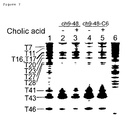
- Figure 7 depicts a photograph demonstrating the modification of thymine residues of ch9-48 and ch9-48-C6 with osmium tetraoxide.
- lane 1: control nucleic acid that was denatured to a single strand;
- lane 2: ch9-48, without cholic acid;
- lane 3: ch9-48, with cholic acid;
- lane 4: ch9-48-C6, without cholic acid;
- lane 5: ch9-48-C6, with cholic acid; and
- lane 6: A+G ladder of ch9-48 obtained by the Maxam-Gilbert method.
- Figure 8 depicts the predicted secondary structure of ch9-48. Thymine (T) is indicated with boldface letters.
- Figures 9-21 depict the source code list of the software program used in the Example for selecting nucleotide sequences forming a nucleic acid aptamer of the present invention.
- The present invention is specifically illustrated below with Examples.
- DNA aptamers that specifically bind to cholic acid were selected from a single-stranded DNA (hereinafter abbreviated as "ssDNA") pool containing single-stranded 100-mer oligonucleotides, which include random inserts of 64 nucleotides (5'-GTACCAGCTTATTCAATT-N64-AGATAGTATGTTCATCAG-3'; SEQ ID NO: 1) (N64 represents a random sequence with 64 nucleotides), by the SELEX method (Nature 355, 564-566, 1990). The single-stranded DNA library contained about
9x 10¹⁴ independent sequences. The ssDNAs were synthesized by the phosphoamidate method, and were purified by high-performance liquid chromatography. The 100-mer DNAs were purified by solid-phase extraction using reverse-phase resins. - Each round of the selection was conducted as follows. First, a 100 mer single-strand oligonucleotide with a random insert of 64 nucleotides was denatured in selection buffer (50 mM Tris-HCl, 300 mM NaCl, 30 mM KCl, 5 mM MgCl2, pH 7.6) at 95°C for 5 min, and then allowed to cool slowly to room temperature. The folded ssDNA (45 µg in the first cycle, and 2 to 3 µg in the subsequent cycles) in the selection buffer was loaded onto 500 µl cholic acid-agarose column (2 µmol cholic acid/g gel; SIGMA; hereinafter, abbreviated as selection column), which had been equilibrated with more than 10 ml of the selection buffer. After equilibration for 30 min, the column was washed with 5 to 10 column volumes of the selection buffer. The remaining ssDNA was eluted with 1.5 ml selection buffer containing 5 mM cholic acid, collected, and then precipitated with ethanol in the presence of 20 µg/ml glycogen. The amount of specifically eluted ssDNA with 5 mM cholic acid was estimated by UV absorbance of the collected fractions at 260 nm. The ssDNA was amplified by polymerase chain reaction (PCR).
-
Affigel 102, on which cholic acid had been immobilized in place of cholic acid-agarose, was used in selection rounds 10 and 12 to eliminate the ssDNAs which bind to the aminohexyl linker of cholic acid-agarose. The immobilization of amino ethyl linker through cholic acid toAffigel 102 was conducted as follows: - 20 mg cholic acid was coupled to 3
ml Affigel 102 with 100 mg EDC as the condensing agent in 10 ml of 20 mM HEPES (pH 7.5). The mixture was incubated at room temperature for 10 hr with occasional agitation. The concentration of cholic acid on Affigel 102 (8 µmol/ml gel) was determined by the reaction with 2,4,6-trinitrobenzene sulfonic acid. The cholic-acid immobilizedAffigel 102 was mixed with 3 volumes ofAffigel 102 to adjust the concentration of cholic acid in the column approximately 2 µmol/ml. - Counter selections were also done, after selection rounds 5, 6, 7, and 13, to remove the ssDNAs that bind directly to agarose matrix. More specifically, ssDNA was loaded on the Sepharose 4B control column (SIGMA) to remove the ssDNAs bound to the agarose matrix.
- PCR was carried out using the
primers 5'-biotin-CTGATGAACATACTATCT-3' (SEQ ID NO: 2) and 5'-GTACCAGCTTATTCAATT-3' (SEQ ID NO: 3). 100µl PCR mixture, which contains 1 unit of Ex Taq DNA polymerase (TaKaRa), 60 pmol of each primer, 20 nmol of each dNTP, and 0.4 to 0.8 µg of Perfect Match (E. coli ssDNA binding protein) (Stratagene), was added to improve the fidelity of the polymerase reaction. The thermal cycling for PCR was 94°C for 30 sec, 46°C for 30 sec, and 72°C for 30 sec. The amplified biotinylated double-strand DNA was extracted with phenol/chloroform, precipitated with ethanol, and bound to the avidin immobilized on column for the generation of ssDNA. The obtained ssDNAs were used as the input for the subsequent selection round. These steps were repeated to enrich the ssDNAs having the binding affinity for cholic acid. - The PCR products of double-stranded DNA amplified from the library of
selection round 13, were cloned into pGEM-T vector (Promega) to determine their sequences by the dideoxy method. Sequence homology analysis and prediction of the secondary structures of ssDNAs according to the free energy minimization method were achieved by the MacDNASIS Pro v1.0 program (HITACHI Software Engineering). Search for complementary sequences forming three stems in each sequence clone was carried out with original computer program written by the C language. Figures 9-21 show the source code list for the computer program. Further, the algorithm of the computer program is summarized below. - This algorithm determines all potential three-way-junction structures in a given nucleotide sequence. As described above, a three-way-junction structure represents a
structure containing stem 1,stem 2, andstem 3 that intersect at a single point (Figure 8). According to the algorithm, first, possible nucleotidesequences forming stem 1 are assessed by repeating the search for complementary nucleotide sequences. In the next step, whenstem 1 could be formed, then the search for a nucleotide sequence that can formstem 2 is carried in the remaining region. When thestem 2 could also be formed, then the search for a nucleotide sequence that can formstem 3 is carried out in the remaining region. Each step of the search is carried out as follows: - Search is started from the 5'-end of a given nucleotide sequence, and proceeds toward the 3'-end, nucleotide by nucleotide. Each nucleotide is taken as the starting point of
stem 1, which is referred to as stem 1b, and the possibility ofstem 1 formation are assessed as follows. Specifically, a nucleotide sequence complementary to the stem 1b is searched starting from the 3'-end, and the complementary nucleotide is referred to as stem 1e, the terminal residue of thestem 1. When a nucleotide sequence, with the same length, of "minimal stem length" from the stem 1b toward the 3'-end direction is complementary to the nucleotide sequence starting from the stem 1e extending toward the 5'-end direction, thestem 1 is assumed to be formed. The "minimal stem length" may be any arbitrary value. - Whether a
stem 2 can be formed in the sequence of the region excluding those corresponding to thestem 1 is determined by the same method as in the above 1). However, it is necessary that the length of loop portion in thestem 2 has the "minimal loop length" or longer. Further, the stem 2b, the starting position ofstem 2, is selected so that the distance between the stem 2b and thestem 1 is the "gap size" or shorter. The "minimal loop length" and "gap size" can be any arbitrary values. - Whether a
stem 3 can be formed in the sequence of the region excluding thoseregions constituting stem 1 andstem 2 is determined by the same method as in the above 2). However, the starting position ofstem 3, stem 3b, and the termination position ofstem 3, stem 3e, are selected so that the distance between the stem 3b and thestem 2, as well as the distance between the stem 3e and thestem 2 are the above described "gap size" or shorter. - After the first round of selection as described above, 1% or less of the ssDNAs loaded were trapped on the selection column. In the subsequent rounds, the quantity of ssDNAs bound to cholic acid markedly increased. In the
selection round 13 and later rounds, about 70% of the loaded ssDNAs was trapped on the column, and were then eluted (Figure 1). - After
round 13, the nucleotide sequences of 19 clones were determined to define a common motif sequence responsible for the binding with cholic acid. The sequences of the 19 clones were entirely different from one another, and no significant homology could be detected among the sequences by primary sequence homology search using the MacDNASIS program. However, as depicted in Table 1, sequences presumed to form three-way junctions as a secondary structure were observed. Three pairs of three-way regions (regions constituting the double-stranded chain) are underlined, indicated with boldface letters, and indicated with outlined letters, respectively, in the nucleotide sequences demonstrated in Table 1. The italicized letters indicate mismatched or wobble (T-G) position in the stem region.
- a) Clone-1: SEQ ID NO: 8
- Clone-2: SEQ ID NO: 9
- Clone-5: SEQ ID NO: 10
- Clone-7: SEQ ID NO: 11
- Clone-9: SEQ ID NO: 12
- Clone-11: SEQ ID NO: 13
- b) Clone-3: SEQ ID NO: 14
- Clone-4: SEQ ID NO: 15
- Clone-6: SEQ ID NO: 16
- Clone-8: SEQ ID NO: 17
- Clone-10:SEQ ID NO: 18
- Clone-12: SEQ ID NO: 19
- Clone-13: SEQ ID NO: 20
- Clone-14: SEQ ID NO: 21
- Clone-15: SEQ ID NO: 22
- Clone-16: SEQ ID NO: 23
- Clone-17: SEQ ID NO: 24
- Clone-18: SEQ ID NO: 25
- Clone-19: SEQ ID NO: 26
- The affinity of cholic acid with ssDNAs was analyzed by the equilibrium-filtration method (Science 263: 1425-1429, 1994). Cholic acid was added to the DNA samples in the selection buffer (200 µl) at a final concentration of 50 µM. Each binding mixture was incubated at 25°C for 5 min. The mixture was then placed in
Microcon 10 filtration device (Amicon) and centrifuged for 15 min at 850 xg. Cholic acid that is not bound to the ssDNAs can be recovered as the filtrate by this procedure. The concentrations of cholic acid in 20 to 30 µl of filtrate were determined by diagnostic kit (WAKO chemical) for quantitative determination using 3α-hydroxysteroid dehydrogenase and diaphorase including p-nitrotetrazolium blue dye and nicotinamide adenine dinucleotide. For each ssDNA sample, the concentration of bound cholic acid (Cb) was determined by the difference of cholic-acid concentration between the filtrate and retentate, which was calculated by the following equation (equation 1) :
(wherein, Vf(µl) is the volume of filtrate; and Cr and Cf(µM) represent the concentrations of cholic acid in the retentate and filtrate, respectively). - The equilibrium-dissociation constants (K d) were calculated from the following standard quadratic binding equation (equation 2) :

(wherein, Dt is the total concentration of ssDNA). - To determine the minimal sequence required for the binding with cholic acid, the present inventors prepared a series of deletion mutants derived from
clones clone 1,clone 2, and each of the deletion mutants, was applied to selection column, and the amount of ssDNA specifically eluted from the selection column with 5 mM cholic acid was measured (75% and 77% ofclones clone 1, ch1-39 (5'-GAGGGCAGCGATAGCTGGGCTAATAAGGTTAGCCCCATC-3'; SEQ ID NO: 4), and the 38mer deletion mutant ofclone 2, ch2-38 (5'-GCGCCGATTGACCCAAATCGTTTTGTATGCAAAAGCGC-3'; SEQ ID NO: 5), the deletion mutants were determined to correspond to the minimal sequences essential for the binding with cholic acid. 22% and 40% of ch1-39 and ch2-38, respectively, were eluted from the selection column. However, no elution could be observed for deletion mutants with shorter sequences. Interestingly, the secondary structures ofclones - Then, the present inventors prepared deletion mutants of
clones equation clones Table 2 Clone name Dissociation constant (µM) ch5-63 28.7 ch7-69 16.7 ch9-48 5.0 ch11-76 52.1 - The affinity of
clones - The predicted secondary structure of
clone 11 was more complex than the other 5 clones. A 35mer mutant oligomer ofclone 11 was prepared by truncating nucleotides except those of the three-way-junction region to determine the dissociation constant for cholic acid. As a result, the dissociation constant was demonstrated to be 76.8 µM, which is comparable to that of the 76mer deletion mutant (52.1 µM). - By comparing the predicted three-way-junction structures of the 6 clones described above, two common features emerged. First, a high affinity is provided when the 3 stems and 2 loops are constituted of 4 base pairs or more. Second, the affinity is elevated by constituting the 3 base pairs flanking the junction with 2 or 3 GC base pairs.
- Furthermore, the sequences of another 13 clones were analyzed with computer program based on the above-mentioned algorithm. The condition for analysis by the program was as follows. First, each sequence was searched with the requirement that the minimal stem-2 length is 2, minimal loop length is 4, and gap size is 0. Then, nucleotide sequences, whose junction moiety contains two or more GC base pairs and at the same time whose stems of the three-way junction constitute of 3 base pairs or are longer, were selected visually (namely, without using any program) from the nucleotide sequences that meet the requirement. At this stage, the stem was permitted to contain mismatches. Namely, like ch1-47, the sequences were permitted to contain mismatches in the third and subsequent base pairs from the junction in the stem. In such cases, mismatches are not counted as the stem length. Thus, sequences whose length of each stem was 3 base pairs or longer excluding mismatched base pairs was selected. As a result, 12 clones comprising of sequences that contain the three-way-junction structure; each contained stems having 3 or more base pairs, and 2 or 3 GC base pairs were present near the junction. The remaining 1 clone also comprised a sequence containing the three-way-junction structure while one of the three stems contained only 2 base pairs. Deletion mutants of the 13 clones, which contained the normal three-way junction region, were prepared, and their affinity for cholic acid was tested. Table 3 shows the nucleotide sequences of these deletion mutants of the 13 clones and the above-mentioned 6 clones. These nucleotide sequences correspond to those existing between the nucleotide sequences underlined in the nucleotide sequences indicated in Table 1.

Table 4 Clone name Dissociation constant (µM) ch3-63 31.1 ch4-74 15.6 ch6-56 28.9 ch8-47 12.6 ch10-52 60.7 ch12-61 12.2 ch13-53 18.7 ch14-37 6.4 ch15-54 67.5 ch16-40 10.7 ch17-63 23.4 ch18-56 36.6 ch19-40 40.5 - All the 19 clones whose sequences had been determined contained the cholic acid binding sequence, which sequence was predicted to form the common secondary structure (i.e., the three-way junction).
- In order to determine the important components responsible for the formation of predicted three-way-j unction structure and the binding with cholic acid in the selected clones, the deletion mutant ch2-40, which contains the three-way-junction region of
clone 2, was subjected for mutation analysis. A series of ch2-40 mutants, which contain single-nucleotide substitution, single-base-pair substitution, or single-nucleotide deletion or insertion, were synthesized. The relative binding affinity of each mutant for cholic acid was assessed by the "equilibrium-filtration method". Figure 5a shows the binding affinity of the deletion mutant ch2-40, when the nucleotide has been substituted with the nucleotide indicated with an arrow. Figure 5b shows the binding affinity of ch2-40, when a base pair forming the stem region of the three-way junction has been substituted with the base pair indicated by an arrow. - As shown in Figure 5a, pairwise compensatory substitutions on the possible 3 stems from G-C to A-T base pair reduced the relative binding affinity 65.3% (C6-G21 to T6-A21), but still demonstrated some affinity. In contrast, several single base-substitutions around the junction abolished the binding affinity completely (figure 5b; G4 to C4, C6 to G6, A22 to T22, and G36 to C36). Comparing the effect of single base and pairwise substitutions to the binding affinity, the former, which disrupt the proposed Watson-Crick base pair in the predicted secondary structure, reduced the binding affinity more significantly than the latter.
- This result supports the formation of the predicted secondary structure and indicates the importance of the formation of the three-way-junction structure for maintaining the affinity. Both single base and base pair substitutions around the junction of ch2-40 significantly reduced the binding affinity, whereas those base and base pair substitutions near the 5'-end, 3'- end, and on the 2 loops did not decrease. Furthermore, single base substitutions on the 2 loops, as well as deletion of A12 and A28, and insertion of an adenosine between A12 and C13, or A28 and T29 hardly affected the binding affinity. In conjunction with the fact that no apparent similarity of the sequence and length of the stem and loop regions could be observed among the 19 clones, the binding site for cholic acid was indicated to be the junction of 3 stems.
- Next, the present inventors focused on the variation and arrangement of 3 base pairs in the junction of the selected 19 clones. The junctions of these clones consisted of either 2 G-C and 1 A-T, or 3 G-C base pairs (termed 2G-C and 3G-C junction, respectively) (Figure 6a). 2G-C junction and 3G-C junction, ch9-48 and ch16-40, respectively, are represented in Figure 6b. Of the selected 19 clones, 9 clones possessed the 2G-C junction, and the other 10 clones the 3G-C junction. Comparing the arrangement of the 3 base pairs on the 3G-C junctions, 9 of the 10 clones had the same arrangement. 6 of the 9 clones with the 2G-C junction had a similar arrangement, and the remaining 3 clones had varied arrangement.
- To further evaluate the structural requirement of the 3 base pairs on the junctions for the observed interaction, base pair substitutions on the junction of ch16-40 and ch9-48, which are deletion mutants including the intact three-way-junction region of
clone 9 and 16 that demonstrate the lowest K d values, were carried out. The relative binding affinity for cholic acid of ch16-40 and ch9-48, which contains single-nucleotide substitution, is shown in Table 5. The number in the table indicates the relative affinity (%) taking the binding affinity of clones without substitution as 100.Table 5 (a) ch9-48 Relative binding affinity (%) Original base pair G5- C44 100 G6- C31 100 A32- T43 100 Substituted base pair C5-G44 74 C6- G31 90 T32- A43 93 A5-T44 46 A6-T31 27 G32-C43 57 I5-C44 44 I6- C31 95 I32- C43 5 G5- T44 0 G6- T31 0 G32- T43 0 C5- C44 0 C6- C31 0 T32-T43 0 (b) ch16-40 Relative binding affinity (%) Original base pair G18- C5 100 G19- C36 100 G4- C37 100 Substituted base pair C18-G5 81 C19- G36 86 C4-G37 68 A18-T5 55 A19- T36 63 A4- T37 60 I18-C5 59 I19- C36 42 I4-C37 18 G18- T5 0 G19- T36 0 G4- T37 0 C18- C5 0 G19- G36 0 C4- C37 0 - Interestingly, any substitutions by Watson-Crick base pairs did not lead to the loss of affinity whereas mismatched and wobble base pairs (G-T) completely resulted in the loss of affinity. By constructing a CPK model of the 6 nucleotides forming 3 Watson-Crick base pairs, these nucleotides were demonstrated to form a cyclophane-like cyclic structure consisting of 3 purine and 3 pyrimidine bases with a cavity of a inner diameter from 12 to 17 Å. Furthermore, the shape and size of this cavity is suited for the inclusion of cholic acid whose size is roughly 15 Å. Herein, a CPK model (Corey-Pauling-Koltun space filling molecular model) is a kind of molecular model of the spatial three-dimensional models. An improved molecular dynamics of the Corey-Pauling molecular model is merchandised by ad hoc committee to design and develop new atom and bond models of the US National Institutes of Health (NIH).
- Even though with a few exceptions, the substitution with Watson-Crick base pairs did not influence the binding affinity. Substitutions from G-C to A-T reduced the binding affinity (C5-G36 to T5-A36: 56.1%; C6-G21 to T6-A21: 34.7%), whereas almost no reduction of the binding affinity was observed with substitution of A-T to G-C (T22-A35 to C22-G35: 95.2%). These results agree with the feature of the selected 19 clones having 2 or 3G-C within the junction and without junctions with 1 or none of the G-C base pairs.
- The alterations in the ligand affinity due to mutations in the nucleotide sequence constituting the three-way junction in the aptamer of the present invention, which is constituted by two molecules, i.e. a target nucleotide sequence and a probe, were examined. More specifically, the affinity for cholic acid of the three-way junctions, wherein a target nucleotide sequence and a probe with nucleotide sequences described below were hybridized, was measured by SPR. 5 µM target nucleotide sequence and 5 µM probe were mixed, and the mixture was denatured with heat under the same condition as described in Example 1-(1). By loading 20 µl solution of the probe-target nucleotide sequence on a cholic acid-immobilized sensor chip BIAcore2000 (BIAcore), an SPR signal change of 600 RU (RU= resonance unit) was observed. On the other hand, when a single-nucleotide mutation had been introduced into the target nucleotide sequence (the underlined segment in the target nucleotide sequence was altered to CCTAGCAGCCGGAGCGGTGG). However, no SPR signal change could be observed. Additionally, not only the above sequence combination, but also when a mismatch had been introduced in the base pairs adjacent to the junction, the binding affinity decreased below the detectable level.
- The sequence of the target nucleotide sequence/SEQ ID NO: 27 (the segment indicated with lower-case letters is complementary to the probe, and the junction is located at the position corresponding to the nucleotides flanking the single space)
5'-GTACCAGCTTATTCAATTACAGATCGAGGGCAGCGATAGCcctagcagcg ggagcggtg gCATCGGTCCTGGACTTGGGACTAGATAGTATGTTCATCAG-3'; and
the sequence of the probe (CH3J-1-100)/SEQ ID NO: 28 (the segment indicated with lower-case letters is complementary to the target nucleotide sequence)
5'-ccaccgctccACTCAACTGGTTTTCCAGTTGAGTcgctgctagg-3'. - Next, using cholic acid, probe, and target nucleotide sequence (50 µM each), the amount of cholic acid binding to the probe-target nucleotide sequence complex was determined by the equilibrium-filtration method described in Example 2(1). The nucleotide sequences of the used target nucleotide sequence and probe are shown below:
- the sequence of the target nucleotide sequence/SEQ ID NO: 29 5'-CCTAGCAGCGGGAGCGGTGG-3'; and
- the sequence of the probe/SEQ ID NO: 30 (the segment indicated with lower-case letters is complementary to the target nucleotide sequence)
5'-ccaccgctccACTCAACTGGTTTTCCAGTTGAGTcgctgctagg-3'. - Result of the measurement demonstrated that the amount of bound cholic acid to be 36.6 µM (for ch2-40 under the same condition, the amount of bound cholic acid was 27.8 µM). On the other hand, when a single-nucleotide mutation had been introduced into the target nucleotide sequence (5'-CCTAGCAGCcGGAGCGGTGG-3'/ SEQ ID NO: 31; the lower-case letter corresponds to the mutation), the sequence didn't bind cholic acid under the same condition (the amount of bound cholic acid was 0 µM).
- The present inventors carried out chemical-modification studies on ch9-48 using an oxidizing agent, osmium tetroxide, to further evaluate the secondary structure of the aptamer and the bases related to the interaction. Osmium tetroxide selectively reacts with thymine (T) in a single-stranded state in the presence of pyridine (Furlong J. C. et al., Biochemistry, 28, 2009-2017, 1989). This selectivity is suggested to be the result of the activity of osmium tetroxide to attack C-5, 6 double bond of thymine (Nielsen P.E. et al., J.Mol.Recog., 3, 1-25, 1990). The sites of osmylation can be cleaved by alkaline treatment.
- 5 µM of 3'-FITC labeled ch9-48 or ch9-48-C6 were incubated with 100 µl binding buffer comprising 1 mM osmium tetroxide and 3% pyridine, with or without 5 mM cholic acid, for 15 min at 20°C. The control was incubated at 80°C to denature the sequence to single strands. The reactions were stopped by two sequential ethanol precipitations. The aptamers were cleaved at the sites of osmate adducts (at the site of single stranded T) by the treatment with 15 µM of 1 M piperidine at 90°C for 30 min. After the addition of 15 µl formamide loading buffer, the cleaved products were electrophoresed on 10% polyacrylamide gels (19%; the ratio of monomer:bis was 19:1) containing 7 M urea. The buffer system consisted of 90 mM Tris-borate (pH 8.0) and 2 mM EDTA. The gels were scanned and analyzed using Fluorlmager 595. The results are shown in Figure 7.
- The photograph demonstrating the result of polyacrylamide gel electrophoresis is depicted in Figure 7. The pattern of modifications on ch9-48 under the folding condition (i.e., in the binding buffer at 20°C) was highly consistent with the proposed secondary structure (Figure 8). A strong modification was observed at T43 flanking the junction, while the T's in the loop regions were also highly reactive. The T's in the stem regions were efficiently protected except T23 and T46: T23 was reactive due to its position at the end of the stem region, and T46 seems to be reactive due to the instability of the short stem structure lacking a loop. A mutant ch9-48-C6, a mutant of ch9-48, which lacks the ability to bind to cholic acid, was prepared by substituting the 6th G to C, and the modification pattern was compared with ch9-48. In the presence of cholic acid, the degree of modification of T43 of ch9-48 at the branching point was reduced to less than 40%. In contrast, the reactivity of T43 in ch9-48-C6 was only slightly suppressed by cholic acid. Thus, T43 in ch9-48 appears to be directly involved in the binding with cholic acid.
- The present invention provides a novel detection method for target nucleotide sequences utilizing a nucleic acid aptamer formation. A specific detection of SNP is possible by the detection method of the present invention due to the use of a nucleic acid aptamer whose ligand-binding affinity greatly changes depending on the presence of a single-nucleotide mismatch. No detection principle wherein a single-nucleotide mismatch provides such significant difference has been known in the art.
- Furthermore, the present invention provides nucleic acid aptamers with a novel structure that is useful for detecting target nucleotide sequences of the present invention. The ligand-binding affinity of the nucleic acid aptamers of the present invention that capture a ligand, such as cholic acid, at the three-way junction is lost by a mismatch alone in any one of the three base pairs of the three junction moiety. Thus, the nucleic acid aptamers comprising the three-way junction are highly preferable for the application to the detection method of SNPs.
-
- <110> Center for Advanced Science and Technology Incubation, Ltd.
- <120> Method for detecting target DNA sequence.
- <130> SEN-112PCT
- <140>
<141> - <150> JP 1999-357913
<151> 1999-12-16 - <160> 31
- <170> PatentIn Ver. 2.0
- <210> 1
<211> 100
<212> DNA
<213> Artificial Sequence - <220>
<223> Description of Artificial Sequence:Artificially Synthesized Sequence - <400> 1

- <210> 2
<211> 18
<212> DNA
<213> Artificial Sequence - <220>
<223> Description of Artificial Sequence:Artificially Synthesized Primer Sequence - <400> 2

- <210> 3
<211> 18
<212> DNA
<213> Artificial Sequence - <220>
<223> Description of Artificial Sequence:Artificially Synthesized Primer Sequence - <400> 3

- <210> 4
<211> 39
<212> DNA
<213> Artificial Sequence - <220>
<223> Description of Artificial Sequence:Artificially Synthesized Sequence - <400> 4

- <210> 5
<211> 38
<212> DNA
<213> Artificial Sequence - <220>
<223> Description of Artificial Sequence:Artificially Synthesized Sequence - <400> 5

- <210> 6
<211> 47
<212> DNA
<213> Artificial Sequence - <220>
<223> Description of Artificial Sequence:Artificially Synthesized Sequence - <400> 6

- <210> 7
<211> 40
<212> DNA
<213> Artificial Sequence - <220>
<223> Description of Artificial Sequence:Artificially Synthesized Sequence - <400> 7

- <210> 8
<211> 100
<212> DNA
<213> Artificial Sequence - <220>
<223> Description of Artificial Sequence:Artificially Synthesized Sequence - <400> 8

- <210> 9
<211> 100
<212> DNA
<213> Artificial Sequence - <220>
<223> Description of Artificial Sequence:Artificially Synthesized Sequence - <400> 9

- <210> 10
<211> 100
<212> DNA
<213> Artificial Sequence - <220>
<223> Description of Artificial Sequence:Artificially Synthesized Sequence - <400> 10

- <210> 11
<211> 100
<212> DNA
<213> Artificial Sequence - <220>
<223> Description of Artificial Sequence:Artificially Synthesized Sequence - <400> 11

- <210> 12
<211> 100
<212> DNA
<213> Artificial Sequence - <220>
<223> Description of Artificial Sequence:Artificially Synthesized Sequence - <400> 12

- <210> 13
<211> 99
<212> DNA
<213> Artificial Sequence - <220>
<223> Description of Artificial Sequence:Artificially Synthesized Sequence - <400> 13

- <210> 14
<211> 100
<212> DNA
<213> Artificial Sequence - <220>
<223> Description of Artificial Sequence:Artificially Synthesized Sequence - <400> 14


- <210> 15
<211> 99
<212> DNA
<213> Artificial Sequence - <220>
<223> Description of Artificial Sequence:Artificially Synthesized Sequence - <400> 15

- <210> 16
<211> 100
<212> DNA
<213> Artificial Sequence - <220>
<223> Description of Artificial Sequence:Artificially Synthesized Sequence - <400> 16

- <210> 17
<211> 100
<212> DNA
<213> Artificial Sequence - <220>
<223> Description of Artificial Sequence:Artificially Synthesized Sequence - <400> 17

- <210> 18
<211> 100
<212> DNA
<213> Artificial Sequence - <220>
<223> Description of Artificial Sequence:Artificially Synthesized Sequence - <400> 18

- <210> 19
<211> 100
<212> DNA
<213> Artificial Sequence - <220>
<223> Description of Artificial Sequence:Artificially Synthesized Sequence - <400> 19

- <210> 20
<211> 100
<212> DNA
<213> Artificial Sequence - <220>
<223> Description of Artificial Sequence:Artificially Synthesized Sequence - <400> 20

- <210> 21
<211> 100
<212> DNA
<213> Artificial Sequence - <220>
<223> Description of Artificial Sequence:Artificially Synthesized Sequence - <400> 21

- <210> 22
<211> 100
<212> DNA
<213> Artificial Sequence - <220>
<223> Description of Artificial Sequence:Artificially Synthesized Sequence - <400> 22

- <210> 23
<211> 95
<212> DNA
<213> Artificial Sequence - <220>
<223> Description of Artificial Sequence:Artificially Synthesized Sequence - <400> 23

- <210> 24
<211> 99
<212> DNA
<213> Artificial Sequence - <220>
<223> Description of Artificial Sequence:Artificially Synthesized Sequence - <400> 24

- <210> 25
<211> 97
<212> DNA
<213> Artificial Sequence - <220>
<223> Description of Artificial Sequence:Artificially Synthesized Sequence - <400> 25

- <210> 26
<211> 100
<212> DNA
<213> Artificial Sequence - <220>
<223> Description of Artificial Sequence:Artificially Synthesized Sequence - <400> 26

- <210> 27
<211> 100
<212> DNA
<213> Artificial Sequence - <220>
<223> Description of Artificial Sequence:Artificially Synthesized Sequence - <400> 27

- <210> 28
<211> 44
<212> DNA
<213> Artificial Sequence - <220>
<223> Description of Artificial Sequence:Artificially Synthesized Probe Sequence - <400> 28

- <210> 29
<211> 20
<212> DNA
<213> Artificial Sequence - <220>
<223> Description of Artificial Sequence:Artificially Synthesized Sequence - <400> 29

- <210> 30
<211> 44
<212> DNA
<213> Artificial Sequence - <220>
<223> Description of Artificial Sequence:Artificially Synthesized Probe Sequence - <400> 30

- <210> 31
<211> 20
<212> DNA
<213> Artificial Sequence - <220>
<223> Description of Artificial Sequence:Artificially Synthesized Sequence - <400> 31

Claims (10)
- A method for detecting a target nucleotide sequence, which comprises the steps of:(a) hybridizing a probe to the target nucleotide sequence to form a nucleic acid aptamer having binding affinity for a ligand which can be any component other than nucleic acids; wherein the formed aptamer comprises a nucleotide sequence that forms a non-stem structure in the vicinity of the complementary nucleotide sequence, wherein the non-stem structure is selected from the group consisting of a loop, bulge, non-Watson-Crick type base pair and stem-junction structure, and constitutes a ligand-binding site or a part thereof, and(b) detecting the presence of the target nucleotide sequence using the affinity of the aptamer as an index.
- The method of claim 1, wherein the target nucleotide sequence contains single nucleotide polymorphism, and the affinity of the probe for the ligand vary depending on the single nucleotide substitution in the target nucleotide sequence.
- Use of a probe for detecting a target nucleotide sequence, wherein the probe acquires binding affinity for a ligand which can be any component other than nucleic acids by the hybridizing to the target nucleotide sequence, wherein the formed aptamer comprises a nucleotide sequence that forms a non-stem structure in the vicinity of the complementary nucleotide sequence, wherein the non-stem structure is selected from the group consisting of a loop, bulge, non-Watson-Crick type base pair and stem-junction structure, and constitutes a ligand-binding site or a part thereof.
- Use according to claim 3, wherein the probe forms a structure wherein three or more stems intersect one another by hybridization with the target nucleotide sequence, and binds to the ligand at the region where the stems intersect.
- The use of claim 3, wherein the aptamer comprises three stems and wherein at least two of the three base pairs constituting the junction are G-C nucleotide pairs.
- The use of claim 4, wherein each stem is longer than three base pairs.
- A nucleic acid aptamer, which consists of three stems and binds with cholic acid at the position where the stems intersect, wherein the nucleic acid aptamer has a structure wherein a single strand comprising a stem loop is hybridized to another single strand to form two stems.
- The nucleic acid aptamer of claim 7, wherein the aptamer comprises at least two of the base pairs constituting the junction and which are G-C nucleotide pairs.
- A kit for detecting a target nucleotide sequence comprising a probe and cholic acid, wherein the probe and the target nucleotide sequence form upon hybridization the nucleic acid aptamer having binding affinity for cholic acid, and wherein the formed aptamer comprises a nucleotide sequence that forms a non-stem structure in the vicinity of the complementary nucleotide sequence, wherein the non-stem is selected from the group consisting of a loop, bulge, non-Watson-Crick type base pair and stem-junction structure, and constitutes a ligand-binding site or a part thereof
- The kit of claim 9, wherein the aptamer comprises at least two of the base pairs constituting the junction and which are G-C nucleotide pairs.
Applications Claiming Priority (3)
| Application Number | Priority Date | Filing Date | Title |
|---|---|---|---|
| JP35791399 | 1999-12-16 | ||
| JP35791399 | 1999-12-16 | ||
| PCT/JP2000/008909 WO2001044509A1 (en) | 1999-12-16 | 2000-12-15 | Method of detecting target base sequence |
Publications (3)
| Publication Number | Publication Date |
|---|---|
| EP1243659A1 EP1243659A1 (en) | 2002-09-25 |
| EP1243659A4 EP1243659A4 (en) | 2003-07-02 |
| EP1243659B1 true EP1243659B1 (en) | 2006-04-12 |
Family
ID=18456591
Family Applications (1)
| Application Number | Title | Priority Date | Filing Date |
|---|---|---|---|
| EP00981762A Expired - Lifetime EP1243659B1 (en) | 1999-12-16 | 2000-12-15 | Method for detecting target nucleotide sequences |
Country Status (7)
| Country | Link |
|---|---|
| US (1) | US7303867B2 (en) |
| EP (1) | EP1243659B1 (en) |
| JP (1) | JP4679022B2 (en) |
| AU (1) | AU1891701A (en) |
| CA (1) | CA2394428A1 (en) |
| DE (1) | DE60027322T2 (en) |
| WO (1) | WO2001044509A1 (en) |
Families Citing this family (18)
| Publication number | Priority date | Publication date | Assignee | Title |
|---|---|---|---|---|
| US6936496B2 (en) | 2002-12-20 | 2005-08-30 | Hewlett-Packard Development Company, L.P. | Nanowire filament |
| WO2004092384A2 (en) * | 2003-04-17 | 2004-10-28 | Affectis Pharmaceuticals Ag | Means and methods for diagnosing and treating affective disorders |
| US7223611B2 (en) * | 2003-10-07 | 2007-05-29 | Hewlett-Packard Development Company, L.P. | Fabrication of nanowires |
| US7132298B2 (en) | 2003-10-07 | 2006-11-07 | Hewlett-Packard Development Company, L.P. | Fabrication of nano-object array |
| US7407738B2 (en) * | 2004-04-02 | 2008-08-05 | Pavel Kornilovich | Fabrication and use of superlattice |
| WO2005106035A2 (en) * | 2004-04-09 | 2005-11-10 | Cornell Research Foundation, Inc. | Modular design and construction of nucleic acid molecules, aptamer-derived nucleic acid constructs, rna scaffolds, their expression, and methods of use |
| US20050241959A1 (en) * | 2004-04-30 | 2005-11-03 | Kenneth Ward | Chemical-sensing devices |
| US7683435B2 (en) | 2004-04-30 | 2010-03-23 | Hewlett-Packard Development Company, L.P. | Misalignment-tolerant multiplexing/demultiplexing architectures |
| US7247531B2 (en) | 2004-04-30 | 2007-07-24 | Hewlett-Packard Development Company, L.P. | Field-effect-transistor multiplexing/demultiplexing architectures and methods of forming the same |
| US20060024814A1 (en) * | 2004-07-29 | 2006-02-02 | Peters Kevin F | Aptamer-functionalized electrochemical sensors and methods of fabricating and using the same |
| US7375012B2 (en) * | 2005-02-28 | 2008-05-20 | Pavel Kornilovich | Method of forming multilayer film |
| WO2006130632A2 (en) * | 2005-05-31 | 2006-12-07 | Invitrogen Corporation | Separation and purification of nucleic acid from paraffin-containing samples |
| JP5463621B2 (en) * | 2008-02-27 | 2014-04-09 | ソニー株式会社 | Target substance quantification method |
| JP5594838B2 (en) * | 2008-07-09 | 2014-09-24 | 公立大学法人大阪市立大学 | Oligonucleotide structure and gene expression control method |
| CN105675605A (en) * | 2016-03-29 | 2016-06-15 | 中国药科大学 | Method for detecting bile acid using aptamer-functionalized gold nanoparticles |
| CN107164464B (en) * | 2017-04-27 | 2021-12-21 | 武汉华大医学检验所有限公司 | Method and primer for detecting pollution of sequencing platform index sequence |
| US11807623B2 (en) | 2017-11-30 | 2023-11-07 | Arrakis Therapeutics, Inc. | Nucleic acid-binding photoprobes and uses thereof |
| EP4073246A4 (en) * | 2019-12-12 | 2023-12-06 | President And Fellows Of Harvard College | Compositions and methods for light-directed biomolecular barcoding |
Family Cites Families (2)
| Publication number | Priority date | Publication date | Assignee | Title |
|---|---|---|---|---|
| US5633133A (en) * | 1994-07-14 | 1997-05-27 | Long; David M. | Ligation with hammerhead ribozymes |
| WO2001023612A2 (en) | 1999-09-25 | 2001-04-05 | The University Of North Carolina At Chapel Hill | Determining mutations by selective reaction of the 2'-ribose position in hybridized oligonucleotides |
-
2000
- 2000-12-15 EP EP00981762A patent/EP1243659B1/en not_active Expired - Lifetime
- 2000-12-15 CA CA002394428A patent/CA2394428A1/en not_active Abandoned
- 2000-12-15 AU AU18917/01A patent/AU1891701A/en not_active Abandoned
- 2000-12-15 JP JP2001545586A patent/JP4679022B2/en not_active Expired - Lifetime
- 2000-12-15 US US10/149,869 patent/US7303867B2/en not_active Expired - Lifetime
- 2000-12-15 WO PCT/JP2000/008909 patent/WO2001044509A1/en not_active Ceased
- 2000-12-15 DE DE60027322T patent/DE60027322T2/en not_active Expired - Lifetime
Also Published As
| Publication number | Publication date |
|---|---|
| DE60027322T2 (en) | 2007-01-11 |
| JP4679022B2 (en) | 2011-04-27 |
| EP1243659A4 (en) | 2003-07-02 |
| US7303867B2 (en) | 2007-12-04 |
| DE60027322D1 (en) | 2006-05-24 |
| WO2001044509A1 (en) | 2001-06-21 |
| AU1891701A (en) | 2001-06-25 |
| CA2394428A1 (en) | 2001-06-21 |
| EP1243659A1 (en) | 2002-09-25 |
| US20030170650A1 (en) | 2003-09-11 |
Similar Documents
| Publication | Publication Date | Title |
|---|---|---|
| US7303867B2 (en) | Method for detecting target nucleotide sequences | |
| JP3937136B2 (en) | Nucleotide polymorphism detection method | |
| JP4558932B2 (en) | Pyrazolo [3,4-d] pyrimidine-containing oligonucleotides for hybridization and discrepancy identification | |
| US8334116B2 (en) | Methods and compositions for generation of multiple copies of nucleic acid sequences and methods of detection thereof | |
| US7892796B2 (en) | Solid support assay systems and methods utilizing non-standard bases | |
| KR102592367B1 (en) | Systems and methods for clonal replication and amplification of nucleic acid molecules for genomic and therapeutic applications | |
| US20030077609A1 (en) | Modified oligonucleotides and uses thereof | |
| US20050164184A1 (en) | Hybridization portion control oligonucleotide and its uses | |
| JP6553598B2 (en) | Method of enriching nucleic acids mutated from mixtures | |
| EP0382433A2 (en) | Detection of nucleic acid sequences using fluorescence polarisation | |
| CN101421420B (en) | Mutation detection method and kit used in the method | |
| US20040018530A1 (en) | In vitro evolution of functional RNA and DNA using electrophoretic selection | |
| CA2469383A1 (en) | Hybridization portion control oligonucleotide and its uses | |
| JPWO2001044509A1 (en) | Method for detecting target base sequence | |
| KR20190137718A (en) | Method for Detecting Target Nucleic Acid Using Rolling Circle Amplification and Composition for Detecting Target Nucleic Acid | |
| CN104854121B (en) | Labeled oligonucleotide probes used for nucleic acid sequence analysis | |
| EP1737978A1 (en) | Nucleic acid sequencing | |
| EP1384789A1 (en) | Fluorescent hybridization probes with reduced background | |
| Ahle et al. | Sequence determination of nucleic acids containing 5-methylisocytosine and isoguanine: identification and insight into polymerase replication of the non-natural nucleobases | |
| US20030096252A1 (en) | Helper probes for detection of a target sequence by a capture oligonucleotide | |
| JP2008079604A (en) | Primer set, probe set, method and kit for predicting alcohol resolution and drunkenness tolerance | |
| Ünsal et al. | A novel method of multiplex SNP genotyping assay through variable fragment length allele-specific polymerase chain reaction: multiplex VFLASP-ARMS | |
| Ranasinghe | Novel techniques for genetic analysis |
Legal Events
| Date | Code | Title | Description |
|---|---|---|---|
| PUAI | Public reference made under article 153(3) epc to a published international application that has entered the european phase |
Free format text: ORIGINAL CODE: 0009012 |
|
| 17P | Request for examination filed |
Effective date: 20020708 |
|
| AK | Designated contracting states |
Kind code of ref document: A1 Designated state(s): AT BE CH CY DE DK ES FI FR GB GR IE IT LI LU MC NL PT SE TR |
|
| AX | Request for extension of the european patent |
Free format text: AL;LT;LV;MK;RO;SI |
|
| RIC1 | Information provided on ipc code assigned before grant |
Ipc: 7C 12Q 1/68 A Ipc: 7C 12N 15/10 B Ipc: 7C 12N 15/09 B |
|
| A4 | Supplementary search report drawn up and despatched |
Effective date: 20030519 |
|
| RAP1 | Party data changed (applicant data changed or rights of an application transferred) |
Owner name: KATAYANAGI INSTITUTE |
|
| 17Q | First examination report despatched |
Effective date: 20030904 |
|
| RAP1 | Party data changed (applicant data changed or rights of an application transferred) |
Owner name: KATAYANAGI INSTITUTE |
|
| RBV | Designated contracting states (corrected) |
Designated state(s): DE FR GB IT |
|
| GRAP | Despatch of communication of intention to grant a patent |
Free format text: ORIGINAL CODE: EPIDOSNIGR1 |
|
| GRAS | Grant fee paid |
Free format text: ORIGINAL CODE: EPIDOSNIGR3 |
|
| GRAA | (expected) grant |
Free format text: ORIGINAL CODE: 0009210 |
|
| AK | Designated contracting states |
Kind code of ref document: B1 Designated state(s): DE FR GB IT |
|
| REG | Reference to a national code |
Ref country code: GB Ref legal event code: FG4D |
|
| RIN1 | Information on inventor provided before grant (corrected) |
Inventor name: KATO, TERU Inventor name: KARUBE, ISAO |
|
| REF | Corresponds to: |
Ref document number: 60027322 Country of ref document: DE Date of ref document: 20060524 Kind code of ref document: P |
|
| ET | Fr: translation filed | ||
| PLBE | No opposition filed within time limit |
Free format text: ORIGINAL CODE: 0009261 |
|
| STAA | Information on the status of an ep patent application or granted ep patent |
Free format text: STATUS: NO OPPOSITION FILED WITHIN TIME LIMIT |
|
| 26N | No opposition filed |
Effective date: 20070115 |
|
| REG | Reference to a national code |
Ref country code: FR Ref legal event code: PLFP Year of fee payment: 16 |
|
| REG | Reference to a national code |
Ref country code: FR Ref legal event code: PLFP Year of fee payment: 17 |
|
| REG | Reference to a national code |
Ref country code: FR Ref legal event code: PLFP Year of fee payment: 18 |
|
| PGFP | Annual fee paid to national office [announced via postgrant information from national office to epo] |
Ref country code: DE Payment date: 20191210 Year of fee payment: 20 |
|
| PGFP | Annual fee paid to national office [announced via postgrant information from national office to epo] |
Ref country code: IT Payment date: 20191230 Year of fee payment: 20 Ref country code: FR Payment date: 20191219 Year of fee payment: 20 |
|
| PGFP | Annual fee paid to national office [announced via postgrant information from national office to epo] |
Ref country code: GB Payment date: 20191220 Year of fee payment: 20 |
|
| REG | Reference to a national code |
Ref country code: DE Ref legal event code: R071 Ref document number: 60027322 Country of ref document: DE |
|
| REG | Reference to a national code |
Ref country code: GB Ref legal event code: PE20 Expiry date: 20201214 |
|
| PG25 | Lapsed in a contracting state [announced via postgrant information from national office to epo] |
Ref country code: GB Free format text: LAPSE BECAUSE OF EXPIRATION OF PROTECTION Effective date: 20201214 |