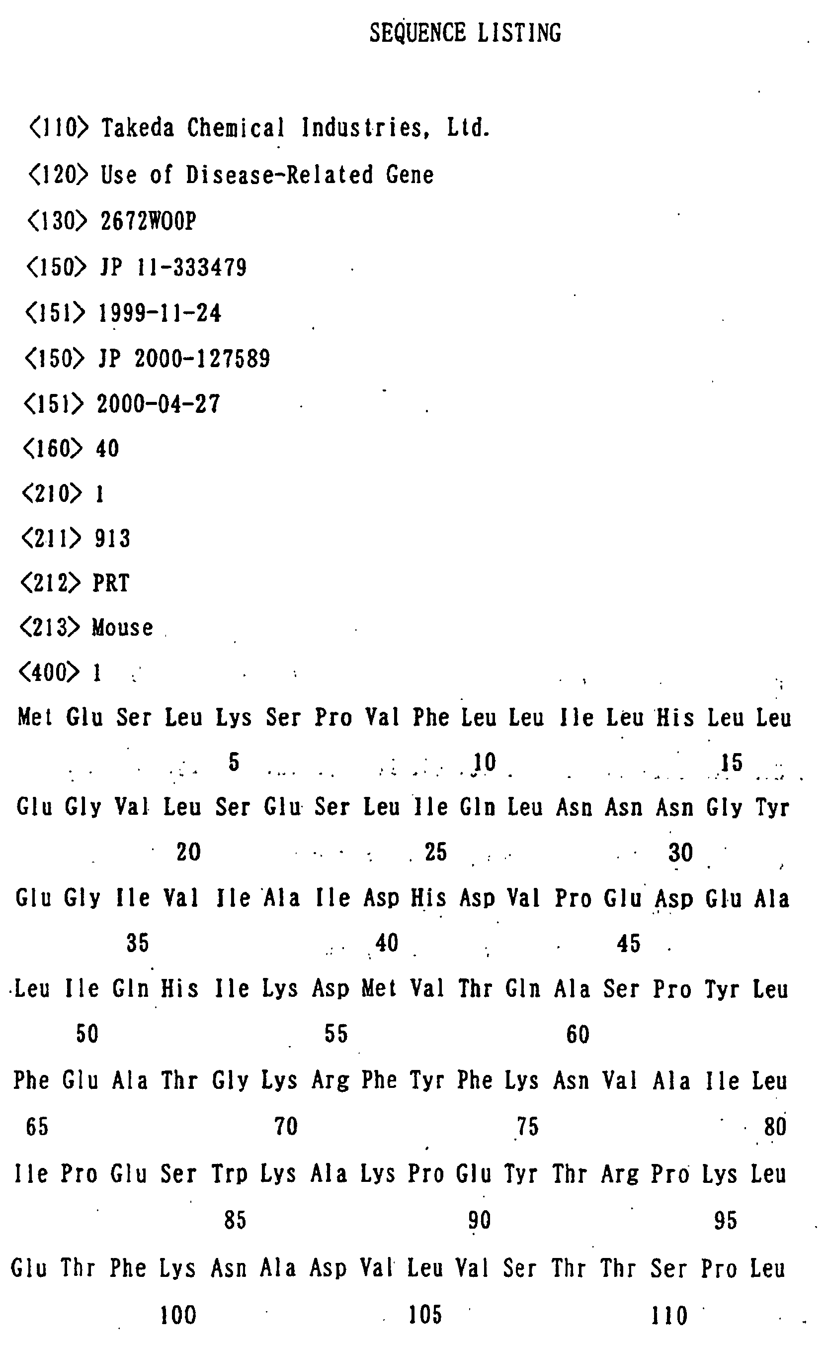
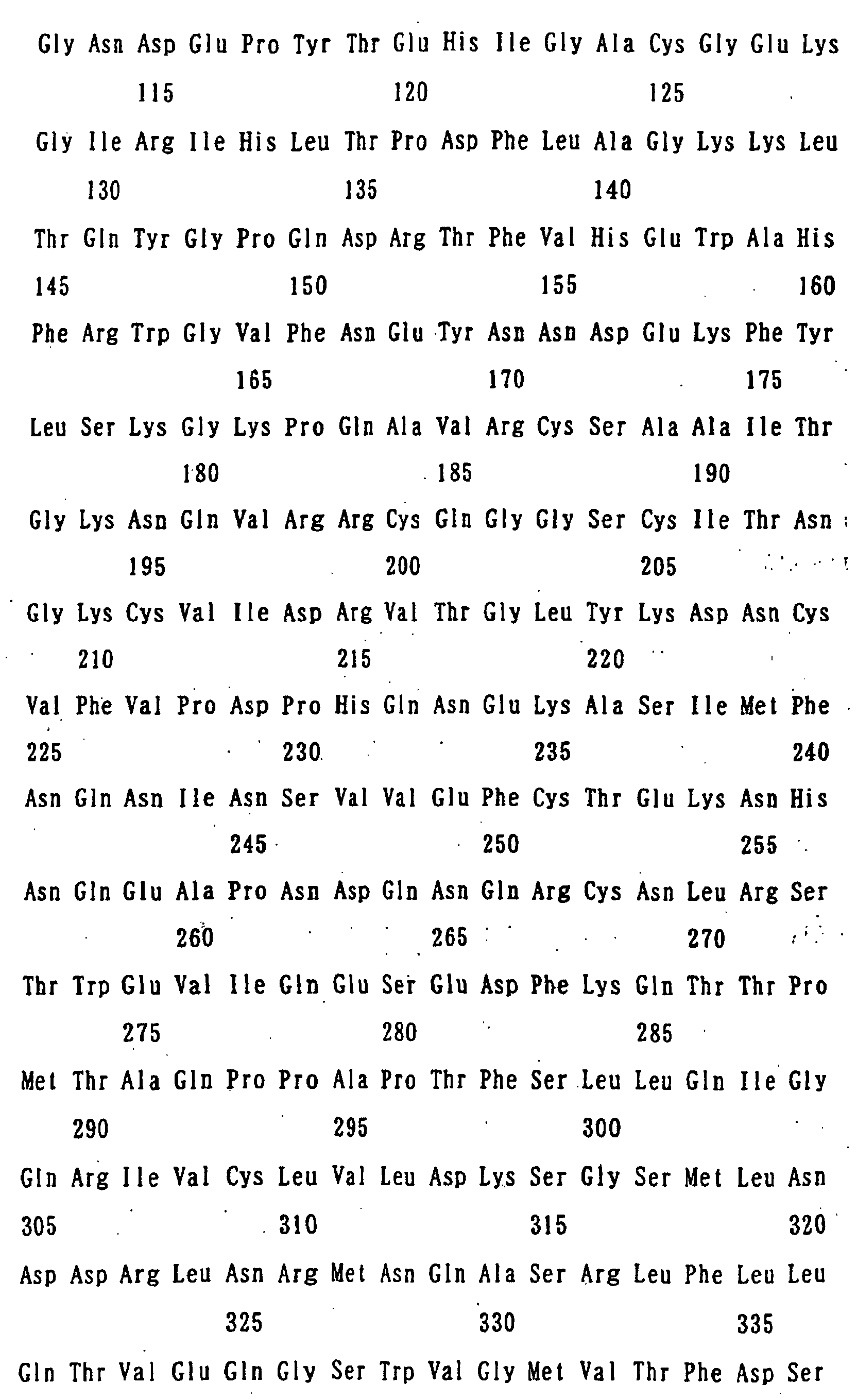
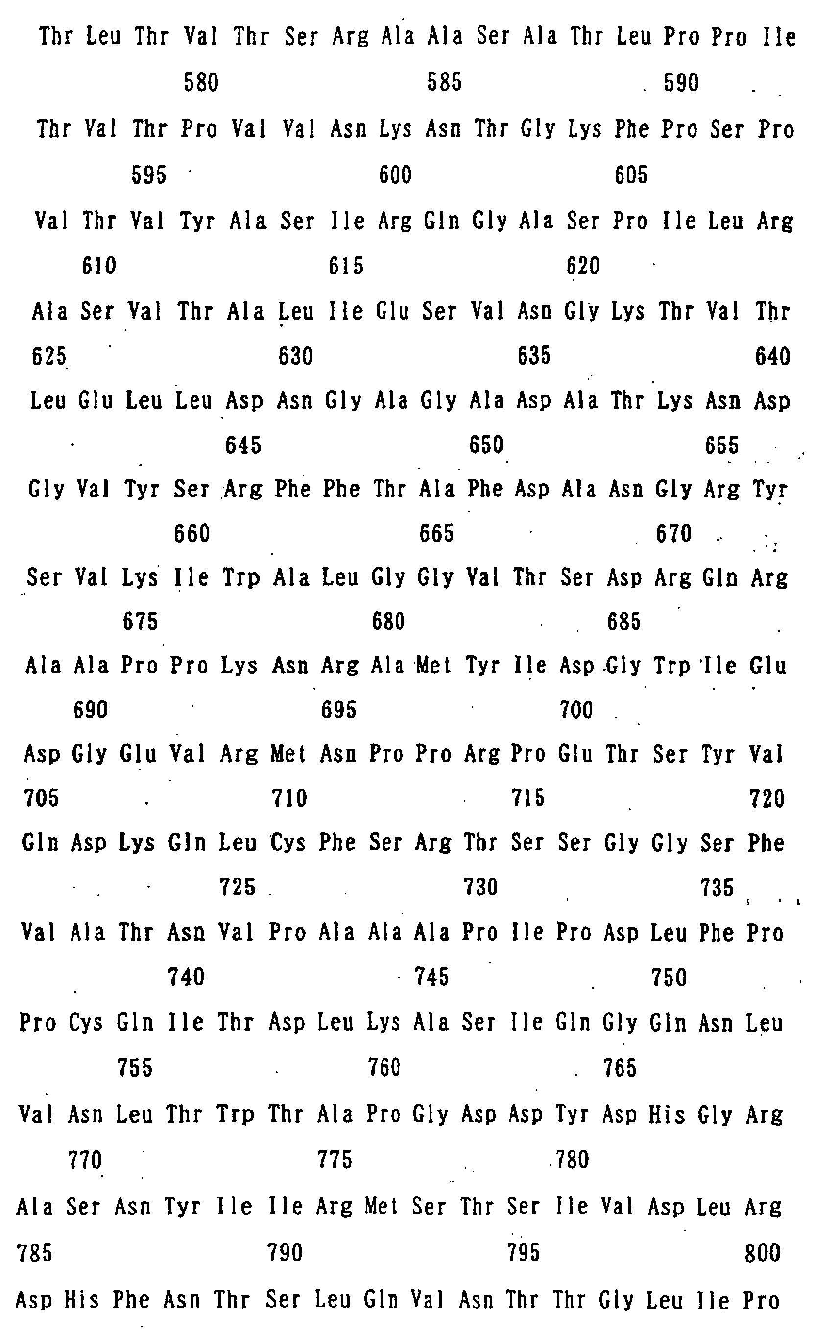
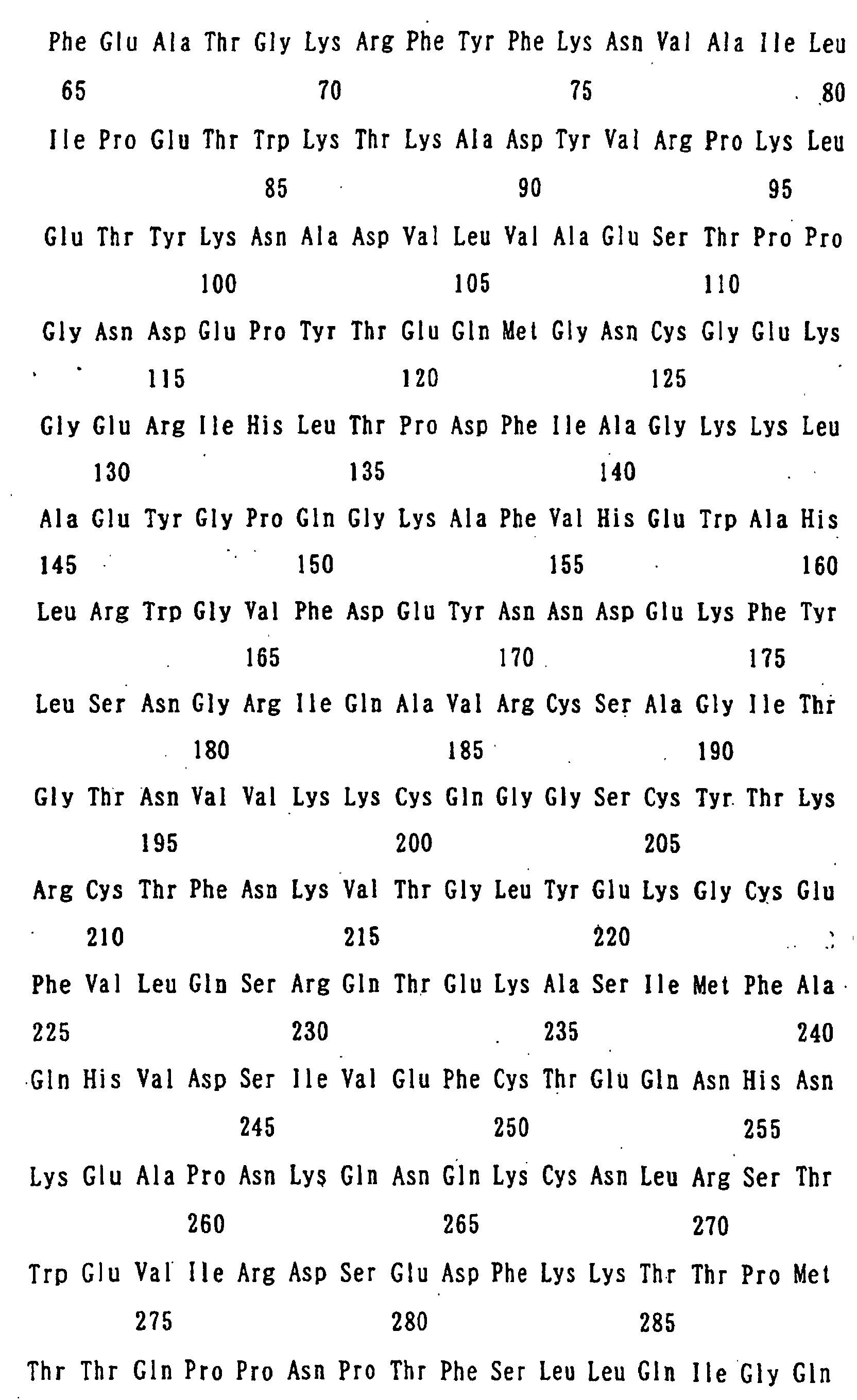
FIELD OF THE INVENTION
This invention relates to use of a disease-associated
gene. More particularly, the invention relates to an
antisense nucleotide to a disease-associated gene, which
is useful as a diagnostic marker for bronchial asthma,
chronic obstructive pulmonary disease, etc., an antibody
against a disease-associated gene product, a method of
screening a drug by using the disease-associated gene product,
etc.
BACKGROUND ART
Bronchial asthma is a chronic inflammatory disease of
airways showing respiratory stenosis, in which symptoms such
as paroxysmal dyspnea, stridor, cough, etc. are observed.
Many cells such as bronchial epithelial cells, mast cells,
eosinophils, T lymphocytes, etc are involved in its onset
and development. One of the most important characteristics
of bronchial asthma is that airways are hyperresponsive to
irritants (airway hyperresponsiveness). This airway
hyperresponsiveness is attributable to airway inflammation
caused mainly due to exfoliation of bronchial epithelial
cells by neurotransmitters secreted from the cells such as
eosinophils, etc., infiltrated into airways, and it is also
considered that genetic factors or environmental factors
will affect the airway hyperresponsiveness complicatedly.
When the inflammatory reaction of airways is triggered
by external irritants (allergens, exhausts) or viral
infection, adhesion molecules including VCAM-1, ICAM-1 and
the like are expressed on bronchial epithelial cells or on
capillary endothelial cells around the bronchi [J. Allergy
Clin. Immunol., 96, 941 (1995)] to produce cytokines or
chemotactic substances. In patients with bronchial asthma,
the function of Th2 type helper T cells is activated to
increase the production of Th2 type cytokines such as IL-3,
IL-4, IL-5, IL-13, GM-CSF, etc., or chemokines such as
eotaxin, RANTES, etc. IL-4 or IL-13 has an activity of
inducing IgE production, and IL-3 or IL-4 has an activity
of inducing the growth of mast cells. Furthermore,
eosinophils differentiate and proliferate in response to
IL-5, GM-CSF, etc. and infiltrate into the airways in
response to eotaxin or RANTES [Allergy Asthma Proc., 20,
141 (1999)].
Epithelial cells that cover the tracheal and bronchial
mucosa not only have the barrier function to prevent direct
transmittance of external irritants to submucosal tissues
and the function to excrete mucus secretions or foreign
matters, but also regulate bronchoconstriction by secreted
epithelium-derived smooth muscle relaxing factors, etc.
Eosinophils infiltrated into the airways of patient with
bronchial asthma release through degranulation
intracellular granule proteins such as activated MBP (major
basic protein), ECP (eosinophil cationic protein), etc.
[Compr. Ther., 20, 651 (1994)]. By the cytotoxic action
of these granular proteins, the exfoliation and damages of
epithelial cells occur. The exfoliation of epithelial cells
leads to exposure of sensory nerve terminals, increase in
epithelial permeability and loss of epithelium-derived
smooth muscle relaxing factors. Also, leukotriene C4
(LTC4) or platelet activating factor (PAF) produced by
eosinophils increase tension of bronchial smooth muscle.
It is considered that when the foregoing changes are repeated
to make it chronic, the bronchial walls would be thickened
to cause airway hyperresponsiveness.
As stated above, it is known that genes of the cytokines
or adhesion molecules described above are increasingly
expressed, accompanied by inflammation of the airways, but
there is no report to systematically analyze the change of
a gene, expression of which is localized in the lesion of
lung/bronchi and which is associated with the onset of airway
hyperresponsiveness.
On the other hand, involvement of chloride channel in
a disease is reported only on CFTR associated with cystic
fibrosis [Science, 257, 1125 (1992)] and CIC chloride channel
with myotonia [Nature, 354, 304 (1991)], but there is no
report that calcium-dependent chloride channel would be
associated with a disease such as bronchial asthma, etc.
Human-derived CLCA1cDNA and protein and mouse-derived
Gob-5cDMA and protein of the present invention are described
in the literature [Genomics, 54, 200 (1998) ; Biochem. Biophys.
Res. Commun., 255, 347 (1999)], but the literature is silent
on any involvement of these DNAs or proteins in bronchial
asthma, chronic obstructive pulmonary disease, etc.
The present invention provides a method of screening
a compound capable of inhibiting the activity of a protein,
expression of which increases in the lung/bronchi having
increased airway hyperresponsiveness, a compound
obtainable by the screening method, etc.
DISCLOSURE OF THE INVENTION
The present inventors made extensive investigations
to solve the problems above and as a result, discovered a
cDNA, which expression markedly increases in the
lung/bronchi of mouse asthma model. On further studies of
this cDNA expression pattern in detail, it has been revealed
that the expression is induced locally in the epithelial
cells of airways with allergic airway inflammation.
Based on these findings, the inventors continued further
investigations so that the present invention has been
attained.
That is, the present invention relates to the following
features.
BRIEF DESCRIPTION OF THE DRAWINGS
FIG. 1 shows tissue distribution of Gob-5 gene (mRNA)
in normal mouse and model mouse with increased airway
hyperresponsiveness, in which Lu, H, Li, Ki, Br, Thy, Sp,
SI, LI, St and PBL designate lung, heart, liver, kidney,
brain, thymus, spleen, small intestine, large intestine,
stomach and peripheral blood lymphocyte, respectively.
FIG. 2 shows change in increased airway
hyperresponsiveness with time course, by administration of
acetylcholine(Ach)in a dose of 500 µg/kg. **: p<0.01
(Dunnett's Test)
FIG. 3 shows change with time course in the count of
infiltrated cells in the alveolar lavage fluid shown in
EXAMPLE 3, wherein M, Eos, Neu and Lym designate macrophage,
eosinophil, neutrophil and lymphocyte, respectively.
FIG. 4 shows change of Gob-5 gene (mRNA) with time course
in model mouse with increased airway hyperresponsiveness
shown in EXAMPLE 3.
FIG. 5 indicates the Gob-5 gene expression site on frozen
lung sections from model mouse with increased airway
hyperresponsiveness or normal mouse, shown in EXAMPLE 4.
FIG. 6 shows PAS staining of the frozen lung sections
from model mouse with increased airway hyperresponsiveness
or normal mouse, shown in EXAMPLE 4.
FIG. 7 shows the results of the experiment shown
in EXAMPLE 7, in which the antisense nucleotide to Gob-5
gene was administered. **: p<0.01
FIG. 8 shows the airway responsiveness to acetylcholine
in mouse administered with AdV Gob-5-AS or control mouse
administered with AdV-wt, shown in EXAMPLE 9, wherein the
data represent mean values ± standard error. The two groups
of the OVA inhalation group and the PBS inhalation group
were compared by t test (#: p<0.05, ##: p<0.01) and a multiple
comparison between the OVA inhalation group and the AdV
inhalation group was made using the Dunnett's test (# : p<0.05,
##: p<0.01).
In the figure, Columns 1, 2, 3 and 4 designate the PBS
inhalation group, the OVA inhalation group, the group
administered with AdV-wt followed by OVA inhalation, and
the group administered with AdV Gob-5-AS followed by OVA
inhalation, respectively.
FIG. 9 shows PAS staining of lung frozen sections from
mouse administered with AdV Gob-5-AS or control mouse
administered with AdV-wt, shown in EXAMPLE 9.
FIG. 10 shows the Cl- ion secretion assay using CHO cells
or human CLCA1-expressed CHO cells used in EXAMPLE 10,
and changes of fluorescent intensity with time course without
stimulation or stimulated with 10 µM of ionomycin.
In the figure, -o-, --, -Δ- and -▴- denote CHO cells
with no stimulation, CHO cells stimulated with 10 µM of
ionomycin, human CLCA1-expressed CHO cells with no
stimulation and human CLCA1-expressed CHO cells stimulated
with 10 µM of ionomycin, respectively.
FIG. 11 shows the action of human CLCA1-expressed CHO
cells shown in EXAMPLE 10 inhibiting the secretion of Cl-
ions by NFA (niflumic acid), DIDS (4,4'-diisothiocyanastilbene-2,2'-disulfonic
acid) and DTT (dithiothreitol).
In the figure, the columns designate the following
cells.
- Column 1:
- no stimulation
- Column 2:
- added with no anion transportation inhibitor
and stimulated with ionomycin (10 µM)
- Column 3:
- added with NFA (200 µM) and stimulated with
ionomycin (10 µM)
- Column 4:
- added with NFA (10 µM) and stimulated with
ionomycin (10 µM)
- Column 5:
- added with DIDS (200 µM) and stimulated with
ionomycin (10 µM)
- Column 6:
- added with DIDS (10 µM) and stimulated with
ionomycin (10 µM)
- Column 7:
- added with DTT (200 µM) and stimulated with
ionomycin (10 µM)
- Column 8:
- added with DTT (10 µM) and stimulated with
ionomycin (10 µM)
BEST MODE FOR CARRYING OUT THE INVENTION
The protein used in the present invention, which
contains the same or substantially the same amino acid
sequence as the amino acid sequence represented by SEQ ID
NO: 1 (hereinafter sometimes referred to as the protein of
the invention or the protein used in the present invention)
may be any protein derived from any cells of human and other
warm-blooded animals (e.g., guinea pig, rat, mouse, chicken,
rabbit, swine, sheep, bovine, monkey, etc.) such as
hepatocyte, splenocyte, nerve cells, glial cells, β cells
of pancreas, bone marrow cells, mesangial cells, Langerhans'
cells, epidermic cells, epithelial cells, goblet cells,
endothelial cells, smooth muscle cells, fibroblasts,
fibrocytes, myocytes, fat cells, immune cells (e.g.,
macrophage, T cells, B cells, natural killer cells, mast
cells, neutrophils, basophils, eosinophils, monocytes),
megakaryocytes, synovial cells, chondrocytes, bone cells,
osteoblasts, osteoclasts, mammary gland cells, hepatocyte
or interstitial cells; or the corresponding precursor cells,
stem cells, cancer cells, etc.; or any tissues where such
cells are present, such as brain or any of brain regions
(e.g., olfactory bulb, amygdaloid nucleus, basal ganglia,
hippocampus, thalamus, hypothalamus, cerebral cortex,
medulla oblongata, cerebellum), spinal cord, hypophysis,
stomach, pancreas, kidney, liver, gonad, thyroid,
gall-bladder, bone marrow, adrenal gland, skin, muscle, lung,
gastrointestinal tract (e.g., large intestine and small
intestine), blood vessel, heart, thymus, spleen,
submandibular gland, peripheral blood, prostate, testis,
ovary, placenta, uterus, bone, joint, skeletal muscle, etc.;
the proteins may also be synthetic proteins.
As the amino acid sequence having substantially the
same amino acid sequence as that shown by SEQ ID NO: 1, there
are amino acid sequences having at least about 50% homology,
preferably at least about 60% homology, more preferably at
least about 70% homology, much more preferably at least about
80% homology, further much more preferably at least about
90% homology and most preferably at least about 95% homology,
to the amino acid sequence shown by SEQ ID NO: 1.
Preferred examples of the protein containing
substantially the same amino acid sequence as the amino acid
sequence shown by SEQ ID NO: 1 include proteins having
substantially the same amino acid sequence as the amino acid
sequence shown by SEQ ID NO: 1 and having a property
substantially equivalent to that of the protein having the
amino acid sequence shown by SEQ ID NO: 1, etc.
As the substantially equivalent activity, there are,
e.g., a chloride channel-like activity (calcium-dependent
chloride channel-like activity), and the like. The
substantially equivalent is used to mean that the nature
of these properties is equivalent in property (e.g.,
physiologically or pharmacologically). Preferably, the
chloride channel-like activity is equivalent (e.g.,
approximately 0.01 to 100 times, preferably approximately
0.1 to 10 times, more preferably 0.5 to twice), but
differences in degree such as a level of these properties,
quantitative factors such as a molecular weight, etc. may
be present and allowable.
Measurement of the activities such as the chloride
channel-like activity, etc. may be made by a modification
of publicly known methods; for example, the measurement may
be made by the method described in Genomics, 54, 200 (1998)
or its modification.
Examples of the protein containing substantially the
same amino acid sequence represented by SEQ ID NO: 1 include
the protein containing the amino acid sequence represented
by SEQ ID NO: 2, and the like.
Furthermore, examples of the protein used in the present
invention include so-called muteins such as proteins
containing 1) the amino acid sequence represented by SEQ
ID NO: 1 or SEQ ID NO: 2, of which at least 1 or 2 (preferably
about 1 to about 30, more preferably about 1 to about 10
and most preferably several (1 to 5)) amino acids are deleted;
2) the amino acid sequence represented by SEQ ID NO: 1 or
SEQ ID NO: 2, to which at least 1 or 2 (preferably about
1 to about 30, more preferably about 1 to about 10 and most
preferably several (1 to 5)) amino acids are added; 3) the
amino acid sequence represented by SEQ ID NO: 1 or SEQ ID
NO: 2, in which at least 1 or 2 (preferably about 1 to about
30, more preferably about 1 to about 10 and most preferably
several (1 to 5)) amino acids are inserted; 4) the amino
acid sequence represented by SEQ ID NO: 1 or SEQ ID NO: 2,
in which at least 1 or 2 (preferably about 1 to about 30,
more preferably about 1 to about 10 and most preferably
several (1 to 5)) amino acids are substituted by other amino
acids; or 5) a combination of the above amino acid sequences.
When the amino acid sequences are inserted, deleted
or substituted as described above, the positions at which
the insertion, deletion or substitution occurs are not
particularly limited, but may be at the positions other than
those of the amino acid sequences common to the amino acid
sequence represented by SEQ ID NO: 1 or SEQ ID NO: 2.
Throughout the specification, the proteins are
represented in accordance with the conventional way of
describing proteins, that is, the N-terminus (amino
terminus) at the left hand and the C-terminus (carboxyl
terminus) at the right hand. In the proteins used in the
present invention including the protein containing the amino
acid sequence shown by SEQ ID NO: 1, the C-terminus is usually
in the form of a carboxyl group (-COOH) or a carboxylate
(-COO-) but may be in the form of an amide (-CONH2) or an
ester (-COOR).
Examples of the ester group shown by R include a C1-6
alkyl group such as methyl, ethyl, n-propyl, isopropyl,
n-butyl, etc.; a C3-8 cycloalkyl group such as cyclopentyl,
cyclohexyl, etc.; a C6-12 aryl group such as phenyl, α-naphthyl,
etc.; a C7-14 aralkyl such as a phenyl-C1-2 alkyl group, e.g.,
benzyl, phenethyl, etc.; an α-naphthyl-C1-2 alkyl group such
as α-naphthylmethyl, etc.; pivaloyloxymethyl and the like.
Where the protein used in the present invention contains
a carboxyl group (or a carboxylate) at a position other than
the C-terminus, it may be amidated or esterified and such
an amide or ester is also included within the protein of
the present invention. The ester group may be the same group
as that described with respect to the above C-terminal.
Furthermore, examples of the protein used in the present
invention include variants of the above proteins, wherein
the amino group at the N-terminus (e.g., methionine residue)
of the protein is protected with a protecting group (e.g.,
a C1-6 acyl group such as a C1-6 alkanoyl group, e.g., formyl
group, acetyl group, etc.); those wherein the N-terminal
region is cleaved in vivo and the glutamyl group thus formed
is pyroglutaminated; those wherein a substituent (e.g., OH,
-SH, amino group, imidazole group, indole group, guanidino
group, etc.) on the side chain of an amino acid in the molecule
is protected with a suitable protecting group (e.g., a C1-6
acyl group such as a C1-6 alkanoyl group, e.g., formyl group,
acetyl group, etc.), or conjugated proteins such as
glycoproteins having sugar chains.
Specific examples of the protein used in the present
invention include a mouse-derived protein containing the
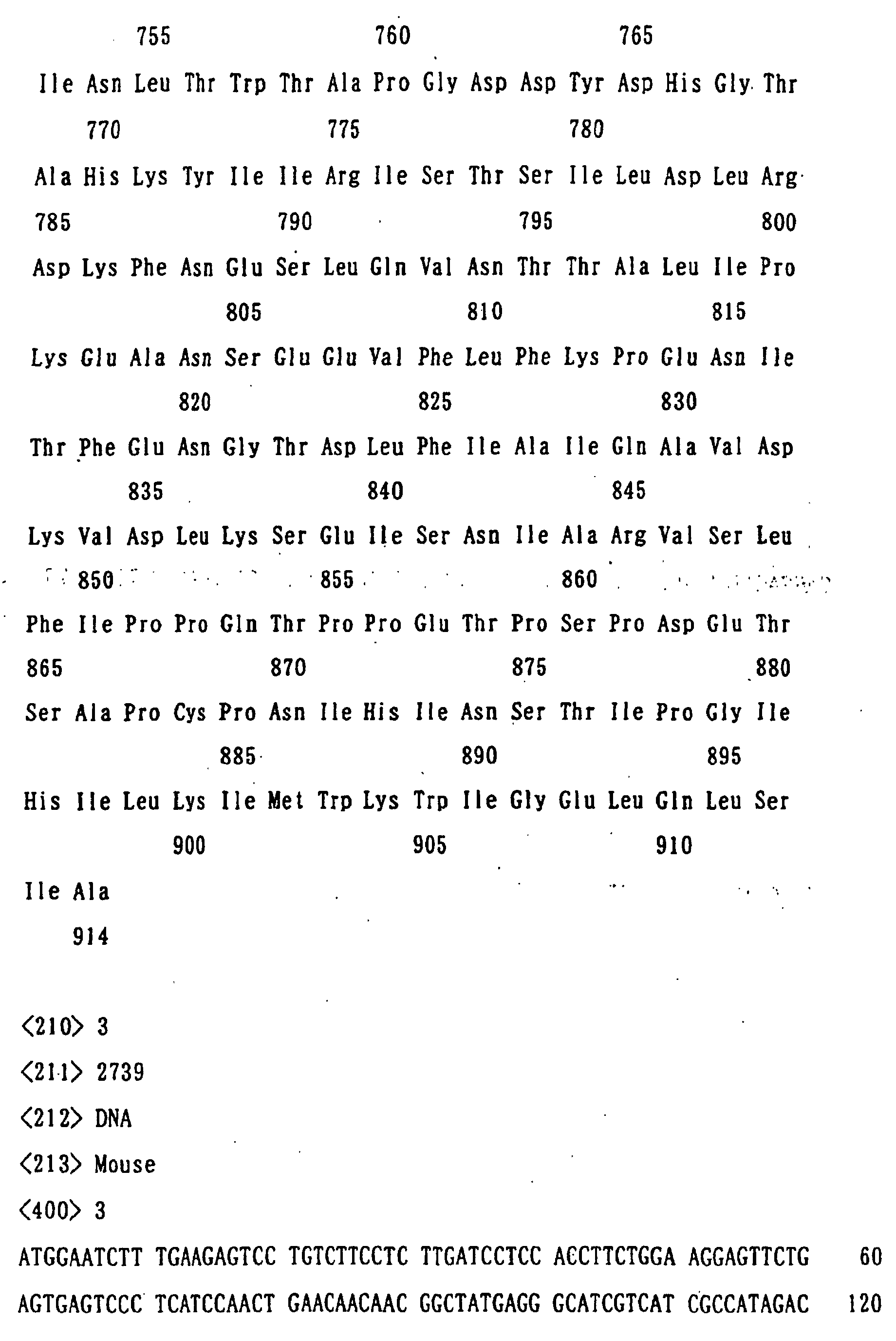
amino acid sequence represented by SEQ ID NO: 1, a human
small intestine-derived protein containing the amino acid
sequence represented by SEQ ID NO: 2, and the like.
The partial peptide of the protein used in the present
invention may be any peptide, so long as it is a partial
peptide of the protein used in the present invention
described above and preferably has the property equivalent
to that of the protein used in the present invention described
above.
Specific examples for the purpose of preparing the
antibody of the present invention later described include
a peptide having the 22-913 amino acid sequence in the amino
acid sequence represented by SEQ ID NO: 1, a peptide having
the 22-914 amino acid sequence in the amino acid sequence
represented by SEQ ID NO: 2, and the like. Also, for the
screening method or the screening kit of the present
invention later described, there are preferably employed
a peptide containing the cell membrane binding site (the
1-21 amino acid sequence in the amino acid sequence
represented by SEQ ID NO: 1 or SEQ ID NO: 2) and having a
part of the 22-913 amino acid sequence in the amino acid
sequence represented by SEQ ID NO: 1, or having a part of
the 22-914 amino acid sequence in the amino acid sequence
represented by SEQ ID NO: 2. Preferably used are peptides
containing the sequence of at least 20, preferably at least
50, more preferably at least 70, much more preferably at
least 100, and most preferably at least 200, amino acids
in the constituent amino acid sequence of the protein used
in the present invention, and the like.
The partial peptides used in the present invention may
be those containing the amino acid sequence, of which at
least 1 or 2 (preferably about 1 to about 10 and more preferably
several (1 to 5)) amino acids are deleted; to which at least
1 or 2 (preferably about 1 to about 10 and more preferably
several (1 to 5)) amino acids are added; in which at least
1 or 2 (preferably about 1 to about 10 and more preferably
several (1 to 5)) amino acids are inserted; or, in which
at least 1 or 2 (preferably about 1 to about 10 and more
preferably several (1 to 5)) amino acids are substituted
by other amino acids.
In the partial peptide used in the present invention,
the C-terminus is usually in the form of a carboxyl group
(-COOH) or a carboxylate (-COO-) but may be in the form of
an amide (-CONH2) or an ester (-COOR), as in the aforesaid
proteins employed in the present invention.
The partial peptides used in the invention include those
containing carboxyl groups (or carboxylates) other than at
the C-terminus, those wherein the amino group of the
N-terminal amino acid residue (e.g., methionine residue)
is protected with a protecting group, those wherein the
N-terminal region is cleaved in vivo and the glutamine thus
formed is pyroglutaminated, those wherein a substituent on
the side chain of an amino acid in the molecule is protected
with a suitable protecting group, or conjugated proteins
such as so-called glycoproteins to which sugar chains are
bound, etc., as in the proteins used in the present invention
described above.
The partial peptide used in the invention may also be
used as an antigen for producing antibodies.
The protein or its partial peptide used in the present'
invention may be employed in the form of salts with
physiologically acceptable acids (e.g., inorganic acids or
organic acids) or bases (e.g., alkali metal salts),
preferably in the form of physiologically acceptable acid
addition salts. Examples of such salts are salts with
inorganic acids (e.g., hydrochloric acid, phosphoric acid,
hydrobromic acid, and sulfuric acid), salts with organic
acids (e.g., acetic acid, formic acid, propionic acid,
fumaric acid, maleic acid, succinic acid, tartaric acid,
citric acid, malic acid, oxalic acid, benzoic acid,
methanesulfonic acid, benzenesulfonic acid) and the like.
The proteins or partial peptides used in the present
invention, or salts thereof, may be manufactured by a
publicly known method used to purify a protein from human
or other warm-blooded animal cells or tissues described above.
Alternatively, they may also be manufactured by culturing
transformants containing DNAs encoding the proteins.
Furthermore, they may also be manufactured by a modification
of the methods for peptide synthesis, which will be later
described.
Where these proteins or peptides are manufactured from
human or mammalian tissues or cells, human or mammalian
tissues or cells are homogenized, then extracted with an
acid or the like, and the proteins or peptides are isolated
and purified from the extract by a combination of
chromatography techniques such as reverse phase
chromatography, ion exchange chromatography, and the like.
To synthesize the proteins or partial peptides used
in the present invention, or salts thereof or amides thereof,
commercially available resins that are used for protein
synthesis may be used. Examples of such resins include
chloromethyl resin, hydroxymethyl resin, benzhydrylamine
resin, aminomethyl resin, 4-benzyloxybenzyl alcohol resin,
4-methylbenzhydrylamine resin, PAM resin,
4-hydroxymethylmethylphenyl acetamidomethyl resin,
polyacrylamide resin, 4-(2',4'-dimethoxyphenylhydroxymethyl)phenoxy
resin, 4-(2',4'-dimethoxyphenyl-Fmoc-aminoethyl)
phenoxy resin, etc. Using these resins,
amino acids, in which α-amino groups and functional groups
on the side chains are appropriately protected are condensed
on the resin in the order of the sequence of the objective
protein according to various condensation methods publicly
known in the art. At the end of the reaction, the protein
or partial peptide is excised from the resin and at the same
time, the protecting groups are removed. Then,
intramolecular disulfide bond-forming reaction is
performed in a highly diluted solution to obtain the
objective protein or partial peptide, or amides thereof.
For condensation of the protected amino acids described
above, a variety of activation reagents for protein synthesis
may be used, but carbodiimides are particularly preferably
employed. Examples of such carbodiimides include DCC,
N,N'-diisopropylcarbodiimide, N-ethyl-N'-(3-dimethylaminopropyl)carbodiimide,
etc. For activation by these
reagents, the protected amino acids in combination with a
racemization inhibitor (e.g., HOBt, HOOBt) are added
directly to the resin, or the protected amino acids are
previously activated in the form of symmetric acid anhydrides,
HOBt esters or HOOBt esters, followed by adding the thus
activated protected amino acids to the resin.
Solvents suitable for use to activate the protected
amino acids or condense with the resin may be chosen from
solvents that are known to be usable for protein condensation
reactions. Examples of such solvents are acid amides such
as N,N-dimethylformamide, N,N-dimethylacetamide,
N-methylpyrrolidone, etc.; halogenated hydrocarbons such
as methylene chloride, chloroform, etc.; alcohols such as
trifluoroethanol, etc.; sulfoxides such as
dimethylsulfoxide, etc.; ethers such as pyridine, dioxane,
tetrahydrofuran, etc.; nitriles such as acetonitrile,
propionitrile, etc.; esters such as methyl acetate, ethyl
acetate, etc.; and appropriate mixtures of these solvents.
The reaction temperature is appropriately chosen from the
range known to be applicable to protein binding reactions
and is usually selected in the range of approximately -20°C
to 50°C. The activated amino acid derivatives are used
generally in an excess of 1.5 to 4 times. The condensation
is examined using the ninhydrin reaction; when the
condensation is insufficient, the condensation can be
completed by repeating the condensation reaction without
removal of the protecting groups. When the condensation is
yet insufficient even after repeating the reaction,
unreacted amino acids are acetylated with acetic anhydride
or acetylimidazole to cancel any possible adverse affect
on the subsequent reaction.
Examples of the protecting groups used to protect the
starting amino groups include Z, Boc, t-pentyloxycarbonyl,
isobornyloxycarbonyl, 4-methoxybenzyloxycarbonyl, Cl-Z,
Br-Z, adamantyloxycarbonyl, trifluoroacetyl, phthaloyl,
formyl, 2-nitrophenylsulphenyl, diphenylphosphinothioyl,
Fmoc, etc.
A carboxyl group can be protected by, e.g., alkyl
esterification (in the form of linear, branched or cyclic
alkyl esters of the alkyl moiety such as methyl, ethyl, propyl,
butyl, t-butyl, cyclopentyl, cyclohexyl, cycloheptyl,
cyclooctyl, 2-adamantyl, etc.), aralkyl esterification
(e.g., esterification in the form of benzyl ester,
4-nitrobenzylester, 4-methoxybenzylester, 4-chlorobenzyl
ester, benzhydryl ester, etc.), phenacyl esterification,
benzyloxycarbonyl hydrazidation, t-butoxycarbonyl
hydrazidation, trityl hydrazidation, or the like.
The hydroxyl group of serine can be protected through,
for example, its esterification or etherification. Examples
of groups appropriately used for the esterification include
a lower C1-6 alkanoyl group, such as acetyl group, an aroyl
group such as benzoyl group, and a group derived from carbonic
acid such as benzyloxycarbonyl group and ethoxycarbonyl
group. Examples of a group appropriately used for the
etherification include benzyl group, tetrahydropyranyl
group, t-butyl group, etc.
Examples of groups for protecting the phenolic hydroxyl
group of tyrosine include Bzl, Cl2-Bzl, 2-nitrobenzyl, Br-Z,
t-butyl, etc.
Examples of groups used to protect the imidazole moiety
of histidine include Tos, 4-methoxy-2,3,6-trimethylbenzenesulfonyl,
DNP, benzyloxymethyl, Bum, Boc, Trt, Fmoc,
etc.
Examples of the activated carboxyl groups in the
starting amino acids include the corresponding acid
anhydrides, azides, activated esters (esters with alcohols
(e.g., pentachlorophenol, 2,4,5-trichlorophenol,
2,4-dinitrophenol, cyanomethyl alcohol, p-nitrophenol,
HONB, N-hydroxysuccimide, N-hydroxyphthalimide, HOBt)). As
the activated amino acids in which the amino groups are
activated in the starting material, the corresponding
phosphoric amides are employed.
To eliminate (split off) the protecting groups, there
are used catalytic reduction under hydrogen gas flow in the
presence of a catalyst such as Pd-black or Pd-carbon; an
acid treatment with anhydrous hydrogen fluoride,
methanesulfonic acid, trifluoromethanesulfonic acid or
trifluoroacetic acid, or a mixture solution of these acids;
a treatment with a base such as diisopropylethylamine,
triethylamine, piperidine or piperazine; and reduction with
sodium in liquid ammonia. The elimination of the protecting
group by the acid treatment described above is carried out
generally at a temperature of approximately -20°C to 40°C.
In the acid treatment, it is efficient to add a cation
scavenger such as anisole, phenol, thioanisole, m-cresol,
p-cresol, dimethylsulfide, 1,4-butanedithiol or
1,2-ethanedithiol. Furthermore, 2,4-dinitrophenyl group
known as the protecting group for the imidazole of histidine
is removed by a treatment with thiophenol. Formyl group used
as the protecting group of the indole of tryptophan is
eliminated by the aforesaid acid treatment in the presence
of 1,2-ethanedithiol or 1,4-butanedithiol, as well as by
a treatment with an alkali such as a dilute sodium hydroxide
solution and dilute ammonia.
Protection of functional groups that should not be
involved in the reaction of the starting materials,
protecting groups, elimination of the protecting groups and
activation of functional groups involved in the reaction
may be appropriately selected from publicly known groups
and publicly known means.
In another method for obtaining the amides of the desired
protein or partial peptide, for example, the α-carboxyl
group of the carboxy terminal amino acid is first protected
by amidation; the peptide (protein) chain is then extended
from the amino group side to a desired length. Thereafter,
a protein or partial peptide, in which only the protecting
group of the N-terminal α-amino group of the peptide chain
has been eliminated, and a protein or partial peptide, in
which only the protecting group of the C-terminal carboxyl
group has been eliminated are manufactured. The two proteins
or peptides are condensed in a mixture of the solvents
described above. The details of the condensation reaction
are the same as described above. After the protected protein
or peptide obtained by the condensation is purified, all
the protecting groups are eliminated by the method described
above to give the desired crude protein or peptide. This
crude protein or peptide is purified by various known
purification means. Lyophilization of the major fraction
gives the amide of the desired protein or peptide.
To prepare the esterified protein or peptide, for
example, the α-carboxyl group of the carboxy terminal amino
acid is condensed with a desired alcohol to prepare the amino
acid ester, which is followed by procedure similar to the
preparation of the amidated protein or peptide above to give
the desired esterified protein or peptide.
The partial peptide or salts thereof used in the present
invention can be manufactured by publicly known methods for
peptide synthesis, or by cleaving the protein used in the
present invention with an appropriate peptidase. For the
methods for peptide synthesis, for example, either solid
phase synthesis or liquid phase synthesis may be used. That
is, the partial peptide or amino acids that can construct
the partial peptide used in the present invention are
condensed with the remaining part. Where the product contains
protecting groups, these protecting groups are removed to
give the desired peptide. Publicly known methods for
condensation and elimination of the protecting groups are
described in 1) to 5) below.
After completion of the reaction, the product may be
purified and isolated by a combination of conventional
purification methods such as solvent extraction,
distillation, column chromatography, liquid chromatography
and recrystallization to give the partial peptide used in
the present invention. When the partial peptide obtained
by the above methods is in a free form, the peptide can be
converted into an appropriate salt by a publicly known
method; conversely when the partial peptide is obtained in
a salt form, it can be converted into a free form or other
different salt form by a publicly known method.
The DNA encoding the protein used in the present
invention may be any DNA so long as it contains the base
sequence encoding the protein used in the present invention
described above. The DNA may also be any one of genomic DNA,
genomic DNA library, cDNA derived from the cells or tissues
described above, cDNA library derived from the cells or
tissues described above and synthetic DNA.
The vector to be used for the library may be any of
bacteriophage, plasmid, cosmid, phagemid and the like. In
addition, the DNA can be amplified by reverse transcriptase
polymerase chain reaction (hereinafter abbreviated as
RT-PCR) with total RNA or mRNA fraction prepared from the
above-described cells or tissues.
Specifically, the DNA encoding the protein used in the
present invention may be any one of, for example, a DNA having
the base sequence represented by SEQ ID NO: 3 or SEQ ID NO:
4, or any DNA having a base sequence hybridizable to a DNA
having the base sequence represented by SEQ ID NO: 3 or SEQ
ID NO: 4 under high stringent conditions and encoding a
protein which has the properties substantially equivalent
to those of the protein used in the present invention.
Specific examples of the DNA that is hybridizable to
DNA having the base sequence represented by SEQ ID NO: 3
or SEQ ID NO: 4 under high stringent conditions include DNAs
having at least about 50% homology, preferably at least about
60% homology, more preferably at least about 70% homology,
much more preferably at least about 80% homology, further
much more preferably at least about 90% homology and most
preferably at least about 95% homology, to the base sequence
represented by SEQ ID NO: 3 or SEQ ID NO: 4.
The hybridization can be carried out by publicly known
methods or by a modification thereof, for example, according
to the method described in Molecular Cloning, 2nd Ed., J.
Sambrook et al., Cold Spring Harbor Lab. Press, (1989). A
commercially available library may also be used according
to the instructions of the attached manufacturer's protocol.
The hybridization can be carried out preferably under high
stringent conditions.
The high stringent conditions used herein are, for
example, those in a sodium concentration at about 19 mM to
about 40 mM, preferably about 19 mM to about 20 mM at a
temperature of about 50°C to about 70°C, preferably about
60°C to about 65°C. In particular, hybridization conditions
in a sodium concentration at about 19 mM at a temperature
of about 65°C are most preferred.
More specifically, as the DNA encoding the protein
having the amino acid sequence represented by SEQ ID NO:
1, there may be employed a DNA containing a DNA having the
base sequence represented by SEQ ID NO: 3. As the DNA encoding
the protein having the amino acid sequence represented by
SEQ ID NO: 2, there may be employed a DNA containing a DNA
having the base sequence represented by SEQ ID NO: 4.
The DNA encoding the partial peptide used in the present
invention may be any DNA so long as it contains the base
sequence encoding the partial peptide used in the present
invention described above. The DNA may also be any of genomic
DNA, genomic DNA library, cDNA derived from the cells and
tissues described above, cDNA library derived from the cells
and tissues described above and synthetic DNA.
Specifically as the DNA encoding the partial peptide
used in the present invention, there may be employed any
one of a DNA having a part of a DNA containing the base sequence
represented by SEQ ID NO: 3 or SEQ ID NO: 4, a DNA having
a base sequence hybridizable to a DNA having the base sequence
represented by SEQ ID NO: 3 or SEQ ID NO: 4 under high stringent
conditions and containing a part of a DNA encoding a protein
having an activity substantially equivalent to that of the
protein used in the present invention, and the like.
The DNA hybridizable to the base sequence represented
by SEQ ID NO: 3 or SEQ ID NO: 4 has the same significance
as described hereinabove.
Methods for the hybridization and the high stringent
conditions that can be used are the same as those described
above.
As a means of cloning a DNA that completely encodes
the protein or partial peptide used in the present invention
(hereinafter sometimes merely referred to as the protein
of the present invention in describing the cloning and
expression of the DNAs encoding the same), the DNA may be
amplified either by PCR using synthetic DNA primers
containing a part of the base sequence of the protein of
the present invention, or the DNA inserted into an
appropriate vector may be screened by hybridization with
a labeled DNA fragment or synthetic DNA that encodes a part
or entire region of the protein of the present invention.
The hybridization can be carried out. for example, according
to the method described in Molecular Cloning, 2nd (J.
Sambrook et al., Cold Spring Harbor Lab. Press, 1989). Where
the hybridization is carried out using commercially
available library, the procedures may be conducted in
accordance with the protocol described in the attached
instructions.
Substitution of the base sequence of DNA can be effected
by publicly known methods such as the ODA-LA PCR method,
the Gapped duplex method or the Kunkel method, or
modifications thereof by using publicly known kits available
as Mutan™-super Express Km, Mutan™-K (both manufactured
by Takara Shuzo Co., Ltd.), etc.
The cloned DNA encoding the protein can be used as it
is depending upon purpose or, if desired, after digestion
with a restriction enzyme or after addition of a linker
thereto. The DNA may contain ATG as a translation initiation
codon at the 5' end thereof and TAA, TGA or TAG as a translation
termination codon at the 3' end thereof. These translation
initiation and termination codons may also be added by using
an appropriate synthetic DNA adapter.
The expression vector of the protein of the present
invention can be manufactured, for example, by (a) excising
the desired DNA fragment from the DNA encoding the protein
of the present invention, (b) and then ligating the DNA
fragment with an appropriate expression vector downstream
a promoter in the vector.
Examples of the vector include plasmids derived form
E. coli (e.g., pBR322, pBR325, pUC12, pUC13), plasmids
derived from Bacillus subtilis (e.g., pUB110, pTP5, pC194),
plasmids derived from yeast (e.g., pSH19, pSH15),
bacteriophages such as λ phage, etc., animal viruses such
as retrovirus, vaccinia virus, baculovirus, etc. as well
as pAl-11, pXT1, pRc/CMV, pRc/RSV, pcDNAI/Neo, etc.
The promoter used in the present invention may be any
promoter if it matches well with a host to be used for gene
expression. In the case of using animal cells as the host,
examples of the promoter include SRα promoter, SV40 promoter,
LTR promoter, CMV promoter, HSV-TK promoter, etc.
Among them, CMV (cytomegalovirus) promoter or SRα
promoter is preferably used. Where the host is bacteria of
the genus Escherichia, preferred examples of the promoter
include trp promoter, lac promoter, recA promoter, γPL
promoter, lpp promoter, T7 promoter, etc. In the case of
using bacteria of the genus Bacillus as the host, preferred
example of the promoter are SPO1 promoter, SPO2 promoter,
penP promoter, etc. When yeast is used as the host, preferred
examples of the promoter are PHO5 promoter, PGK promoter,
GAP promoter, ADH promoter, etc. When insect cells are used
as the host, preferred examples of the promoter include
polyhedrin prompter, P10 promoter, etc.
In addition to the foregoing examples, the expression
vector may further optionally contain an enhancer, a splicing
signal, a poly A addition signal, a selection marker, SV40
replication origin (hereinafter sometimes abbreviated as
SV40ori), etc. Examples of the selection marker include
dihydrofolate reductase (hereinafter sometimes abbreviated
as dhfr) gene [methotrexate (MTX) resistance], ampicillin
resistant gene (hereinafter sometimes abbreviated as Ampr),
neomycin resistant gene (hereinafter sometimes abbreviated
as Neor, G418 resistance), etc. In particular, when dhfr
gene is used as the selection marker using dhfr
gene-deficient Chinese hamster cells, selection can also
be made on a thymidine free medium.
If necessary, a signal sequence that matches with a
host is added to the N-terminus of the protein of the present
invention. Examples of the signal sequence that can be used
are PhoA signal sequence, OmpA signal sequence, etc. in the
case of using bacteria of the genus Escherichia as the host;
α-amylase signal sequence, subtilisin signal sequence, etc.
in the case of using bacteria of the genus Bacillus as the
host; MFα signal sequence, SUC2 signal sequence, etc. in
the case of using yeast as the host; and insulin signal
sequence, α-interferon signal sequence, antibody molecule
signal sequence, etc. in the case of using animal cells as
the host, respectively.
Using the vector containing the DNA encoding the protein
of the present invention thus constructed, transformants
can be manufactured.
Examples of the host, which may be employed, are bacteria
belonging to the genus Escherichia, bacteria belonging to
the genus Bacillus, yeast, insect cells, insects and animal
cells, etc.
Specific examples of the bacteria belonging to the genus
Escherichia include Escherichia coli K12 DH1 [Proc. Natl.
Acad. Sci. U.S.A., 60, 160 (1968)], JM103 [Nucleic Acids
Research, 9, 309 (1981)], JA221 [Journal of Molecular Biology,
120, 517 (1978)], HB101 [Journal of Molecular Biology, 41,
459 (1969)], C600 [Genetics, 39, 440 (1954)], etc.
Examples of the bacteria belonging to the genus Bacillus
include Bacillus subtilis MI114 [Gene, 24, 255 (1983)],
207-21 [Journal of Biochemistry, 95, 87 (1984)], etc.
Examples of yeast include Saccharomyces cereviseae AH22,
AH22R-, NA87-11A, DKD-5D, 20B-12, Schizosaccharomyces pombe
NCYC1913, NCYC2036, Pichia pastoris KM71, etc.
Examples of insect cells include, for the virus AcNPV,
Spodoptera frugiperda cell (Sf cell), MG1 cell derived from
mid-intestine of Trichoplusia ni, High Five™ cell derived
from egg of Trichoplusia ni, cells derived from Mamestra
brassicae, cells derived from Estigmena acrea, etc.; and
for the virus BmNPV, Bombyx mori N cell (BmN cell), etc.
is used. Examples of the Sf cell which can be used are Sf9
cell (ATCC CRL1711), Sf21 cell (both cells are described
in Vaughn, J. L. et al., In Vivo, 13, 213-217 (1977), etc.
As the insect, for example, a larva of Bombyx mori can
be used [Maeda et al., Nature, 315, 592 (1985)].
Examples of animal cells include monkey cell COS-7,
Vero, Chinese hamster cell CHO (hereinafter referred to as
CHO cell), dhfr gene-deficient Chinese hamster cell CHO
(hereinafter simply referred to as CHO (dhfr-) cell), mouse
L cell, mouse AtT-20, mouse myeloma cell, rat GH 3, human
FL cell, etc.
Bacteria belonging to the genus Escherichia can be
transformed, for example, by the method described in Proc.
Natl. Acad. Sci. U.S.A., 69, 2110 (1972), Gene, 17, 107 (1982),
etc.
Bacteria belonging to the genus Bacillus can be
transformed, for example, by the method described in
Molecular & General Genetics, 168, 111 (1979), etc.
Yeast can be transformed, for example, by the method
described in Methods in Enzymology, 194, 182-187 (1991),
Proc. Natl. Acad. Sci. U.S.A., 75, 1929 (1978), etc.
Insect cells or insects can be transformed, for example,
according to the method described in Bio/Technology, 6,
47-55(1988), etc.
Animal cells can be transformed, for example, according
to the method described in Saibo Kogaku (Cell Engineering),
extra issue 8, Shin Saibo Kogaku Jikken Protocol (New Cell
Engineering Experimental Protocol), 263-267
(1995) (published by Shujunsha), or Virology, 52, 456 (1973).
Thus, the transformants transformed with the expression
vectors containing the DNAs encoding the protein can be
obtained.
Where the host is bacteria belonging to the genus
Escherichia or the genus Bacillus, the transformant can be
appropriately cultured in a liquid medium which contains
materials required for growth of the transformant such as
carbon sources, nitrogen sources, inorganic materials, etc.
Examples of the carbon sources include glucose, dextrin,
soluble starch, sucrose, etc.; examples of the nitrogen
sources include inorganic or organic materials such as
ammonium salts, nitrate salts, corn steep liquor, peptone,
casein, meat extract, soybean cake, potato extract, etc.;
and, examples of the inorganic materials are calcium chloride,
sodium dihydrogenphosphate, magnesium chloride, etc. In
addition, yeast, vitamins, growth promoting factors etc.
may also be added to the medium. Preferably, pH of the medium
is adjusted to about 5 to about 8.
A preferred example of the medium for culturing the
bacteria belonging to the genus Escherichia is M9 medium
supplemented with glucose and Casamino acids [Miller,
Journal of Experiments in Molecular Genetics, 431-433, Cold
Spring Harbor Laboratory, New York, 1972]. If necessary,
a chemical such as 3β-indolylacrylic acid can be added to
the medium thereby to activate the promoter efficiently.
Where the bacteria belonging to the genus Escherichia
are used as the host, the transformant is usually cultivated
at about 15°C to about 43°C for about 3 hours to about 24
hours. If necessary, the culture may be aerated or agitated.
Where the bacteria belonging to the genus Bacillus are
used as the host, the transformant is cultured generally
at about 30°C to about 40°C for about 6 hours to about 24
hours. If necessary, the culture can be aerated or agitated.
Where yeast is used as the host, the transformant is
cultivated, for example, in Burkholder's minimal medium
[Bostian, K. L. et al., Proc. Natl. Acad. Sci. U.S.A., 77,
4505 (1980)] or in SD medium supplemented with 0.5% Casamino
acids [Bitter, G. A. et al., Proc. Natl. Acad. Sci. U.S.A.,
81, 5330 (1984)]. Preferably, pH of the medium is adjusted
to about 5 to about 8. In general, the transformant is
cultivated at about 20°C to about 35°C for about 24 hours
to about 72 hours. If necessary, the culture can be aerated
or agitated.
Where insect cells or insects are used as the host,
the transformant is cultivated in, for example, Grace's
Insect Medium (Grace, T. C. C., Nature, 195, 788 (1962))
to which an appropriate additive such as immobilized 10%
bovine serum is added. Preferably, pH of the medium is
adjusted to about 6.2 to about 6.4. Normally, the
transformant is cultivated at about 27°C for about 3 days
to about 5 days and, if necessary, the culture can be aerated
or agitated.
Where animal cells are employed as the host, the
transformant is cultured in, for example, MEM medium
containing about 5% to about 20% fetal bovine serum [Science,
122, 501 (1952)], DMEM medium [Virology, 8, 396 (1959)],
RPMI 1640 medium [The Journal of the American Medical
Association, 199, 519 (1967)], 199 medium [Proceeding of
the Society for the Biological Medicine, 73, 1 (1950)], etc.
Preferably, pH of the medium is adjusted to about 6 to about
8. The transformant is usually cultivated at about 30°C to
about 40°C for about 15 hours to about 60 hours and, if
necessary, the culture can be aerated or agitated.
As described above, the protein of the present invention
can be produced intracellularly, on the cell membrane, or
extracellularly of the transformant.
The protein of the present invention can be separated
and purified from the culture described above by the
following procedures.
When the protein of the present invention is extracted
from the culture or cells, the transformant or cell is
collected after culturing by a publicly known method and
suspended in a appropriate buffer. The transformant or cell
is then disrupted by publicly known methods such as
ultrasonication, a treatment with lysozyme and/or
freeze-thaw cycling, followed by centrifugation,
filtration, etc. Thus, the crude extract of the protein can
be obtained. The buffer used for the procedures may contain
a protein modifier such as urea or guanidine hydrochloride,
or a surfactant such as Triton X-100™, etc. When the protein
is secreted in the culture broth, the supernatant can be
separated, after completion of the cultivation, from the
transformant or cell to collect the supernatant by a publicly
known method.
The supernatant or the protein contained in the extract
thus obtained can be purified by appropriately combining
the publicly known methods for separation and purification.
Such publicly known methods for separation and purification
include a method utilizing difference in solubility such
as salting out, solvent precipitation, etc.; a method mainly
utilizing difference in molecular weight such as dialysis,
ultrafiltration, gel filtration, SDS-polyacrylamide gel
electrophoresis, etc.; a method utilizing difference in
electric charge such as ion exchange chromatography, etc.;
a method utilizing difference in specific affinity such as
affinity chromatography, etc.; a method utilizing
difference in hydrophobicity such as reverse phase high
performance liquid chromatography, etc.; a method utilizing
difference in isoelectric point such as isoelectrofocusing
electrophoresis; and the like.
When the protein thus obtained is in a free form, the
protein can be converted into the salt by publicly known
methods or modifications thereof. On the other hand, when
the protein is obtained in the form of a salt, it can be
converted into the free form or in the form of a different
salt by publicly known methods or modifications thereof.
The protein produced by the recombinant can be treated,
prior to or after the purification, with an appropriate
protein-modifying enzyme so that the protein can be
appropriately modified to partially remove the polypeptide.
Examples of the protein-modifying enzyme include trypsin,
chymotrypsin, arginyl endopeptidase, protein kinase,
glycosidase and the like.
The presence of the thus produced protein of the present
invention can be determined by a binding test to a labeled
ligand and by an enzyme immunoassay using a specific
antibody.
The antibodies to the protein or partial peptide, or
its salts used in the present invention may be any of
polyclonal and monoclonal antibodies, as long as they are
capable of recognizing the protein or partial peptide, or
its salts, used in the present invention.
The antibodies to the protein or partial peptide, or
its salts used in the present invention (hereinafter
sometimes simply referred to as the protein of the invention
in describing the antibodies) may be produced by a publicly
known method of producing an antibody or antiserum, using
the protein of the-invention as an antigen.
[Preparation of monoclonal antibody]
(a) Preparation of monoclonal antibody-producing cells
The protein of the present invention is administered
to warm-blooded animals either solely or together with
carriers or diluents to the site where the production of
antibody is possible by the administration. In order to
potentiate the antibody productivity upon the
administration, complete Freund's adjuvants or incomplete
Freund's adjuvants may be administered. The administration
is usually carried out once every two to six weeks and twice
to ten times in total. Examples of the applicable
warm-blooded animals are monkeys, rabbits, dogs, guinea pigs,
mice, rats, sheep, goats and chickens, with the use of mice
and rats being preferred.
In the preparation of monoclonal antibody-producing
cells, a warm-blooded animal, e.g., mice, immunized with
an antigen wherein the antibody titer is noted is selected,
then spleen or lymph node is collected after two to five
days from the final immunization and antibody-producing
cells contained therein are fused with myeloma cells from
homozoic or heterozoic animal to give monoclonal
antibody-producing hybridomas. Measurement of the antibody
titer in antisera may be carried out, for example, by reacting
a labeled protein, which will be described later, with the
antiserum followed by assaying the binding activity of the
labeling agent bound to the antibody. The fusion may be
carried out, for example, by the known method by Koehler
and Milstein [Nature, 256, 495, (1975)]. Examples of the
fusion accelerator are polyethylene glycol (PEG), Sendai
virus, etc., of which PEG is preferably employed.
Examples of the myeloma cells are those collected from
warm-blooded animals such as NS-1, P3U1, SP2/0, AP-1, etc.
In particular, P3U1 is preferably employed. A preferred ratio
of the count of the antibody-producing cells used (spleen
cells) to the count of myeloma cells is within a range of
approximately 1:1 to 20:1. When PEG (preferably, PEG 1000
to PEG 6000) is added in a concentration of approximately
10 to 80% followed by incubation at 20 to 40°C, preferably
at 30 to 37°C for 1 to 10 minutes, an efficient cell fusion
can be carried out.
Various methods can be used for screening of monoclonal
antibody-producing hybridomas. Examples of such methods
include a method which comprises adding the supernatant of
a hybridoma to a solid phase (e.g., a microplate) adsorbed
with the protein as an antigen directly or together with
a carrier, adding an anti-immunoglobulin antibody (where
mouse cells are used for the cell fusion, anti-mouse
immunoglobulin antibody is used) labeled with a radioactive
substance or an enzyme or Protein A and detecting the
monoclonal antibody bound to the solid phase, and a method
which comprises adding the supernatant of hybridoma to a
solid phase adsorbed with an anti-immunoglobulin antibody
or Protein A, adding the protein labeled with a radioactive
substance or an enzyme and detecting the monoclonal antibody
bound to the solid phase, or the like.
The monoclonal antibody can be screened according to
publicly known methods or their modifications. In general,
the screening can be effected in a medium for animal cells
supplemented with HAT (hypoxanthine, aminopterin and
thymidine) . Any screening and growth medium can be employed
as far as the hybridoma can grow there. For example, RPMI
1640 medium containing 1% to 20%, preferably 10% to 20% fetal
bovine serum, GIT medium (Wako Pure Chemical Industries,
Ltd.) containing 1 to 10% fetal bovine serum, a serum free
medium for cultivation of a hybridoma (SFM-101, Nissui
Seiyaku Co., Ltd.) and the like, can be used for the screening
and growth medium. The culture is carried out generally at
20°C to 40°C, preferably at 37°C, for about 5 days to about
3 weeks, preferably 1 to 2 weeks, normally in 5% CO2. The
antibody titer of the culture supernatant of a hybridoma
can be determined as in the assay for the antibody titer
in antisera described above.
(b) Purification of monoclonal antibody
Separation and purification of a monoclonal antibody
can be carried out by publicly known methods, such as
separation and purification of immunoglobulins [for example,
salting-out, alcohol precipitation, isoelectric point
precipitation, electrophoresis, adsorption and desorption
with ion exchangers (e.g., DEAE), ultracentrifugation, gel
filtration, or a specific purification method which
comprises collecting only an antibody with an activated
adsorbent such as an antigen-binding solid phase, Protein
A or Protein G and dissociating the binding to obtain the
antibody.]
[Preparation of polyclonal antibody]
The polyclonal antibody of the present invention can
be manufactured by publicly known methods or modifications
thereof. For example, a warm-blooded animal is immunized
with an immunogen (protein antigen) per se, or a complex
of immunogen and a carrier protein is formed and a
warm-blooded animal is immunized with the complex in a manner
similar to the method described above for the manufacture
of monoclonal antibodies. The product containing the
antibody to the protein of the present invention is collected
from the immunized animal followed by separation and
purification of the antibody.
In the complex of immunogen and carrier protein used
to immunize a warm-blooded animal, the type of carrier
protein and the mixing ratio of carrier to hapten may be
any type and in any ratio, as long as the antibody is
efficiently produced to the hapten immunized by crosslinking
to the carrier. For example, bovine serum albumin, bovine
thyroglobulin or hemocyanin is coupled to hapten in a
carrier-to-hapten weight ratio of approximately 0.1 to 20,
preferably about 1 to about 5.
A variety of condensation agents can be used for the
coupling of carrier to hapten. Glutaraldehyde,
carbodiimide, maleimide activated ester and activated ester
reagents containing thiol group or dithiopyridyl group are
used for the coupling.
The condensation product is administered to
warm-blooded animals either solely or together with carriers
or diluents to the site that can produce the antibody by
the administration. In order to potentiate the antibody
productivity upon the administration, complete Freund's
adjuvant or incomplete Freund's adjuvant may be administered.
The administration is usually made once every 2 to 6 weeks
and 3 to 10 times in total.
The polyclonal antibody can be collected from the blood,
ascites, etc., preferably from the blood of warm-blooded
animal immunized by the method described above.
The polyclonal antibody titer in antiserum can be
assayed by the same procedure as that for the determination
of serum antibody titer described above. The separation and
purification of the polyclonal antibody can be carried out,
following the method for the separation and purification
of immunoglobulins performed as in the separation and
purification of monoclonal antibodies described
hereinabove.
The antisense nucleotide having a complementary or
substantial complementary base sequence to the DNA encoding
the protein or partial peptide used in the present invention
(hereinafter these DNAs are sometimes collectively referred
to as the DNAs of the present invention in the following
description of antisense nucleotide) can be any antisense
nucleotide, so long as it possesses a base sequence
complementary or substantially complementary to the base
sequence of the DNA of the present invention and capable
of suppressing expression of the DNA, but it is preferably
an antisense DNA.
The base sequence substantially complementary to the
DNA of the present invention may, for example, be a base
sequence having at least about 70% homology, preferably at
least about 80% homology, more preferably at least-about
90% homology and most preferably at least about 95% homology,
to the full-length base sequence or partial base sequence
of the base sequence complementary to the DNA of the present
invention (i.e., complementary strand to the DNA used in
the present invention), and the like. In the entire base
sequence of the complementary strand to the DNA of the present
invention, an antisense nucleotide having at least about
70% homology, preferably at least about 80% homology, more
preferably at least about 90% homology and most preferably
at least about 95% homology, to the complementary strand
of the base sequence which encodes the N-terminal region
in the protein of the present invention (e.g., the base
sequence around the initiation codon).
Specific examples include an antisense nucleotide
containing a base sequence complementary or substantially
complementary to the base sequence of a DNA having the base
sequence represented by SEQ ID NO: 3, SEQ ID NO: 4, SEQ ID
NO: 22 or SEQ ID NO: 26, or a part of the complementary or
substantially complementary base sequence (more preferably
an antisense nucleotide containing a base sequence
complementary to the base sequence of a DNA having the base
sequence represented by SEQ ID NO: 3 or SEQ ID NO: 4, or
a part thereof); an antisense nucleotide containing a base
sequence complementary to the base sequence of a DNA having
the 1-14 base sequence represented by SEQ ID NO: 22, or a
part thereof; an antisense nucleotide containing a base
sequence complementary to the base sequence of a DNA having
the 2764-2843 base sequence represented by SEQ ID NO: 22,
or a part thereof; an antisense nucleotide containing a base
sequence complementary to the base sequence of a DNA having
the 1-22 base sequence represented by SEQ ID NO: 26, or a
part thereof; an antisense nucleotide containing a base
sequence complementary to the base sequence of a DNA having
the 2765-2825 base sequence represented by SEQ ID NO: 26,
or a part thereof; and the like.
The antisense nucleotide is generally constructed by
bases of about 10 to about 40, preferably about 15 to about
30.
To prevent digestion with a hydrolase such as nuclease,
etc., the phosphoric acid residue (phosphate) of each
nucleotide that constitutes the antisense DNA may be
substituted with chemically modified phosphoric acid
residues, e.g., phosphorothioate, methylphosphonate,
phosphorodithionate, etc. These anti sense nucleotides may
be synthesized using a publicly known DNA synthesizer, etc.
Hereinafter, the protein or partial peptide, or its
salts of the present invention (hereinafter sometimes merely
referred to as the protein of the present invention), the
DNA encoding the protein or partial peptide (hereinafter
sometimes merely referred to as the DNA of the present
invention), the antibody to the protein, partial peptide,
or its salts of the present invention (hereinafter sometimes
merely referred to as the antibody of the present invention)
and the antisense nucleotide to the DNA of the present
invention (hereinafter sometimes merely referred to as the
anti sense nucleotide of the present invention) are explained
with respect to the utilities.
The protein of the present invention can be utilized
as a disease marker, since expression of the protein
increases tissue-specifically in the lung/bronchi of asthma
model animal. That is, the protein is useful as a marker
for early diagnosis in lung/chest diseases accompanied by
inflammation of the lung/airways, judgment of severity in
conditions, or predicted development of diseases.
Furthermore, as will be described in EXAMPLES below,
increased airway hyperresponsiveness was suppressed by
administering the antisense nucleotide of gene encoding the
protein of the present invention. It is thus considered that
a compound or its salt that inhibits the activities of the
protein of the present invention, or the antibody against
the protein of the present invention would exhibit the same
activity.
Therefore, pharmaceuticals containing the antisense
nucleotide of the gene encoding the protein of the present
invention, the compound or its salt that inhibits the
activities of the protein of the present invention, or the
antibody against the protein of the present invention, may
be employed as therapeutic/preventive agents for the
lung/chest diseases accompanied by inflammation of lung or
airways, such as bronchial asthma, chronic obstructive
pulmonary disease, etc.
(1) Screening of drug candidate compounds for disease
Since the protein of the present invention is
increasingly expressed prior to inflammation of the
lung/bronchi, the compound or its salt that inhibits the
activities of the protein of the present invention can be
used as a pharmaceutical for the treatment/prevention of
lung/chest diseases accompanied by inflammation of the
lung/airways, including bronchial asthma, chronic
obstructive pulmonary disease, etc.
Therefore, the protein of the present invention is
useful as a reagent for screening the compound or its salts
that inhibit the activities of the protein of the present
invention.
That is, the present invention provides:
Specifically, in the screening method described above,
the method is characterized by measuring, e.g., the chloride
channel activity of the protein of the present invention
in the cases (i) and (ii), and comparing them.
As the calcium activator, there are employed, e.g.,
ionomycin, A23187 (calcimycin), and the like.
Examples of the test compound included are a peptide,
a protein, a non-peptide compound, a synthetic compound,
a fermentation product, a cell extract, a plant extract,
an animal tissue extract, and the like. These compounds may
be novel compounds or publicly known compounds.
To perform the screening method described above, the
protein of the present invention is suspended in a buffer
suitable for screening to prepare a specimen for the protein
of the present invention. Any buffer having pH of
approximately 4 to 10 (desirably a pH of approximately 6
to 8) such as a phosphate buffer, Tris-hydrochloride buffer,
etc. maybe used, so long as it does not interfere the chloride
channel activity of the protein of the present invention.
As the cell capable of producing the protein of the
present invention, there is employed, e.g., the aforesaid
host (transformant) transformed with a vector containing
the DNA encoding the protein of the present invention.
Preferably, animal cells such as CHO cells, etc. are used
as the host. For the screening, the transformant, on the
cell membrane of which the protein of the present invention
has been expressed, e.g., by culturing through the procedure
described above, is preferably employed.
The chloride channel activity of the protein of the
present invention can be assayed by the method described
in, e.g., Genomics, 54, 200 (1998), or its modification.
For example, when a test compound inhibits the chloride
channel activity in the case (ii) described above by at least
about 20%, preferably at least 30%, more preferably at least
about 50%, as compared to the case (i) above, the test compound
can be selected to be a compound capable of inhibiting the
activity of the protein of the present invention.
The screening kit of the present invention comprises
the protein, partial peptide or its salts used in the present
invention, or the cell capable of producing the protein or
partial peptide used in the present invention.
The compounds or salts thereof obtained using the
screening method or screening kit of the present invention
are compounds selected from the test compounds described
above, for example, peptides, proteins, non-peptide
compounds, synthetic compounds, fermentation products,
cell extracts, plant extracts, animal tissue extracts, blood
plasma, etc., and are the compounds or salts thereof that
inhibit the activities (e.g., a chloride channel activity,
etc.) of the protein of the present invention.
As salts of these compounds, there may be employed the
same salts as those of the protein of the present invention
described above.
The compound or its salt that inhibits the activities
of the protein of the present invention is useful as a
pharmaceutical for the treatment/prevention of lung/chest
diseases accompanied by inflammation of the lung/airways,
including bronchial asthma, chronic obstructive pulmonary
disease, etc.
When the compounds or salts thereof obtained using the
screening methods or screening kits of the present invention
are used as the therapeutic/prophylactic agents described
above, these compounds may be prepared into pharmaceutical
preparations in a conventional manner. The compounds may
be prepared, for example, in the form of tablets, capsules,
elixirs, microcapsules, a sterile solution, a suspension,
etc.
Since the thus obtained pharmaceutical preparation is
safe and low toxic, the preparation can be administered
orally or non-orally to human or other warm-blooded animal
(e.g., mouse, rat, rabbit, sheep, swine, bovine, horse,
chicken, cat, dog, monkey, chimpanzee, etc.).
The dose of the compound or salt thereof varies depending
on its action, target disease, subject to be administered,
route for administration, etc.; when the compound or its
salt that inhibits the activities of the protein of the
present invention is orally administered for the treatment
of, e.g., bronchial asthma, the compound or its salt is
normally administered in a dose of about 0.1 to about 100
mg, preferably about 1.0 to about 50 mg, and more preferably
about 1.0 to about 20 mg per day for adult (as 60 kg body
weight) . In parenteral administration, a single dose of the
compound or its salt varies depending on subject to be
administered, target disease, etc. but it is advantageous
for the treatment of, e.g., bronchial asthma, to administer
the compound or its salt capable of inhibiting the activities
of the protein of the present invention intravenously in
the form of injection in a daily dose of about 0.01 to about
30mg, preferably about 0.1 to about 20 mg, and more preferably
about 0.1 to about 10 mg for adult (as 60 kg body weight).
For other animal species, the corresponding dose as converted
per 60 kg body weight can be administered.
(2) Quantification for the protein of the invention or its
peptide, or salts thereof
The antibody to the protein of the present invention
(hereinafter sometimes merely referred to as the antibody
of the present invention) is capable of specifically
recognizing the protein of the present invention, and can
thus be used for a quantification of the protein of the present
invention in a test sample fluid, in particular, for a
quantification by sandwich immunoassay, etc.
That is, the present invention provides:
In the method (ii) for quantification described above,
it is preferred that one antibody is capable of recognizing
the N-terminal region of the protein of the present invention,
while another antibody is capable of recognizing the
C-terminal region of the protein of the present invention.
The monoclonal antibody to the protein of the present
invention (hereinafter sometimes referred to as the
monoclonal antibody of the invention) may be used to assay
the protein of the present invention. Furthermore, the
protein can be detected by means of a tissue staining;as
well. For these purposes, the antibody molecule per se may
be used or F(ab')2, Fab' or Fab fractions of the antibody
molecule may also be used.
There is no particular limitation to the method for
quantification of the protein of the present invention using
the antibody of the present invention; any method may be
used, so far as it relates to a method, in which the amount
of an antibody, antigen or antibody-antigen complex can be
detected by a chemical or physical means, depending on or
corresponding to the amount of antigen (e.g., the amount
of a protein) in a test sample fluid to be assayed, and then
calculated using a standard curve prepared by a standard
solution containing the known amount of antigen.
Advantageously used are, for example, nephrometry,
competitive method, immunometric method and sandwich
method; in terms of sensitivity and specificity, the sandwich
method, which will be described later, is particularly
preferred.
Examples of the labeling agent used in the assay method
using the labeling substance are radioisotopes, enzymes,
fluorescent substances, luminescent substances, and the
like. Examples of the radioisotope are [125I], [131I], [3H],
[14C], etc. Preferred examples of the enzyme described above
are those that are stable and have a high specific activity,
which include β-galactosidase, β-glucosidase, alkaline
phosphatase, peroxidase, malate dehydrogenase, etc.
Examples of the fluorescent substance are fluorescamine,
fluorescein isothiocyanate, etc. Examples of the
luminescent substance are luminol, a luminol derivative,
luciferin, lucigenin, etc. Furthermore, the biotin-avidin
system may also be used for binding of an antibody or antigen
to a labeling agent.
In the immobilization of antigens or antibodies,
physical adsorption may be used. Alternatively, chemical
binding that is conventionally used for immobilization of
proteins or enzymes may be used as well. Examples of the
carrier include insoluble polysaccharides such as agarose,
dextran and cellulose; synthetic resins such as polystyrene.
polyacrylamide and silicone; glass; etc.
In the sandwich method, a test sample fluid is reacted
with an immobilized monoclonal antibody of the present
invention (first reaction), then reacted with another
labeled monoclonal antibody of the present invention (second
reaction) and the activity of the labeling agent on the
immobilized carrier is assayed, whereby the amount of the
protein of the present invention in the test sample fluid
can be quantified. The first and second reactions may be
carried out in a reversed order, simultaneously or
sequentiallywith an interval. The type of the labeling agent
and the method for immobilization may be the same as those
described hereinabove. In the immunoassay by the sandwich
method, it is not always necessary that the antibody used
for the labeled antibody and for the solid phase should be
one type or one species but a mixture of two or more antibodies
may also be used for the purpose of improving the measurement
sensitivity, etc.
In the method according to the present invention for
assaying the protein of the present invention by the sandwich
method, preferred monoclonal antibodies of the present
invention used for the first and the second reactions are
antibodies, which binding sites to the protein of the present
invention are different from one another. That is, the
antibodies used in the first and the second reactions are
those wherein, when the antibody used in the second reaction
recognizes the C-terminal region of the protein of the
present invention, the antibody recognizing the site other
than the C-terminal regions, e.g., recognizing the
N-terminal region, is preferably used in the first reaction.
The monoclonal antibody of the present invention may
be used in an assay system other than the sandwich method,
such as a competitive method, an immunometric method, a
nephrometry, etc.
In the competitive method, an antigen in a test sample
fluid and a labeled antigen are competitively reacted with
an antibody, then the unreacted labeled antigen (F) and the
labeled antigen bound to the antibody (B) are separated (i.e.,
B/F separation) and the labeled amount of either B or F is
measured to determine the amount of the antigen in the test
sample fluid. In the reactions for such a method, there are
a liquid phase method in which a soluble antibody is used
as the antibody and the B/F separation is effected by
polyethylene glycol while a second antibody to the antibody
is used, and a solid phase method in which an immobilized
antibody is used as the first antibody or a soluble antibody
is used as the first antibody while an immobilized antibody
is used as the second antibody.
In the immunometric method, an antigen in a test sample
fluid and an immobilized antigen are competitively reacted
with a given amount of a labeled antibody followed by
separating the solid phase from the liquid phase; or an
antigen in a test sample fluid and an excess amount of labeled
antibody are reacted, then an immobilized antigen is added
to bind an unreacted labeled antibody to the solid phase
and the solid phase is separated from the liquid phase.
Thereafter, the labeled amount of any of the phases is
measured to determine the antigen amount in the test sample
fluid.
In the nephrometry, the amount of insoluble sediment,
which is produced as a result of the antigen-antibody
reaction in a gel or in a solution, is measured. Even when
the amount of an antigen in a test sample fluid is small
and only a small amount of the sediment is obtained, a laser
nephrometry utilizing laser scattering can be suitably used.
In applying each of those immunoassays to the assay
method of the present invention, any special conditions or
operations are not required to set forth. The assay system
for the protein of the present invention may be constructed
in addition to conditions or operations conventionally used
for each of the methods, taking the technical consideration
of one skilled in the art into account consideration. For
the details of such conventional technical means, a variety
of reviews, reference books, etc. may be referred to.
For example, there are Hiroshi Irie (ed.):
"Radioimmunoassay" (published by Kodansha, 1974); Hiroshi
Irie (ed.): "Radioimmunoassay; Second Series" (published
by Kodansha, 1979); Eiji Ishikawa, et al. (ed.): "Enzyme
Immunoassay" (published by Igaku Shoin, 1978); Eiji Ishikawa,
et al. (ed.): "Enzyme Immunoassay" (Second Edition)
(published by Igaku Shoin, 1982); Eiji Ishikawa, et al.
(ed.): "Enzyme Immunoassay" (Third Edition) (published by
Igaku Shoin, 1987); "Methods in Enzymology" Vol. 70
(Immunochemical Techniques (Part A)); ibid., Vol. 73
(Immunochemical Techniques (Part B)); ibid., Vol. 74
(Immunochemical Techniques (Part C)); ibid., Vol. 84
(Immunochemical Techniques (Part D: Selected
Immunoassays)); ibid., Vol. 92 (Immunochemical Techniques
(Part E: Monoclonal Antibodies and General Immunoassay
Methods)); ibid., Vol. 121 (Immunochemical Techniques (Part
I: Hybridoma Technology and Monoclonal Antibodies))
(published by Academic Press); etc.)
As described above, the protein of the present invention
can be quantified with high sensitivity, using the antibody
of the present invention.
Furthermore, (1) when an increased level of the protein
of the present invention is detected by quantifying the level
of the protein of the present invention using the antibody
of the present invention, it can be diagnosed that one suffers
from diseases such as lung/chest diseases accompanied by
inflammation of the lung/airways, including bronchial
asthma, chronic obstructive pulmonary disease, etc., or it
is highly likely to suffer from these disease in the future.
The antibody of the present invention can be employed
for detecting the protein of the present invention, which
is present in a test sample fluid such as a body fluid, a
tissue, etc. The antibody can also be used to prepare an
antibody column for purification of the protein of-the
present invention, detect the protein of the present
invention in each fraction upon purification, and analyze
the behavior of the protein of the present invention in the
cells under investigation.
(3) Gene diagnostic agent
By using the DNA of the present invention, e.g., as
a probe, an abnormality (gene abnormality) of the DNA or
mRNA encoding the protein of the present invention or its
partial peptide in human or warm-blooded animal (e.g., rat,
mouse, guinea pig, rabbit, chicken, sheep, swine, bovine,
horse, cat, dog, monkey, chimpanzee, etc.)can be detected.
Therefore, the DNA is useful as a gene diagnostic agent for
detecting damages to the DNA or mRNA, its mutation, or
decreased expression, increased expression, overexpression,
etc. of the DNA or mRNA.
The gene diagnosis described above using the DNA of
the present invention can be performed by, for example, the
publicly known Northern hybridization assay or the PCR-SSCP
assay (Genomics, 5, 874-879 (1989); Proceedings of the
National Academy of Sciences of the United States of America,
86, 2766-2770 (1989)), etc.
In case that overexpression is detected by, e.g., the
Northern hybridization or DNA mutation is detected by the
PCR-SSCP assay, it can be diagnosed that it is highly likely
to suffer from diseases such as lung/chest diseases
accompanied by inflammation of the lung/airways including
bronchial asthma, chronic obstructive pulmonary disease,
etc.
(4) Pharmaceutical comprising an antisense nucleotide
The antisense nucleotide of the present invention that
binds complementarily to the DNA of the present invention
to inhibit expression of the DNA can be used as the agent
for the treatment/prevention of diseases such as lung/chest
diseases accompanied by inflammation of the lung/airways
including bronchial asthma, chronic obstructive pulmonary
disease, etc., since the antisense nucleotide is low toxic
and can suppress the function (e.g., a chloride channel
activity) of the protein of the present invention or the
DNA of the present invention in vivo.
In the case that the antisense nucleotide described
above is used as the therapeutic/prophylactic agent
described above, the antisense nucleotide can be prepared
into pharmaceutical preparations and administered by a
publicly known method.
For example, when the antisense nucleotide is used,
the antisense nucleotide is administered directly, or the
antisense nucleotide is inserted into an appropriate vector
such as retrovirus vector, adenovirus vector,
adenovirus-associated virus vector, etc., followed by
treating in a conventional manner. The antisense nucleotide
may be administered as it stands, or with a physiologically
acceptable carrier to assist its uptake by gene gun or through
a catheter such as a catheter with a hydrogel. Alternatively,
the antisense nucleotide may be prepared into an aerosol,
which is locally administered intrabronchially as an
inhalant.
The dose of the antisense nucleotide varies depending
on target disease, subject to be administered, route for
administration, etc.; for example, when the antisense
nucleotide of the present invention is locally administered
intrabronchially as an inhalant for the treatment of, e.g.,
bronchial asthma, the dose is normally about 0.1 to about
100 mg per day for adult (as 60 kg body weight).
In addition, the antisense nucleotide may also be
employed as an oligonucleotide probe for diagnosis to examine
the presence of the DNA of the present invention in tissues
or cells and states of its expression.
(5) Pharmaceutical comprising the antibody of the present
invention
The antibody of the present invention having an activity
of neutralizing the protein of the present invention can
be used as a medicament for diseases such as lung/chest
diseases accompanied by inflammation of the lung/airways
including bronchial asthma, chronic obstructive pulmonary
disease, etc.
The aforesaid therapeutic/prophylactic agent
containing the antibody of the invention for the diseases
described above is low toxic and can be administered orally
or parenterally to human or other warm-blooded animal (e.g.,
rat, rabbit, sheep, swine, bovine, cat, dog, monkey, etc.),
in its liquid form as it stands, or as a pharmaceutical
composition in a suitable preparation form. The dose varies
depending on subject to be administered, target disease,
condition, route for administration, etc.; when the
pharmaceutical is administered to adult for the
treatment/prevention of, e.g., bronchial asthma, the
antibody of the present invention is normally advantageously
administered intravenously, about 1 to about 5 times a day,
preferably about 1 to 3 times a day, in a single dose of
about 0.01 to about 20 mg/kg body weight, preferably about
0.1 to about 10 mg/kg body weight, and more preferably about
0.1 to about 5 mg/kg body weight for adult. For other
parenteral administration and oral administration, the dose
corresponding to the dose above can be administered; when
the condition is especially severe, the dose may be increased
accordingly to the condition.
The antibody of the present invention may be
administered in itself or as an appropriate pharmaceutical
composition. The pharmaceutical composition used for the
administration described above contains a
pharmacologically acceptable carrier with the aforesaid
compounds or salts thereof, a diluent or excipient. Such
a composition is provided in the preparation suitable for
oral or parenteral administration.
That is, examples of the composition for oral
administration include solid or liquid preparations;
specifically, tablets (including dragees and film-coated
tablets), pills, granules, powdery preparations, capsules
(including soft capsules), syrup, emulsions, suspensions,
etc. Such a composition is manufactured by publicly known
methods and contains a vehicle, a diluent or an excipient
conventionally used in the field of pharmaceutical
preparations. Examples of the vehicle or excipient for
tablets are lactose, starch, sucrose, magnesium stearate,
etc.
Examples of the composition for parenteral
administration that can be used are injections,
suppositories, etc. and the injections include the form of
intravenous, subcutaneous, transcutaneous, intramuscular
and drip injections, etc. Such injections are prepared by
publicly known methods, e.g., by dissolving, suspending or
emulsifying the aforesaid antibody or its salts in a sterile
aqueous or oily liquid medium. For the aqueous medium for
injection, for example, physiological saline and isotonic
solutions containing glucose and other adjuvant, etc. are
used. Appropriate dissolution aids, for example, alcohol
(e.g., ethanol), polyalcohol (e.g., propylene glycol,
polyethylene glycol), nonionic surfactant (e.g.,
polysorbate 80™, HCO-50 (polyoxyethylene (50 mol) adduct
of hydrogenated castor oil)) may be used in combination.
For the oily solution, for example, sesame oil, soybean oil
and the like are used, and dissolution aids such as benzyl
benzoate and benzyl alcohol may be used in combination. The
thus-prepared liquid for injection is normally filled in
an appropriate ampoule. The suppository used for rectal
administration is prepared by mixing the aforesaid antibody
or its salts with conventional suppository base.
The oral or parenteral pharmaceutical composition
described above is advantageously prepared in a unit dosage
form suitable for the dose of the active ingredient. Examples
of such unit dosage form include tablets, pills, capsules,
injections (ampoules), suppositories, etc. It is preferred
that the antibody described above is contained generally
in a dose of 5 to 500 mg per unit dosage form, 5 to 100 mg
especially for injections and 10 to 250 mg for other dosage
forms.
Each composition described above may further contain
other active components unless formulation with the antibody
causes any adverse interaction.
In the specification and drawings, the codes of bases
and amino acids are denoted in accordance with the IUPAC-IUB
Commission on Biochemical Nomenclature or by the common codes
in the art, examples of which are shown below. For amino
acids that may have the optical isomer, L form is presented
unless otherwise indicated.
- DNA :
- deoxyribonucleic acid
- cDNA :
- complementary deoxyribonucleic acid
- A :
- adenine
- T :
- thymine
- G :
- guanine
- C :
- cytosine
- RNA :
- ribonucleic acid
- mRNA :
- messenger ribonucleic acid
- dATP :
- deoxyadenosine triphosphate
- dTTP :
- deoxythymidine triphosphate
- dGTP :
- deoxyguanosine triphosphate
- dCTP :
- deoxycytidine triphosphate
- ATP :
- adenosine triphosphate
- EDTA :
- ethylenediaminetetraacetic acid
- SDS :
- sodium dodecyl sulfate
- Gly :
- glycine
- Ala :
- alanine
- Val :
- valine
- Leu :
- leucine
- Ile :
- isoleucine
- Ser :
- serine
- Thr :
- threonine
- Cys :
- cysteine
- Met :
- methionine
- Glu :
- glutamic acid
- Asp :
- aspartic acid
- Lys :
- lysine
- Arg :
- arginine
- His :
- histidine
- Phe :
- phenylalanine
- Tyr :
- tyrosine
- Trp :
- tryptophan
- Pro :
- proline
- Asn :
- asparagine
- Gln :
- glutamine
- pGlu :
- pyroglutamic acid
Substituents, protecting groups, and reagents used in
this specification are presented as the codes below.
- Me :
- methyl group
- Et :
- ethyl group
- Bu :
- butyl group
- Ph :
- phenyl group
- TC :
- thiazolidine-4(R)-carboxamide group
- Tos :
- p-toluenesulfonyl
- CHO :
- formyl
- Bzl :
- benzyl
- Cl2-Bzl :
- 2,6-dichlorobenzyl
- Bom :
- benzyloxymethyl
- Z :
- benzyloxycarbonyl
- Cl-Z :
- 2-chlorobenzyl oxycarbonyl
- Br-Z :
- 2-bromobenzyl oxycarbonyl
- Boc :
- t-butoxycarbonyl
- DNP :
- dinitrophenol
- Trt :
- trityl
- Bum :
- t-butoxymethyl
- Fmoc :
- N-9-fluorenyl methoxycarbonyl
- HOBt :
- 1-hydroxybenztriazole
- HOOBt :
- 3,4-dihydro-3-hydroxy-4-oxo-1,2,3-benzotriazine
- HONB :
- 1-hydroxy-5-norbornene-2,3-dicarboxyimide
- DCC :
- N,N'-dichlorohexylcarbodiimide
The sequence identification numbers in the sequence
listing of the specification indicate the following
sequences.
[SEQ ID NO: 1]
This shows the amino acid sequence of mouse Gob-5
protein.
[SEQ ID NO: 2]
This shows the amino acid sequence of human CLCA1
protein.
[SEQ ID NO: 3]
This shows the base sequence of the DNA encoding mouse
Gob-5 protein having the amino acid sequence represented
by SEQ ID NO: 1.
[SEQ ID NO: 4]
This shows the base sequence of the DNA encoding the
human CLCA1 protein having the amino acid sequence
represented by SEQ ID NO: 2.
[SEQ ID NO: 5]
This shows the base sequence of primer PR1 used in EXAMPLE
1.
[SEQ ID NO: 6]
This shows the base sequence of primer PR2 used in EXAMPLE
1.
[SEQ ID NO: 7]
This shows the base sequence of primer PR3 used in EXAMPLE
1.
[SEQ ID NO: 8]
This shows the base sequence of primer PR4 used in EXAMPLE
1.
[SEQ ID NO: 9]
This shows the base sequence of primer PR5 used in EXAMPLE
1.
[SEQ ID NO: 10]
This shows the base sequence of primer PR6 used in EXAMPLE
1.
[SEQ ID NO: 11]
This shows the base sequence of primer PR7 used in EXAMPLE
1.
[SEQ ID NO: 12]
This shows the base sequence of primer PR8 used in EXAMPLE
1.
[SEQ ID NO: 13]
This shows the base sequence of primer PR9 used in EXAMPLE
1.
[SEQ ID NO: 14]
This shows the base sequence of primer PR10 used in
EXAMPLE 1.
[SEQ ID NO: 15]
This shows the base sequence of primer PR11 used in
EXAMPLE 1.
[SEQ ID NO: 16]
This shows the base sequence of primer PR12 used in
EXAMPLE 1.
[SEQ ID NO: 17]
This shows the base sequence of primer PR13 used in
EXAMPLE 1.
[SEQ ID NO: 18]
This shows the base sequence of primer PR14 used in
EXAMPLE 1.
[SEQ ID NO: 19]
This shows the base sequence of primer PR15 used in
EXAMPLE 1.
[SEQ ID NO: 20]
This shows the base sequence of primer PR16 used in
EXAMPLE 1.
[SEQ ID NO: 21]
This shows the base sequence of primer PR17 used in
EXAMPLE 1.
[SEQ ID NO: 22]
This shows the base sequence of cDNA containing the
full-length gene of Gob-5 acquired in EXAMPLE 1.
[SEQ ID NO: 23]
This shows the sequence of Gob-5 gene probe used in
EXAMPLE 2.
[SEQ ID NO: 24]
This shows the base sequence of primer PR18 used in
EXAMPLE 5.
[SEQ ID NO: 25]
This shows the base sequence of primer PR19 used in
EXAMPLE 5.
[SEQ ID NO: 26]
This shows the base sequence of cDNA containing the
full-length gene of CLCA1 acquired in EXAMPLE 5.
[SEQ ID NO: 27]
This shows the base sequence of primer PR20 used in
EXAMPLE 5.
[SEQ ID NO: 28]
This shows the base sequence of primer PR21 used in
EXAMPLE 5.
[SEQ ID NO: 29]
This shows the base sequence of primer PR22 used in
EXAMPLE 5.
[SEQ ID NO: 30]
This shows the base sequence of primer PR23 used in
EXAMPLE 5.
[SEQ ID NO: 31]
This shows the base sequence of primer PR24 used in
EXAMPLE 5.
[SEQ ID NO: 32]
This shows the base sequence of primer PR25 used in
EXAMPLE 5.
[SEQ ID NO: 33]
This shows the base sequence of primer PR26 used in
EXAMPLE 5.
[SEQ ID NO: 34]
This shows the base sequence of primer PR27 used in
EXAMPLE 5.
[SEQ ID NO: 35]
This shows the base sequence of primer PR28 used in
EXAMPLE 5.
[SEQ ID NO: 36]
This shows the base sequence of primer PR29 used in
EXAMPLE 5.
[SEQ ID NO: 37]
This shows the base sequence of primer PR30 used in
EXAMPLE 5.
[SEQ ID NO: 38]
This shows the base sequence of primer PR31 used in
EXAMPLE 5.
[SEQ ID NO: 39]
This shows the base sequence of oligonucleotide Gob-5AS
used in EXAMPLE 7.
[SEQ ID NO: 40]
This shows the base sequence of oligonucleotide Gob-5MM
used in EXAMPLE 7.
EXAMPLES
Hereinafter the present invention will be described
in more detail with reference to EXAMPLES but is not deemed
to be limited thereto. The gene manipulation procedures
using Escherichia coli were performed according to the
methods described in the Molecular Cloning.
Escherichia coli JM109/pT7-mGob-5 obtained in EXAMPLE
1 by cloning the Gob-5 full length gene DNA-fragment to pT7
Blue-T Vector was on deposit with the Ministry of
International Trade and Industry, Agency of Industrial
Science and Technology, National Institute of Bioscience
and Human Technology (NIBH) at 1-3, Higashi 1-chome,
Tsukuba-shi,Ibaraki-ken, Japan (zip code 305-8566), as the
Accession Number FERM BP-6882 on September 20, 1999 and with
Institute for Fermentation, Osaka (IFO) at 17-85,
Juso-honmachi 2-chome, Yodogawa-ku, Osaka-shi, Japan (zip
code 532-8686), as the Accession Number IFO 16316 on August
24, 1999.
Escherichia coli DH5α/pcDNA-hCLCA1 transformed by
cloning with plasmid pcDNA-hCLCA1 obtained in EXAMPLE 6 was
on deposit with NIBH as the Accession Number FERM BP-6877
on September 20, 1999 and with IFO as the Accession Number
IFO 16311 on August 24, 1999.
EXAMPLE 1
Cloning of Gob-5 gene showing increased expression in model
mouse with increased airway hyperresponsiveness
Model mice with increased airway hyperresponsiveness
were prepared through sensitization by intraperitoneally
injecting 400 µl of saline containing 20 µg of OVA (ovalbumin)
and 2 mg of alum to BALB/c mice (male, 6 weeks old) and then
boosting by intraperitoneal injection of 200 µl of saline
containing 10 µg of OVA and 1 mg of alum to the animals one
week after, followed by inhalation of 5% OVA solution
dissolved in PBS of 1/2 concentration for 25 minutes under
unanaesthetised and spontaneous respiration conditions
over 7 consecutive days from one week after.
Aerozollization was carried out using a ultrasonic nebulizer
(Soniclizer 305, ATOM Medical). Increased airway
hyperresponsiveness was determined by the Konzett-Rossler
method in terms of bronchoconstriction induced by
acetylcholine (62.5 - 2000 µg/kg) given 24 hours after the
final antigen inhalation. Bronchoalveolar lavage fluid
(BALF) was prepared, after death under pentobarbital
anesthesia, by inserting a tracheal cannula into the animal
and washing 3 times with 0.5 ml of PBS. Next, a smear specimen
was prepared using cytospin (700 rpm, 1 min.). After
Diff-Quick staining and microscopic inspection, the
proportion of macrophages, eosinophils, neutrophils,
lymphocytes and other cells was calculated.
Poly (A) +RNA used as a sample was prepared by extracting
the total RNA from the lung/bronchi of normal mice and the
lung/bronchi of model mice with increased airway
hyperresponsiveness, using ISOGEN (manufactured by Wako
Pure Chemical Industries, Ltd.), and then passing through
oligo-dT cellulose column (manufactured by Pharmacia).
Using 2 µg aliquot each of these poly (A) +RNAs as the starting
material, cDNA fragments (fragments wherein a part of cDNA
is amplified by PCR) specifically expressed in the
lung/bronchi of model mice with increased airway
hyperresponsiveness were collected by subtraction using
PCR-select cDNA subtraction kit (manufactured by Clontech
Laboratories, Inc.). The adaptor sequence for the
subtraction added to the resulting PCR fragment at the both
ends thereof was removed by digestion with restriction enzyme
RsaI to change to the DNA fragment with blunt ends. The
fragment was then subcloned to pT7Blue T-Vector
(manufactured by Novagen, Inc.). The DNA base sequence of
the subcloned cDNA fragment was decoded, and based on the
decoded base sequence, homology search was conducted by blast
N using public Geneble database.
The result revealed that 79 out of 120 clones checked
on all coincided with the base sequence encoding the known
mouse Gob-5 gene. Thus, cDNA was synthesized from poly (A) +RNA
in the lung of model mouse with increased airway
hyperresponsiveness, using cDNA synthesis kit
(manufactured by Takara Shuzo Co., Ltd.). This cDNA was
used as a template and PCR was carried out using 2 primers
of 5'-non-translational region (PR1: SEQ ID NO: 5) and
3,-non-translational region (PR2: SEQ ID NO: 6) of the Gob-5
gene to acquire the Gob-5 full-length gene (SEQ ID NO: 22).
Using Takara EX Taq (manufactured by Takara Shuzo Co., Ltd.),
the reaction was carried out by initially reacting at 98°C
for 1 minute and then repeating 30 cycles in Thermal Cycler
Gene Amp PCR System 9700 (manufacturedbyPerkin-Elmer, Inc.),
in which one cycle was set to include 98°C for 10 seconds,
60°C for 1 minute and then 72°C for 3 minutes, and finally
by reacting at 72°C for 10 minutes. The resulting Gob-5
full-length gene DNA fragment was cloned to pT7 Blue-T Vector.
Using synthetic primers (PR1 to PR17: SEQ ID NO: 5 to SEQ
ID NO: 21), cycle sequencing was conducted to identify the
base sequence of the product obtained by a fluorescent DNA
sequencer (ABI PRISM TM377, manufactured by Perkin-Elmer,
Inc.).
EXAMPLE 2
Analysis on tissue distribution of the Gob-5 gene in model
mice with increased airway hyperresponsiveness
Each organ (lung, heart, liver, kidney, brain, thymus,
spleen, small intestine, large intestine, stomach) was
isolated from normal mice and model mice with increased
airway hyperresponsiveness, and total RNAs were prepared
therefrom, using ISOGEN (manufactured by Wako Pure Chemical
Industries, Ltd.). The total RNAs were passed through
oligo-dT cellulose column (manufactured by Pharmacia, Inc.)
to prepare poly (A) +RNAs. After 0.5 µg of this poly (A) +RNAs
were electrophoresed on 1.1% agarose gel electrophoresis
containing 2.2 M formalin, the RNAs were blotted to nylon
membrane filters (Hybond-N+, made by Amersham Pharmacia
Biotech, Inc.) by capillary blotting for 18 hours. The RNAs
were fixed on the nylon membrane filters through UV treatment,
followed by prehybridization at 65°C in Express Hyb
Hybridization Solution (manufactured by Clontech
Laboratories, Inc.). On the other hand, one (SEQ ID NO:
23) of the Gob-5 cDNA fragments shown in EXAMPLE 1 was labeled
with [α-32P]dCTP using Bca BEST Labeling Kit (manufactured
by Takara Shuzo Co., Ltd.) as a probe. Hybridization was
carried out at 65°C for 2 hours in Express Hyb Hybridization
Solution. Filters were finally rinsed with 0.1 x SSC in
0.1% SDS solution at 50°C followed by detection using BAS-2000
(manufactured by Fuji Photo Film Co., Ltd.). As a result,
expression of the Gob-5 gene (mRNA) was observed in the small
intestine, large intestine and stomach of normal mouse but
not in the lung. In the mouse with increased airway
hyperresponsiveness, the expression was markedly observed
in the lung, indicating that the expression of Gob-5 was
strongly induced accompanied by increased airway
hyperresponsiveness. An increase of the Gob-5 expression
in the small intestine, large intestine and stomach in the
model mouse with increased airway hyperresponsiveness was
on the same level as observed in the normal mouse. No
potentiation of expression accompanied by increased airway
hyperresponsiveness was noted (FIG. 1).
EXAMPLE 3
Analysis on the Gob-5 gene with passage of time in model
mouse with increased airway hyperresponsiveness
Using the airway hyperresponsive model mouse explained
in EXAMPLE 1 above, the increased airway hyperresponsiveness
and the count of infiltrated cells into alveolar lavage
fluids were measured before OVA inhalation and on Days 2,
3, 4, 5, 6 and 7 after OVA inhalation as in EXAMPLE 1 (FIGS.
2 and 3). In addition, the lung before OVA inhalation and
on Days 1, 2, 3, 5 and 7 after OVA inhalation was isolated,
and subjected to Northern blotting analysis as in EXAMPLE
2 (FIG. 4). As a result, the increased airway
hyperresponsiveness and infiltration of eosinophils into
alveolar lavage fluids were induced on or after Day 4 after
OVA inhalation, whereas expression of the Gob-5 gene was
markedly induced from Day 2 after OVA inhalation. That is,
expression of the Gob-5 gene occurred prior to the increased
airway hyperresponsiveness and eosinophil infiltration,
suggesting the possibility that the Gob-5 gene was not
expressed as the outcome of airway inflammation and that
induction of the Gob-5 gene expression would cause the
increased airway hyperresponsiveness and eosinophil
infiltration into alveolar lavage fluids.
EXAMPLE 4
Identification of the Gob-5 gene expression site in model
mouse with increased airway hyperresponsiveness
After perfusion and fixation in the lung of normal mouse
and model mouse with increased airway hyperresponsiveness
with 4% paraformaldehyde, the lung was isolated and fixed
at 4°C overnight. Thereafter, a sucrose-HBSS solution was
replaced by gradually increasing the concentration of
sucrose to finally reach 18% sucrose-HBSS solution, and the
lung was frozen in dry ice. After allowing to stand in a
cryostat at -14°C for 3 hours, the frozen lung was cut into
a thickness of 10-15 µl and put up on an APS-coated slide
glass. For preparing a DIG-labeled probe, the Gob-5 DNA
fragment of 0.5 kb was amplified by PCR using the Gob-5
full-length gene fragment obtained in EXAMPLE 1 as a template
and using synthetic primers (PR2: SEQ ID NO: 6, PR3: SEQ
IDNO: 7). Using Takara EX Taq (manufactured by Takara Shuzo
Co., Ltd.), the reaction was carried out by initially
reacting at 94°C for 1 minute and then repeating 30 cycles
in Thermal Cycler Gene Amp PCR System 9700 (manufactured
by Perkin-Elmer, Inc.), in which one cycle was set to include
94°C for 10 seconds, 60°C for 30 seconds and then 72°C for
90 seconds, and finally by reacting at 72°C for 10 minutes.
The amplified DNA fragment was inserted into PCRII-TOPO
vector (manufactured by Invitrogen, Inc.), and extended from
both sides of the vector by SP6 RNA polymerase and T7 RNA
polymerase using DIG Labeling Kit (manufactured by
Boehringer Mannheim) in accordance with the manual attached
to prepare DIG-labeled antisense and sense probe. In situ
hybridization was conducted using ISHR Starter Kit
(manufactured by Nippon Gene Co., Ltd.) in accordance with
the manual attached thereto. As a result, it was found that
the Gob-5 gene was highly expressed at the site around bronchi
in the model animal with increased airway
hyperresponsiveness. In the normal mouse, no expression
of the Gob-5 gene was observed in any part (FIG. 5). It was
reported that the Gob-5 gene was expressed on goblet cells
in the small and large intestines [Biochem. Biophys. Res.
Commun., 255, 347 (1999)]. In the bronchi of normal mouse,
goblet cells are hardly present, but it is reported that
goblet cells are overly expressed in the model mouse with
increased airway hyperresponsiveness [Am. J. Respir. Cell
Mol. Biol., 14, 425 (1996)]. Therefore, goblet cells were
specifically stained by PAS staining to see if the expression
of Gob-5 gene around the bronchi was attributable to goblet
cells. PAS staining was performed by fixing each slice of
the lung from normal mouse and from model mouse with increased
airway hyperresponsiveness with 10% formalin solution for
10 minutes, oxidizing with 0.5% periodic acid aqueous
solution for 5 minutes, treating with Schiff's base for 15
minutes and with an aqueous sulfurous acid solution for 3
minutes, dehydrating with ethanol, and penetrating with
lemosol A. The results indicate that the stained sites of
goblet cells around the bronchi well coincided with the Gob-5
expression sites, and goblet cells of the bronchi are
considered to be Gob-5 expressing cells (FIG. 6).
EXAMPLE 5
Acquisition of CLCA1 gene as a human counterpart to the Gob-5
gene
Using the Geneble database, homology search of the Gob-5
gene by blast N was performed. The results reveal that the
gene belongs to the calcium-dependent chloride channel
family, showing extremely high homology to human CLCA1 gene
[Genomics, 54, 200 (1998)], which was 72% on a base level
and 74% on an amino acid level. Since the CLCA1 gene shows
properties similar to those of the Gob-5 gene in that the
gene is tissue-dependently expressed in the small/large
intestines in healthy subject and expressed strongly in
goblet cells present in the intestine, human counterpart
of Gob-5 was assumed to be CLCA1.PCR was thus conducted using
human small intestine cDNA library (manufactured by Clontech
Laboratories, Inc.) as a template and two primer DNAs of
CLCA1 gene 5'-non-translational region (PR18: SEQ ID NO:
24) and 3'-non-translational region (PR19: SEQ ID NO: 25)
to obtain CLCA1 full-length gene (SEQ ID NO: 26). Using Takara
EX Taq (manufactured by Takara Shuzo Co., Ltd.), the reaction
was carried out by initially reacting at 98°C for 1 minute
and then repeating 30 cycles in Thermal Cycler Gene Amp PCR
System 9700 (manufactured by Perkin-Elmer, Inc.), in which
one cycle was set to include 98°C for 10 seconds, 60°C for
1 minutes and then 72°C for 3 minutes, and finally by reacting
at 72°C for 10 minutes. The amplified DNA fragment was
inserted into pCRII-TOPO vector (manufactured by Invitrogen,
Inc.). Cycle sequencing was further conducted using
synthetic primers (PR20 to PR 31: SEQ ID NO: 27 to SEQ ID
NO: 38) to identify the base sequence of the reaction product
by a fluorescent DNA sequencer (ABI PRISMTM377,manufactured
by Perkin-Elmer, Inc.).
EXAMPLE 6
Construction of vector to express human CLCA1 gene in animal
cells
pCRII-TOPO Vector inserted with the human CLCA1 gene
shown in EXAMPLE 5 was digested with KpnI-NotI. The resulting
DNA fragment of 2.9 kbp containing the CLCA1 gene was inserted
into pcDNA 3.1 plasmid (manufactured by Invitrogen, Inc.)
similarly digested with KpnI-NotI to construct plasmid
pcDNA-hCLCA1 bearing the human CLCA1 gene downstream
cytomegalovirus enhancer/promoter and having a neomycin
resistant gene as a selection marker.
EXAMPLE 7
Suppression of the increased airway hyperresponsiveness by
administration of the antisense oligonucleotide to the Gob-5
gene
To clarify that expression of the Gob-5 gene would be
associated with increased airway hyperresponsiveness, the
effect on airway reactivity was examined by administering
phosphorothioate type antisense oligonucleotide (Gob-SAS:
SEQ ID NO: 39) near the translation initiation site of the
Gob-5 gene and mismatched mutation phosphorothioate type
antisense oligonucleotide (Gob-5MM: SEQ ID NO: 40) for
control. In the model mouse with increased airway
hyperresponsiveness shown in EXAMPLE 1, Gob-5AS and Gob-5MM
suspended, respectively, in physiological saline (1 mg/kg)
was intrabronchially given for 7 consecutive days an hour
before and 4 hours after OVA inhalation. The airway
reactivity was determined by the method shown in EXAMPLE
1. The results indicate that the increased airway
hyperresponsiveness was significantly suppressed by
administration of Gob-5AS but no significant suppression
was noted with Gob-5MM for control (FIG. 7), clearly showing
that the Gob-5 gene was associated with the increased airway
hyperresponsiveness.
EXAMPLE 8
Construction of virus vector for expressing antisense strand
RNA to Gob-5 gene
The Gob-5 gene-inserted pT7 Blue-T Vector shown in
EXAMPLE 1 was digested with HincII-SmaI. The resulting DNA
fragment of 2.9 kbp containing the Gob-5 gene was inserted
into SwaI-digested pAxCAwt cosmid vector (manufactured by
Takara Shuzo Co., Ltd.) to construct cosmid vector
pAxCA-Gob5-AS inversely inserted with the Gob-5 gene at the
downstream of CAG promoter. This vector was transfected to
293 cells with restriction enzyme-treated DNA-TPC, in
accordance with the attached manual, using Adenovirus
Expression Vector Kit (manufactured by Takara Shuzo Co.,
Ltd.) to produce recombinant adenovirus AdV Gob-5-AS.
Furthermore, the virus solution obtained by repeating
infection to 293 cells 4 times was desalted and condensed
using CentriconPlus-20 (manufactured by Millipore
Corporation) to produce the virus solution having a high
titer of 2.6 x 10¹¹ PFU/ml. For control, recombinant
adenovirus AdV-wt was produced from cosmid vector pAxCAwt
in a similar manner to acquire the high titer virus solution
of 6.6 x 10¹⁰ PFU/ml.
EXAMPLE 9
Suppression of the increased airway hyperresponsiveness by
administration of a virus vector for expressing the antisense
strand RNA to the Gob-5 gene
To confirm that expression of the Gob-5 gene would be
associated with increased airway hyperresponsiveness shown
in EXAMPLE 7, the effect on airway reactivity was examined
by administering a virus vector for expressing the antisense
strand RNA to the Gob-5 gene shown in EXAMPLE 8. In the model
mouse with increased airway hyperresponsiveness shown in
EXAMPLE 1, AdV Gob-5-AS and AdV-wt (5 x 10⁸ PFU/40 µl) diluted
with distilled water was intrabronchially given one day
before OVA inhalation. The airway reactivity was determined
by the method shown in EXAMPLE 1. The results indicate that
the increased airway hyperresponsiveness was significantly
suppressed by administration of AdV Gob-5-AS but no
significant suppression was noted with AdV-wt for control
(FIG. 8). Thus, association of the Gob-5 gene with increased
airway hyperresponsiveness was confirmed. Further after the
airway reactivity assay, the lung was withdrawn from these
mice and frozen slices were prepared in accordance with the
procedure shown in EXAMPLE 4 for histological examination.
In the airway epithelium of the AdV gob-5-AS-administered
mouse, the count of PAS staining-positive goblet cells
associated with mucous secretion was markedly reduced as
compared to the control mouse administered with AdV-wt. The
results indicate that Gob-5 takes part in differential
growth/mucous secretion of goblet cells (FIG. 9).
EXAMPLE 10
Assay for Cl- ion secretion using human CLCA1-expressing
CHO cells
Human CLCA1-expressing plasmid pcDNA-hCLCA1 shown in
EXAMPLE 6 was transfected to CHO cells using Gene transfer
lyo (manufactured by Wako Pure Chemical Industries, Ltd.)
in accordance with the attached manual. The cells were
subjected to selective culture for 10 days in MEM-α medium
(manufactured by GIBCO BRL) supplemented with 1 mg/ml of
neomycin (G418) (manufactured by GIBCO BRL) to obtain the
cells grown as human CLCA1 expressing CHO cells. The human
CLCA1 expressing CHO cells and parent CHO cells were seeded
on a 96-well plate (manufactured by Packard Inc.) coated
with 2 µg/ml of fibronectin (manufactured by Itoham Foods,
Inc.), respectively, in a concentration of 4 x 10⁴ cells/well,
followed by culturing at 37°C for 24 hours under 5% CO2.
After completion, the culture solution was washed once with
a buffer solution containing no Cl- ions (2.4 mM K2HPO4, 0.6
mM KH2PO4, 10 mM D-glucose, 1 mM MgSO4, 1 mM CaSO4, 135 mM
NaNO3, 10 mM HEPES, pH 7.4), and 1 mM of Cl- ion fluorescence
indicator 6-methoxy-N-ethylquinolinium iodide (MEQ,
manufactured by Moelcular Probes, Inc.) was added in an
amount of 100 µl/well to the culture solution, followed by
incubation at 37°C for further 2 hours. The culture solution
was further washed twice with a Cl- ion-containing buffer
solution (2.4 mM K2HPO4, 0.6 mM KH2PO4, 10 mM D-glucose, 1
mM MgSO4, 1 mM CaSO4, 135 mM NaCl, 10 mM HEPES, pH 7.4),
and then allowed to stand at room temperature for 30 minutes.
Each well was replaced by the buffer solution containing
no Cl- ions and ionomycin was added to make the concentration
of 10 µM. Immediately thereafter, the fluorescence intensity
was measured at an excited wavelength of 355 nm and a
measurement wavelength of 460 nm, usingamulti-label counter
Wallac1420ARVOsx (manufactured by Amersham Pharmacia
Biotech).
In either parent CHO cells or human CLCA1 expressing
CHO cells, little increase in fluorescence intensity was
noted in the state with no stimulation. When stimulated with
10 µM ionomycin (manufactured by Wako Pure Chemical
Industries, Inc.), the fluorescence intensity increased in
human CLCA1 expressing CHO cells by about twice, as compared
to the parent CHO cells (FIG. 10). The results indicate that
CLCA1 functions as Ca2+-dependent Cl- ion channel.
In the above experimental procedure, the culture
solution was washed twice with the Cl- - ion-containing buffer
solution, and added an anionic transport inhibitor niflumic
acid (NFA, manufactured by Cayman Chemical Co.),
4,4'-diisothiocyanastilbene-2,2'-disulfonic acid (DIDS,
manufactured by Molecular Probes, Inc.) or dithiothreitol
(DTT, manufactured by Wako Pure Chemical Industries, Inc.),
and the mixture was allowed to stand at room temperature
for 30 minutes. In this case, an increase of the fluorescence
intensity by ionomycin stimulation of human CLCA1 expressing
CHO cells was suppressed (FIG. 11).
The foregoing reveals that compounds or salts thereof
that have the Cl- ion channel inhibiting activity can be
screened using the human CLCA1 expressing CHO cells. The
compounds or salts thereof obtainable by this screening can
be used for the prevention and/or treatment of, e.g.,
bronchial asthma, chronic obstructive pulmonary disease,
etc.
INDUSTRIAL APPLICABILITY
The protein having the same or substantially the same
amino acid sequence as the amino acid sequence represented
by SEQ ID NO: 1 is a diagnostic marker for bronchial asthma
or chronic obstructive pulmonary disease, and therefore,
the compound or its salts capable of inhibiting the activity
of the protein, and the neutralizing antibodies that inhibit
the activity of the protein can be used for the
preventive/remedy for, e.g., bronchial asthma or chronic
obstructive pulmonary disease, etc.
The antisense nucleotide of the present invention can
suppress the expression of the protein having the same or
substantially the same amino acid sequence as the amino acid
sequence represented by SEQ ID NO: 1, and can thus be used
for the preventive/remedy for, e.g., bronchial asthma or
chronic obstructive pulmonary disease, etc.