EP1231930B1 - HUMAN IgM ANTIBODIES TO CHEMOKINE RECEPTORS - Google Patents
HUMAN IgM ANTIBODIES TO CHEMOKINE RECEPTORS Download PDFInfo
- Publication number
- EP1231930B1 EP1231930B1 EP00978542A EP00978542A EP1231930B1 EP 1231930 B1 EP1231930 B1 EP 1231930B1 EP 00978542 A EP00978542 A EP 00978542A EP 00978542 A EP00978542 A EP 00978542A EP 1231930 B1 EP1231930 B1 EP 1231930B1
- Authority
- EP
- European Patent Office
- Prior art keywords
- igm
- hiv
- cells
- binding
- receptors
- Prior art date
- Legal status (The legal status is an assumption and is not a legal conclusion. Google has not performed a legal analysis and makes no representation as to the accuracy of the status listed.)
- Expired - Lifetime
Links
Images


Classifications
-
- G—PHYSICS
- G01—MEASURING; TESTING
- G01N—INVESTIGATING OR ANALYSING MATERIALS BY DETERMINING THEIR CHEMICAL OR PHYSICAL PROPERTIES
- G01N33/00—Investigating or analysing materials by specific methods not covered by groups G01N1/00 - G01N31/00
- G01N33/48—Biological material, e.g. blood, urine; Haemocytometers
- G01N33/50—Chemical analysis of biological material, e.g. blood, urine; Testing involving biospecific ligand binding methods; Immunological testing
- G01N33/68—Chemical analysis of biological material, e.g. blood, urine; Testing involving biospecific ligand binding methods; Immunological testing involving proteins, peptides or amino acids
- G01N33/6854—Immunoglobulins
-
- A—HUMAN NECESSITIES
- A61—MEDICAL OR VETERINARY SCIENCE; HYGIENE
- A61P—SPECIFIC THERAPEUTIC ACTIVITY OF CHEMICAL COMPOUNDS OR MEDICINAL PREPARATIONS
- A61P29/00—Non-central analgesic, antipyretic or antiinflammatory agents, e.g. antirheumatic agents; Non-steroidal antiinflammatory drugs [NSAID]
-
- A—HUMAN NECESSITIES
- A61—MEDICAL OR VETERINARY SCIENCE; HYGIENE
- A61P—SPECIFIC THERAPEUTIC ACTIVITY OF CHEMICAL COMPOUNDS OR MEDICINAL PREPARATIONS
- A61P31/00—Antiinfectives, i.e. antibiotics, antiseptics, chemotherapeutics
- A61P31/12—Antivirals
-
- A—HUMAN NECESSITIES
- A61—MEDICAL OR VETERINARY SCIENCE; HYGIENE
- A61P—SPECIFIC THERAPEUTIC ACTIVITY OF CHEMICAL COMPOUNDS OR MEDICINAL PREPARATIONS
- A61P31/00—Antiinfectives, i.e. antibiotics, antiseptics, chemotherapeutics
- A61P31/12—Antivirals
- A61P31/14—Antivirals for RNA viruses
- A61P31/18—Antivirals for RNA viruses for HIV
-
- A—HUMAN NECESSITIES
- A61—MEDICAL OR VETERINARY SCIENCE; HYGIENE
- A61P—SPECIFIC THERAPEUTIC ACTIVITY OF CHEMICAL COMPOUNDS OR MEDICINAL PREPARATIONS
- A61P37/00—Drugs for immunological or allergic disorders
- A61P37/02—Immunomodulators
- A61P37/06—Immunosuppressants, e.g. drugs for graft rejection
-
- A—HUMAN NECESSITIES
- A61—MEDICAL OR VETERINARY SCIENCE; HYGIENE
- A61P—SPECIFIC THERAPEUTIC ACTIVITY OF CHEMICAL COMPOUNDS OR MEDICINAL PREPARATIONS
- A61P43/00—Drugs for specific purposes, not provided for in groups A61P1/00-A61P41/00
-
- C—CHEMISTRY; METALLURGY
- C07—ORGANIC CHEMISTRY
- C07K—PEPTIDES
- C07K16/00—Immunoglobulins [IGs], e.g. monoclonal or polyclonal antibodies
- C07K16/18—Immunoglobulins [IGs], e.g. monoclonal or polyclonal antibodies against material from animals or humans
- C07K16/28—Immunoglobulins [IGs], e.g. monoclonal or polyclonal antibodies against material from animals or humans against receptors, cell surface antigens or cell surface determinants
- C07K16/2866—Immunoglobulins [IGs], e.g. monoclonal or polyclonal antibodies against material from animals or humans against receptors, cell surface antigens or cell surface determinants against receptors for cytokines, lymphokines, interferons
-
- A—HUMAN NECESSITIES
- A61—MEDICAL OR VETERINARY SCIENCE; HYGIENE
- A61K—PREPARATIONS FOR MEDICAL, DENTAL OR TOILETRY PURPOSES
- A61K39/00—Medicinal preparations containing antigens or antibodies
- A61K2039/505—Medicinal preparations containing antigens or antibodies comprising antibodies
-
- C—CHEMISTRY; METALLURGY
- C07—ORGANIC CHEMISTRY
- C07K—PEPTIDES
- C07K2317/00—Immunoglobulins specific features
- C07K2317/20—Immunoglobulins specific features characterized by taxonomic origin
- C07K2317/21—Immunoglobulins specific features characterized by taxonomic origin from primates, e.g. man
-
- C—CHEMISTRY; METALLURGY
- C07—ORGANIC CHEMISTRY
- C07K—PEPTIDES
- C07K2317/00—Immunoglobulins specific features
- C07K2317/50—Immunoglobulins specific features characterized by immunoglobulin fragments
- C07K2317/52—Constant or Fc region; Isotype
Definitions
- the present invention relates generally to IgM autoantibodies having specificity to extracellular receptors present on lymphocytes for use in inhibiting HIV-1 from infecting cells in a human or for inhibiting of an HIV-1 infection in a human.
- Chemokines or chemotactic cytokines, are a class of cytokine molecules capable of chemotactically attracting migratory cells. Chemokines are essential in attracting cells to inflammatory sites irrespective of the aetiology, i.e., immunologic, infective, ischaemic, drug-induced, etc., causing the inflammation. Chemokines generally have small molecular weights in the range of about 8-10 kD.
- chemokines can be divided into three major families, CC, CXC and CXXXC, based on the number of amino acids (referred to as "X") separating the two cysteines (referred to as "C") in the chemokine molecule.
- CC chemokine sub-families include the monocyte chemoattractant protein ("MCP") sub-family and the sub-family including macrophage inhibitory protein-1 ⁇ (“MIP-1 ⁇ ”), macrophage inhibitory protein-1 ⁇ (“MIP-1 ⁇ ”) and regulated on activation normal T cell expressed (“RANTES").
- MCP monocyte chemoattractant protein
- MIP-1 ⁇ macrophage inhibitory protein-1 ⁇
- MIP-1 ⁇ macrophage inhibitory protein-1 ⁇
- RANTES regulated on activation normal T cell expressed
- CXC chemokine sub-families include the IP-10 and Mig sub-family; the interleukin-8 ("IL-8") sub-family; and the PF4 sub-family.
- the chemokines stromal cell-derived factor 1 ⁇ (“SDF-1 ⁇ ”) and stromal cell-derived factor 1 ⁇ (“SDF-1 ⁇ ”) form a chemokine family that is approximately equally related by amino acid sequence similarity to the CC and CXC chemokine families. Close to 40 different chemokines have been described and cloned, each exerting a predominant functional effect. For example, RANTES attracts T lymphocytes to inflammatory sites, while IL-8 typically attracts neutrophils to inflammatory sites.
- Chemokines exert their effect by binding to chemokine receptors.
- CC chemokines typically bind to members of the CCR class of receptors, while CXC chemokines generally bind to members of the CXCR class of receptors. These receptors are important in regulating the extent and nature of inflammation, and certain receptors tend to be localized in certain tissues and cells.
- Chemokine receptors are involved in certain functions such as, for example, chemotaxis and interacting with viral proteins.
- the HIV-1 virus is known to bind to certain proteins on the surface of cells, i.e ., the CD4 antigen on lymphocytes.
- the HIV-1 virus must bind to another receptor, i.e ., predominantly, CXCR4 and CCR5 chemokine receptors.
- Different HIV-1 viral strains use specific chemokine receptors, i.e ., the X4 virus uses CXCR4 receptors, while the R5 virus uses CCR5 receptors.
- Viral entry through chemokine receptors is of prime importance in influencing viral replication and disease progression after HIV-1 infection.
- individuals with genetic defects in chemokine receptors have been associated with a prolonged latency period after HIV-1 infection, i.e ., progression of HIV-1 to AIDS.
- Immunoglobulin M circulating Immunoglobulin M
- leukocytes such as, for example, B and T lymphocytes
- anti-lymphocyte autoantibodies Such antibodies may also be referred to herein as "IgM anti-leukocyte antibodies” or “IgM anti-leukocyte autoantibodies” because they bind to macrophages and neutrophils in addition to lymphocytes and, furthermore, because they bind to allogenic leukocytes in addition to autologous leukocytes.
- IgM autoantibodies Very little is known about the leukocyte or lymphocyte antigens or receptors that bind to IgM autoantibodies.
- Levels of such anti-leukocyte autoantibodies increase during inflammatory states, including autoimmune diseases and infectious diseases (i.e ., virus-mediated diseases) such as, for example, systemic lupus erythematosus (“SLE”), sarcoidosis, HIV-1, malaria, Epstein-Barr virus (“EBV”) and cytomegalovirus (“CMV”).
- SLE systemic lupus erythematosus
- EBV Epstein-Barr virus
- CMV cytomegalovirus
- chemokine receptors are one of the cell membrane receptors that bind to these IgM autoantibodies and that, through this mechanism, such IgM autoantibodies inhibit HIV-1 from infecting cells.
- the inventor's studies also show that IgM autoantibodies that bind to chemokine receptors are heterogeneous and that only some of these antibodies have the ability to inhibit HIV-1 from infecting cells.
- Levels of IgM antibodies that inhibit HIV-1 from infecting cells are very low or are undetectable in patients with AIDS. Thus, while individuals with asymptomatic HIV-1 infection have increased levels of IgM autoantibodies that inhibit HIV-1 infectivity, these levels, however, significantly decrease as the disease progress to AIDS. Total serum IgM does not decrease, however, as the disease progresses to AIDS.
- IgM autoantibodies that bind lymphocytes remain unknown because, in part, very little is known about which membrane receptors are recognized and are bound by these IgM autoantibodies. It is unresolved, therefore, whether the increased production of these IgM autoantibodies after a viral infection is merely a non-specific response resulting from direct polyclonal activation of B cell precursors by EBV and/or the gp120 glycoprotein or is designed for a specific purpose, i.e., to function as protective antibodies. That the normal B cell repertoire has a high frequency (about 3 to 10%) of B cells committed to the production of IgM autoantibodies supports the theory that such increased production of IgM autoantibodies is designed for a specific purpose.
- IgM anti-lymphocyte autoantibodies specifically inhibit binding of chemokines to their receptors, enhance or inhibit chemotaxis and inhibit HIV-1 from infecting cells.
- IgM autoantibodies that inhibit HIV-1 from infecting cells are depleted in patients with AIDS but not in asymptomatic HIV-1 infected individuals or in normal individuals.
- an object of the present invention is a pool of polyreactive human IgM antibodies comprising IgM autoantibodies having specificity to extracellular receptors present on lymphocytes for use in inhibiting HIV-1 from infecting cells or for inhibiting progression of an HIV-1 infection in a human.
- another object of the present invention is the use of a pool of polyreactive human IgM antibodies comprising IgM autoantibodies having specificity to extracellular receptors present on lymphocytes for the preparation of a pharmaceutical comprising for inhibiting HIV-1 from infecting cells in a human or for inhibiting progression of an HIV-1 infection in a human.
- the IgM antibodies are preferably heterologous, autologous or allogeneic.
- the IgM antibodies are obtained from healthy individuals, pooled, heat-inactivated sera of healthy individually or asymptomatic patients with HIV-1 infections.
- IgM antibodies are to be administered to an individual preferably intravenously or intramuscularly.
- the present invention relates to a pool of polyreactive human IgM receptor-binding antibodies comprising IgM autoantibodies to address HIV-1 infections and disease states induced thereby.
- the IgM autoantibodies in the blood of normal individuals have been found to bind to various extracellular receptors present on lymphocytes.
- Such receptors include, but are not limited to, chemokine receptors and other lymphocyte-surface receptors.
- Representative chemokine receptors are, for example, CXCR4, CCR5, CCR3 and CCR2b.
- Representative lymphocyte-surface receptors include, for example, glycolipid receptors.
- IgM anti-lymphocyte autoantibodies present in normal sera bind to chemokine and other lymphocyte-surface receptors and, through this mechanism, inhibit HIV-1 from infecting cells. These IgM autoantibodies prevent lymphocytes from being infected with HIV-1.
- IgM anti-leukocyte autoantibodies reactive with chemokine and other leukocyte-surface receptors Human sera from normal, uninfected individuals contain low levels of IgM anti-leukocyte autoantibodies reactive with chemokine and other leukocyte-surface receptors. These anti-leukocyte antibodies increase after HIV-1 infection and may play a role in the slow progression of HIV-1 disease by limiting entry of the virus into lymphocytes, macrophages and other cells. IgM anti-chemokine receptor antibodies function as "blocking" antibodies as they are not cytolytic at 37° C.
- IgM autoantibodies While not wishing to be bound to a specific theory, it is believed that normal individuals are born with the capacity to form IgM autoantibodies to chemokine and other lymphocyte-surface receptors. These IgM autoantibodies do not damage the cells bearing chemokine receptors because these antibodies cannot activate complement at body temperature (37° C). It is also believed that these IgM autoantibodies are heterogeneous and function normally to regulate receptor expression and are protective ( i.e ., prevent viral entry into cells). A subset of IgM anti-chemokine receptor autoantibody inhibits HIV-1 from infecting cells. It is further believed that the increase in these antibodies after HIV-1 infection protects individuals from developing AIDS in less than one year.
- IgM which inhibits HIV-1 from infecting cells in AIDS patients but not in asymptomatic HIV-1 infected individuals indicates, therefore, that the presence of such IgM autoantibodies may be important in slowing down disease progression in the event of viral infection or other virus-mediated disease.
- Sup T-1 and Jurkat cells are lymphoma T cell lines expressing the CXCR4 chemokine receptor. These cell lines are obtained from the AIDS Reagent Program at NIH.
- HOS osteosarcoma cell line is co-transfected with CD4 and either CXCR4 or CCR5 genes to produce HOS-CD4, HOS-CD4-CXCR4 and HOS-CD4-CCR5 cells.
- ghost CCR5 and ghost CXCR4 are HOS-CD4 cells co-transfected with the HIV-2 LTR driving hGFP construct and either CCR5 or CXCR4 genes, respectively.
- the cell line and the transfectants are obtained from the AIDS Reagent Program at NIH.
- a glioblastoma cell line, U373-MAGI is co-transfected with CD4 and either CXCR4 or CCR5 to produce U373-MAGI-CXCR4 and U373-MAGI-CCR5, respectively.
- the cell line and the transfectants are obtained from the AIDS Reagent Program at NIH.
- transfected cell lines stably express CCR5 or CXCR4, with the U373-MAGI cells having the highest expression of these receptors.
- PBL Human peripheral blood lymphocytes
- IL-2 Human peripheral blood lymphocytes
- PHA-P phytohemagglutinin
- Such PHA pre-treated cells are then kept growing for about another 6 to 7 days supplemented with 20% fetal calf serum and IL-2 (40 units/ml) before being used in chemokine binding assays.
- the R5 HIV-1 virus used to infect ghost CCR5 is obtained from Dr. Homayoon Garadegan at Johns Hopkins University.
- the X4 virus IIIB and RF used to infect ghost CXCR4 is obtained from the AIDS Reagent Program at NIH.
- Human IgM is obtained from the following sources: affinity purified, pooled IgM obtained from sera of normal, healthy individuals (referred to as “affinity purified Accurate IgM") available from Accurate Chemicals of Westbury, New York (catalog number AI-MO2); IgM purified by size exclusion chromatography from pooled, heat-inactivated sera (56°C) of normal, healthy individuals (referred to as “Normal IgM”), from asymptomatic patients with HIV-1 infection (referred to as “HIV IgM”) and from patients diagnosed with AIDS (referred to as "AIDS IgM”); and culture supernatants of EBV transformed human B cell clones. Size exclusion chromatography (Sephacryl S-300 HR) is used to remove low molecular weight substances (i.e., chemokines, anti-viral drugs, etc.) and IgG anti-HIV-1 antibodies that could affect the experiment.
- affinity purified Accurate IgM obtained from sera of normal, healthy individuals
- the HIV IgM is pooled from seven asymptomatic HIV-1 infected patients not taking antiviral agents.
- the HIV-1 infected patients all have greater than 500 CD4 positive cells per ml and a DNA viral load of less than 2,000.
- the AIDS IgM is pooled from nine AIDS patients having an opportunistic infection and less than 150 CD4 positive cells per ml and a viral load of greater than 10,000 despite antiviral agents.
- Normal IgM from six normal, healthy subjects is used either individually or pooled. Data exemplified in the drawings are from pooled IgM unless otherwise indicated.
- the culture supernatants of EBV transformed human B cell clones are separated by Sephacryl S-300 HR column chromatography, which separates proteins by size.
- the human B cell clones are derived from B lymphocytes isolated from the blood of a patient with SLE.
- the B cell clones are developed by infecting B cells with the EBV virus, which makes the B cells immortal and capable of secreting a specific antibody, i.e ., IgM. More particularly, non-T cells are isolated from PBL after removal of T cells using a sheep erythrocyte rosetting technique. About 2x10 3 non-T cells in about 0.1 ml RPMI 1640 cell culture media containing about 10% fetal calf serum are added to each well of a 96-well plate.
- each well is then added about 50 lambda of EBV-containing B95-8 cell line supernatant.
- about 10 4 allogenic irradiated (3,000 rads) PBL in 0.05 ml are added as feeder cells.
- the plates are incubated at 37° C in about 5 % CO 2 .
- the culture medium is replaced about every 4 to 5 days.
- B cell lines appear as "clumps" in the wells. Feeder cells die during this period. When the "clumps" appear, these clumped cells are transferred to a 24-well plate, i.e ., cells from one well are transferred into a single larger well.
- Culture media is changed when the media changes to a yellowish color, usually about 3 to 5 days.
- the clones are screened to identify and obtain those clones that react with CCR5 and CXCR4 chemokine receptors present on the transfected cells. Such clones have increased IgM binding by flow cytometry to the HOS-CD4 transfectants (i.e., HOS-CD4-CXCR4 and HOS-CD4-CCR5) when compared to the HOS-CD4 control.
- HOS-CD4 transfectants i.e., HOS-CD4-CXCR4 and HOS-CD4-CCR5
- any contaminating IgG is removed from the IgM preparations that is isolated from the sera and the culture supernatants by exposure to both protein G-Agarose (available from Sigma) and goat anti-human IgG (Fc specific)-Agarose (available from Sigma).
- IgM is also obtained using Sephacryl S-300 HR column chromoatography from sera of a patient diagnosed with Waldenstrom macroglobulinemia (a form of B cell lymphoma) and which, on serum protein electrophoresis, has a single peak for IgM (monoclonal). This latter IgM preparation is hereinafter referred to as "Waldenstrom IgM.”
- RANTES, SDF-1 ⁇ and biotin-labeled SDF-1 ⁇ are obtained from Becton Dickinson of La Jolla, California.
- Radio-labeled RANTES (referred to as "I 125 RANTES” or "I 125 ") is obtained from NEN Life Science of Boston, Massachusetts.
- RANTES binds to CCR5.
- SDF-1 ⁇ binds to CXCR4.
- the 12G5 and 2D7 Two murine IgG monoclonal antibodies, 12G5 and 2D7, specific for CXCR4 and CCR5, respectively, are used.
- the 12G5 is obtained from Becton Dickinson, while the 2D7 is obtained from the AIDS Reagent Program.
- Flow cytometry is used to quantify IgM binding to the cells.
- about 1x10 5 cells Prior to flow cytometry, about 1x10 5 cells are initially incubated at about 4° C with about 150 nM of each of Normal IgM, HIV IgM and AIDS IgM. The cells are then washed, followed by staining with fluorescein-isothiocyanate (“FITC”) goat anti-human IgM (Fc specific). Binding of IgM to human peripheral blood T lymphocytes is quantified by two color flow cytometries, i.e ., using phycoerytherin (“PE”)-labeled anti-CD3 and FITC-labeled goat anti-human IgM (Fc specific).
- PE phycoerytherin
- Western blot assays are conducted to determine if IgM autoantibodies bind to cell membrane proteins.
- Four controls are made by combining about 50 ⁇ g of crude membrane proteins obtained from U373-MAGI with about 100 ⁇ l supernatant from four different EBV transformed B cell clones.
- Two additional controls are made by combining about 50 ⁇ g of crude membrane proteins obtained from U373-MAGI with about 100 ⁇ l each of HIV-1 serum and AIDS serum. These controls are compared to cell membrane proteins obtained from the same cell line, but transfected with and expressing either CXCR4 or CCR5, and prepared in the same manner.
- IgM inhibits binding of chemokines to chemokine receptors.
- the focus is whether IgM with anti-CCR5 activity can inhibit binding of I 125 RANTES to CCR5.
- This study is performed with affinity purified IgM and with supernatants from EBV transformed B cell clones, specifically, CK15. Controls are supernatants containing IgM Rheumatoid factor and purified human IgG.
- the first approach is to determine if affinity purified Accurate IgM and/or CK15 IgM inhibit binding of I 125 RANTES to non-denatured, crude membrane proteins obtained from U373-MAGI-CCR5.
- each of affinity purified Accurate IgM and CK15 IgM or 500 fold unlabeled RANTES relative to labeled RANTES in varying molar concentrations ranging from about 10 -6 to about 10 -10 are incubated with about 5 ⁇ g of non-denatured U373-MAGI-CCRS membrane proteins for about 1 hour at room temperature in the presence of Ca +2 and Mg +2 and a protease inhibitor.
- I 125 RANTES available from NEN Life Science is added to each mixture, and each mixture is further incubated at room temperature for about another 2 hours. Each mixture is then harvested over fiberglass filters and washed three times to remove unbound I 125 RANTES. Specific I 125 RANTES binding is calculated by subtracting the counts of radioactivity per minute ("c.p.m.") of I 125 RANTES when used with 500 fold molar excess of unlabeled RANTES from the data obtained with I 125 RANTES.
- IgM inhibition of I 125 RANTES binding to CCR5 is detected by Western blotting.
- about 250 ⁇ g of non-denatured U373-MAGI-CCR5 membrane proteins are incubated, under non-reducing conditions, with each of about 0.1 nM Normal IgM, 350 nM IgG, 0.4 mcM unlabeled RANTES and a culture media control at room temperature for about 1 hour prior to adding about 1.0 nM I 125 RANTES available from NEN Life Science to each mixture.
- a procedure is used to determine if IgM inhibits binding of RANTES to CCR5 receptors present on intact cells, e.g., U373-MAGI-CCR5E and IL-2-activated human lymphocytes.
- About 1x10 5 cells are initially incubated at room temperature for about 45 minutes with RPMI media containing about 10% fetal calf serum or with about 150 nM of affinity purified Accurate IgM or with about 400 nM purified human IgG or with about 5 nM, about1.25 nM or about 0.45 nM of CK15 IgM prior to adding about 1 microgram of RANTES to each mixture.
- the cells are then re-incubated for about 90 minutes at 4° C and then washed at 4° C.
- Goat anti-RANTES antibody obtained from R&D of Minneapolis, Minnesota
- Goat anti-RANTES antibody obtained from R&D of Minneapolis, Minnesota
- the cells are incubated for about 45 minutes at 4° C prior to being washed and then stained with FITC rabbit anti-goat antibody.
- the quantity of FITC-labeled RANTES binding to CCR5 on these cells is analyzed by flow cytometry.
- a procedure is used to determine if IgM inhibits binding of SDF-1 ⁇ to chemokine receptors (i.e ., CXCR4) present on intact Sup T-1 cells.
- About 1x10 5 Sup T-1 cells are initially incubated at room temperature for about 45 minutes with RPM1 media containing about 10% fetal calf serum or with about 150 nM of each of Normal IgM, HIV IgM and AIDS IgM or with about 5 nM of each of Waldenstrom IgM and CK15 IgM prior to adding about 25 ng of biotin-labeled SDF-1 ⁇ to each mixture.
- the cells are then re-incubated for about 90 minutes at 4°C. Following re-incubation, FITC avidin is added to the cells, and the cells are washed. The quantity of FITC-labeled SDF-1 ⁇ binding to CXCR4 is analyzed by flow cytometry.
- Additional procedures are performed to determine if IgM inhibition of chemokines binding to CCR5 or CXCR4 is indeed specific for chemokines. More particularly, these procedures are used to determine if IgM, through some non-specific mechanism, also inhibits binding of radio-labeled IL-2 to the IL-2R present on phytohemagglutinin-activated PBL using methods as previously described in, for example, Teshigawara, K. et al., J. Exp. Med., 165:223-238 (1987 ).
- PBL is interacted with each of excess unlabeled IL-2 (2.0 mcM), pooled human IgG (300 nM), IgM Rheumatoid factor and affinity purified Accurate IgM (100 nM) prior to adding I 125 labeled IL-2.
- a chemotaxis assay is performed with each of Normal IgM, HIV IgM, AIDS IgM and Waldenstrom IgM at concentrations of about 20 nM, about 40 nM, about 100 nM and about 200 nM IgM using 24-well Costar transwell tissue culture inserts with 5 micron polycarbonate filters.
- the IgM is placed in the upper transwell containing about 2x10 4 Jurkat cells in about 0.1 ml RPM1 containing about 5% fetal calf serum.
- approximately 50 ng of SDF - 1 ⁇ is added to the bottom well containing about 0.6 ml of the same media as in the upper well.
- chemotaxic index is calculated by dividing the total number of cells migrating in the presence of SDF-1 ⁇ by the number of cells migrating in the absence of SDF-1 ⁇ .
- the baseline chemotactic index of SDF-1 ⁇ alone i.e ., without IgM is about 3.1.
- Assays are performed to determine intracytosolic Ca +2 flux using known methods, for example, as described in Haverstick, G., MD, Molecular Biol. of Cell. 4:173-184 (1993 ).
- about 45 nM of HIV IgM is added to Jurkat cells at a time of about 20 seconds.
- about 100 ng of SDF-1 ⁇ is added, and the magnitude of change in cytosolic Ca +2 after adding SDF-1 ⁇ is measured.
- a second assay is done using about 45 nM of AIDS IgM in place of HIV IgM.
- no IgM is added, but SDF-1 ⁇ is still added at a time of about 80 seconds.
- IgM anti-leukocyte antibody Temperature dependence for the cytolytic effects of IgM anti-leukocyte antibody is evaluated by a complement dependent microlymphocytotoxicity assay. Various dilutions of IgM antibody are reacted for 1 hour with either 2x10 5 PBL or IL-2-activated PBL (7 days) before adding fresh rabbit serum as a source of complement. After about 2 hours, the cells are washed twice before adding trypan blue and enumerating dead cells that stain blue. Experiments are performed at 15° C and 37° C.
- HIV-1 R5 virus utilizes CCR5 receptors for cell entry, while the HIV-1 X4 virus uses CXCR4 receptors. Studies are conducted, therefore, to determine whether IgM inhibits HIV-1 infectivity in light of such observations.
- Ghost CCR5 and ghost CXCR4 transfectant cell lines are infected with HIV-1.
- the ghost cells are derived from HOS cells transfected with either CCR5 or CXCR4 genes and also co-transfected with the HIV-2 LTR driving hGFP construct.
- the hGFP construct enables cells infected with HIV-1 virus to emit a green fluorescence so that the number of infected cells can be quantified using flow cytometry.
- each of ghost CCR5 and CXCR4 cells are separately cultured for about 12 hours in about 1 ml RPM1 media containing about 10% fetal calf serum in a 12-well plate.
- Normal IgM is then added to each of the ghost CCR5 and CXCR4 cells about 30 minutes prior to adding the R5 HIV-1 virus to ghost CCR5 and the X4 HIV-1 virus to ghost CXCR4. Both virus and antibody are present throughout the 48-hour culture period. No polybrene is used to enhance viral entry into the cells. The same procedure is repeated twice, replacing Normal IgM first with HIV IgM and then AIDS IgM.
- IgM autoantibodies bind to several cell membrane proteins.
- IgM bound to an additional 48 kD protein present in cell lines transfected with cDNA of CCR5 ("CCR5") and CXCR4 ("CXCR4").
- Membrane proteins of 48 kD are similar to the molecular weight of CXCR4 and CCR5 receptors implying, therefore, that IgM also bound to the chemokine receptors that were present in the transfected cells.
- IgM can bind to several membrane proteins because the IgM antibody is known to be polyreactive, especially in Western blot assays.
- other assay systems are employed to better define specificity of IgM binding to chemokine receptors using assays that evaluate whether IgM in a specific manner inhibits binding of chemokines to their receptor and alters chemokine receptor function.
- IgM autoantibodies inhibit binding of radio-labeled RANTES to CCR5 receptors but not binding of radio-labeled IL-2 to IL-2 receptors (i.e ., IL-2R). This supports the concept that IgM-mediated inhibition is indeed specific for chemokines. Moreover, normal IgG does not inhibit radio-labeled RANTES from binding to CCR5 receptors.
- FIG. 2A Representative data from initial studies conducted to determine if affinity purified Accurate IgM and/or CK15 IgM inhibit binding of I 125 RANTES to non-denatured, crude membrane proteins obtained from U373-MAGI-CCR5E is depicted in FIG. 2A .
- both the affinity purified Accurate IgM and the CK15 IgM inhibit binding of I 125 RANTES to CCR5 in a dose-dependent manner.
- Pooled Normal IgG and IgM Rheumatoid factor even when used at 10 -6 M, fail to inhibit binding of I 125 RANTES to CCR5.
- Unlabeled RANTES inhibits I 125 RANTES binding in a dose-dependent manner with a kD of 0.095 nM.
- IgM inhibition of the binding of I 125 RANTES to CCR5 as detected by Western blotting is depicted in Fig. 2B .
- affinity purified Accurate IgM and unlabeled RANTES inhibit binding of I 125 RANTES to CCR5.
- Neither the pooled human IgG nor the RANTES protein control inhibits binding of I 125 RANTES to CCR5. This latter observation would appear to indicate that IgM inhibition of I 125 RANTES binding to CCR5 is a result of receptor blockade and is specific for IgM having a specificity for CCR5.
- IL-2-activated lymphocytes which express more CCR5 receptors when compared to unactivated lymphocytes, to determine if IgM inhibits chemokines from binding to their receptors present on intact cells.
- FIG. 2C about 22.45% of IL-2-activated lymphocytes bound to RANTES.
- CK15 IgM the binding decreased, with about 6.8% of the lymphocytes binding to RANTES.
- Less inhibition is observed with less CK15 IgM.
- affinity purified Accurate IgM, but not human IgG inhibited binding of RANTES to intact U373-MAGI-CCR5E cells.
- the CK15 IgM which inhibited binding of RANTES to CCR5, failed to inhibit binding of SDF-1 ⁇ to CXCR4. While not wishing to be bound to any particular theory, one possible explanation is that IgM anti-leukocyte autoantibodies are heterogeneous and recognize different epitopes on the chemokine receptor. It is, therefore, possible that AIDS IgM lacks the subset of IgM antibodies that inhibit SDF-1 ⁇ binding, even though the AIDS IgM binds to the chemokine receptor as will be evident from the data obtained in the chemotaxis assay (described in detail below).
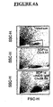
- FIG. 4A shows flow cytometry data from another experiment to visually demonstrate the cells that have, through chemotaxis, migrated to the bottom well. Cells to be enumerated are gated to prevent enumeration of debris. All IgM preparations in the absence of SDF-1 ⁇ did not affect baseline chemotaxis. In the presence of SDF-1 ⁇ , however, pre-treatment of Jurkat cells with the various IgM preparations affected chemotaxis. Particularly, as seen in FIG. 4B , all pooled IgM preparations enhanced chemotaxis, with AIDS IgM showing the most enhancement.
- FIGS. 5A-5C show the effect of various IgM antibodies on HIV-1 infectivity of ghost cells. Referring first to FIG. 5A , which shows the percent inhibition by IgM of HIV-1 infectivity in ghost CCR5 cells, it can be seen that Normal IgM and HIV IgM inhibit HIV-1 infectivity. No or minimal inhibitory effects are seen with AIDS IgM. Similar observations are seen when using IgM isolated from five individual normal sera and one HIV sera.
- IgM from pooled normal sera partially inhibits (about 40 to 50% inhibition) HIV-1 infectivity with the R5 virus 8442 and the X4 virus RF, thereby suggesting that differences in epitope binding of virus and antibody may influence degree of inhibition.
- the IgG antibody 2D7 a murine anti-CCR5 antibody, inhibited infectivity (by approximately 80%) of two of the three R5 viruses ( i.e ,. 8397 and 8442 but not 8658) when used at about 100 nM.
- HIV-1 infectivity Similar inhibitory activity towards HIV-1 infectivity is obtained when using heat-inactivated (56°) normal human sera at final concentrations of about 8 to 15% (vol/vol) with culture media. Culture media containing about 15% pooled human serum is calculated to have about 168 nM IgM. HIV-1 infected sera and AIDS sera are not used, as the data would not be interpretable in the presence of anti-viral agents or IgG anti-HIV-1 antibodies. No inhibition of HIV-1 infectivity is detected with normal human IgG or albumin in this assay system.
- IgM anti-Tat and IgM anti-gp120 present in normal sera do not have HIV-1 neutralizing activity. This supports the concept that IgM-mediated inhibition of HIV-1 infectivity, such as observed herein, is mediated via reactivity of IgM to chemokine receptors and other lymphocyte-surface receptors important for HIV-1 entry into cells that are present on lymphocytes.
- Purified IgM mediates inhibition of HIV-1 infectivity through binding of IgM to receptors important for HIV-1 entry into cells.
- receptors include, but are not limited to, CXCR4 and CCR5 receptors.
- IgM from normal sera has no direct anti-viral neutralizing effect and yet has the most inhibitory effect on HIV-1 infectivity.
- IgM purified from AIDS sera has minimal or no effect on HIV-1 infectivity, even though the AIDS IgM binds to ghost cells and T cell lines and also enhances chemotaxis and cytosolic Ca +2 induced by SDF-1.
- a second subset of IgM autoantibodies that binds to a different epitope enhances Ca +2 flux in response to SDF-1 ⁇ and enhances chemotaxis.
- This second subset of autoantibodies appears to be present in IgM from all sera ( i.e ., Normal, HIV-1 and AIDS) but is most prominent in AIDS IgM.
- This second subset of IgM anti-chemokine receptor antibody may be more important in influencing inflammatory states as it facilitates chemotaxis.
- IgM anti-lymphocyte autoantibodies limit the entry of the HIV-1 virus into cells and prolong the latency period because these antibodies bind to chemokine and other lymphocyte-surface receptors without lysing the cells at body temperature.
- the results shown herein indicate that disease progression to AIDS is associated with a marked reduction of IgM anti-lymphocyte autoantibodies, especially the subset of antibodies that inhibit HIV-1 entry into cells.
- IgM anti-lymphocyte antibody may only partially prevent entry of certain HIV-1 viral isolates, as indicated by some of the studies herein. This latter mechanism may provide another explanation for disease progression despite the presence of IgM anti-chemokine receptor autoantibodies.
- IgM autoantibodies with specificity for CCR5 inhibit RANTES binding to CCR5 and inhibit macrophage-tropic HIV-1 virus from replicating supports the premise for a protective role mediated by these IgM anti-leukocyte antibodies.
- the use of human IgM anti-leukocyte antibodies to reduce HIV-1 infectivity i.e ., through receptor blockade is an alternative approach for passive immunization, especially because it has been difficult to isolate human antibodies reactive to conserved neutralization epitopes on the HIV-1 virus.
- Receptor blockade employing IgM with reactivity to a broad range of chemokine and other receptors present on the lymphocytes may be particularly useful in situations where the HIV-1 virus switches its receptor usage, e.g ., from CCR5 to CXCR4. Maintaining increased levels of such protective antibodies could also increase the latency period after HIV-1 infection.
- the source of IgM antibodies may be heterologous, autologous or allogenic.
- IgM antibodies with specificity for chemokine and other receptors on the leukocytes may be raised in vivo (i.e., in mice or other animals or in humans) or in vitro using cell culture techniques.
- IgM antibodies may be produced either in vivo or in vitro by genetic engineering whereby genes specific for IgM anti-lymphocyte antibodies are introduced into antibody-producing cells. These antibody-producing cells may then be introduced into an infected human or into immunodeficient animals where the cells produce IgM antibodies. In the alternative, these antibody-producing cells may be grown in vitro using hybridoma or other technology.
- IgM antibodies with specificity for chemokine receptors may also be produced by isolating human antibody-producing cells specific for IgM antibodies and enhancing antibody production by such cells using hybridoma or other technology, including introduction of the cells into animals or humans.
- human lymphocytes may be transplanted into immunodeficient mice, and the lymphocytes may then be stimulated with an agent that will activate B cells such as lipopolysaccharide ("LPS")
- LPS lipopolysaccharide
- IgM antibodies Another method of producing IgM antibodies is by isolating human antibody-producing cells capable of generating human IgM from animals such as, for example, the XenoMouse TM . IgM antibody production by such cells may then be enhanced in vitro employing hybridoma or other technology such as, for example, stimulating the isolated lymphocytes with LPS or other agent that will activate the cells, e.g ., the EBV virus.
- hybridoma or other technology such as, for example, stimulating the isolated lymphocytes with LPS or other agent that will activate the cells, e.g ., the EBV virus.
- IgM antibodies may also be produced in vitro by isolating, from an individual, lymphocytes that can be then transformed with the EBV virus and introduced in a culture. A subset of these EBV transformed B lymphocytes will secrete IgM antibodies such that the resulting culture fluid contains these antibodies.
- viruses, bacteria and other antigens may be used to stimulate B cells in vivo to generate IgM antibodies to leukocytes.
- IgM antibodies produced outside an infected individual may be delivered to the individual by one of several routes of administration including, but not limited to, intravenous and intramuscular delivery.
Landscapes
- Health & Medical Sciences (AREA)
- Life Sciences & Earth Sciences (AREA)
- Chemical & Material Sciences (AREA)
- Immunology (AREA)
- Organic Chemistry (AREA)
- Engineering & Computer Science (AREA)
- General Health & Medical Sciences (AREA)
- Medicinal Chemistry (AREA)
- Molecular Biology (AREA)
- Pharmacology & Pharmacy (AREA)
- Nuclear Medicine, Radiotherapy & Molecular Imaging (AREA)
- General Chemical & Material Sciences (AREA)
- Animal Behavior & Ethology (AREA)
- Chemical Kinetics & Catalysis (AREA)
- Public Health (AREA)
- Veterinary Medicine (AREA)
- Biochemistry (AREA)
- Urology & Nephrology (AREA)
- Hematology (AREA)
- Proteomics, Peptides & Aminoacids (AREA)
- Biomedical Technology (AREA)
- Bioinformatics & Cheminformatics (AREA)
- Virology (AREA)
- Cell Biology (AREA)
- General Physics & Mathematics (AREA)
- Biotechnology (AREA)
- Genetics & Genomics (AREA)
- Biophysics (AREA)
- Microbiology (AREA)
- Communicable Diseases (AREA)
- Food Science & Technology (AREA)
- Physics & Mathematics (AREA)
- Analytical Chemistry (AREA)
- Oncology (AREA)
- Pathology (AREA)
- Tropical Medicine & Parasitology (AREA)
- Transplantation (AREA)
- AIDS & HIV (AREA)
- Pain & Pain Management (AREA)
- Rheumatology (AREA)
Abstract
Description
- The present invention relates generally to IgM autoantibodies having specificity to extracellular receptors present on lymphocytes for use in inhibiting HIV-1 from infecting cells in a human or for inhibiting of an HIV-1 infection in a human.
- Chemokines, or chemotactic cytokines, are a class of cytokine molecules capable of chemotactically attracting migratory cells. Chemokines are essential in attracting cells to inflammatory sites irrespective of the aetiology, i.e., immunologic, infective, ischaemic, drug-induced, etc., causing the inflammation. Chemokines generally have small molecular weights in the range of about 8-10 kD.
- Most chemokines can be divided into three major families, CC, CXC and CXXXC, based on the number of amino acids (referred to as "X") separating the two cysteines (referred to as "C") in the chemokine molecule. Within the CC and CXC families, chemokines are further grouped into related sub-families based on amino acid sequence similarity between them. CC chemokine sub-families include the monocyte chemoattractant protein ("MCP") sub-family and the sub-family including macrophage inhibitory protein-1α ("MIP-1α"), macrophage inhibitory protein-1β ("MIP-1β") and regulated on activation normal T cell expressed ("RANTES"). CXC chemokine sub-families include the IP-10 and Mig sub-family; the interleukin-8 ("IL-8") sub-family; and the PF4 sub-family. The chemokines stromal cell-derived factor 1α ("SDF-1α") and stromal cell-derived factor 1β ("SDF-1β") form a chemokine family that is approximately equally related by amino acid sequence similarity to the CC and CXC chemokine families. Close to 40 different chemokines have been described and cloned, each exerting a predominant functional effect. For example, RANTES attracts T lymphocytes to inflammatory sites, while IL-8 typically attracts neutrophils to inflammatory sites.
- Chemokines exert their effect by binding to chemokine receptors. CC chemokines typically bind to members of the CCR class of receptors, while CXC chemokines generally bind to members of the CXCR class of receptors. These receptors are important in regulating the extent and nature of inflammation, and certain receptors tend to be localized in certain tissues and cells.
- Chemokine receptors are involved in certain functions such as, for example, chemotaxis and interacting with viral proteins. The HIV-1 virus is known to bind to certain proteins on the surface of cells, i.e., the CD4 antigen on lymphocytes. However, in order to gain entrance into these cells and replicate, the HIV-1 virus must bind to another receptor, i.e., predominantly, CXCR4 and CCR5 chemokine receptors. Different HIV-1 viral strains use specific chemokine receptors, i.e., the X4 virus uses CXCR4 receptors, while the R5 virus uses CCR5 receptors.
- Viral entry through chemokine receptors is of prime importance in influencing viral replication and disease progression after HIV-1 infection. For example, individuals with genetic defects in chemokine receptors have been associated with a prolonged latency period after HIV-1 infection, i.e., progression of HIV-1 to AIDS.
- Researchers and pharmaceutical companies have begun looking into strategies to block or inactivate specific chemokine receptors in an effort to inhibit HIV-1 viral replication, especially because fresh human sera and their antibodies (including Immunoglobulin G ("IgG") anti-HIV-1) have no direct lytic or neutralizing activity on the HIV-1 virus. Some of these strategies include the use of peptides and IgG monoclonal antibodies that will bind to specific chemokine receptors. Such strategies, however, have not been shown to be effective.
- Other strategies apply antibodies that bind a T cell antigen complex comprising CD4 and a chemokine receptor (
US 5,961,976 ). Alternatively, antibodies against the chemokine receptor CXCR4 are applied to inhibit HIV infection (WO 97/49424 US 5,606,026 ; Hurez et al. (1994), Therap. Immunol. 1(5), 269). - Normal (i.e., non-infected) individuals have in their blood low levels of circulating Immunoglobulin M ("IgM") antibodies that bind to their own leukocytes such as, for example, B and T lymphocytes, without causing cell lysis at 37° C. Such IgM antibodies are, therefore, typically referred to as "anti-lymphocyte autoantibodies". These antibodies may also be referred to herein as "IgM anti-leukocyte antibodies" or "IgM anti-leukocyte autoantibodies" because they bind to macrophages and neutrophils in addition to lymphocytes and, furthermore, because they bind to allogenic leukocytes in addition to autologous leukocytes. Very little is known about the leukocyte or lymphocyte antigens or receptors that bind to IgM autoantibodies. Levels of such anti-leukocyte autoantibodies increase during inflammatory states, including autoimmune diseases and infectious diseases (i.e., virus-mediated diseases) such as, for example, systemic lupus erythematosus ("SLE"), sarcoidosis, HIV-1, malaria, Epstein-Barr virus ("EBV") and cytomegalovirus ("CMV"). Individuals with asymptomatic HIV-1, therefore, have high levels of IgM anti-leukocyte autoantibodies. The inventor's studies show, however, that chemokine receptors are one of the cell membrane receptors that bind to these IgM autoantibodies and that, through this mechanism, such IgM autoantibodies inhibit HIV-1 from infecting cells. The inventor's studies also show that IgM autoantibodies that bind to chemokine receptors are heterogeneous and that only some of these antibodies have the ability to inhibit HIV-1 from infecting cells. Levels of IgM antibodies that inhibit HIV-1 from infecting cells are very low or are undetectable in patients with AIDS. Thus, while individuals with asymptomatic HIV-1 infection have increased levels of IgM autoantibodies that inhibit HIV-1 infectivity, these levels, however, significantly decrease as the disease progress to AIDS. Total serum IgM does not decrease, however, as the disease progresses to AIDS.
- The physiological and pathological functions of IgM autoantibodies that bind lymphocytes remain unknown because, in part, very little is known about which membrane receptors are recognized and are bound by these IgM autoantibodies. It is unresolved, therefore, whether the increased production of these IgM autoantibodies after a viral infection is merely a non-specific response resulting from direct polyclonal activation of B cell precursors by EBV and/or the gp120 glycoprotein or is designed for a specific purpose, i.e., to function as protective antibodies. That the normal B cell repertoire has a high frequency (about 3 to 10%) of B cells committed to the production of IgM autoantibodies supports the theory that such increased production of IgM autoantibodies is designed for a specific purpose.
- It has now been discovered that non-lytic IgM anti-lymphocyte autoantibodies specifically inhibit binding of chemokines to their receptors, enhance or inhibit chemotaxis and inhibit HIV-1 from infecting cells. Moreover, IgM autoantibodies that inhibit HIV-1 from infecting cells are depleted in patients with AIDS but not in asymptomatic HIV-1 infected individuals or in normal individuals.
- Accordingly, an object of the present invention is a pool of polyreactive human IgM antibodies comprising IgM autoantibodies having specificity to extracellular receptors present on lymphocytes for use in inhibiting HIV-1 from infecting cells or for inhibiting progression of an HIV-1 infection in a human.
- Likewise, another object of the present invention is the use of a pool of polyreactive human IgM antibodies comprising IgM autoantibodies having specificity to extracellular receptors present on lymphocytes for the preparation of a pharmaceutical comprising for inhibiting HIV-1 from infecting cells in a human or for inhibiting progression of an HIV-1 infection in a human. The IgM antibodies are preferably heterologous, autologous or allogeneic.
- In another preferred aspect, the IgM antibodies are obtained from healthy individuals, pooled, heat-inactivated sera of healthy individually or asymptomatic patients with HIV-1 infections.
- These IgM antibodies are to be administered to an individual preferably intravenously or intramuscularly.
- The above and other objects, advantages and features of the present invention will become more apparent from the following detailed description of the presently preferred embodiments, when considered in conjunction with the figures, and to the appended claims.
-
-
FIG. 1A is a graph depicting binding of Normal IgM, HIV IgM and AIDS IgM to Sup T-1 cells. -
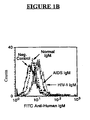
FIG. 1B is a graph depicting binding of Normal IgM, HIV IgM and AIDS IgM to Ghost CXCR4 cells. -
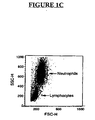
FIG. 1C is a flow cytometry (FASCAN) dot plot showing lymphocytes and neutrophils separated by size and derived from human blood. -
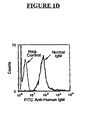
FIG. 1D is a graph depicting binding of Normal IgM to human T lymphocyte derived from peripheral blood cells. -
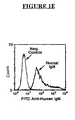
FIG. 1E is a graph depicting binding of Normal IgM to human neutrophils derived from peripheral blood cells. -
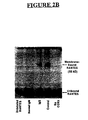
FIGS. 1F-1G are Western blot assays depicting IgM binding to cell membrane proteins obtained from U373-MAGI cells and the same cells transfected with and expressing either CXCR4 or CCR5. -
FIG. 2A is a graph depicting affinity purified Accurate IgM inhibition of I125 RANTES binding to CCR5 present in denatured U373-MAGI-CCR5E membrane proteins as quantified by liquid scintillation. -
FIG. 2B is a Western blot assay depicting affinity purified Accurate IgM inhibition of I125 RANTES binding to CCR5 present in denatured U373-MAGI-CCR5E membrane proteins. -
FIG. 2C is a dot plot FASCAN display depicting CK15 IgM inhibition of RANTES binding to IL-2-activated human lymphocytes. -
FIG. 2D is a graph depicting affinity purified Accurate IgM inhibition of RANTES binding to intact U373-MAGI-CCR5E cells. -
FIG. 3A is a graph depicting AIDS and HIV-1 IgM inhibition of SDF-1α binding to CXCR4 on intact Sup T-1 cells. -
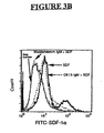
FIG. 3B is another graph depicting IgM inhibition of SDF-1α binding to CXCR4 on intact Sup T-1 cells. -
FIG. 4A is a FACSCAN exemplifying a dot plot quantitating SDF-1α induced chemotaxis of Jurkat cells into the bottom well. The bottom panel depicts enhanced migration into the bottom well when AIDS IgM is added to the upper transwell. -
FIG. 4B is a graph depicting the effect of AIDS IgM, HIV IgM, Normal IgM and Waldenstrom IgM on SDF-1α induced chemotaxis of Jurkat cells. -
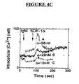
FIG. 4C is a graph measuring the effect of IgM on SDF-1α induced change in intracytosolic Ca+2 of Jurkat cells. Tracing A depicts the effect of AIDS IgM and SDF-1α; tracing B depicts the effect of SDF-1α only; and tracing C depicts the effect of HIV IgM and SDF-1α. -
FIG. 5A depicts the inhibitory effect of IgM from normal, HIV and AIDS sera on infection of Ghost CCR5 cells by the R5 virus 8658. -
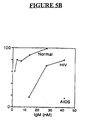
FIG. 5B depicts the inhibitory effect of IgM from normal, HIV and AIDS sera on infection of Ghost CXCR4 cells by the X4 virus III B. -
FIG. 5C is a flow cytometry dot plot exemplifying the inhibitory effect of Normal IgM on infection of Ghost CCR5 cells by the R5 virus 8658. The middle dot plot depicts that about 17.2 % of Ghost CCR5 cells were infected, and infected cells identified by detection of green fluorescence, which these cells emitted. - To achieve the foregoing and other objects, and in accordance with the purpose of the present invention as embodied and described herein, the present invention relates to a pool of polyreactive human IgM receptor-binding antibodies comprising IgM autoantibodies to address HIV-1 infections and disease states induced thereby.
- The IgM autoantibodies in the blood of normal individuals have been found to bind to various extracellular receptors present on lymphocytes. Such receptors include, but are not limited to, chemokine receptors and other lymphocyte-surface receptors. Representative chemokine receptors are, for example, CXCR4, CCR5, CCR3 and CCR2b. Representative lymphocyte-surface receptors include, for example, glycolipid receptors.
- According to the present invention, IgM anti-lymphocyte autoantibodies present in normal sera bind to chemokine and other lymphocyte-surface receptors and, through this mechanism, inhibit HIV-1 from infecting cells. These IgM autoantibodies prevent lymphocytes from being infected with HIV-1.
- Human sera from normal, uninfected individuals contain low levels of IgM anti-leukocyte autoantibodies reactive with chemokine and other leukocyte-surface receptors. These anti-leukocyte antibodies increase after HIV-1 infection and may play a role in the slow progression of HIV-1 disease by limiting entry of the virus into lymphocytes, macrophages and other cells. IgM anti-chemokine receptor antibodies function as "blocking" antibodies as they are not cytolytic at 37° C.
- While not wishing to be bound to a specific theory, it is believed that normal individuals are born with the capacity to form IgM autoantibodies to chemokine and other lymphocyte-surface receptors. These IgM autoantibodies do not damage the cells bearing chemokine receptors because these antibodies cannot activate complement at body temperature (37° C). It is also believed that these IgM autoantibodies are heterogeneous and function normally to regulate receptor expression and are protective (i.e., prevent viral entry into cells). A subset of IgM anti-chemokine receptor autoantibody inhibits HIV-1 from infecting cells. It is further believed that the increase in these antibodies after HIV-1 infection protects individuals from developing AIDS in less than one year. Depletion of IgM, which inhibits HIV-1 from infecting cells in AIDS patients but not in asymptomatic HIV-1 infected individuals indicates, therefore, that the presence of such IgM autoantibodies may be important in slowing down disease progression in the event of viral infection or other virus-mediated disease.
- Sup T-1 and Jurkat cells are lymphoma T cell lines expressing the CXCR4 chemokine receptor. These cell lines are obtained from the AIDS Reagent Program at NIH.
- An HOS osteosarcoma cell line is co-transfected with CD4 and either CXCR4 or CCR5 genes to produce HOS-CD4, HOS-CD4-CXCR4 and HOS-CD4-CCR5 cells. Ghost CCR5 and Ghost CXCR4 are HOS-CD4 cells co-transfected with the HIV-2 LTR driving hGFP construct and either CCR5 or CXCR4 genes, respectively. The cell line and the transfectants are obtained from the AIDS Reagent Program at NIH.
- A glioblastoma cell line, U373-MAGI, is co-transfected with CD4 and either CXCR4 or CCR5 to produce U373-MAGI-CXCR4 and U373-MAGI-CCR5, respectively. Again, the cell line and the transfectants are obtained from the AIDS Reagent Program at NIH.
- All of the transfected cell lines stably express CCR5 or CXCR4, with the U373-MAGI cells having the highest expression of these receptors.
- Human peripheral blood lymphocytes ("PBL") is activated with IL-2 to enhance CCR5 expression. PBL (2x10⁶ cells in 1 ml RPMI culture media containing 10% fetal calf serum are activated by initially pre-treating ficol/hypaque separated PBL with IL-2 (40 units/ml) and phytohemagglutinin ("PHA-P", 5 mcg/ml) and then washing the PBL after the cells are cultured at 37° C in about 5% CO2 for 24 to 48 hours. Such PHA pre-treated cells are then kept growing for about another 6 to 7 days supplemented with 20% fetal calf serum and IL-2 (40 units/ml) before being used in chemokine binding assays.
- The R5 HIV-1 virus used to infect Ghost CCR5 is obtained from Dr. Homayoon Garadegan at Johns Hopkins University. The X4 virus IIIB and RF used to infect Ghost CXCR4 is obtained from the AIDS Reagent Program at NIH.
- Human IgM is obtained from the following sources: affinity purified, pooled IgM obtained from sera of normal, healthy individuals (referred to as "affinity purified Accurate IgM") available from Accurate Chemicals of Westbury, New York (catalog number AI-MO2); IgM purified by size exclusion chromatography from pooled, heat-inactivated sera (56°C) of normal, healthy individuals (referred to as "Normal IgM"), from asymptomatic patients with HIV-1 infection (referred to as "HIV IgM") and from patients diagnosed with AIDS (referred to as "AIDS IgM"); and culture supernatants of EBV transformed human B cell clones. Size exclusion chromatography (Sephacryl S-300 HR) is used to remove low molecular weight substances (i.e., chemokines, anti-viral drugs, etc.) and IgG anti-HIV-1 antibodies that could affect the experiment.
- The HIV IgM is pooled from seven asymptomatic HIV-1 infected patients not taking antiviral agents. The HIV-1 infected patients all have greater than 500 CD4 positive cells per ml and a DNA viral load of less than 2,000. The AIDS IgM is pooled from nine AIDS patients having an opportunistic infection and less than 150 CD4 positive cells per ml and a viral load of greater than 10,000 despite antiviral agents. Normal IgM from six normal, healthy subjects is used either individually or pooled. Data exemplified in the drawings are from pooled IgM unless otherwise indicated.
- The culture supernatants of EBV transformed human B cell clones are separated by Sephacryl S-300 HR column chromatography, which separates proteins by size. The human B cell clones are derived from B lymphocytes isolated from the blood of a patient with SLE. The B cell clones are developed by infecting B cells with the EBV virus, which makes the B cells immortal and capable of secreting a specific antibody, i.e., IgM. More particularly, non-T cells are isolated from PBL after removal of T cells using a sheep erythrocyte rosetting technique. About 2x10³ non-T cells in about 0.1 ml RPMI 1640 cell culture media containing about 10% fetal calf serum are added to each well of a 96-well plate. To each well is then added about 50 lambda of EBV-containing B95-8 cell line supernatant. Before incubation, about 104 allogenic irradiated (3,000 rads) PBL in 0.05 ml are added as feeder cells. The plates are incubated at 37° C in about 5 % CO2. The culture medium is replaced about every 4 to 5 days. After about 3 to 4 weeks, B cell lines appear as "clumps" in the wells. Feeder cells die during this period. When the "clumps" appear, these clumped cells are transferred to a 24-well plate, i.e., cells from one well are transferred into a single larger well. Culture media is changed when the media changes to a yellowish color, usually about 3 to 5 days. After about 2 weeks, supernatants are checked for IgM antibody. Wells containing lines with desired antibody specificity are further subcloned with limiting dilution in a 96-well plate. About 105 feeder cells are added to each well containing these lines. Supernatants are rechecked to isolate clones with desired antibody specificity. Supernatants are refrigerated, but not frozen as IgM can precipitate out. Clones secreting IgM antibodies that are useful in inhibiting HIV-1 infectivity are cryopreserved. Supernatants from such clones usually contain about 0.5 to about 0.7 µg/ml antibody. Clones of particular interest can be fused with K6H6/B5 plasmacytoma cell line to develop hybridomas. The clones are screened to identify and obtain those clones that react with CCR5 and CXCR4 chemokine receptors present on the transfected cells. Such clones have increased IgM binding by flow cytometry to the HOS-CD4 transfectants (i.e., HOS-CD4-CXCR4 and HOS-CD4-CCR5) when compared to the HOS-CD4 control.
- Any contaminating IgG is removed from the IgM preparations that is isolated from the sera and the culture supernatants by exposure to both protein G-Agarose (available from Sigma) and goat anti-human IgG (Fc specific)-Agarose (available from Sigma).
- IgM is also obtained using Sephacryl S-300 HR column chromoatography from sera of a patient diagnosed with Waldenstrom macroglobulinemia (a form of B cell lymphoma) and which, on serum protein electrophoresis, has a single peak for IgM (monoclonal). This latter IgM preparation is hereinafter referred to as "Waldenstrom IgM."
- Four chemokines preparations are used in the following studies. RANTES, SDF-1α and biotin-labeled SDF-1α are obtained from Becton Dickinson of La Jolla, California. Radio-labeled RANTES (referred to as "I125 RANTES" or "I125") is obtained from NEN Life Science of Boston, Massachusetts. RANTES binds to CCR5. while SDF-1α binds to CXCR4.
- Two murine IgG monoclonal antibodies, 12G5 and 2D7, specific for CXCR4 and CCR5, respectively, are used. The 12G5 is obtained from Becton Dickinson, while the 2D7 is obtained from the AIDS Reagent Program.
- Flow cytometry is used to quantify IgM binding to the cells. Prior to flow cytometry, about 1x10⁵ cells are initially incubated at about 4° C with about 150 nM of each of Normal IgM, HIV IgM and AIDS IgM. The cells are then washed, followed by staining with fluorescein-isothiocyanate ("FITC") goat anti-human IgM (Fc specific). Binding of IgM to human peripheral blood T lymphocytes is quantified by two color flow cytometries, i.e., using phycoerytherin ("PE")-labeled anti-CD3 and FITC-labeled goat anti-human IgM (Fc specific).
- Western blot assays are conducted to determine if IgM autoantibodies bind to cell membrane proteins. Four controls are made by combining about 50 µg of crude membrane proteins obtained from U373-MAGI with about 100 µl supernatant from four different EBV transformed B cell clones. Two additional controls are made by combining about 50 µg of crude membrane proteins obtained from U373-MAGI with about 100 µl each of HIV-1 serum and AIDS serum. These controls are compared to cell membrane proteins obtained from the same cell line, but transfected with and expressing either CXCR4 or CCR5, and prepared in the same manner.
- Various studies are employed to determine if IgM inhibits binding of chemokines to chemokine receptors. In this study, the focus is whether IgM with anti-CCR5 activity can inhibit binding of I125 RANTES to CCR5. This study is performed with affinity purified IgM and with supernatants from EBV transformed B cell clones, specifically, CK15. Controls are supernatants containing IgM Rheumatoid factor and purified human IgG.
- The first approach is to determine if affinity purified Accurate IgM and/or CK15 IgM inhibit binding of I125 RANTES to non-denatured, crude membrane proteins obtained from U373-MAGI-CCR5. Here, each of affinity purified Accurate IgM and CK15 IgM or 500 fold unlabeled RANTES relative to labeled RANTES in varying molar concentrations ranging from about 10-6 to about 10-10 are incubated with about 5 µg of non-denatured U373-MAGI-CCRS membrane proteins for about 1 hour at room temperature in the presence of Ca+2 and Mg+2 and a protease inhibitor. At the end of the incubation, about 0.25 nM I125 RANTES available from NEN Life Science is added to each mixture, and each mixture is further incubated at room temperature for about another 2 hours. Each mixture is then harvested over fiberglass filters and washed three times to remove unbound I125 RANTES. Specific I125 RANTES binding is calculated by subtracting the counts of radioactivity per minute ("c.p.m.") of I125 RANTES when used with 500 fold molar excess of unlabeled RANTES from the data obtained with I125 RANTES.
- In a second approach, IgM inhibition of I125 RANTES binding to CCR5 is detected by Western blotting. Here, about 250 µg of non-denatured U373-MAGI-CCR5 membrane proteins are incubated, under non-reducing conditions, with each of about 0.1 nM Normal IgM, 350 nM IgG, 0.4 mcM unlabeled RANTES and a culture media control at room temperature for about 1 hour prior to adding about 1.0 nM I125 RANTES available from NEN Life Science to each mixture. After about 2 hours incubation, about 5 nM of a cross-linker (BS-3 available from Pierce of Rockford, Illinois) is added to each mixture to crosslink amine residues on I125 RANTES bound to CCR5. Also, I125 RANTES in the absence of U373-MAGI-CCR5 membrane proteins is used. Each mixture is then electrophoresed onto 12% gel SDS-PAGE, and radiographs of the gel are obtained.
- A procedure is used to determine if IgM inhibits binding of RANTES to CCR5 receptors present on intact cells, e.g., U373-MAGI-CCR5E and IL-2-activated human lymphocytes. About 1x10⁵ cells are initially incubated at room temperature for about 45 minutes with RPMI media containing about 10% fetal calf serum or with about 150 nM of affinity purified Accurate IgM or with about 400 nM purified human IgG or with about 5 nM, about1.25 nM or about 0.45 nM of CK15 IgM prior to adding about 1 microgram of RANTES to each mixture. The cells are then re-incubated for about 90 minutes at 4° C and then washed at 4° C. Goat anti-RANTES antibody (obtained from R&D of Minneapolis, Minnesota) is then added, and the cells are incubated for about 45 minutes at 4° C prior to being washed and then stained with FITC rabbit anti-goat antibody. The quantity of FITC-labeled RANTES binding to CCR5 on these cells is analyzed by flow cytometry.
- A procedure is used to determine if IgM inhibits binding of SDF-1α to chemokine receptors (i.e., CXCR4) present on intact Sup T-1 cells. About 1x10⁵ Sup T-1 cells are initially incubated at room temperature for about 45 minutes with RPM1 media containing about 10% fetal calf serum or with about 150 nM of each of Normal IgM, HIV IgM and AIDS IgM or with about 5 nM of each of Waldenstrom IgM and CK15 IgM prior to adding about 25 ng of biotin-labeled SDF-1α to each mixture. The cells are then re-incubated for about 90 minutes at 4°C. Following re-incubation, FITC avidin is added to the cells, and the cells are washed. The quantity of FITC-labeled SDF-1α binding to CXCR4 is analyzed by flow cytometry.
- Additional procedures are performed to determine if IgM inhibition of chemokines binding to CCR5 or CXCR4 is indeed specific for chemokines. More particularly, these procedures are used to determine if IgM, through some non-specific mechanism, also inhibits binding of radio-labeled IL-2 to the IL-2R present on phytohemagglutinin-activated PBL using methods as previously described in, for example, Teshigawara, K. et al., J. Exp. Med., 165:223-238 (1987). Specifically, three day phytohemagglutinin-activated PBL (1x10⁶) is incubated with I125 labeled IL-2 (available from NEN Life Science), and the I125 labeled IL-2 bound to PBL is quantified by overlaying the PBL over oil and centrifuging the microfuge tube to separate unbound I125 labeled IL-2 from the cell pellet. Radioactivity of I125 is quantitated in the cell pellet. In these procedures, PBL is interacted with each of excess unlabeled IL-2 (2.0 mcM), pooled human IgG (300 nM), IgM Rheumatoid factor and affinity purified Accurate IgM (100 nM) prior to adding I125 labeled IL-2.
- A chemotaxis assay is performed with each of Normal IgM, HIV IgM, AIDS IgM and Waldenstrom IgM at concentrations of about 20 nM, about 40 nM, about 100 nM and about 200 nM IgM using 24-well Costar transwell tissue culture inserts with 5 micron polycarbonate filters. For each assay, the IgM is placed in the upper transwell containing about 2x10⁴ Jurkat cells in about 0.1 ml RPM1 containing about 5% fetal calf serum. About 30 minutes later, approximately 50 ng of SDF-1α is added to the bottom well containing about 0.6 ml of the same media as in the upper well. After about 4 hours, cells migrating to the bottom well are enumerated by flow cytometry. The chemotaxic index ("CI") is calculated by dividing the total number of cells migrating in the presence of SDF-1α by the number of cells migrating in the absence of SDF-1α. The baseline chemotactic index of SDF-1α alone (i.e., without IgM) is about 3.1.
- Assays are performed to determine intracytosolic Ca+2 flux using known methods, for example, as described in Haverstick, G., MD, Molecular Biol. of Cell. 4:173-184 (1993). In one assay, about 45 nM of HIV IgM is added to Jurkat cells at a time of about 20 seconds. Approximately 60 seconds later, about 100 ng of SDF-1α is added, and the magnitude of change in cytosolic Ca+2 after adding SDF-1α is measured. A second assay is done using about 45 nM of AIDS IgM in place of HIV IgM. In a third assay, no IgM is added, but SDF-1α is still added at a time of about 80 seconds.
- Temperature dependence for the cytolytic effects of IgM anti-leukocyte antibody is evaluated by a complement dependent microlymphocytotoxicity assay. Various dilutions of IgM antibody are reacted for 1 hour with either 2x10⁵ PBL or IL-2-activated PBL (7 days) before adding fresh rabbit serum as a source of complement. After about 2 hours, the cells are washed twice before adding trypan blue and enumerating dead cells that stain blue. Experiments are performed at 15° C and 37° C.
- It has been observed that the HIV-1 R5 virus utilizes CCR5 receptors for cell entry, while the HIV-1 X4 virus uses CXCR4 receptors. Studies are conducted, therefore, to determine whether IgM inhibits HIV-1 infectivity in light of such observations.
- In these studies, Ghost CCR5 and Ghost CXCR4 transfectant cell lines are infected with HIV-1. The Ghost cells are derived from HOS cells transfected with either CCR5 or CXCR4 genes and also co-transfected with the HIV-2 LTR driving hGFP construct. The hGFP construct enables cells infected with HIV-1 virus to emit a green fluorescence so that the number of infected cells can be quantified using flow cytometry. These cell lines are particularly suited for these studies because single-cycle viral replication can be detected in less than 48 hours.
- About 2x10⁴ each of Ghost CCR5 and CXCR4 cells are separately cultured for about 12 hours in about 1 ml RPM1 media containing about 10% fetal calf serum in a 12-well plate. Normal IgM is then added to each of the Ghost CCR5 and CXCR4 cells about 30 minutes prior to adding the R5 HIV-1 virus to Ghost CCR5 and the X4 HIV-1 virus to Ghost CXCR4. Both virus and antibody are present throughout the 48-hour culture period. No polybrene is used to enhance viral entry into the cells. The same procedure is repeated twice, replacing Normal IgM first with HIV IgM and then AIDS IgM.
- After the 48-hour incubation period, cells are harvested and fixed in formalin. Infected cells emitting green fluorescence are enumerated with flow cytometry.
- Additionally, similar data is obtained when the virus or IgM antibody is washed about 4 hours after incubating with Ghost cells.
- The results of the various experimental studies indicate that IgM autoantibodies bind to chemokine receptors present on lymphocytes and other cells. The results also indicate that binding of IgM autoantibodies is specific for chemokine receptors.
- The data from the above-discussed studies shows that IgM autoantibodies bind to receptors present on normal lymphocytes and malignant cells. As seen in
FIGS. 1A and1B , Normal IgM, AIDS IgM and HIV IgM contain IgM antibodies that bind to Sup T-1 (FIG. 1A ) and Ghost CD4-CXCR4 cells (FIG. 1B ). As seen inFIGS. 1D and1E , Normal IgM contains antibodies that bind to T-lymphocytes isolated from peripheral blood (FIG. ID) and neutrophils isolated from peripheral blood (FIG. 1E ). The negative control in each figure indicates that no IgM was incubated with the various cells. - Referring now to
FIGS. 1F and1G , it is evident that IgM autoantibodies bind to several cell membrane proteins. However, IgM bound to an additional 48 kD protein present in cell lines transfected with cDNA of CCR5 ("CCR5") and CXCR4 ("CXCR4"). Membrane proteins of 48 kD are similar to the molecular weight of CXCR4 and CCR5 receptors implying, therefore, that IgM also bound to the chemokine receptors that were present in the transfected cells. IgM can bind to several membrane proteins because the IgM antibody is known to be polyreactive, especially in Western blot assays. Hence, other assay systems are employed to better define specificity of IgM binding to chemokine receptors using assays that evaluate whether IgM in a specific manner inhibits binding of chemokines to their receptor and alters chemokine receptor function. - IgM autoantibodies inhibit binding of radio-labeled RANTES to CCR5 receptors but not binding of radio-labeled IL-2 to IL-2 receptors (i.e., IL-2R). This supports the concept that IgM-mediated inhibition is indeed specific for chemokines. Moreover, normal IgG does not inhibit radio-labeled RANTES from binding to CCR5 receptors.
- Representative data from initial studies conducted to determine if affinity purified Accurate IgM and/or CK15 IgM inhibit binding of I125 RANTES to non-denatured, crude membrane proteins obtained from U373-MAGI-CCR5E is depicted in
FIG. 2A . As seen therein, both the affinity purified Accurate IgM and the CK15 IgM inhibit binding of I125 RANTES to CCR5 in a dose-dependent manner. Pooled Normal IgG and IgM Rheumatoid factor, even when used at 10-6 M, fail to inhibit binding of I125 RANTES to CCR5. Unlabeled RANTES inhibits I125 RANTES binding in a dose-dependent manner with a kD of 0.095 nM. - IgM inhibition of the binding of I125 RANTES to CCR5 as detected by Western blotting is depicted in
Fig. 2B . As seen therein, affinity purified Accurate IgM and unlabeled RANTES inhibit binding of I125 RANTES to CCR5. Neither the pooled human IgG nor the RANTES protein control inhibits binding of I125 RANTES to CCR5. This latter observation would appear to indicate that IgM inhibition of I125 RANTES binding to CCR5 is a result of receptor blockade and is specific for IgM having a specificity for CCR5. - Additional studies were performed using IL-2-activated lymphocytes, which express more CCR5 receptors when compared to unactivated lymphocytes, to determine if IgM inhibits chemokines from binding to their receptors present on intact cells. As indicated in
FIG. 2C , about 22.45% of IL-2-activated lymphocytes bound to RANTES. In the presence of about 5 nM CK15 IgM, the binding decreased, with about 6.8% of the lymphocytes binding to RANTES. Less inhibition is observed with less CK15 IgM. As indicated inFIG. 2D , affinity purified Accurate IgM, but not human IgG, inhibited binding of RANTES to intact U373-MAGI-CCR5E cells. - As seen in
FIG. 3A , Normal IgM and HIV IgM suppresses biotin SDF-1α binding, while AIDS IgM does not. A negative control is used to indicate background fluorescence of cells without IgM and SDF-1α. "SDF" indicates SDF-1α binding in the absence of IgM. Note that a small subset (15%) of Sup T-1 cells had much stronger binding to biotin SDF-1α. As seen inFIG. 3B , Waldenstrom IgM, but not CK15 IgM, inhibited binding of SDF-1α to Sup-T cells. - An additional study to determine if IgM inhibition of chemokines binding to their receptors is indeed specific for chemokines shows that binding of I125-labeled IL-2 is inhibited by unlabeled IL-2 (>90%) and anti-TAC (anti-IL2R murine IgG antibody) but not by pooled human, affinity purified Accurate IgM, even when used at 100 nM. The results are shown in Table 1 below.
TABLE 1 INHIBITION OF I125-LABELED IL-2 BINDING TO IL-2R ON ACTIVATED PBL Specific I125 IL-2 bound to PBMC (cpm) Control Media 9,468 Human IgG 8,293 Human IgM 8,809 Murine anti-TAC 1,948 Unlabeled IL-2 2,353 - The above results, therefore, indicate that inhibition of SDF-1α binding to CXCR4 by Normal IgM and HIV-1 IgM is specific. Data demonstrating inhibition of biotin SDF-1α binding when using Normal IgM and HIV IgM cannot be explained on basis of stearic hindrance as AIDS IgM has similar binding to Sup T-1 cells, yet fails to inhibit SDF-1α binding (see
FIG. 1A ). Similarly, data fromFIG. 2 andFIG. 3B clearly indicate that inhibition of chemokines to their receptors by monoclonal IgM is indeed specific as Waldenstrom IgM totally inhibited binding of SDF-1α to CXCR4. The CK15 IgM, which inhibited binding of RANTES to CCR5, failed to inhibit binding of SDF-1α to CXCR4. While not wishing to be bound to any particular theory, one possible explanation is that IgM anti-leukocyte autoantibodies are heterogeneous and recognize different epitopes on the chemokine receptor. It is, therefore, possible that AIDS IgM lacks the subset of IgM antibodies that inhibit SDF-1α binding, even though the AIDS IgM binds to the chemokine receptor as will be evident from the data obtained in the chemotaxis assay (described in detail below). - The possibility of heterogeneity of IgM is analyzed in functional assays of SDF-1α induced chemotaxis (
FIGS. 4A and4B ) and intracellular Ca+2 flux (FIG. 4C ). -
FIG. 4A shows flow cytometry data from another experiment to visually demonstrate the cells that have, through chemotaxis, migrated to the bottom well. Cells to be enumerated are gated to prevent enumeration of debris. All IgM preparations in the absence of SDF-1α did not affect baseline chemotaxis. In the presence of SDF-1α, however, pre-treatment of Jurkat cells with the various IgM preparations affected chemotaxis. Particularly, as seen inFIG. 4B , all pooled IgM preparations enhanced chemotaxis, with AIDS IgM showing the most enhancement. Enhanced migration into the bottom well after adding IgM to the upper well was mediated by chemotaxis-induced migration and not from some non-specific process, as adding 500 ng of SDF-1α to the upper transwell totally inhibited this enhanced migration. Control CI in the presence of SDF-1α is about 2.8. Waldenstrom IgM inhibited chemotaxis (CI = about 1.1). CK12 IgM and CK15 IgM mildly enhanced chemotaxis with the CI varying from about 5.3 to about 7.1, which is significantly more than that observed with SDF-1α alone (CI=3.1). - These findings prompted a determination of whether IgM similarly affected changes in cytosolic Ca+2 that occur when chemokines bind to the CXCR4 receptor present on Jurkat cells. Representative data from three such experiments is depicted in
FIG. 4C , where the symbol Δ indicates the magnitude of change in cytosolic Ca+2 after adding SDF-1α. Tracing A represents Jurkat cells with AIDS IgM; tracing C represents Jurkat cells with HIV IgM; and tracing B represents Jurkat cells with no IgM. As seen inFIG. 4C , none of the IgM antibodies in the absence of SDF-1α (i.e., prior to adding of SDF-1α) elicit a rise in intracellular Ca+2. Tracing A indicates that AIDS IgM enhances the rise in the intracellular Ca+2 response to SDF-1α. Tracing C indicates that no enhancement occurs with HIV IgM. - Specificity of IgM interaction with CXCR4 in these Ca+2 flux assays is ascertained by performing similar studies on another receptor present on Jurkat cells, i.e., the CD3 receptor. No enhancement in cytosolic Ca+2 is observed by adding AIDS IgM prior to stimulating CD3 with OKT3, a murine IgG anti-CD3 antibody. Such data provides more evidence to support specificity of IgM binding to the CXCR4 receptor. Furthermore, these findings clearly support the concept of functional heterogeneity within IgM anti-lymphocyte autoantibodies, with AIDS IgM containing IgM that predominantly enhances chemotaxis and Ca+2 flux after binding to the chemokine receptor.
- About 40 to 60% cell lysis was observed when the assay was performed at 15° C. Higher levels of cell lysis was observed with IL-2-activated lymphocytes, which have increased expression of chemokine receptors. Affinity purified Accurate IgM, when used at amounts of about 1.0 microgram or more, caused cell lysis, while CK15 lysed cells at concentrations of about 5 nanograms or more. When the assay was performed at 37° C, however, less than about 10% lysis was observed, and the level of lysis was similar to control cells incubated within the body. These observations are in agreement with several reports clearly demonstrating that IgM anti-lymphocyte autoantibodies are lytic at colder temperatures but not at 37° C.
- IgM anti-chemokine receptor autoantibodies contribute to the resistance against HIV-1 infection.
FIGS. 5A-5C show the effect of various IgM antibodies on HIV-1 infectivity of Ghost cells. Referring first toFIG. 5A , which shows the percent inhibition by IgM of HIV-1 infectivity in Ghost CCR5 cells, it can be seen that Normal IgM and HIV IgM inhibit HIV-1 infectivity. No or minimal inhibitory effects are seen with AIDS IgM. Similar observations are seen when using IgM isolated from five individual normal sera and one HIV sera. - Interestingly, IgM from pooled normal sera partially inhibits (about 40 to 50% inhibition) HIV-1 infectivity with the R5 virus 8442 and the X4 virus RF, thereby suggesting that differences in epitope binding of virus and antibody may influence degree of inhibition.
- The IgG antibody 2D7, a murine anti-CCR5 antibody, inhibited infectivity (by approximately 80%) of two of the three R5 viruses (i.e,. 8397 and 8442 but not 8658) when used at about 100 nM.
- Similar inhibitory activity towards HIV-1 infectivity is obtained when using heat-inactivated (56°) normal human sera at final concentrations of about 8 to 15% (vol/vol) with culture media. Culture media containing about 15% pooled human serum is calculated to have about 168 nM IgM. HIV-1 infected sera and AIDS sera are not used, as the data would not be interpretable in the presence of anti-viral agents or IgG anti-HIV-1 antibodies. No inhibition of HIV-1 infectivity is detected with normal human IgG or albumin in this assay system. These data indicate that Normal IgM and HIV IgM when used in nM quantities inhibit HIV-1 infection of certain HIV-1 viral strains even though high doses of viruses are used, i.e., enough to infect about 8 to 20% cells with a single cycle of replication.
- Several investigators have shown that normal human sera has no direct neutralizing activity towards the HIV-1 virus. Others have shown that IgM anti-Tat and IgM anti-gp120 present in normal sera do not have HIV-1 neutralizing activity. This supports the concept that IgM-mediated inhibition of HIV-1 infectivity, such as observed herein, is mediated via reactivity of IgM to chemokine receptors and other lymphocyte-surface receptors important for HIV-1 entry into cells that are present on lymphocytes.
- The results of the various studies, as discussed above, indicate that IgM purified from sera inhibits HIV-1 infectivity of cells. Purified IgM mediates inhibition of HIV-1 infectivity through binding of IgM to receptors important for HIV-1 entry into cells. Such receptors include, but are not limited to, CXCR4 and CCR5 receptors. IgM from normal sera has no direct anti-viral neutralizing effect and yet has the most inhibitory effect on HIV-1 infectivity. IgM purified from AIDS sera has minimal or no effect on HIV-1 infectivity, even though the AIDS IgM binds to Ghost cells and T cell lines and also enhances chemotaxis and cytosolic Ca+2 induced by SDF-1. With respect to IgM binding and inhibition of HIV-1 infectivity, one plausible explanation for the observed difference between Normal IgM and HIV-1 IgM on the one hand and AIDS IgM on the other is that Normal IgM and AIDS IgM contain a heterogeneous group of IgM anti-lymphocyte antibodies with different binding epitopes that give rise to different functional effects.
- The results suggest that the IgM antibody that inhibits SDF-1α binding on intact cells is probably the same IgM antibody that inhibits HIV-1 infectivity. AIDS IgM appears to lack this subset of IgM anti-lymphocyte antibody.
- A second subset of IgM autoantibodies that binds to a different epitope enhances Ca+2 flux in response to SDF-1α and enhances chemotaxis. This second subset of autoantibodies appears to be present in IgM from all sera (i.e., Normal, HIV-1 and AIDS) but is most prominent in AIDS IgM. This second subset of IgM anti-chemokine receptor antibody may be more important in influencing inflammatory states as it facilitates chemotaxis.
- According to the present invention, IgM anti-lymphocyte autoantibodies limit the entry of the HIV-1 virus into cells and prolong the latency period because these antibodies bind to chemokine and other lymphocyte-surface receptors without lysing the cells at body temperature. The results shown herein indicate that disease progression to AIDS is associated with a marked reduction of IgM anti-lymphocyte autoantibodies, especially the subset of antibodies that inhibit HIV-1 entry into cells.
- While not wishing to be bound to any particular theory, there are several possible explanations for the entry of the HIV-1 virus into cells and increased viral replication despite the presence of a good level of IgM autoantibody to chemokine receptor during the asymptomatic state. One such explanation is the possibility that there exists a delicate balance between these low-avidity binding IgM antibodies and the viral load. Factors that predispose an individual to an increased viral load or that inhibit the B cells secreting IgM autoantibodies will lead to viral entry into cells and to disease progression. It is also possible that the recently described subset of B cells expressing CD4, CXCR4 and CCR5 receptors may be the same subset that secretes IgM autoantibodies. Over the years, this B cell subset could be exhausted or could be infected with HIV-1, thereby leading to a decrease in antibody production. Additionally, one cannot underscore the importance of other host factors (e.g., anti-viral IgG antibodies, chemokines and complement and cytotoxic T cells) that decrease the viral load. Perturbation in any of these host defense mechanisms could lead to an increased viral load.
- Secondly, it is possible that in some HIV-1 infected individuals, IgM anti-lymphocyte antibody may only partially prevent entry of certain HIV-1 viral isolates, as indicated by some of the studies herein. This latter mechanism may provide another explanation for disease progression despite the presence of IgM anti-chemokine receptor autoantibodies.
- That IgM autoantibodies with specificity for CCR5 inhibit RANTES binding to CCR5 and inhibit macrophage-tropic HIV-1 virus from replicating supports the premise for a protective role mediated by these IgM anti-leukocyte antibodies. The use of human IgM anti-leukocyte antibodies to reduce HIV-1 infectivity (i.e., through receptor blockade) is an alternative approach for passive immunization, especially because it has been difficult to isolate human antibodies reactive to conserved neutralization epitopes on the HIV-1 virus. Receptor blockade employing IgM with reactivity to a broad range of chemokine and other receptors present on the lymphocytes may be particularly useful in situations where the HIV-1 virus switches its receptor usage, e.g., from CCR5 to CXCR4. Maintaining increased levels of such protective antibodies could also increase the latency period after HIV-1 infection.
- The source of IgM antibodies may be heterologous, autologous or allogenic. For example, IgM antibodies with specificity for chemokine and other receptors on the leukocytes may be raised in vivo (i.e., in mice or other animals or in humans) or in vitro using cell culture techniques.
- For example, IgM antibodies may be produced either in vivo or in vitro by genetic engineering whereby genes specific for IgM anti-lymphocyte antibodies are introduced into antibody-producing cells. These antibody-producing cells may then be introduced into an infected human or into immunodeficient animals where the cells produce IgM antibodies. In the alternative, these antibody-producing cells may be grown in vitro using hybridoma or other technology.
- IgM antibodies with specificity for chemokine receptors may also be produced by isolating human antibody-producing cells specific for IgM antibodies and enhancing antibody production by such cells using hybridoma or other technology, including introduction of the cells into animals or humans. For example, human lymphocytes may be transplanted into immunodeficient mice, and the lymphocytes may then be stimulated with an agent that will activate B cells such as lipopolysaccharide ("LPS")
- Another method of producing IgM antibodies is by isolating human antibody-producing cells capable of generating human IgM from animals such as, for example, the XenoMouse™. IgM antibody production by such cells may then be enhanced in vitro employing hybridoma or other technology such as, for example, stimulating the isolated lymphocytes with LPS or other agent that will activate the cells, e.g., the EBV virus.
- IgM antibodies may also be produced in vitro by isolating, from an individual, lymphocytes that can be then transformed with the EBV virus and introduced in a culture. A subset of these EBV transformed B lymphocytes will secrete IgM antibodies such that the resulting culture fluid contains these antibodies.
- In addition, viruses, bacteria and other antigens (e.g., mitogens) may be used to stimulate B cells in vivo to generate IgM antibodies to leukocytes.
- IgM antibodies produced outside an infected individual may be delivered to the individual by one of several routes of administration including, but not limited to, intravenous and intramuscular delivery.
Claims (5)
- Use of a pool of polyreactive human IgM antibodies comprising IgM autoantibodies having specificity to extracellular receptors present on lymphocytes for the preparation of a pharmaceutical composition for inhibiting HIV-1 from infecting cells in a human or for inhibiting progression of an HIV-1 infection in a human.
- A pool of polyreactive human IgM antibodies comprising IgM autoantibodies having specificity to extracellular receptors present on lymphocytes for use in inhibiting HIV-1 from infecting cells in a human or for inhibiting progression of HIV-1 infection in a human.
- The use of any one of claims 1-2, wherein the IgM antibodies are heterologous, autologous or allogeneic.
- The use of any one of claims 1-3, wherein the IgM antibodies are obtained from healthy individuals, pooled, heat-inactivated sera of healthy individuals or asymptomatic patients with HIV-1 infections.
- The use of any one of claims 1 to 4, wherein the IgM antibodies or the pharmaceutical composition are to be administered to the individual intravenously or intramuscularly.
Applications Claiming Priority (5)
| Application Number | Priority Date | Filing Date | Title |
|---|---|---|---|
| US684813 | 1984-12-21 | ||
| US439690 | 1999-11-14 | ||
| US43969099A | 1999-11-15 | 1999-11-15 | |
| US09/684,813 US6610834B1 (en) | 1998-11-18 | 2000-10-10 | Human IgM antibodies to chemokine receptors |
| PCT/US2000/031051 WO2001035980A1 (en) | 1999-11-14 | 2000-11-14 | HUMAN IgM ANTIBODIES TO CHEMOKINE RECEPTORS |
Publications (3)
| Publication Number | Publication Date |
|---|---|
| EP1231930A1 EP1231930A1 (en) | 2002-08-21 |
| EP1231930A4 EP1231930A4 (en) | 2005-07-27 |
| EP1231930B1 true EP1231930B1 (en) | 2010-04-21 |
Family
ID=27032133
Family Applications (1)
| Application Number | Title | Priority Date | Filing Date |
|---|---|---|---|
| EP00978542A Expired - Lifetime EP1231930B1 (en) | 1999-11-15 | 2000-11-14 | HUMAN IgM ANTIBODIES TO CHEMOKINE RECEPTORS |
Country Status (8)
| Country | Link |
|---|---|
| US (1) | US6610834B1 (en) |
| EP (1) | EP1231930B1 (en) |
| JP (1) | JP2003516940A (en) |
| AT (1) | ATE464909T1 (en) |
| AU (1) | AU1599601A (en) |
| CA (1) | CA2389851A1 (en) |
| DE (1) | DE60044259D1 (en) |
| WO (1) | WO2001035980A1 (en) |
Families Citing this family (14)
| Publication number | Priority date | Publication date | Assignee | Title |
|---|---|---|---|---|
| US20080118522A1 (en) * | 1998-11-18 | 2008-05-22 | Lobo Peter I | Naturally occuring IgM antibodies that bind lymphocytes |
| US20030099645A1 (en) * | 2000-10-10 | 2003-05-29 | Lobo Peter Isaac | Naturally occuring IgM antibodies that bind to membrane receptors on lymphocytes |
| WO2006059176A1 (en) * | 2004-12-01 | 2006-06-08 | Lobo Peter I | Naturally occuring igm antibodies that bind lymphocytes |
| CA2563334A1 (en) * | 2004-04-16 | 2005-11-17 | Genentech, Inc. | Assay for antibodies |
| TW200720289A (en) | 2005-04-01 | 2007-06-01 | Hoffmann La Roche | Antibodies against CCR5 and uses thereof |
| EP1993584B1 (en) | 2006-02-02 | 2012-05-30 | Allergan, Inc. | Inhibitors of CXCR4 activity for use in the treatment of ocular disorders |
| MX2009001204A (en) | 2006-08-17 | 2009-02-11 | Hoffmann La Roche | A conjugate of an antibody against ccr5 and an antifusogenic peptide. |
| TW200817438A (en) * | 2006-08-17 | 2008-04-16 | Hoffmann La Roche | A conjugate of an antibody against CCR5 and an antifusogenic peptide |
| BRPI0717512A2 (en) | 2006-09-29 | 2013-11-19 | Hoffmann La Roche | CCR5 ANTIBODIES AND USES OF THE SAME |
| CL2008000707A1 (en) * | 2007-03-13 | 2008-09-22 | Hoffmann La Roche | CONJUGATE OF ANTIFUSOGENIC POLYPEPTIDES AND POLYPEPTIDES DERIVED FROM THE GLOBULAR HEAD OF THE COMPLEMENT FACTOR C1Q; PHARMACEUTICAL COMPOSITION THAT UNDERSTANDS IT; ITS USE TO TREAT VIRIC INFECTIONS; AND PRODUCTION METHOD. |
| GB201002238D0 (en) | 2010-02-10 | 2010-03-31 | Affitech As | Antibodies |
| WO2013024022A1 (en) | 2011-08-12 | 2013-02-21 | INSERM (Institut National de la Santé et de la Recherche Médicale) | Methods and pharmaceutical compositions for treatment of pulmonary hypertension |
| EP3835787A1 (en) * | 2016-02-16 | 2021-06-16 | CellTrend GmbH | Anti-cxc chemokine receptor antibodies and c-x-c chemokines for the prognosis of acute graft-versus-host disease |
| US11629196B2 (en) | 2020-04-27 | 2023-04-18 | Incelldx, Inc. | Method of treating SARS-CoV-2-associated hypercytokinemia by administering a human monoclonal antibody (PRO-140) that inhibits CCR5/CCL5 binding interactions |
Citations (1)
| Publication number | Priority date | Publication date | Assignee | Title |
|---|---|---|---|---|
| US5961976A (en) * | 1996-06-03 | 1999-10-05 | United Biomedical, Inc. | Antibodies against a host cell antigen complex for pre- and post-exposure protection from infection by HIV |
Family Cites Families (11)
| Publication number | Priority date | Publication date | Assignee | Title |
|---|---|---|---|---|
| GB8605316D0 (en) * | 1986-03-04 | 1986-04-09 | Royal Free Hosp School Med | Immunosuppression |
| US5215913A (en) * | 1987-11-30 | 1993-06-01 | Roger Williams General Hospital | IgG-1 human monoclonal antibody reactive with an HIV-1 antigen and methods of use |
| US5606026A (en) * | 1988-03-25 | 1997-02-25 | The Institute For Human Genetics And Biochemistry | Natural human IgM antibodies immunoreactive with the Tat protein of HIV-1 |
| ATE199392T1 (en) * | 1992-12-04 | 2001-03-15 | Medical Res Council | MULTIVALENT AND MULTI-SPECIFIC BINDING PROTEINS, THEIR PRODUCTION AND USE |
| US5959085A (en) * | 1993-11-23 | 1999-09-28 | Schering Corporation | Human monoclonal antibodies against human cytokines and methods of making and using such antibodies |
| US5994515A (en) * | 1996-06-25 | 1999-11-30 | Trustees Of The University Of Pennsylvania | Antibodies directed against cellular coreceptors for human immunodeficiency virus and methods of using the same |
| WO1997049424A1 (en) | 1996-06-25 | 1997-12-31 | The Trustees Of The University Of Pennsylvania | Antibodies directed against cellular coreceptors for human immunodeficiency virus and methods of using the same |
| US6057102A (en) * | 1996-08-08 | 2000-05-02 | The Aaron Diamond Aids Research Center | HIV coreceptor mutants |
| US6100387A (en) | 1997-02-28 | 2000-08-08 | Genetics Institute, Inc. | Chimeric polypeptides containing chemokine domains |
| US6312689B1 (en) * | 1998-07-23 | 2001-11-06 | Millennium Pharmaceuticals, Inc. | Anti-CCR2 antibodies and methods of use therefor |
| JP2000229997A (en) * | 1999-02-10 | 2000-08-22 | Nissui Pharm Co Ltd | Cyclic peptide and aids vaccine |
-
2000
- 2000-10-10 US US09/684,813 patent/US6610834B1/en not_active Expired - Fee Related
- 2000-11-14 AU AU15996/01A patent/AU1599601A/en not_active Abandoned
- 2000-11-14 CA CA002389851A patent/CA2389851A1/en not_active Abandoned
- 2000-11-14 EP EP00978542A patent/EP1231930B1/en not_active Expired - Lifetime
- 2000-11-14 JP JP2001537971A patent/JP2003516940A/en active Pending
- 2000-11-14 WO PCT/US2000/031051 patent/WO2001035980A1/en active Application Filing
- 2000-11-14 DE DE60044259T patent/DE60044259D1/en not_active Expired - Lifetime
- 2000-11-14 AT AT00978542T patent/ATE464909T1/en not_active IP Right Cessation
Patent Citations (1)
| Publication number | Priority date | Publication date | Assignee | Title |
|---|---|---|---|---|
| US5961976A (en) * | 1996-06-03 | 1999-10-05 | United Biomedical, Inc. | Antibodies against a host cell antigen complex for pre- and post-exposure protection from infection by HIV |
Non-Patent Citations (6)
| Title |
|---|
| HUREZ V. ET AL: "Anti-CD4 activity of normal human immunoglobulin G for therapeutic use. (Intravenous immunoglobulin, IVIg).", THERAPEUTIC IMMUNOLOGY, vol. 1, no. 5, October 1994 (1994-10-01), ENGLAND, pages 269 * |
| LOBO PETER I. ET AL: "Inhibition of HIV-1 infectivity through an innate mechanism involving naturally occurring IgM anti-leukocyte autoantibodies.", JOURNAL OF IMMUNOLOGY (BALTIMORE, MD. : 1950) 1 FEB 2008, vol. 180, no. 3, 1 February 2008 (2008-02-01), pages 1769 - 1779, ISSN: 0022-1767 * |
| LOBO PETER I. ET AL: "Naturally occurring IgM anti-leukocyte autoantibodies (IgM-ALA) inhibit T cell activation and chemotaxis.", JOURNAL OF IMMUNOLOGY (BALTIMORE, MD. : 1950) 1 FEB 2008, vol. 180, no. 3, 1 February 2008 (2008-02-01), pages 1780 - 1791, ISSN: 0022-1767 * |
| RUIZ JAIME E. ET AL: "Effect of intravenous immunoglobulin G on natural killer cell cytotoxicity in vitro in women with recurrent spontaneous abortion", JOURNAL OF REPRODUCTIVE IMMUNOLOGY, ELSEVIER SCIENCE IRELAND LTD, IE, vol. 31, no. 1-2, 1 January 1996 (1996-01-01), pages 125 - 141, XP002484364, ISSN: 0165-0378 * |
| W. E. PAUL: "Fundamental Immunology, Second Edition", part Chapter 9 1989, RAVEN PRESS LTD., New York, US, article C. A. HASEMANN AND J.D. CAPRA: "Immunoglobulins: Structure and Function", pages: 209, 226 * |
| WU L. ET AL: "INTERACTION OF CHEMOKINE RECEPTOR CCR5 WITH ITS LIGANDS: MULTIPLE DOMAINS FOR HIV-1 GP120 BINDING AND A SINGLE DOMAIN FOR CHEMOKINE BINDING", JOURNAL OF EXPERIMENTAL MEDICINE, ROCKEFELLER UNIVERSITY PRESS, JP, vol. 186, no. 8, 1 October 1997 (1997-10-01), pages 1373 - 1381, XP002944588, ISSN: 0022-1007 * |
Also Published As
| Publication number | Publication date |
|---|---|
| EP1231930A1 (en) | 2002-08-21 |
| WO2001035980A1 (en) | 2001-05-25 |
| CA2389851A1 (en) | 2001-05-25 |
| DE60044259D1 (en) | 2010-06-02 |
| JP2003516940A (en) | 2003-05-20 |
| ATE464909T1 (en) | 2010-05-15 |
| US6610834B1 (en) | 2003-08-26 |
| EP1231930A4 (en) | 2005-07-27 |
| AU1599601A (en) | 2001-05-30 |
Similar Documents
| Publication | Publication Date | Title |
|---|---|---|
| EP1231930B1 (en) | HUMAN IgM ANTIBODIES TO CHEMOKINE RECEPTORS | |
| Fröland et al. | Class, subclass, and allelic exclusion of membrane-bound Ig of human B lymphocytes | |
| AU724037B2 (en) | Antibodies binding to monocyte chemoattractant protein 1 (MCP-1) receptor (CCR2) | |
| Poggi et al. | Migration of Vδ1 and Vδ2 T cells in response to CXCR3 and CXCR4 ligands in healthy donors and HIV-1–infected patients: competition by HIV-1 Tat | |
| Bettaieb et al. | Presence of cross-reactive antibody between human immunodeficiency virus (HIV) and platelet glycoproteins in HIV-related immune thrombocytopenic purpura | |
| Chen et al. | Shikonin, a component of Chinese herbal medicine, inhibits chemokine receptor function and suppresses human immunodeficiency virus type 1 | |
| ES2225820T3 (en) | RECEIVER CD40CR AND LIGANDOS OF THE SAME. | |
| Dong et al. | Initiation of autoimmunity to the p53 tumor suppressor protein by complexes of p53 and SV40 large T antigen. | |
| US20050220787A1 (en) | Naturally occuring IgM antibodies that bind to lymphocytes | |
| JP5154949B2 (en) | Compositions and methods for treating viral infections | |
| Jelcic et al. | Broadly neutralizing human monoclonal JC polyomavirus VP1–specific antibodies as candidate therapeutics for progressive multifocal leukoencephalopathy | |
| Neilson et al. | Suppression of interstitial nephritis by auto-anti-idiotypic immunity. | |
| JP5394916B2 (en) | NKG2D regulation method | |
| EP3087100A1 (en) | Therapeutic methods and compositions | |
| US20030099645A1 (en) | Naturally occuring IgM antibodies that bind to membrane receptors on lymphocytes | |
| Lobo et al. | Naturally occurring IgM anti-leukocyte autoantibodies (IgM-ALA) inhibit T cell activation and chemotaxis | |
| Wroński et al. | Humoral and cellular immunogenicity of COVID-19 booster dose vaccination in inflammatory arthritis patients | |
| Warren et al. | Convergence between CD98 and integrin‐mediated T‐lymphocyte co‐stimulation | |
| Zhu et al. | Suppression of allo-human leucocyte antigen (HLA) antibodies secreted by B memory cells in vitro: intravenous immunoglobulin (IVIg) versus a monoclonal anti-HLA-E IgG that mimics HLA-I reactivities of IVIg | |
| Abdel-Salam et al. | Autoantibodies to neutrophil cytoplasmic antigens (ANCA) do not bind to polymorphonuclear neutrophils in blood | |
| Silvestris et al. | Cross-linking of Fas By Antibodies to a Peculiar Domain of gp120 V3 Loop Can Enhance T Cell Apoptosis in HIV-1–infected Patients | |
| US20080118522A1 (en) | Naturally occuring IgM antibodies that bind lymphocytes | |
| Marchalonis et al. | Analysis of autoantibodies to T-cell receptors among HIV-infected individuals: epitope analysis and time course | |
| US20080112950A1 (en) | Naturally occurring IgM antibodies that bind lymphocytes | |
| Gurevich‐Shapiro et al. | Intravenous immunoglobulin–induced acute thrombocytopenia |
Legal Events
| Date | Code | Title | Description |
|---|---|---|---|
| PUAI | Public reference made under article 153(3) epc to a published international application that has entered the european phase |
Free format text: ORIGINAL CODE: 0009012 |
|
| 17P | Request for examination filed |
Effective date: 20020614 |
|
| AK | Designated contracting states |
Kind code of ref document: A1 Designated state(s): AT BE CH CY DE DK ES FI FR GB GR IE IT LI LU MC NL PT SE TR |
|
| AX | Request for extension of the european patent |
Free format text: AL;LT;LV;MK;RO;SI |
|
| A4 | Supplementary search report drawn up and despatched |
Effective date: 20050610 |
|
| RAP1 | Party data changed (applicant data changed or rights of an application transferred) |
Owner name: LOBO, PETER I. |
|
| RIN1 | Information on inventor provided before grant (corrected) |
Inventor name: LOBO, PETER I. |
|
| 17Q | First examination report despatched |
Effective date: 20071011 |
|
| GRAP | Despatch of communication of intention to grant a patent |
Free format text: ORIGINAL CODE: EPIDOSNIGR1 |
|
| RIC1 | Information provided on ipc code assigned before grant |
Ipc: A61P 31/18 20060101ALI20091130BHEP Ipc: G01N 33/53 20060101ALI20091130BHEP Ipc: A61K 39/42 20060101AFI20091130BHEP Ipc: A61K 39/395 20060101ALI20091130BHEP Ipc: C12N 5/12 20060101ALI20091130BHEP Ipc: A61K 38/04 20060101ALI20091130BHEP Ipc: G01N 33/567 20060101ALI20091130BHEP |
|
| GRAS | Grant fee paid |
Free format text: ORIGINAL CODE: EPIDOSNIGR3 |
|
| GRAA | (expected) grant |
Free format text: ORIGINAL CODE: 0009210 |
|
| AK | Designated contracting states |
Kind code of ref document: B1 Designated state(s): AT BE CH CY DE DK ES FI FR GB GR IE IT LI LU MC NL PT SE TR |
|
| REG | Reference to a national code |
Ref country code: GB Ref legal event code: FG4D |
|
| REG | Reference to a national code |
Ref country code: CH Ref legal event code: EP |
|
| REG | Reference to a national code |
Ref country code: IE Ref legal event code: FG4D |
|
| REF | Corresponds to: |
Ref document number: 60044259 Country of ref document: DE Date of ref document: 20100602 Kind code of ref document: P |
|
| REG | Reference to a national code |
Ref country code: NL Ref legal event code: VDEP Effective date: 20100421 |
|
| PG25 | Lapsed in a contracting state [announced via postgrant information from national office to epo] |
Ref country code: ES Free format text: LAPSE BECAUSE OF FAILURE TO SUBMIT A TRANSLATION OF THE DESCRIPTION OR TO PAY THE FEE WITHIN THE PRESCRIBED TIME-LIMIT Effective date: 20100801 Ref country code: NL Free format text: LAPSE BECAUSE OF FAILURE TO SUBMIT A TRANSLATION OF THE DESCRIPTION OR TO PAY THE FEE WITHIN THE PRESCRIBED TIME-LIMIT Effective date: 20100421 Ref country code: SE Free format text: LAPSE BECAUSE OF FAILURE TO SUBMIT A TRANSLATION OF THE DESCRIPTION OR TO PAY THE FEE WITHIN THE PRESCRIBED TIME-LIMIT Effective date: 20100421 |
|
| PG25 | Lapsed in a contracting state [announced via postgrant information from national office to epo] |
Ref country code: FI Free format text: LAPSE BECAUSE OF FAILURE TO SUBMIT A TRANSLATION OF THE DESCRIPTION OR TO PAY THE FEE WITHIN THE PRESCRIBED TIME-LIMIT Effective date: 20100421 Ref country code: AT Free format text: LAPSE BECAUSE OF FAILURE TO SUBMIT A TRANSLATION OF THE DESCRIPTION OR TO PAY THE FEE WITHIN THE PRESCRIBED TIME-LIMIT Effective date: 20100421 |
|
| PG25 | Lapsed in a contracting state [announced via postgrant information from national office to epo] |
Ref country code: CY Free format text: LAPSE BECAUSE OF FAILURE TO SUBMIT A TRANSLATION OF THE DESCRIPTION OR TO PAY THE FEE WITHIN THE PRESCRIBED TIME-LIMIT Effective date: 20100421 Ref country code: GR Free format text: LAPSE BECAUSE OF FAILURE TO SUBMIT A TRANSLATION OF THE DESCRIPTION OR TO PAY THE FEE WITHIN THE PRESCRIBED TIME-LIMIT Effective date: 20100722 |
|
| PG25 | Lapsed in a contracting state [announced via postgrant information from national office to epo] |
Ref country code: PT Free format text: LAPSE BECAUSE OF FAILURE TO SUBMIT A TRANSLATION OF THE DESCRIPTION OR TO PAY THE FEE WITHIN THE PRESCRIBED TIME-LIMIT Effective date: 20100823 Ref country code: DK Free format text: LAPSE BECAUSE OF FAILURE TO SUBMIT A TRANSLATION OF THE DESCRIPTION OR TO PAY THE FEE WITHIN THE PRESCRIBED TIME-LIMIT Effective date: 20100421 |
|
| PLBE | No opposition filed within time limit |
Free format text: ORIGINAL CODE: 0009261 |
|
| STAA | Information on the status of an ep patent application or granted ep patent |
Free format text: STATUS: NO OPPOSITION FILED WITHIN TIME LIMIT |
|
| PG25 | Lapsed in a contracting state [announced via postgrant information from national office to epo] |
Ref country code: BE Free format text: LAPSE BECAUSE OF FAILURE TO SUBMIT A TRANSLATION OF THE DESCRIPTION OR TO PAY THE FEE WITHIN THE PRESCRIBED TIME-LIMIT Effective date: 20100421 |
|
| 26N | No opposition filed |
Effective date: 20110124 |
|
| PG25 | Lapsed in a contracting state [announced via postgrant information from national office to epo] |
Ref country code: IT Free format text: LAPSE BECAUSE OF FAILURE TO SUBMIT A TRANSLATION OF THE DESCRIPTION OR TO PAY THE FEE WITHIN THE PRESCRIBED TIME-LIMIT Effective date: 20100421 |
|
| PG25 | Lapsed in a contracting state [announced via postgrant information from national office to epo] |
Ref country code: MC Free format text: LAPSE BECAUSE OF NON-PAYMENT OF DUE FEES Effective date: 20101130 |
|
| REG | Reference to a national code |
Ref country code: CH Ref legal event code: PL |
|
| GBPC | Gb: european patent ceased through non-payment of renewal fee |
Effective date: 20101114 |
|
| PG25 | Lapsed in a contracting state [announced via postgrant information from national office to epo] |
Ref country code: LI Free format text: LAPSE BECAUSE OF NON-PAYMENT OF DUE FEES Effective date: 20101130 Ref country code: CH Free format text: LAPSE BECAUSE OF NON-PAYMENT OF DUE FEES Effective date: 20101130 |
|
| REG | Reference to a national code |
Ref country code: FR Ref legal event code: ST Effective date: 20110801 |
|
| REG | Reference to a national code |
Ref country code: DE Ref legal event code: R119 Ref document number: 60044259 Country of ref document: DE Effective date: 20110601 Ref country code: DE Ref legal event code: R119 Ref document number: 60044259 Country of ref document: DE Effective date: 20110531 |
|
| PG25 | Lapsed in a contracting state [announced via postgrant information from national office to epo] |
Ref country code: FR Free format text: LAPSE BECAUSE OF NON-PAYMENT OF DUE FEES Effective date: 20101130 Ref country code: IE Free format text: LAPSE BECAUSE OF NON-PAYMENT OF DUE FEES Effective date: 20101114 |
|
| PG25 | Lapsed in a contracting state [announced via postgrant information from national office to epo] |
Ref country code: GB Free format text: LAPSE BECAUSE OF NON-PAYMENT OF DUE FEES Effective date: 20101114 |
|
| PG25 | Lapsed in a contracting state [announced via postgrant information from national office to epo] |
Ref country code: LU Free format text: LAPSE BECAUSE OF NON-PAYMENT OF DUE FEES Effective date: 20101114 |
|
| PG25 | Lapsed in a contracting state [announced via postgrant information from national office to epo] |
Ref country code: TR Free format text: LAPSE BECAUSE OF FAILURE TO SUBMIT A TRANSLATION OF THE DESCRIPTION OR TO PAY THE FEE WITHIN THE PRESCRIBED TIME-LIMIT Effective date: 20100421 |
|
| PG25 | Lapsed in a contracting state [announced via postgrant information from national office to epo] |
Ref country code: DE Free format text: LAPSE BECAUSE OF NON-PAYMENT OF DUE FEES Effective date: 20110531 |