EP1224486B1 - Use of double-quantum 1h-nmr spectroscopy for the identification of ligands interacting with target biomolecules - Google Patents
Use of double-quantum 1h-nmr spectroscopy for the identification of ligands interacting with target biomolecules Download PDFInfo
- Publication number
- EP1224486B1 EP1224486B1 EP00972833A EP00972833A EP1224486B1 EP 1224486 B1 EP1224486 B1 EP 1224486B1 EP 00972833 A EP00972833 A EP 00972833A EP 00972833 A EP00972833 A EP 00972833A EP 1224486 B1 EP1224486 B1 EP 1224486B1
- Authority
- EP
- European Patent Office
- Prior art keywords
- quantum
- double
- target biomolecule
- protein
- spectrum
- Prior art date
- Legal status (The legal status is an assumption and is not a legal conclusion. Google has not performed a legal analysis and makes no representation as to the accuracy of the status listed.)
- Expired - Lifetime
Links
- 239000003446 ligand Substances 0.000 title claims description 41
- 238000005160 1H NMR spectroscopy Methods 0.000 title 1
- 238000001228 spectrum Methods 0.000 claims abstract description 59
- 102000004169 proteins and genes Human genes 0.000 claims abstract description 48
- 108090000623 proteins and genes Proteins 0.000 claims abstract description 48
- 150000001875 compounds Chemical class 0.000 claims abstract description 44
- 238000000034 method Methods 0.000 claims abstract description 39
- 238000012216 screening Methods 0.000 claims abstract description 11
- 239000000126 substance Substances 0.000 claims description 26
- 238000001472 pulsed field gradient Methods 0.000 claims description 15
- 239000002904 solvent Substances 0.000 claims description 11
- 239000011347 resin Substances 0.000 claims description 8
- 229920005989 resin Polymers 0.000 claims description 8
- 102000014400 SH2 domains Human genes 0.000 claims description 7
- 108050003452 SH2 domains Proteins 0.000 claims description 7
- 101100015729 Drosophila melanogaster drk gene Proteins 0.000 claims description 5
- 101150098203 grb2 gene Proteins 0.000 claims description 5
- 238000012360 testing method Methods 0.000 claims description 3
- 239000003599 detergent Substances 0.000 claims description 2
- 239000012634 fragment Substances 0.000 claims description 2
- 239000000693 micelle Substances 0.000 claims description 2
- 239000000203 mixture Substances 0.000 abstract description 41
- 230000001225 therapeutic effect Effects 0.000 abstract description 2
- 229910018828 PO3H2 Inorganic materials 0.000 description 18
- 238000002474 experimental method Methods 0.000 description 14
- SIKJAQJRHWYJAI-UHFFFAOYSA-N Indole Chemical compound C1=CC=C2NC=CC2=C1 SIKJAQJRHWYJAI-UHFFFAOYSA-N 0.000 description 12
- 230000001629 suppression Effects 0.000 description 12
- 239000000243 solution Substances 0.000 description 8
- 229920002521 macromolecule Polymers 0.000 description 7
- 238000001208 nuclear magnetic resonance pulse sequence Methods 0.000 description 7
- 230000003595 spectral effect Effects 0.000 description 7
- 238000005481 NMR spectroscopy Methods 0.000 description 6
- 125000003118 aryl group Chemical group 0.000 description 6
- 238000001514 detection method Methods 0.000 description 6
- PZOUSPYUWWUPPK-UHFFFAOYSA-N indole Natural products CC1=CC=CC2=C1C=CN2 PZOUSPYUWWUPPK-UHFFFAOYSA-N 0.000 description 6
- RKJUIXBNRJVNHR-UHFFFAOYSA-N indolenine Natural products C1=CC=C2CC=NC2=C1 RKJUIXBNRJVNHR-UHFFFAOYSA-N 0.000 description 6
- 238000004611 spectroscopical analysis Methods 0.000 description 6
- 230000005284 excitation Effects 0.000 description 5
- 230000008901 benefit Effects 0.000 description 4
- 230000008859 change Effects 0.000 description 4
- 230000005415 magnetization Effects 0.000 description 4
- 230000037361 pathway Effects 0.000 description 4
- IAZDPXIOMUYVGZ-UHFFFAOYSA-N Dimethylsulphoxide Chemical compound CS(C)=O IAZDPXIOMUYVGZ-UHFFFAOYSA-N 0.000 description 3
- 230000008878 coupling Effects 0.000 description 3
- 238000010168 coupling process Methods 0.000 description 3
- 238000005859 coupling reaction Methods 0.000 description 3
- 108090000765 processed proteins & peptides Proteins 0.000 description 3
- 230000035945 sensitivity Effects 0.000 description 3
- 238000005004 MAS NMR spectroscopy Methods 0.000 description 2
- 238000012565 NMR experiment Methods 0.000 description 2
- 108700011066 PreScission Protease Proteins 0.000 description 2
- PXIPVTKHYLBLMZ-UHFFFAOYSA-N Sodium azide Chemical compound [Na+].[N-]=[N+]=[N-] PXIPVTKHYLBLMZ-UHFFFAOYSA-N 0.000 description 2
- FAPWRFPIFSIZLT-UHFFFAOYSA-M Sodium chloride Chemical compound [Na+].[Cl-] FAPWRFPIFSIZLT-UHFFFAOYSA-M 0.000 description 2
- 238000004458 analytical method Methods 0.000 description 2
- 230000001580 bacterial effect Effects 0.000 description 2
- 239000011230 binding agent Substances 0.000 description 2
- 239000008366 buffered solution Substances 0.000 description 2
- 238000013016 damping Methods 0.000 description 2
- 239000003814 drug Substances 0.000 description 2
- 238000005516 engineering process Methods 0.000 description 2
- 108020001507 fusion proteins Proteins 0.000 description 2
- 102000037865 fusion proteins Human genes 0.000 description 2
- RWSXRVCMGQZWBV-WDSKDSINSA-N glutathione Chemical compound OC(=O)[C@@H](N)CCC(=O)N[C@@H](CS)C(=O)NCC(O)=O RWSXRVCMGQZWBV-WDSKDSINSA-N 0.000 description 2
- 238000002156 mixing Methods 0.000 description 2
- 239000000816 peptidomimetic Substances 0.000 description 2
- 238000002360 preparation method Methods 0.000 description 2
- 102000004196 processed proteins & peptides Human genes 0.000 description 2
- LVTJOONKWUXEFR-FZRMHRINSA-N protoneodioscin Natural products O(C[C@@H](CC[C@]1(O)[C@H](C)[C@@H]2[C@]3(C)[C@H]([C@H]4[C@@H]([C@]5(C)C(=CC4)C[C@@H](O[C@@H]4[C@H](O[C@H]6[C@@H](O)[C@@H](O)[C@@H](O)[C@H](C)O6)[C@@H](O)[C@H](O[C@H]6[C@@H](O)[C@@H](O)[C@@H](O)[C@H](C)O6)[C@H](CO)O4)CC5)CC3)C[C@@H]2O1)C)[C@H]1[C@H](O)[C@H](O)[C@H](O)[C@@H](CO)O1 LVTJOONKWUXEFR-FZRMHRINSA-N 0.000 description 2
- 230000005855 radiation Effects 0.000 description 2
- 239000000523 sample Substances 0.000 description 2
- 238000004904 shortening Methods 0.000 description 2
- 230000019491 signal transduction Effects 0.000 description 2
- 239000007790 solid phase Substances 0.000 description 2
- 239000006228 supernatant Substances 0.000 description 2
- 230000002123 temporal effect Effects 0.000 description 2
- 238000012584 2D NMR experiment Methods 0.000 description 1
- 241000709661 Enterovirus Species 0.000 description 1
- 241000588724 Escherichia coli Species 0.000 description 1
- 108010024636 Glutathione Proteins 0.000 description 1
- OKKJLVBELUTLKV-UHFFFAOYSA-N Methanol Chemical compound OC OKKJLVBELUTLKV-UHFFFAOYSA-N 0.000 description 1
- 229910019142 PO4 Inorganic materials 0.000 description 1
- 229920002684 Sepharose Polymers 0.000 description 1
- 239000012505 Superdex™ Substances 0.000 description 1
- 230000002378 acidificating effect Effects 0.000 description 1
- 150000001408 amides Chemical class 0.000 description 1
- 150000001413 amino acids Chemical class 0.000 description 1
- -1 antibodies Proteins 0.000 description 1
- 239000007864 aqueous solution Substances 0.000 description 1
- 238000003556 assay Methods 0.000 description 1
- 230000002238 attenuated effect Effects 0.000 description 1
- 238000004166 bioassay Methods 0.000 description 1
- 210000004899 c-terminal region Anatomy 0.000 description 1
- 210000004027 cell Anatomy 0.000 description 1
- 239000013592 cell lysate Substances 0.000 description 1
- 238000005119 centrifugation Methods 0.000 description 1
- 150000005829 chemical entities Chemical class 0.000 description 1
- 238000006243 chemical reaction Methods 0.000 description 1
- 238000003776 cleavage reaction Methods 0.000 description 1
- 239000000356 contaminant Substances 0.000 description 1
- 230000003013 cytotoxicity Effects 0.000 description 1
- 231100000135 cytotoxicity Toxicity 0.000 description 1
- 238000010511 deprotection reaction Methods 0.000 description 1
- 239000012470 diluted sample Substances 0.000 description 1
- 229940079593 drug Drugs 0.000 description 1
- 238000012912 drug discovery process Methods 0.000 description 1
- 238000001641 gel filtration chromatography Methods 0.000 description 1
- 229960003180 glutathione Drugs 0.000 description 1
- 238000013537 high throughput screening Methods 0.000 description 1
- 229910052739 hydrogen Inorganic materials 0.000 description 1
- 239000001257 hydrogen Substances 0.000 description 1
- 125000004435 hydrogen atom Chemical group [H]* 0.000 description 1
- 230000002209 hydrophobic effect Effects 0.000 description 1
- 229940030980 inova Drugs 0.000 description 1
- 230000003993 interaction Effects 0.000 description 1
- 238000002372 labelling Methods 0.000 description 1
- 150000002605 large molecules Chemical class 0.000 description 1
- 150000002611 lead compounds Chemical class 0.000 description 1
- 239000012528 membrane Substances 0.000 description 1
- 150000007524 organic acids Chemical class 0.000 description 1
- 235000005985 organic acids Nutrition 0.000 description 1
- 150000007530 organic bases Chemical class 0.000 description 1
- 230000003647 oxidation Effects 0.000 description 1
- 238000007254 oxidation reaction Methods 0.000 description 1
- NBIIXXVUZAFLBC-UHFFFAOYSA-K phosphate Chemical compound [O-]P([O-])([O-])=O NBIIXXVUZAFLBC-UHFFFAOYSA-K 0.000 description 1
- 239000010452 phosphate Substances 0.000 description 1
- 150000008300 phosphoramidites Chemical class 0.000 description 1
- 230000026731 phosphorylation Effects 0.000 description 1
- 238000006366 phosphorylation reaction Methods 0.000 description 1
- 230000000704 physical effect Effects 0.000 description 1
- 230000003389 potentiating effect Effects 0.000 description 1
- 238000000079 presaturation Methods 0.000 description 1
- 238000012552 review Methods 0.000 description 1
- 230000007017 scission Effects 0.000 description 1
- 150000003384 small molecules Chemical class 0.000 description 1
- 239000011780 sodium chloride Substances 0.000 description 1
- 239000011550 stock solution Substances 0.000 description 1
- 238000012546 transfer Methods 0.000 description 1
- 230000009466 transformation Effects 0.000 description 1
- 230000007704 transition Effects 0.000 description 1
- 125000001493 tyrosinyl group Chemical group [H]OC1=C([H])C([H])=C(C([H])=C1[H])C([H])([H])C([H])(N([H])[H])C(*)=O 0.000 description 1
- 230000000007 visual effect Effects 0.000 description 1
- 238000005406 washing Methods 0.000 description 1
- XLYOFNOQVPJJNP-UHFFFAOYSA-N water Substances O XLYOFNOQVPJJNP-UHFFFAOYSA-N 0.000 description 1
Images



Classifications
-
- G—PHYSICS
- G01—MEASURING; TESTING
- G01N—INVESTIGATING OR ANALYSING MATERIALS BY DETERMINING THEIR CHEMICAL OR PHYSICAL PROPERTIES
- G01N33/00—Investigating or analysing materials by specific methods not covered by groups G01N1/00 - G01N31/00
- G01N33/48—Biological material, e.g. blood, urine; Haemocytometers
- G01N33/50—Chemical analysis of biological material, e.g. blood, urine; Testing involving biospecific ligand binding methods; Immunological testing
- G01N33/53—Immunoassay; Biospecific binding assay; Materials therefor
- G01N33/536—Immunoassay; Biospecific binding assay; Materials therefor with immune complex formed in liquid phase
- G01N33/542—Immunoassay; Biospecific binding assay; Materials therefor with immune complex formed in liquid phase with steric inhibition or signal modification, e.g. fluorescent quenching
-
- G—PHYSICS
- G01—MEASURING; TESTING
- G01N—INVESTIGATING OR ANALYSING MATERIALS BY DETERMINING THEIR CHEMICAL OR PHYSICAL PROPERTIES
- G01N33/00—Investigating or analysing materials by specific methods not covered by groups G01N1/00 - G01N31/00
- G01N33/48—Biological material, e.g. blood, urine; Haemocytometers
- G01N33/50—Chemical analysis of biological material, e.g. blood, urine; Testing involving biospecific ligand binding methods; Immunological testing
- G01N33/68—Chemical analysis of biological material, e.g. blood, urine; Testing involving biospecific ligand binding methods; Immunological testing involving proteins, peptides or amino acids
- G01N33/6803—General methods of protein analysis not limited to specific proteins or families of proteins
-
- G—PHYSICS
- G01—MEASURING; TESTING
- G01R—MEASURING ELECTRIC VARIABLES; MEASURING MAGNETIC VARIABLES
- G01R33/00—Arrangements or instruments for measuring magnetic variables
- G01R33/20—Arrangements or instruments for measuring magnetic variables involving magnetic resonance
- G01R33/44—Arrangements or instruments for measuring magnetic variables involving magnetic resonance using nuclear magnetic resonance [NMR]
- G01R33/46—NMR spectroscopy
- G01R33/4625—Processing of acquired signals, e.g. elimination of phase errors, baseline fitting, chemometric analysis
-
- G—PHYSICS
- G01—MEASURING; TESTING
- G01N—INVESTIGATING OR ANALYSING MATERIALS BY DETERMINING THEIR CHEMICAL OR PHYSICAL PROPERTIES
- G01N2500/00—Screening for compounds of potential therapeutic value
-
- G—PHYSICS
- G01—MEASURING; TESTING
- G01R—MEASURING ELECTRIC VARIABLES; MEASURING MAGNETIC VARIABLES
- G01R33/00—Arrangements or instruments for measuring magnetic variables
- G01R33/20—Arrangements or instruments for measuring magnetic variables involving magnetic resonance
- G01R33/44—Arrangements or instruments for measuring magnetic variables involving magnetic resonance using nuclear magnetic resonance [NMR]
- G01R33/46—NMR spectroscopy
- G01R33/4633—Sequences for multi-dimensional NMR
Definitions
- the present invention relates to a method for the use of 1 H two-dimensional double-quantum spectroscopy in the detection of chemical moieties that serve as scaffold of a potential drug that is directed to a target biomolecule.
- the present invention provides a NMR procedure for screening compounds for their binding properties to a particular unlabelled target protein.
- the identification of the molecular entity or entities interacting with the target biomolecule is obtained through the analysis of the two dimensional spectra as above defined in (i) and (ii).
- an optional additional step involves the generation of two dimensional proton (refocussed or not) double-quantum spectra with and without the target protein for the compound, or for each of the compounds, which have been identified as ligands in the above step iii).
- the two dimensional proton (refocussed or not) double-quantum or DQC relaxation weighted double-quantum spectra are recorded with z or magic angle pulse field gradients.
- the method exploits efficiently and simultaneously the different spin-spin and Double-Quantum coherence relaxations of the compounds that interact with a macromolecule.
- One of the aspects of the present invention is its capability to determine directly from the 2D spectra, hence without the need of deconvolution, those molecules that are binders to the target biomolecule.
- Another advantage resides in the good multiple solvent suppression obtained with the use of coherence selection gradients. Remarkable multiple solvent suppression is achieved with coherence selection pulsed field gradients tilted at the magic angle. Therefore, the chemical mixtures can be prepared in non-deuterated solvents.
- Other major advantages of the method of the invention are its high sensitivity and the fact that weak binders can also be identified. In addition, this methodology can be advantageously applied to large target biomolecules with no limits in the upper size and does not require 15 N or 13 C labelled biomolecules.
- an absolute requirement for this method is that the proton spectrum of the molecule, or molecules, to be screened contains at least two doublets. Nevertheless, since most of the molecules contain two protons that are scalar coupled with each other, this is certainly not a stringent requirement.
- the present invention provides a rapid and efficient screening procedure for identifying and providing chemical moieties that interact with a target biomolecule, for instance a therapeutic target biomolecule.
- the molecules are identified by following with 2D 1 H DQ or DQC (refocussed or not) relaxation weighted DQ experiments (preferentially version with PFG) the changes in either the intensities or chemical shifts of the ligand signals in the presence of the target biomolecule.
- the target biomolecule is either free in solution, dissolved in detergents or micelles or, alternatively, coupled to a resin.
- two-dimensional Magic Angle Spinning (MAS) DQ or DQC relaxation weighted DQ experiments preferentially version with PFG are recorded.
- the target biomolecule according to the method object of the present invention is any biomolecule which is sufficiently larger in size in comparison to the ligand and capable of binding to the selected ligand.
- Suitable biomolecules are, for instance, proteins, peptides, antibodies, DNA or RNA fragments and the like.
- the target biomolecule is a protein.
- ⁇ is the population-weighted average of the chemical shifts for the free and bound states of the chemical moiety.
- Ligands with higher affinities for the target biomolecules can exhibit intermediate exchange (K D in the few ⁇ M range) or slow exchange (K D ⁇ 1 ⁇ M). In these cases the ligand resonances will be attenuated or broadened in the 2D DQ spectra recorded in the presence of the protein.
- Fig. 1a and 1b The pulse sequences for the 2D 1 H DQ and DQC relaxation weighted DQ experiments (versions with PFG) are shown in Fig. 1a and 1b, respectively.
- the detected magnetization of spin 1 M 1 + ( t 1 ,t 2 ) created by magnetization of spin 1 M 1 + (0) with the pulse sequence of Fig. 1a and Fig. 1b is given respectively by: M + 1 ( t 1 , t 2 ) ⁇ M + 1 (0) ⁇ ( J 12 .
- f is a trigonometric function that represents the efficiency of the forward and backward conversion between single quantum (SQ) and double quantum coherence.
- T SQ / 2 and T DQ are the experimentally observed spin-spin relaxation of spin 1 and the Double-Quantum relaxation, respectively.
- the other terms in equations (1) and (2) are described in Figure 1.
- the observed T SQ / 2 and T DQ are the average of the proton relaxations of the free molecule and when complexed with the protein weighted by the mole fractions of the two populations.
- One and three additional exchange terms contribute in situation of intermediate exchange to the observed relaxation of (SQ) and (DQ) coherence, respectively. 16 Therefore the binding of a small molecule to a target macromolecule will result in shorter T SQ / 2 and T DQ (i.e. the signals of the cross peaks will broaden in both dimensions).
- the broadening in ⁇ 1 is due to a shortening of T DQ (for large molecules the DQ relaxation is very rapid 21 ) and in ⁇ 2 is due to a shortening of T SQ / 2. This broadening will be measured by comparing 2D DQ spectra of the ligand with and without the protein present.
- the steps involved in the detection of binding are preferably the following:
- step (f) which involves the generation of a two dimensional proton double-quantum spectra with and without the target protein for the compound, or for each of the compounds, identified as ligand(s), might be useful in rather complex mixtures so as to confirm the finding of step (e), that is the binding between the ligand and the target biomolecule. From the foregoing, since the method of the present invention can be applied as well to one or more, that is from 1 to n chemical moieties which could interact to the target biomolecule, it is clear to the man skilled in the art that step (f) would be particularly useful with n>1 compounds to be tested.
- the method of the invention is applied to any mixture of compounds to be tested wherein said mixture preferably comprises from 1 to 10 or, even more preferably, from 1 to 5 compounds.
- the present invention based on 2D DQ spectroscopy for the screening has several unique advantages which are shortly listed below.
- the NMR method of the invention based on the newly optimized 1 H Double-Quantum spectroscopy, for the identification of compounds that interact with a target biomolecule was described.
- the invention exploits efficiently and simultaneously the different spin-spin and Double-Quantum coherence relaxations of the compounds that interact with a macromolecule.
- FIG. 1 illustrates the DQ fingerprint region of a compound mixture (mixture 1) recorded with the pulse sequence of Figure 1a in the absence (a) and in the presence (b) of the protein.
- Mixture 1 contained a final concentration of 100 ⁇ M of Ac-Tyr-Val-Asn-Val-OH, Ac-Tyr(PO 3 H 2 )-Val-NH 2 and Ac-(N-Me)Phe(p-CH 2 PO 3 H 2 )-Val-Asn-NH-CH 2 -CH(CH 3 ) 2 - Close comparison of the two spectra allows rapid identification of compound Ac-(N-Me)Phe(p-CH 2 PO 3 H 2 )-Val-Asn-NH-CH 2 -CH(CH 3 ) 2 (resonances C) as a ligand for Grb2-SH2.
- the NH protein resonances are not visible in Figure 2 (b) due to their short T SQ / 2 and T DQ and the lower protein concentration (50 ⁇ M).
- the protein aromatic resonances have a longer T SQ / 2 and T DQ compared to the amide resonances and their presence in the DQ spectrum may interfere with the observation of the ligand signals. It is possible to suppress them by employing a DQC relaxation filter as shown in Figure 1b.
- Figure 3(a) illustrates the aromatic region of the 2D relaxation weighted DQ spectrum for mixture 1.
- Figures 3 (b) and (c) depict the same spectral region of the 2D DQ and relaxation weighted DQ spectrum of mixture 1 in the presence of the Grb2 SH2 domain, respectively.
- the residual aromatic protein signals in (b) are completely suppressed in (c) due to the rapid DQ relaxation of the protein resonances.
- the missing ligand signals in (b) and (c) are those of the aromatic moiety of compound Ac-(N-Me)Phe(p-CH 2 PO 3 H 2 )-Val-Asn-NH-CH 2 -CH(CH 3 ) 2 . It should be pointed out that for medium and large size proteins for membrane bound proteins or for proteins bound to a resin the DQC relaxation filter is not necessary. The large linewidth of all the protein resonances and the antiphase character of the DQ multiplets along ⁇ 2 results in complete cancellation of the positive and negative lobes.
- Figure 4 demonstrates the power of 2D DQ difference spectroscopy in the identification of very weak ligands.
- the DQ fingerprint region of Mixture 2 300 ⁇ M of Ac-Tyr-Val-Asn-Val-OH, Ac-Tyr(PO 3 H 2 )-Val-NH 2 , Ac-Tyr(PO 3 H 2 )-Val-NH-CH 2 -CH 2 -CONH 2 and indole) in the absence (a) and in the presence of the protein (b) recorded with the pulse sequence of Figure la is shown.
- the 2D difference spectrum (c) permits also the identification of compound Ac-Tyr(PO 3 H 2 )-Val-NH 2 as a potential very weak ligand.
- the small chemical shift change observed for the NH of Val is about 8Hz that is equal to the value of 3 J NH ⁇ H ⁇ Therefore the signal in the difference spectrum appears as a triplet of intensity -1,+2,-1. Only the compound resonances that display changes in intensity or chemical shift in the presence of the protein will be visible in the difference spectrum.
- the excitation DQ period ⁇ was 42 ms long and the angle ⁇ of the detection pulse was set to 45° and 135° for the antiecho and echo coherence pathway selection, respectively.
- the coherence selection gradients G 1 to G 3 were set at the magic angle using only the x and z components of the gradients.
- the first two PFGs applied at the beginning and end of the excitation double-quantum period were weak gradients for radiation damping suppression.
- the 2D spectral differencing was performed with the software provided by Varian.
- the scaling factor used in the examples reported here was -1. However it can be adjusted manually for allowing small variations in concentration of the protein-ligand mixture and ligand mixture solutions. Two different compound mixtures consisting of three and four molecules were prepared.
- Mixture 1 contained a final concentration of 100 ⁇ M of Ac-Tyr-Val-Asn-Val-OH, Ac-Tyr(PO 3 H 2 )-Val-NH 2 and Ac-(N-Me)Phe(p-CH 2 PO 3 H 2 )-Val-Asn-NH-CH 2 -CH(CH 3 ) 2 while mixture 2 contained 300 ⁇ M of Ac-Tyr-Val-Asn-Val-OH, Ac-Tyr(PO 3 H 2 )-Val-NH 2 , Ac-Tyr(PO 3 H 2 )-Val-NH-CH 2 -CH 2 -CONH 2 and indole. Some of these small peptidomimetics have been shown to bind to the SH2 domain of Grb2.
- the peptides and related peptidomimetics have been synthesized by solid phase methodology.
- the protected peptide on resin was synthesized by sequential coupling of the N-Fmoc protected amino acids, some of them also protected at the side chain, starting from the C-terminal. After the last coupling the tyrosine residue was phosphorylated on solid phase by using phosphoramidite chemistry and successive oxidation. The phosphorylation was followed by acidic treatment leading to side-chain deprotection and detachment from the resin.
- the SH2 domain of Grb2 was expressed in E. coli as GST fusion proteins with a recognition site for cleavage with rhinovirus Prescission Protease (Amersham Pharmacia Biotech).
- the protein was present in the supernatant of the cell lysate after bacterial cell disruption and centrifugation.
- the supernatant containing the fusion protein was loaded onto a Glutathione Sepharose resin and bacterial contaminants eliminated after washing with PBS.
- To remove the GST moiety the resin carrying the GST-SH2 was incubated with Prescission Protease.
- the SH2 domain was further purified to homogeneity using Superdex 75 gel filtration chromatography.
- the protein concentration for the NMR experiments was 50 ⁇ M.
- the samples were in aqueous (95% H 2 O / 5% D 2 O) buffered solutions (50 mM Phosphate, 100 mM NaCl pH 6.2 and 0.02% NaN 3 .).
Landscapes
- Health & Medical Sciences (AREA)
- Life Sciences & Earth Sciences (AREA)
- Engineering & Computer Science (AREA)
- Physics & Mathematics (AREA)
- Immunology (AREA)
- Molecular Biology (AREA)
- Chemical & Material Sciences (AREA)
- Urology & Nephrology (AREA)
- Hematology (AREA)
- Biomedical Technology (AREA)
- General Physics & Mathematics (AREA)
- Cell Biology (AREA)
- Pathology (AREA)
- General Health & Medical Sciences (AREA)
- Biochemistry (AREA)
- Biotechnology (AREA)
- Analytical Chemistry (AREA)
- Medicinal Chemistry (AREA)
- Food Science & Technology (AREA)
- Microbiology (AREA)
- Proteomics, Peptides & Aminoacids (AREA)
- Biophysics (AREA)
- High Energy & Nuclear Physics (AREA)
- Condensed Matter Physics & Semiconductors (AREA)
- Bioinformatics & Cheminformatics (AREA)
- Bioinformatics & Computational Biology (AREA)
- Signal Processing (AREA)
- Spectroscopy & Molecular Physics (AREA)
- Investigating Or Analysing Biological Materials (AREA)
- Investigating, Analyzing Materials By Fluorescence Or Luminescence (AREA)
- Medicines That Contain Protein Lipid Enzymes And Other Medicines (AREA)
- Testing Or Measuring Of Semiconductors Or The Like (AREA)
- Spectrometry And Color Measurement (AREA)
- Investigating Or Analysing Materials By Optical Means (AREA)
- Peptides Or Proteins (AREA)
Abstract
Description
- The present invention relates to a method for the use of 1H two-dimensional double-quantum spectroscopy in the detection of chemical moieties that serve as scaffold of a potential drug that is directed to a target biomolecule.
- The rapid identification of potential lead compounds is the first important step in the drug-discovery process.
Often, lead molecules are identified by using biological screening techniques such as random or directed sublibrary high-throughput screening. The availability of proprietary chemical libraries and robotic systems for bioassay allows testing of hundred of thousands of compounds in few months.
However, these assays may suffer from artifacts originating from different sources of potential errors (e.g. detection system, cytotoxicity, not binding to the right target).
Over the last years NMR spectroscopy has emerged as a useful tool for identifying compounds interacting with the desired target1-10 and some of these techniques are known in the art. See, for a reference, the international patent applications WO 97/18469, WO 97/18471 and WO 98/48264, all in the name of Abbott Laboratories, and WO 98/57155 in the name of Vertex Pharmaceuticals Inc.
These known methodologies can be essentially classified in two categories: those that monitor the signals of the protein1.2.10 and those that monitor the signal of the ligands.3-7 Both techniques, however, suffer from several shortcomings.
As an example, whilst the former typically makes use of 15N labelled protein targets and is currently limited only to proteins of molecular weight (<30 kDa), the latter uses one-dimensional (ID) NMR experiments for the identification of ligands among a mixture of molecules. In this respect, severe problems of overlapping in the 1D spectra of compound mixtures, particularly when containing more than ten compounds, make difficult the identification of molecules interacting with the target biomolecule.
A two-dimensional (2D) homonuclear method based on transfer NOE has been proposed for screening compound mixtures.11,12 This technique has been disclosed in the German patent DE 19649359 (Peter Thomas). However, since the experiment is not sensitive and high ligand concentrations are required, major drawbacks arise when the ligand is not very soluble in water and/or when mixtures of many different compounds have to be tested, hence making difficult to reach the above ligand concentrations. - Dalvit, et al. J. Magn. Reson. 1998, 131, 148-153 discusses 2D double quantum experiments. However, the target/biomolecule requires C13 and N15 labeling.
- Therefore, we propose a different NMR technique for a new, rapid, reliable and efficient means of screening compound mixtures in the identification of ligands that bind to a particular biomolecular target.
- The present invention provides a NMR procedure for screening compounds for their binding properties to a particular unlabelled target protein.
- It is therefore the object of the present invention a method for providing a ligand which interacts with a target biomolecule, which method comprises:
- i) generating a two dimensional proton (refocussed or not) double-quantum or a DQC (double-quantum coherence) relaxation weighted double-quantum spectrum of a solution comprising one or more chemical molecules at least containing two doublets;
- ii) generating a two dimensional proton (refocussed or not) double-quantum or a DQC relaxation weighted double-quantum spectrum of the above solution further comprising the target biomolecule;
- iii) comparing spectrum in (i) with spectrum in (ii) so as to detect any differences that identify the presence of one or more chemical molecules that bind to the target biomolecule and selecting the said one or more chemical molecules as a ligand or ligands to the target biomolecule.
-
- The identification of the molecular entity or entities interacting with the target biomolecule is obtained through the analysis of the two dimensional spectra as above defined in (i) and (ii).
- According to a preferred embodiment of the present invention, an optional additional step involves the generation of two dimensional proton (refocussed or not) double-quantum spectra with and without the target protein for the compound, or for each of the compounds, which have been identified as ligands in the above step iii).
- According to another preferred embodiment of the invention, the two dimensional proton (refocussed or not) double-quantum or DQC relaxation weighted double-quantum spectra are recorded with z or magic angle pulse field gradients.
- The method exploits efficiently and simultaneously the different spin-spin and Double-Quantum coherence relaxations of the compounds that interact with a macromolecule. One of the avantages of the present invention is its capability to determine directly from the 2D spectra, hence without the need of deconvolution, those molecules that are binders to the target biomolecule. Another advantage resides in the good multiple solvent suppression obtained with the use of coherence selection gradients. Remarkable multiple solvent suppression is achieved with coherence selection pulsed field gradients tilted at the magic angle.
Therefore, the chemical mixtures can be prepared in non-deuterated solvents.
Other major advantages of the method of the invention are its high sensitivity and the fact that weak binders can also be identified.
In addition, this methodology can be advantageously applied to large target biomolecules with no limits in the upper size and does not require 15N or 13C labelled biomolecules. - As set forth above, an absolute requirement for this method is that the proton spectrum of the molecule, or molecules, to be screened contains at least two doublets. Nevertheless, since most of the molecules contain two protons that are scalar coupled with each other, this is certainly not a stringent requirement.
-
- Figure 1 depicts the 2D 1H double-quantum (a) and DQC relaxation weighted double-quantum (b) pulse sequences (version with z or magic angle pulsed field gradients). The gradients G1 to G3 are the coherence selection gradients and they should be applied at the magic angle13 if triple axis gradient hardware is available. The first two weak pulsed field gradients (PFG) are used for suppression of radiation damping. The delay τ is between 30 to 50 ms whereas the delay τ1 is set between 10 to 200 ms. A very weak (<20 Hz) H2O presaturation field can be applied before the excitation DQ period in the z-PFG version of the DQ experiments if the H2O suppression is not optimal. The technical details of the experiment together with the pulse sequence for the refocussed DQ experiment are found in a recent review article.14
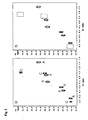
- Figure 2, panel (a), depicts the fingerprint region of the 2D DQ spectrum of mixture 1 (Ac-Tyr-Val-Asn-Val-OH (resonances A), Ac-Tyr(PO3H2)-Val-NH2 (resonances B) and Ac-(N-Me)Phe(p-CH2PO3H2)-Val-Asn-NH-CH2-CH(CH3)2 (resonances C)) in the absence of the target protein. Figure 2, panel (b), depicts the same 2D DQ fingerprint region of the mixture in the presence of the Grb2 SH2 domain. The delay τ was 42 ms long. Each of the molecules is present at a molar ratio to the target protein of 2:1 (100 µM : 50 µM). The rectangles in (b) indicate the position of the missing cross peaks.
- Figure 3, panel (a), depicts the aromatic region of the 2D DQC relaxation weighted double-quantum spectrum of mixture 1 (Ac-Tyr-Val-Asn-Val-OH (resonances A), Ac-Tyr(PO3H2)-Val-NH2 (resonances B) and Ac-(N-Me)Phe(p-CH2PO3H2)-Val-Asn-NH-CH2-CH(CH3)2 (resonances C)) in the absence of the target protein. Figure 3, panel (b) and (c) depicts the same spectral region of the 2D double-quantum and DQC relaxation weighted double-quantum spectrum of mixture 1 in the presence of the Grb2 SH2 domain, respectively. The delay τ and τ1 were 42 and 24 ms long, respectively The dashed line corresponds to the double-quantum pseudodiagonal. The direct peaks are disposed symmetrically with respect to the diagonal. The missing ligand resonances in (b) and (c) are those of the aromatic moiety of compound C.
- Figure 4, panel (a), depicts the fingerprint region of the 2D DQ spectrum of mixture 2 (Ac-Tyr-Val-Asn-Val-OH (resonances A), Ac-Tyr(PO3H2)-Val-NH2 (resonances B), indole (resonances C) and Ac-Tyr(PO3H2)-Val-NH-CH2-CH2-CONH2 (resonances D)). The resonances of indole are not visible in this spectral region. Figure 4, panel (b), depicts the same 2D DQ fingerprint region of the four compound mixture in the presence of the Grb2-SH2 domain. Figure 4, panel (c), depicts the 2D difference spectrum obtained by subtracting the two 2D DQ spectra (a) and (b). The visible signals in (c) are those of compound C that disappear in the spectrum (b) and one signal of compound B that displays a small chemical shift change in the presence of the protein. This is in indication of a possible weak interaction between compound B and the Grb2-SH2 domain. Each of the molecules is present at a molar ratio to the target protein of 6:1 (300 µM : 50 µM). The resonances of the indole are not affected by the presence of the protein (data not shown).
- Figure 5 depicts an expanded region of the 2D DQ spectrum of
mixture 2 in the absence (a) and in the presence of the protein Grb2-SH2 (b). The spectrum in (c) depicts the 2D difference spectrum obtained by subtracting the two 2D DQ spectra (a) and (b). Other experimental parameters are same as described in Figure 4. Despite the strong overlap of cross peaks A and D in (a) it is possible to identify in the 2D DQ difference spectrum (c) the cross peak that is affected by the presence of the protein. -
- The present invention provides a rapid and efficient screening procedure for identifying and providing chemical moieties that interact with a target biomolecule, for instance a therapeutic target biomolecule.
The molecules are identified by following with 2D 1H DQ or DQC (refocussed or not) relaxation weighted DQ experiments (preferentially version with PFG) the changes in either the intensities or chemical shifts of the ligand signals in the presence of the target biomolecule.
The target biomolecule is either free in solution, dissolved in detergents or micelles or, alternatively, coupled to a resin.
For the resin samples, two-dimensional Magic Angle Spinning (MAS) DQ or DQC relaxation weighted DQ experiments (preferentially version with PFG) are recorded. - The target biomolecule according to the method object of the present invention is any biomolecule which is sufficiently larger in size in comparison to the ligand and capable of binding to the selected ligand.
Suitable biomolecules are, for instance, proteins, peptides, antibodies, DNA or RNA fragments and the like.
Preferably, the target biomolecule is a protein. - For ligands in fast exchange on the NMR time scale (with KD >30-50 µM), a change in chemical shift (ΔΩ) is observed in the presence of the target biomolecule, e.g. the protein. The ΔΩ is the population-weighted average of the chemical shifts for the free and bound states of the chemical moiety. Ligands with higher affinities for the target biomolecules can exhibit intermediate exchange (KD in the few µM range) or slow exchange (KD < 1 µM). In these cases the ligand resonances will be attenuated or broadened in the 2D DQ spectra recorded in the presence of the protein.
The pulse sequences for the 2D 1H DQ and DQC relaxation weighted DQ experiments (versions with PFG) are shown in Fig. 1a and 1b, respectively.
Let us consider twospins 1 and 2 of a ligand that interacts with a target macromolecule. The two spins are scalar coupled with a J 12 coupling constant. The detected magnetization of spin 1 M 1 + (t 1 ,t 2 ) created by magnetization of spin 1 M1 + (0) with the pulse sequence of Fig. 1a and Fig. 1b is given respectively by:t t - Similar equations are derived for detected magnetization of spin 1 M + 1 (t 1 , t 2 ) created by magnetization of spin 2 M + 2(0). In deriving equations (1) and (2) we have assumed that the parallel transitions of the multiplets have the same spin-spin relaxation. In other words we have considered the 1H DD-CSA cross correlation term as negligible.
- f is a trigonometric function that represents the efficiency of the forward and backward conversion between single quantum (SQ) and double quantum coherence. For a two-spin system the trigonometric function for the echo pathway selection is 14,18-20:
- From a practical point of view, the steps involved in the detection of binding are preferably the following:
- a) mixing together from 1 to n chemical compounds not interacting with each other;
- b) recording a two-dimensional 1H DQ or DQC (refocussed or not) relaxation weighted DQ spectrum (preferentially version with PFG) for the said compound mixture in the absence of the target biomolecule;
- c) mixing the said compound mixture with the target biomolecule where each of the molecules comprising the mixture is preferably present at a molar ratio to the target biomolecule of about 1:1 to about 20:1
- d) recording a two-dimensional 1H DQ or DQC (refocussed or not) relaxation weighted DQ spectrum (preferentially version with PFG) for the said compound mixture in the presence of the target biomolecule;
- e) comparing the spectra recorded in steps b) and d) to detect which, if any of the chemical entities of the mixture, has interacted with the target biomolecule; and, optionally
- f) recording a two-dimensional 1H DQ or DQC (refocussed or not) relaxation weighted DQ spectrum (preferentially version with PFG) for each of the compounds which have been detected in step (e) above, with and without the target protein.
-
- The optional additional step (f) which involves the generation of a two dimensional proton double-quantum spectra with and without the target protein for the compound, or for each of the compounds, identified as ligand(s), might be useful in rather complex mixtures so as to confirm the finding of step (e), that is the binding between the ligand and the target biomolecule.
From the foregoing, since the method of the present invention can be applied as well to one or more, that is from 1 to n chemical moieties which could interact to the target biomolecule, it is clear to the man skilled in the art that step (f) would be particularly useful with n>1 compounds to be tested.
The method of the invention is applied to any mixture of compounds to be tested wherein said mixture preferably comprises from 1 to 10 or, even more preferably, from 1 to 5 compounds.
The present invention based on 2D DQ spectroscopy for the screening has several unique advantages which are shortly listed below. - The experiment, in particular the PFG versions recorded with β=45°/135° or β=60°/120°, represents the most sensitive 2D NMR experiment for analysis of small and medium size molecules.
- A concentration in the order of 50 to 100 µM is sufficient for obtaining good quality 2D DQ spectra in relatively short time. The utilization of the cryoprobe technology22-24 with 2D PFG DQ spectroscopy allows one to reduce currently the necessary ligand concentration to about 13 to 25 µM. Therefore, this permits the preparation of complicated mixtures comprising many molecules and avoids changes in the pH of the solution, e.g. of the aqueous buffered solution, for instance originating from the presence of organic acids or bases. The low ligand concentration needed for these experiments permits the use of hydrophobic compounds that otherwise at higher concentrations would be insoluble in aqueous solutions. Most important, the high sensitivity of the experiment coupled with the cryoprobe technology reduces the amount of precious target biomolecule, e.g. protein, needed for the screening. The protein concentration for each sample can be as low as from about 1 to about 5 µM.
- The method exploits the combined use of the two physical properties T SQ / 2 and T DQ , as shown in equations (1) and (2), and therefore it represents a sensitive tool for detection of weak ligands. The difference in T DQ for a molecule free in solution and when complexed with a macromolecule is very large.
- Good solvent suppression is obtained with the use of coherence selection gradients. Remarkable multiple solvent suppression is then achieved with coherence selection gradients tilted at the magic angle. This becomes particularly relevant in the observation of very diluted samples, as in screening by NMR where the dynamic range of the receiver needs to be maximized in an optimal way. Furthermore, the excellent multiple solvent suppression allows the preparation of the chemical mixtures stock solutions in non-deuterated solvents (i.e. DMSO, CH3OH). Another important advantage of the use of samples dissolved in H2O is the observation of the NH doublet exchangeable resonances. These signals are absent in the spectra recorded in D2O solutions. The NH proton signals are in a spectral region with no or little overlap with other signals and therefore can be used for assignment purposes in complicated mixtures. Furthermore the difference in chemical shift of a ligand NH free in solution and when hydrogen bonded to a protein is typically large whereas this difference can be very small for other protons of the same ligand. The large chemical shift difference helps in identifying weak ligands via the NH resonances either due to a more pronounced broadening deriving from a larger chemical exchange contribution to the observed T SQ / 2 and T DQ relaxation or due to a detectable chemical shift change. The latter is probably the explanation for the identification in Fig. 4 of compound Ac-Tyr(PO3H2)-Val-NH2 as a very weak ligand.
- The 2D DQ experiment is a cosine modulated experiment and therefore
maximum signal is observed in the first t1 increments. The experiment can be
recorded rapidly by acquiring only a limited number of t1 increments. Linear
prediction can then be applied in the t1 dimension for improving the resolution.
Extensive folding can also be performed in ω1 therefore reducing the spectral width and the number ot t1 increments. - Removal of the extensive overlapping present in the one-dimensional spectra
allows an easy identification, in the 2D DQ spectrum, of the signals affected by
the presence of the target biomolecule. Due to the reduced problem of
overlapping and to the complete absence of biomolecule signals, the 2D DQ
difference spectrum is less subject to artefacts when compared to the 1D
difference spectrum.
Furthermore, the absence of strong diagonal peaks and singlet resonances with consequent absence of t1 noise and the excellent solvent suppression in DQ spectra allow a more precise and reliable use of the 2D subtraction technique. - The scalar connectivities observed in the 2D DQ spectrum allow direct identification of the molecules interacting with the target biomolecule without the need to deconvolute the mixture.
- Suppression of the biomolecule signals is very efficient not only due to the presence of the excitation double quantum period and the relaxation of DQC during t1 and τ1, but also due to the antiphase character of the multiplets along ω2. The large linewidth of the protein resonances results in extensive cancellation of the positive and negative lobes along ω2.
- The NMR method of the invention, based on the newly optimized 1H Double-Quantum spectroscopy, for the identification of compounds that interact with a target biomolecule was described. The invention exploits efficiently and simultaneously the different spin-spin and Double-Quantum coherence relaxations of the compounds that interact with a macromolecule.
The high sensitivity, remarkable multiple solvent suppression, absence of diagonal peaks and singlets, reduced problems of resonance overlapping, all allow reliable application of this methodology to the screening of complex chemical mixtures against a target biomolecules. Both strong and weak ligands can be directly identified from a mixture. - The following examples are intended to better illustrate the present invention without pose any limitation to it.
- The feasibility of the invention in identifying ligands was tested with mixtures of compounds against the Grb2-SH2 domain. This represents a challenging test case due to the small size of the protein employed (MW=11839). It should be noticed that the technique performs even better with medium or large size target proteins due to the very short T SQ / 2 and T DQ for these proteins.
Figure 2 illustrates the DQ fingerprint region of a compound mixture (mixture 1) recorded with the pulse sequence of Figure 1a in the absence (a) and in the presence (b) of the protein. Mixture 1 contained a final concentration of 100 µM of Ac-Tyr-Val-Asn-Val-OH, Ac-Tyr(PO3H2)-Val-NH2 and Ac-(N-Me)Phe(p-CH2PO3H2)-Val-Asn-NH-CH2-CH(CH3)2- Close comparison of the two spectra allows rapid identification of compound Ac-(N-Me)Phe(p-CH2PO3H2)-Val-Asn-NH-CH2-CH(CH3)2 (resonances C) as a ligand for Grb2-SH2. The NH protein resonances are not visible in Figure 2 (b) due to their short T SQ / 2 and T DQ and the lower protein concentration (50 µM).
The protein aromatic resonances have a longer T SQ / 2 and T DQ compared to the amide resonances and their presence in the DQ spectrum may interfere with the observation of the ligand signals. It is possible to suppress them by employing a DQC relaxation filter as shown in Figure 1b.
Figure 3(a) illustrates the aromatic region of the 2D relaxation weighted DQ spectrum for mixture 1. Figures 3 (b) and (c) depict the same spectral region of the 2D DQ and relaxation weighted DQ spectrum of mixture 1 in the presence of the Grb2 SH2 domain, respectively. The residual aromatic protein signals in (b) are completely suppressed in (c) due to the rapid DQ relaxation of the protein resonances. The missing ligand signals in (b) and (c) are those of the aromatic moiety of compound Ac-(N-Me)Phe(p-CH2PO3H2)-Val-Asn-NH-CH2-CH(CH3)2.
It should be pointed out that for medium and large size proteins for membrane bound proteins or for proteins bound to a resin the DQC relaxation filter is not necessary. The large linewidth of all the protein resonances and the antiphase character of the DQ multiplets along ω2 results in complete cancellation of the positive and negative lobes. Nevertheless the DQC relaxation filter can be used also in the detection of very weak ligands by exploiting the large differences in T DQ for molecules interacting with a target macromolecule.
Figure 4 demonstrates the power of 2D DQ difference spectroscopy in the identification of very weak ligands. The DQ fingerprint region of Mixture 2 (300 µM of Ac-Tyr-Val-Asn-Val-OH, Ac-Tyr(PO3H2)-Val-NH2, Ac-Tyr(PO3H2)-Val-NH-CH2-CH2-CONH2 and indole) in the absence (a) and in the presence of the protein (b) recorded with the pulse sequence of Figure la is shown. Although visual comparison allows immediate identification of compound Ac-Tyr(PO3H2)-Val-NH-CH2-CH2-CONH2 (resonances D) as a potent ligand, the 2D difference spectrum (c) permits also the identification of compound Ac-Tyr(PO3H2)-Val-NH2 as a potential very weak ligand. The small chemical shift change observed for the NH of Val is about 8Hz that is equal to the value of 3JNHαH· Therefore the signal in the difference spectrum appears as a triplet of intensity -1,+2,-1. Only the compound resonances that display changes in intensity or chemical shift in the presence of the protein will be visible in the difference spectrum. The resonances of compound Ac-Tyr-Val-Asn-Val-OH (resonances A) and Indole (not present in this spectral region) that do not interact with the protein are not visible in the difference spectrum.
The absence of diagonal peaks, t1 noise and singlets and the excellent solvent suppression make the 2D DQ difference spectroscopy a very reliable technique. This can be appreciated in Figure 5 that shows an expanded region of the DQ spectra recorded formixture 2. Despite the strong overlap of cross peaks A and D in (a) it is possible to identify peak D in the DQ difference spectrum (c) as the cross peak that is affected by the presence of the protein. - All the spectra were acquired at 25°C with the pulse sequences of Figure 1 using a Varian Inova 600 MHz spectrometer equipped with a 5 mm triple-resonance inverse probe and actively shielded x, y, and z- gradient coils. The DQ spectra were collected using 8 to 32 transients for each of the 256 t1 increments (128 t1 values for both the echo and antiecho pathway selections) and with 2048 points in t2. The data were multiplied with a cosine window function in both dimensions prior to the Fourier transformation.
- The excitation DQ period τ was 42 ms long and the angle β of the detection pulse was set to 45° and 135° for the antiecho and echo coherence pathway selection, respectively. The coherence selection gradients G1 to G3 were set at the magic angle using only the x and z components of the gradients. The first two PFGs applied at the beginning and end of the excitation double-quantum period were weak gradients for radiation damping suppression. The 2D spectral differencing was performed with the software provided by Varian. The scaling factor used in the examples reported here was -1. However it can be adjusted manually for allowing small variations in concentration of the protein-ligand mixture and ligand mixture solutions.
Two different compound mixtures consisting of three and four molecules were prepared. Mixture 1 contained a final concentration of 100 µM of Ac-Tyr-Val-Asn-Val-OH, Ac-Tyr(PO3H2)-Val-NH2 and Ac-(N-Me)Phe(p-CH2PO3H2)-Val-Asn-NH-CH2-CH(CH3)2 whilemixture 2 contained 300 µM of Ac-Tyr-Val-Asn-Val-OH, Ac-Tyr(PO3H2)-Val-NH2, Ac-Tyr(PO3H2)-Val-NH-CH2-CH2-CONH2 and indole. Some of these small peptidomimetics have been shown to bind to the SH2 domain of Grb2.25-27
The peptides and related peptidomimetics have been synthesized by solid phase methodology. The protected peptide on resin was synthesized by sequential coupling of the N-Fmoc protected amino acids, some of them also protected at the side chain, starting from the C-terminal. After the last coupling the tyrosine residue was phosphorylated on solid phase by using phosphoramidite chemistry and successive oxidation. The phosphorylation was followed by acidic treatment leading to side-chain deprotection and detachment from the resin.
The SH2 domain of Grb2 was expressed in E. coli as GST fusion proteins with a recognition site for cleavage with rhinovirus Prescission Protease (Amersham Pharmacia Biotech). The protein was present in the supernatant of the cell lysate after bacterial cell disruption and centrifugation. The supernatant containing the fusion protein was loaded onto a Glutathione Sepharose resin and bacterial contaminants eliminated after washing with PBS. To remove the GST moiety the resin carrying the GST-SH2 was incubated with Prescission Protease. The SH2 domain was further purified to homogeneity using Superdex 75 gel filtration chromatography. The protein concentration for the NMR experiments was 50 µM. The samples were in aqueous (95% H2O / 5% D2O) buffered solutions (50 mM Phosphate, 100 mM NaCl pH 6.2 and 0.02% NaN3.). -
- (1) Shuker, S.B.; Hajduk, P.J.; Meadows R.P.; Fesik, S.W. Science, 1996, 274, 1531-1534.
- (2) Hajduk, P. J.; Sheppard, D. G.; Nettesheim, D. G.; Olejniczak, E. T.; Shuker, S. B.; Meadows, R. P.; Steinman, D. H.; Carrera, G. M., Jr.; Marcotte, P. M.; Severin, J.; Walter, K.; Smith, H.; Gubbins, E.; Simmer, R.; Holzman, T. F.; Morgan, D. W.; Davidsen, S. K.; Summers, J. B.; Fesik, S. W. J. Am. Chem. Soc. 1997, 119, 5818-5827.
- (3) Hajduk, P.J.; Olejniczak E.T.; Fesik, S.W. J. Am. Chem. Soc., 1997, 119, 12257-12261.
- (4) Lin, M.; Shapiro, M.J.; Wareing, J.R. J. Am. Chem. Soc. 1997, 119, 5249-5250.
- (5) Dalvit, C.; Ramage, P.; Hommel, U. J. Magn. Reson. 1998 , 131, 148-153.
- (6) Bleicher, K.; Lin, M.; Shapiro, M. J.; Wareing, J. R. J. Org. Chem. 1998 , 63, 8486-8490.
- (7) Gonnella, N.; Lin, M.; Shapiro, M. J.; Wareing, J. R.; Zhang, X. J. Magn. Reson. 1998, 131, 336-338.
- (8) Stockman, B. J. Progress in Nuclear Magnetic resonance Spectroscopy, 1998, 33, 109-151.
- (9) Moore, J.M. Analytical Biotechnology, 1999, 10, 54-58.
- (10) Hajduk, P. J.; Gerfin, T.; Bohlen, J.-M.; Haeberli, M.; Marek, D.; Fesik, S. W. J. Med. Chem., 1999, 42, 2315-2317.
- (11) Meyer, B.; Weimar, T.; Peters, T. Eur. J. Biochem. 1997, 246, 705-709.
- (12) Henrichsen, D.; Ernst, B.; Magnani, J. L.; Wang, W.-T.; Meyer, B.; Peters, T.; Angew. Chem. Int. Ed, 1999,38,98-102.
- (13) Dalvit, C.; Böhlen, J. M. J. Magn. Reson. B 1996, 111, 76-80.
- (14) Dalvit, C.; Böhlen, J. M. in Annual Reports on NMR Spectroscopy, (Webb, A.; Ed.), Academic Press, 1999, Vol. 37, pp 204-271.
- (15) Feeney, J.; Batchelor, G.; Albrand, J. P.; Roberts G. C. K. J. Magn. Reson. 1979 , 33, 519-529.
- (16) Lian L. Y.; Roberts G. C. K. in NMR of Macromolecules (G.C.K. Roberts, Ed.) Oxford University Press, 1993, pp. 153-182.
- (17) Feeney J.; Birdsall B. in NMR of Macromolecules (G.C.K. Roberts, Ed.) Oxford University Press 1993, pp. 183-215.
- (18) Braunschweiler, L.; Bodenhausen, G.; Ernst, R. R. Mol. Phys., 1983 , 48, 535-560.
- (19) Mareci, T. H.; Freeman, R. J. Magn. Reson., 1983, 51,531-535.
- (20) Ernst, R. R.; Bodenhausen, G.; Wokaun, A. Principles of Nuclear Magnetic Resonance in One and Two Dimensions, Clarendon Press, Oxford, 1987.
- (21) Konrat, R.; Sterk, H. Chem Phys. Lett., 1993, 203, 75-80.
- (22) Styles, P.; Soffe, N. F.; Scott, C. A.; Cragg, D. A.; Row, F.; White, D. J.; White, P.C.J. J. Magn. Res. 1984 , 60, 397-404.
- (23) Black, R.D.; Early, T.A.; Roemer, P.B.; Mueller, O.M.; Mogro-Campero, A.; Turner, L.G.; Johnson, G.A. Science 1993, 259, 793.
- (24) Marek, D.; Haeberli, M.; Triebe, R.; Baselgia, L.; Gerfin, T.; Schett, O.; Laukien, F. ENC97 1997; PosterP122.
- (25) Battistini, C.; Giordano, P.; De Rosa, S.; Corradi, F.; Comoglio, P.; Bardelli, A.; Intern. Appl. WO 97/30079 (Aug. 21, 1997). Priority GB application UK 9603227.1
- (26) Schiering, N.; Casale, E.; Giordano, P.; De Rosa, S.; Caccia, P.; Sgarella, L.; Bolis, G.; Battistini, C. Poster at Keystone Symposium: Temporal and Spatial Determinants of specificity in Signal Transduction, Keystone (CO) USA, March 31 - April 16, 1997.
- (27) Battistini C.; Giordano, P.; De Rosa, S.; Corradi, F.; Bardelli, A.; Gramaglia, D.; Comoglio, P. Poster at Keystone Symposium: Temporal and Spatial Determinants of specificity in Signal Transduction, Keystone (CO) USA, March 31 - April 6, 1997.
-
Claims (10)
- A method for screening compounds for their binding properties to a target biomolecule, which method comprises:i) generating a two dimensional proton double-quantum or a DQC (double-quantum coherence) relaxation weighted double-quantum spectrum of a solution comprising one or more chemical molecules at least containing two doublets ;ii) generating a two dimensional proton double-quantum or a DQC relaxation weighted double-quantum spectrum of the above solution further comprising the target biomolecule;iii) comparing spectrum in (i) with spectrum in (ii) so as to detect any differences that identify the presence of one or more chemical molecules that bind to the target biomolecule and wherein the two dimensional proton double-quantum spectrum is refocused or not.
- A method according to claim 1 comprising the additional step of generating two dimensional proton double-quantum spectra with and without the target protein for the compound, or for each of the compounds, which have been identified as ligands, to the target biomolecule.
- A method according to claim 2 wherein the differences that identify the presence of one or more chemical molecules that bind to the target biomolecule is in the chemical shift or intensity of one or more signals.
- A method according to any one of the preceding claims wherein each of the two dimensional proton double-quantum or DQC relaxation weighted double-quantum spectra are recorded with z or magic angle pulsed field gradients.
- A method according to any one of the preceding claims wherein the two dimensional proton double-quantum or DQC relaxation weighted double-quantum spectra are recorded in non-deuterated solvents.
- A method according to claim 1 wherein each test compound is present in the sample in a molar ratio to the target biomolecule of from about 1:1 to about 20:1.
- A method according to claim 1 wherein the target biomolecule is free in solution, dissolved in detergents or micelles, or coupled to a resin.
- A method according to claim 1 wherein the target biomolecule is a protein, an antibody or a DNA or RNA fragment.
- A method according to claim 8 wherein the target biomolecule is a protein.
- A method according to claim 9 wherein the protein is the SH2 domain of Grb2 protein.
Priority Applications (1)
| Application Number | Priority Date | Filing Date | Title |
|---|---|---|---|
| SI200030809T SI1224486T1 (en) | 1999-10-29 | 2000-10-19 | Use of double-quantum 1h-nmr spectroscopy for the identification of ligands interacting with target biomolecules |
Applications Claiming Priority (3)
| Application Number | Priority Date | Filing Date | Title |
|---|---|---|---|
| GB9925677 | 1999-10-29 | ||
| GBGB9925677.8A GB9925677D0 (en) | 1999-10-29 | 1999-10-29 | Use of double-quantum 1H-NMR spectroscopy for the identification of ligands interacting with target biomolecules |
| PCT/EP2000/010370 WO2001033243A1 (en) | 1999-10-29 | 2000-10-19 | Use of double-quantum 1h-nmr spectroscopy for the identification of ligands interacting with target biomolecules |
Publications (2)
| Publication Number | Publication Date |
|---|---|
| EP1224486A1 EP1224486A1 (en) | 2002-07-24 |
| EP1224486B1 true EP1224486B1 (en) | 2005-12-28 |
Family
ID=10863638
Family Applications (1)
| Application Number | Title | Priority Date | Filing Date |
|---|---|---|---|
| EP00972833A Expired - Lifetime EP1224486B1 (en) | 1999-10-29 | 2000-10-19 | Use of double-quantum 1h-nmr spectroscopy for the identification of ligands interacting with target biomolecules |
Country Status (11)
| Country | Link |
|---|---|
| EP (1) | EP1224486B1 (en) |
| JP (1) | JP2003513290A (en) |
| AT (1) | ATE314660T1 (en) |
| AU (1) | AU780921B2 (en) |
| CA (1) | CA2388216A1 (en) |
| CY (1) | CY1105315T1 (en) |
| DE (1) | DE60025246T2 (en) |
| DK (1) | DK1224486T3 (en) |
| ES (1) | ES2254245T3 (en) |
| GB (1) | GB9925677D0 (en) |
| WO (1) | WO2001033243A1 (en) |
Families Citing this family (10)
| Publication number | Priority date | Publication date | Assignee | Title |
|---|---|---|---|---|
| NO313899B1 (en) * | 2001-07-09 | 2002-12-16 | Leiv Eiriksson Nyfotek As | Method for measuring various constituents in organic and biological material |
| US7470543B2 (en) | 2002-06-05 | 2008-12-30 | Nerviano Medical Sciences S.R.L. | Methods for identifying ligands using competitive binding 1H NMR experiments |
| KR100901309B1 (en) * | 2002-06-15 | 2009-06-09 | 크리스탈지노믹스(주) | How to Select a Compound That Binds to an Active Site of a Protein |
| NO20025738D0 (en) * | 2002-11-29 | 2002-11-29 | Amersham Health As | Method |
| GB0308586D0 (en) * | 2003-04-14 | 2003-05-21 | Amersham Health R & D Ab | Method and arrangements in NMR spectroscopy |
| EP2994143A4 (en) | 2013-05-08 | 2017-02-01 | Board of Regents, The University of Texas System | Stat6 inhibitors |
| CN104181186B (en) * | 2014-09-03 | 2016-08-24 | 山西大学 | A Construction Method of 1H-NMR Fingerprint of Astragalus Injection |
| CN105823788B (en) * | 2016-05-12 | 2017-10-17 | 山西大学 | Shanxi mature vinegar1The construction method of H NMR finger-prints and application |
| CN107102021A (en) * | 2017-05-03 | 2017-08-29 | 山西广誉远国药有限公司 | The construction method and purposes of a kind of hippocampus 1H NMR finger-prints |
| CN114624276B (en) * | 2022-03-14 | 2023-03-24 | 中国科学院精密测量科学与技术创新研究院 | High-resolution solid NMR heteronuclear correlation spectrum method for hydrogen detection of half-integer quadrupole nucleus |
Family Cites Families (4)
| Publication number | Priority date | Publication date | Assignee | Title |
|---|---|---|---|---|
| US5891643A (en) * | 1995-11-14 | 1999-04-06 | Abbott Laboratories | Use of nuclear magnetic resonance to design ligands to target biomolecules |
| EP0870197B2 (en) * | 1995-11-14 | 2006-11-08 | Abbott Laboratories | Use of nuclear magnetic resonance to design ligands to target biomolecules |
| US5698401A (en) * | 1995-11-14 | 1997-12-16 | Abbott Laboratories | Use of nuclear magnetic resonance to identify ligands to target biomolecules |
| DE19649359C1 (en) * | 1996-11-28 | 1998-02-12 | Thomas Prof Dr Peters | Detecting biologically active substances in substance libraries |
-
1999
- 1999-10-29 GB GBGB9925677.8A patent/GB9925677D0/en not_active Ceased
-
2000
- 2000-10-19 JP JP2001535075A patent/JP2003513290A/en active Pending
- 2000-10-19 WO PCT/EP2000/010370 patent/WO2001033243A1/en not_active Ceased
- 2000-10-19 EP EP00972833A patent/EP1224486B1/en not_active Expired - Lifetime
- 2000-10-19 ES ES00972833T patent/ES2254245T3/en not_active Expired - Lifetime
- 2000-10-19 AT AT00972833T patent/ATE314660T1/en not_active IP Right Cessation
- 2000-10-19 DE DE60025246T patent/DE60025246T2/en not_active Expired - Lifetime
- 2000-10-19 CA CA002388216A patent/CA2388216A1/en not_active Abandoned
- 2000-10-19 AU AU11426/01A patent/AU780921B2/en not_active Ceased
- 2000-10-19 DK DK00972833T patent/DK1224486T3/en active
-
2006
- 2006-03-08 CY CY20061100322T patent/CY1105315T1/en unknown
Also Published As
| Publication number | Publication date |
|---|---|
| EP1224486A1 (en) | 2002-07-24 |
| AU1142601A (en) | 2001-05-14 |
| AU780921B2 (en) | 2005-04-28 |
| ATE314660T1 (en) | 2006-01-15 |
| DE60025246D1 (en) | 2006-02-02 |
| JP2003513290A (en) | 2003-04-08 |
| DE60025246T2 (en) | 2006-09-07 |
| WO2001033243A1 (en) | 2001-05-10 |
| GB9925677D0 (en) | 1999-12-29 |
| ES2254245T3 (en) | 2006-06-16 |
| DK1224486T3 (en) | 2006-02-13 |
| CA2388216A1 (en) | 2001-05-10 |
| CY1105315T1 (en) | 2010-03-03 |
Similar Documents
| Publication | Publication Date | Title |
|---|---|---|
| Diercks et al. | Applications of NMR in drug discovery | |
| EP0975954B2 (en) | Use of one-dimensional nuclear magnetic resonance to identify ligands to target biomolecules | |
| Bermel et al. | Novel 13C direct detection experiments, including extension to the third dimension, to perform the complete assignment of proteins | |
| Pauli et al. | Backbone and side‐chain 13C and 15N signal assignments of the α‐spectrin SH3 domain by magic angle spinning solid‐state NMR at 17.6 Tesla | |
| Lucas et al. | Measuring ligand‐protein binding using NMR diffusion experiments | |
| Schuetz et al. | Protocols for the Sequential Solid‐State NMR Spectroscopic Assignment of a Uniformly Labeled 25 kDa Protein: HET‐s (1‐227) | |
| Dalvit et al. | Fluorine-NMR competition binding experiments for high-throughput screening of large compound mixtures | |
| Eletsky et al. | A novel strategy for the assignment of side-chain resonances in completely deuterated large proteins using 13C spectroscopy | |
| Homans | NMR spectroscopy tools for structure‐aided drug design | |
| Wijesekara et al. | Fast acquisition of proton‐detected HETCOR solid‐state NMR spectra of quadrupolar nuclei and rapid measurement of NH bond lengths by frequency selective HMQC and RESPDOR pulse sequences | |
| Marin-Montesinos et al. | Selective high-resolution DNP-enhanced NMR of biomolecular binding sites | |
| Simorre et al. | Assignment of NMR spectra of proteins using triple-resonance two-dimensional experiments | |
| AU780921B2 (en) | Use of double-quantum 1H-NMR spectroscopy for the identification of ligands interacting with target biomolecules | |
| Olivieri et al. | Simultaneous detection of intra-and inter-molecular paramagnetic relaxation enhancements in protein complexes | |
| Pellecchia et al. | The 2D NMR Experiments H (C) CO~ 2 and HCCO~ 2 for Assignment and pH Titration of Carboxylate Groups in Uniformly^ 1^ 5N/^ 1^ 3C-Labeled Proteins | |
| JP2005536763A (en) | Use of fluorine NMR for high throughput screening | |
| JP2005529328A (en) | Method for identifying ligands using competitive binding 1H NMR experiments | |
| Reid et al. | Introduction to the NMR of proteins | |
| AU2005203326A1 (en) | Use of double-quantum 1H-NMR spectroscopy for the identification of ligands interacting with target biomolecules | |
| Kadavath et al. | TROSY: Principles and Applications | |
| Wüthrich et al. | Transverse relaxation–optimized NMR spectroscopy with biomacromolecular structures in solution | |
| Lapin et al. | 1H/13C/15N triple-resonance experiments for structure determinaton of membrane proteins by oriented-sample NMR | |
| US20040072211A1 (en) | Methods for identifying ligands using waterlogsy NMR | |
| Tsang et al. | [11] Isotope-edited nuclear magnetic resonance studies of fab-peptide complexes | |
| Tsang et al. | Some practical aspects of double-resonance techniques in solution-state NMR studies of high-molecular-weight systems |
Legal Events
| Date | Code | Title | Description |
|---|---|---|---|
| PUAI | Public reference made under article 153(3) epc to a published international application that has entered the european phase |
Free format text: ORIGINAL CODE: 0009012 |
|
| 17P | Request for examination filed |
Effective date: 20020510 |
|
| AK | Designated contracting states |
Kind code of ref document: A1 Designated state(s): AT BE CH CY DE DK ES FI FR GB GR IE IT LI LU MC NL PT SE |
|
| AX | Request for extension of the european patent |
Free format text: AL PAYMENT 20020510;LT PAYMENT 20020510;LV PAYMENT 20020510;MK PAYMENT 20020510;RO PAYMENT 20020510;SI PAYMENT 20020510 |
|
| RIN1 | Information on inventor provided before grant (corrected) |
Inventor name: DALVIT, CLAUDIO Inventor name: CACCIA, PAOLO Inventor name: PEVARELLO, PAOLO Inventor name: BATTISTINI, CARLO Inventor name: GIORDANO, PATRIZIA Inventor name: SUNDSTROEM, MICHAEL Inventor name: TATO', MARCO |
|
| 17Q | First examination report despatched |
Effective date: 20040428 |
|
| GRAP | Despatch of communication of intention to grant a patent |
Free format text: ORIGINAL CODE: EPIDOSNIGR1 |
|
| GRAS | Grant fee paid |
Free format text: ORIGINAL CODE: EPIDOSNIGR3 |
|
| GRAA | (expected) grant |
Free format text: ORIGINAL CODE: 0009210 |
|
| AK | Designated contracting states |
Kind code of ref document: B1 Designated state(s): AT BE CH CY DE DK ES FI FR GB GR IE IT LI LU MC NL PT SE |
|
| AX | Request for extension of the european patent |
Extension state: AL LT LV MK RO SI |
|
| PG25 | Lapsed in a contracting state [announced via postgrant information from national office to epo] |
Ref country code: IT Free format text: LAPSE BECAUSE OF FAILURE TO SUBMIT A TRANSLATION OF THE DESCRIPTION OR TO PAY THE FEE WITHIN THE PRESCRIBED TIME-LIMIT;WARNING: LAPSES OF ITALIAN PATENTS WITH EFFECTIVE DATE BEFORE 2007 MAY HAVE OCCURRED AT ANY TIME BEFORE 2007. THE CORRECT EFFECTIVE DATE MAY BE DIFFERENT FROM THE ONE RECORDED. Effective date: 20051228 |
|
| REG | Reference to a national code |
Ref country code: GB Ref legal event code: FG4D |
|
| REG | Reference to a national code |
Ref country code: CH Ref legal event code: EP |
|
| REG | Reference to a national code |
Ref country code: IE Ref legal event code: FG4D |
|
| REF | Corresponds to: |
Ref document number: 60025246 Country of ref document: DE Date of ref document: 20060202 Kind code of ref document: P |
|
| REG | Reference to a national code |
Ref country code: DK Ref legal event code: T3 |
|
| REG | Reference to a national code |
Ref country code: SE Ref legal event code: TRGR |
|
| REG | Reference to a national code |
Ref country code: CH Ref legal event code: NV Representative=s name: E. BLUM & CO. PATENTANWAELTE |
|
| REG | Reference to a national code |
Ref country code: GR Ref legal event code: EP Ref document number: 20060400620 Country of ref document: GR |
|
| REG | Reference to a national code |
Ref country code: ES Ref legal event code: FG2A Ref document number: 2254245 Country of ref document: ES Kind code of ref document: T3 |
|
| LTIE | Lt: invalidation of european patent or patent extension |
Effective date: 20051228 |
|
| RAP2 | Party data changed (patent owner data changed or rights of a patent transferred) |
Owner name: PFIZER ITALIA S.R.L. |
|
| ET | Fr: translation filed | ||
| NLT2 | Nl: modifications (of names), taken from the european patent patent bulletin |
Owner name: PFIZER ITALIA S.R.L. Effective date: 20060802 |
|
| PG25 | Lapsed in a contracting state [announced via postgrant information from national office to epo] |
Ref country code: MC Free format text: LAPSE BECAUSE OF NON-PAYMENT OF DUE FEES Effective date: 20061031 |
|
| PLBE | No opposition filed within time limit |
Free format text: ORIGINAL CODE: 0009261 |
|
| STAA | Information on the status of an ep patent application or granted ep patent |
Free format text: STATUS: NO OPPOSITION FILED WITHIN TIME LIMIT |
|
| 26N | No opposition filed |
Effective date: 20060929 |
|
| PGFP | Annual fee paid to national office [announced via postgrant information from national office to epo] |
Ref country code: DK Payment date: 20070920 Year of fee payment: 8 |
|
| REG | Reference to a national code |
Ref country code: CH Ref legal event code: PFA Owner name: PHARMACIA ITALIA S.P.A. Free format text: PHARMACIA ITALIA S.P.A.#VIA ROBERT KOCH 1.2#20152 MILANO (IT) -TRANSFER TO- PHARMACIA ITALIA S.P.A.#VIA ROBERT KOCH 1.2#20152 MILANO (IT) |
|
| PGFP | Annual fee paid to national office [announced via postgrant information from national office to epo] |
Ref country code: FI Payment date: 20070928 Year of fee payment: 8 |
|
| PGFP | Annual fee paid to national office [announced via postgrant information from national office to epo] |
Ref country code: NL Payment date: 20070920 Year of fee payment: 8 |
|
| PGFP | Annual fee paid to national office [announced via postgrant information from national office to epo] |
Ref country code: CH Payment date: 20071009 Year of fee payment: 8 Ref country code: AT Payment date: 20070918 Year of fee payment: 8 |
|
| PGFP | Annual fee paid to national office [announced via postgrant information from national office to epo] |
Ref country code: SE Payment date: 20071005 Year of fee payment: 8 Ref country code: BE Payment date: 20071102 Year of fee payment: 8 |
|
| PGFP | Annual fee paid to national office [announced via postgrant information from national office to epo] |
Ref country code: GR Payment date: 20071025 Year of fee payment: 8 Ref country code: PT Payment date: 20071010 Year of fee payment: 8 |
|
| PGFP | Annual fee paid to national office [announced via postgrant information from national office to epo] |
Ref country code: LU Payment date: 20080401 Year of fee payment: 8 |
|
| PGFP | Annual fee paid to national office [announced via postgrant information from national office to epo] |
Ref country code: CY Payment date: 20071015 Year of fee payment: 8 |
|
| REG | Reference to a national code |
Ref country code: PT Ref legal event code: MM4A Free format text: LAPSE DUE TO NON-PAYMENT OF FEES Effective date: 20090420 |
|
| BERE | Be: lapsed |
Owner name: *PHARMACIA ITALIA S.P.A. Effective date: 20081031 |
|
| REG | Reference to a national code |
Ref country code: CH Ref legal event code: PL |
|
| REG | Reference to a national code |
Ref country code: DK Ref legal event code: EBP |
|
| EUG | Se: european patent has lapsed | ||
| NLV4 | Nl: lapsed or anulled due to non-payment of the annual fee |
Effective date: 20090501 |
|
| PG25 | Lapsed in a contracting state [announced via postgrant information from national office to epo] |
Ref country code: FI Free format text: LAPSE BECAUSE OF NON-PAYMENT OF DUE FEES Effective date: 20081019 Ref country code: NL Free format text: LAPSE BECAUSE OF NON-PAYMENT OF DUE FEES Effective date: 20090501 |
|
| PG25 | Lapsed in a contracting state [announced via postgrant information from national office to epo] |
Ref country code: PT Free format text: LAPSE BECAUSE OF NON-PAYMENT OF DUE FEES Effective date: 20090420 Ref country code: AT Free format text: LAPSE BECAUSE OF NON-PAYMENT OF DUE FEES Effective date: 20081019 |
|
| REG | Reference to a national code |
Ref country code: SI Ref legal event code: KO00 Effective date: 20090602 |
|
| PG25 | Lapsed in a contracting state [announced via postgrant information from national office to epo] |
Ref country code: BE Free format text: LAPSE BECAUSE OF NON-PAYMENT OF DUE FEES Effective date: 20081031 |
|
| PG25 | Lapsed in a contracting state [announced via postgrant information from national office to epo] |
Ref country code: CH Free format text: LAPSE BECAUSE OF NON-PAYMENT OF DUE FEES Effective date: 20081031 Ref country code: LI Free format text: LAPSE BECAUSE OF NON-PAYMENT OF DUE FEES Effective date: 20081031 Ref country code: CY Free format text: LAPSE BECAUSE OF NON-PAYMENT OF DUE FEES Effective date: 20081019 Ref country code: DK Free format text: LAPSE BECAUSE OF NON-PAYMENT OF DUE FEES Effective date: 20081031 |
|
| PG25 | Lapsed in a contracting state [announced via postgrant information from national office to epo] |
Ref country code: GR Free format text: LAPSE BECAUSE OF NON-PAYMENT OF DUE FEES Effective date: 20090505 |
|
| PGFP | Annual fee paid to national office [announced via postgrant information from national office to epo] |
Ref country code: DE Payment date: 20091028 Year of fee payment: 10 Ref country code: IE Payment date: 20091026 Year of fee payment: 10 Ref country code: ES Payment date: 20091026 Year of fee payment: 10 |
|
| REG | Reference to a national code |
Ref country code: GB Ref legal event code: 732E Free format text: REGISTERED BETWEEN 20100107 AND 20100113 |
|
| REG | Reference to a national code |
Ref country code: FR Ref legal event code: TP |
|
| PGFP | Annual fee paid to national office [announced via postgrant information from national office to epo] |
Ref country code: GB Payment date: 20091026 Year of fee payment: 10 Ref country code: IT Payment date: 20091026 Year of fee payment: 10 Ref country code: FR Payment date: 20091029 Year of fee payment: 10 |
|
| PG25 | Lapsed in a contracting state [announced via postgrant information from national office to epo] |
Ref country code: SE Free format text: LAPSE BECAUSE OF NON-PAYMENT OF DUE FEES Effective date: 20081020 Ref country code: LU Free format text: LAPSE BECAUSE OF NON-PAYMENT OF DUE FEES Effective date: 20081019 |
|
| GBPC | Gb: european patent ceased through non-payment of renewal fee |
Effective date: 20101019 |
|
| PG25 | Lapsed in a contracting state [announced via postgrant information from national office to epo] |
Ref country code: FR Free format text: LAPSE BECAUSE OF NON-PAYMENT OF DUE FEES Effective date: 20101102 |
|
| REG | Reference to a national code |
Ref country code: FR Ref legal event code: ST Effective date: 20110630 |
|
| PG25 | Lapsed in a contracting state [announced via postgrant information from national office to epo] |
Ref country code: GB Free format text: LAPSE BECAUSE OF NON-PAYMENT OF DUE FEES Effective date: 20101019 |
|
| REG | Reference to a national code |
Ref country code: DE Ref legal event code: R119 Ref document number: 60025246 Country of ref document: DE Effective date: 20110502 |
|
| PG25 | Lapsed in a contracting state [announced via postgrant information from national office to epo] |
Ref country code: IE Free format text: LAPSE BECAUSE OF NON-PAYMENT OF DUE FEES Effective date: 20101019 |
|
| REG | Reference to a national code |
Ref country code: ES Ref legal event code: FD2A Effective date: 20111118 |
|
| PG25 | Lapsed in a contracting state [announced via postgrant information from national office to epo] |
Ref country code: IT Free format text: LAPSE BECAUSE OF NON-PAYMENT OF DUE FEES Effective date: 20101019 |
|
| PG25 | Lapsed in a contracting state [announced via postgrant information from national office to epo] |
Ref country code: ES Free format text: LAPSE BECAUSE OF NON-PAYMENT OF DUE FEES Effective date: 20101020 |
|
| PG25 | Lapsed in a contracting state [announced via postgrant information from national office to epo] |
Ref country code: DE Free format text: LAPSE BECAUSE OF NON-PAYMENT OF DUE FEES Effective date: 20110502 |