Technical Field
The present invention relates to transporter families, proteins
involved in the transport of substances from the outside to the inside
of cells and vice versa.
Background Art
Recently, the involvement of various transporters localized on
the plasma membrane in the uptake system for nutrients and endogenous
substances into cells and their transport mechanisms have been
clarified (Tsuji, A. and Tamai, I., Pharm. Res., 13, 963-977, 1996).
These transporters recognize the structures of substances to be
transported to selectively transport specific substances across
biological membranes. Transporters that recognize structures of a
relatively wide range may possibly recognize foreign substances, such
as drugs, by mistake, and actively take in them into cells. It is
believed that drugs permeate through the plasma membrane fundamentally
by simple diffusion, depending on their physicochemical properties
such as molecular size, fat-solubility, and hydrogen-binding capacity.
Particularly, according to the pH partition hypothesis, in the case
of ionic drugs, only molecules in the non-dissociated form can permeate
through the plasma membrane. However, it has become evident that a
number of drugs penetrate through the cell membrane by a specific
mechanism other than simple diffusion, that is, an active transport
mediated by transporters, particularly in organs that require
efficient exchange of intracellular and extracellular substances,
including small intestine, uriniferous tubule, placenta, epithelial
cells of choroid plexus, hepatocytes, and blood-brain barrier (Tamai,
I. and Tsuji, A., Pharmacia, 31, 493-497, 1995; Saito, H. and Inui,
K., Igaku no Ayumi, 179, 393-397, 1996; Tamai, I., Yakubutsu Dotai
(Pharmacokinetics), 11, 642-650, 1996). For example, it is known that
although oral β-lactam antibiotics of the non-esterified type are
amphoteric or negatively charged in physiological pHs and sparingly
soluble in fat, they are readily absorbed through the intestine. A
transport study using the isolated membrane-vesicles system
demonstrated that an H+-driven peptide transporter localized on the
brush-border membrane is involved in the absorption process of these
drugs (Okano, T. et al., J. Biol. Chem. 261, 14130-14134, 1986).
Although the specificity of a peptide transport system in terms of
the peptide size is so strict as to recognize di- or tri-peptides
but not tetrapeptides or larger peptides, it has a rather broad substrate
specificity so as to recognize peptides comprising non-natural amino
acids. The peptide transporter mistakenly mediates transport of
β-lactam antibiotics due to its broad substrate specificity. This
property has been unexpectedly utilized in the clinical field (Tsuji,
A., American Chemical Society (eds. Taylor, M. D., Amidon, G. L.),
Washington, D. C., 101-134, 1995). Furthermore, it has been reported
that a transporter is possibly also involved in permeation of substances
with a high fat-solubility such as fatty acids through the plasma
membrane (Schaffer, J. and Lodish, H., Cell, 79, 427-436, 1994).
Since various transporters are presumed to be distributed in organs
and cells based on the physiological roles of the organs and cells,
their distribution and functions may be specific to organs. Therefore,
transporters are expected to be used to impart an organ specificity
to pharmacokinetics. In other words, an organ-specific drug delivery
system (DDS) can be constructed utilizing transporters. If drug
absorption that relies solely on simple diffusion is improved by
elevating its fat-solubility, the effect of the drug obtained in the
initial transport in the liver can be enhanced and the drug can
non-specifically translocate into any organ. In addition, it would
also be possible to increase the drug absorption independently of
its fat-solubility by designing the drug based on the substrate
specificity of transporters (Hayashi, K. et al., Drug Delivery System,
11, 205-213, 1996). For this purpose, it is necessary to identify
various transporters at the molecular level and analyze their
properties in detail. However, molecular level identification is
greatly behind studies on membrane physiology because the transporters
are difficult to handle biochemically and require complicated
processes in their functional assays.
Recently, cDNAs of several transporters have been cloned by the
expression cloning method using Xenopus oocytes, a foreign gene
expression system, and the structural homology among them has been
revealed (Fei, Y.-J. et al. , Nature, 368, 563-566, 1994). For example,
Koepsell et al. cloned an organic cation transporter, OCT1, which
is presumed to be localized on a basement membrane, using the expression
cloning method in 1994 (Grundemann, D. et al., Nature, 372, 549-552,
1994). Subsequently, OCT2 was identified by homology cloning based
on the sequence of OCT1 (Okuda, M. et al., Biochem. Biophys. Res.
Commun., 224, 500-507, 1996). OCT1 and OCT2 show homology as high
as 67% to each other (Grundemann, D. et al., J. Biol. Chem., 272,
10408-10413, 1997). Both of them are intensely expressed in the kidney,
but differ in the organ distribution; OCT1 is also expressed in the
liver, colon, and small intestine, while OCT2 expression is specific
to the kidney.
In addition, another transporter, the human OATP transporter
(hereinafter, referred to as "OATP-A"; Gastroenterology 109 (4),
1274-1282 (1995)), has been reported. This transporter is a protein
capable of transporting various endogenous and foreign substances
in a sodium ion-independent manner. Known substances transported by
OATP-A include bromosulfophthalein, bile acids, steroid hormones,
etc. Since PGT, a transporter capable of transporting prostaglandins,
also shows significant homology to OATP-A, genes encoding these
transporters are thought to form a gene family (the OATP family).
Only a few reports are available on identifications of
transporters at the molecular level, including above reports, and
it is believed that many unidentified transporters exist that can
be clinically useful.
Disclosure of the Invention
It is an object of this invention to provide novel transporter
genes belonging to the OATP family, proteins encoded by these genes,
and use of the genes and proteins.
In order to find genes encoding novel transporters, the present
inventors performed a tBLASTn search of the human EST (Expressed
Sequence Tag) database (URL:
http://www.ncbi.nlm.nih.gov/blast/blast.cgi) in NCBI (National
Center for Biotechnology Information in U.S.A., URL:
http://www.ncbi.nlm.nih.gov/index.html) with the protein sequence
of human OATP transporter (Gastroenterology 109 (4), 1274-1282 (1995))
as a query. As a result, several ESTs which may encode amino acid
sequences having significant homologies with the human OATP-A protein
were found. Next, further searches of the database with these EST
sequences as the query revealed that all of the ESTs are derived from
genes of unknown functions, with the exception of those which were
clearly judged to be derived from the known human OATP-A gene and
human prostaglandin transporter gene (J. Clin. Invest. 98 (5),
1142-1149 (1996)) (hereinafter, abbreviated as PGT). This finding
indicated that these ESTs are derived from transporter genes which
have not been identified so far. Therefore, cloning of the full-length
cDNA was performed by screening cDNA libraries with these EST sequences,
using PCR and plaque hybridization methods, which resulted in the
successful cloning of four genes encoding four novel transporter-like
proteins. Since all of the proteins encoded by these genes have
significant homologies with the human OATP-A protein, the genes have
been designated as OATP-B, C, D and E, respectively. Thus, the present
inventors discovered that all of the ESTs found by the tBLASTn search
are those derived from human OATP-A, B, C, D and E as well as human
PGT gene.
As described above, it has been known that the human OATP-A is
a transporter protein capable of transporting a variety of endogenous
and foreign substances, including bromosulfophthalein, bile acids,
steroid hormones, etc., in a sodium ion-independent manner, and that
the PGT protein capable of transporting prostaglandins shows a
significant homology with the OATP-Aprotein. These transporter genes
have been thought to form a family of genes (the OATP family) potentially
involved in the removal of substances unnecessary for living bodies
and regulation of concentrations of a variety of substances in vivo
(J. Biol. Chem., 1998 Aug. 28; 273 (35): 22395-401).
It is presumed that novel members of the OATP family found in
the present invention also share similar functions to OATP-A and PGT,
in the regulation of in vivo concentrations of substances essential
or unnecessary to living bodies. Furthermore, the present inventors
also demonstrated a capability of the OATP-C protein to transport
drugs, such as β-lactam antibiotics. According to these facts, it
was presumed that drugs which are originally foreign substances to
living bodies may possibly be taken up into or excreted from cells
in a manner mediated by the OATP family proteins. Therefore, it may
be possible to control pharmacodynamics and speedily design or screen
drugs with a higher absorbability by utilizing the transport
specificity, such as substrate specificity, and distribution pattern
in vivo of the OATP-family proteins. In particular, the present
inventors discovered, by analysis using RT-PCR method, that OATP-E
is highly expressed in a variety of solid cancer cells but seldom
in hemocytes. Thus, anticancer agents may be obtained by constructing
a screening system using the OATP-E gene to screen compounds which
are specifically transported into cells mediated by the OATP-E protein.
Such agents are expected to have reduced cytotoxicity to hemocytes.
In addition, if the OATP family proteins are involved in the
control of pharmacodynamics of drugs in vivo, the pharmacodynamics
are expected to be modified by genetic polymorphism of the proteins.
It has been already known that the genetic polymorphism such as the
single nucleotide polymorphism (SNP) causes differences in gene
expression level and in the amino acid sequences encoded among
individuals (Nat. Genet. 1999 Jul. 22 (3): 231-8; Nat Genet., 1999
Jul. 22 (3) : 239-47). It is expected that the genetic polymorphism
in OATP family genes causes differences in transport specificities,
such as transport activity and substrate specificity, of OATP proteins
among individuals, leading to individual differences in in vivo
pharmacodynamics of drugs and such which are controlled by OATP family
proteins. In fact, it is presumed that such differences among
individuals may lead to differences in effectiveness and reactivity
of particular drugs. Thus, it is postulated that examination of the
polymorphism, such as SNP, in OATP family genes in detail, particularly
the accumulation of information on relationship between genotype and
phenotype (reactivity to drugs) of these genes, can enable the
prediction of the reactivity of individuals to drugs by performing
genetic diagnosis of genotype of these genes in a particular individual.
In fact, the present inventors found SNPs with the following
three types of amino acid mutations in normal individuals during the
cloning process of OATP family genes:
- polymorphism in the 486th codon of OATP-B gene (tct:Ser or
ttt:Phe),
- polymorphism in the 130th codon of OATP-C gene (aat:Asn or
gat:Asp), and
- polymorphism in the 174th codon of OATP-C gene (gtg:Val or
gcg:Ala).
It is presumed that additional polymorphisms, besides those
described herein, exist in the OATP gene family to be related to
phenotypes.
In addition, in view of such critical roles of the OATP family
proteins in vivo, it is presumed that disorders exist which are caused
by deficiencies in transport functions due to gene mutations of these
proteins. In fact, it has been reported that the gene mutation in
the OCTN2 transporter, which is one of the organic cation transporters,
causes systemic carnitine deficiency (SCD) (Nat. Genet. 1999 Jan;
21 (1) : 91-4), proving the actual existence of genetic disorders caused
by the mutation of transporter genes. Genetic diagnosis based on direct
examination of causative transporter genes is clinically very
important for such genetic disorders caused by the mutations in the
transporter genes.
Genetic diagnosis to detect polymorphisms and mutations in genes
of the OATP family is made possible by the structure of the OATP family
genes revealed in this invention. Specifically, genetic diagnosis
can be made using genes of the OATP family themselves or synthetic
oligonucleotides prepared from the nucleotide sequences thereof as
the primers for PCR. In addition, it has recently become possible
to more conveniently detect structures or expression levels of genes
by techniques referred to as DNA chip or DNA microarray techniques
(Nat. Genet. 1999 volume 21 Supplement pp 1-60; Science 1999 Jan 1;
283 (5398): 83-7). Such methods can be also carried out using genes
of the OATP family themselves or synthetic oligonucleotides prepared
from nucleotide sequences thereof.
Accordingly, this invention relates to novel transporters OATP-B,
C, D, and E, genes encoding these transporters, and uses thereof.
More specifically, the present invention provides:
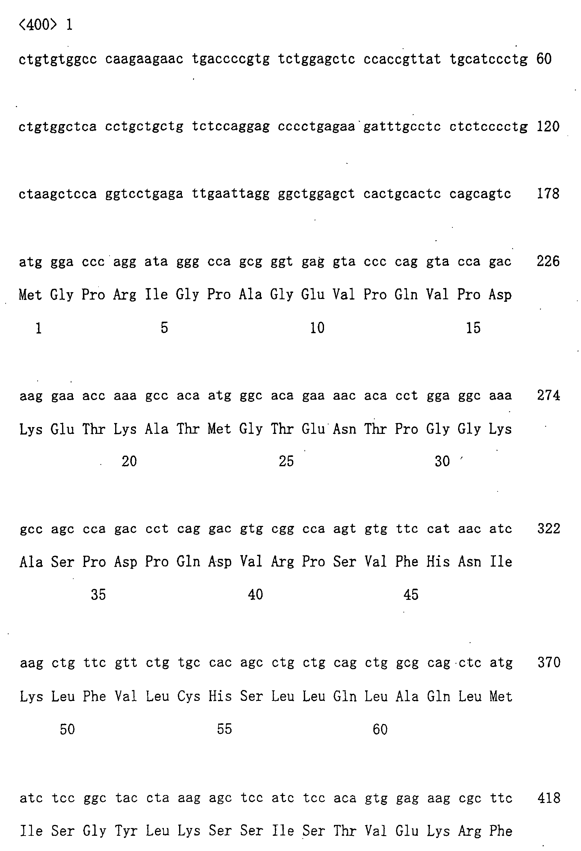
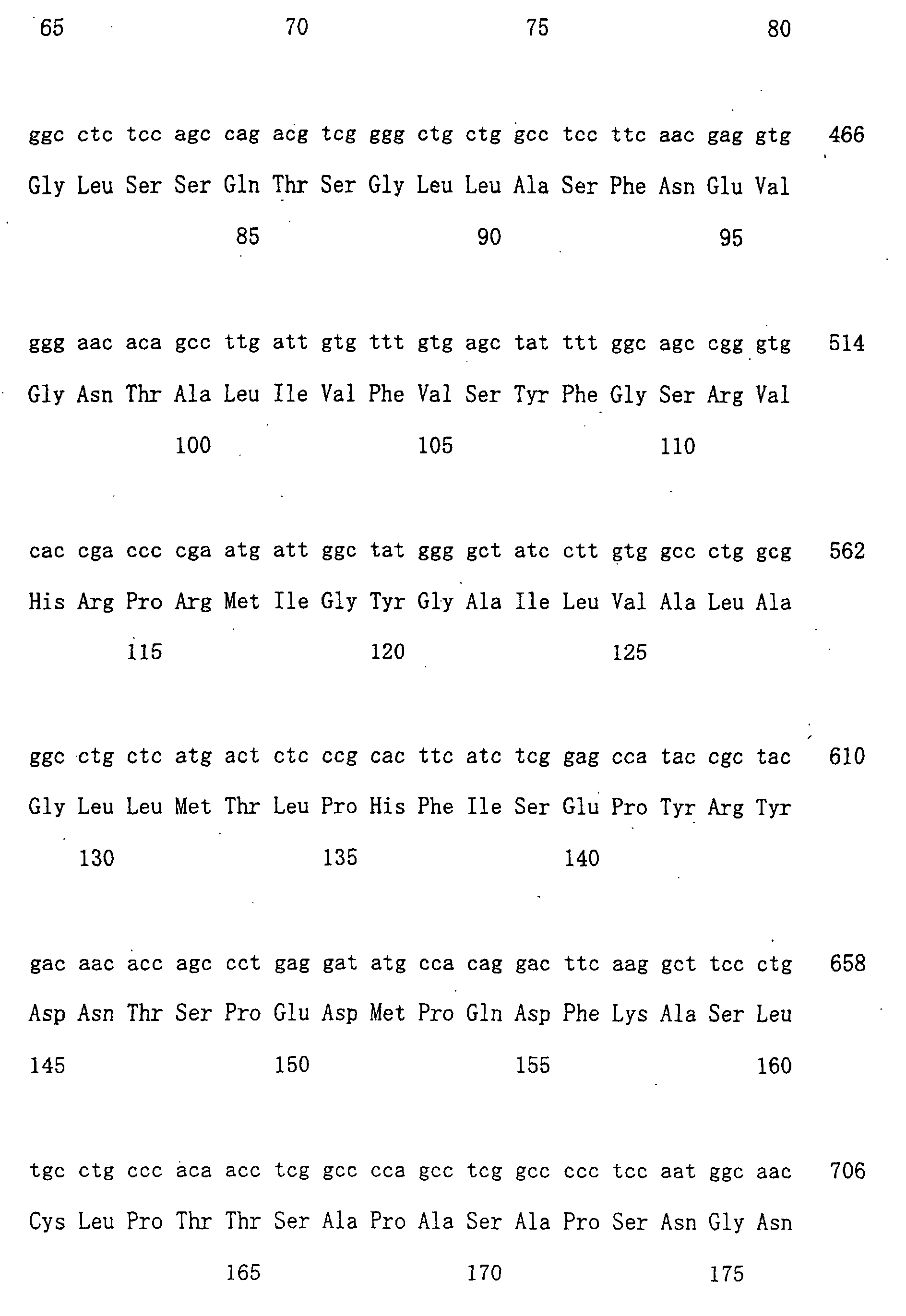
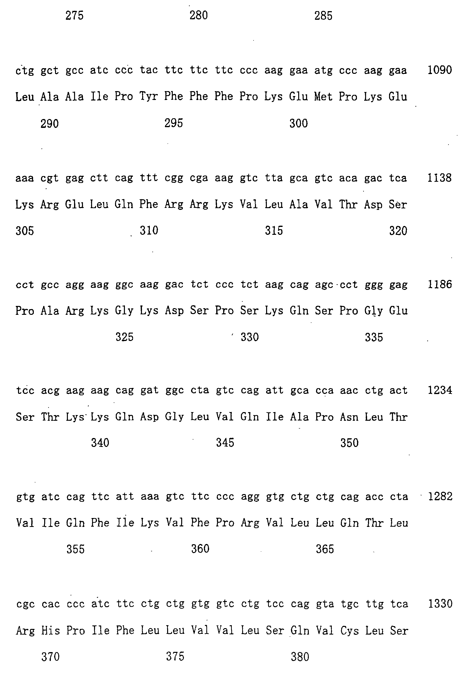
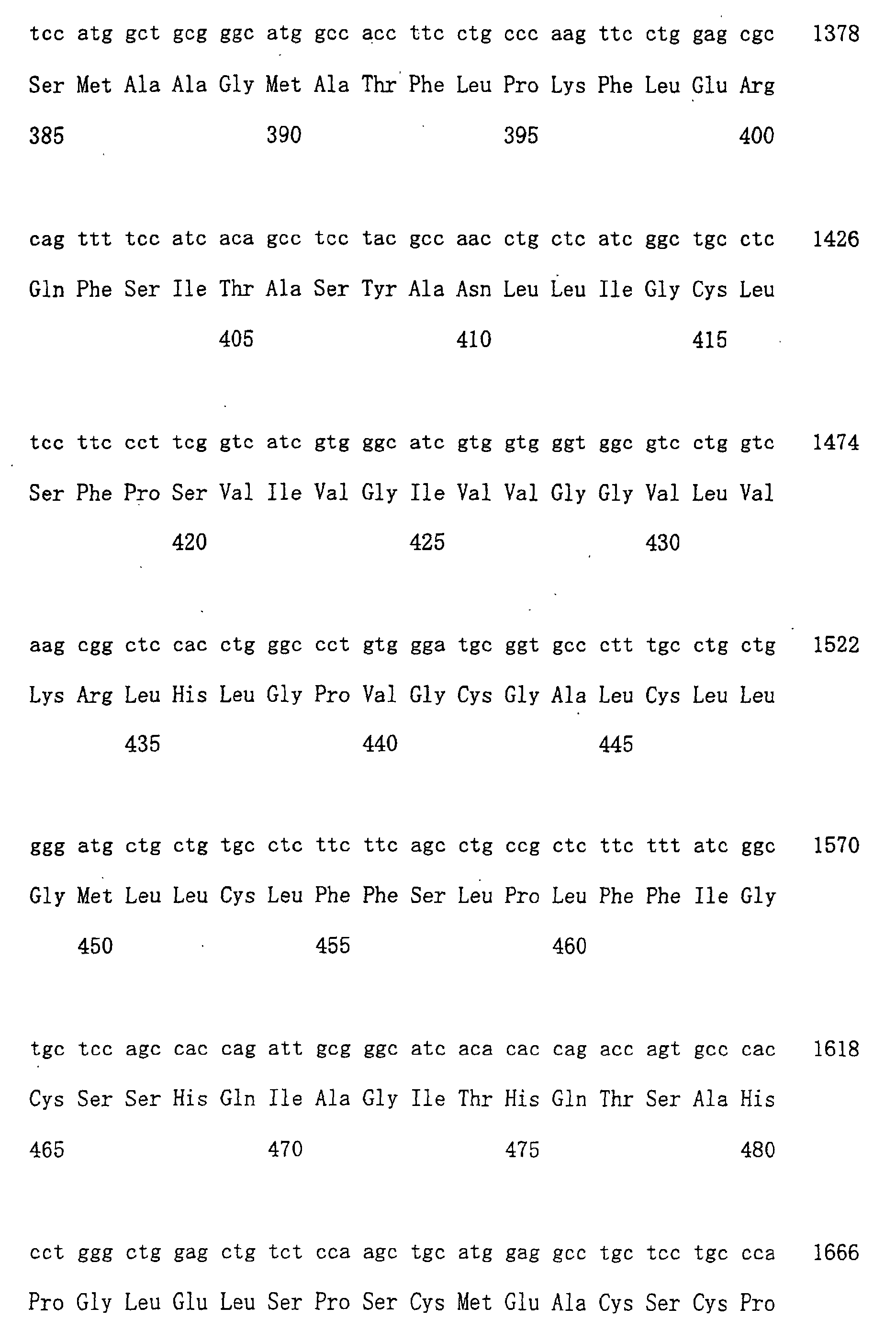
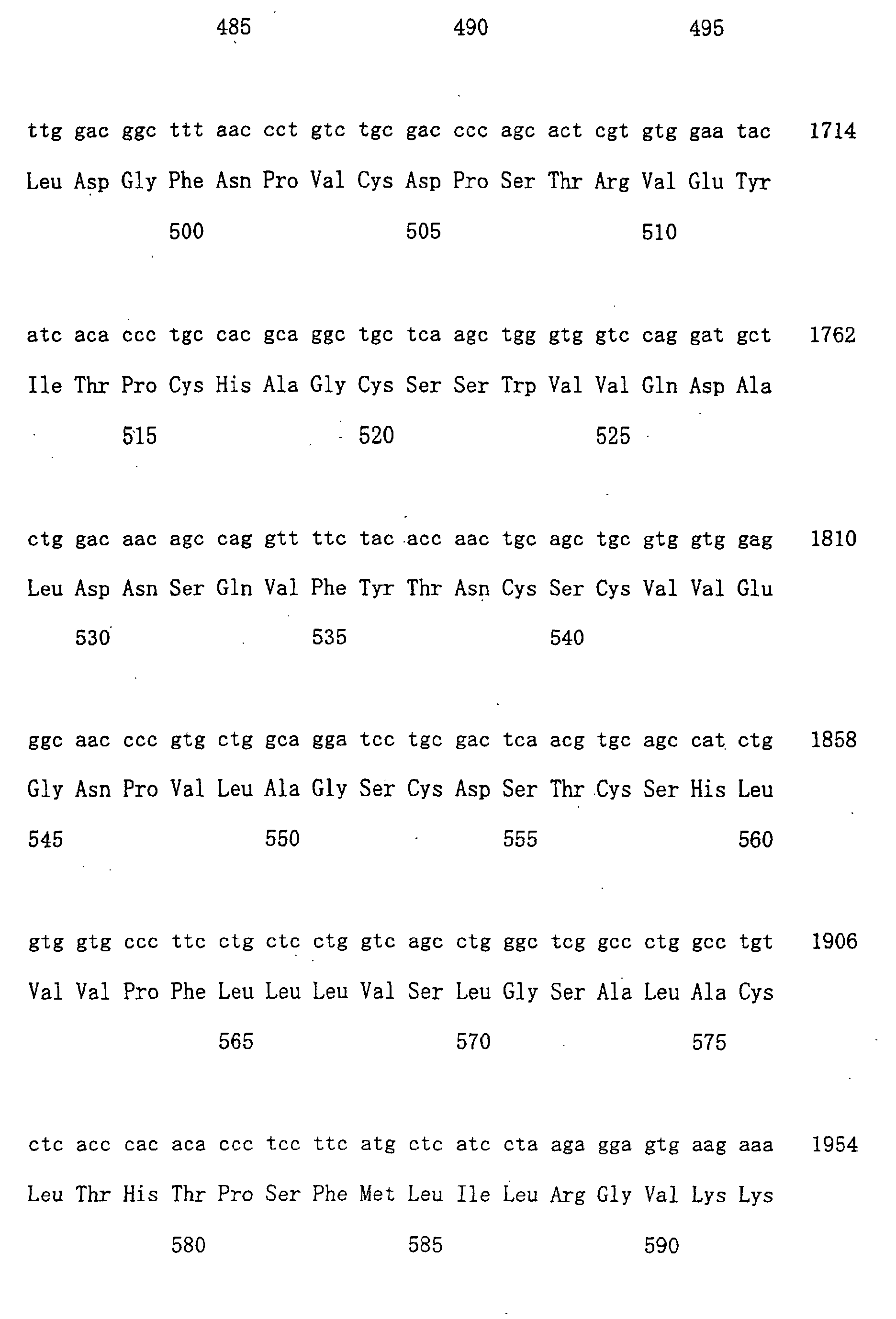
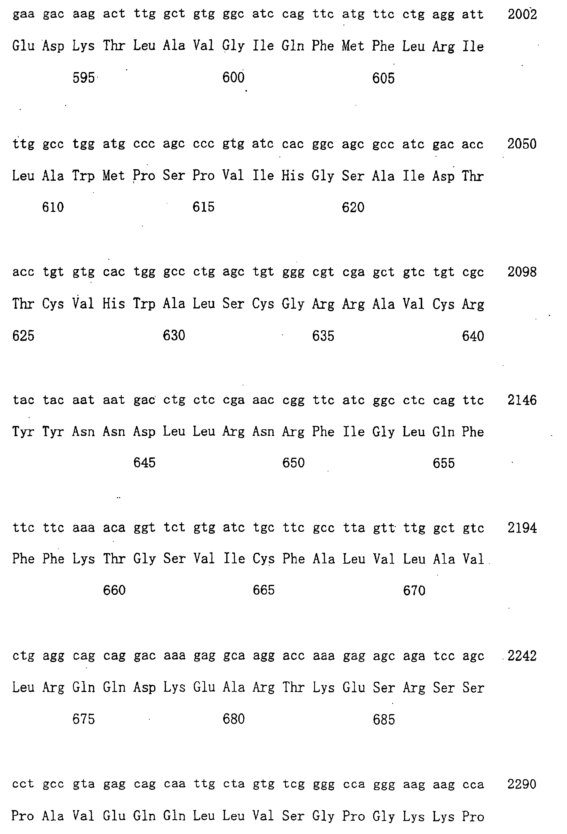
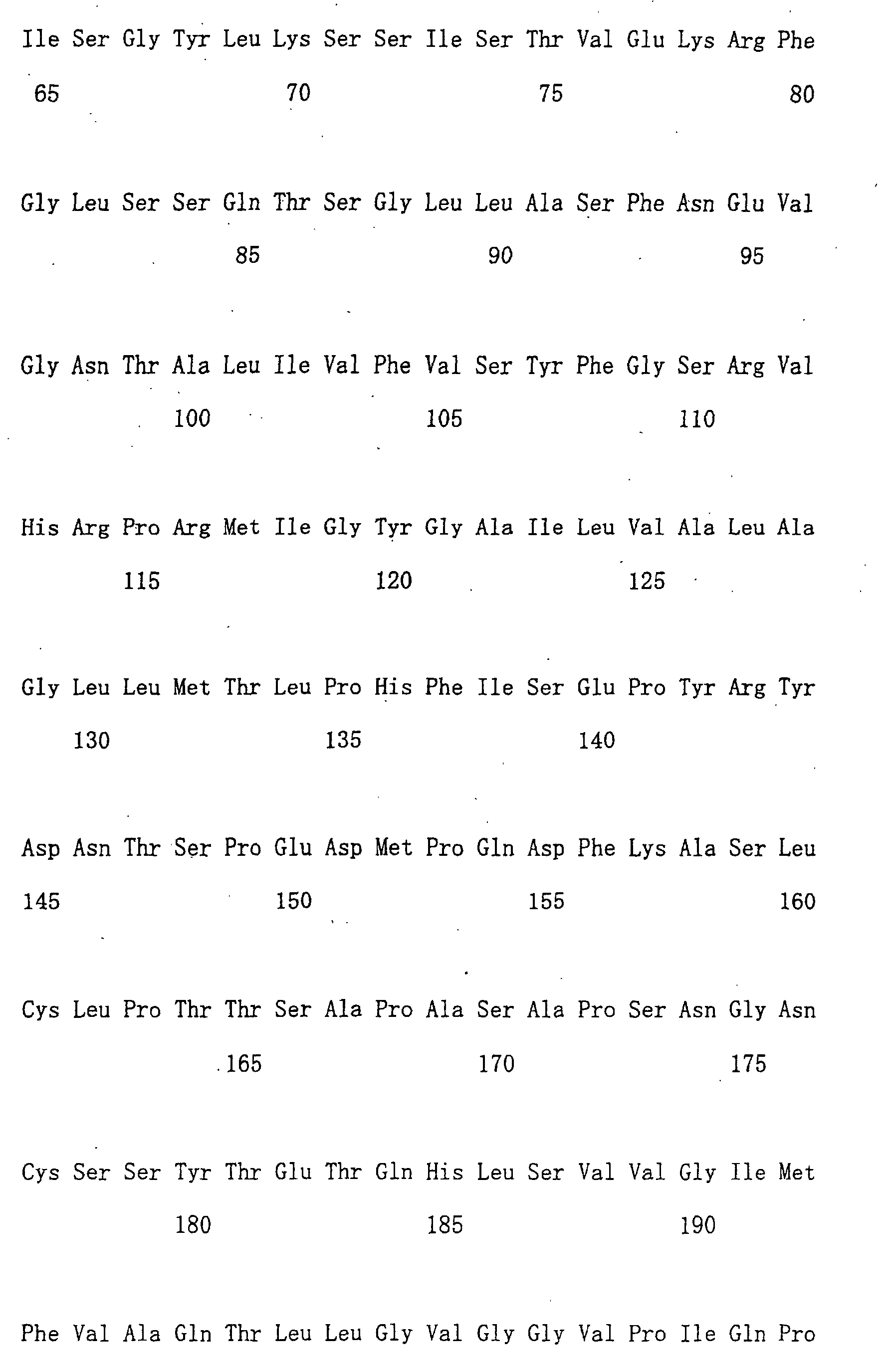
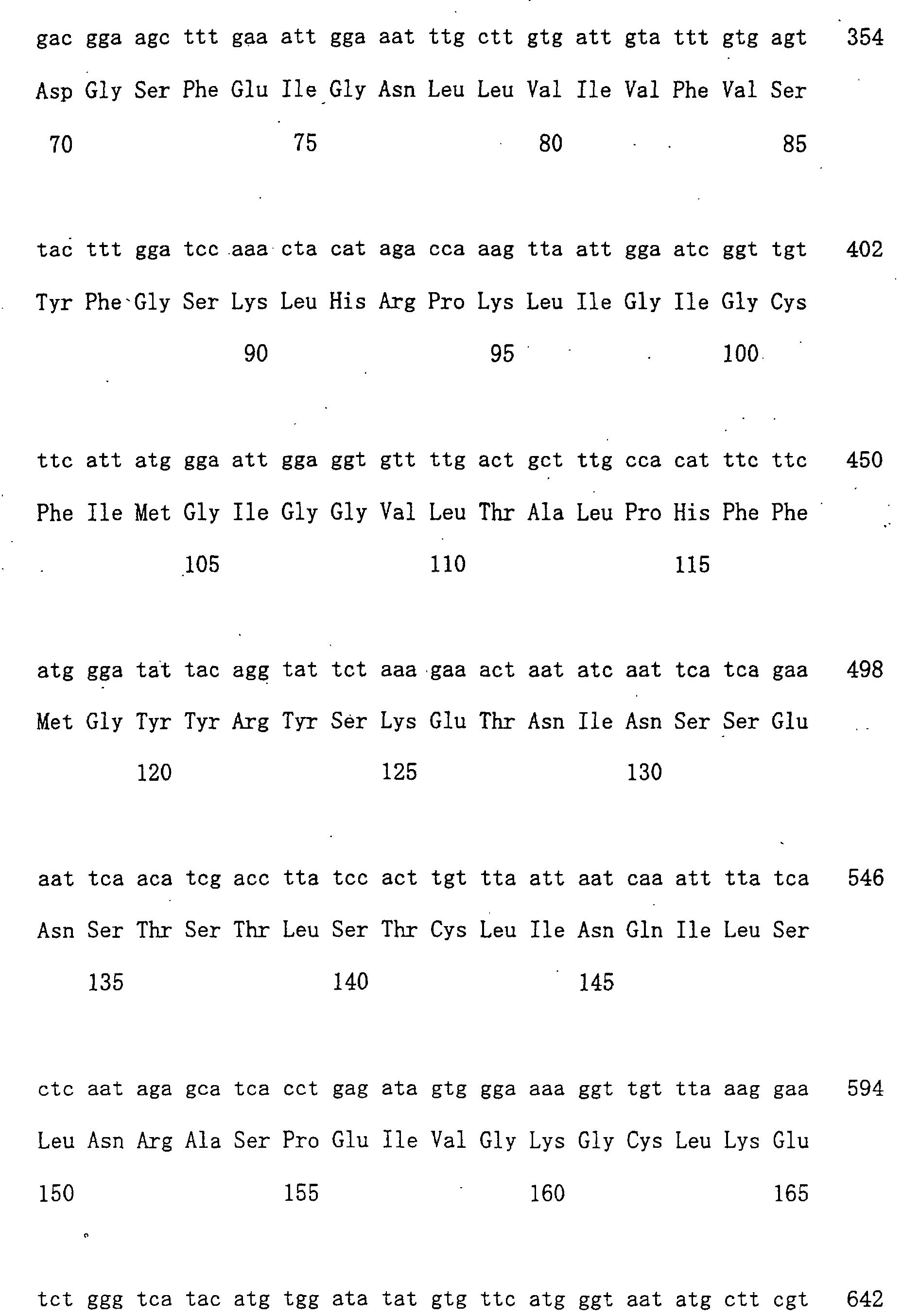
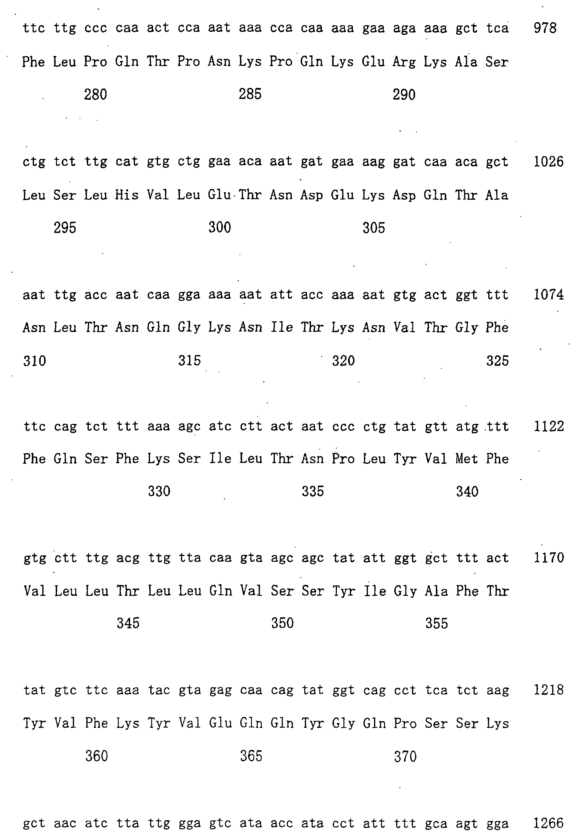
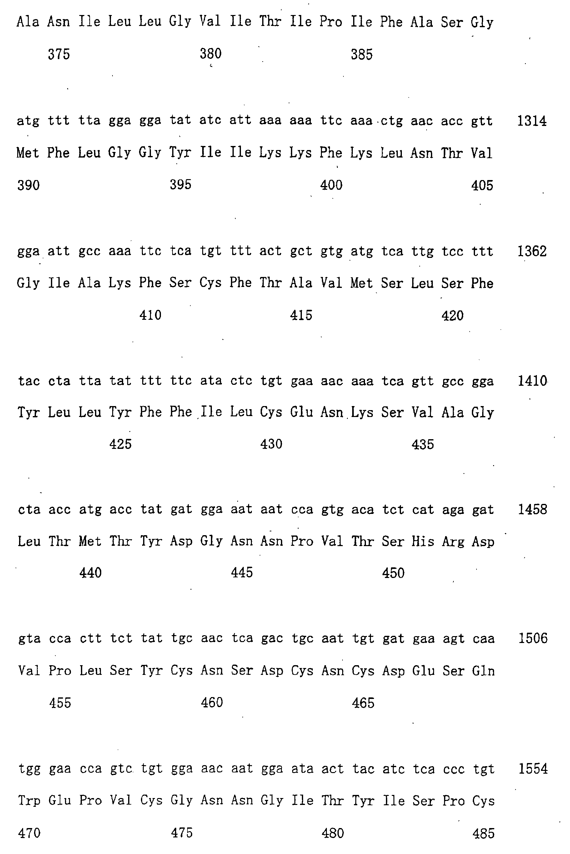
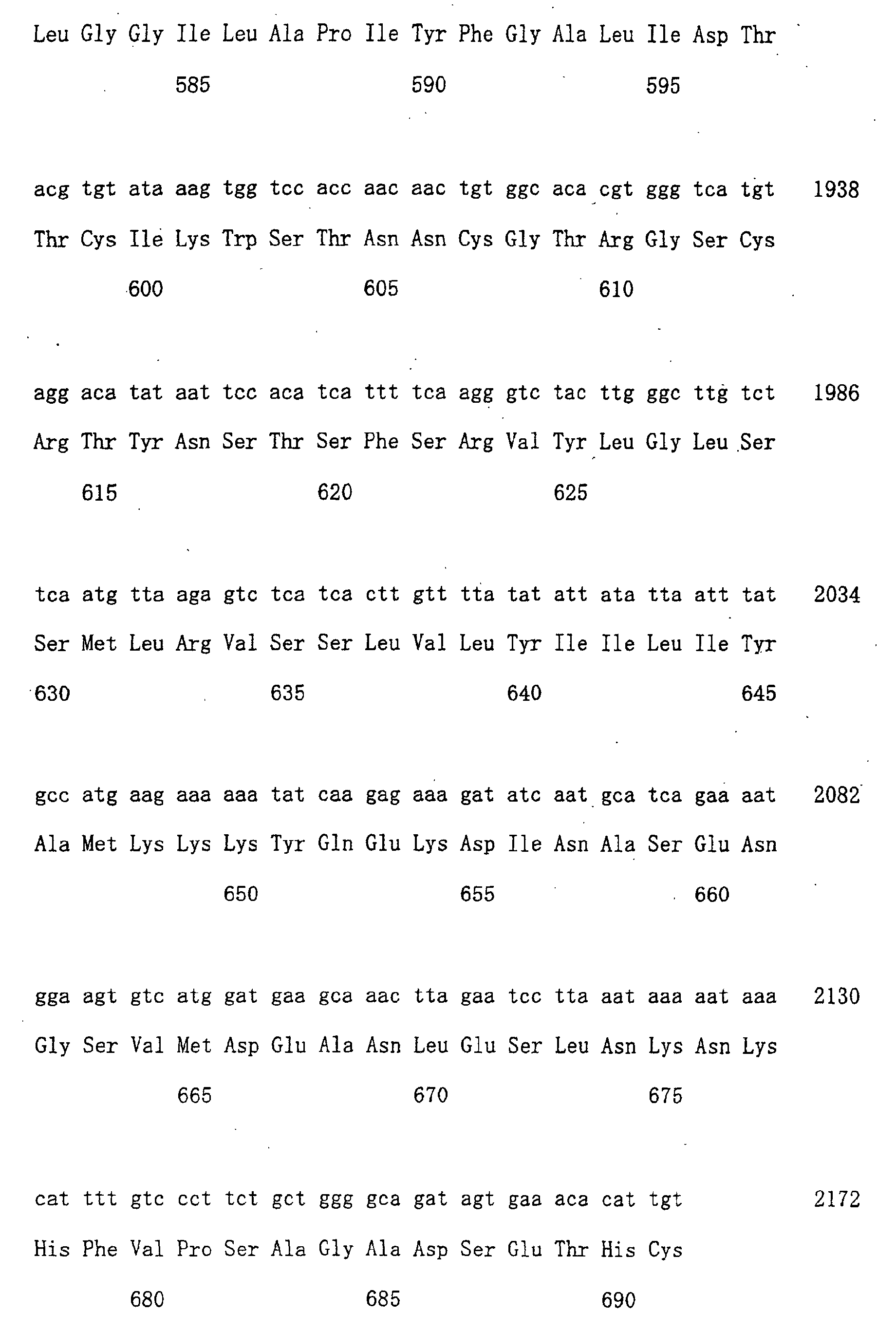
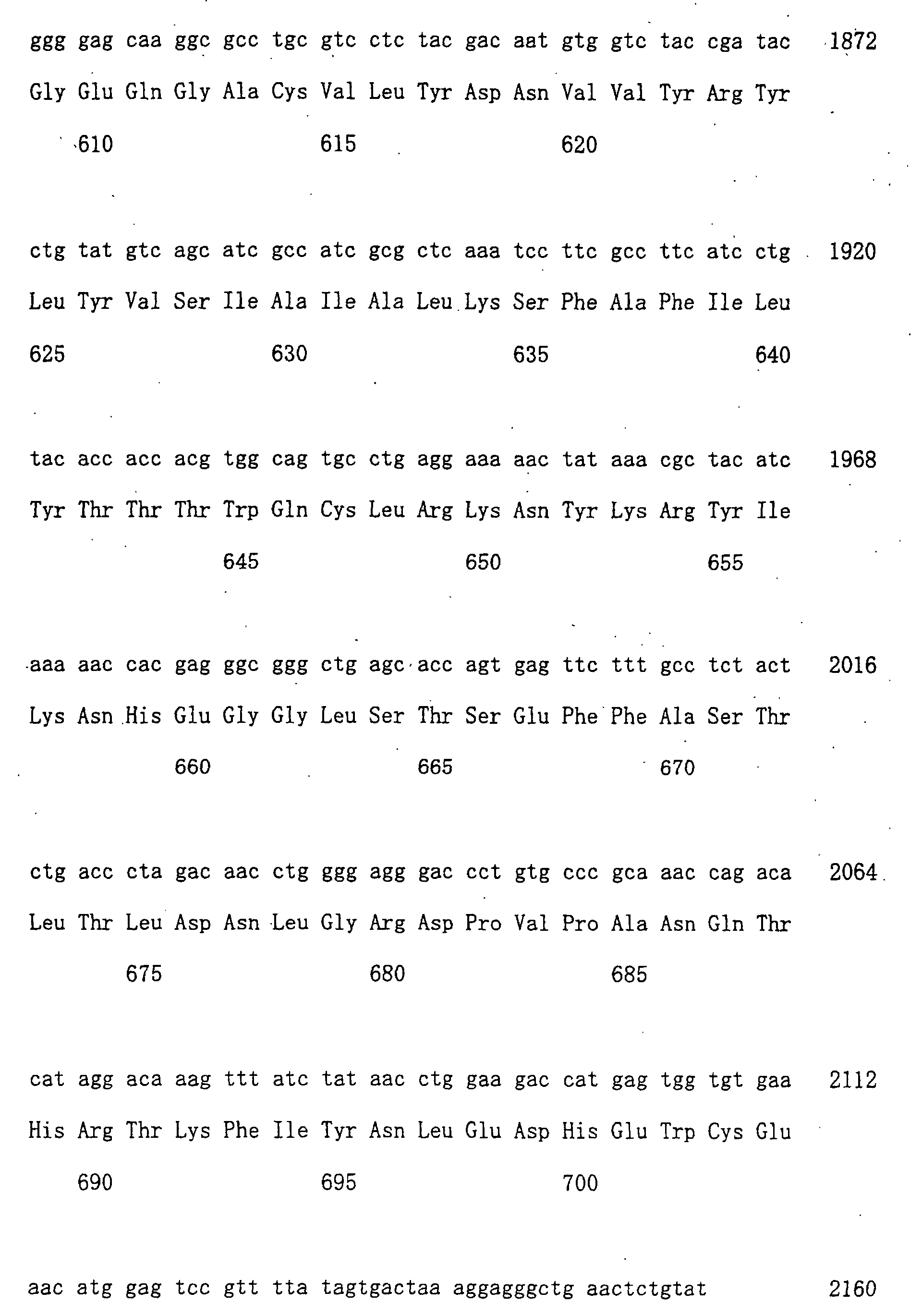
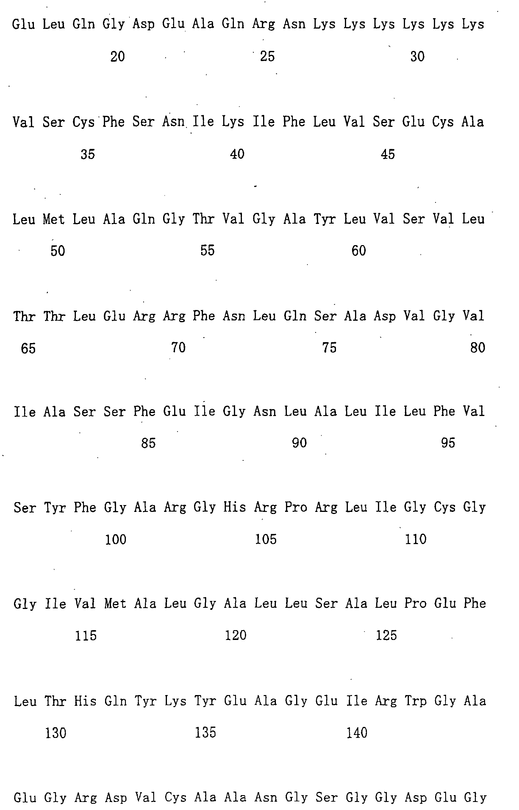
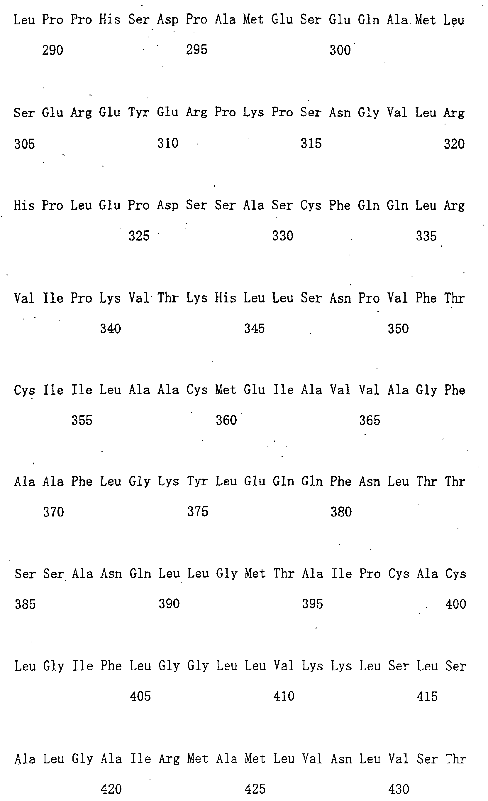
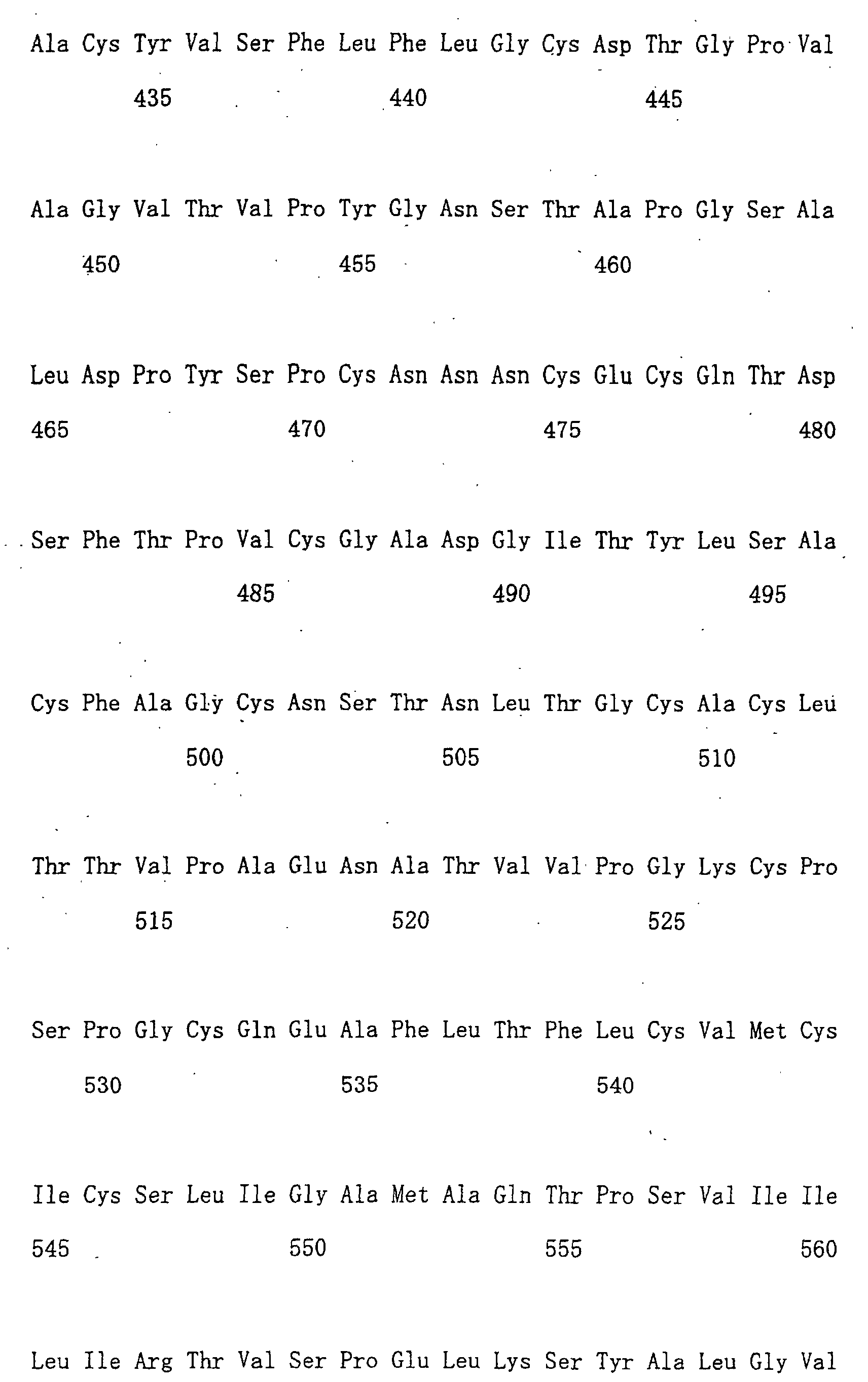
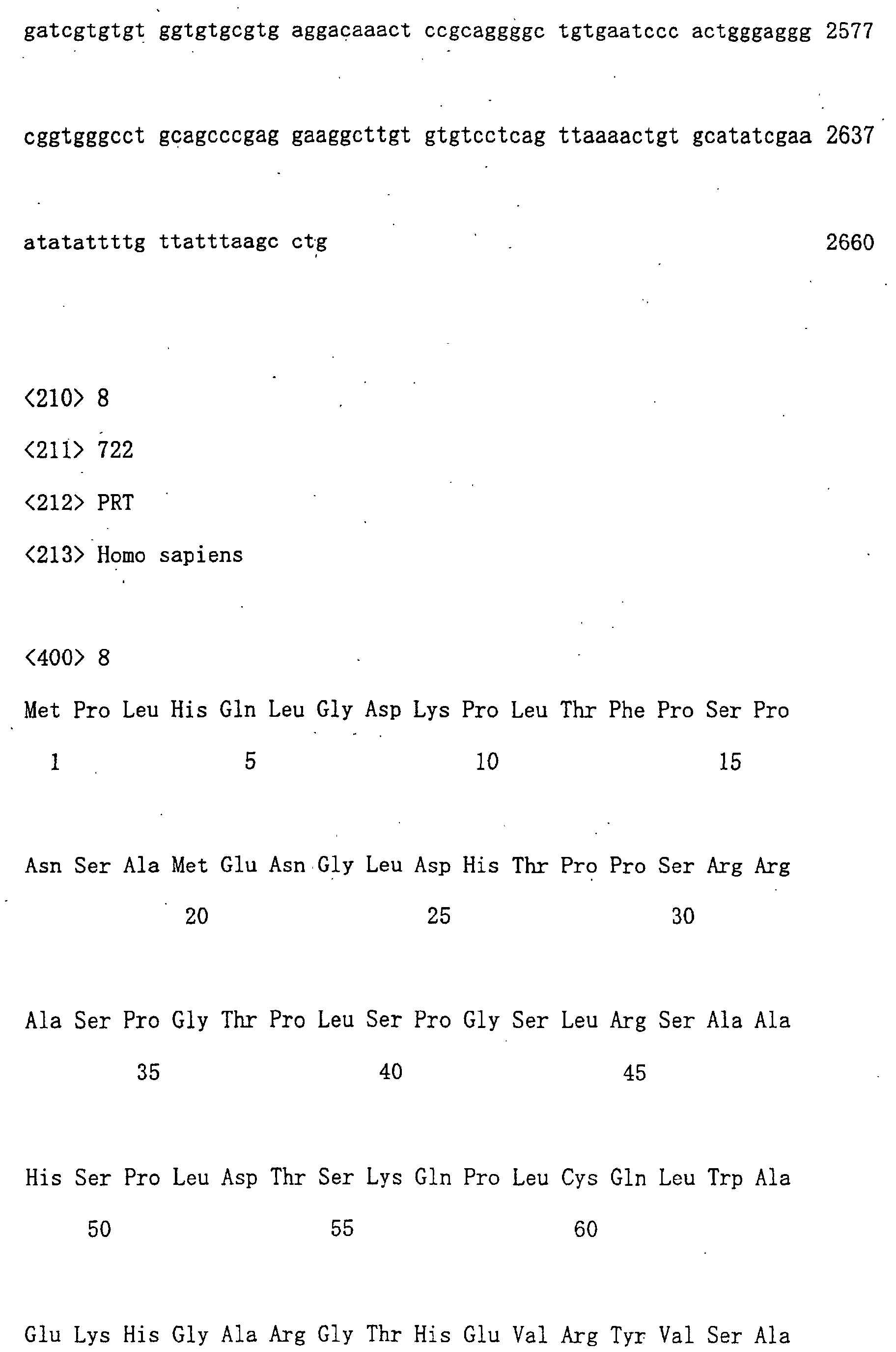
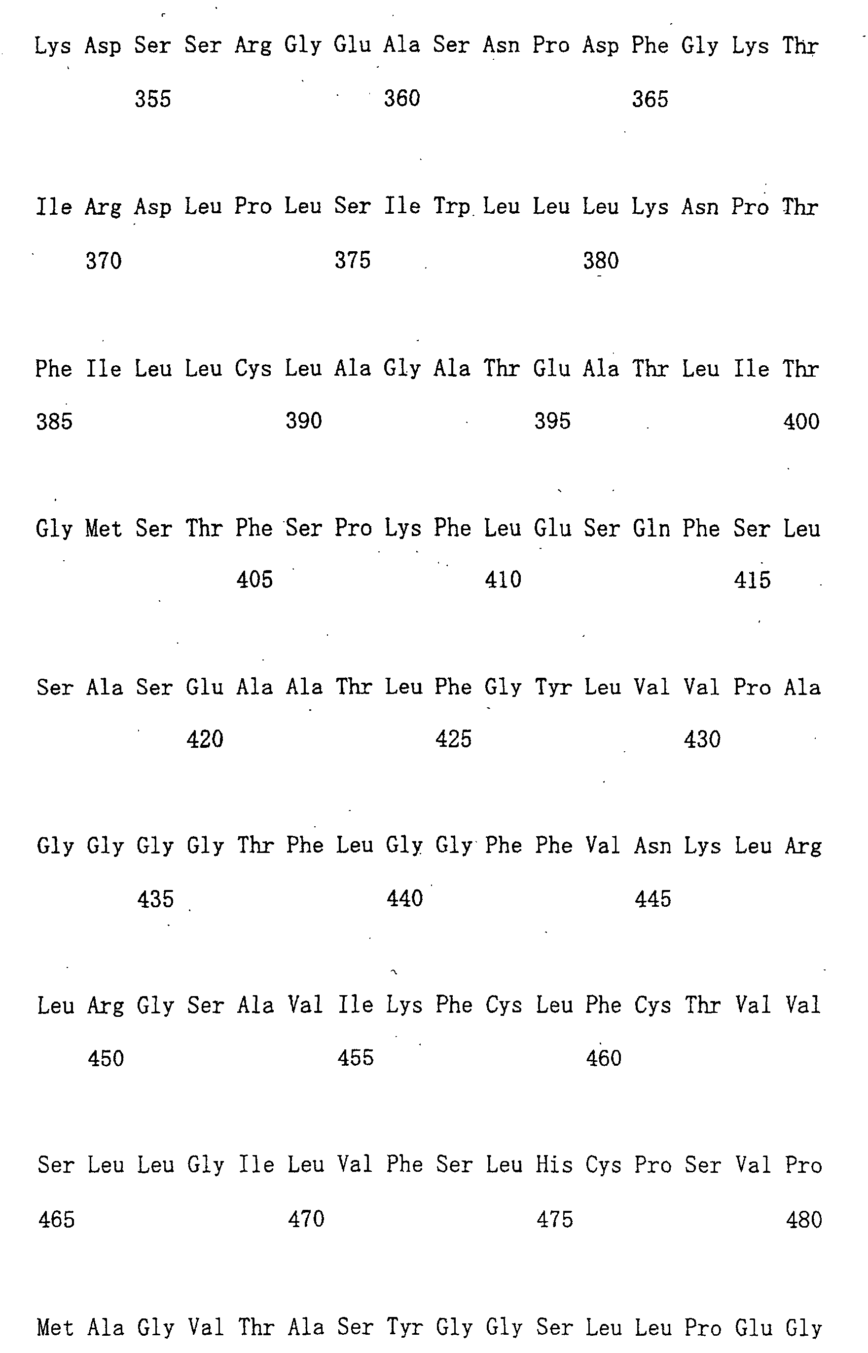
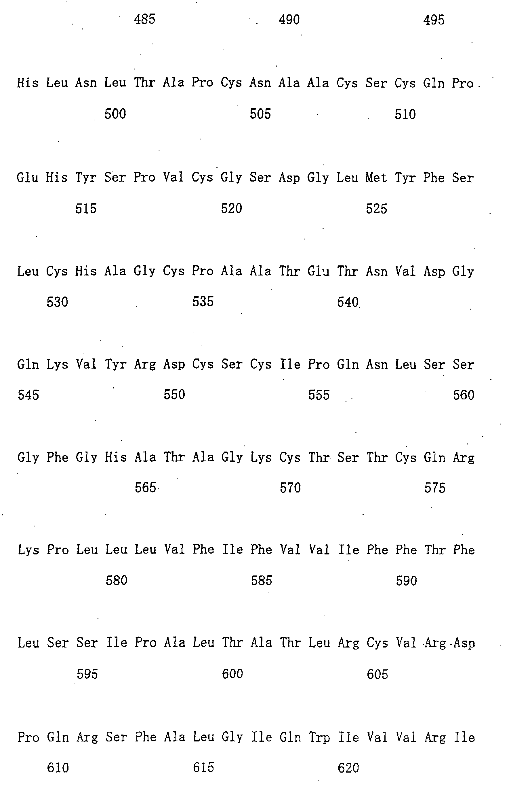
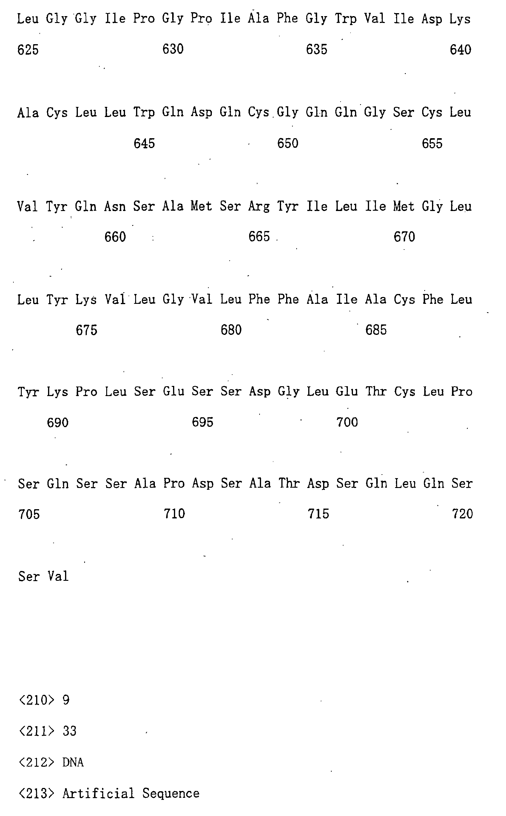
The nucleotide sequences of cDNAs of novel transporters isolated
by the present inventors, "OATP-B", "OATP-C", "OATP-D", and "OATP-E",
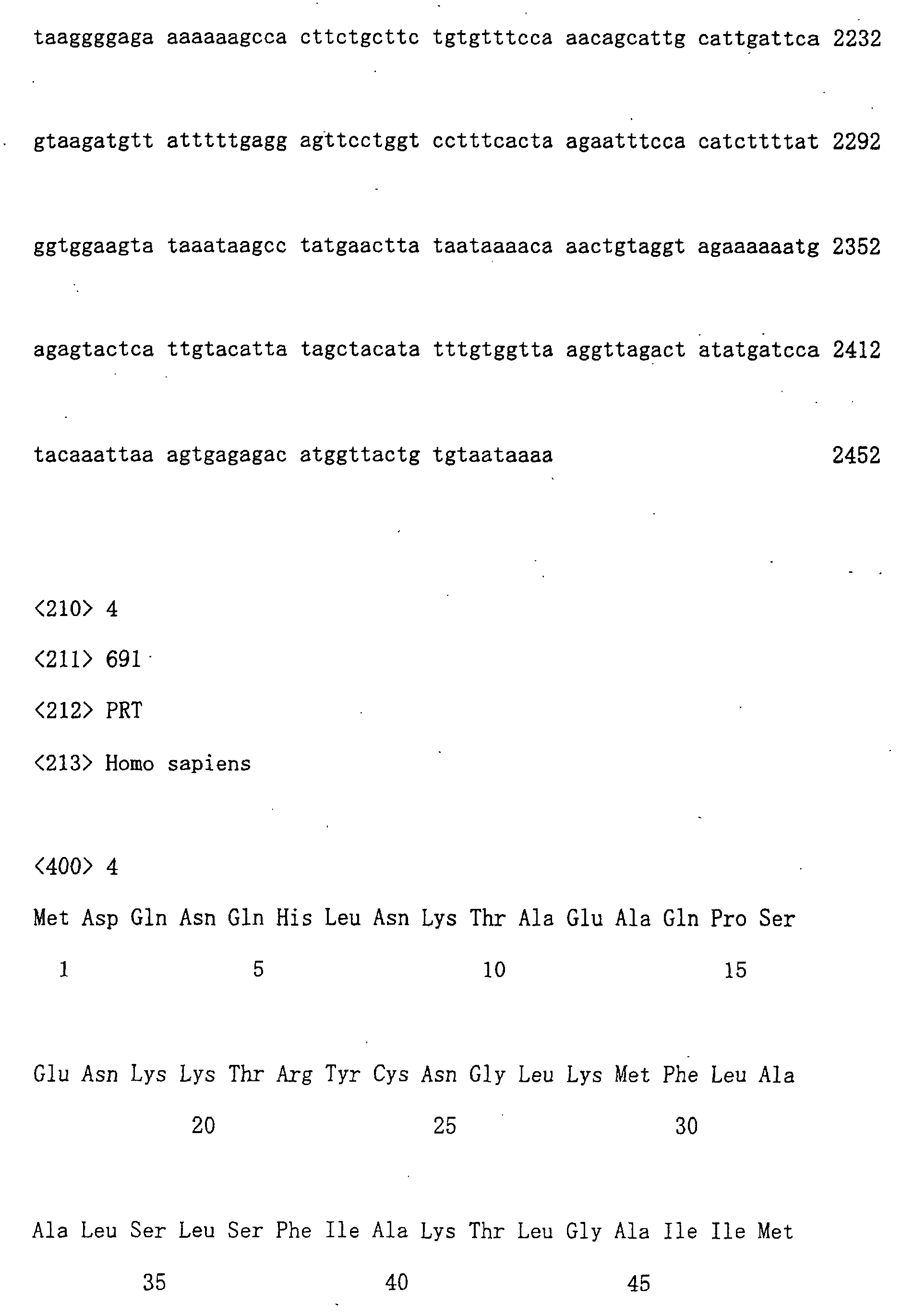
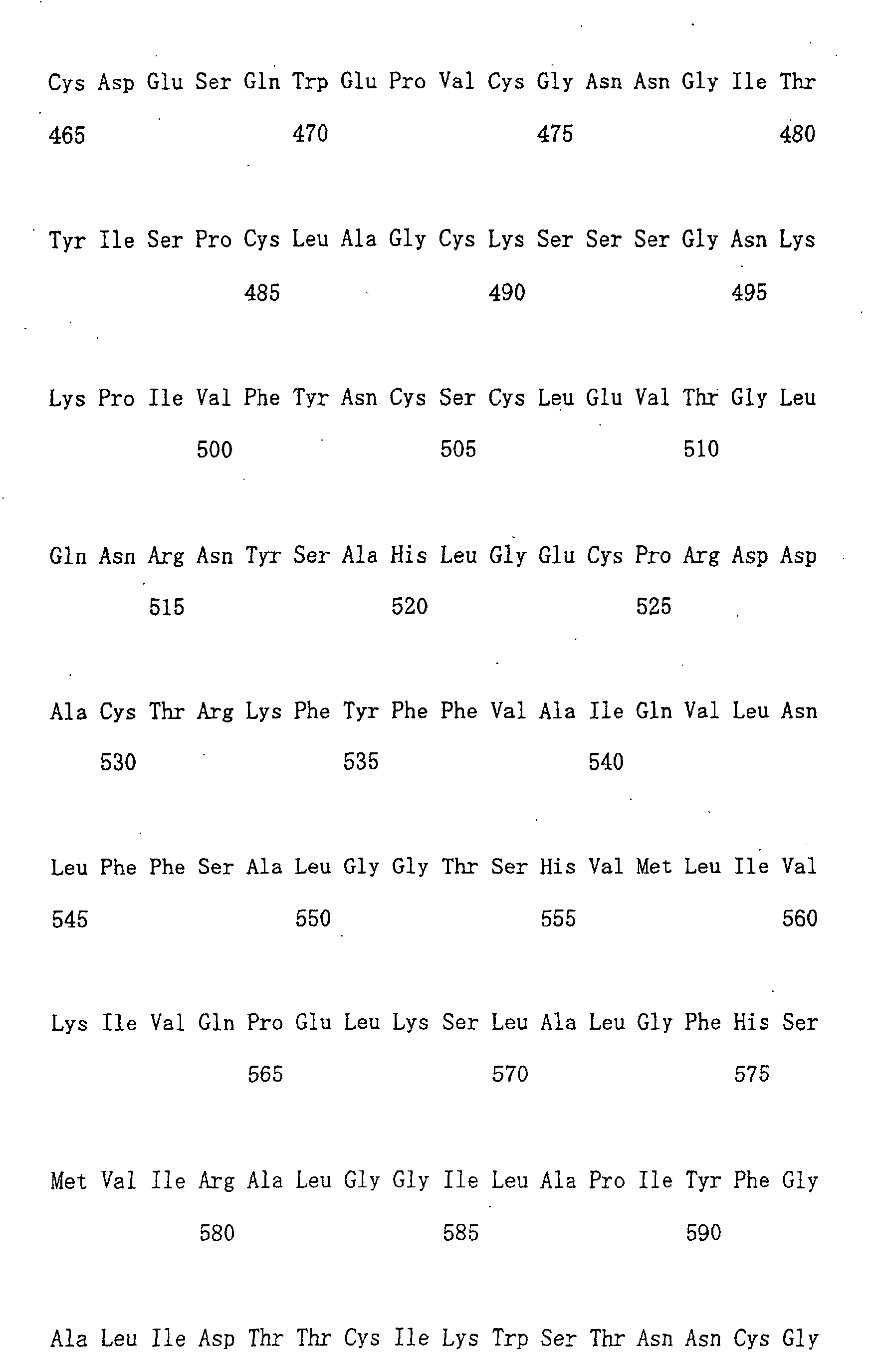
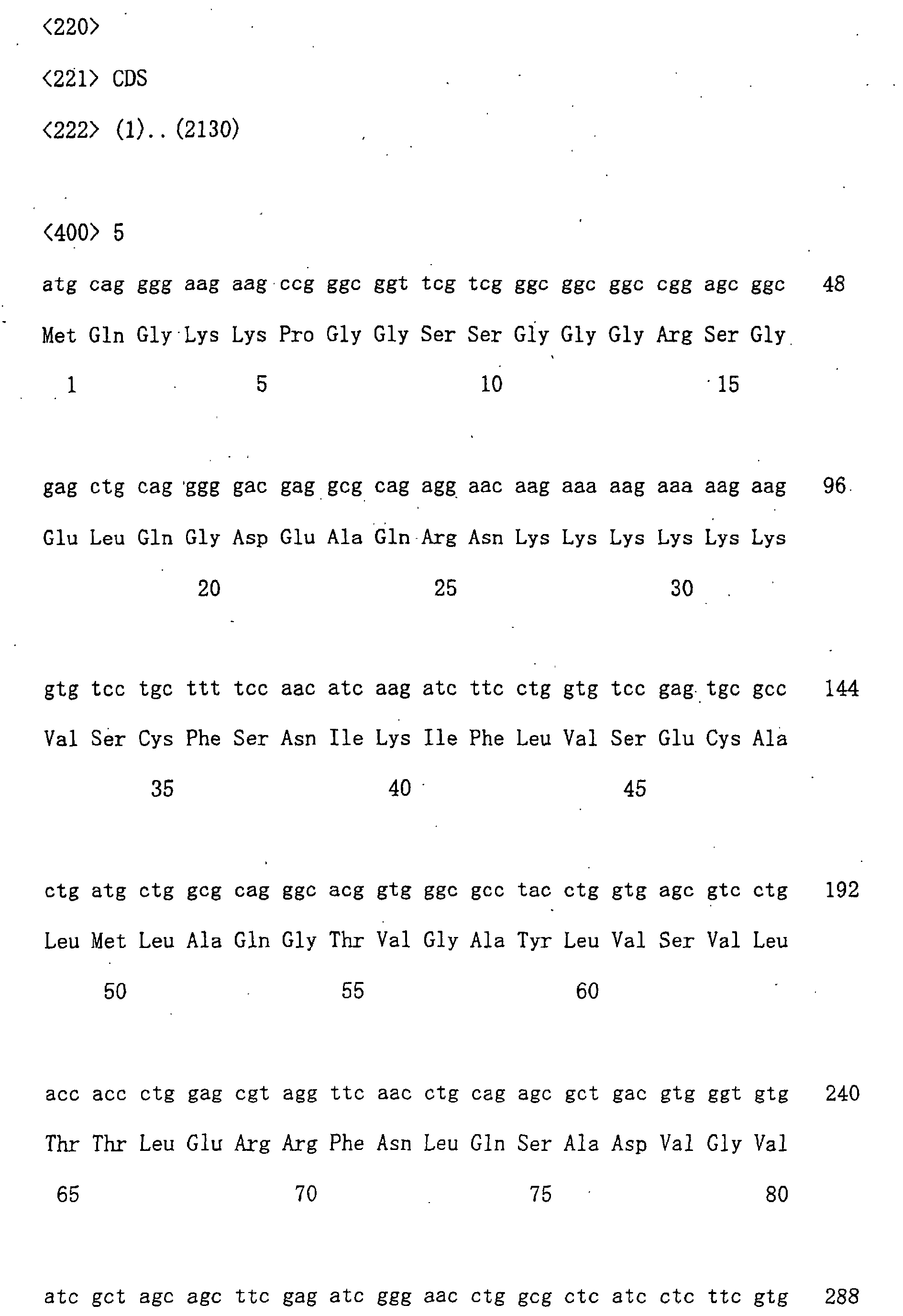
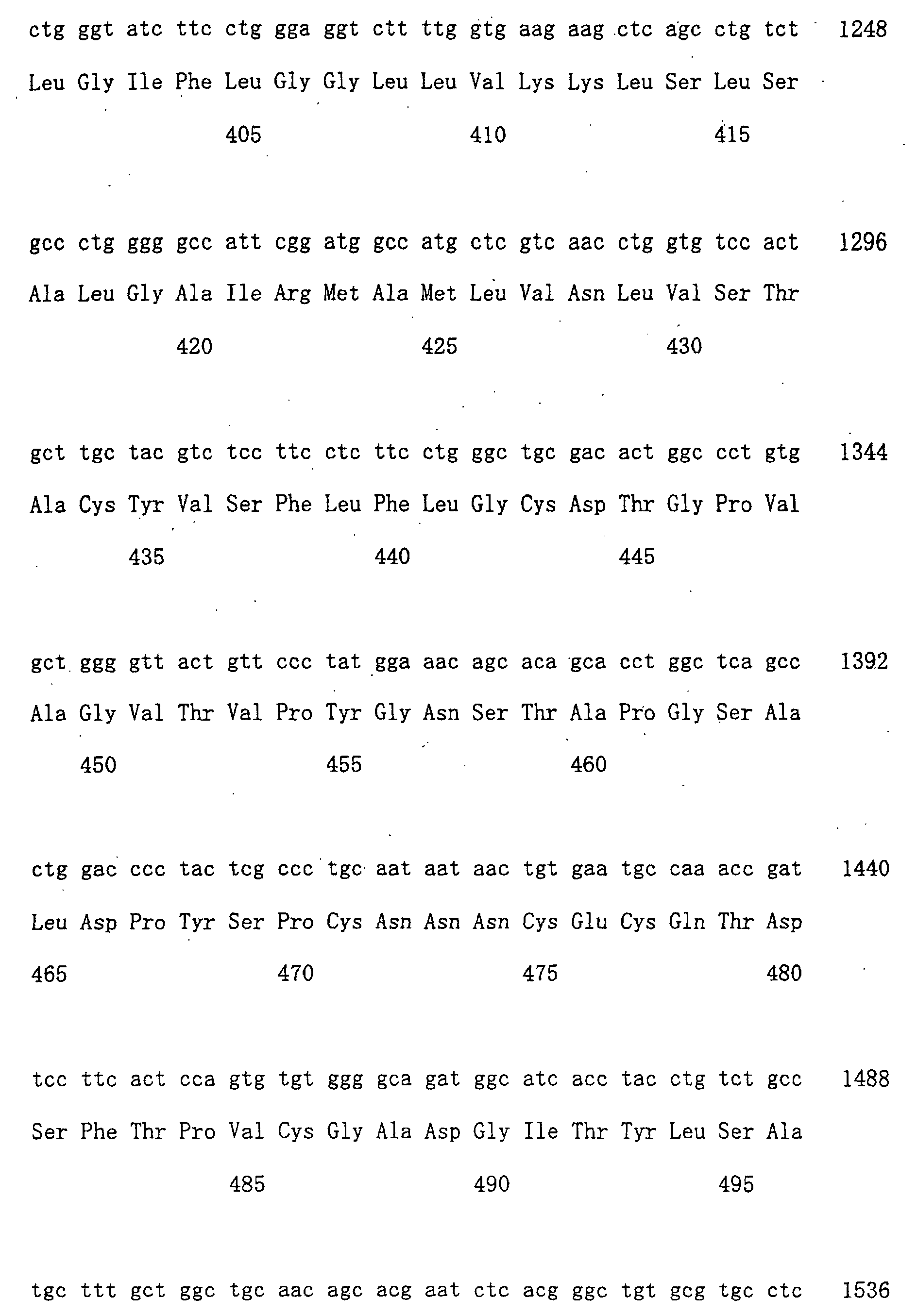
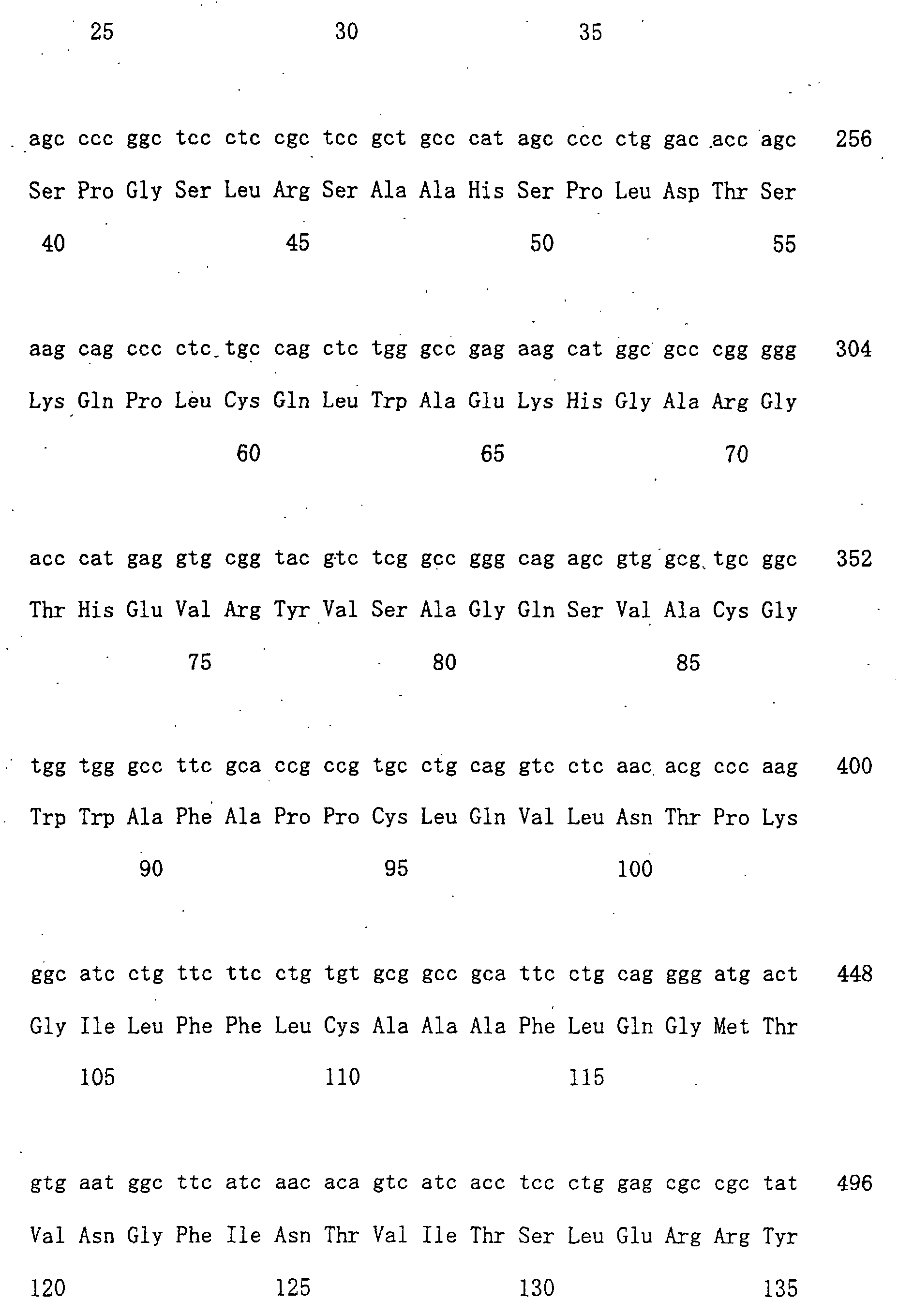
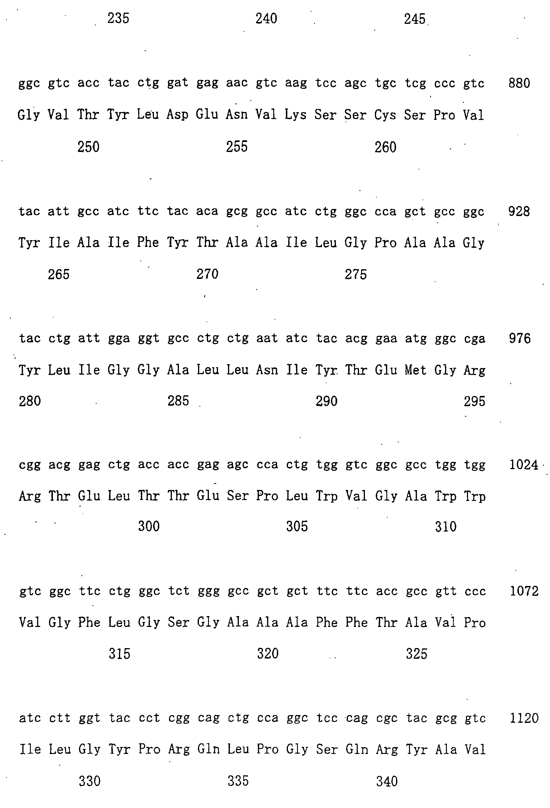
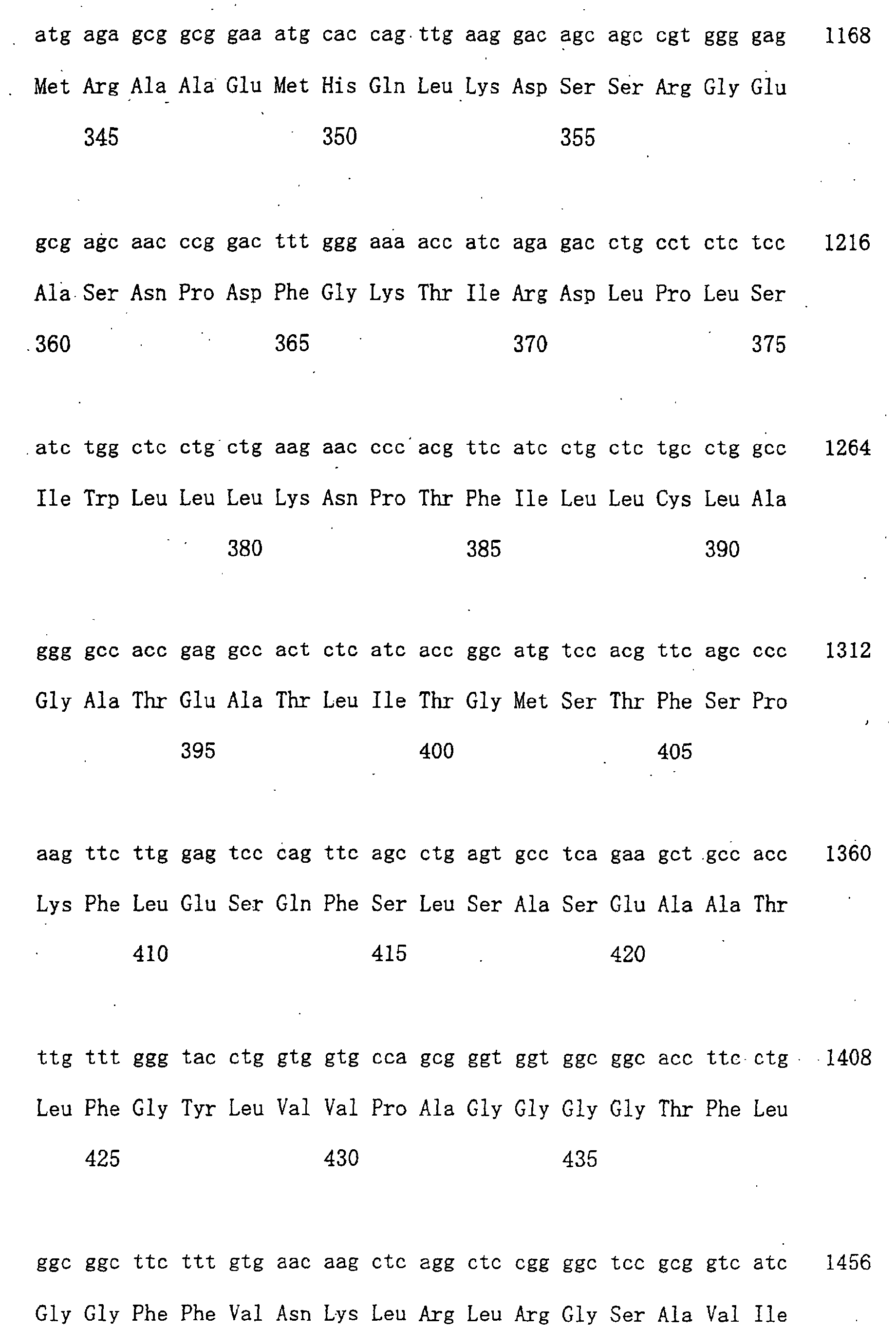
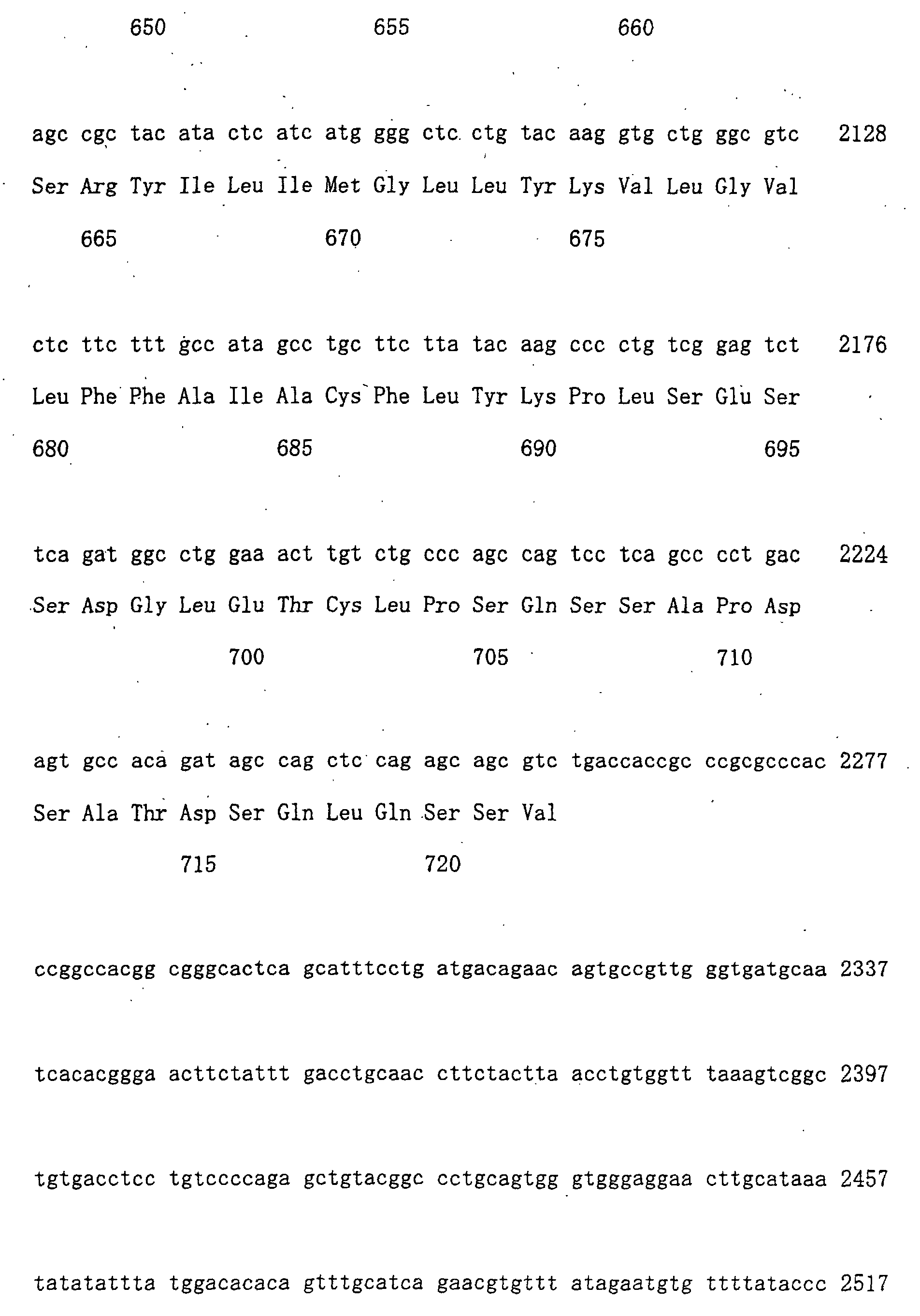
are set forth in SEQ ID NOs: 1, 3, 5, and 7, respectively, and the
amino acid sequences of the proteins encoded by said cDNAs are set
forth in SEQ ID NOs : 2, 4, 6, and 8, respectively. All of these proteins
have structural similarity with the human OATP-A transporter, and
all are thought to form a family (the "OATP" family).
The transporters of this invention are presumed to regulate the
in vivo concentrations of substances, which are either essential or
unnecessary for living bodies. It is also presumed that a variety
of drugs are incorporated into cells or are excreted from cells by
the OATP family proteins. Therefore, it may be possible to control
the pharmacodynamics of drugs, and speedily design or screen drugs
with an improved absorbability using the proteins of the OATP family.
The transporter proteins of this invention include mutants of
the above-described human transporters, "OATP-B", "OATP-C", "OATP-D",
and "OATP-E" protein. Herein, "mutants" are referred to as proteins
that have amino acid sequences which have been mutated by substitution,
deletion, addition, or insertion of amino acids from the natural
"OATP-B", "OATP-C", "OATP-D", or "OATP-E" proteins of SEQ ID NOs:
2, 4, 6, or 8 , and that still retain the transporter activity. Mutations
of amino acids in proteins may occur artificially or naturally.
The phrase "having the transporter activity" as used herein means
that a protein has the activity to transport an organic compound across
membranes. Examples of organic compounds include
estradiol-17β-glucuronide, estron-3-sulfate, benzyl penicillin,
prostaglandin E2, and so on, but are not limited thereto.
In addition, the phrase "activity to transport" as used herein
includes not only the activity to transport an organic compound from
the outside to inside of cells, but also that from the inside to outside
of cells. The transporter proteins of this invention include those
having both of these activities and those with either one of the
activities. The activity of a protein to transport an organic compound
can be measured, for example, by adding a labeled organic compound
to cells to detect the uptake or excretion thereof, for example, by
the method described in Examples.
During the cloning process of genes of the OATP family, the present
inventors found SNPs with following three types of amino acid mutations
in normal individuals:
- polymorphism in the 486th codon of OATP-B gene (tct:Ser or
ttt:Phe),
- polymorphism in the 130th codon of OATP-C gene (aat:Asn or
gat:Asp), and
- polymorphism in the 174th codon of OATP-C gene (gtg:Val or
gcg:Ala).
The transporter of this invention includes proteins having the
above-described mutations, namely: a protein comprising the amino
acid sequence of SEQ ID NO: 2, wherein the amino acid at position
486 is substituted with Phe; a protein comprising the amino acid sequence
of SEQ ID NO: 4, wherein the amino acid at position 130 is substituted
with Asp; and a protein comprising the amino acid sequence of SEQ
ID NO: 4, wherein the amino acid at position 174 is substituted with
Ala.
Polymorphisms other than those described above are presumed to
exist for "OATP-B", "OATP-C", "OATP-D", and "OATP-E", and such
polymorphisms of "OATP-B", "OATP-C", "OATP-D", and "OATP-E" are also
included in this invention. These polymorphisms are presumed to affect
the expression level or activity of transporters and are probably
related to their phenotypes. Genetic diagnosis, wherein polymorphism
and mutation of the OATP family genes are detected, is made possible
by further elucidating the relationship between the genes of OATP
family and their phenotypes.
Examples of methods for artificially altering amino acids well
known to those skilled in the art include the site-specific mutagenesis
system by PCR (GIBCO-BRL, Gaithersburg, Maryland); site-specific
mutagenesis using oligonucleotides (Kramer, W. and Fritz, H. J. (1987)
Methods in Enzymol., 154: 350-367); the Kunkel's method (Methods
Enzymol. 85, 2763-2766 (1988)); etc. There is no particular limitation
in the number and site of amino acid mutations so long as the mutant
proteins retain the transporter activity of this invention. The
preferred number of amino acids to be substituted is typically 10
amino acid residues or less, preferably 6 or less, and more preferably
3 or less.
As for the amino acid residue to be mutated, it is preferable
that the amino acid be mutated into a different amino acid that allows
the properties of the amino acid side-chain to be conserved. Examples
of properties of amino acid side chains include hydrophobic amino
acids (A, I, L, M, F, P, W, Y, V), hydrophilic amino acids (R, D,
N, C, E, Q, G, H, K, S, T), and amino acids comprising the following
side chains: an aliphatic side-chain (G, A, V, L, I, P); a hydroxyl
group containing side-chain (S, T, Y); a sulfur atom containing
side-chain (C, M) ; a carboxylic acid and amide containing side-chain
(D, N, E, Q) ; a base containing side-chain (R, K, H) ; and an aromatic
containing side-chain (H, F, Y, W) (The parenthetic letters indicate
the one-letter codes of amino acids).
It is well known that a protein having deletion, addition, and/or
substitution of one or more amino acid residues in its protein sequence
can retain the biological activity of the original protein (Mark,
D.F. et al., Proc. Natl. Acad. Sci. U.S.A. 81:5662-5666 (1984); Zoller,
M.J. and Smith, M., Nucleic Acids Res. 10:6487-6500 (1982); Wang,
A. et al., Science 224:1431-1433; Dalbadie-McFarland, G. et al., Proc.
Natl. Acad. Sci. U.S.A. 79:6409-6413 (1982)).
An example of a protein to which plural amino acid residues are
added to the amino acid sequence of human "OATP-B", "OATB-C", "OATP-D",
or "OATP-E" protein (SEQ ID NOs: 2, 4, 6, or 8) is a fusion protein
comprising human "OATP-B", "OATP-C", "OATP-D", or "OATP-E" protein.
Fusion proteins are fusions of the human "OATP-B", "OATP-C", "OATP-D",
or "OATP-E" protein and other peptides or proteins, and are included
in the present invention. Fusion proteins can be made by techniques
well known to a person skilled in the art, such as by linking the
DNA encoding the human "OATP-B", "OATP-C", "OATP-D", or "OATP-E"
protein of the invention with DNA encoding the other peptides or proteins,
so that the frames match, inserting this fusion DNA into an expression
vector, and expressing it in a host. There is no restriction as to
the peptides or proteins fused to the protein of the present invention.
However, examples of known peptides that can be used as peptides
to be fused to the protein of the present invention include: FLAG
(Hopp, T.P. et al., Biotechnology (1988) 6: 1204-1210), 6x His
consisting of six His (histidine) residues, 10x His, Influenza
agglutinin (HA), human c-myc fragment, VSV-GP fragment, p18HIV
fragment, T7-tag, HSV-tag, E-tag, SV40T antigen fragment, lck tag,
α-tubulin fragment, B-tag, Protein C fragment, and such. Additional
examples of proteins that can be fused to a protein of the present
invention include GST (glutathione-S-transferase), Influenza
agglutinin (HA), immunoglobulin constant region, β-galactosidase, MBP
(maltose-binding protein), and such.
Fusion proteins can be prepared by fusing commercially available
DNA encoding these peptides or proteins with the DNA encoding a protein
of the present invention and expressing the fused DNA prepared.
In addition, the transporter proteins of the present invention
include proteins having a high structural homology with the
above-described human "OATP-B", "OATP-C", "OATP-D", or "OATP-E"
proteins, and retaining the transporter activity. Such proteins
include proteins, for example, derived from non-human mammals, which
correspond to the human "OATP-B", "OATP-C", "OATP-D", or "OATP-E"
proteins. A protein having a high structural homology with the human
"OATP-B", "OATP-C", "OATP-D", or "OATP-E" proteins can be isolated
by, for example, the hybridization technique (Sambrook, J. et al.,
Molecular Cloning 2nd ed., 9.47-9.58, Cold Spring Harbor Lab. press,
1989). Specifically, based on the DNA sequences (SEQ ID NOs: 1, 3,
5, or 7) encoding the above-described human "OATP-B", "OATP-C",
"OATP-D", or "OATP-E" proteins or portions thereof, a DNA derived
from non-human mammals having a high homology with the aforementioned
DNAs can be isolated using the affinity among DNAs, and the desired
protein can be prepared from the isolated DNA. Non-human mammals to
be used for isolating DNAs include monkeys, mice, rats, rabbits, cattle,
pigs, dogs, cats, and so on, but the invention is not limited to them.
One example of hybridization conditions (stringent conditions)
for isolating such DNAs is as follows. That is, after the
pre-hybridization at 55°C for 30 min or more in the "ExpressHyb
Hybridization Solution" (CLONTECH), a labeled probe is added, and
hybridization is performed by heating the reaction mixture at 37°C
to 55°C for 1 h or more. Then, the reaction product is successively
washed in 2 x SSC and 0.1% SDS three times at room temperature for
20 min, and then in 1 x SSC and 0.1% SDS once at 37°C for 20 min.
More preferable conditions (more stringent conditions) are as
follows: After the pre-hybridization at 60°C for 30 min or more in
the "ExpressHyb Hybridization Solution" (CLONTECH), a labeled probe
is added, and hybridization is performed by heating the reaction mixture
at 60°C for 1 h or more. Then, the reaction product is successively
washed in 2 x SSC and 0.1% SDS three times at room temperature for
20 min, and then in 1 x SSC and 0.1% SDS twice at 50°C for 20 min.
Still more preferable conditions (still more stringent
conditions) are as follows: After pre-hybridization at 68°C for 30
min or more in the "ExpressHyb Hybridization Solution" (CLONTECH),
a labeled probe is added, and hybridization is performed by heating
the reaction mixture at 68°C for 1 h or more. Then, the reaction product
is successively washed in 2 x SSC and 0.1% SDS three times at room
temperature for 20 min, and then in 0.1 x SSC and 0.1% SDS twice at
50°C for 20 min. However, several factors, such as temperature or
salt concentration, can influence the stringency of hybridization
and one skilled in the art can suitably select the factors to accomplish
a similar stringency.
It is also possible for those skilled in the art to similarly
isolate a gene having a high homology with the human "OATP-B", "OATP-C",
"OATP-D", or "OATP-E" genes, and obtain desired proteins from these
genes using techniques other than the hybridization technique, for
example, the polymerase chain reaction.
Such proteins isolated by hybridization and polymerase chain
reaction techniques are thought to have a high homology with the human
"OATP-B", "OATP-C", "OATP-D", or "OATP-E" proteins. "High homology"
means at least 80% or more, preferably 90% or more, and more preferably
95% or more of homology at the amino acid level. The homology of a
protein can be determined by following the algorithm in "Wilbur, W.J.
and Lipman, D.J., Proc. Natl. Acad. Sci. USA (1983) 80, 726-730".
A protein of the present invention may have variations in amino
acid sequence, molecular weight, isoelectric point, the presence or
absence of sugar chains, or form, depending on the after-mentioned
cell or host used to produce it or the purification method utilized.
Nevertheless, so long as the obtained protein has the transporter
activity, it is within the scope of the present invention.
A proteinof the present invention can be prepared as a
recombinant protein or a natural protein by methods well known to
those skilled in the art. A recombinant protein can be prepared by
inserting a DNA (for example, the DNA comprising the nucleotide sequence
of SEQ ID NOs: 1, 3, 5, or 7) encoding a protein of the present invention
into an appropriate expression vector, introducing the vector into
appropriate host cells, collecting the recombinant, obtaining the
extract, and purifying by subjecting the extract to chromatography
such as ion exchange, reverse, gel filtration, or affinity
chromatography in which an antibody against a protein of the present
invention is fixed on column or by combining more than one of these
columns.
Also, when a protein of the present invention is expressed within
host cells (for example, animal cells and E. coli) as a fusion protein
with glutathione-S-transferase protein or as a recombinant protein
supplemented with multiple histidines, the expressed recombinant
protein can be purified using a glutathione column or nickel column.
After purifying the fusion protein, it is also possible to exclude
regions other than the objective protein by cutting with thrombin
or factor-Xa as required.
A natural protein can be isolated by methods well known to those
skilled in the art, for example, by contacting the extract of tissues
or cells expressing a protein of the present invention to an affinity
column in which an antibody binding to a protein of the present invention
(described below) is bound so as to isolate and purify the protein
from the extract. An antibody can be a polyclonal or a monoclonal
antibody.
The present invention also includes a partial peptide of a protein
of the present invention. A partial peptide comprising the amino acid
sequence specific to the protein of the present invention comprises
at least 7 amino acids, preferably 8 amino acids or more, and more
preferably 9 amino acids or more. The partial peptide can be used,
for example, to prepare an antibody against a protein of the present
invention, to screen for a compound binding to a protein of the present
invention, and to screen for accelerators or inhibitors of a protein
of the present invention. Partial peptides of the proteins of the
present invention include those peptides comprising the functional
domains of original proteins, for example, comprising the amino acid
sequence of SEQ ID NOs : 2, 4, 6, or 8. Other examples of partial peptides
are those comprising one or more hydrophobic and hydrophilic regions
predicted from the hydrophobicity plot analysis. These partial
peptides may contain either a partial or entire area of one hydrophobic
or hydrophilic region.
A partial peptide of the invention can be produced by genetic
engineering, known methods of peptide synthesis, or by digesting a
protein of the invention with an appropriate peptidase. For peptide
synthesis, for example, solid phase synthesis or liquid phase synthesis
may be used.
A DNA encoding a protein of the present invention can be used
to produce a protein of the present invention in vivo or in vitro
as described above. In addition, it may, for example, find application
to gene therapy for diseases attributed to genetic abnormality in
a gene encoding a protein of the present invention and diseases treatable
with a protein of the present invention. Any form of the DNA of the
present invention can be used, so long as it encodes a protein of
the present invention. Specifically, cDNA synthesized from the mRNA,
genomic DNA, or chemically synthesized DNA can be used. The present
invention includes a DNA comprising a given nucleotide sequence based
on degeneracy of genetic codons, so long as it encodes a protein of
the present invention.
The DNA of the present invention can be prepared by methods known
to those skilled in the art. For example, a DNA of the present invention
can be prepared by preparing a cDNA library from cells which express
the protein of the present invention, and conducting hybridization
using a partial sequence of a DNA of the present invention (e.g. SEQ
ID NOs: 1, 3, 5, or 7) as a probe. A cDNA library can be prepared,
for example, by the method described in Sambrook, J. et al., Molecular
Cloning, Cold Spring Harbor Laboratory Press (1989), or by using
commercially available cDNA libraries. A cDNA library can be also
prepared by preparing RNA from cells expressing a protein of the present
invention, synthesizing cDNA based on the RNA using reverse
transcriptase, synthesizing an oligo DNA based on the sequence of
the DNA of the present invention (for example, SEQ ID NOs: 1, 3, 5,
or 7), conducting PCR by using these as primers, and amplifying cDNA
encoding the protein of the present invention.
In addition, by sequencing the nucleotides of the obtained cDNA,
a translation region encoded by it can be determined, and an amino
acid sequence of a protein of the present invention can be obtained.
Moreover, by screening the genomic DNA library using the obtained
cDNA as a probe, genomic DNA can be isolated.
More specifically, mRNAs may first be isolated from a cell, tissue,
or organ in which a protein of the invention is expressed. Known methods
can be used to isolate mRNAs: for instance, total RNA may be prepared
by guanidine ultracentrifugation (Chirgwin, J.M. et al., Biochemistry
18:5294-5299 (1979)) or by the AGPC method (Chomczynski, P. and Sacchi,
N., Anal. Biochem. 162:156-159 (1987)), and mRNA may then be purified
from total RNA using an mRNA Purification Kit (Pharmacia) and such.
Alternatively, mRNA may be directly purified by QuickPrep mRNA
Purification Kit (Pharmacia).
The obtained mRNA is used to synthesize cDNA using reverse
transcriptase. cDNA may be synthesized using a kit such as the AMV
Reverse Transcriptase First-strand cDNA Synthesis Kit (Seikagaku
Kogyo). Alternatively, cDNA may be synthesized and amplified
following the 5'-RACE method (Frohman, M.A. et al., Proc. Natl Acad.
Sci. U.S.A. 85:8998-9002 (1988); Belyavsky, A. et al., Nucleic Acids
Res. 17:2919-2932 (1989)) which uses a primer and such, described
herein, the 5'-Ampli FINDER RACE Kit (Clontech), and polymerase chain
reaction (PCR).
A desired DNA fragment is prepared from the PCR products and
ligated with a vector DNA. The recombinant vectors are used to
transform E. coli and such, and a desired recombinant vector is prepared
from a selected colony. The nucleotide sequence of the desired DNA
can be verified by conventional methods, such as dideoxynucleotide
chain termination.
A DNA of the invention may be designed to have a nucleotide sequence
that is expressed more efficiently by taking into account the frequency
of codon usage in the host to be used for expression (Grantham, R.
et al., Nucleic Acids Res. 9:43-74 (1981)). The DNA of the present
invention may be altered by a commercially available kit or a
conventional method. For instance, the DNA may be altered by digestion
with restriction enzymes, insertion of synthetic oligonucleotides
or appropriate DNA fragments, addition of a linker, or insertion of
the initiation codon (ATG) and/or a stop codon (TAA, TGA, or TAG).
Specifically, DNAs of this invention include DNAs comprising
bases A179 through G2305 of the nucleotide sequence of SEQ ID NO:
1, bases A100 through T2172 of the nucleotide sequence of SEQ ID NO:
3, bases A1 through A2130 of the nucleotide sequence of SEQ ID NO:
5, and bases A92 through C2257 of the nucleotide sequence of SEQ ID
NO: 7.
Moreover, DNAs of this invention includes DNAs comprising bases
A179 through G2305 of the nucleotide sequence of SEQ ID NO: 1, wherein
the C1635 has been substituted with T; bases A100 through T2172 of
the nucleotide sequence of SEQ ID NO: 3, wherein the A487 has been
substituted with G; bases A100 through T2172 of the nucleotide sequence
of SEQ ID NO: 3, wherein the T620 has been substituted with C.
Furthermore, the present invention provides DNAs that are capable
of hybridizing with a DNA having a nucleotide sequence of SEQ ID NOs :
1, 3, 5, or 7, and encoding a protein having the transporter activity.
Suitable hybridization conditions include the above conditions. The
hybridizing DNA is preferably a natural DNA, for example, cDNA or
chromosomal DNA.
The present invention also relates to a vector into which a DNA
of the present invention is inserted. Avector of the present invention
is useful for keeping the DNA of the present invention in a host cell,
or to express a protein of the present invention.
When E. coli is the host cell and the vector is amplified and
produced in large amounts in E. coli (e.g., JM109, DH5α, HB101, or
XL1Blue) and such, the vector should have "ori" to be amplified in
E. coli and a marker gene for selecting transformed E. coli (for example,
a drug-resistance gene selected by a drug (e.g., ampicillin,
tetracycline, kanamycin, or chloramphenicol)). For example,
M13-series vectors, pUC-series vectors, pBR322, pBluescript,
pCR-Script, and so on can be used. Additionally, pGEM-T, pDIRECT,
and pT7 can also be used for subcloning and extracting cDNA as well
as the vectors described above. When a vector is used to produce a
protein of the present invention, an expression vector is especially
useful. For example, an expression vector to be expressed in E. coli
should have the above characteristics so as to be amplified in E.
coli. When E. coli, such as JM109, DH5α, HB101, or XL1 Blue, are used
as the host cell, the vector should have a promoter as well as the
above characters such as the vector is copied in the host; for example,
the lacZ promoter (Ward et al., Nature (1989) 341, 544-546; FASEB
J. (1992) 6, 2422-2427), the araB promoter (Better et al., Science
(1988) 240, 1041-1043) , and the T7 promoter and such can efficiently
express the desired gene in E. coli. As such a vector, in addition
to the above vectors, pGFX-5X-1 (Pharmacia), "QIAexpress system"
(Qiagen), pEGFP, and pET (in this case, the host is preferably BL21
which expresses T7 RNA polymerase) can be used.
A vector also may contain a signal sequence for polypeptide
secretion. As a signal sequence for protein secretion, pelB signal
sequence (Lei, S. P. et al., J. Bacteriol. (1987) 169, 4379) can be
used in the case of producing protein into the periplasm of E. coli.
For introducing a vector into host cells, for example, the calcium
chloride method, and the electroporation method can be used.
In addition to E. coli, for example, expression vectors derived
from mammals (for example, pcDNA3 (Invitrogen), pEGF-BOS (Nucleic
Acids. Res. 1990, 18 (17), p5322), pEF, and pCDM8) ; expression vectors
derived from insect cells (for example, "Bac-to-BAC baculovirus
expression system" (GIBCO BRL), pBacPAK8) ; expression vectors derived
from plants (for example, pMH1, pMH2), expression vectors derived
from animal viruses (for example, pHSV, pMV, pAdexLcw); expression
vectors derived from retroviruses (for example, pZIpneo) ; expression
vector derived from yeast (for example, "Pichia Expression Kit"
(Invitrogen), pNV11, SP-Q01); and expression vectors derived from
Bacillus subtilis (for example, pPL608, pKTH50) can be used to produce
a protein of the present invention.
In order to express the vector in animal cells, such as CHO,
COS, or NIH3T3 cells, the vector should have a promoter necessary
for expression in such cells, for example, the SV40 promoter (Mulligan
et al., Nature (1979) 277, 108), the MMLV-LTR promoter, the
EF1αpromotor (Mizushima et al., Nucleic Acids Res. (1990) 18, 5322),
or the CMV promoter, and such, and preferably should also include
a marker gene for selecting transformants (for example, a drug
resistance gene selected by a drug (e.g., neomycin, G418)). Examples
of the vectors with these characteristics include pMAM, pDR2, pBK-RSV,
pBK-CMV, pOPRSV, pOp13, and so on.
In addition, in order to stably express a gene and amplify the
copy number of the gene in cells, an exemplary method can use the
following steps: introducing a vector comprising the complementary
DHFR gene (for example pCHO I) into CHO cells in which the nucleic
acid synthesizing pathway is deleted, and amplifying by methotrexate
(MTX); in the case of transient expression of a gene, an exemplary
method can use the steps of transforming with a vector (e.g. pcD)
comprising replication origin of SV40 using COS cells comprising the
gene expressing SV40 T antigen on chromosomes. The origin used for
replication may also be those of polyomavirus, adenovirus, bovine
papilloma virus (BPV), and the like. In addition, the expression
vector may include a selection marker gene for amplification of the
gene copies in host cells. Examples of such markers include, but are
not limited to, the aminoglycoside transferase (APH) gene, the
thymidine kinase (TK) gene, the E. coli xanthine-guanine
phosphoribosyl transferase (Ecogpt) gene, and the dihydrofolate
reductase (dhfr) gene.
On the other hand, a DNA of the present invention can be expressed
in vivo in animals, for example, by inserting a DNA of the present
invention into an appropriate vector and introducing it into the living
body by a method such as the retrovirus method, the liposome method,
the cationic liposome method, or the adenovirus method. By using these,
gene therapy against diseases attributed to the mutation of a
transporter gene of the present invention can be effected. As a vector
to be used, for example,.adenovirus vector (for example pAdexlcw),
and retrovirus vector (for example, pZIPneo) can be used; however,
the present invention is not restricted thereto. Common gene
manipulation, for example, insertion of the DNA of the present invention
to a vector, can be performed according to conventional methods
(Molecular Cloning, 5. 61-5. 63). Administration into a living body
can be either an ex vivo method, or in vivo method.
The present invention also relates to a transformed cell into
which a DNA or a vector of the present invention has been introduced.
The host cell into which the vector of the invention is introduced
is not particularly limited. For example, E. coli or various animal
cells can be used. The transformed cells of the present invention
can be used as, for example, a production system for producing or
expressing a protein of the present invention. The present invention
provides methods of producing a protein of the invention both in vitro
or in vivo. For in vitro production, eukaryotic cells or prokaryotic
cells can be used as host cells.
Eukaryotic cells useful as host cells may be animal, plant, or
fungi cells. As animal cells, mammalian cells, such as CHO (J. Exp.
Med. 108:945 (1995)), COS, 3T3, myeloma, baby hamster kidney (BHK),
HeLa, and Vero cells; or amphibian cells, such as Xenopus oocytes
(Valle, et al., Nature 291:340-358 (1981)) ; or insect cells, such
as Sf9, Sf21, or Tn5 cells, can be used. CHO cells lacking the DHFR
gene (dhfr-CHO) (Proc. Natl. Acad. Sci. U.S.A. 77:4216-4220 (1980))
or CHO K-1 (Proc. Natl. Acad. Sci. U.S.A. 60:1275 (1968)) may also
be used. In animal cells, CHO cells are particularly preferable for
mass expression. A vector can be introduced into host cells by, for
example, the calcium phosphate method, the DEAE dextran method, the
cationic liposome DOTAP (Boehringer Mannheim), the electroporation
method, the lipofection method, and such. A recombinant protein
derived from the obtained transformants can be purified by standard
methods, for example, the method described in "The Qiaexpressionist
handbook, Qiagen, Hilden, Germany".
As plant cells, plant cells originating from Nicotiana tabacum
are known as protein-production system, and may be used as callus
cultures. As fungi cells, yeast cells, such as Saccharomyces,
including Saccharomyces cerevisiae, or filamentous fungi, such as
Aspergillus, including Aspergillus niger, are known and may be used
herein.
Prokaryotic cells suitable for use in the production system
of the present invention include, but are not limited to, bacterial
cells. A known example of these bacterial cells is E. coli, such as
JM109, DH5α, HB101. Regarding others, Bacillus subtilis is known in
the art.
These cells are transformed by a desired DNA, and the resulting
transformants are cultured in vitro to obtain the protein.
Transformants can be cultured using known methods. Culture medium
for animal cells, for example, DMEM, MEM, RPMI1640, or IMDM, may be
used with or without serum supplement, such as fetal calf serum (FCS).
The pH of the culture medium is preferably between about pH 6 to 8.
Such cells are typically cultured at about 30 to 40°C for about 15
to 200 hr, and the culture medium may be replaced, aerated, and/or
stirred if necessary.
On the other hand, animal and plant hosts may be used for in
vivo production. For example, a desired DNA can be introduced into
an animal or plant host. Encoded proteins are produced in vivo, and
then recovered. These animal and plant hosts are included in the host
cells of the present invention.
Animals to be used for the production system described above
include, but are not limited to, mammals and insects. Mammals such
as goat, porcine, sheep, mouse, and bovine may be used (Vicki Glaser,
SPECTRUM Biotechnology Applications (1993)). Alternatively, the
mammals may be transgenic animals.
For instance, a desired DNA may be prepared as a fusion gene
with a gene encoding a protein specifically produced into milk, such
as goat β casein. DNA fragments, comprising the fusion gene having
the desired DNA, are injected into goat embryos, which are then
introduced back to female goats. Proteins are recovered from milk
produced by the transgenic goats (i.e., those born from the goats
that had received the modified embryos) or from their offspring. To
increase the amount of milk containing the proteins produced by
transgenic goats, appropriate hormones may be administered to them
(Ebert, K.M. et al., Bio/Technology 12:699-702 (1994)).
Alternatively, insects, such as the silkworm, may be used.
Baculovirus inserted a DNA encoding a desired protein can be used
to infect silkworms, and the desired protein is recovered from their
body fluid (Susumu, M. et al., Nature 315:592-594 (1985)).
As plants, for example, tobacco can be used. In use of tobacco,
a DNA encoding a desired protein may be inserted into a plant expression
vector, such as pMON530, which is then introduced into bacteria, such
as Agrobacterium tumefaciens. Then, the bacteria is used to infect
a tobacco plant, such as Nicotiana tabacum, and a desired polypeptide
is recovered from the plant's leaves (Julian K.-C. Ma et al., Eur.
J. Immunol. 24:131-138 (1994)).
A protein of the present invention obtained as above may be
isolated from the interior or the exterior (e.g., the culture medium)
of transformed cells, and purified as a substantially pure homogeneous
protein. The method for protein isolation and purification is not
limited to any specific method; in fact, any standard method may be
used. For instance, column chromatography, filter, ultrafiltration,
salting-out, solvent precipitation, solvent extraction, distillation,
immunoprecipitation, SDS-polyacrylamide gel electrophoresis,
isoelectric point electrophoresis, dialysis, recrystallization, and
such may be appropriately selected and combined to isolate and purify
the protein.
For chromatography, for example, affinity chromatography,
ion-exchange chromatography, hydrophobic chromatography, gel
filtration, reverse-phase chromatography, adsorption chromatography,
and such may be used (Strategies for Protein Purification and
Characterization: A Laboratory Course Manual. Ed. Daniel R. Marshak
et al., Cold Spring Harbor Laboratory Press (1996)). These
chromatographies may be performed by liquid-phase chromatography,
such as HPLC and FPLC. Thus, the present invention provides for highly
purified proteins produced by the above methods.
A protein of the present invention may be optionally modified
or partially deleted by treating it with an appropriate protein
modification enzyme before or after purification. Useful protein
modification enzymes include, but are not limited to, trypsin,
chymotrypsin, lysylendopeptidase, protein kinase, and glucosidase.
The present invention also provides antibodies that bind to the
proteins of the invention. An antibody of the invention can be used
in any form, such as monoclonal or polyclonal antibodies, and includes
antiserum obtained by immunizing a rabbit with a protein of the invention,
all classes of polyclonal and monoclonal antibodies, human antibodies,
and humanized antibodies produced by genetic recombination.
A protein of the invention used as an antigen to obtain an antibody
may be derived from any animal species, but preferably from a mammal
such as a human, mouse, or rat, or more preferably from a human. A
human-derived protein may be obtained from the nucleotide or amino
acid sequences disclosed herein.
In the present invention, a protein to be used as an immunization
antigen may be a complete protein or a partial peptide of the protein.
A partial peptide may be, for example, an amino (N) -terminal or carboxy
(C) -terminal fragment of the protein. Herein, "an antibody" is defined
as a molecule that specifically reacts with either the full-length
protein or a fragment of the protein.
A gene encoding a protein of the invention or its fragment may
be inserted into a known expression vector, which may then be used
to transform a host cell as described herein. The desired protein
or its fragment may be recovered from the exterior or the interior
of the transformed host cells, by any standard method, and may be
used as an antigen. Alternatively, cells expressing the protein or
their lysates, or a chemically synthesized protein of the present
invention may be used as an antigen.
Any mammalian animal may be immunized with the antigen, but
preferably the compatibility with parental cells used for cell fusion
is taken into account. In general, animals of the Rodentia order,
Lagomorphs, or Primates may be used.
Animals of the order Rodentia include, for example, mouse, rat,
and hamster. Lagomorphs include, for example, rabbit. Primates
include, for example, a monkey of catarrhine (old world monkey) such
as Macaca fascicularis, rhesus monkey, sacred baboon, or chimpanzee.
Methods for immunizing animals against antigens are known in
the art. Intraperitoneal injection or subcutaneous injection of
antigens is used as a standard method for immunization of mammals.
More specifically, antigens may be diluted and suspended in an
appropriate amount with phosphate buffered saline (PBS), physiological
saline, etc. If desired, the antigen suspension may be mixed with
an appropriate amount of a standard adjuvant, such as Freund's complete
adjuvant, made into emulsion, and then administered to mammalian
animals. Preferably, it is followed by several administrations of
antigen mixed with an appropriately amount of Freund's incomplete
adjuvant every 4 to 21 days. An appropriate carrier may also be used
for immunization. After immunization as above, serum is examined for
increase of the amount of desired antibodies by a standard method.
Polyclonal antibodies against the proteins of the present
invention may be prepared by collecting blood from the immunized mammal
examined for the increase of desired antibodies in the serum, and
by separating serum from the blood by any conventional method.
Polyclonal antibodies may be used as serum containing the polyclonal
antibodies, or if necessary, a fraction containing the polyclonal
antibodies may be isolated from the serum. Immunoglobulin G or M can
be prepared by obtaining a fraction which recognizes only the protein
of the present invention using an affinity column coupled with the
protein of the present invention and further purifying this fraction
by using protein A or protein G column.
To prepare monoclonal antibodies, immune cells are collected
from the mammal immunized against the antigen and checked for increased
level of desired antibodies in the serum as described above, and are
subjected to cell fusion. The immune cells used for cell fusion are
preferably obtained from spleen. As the other parental cells to be
fused with the above immunocyte, for example, preferably myeloma cells
of mammalians, more preferably myeloma cells which acquired the
property for selecting fused cells by drugs, can be used.
The above immunocyte andmyeloma cells can be fused by known methods,
for example, the method by Milstein et al. (Galfre, G. and Milstein,
C., Methods Enzymol. (1981) 73, 3-46).
Resulting hybridomas obtained from cell fusion may be selected
by cultivating them in a standard selection medium, such as HAT medium
(culture medium containing hypoxanthine, aminopterin, and thymidine).
The cell culture is typically continued in the HAT medium for several
days to several weeks, the sufficient time to allow all the other
cells, except desired hybridoma (non-fused cells), to die. Then, by
the standard limiting dilution method, a hybridoma cell producing
the desired antibody is screened and cloned.
In addition to the above method, in which a non human animal
is immunized against an antigen for preparing hybridoma, human
lymphocytes, such as that infected by EB virus, may be immunized with
a protein, protein-expressing cells, or their lysates in vitro. Then,
the immunized lymphocytes are fused with human-derived myeloma cells
capable of indefinitely dividing, such as U266, to yield a hybridoma
producing a desired human antibody having binding activity to the
protein (Unexamined Published Japanese Patent Application (JP-A) No.
Sho 63-17688).
Next, the monoclonal antibody obtained by transplanting the
obtained hybridomas into the abdominal cavity of a mouse and by
extracting ascites can be purified by, for example, ammonium sulfate
precipitation, protein A or protein G column, DEAE ion exchange
chromatography, or an affinity column to which the protein of the
present invention is coupled. An antibody of the present invention
can be used not only for purification and detection of the protein
of the present invention, but also as a candidate agonist or antagonist
of a protein of the present invention. In addition, the antibody can
be utilized in antibody treatment for diseases associated with a protein
of the present invention. When the obtained antibody is used for the
administration to the human body (antibody treatment) , a human antibody
or a humanized antibody is preferable for reducing immunogenicity.
For example, transgenic animals having a repertory of human
antibody genes may be immunized against a protein, protein-expressing
cells, or their lysates as an antigen. Antibody-producing cells are
collected from the animals, and fused with myeloma cells to obtain
hybridoma, from which human antibodies against the protein can be
prepared (see WO92-03918, WO93-2227, WO94-02602, WO94-25585,
WO96-33735, and WO96-34096).
Alternatively, an immune cell, such as an immunized lymphocyte,
producing antibodies may be immortalized by an oncogene and used for
preparing monoclonal antibodies.
Monoclonal antibodies thus obtained can be also recombinantly
prepared using genetic engineering techniques (see, for example,
Borrebaeck, C.A.K. and Larrick, J.W., THERAPEUTIC MONOCLONAL
ANTIBODIES, published in the United Kingdom by MACMILLAN PUBLISHERS
LTD (1990)). A DNA encoding an antibody may be cloned from an immune
cell, such as a hybridoma or an immunized lymphocyte producing the
antibody, inserted into an appropriate vector, and introduced into
host cells to prepare a recombinant antibody. The present invention
also provides recombinant antibodies prepared as described above.
Furthermore, an antibody of the present invention may be a
fragment of an antibody or modified antibody, so long as it binds
to one or more of the proteins of the invention. For instance, the
antibody fragment may be Fab, F(ab')2, Fv, or single chain Fv (scFv),
in which Fv fragments from H and L chains are ligated by an appropriate
linker (Huston, J.S. et al., Proc. Natl. Acad. Sci. U.S.A. 85:5879-5883
(1988)). More specifically, an antibody fragment may be generated
by treating an antibody with an enzyme such as papain or pepsin.
Alternatively, a gene encoding the antibody fragment may be constructed,
inserted into an expression vector, and expressed in an appropriate
host cell (see, for example, Co, M.S. et al., J. Immunol. 152:2968-2976
(1994); Better, M. and Horwitz A.H., Methods Enzymol. 178:476-496
(1989); Pluckthun, A. and Skerra, A., Methods Enzymol. 178:497-515
(1989); Lamoyi, E., Methods Enzymol. 121:652-663 (1986); Rousseaux,
J. et al., Methods Enzymol. 121:663-669 (1986) ; Bird, R.E. and Walker,
B.W., Trends Biotechnol. 9:132-137 (1991)).
An antibody may be modified by conjugation with a variety of
molecules, such as polyethylene glycol (PEG) . The term "an antibody"
as used herein includes such modified antibodies. The modified
antibody can be obtained by chemically modifying an antibody. These
modification methods are conventional in this field.
Alternatively, an antibody of the present invention may be
obtained as a chimeric antibody by using known technique, between
a variable region derived from nonhuman antibody and the constant
region derived from human antibody, or as a humanized antibody,
comprising the complementarity determining region (CDR) derived from
nonhuman antibody, the frame work region (FR) derived from human
antibody, and the constant region.
Obtained antibodies may be purified into homogeneity. An
antibody used in the present invention can be separated and purified
by the method used for separating and purifying usual proteins. For
example, the separation and purification of the protein can be performed
by the appropriately selected and combined use of column chromatography
such as affinity chromatography, filter, uitrafiltration, salting-out,
dialysis, SDS polyacrylamide gel electrophoresis, isoelectric
focusing, and others (Antibodies: A Laboratory Manual. Ed Harlow and
David Lane, Cold Spring Harbor Laboratory, 1988), but the methods
are not limited thereto. The level of the obtained antibody can be
determined by measurement of absorbance, enzyme-linked immunosorbent
assay (ELISA), and such.
Examples of columns used for affinity chromatography include
protein A column and protein G column. Examples of columns using
protein A column include Hyper D, POROS, Sepharose F. F. (Pharmacia),
etc.
In addition to affinity chromatography, chromatography includes,
for example, ion-exchange chromatography, hydrophobic chromatography,
gel filtration, reverse-phase chromatography, adsorption
chromatography, and such (Strategies for Protein Purification and
Characterization: A Laboratory Course Manual. Ed Daniel R. Marshak
et al., Cold Spring Harbor Laboratory Press, 1996). The
chromatographic procedures can be carried out by liquid-phase
chromatography such as HPLC, FPLC, and so on.
For example, measurement of absorbance, enzyme-linked
immunosorbent assay (ELISA), enzyme immunoassay (EIA),
radioimmunoassay (RIA), and/or immunofluorescence may be used to
measure the antigen binding activity of the antibody of the invention.
In ELISA, an antibody of the present invention is immobilized on a
plate, protein of the invention is applied to the plate, and then
a sample containing a desired antibody, such as culture supernatant
of antibody-producing cells or purified antibodies, is applied. Then,
a secondary antibody that recognizes the primary antibody and is labeled
with an enzyme, such as alkaline phosphatase, is applied, and the
plate is incubated. Next, after washing, an enzyme substrate, such
as p-nitrophenyl phosphate, is added to the plate, and the absorbance
is measured to evaluate the antigen binding activity of the sample.
A fragment of the protein, such as a C-terminal or N-terminal fragment,
may be used as a protein. BIAcore (Pharmacia) may be used to evaluate
the activity of the antibody according to the present invention.
The above methods allow for the detection or measurement of a
protein of the invention, by exposing an antibody of the invention
to a sample assumed to contain a protein of the invention, and detecting
or measuring the immune complex formed by the antibody and the protein.
Because the method of detection or measurement of proteins
according to the invention can specifically detect or measure proteins,
the method may be useful in a variety of experiments in which the
protein is used.
The present invention also provides a polynucleotide comprising
at least 15 nucleotides which is complementary to a DNA encoding a
human "OATP-B", "OATP-C", "OATP-D", or "OATP-E" protein (SEQ ID NOs:
1, 3, 5, or 7), or complementary strand thereof.
Herein, the term "complementary strand" is defined as one strand
of a double strand DNA composed of A:T and G:C base pair to the other
strand. Also, "complementary" is defined as not only those completely
matching within a continuous region of at least 15 nucleotides, but
also those having a homology of at least 70%, preferably at least
80%, more preferably 90%, and still more preferably 95% or more within
that region. The homology may be determined using the algorithm
described herein.
Such nucleotide includes, probes, primers, nucleotides and
nucleotide derivatives (for example, antisense oligonucleotides,
ribozymes, and so on) for inhibiting the expression of a protein of
the present invention, which are used to detect or amplify DNA encoding
a protein of the invention. Moreover, such DNA can be utilized in
preparation of DNA chip.
When used as primers, such nucleic acids are complementary at
the 3'-end, and restriction enzyme recognition sequences or tags can
be added to the 5'-end.
The antisense oligonucleotides of the present invention include
nucleotides that hybridize with any site within the nucleotide sequence
any one of SEQ ID NOs: 1, 3, 5, and 7. An antisense oligonucleotide
is preferably against at least 15 continuous nucleotides in the
nucleotide sequence any one of SEQ ID NO: 1, 3, 5, or 7. The
above-mentioned antisense oligonucleotide, which contains an
initiation codon in the above-mentioned at least 15 continuous
nucleotides, is even more preferred.
Derivatives or modified products of antisense oligonucleotides
can be used as antisense oligonucleotides. Examples of such modified
products are lower alkyl phosphonate modifications, such as
methyl-phosphonate-type or ethyl-phosphonate-type; phosphothioate
modifications; and phosphoamidate modifications.
The term "antisense oligonucleotides" as used herein means,
not only those in which the entire nucleotides corresponding to those
constituting a specified region of a DNA or mRNA are complementary,
but also those having a mismatch of one or more nucleotides, so long
as DNA or mRNA and an oligonucleotide can specifically hybridize with
the nucleotide sequence of SEQ ID NO: 1, 3, 5, or 7.
An antisense oligonucleotide derivative of the present invention
has inhibitory effect on the function of a protein of the present
invention wherein the derivative inhibits the expression of the protein
of the invention by acting upon cells producing the protein of the
invention and by binding to the DNA or mRNA encoding the protein to
inhibit its transcription or translation or to promote the degradation
of the mRNA.
An antisense oligonucleotide derivative of the present invention
can be made into an external preparation, such as a liniment or a
poultice, by mixing with a suitable base material which is inactive
against the derivatives.
Also, as necessary, the derivatives can be formulated into
tablets, powders, granules, capsules, liposome capsules, injections,
solutions, nose-drops, and freeze-drying agents, and such by adding
excipients, isotonic agents, solubilizing agents, stabilizers,
preservativesubstance,pain-killers, and such. These can be prepared
by following usual methods.
An antisense oligonucleotide derivative of the present invention
is given to the patient by directly applying onto the ailing site
or by injecting into a blood vessel so that it will reach the site
of ailment. An antisense-mounting medium can also be used to increase
durability and membrane-permeability. Examples are, liposome,
poly-L-lysine, lipid, cholesterol, lipofectin, or derivatives of
these.
The dosage of the antisense oligonucleotide derivative of the
present invention can be adjusted suitably according to the patient's
condition and used in desired amounts. For example, a dose range of
0.1 to 100 mg/kg, preferably 0.1 to 50 mg/kg can be administered.
An antisense oligonucleotide of the invention inhibits the
expression of a protein of the invention and is thereby useful for
suppressing the biological activity of the protein. Also,
expression-inhibitors comprising the antisense oligonucleotide of
the invention are useful in that they can inhibit the biological activity
of a protein of the invention.
The transporter proteins of this invention can be used to control
internal absorption and dynamics of drugs. Based on the results of
detailed analysis of the substrate specificity of transporter proteins
of this invention, drugs can be designed so as to be transported by
these transporters and internal absorbability of the drugs mediated
by these transporter proteins can be improved. Conventional
modifications to enhance fat-solubility are no longer necessary for
drugs so designed, which enables the speedy and efficient development
of water-soluble drugs that are easy to handle. The drugs thus
developed are thought to be absorbed principally depending on the
internal distribution pattern of transporter proteins of this
invention, and an organ-specific delivery of the drugs thus becomes
possible. In particular, if the transporter proteins of this invention
are distributed in the target organ of the drug, an ideal drug delivery
system (DDS) can be developed. If a drug is to be absorbed mediated
by not the transporter proteins of this invention but other transporters,
the drug can be designed so as to be specific to other transporter
proteins by designing it considering the substrate specificity of
the transporter proteins of this invention. For example, it has been
revealed that the OATP-E gene is expressed in high frequency in a
variety of solid cancer cells but seldom in hemocytes. Therefore,
anticancer agents can be obtained by constructing a screening system
using the OATP-E gene to screen compounds which are specifically
transported into cells mediated by the OATP-E protein; these agents
are expected to be anticancer agents with reduced cytotoxicity to
hemocytes.
Screening of compounds which are transported into cells from
the outside of the cells mediated by a protein of the present invention
can be carried out, for example, as follows. First, cells are provided
which express a protein of this invention on their cell membranes.
More specifically, for example, a vector to express a protein of this
invention may be constructed and transferred into appropriate cells.
Then, a labeled compound to be tested is brought into contact with
said cells. For example, low molecular weight compounds can be used
as compounds to be tested. There is no particular limitation on the
label used for labeling the compounds to be tested, so long as it
can be readily detected; for example, radiolabels, fluorescence labels,
and such can be used. Then, labeled test compounds which are taken
up into said cells are detected. The detection can be performed by
measuring radioactivity using a liquid scintillation counter, and
such in case of radiolabeled compounds, and fluorescence using a
fluorometer, etc. in case of fluorescence labeled compounds. In
addition, even when using non-labeled compounds, the amount of the
compounds which has been transported into the cell mediated by a protein
of the present invention can be measured by the bioassay relied
biological activities (e.g. cytotoxicity, cell proliferation
stimulating activity), and such of said compounds as the indicator.
Then, based on the results of the above-described detection, the
compound taken up into cells is selected. Specifically, in this
screening, the detection system for the transport activity as described
in Example 3 can be used. Compounds thus isolated can be used to create
the above-described drugs.
Another possible application of this invention is to develop
a drug that targets the transporter proteins of this invention. The
transporters play important roles in the absorption mechanism of
nutrients and drugs, or the excretion mechanism of drugs and internal
metabolites. Thus, damage or abnormal elevation of the transporter's
functions may cause some disorders. It is considered to be efficacious
against such disorders to administer a drug containing a compound
that inhibits or enhances functions of the transporter proteins of
this invention, or regulates the expression level of the transporter
gene of this invention and the amount of the transporter proteins.
Screening of compounds which promote or inhibit the transporter
activity of the proteins of this invention can be carried out, for
example, as follows. First, cells are provided which express one or
more of the proteins of this invention on their cell membranes. Then,
a test compound and a labeled organic compound which is transported
through the intermediary of the proteins of this invention are brought
into contact with said cells. Examples of organic compounds to be
used include estradiol-17β- glucuronide, estron-3-sulfate, benzyl
penicillin, prostaglandin E2, and such, but the present invention
is not limited to them. Then, the amount of labeled organic compound
which has been taken up into said cells is measured. Then, a compound
is selected which increases or decreases the amount of the labeled
organic compound taken up into said cell compared with that in case
of a similar measurement conducted in the absence of a test compound
(control) . Specifically, in this screening, the detection system for
the transport activity as described in Example 3 can be used. As a
result, when the amount of labeled organic compound to be taken up
into said cells is increased by the contact with a test compound,
said compound is judged to promote the activity of a protein of this
invention to transport the organic compound. On the other hand, when
the amount of labeled organic compound to be taken up into said cells
is decreased by the contact with a compound to be tested, said compound
is judged to inhibit the activity of a protein of this invention to
transport the organic compound.
Compounds thus obtained by the screening method of this invention
may be applied to drug therapy using a protein of this invention and
treatment mediated by the control of substance transport by the protein.
The structure of compounds obtained by the screening method of this
invention may be partially modified by addition, deletion and/or
substitution.
When a compound binding to a protein of the invention, a protein
of the invention, and a partial peptide thereof are used as a
pharmaceutical for humans and other mammals , such as mice, rats, guinea
pigs, rabbits, chicken, cats, dogs, sheep, pigs, bovines, monkeys,
baboons, chimpanzees, the protein or the isolated compound can be
administered not only directly, but also in a dosage form using known
pharmaceutical preparation methods. For example, according to needs,
the drugs can be taken orally, as sugarcoated tablets, capsules, elixirs,
and microcapsules, or non-orally, in the form of injections of sterile
solutions or suspensions with water or any other pharmaceutically
acceptable liquid. For example, the compounds can be mixed with
pharmacologically acceptable carriers or medium, specifically,
sterilized water, physiological saline, plant-oil, emulsifiers,
suspending agent, surface-active agent, stabilizers, flavoring agents,
excipients, vehicles, preservatives, binders, and such into a unit
dose form required for generally accepted drug implementation. The
amount of active ingredient in these preparations makes a suitable
dosage within the indicated range acquirable.
Examples of additives which can be mixed to tablets and capsules
include: binders, such as gelatin, corn starch, tragacanth gum, and
gum acacia; excipients, such as crystalline cellulose; swelling agents,
such as corn starch, gelatin, and alginic acid; lubricants, such as
magnesium stearate; sweeteners, such as sucrose, lactose, or
saccharin; and flavoring agents, such as peppermint, Gaultheria
adenothrix oil, and cherry. When the unit dosage form is a capsule,
a liquid carrier, such as oil, can also be included in the above
ingredients. Sterile composites for injections can be formulated
following normal drug implementations using vehicles such as distilled
water used for injections.
Physiological saline, glucose, and other isotonic liquids,
including adjuvants, such as D-sorbitol, D-mannose, D-mannitol, and
sodium chloride, can be used as aqueous solutions for injections.
These can be used in conjunction with suitable solubilizers, such
as alcohol, specifically ethanol, polyalcohols such as propylene
glycol and polyethylene glycol, and non-ionic surfactants, such as
Polysorbate 80 (TM) and HCO-50.
Sesame oil or soy-bean oil can be used as an oleaginous liquid
and may be used in conjunction with benzyl benzoate or benzyl alcohol
as solubilizers; alternatively, they may be formulated with a buffer
such as phosphate buffer and sodium acetate buffer, a pain-killer
such as procaine hydrochloride, a stabilizer such as benzyl alcohol
and phenol, and an anti-oxidant. The prepared injection may then be
filled into a suitable ampule.
Methods well known to those skilled in the art may be used to
administer the pharmaceutical compounds of the present invention to
patients. Examples of suitable administration methods include
intraarterial, intravenous, subcutaneous injections and also as
intranasal, transbronchial, intramuscular, percutaneous, or oral
administrations. The dosage varies according to the body-weight and
age of a patient and the administration method, but one skilled in
the art can suitably select them. If the compound can be encoded by
a DNA, the DNA can be inserted into a vector for gene therapy and
perform the therapy. The dosage and method of administration vary
according to the body-weight, age, and symptoms of a patient, but
one skilled in the art can select them suitably.
The DNAs of this invention can be applied to gene therapy for
disorders caused by aberrations in the activity and expression level
of the proteins of this invention. In the case of using DNA in gene
therapy, a DNA of this invention is inserted to an adenovirus vector
(e.g. pAdexLcw), a retrovirus vector (e.g. pZIPneo), and such for
administration into the living body. The transformed vector may be
administered into the living body by the ex vivo method or in vivo
method. Gene therapy can also be performed by administering a synthetic
antisense DNA to the living body either directly or after being inserted
into above-described vectors. The DNAs of this invention can be applied
also to the diagnosis for disorders caused by aberrant activities
and expression levels of the proteins of this invention.
Brief Description of the Drawings
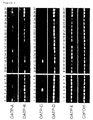
Fig. 1 is a photograph showing the results of examination by
the RT-PCR method for the expression level of respective OATP family
genes in human fetal and adult tissues: 1. fetal brain, 2. fetal heart,
3. fetal kidney, 4. fetal liver, 5. fetal lung, 6. fetal skeletal
muscle, 7. fetal spleen, 8. fetal thymus, 9. adult pancreas, 10. adult
kidney, 11. adult skeletal muscle, 12. adult liver, 13. adult lung,
14. adult placenta, 15. adult brain, 16. adult heart, 17. adult
peripheral blood leukocytes, 18. adult large intestine, 19. adult
small intestine, 20. adult ovary, 21. adult testis, 22. adult prostate,
23. adult thymus, 24. adult spleen, 25. adult bone marrow, 26. adult
lymph node, and 27. adult tonsil.
Fig. 2 is a photograph showing the results of examination by
the RT-PCR method for the expression level of respective OATP family
genes in human cancer cells: 1. mammary cancer cells (GI-101), 2.
lung cancer cells (LX-1), 3. large intestinal adenoma cells (CX-1),
4. lung cancer cells (GI-117), 5. prostatic adenoma cells, 6. large
intestinal adenoma cells (GI-112), 7. ovary cancer cells (GI-102),
and 8. pancreatic adenoma cells (GI-103).
Fig. 3 is a bar graph showing the results of transport experiments
for various labeled compounds. More particularly, the transport
activities for various labeled compounds of HEK293 cells transfected
with OATP-C (OATP-C) or with the vector alone (mock) are shown.
Fig. 4 is a graph showing the concentration-dependency of the
transport activity for PCG of HEK293 cells expressing the OATP-C
protein.
Fig. 5 is a bar graph showing the effect of various β-lactam
antibiotics on the transport activity for PCG in HEK293 cells wherein
the OATP-C protein is expressed. The transport activity in the control
(with no inhibitor) is taken as 100%.
Fig. 6 is a bar graph showing the sodium ion and chloride ion
dependencies of the transport activity for estradiol-17β-glucuronide
"OATP-C" represents the transport activity for
estradiol-17β-glucuronide of HEK293 cells transfected with OATP-C ,
and "Mock" represents the transport activity for
estradiol-17β-glucuronide of HEK293 cells transfected with the vector
alone.
Best Mode for Carrying out the Invention
The present invention is described below in more detail with
reference to examples, but is not construed as being limited thereto.
Molecular biological experimental techniques in general have been
performed principally according to methods described in usual
experimental textbooks, such as "Sambrook, J., Fritsch, E. F., Maniatis,
T., Molecular Cloning, Cold Spring Harbor Lab. press (1989)".
[Example 1] Cloning of cDNAs comprising the entire open reading frame
(ORF) of OATP-B, C, D, or E gene
OATP-B
The OABE-1 primer (5' gat aag ctt ctg tgt ggc cca aga aga act
gac 3' /SEQ ID NO: 9) and OABE-6 primer (5' gat aag ctt tac tgc tgt
ggc tgc tac tct tgg 3' /SEQ ID NO: 10) were prepared based on the
nucleotide sequences of W19504 and AI052501, ESTs possibly encoding
the amino acid sequence having significant homology with the human
OATP-A protein. Using these primers, PCR was performed against human
adult brain polyA+ RNA-derived cDNA as a template to amplify the OATP-B
cDNA comprising the entire ORF. The OATP-B cDNA thus amplified by
PCR was cleaved at the Hind III site added to the primer, and incorporated
into the Hind III site of the pcDNA3 vector (Invitrogen) , an expression
vector for mammalian cells. By sequencing a plurality of clones, a
clone (pcDNA3/OATP-B) with no PCR error was selected to be used in
expression experiments.
OATP-C
Respective primers were prepared from the nucleotide sequences
of the following ESTs possibly encoding amino acid sequences having
a significant homology with the human OATP-A:
Using these primers, the human OATP-C divided into three
fragments was cloned.
The 5'-end fragments were cloned by the 5' RACE (Rapid
Amplification cDNAEnds) method. More specifically, PCR was performed
against the human fetal liver-derived Marathon-Ready™ cDNA (CLONTECH)
as a template using a combination of the AP1 primer, a linker primer
attached to the kit, and the 2893-4 primer to amplify the 5'-end fragment
of the human OATP-C cDNA of about 400 bp. This cDNA fragment was
incorporated into the pT7Blue-T vector (Novagen) by the TA cloning
method, and a plurality of subclones thus obtained were sequenced
to determine the 5'-end sequence of the human OATP-C cDNA. The 3'-end
sequence was similarly cloned by the 3' RACE method. Specifically,
PCR was performed with the human fetal liver-derived Marathon-Ready™
cDNA (CLONTECH) as a template using a combination of the AP1 primer,
a linker primer attached to the kit, and the 3863-1 primer to amplify
the 3'-end fragment of the human OATP-C cDNA of about 1.5 kbp. This
cDNA fragment was incorporated into the pT7Blue-T vector by the TA
cloning method, and a plurality of subclones thus obtained were
sequenced to determine the 3'-end sequence of the human OATP-C cDNA.
In addition, the intermediate fragment between the 5'-end sequence
and the 3'-end sequence was amplified by PCR with cDNA derived from
the human adult liver as a template using a combination of the 2893-1
and 9414-2 primers. The fragment of about 1.2 kbp thus obtained was
purified by the gel filtration method and directly sequenced to
determine the nucleotide sequence. By combining the obtained
sequences, the cDNA sequence comprising the entire ORF of human OATP-C
was determined.
The expression plasmid was constructed as follows. The human
OATP-C was divided into two fragments, and they were amplified by
PCR with the human adult liver-derived cDNA as a template using a
combination of the following primers:
5'-end
3'-end
These fragments were subcloned into the pT7Blue-T vector,
respectively, to select clones with no PCR error. After both clones
were linked at the Bgl II sites existing in the overlapping regions,
the product was cleaved at the Kpn I sites existing at both ends,
and incorporated into the Kpn I site of the pcDNA3 vector to obtain
the expression plasmid, pcDNA3/OATP-C.
OATP-D
0224-3 primer (5' cgc cct cgt ggt ttt tga tgt agc 3' /SEQ ID
NO: 19) was prepared from EST AA280224, an EST possibly encoding the
amino acid sequence having significant homology with the human OATP-A
protein. Furthermore, it was found that a partial sequence of the
PAC clone (pDJ430i19) derived from q26.1 region of human chromosome
15 could also encode the amino acid sequence having significant homology
with the human OATP-A protein. PAC151-2 primer (5' gcg gtg cct tac
tct tct tct ctt 3' /SEQ ID NO: 20) was prepared from this sequence.
PCR was performed using these primers on the human adult brain-derived
cDNA as a template to amplify a cDNA fragment of about 1.1 kbp. Using
this cDNA fragment as a probe, the human adult kidney-derived 5'-STRETCH
PLUS cDNA library (CLONTECH) was screened by the plaque hybridization
method. PCR was performed with a phage suspension of obtained positive
clones as a template using a combination of the above-described primers,
or OATP-D gene specific primer prepared from the above-elucidated
sequence and GT10 S1 primer (5' ctt ttg agc aag ttc agc ct 3' /SEQ
ID NO: 21) or GT10 A1 primer (5' aga ggt ggc tta tga gta ttt ctt 3'
/SEQ ID NO: 22) prepared from the sequence of λgt-10 vector, and the
fragments thus amplified were directly sequenced to determine the
nucleotide sequence. Furthermore, the region covered by the phage
clone was extended to determine the entire ORF sequence by the screening
using the DNA fragment comprising the newly obtained region as a probe.
OATP-E
7130-1 primer (5' tgt aca agg tgc tgg gcg tcc tct 3' /SEQ ID
NO: 23) and 7130-4 primer (5' cga tcg ggt ata aaa cac att cta 3' /SEQ
ID NO: 24) were prepared from EST AI347130, an EST potentially encoding
the amino acid sequence having significant homology with the human
OATP-A protein. PCR was performed using these primers with the human
adult lung-derived cDNA as a template to amplify a cDNA fragment of
about 400 bp. The human adult kidney-derived 5'-STRETCH PLUS cDNA
library (CLONTECH) was screened using this CDNA as a probe by the
plaque hybridization method. PCR was performed with a phage suspension
of obtained positive clones as a template using a combination of the
above-described primers or the OATP-E gene specific primer prepared
from the above-elucidated sequence and the GT10-S1 primer or GT10-A1
primer prepared from the λgt-10 vector sequence, and amplified
fragments thus obtained were directly sequenced to determine the
nucleotide sequence. Furthermore, the region covered by the phage
clone was extended to determine the entire ORF sequence by the screening
using the DNA fragment comprising the newly obtained region as a probe.
The expression plasmid was constructed as follows. The human
OATP-E was divided into two fragments, 5'-end and 3'-end fragments,
which were amplified by PCR using combinations of the following primers.
The 5'-end fragment was amplified using human adult lung-derived cDNA
as a template, and the 3'-end fragment was amplified using human fetal
lung-derived cDNA as a template, respectively.
5'-end:
3'-end:
These fragments were subcloned into the Hind III/BamH I site
of the pcDNA3 vector, respectively, to select clones with no PCR error.
Both clones were linked at the BstP I sites existing in the overlapping
regions to obtain the expression plasmid, pcDNA3/OATP-E.
PCR
PCR was fundamentally performed under the following basic
conditions with appropriate modifications if necessary.
<Composition of reaction solution>
template DNA,
10 x ExTaq buffer (TaKaRa) 5 µl,
2.5 mM dNTPs (TaKaRa) 4 µl,
ExTaq (TaKaRa) 0.5 µl,
TaqStart™ Antibody (CLONTECH) 0.5 µl,
sense primer 10 to 20 pmol, and
antisense primer 10 to 20 pmol
/total volume 50 µl.
<Reaction conditions>
PCR in general:
94°C, 2 min -> (94°C, 30 s -> 55 to 62°C, 30 s -> 72°C, 2 to
3 min) x 25 to 40 cycles -> 72°C, 10 min.
RACE method:
94°C, 2 min -> (94°C, 30 s -> 68°C, 4 min) x 5 cycles -> (94°C,
30 s -> 62°C, 30 s -> 72°C, 2 min) x 30 cycles -> 72°C, 10 min.
Synthesis of cDNA
cDNAs used as templates for PCR were prepared using the
SUPERSCRIPT™ II RNase H- reverse transcriptase (GIBCO BRL) according
to the usual method recommended by the supplier. Specifically, 10
µg of the total RNA or 2 µg of poly A+ RNA and about 1 µg of the oligo
dT primer (GIBCO BRL) or about 0.5 µg of the random hexamer primer
(GIBCO BRL) were mixed, heated at 70°C for 10 min, and then cooled
on ice. The first strand buffer (GIBCO BRL), DDT (final concentration
of 10 mM), dNTPs (final concentration of 0.5 mM; GIBCO BRL), and 400
to 800 U of SUPERSCRIPT™ II RNase H- reverse transcriptase were added
to this mixture, and the resulting mixture was warmed at 42°C for
1 h to synthesize cDNA. The mixture was then heated at 70°C for 15
min, and a portion thereof was used as the template.
Hybridization
DNA fragments amplified by PCR, or purified from gels after
agarose electrophoresis, and such were labeled with [α-32P]dCTP using
the Ready-to Go DNA labeling beads (Pharmacia) by the random primer
method to be used as the primer. The hybridization was performed using
the ExpressHyb Hybridization Solution (CLONTECH) by heating at 68°C
for 2 h or more according to the method recommended by the supplier.
After the hybridization, the filter was washed twice in a solution
of 2 X SSC and 0.1% SDS solution at room temperature for 20 min, and
then, twice in a solution of 0.1 x SSC and 0.1% SDS at 50°C for 20
min.
[Example 2] Analysis by RT-PCR method
Using the following primers specific for respective genes, the
tissue distribution of the expression of each gene was analyzed by
the RT-PCR method.
OATP-A
OATP-B
OATP-C
OATP-D
OATP-E
G3PDH
PCR was performed with appropriate amounts of cDNAs derived from
various organs and cells contained in the Multiple Tissue cDNA (MTC™)
Panel (CLONTECH) as template using the above-described primers.
PCR-amplified products were analyzed by agarose electrophoresis (Fig.
1 and 2). OATP-A showed an expression pattern comparatively restricted
in brain and liver. Expression of OATP-C was discovered to be
restricted in liver in both fetal and adult tissues examined. Although
OATP-B, OATP-D, and OATP-E were found to be expressed in a relatively
wide range of tissues, it was revealed that OATP-B and OATP-E, among
them, were expressed extremely low in the peripheral blood leukocytes ,
thymus, and spleen. These findings strongly indicate that expressions
of OATP-B and OATP-E are low in hemocytes. On the other hand, the
examination of OATPs in cancer cells have revealed that both OATP-D
and OATP-E are expressed in a high frequency in these cells (Fig.
2). From these results, the potential production of an anticancer
agent which is specifically incorporated into cells by the OATP-E
protein is expected, such an agent being an anticancer agent with
an attenuated side effects on hematopoietic cells (e.g. bone marrow
suppression, and such).
[Example 3] Transport experiment
The human fetal kidney-derived cell strain, HEK 293 cells, were
transfected with the plasmid pcDNA3/OATP-C or the pcDNA3 vector
containing no insert as a control (mock) by the calcium phosphate
method. Specifically, the plasmid DNA (10 µg), a Hepes buffer solution
(137 mM NaCl, 5 mM KCl, 0.7 mM Na2HFO4, 6 mM dextrose, and 21 mM Hepes
pH 7.1) (1 ml), and 2 M CaCl2 (62.5 µl) were mixed and the resultant
mixture was allowed to stand at room temperature for 30 min or more
to form calcium phosphate coprecipitates. After the cells were plated
on 10-cm diameter plates at a concentration of 1.5 x 10⁶ cells per
plate, and the cells were cultured for 24 h. The above-described
calcium phosphate coprecipitates were added thereto, and the cells
were further cultured for 24 h. Then, plates were washed with phosphate
buffered saline (PBS), and after the addition of fresh culture medium,
the cells were further cultured for 24 h.
Transport experiment was performed using the cells transfected
with the plasmid DNA according to the following procedures. The cells
were detached from the plates using a rubber policeman, suspended
in a transport buffer (containing 125 mM NaCl, 4.8 mM KCl, 5.6 mM
(+)-glucose, 1.2 mM CaCl2, 1.2 mM KH2PO4, 1.2 mM MgSO4, and 25 mM Hepes
pH 7.4), and pre-incubated for 20 min. Each of various labeled
substrates ([3H]methotrexate, [3H]digoxin, [3H]ouabain,
[3H]prostaglandin E2, [3H]estradiol-17β-glucuronide,
[3H]estran-3-sulfate, [14C]PCG<benzylpenicillin>, and so on) was then
added in an appropriate amount to the above-described cell suspension,
and the resulting mixture was incubated at 37°C for a predetermined
period of time. Incubated cells were overlaid on a silicon layer formed
by laying a mixture of silicon oil and liquid paraffin (specific gravity
= 1.022) on a 3 M KCl layer, and separated by centrifugation.
Radioactivity of cells was measured to determine the into-the-cell
transport activity. In this case, 1 x 10⁶ cells were used as one point
of cells.
Herein, the culture of HEK 293 cells was performed using
Dulbecco's MEM containing 10% FCS (fetal calf serum) as the culture
medium in an atmosphere of 5% carbon dioxide at 37°C.
From measuring the transport activity in HEK293 cells wherein
the OATP-C proteins had been expressed, transport was obviously
observed with estradiol-17β-glucuronide, estron-3-sulfate, and PCG.
A weak transport activity was also observed with methotrexate, ouabain,
and prostaglandin E2 (Fig. 3).
Further, to obtain the Km (Michaelis constant) value of the PCG
transport mediated by the OATP-C protein, the uptake of [14C]PCG added
at various concentrations was measured (Fig. 4). From the
Lineweaver-Burk reciprocal plot of the net PCG uptake obtained in
the cells wherein the OATP-C proteins are expressed, a Km value of
983±289 µM PCG was obtained with a maximal velocity Vmax of 5.45
±0.63 (nmol/mg/15 min).
Furthermore, the effects of the addition of various β-lactam
antibiotics on the PCG transport mediated by the OATP-C protein were
examined (Fig. 5). Remarkable inhibitory activity was observed with
cefazolin, cefoperazone, cefpiramide, and nafcillin when effects of
various β-lactam antibiotics were examined by adding them at a
concentration of 1 mM, respectively, to the transport activity of
4µM [14C]PCG. A weak inhibitory activity was also seen with
cefaloridine and cefalexin. These results strongly indicate that
these β-lactam antibiotics can be also transported by the OATP family
proteins similarly as PCG belonging to the same β-lactam antibiotics.
Further, the requirement for sodium and chloride ions in the
transport of estradiol-17β-glucuronide mediated by the OATP-C protein
was examined (Fig. 6). No alteration was observed in the transport
of estradiol-17β-glucuronide either in case where the sodium ion was
replaced with N-methylglucamine, or where the chloride ion was
substituted with gluconate. According to these results, it has been
revealed that the transport mediated by the OATP-C protein is
independent to sodium ion.
Industrial Applicability
The present invention provides novel transporter proteins and
genes encoding these proteins. These proteins and genes are useful
for developing drugs with a new design that can be transported by
transporter proteins of this invention, and pharmaceuticals for the
treatment of disorders caused by expressional and functional
aberrations of these transporter proteins. Furthermore, these
proteins and genes can be applied to genetic diagnosis and gene therapy.
For example, by the SNP diagnosis and such of transporter genes of
this invention, it is possible to design a tailor-made treatment plan,
taking individual differences in efficacy of drugs into consideration.