EP1219711A2 - Lawsonia intracellularis vaccine - Google Patents
Lawsonia intracellularis vaccine Download PDFInfo
- Publication number
- EP1219711A2 EP1219711A2 EP01204919A EP01204919A EP1219711A2 EP 1219711 A2 EP1219711 A2 EP 1219711A2 EP 01204919 A EP01204919 A EP 01204919A EP 01204919 A EP01204919 A EP 01204919A EP 1219711 A2 EP1219711 A2 EP 1219711A2
- Authority
- EP
- European Patent Office
- Prior art keywords
- protein
- acid sequence
- nucleic acid
- lawsonia intracellularis
- amino acid
- Prior art date
- Legal status (The legal status is an assumption and is not a legal conclusion. Google has not performed a legal analysis and makes no representation as to the accuracy of the status listed.)
- Granted
Links
Images

Classifications
-
- C—CHEMISTRY; METALLURGY
- C07—ORGANIC CHEMISTRY
- C07K—PEPTIDES
- C07K14/00—Peptides having more than 20 amino acids; Gastrins; Somatostatins; Melanotropins; Derivatives thereof
- C07K14/195—Peptides having more than 20 amino acids; Gastrins; Somatostatins; Melanotropins; Derivatives thereof from bacteria
- C07K14/205—Peptides having more than 20 amino acids; Gastrins; Somatostatins; Melanotropins; Derivatives thereof from bacteria from Campylobacter (G)
-
- A—HUMAN NECESSITIES
- A61—MEDICAL OR VETERINARY SCIENCE; HYGIENE
- A61P—SPECIFIC THERAPEUTIC ACTIVITY OF CHEMICAL COMPOUNDS OR MEDICINAL PREPARATIONS
- A61P1/00—Drugs for disorders of the alimentary tract or the digestive system
-
- A—HUMAN NECESSITIES
- A61—MEDICAL OR VETERINARY SCIENCE; HYGIENE
- A61P—SPECIFIC THERAPEUTIC ACTIVITY OF CHEMICAL COMPOUNDS OR MEDICINAL PREPARATIONS
- A61P1/00—Drugs for disorders of the alimentary tract or the digestive system
- A61P1/12—Antidiarrhoeals
-
- A—HUMAN NECESSITIES
- A61—MEDICAL OR VETERINARY SCIENCE; HYGIENE
- A61P—SPECIFIC THERAPEUTIC ACTIVITY OF CHEMICAL COMPOUNDS OR MEDICINAL PREPARATIONS
- A61P3/00—Drugs for disorders of the metabolism
-
- A—HUMAN NECESSITIES
- A61—MEDICAL OR VETERINARY SCIENCE; HYGIENE
- A61P—SPECIFIC THERAPEUTIC ACTIVITY OF CHEMICAL COMPOUNDS OR MEDICINAL PREPARATIONS
- A61P31/00—Antiinfectives, i.e. antibiotics, antiseptics, chemotherapeutics
- A61P31/04—Antibacterial agents
-
- A—HUMAN NECESSITIES
- A61—MEDICAL OR VETERINARY SCIENCE; HYGIENE
- A61P—SPECIFIC THERAPEUTIC ACTIVITY OF CHEMICAL COMPOUNDS OR MEDICINAL PREPARATIONS
- A61P7/00—Drugs for disorders of the blood or the extracellular fluid
- A61P7/06—Antianaemics
-
- C—CHEMISTRY; METALLURGY
- C07—ORGANIC CHEMISTRY
- C07K—PEPTIDES
- C07K14/00—Peptides having more than 20 amino acids; Gastrins; Somatostatins; Melanotropins; Derivatives thereof
- C07K14/195—Peptides having more than 20 amino acids; Gastrins; Somatostatins; Melanotropins; Derivatives thereof from bacteria
-
- A—HUMAN NECESSITIES
- A61—MEDICAL OR VETERINARY SCIENCE; HYGIENE
- A61K—PREPARATIONS FOR MEDICAL, DENTAL OR TOILETRY PURPOSES
- A61K38/00—Medicinal preparations containing peptides
-
- A—HUMAN NECESSITIES
- A61—MEDICAL OR VETERINARY SCIENCE; HYGIENE
- A61K—PREPARATIONS FOR MEDICAL, DENTAL OR TOILETRY PURPOSES
- A61K39/00—Medicinal preparations containing antigens or antibodies
-
- A—HUMAN NECESSITIES
- A61—MEDICAL OR VETERINARY SCIENCE; HYGIENE
- A61K—PREPARATIONS FOR MEDICAL, DENTAL OR TOILETRY PURPOSES
- A61K48/00—Medicinal preparations containing genetic material which is inserted into cells of the living body to treat genetic diseases; Gene therapy
Definitions
- the present invention relates to nucleic acid sequences encoding novel Lawsonia intracellularis proteins, to DNA fragments, recombinant DNA molecules and live recombinant carriers comprising these sequences, to host cells comprising such nucleic acid sequences, DNA fragments, recombinant DNA molecules and live recombinant carriers, to the proteins encoded by these nucleotide sequences, to vaccines for combating Lawsonia intracellularis infections and methods for the preparation thereof, and to diagnostic tools for the detection of Lawsonia intracellularis .
- Porcine proliferative enteropathy has become an important disease of the modern pig industry world-wide. The disease affects 15% to 50% of the growing herds and up to 30% of the individual animals in established problem herds. Today annual economical losses have been estimated US$ 5-10 in extra feed and facility time costs per affected pig.
- PPE is a group of chronic and acute conditions of widely differing clinical signs (death, pale and anaemic animals, watery, dark or bright red diarrhoea, depression, reduced appetite and reluctance to move, retarded growth and increased FCR).
- the first a pathological change only visible at necropsy, is a thickening of the small intestine and colon mucosa.
- the second is the occurrence of intracytoplasmatic small-curved bacteria in the enterocytes of the affected intestine. These bacteria have now been established as the etiological agent of PPE and have been name Lawsonia intracellularis .
- Lawsonia intracellularis has been found to affect virtually all animals including monkeys, rabbits, ferrets, hamsters, fox, horses, and other animals as diverse as ostrich and emoe.
- Lawsonia intracellularis is a gram-negative, flagellated bacterium that multiplies in eukaryotic enterocytes only and no cell-free culture has been described.
- Lawsonia intracellularis In order to persist and multiply in the cell Lawsonia intracellularis must penetrate dividing crypt cells. The bacterium associates with the cell membrane and quickly enters the enterocyte via an entry vacuole. This then rapidly breaks down (within 3 hours) and the bacteria flourish and multiply freely in the cytoplasm. The mechanisms by which the bacteria cause infected cells to fail to mature, continue to undergo mitosis and form hypoplastic crypt cells is not yet understood.
- Lawsonia intracellularis infection, treatment and control of the disease has-been hampered by the fact that Lawsonia intracellularis can not be cultivated in cell-free media. Although there are reports of successful co-culturing Lawsonia intracellularis in rat enterocytes this has not lead to the development of vaccines for combating Lawsonia intracellularis , although there clearly is a need for such vaccines.
- Lawsonia intracellularis produces three novel outer membrane proteins (OMPs) that, alone or in combination, are capable of inducing protective immunity against Lawsonia intracellularis .
- OMPs outer membrane proteins
- the three novel outer membrane proteins will be referred to as the 19/21 kD, 37 kD and 50 kD protein.
- the 19/21 kD protein is found in two different forms, a 19 kD form and a 21 kD form, one protein being a modified form of the other and both comprising an identical amino acid sequence.
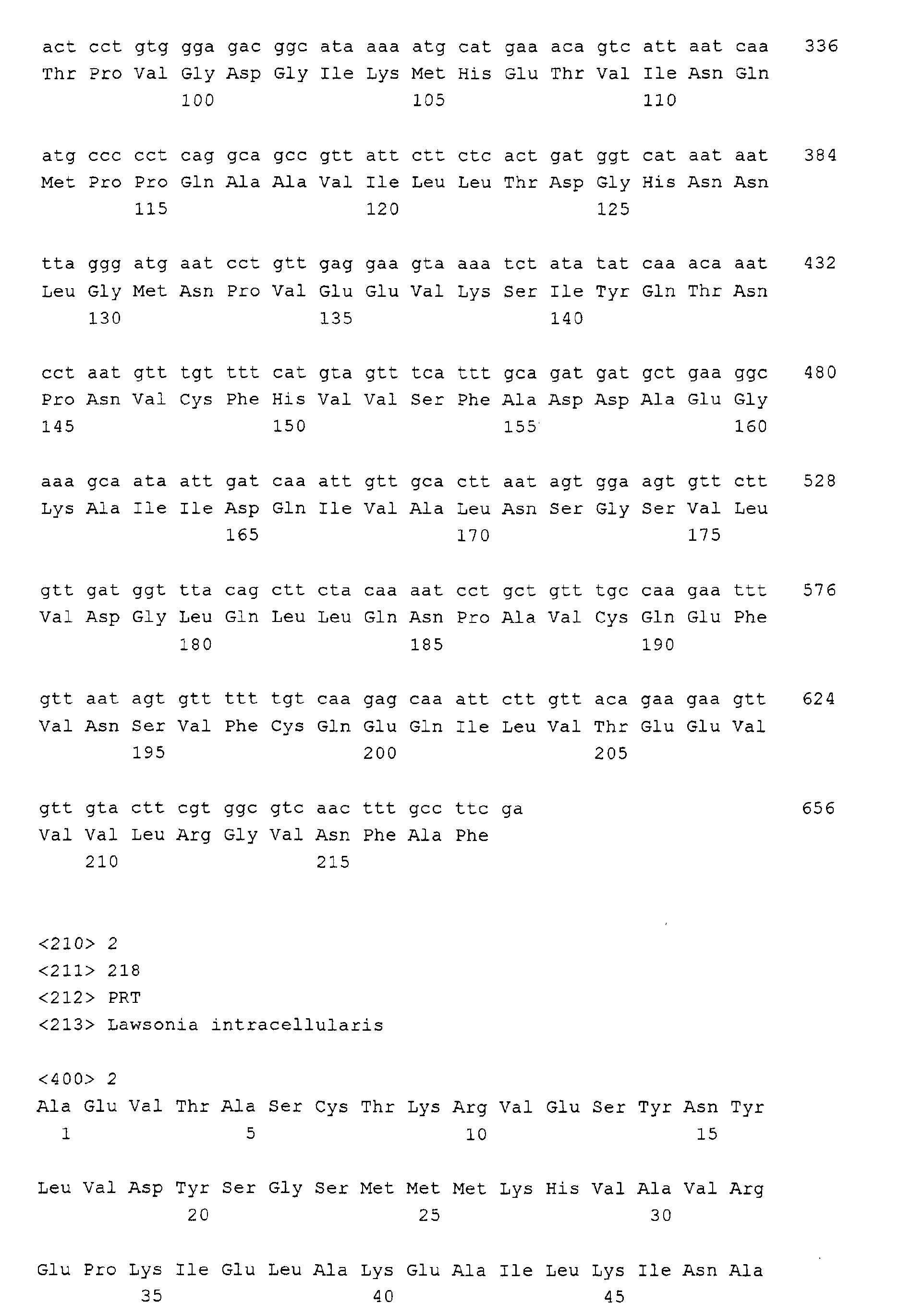
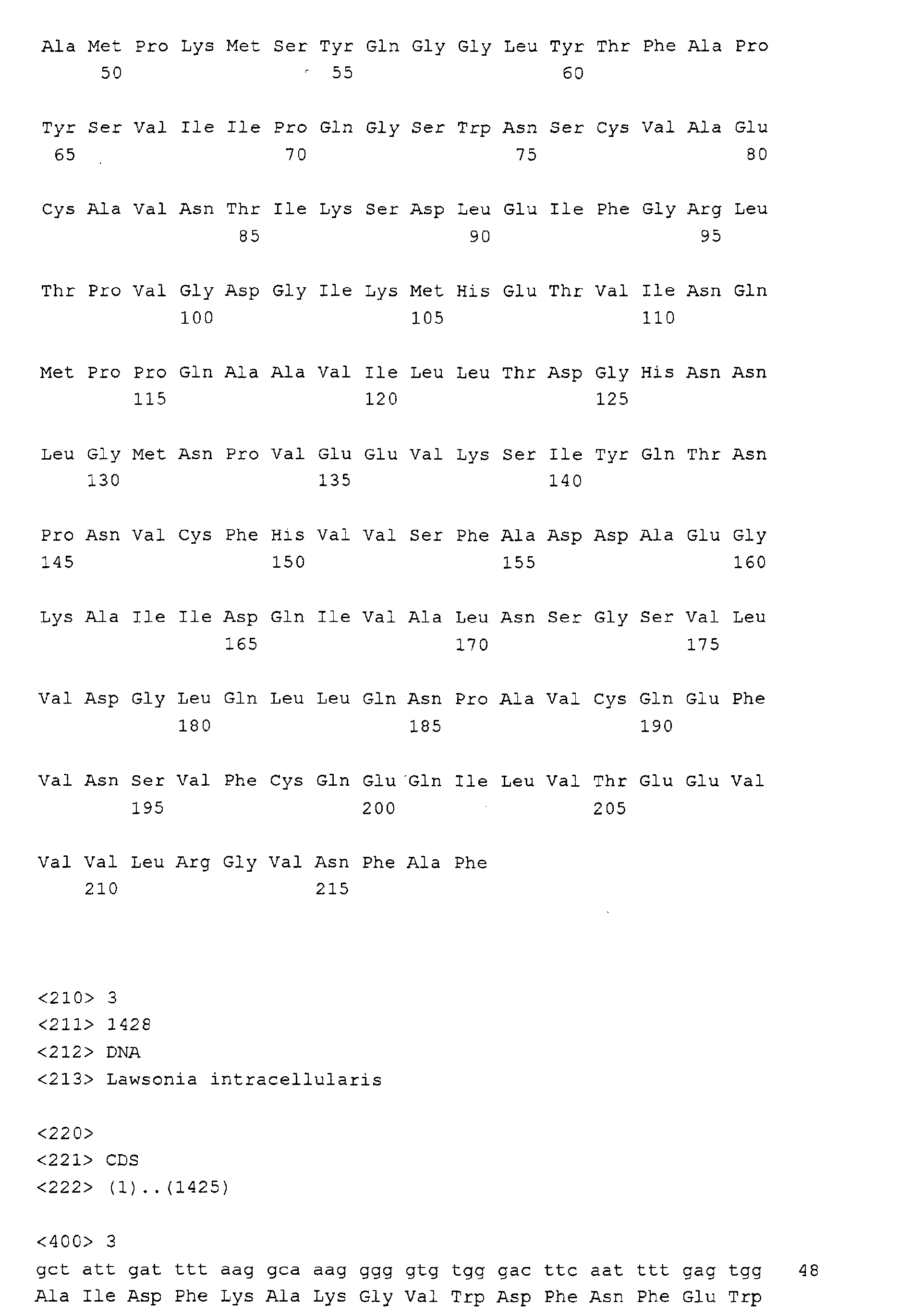
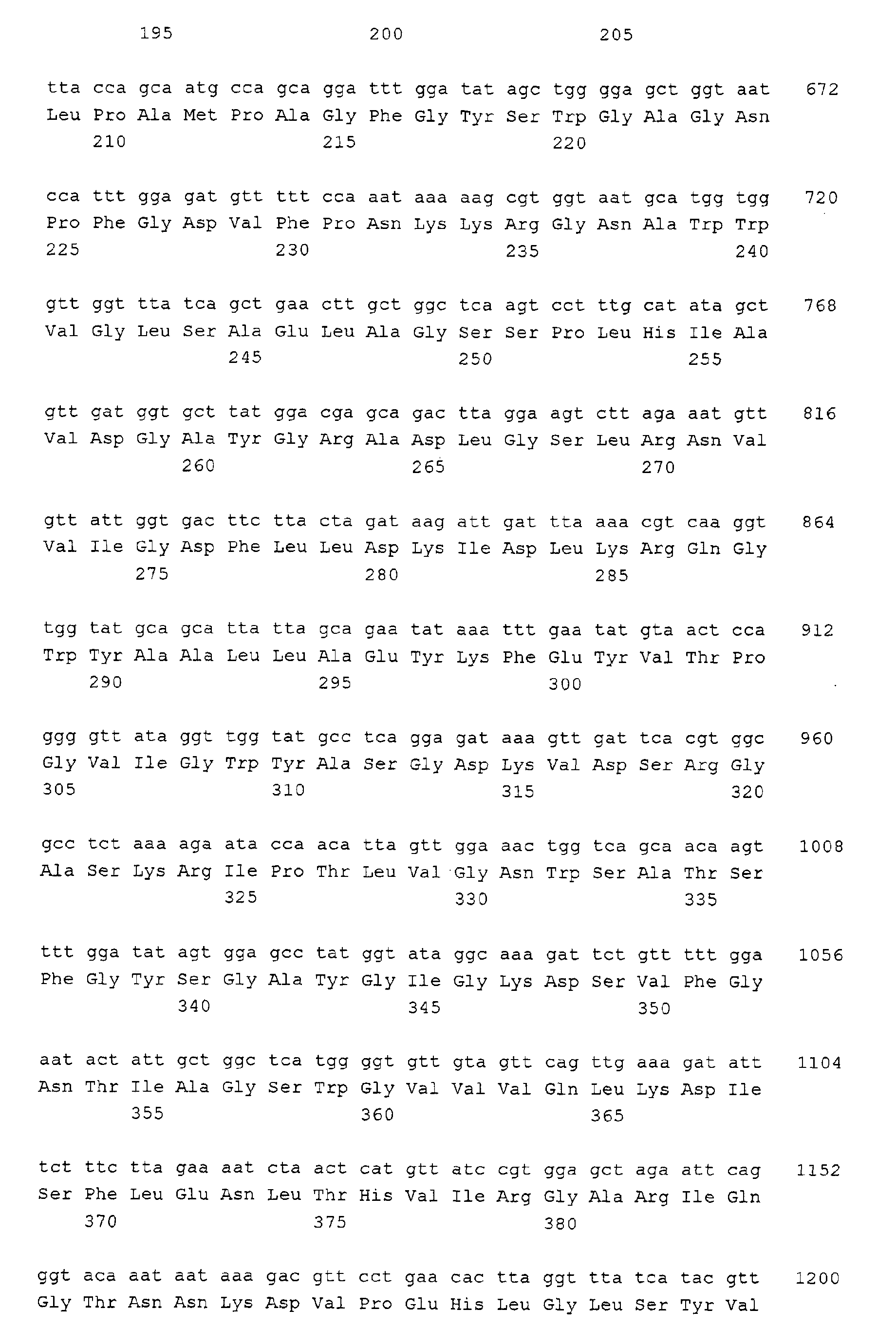
- the amino acid sequences of the 37 kD and 50 kD protein are presented in sequence identifiers SEQ ID NO: 2 and 4.
- the genes encoding these two proteins have been sequenced and their nucleic acid sequence is shown in sequence identifiers SEQ ID NO: 1 and 3.
- the 19/21 kD protein is characterised by three internal amino acid sequences of respectively 7, 12 and 12 amino acids. These amino acid sequences are presented in SEQ ID NO: 5, 6 and 7.
- nucleic acid sequences can encode one and the same protein. This phenomenon is commonly known as wobble in the second and especially the third base of each triplet encoding an amino acid. This phenomenon can result in a heterology of about 30% for two nucleic acid sequences still encoding the same protein. Phenomenon. Therefore, two nucleic acid sequences having a sequence homology of about 70 % can still encode one and the same protein.
- one embodiment relates to nucleic acid sequences encoding a Lawsonia intracellularis protein and to parts of that nucleic acid sequence that encode an immunogenic fragment of that protein, wherein those nucleic acid sequences or parts thereof have a level of homology with the nucleic acid sequence of SEQ ID NO: 1 of at least 70 %.
- the nucleic acid sequence encoding this Lawsonia intracellularis protein or the part of said nucleic acid sequence has at least 80 %, preferably 90 %, more preferably 95 % homology with the nucleic acid sequence of SEQ ID NO: 1. Even more preferred is a homology level of 98% or even 100%.
- this embodiment relates to nucleic acid sequences encoding a Lawsonia intracellularis protein and to parts of that nucleic acid sequence that encode an immunogenic fragment of that protein, that have a level of homology with the nucleic acid sequence of SEQ ID NO: 3 of at least 70 %.
- the nucleic acid sequence encoding this Lawsonia intracellularis protein or the part of said nucleic acid sequence has at least 80 %, preferably 90 %, more preferably 95 % homology with the nucleic acid sequence of SEQ ID NO: 3. Even more preferred is a homology level of 98% or even 100%
- the level of nucleotide homology can be determined with the computer program "BLAST 2 SEQUENCES” by selecting sub-program: “BLASTN” that can be found at www.ncbi.mm.nih.gov/blast/b12seg/b12.html.
- a reference for this program is Tatiana A. Tatusova, Thomas L. Madden FEMS Microbiol. Letters 174: 247-250 (1999). Parameters used are the default parameters: Reward for a match: +1. Penalty for a mismatch: -2. Open gap: 5. Extension gap: 2. Gap x_dropoff: 50.
- one form of this embodiment of the invention relates to nucleic acid sequences encoding a novel Lawsonia intracellularis protein comprising an amino acid sequence as depicted in SEQ ID NO: 2, or an immunogenic fragment of that polypeptide.
- nucleic acid sequence has a homology of at least 90 %, more preferably 95 %, 98 % or even 100 % with the nucleic acid sequence as depicted in SEQ ID NO: 1.
- one form of this embodiment of the invention relates to nucleic acid sequences encoding a novel Lawsonia intracellularis protein having an amino acid sequence as depicted in SEQ ID NO: 4, or an immunogenic fragment of said polypeptide.
- nucleic acid sequence has a homology of at least 90, more preferably 95 %, 98 % or even 100 % % with the nucleic acid sequence as depicted in SEQ ID NO: 3.
- the invention relates to DNA fragments comprising a nucleic acid sequence according to the invention.
- DNA fragments can e.g. be plasmids, into which a nucleic acid sequence according to the invention is cloned.
- DNA fragments are e.g. useful for enhancing the amount of DNA for use as a primer, as described below.
- nucleic acid sequence An essential requirement for the expression of the nucleic acid sequence is an adequate promoter functionally linked to the nucleic acid sequence, so that the nucleic acid sequence is under the control of the promoter. It is obvious to those skilled in the art that the choice of a promoter extends to any eukaryotic, prokaryotic or viral promoter capable of directing gene transcription in cells used as host cells for protein expression.
- an even more preferred form of this embodiment relates to a recombinant DNA molecule comprising a DNA fragment or a nucleic acid sequence according to the invention that is placed under the control of a functionally linked promoter.
- a functionally linked promoter This can be obtained by means of e.g. standard molecular biology techniques. (Maniatis/Sambrook (Sambrook, J. Molecular cloning: a laboratory manual, 1989. ISBN 0-87969-309-6).
- Functionally linked promoters are promoters that are capable of controlling the transcription of the nucleic acid sequences to which they are linked.
- Such a promoter can be a Lawsonia promoter e.g.
- useful expression control sequences which may be used include the Trp promoter and operator (Goeddel, et al., Nucl. Acids Res., 8, 4057, 1980); the lac promoter and operator (Chang, et al., Nature, 275, 615, 1978); the outer membrane protein promoter (Nakamura, K.
- illustrative useful expression control sequences include the SV-40 promoter (Berman, P.W. et al., Science, 222, 524-527, 1983) or the metallothionein promoter (Brinster, R.L., Nature, 296, 39-42, 1982) or a heat shock promoter (Voellmy et al., Proc. Natl. Acad. Sci. USA, 82, 4949-53, 1985).
- Bacterial, yeast, fungal, insect and mammalian cell expression systems are very frequently used systems. Such systems are well-known in the art and generally available, e.g. commercially through Clontech Laboratories, Inc. 4030 Fabian Way, Palo Alto, California 94303-4607, USA. Next to these expression systems, parasite-based expression systems are very attractive expression systems. Such systems are e.g. described in the French Patent Application with Publication number 2 714 074, and in US NTIS Publication No US 08/043109 (Hoffman, S. and Rogers, W.: Public. Date 1 December 1993).
- a still even more preferred form of this embodiment of the invention relates to Live Recombinant Carriers (LRCs) comprising a nucleic acid sequence encoding the 19/21 kD, 37 kD or 50 kD protein or an immunogenic fragment thereof according to the invention, a DNA fragment according to the invention or a recombinant DNA molecule according to the invention.
- LRCs Live Recombinant Carriers
- Such carriers are e.g. bacteria and viruses.
- These LRCs are micro-organisms or viruses in which additional genetic information, in this case a nucleic acid sequence encoding the 19/21 kD, 37 kD or 50 kD protein or an immunogenic fragment thereof according to the invention has been cloned.
- LRCs Animals infected with such LRCs will produce an immunogenic response not only against the immunogens of the carrier, but also against the immunogenic parts of the protein(s) for which the genetic code is additionally cloned into the LRC, e.g. the 19/21 kD, 37 kD or 50 kD gene.
- LRC viruses may be used as a way of transporting the nucleic acid sequence into a target cell.
- Live recombinant carrier viruses are also called vector viruses.
- Viruses often used as vectors are Vaccinia viruses (Panicali et al; Proc. Natl. Acad. Sci. USA, 79: 4927 (1982), Herpesviruses (E.P.A. 0473210A2), and Retroviruses (Valerio, D. et al; in Baum, S.J., Dicke, K.A., Lotzova, E. and Pluznik, D.H. (Eds.), Experimental Haematology today - 1988. Springer Verlag, New York: pp. 92-99 (1989)).
- the technique of in vivo homologous recombination can be used to introduce a recombinant nucleic acid sequence into the genome of a bacterium, parasite or virus of choice, capable of inducing expression of the inserted nucleic acid sequence according to the invention in the host animal.
- a host cell comprising a nucleic acid sequence encoding a protein according to the invention, a DNA fragment comprising such a nucleic acid sequence or a recombinant DNA molecule comprising such a nucleic acid sequence under the control of a functionally linked promoter.
- This form also relates to a host cell containing a live recombinant carrier containing a nucleic acid molecule encoding a 19/21 kD, 37 kD or 50 kD protein or a fragment thereof according to the invention.
- a host cell may be a cell of bacterial origin, e.g.
- Escherichia coli, Bacillus subtilis and Lactobacillus species in combination with bacteria-based plasmids as pBR322, or bacterial expression vectors as pGEX, or with bacteriophages.
- the host cell may also be of eukaryotic origin, e.g. yeast-cells in combination with yeast-specific vector molecules, or higher eukaryotic cells like insect cells (Luckow et al; Bio-technology 6: 47-55 (1988)) in combination with vectors or recombinant baculoviruses, plant cells in combination with e.g. Ti-plasmid based vectors or plant viral vectors (Barton, K.A. et al; Cell 32: 1033 (1983), mammalian cells like Hela cells, Chinese Hamster Ovary cells (CHO) or Crandell Feline Kidney-cells, also with appropriate vectors or recombinant viruses.
- yeast-cells in combination with yeast-specific vector molecules
- Another embodiment of the invention relates to the novel proteins: the 19/21 kD protein, the 37 kD and 50 kD protein and to immunogenic fragments thereof according to the invention.
- One form of this embodiment relates i.a. to Lawsonia intracellularis proteins that have an amino acid sequence that is at least 70 % homologous to the amino acid sequence as depicted in SEQ ID NO: 2 and to immunogenic fragments of said protein.
- the embodiment relates to such Lawsonia intracellularis proteins that have a sequence homology of at least 80 %, preferably 90 %, more preferably 95 % homology to the amino acid sequence as depicted in SEQ ID NO: 2 and to immunogenic fragments of such proteins. Even more preferred is a homology level of 98% or even 100%.
- Another form of this embodiment relates i.a. to Lawsonia intracellularis proteins that have an amino acid sequence that is at least 70 % homologous to the amino acid sequence as depicted in SEQ ID NO: 4 and to immunogenic fragments of said protein.
- a preferred form relates to such Lawsonia intracellularis proteins that have a sequence homology of at least 80 %, preferably 90 %, more preferably 95 % homology to the amino acid sequence as depicted in SEQ ID NO: 4 and to immunogenic fragments of such proteins. Even more preferred is a homology level of 98% or even 100%.
- Still another form of this embodiment relates to a Lawsonia intracellularis Outer Membrane Protein having a molecular weight of 19/21 kD, which Outer Membrane Protein is obtainable by a process comprising the steps of
- Example 1 an example of how to take these steps is explained in detail: first the step of isolation of L . intracellularis from infected porcine ilea is described, followed by a description, of how to obtain a L . intracellularis outer membrane protein preparation. Finally, under “Outer membrane protein sequencing” it is explained how to isolate the 19 or 21 kD band from the gel.
- this Lawsonia intracellularis protein or an immunogenic fragment of that protein has an internal amino acid sequence that is at least 70 % homologous to the amino acid sequence as depicted in SEQ ID NO: 5, an internal amino acid sequence that is at least 70 % homologous to the amino acid sequence as depicted in SEQ ID NO: 6 or an internal amino acid sequence that is at least 70 % homologous to the amino acid sequence as depicted in SEQ ID NO: 7.
- this Lawsonia intracellularis protein or an immunogenic fragment of that protein has a sequence homology of at least 80 %, preferably 90 %, more preferably 95 % homology to the amino acid sequence as depicted in SEQ ID NO: 5, 6 or 7. Even more preferred is a homology level of 98% or even 100%
- the level of protein homology can be determined with the computer program "BLAST 2 SEQUENCES” by selecting sub-program: “BLASTP”, that can be found at www.ncbi.nlm.nih.gov/blast/bl2seq/bl2.html.
- a reference for this program is Tatiana A. Tatusova, Thomas L. Madden FEMS Microbiol. Letters 174: 247-250 (1999).
- Matrix used "blosum62”. Parameters used are the default parameters: Open gap: 11. Extension gap: 1. Gap x_dropoff: 50.
- Amino acid replacements between related amino acids or replacements which have occurred frequently in evolution are, inter alia, Ser/Ala, Ser/Gly, Asp/Gly, Asp/Asn, Ile/Val (see Dayhof, M.D., Atlas of protein sequence and structure, Nat. Biomed. Res. Found., Washington D.C., 1978, vol. 5, suppl. 3).
- Other amino acid substitutions include Asp/Glu, Thr/Ser, Ala/Gly, Ala/Thr, Ser/Asn, Ala/Val, Thr/Phe, Ala/Pro, Lys/Arg, Leu/Ile, Leu/Val and Ala/Glu.
- immunogenic fragment is understood to be a fragment of the full-length protein that still has retained its capability to induce an immune response in the host, i.e. comprises a B- or T-cell epitope. At this moment, a variety of techniques is available to easily identify DNA fragments encoding antigenic fragments (determinants).
- PEPSCAN Proc. Natl Acad. Sci. 81: 3998-4002 (1984), J. Imm. Meth. 102, 259-274 (1987), the so-called PEPSCAN method is an easy to perform, quick and well-established method for the detection of epitopes; the immunologically important regions of the protein.
- the method is used world-wide and as such well-known to man skilled in the art. This (empirical) method is especially suitable for the detection of B-cell epitopes.
- T-cell epitopes can likewise be predicted from the sequence by computer with the aid of Berzofsky's amphiphilicity criterion (Science 235, 1059-1062 (1987) and US Patent application NTIS US 07/005,885).
- a condensed overview is found in: Shan Lu on common principles: Tibtech 9: 238-242 (1991), Good et al on Malaria epitopes; Science 235: 1059-1062 (1987), Lu for a review; Vaccine 10: 3-7 (1992), Berzowsky for HIV-epitopes; The FASEB Journal 5:2412-2418 (1991).
- one form of still another embodiment of the invention relates to vaccines capable of protecting pigs against Lawsonia intracellularis infection, that comprise one or more proteins or immunogenic fragments thereof, according to the invention as described above together with a pharmaceutically acceptable carrier.
- Still another embodiment of the present invention relates to the proteins according to the invention for use in a vaccine.
- Still another embodiment relates to the use of a protein according to the invention for the manufacturing of a vaccine for combating Lawsonia intracellularis infections.
- One way of making a vaccine according to the invention is by biochemical purification of the proteins or immunogenic fragments thereof according to the invention from bacteria obtained through mucosal scrapings taken from the infected intestine wall. This is however a very time-consuming way of making the vaccine.
- the gene encoding the 19/21 kD protein can be located and isolated, equal to the way the genes encoding the 37 kD and 50 kD proteins have been isolated (see Example 1; "Amplification of outer membrane protein genes").
- Such vaccines based upon the expression products of these genes can easily be made by admixing one or more proteins according to the invention or immunogenic fragments thereof according to the invention with a pharmaceutically acceptable carrier as described below.
- a vaccine according to the invention can comprise live recombinant carriers as described above, capable of expressing the proteins according to the invention or immunogenic fragments thereof according to the invention.
- Such vaccines e.g. based upon a Salmonella carrier or a viral carrier infecting the gastric epithelium have the advantage over subunit vaccines that they better mimic the natural way of infection of Lawsonia intracellularis .
- their self-propagation is an advantage since only low amounts of the recombinant carrier are necessary for immunisation.
- Vaccines described above all contribute to active vaccination, i.e. the host's immune system is triggered by one or more proteins according to the invention or immunogenic fragments thereof, to make antibodies against these proteins.
- such antibodies can be raised in e.g. rabbits or can be obtained from antibody-producing cell lines as described below.
- Such antibodies can then be administered to the host animal.
- This method of vaccination, passive vaccination is the vaccination of choice when an animal is already infected, and there is no time to allow the natural immune response to be triggered. It is also the preferred method for vaccinating immune-compromised animals.
- Administered antibodies against Lawsonia intracellularis can in these cases bind directly to the bacteria. This has the advantage that it immediately decreases or stops Lawsonia intracellularis growth. Therefore, one other form of this embodiment of the invention relates to vaccines comprising antibodies against any of the three Lawsonia intracellularis proteins according to the invention.
- Vaccines can also be based upon host cells as described above, that comprise the proteins or immunogenic fragments thereof according to the invention.
- DNA vaccines can easily be administered through intradermal application e.g. using a needle-less injector. This way of administration delivers the DNA directly into the cells of the animal to be vaccinated. Amounts of DNA in the microgram range between 1 and 100 ⁇ g provide very good results.
- the vaccine according to the present invention additionally comprises one or more antigens derived from other pig pathogenic organisms and viruses, or genetic information encoding such antigens.
- Such organisms and viruses are preferably selected from the group of Pseudorabies virus, Porcine influenza virus, Porcine parvo virus, Transmissible gastro-enteritis virus, Rotavirus, Escherichia coli , Erysipelo rhusiopathiae , Bordetella bronchiseptica, Salmonella cholerasuis , Haemophilus parasuis, Pasteurella multocida, Streptococcus suis, Mycoplasma hyopneumoniae and Actinobacillus pleuropneumoniae .
- All vaccines according to the present invention comprise a pharmaceutically acceptable carrier.
- a pharmaceutically acceptable carrier can be e.g. sterile water or a sterile physiological salt solution.
- the carrier can e.g. be a buffer.
- Methods for the preparation of a vaccine comprise the admixing of a protein according to the invention, or an immunogenic fragment thereof, and a pharmaceutically acceptable carrier.
- Vaccines according to the present invention may in a preferred presentation also contain an adjuvant.
- Adjuvants in general comprise substances that boost the immune response of the host in a non-specific manner.
- a number of different adjuvants are known in the art. Examples of adjuvants are Freunds Complete and Incomplete adjuvant, vitamin E, nonionic block polymers, muramyldipeptides, Quill A (R) , mineral oil e.g. Bayol (R) or Markol (R) , vegetable oil, and Carbopol (R) (a homopolymer), or Diluvac (R) Forte.
- the vaccine may also comprise a so-called "vehicle”.
- a vehicle is a compound to which the polypeptide adheres, without being covalently bound to it.
- vehicle compounds are e.g. aluminium hydroxide, -phosphate or -oxide, silica, Kaolin, and Bentonite.
- a special form of such a vehicle, in which the antigen is partially embedded in the vehicle, is the so-called ISCOM (EP 109.942, EP 180.564, EP 242.380)
- the vaccine may comprise one or more suitable surface-active compounds or emulsifiers, e.g. Span or Tween.
- the vaccine is mixed with stabilisers, e.g. to protect degradation-prone polypeptides from being degraded, to enhance the shelf-life of the vaccine, or to improve freeze-drying efficiency.
- Useful stabilisers are i.a. SPGA (Bovarnik et al; J. Bacteriology 59: 509 (1950)), carbohydrates e.g. sorbitol, mannitol, trehalose, starch, sucrose, dextran or glucose, proteins such as albumin or casein or degradation products thereof, and buffers, such as alkali metal phosphates.
- the vaccine may be suspended in a physiologically acceptable diluent. It goes without saying, that other ways of adjuvating, adding vehicle compounds or diluents, emulsifying or stabilising a polypeptide are also embodied in the present invention.
- Vaccines according to the invention can very suitably be administered in amounts ranging between 1 and 100 micrograms, although smaller doses can in principle be used. A dose exceeding 100 micrograms will, although immunologically very suitable, be less attractive for commercial reasons.
- Vaccines based upon live attenuated recombinant carriers, such as the LRC-viruses and bacteria described above can be administered in much lower doses, because they multiply themselves during the infection. Therefore, very suitable amounts would range between 10 3 and 10 9 CFU/PFU for respectively bacteria and viruses.
- Systemic application is a suitable way of administration, e.g. by intramuscular application of the vaccine. If this route is followed, standard procedures known in the art for systemic application are well-suited. Oral application is also an attractive way of administration, because the infection is an infection of the digestive tract.
- a preferred way of oral administration is the packaging of the vaccine in capsules, known and frequently used in the art, that only disintegrate in the highly acidic environment of the stomach. Also, the vaccine could be mixed with compounds known in the art for temporarily enhancing the pH of the stomach.
- Systemic application is also suitable, e.g. by intramuscular application of the vaccine. If this route is followed, standard procedures known in the art for systemic application are well-suited.
- a diagnostic test for the detection of Lawsonia intracellularis is e.g. based upon the reaction of bacterial DNA isolated from the animal to be tested, with specific probes or PCR-primers based upon the coding sequence of the 19/21 kD, the 37 kD or the 50 kD genes. If Lawsonia intracellularis DNA is present in the animal, this will e.g. specifically bind to specific PCR-primers and will subsequently become amplified in PCR-reaction. The PCR-reaction product can then easily be detected in DNA gel electrophoresis. The DNA can most easily be isolated from the micro-organisms present in swabs taken from the digestive tract of the animal to be tested.
- Standard PCR-textbooks give methods for determining the length of the primers for selective PCR-reactions with Lawsonia intracellularis DNA.
- Primers with a nucleotide sequence of at least 12 nucleotides are frequently used, but primers of more than 15, more preferably 18 nucleotides are somewhat more selective.
- Especially primers with a length of at least 20, preferably at least 30 nucleotides are very generally applicable.
- PCR-techniques are extensively described in (Dieffenbach & Dreksler; PCR primers, a laboratory manual. ISBN 0-87969-447-5 (1995)).
- Such nucleic acid sequences can be used as primers in PCR-reactions in order to enhance the amount of DNA that they encode. This allows the quick amplification of specific nucleotide sequences for use as a diagnostic tool for e.g. the detection of Lawsonia in tissue as indicated above.
- Another DNA-based test is based upon growth of bacterial material obtained from the swab, followed by classical DNA purification followed by classical hybridisation with radioactively or colour-labelled 19/21 kD, 37 kD or 50 kD protein-specific DNA-fragments. Both PCR-reactions and hybridisation reactions are well-known in the art and are i.a. described in Maniatis/Sambrook (Sambrook, J. et al . Molecular cloning: a laboratory manual. ISBN 0-87969-309-6).
- one embodiment of the invention relates to a diagnostic test for the detection of Lawsonia intracellularis DNA.
- a diagnostic test for the detection of Lawsonia intracellularis DNA.
- Such a test comprises a nucleic acid sequence according to the invention or a fragment thereof that is specific for the DNA encoding the 19/21 kD, 37 kD or 50 kD protein.
- a fragment that is specific for that DNA is understood to be a fragment that, under comparable conditions, binds better to the Lawsonia intracellularis DNA than to DNA of other bacteria, due to higher homology with the Lawsonia intracellularis DNA, e.g. a primer of at least 12 nucleotides as described above.
- a diagnostic test for the detection of Lawsonia intracellularis antibodies in sera can be e.g. a simple standard sandwich-ELISA-test in which 19/21 kD, 37 kD or 50 kD protein or antigenic fragments thereof according to the invention are coated to the wall of the wells of an ELISA-plate.
- a method for the detection of such antibodies is e.g. incubation of 19/21 kD, 37 kD or 50 kD protein or antigenic fragments thereof with serum from mammals to be tested, followed by e.g. incubation with a labelled antibody against the relevant mammalian antibody.
- a colour reaction can then reveal the presence or absence of antibodies against Lawsonia intracellularis .
- Another example of a diagnostic test system is e.g. the incubation of a Western blot comprising the 19/21 kD, 37 kD or 50 kD protein or an antigenic fragment thereof according to the invention, with serum of mammals to be tested, followed by analysis of the blot.
- Another embodiment of the present invention relates to diagnostic tests for the detection of antibodies against Lawsonia intracellularis .
- Such tests comprise a protein or a fragment thereof according to the invention.
- the invention relates to methods for the detection in serum of antibodies against Lawsonia intracellularis , in which the method comprises the incubation of serum with the 19/21 kD, 37 kD or 50 kD protein or antigenic fragments thereof according to the invention.
- a diagnostic test based upon the detection of antigenic material of the specific 19/21 kD, 37 kD and 50 kD proteins of Lawsonia intracellularis antigens and therefore suitable for the detection of Lawsonia intracellularis infection can e.g. also be a standard ELISA test.
- the walls of the wells of an ELISA plate are coated with antibodies directed against the 19/21 kD, 37 kD or 50 kD protein.
- labelled anti-Lawsonia intracellularis antibodies are added to the wells.
- a colour reaction then reveals the presence of antigenic material from Lawsonia intracellularis . Therefore, still another embodiment of the present invention relates to diagnostic tests for the detection of antigenic material of Lawsonia intracellularis .
- Such tests comprise antibodies against a protein or a fragment thereof according to the invention.
- polypeptides or immunogenic fragments thereof according to the invention expressed as characterised above can be used to produce antibodies, which may be polyclonal, monospecific or monoclonal (or derivatives thereof). If polyclonal antibodies are desired, techniques for producing and processing polyclonal sera are well-known in the art (e.g. Mayer and Walter, eds. Immunochemical Methods in Cell and Molecular Biology, Academic Press, London, 1987). Monoclonal antibodies, reactive against the polypeptide according to the invention (or variants or fragments thereof) according to the present invention, can be prepared by immunising inbred mice by techniques also known in the art (Kohler and Milstein, Nature , 256, 495-497, 1975).
- Still another embodiment of the invention relates to methods for the detection of antigenic material from Lawsonia intracellularis in which the method comprises the incubation of serum, tissue of body fluids with antibodies against the 19/21 kD, the 37 kD or the 50 kD protein or an antigenic fragment thereof according to the invention.
- an embodiment of the invention relates to nucleic acid sequences encoding a Lawsonia intracellularis protein or parts of those nucleic acid sequences having a length of at least 20, preferably 25, 30, 35 or 40 nucleotides in that order of preference, wherein the nucleic acid sequences or parts hereof have at least 70 % homology with the nucleic acid sequence as depicted in SEQ ID NO: 1 or 3.
- Such nucleic acid sequences can be used as primers in PCR-reactions in order to enhance the amount of DNA that they encode. This allows the quick amplification of specific nucleotide sequences for use as a diagnostic tool for e.g. the detection of Lawsonia in tissue as indicated above.
- L . intracellularis infected ilea confirmed by histopathology and acid-fast Ziehl-Neelsen staining, were collected from pigs died with PE, and stored at -80°C. After thawing L. intracellularis bacteria were isolated from mucosal scrapings taken from the infected intestinal wall. The ileal scrapings were homogenized repeatedly in PBS in an omnimixer to release the intracellular bacteria as described by Lawson et al. (Vet. Microbiol. 10: 303-323 (1985)). Supernatant obtained after low-speed centrifugation to remove cell debris was filtered through 5.0, 3.0, 1.2, and 0.8 ⁇ m filters (Millipore).
- the filtrate was subsequently centrifuged at 8000 g for 30 min, giving a small pallet of L . intracellularis bacteria. These bacteria were further purified using a Percoll gradient. The identity of the purified bacteria was assessed by PCR (Jones et al., J. Clin. Microbiol. 31: 2611-2615 (1993)) whereas purity of the isolated bacteria (>95%) was assessed by phase contrast microscopy to reveal any contaminating bacteria or gut debris present.
- Outer membrane proteins (OMP) from L . intracellularis were purified essentially as described by Barenkamp et al., J. Inf. Dis. 148: 1127 (1983)). Briefly, Percoll-gradient-purified bacteria were disrupted ultrasonically. Membrane fragments were harvested by differential centrifugation, treated with Sarkosyl and insoluble Sarkosyl OMPs were pelleted by ultracentrifugation. The pellet was redissolved in 50 mM TRIS/HCl (pH 7.5). The OMPs were separated on a 4-12% BIS/TRIS NuPAGE SDS polyacrylamide gel (NOVEX) according the description of the manufacturer (Fig. 1; panel A). In the adjacent lane total L.
- intracellularis cell protein was loaded for comparison reasons.
- the proteins were stained using Coomassie Brilliant Blue R250.
- Coomassie Brilliant Blue R250 In the outer membrane preparation clearly visible enhancement of protein bands at 50, 37, and 19/21 kDa could be seen in comparison to whole cell preparation, indicating that these proteins are OMPs.
- the OMP preparation was loaded on a 4-12% BIS/TRIS NuPAGE SDS-PAGE (NOVEX). After separation the proteins we blotted to Immobilon-P PVDF membrane (Millipore) in 0.025 M TRIS/0.192 M glycine/20% methanol basically according to Towbin et al. (Natl. Proc. Acad. Sci., 76: 4350-4354 (1979)). Membranes were blocked with 1% skimmed milk powder in 0.04 M PBS containing 0.05% Tween 20 (PBST) and then incubated with rabbit R279 antiserum (Fig.
- the OMP suspension was loaded on a preparative 10% SDS-PAGE gel using the BioRad Protean II system according to the manual.
- Four protein bands (19/21, 37 and 50 kD) were cut out of the gel and were shipped to Eurosequence (Groningen, The Netherlands) at 4°C.
- the protein sequences of N-terminus and of isolated peptides obtained after tryptic digest of the whole protein were determined by automated Edman degradation on a Applied Biosystems 120A PTH Sequenator.
- the obtained protein sequences (Table 1) were used for the generation of PCR primers for the amplification of the encoding genes. From the protein sequences it was concluded that the 19 kD and 21 kD protein basically represent the same protein. The difference in size is probably due a post-translational modification(s).
- L. intracellularis genomic DNA was isolated from Percoll-gradient-purified bacteria using QIAGEN Genomic Tip 100 as described by the manufacturer. This DNA was used in PCR using degenerated primers based on obtained protein sequences. The DNA encoding the 50 kD protein was amplified using primers 911 (ggI gtI tgg gaY ttY aa) and 912 (tcc caI gcR taR tcY tt).
- the DNA encoding the 37 kD protein was amplified using primers 990 (tcR aaI gcR aaR ttIacI cc) and 1021 (gcI gaR gtI acI gcI ag) using the EXPAND system (Boehringer Mannheim) with 2.5 mM MgCl 2 . Then, 1 ⁇ l from the PCR mixture was taken and used in a nested PCR using the same primers. This gave bands of 1260 bp and 656 bp for the 50 kD and 37 kD protein respectively.
- PCR products were cut out from agarose gel and purified using QIAGEN spinprep kit and cloned into pCR-TOPO-blunt II (Novagen). The cloning mix was transformed to E . coli TOP10F. Putative transformants were screened for inserts by colony PCR, using M13 forward and M13 reverse primers. From the putative clones containing a plasmid with insert, plasmid DNA was isolated using QIAGEN miniprep Kit. Subsequently, inserts were sequenced using the PRISM Ready Reaction DyeDeoxy Terminator Sequencing Kit (Applied Biosystems) according manufacturers protocol using the M13 forward and reverse primers.
- the C-terminal part of the 50 kD coding region was amplified using c-tailing PCR using primer 923 (tat agc tgt tga tgg tgc tt) in the first PCR and 936 (ggt gat aat atg ctt tac t) and a poly-G primer (ata tgg ggg ggg ggg ggg g) in the nested PCR. This gave a band of 840 bp, which was cloned and sequenced as above.
- the DNA coding for the mature part of the 50 kD protein was amplified using primers 967 (gga att cca tat gta ttg att tta agg caa a) and 968 (cgc gga tcc gcg atc ctt gat aat tca agg) and the EXPAND system.
- the PCR product was isolated from gel and cut with NdeI and BamHI and ligated into NdeI and BamHI cut pET24a (Novagen) giving plasmid pP5-a.
- induction of pP5-a mediated 50 kD protein expression should yield a 50 kD protein localized in the cytoplasm, because protein sequence analysis of cloned P5 did not lead to the identification of an excretion signal of any kind. It has been well established that OMPs only fold properly and therefor are only antigenically active, when expression is followed by export to its natural localization, the outer membrane.
- Primer 969 was designed in such that cloning led to the addition of 6xHis-tag at the C-terminal portion of the 50 kD protein.
- Plasmids pP5-a and pP5-f were transformed to BL21(DE3).
- the obtained strains BL21-P5-a and BL21-P5-f were after o/n growth a rotary shaker (180 rpm) at 37°C, 1:100 diluted in fresh 5 ml LB.
- T7 RNA polymerase was induced with 50 ⁇ M isopropylthiogalactoside (IPTG), and cultivation was continued for 3 hours.
- Cells were harvested by centrifugation and samples were loaded with the appropriate controls on two 4-12% BIS/TRIS NuPAGE SDS polyacrylamide gel (NOVEX) according the description of the manufacturer. The first gel was stained with Coomassie brilliant blue 250R (Fig.
- strain BL21-P5-a (lane 2) and BL21-P5-f (lane 3) which is lacking in the negative control (lane 5).
- the protein produced in strain BL21-P5-f ran at a slightly higher molecular weight as the native 50 kD protein (lane 4) probably due to the C-terminal His-tag.
- Fig. 1 SDS-PAGE gel electrophoresis and immunoblots of L. intracellularis whole cells and L. intracellularis outer membrane preparation probed with rabbit antisera. Lanes: 1, Prestained precision markers (BioRad); 2, L . intracellularis total cell extract; 3, L. intracellularis outer membrane preparation. Panels; A: protein visualization with Coomassie brilliant blue, B: blot probed with serum raised against purified outer membrane proteins (R279); C, blot probed with serum raised against whole cells (R291). The 19/21 kD, 37 kD and 50 kD protein are indicated with P1/P2, P4 and P5 respectively.
- Fig. 2 Overexpression of the 50 kD protein.
- the protein was overexpressed in BL21(DE3) containing various pET24a-derived constructs as described in text.
- Total cell extracts were separated by SDS-PAGE and either stained with Coomassie brilliant blue (Panel A) or blotted on a Immobilon-P PVDF membrane and probed with antiserum obtained from experimentally infected pigs (Panel B).
- Lane 1 pre-stained precision marker (BioRad) band of 45 kDa; lane 2: BL21-P5-a; Lane 3: BL21-P5-f; lane 4: purified L. intracellularis outer membrane proteins (only 50 kD protein visible).
- Lane 5 BL21-P5-a uninduced.
Landscapes
- Health & Medical Sciences (AREA)
- Chemical & Material Sciences (AREA)
- Organic Chemistry (AREA)
- General Health & Medical Sciences (AREA)
- Life Sciences & Earth Sciences (AREA)
- Medicinal Chemistry (AREA)
- General Chemical & Material Sciences (AREA)
- Animal Behavior & Ethology (AREA)
- Chemical Kinetics & Catalysis (AREA)
- Nuclear Medicine, Radiotherapy & Molecular Imaging (AREA)
- Veterinary Medicine (AREA)
- Pharmacology & Pharmacy (AREA)
- Public Health (AREA)
- Proteomics, Peptides & Aminoacids (AREA)
- Genetics & Genomics (AREA)
- Bioinformatics & Cheminformatics (AREA)
- Engineering & Computer Science (AREA)
- Molecular Biology (AREA)
- Gastroenterology & Hepatology (AREA)
- Biochemistry (AREA)
- Biophysics (AREA)
- Diabetes (AREA)
- Hematology (AREA)
- Oncology (AREA)
- Obesity (AREA)
- Communicable Diseases (AREA)
- Medicines Containing Antibodies Or Antigens For Use As Internal Diagnostic Agents (AREA)
- Peptides Or Proteins (AREA)
- Measuring Or Testing Involving Enzymes Or Micro-Organisms (AREA)
- Micro-Organisms Or Cultivation Processes Thereof (AREA)
- Medicines That Contain Protein Lipid Enzymes And Other Medicines (AREA)
Abstract
Description
The amino acid sequences of the 37 kD and 50 kD protein are presented in sequence identifiers SEQ ID NO: 2 and 4. The genes encoding these two proteins have been sequenced and their nucleic acid sequence is shown in sequence identifiers SEQ ID NO: 1 and 3. The 19/21 kD protein is characterised by three internal amino acid sequences of respectively 7, 12 and 12 amino acids. These amino acid sequences are presented in SEQ ID NO: 5, 6 and 7.
A reference for this program is Tatiana A. Tatusova, Thomas L. Madden FEMS Microbiol. Letters 174: 247-250 (1999). Parameters used are the default parameters: Reward for a match: +1. Penalty for a mismatch: -2. Open gap: 5. Extension gap: 2. Gap x_dropoff: 50.
Therefore, in a more preferred embodiment, the invention relates to DNA fragments comprising a nucleic acid sequence according to the invention. Such DNA fragments can e.g. be plasmids, into which a nucleic acid sequence according to the invention is cloned. Such DNA fragments are e.g. useful for enhancing the amount of DNA for use as a primer, as described below.
Such a promoter can be a Lawsonia promoter e.g. the promoter involved in in vivo expression of the 19/21 kD, the 37 kD or the 50 kD gene, provided that that promoter is functional in the cell used for expression. It can also be a heterologous promoter. When the host cells are bacteria, useful expression control sequences which may be used include the Trp promoter and operator (Goeddel, et al., Nucl. Acids Res., 8, 4057, 1980); the lac promoter and operator (Chang, et al., Nature, 275, 615, 1978); the outer membrane protein promoter (Nakamura, K. and Inouge, M., EMBO J., 1, 771-775, 1982); the bacteriophage lambda promoters and operators (Remaut, E. et al., Nucl. Acids Res., 11, 4677-4688, 1983); the α-amylase (B. subtilis) promoter and operator, termination sequences and other expression enhancement and control sequences compatible with the selected host cell.
When the host cell is yeast, useful expression control sequences include, e.g., α-mating factor. For insect cells the polyhedrin or p10 promoters of baculoviruses can be used (Smith, G.E. et al., Mol. Cell. Biol. 3, 2156-65, 1983). When the host cell is of mammalian origin illustrative useful expression control sequences include the SV-40 promoter (Berman, P.W. et al., Science, 222, 524-527, 1983) or the metallothionein promoter (Brinster, R.L., Nature, 296, 39-42, 1982) or a heat shock promoter (Voellmy et al., Proc. Natl. Acad. Sci. USA, 82, 4949-53, 1985).
As an example of bacterial LRCs, attenuated Salmonella strains known in the art can attractively be used.
Live recombinant carrier parasites have i.a. been described by Vermeulen, A. N. (Int. Journ. Parasitol. 28: 1121-1130 (1998))
Also, LRC viruses may be used as a way of transporting the nucleic acid sequence into a target cell. Live recombinant carrier viruses are also called vector viruses. Viruses often used as vectors are Vaccinia viruses (Panicali et al; Proc. Natl. Acad. Sci. USA, 79: 4927 (1982), Herpesviruses (E.P.A. 0473210A2), and Retroviruses (Valerio, D. et al; in Baum, S.J., Dicke, K.A., Lotzova, E. and Pluznik, D.H. (Eds.), Experimental Haematology today - 1988. Springer Verlag, New York: pp. 92-99 (1989)).
A host cell may be a cell of bacterial origin, e.g. Escherichia coli, Bacillus subtilis and Lactobacillus species, in combination with bacteria-based plasmids as pBR322, or bacterial expression vectors as pGEX, or with bacteriophages. The host cell may also be of eukaryotic origin, e.g. yeast-cells in combination with yeast-specific vector molecules, or higher eukaryotic cells like insect cells (Luckow et al; Bio-technology 6: 47-55 (1988)) in combination with vectors or recombinant baculoviruses, plant cells in combination with e.g. Ti-plasmid based vectors or plant viral vectors (Barton, K.A. et al; Cell 32: 1033 (1983), mammalian cells like Hela cells, Chinese Hamster Ovary cells (CHO) or Crandell Feline Kidney-cells, also with appropriate vectors or recombinant viruses.
Even more preferred is a homology level of 98% or even 100%.
Even more preferred is a homology level of 98% or even 100%.
A reference for this program is Tatiana A. Tatusova, Thomas L. Madden FEMS Microbiol. Letters 174: 247-250 (1999). Matrix used: "blosum62". Parameters used are the default parameters:
Open gap: 11. Extension gap: 1. Gap x_dropoff: 50.
Those variations in the amino acid sequence of a certain protein according to the invention that still provide a protein capable of inducing an immune response against infection with Lawsonia intracellularis or at least against the clinical manifestations of the infection are considered as "not essentially influencing the immunogenicity".
| | | |
| Forward primers | Forward primers | Forward primer |
| ggI acI caR gaR taY aaY tt | gcI taY gaY taY ttR gtI atg | TtY taY gtI atg gtI tgg ac |
| ggI acI caR gaR taY aaY ct | gcI taY gaY taY ctI gtI atg | |
| Reverse primers | Reverse primers | Reverse primer |
| AaR ttR taY tcY tgI gtI cc | cat Iac Yaa Rta Rtc Rta Igc | Gtc caI acc atI acR taR aa |
| AaR ttR taY tcY tgI gtI cc | cat Iac Iag Rta Rtc Rta Igc |
Alternatively, such antibodies can be raised in e.g. rabbits or can be obtained from antibody-producing cell lines as described below. Such antibodies can then be administered to the host animal. This method of vaccination, passive vaccination, is the vaccination of choice when an animal is already infected, and there is no time to allow the natural immune response to be triggered. It is also the preferred method for vaccinating immune-compromised animals. Administered antibodies against Lawsonia intracellularis can in these cases bind directly to the bacteria. This has the advantage that it immediately decreases or stops Lawsonia intracellularis growth.
Therefore, one other form of this embodiment of the invention relates to vaccines comprising antibodies against any of the three Lawsonia intracellularis proteins according to the invention.
This way of vaccination is very attractive for the vaccination of pigs against Lawsonia intracellularis infection.
Therefore, still other forms of this embodiment of the invention relate to vaccines comprising nucleic acid sequences encoding a protein according to the invention or immunogenic fragments thereof according to the invention, and to vaccines comprising DNA fragments that comprise such nucleic acid sequences.
Still other forms of this embodiment relate to vaccines comprising recombinant DNA molecules according to the invention.
DNA vaccines can easily be administered through intradermal application e.g. using a needle-less injector. This way of administration delivers the DNA directly into the cells of the animal to be vaccinated. Amounts of DNA in the microgram range between 1 and 100 µg provide very good results.
Such organisms and viruses are preferably selected from the group of Pseudorabies virus, Porcine influenza virus, Porcine parvo virus, Transmissible gastro-enteritis virus, Rotavirus, Escherichia coli, Erysipelo rhusiopathiae, Bordetella bronchiseptica, Salmonella cholerasuis, Haemophilus parasuis, Pasteurella multocida, Streptococcus suis, Mycoplasma hyopneumoniae and Actinobacillus pleuropneumoniae.
A special form of such a vehicle, in which the antigen is partially embedded in the vehicle, is the so-called ISCOM (EP 109.942, EP 180.564, EP 242.380)
In addition, the vaccine may comprise one or more suitable surface-active compounds or emulsifiers, e.g. Span or Tween.
In addition, the vaccine may be suspended in a physiologically acceptable diluent. It goes without saying, that other ways of adjuvating, adding vehicle compounds or diluents, emulsifying or stabilising a polypeptide are also embodied in the present invention.
Therefore it is another objective of this invention to provide diagnostic tools suitable for the detection of Lawsonia intracellularis infection.
The DNA can most easily be isolated from the micro-organisms present in swabs taken from the digestive tract of the animal to be tested. Standard PCR-textbooks give methods for determining the length of the primers for selective PCR-reactions with Lawsonia intracellularis DNA. Primers with a nucleotide sequence of at least 12 nucleotides are frequently used, but primers of more than 15, more preferably 18 nucleotides are somewhat more selective. Especially primers with a length of at least 20, preferably at least 30 nucleotides are very generally applicable. PCR-techniques are extensively described in (Dieffenbach & Dreksler; PCR primers, a laboratory manual. ISBN 0-87969-447-5 (1995)).
Nucleic acid sequences encoding a Lawsonia intracellularis protein or parts of those nucleic acid sequences having a length of at least 12, preferably 15, more preferably 18, even more preferably 20, 22, 25, 30, 35 or 40 nucleotides in that order of preference, wherein the nucleic acid sequences or parts hereof have at least 70 % homology with the nucleic acid sequence as depicted in SEQ ID NO: 1 or 3. Are therefore also part of the invention. Such nucleic acid sequences can be used as primers in PCR-reactions in order to enhance the amount of DNA that they encode. This allows the quick amplification of specific nucleotide sequences for use as a diagnostic tool for e.g. the detection of Lawsonia in tissue as indicated above.
Therefore, still another embodiment of the present invention relates to diagnostic tests for the detection of antigenic material of Lawsonia intracellularis. Such tests comprise antibodies against a protein or a fragment thereof according to the invention.
Monoclonal antibodies, reactive against the polypeptide according to the invention (or variants or fragments thereof) according to the present invention, can be prepared by immunising inbred mice by techniques also known in the art (Kohler and Milstein, Nature, 256, 495-497, 1975).
The proteins were stained using Coomassie Brilliant Blue R250. In the outer membrane preparation clearly visible enhancement of protein bands at 50, 37, and 19/21 kDa could be seen in comparison to whole cell preparation, indicating that these proteins are OMPs.
The C-terminal part of the 50 kD coding region was amplified using c-tailing PCR using primer 923 (tat agc tgt tga tgg tgc tt) in the first PCR and 936 (ggt gat aat atg ctt tac t) and a poly-G primer (ata tgg ggg ggg ggg ggg g) in the nested PCR. This gave a band of 840 bp, which was cloned and sequenced as above.
After induction an extra protein band appeared in strain BL21-P5-a (lane 2) and BL21-P5-f (lane 3) which is lacking in the negative control (lane 5). The protein produced in strain BL21-P5-f ran at a slightly higher molecular weight as the native 50 kD protein (lane 4) probably due to the C-terminal His-tag.
| Obtained protein sequences | ||
| Protein | Peptide | Sequence |
| 19 kD | Internal | AAYEYLVMLGVN |
| Internal | PFYVMVW | |
| Internal | GTQEYNLALGER | |
| 21 kD | Internal | AAYEYLVMLGVN |
| Internal | PFYVMVW | |
| Internal | GTQEYNLALGER | |
| 37 kD | N-terminal | AEVTASCTKRVG |
| Internal | SDLEIFGR | |
| Internal | GVNFAFDSFALDDTAK | |
| 50 kD | N-terminal | IDFKAKGVWDFN |
| Internal | KDYAWEVDFDT |


Claims (27)
- Nucleic acid sequence encoding a Lawsonia intracellularis protein or a part of said nucleic acid sequence that encodes an immunogenic fragment of said protein, said nucleic acid sequence or said part thereof having at least 70 % homology with the nucleic acid sequence as depicted in SEQ ID NO: 1.
- Nucleic acid sequence or part thereof according to claim 1, characterised in that the sequence has at least 80 %, preferably 90 %, more preferably 95 % homology with the nucleic acid sequence as depicted in SEQ ID NO: 1
- Nucleic acid sequence encoding a Lawsonia intracellularis protein or a part of said nucleic acid sequence that encodes an immunogenic fragment of said protein, said nucleic acid sequence or said part thereof having at least 70 % homology with the nucleic acid sequence as depicted in SEQ ID NO: 3.
- Nucleic acid sequence or part thereof according to claim 1, characterised in that the sequence has at least 80 %, preferably 90 %, more preferably 95 % homology with the nucleic acid sequence as depicted in SEQ ID NO: 3.
- DNA fragment comprising a nucleic acid sequence according to claims 1-4.
- Recombinant DNA molecule comprising a nucleic acid sequence according to claims 1-4 or a DNA fragment according to claim 5, under the control of a functionally linked promoter.
- Live recombinant carrier comprising a DNA fragment according to claim 5 or a recombinant DNA molecule according to claim 6.
- Host cell comprising a nucleic acid sequence according to claims 1-4, a DNA fragment according to claim 5, a recombinant DNA molecule according to claim 6 or a live recombinant carrier according to claim 7.
- Lawsonia intracellularis protein, said protein comprising an amino acid sequence that is at least 70 % homologous to the amino acid sequence as depicted in SEQ ID NO: 2 or an immunogenic fragment of said protein.
- Lawsonia intracellularis protein according to claim 9, having a sequence homology of at least 80 %, preferably 90 %, more preferably 95 % homology to the amino acid sequence as depicted in SEQ ID NO: 2, or an immunogenic fragment of said protein.
- Lawsonia intracellularis protein, said protein comprising an amino acid sequence that is at least 70 % homologous to the amino acid sequence as depicted in SEQ ID NO: 4 or an immunogenic fragment of said protein.
- Lawsonia intracellularis protein according to claim 11, having a sequence homology of at least 80 %, preferably 90 %, more preferably 95 % homology to the amino acid sequence as depicted in SEQ ID NO: 4, or an immunogenic fragment of said protein.
- Lawsonia intracellularis Outer Membrane Protein having a molecular weight of 19/21 kD, said Outer Membrane Protein being obtainable by a process comprising the steps ofor an immunogenic fragment of said protein.a) subjecting an outer membrane preparation to SDS-PAGEb) excision of the 19 or 21 kD band from the gel
- Lawsonia intracellularis protein according to claim 13, characterised in that said protein has an N-terminal amino acid sequence that is at least 70 % homologous to the amino acid sequence as depicted in SEQ ID NO: 5, an internal amino acid sequence that is at least 70 % homologous to the amino acid sequence as depicted in SEQ ID NO: 6 or an internal amino acid sequence that is at least 70 % homologous to the amino acid sequence as depicted in SEQ ID NO: 7, or an immunogenic fragment of said protein.
- Lawsonia intracellularis protein according to claim 14, having a sequence homology of at least 80 %, preferably 90 %, more preferably 95 % homology to the amino acid sequence as depicted in SEQ ID NO: 5, 6 or 7, or an immunogenic fragment of said protein.
- Lawsonia intracellularis protein according to claims 9-15 for use in a vaccine.
- Use of a Lawsonia intracellularis protein according to claims 9-15 for the manufacturing of a vaccine for combating Lawsonia intracellularis infections.
- Vaccine for combating Lawsonia intracellularis infections, characterised in that it comprises a nucleic acid sequence according to claims 1-4, a DNA fragment according to claim 5, a recombinant DNA molecule according to claim 6, a live recombinant carrier according to claim 7, a host cell according to claim 8 or a protein according to claims 9-15, and a pharmaceutically acceptable carrier.
- Vaccine according to claim 18, characterised in that it comprises an adjuvant.
- Vaccine according to claim 18 or 19, characterised in that it comprises an additional antigen derived from a virus or micro-organism pathogenic to pigs or genetic information encoding said antigen.
- Vaccine according to claim 20, characterised in that said virus or micro-organism pathogenic to pigs is selected from the group of Pseudorabies virus, Porcine influenza virus, Porcine parvo virus, Transmissible gastro-enteritis virus, Rotavirus, Escherichia coli, Erysipelo rhusiopathiae, Bordetella bronchiseptica, Salmonella cholerasuis, Haemophilus parasuis, Pasteurella multocida, Streptococcus suis, Mycoplasma hyopneumoniae and Actinobacillus pleuropneumoniae.
- Vaccine for combating Lawsonia intracellularis infections, characterised in that it comprises antibodies against a protein according to claims 9-15.
- Method for the preparation of a vaccine according to claims 18-21, said method comprising the admixing of a nucleic acid sequence according to claims 1-4, a DNA fragment according to claim 5, a recombinant DNA molecule according to claim 6, a live recombinant carrier according to claim 7, a host cell according to claim 8 or a protein according to claims 9-15 and a pharmaceutically acceptable carrier.
- Method for the preparation of a vaccine according to claim 22, said method comprising the admixing of said antibodies and a pharmaceutically acceptable carrier.
- Diagnostic test for the detection of Lawsonia intracellularis specific DNA characterised in that the test comprises a nucleic acid sequence according to claims 1-4, or a fragment thereof having a length of at least 12, preferably 15, more preferably 18 nucleotides.
- Diagnostic test for the detection of antibodies against Lawsonia intracellularis, characterised in that said test comprises a protein or a fragment thereof as defined in claims 9-15.
- Diagnostic test for the detection of antigenic material of Lawsonia intracellularis, characterised in that said test comprises antibodies against a protein or a fragment thereof as defined in claims 9-15.
Priority Applications (3)
| Application Number | Priority Date | Filing Date | Title |
|---|---|---|---|
| EP01204919A EP1219711B1 (en) | 2000-12-20 | 2001-12-14 | Lawsonia intracellularis vaccine |
| DK05104073.1T DK1586646T3 (en) | 2000-12-20 | 2001-12-14 | Lawsonia intracellularis vaccine |
| EP05104073A EP1586646B1 (en) | 2000-12-20 | 2001-12-14 | Lawsonia intracellularis vaccine |
Applications Claiming Priority (3)
| Application Number | Priority Date | Filing Date | Title |
|---|---|---|---|
| EP00204660 | 2000-12-20 | ||
| EP00204660 | 2000-12-20 | ||
| EP01204919A EP1219711B1 (en) | 2000-12-20 | 2001-12-14 | Lawsonia intracellularis vaccine |
Related Child Applications (1)
| Application Number | Title | Priority Date | Filing Date |
|---|---|---|---|
| EP05104073A Division EP1586646B1 (en) | 2000-12-20 | 2001-12-14 | Lawsonia intracellularis vaccine |
Publications (3)
| Publication Number | Publication Date |
|---|---|
| EP1219711A2 true EP1219711A2 (en) | 2002-07-03 |
| EP1219711A3 EP1219711A3 (en) | 2002-11-06 |
| EP1219711B1 EP1219711B1 (en) | 2006-06-14 |
Family
ID=8172486
Family Applications (2)
| Application Number | Title | Priority Date | Filing Date |
|---|---|---|---|
| EP05104073A Expired - Lifetime EP1586646B1 (en) | 2000-12-20 | 2001-12-14 | Lawsonia intracellularis vaccine |
| EP01204919A Expired - Lifetime EP1219711B1 (en) | 2000-12-20 | 2001-12-14 | Lawsonia intracellularis vaccine |
Family Applications Before (1)
| Application Number | Title | Priority Date | Filing Date |
|---|---|---|---|
| EP05104073A Expired - Lifetime EP1586646B1 (en) | 2000-12-20 | 2001-12-14 | Lawsonia intracellularis vaccine |
Country Status (12)
| Country | Link |
|---|---|
| US (2) | US6921536B2 (en) |
| EP (2) | EP1586646B1 (en) |
| JP (1) | JP4237960B2 (en) |
| AT (2) | ATE501258T1 (en) |
| AU (1) | AU783210B2 (en) |
| CA (1) | CA2365494A1 (en) |
| DE (2) | DE60144201D1 (en) |
| DK (2) | DK1586646T3 (en) |
| ES (2) | ES2360225T3 (en) |
| HU (1) | HUP0105379A3 (en) |
| PL (1) | PL205964B1 (en) |
| PT (1) | PT1219711E (en) |
Cited By (17)
| Publication number | Priority date | Publication date | Assignee | Title |
|---|---|---|---|---|
| WO2005026200A3 (en) * | 2003-09-12 | 2005-06-23 | Akzo Nobel Nv | Lawsonia intracellularis subunit vaccine |
| WO2005070958A3 (en) * | 2004-01-22 | 2005-11-24 | Akzo Nobel Nv | Lawsonia intracellularis subunit vaccines |
| EP1609870A1 (en) * | 2004-06-24 | 2005-12-28 | Boehringer Ingelheim Vetmedica Gmbh | Method of diagnosing lawsonia intracellularis |
| WO2006012949A1 (en) * | 2004-06-24 | 2006-02-09 | Boehringer Ingelheim Vetmedica Gmbh | Method of diagnosing lawsonia intracellularis |
| WO2006056853A1 (en) * | 2004-11-24 | 2006-06-01 | Pharmacia & Upjohn Company Llc | Methods for cultivating lawsonia intracellularis |
| WO2006099561A1 (en) | 2005-03-14 | 2006-09-21 | Boehringer Ingelheim Vetmedica, Inc. | Immunogenic compositions comprising lawsonia intracellularis |
| WO2006113782A3 (en) * | 2005-04-18 | 2008-08-28 | Boehringer Ingelheim Vetmed | Lawsonia protein useful as a component in subunit vaccine and methods of making and using thereof |
| EP2204184A1 (en) | 2005-07-15 | 2010-07-07 | BOEHRINGER INGELHEIM VETMEDICA, Inc. | Lawsonia vaccine and methods of use thereof |
| US20110129500A1 (en) * | 2008-07-22 | 2011-06-02 | Yvonne Maria Johanna Corina Biermann | Lawsonia intracellularis bacterium of a novel serotype, vaccine based on that bacterium, antibodies suitable for diagnosing the novel lawsonia intracellularis serotype and hybridomas for producing the said antibodies |
| US8398970B2 (en) | 2007-09-17 | 2013-03-19 | Boehringer Ingelheim Vetmedica, Inc. | Method of preventing early Lawsonia intracellularis infections |
| US8470336B2 (en) | 2006-05-25 | 2013-06-25 | Boehringer Ingelheim Vetmedica, Inc. | Vaccination of young animals against Lawsonia intracellularis infections |
| EP2633867A1 (en) | 2008-04-18 | 2013-09-04 | Intervet International B.V. | Vaccine for protection against Lawsonia intracellularis, Mycoplasma hyopneumoniae and Porcine circo virus |
| US20140017268A1 (en) * | 2012-06-05 | 2014-01-16 | Regents Of The University Of Minnesota | Composition and Methods for Detecting or Preventing Lawsonia intracellularis Infections |
| EP2859900A1 (en) | 2006-12-11 | 2015-04-15 | Boehringer Ingelheim Vetmedica, Inc. | Effective method of treatment of porcine circovirus and lawsonia intracellularis infections |
| EP3351267A1 (en) | 2008-04-18 | 2018-07-25 | Intervet International B.V. | Vaccine for protection against lawsonia intracellularis |
| EP3253410B1 (en) | 2015-02-04 | 2020-05-06 | Intervet International B.V. | A vaccine for use against subclinical lawsonia infection in a pig |
| EP3253409B1 (en) | 2015-02-04 | 2020-05-06 | Intervet International B.V. | A vaccine for use against subclinical lawsonia infection in a pig |
Families Citing this family (8)
| Publication number | Priority date | Publication date | Assignee | Title |
|---|---|---|---|---|
| ES2360225T3 (en) * | 2000-12-20 | 2011-06-02 | Intervet International Bv | VACCINE AGAINST LAWSONIA INTRACELLULARIS. |
| EP1514927B1 (en) | 2002-05-29 | 2010-05-19 | Yamasa Corporation | Method for producing nucleoside diphosphate using polyphosphate: amp phosphotransferase |
| US20080241190A1 (en) * | 2006-11-13 | 2008-10-02 | Boehringer Ingelheim Vetmedica, Inc. | Vaccination of horses against lawsonia intracellularis |
| KR101837768B1 (en) | 2011-05-25 | 2018-03-12 | 건국대학교 산학협력단 | Protein for dianosing porcine proliferative enteritis and application thereof |
| RU2744193C2 (en) * | 2015-10-16 | 2021-03-03 | Канзас Стейт Юниверсити Рисерч Фаундейшн | Immunogenic compositions for immunization of pigs against circovirus type 3 and methods of prduction and application thereof |
| CL2017002196A1 (en) * | 2017-08-30 | 2017-11-17 | Univ De Concepción | Recombinant vaccine against proliferative enteropathy in animals. |
| CN113528683A (en) * | 2021-07-09 | 2021-10-22 | 南京农业大学 | Absolute fluorescence quantitative PCR primer pair, kit and detection method for detecting lawsonia intracellularis |
| CN120559226A (en) * | 2025-06-26 | 2025-08-29 | 杭州缇牧生物科技有限公司 | Test strips for detecting antibodies to Lawsonia intracellularis in pigs and their application |
Family Cites Families (15)
| Publication number | Priority date | Publication date | Assignee | Title |
|---|---|---|---|---|
| US4310993A (en) | 1980-01-07 | 1982-01-19 | Louvers & Dampers, Inc. | Louver assembly |
| ES2056565T3 (en) | 1990-07-30 | 1994-10-01 | Akzo Nobel Nv | RECOMBINANT VIRUSES OF MAREK'S DISEASE. |
| WO1993004163A1 (en) | 1991-08-23 | 1993-03-04 | Massachusetts Institute Of Technology | Strain of chlamydia |
| US5610059A (en) | 1992-11-09 | 1997-03-11 | University Of Arizona | Etiological agent for porcine enteritis |
| FR2714074B1 (en) | 1993-12-20 | 1996-03-01 | Pasteur Institut | Promoters of the GRA1, GRA2, GRA5 and GRA6 genes of toxoplasma gondii and expression vectors comprising said promoters. |
| US5885823A (en) | 1995-06-05 | 1999-03-23 | Nobl Laboratories, Inc. | Lawsonia intracellularis cultivation, anti-Lawsonia intracellularis vaccines and diagnostic agents |
| US5714375A (en) | 1995-06-05 | 1998-02-03 | Nobl Laboratories, Inc. | Ileal symbiont intracellularis propagation in suspended host cells |
| ATE329031T1 (en) * | 1995-11-30 | 2006-06-15 | Agriculture Victoria Serv Pty | THERAPEUTIC COMPOSITIONS FOR THE TREATMENT OF LAWSONIA INTRACELLULARIS INFECTIONS |
| JP2003501013A (en) | 1999-05-13 | 2003-01-14 | ファイザー プロダクツ インコーポレーティッド | Lawsonia-derived genes and related SodC polypeptides, peptides and proteins and uses thereof |
| JP2003516113A (en) | 1999-05-13 | 2003-05-13 | ファイザー プロダクツ インコーポレーティッド | Lawsonia-derived genes and related FlgE polypeptides, peptides and proteins and uses thereof |
| MXPA01011570A (en) * | 1999-05-13 | 2003-08-20 | Australian Pork Ltd | Lawsonia. |
| CA2372105A1 (en) | 1999-05-13 | 2000-11-23 | Pfizer Products Inc. | Lawsonia derived gene and related hemolysin polypeptides, peptides and proteins and their uses |
| EP1094070A3 (en) | 1999-10-22 | 2002-01-09 | Pfizer Products Inc. | Lawsonia intracellularis proteins, and related methods and materials |
| US6605696B1 (en) * | 1999-10-22 | 2003-08-12 | Pfizer, Inc. | Lawsonia intracellularis proteins, and related methods and materials |
| ES2360225T3 (en) * | 2000-12-20 | 2011-06-02 | Intervet International Bv | VACCINE AGAINST LAWSONIA INTRACELLULARIS. |
-
2001
- 2001-12-14 ES ES05104073T patent/ES2360225T3/en not_active Expired - Lifetime
- 2001-12-14 ES ES01204919T patent/ES2266090T3/en not_active Expired - Lifetime
- 2001-12-14 PT PT01204919T patent/PT1219711E/en unknown
- 2001-12-14 DE DE60144201T patent/DE60144201D1/en not_active Expired - Lifetime
- 2001-12-14 EP EP05104073A patent/EP1586646B1/en not_active Expired - Lifetime
- 2001-12-14 DE DE60120621T patent/DE60120621T2/en not_active Expired - Lifetime
- 2001-12-14 DK DK05104073.1T patent/DK1586646T3/en active
- 2001-12-14 AT AT05104073T patent/ATE501258T1/en not_active IP Right Cessation
- 2001-12-14 AT AT01204919T patent/ATE330013T1/en active
- 2001-12-14 DK DK01204919T patent/DK1219711T3/en active
- 2001-12-14 EP EP01204919A patent/EP1219711B1/en not_active Expired - Lifetime
- 2001-12-18 CA CA002365494A patent/CA2365494A1/en not_active Abandoned
- 2001-12-19 PL PL351277A patent/PL205964B1/en not_active IP Right Cessation
- 2001-12-19 HU HU0105379A patent/HUP0105379A3/en unknown
- 2001-12-19 JP JP2001385373A patent/JP4237960B2/en not_active Expired - Fee Related
- 2001-12-20 US US10/034,500 patent/US6921536B2/en not_active Expired - Fee Related
- 2001-12-20 AU AU97371/01A patent/AU783210B2/en not_active Ceased
-
2005
- 2005-07-13 US US11/180,997 patent/US7491401B2/en not_active Expired - Fee Related
Cited By (43)
| Publication number | Priority date | Publication date | Assignee | Title |
|---|---|---|---|---|
| US7662390B2 (en) | 2003-09-12 | 2010-02-16 | Intarvet International B.V. | Lawsonia intracellularis subunit vaccine |
| WO2005026200A3 (en) * | 2003-09-12 | 2005-06-23 | Akzo Nobel Nv | Lawsonia intracellularis subunit vaccine |
| US8025884B2 (en) | 2004-01-22 | 2011-09-27 | Intervet International B.V. | Lawsonia intracellularis subunit vaccines |
| WO2005070958A3 (en) * | 2004-01-22 | 2005-11-24 | Akzo Nobel Nv | Lawsonia intracellularis subunit vaccines |
| US7993649B1 (en) | 2004-06-24 | 2011-08-09 | Boehringer Ingelheim Vetmedica Gmbh | Hybridoma producing antibodies to Lawsonia intracellularis |
| US8003107B1 (en) | 2004-06-24 | 2011-08-23 | Boehringer Ingelheim Vetmedica Gmbh | Antibodies to Lawsonia intracellularis |
| US7303891B2 (en) | 2004-06-24 | 2007-12-04 | Boehringer Ingelheim Vetmedica Gmbh | Method of detecting Lawsonia intracellularis antibodies |
| US8007801B1 (en) | 2004-06-24 | 2011-08-30 | Boehringer Ingelheim Vetmedica Gmbh | Antibodies to Lawsonia intracellularis |
| US8058062B1 (en) | 2004-06-24 | 2011-11-15 | Boehringer Ingelheim Vetmedica Gmbh | Hybridoma producing antibodies to Lawsonia intracellularis |
| US7635590B2 (en) | 2004-06-24 | 2009-12-22 | Boehringer Ingelheim Vetmedica Gmbh | Method of diagnosing Lawsonia intracellularis |
| EP1609870A1 (en) * | 2004-06-24 | 2005-12-28 | Boehringer Ingelheim Vetmedica Gmbh | Method of diagnosing lawsonia intracellularis |
| US8021663B2 (en) | 2004-06-24 | 2011-09-20 | Boehringer Ingelheim Vetmedica Gmbh | Antibodies to Lawsonia intracellularis |
| US7799562B2 (en) | 2004-06-24 | 2010-09-21 | Boehringer Ingelheim Vetmedica Gmbh | Hybridoma producing antibodies to lawsonia intracellularis |
| US8114667B2 (en) | 2004-06-24 | 2012-02-14 | Boehringer Ingelheim Vetmedica Gmbh | Hybridoma producing antibodies to Lawsonia intracellularis |
| US7960174B2 (en) | 2004-06-24 | 2011-06-14 | Boehringer Ingelheim Vetmedica Gmbh | Hybridoma producing antibodies to Lawsonia intracellularis |
| US8007802B1 (en) | 2004-06-24 | 2011-08-30 | Boehringer Ingelheim Vetmedica Gmbh | Antibodies to Lawsonia intracellularis |
| AU2005269043B2 (en) * | 2004-06-24 | 2011-10-27 | Boehringer Ingelheim Vetmedica Gmbh | Method of diagnosing lawsonia intracellularis |
| US8114666B2 (en) | 2004-06-24 | 2012-02-14 | Boehringer Ingelheim Vetmedica Gmbh | Hybridoma producing antibodies to lawsonia intracellularis |
| WO2006012949A1 (en) * | 2004-06-24 | 2006-02-09 | Boehringer Ingelheim Vetmedica Gmbh | Method of diagnosing lawsonia intracellularis |
| WO2006056853A1 (en) * | 2004-11-24 | 2006-06-01 | Pharmacia & Upjohn Company Llc | Methods for cultivating lawsonia intracellularis |
| EP2992897A1 (en) | 2005-03-14 | 2016-03-09 | Boehringer Ingelheim Vetmedica, Inc. | Immunogenic compositions comprising lawsonia intercellularis |
| WO2006099561A1 (en) | 2005-03-14 | 2006-09-21 | Boehringer Ingelheim Vetmedica, Inc. | Immunogenic compositions comprising lawsonia intracellularis |
| US8834891B2 (en) | 2005-03-14 | 2014-09-16 | Boehringer Ingelheim Vetmedica, Inc. | Immunogenic compositions comprising Lawsonia intracellularis |
| EP3906941A1 (en) | 2005-03-14 | 2021-11-10 | Boehringer Ingelheim Animal Health USA Inc. | Immunogenic compositions comprising lawsonia intercellularis |
| EP3354279A2 (en) | 2005-03-14 | 2018-08-01 | Boehringer Ingelheim Vetmedica, Inc. | Immunogenic compositions comprising lawsonia intracellularis |
| EP1874804A4 (en) * | 2005-04-18 | 2009-05-20 | Boehringer Ingelheim Vetmed | LAWSONIA PROTEIN AS COMPONENT IN SUBUNIT VACCINE AND METHODS OF MAKING AND USING SAME |
| WO2006113782A3 (en) * | 2005-04-18 | 2008-08-28 | Boehringer Ingelheim Vetmed | Lawsonia protein useful as a component in subunit vaccine and methods of making and using thereof |
| EP2204184A1 (en) | 2005-07-15 | 2010-07-07 | BOEHRINGER INGELHEIM VETMEDICA, Inc. | Lawsonia vaccine and methods of use thereof |
| US8398994B2 (en) | 2005-07-15 | 2013-03-19 | Boehringer Ingelheim Vetmedica, Inc. | Lawsonia vaccine and methods of use thereof |
| US8470336B2 (en) | 2006-05-25 | 2013-06-25 | Boehringer Ingelheim Vetmedica, Inc. | Vaccination of young animals against Lawsonia intracellularis infections |
| EP2859900A1 (en) | 2006-12-11 | 2015-04-15 | Boehringer Ingelheim Vetmedica, Inc. | Effective method of treatment of porcine circovirus and lawsonia intracellularis infections |
| US8734781B2 (en) | 2007-09-17 | 2014-05-27 | Boehringer Ingelheim Vetmedica, Inc. | Method of preventing early Lawsonia intracellularis infections |
| US8398970B2 (en) | 2007-09-17 | 2013-03-19 | Boehringer Ingelheim Vetmedica, Inc. | Method of preventing early Lawsonia intracellularis infections |
| US9084768B2 (en) | 2008-04-18 | 2015-07-21 | Intervet International B.V. | Vaccine for protection against Lawsonia intracellularis, Mycoplasma hyopneumoniae and Porcine circo virus |
| EP2633867A1 (en) | 2008-04-18 | 2013-09-04 | Intervet International B.V. | Vaccine for protection against Lawsonia intracellularis, Mycoplasma hyopneumoniae and Porcine circo virus |
| EP3351267A1 (en) | 2008-04-18 | 2018-07-25 | Intervet International B.V. | Vaccine for protection against lawsonia intracellularis |
| EP3351267B1 (en) | 2008-04-18 | 2020-08-05 | Intervet International B.V. | Vaccine for protection against lawsonia intracellularis |
| EP3708186A1 (en) | 2008-04-18 | 2020-09-16 | Intervet International B.V. | Vaccine for protection against lawsonia intracellularis |
| US8557254B2 (en) * | 2008-07-22 | 2013-10-15 | Intervet Inc. | Lawsonia intracellularis bacterium of a novel serotype, vaccine based on that bacterium, antibodies suitable for diagnosing the novel Lawsonia intracellularis serotype and hybridomas for producing the said antibodies |
| US20110129500A1 (en) * | 2008-07-22 | 2011-06-02 | Yvonne Maria Johanna Corina Biermann | Lawsonia intracellularis bacterium of a novel serotype, vaccine based on that bacterium, antibodies suitable for diagnosing the novel lawsonia intracellularis serotype and hybridomas for producing the said antibodies |
| US20140017268A1 (en) * | 2012-06-05 | 2014-01-16 | Regents Of The University Of Minnesota | Composition and Methods for Detecting or Preventing Lawsonia intracellularis Infections |
| EP3253410B1 (en) | 2015-02-04 | 2020-05-06 | Intervet International B.V. | A vaccine for use against subclinical lawsonia infection in a pig |
| EP3253409B1 (en) | 2015-02-04 | 2020-05-06 | Intervet International B.V. | A vaccine for use against subclinical lawsonia infection in a pig |
Also Published As
| Publication number | Publication date |
|---|---|
| US20050250150A1 (en) | 2005-11-10 |
| HUP0105379A3 (en) | 2004-07-28 |
| EP1219711A3 (en) | 2002-11-06 |
| HU0105379D0 (en) | 2002-02-28 |
| AU9737101A (en) | 2002-06-27 |
| ES2360225T3 (en) | 2011-06-02 |
| CA2365494A1 (en) | 2002-06-20 |
| US7491401B2 (en) | 2009-02-17 |
| PL351277A1 (en) | 2002-07-01 |
| ATE501258T1 (en) | 2011-03-15 |
| ATE330013T1 (en) | 2006-07-15 |
| EP1219711B1 (en) | 2006-06-14 |
| JP4237960B2 (en) | 2009-03-11 |
| US20050069559A1 (en) | 2005-03-31 |
| EP1586646B1 (en) | 2011-03-09 |
| DK1219711T3 (en) | 2006-09-25 |
| PL205964B1 (en) | 2010-06-30 |
| AU783210B2 (en) | 2005-10-06 |
| DK1586646T3 (en) | 2011-06-27 |
| EP1586646A3 (en) | 2007-08-01 |
| DE60120621T2 (en) | 2006-12-14 |
| EP1586646A2 (en) | 2005-10-19 |
| DE60144201D1 (en) | 2011-04-21 |
| HUP0105379A2 (en) | 2003-01-28 |
| US6921536B2 (en) | 2005-07-26 |
| JP2003000276A (en) | 2003-01-07 |
| ES2266090T3 (en) | 2007-03-01 |
| PT1219711E (en) | 2006-10-31 |
| DE60120621D1 (en) | 2006-07-27 |
Similar Documents
| Publication | Publication Date | Title |
|---|---|---|
| US6921536B2 (en) | Lawsonia intracellularis vaccine | |
| US8025884B2 (en) | Lawsonia intracellularis subunit vaccines | |
| US7662390B2 (en) | Lawsonia intracellularis subunit vaccine | |
| US20070212373A1 (en) | Lawsonia Intracellularis 26 Kd Subunit Vaccine | |
| AU2004297018A1 (en) | Lawsonia intracellularis 26 KD subunit vaccine | |
| EP1176192A2 (en) | Helicobacter felis vaccine | |
| MXPA06006282A (en) | Lawsonia intracellularis 26 kd subunit vaccine |
Legal Events
| Date | Code | Title | Description |
|---|---|---|---|
| PUAI | Public reference made under article 153(3) epc to a published international application that has entered the european phase |
Free format text: ORIGINAL CODE: 0009012 |
|
| AK | Designated contracting states |
Kind code of ref document: A2 Designated state(s): AT BE CH CY DE DK ES FI FR GB GR IE IT LI LU MC NL PT SE TR |
|
| AX | Request for extension of the european patent |
Free format text: AL;LT;LV;MK;RO;SI |
|
| PUAL | Search report despatched |
Free format text: ORIGINAL CODE: 0009013 |
|
| AK | Designated contracting states |
Kind code of ref document: A3 Designated state(s): AT BE CH CY DE DK ES FI FR GB GR IE IT LI LU MC NL PT SE TR |
|
| AX | Request for extension of the european patent |
Free format text: AL;LT;LV;MK;RO;SI |
|
| 17P | Request for examination filed |
Effective date: 20030506 |
|
| AKX | Designation fees paid |
Designated state(s): AT BE CH CY DE DK ES FI FR GB GR IE IT LI LU MC NL PT SE TR |
|
| 17Q | First examination report despatched |
Effective date: 20031218 |
|
| GRAP | Despatch of communication of intention to grant a patent |
Free format text: ORIGINAL CODE: EPIDOSNIGR1 |
|
| GRAS | Grant fee paid |
Free format text: ORIGINAL CODE: EPIDOSNIGR3 |
|
| GRAA | (expected) grant |
Free format text: ORIGINAL CODE: 0009210 |
|
| AK | Designated contracting states |
Kind code of ref document: B1 Designated state(s): AT BE CH CY DE DK ES FI FR GB GR IE IT LI LU MC NL PT SE TR |
|
| RAP1 | Party data changed (applicant data changed or rights of an application transferred) |
Owner name: INTERVET INTERNATIONAL BV |
|
| REG | Reference to a national code |
Ref country code: GB Ref legal event code: FG4D |
|
| REG | Reference to a national code |
Ref country code: CH Ref legal event code: EP |
|
| REG | Reference to a national code |
Ref country code: IE Ref legal event code: FG4D |
|
| REF | Corresponds to: |
Ref document number: 60120621 Country of ref document: DE Date of ref document: 20060727 Kind code of ref document: P |
|
| REG | Reference to a national code |
Ref country code: DK Ref legal event code: T3 |
|
| REG | Reference to a national code |
Ref country code: CH Ref legal event code: NV Representative=s name: ISLER & PEDRAZZINI AG |
|
| REG | Reference to a national code |
Ref country code: SE Ref legal event code: TRGR |
|
| REG | Reference to a national code |
Ref country code: GR Ref legal event code: EP Ref document number: 20060402662 Country of ref document: GR |
|
| REG | Reference to a national code |
Ref country code: PT Ref legal event code: SC4A Effective date: 20060904 |
|
| ET | Fr: translation filed | ||
| PG25 | Lapsed in a contracting state [announced via postgrant information from national office to epo] |
Ref country code: MC Free format text: LAPSE BECAUSE OF NON-PAYMENT OF DUE FEES Effective date: 20061231 |
|
| REG | Reference to a national code |
Ref country code: ES Ref legal event code: FG2A Ref document number: 2266090 Country of ref document: ES Kind code of ref document: T3 |
|
| PLBE | No opposition filed within time limit |
Free format text: ORIGINAL CODE: 0009261 |
|
| STAA | Information on the status of an ep patent application or granted ep patent |
Free format text: STATUS: NO OPPOSITION FILED WITHIN TIME LIMIT |
|
| 26N | No opposition filed |
Effective date: 20070315 |
|
| REG | Reference to a national code |
Ref country code: CH Ref legal event code: PCAR Free format text: ISLER & PEDRAZZINI AG;POSTFACH 1772;8027 ZUERICH (CH) |
|
| PG25 | Lapsed in a contracting state [announced via postgrant information from national office to epo] |
Ref country code: TR Free format text: LAPSE BECAUSE OF FAILURE TO SUBMIT A TRANSLATION OF THE DESCRIPTION OR TO PAY THE FEE WITHIN THE PRESCRIBED TIME-LIMIT Effective date: 20060614 |
|
| PG25 | Lapsed in a contracting state [announced via postgrant information from national office to epo] |
Ref country code: CY Free format text: LAPSE BECAUSE OF FAILURE TO SUBMIT A TRANSLATION OF THE DESCRIPTION OR TO PAY THE FEE WITHIN THE PRESCRIBED TIME-LIMIT Effective date: 20060614 |
|
| PGFP | Annual fee paid to national office [announced via postgrant information from national office to epo] |
Ref country code: AT Payment date: 20101119 Year of fee payment: 10 Ref country code: DK Payment date: 20101228 Year of fee payment: 10 Ref country code: IE Payment date: 20101227 Year of fee payment: 10 |
|
| PGFP | Annual fee paid to national office [announced via postgrant information from national office to epo] |
Ref country code: FI Payment date: 20101229 Year of fee payment: 10 Ref country code: PT Payment date: 20101123 Year of fee payment: 10 Ref country code: LU Payment date: 20110103 Year of fee payment: 10 Ref country code: CH Payment date: 20101227 Year of fee payment: 10 |
|
| PGFP | Annual fee paid to national office [announced via postgrant information from national office to epo] |
Ref country code: SE Payment date: 20101229 Year of fee payment: 10 Ref country code: GR Payment date: 20101228 Year of fee payment: 10 Ref country code: GB Payment date: 20101229 Year of fee payment: 10 Ref country code: IT Payment date: 20101223 Year of fee payment: 10 |
|
| PGFP | Annual fee paid to national office [announced via postgrant information from national office to epo] |
Ref country code: BE Payment date: 20101224 Year of fee payment: 10 |
|
| PGFP | Annual fee paid to national office [announced via postgrant information from national office to epo] |
Ref country code: ES Payment date: 20111226 Year of fee payment: 11 |
|
| REG | Reference to a national code |
Ref country code: PT Ref legal event code: MM4A Free format text: LAPSE DUE TO NON-PAYMENT OF FEES Effective date: 20120614 |
|
| BERE | Be: lapsed |
Owner name: *INTERVET INTERNATIONAL B.V. Effective date: 20111231 |
|
| PGFP | Annual fee paid to national office [announced via postgrant information from national office to epo] |
Ref country code: NL Payment date: 20120103 Year of fee payment: 11 |
|
| REG | Reference to a national code |
Ref country code: CH Ref legal event code: PL Ref country code: SE Ref legal event code: EUG |
|
| GBPC | Gb: european patent ceased through non-payment of renewal fee |
Effective date: 20111214 |
|
| PG25 | Lapsed in a contracting state [announced via postgrant information from national office to epo] |
Ref country code: FI Free format text: LAPSE BECAUSE OF NON-PAYMENT OF DUE FEES Effective date: 20111214 Ref country code: PT Free format text: LAPSE BECAUSE OF NON-PAYMENT OF DUE FEES Effective date: 20120614 |
|
| REG | Reference to a national code |
Ref country code: GR Ref legal event code: ML Ref document number: 20060402662 Country of ref document: GR Effective date: 20120704 |
|
| REG | Reference to a national code |
Ref country code: IE Ref legal event code: MM4A |
|
| PG25 | Lapsed in a contracting state [announced via postgrant information from national office to epo] |
Ref country code: GB Free format text: LAPSE BECAUSE OF NON-PAYMENT OF DUE FEES Effective date: 20111214 Ref country code: SE Free format text: LAPSE BECAUSE OF NON-PAYMENT OF DUE FEES Effective date: 20111215 Ref country code: IE Free format text: LAPSE BECAUSE OF NON-PAYMENT OF DUE FEES Effective date: 20111214 Ref country code: BE Free format text: LAPSE BECAUSE OF NON-PAYMENT OF DUE FEES Effective date: 20111231 Ref country code: LI Free format text: LAPSE BECAUSE OF NON-PAYMENT OF DUE FEES Effective date: 20111231 Ref country code: CH Free format text: LAPSE BECAUSE OF NON-PAYMENT OF DUE FEES Effective date: 20111231 |
|
| PG25 | Lapsed in a contracting state [announced via postgrant information from national office to epo] |
Ref country code: IT Free format text: LAPSE BECAUSE OF NON-PAYMENT OF DUE FEES Effective date: 20111214 Ref country code: GR Free format text: LAPSE BECAUSE OF NON-PAYMENT OF DUE FEES Effective date: 20120704 |
|
| REG | Reference to a national code |
Ref country code: AT Ref legal event code: MM01 Ref document number: 330013 Country of ref document: AT Kind code of ref document: T Effective date: 20111214 |
|
| PG25 | Lapsed in a contracting state [announced via postgrant information from national office to epo] |
Ref country code: AT Free format text: LAPSE BECAUSE OF NON-PAYMENT OF DUE FEES Effective date: 20111214 |
|
| PGFP | Annual fee paid to national office [announced via postgrant information from national office to epo] |
Ref country code: FR Payment date: 20121219 Year of fee payment: 12 |
|
| PGFP | Annual fee paid to national office [announced via postgrant information from national office to epo] |
Ref country code: DE Payment date: 20121221 Year of fee payment: 12 |
|
| PG25 | Lapsed in a contracting state [announced via postgrant information from national office to epo] |
Ref country code: LU Free format text: LAPSE BECAUSE OF NON-PAYMENT OF DUE FEES Effective date: 20111214 |
|
| REG | Reference to a national code |
Ref country code: NL Ref legal event code: V1 Effective date: 20130701 |
|
| REG | Reference to a national code |
Ref country code: DK Ref legal event code: EBP |
|
| PG25 | Lapsed in a contracting state [announced via postgrant information from national office to epo] |
Ref country code: NL Free format text: LAPSE BECAUSE OF NON-PAYMENT OF DUE FEES Effective date: 20130701 |
|
| PG25 | Lapsed in a contracting state [announced via postgrant information from national office to epo] |
Ref country code: DK Free format text: LAPSE BECAUSE OF NON-PAYMENT OF DUE FEES Effective date: 20130102 |
|
| REG | Reference to a national code |
Ref country code: ES Ref legal event code: FD2A Effective date: 20140307 |
|
| PG25 | Lapsed in a contracting state [announced via postgrant information from national office to epo] |
Ref country code: ES Free format text: LAPSE BECAUSE OF NON-PAYMENT OF DUE FEES Effective date: 20121215 |
|
| REG | Reference to a national code |
Ref country code: DE Ref legal event code: R119 Ref document number: 60120621 Country of ref document: DE |
|
| REG | Reference to a national code |
Ref country code: FR Ref legal event code: ST Effective date: 20140829 |
|
| REG | Reference to a national code |
Ref country code: DE Ref legal event code: R119 Ref document number: 60120621 Country of ref document: DE Effective date: 20140701 |
|
| PG25 | Lapsed in a contracting state [announced via postgrant information from national office to epo] |
Ref country code: DE Free format text: LAPSE BECAUSE OF NON-PAYMENT OF DUE FEES Effective date: 20140701 |
|
| PG25 | Lapsed in a contracting state [announced via postgrant information from national office to epo] |
Ref country code: FR Free format text: LAPSE BECAUSE OF NON-PAYMENT OF DUE FEES Effective date: 20131231 |