EP1217943B1 - Method and apparatus for determining autofluorescence of skin tissue - Google Patents
Method and apparatus for determining autofluorescence of skin tissue Download PDFInfo
- Publication number
- EP1217943B1 EP1217943B1 EP99949448A EP99949448A EP1217943B1 EP 1217943 B1 EP1217943 B1 EP 1217943B1 EP 99949448 A EP99949448 A EP 99949448A EP 99949448 A EP99949448 A EP 99949448A EP 1217943 B1 EP1217943 B1 EP 1217943B1
- Authority
- EP
- European Patent Office
- Prior art keywords
- radiation
- skin
- irradiation
- window
- measuring
- Prior art date
- Legal status (The legal status is an assumption and is not a legal conclusion. Google has not performed a legal analysis and makes no representation as to the accuracy of the status listed.)
- Expired - Lifetime
Links
- 238000000034 method Methods 0.000 title claims abstract description 33
- 230000005855 radiation Effects 0.000 claims abstract description 104
- 230000004044 response Effects 0.000 claims abstract description 22
- 230000005670 electromagnetic radiation Effects 0.000 claims abstract description 12
- 238000001727 in vivo Methods 0.000 claims abstract description 7
- 230000005284 excitation Effects 0.000 claims description 33
- 238000005259 measurement Methods 0.000 claims description 25
- 230000003287 optical effect Effects 0.000 claims description 9
- 239000000463 material Substances 0.000 claims description 7
- 230000036074 healthy skin Effects 0.000 claims description 5
- 230000001678 irradiating effect Effects 0.000 claims description 5
- 230000036252 glycation Effects 0.000 claims description 4
- 239000012925 reference material Substances 0.000 claims description 4
- 239000007795 chemical reaction product Substances 0.000 claims description 3
- 230000013595 glycosylation Effects 0.000 claims description 3
- 238000006206 glycosylation reaction Methods 0.000 claims description 3
- 230000004931 aggregating effect Effects 0.000 claims 1
- 230000011664 signaling Effects 0.000 claims 1
- 230000037311 normal skin Effects 0.000 abstract description 2
- 210000001519 tissue Anatomy 0.000 description 37
- 206010012601 diabetes mellitus Diseases 0.000 description 10
- 238000010521 absorption reaction Methods 0.000 description 5
- 230000008901 benefit Effects 0.000 description 5
- 238000001574 biopsy Methods 0.000 description 5
- 239000013307 optical fiber Substances 0.000 description 5
- 206010028980 Neoplasm Diseases 0.000 description 4
- 230000015572 biosynthetic process Effects 0.000 description 4
- 238000009826 distribution Methods 0.000 description 4
- 230000000694 effects Effects 0.000 description 4
- 238000001228 spectrum Methods 0.000 description 4
- 238000012937 correction Methods 0.000 description 3
- AYEKKSTZQYEZPU-RYUDHWBXSA-N pentosidine Chemical compound OC(=O)[C@@H](N)CCCCN1C=CC=C2N=C(NCCC[C@H](N)C(O)=O)N=C12 AYEKKSTZQYEZPU-RYUDHWBXSA-N 0.000 description 3
- 102000004169 proteins and genes Human genes 0.000 description 3
- 108090000623 proteins and genes Proteins 0.000 description 3
- 239000000725 suspension Substances 0.000 description 3
- 102000008186 Collagen Human genes 0.000 description 2
- 108010035532 Collagen Proteins 0.000 description 2
- WQZGKKKJIJFFOK-GASJEMHNSA-N Glucose Natural products OC[C@H]1OC(O)[C@H](O)[C@@H](O)[C@@H]1O WQZGKKKJIJFFOK-GASJEMHNSA-N 0.000 description 2
- 206010067584 Type 1 diabetes mellitus Diseases 0.000 description 2
- 230000002547 anomalous effect Effects 0.000 description 2
- 238000006243 chemical reaction Methods 0.000 description 2
- 229920001436 collagen Polymers 0.000 description 2
- 238000010586 diagram Methods 0.000 description 2
- 208000037265 diseases, disorders, signs and symptoms Diseases 0.000 description 2
- 230000002349 favourable effect Effects 0.000 description 2
- 239000008103 glucose Substances 0.000 description 2
- 230000002427 irreversible effect Effects 0.000 description 2
- 238000012545 processing Methods 0.000 description 2
- 238000007390 skin biopsy Methods 0.000 description 2
- 238000012360 testing method Methods 0.000 description 2
- 239000004475 Arginine Substances 0.000 description 1
- 238000012935 Averaging Methods 0.000 description 1
- 208000032544 Cicatrix Diseases 0.000 description 1
- 102000016942 Elastin Human genes 0.000 description 1
- 108010014258 Elastin Proteins 0.000 description 1
- KDXKERNSBIXSRK-UHFFFAOYSA-N Lysine Natural products NCCCCC(N)C(O)=O KDXKERNSBIXSRK-UHFFFAOYSA-N 0.000 description 1
- 239000004472 Lysine Substances 0.000 description 1
- 208000007256 Nevus Diseases 0.000 description 1
- 208000017442 Retinal disease Diseases 0.000 description 1
- 206010038923 Retinopathy Diseases 0.000 description 1
- FAPWRFPIFSIZLT-UHFFFAOYSA-M Sodium chloride Chemical compound [Na+].[Cl-] FAPWRFPIFSIZLT-UHFFFAOYSA-M 0.000 description 1
- 101710172711 Structural protein Proteins 0.000 description 1
- 206010042496 Sunburn Diseases 0.000 description 1
- 239000004809 Teflon Substances 0.000 description 1
- 229920006362 Teflon® Polymers 0.000 description 1
- 206010047141 Vasodilatation Diseases 0.000 description 1
- 208000000260 Warts Diseases 0.000 description 1
- 230000002159 abnormal effect Effects 0.000 description 1
- 230000009471 action Effects 0.000 description 1
- 125000000539 amino acid group Chemical group 0.000 description 1
- ODKSFYDXXFIFQN-UHFFFAOYSA-N arginine Natural products OC(=O)C(N)CCCNC(N)=N ODKSFYDXXFIFQN-UHFFFAOYSA-N 0.000 description 1
- 239000008280 blood Substances 0.000 description 1
- 210000004369 blood Anatomy 0.000 description 1
- 230000036770 blood supply Effects 0.000 description 1
- 210000004204 blood vessel Anatomy 0.000 description 1
- 210000001217 buttock Anatomy 0.000 description 1
- 244000309466 calf Species 0.000 description 1
- 201000011510 cancer Diseases 0.000 description 1
- 230000008859 change Effects 0.000 description 1
- 238000004891 communication Methods 0.000 description 1
- 239000000470 constituent Substances 0.000 description 1
- 238000010276 construction Methods 0.000 description 1
- 230000003247 decreasing effect Effects 0.000 description 1
- 230000007812 deficiency Effects 0.000 description 1
- 238000011161 development Methods 0.000 description 1
- 201000010099 disease Diseases 0.000 description 1
- 208000035475 disorder Diseases 0.000 description 1
- 229920002549 elastin Polymers 0.000 description 1
- 238000000605 extraction Methods 0.000 description 1
- 239000000835 fiber Substances 0.000 description 1
- 239000003365 glass fiber Substances 0.000 description 1
- 238000010438 heat treatment Methods 0.000 description 1
- 230000002440 hepatic effect Effects 0.000 description 1
- 238000000265 homogenisation Methods 0.000 description 1
- 208000017169 kidney disease Diseases 0.000 description 1
- 210000002414 leg Anatomy 0.000 description 1
- 150000002632 lipids Chemical class 0.000 description 1
- 238000002690 local anesthesia Methods 0.000 description 1
- 230000007774 longterm Effects 0.000 description 1
- 230000036564 melanin content Effects 0.000 description 1
- 238000013508 migration Methods 0.000 description 1
- 230000005012 migration Effects 0.000 description 1
- 230000004048 modification Effects 0.000 description 1
- 238000012986 modification Methods 0.000 description 1
- 238000002360 preparation method Methods 0.000 description 1
- 239000000047 product Substances 0.000 description 1
- 230000000541 pulsatile effect Effects 0.000 description 1
- 238000005070 sampling Methods 0.000 description 1
- 231100000241 scar Toxicity 0.000 description 1
- 230000037387 scars Effects 0.000 description 1
- 238000012216 screening Methods 0.000 description 1
- 201000010153 skin papilloma Diseases 0.000 description 1
- 230000003595 spectral effect Effects 0.000 description 1
- 210000004003 subcutaneous fat Anatomy 0.000 description 1
- 239000000126 substance Substances 0.000 description 1
- 230000000153 supplemental effect Effects 0.000 description 1
- 230000002792 vascular Effects 0.000 description 1
- 230000024883 vasodilation Effects 0.000 description 1
- 238000005406 washing Methods 0.000 description 1
Images

Classifications
-
- A—HUMAN NECESSITIES
- A61—MEDICAL OR VETERINARY SCIENCE; HYGIENE
- A61B—DIAGNOSIS; SURGERY; IDENTIFICATION
- A61B5/00—Measuring for diagnostic purposes; Identification of persons
- A61B5/44—Detecting, measuring or recording for evaluating the integumentary system, e.g. skin, hair or nails
- A61B5/441—Skin evaluation, e.g. for skin disorder diagnosis
- A61B5/444—Evaluating skin marks, e.g. mole, nevi, tumour, scar
-
- A—HUMAN NECESSITIES
- A61—MEDICAL OR VETERINARY SCIENCE; HYGIENE
- A61B—DIAGNOSIS; SURGERY; IDENTIFICATION
- A61B5/00—Measuring for diagnostic purposes; Identification of persons
- A61B5/0059—Measuring for diagnostic purposes; Identification of persons using light, e.g. diagnosis by transillumination, diascopy, fluorescence
-
- A—HUMAN NECESSITIES
- A61—MEDICAL OR VETERINARY SCIENCE; HYGIENE
- A61B—DIAGNOSIS; SURGERY; IDENTIFICATION
- A61B5/00—Measuring for diagnostic purposes; Identification of persons
- A61B5/0059—Measuring for diagnostic purposes; Identification of persons using light, e.g. diagnosis by transillumination, diascopy, fluorescence
- A61B5/0071—Measuring for diagnostic purposes; Identification of persons using light, e.g. diagnosis by transillumination, diascopy, fluorescence by measuring fluorescence emission
-
- A—HUMAN NECESSITIES
- A61—MEDICAL OR VETERINARY SCIENCE; HYGIENE
- A61B—DIAGNOSIS; SURGERY; IDENTIFICATION
- A61B5/00—Measuring for diagnostic purposes; Identification of persons
- A61B5/44—Detecting, measuring or recording for evaluating the integumentary system, e.g. skin, hair or nails
- A61B5/441—Skin evaluation, e.g. for skin disorder diagnosis
Definitions
- This invention relates to a method and an apparatus for determining an autofluorescence value of clinically healthy skin tissue. It is supposed that autofluorescence of clinically healthy skin tissue can serve as an indication for an AGE content in a tissue of a patient. This involves measuring the extent to which a portion of the tissue exhibits fluorescence upon excitation with light and/or with radiation in a wavelength region near the visible region.
- the AGE level increases with age, but in healthy people this increase is considerably smaller than in patients with diabetes mellitus.
- a method as set forth in the introductory part of claim 1 is known from Marek, H. et al, Increased Collagen-Linked Fluorescence in Skin of Young Patients With Type I Diabetes Mellitus, Diabetes 1990; 5:468-472 .
- the autofluorescence value is determined ex vivo by exciting suspension preparations obtained from material of a skin tissue biopsy. By measuring the native fluorescence of that skin material, the AGE level in a person to be examined can, at least supposedly so, be determined, and it is possible to make predictions about the chances of complications in diabetes.
- Drawbacks of this method of determining autofluorescence are that it is necessary to take biopsies from the skin of a patient, that the processing of the skin material for measuring autofluorescence is laborious and time consuming, and that a long period of time elapses between the time when a biopsy is taken and the time when the measuring results become available, so that these results cannot be communicated directly to the patient, which necessitates separate communication to that effect.
- the object of the invention is to provide a solution which enables the autofluorescence of skin tissue of patients to be measured in a simpler manner, whereby nonetheless a sufficiently reliable measuring result is obtained.
- a method andan apparatus according to claim 1 and 16 is known from document US-A-5601079 .
- the invention provides a method according to claim 1.
- the invention further provides an apparatus according to claim 16, which is specifically suitable for carrying out the proposed method.
- a measurement of the autofluorescence of the skin tissue is obtained which can serve as a measure for the AGE content (and possibly also for a potential other underlying explanation for the relation between autofluorescence and diabetes mellitus or possible other disorders, such as renal or hepatic deficiency), without requiring that skin samples be taken and processed to form a homogenized suspension.
- the autofluorescence of the clinically healthy skin that is determined.
- Locally anomalous skin tissue such as nevi, warts, scars, skin tissue affected by sunburn, tattoos and very hairy skin tissue, is avoided as much as possible and not separately measured.
- the measuring system 1 shown in Fig. 1 for measuring an AGE content in a tissue of a patient, constitutes a currently most preferred exemplary embodiment of the invention.
- the measuring system 1 according to this example comprises a measuring unit 13 having as a light source a fluorescent lamp in the form of a blacklight fluorescent tube 2, which is arranged within a supporting structure in the form of a light-shielding casing 6.
- the casing 6 has a contact surface 14 which is placed against the skin 7.
- An opening in the contact surface 14 forms an irradiation window 8 through which a portion of the surface of the skin 7 located behind that irradiation window 8 and adjacent to the window opening, can be irradiated.
- filters may be adapted, for instance, for passing radiation in a wavelength band around a desired wavelength in the range of 300 to 420 nm or may be high-pass filters for passing light radiation in a wavelength band from, for instance, 320, 375, 385 or 395 nm.
- the upper limit of the wavelength spectrum reaching the skin is then determined by the upper limit of the wavelength range of the light emitted by the proposed blacklight fluorescent tube (in this example 420 nm).
- Suitable filter types are, for instance, the 2 mm filters WG320, GG375, GG385 and GG395 of Schott Glastechnike, Mainz, Germany.
- the irradiation window 8 may also be formed by a fully open passage. Further, the irradiation window may be of a rectangular, circular or different shape.
- the skin 7 In response to the radiation reaching the skin, the skin 7 emits radiation which passes back through the irradiation window 8.
- the greater part of that radiation has a wavelength in the wavelength range of the radiation sent to the skin.
- a part of the radiation sent back via the irradiation window is formed by radiation having longer wavelengths, which is generated as a result of a fluorescent action of constituents of the skin in response to excitation by the light sent to the skin.
- an end 18 of an optical fiber 3 Located adjacent an edge of the irradiation window 8 is an end 18 of an optical fiber 3, which end forms a measuring window 18 via which radiation to be detected, coming from the skin, is passed to a detector.
- the optical fiber 3 passes the radiation received via the measuring window 18 to a spectrophotometer unit 15 with an array 22 of detectors.
- a suitable type of optical fiber is, for instance, a 200/250 ⁇ m glass fiber of a length of 1 m and with FSMA connectors.
- the spectrometer is designed as a digitally computer-readable plug-in card in a standard computer 16.
- a suitable type is, for instance, the PC 1000 van Ocean Optics with a sampling frequency of 10 kHz.
- Such a spectrometer analyzes the optical spectrum into a large number of fractions (in this example 1100 fractions in the range between 233 and 797 nm), which data can be further analyzed by means of the computer 16.
- the computer 16 is programmed with a program for generating signals representing an AGE content in the skin 7 on a display 17.
- the detector array 22 is adapted for separately measuring radiation coming from the irradiated portion of the skin 7, firstly in a wavelength range outside the wavelength range of the radiation with which the skin 7 is irradiated and, secondly, in a wavelength range within or equal to the wavelength range of the radiation with which the skin 7 is irradiated.
- the measurement in a wavelength range within or equal to the wavelength range of the radiation with which the skin 7 or a reference material is irradiated serves to normalize for the amount of light emitted by the lamp and for optical properties of the skin tissue of the patient.
- the amount of electromagnetic radiation emitted by the skin tissue 7 in response to the irradiation, in a wavelength range outside the wavelength range of the radiation applied to the skin 7 is measured by means of the detector array 22.
- the spectrometer generates a digital signal which is fed to the computer 16.
- Software loaded into the computer then provides, by means of the display 17, for the generation of a signal which represents a measured autofluorescence in agreement with the measured amount of electromagnetic radiation in the wavelength range outside the wavelength range of the radiation applied to the skin 7.
- the software is further designed for optionally processing the amount of electromagnetic radiation, measured via measuring window 18, in the wavelength range within the wavelength range of the radiation applied to the skin 7, for the purpose of correcting for the optical properties of the skin tissue.
- the signal which represents a measured autofluorescence is subsequently converted to a signal which represents an AGE content and which may be in the form of, for instance, a number shown, which represents the determined content of AGE's, but may also be in the form of, for instance, an index number shown, which represents a value relative to an average AGE value for the age of the patient in question. Also, for instance, positions on a scale may be indicated, so that, along with the measured value, a reference framework is furnished at the same time.
- a spectrometer provides the advantage that it can be accurately determined per narrow wavelength band to what extent it is being taken into account as an indicator of the presence of AGE's.
- the irradiation window 8 is bounded by an edge 19 to be held against a skin of a patient and thus limits the skin surface to be irradiated.
- the measuring window 18 too has a particular surface for passing light to be detected coming from the irradiated portion of the skin tissue 7.
- the irradiation window 8 and the measuring window 18 each have a passage surface, the passage surface of the irradiation window 8 being greater than the passage surface of the measuring window 18.
- the surface 23 of the skin 7 within the irradiation window 8 from where light can be received by the fiber via the measuring window 18 is preferably greater than 0.1 cm 2 and in particular 1-4 cm 2 , but smaller than the irradiation window and hence smaller than the irradiated surface of the skin 7.
- the surface 23 of the skin 7 within the irradiation window 8 from where light can be received via the measuring window 18 is further preferably greater, and in particular preferably at least 3-20 times greater, than the measuring window, so that a large irradiated and measured skin surface is combined with a compact construction of the measuring unit.
- the skin surface to be measured is then preferably at least about 0.1 cm 2 and in particular preferably 1 to 4 cm 2 .
- measurement thus involves an averaging of radiation coming from a large skin surface, so that a possible influence of local differences in skin properties on the detected fluorescence of the skin tissue is eliminated.
- a large skin surface can be properly irradiated with a fluorescent lamp.
- Relatively much radiation energy of the fluorescent tube is emitted in a wavelength range of 300-420 nm.
- a large portion of the emitted radiation is sent to the skin and only little radiation is absorbed by the casing 6.
- relatively much fluorescence is generated without the skin being locally subject to strong heating, which might be painful and entail skin burning phenomena or at least an increased chance of artifacts as a result of reactions of the skin, such as modifications in the blood supply (vasodilatation).
- optical filter 5 located between the radiation source 2 and the skin 7 passes substantially exclusively radiation which serves for exciting fluorescence.
- the measuring window 18 formed by the end of the optical fiber 3 proximal to the skin 7 is oriented at an angle ⁇ of about 45° relative to the irradiation window 8. As a result, in use the measuring window 18 is held at an angle of about 45° relative to the irradiated surface of the skin 7.
- the position of the measuring window 18 at an angle of 25-65° and preferably at an angle of about 45° relative to the irradiated surface of the skin 7 is advantageous, because radiation reflected by the surface of the skin 7 by mirror reflection, which is not of interest in this context, is reflected relatively strongly in a direction perpendicular to the skin surface. Thus, owing to the inclined position of the measuring window, relatively little radiation reflected mirror-wise by the surface of the skin 7 is received.
- the distribution of the proportion of the light received from different zones of the irradiated skin surface is more uniform than in the use of a measuring window parallel to the irradiation window.
- overrepresentation of radiation coming from a portion of the irradiated skin surface closest to the measuring window 18 is prevented.
- An advantage of the inclined position of the measuring window 18 that holds in particular if the skin 7 is irradiated in a direction perpendicular thereto is that the measuring window 18 is not directed in a direction in which it casts a shadow.
- the attachment thereof is simplified, and what is prevented, further, is that the measuring window 18, and a part of the optical fiber 3 adjacent thereto, are situated in the path of radiation between the lamp 2 and the portion of the skin 7 to be irradiated.
- the measuring window 18 is spaced from the passage surface of the irradiation window 8, so that in use the measuring window 18 is held at a distance d from the skin 7.
- a suitable distance d is, for instance, 5-9 mm. In the example described, the distance d is approximately 7 mm.
- a suitable guideline for the distance d is further that it be selected such that the middle of the measuring window 18 is directed at a central area of the irradiation window 8.
- the distance d according to that guideline is half of the transverse dimension of the irradiation window 8 in a direction of the directional component parallel to the irradiation window 8 in which the measuring window 18 is directed.
- such a distance gives a suitable distribution of the yield of fluorescent radiation coming from the skin 7 over zones at different depths from the skin surface.
- the lamp is a blacklight fluorescent tube having a wavelength range of ⁇ A 300-420 nm.
- a UV lamp has a bandwidth such that by changing the optical filter 5 autofluorescence measurements at different excitation wavelengths can be readily carried out.
- a LED 102 is used which, according to this example, emits radiation of a wavelength of 370 nm. Because the measuring unit 113 is designed as a compact measuring head, it can simply be held against a patient's body at different points.
- a LED of a wavelength in the range of ⁇ A 300-420 nm emits light of a narrow band (width at half of the highest intensity, for instance, 10 nm), so that no or little radiation outside the desired wavelength range is emitted and correspondingly little energy leads to heat development. LED's moreover are highly efficient light sources and therefore remain relatively cool themselves too.
- the measuring unit 113 has a screening 106 and an irradiation window 108 having a limiting edge 119 to be placed against the skin 7.
- two detectors 120, 122 are used which simultaneously detect radiation coming from the skin 7.
- a long pass filter 121 Arranged between the detector 122 and the skin 7 is a long pass filter 121, which passes only radiation of a wavelength greater than, for instance, 400 nm, so that the detector 122 only detects the fluorescence-induced radiation passing the window 118.
- the detector 120 detects the total amount of light coming from the skin 7 and passing the window 118'.
- a correction can be made for differences in intensity of the radiation source and differences in optical properties of the skin 7 by carrying out the generation of the AGE content signal partly in accordance with the amount of detected electromagnetic radiation in the wavelength range of the excitation radiation.
- the ratio between measured intensities solely in the wavelength range of the fluorescence and of radiation over the totality of wavelengths can then be simply determined.
- a ratio can also be determined with, for instance, a single detector and a chopper which passes alternately radiation of all wavelengths and radiation solely above a particular wavelength.
- LED's and/or laser diode sources excite sequentially at different wavelengths and to measure the autofluorescence obtained upon excitation at those different wavelengths.
- Another option is to use a LED or laser diode which emits radiation in the region of the fluorescence generated.
- the reflection can also be measured at the wavelengths of the generated fluorescence, which provides information about the optical properties of the skin tissue and hence the propagation behavior of light of those wavelengths through the skin tissue. This information can subsequently be used for correcting measuring results for differences in optical properties of the skin as regards propagation properties of light of the wavelengths generated by fluorescence.
- detectors For each wavelength range, several detectors can be used which are placed at different distances from the skin and (parallel to the skin) from the radiation source for simultaneously measuring radiation with different distributions of contributions from different depth zones of the skin.
- the wavelength range of the excitation radiation preferably includes at least one wavelength in a range of 300-420 nm and the wavelength range in which fluorescent radiation is measured preferably includes at least one wavelength in a range ⁇ 600 nm.
- the aggregated, for instance summed, amount of detected fluorescent radiation is measured, and the generation of the signal occurs in accordance with the aggregated amount of detected fluorescent radiation.
- fluorescence may occur at several wavelengths, so that a spectrum is emitted having a wideband long-wave region in the range of about 420 - 600 nm.
- the measurement can be carried out with a detector which measures the response to excitation light after the irradiation with excitation light (for instance with a light pulse) has been stopped or at least has been changed, for instance in intensity.
- the light coming from the skin in response to excitation light initially consists of light with the reflected excitation light and thereafter contains fluorescent light exponentially decreasing in strength.
- the extent to which changes in light coming from the skin lag behind changes in the excitation light also forms a measure for the autofluorescence.
- the skin surface can be irradiated with, for instance, pulsed or modulated light, utilizing a delay or phase lag of light received from the skin in response thereto as a measure for the autofluorescence value.
- the wavelength of the excitation light can be changed, while changes in the light coming from the skin in response to changes in the wavelength of the excitation light serve as a measure for the autofluorescence.
- different AGE's can be excited and emit fluorescent light.
- both the measurements at different times and the half value of the response signal can then be used as an indication.
- An example of measuring the response to modulated excitation light is described in more detail in R.H. Mayer et al., Measurement Of The Fluorescence Lifetime In Scattering Media By Frequency Domain Migration, Applied Optics, Aug 1999, pp. 4930-4938 , which is hereby referred to.
- An advantage of the consecutive irradiation of skin tissue and subsequent measurement of fluorescence is that the wavelengths of the fluorescent light and of the excitation light can overlap without this disturbing the measurement.
- the consecutive irradiation and measurement of fluorescence can be achieved, for instance, by having the LED or laser diode 102 in Fig. 2 radiate in a pulsatile manner and carrying out the measurement with the detector 120 - the detector 122 and the filter 121 can then be omitted - in alternation with the irradiation, so that measurement takes place when the LED or laser diode 102 emits no light, or at least a different amount of light.
- a dark measurement is carried out by measuring the dark current of the detector when the opening is covered, without light irradiation.
- the temperature of the spectrograph is subsequently maintained constant to prevent variation of the dark current.
- the blacklight fluorescent tube 2 is turned on, followed by waiting for some time (for instance at least 5 minutes) until the tube 2 generates a substantially constant light output.
- the measuring unit 13 is placed on the skin 7 of a person to be examined. This can be, for instance, a part of the lower arm or of the leg (such as the calf). A measurement is performed, and the spectral intensity distribution of radiation coming from the skin 7 as determined by the spectrometer is stored in the computer 16.
- a reference measurement is performed, by performing the same measurement on a reference material, for instance white Teflon. This may also be done prior to the preceding step.
- Performing the reference measurement serves to enable correction for differences in the amount of absorption and scattering that occur between different test subjects.
- the amount of absorption is associated inter alia with age, the melanin content of the skin tissue, and the amount of blood in the skin.
- Fig. 4 shows a result of the autofluorescence measured in diabetic patients (solid line) and a healthy control group corresponding in age and gender (dotted line).
- the diagram has been normalized by dividing the measured intensities by the sum of the intensities measured between 285 and 425 nm. It is clear to see that over the measured region between 400 and 500 nm, the autofluorescence in the diabetics is essentially higher, which is consistent with results achieved heretofore using other, more laborious measuring techniques.
- the registered measurements further confirm that an important measure for autofluorescence is the aggregated light intensity AFt in the range of 420-600 nm. Diabetic patients (both with and without correction for absorption) proved to have a considerably higher fluorescence level than healthy individuals.
- the measuring values found, corrected for absorption, also prove to correlate with age (r 0.52; p ⁇ 0.001, where r represents the correlation coefficient between the measured autofluorescence and age, and p represents the level of statistical significance of the correlation).
- age 0.52; p ⁇ 0.001, where r represents the correlation coefficient between the measured autofluorescence and age, and p represents the level of statistical significance of the correlation.
- the two groups of test subjects had ages distributed over a wide range. Thus, upon a comparison by age, still greater differences are found. In all of the 46 pairs matched for age and gender, a higher AFt was found in the diabetic patient than in the control.
- the measuring values found, corrected for absorption, moreover prove to correlate with the amount of laboratory-determined values for HbAlc (r 0.4; p ⁇ 0.01, where r is the correlation coefficient between the measured autofluorescence and the measured HbAlc value - a frequently used measure for long-term glucose levels).
Landscapes
- Health & Medical Sciences (AREA)
- Life Sciences & Earth Sciences (AREA)
- Heart & Thoracic Surgery (AREA)
- Medical Informatics (AREA)
- Biophysics (AREA)
- Pathology (AREA)
- Engineering & Computer Science (AREA)
- Biomedical Technology (AREA)
- Veterinary Medicine (AREA)
- Physics & Mathematics (AREA)
- Molecular Biology (AREA)
- Surgery (AREA)
- Animal Behavior & Ethology (AREA)
- General Health & Medical Sciences (AREA)
- Public Health (AREA)
- Dermatology (AREA)
- Investigating, Analyzing Materials By Fluorescence Or Luminescence (AREA)
Abstract
Description
- This invention relates to a method and an apparatus for determining an autofluorescence value of clinically healthy skin tissue. It is supposed that autofluorescence of clinically healthy skin tissue can serve as an indication for an AGE content in a tissue of a patient. This involves measuring the extent to which a portion of the tissue exhibits fluorescence upon excitation with light and/or with radiation in a wavelength region near the visible region.
- Studies have shown there is a relation between the occurrence of complications in diabetes mellitus patients and the amount of native fluorescence, or autofluorescence, of the eye lens and of skin biopsies. Reference is made to, for instance, Sell, D.R. et al., Pentosidine formation in skin correlates with severity of complications in individuals with long-standing IDDM, Diabetes 1992; 41:1286-92.
- According to the current insights, this relation is attributed to the presence of so-called AGE's (advanced glycation/glycosylation end products), such as pentosidine. In patients with diabetes mellitus, the AGE level in the skin proves to correlate accurately with preclinical nephropathy and early retinopathy. AGE's are substances originating from irreversible glycoxidation reactions of glucose with amino acid groups of proteins. An example is the pentosidine referred to, which consists of a crosslink between lysine and arginine. The formation of irreversible glycation products on proteins may interfere with the function of such proteins. Formation of AGE's on the collagen of the vascular wall may lead to structural changes, such as less elastic blood vessels. Especially for long-lived structural proteins, such as collagen and elastin, it is of importance that the formation of AGE's proceeds slowly.
- The AGE level increases with age, but in healthy people this increase is considerably smaller than in patients with diabetes mellitus.
- A method as set forth in the introductory part of claim 1 is known from Marek, H. et al, Increased Collagen-Linked Fluorescence in Skin of Young Patients With Type I Diabetes Mellitus, Diabetes 1990; 5:468-472. In it, the autofluorescence value is determined ex vivo by exciting suspension preparations obtained from material of a skin tissue biopsy. By measuring the native fluorescence of that skin material, the AGE level in a person to be examined can, at least supposedly so, be determined, and it is possible to make predictions about the chances of complications in diabetes.
- In this known method, by incision with local anesthesia, a biopsy of clinically normal skin of the buttock is taken. The biopsies are deep-frozen until they are analyzed. Prior to analysis, the skin samples are defrosted and subcutaneous fat is scraped off the skin samples. The residual tissue is washed in a 0.15 M saline solution, dried with filter paper and weighed. Thereafter, selected samples are subjected to homogenization, washing and lipid extraction. The autofluorescence of a thus obtained suspension of the tissue material is measured. For that matter, it has also been previously described that AGE levels in the tissue material of skin biopsies can be determined in ways other than by measurement of autofluorescence.
- Drawbacks of this method of determining autofluorescence are that it is necessary to take biopsies from the skin of a patient, that the processing of the skin material for measuring autofluorescence is laborious and time consuming, and that a long period of time elapses between the time when a biopsy is taken and the time when the measuring results become available, so that these results cannot be communicated directly to the patient, which necessitates separate communication to that effect.
- The object of the invention is to provide a solution which enables the autofluorescence of skin tissue of patients to be measured in a simpler manner, whereby nonetheless a sufficiently reliable measuring result is obtained.
- A method andan apparatus according to
claim 1 and 16 is known from documentUS-A-5601079 . - To achieve the above-mentioned object, the invention provides a method according to claim 1. The invention further provides an apparatus according to
claim 16, which is specifically suitable for carrying out the proposed method. - By irradiating a portion of the skin tissue in vivo and carrying out the irradiation noninvasively, a measurement of the autofluorescence of the skin tissue is obtained which can serve as a measure for the AGE content (and possibly also for a potential other underlying explanation for the relation between autofluorescence and diabetes mellitus or possible other disorders, such as renal or hepatic deficiency), without requiring that skin samples be taken and processed to form a homogenized suspension.
- This is despite the fact that in irradiating the skin a part of the radiation is reflected on the skin surface, another part of the radiation, though it penetrates the skin, is absorbed there or, after being scattered in the tissue, exits again, and thus only a small part of the radiation directed at the skin leads to fluorescent radiation. Incidentally, of the fluorescent radiation too, a portion is absorbed in the skin, while the skin tissue in vivo has not been brought into a normalized, homogenized condition as is the case in the known measuring technique. The method and the apparatus according to the present invention nonetheless enable determination of the weak fluorescence from the intact, in vivo skin tissue, which can serve as a measure for AGE contents.
- It is noted that methods and apparatuses for determining autofluorescence of skin tissue in vivo are known per se, for instance from
German patent application 37 18 202 . However, that involves the identification of abnormal differences in autofluorescence between different portions of the skin as a result of diseases of the skin, in particular as a result of cancer. There is a considerable difference in skin fluorescence between healthy tissue and tumor tissue, and therefore tumor tissue can be detected by analysis of the long-wave spectrum which, upon irradiation of ultraviolet light, is emitted through fluorescence in the skin. The accurate determination of the size of the tumor tissue is important there. - In the application of the method and apparatus according to the invention, by contrast, it is the autofluorescence of the clinically healthy skin that is determined. This involves the determination of a general autofluorescence value which is individual-specific and holds for non-locally anomalous skin tissue. Measurement is done on skin tissue which may be assumed a priori to be clinically healthy and not to exhibit any autofluorescence essentially different from the autofluorescence of most other parts of the skin tissue of the patient. Locally anomalous skin tissue, such as nevi, warts, scars, skin tissue affected by sunburn, tattoos and very hairy skin tissue, is avoided as much as possible and not separately measured.
- Further objects, aspects, effects and details of the invention are explained in the following in and by the description, with reference to the drawings.
-
-
Fig. 1 is a diagrammatic representation in cross-section of an exemplary embodiment of an apparatus according to the invention; -
Fig. 2 is diagrammatic representation of a measuring unit of a system according to a second exemplary embodiment of the invention; -
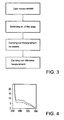
Fig. 3 is a diagram of an example of a method according to the invention; and -
Fig. 4 is an example of a normalized spectrogram of light intensities observed in the measured wavelength range. - The measuring system 1 shown in
Fig. 1 , for measuring an AGE content in a tissue of a patient, constitutes a currently most preferred exemplary embodiment of the invention. The measuring system 1 according to this example comprises ameasuring unit 13 having as a light source a fluorescent lamp in the form of a blacklightfluorescent tube 2, which is arranged within a supporting structure in the form of a light-shielding casing 6. Thecasing 6 has acontact surface 14 which is placed against theskin 7. An opening in thecontact surface 14 forms anirradiation window 8 through which a portion of the surface of theskin 7 located behind thatirradiation window 8 and adjacent to the window opening, can be irradiated. - To provide that, of the radiation generated by the fluorescent tube, only UV light in the desired wavelength range reaches the
skin 7, there is placed, according to this example, afilter 5 in front of theirradiation window 8. Such filters may be adapted, for instance, for passing radiation in a wavelength band around a desired wavelength in the range of 300 to 420 nm or may be high-pass filters for passing light radiation in a wavelength band from, for instance, 320, 375, 385 or 395 nm. The upper limit of the wavelength spectrum reaching the skin is then determined by the upper limit of the wavelength range of the light emitted by the proposed blacklight fluorescent tube (in this example 420 nm). Suitable filter types are, for instance, the 2 mm filters WG320, GG375, GG385 and GG395 of Schott Glaswerke, Mainz, Germany. In principle, theirradiation window 8 may also be formed by a fully open passage. Further, the irradiation window may be of a rectangular, circular or different shape. - In response to the radiation reaching the skin, the
skin 7 emits radiation which passes back through theirradiation window 8. The greater part of that radiation has a wavelength in the wavelength range of the radiation sent to the skin. A part of the radiation sent back via the irradiation window, however, is formed by radiation having longer wavelengths, which is generated as a result of a fluorescent action of constituents of the skin in response to excitation by the light sent to the skin. - Located adjacent an edge of the
irradiation window 8 is anend 18 of anoptical fiber 3, which end forms ameasuring window 18 via which radiation to be detected, coming from the skin, is passed to a detector. Theoptical fiber 3 passes the radiation received via themeasuring window 18 to aspectrophotometer unit 15 with anarray 22 of detectors. A suitable type of optical fiber is, for instance, a 200/250 µm glass fiber of a length of 1 m and with FSMA connectors. The spectrometer is designed as a digitally computer-readable plug-in card in astandard computer 16. A suitable type is, for instance, the PC 1000 van Ocean Optics with a sampling frequency of 10 kHz. Such a spectrometer analyzes the optical spectrum into a large number of fractions (in this example 1100 fractions in the range between 233 and 797 nm), which data can be further analyzed by means of thecomputer 16. Thecomputer 16 is programmed with a program for generating signals representing an AGE content in theskin 7 on adisplay 17. - The
detector array 22 is adapted for separately measuring radiation coming from the irradiated portion of theskin 7, firstly in a wavelength range outside the wavelength range of the radiation with which theskin 7 is irradiated and, secondly, in a wavelength range within or equal to the wavelength range of the radiation with which theskin 7 is irradiated. The measurement in a wavelength range within or equal to the wavelength range of the radiation with which theskin 7 or a reference material is irradiated serves to normalize for the amount of light emitted by the lamp and for optical properties of the skin tissue of the patient. - The amount of electromagnetic radiation emitted by the
skin tissue 7 in response to the irradiation, in a wavelength range outside the wavelength range of the radiation applied to theskin 7 is measured by means of thedetector array 22. The spectrometer generates a digital signal which is fed to thecomputer 16. Software loaded into the computer then provides, by means of thedisplay 17, for the generation of a signal which represents a measured autofluorescence in agreement with the measured amount of electromagnetic radiation in the wavelength range outside the wavelength range of the radiation applied to theskin 7. According to this example, the software is further designed for optionally processing the amount of electromagnetic radiation, measured via measuringwindow 18, in the wavelength range within the wavelength range of the radiation applied to theskin 7, for the purpose of correcting for the optical properties of the skin tissue. - The signal which represents a measured autofluorescence is subsequently converted to a signal which represents an AGE content and which may be in the form of, for instance, a number shown, which represents the determined content of AGE's, but may also be in the form of, for instance, an index number shown, which represents a value relative to an average AGE value for the age of the patient in question. Also, for instance, positions on a scale may be indicated, so that, along with the measured value, a reference framework is furnished at the same time.
- It is noted that the use of a spectrometer provides the advantage that it can be accurately determined per narrow wavelength band to what extent it is being taken into account as an indicator of the presence of AGE's.
- The
irradiation window 8 is bounded by anedge 19 to be held against a skin of a patient and thus limits the skin surface to be irradiated. The measuringwindow 18 too has a particular surface for passing light to be detected coming from the irradiated portion of theskin tissue 7. Theirradiation window 8 and the measuringwindow 18 each have a passage surface, the passage surface of theirradiation window 8 being greater than the passage surface of the measuringwindow 18. Further, thesurface 23 of theskin 7 within theirradiation window 8 from where light can be received by the fiber via the measuringwindow 18 is preferably greater than 0.1 cm2 and in particular 1-4 cm2, but smaller than the irradiation window and hence smaller than the irradiated surface of theskin 7. - The
surface 23 of theskin 7 within theirradiation window 8 from where light can be received via the measuringwindow 18 is further preferably greater, and in particular preferably at least 3-20 times greater, than the measuring window, so that a large irradiated and measured skin surface is combined with a compact construction of the measuring unit. - Inasmuch as the skin surface irradiated simultaneously in operation is relatively large and the radiation in response thereto stemming from different portions of that skin surface is detected in mutual simultaneity, different effects are achieved which are of particular advantage in determining an AGE content of skin tissue which is representative of a particular patient. The skin surface to be measured is then preferably at least about 0.1 cm2 and in particular preferably 1 to 4 cm2.
- In the first place, measurement thus involves an averaging of radiation coming from a large skin surface, so that a possible influence of local differences in skin properties on the detected fluorescence of the skin tissue is eliminated.
- In the second place, what is achieved by the use of a large irradiation window and measuring a
large skin surface 23 is that a considerable portion of fluorescent radiation which has been scattered further by the skin before exiting is received as well, so that the proportion of fluorescent radiation with respect to light reflected from the surface of the skin is increased. - Thirdly, a large skin surface can be properly irradiated with a fluorescent lamp. Relatively much radiation energy of the fluorescent tube is emitted in a wavelength range of 300-420 nm. As a result, a large portion of the emitted radiation is sent to the skin and only little radiation is absorbed by the
casing 6. Accordingly, relatively much fluorescence is generated without the skin being locally subject to strong heating, which might be painful and entail skin burning phenomena or at least an increased chance of artifacts as a result of reactions of the skin, such as modifications in the blood supply (vasodilatation). - Further, the
optical filter 5 located between theradiation source 2 and theskin 7 passes substantially exclusively radiation which serves for exciting fluorescence. - The measuring
window 18 formed by the end of theoptical fiber 3 proximal to theskin 7 is oriented at an angle α of about 45° relative to theirradiation window 8. As a result, in use the measuringwindow 18 is held at an angle of about 45° relative to the irradiated surface of theskin 7. - The position of the measuring
window 18 at an angle of 25-65° and preferably at an angle of about 45° relative to the irradiated surface of theskin 7 is advantageous, because radiation reflected by the surface of theskin 7 by mirror reflection, which is not of interest in this context, is reflected relatively strongly in a direction perpendicular to the skin surface. Thus, owing to the inclined position of the measuring window, relatively little radiation reflected mirror-wise by the surface of theskin 7 is received. - Owing to the inclined position of the measuring window, further, the distribution of the proportion of the light received from different zones of the irradiated skin surface is more uniform than in the use of a measuring window parallel to the irradiation window. In particular, overrepresentation of radiation coming from a portion of the irradiated skin surface closest to the measuring
window 18 is prevented. - An advantage of the inclined position of the measuring
window 18 that holds in particular if theskin 7 is irradiated in a direction perpendicular thereto is that the measuringwindow 18 is not directed in a direction in which it casts a shadow. By moreover placing the measuringwindow 18 at an edge of theirradiation window 8, the attachment thereof is simplified, and what is prevented, further, is that the measuringwindow 18, and a part of theoptical fiber 3 adjacent thereto, are situated in the path of radiation between thelamp 2 and the portion of theskin 7 to be irradiated. - As appears from
Fig. 1 , the measuringwindow 18 is spaced from the passage surface of theirradiation window 8, so that in use the measuringwindow 18 is held at a distance d from theskin 7. A suitable distance d is, for instance, 5-9 mm. In the example described, the distance d is approximately 7 mm. A suitable guideline for the distance d is further that it be selected such that the middle of the measuringwindow 18 is directed at a central area of theirradiation window 8. Given a position of the measuringwindow 18 at an angle of 45° relative to a plane in which theirradiation window 8 extends, and placement of the measuring window in front of an edge of theirradiation window 8, the distance d according to that guideline is half of the transverse dimension of theirradiation window 8 in a direction of the directional component parallel to theirradiation window 8 in which the measuringwindow 18 is directed. In practice, such a distance gives a suitable distribution of the yield of fluorescent radiation coming from theskin 7 over zones at different depths from the skin surface. - According to this example, the lamp is a blacklight fluorescent tube having a wavelength range of λA 300-420 nm. Such a UV lamp has a bandwidth such that by changing the
optical filter 5 autofluorescence measurements at different excitation wavelengths can be readily carried out. - In the considerably
smaller measuring unit 113 shown inFig. 2 , as radiation source aLED 102 is used which, according to this example, emits radiation of a wavelength of 370 nm. Because the measuringunit 113 is designed as a compact measuring head, it can simply be held against a patient's body at different points. A LED of a wavelength in the range of λA 300-420 nm emits light of a narrow band (width at half of the highest intensity, for instance, 10 nm), so that no or little radiation outside the desired wavelength range is emitted and correspondingly little energy leads to heat development. LED's moreover are highly efficient light sources and therefore remain relatively cool themselves too. Further, LED's are easy to control in a pulsed or modulated fashion, which is advantageous for correcting, for instance, for dark current due to thedetector 122 or ambient light. The measuringunit 113 has ascreening 106 and anirradiation window 108 having a limitingedge 119 to be placed against theskin 7. - For detecting radiation coming from the
skin 7, twodetectors skin 7. Arranged between thedetector 122 and theskin 7 is along pass filter 121, which passes only radiation of a wavelength greater than, for instance, 400 nm, so that thedetector 122 only detects the fluorescence-induced radiation passing thewindow 118. Thedetector 120 detects the total amount of light coming from theskin 7 and passing the window 118'. - By measuring not only the fluorescence-induced long-wave radiation, but also the radiation coming from the
skin 7 in the wavelength range of the excitation radiation, as inFig. 2 with thedetector 120, a correction can be made for differences in intensity of the radiation source and differences in optical properties of theskin 7 by carrying out the generation of the AGE content signal partly in accordance with the amount of detected electromagnetic radiation in the wavelength range of the excitation radiation. - By measuring with two detectors, the ratio between measured intensities solely in the wavelength range of the fluorescence and of radiation over the totality of wavelengths can then be simply determined. In principle, such a ratio can also be determined with, for instance, a single detector and a chopper which passes alternately radiation of all wavelengths and radiation solely above a particular wavelength. This provides the advantage that measuring errors as a result of differences between the two detectors are prevented, but leads to an increase of the dimensions and the mechanical complexity of the measuring unit. It appears, incidentally, that the ratio between the intensity of the autofluorescence and the intensity of the total amount of light reflected and emitted by the skin is about 1%.
- It may be advantageous to have several LED's and/or laser diode sources excite sequentially at different wavelengths and to measure the autofluorescence obtained upon excitation at those different wavelengths.
- Another option is to use a LED or laser diode which emits radiation in the region of the fluorescence generated. Thus, the reflection can also be measured at the wavelengths of the generated fluorescence, which provides information about the optical properties of the skin tissue and hence the propagation behavior of light of those wavelengths through the skin tissue. This information can subsequently be used for correcting measuring results for differences in optical properties of the skin as regards propagation properties of light of the wavelengths generated by fluorescence.
- For each wavelength range, several detectors can be used which are placed at different distances from the skin and (parallel to the skin) from the radiation source for simultaneously measuring radiation with different distributions of contributions from different depth zones of the skin.
- For the accuracy of the determination of AGE contents, if there is simultaneous irradiation with all excitation wavelengths used and also the fluorescent radiation is detected simultaneously, it is favorable if all wavelengths of the wavelength range of the excitation radiation are lower than all wavelengths of the wavelength range in which fluorescent radiation is measured. The wavelength range of the excitation radiation preferably includes at least one wavelength in a range of 300-420 nm and the wavelength range in which fluorescent radiation is measured preferably includes at least one wavelength in a range ≤600 nm.
- For the accuracy of the determination of AGE contents, it is further favorable if over the wavelength range in which fluorescent radiation is measured the aggregated, for instance summed, amount of detected fluorescent radiation is measured, and the generation of the signal occurs in accordance with the aggregated amount of detected fluorescent radiation. The fact is that because the excitation and emission bands of different specific types of AGE's present in the patient may overlap, fluorescence may occur at several wavelengths, so that a spectrum is emitted having a wideband long-wave region in the range of about 420 - 600 nm. By measuring over such a wideband long-wave region, autofluorescence owing to these effects is also taken into account.
- To be able to accurately measure the accuracy of the weak autofluorescence as a result of the presence of AGE's over a longer time as well, it is advantageous to regularly carry out a reference measurement on a reference material, and to have the generation of the AGE content signal partly influenced by an amount of electromagnetic radiation detected in the reference measurement.
- It is also possible to carry out the measurement with a detector which measures the response to excitation light after the irradiation with excitation light (for instance with a light pulse) has been stopped or at least has been changed, for instance in intensity. The fact is that the light coming from the skin in response to excitation light initially consists of light with the reflected excitation light and thereafter contains fluorescent light exponentially decreasing in strength. Thus, the extent to which changes in light coming from the skin lag behind changes in the excitation light also forms a measure for the autofluorescence. The skin surface can be irradiated with, for instance, pulsed or modulated light, utilizing a delay or phase lag of light received from the skin in response thereto as a measure for the autofluorescence value. Instead of, or supplemental to, the change of the intensity of the excitation light, also the wavelength of the excitation light can be changed, while changes in the light coming from the skin in response to changes in the wavelength of the excitation light serve as a measure for the autofluorescence. At different wavelengths, for instance, different AGE's can be excited and emit fluorescent light.
- For determining the AGE content, both the measurements at different times and the half value of the response signal can then be used as an indication. An example of measuring the response to modulated excitation light is described in more detail in R.H. Mayer et al., Measurement Of The Fluorescence Lifetime In Scattering Media By Frequency Domain Migration, Applied Optics, Aug 1999, pp. 4930-4938, which is hereby referred to.
- An advantage of the consecutive irradiation of skin tissue and subsequent measurement of fluorescence is that the wavelengths of the fluorescent light and of the excitation light can overlap without this disturbing the measurement.
- The consecutive irradiation and measurement of fluorescence can be achieved, for instance, by having the LED or
laser diode 102 inFig. 2 radiate in a pulsatile manner and carrying out the measurement with the detector 120 - thedetector 122 and thefilter 121 can then be omitted - in alternation with the irradiation, so that measurement takes place when the LED orlaser diode 102 emits no light, or at least a different amount of light. Incidentally, it is also possible to measure fluorescence both simultaneously with excitation and after changes in the excitation light. The distinction between reflected excitation light and fluorescent light is then made on the one hand on the basis of a difference in wavelength and on the other on the basis of the delay by which fluorescent light is emitted. - Hereinafter, a tested method for determining the autofluorescence in the skin is described in more detail. A measuring system according to
Fig. 1 was used. The steps of this method are schematically represented inFig. 3 . - First, a dark measurement is carried out by measuring the dark current of the detector when the opening is covered, without light irradiation. The temperature of the spectrograph is subsequently maintained constant to prevent variation of the dark current.
- Next, the
blacklight fluorescent tube 2 is turned on, followed by waiting for some time (for instance at least 5 minutes) until thetube 2 generates a substantially constant light output. - Thereafter, the measuring
unit 13 is placed on theskin 7 of a person to be examined. This can be, for instance, a part of the lower arm or of the leg (such as the calf). A measurement is performed, and the spectral intensity distribution of radiation coming from theskin 7 as determined by the spectrometer is stored in thecomputer 16. - Thereafter, a reference measurement is performed, by performing the same measurement on a reference material, for instance white Teflon. This may also be done prior to the preceding step.
- Performing the reference measurement serves to enable correction for differences in the amount of absorption and scattering that occur between different test subjects. The amount of absorption is associated inter alia with age, the melanin content of the skin tissue, and the amount of blood in the skin.
-
Fig. 4 shows a result of the autofluorescence measured in diabetic patients (solid line) and a healthy control group corresponding in age and gender (dotted line). The diagram has been normalized by dividing the measured intensities by the sum of the intensities measured between 285 and 425 nm. It is clear to see that over the measured region between 400 and 500 nm, the autofluorescence in the diabetics is essentially higher, which is consistent with results achieved heretofore using other, more laborious measuring techniques. - The registered measurements further confirm that an important measure for autofluorescence is the aggregated light intensity AFt in the range of 420-600 nm. Diabetic patients (both with and without correction for absorption) proved to have a considerably higher fluorescence level than healthy individuals.
- This is represented in the following table (arbitrary units):
AFt 420-600 nm Diabetics (n=46) Control (n=46) Statistic significance lower arm 1598 ± 703 1184 ± 618 p < 0.004 lower leg 1637 ± 923 1255 ± 847 p < 0.04 - The measuring values found, corrected for absorption, also prove to correlate with age (r=0.52; p<0.001, where r represents the correlation coefficient between the measured autofluorescence and age, and p represents the level of statistical significance of the correlation). The two groups of test subjects had ages distributed over a wide range. Thus, upon a comparison by age, still greater differences are found. In all of the 46 pairs matched for age and gender, a higher AFt was found in the diabetic patient than in the control.
- The measuring values found, corrected for absorption, moreover prove to correlate with the amount of laboratory-determined values for HbAlc (r=0.4; p<0.01, where r is the correlation coefficient between the measured autofluorescence and the measured HbAlc value - a frequently used measure for long-term glucose levels).
- It appears from these data that the clinically significant indications of the AGE values of skin tissue can be determined in the noninvasive manner according to the invention.
- It will be clear to those skilled in the art that within the framework of the invention many other embodiments and modes are possible and that the invention is not limited to the examples described hereinbefore.
Claims (30)
- A method for determining an autofluorescence value of clinically healthy skin tissue (7) of a patient, comprising:irradiating material of said skin tissue (7) with electromagnetic excitation radiation;measuring an amount of electromagnetic, fluorescent radiation emitted by said material (7) in response to said irradiation; andgenerating, in response to said measured amount of fluorescent radiation, a signal which represents a determined autofluorescence value for the respective patient;wherein said skin tissue (7) is intact skin tissue (7) in vivo of which a surface is irradiated noninvasively and simultaneously in its entirety,
wherein fluorescent radiation emitted in response to said irradiation is simultaneously received from different portions of the skin surface (23), characterized in that the surface area of the skin surface (23) within the skin surface that is irradiated from which fluorescent radiation is received is at least 0.1 cm2 - A method according to claim 1, wherein in response to at least one determined autofluorescence value an advanced glycation/glycosylation end product (AGE) content for said patient is determined and signaled.
- A method according to claim 1 or 2, wherein a first skin surface is simultaneously irradiated, wherein fluorescent radiation which in response to said irradiation comes from different portions of a second skin surface (23) within said first skin surface is simultaneously received via a measuring window (18; 118) with a particular surface, and wherein said second skin surface is greater than the surface of said measuring window (18; 118).
- A method according to claim 3, wherein said second skin surface is at least three times greater than the surface of said measuring window (18; 118).
- A method according to any one of the preceding claims, wherein said fluorescent radiation is received via a measuring window (18) and
wherein said measuring window (18) is held at an angle of 25-65° and preferably of about 46° relative to the irradiated surface of the skin (7). - A method according to any one of the preceding claims, wherein said fluorescent radiation is received via a measuring window (18) and
wherein said measuring window (18; 118) is held at a distance from the skin (7). - A method according to any one of the preceding claims, wherein the irradiation with electromagnetic excitation radiation in a first wavelength range and the measurement of emitted electromagnetic fluorescent radiation in a second wavelength range outside said first wavelength range takes place simultaneously with the irradiation, while all wavelengths of said first wavelength range are smaller than all wavelengths of said second wavelength range, and said first wavelength range comprises a wavelength in a range of 300-420 nm, and said second wavelength range comprises a longer wavelength in a range of ≤600 nm.
- A method according to any one of the preceding claims, further comprising determining an aggregated amount of detected electromagnetic radiation over a particular wavelength range, while determining said autofluorescence value occurs in response to said aggregated amount of detected electromagnetic radiation.
- A method according to any one of the preceding claims, further comprising: passing radiation coming from said skin tissue (7) to a spectrometer (15), dividing received radiation within a measuring range of wavelengths into fractions per wavelength sub-range, and aggregating detected fractions of fluorescent radiation to an aggregated amount of detected electromagnetic radiation, while determining said autofluorescence value occurs in response to said aggregated amount of detected electromagnetic radiation.
- A method according to any one of the preceding claims, further comprising detecting reflected excitation radiation, while generating said autofluorescence value occurs partly in response to a detected amount of said electromagnetic excitation radiation.
- A method according to claim 10, wherein reflected excitation radiation is detected by a first detector (120) and wherein said fluorescent radiation is detected by another, separate detector (122).
- A method according to any one of the preceding claims, wherein said fluorescent radiation coming from at least a portion of said irradiated skin surface is detected after the irradiation of said at least one portion of said skin surface has been changed.
- A method according to claim 12, wherein said fluorescent radiation is detected in at least one wavelength corresponding with at least one wavelength of said excitation radiation.
- A method according to claim 12 or 13, wherein said excitation radiation is emitted in a pulsating or modulated fashion.
- A method according to any one of the preceding claims, further comprising performing a reference measurement on a reference material, while generating said signal occurs partly in response to at least one amount of electromagnetic radiation detected in said reference measurement.
- An apparatus for determining an autofluorescence value of clinically healthy skin tissue (7) of a patient, comprising:a pick-up unit with a radiation source (2; 102), for in vivo and noninvasively irradiating a surface (23) of intact skin tissue (7) behind a irradiation window (8; 108) with electromagnetic excitation radiationa detector (22; 122) for measuring electromagnetic fluorescent radiationmeans (17) for generating an autofluorescence value for said skin tissue (7) in agreement with a measured amount of fluorescent radiation originating from said surface area of said skin tissue (7), characterised in that the detector is arranged to receive electromagnetic fluorescent radiation from a surface area of said skin tissue surface behind said irradiation window (8; 108) of at least 0.1 cm. 2
- An apparatus according to claim 16, further comprising means (16, 17) for determining and signaling in response to at least one determined autofluorescence value an advanced glycation/glycosylation end product (AGE) content for said patient.
- An apparatus according to claim 16 or 17, wherein the irradiation window (8; 108) is adapted to determine a first surface of said skin tissue to be irradiated, further comprising a measuring window (18; 118) with a surface for passing fluorescent radiation to be detected coming from a second surface within said first surface, said second surface being greater than the surface of said measuring window (18; 118).
- An apparatus according to claim 18, wherein said second surface is at least three times greater than the surface of said measuring window (18; 118).
- An apparatus according to any one of claims 16-19, further comprising a supporting structure (6) to be held against a skin (7) of a patient, for defining a plane in which a surface of said skin tissue to be irradiated is located, wherein the supporting structure (6) supports the measuring window (18) for passing light to be detected coming from said irradiated skin tissue (7), said measuring window (18) being oriented at an angle of 25-65°, preferably at an angle of about 45° relative to said surface.
- An apparatus according to claim 20, wherein said supporting structure (6) comprises an irradiation window (8) for delimiting a surface of said skin tissue (7) to be irradiated, said measuring window (18) being located adjacent an edge (19) of said irradiation window (8).
- An apparatus according to any one of claims 16-21, wherein said measuring window (18; 118) is spaced away from the passage surface of said surface.
- An apparatus according to any one of claims 16-22, wherein the position of the measuring window (18) relative to said surface is adjustable for adjusting the distance between said measuring window (18) and said irradiation window (8).
- An apparatus according to any one of claims 16-23, further comprising an optical filter between said radiation source (2) and said irradiation window (8).
- An apparatus according to any one of claims 16-24, wherein said radiation source (2) is an electrofluorescent lamp for emitting radiation in a wavelength range of 300-420 nm.
- An apparatus according to any one of claims 16-24, wherein said radiation source (102; 202) is a light-emitting diode or laser diode for emitting radiation having at least one wavelength in a wavelength range of 300-420 nm, and preferably for emitting radiation having a wavelength of 370 nm.
- An apparatus according to any one of claims 16-26, further comprising a spectrometer connected with said measuring window (18) for receiving radiation passing through said measuring window (18).
- An apparatus according to any one of claims 16-27, further comprising separate detectors (122, 120) for detecting reflected excitation radiation and fluorescent radiation.
- An apparatus according to any one of claims 16-28, further comprising control means for changing the excitation radiation such that it is different in a second irradiation period than in a first irradiation period.
- An apparatus according to claim 29, adapted for intermittently irradiating said skin tissue (7) and for separately detecting radiation coming from said skin tissue (7) in periods alternating with said intermittent irradiation.
Applications Claiming Priority (1)
| Application Number | Priority Date | Filing Date | Title |
|---|---|---|---|
| PCT/NL1999/000607 WO2001022869A1 (en) | 1999-09-30 | 1999-09-30 | Method and apparatus for determining autofluorescence of skin tissue |
Publications (2)
| Publication Number | Publication Date |
|---|---|
| EP1217943A1 EP1217943A1 (en) | 2002-07-03 |
| EP1217943B1 true EP1217943B1 (en) | 2009-04-22 |
Family
ID=19866615
Family Applications (1)
| Application Number | Title | Priority Date | Filing Date |
|---|---|---|---|
| EP99949448A Expired - Lifetime EP1217943B1 (en) | 1999-09-30 | 1999-09-30 | Method and apparatus for determining autofluorescence of skin tissue |
Country Status (8)
| Country | Link |
|---|---|
| US (1) | US7966060B2 (en) |
| EP (1) | EP1217943B1 (en) |
| AT (1) | ATE429171T1 (en) |
| AU (1) | AU6232099A (en) |
| DE (1) | DE69940787D1 (en) |
| DK (1) | DK1217943T3 (en) |
| ES (1) | ES2326397T3 (en) |
| WO (1) | WO2001022869A1 (en) |
Cited By (2)
| Publication number | Priority date | Publication date | Assignee | Title |
|---|---|---|---|---|
| US9042967B2 (en) | 2008-05-20 | 2015-05-26 | University Health Network | Device and method for wound imaging and monitoring |
| US10438356B2 (en) | 2014-07-24 | 2019-10-08 | University Health Network | Collection and analysis of data for diagnostic purposes |
Families Citing this family (54)
| Publication number | Priority date | Publication date | Assignee | Title |
|---|---|---|---|---|
| US20040038320A1 (en) * | 2001-03-09 | 2004-02-26 | Bhaskar Banerjee | Methods of detecting cancer using cellular autofluorescence |
| US7139598B2 (en) * | 2002-04-04 | 2006-11-21 | Veralight, Inc. | Determination of a measure of a glycation end-product or disease state using tissue fluorescence |
| US20040146290A1 (en) * | 2001-11-08 | 2004-07-29 | Nikiforos Kollias | Method of taking images of the skin using blue light and the use thereof |
| US8238993B2 (en) * | 2002-04-04 | 2012-08-07 | Veralight, Inc. | Determination of a measure of a glycation end-product or disease state using tissue fluorescence lifetime |
| US7267648B2 (en) * | 2003-03-31 | 2007-09-11 | Olympus Corporation | Magnifying image pickup unit for an endoscope, an endoscope for in vivo cellular observation that uses it, and endoscopic, in vivo cellular observation methods |
| GB0329849D0 (en) | 2003-12-23 | 2004-01-28 | Precisense As | Fluorometers |
| WO2006009906A2 (en) * | 2004-06-17 | 2006-01-26 | The Regents Of The University Of California | Time-resolved non-invasive autofluorescence device for detecting diabetes |
| WO2006009910A2 (en) | 2004-06-17 | 2006-01-26 | The Regents Of The University Of California | Time-resolved optometric fluorescence detection for skin diagnostics |
| US20080255462A1 (en) * | 2004-09-28 | 2008-10-16 | Zila Pharmaceuticals, Inc. | Light stick |
| US20060241494A1 (en) * | 2004-09-28 | 2006-10-26 | Zila Pharmaceuticals, Inc. | Methods for detecting abnormal epithelial tissue |
| US20060241501A1 (en) * | 2004-09-28 | 2006-10-26 | Zila Pharmaceuticals, Inc. | Method and apparatus for detecting abnormal epithelial tissue |
| US20090118624A1 (en) * | 2004-09-28 | 2009-05-07 | Zila Pharmaceuticals, Inc. | Device for oral cavity examination |
| US20070004972A1 (en) * | 2005-06-29 | 2007-01-04 | Johnson & Johnson Consumer Companies, Inc. | Handheld device for determining skin age, proliferation status and photodamage level |
| US20070002479A1 (en) * | 2005-06-29 | 2007-01-04 | Johnson & Johnson Consumer Companies, Inc. | Apparatus and method for viewing the skin |
| US7558416B2 (en) | 2006-10-02 | 2009-07-07 | Johnson & Johnson Consumer Companies, Inc. | Apparatus and method for measuring photodamage to skin |
| US7764303B2 (en) * | 2006-10-02 | 2010-07-27 | Johnson & Johnson Consumer Companies, Inc. | Imaging apparatus and methods for capturing and analyzing digital images of the skin |
| DE102007058563A1 (en) * | 2007-11-30 | 2009-06-04 | Sentronic GmbH Gesellschaft für optische Meßsysteme | Spectrometer measuring head for analyzing characteristics of liquid, pasty or solid substances |
| US20090163831A1 (en) * | 2007-12-24 | 2009-06-25 | Mi Hope Inc. Dba Progressive University | Methods for the detection of ultraviolet light reactive alternative cellular energy pigments (ACE-pigments) |
| WO2009108024A2 (en) * | 2008-02-29 | 2009-09-03 | 주식회사 이노제스트 | Device and method for measuring fluorescence of skin tissues |
| US8983581B2 (en) | 2008-05-27 | 2015-03-17 | Massachusetts Institute Of Technology | System and method for large field of view, single cell analysis |
| US20100003664A1 (en) * | 2008-07-01 | 2010-01-07 | Lou Reinisch | System for Glycated Protein Detection |
| US9155471B2 (en) | 2009-05-27 | 2015-10-13 | Lumicell, Inc'. | Methods and systems for spatially identifying abnormal cells |
| US20110028806A1 (en) * | 2009-07-29 | 2011-02-03 | Sean Merritt | Reflectance calibration of fluorescence-based glucose measurements |
| US9615747B2 (en) * | 2010-01-19 | 2017-04-11 | Access Business Group International Llc | Method for determining skin glycation |
| US20110306077A1 (en) * | 2010-06-13 | 2011-12-15 | William John Martin | Methods for the detection of alternative cellular energy (ACE) pigments and for monitoring of the ACE pathway in the diagnosis and therapy of diseases |
| CN103167830B (en) * | 2010-06-18 | 2016-08-31 | 代克诺普提克斯控股有限公司 | For the method and apparatus determining the autofluorescence value of skin histology |
| US9314304B2 (en) | 2010-12-08 | 2016-04-19 | Lumicell, Inc. | Methods and system for image guided cell ablation with microscopic resolution |
| JP5970785B2 (en) * | 2011-11-16 | 2016-08-17 | ソニー株式会社 | Biological measuring device, biological measuring method, program, and recording medium |
| US9955871B2 (en) | 2012-03-21 | 2018-05-01 | Korea Electrotechnology Research Institute | Transmitted light detection type measurement apparatus for skin autofluorescence |
| US9155473B2 (en) | 2012-03-21 | 2015-10-13 | Korea Electrotechnology Research Institute | Reflection detection type measurement apparatus for skin autofluorescence |
| US9723990B2 (en) | 2012-03-21 | 2017-08-08 | Korea Electro Technology Research Institute | Transmitted light detection type measurement apparatus for skin autofluorescence |
| KR20160037834A (en) | 2013-03-14 | 2016-04-06 | 루미셀, 아이엔씨. | Medical imaging device and methods of use |
| US9888855B2 (en) | 2013-03-18 | 2018-02-13 | Korea Electrotechnology Research Institute | Reflection detection type measurement apparatus and method for skin autofluorescence |
| WO2016054079A1 (en) | 2014-09-29 | 2016-04-07 | Zyomed Corp. | Systems and methods for blood glucose and other analyte detection and measurement using collision computing |
| US10359414B2 (en) | 2015-03-09 | 2019-07-23 | The Regents Of The University Of Michigan | Frequency domain discrimination of tissue proteins |
| AU2016260153B2 (en) * | 2015-05-08 | 2019-04-04 | Jiangsu Kunhui Biotechnology Co., Ltd. | Method for detecting, in noninvasive in-vivo manner, skin damage induced by ultraviolet light, and detection device thereof |
| CN107847145B (en) * | 2015-07-20 | 2020-08-14 | 徐敏 | Photonic structures and chemometric pathological systems |
| US10441316B2 (en) * | 2015-09-11 | 2019-10-15 | Pop Test LLC | Intradermal applicator/analyte sensor placement using fluorescence |
| US9554738B1 (en) | 2016-03-30 | 2017-01-31 | Zyomed Corp. | Spectroscopic tomography systems and methods for noninvasive detection and measurement of analytes using collision computing |
| US11266331B2 (en) | 2016-05-16 | 2022-03-08 | Sony Corporation | Optical apparatus and information processing method |
| US11426075B1 (en) | 2017-08-23 | 2022-08-30 | Lumicell, Inc. | System and method for residual cancer cell detection |
| DE102017222530A1 (en) * | 2017-12-12 | 2019-06-13 | Henkel Ag & Co. Kgaa | Arrangement for determining metabolic end products in the skin |
| US11051727B2 (en) * | 2018-01-09 | 2021-07-06 | Medtronic Monitoring, Inc. | System and method for non-invasive monitoring of advanced glycation end-products (AGE) |
| US11154224B2 (en) | 2018-01-09 | 2021-10-26 | Medtronic Monitoring, Inc. | System and method for non-invasive monitoring of hematocrit concentration |
| US11039768B2 (en) | 2018-01-09 | 2021-06-22 | Medtronic Monitoring, Inc. | System and method for non-invasive monitoring of hemoglobin |
| WO2020053933A1 (en) * | 2018-09-10 | 2020-03-19 | オリンパス株式会社 | Thermal insult observation device and thermal insult observation method |
| KR102640331B1 (en) * | 2018-10-19 | 2024-02-26 | 삼성전자주식회사 | Apparatus and method for estimating bio-information, and apparatus for supporting bio-information estimation |
| US10885363B2 (en) | 2018-10-25 | 2021-01-05 | Advanced New Technologies Co., Ltd. | Spoof detection using structured light illumination |
| US11914692B2 (en) * | 2018-11-06 | 2024-02-27 | Sony Corporation | Information processing apparatus and information processing method |
| US11170242B2 (en) * | 2018-12-26 | 2021-11-09 | Advanced New Technologies Co., Ltd. | Spoof detection using dual-band fluorescence |
| US10970574B2 (en) | 2019-02-06 | 2021-04-06 | Advanced New Technologies Co., Ltd. | Spoof detection using dual-band near-infrared (NIR) imaging |
| US11328043B2 (en) | 2019-03-15 | 2022-05-10 | Advanced New Technologies Co., Ltd. | Spoof detection by comparing images captured using visible-range and infrared (IR) illuminations |
| WO2022026476A1 (en) * | 2020-07-27 | 2022-02-03 | Bio-Rad Laboratories, Inc. | Electrophoresis apparatus with minimal autofluorescence to enable gel processing in situ |
| WO2026005422A1 (en) * | 2024-06-25 | 2026-01-02 | 삼성전자 주식회사 | Electronic device and wearable electronic device for measuring biosignals |
Family Cites Families (13)
| Publication number | Priority date | Publication date | Assignee | Title |
|---|---|---|---|---|
| DE2963757D1 (en) * | 1978-01-03 | 1982-11-11 | Shapiro H M | Apparatus for non-invasive detection of zinc protoporphyrin in erythrocytes |
| IT1128806B (en) | 1980-06-17 | 1986-06-04 | Cselt Centro Studi Lab Telecom | OPTOCOUPLER FOR TRANSCEIVING ON OPTICAL FIBER |
| DE3718202C1 (en) | 1987-05-29 | 1988-11-17 | Wolfgang Prof Dr Lohmann | Arrangement for measuring a condition value for organic tissue areas |
| US5279297A (en) * | 1991-09-20 | 1994-01-18 | Trustees Of The University Of Pennsylvania | Method and apparatus for oxygen mapping |
| US5370114A (en) * | 1992-03-12 | 1994-12-06 | Wong; Jacob Y. | Non-invasive blood chemistry measurement by stimulated infrared relaxation emission |
| US5452723A (en) * | 1992-07-24 | 1995-09-26 | Massachusetts Institute Of Technology | Calibrated spectrographic imaging |
| US5553615A (en) * | 1994-01-31 | 1996-09-10 | Minnesota Mining And Manufacturing Company | Method and apparatus for noninvasive prediction of hematocrit |
| GB9521784D0 (en) * | 1995-10-24 | 1996-01-03 | Rosslyn Medical Ltd | Diagnostic apparatus |
| US5851181A (en) | 1996-08-30 | 1998-12-22 | Esc Medical Systems Ltd. | Apparatus for simultaneously viewing and spectrally analyzing a portion of skin |
| CA2305721C (en) | 1997-10-08 | 2009-02-24 | The General Hospital Corporation | Phototherapy methods and systems |
| US6721582B2 (en) | 1999-04-06 | 2004-04-13 | Argose, Inc. | Non-invasive tissue glucose level monitoring |
| US7899518B2 (en) | 1998-04-06 | 2011-03-01 | Masimo Laboratories, Inc. | Non-invasive tissue glucose level monitoring |
| US6505059B1 (en) | 1998-04-06 | 2003-01-07 | The General Hospital Corporation | Non-invasive tissue glucose level monitoring |
-
1999
- 1999-09-30 WO PCT/NL1999/000607 patent/WO2001022869A1/en not_active Ceased
- 1999-09-30 DK DK99949448T patent/DK1217943T3/en active
- 1999-09-30 AT AT99949448T patent/ATE429171T1/en not_active IP Right Cessation
- 1999-09-30 ES ES99949448T patent/ES2326397T3/en not_active Expired - Lifetime
- 1999-09-30 AU AU62320/99A patent/AU6232099A/en not_active Abandoned
- 1999-09-30 EP EP99949448A patent/EP1217943B1/en not_active Expired - Lifetime
- 1999-09-30 DE DE69940787T patent/DE69940787D1/en not_active Expired - Lifetime
-
2004
- 2004-01-29 US US10/767,147 patent/US7966060B2/en not_active Expired - Fee Related
Cited By (9)
| Publication number | Priority date | Publication date | Assignee | Title |
|---|---|---|---|---|
| US9042967B2 (en) | 2008-05-20 | 2015-05-26 | University Health Network | Device and method for wound imaging and monitoring |
| US11154198B2 (en) | 2008-05-20 | 2021-10-26 | University Health Network | Method and system for imaging and collection of data for diagnostic purposes |
| US11284800B2 (en) | 2008-05-20 | 2022-03-29 | University Health Network | Devices, methods, and systems for fluorescence-based endoscopic imaging and collection of data with optical filters with corresponding discrete spectral bandwidth |
| US11375898B2 (en) | 2008-05-20 | 2022-07-05 | University Health Network | Method and system with spectral filtering and thermal mapping for imaging and collection of data for diagnostic purposes from bacteria |
| US12226186B2 (en) | 2008-05-20 | 2025-02-18 | University Health Network | Devices, methods, and systems with spectral filtering for detecting wound and identifying bacteria based on fluorescence signature |
| US12251191B2 (en) | 2008-05-20 | 2025-03-18 | University Health Network | Diagnostic method and system with optical and temperature sensors for imaging and mapping fluorescence intensities of tissue |
| US12616372B2 (en) | 2008-05-20 | 2026-05-05 | University Health Network | Devices, methods, and systems for fluorescence-based imaging and detection of location(s) and/or source(s) of potential contamination and/or pollution |
| US10438356B2 (en) | 2014-07-24 | 2019-10-08 | University Health Network | Collection and analysis of data for diagnostic purposes |
| US11676276B2 (en) | 2014-07-24 | 2023-06-13 | University Health Network | Collection and analysis of data for diagnostic purposes |
Also Published As
| Publication number | Publication date |
|---|---|
| AU6232099A (en) | 2001-04-30 |
| ATE429171T1 (en) | 2009-05-15 |
| US7966060B2 (en) | 2011-06-21 |
| US20040186363A1 (en) | 2004-09-23 |
| ES2326397T3 (en) | 2009-10-08 |
| DE69940787D1 (en) | 2009-06-04 |
| DK1217943T3 (en) | 2009-08-17 |
| EP1217943A1 (en) | 2002-07-03 |
| WO2001022869A1 (en) | 2001-04-05 |
Similar Documents
| Publication | Publication Date | Title |
|---|---|---|
| US7966060B2 (en) | Method and apparatus for determining autofluorescence of skin tissue | |
| US4894547A (en) | Optical method and apparatus for detecting and measuring aging, photoaging, dermal disease and pigmentation in skin | |
| US10582855B2 (en) | Compact spectrometer system for non-invasive measurement of absorption and transmission spectra in biological tissue samples | |
| US6377841B1 (en) | Tumor demarcation using optical spectroscopy | |
| US6317624B1 (en) | Apparatus and method for demarcating tumors | |
| US6741876B1 (en) | Method for determination of analytes using NIR, adjacent visible spectrum and discrete NIR wavelenths | |
| US5840035A (en) | Method for the spectroscopic examination of a biological tissue | |
| Huang et al. | Novel quantitative analysis of autofluorescence images for oral cancer screening | |
| US6289236B1 (en) | Methods and apparatus for distinguishing inflamed and tumorous bladder tissue | |
| KR101097399B1 (en) | Disease diagnosis device by evaluation of final glycation product amount using skin autofluorescence | |
| US20100121199A1 (en) | Method and apparatus for non-invasive cancerous tissue diagnosis and tomography using terahertz image | |
| JP2003508744A (en) | Analyte quantification method using NIR, adjacent visible spectrum and discrete NIR wavelength | |
| JP2003534087A (en) | Method and apparatus for detecting substances in body fluids using Raman spectroscopy | |
| US20090124902A1 (en) | Method for in vivo tissue classification | |
| JP7221285B2 (en) | Imaging method and system for intraoperative assessment of resection margins | |
| JP2003527919A (en) | Non-invasive measurement of skin bilirubin levels | |
| Uk et al. | Spectrometer for fluorescence–reflection biomedical research | |
| Drakaki et al. | Laser-induced fluorescence and reflectance spectroscopy for the discrimination of basal cell carcinoma from the surrounding normal skin tissue | |
| CN115568822A (en) | Device for detecting skin autofluorescence and anti-glycation efficacy evaluation method | |
| JPH07132120A (en) | Nonaggressive measuring method and device of specimen concentration using discontinuous emission | |
| JP2014140423A (en) | Skin condition measuring apparatus | |
| Jain et al. | RAMAN SPECTROSCOPY STUDY OF HEALTHY AND CANCEROUS HUMAN BREAST TISSUE FOR CANCER DETECTION. | |
| JP2015062572A (en) | Skin condition measuring method and skin condition measuring apparatus | |
| US9392943B2 (en) | System for glycated protein detection | |
| WO2010002278A1 (en) | System for glycated protein detection |
Legal Events
| Date | Code | Title | Description |
|---|---|---|---|
| PUAI | Public reference made under article 153(3) epc to a published international application that has entered the european phase |
Free format text: ORIGINAL CODE: 0009012 |
|
| 17P | Request for examination filed |
Effective date: 20020405 |
|
| AK | Designated contracting states |
Kind code of ref document: A1 Designated state(s): AT BE CH CY DE DK ES FI FR GB GR IE IT LI LU MC NL PT SE |
|
| AX | Request for extension of the european patent |
Free format text: AL;LT;LV;MK;RO;SI |
|
| GRAP | Despatch of communication of intention to grant a patent |
Free format text: ORIGINAL CODE: EPIDOSNIGR1 |
|
| RAP1 | Party data changed (applicant data changed or rights of an application transferred) |
Owner name: DIAGNOPTICS HOLDING B.V. |
|
| GRAS | Grant fee paid |
Free format text: ORIGINAL CODE: EPIDOSNIGR3 |
|
| GRAA | (expected) grant |
Free format text: ORIGINAL CODE: 0009210 |
|
| AK | Designated contracting states |
Kind code of ref document: B1 Designated state(s): AT BE CH CY DE DK ES FI FR GB GR IE IT LI LU MC NL PT SE |
|
| REG | Reference to a national code |
Ref country code: GB Ref legal event code: FG4D |
|
| REG | Reference to a national code |
Ref country code: CH Ref legal event code: EP |
|
| REG | Reference to a national code |
Ref country code: IE Ref legal event code: FG4D |
|
| REF | Corresponds to: |
Ref document number: 69940787 Country of ref document: DE Date of ref document: 20090604 Kind code of ref document: P |
|
| REG | Reference to a national code |
Ref country code: CH Ref legal event code: NV Representative=s name: R. A. EGLI & CO. PATENTANWAELTE |
|
| REG | Reference to a national code |
Ref country code: DK Ref legal event code: T3 |
|
| REG | Reference to a national code |
Ref country code: SE Ref legal event code: TRGR |
|
| REG | Reference to a national code |
Ref country code: ES Ref legal event code: FG2A Ref document number: 2326397 Country of ref document: ES Kind code of ref document: T3 |
|
| PG25 | Lapsed in a contracting state [announced via postgrant information from national office to epo] |
Ref country code: PT Free format text: LAPSE BECAUSE OF FAILURE TO SUBMIT A TRANSLATION OF THE DESCRIPTION OR TO PAY THE FEE WITHIN THE PRESCRIBED TIME-LIMIT Effective date: 20090822 Ref country code: FI Free format text: LAPSE BECAUSE OF FAILURE TO SUBMIT A TRANSLATION OF THE DESCRIPTION OR TO PAY THE FEE WITHIN THE PRESCRIBED TIME-LIMIT Effective date: 20090422 Ref country code: AT Free format text: LAPSE BECAUSE OF FAILURE TO SUBMIT A TRANSLATION OF THE DESCRIPTION OR TO PAY THE FEE WITHIN THE PRESCRIBED TIME-LIMIT Effective date: 20090422 |
|
| PLBE | No opposition filed within time limit |
Free format text: ORIGINAL CODE: 0009261 |
|
| STAA | Information on the status of an ep patent application or granted ep patent |
Free format text: STATUS: NO OPPOSITION FILED WITHIN TIME LIMIT |
|
| 26N | No opposition filed |
Effective date: 20100125 |
|
| PG25 | Lapsed in a contracting state [announced via postgrant information from national office to epo] |
Ref country code: MC Free format text: LAPSE BECAUSE OF NON-PAYMENT OF DUE FEES Effective date: 20090930 |
|
| PG25 | Lapsed in a contracting state [announced via postgrant information from national office to epo] |
Ref country code: GR Free format text: LAPSE BECAUSE OF FAILURE TO SUBMIT A TRANSLATION OF THE DESCRIPTION OR TO PAY THE FEE WITHIN THE PRESCRIBED TIME-LIMIT Effective date: 20090723 |
|
| PG25 | Lapsed in a contracting state [announced via postgrant information from national office to epo] |
Ref country code: LU Free format text: LAPSE BECAUSE OF NON-PAYMENT OF DUE FEES Effective date: 20090930 |
|
| PG25 | Lapsed in a contracting state [announced via postgrant information from national office to epo] |
Ref country code: CY Free format text: LAPSE BECAUSE OF FAILURE TO SUBMIT A TRANSLATION OF THE DESCRIPTION OR TO PAY THE FEE WITHIN THE PRESCRIBED TIME-LIMIT Effective date: 20090422 |
|
| PGFP | Annual fee paid to national office [announced via postgrant information from national office to epo] |
Ref country code: IE Payment date: 20140918 Year of fee payment: 16 Ref country code: DK Payment date: 20140919 Year of fee payment: 16 |
|
| PGFP | Annual fee paid to national office [announced via postgrant information from national office to epo] |
Ref country code: SE Payment date: 20140918 Year of fee payment: 16 |
|
| PGFP | Annual fee paid to national office [announced via postgrant information from national office to epo] |
Ref country code: BE Payment date: 20140919 Year of fee payment: 16 |
|
| REG | Reference to a national code |
Ref country code: DK Ref legal event code: EBP Effective date: 20150930 |
|
| REG | Reference to a national code |
Ref country code: SE Ref legal event code: EUG |
|
| REG | Reference to a national code |
Ref country code: IE Ref legal event code: MM4A |
|
| PG25 | Lapsed in a contracting state [announced via postgrant information from national office to epo] |
Ref country code: IE Free format text: LAPSE BECAUSE OF NON-PAYMENT OF DUE FEES Effective date: 20150930 |
|
| PG25 | Lapsed in a contracting state [announced via postgrant information from national office to epo] |
Ref country code: SE Free format text: LAPSE BECAUSE OF NON-PAYMENT OF DUE FEES Effective date: 20151001 |
|
| REG | Reference to a national code |
Ref country code: FR Ref legal event code: PLFP Year of fee payment: 18 |
|
| PG25 | Lapsed in a contracting state [announced via postgrant information from national office to epo] |
Ref country code: DK Free format text: LAPSE BECAUSE OF NON-PAYMENT OF DUE FEES Effective date: 20150930 |
|
| PG25 | Lapsed in a contracting state [announced via postgrant information from national office to epo] |
Ref country code: BE Free format text: LAPSE BECAUSE OF NON-PAYMENT OF DUE FEES Effective date: 20150930 |
|
| REG | Reference to a national code |
Ref country code: FR Ref legal event code: PLFP Year of fee payment: 19 |
|
| PGFP | Annual fee paid to national office [announced via postgrant information from national office to epo] |
Ref country code: IT Payment date: 20170929 Year of fee payment: 19 Ref country code: FR Payment date: 20170929 Year of fee payment: 19 Ref country code: GB Payment date: 20170929 Year of fee payment: 19 |
|
| PGFP | Annual fee paid to national office [announced via postgrant information from national office to epo] |
Ref country code: NL Payment date: 20170929 Year of fee payment: 19 |
|
| PGFP | Annual fee paid to national office [announced via postgrant information from national office to epo] |
Ref country code: DE Payment date: 20170929 Year of fee payment: 19 |
|
| PGFP | Annual fee paid to national office [announced via postgrant information from national office to epo] |
Ref country code: ES Payment date: 20171025 Year of fee payment: 19 Ref country code: CH Payment date: 20171019 Year of fee payment: 19 |
|
| REG | Reference to a national code |
Ref country code: DE Ref legal event code: R119 Ref document number: 69940787 Country of ref document: DE |
|
| REG | Reference to a national code |
Ref country code: CH Ref legal event code: PL |
|
| REG | Reference to a national code |
Ref country code: NL Ref legal event code: MM Effective date: 20181001 |
|
| GBPC | Gb: european patent ceased through non-payment of renewal fee |
Effective date: 20180930 |
|
| PG25 | Lapsed in a contracting state [announced via postgrant information from national office to epo] |
Ref country code: NL Free format text: LAPSE BECAUSE OF NON-PAYMENT OF DUE FEES Effective date: 20181001 |
|
| PG25 | Lapsed in a contracting state [announced via postgrant information from national office to epo] |
Ref country code: IT Free format text: LAPSE BECAUSE OF NON-PAYMENT OF DUE FEES Effective date: 20180930 Ref country code: DE Free format text: LAPSE BECAUSE OF NON-PAYMENT OF DUE FEES Effective date: 20190402 |
|
| PG25 | Lapsed in a contracting state [announced via postgrant information from national office to epo] |
Ref country code: CH Free format text: LAPSE BECAUSE OF NON-PAYMENT OF DUE FEES Effective date: 20180930 Ref country code: FR Free format text: LAPSE BECAUSE OF NON-PAYMENT OF DUE FEES Effective date: 20180930 Ref country code: LI Free format text: LAPSE BECAUSE OF NON-PAYMENT OF DUE FEES Effective date: 20180930 |
|
| PG25 | Lapsed in a contracting state [announced via postgrant information from national office to epo] |
Ref country code: GB Free format text: LAPSE BECAUSE OF NON-PAYMENT OF DUE FEES Effective date: 20180930 |
|
| REG | Reference to a national code |
Ref country code: ES Ref legal event code: FD2A Effective date: 20191104 |
|
| PG25 | Lapsed in a contracting state [announced via postgrant information from national office to epo] |
Ref country code: ES Free format text: LAPSE BECAUSE OF NON-PAYMENT OF DUE FEES Effective date: 20181001 |