EP1216707A1 - New therapeutic uses of a SMR-1-peptide - Google Patents
New therapeutic uses of a SMR-1-peptide Download PDFInfo
- Publication number
- EP1216707A1 EP1216707A1 EP00403670A EP00403670A EP1216707A1 EP 1216707 A1 EP1216707 A1 EP 1216707A1 EP 00403670 A EP00403670 A EP 00403670A EP 00403670 A EP00403670 A EP 00403670A EP 1216707 A1 EP1216707 A1 EP 1216707A1
- Authority
- EP
- European Patent Office
- Prior art keywords
- nep
- smr1
- peptide
- use according
- qhnpr
- Prior art date
- Legal status (The legal status is an assumption and is not a legal conclusion. Google has not performed a legal analysis and makes no representation as to the accuracy of the status listed.)
- Granted
Links
Images


Classifications
-
- A—HUMAN NECESSITIES
- A61—MEDICAL OR VETERINARY SCIENCE; HYGIENE
- A61K—PREPARATIONS FOR MEDICAL, DENTAL OR TOILETRY PURPOSES
- A61K38/00—Medicinal preparations containing peptides
- A61K38/16—Peptides having more than 20 amino acids; Gastrins; Somatostatins; Melanotropins; Derivatives thereof
- A61K38/17—Peptides having more than 20 amino acids; Gastrins; Somatostatins; Melanotropins; Derivatives thereof from animals; from humans
- A61K38/22—Hormones
-
- A—HUMAN NECESSITIES
- A61—MEDICAL OR VETERINARY SCIENCE; HYGIENE
- A61P—SPECIFIC THERAPEUTIC ACTIVITY OF CHEMICAL COMPOUNDS OR MEDICINAL PREPARATIONS
- A61P1/00—Drugs for disorders of the alimentary tract or the digestive system
- A61P1/04—Drugs for disorders of the alimentary tract or the digestive system for ulcers, gastritis or reflux esophagitis, e.g. antacids, inhibitors of acid secretion, mucosal protectants
-
- A—HUMAN NECESSITIES
- A61—MEDICAL OR VETERINARY SCIENCE; HYGIENE
- A61P—SPECIFIC THERAPEUTIC ACTIVITY OF CHEMICAL COMPOUNDS OR MEDICINAL PREPARATIONS
- A61P1/00—Drugs for disorders of the alimentary tract or the digestive system
- A61P1/12—Antidiarrhoeals
-
- A—HUMAN NECESSITIES
- A61—MEDICAL OR VETERINARY SCIENCE; HYGIENE
- A61P—SPECIFIC THERAPEUTIC ACTIVITY OF CHEMICAL COMPOUNDS OR MEDICINAL PREPARATIONS
- A61P13/00—Drugs for disorders of the urinary system
- A61P13/02—Drugs for disorders of the urinary system of urine or of the urinary tract, e.g. urine acidifiers
-
- A—HUMAN NECESSITIES
- A61—MEDICAL OR VETERINARY SCIENCE; HYGIENE
- A61P—SPECIFIC THERAPEUTIC ACTIVITY OF CHEMICAL COMPOUNDS OR MEDICINAL PREPARATIONS
- A61P19/00—Drugs for skeletal disorders
- A61P19/02—Drugs for skeletal disorders for joint disorders, e.g. arthritis, arthrosis
-
- A—HUMAN NECESSITIES
- A61—MEDICAL OR VETERINARY SCIENCE; HYGIENE
- A61P—SPECIFIC THERAPEUTIC ACTIVITY OF CHEMICAL COMPOUNDS OR MEDICINAL PREPARATIONS
- A61P25/00—Drugs for disorders of the nervous system
-
- A—HUMAN NECESSITIES
- A61—MEDICAL OR VETERINARY SCIENCE; HYGIENE
- A61P—SPECIFIC THERAPEUTIC ACTIVITY OF CHEMICAL COMPOUNDS OR MEDICINAL PREPARATIONS
- A61P25/00—Drugs for disorders of the nervous system
- A61P25/04—Centrally acting analgesics, e.g. opioids
-
- A—HUMAN NECESSITIES
- A61—MEDICAL OR VETERINARY SCIENCE; HYGIENE
- A61P—SPECIFIC THERAPEUTIC ACTIVITY OF CHEMICAL COMPOUNDS OR MEDICINAL PREPARATIONS
- A61P25/00—Drugs for disorders of the nervous system
- A61P25/14—Drugs for disorders of the nervous system for treating abnormal movements, e.g. chorea, dyskinesia
-
- A—HUMAN NECESSITIES
- A61—MEDICAL OR VETERINARY SCIENCE; HYGIENE
- A61P—SPECIFIC THERAPEUTIC ACTIVITY OF CHEMICAL COMPOUNDS OR MEDICINAL PREPARATIONS
- A61P25/00—Drugs for disorders of the nervous system
- A61P25/18—Antipsychotics, i.e. neuroleptics; Drugs for mania or schizophrenia
-
- A—HUMAN NECESSITIES
- A61—MEDICAL OR VETERINARY SCIENCE; HYGIENE
- A61P—SPECIFIC THERAPEUTIC ACTIVITY OF CHEMICAL COMPOUNDS OR MEDICINAL PREPARATIONS
- A61P25/00—Drugs for disorders of the nervous system
- A61P25/20—Hypnotics; Sedatives
-
- A—HUMAN NECESSITIES
- A61—MEDICAL OR VETERINARY SCIENCE; HYGIENE
- A61P—SPECIFIC THERAPEUTIC ACTIVITY OF CHEMICAL COMPOUNDS OR MEDICINAL PREPARATIONS
- A61P25/00—Drugs for disorders of the nervous system
- A61P25/22—Anxiolytics
-
- A—HUMAN NECESSITIES
- A61—MEDICAL OR VETERINARY SCIENCE; HYGIENE
- A61P—SPECIFIC THERAPEUTIC ACTIVITY OF CHEMICAL COMPOUNDS OR MEDICINAL PREPARATIONS
- A61P25/00—Drugs for disorders of the nervous system
- A61P25/24—Antidepressants
-
- A—HUMAN NECESSITIES
- A61—MEDICAL OR VETERINARY SCIENCE; HYGIENE
- A61P—SPECIFIC THERAPEUTIC ACTIVITY OF CHEMICAL COMPOUNDS OR MEDICINAL PREPARATIONS
- A61P25/00—Drugs for disorders of the nervous system
- A61P25/28—Drugs for disorders of the nervous system for treating neurodegenerative disorders of the central nervous system, e.g. nootropic agents, cognition enhancers, drugs for treating Alzheimer's disease or other forms of dementia
-
- A—HUMAN NECESSITIES
- A61—MEDICAL OR VETERINARY SCIENCE; HYGIENE
- A61P—SPECIFIC THERAPEUTIC ACTIVITY OF CHEMICAL COMPOUNDS OR MEDICINAL PREPARATIONS
- A61P25/00—Drugs for disorders of the nervous system
- A61P25/30—Drugs for disorders of the nervous system for treating abuse or dependence
-
- A—HUMAN NECESSITIES
- A61—MEDICAL OR VETERINARY SCIENCE; HYGIENE
- A61P—SPECIFIC THERAPEUTIC ACTIVITY OF CHEMICAL COMPOUNDS OR MEDICINAL PREPARATIONS
- A61P27/00—Drugs for disorders of the senses
-
- A—HUMAN NECESSITIES
- A61—MEDICAL OR VETERINARY SCIENCE; HYGIENE
- A61P—SPECIFIC THERAPEUTIC ACTIVITY OF CHEMICAL COMPOUNDS OR MEDICINAL PREPARATIONS
- A61P29/00—Non-central analgesic, antipyretic or antiinflammatory agents, e.g. antirheumatic agents; Non-steroidal antiinflammatory drugs [NSAID]
-
- A—HUMAN NECESSITIES
- A61—MEDICAL OR VETERINARY SCIENCE; HYGIENE
- A61P—SPECIFIC THERAPEUTIC ACTIVITY OF CHEMICAL COMPOUNDS OR MEDICINAL PREPARATIONS
- A61P31/00—Antiinfectives, i.e. antibiotics, antiseptics, chemotherapeutics
-
- A—HUMAN NECESSITIES
- A61—MEDICAL OR VETERINARY SCIENCE; HYGIENE
- A61P—SPECIFIC THERAPEUTIC ACTIVITY OF CHEMICAL COMPOUNDS OR MEDICINAL PREPARATIONS
- A61P31/00—Antiinfectives, i.e. antibiotics, antiseptics, chemotherapeutics
- A61P31/04—Antibacterial agents
-
- A—HUMAN NECESSITIES
- A61—MEDICAL OR VETERINARY SCIENCE; HYGIENE
- A61P—SPECIFIC THERAPEUTIC ACTIVITY OF CHEMICAL COMPOUNDS OR MEDICINAL PREPARATIONS
- A61P31/00—Antiinfectives, i.e. antibiotics, antiseptics, chemotherapeutics
- A61P31/12—Antivirals
-
- A—HUMAN NECESSITIES
- A61—MEDICAL OR VETERINARY SCIENCE; HYGIENE
- A61P—SPECIFIC THERAPEUTIC ACTIVITY OF CHEMICAL COMPOUNDS OR MEDICINAL PREPARATIONS
- A61P35/00—Antineoplastic agents
-
- A—HUMAN NECESSITIES
- A61—MEDICAL OR VETERINARY SCIENCE; HYGIENE
- A61P—SPECIFIC THERAPEUTIC ACTIVITY OF CHEMICAL COMPOUNDS OR MEDICINAL PREPARATIONS
- A61P37/00—Drugs for immunological or allergic disorders
-
- A—HUMAN NECESSITIES
- A61—MEDICAL OR VETERINARY SCIENCE; HYGIENE
- A61P—SPECIFIC THERAPEUTIC ACTIVITY OF CHEMICAL COMPOUNDS OR MEDICINAL PREPARATIONS
- A61P37/00—Drugs for immunological or allergic disorders
- A61P37/02—Immunomodulators
-
- A—HUMAN NECESSITIES
- A61—MEDICAL OR VETERINARY SCIENCE; HYGIENE
- A61P—SPECIFIC THERAPEUTIC ACTIVITY OF CHEMICAL COMPOUNDS OR MEDICINAL PREPARATIONS
- A61P43/00—Drugs for specific purposes, not provided for in groups A61P1/00-A61P41/00
-
- A—HUMAN NECESSITIES
- A61—MEDICAL OR VETERINARY SCIENCE; HYGIENE
- A61P—SPECIFIC THERAPEUTIC ACTIVITY OF CHEMICAL COMPOUNDS OR MEDICINAL PREPARATIONS
- A61P7/00—Drugs for disorders of the blood or the extracellular fluid
- A61P7/10—Antioedematous agents; Diuretics
-
- A—HUMAN NECESSITIES
- A61—MEDICAL OR VETERINARY SCIENCE; HYGIENE
- A61P—SPECIFIC THERAPEUTIC ACTIVITY OF CHEMICAL COMPOUNDS OR MEDICINAL PREPARATIONS
- A61P9/00—Drugs for disorders of the cardiovascular system
- A61P9/10—Drugs for disorders of the cardiovascular system for treating ischaemic or atherosclerotic diseases, e.g. antianginal drugs, coronary vasodilators, drugs for myocardial infarction, retinopathy, cerebrovascula insufficiency, renal arteriosclerosis
-
- A—HUMAN NECESSITIES
- A61—MEDICAL OR VETERINARY SCIENCE; HYGIENE
- A61P—SPECIFIC THERAPEUTIC ACTIVITY OF CHEMICAL COMPOUNDS OR MEDICINAL PREPARATIONS
- A61P9/00—Drugs for disorders of the cardiovascular system
- A61P9/12—Antihypertensives
-
- G—PHYSICS
- G01—MEASURING; TESTING
- G01N—INVESTIGATING OR ANALYSING MATERIALS BY DETERMINING THEIR CHEMICAL OR PHYSICAL PROPERTIES
- G01N33/00—Investigating or analysing materials by specific methods not covered by groups G01N1/00 - G01N31/00
- G01N33/48—Biological material, e.g. blood, urine; Haemocytometers
- G01N33/50—Chemical analysis of biological material, e.g. blood, urine; Testing involving biospecific ligand binding methods; Immunological testing
- G01N33/5005—Chemical analysis of biological material, e.g. blood, urine; Testing involving biospecific ligand binding methods; Immunological testing involving human or animal cells
- G01N33/5008—Chemical analysis of biological material, e.g. blood, urine; Testing involving biospecific ligand binding methods; Immunological testing involving human or animal cells for testing or evaluating the effect of chemical or biological compounds, e.g. drugs, cosmetics
-
- G—PHYSICS
- G01—MEASURING; TESTING
- G01N—INVESTIGATING OR ANALYSING MATERIALS BY DETERMINING THEIR CHEMICAL OR PHYSICAL PROPERTIES
- G01N33/00—Investigating or analysing materials by specific methods not covered by groups G01N1/00 - G01N31/00
- G01N33/48—Biological material, e.g. blood, urine; Haemocytometers
- G01N33/50—Chemical analysis of biological material, e.g. blood, urine; Testing involving biospecific ligand binding methods; Immunological testing
- G01N33/53—Immunoassay; Biospecific binding assay; Materials therefor
- G01N33/566—Immunoassay; Biospecific binding assay; Materials therefor using specific carrier or receptor proteins as ligand binding reagents where possible specific carrier or receptor proteins are classified with their target compounds
- G01N33/567—Immunoassay; Biospecific binding assay; Materials therefor using specific carrier or receptor proteins as ligand binding reagents where possible specific carrier or receptor proteins are classified with their target compounds utilising isolate of tissue or organ as binding agent
-
- G—PHYSICS
- G01—MEASURING; TESTING
- G01N—INVESTIGATING OR ANALYSING MATERIALS BY DETERMINING THEIR CHEMICAL OR PHYSICAL PROPERTIES
- G01N33/00—Investigating or analysing materials by specific methods not covered by groups G01N1/00 - G01N31/00
- G01N33/48—Biological material, e.g. blood, urine; Haemocytometers
- G01N33/50—Chemical analysis of biological material, e.g. blood, urine; Testing involving biospecific ligand binding methods; Immunological testing
- G01N33/74—Chemical analysis of biological material, e.g. blood, urine; Testing involving biospecific ligand binding methods; Immunological testing involving hormones or other non-cytokine intercellular protein regulatory factors such as growth factors, including receptors to hormones and growth factors
Definitions
- the invention relates to new therapeutic uses of a SMR1-peptide.
- SMR1 submandibular rat 1 protein
- the gene encoding SMR1 belongs to a new multigene family, the VCS family, which has been localized to rat chromosome 14, bands p21-p22 (Courty et al., 1996; Rosinsky-Chupin et al., 1995) and for which human gene counterpart has been characterized.
- the gene has an organization similar to a number of hormone precursor genes (Rosinsky-Chupin et al., 1990).
- SMR1 mRNA is expressed in a highly tissue- , age- and sex-specific manner in the acinar cells of the male rat submaxillary gland (SMG) and in the prostate (Rosinsky-Chupin et al., 1993).
- SMR1 is selectively processed at pairs of basic amino acid sites in a tissue- and sex-specific manner to give rise to mature peptide products, in a manner similar to the maturation pathway of peptide-hormone precursors (Rougeot et al., 1994).
- the structurally related peptides generated from SMR1 by cleavage at pairs of arginine residues e.g.
- the undecapeptide: VRGPRRQHNPR; the hexapeptide: RQHNPR; and the pentapeptide: QHNPR are in vivo : selectively matured from the precursor after processing at pairs of basic aminoacid residues by a paired basic aminoacid-converting enzyme, likely the Furine convertase, -differentially accumulated in a tissue-, age- and sex-related manner, and - locally as well as systemically released upon multifactorial neuroendocrine control (Rougeot et al, 1994 and 1993).
- SMR1-Pentapeptide SMR1-Pentapeptide
- SMR1-QHNPR SMR1-Pentapeptide
- the circulating SMR1-Pentapeptide is in vivo rapidly and selectively taken up by peripheral targets through specific binding sites, predominantly within renal, bone and dental tissues.
- SMR1-Pentapeptide might play a local and systemic role in modulating mineral ion homeostasic process, in vivo .
- SMG-specific signaling peptide might participate in mediating integrative reestablishment of dynamic homeostatic responses : to stressful situations within male rat-specific behavioral characteristics such as aggressive and/or sexual intercourses, and in relation to female-specific physiological characteristics such as pregnancy and lactation.
- WO 98/37 100 discloses that the maturation products of the SMR1 protein, specifically the peptide of structural formula XQHNPR, recognize specific target sites in organs that are deeply involved in the mineral ion concentration. This discovery has led the inventors to assign to the SMR1-peptide especially the SMR1-pentapeptide, hexapeptide or undecapeptide an active role in the regulation of the metal ion concentrations in the body fluids and tissues, and thus a therapeutic role of these peptides in all the metabolic disorders related to a mineral ion imbalance.
- the therapeutic peptides disclosed therein are useful for treating or preventing bone, teeth, kidney, intestine, pancreas, stomach, or salivary gland disorders caused by a mineral ion imbalance in the body fluids or tissues, namely hyper- or hypo-parathyroidism, osteoporosis, pancreatitis, submandibular gland lithiasis, nephrolithiasis or osteodystrophy.
- SMR1-peptide especially SMR1-Pentapeptide was found to induce a dose-dependent improvement on the sexual behavior of adult male rats with a loss of the aggressive impulse behavior seen in control rats (PCT/EP 00/06 259 not yet published).
- Neutral EndoPeptidase 24-11 or NEP also named Enkephalinase that belongs to the Neprilysin subfamily, which plays critical role in the functional potency of various peptidergic signals.
- Enkephalinase that belongs to the Neprilysin subfamily, which plays critical role in the functional potency of various peptidergic signals.
- SMR1-Pentapeptide was confirmed in vitro using purified rabbit kidney NEP.
- NEP substrates namely the peptide hormones : Enkephalins, Substance P, Bradykinin, Angiotensin II and atrial natriuretic peptide
- NEP/SRM1-peptide interaction is expected to impact on the control of central and peripheral pain perception, inflammatory phenomena and/or arterial tone.
- Neutral endopeptidase, NEP 24-11, is distributed both in nervous and peripheral tissues of mammals, and in the periphery it is particularly abundant in the kidney and placenta. In these tissues the cell-surface metallopeptidase NEP participates in the postsecretory processing and metabolism of neuropeptides, systemic immunoregulatory peptides and peptide-hormones. By controlling the active levels of circulating or secreted regulatory peptides, NEP modulates their physiological receptor-mediated action.
- the membrane-anchored NEP is involved in regulating the activity of: potent vasoactive peptides such as Substance P, Bradykinin (BK), Atrial Natriuretic peptide (ANP), and Angiotensin II (All) ; potent inflammatory/immunoregulatory peptides such as Substance P and BK and fMet-Leu-Phe (fMLP) ; potent opio ⁇ d neuropeptides such as Met and Leu-Enkephalins (Enk) and potent mineral exchange and fluid homeostasis regulatory peptides such as ANP, C-type Natriuretic Peptide (CNP) and B-type Natriuretic Peptide (BNP).
- potent vasoactive peptides such as Substance P, Bradykinin (BK), Atrial Natriuretic peptide (ANP), and Angiotensin II (All)
- potent inflammatory/immunoregulatory peptides such as Substance P and
- the NEP biological activity is to control the active levels of peptidergic signals involved in arterial tension regulation, in inflammatory phenomena and in water-mineral homeostasis, as well as, in the control of pain processing. From a clinical point of view, this substantiates the fact that NEP is an important drug target in various disease states. For example, by inhibiting NEP, thereby increasing the levels and duration of action of central or peripheral endogenous opio ⁇ ds, an analgesic or antidiarrheal agent could be obtained, or by inhibiting endogenous All formation and BK and ANP inactivation, antihypertensive, natriuretic and diuretic agents could be obtained.
- the main advantage of modifying the concentrations of endogenous peptides by use of NEP inhibitors is that the pharmacological effects are induced only at receptor stimulated by the natural effectors, and are critically dependent on the tonic or stimulus-evoked release of the natural effectors happening upon environmental, behavioral and physiopathological stressful situations (Roques et al, 1993). It is important to stress that in such stressful context, the natural potential NEP-modulator, SMR1-peptide, will be also acutely and tonically released, distributed and taken up by its systemic target tissues, especially by the renal NEP sites (Rougeot et al, 1997).
- the SMR1-peptide would be in vivo kinetically bioavailable to modulate NEP activity and so to optimize the local and systemic inflammatory, pressor and/or ion homeostatic responses to stress.
- the integrative point of view is in concordance with the assumption that circulating Submaxillary Gland (SMG)-derived factors might participate in integrative reestablishment of homeostatic responses to physiological or pathological "stress states" (injury, trauma or infection), rather than contribute to the resting homeostatic steady state (Rougeot et al, 2000).
- SMG Submaxillary Gland
- the SMR1-peptide which is to date the first natural regulator of the peripheral NEP activity identified, seems to be designed as a new class of therapeutic molecules as this metallopeptidase is well-conserved especially between rat, rabbit and human species with sequence homology ⁇ 90 %.
- SMR1-peptide may act as natural modulator/inhibitor of membrane metallopeptidases, notably zinc metallopeptidases.
- ECE Endothelin-Converting Enzymes
- ECE1 and ECE2 the erythrocyte cell-surface antigen KELL and the product of PEX gene associated with X-linked hypophosphatemic rickets, as well as ACE (Angiotensin Converting Enzyme).
- Inhibition of ECE has a significant application in the treatment of hypertension and the prevention and treatment of atherosclerosis.
- Inhibition of related membrane metallopeptidases has therapeutic effects in the treatment of tumors, namely ovarian, colorectal, brain, lung, pancreas, gastric and melanoma cancers, and reducing the incidence of metastasis, arthritis, atherosclerosis, inflammatory bowel disease, hypertension and including pain control.
- Metallopeptidases playing an important role in bacterial infection are for example those of Streptococcus parasanguis, P. aeruginosa, S. pyogenes.
- bacterial metallopeptidases play an important role in the diseases caused by proteolytic toxins, such as the toxins of B. anthracis and C. botulinum.
- proteinase inhibitors Another use of proteinase inhibitors is the treatment of infections caused by Y. pestis.
- Proteinase inhibitors are also active against infections caused by viruses, namely HIV and influenza A virus.
- proteinase inhibitors for the treatment of bacterial or viral diseases may be found in J. Potempa, J. Travis, (Proteinases as virulence factors in bacterial diseases and as potential targets for therapeutic interaction with proteinase inhibitors. In proteases as targets for therapy. 99, 159-188 - Eds K. Helm, B.D. Korant and J.C. Cheronis - Spinger Handbook Exp. Pharm. 140).
- a first subject-matter of the invention is thus the therapeutic use of a SMR1-peptide or a pharmaceutically active amount of said SMR1-peptide, for the preparation of a therapeutic composition for preventing or treating diseases wherein a modulation of the activity of a membrane metallopeptidase, notably a membrane zinc metallopeptidase, is sought, in a mammal, specifically in a human.
- the present invention concerns more specifically the therapeutic use of the SMR1-peptide or a pharmaceutically active amount of a SMR1-peptide, for the preparation of a medicament for preventing or treating diseases wherein modulation of NEP-induced degradation of NEP-sensitive peptides is sought, in a mammal, specifically in human.
- SMR1-peptide means the SMR1 protein, a peptide generated from SMR1, also called a maturation product of the SMR1 protein, or one of the biologically active derivatives of said protein or said maturation product.
- Preferred peptides comprise peptides of sequence :
- Natural NEP substrates are mainly the peptide hormones : Enkephalins, Substance P, Bradykinin, Angiotensin II and Atrial Natriuretic Peptide which play key role in the control of central and peripheral pain perception, inflammatory phenomena and/or arterial tone.
- one object of the present invention is the use of the above described therapeutic peptides as analgesic or antidiarrheal agents by inhibiting NEP, and thereby increasing the levels and duration of action of central or peripheral endogenous opio ⁇ ds.
- a second object is the use of the above described peptides as antihypertensive, natriuretic and diuretic agents by inhibiting endogenous All formation and BK and ANP inactivation.
- a further object is the use of the above described peptides as an agent for preventing or treating atherosclerosis.
- Another object is the use of the above described peptides as anti-tumor agents.
- Another object is the use of the above described peptides as an agent for the treatment of arthritis.
- Another object is the use of the above described peptides as an agent for the treatment of inflammatory bowel disease.
- Another object is the use of the above described peptides as an agent for controlling immuno-inflammatory responses.
- Still another object of the invention is the use of the above described peptides for treating infections such as bacterial or viral diseases.
- mammal is used in its usual taxonomic sense and specifically includes humans.
- a "peptide” is a molecule comprised of a linear array of amino acid residues connected to each other in the linear array by peptide bonds.
- Such linear array may optionally be cyclic, i.e., the ends of the linear peptide or the side chains of amino acids within the peptide may be joined, e . g ., by a chemical bond.
- Such peptides according to the invention may include from about three to about 500 amino acids, and may further include secondary, tertiary or quaternary structures, as well as intermolecular associations with other peptides or other non-peptide molecules.
- Such intermolecular associations may be through, without limitation, covalent bonding ( e . g ., through disulfide linkages), or through chelation, electrostatic interactions, hydrophobic interactions, hydrogen bonding, ion-dipole interactions, dipole-dipole interactions, or any combination of the above.
- Preferred peptides according to the invention comprise an amino acid sequence selected from the group consisting of: wherein the sequences are shown in N to C configuration, and wherein Glp is pyroglutamate, Gln is glutamine, His is histidine, Asn is asparagine, Pro is proline, Arg is Arginine, Gly is Glycine, Val is Valine, Leu is Leucine, and Thr is Threonine.
- certain preferred peptides according to the invention comprise, consist essentially of, or consist of an allelic variant of a peptide shown in any of SEQ ID NO. 1-8.
- an allelic variant is a peptide having from one to two amino acid substitutions from a parent peptide, but retaining the binding specificity and/or physiological activity of the parent peptide.
- "retaining the binding specificity of the parent peptide” means being able to bind to a monoclonal or polyclonal antibody that binds to one of the peptides shown in SEQ ID NOS.
- Peptides according to the invention can be conveniently synthesized using art recognized techniques (see e . g ., Merrifield, J. Am. Chem. Soc. 85 : 2149-2154).
- Preferred peptidomimetics retain the binding specificity and/or physiological activity of the parent peptide, as described above.
- a "peptidomimetic" is an organic molecule that mimics some properties of peptides, preferably their binding specificity and physiological activity.
- Preferred peptidomimetics are obtained by structural modification of peptides according to the invention, preferably using unnatural amino acids, D aminoacid instead of L aminoacid, conformational restraints, isosteric replacement, cyclization, or other modifications.
- Still other preferred modifications include those intented to enhance resistance to enzymatic degradation, improvement in the bioavailability in particular by nervous and gonad tissues and more generally in the pharmacokinetic properties and especially comprise :
- FIGURE 1-A Influence of spinal cord membrane protein concentration on Substance P hydrolysis (25nM) in the presence or absence of the synthetic NEP inhibitor, Phosphoramidon, 10 ⁇ M. Each point represents the percent of 3H-substance P hydrolyzed by spinal cord membrane incubated 15 min. at 30°C in a 250 ⁇ l final volume of Tris/HCl buffer.
- FIGURE 1-B Time course of Substance P hydrolysis (12.5 nM) by rat spinal cord membrane preparations in the presence or absence of different peptidase inhibitors at 10 ⁇ M final concentration: -an ACE inhibitor, captopril, - the CPB and DPPIV inhibitors, GEMSA and DPPIV inhibitor. Each point represents the percent of 3H-substance P hydrolyzed by 250 ⁇ g membrane proteins incubated at 25 °C in a 250 ⁇ l final volume of Tris/HCI buffer.
- FIGURE 2 Met-enkephalinase activity in spinal cord slices, in the presence or absence of different peptidase inhibitors at 10 ⁇ M final concentration: -a NEP inhibitor, Phosphoramidon, -a NEP inhibitor, Thiorphan, - the CPB and DPPIV inhibitors, GEMSA and DPPIV inhibitor, - the SMR1-QHNPR alone or combined with CPB and DPPIV inhibitors.
- Control represents the Met-enkephalin recovery in the absence of tissue slice.
- 2-A Values represent the concentration of intact and immunoreactive Met-enkephalin (mean of 2 determinations) determined by RIA analysis ( ⁇ M) and recovered after 20 min. incubation at 25 °C with 1 mg fresh tissue slices in a 1 ml final volume of KRBG buffer.
- 2-B Values represent the quantity of intact Met-enkephalin (mean of 2 determinations) determined by RP-HPLC analysis (peak height at 18.9 min. Retention time) recovered after 20 min. incubation at 25°C with 1 mg fresh tissue slices in a 1 ml final volume of KRBG buffer.
- FIGURE 3-A Substance P hydrolysis (25 nM) by rat spinal cord slices, in the presence or absence of different peptidase inhibitors at 10 ⁇ M final concentration: - a NEP inhibitor, Phosphoramidon, -a NEP inhibitor, Thiorphan, - the CPB and DPPIV inhibitors, GEMSA and DPPIV inhibitor, - the SMR1-QHNPR alone or combined with CPB and DPPIV inhibitors.
- Control represents the 3H-substance P hydrolysis in absence of tissue slice.
- Each point represents the percent of 3H substance P hydrolyzed by 1 mg fresh tissue slices incubated at 25 °C in a 1 ml final volume of KRBG buffer.
- FIGURE 3-B Concentration-dependent inhibition by SMR1 QHNPR of 3H-Substance P (12.5 nM) catabolism by rat spinal cord membrane preparations. Comparison with - a NEP inhibitor, Phosphoramidon and, - CPB and DPPIV inhibitors, GEMSA + DPPIV inhibitor. Comparison between the inhibitory activity exerted by QHNPR peptide alone or in combination with CPB and DPPIV inhibitors. Each point represents the mean recovery (in percentages) of intact 3H-substance P after 10 min; incubation at 25°C with 250 ⁇ g membrane protein in 250 ⁇ l Tris/HCl buffer (mean of 2 determinations).
- the peptides used according to the present invention may be prepared in a conventional manner by peptide synthesis in liquid or solid phase by successive couplings of the different amino acid residues to be incorporated (from the N-terminal end to the C-terminal end in liquid phase, or from the C-terminal end to the N-terminal end in solid phase) wherein the N-terminal ends and the reactive side chains are previously blocked by conventional groups.
- the peptides used in the therapeutic method according to the present invention may also be obtained using genetic engineering methods.
- the nucleic acid sequence of the cDNA encoding the complete 146 amino acid SMR1 protein has been described in the PCT Patent Application No. WO 90/03891 (Rougeon et al.)
- WO 90/03891 Rosugeon et al.
- the biologically active peptide derivatives of the SMR1-peptide for example a derivative of X 1 QHX 2 X 3 X 4 , a person skilled in the art will refer to the general literature to determine which appropriate codons may be used to synthetize the desired peptide.
- the biologically active derivative of the SMR1-peptide may be a protein, a peptide, a hormone, an antibody or a synthetic compound which is either a peptide or a non peptidic molecule, such as any compound that can be synthesized by the conventional methods of organic chemistry.
- Selection of the biologically active derivatives of the SMR1-peptide of the invention is performed both in assessing the binding of a candidate ligand molecule to the NEP binding site for the QHNPR pentapeptide, and in determining the metabolic changes induced by this candidate molecule on its target, such as the synthesis and/or release of the primary or secondary messenger metabolites as a result of a transduction signal via the protein kinases or adenylate cyclase and the activation of a protein of the G family or the variation of the enzymatic activity of NEP, specifically on the metabolism of natural NEP substrates.
- Binding assays of the candidate molecule are generally performed at 4°C to 25°C or 37°C.
- QHNPR pentapeptide is used instead of or in competition with a biologically active derivative candidate molecule.
- Another object of the present invention is a process for screening ligand molecules that specifically bind to the NEP binding site for the QHNPR pentapeptide, comprising the steps of:
- a half-saturating concentration is the concentration of the labelled QHNPR pentapeptide which binds 50 % of the NEP binding sites.
- This process also allows to define the relative affinity of the candidate molecule compared to the QHNPR affinity.
- Another object of the present invention is a process for determining the relative affinity of ligand molecules that specifically bind to the NEP binding sites for the QHNPR pentapeptide comprising the steps a), b), c) and d) of the above process for each candidate molecule and further comprising the step e) of comparing the affinity of each candidate molecule quantified in step d) to the one of the other candidate molecules.
- Another object of the present invention is a process for determining the affinity of ligand molecules that specifically bind to the NEP binding site for the QHNPR pentapeptide, comprising the steps of:
- the candidate ligand molecule may be radioactively labeled ( 32 P, 35 S, 3 H, 125 I etc..) or nonradioactively labeled (biotin, digoxigenin, fluorescein etc..)
- the present invention also pertains to a process for screening ligand molecules that possess an agonist biological activity on the NEP binding site of the QHNPR pentapeptide, comprising the steps of:
- a half-saturating concentration is the concentration of the QHNPR pentapeptide which reduces by half the degradation of the NEP substrate.
- Another object of the present invention comprises a process for screening ligand molecules that possess an antagonist biological activity on the NEP binding site of the QHNPR pentapeptide, comprising the steps of:
- a submaximal concentration is a concentration of pentapeptide which reduces by at least 50 % and preferably by at least 75 % the degradation of the substrate.
- another metabolic assay in order to assess the agonist or the antagonist activity of the candidate ligand molecule comprises the incubation of the ligand candidate in the presence of a primary cell culture or established cell line or tissue sample of rat, mouse or human origins and an endogenous or exogenous NEP substrate and determining, either or both quantitatively and qualitatively, the hydrolysis of the NEP substrate.
- a preferred tissue sample that is used in the screening methods according to the present invention is a membrane preparation or slices of spinal cord from rats, a tissue known to be appropriated for NEP activity measurement.
- tissue preparations that are known to be enriched in NEP-peptidase and/or to be targets for SMR1-peptide, for example rat renal outer medulla, placenta, testis, prostate and bone.
- such a procedure can also be applied to tissues and/or cells of mouse or human origin or cell lines transfected with human NEP cDNA.
- Preferred biologically active derivatives of SMR1-peptide and specially of X 1 QHX 2 X 3 X 4 of the therapeutic composition according to the present invention have better pharmacodynamic properties than the endogenous natural or synthetic X 1 QHX 2 X 3 X 4 peptide, and thus possess a longer in vivo half-life as compared to their natural counterparts and a better bioavailability in a given tissue/space, especially in nervous and gonad tissues.
- the invention also relates to the SMR1 maturation products and the biologically active derivatives of the SMR1 protein or of its maturation products that have been selected according to the screening processes hereinbefore described, provided that they have not the structure of formula (1) above. Indeed, also excluded, is the 146 amino acid protein constituting the SMR1 protein itself (PCT Patent Application N° WO 90/03981). However, the therapeutic use of these molecules that are excluded as such of the present invention, is a main object of the instant invention.
- Another object of the present invention is a biologically active derivative of the SMR1-peptide characterized by its capacity either to increase or decrease a metallopeptidase activity or to prevent the normal interaction between the SMR1-peptide and said metallopeptidase.
- said metallopeptidase is a membrane-zinc metallopeptidase. More preferably, said membrane-zinc metallopeptidase is NEP.
- the biologically active derivatives of SMR1-peptide so characterized also include SMR1 protein maturation products, provided that they do not have the structure of formula (1) above.
- the SMR1 maturation products and the biologically active derivatives of the SMR1 protein or of its maturation products used in the therapeutic compositions according to the present invention have been, in a preferred embodiment, selected firstly according to their ability to bind to the same targets as the X 1 QHX 2 X 3 X 4 , specifically QHNPR, peptide and secondly by their capacity to modulate hydrolysis of substrate of a metallopeptidase for example the NEP in vitro or in vivo.
- SMR1-peptide has the capacity either to increase or decrease the metallopeptidase activity or to prevent the normal interaction between the SMR1-peptide and the said metallopeptidase.
- the present invention also deals with the use of therapeutic compositions comprising an effective amount of the SMR1-peptide.
- the peptides or peptidomimetics according to the invention may be administered by any of a variety of means.
- administration may be parenteral, most preferably intravenous.
- administration may be intranasal, oral, sublingual, transmucosal, intrarespiratory, or through an inert or iontophoretic patch.
- Dosages of the peptide or peptidomimetic to be administered will depend on the particular patient, the condition, and the route of administration, and can be determined empirically by the reduction or elimination linked to the pathological disorders listed above in response to an elevating dosage regimen.
- Preferred dosages are from about 0.1 ⁇ g/kg to about 1 mg/kg, more preferably from about 1 ⁇ g/kg to about 100 ⁇ g/kg, and most preferably from about 1 ⁇ g/kg to about 50 ⁇ g/kg.
- the present invention also relates to a molecular complex comprising :
- the organs are rapidly removed, dissected on ice, freed of nerve fibers and of adipose tissues and then washed in cold oxygenated glucose- and bicarbonate- containing Krebs Ringer (KRBG) solution, whose composition is the following: 120 mM NaCI - 5 mM KCI -1.2 mM KH 2 PO 4 - 27.5 mM NaHCO 3 -2.6 mM CaCl 2 -0.67 mM MgSO 4 -5.9 mM glucose.
- KRBG Krebs Ringer
- Slices are then dispersed into reaction tubes where they are subjected to three successive washes in ice-cold oxygenated KRBG. Thereafter, they are kept at 4°C in the same buffer supplemented with 10 ⁇ M Bestatin (a membrane aminopeptidase, APN, inhibitor, Roche) and oxygenated under an atmosphere of 95%O2-5%CO2 until used immediately, as enzyme source.
- 10 ⁇ M Bestatin a membrane aminopeptidase, APN, inhibitor, Roche
- the organs dissected out and washed in ice-cold KRBG are homogenized at 4°C in 10 volumes (vol./wt.) of 50 mM Tris/HCl buffered at pH 7.2, using a Teflon-glass homogenizer (3X5 sec.). A first centrifugation of 5 min. at 1000 X g and 4°C makes it possible to remove the tissular debris and the nuclei in the pellet.
- a second centrifugation of the supernatant at 100 000 X g and 5°C concentrates the membrane fraction into the pellet, which will be superficially washed three times with cold Tris/HCl buffer and resuspended in fresh buffer using a Kontes homogenizer, aliquoted and stored at -80°C while waiting to be used as enzyme source, at least until three months.
- Bio-Rad DC protein assay (Bio-Rad) was used.
- the Bio-Rad kit is based on the reaction of sample protein content with an alkaline copper tartrate solution and Folin reagent. The absorbance is read at 750 nm from 15 min. to 2 h. after the addition of reagent.
- the calibration curve is prepared from dilutions of a standard solution of BSA (Bovine Serum Albumin) from 0.2 to 1.44 mg/ml protein.
- BSA Bovine Serum Albumin
- NEP peptidase activity For the experiments of analysis of the NEP peptidase activity, an ex vivo model using incubations of membrane and fresh tissue slice preparations from nervous tissues that are known to be appropriate for exploring NEP peptidase activity: i.e. the dorsal zone of rat spinal cord, was first developed. The metabolism rate of the NEP-sensitive peptides was measured using the both NEP substrates involved in the signaling of the nociceptive response: the neuropeptides Met-enkephalin and Substance P.
- the objective was to measure the NEP-specific endoproteolysis of these substrates. For that, in each test, we analyzed the hydrolysis of substrate both in the presence and in the absence of specific synthetic inhibitors of NEP (10 ⁇ M Phosphoramidon, Roche and/or 1-10 ⁇ M Thiorphan, Sigma), and in all cases in the presence of an inhibitor of APN, the Bestatin (10 ⁇ M).
- the reaction was carried out in the presence of the SMR1-peptide alone or combined with specific inhibitors of membrane peptidases which could inactivate the QHNPR peptide by cleaving its C-terminal end: an inhibitor of carboxypeptidase B (GEMSA, 10 ⁇ M, Sigma) and an inhibitor of dipeptidylpeptidase IV (DPPIV inhibitor, 10 ⁇ M, Roche).
- GEMSA carboxypeptidase B
- DPPIV inhibitor 10 ⁇ M, Roche
- sections of fresh tissue are preincubated in KRBG medium containing 10 ⁇ M bestatin, at 25, 30 or 37°C in a constantly shaken water bath and under an atmosphere of 95%O2-5%CO2, in the presence or in the absence of NEP inhibitor.
- the medium is replaced with fresh medium containing the substrate alone or combined with NEP inhibitor or SMR1-QHNPR and the incubation is carried out at the same incubation conditions as the preincubation.
- the medium is transferred to ice-cold tubes containing hydrochloric acid, such as the final concentration of HCI will be 0.1 N. Samples are kept at -30°C until the measurement of their intact substrate and its metabolites content.
- the temperature and time of incubation as well as the concentration of substrate and of tissue enzyme are defined according to the results such as the NEP hydrolysis activity will be measured under conditions of initial velocity.
- the membrane preparations are preincubated in 50 mM Tris/HCl buffered at pH 7.2 and containing 10 ⁇ M Bestatin, at 25, 30 or 37°C in constantly shaken water, in the presence or in the absence of NEP inhibitor.
- the substrate is added alone or combined with NEP inhibitor or SMR1-QHNPR and the incubation is carried out at the same incubation conditions as the preincubation.
- the reaction is stopped by cooling to 4°C and adding to hydrochloric acid such as the final concentration of HCI will be 0.3 N. Samples are kept at -30°C until the measurement of their intact substrate and its metabolites content.
- the temperature and the time of the incubation as well as the concentration of substrate and of membrane enzyme are defined according to the results such as the NEP hydrolysis activity will be measured under conditions of initial velocity.
- the C-18 Sep-Pak cartridges (Waters) were used to analyze the hydrolysis of the radiolabeled peptides: they separate compounds according to their differences in polarity. This solid-phase extraction procedure allows isolating the substrate from its metabolites, since the hydrophobic character of the peptide metabolites is reduced or even lost compared to the intact peptide substrate.
- 3H-Metabolites of radiolabeled substance P are eluted in two steps: one with H 2 O - 0.1% TFA and the second one with 20% methanol - 0.1% TFA, while the intact 3H-substance P is eluted in the third step with 70 - 100% methanol-0.1% TFA.
- the radioactivity of eluted and isolated compounds is determined by liquid scintillation spectrometry.
- HPLC is a highly resolutive procedure that allows to isolate, and detect by coupled spectrophotometer analysis, the non-radioactive peptides whose concentration is at least 1 to 10 ⁇ M.
- the C-18 RP-HPLC is based on the same principle as the C-18 Sep-Pak cartridge.
- the chromatographic analyses were used to study the hydrolysis of Met-Enkephalin, that were done on a C-18 LUNA analytical column (150 X 4.6 mm inner diameter, AIT) packed with 5 ⁇ m-diameter beads.
- RIA is a fine analytical method, which allows quantifying compounds, whose concentration is between I and 100 nM or even less.
- a competitive RIA system has been used: the quantity of radioactive antigen bound to the antibody decreases in a manner inversely proportional to the quantity of antigen contained in the standard solution or in the sample.
- the free radioactive antigen is separated from the radioactive antigen - antibody complex by immuno-precipitation.
- the activity of enkephalinase NEP is monitored by quantification of the disappearance of the initial Met-enkephalin substrate.
- the first antibody used is a rabbit antibody directed against the C-terminal end of Met-enkephalin (cross-reactivity with metabolites or other peptides is ⁇ 1 %) (Goros et al, J. Neurochem. (1978), 31; 29-39. Radio immunoassay of methionine and leucine enkephalins in regions of rat brain and comparison with endorphins estimated by a radioreceptor assay).
- the second antibody is a horse antibody directed against the rabbit immunoglobulins.
- the radiolabeled antigen is iodinated Met-enkephalin ( 125 I-Met-Enk enkephalin) with a specific radioactivity estimated at 3000 Ci/mmol.
- the Met-enkephalin RIA is performed in 100 mM Tns/HCl buffered at pH 8.6 and containing 0.1% BSA and 0.1% Triton X 100. Standard (1-100 nM) or sample (100 ⁇ l), diluted anti-Met-Enkephalin antibody (100 ⁇ l, 1/2000) and 125 I-Met-Enk (10000 cpm, 100 ⁇ l) are incubated overnight at 4°C. Bound and free fractions are separated by immunoprecipitation with the second anti-rabbit immunoglobulin in presence of polyethylene glycol 6000 (6%). After centrifugation the bound radioactivity of the precipitate is quantified using of a gamma-spectrometer.
- the HPLC chromatography coupled to spectrophotometer analysis is a semi-quantitative technique and the single measurement of the heights or areas of peaks is not sufficiently precise to calculate quantitative proportional relationships. Then, to precisely quantify the Met-enkephalin, a specific quantitative RIA detection was used.
- the HPLC analyses show a strong NEP-specific hydrolysis of the Met-enkephalin substrate by spinal cord slices within the 20 min. incubation at 25°C.
- Phosphoramidon at a concentration of 10 ⁇ M ensures the complete inhibition of Met-enkephalinase activity and addition of Thiorphan (10 ⁇ M) results in a clear reduction by 80 % of the Met-enkephalin degradation.
- the QHNPR peptide at 10 ⁇ M concentration, alone or combined with the inhibitors of CPB and DPPIV proteases, has an inhibitory activity of 70 or 80 %; thus the SMR1-Pentapeptide is able to enter into competition with the enkephalin-pentapeptide for the NEP binding sites, both being in equal concentrations.
- the inhibitors of CPB and DDPIV alone do not have any intrinsic inhibitory activity on the Met-enkephalin degradation by fresh spinal slices. Furthermore, they apparently are no need for protecting SMR1-QHNPR itself, especially at its C-terminal end, from the peptidase activity potentially present in slices of fresh spinal tissue.
- Ten ⁇ M Phosphoramidon or 10 ⁇ M Thiorphan exhibits relatively the same inhibitory activity (60-65% inhibition).
- the QHNPR peptide (10 ⁇ M) is found to be an efficient inhibitor: 75% inhibition of Substance P degradation when it is alone, more than 90% when it is combined with GEMBA (10 ⁇ M) and DPPIV inhibitor (10 ⁇ M). These latter, however, appear to exhibit an inherent inhibiting activity of Substance P degradation by fresh spinal tissue.
- the dose-response curve of the SMR1-QHNPR inhibitory effect on 3H-Substance P degradation by spinal cord membrane preparations shown in figure 3-B right panel, allows the calculation of an lC50 value (concentration of the inhibitor reducing by half the degradation of 3H-substance P) of about 1.10 -7 M.
- comparison with Phosphoramidon reveals that protection of the exogenous Substance P by SMR1-QHNPR is still equivalent than that obtained with Phosphoramidon (fig. 3-B left panel).
- the QHNPR peptide combined with the inhibitors of CPB and DPPIV exhibits a very high NEP inhibiting activity, greater than that of phosphoramidon (figure 3-B, left panel).
- the metabolism rate of the NEP-sensitive peptides has been measured using tritiated substrate coupled to chromatographic analysis (Substance P) or using native substrate coupled to specific RIA quantification (Met-enkephalin).
- Substance P tritiated substrate coupled to chromatographic analysis
- Metal-enkephalin native substrate coupled to specific RIA quantification
- the concentration of SMR1-QHNPR which reduces by half the degradation of Substance P by spinal cord tissues, was calculated to be 1.10 -7 M and its inhibitory potency is equivalent to that of two well-known NEP-specific inhibitors, Thiorphan and Phosphoramidon. From these results it appears that, ex vivo, the SMR1-Pentapeptide efficiently prevents the spinal NEP-induced degradation of both neuropeptides involved in the control of spinal pain perception, e.g. Substance P and Met-Enkephalin.
Abstract
Description
- In a first aspect, the invention relates to new therapeutic uses of a SMR1-peptide.
- The inventors have previously characterized a new rat submandibular gland protein, named SMR1 (
submandibular rat 1 protein), which has the structure of a prohormone and whose synthesis is under androgen control (Rosinsky-Chupin et al., 1988 and PCT Patent Application No. WO 90/03981). The gene encoding SMR1 belongs to a new multigene family, the VCS family, which has been localized torat chromosome 14, bands p21-p22 (Courty et al., 1996; Rosinsky-Chupin et al., 1995) and for which human gene counterpart has been characterized. The gene has an organization similar to a number of hormone precursor genes (Rosinsky-Chupin et al., 1990). SMR1 mRNA is expressed in a highly tissue- , age- and sex-specific manner in the acinar cells of the male rat submaxillary gland (SMG) and in the prostate (Rosinsky-Chupin et al., 1993). - It has been described that, in vivo, SMR1 is selectively processed at pairs of basic amino acid sites in a tissue- and sex-specific manner to give rise to mature peptide products, in a manner similar to the maturation pathway of peptide-hormone precursors (Rougeot et al., 1994). The structurally related peptides generated from SMR1 by cleavage at pairs of arginine residues e.g. the undecapeptide: VRGPRRQHNPR; the hexapeptide: RQHNPR; and the pentapeptide: QHNPR are in vivo : selectively matured from the precursor after processing at pairs of basic aminoacid residues by a paired basic aminoacid-converting enzyme, likely the Furine convertase, -differentially accumulated in a tissue-, age- and sex-related manner, and - locally as well as systemically released upon multifactorial neuroendocrine control (Rougeot et al, 1994 and 1993).
- In such a context, the final mature peptide generated from SMR1, named SMR1-Pentapeptide (SMR1-QHNPR), is synthetized predominantly in response to androgen steroids and is constitutely released into the bloodstream in basal condition and acutely released in response to environmental stress, depending on the state of activation of adrenoreceptors controlling the secretory responsiveness of the SMG.
- In turn, the circulating SMR1-Pentapeptide is in vivo rapidly and selectively taken up by peripheral targets through specific binding sites, predominantly within renal, bone and dental tissues.
- The fact that the target sites of the peptide are mainly localized within the major tissues of ion capture, transport and regulation, gives evidence that SMR1-Pentapeptide might play a local and systemic role in modulating mineral ion homeostasic process, in vivo. Furthermore, associated with the fact that the androgen-regulated SMR1-Pentapeptide is upon environmental stress acutely secreted, these findings led the inventors to postulate that this SMG-specific signaling peptide might participate in mediating integrative reestablishment of dynamic homeostatic responses : to stressful situations within male rat-specific behavioral characteristics such as aggressive and/or sexual intercourses, and in relation to female-specific physiological characteristics such as pregnancy and lactation.
- WO 98/37 100 discloses that the maturation products of the SMR1 protein, specifically the peptide of structural formula XQHNPR, recognize specific target sites in organs that are deeply involved in the mineral ion concentration. This discovery has led the inventors to assign to the SMR1-peptide especially the SMR1-pentapeptide, hexapeptide or undecapeptide an active role in the regulation of the metal ion concentrations in the body fluids and tissues, and thus a therapeutic role of these peptides in all the metabolic disorders related to a mineral ion imbalance.
- Namely, the therapeutic peptides disclosed therein are useful for treating or preventing bone, teeth, kidney, intestine, pancreas, stomach, or salivary gland disorders caused by a mineral ion imbalance in the body fluids or tissues, namely hyper- or hypo-parathyroidism, osteoporosis, pancreatitis, submandibular gland lithiasis, nephrolithiasis or osteodystrophy.
- On the basis of the hypothesis mentioned above, a behavioral pharmacological approach has been undertaken. SMR1-peptide, especially SMR1-Pentapeptide was found to induce a dose-dependent improvement on the sexual behavior of adult male rats with a loss of the aggressive impulse behavior seen in control rats (PCT/EP 00/06 259 not yet published).
- To elucidate the pathways that have taken place in the SMR1-peptide action, one of the essential steps was to investigate the molecular characteristics of the peptide-receptor sites. The isolation of the membrane binding site accessible to the systemic administration or radiolabelled SMR1-Pentapeptide, especially within the renal outer medulla has been achieved. The identification of its amino-acid sequence has revealed that the cell surface molecule which binds the peptide in vivo, is a membrane metallopeptidase and more specifically a mammalian type II integral membrane zinc-containing endopeptidase, i.e. Neutral EndoPeptidase 24-11 or NEP, also named Enkephalinase that belongs to the Neprilysin subfamily, which plays critical role in the functional potency of various peptidergic signals. Moreover, the in vivo interaction of rat kidney NEP and SMR1-Pentapeptide was confirmed in vitro using purified rabbit kidney NEP.
- Furthermore, at the level of whole rat body a good (topological and kinetical) correspondence was found in vivo between the distribution of target organs accessible to circulating radiolabelled SMR1-Pentapeptide and that of known synthetic NEP inhibitor (3HHACBO-Gly) (Sales et al, 1991). Otherwise, a number of observations argues for the hypothesis that SMR1-peptide is a SMG-derived natural modulator, especially an inhibitor, of the NEP activity :
- 1- the SMR1-Pentapeptide tissue uptake was found to be pharmacokinetically and biochemically stable in vivo,
- 2- the SMR1-peptide does not share the residues required to be a NEP substrat, seeing that the NEP preferentially cleaves peptides between the X-Phe bond, and
- 3- the SMR1-Pentapeptide has strong zinc-chelating group, which has been designed for the potent synthetic NEP inhibitors.
-
- In view of the numerous NEP substrates (namely the peptide hormones : Enkephalins, Substance P, Bradykinin, Angiotensin II and atrial natriuretic peptide), physiological consequences of the NEP/SRM1-peptide interaction are expected to impact on the control of central and peripheral pain perception, inflammatory phenomena and/or arterial tone.
- Neutral endopeptidase, NEP 24-11, is distributed both in nervous and peripheral tissues of mammals, and in the periphery it is particularly abundant in the kidney and placenta. In these tissues the cell-surface metallopeptidase NEP participates in the postsecretory processing and metabolism of neuropeptides, systemic immunoregulatory peptides and peptide-hormones. By controlling the active levels of circulating or secreted regulatory peptides, NEP modulates their physiological receptor-mediated action. Hence, the membrane-anchored NEP is involved in regulating the activity of: potent vasoactive peptides such as Substance P, Bradykinin (BK), Atrial Natriuretic peptide (ANP), and Angiotensin II (All) ; potent inflammatory/immunoregulatory peptides such as Substance P and BK and fMet-Leu-Phe (fMLP) ; potent opioïd neuropeptides such as Met and Leu-Enkephalins (Enk) and potent mineral exchange and fluid homeostasis regulatory peptides such as ANP, C-type Natriuretic Peptide (CNP) and B-type Natriuretic Peptide (BNP). However the levels of these peptides are changed through the NEP-induced formation/degradation only in regions where they are tonically released or where their release is triggered by a stimulus.
- From an integrative point of view, the NEP biological activity is to control the active levels of peptidergic signals involved in arterial tension regulation, in inflammatory phenomena and in water-mineral homeostasis, as well as, in the control of pain processing. From a clinical point of view, this substantiates the fact that NEP is an important drug target in various disease states. For example, by inhibiting NEP, thereby increasing the levels and duration of action of central or peripheral endogenous opioïds, an analgesic or antidiarrheal agent could be obtained, or by inhibiting endogenous All formation and BK and ANP inactivation, antihypertensive, natriuretic and diuretic agents could be obtained. The main advantage of modifying the concentrations of endogenous peptides by use of NEP inhibitors is that the pharmacological effects are induced only at receptor stimulated by the natural effectors, and are critically dependent on the tonic or stimulus-evoked release of the natural effectors happening upon environmental, behavioral and physiopathological stressful situations (Roques et al, 1993). It is important to stress that in such stressful context, the natural potential NEP-modulator, SMR1-peptide, will be also acutely and tonically released, distributed and taken up by its systemic target tissues, especially by the renal NEP sites (Rougeot et al, 1997). Thereby, the SMR1-peptide would be in vivo kinetically bioavailable to modulate NEP activity and so to optimize the local and systemic inflammatory, pressor and/or ion homeostatic responses to stress. The integrative point of view is in concordance with the assumption that circulating Submaxillary Gland (SMG)-derived factors might participate in integrative reestablishment of homeostatic responses to physiological or pathological "stress states" (injury, trauma or infection), rather than contribute to the resting homeostatic steady state (Rougeot et al, 2000).
- From a general point of view, evidence of a physiological significance demonstrates the existence of a Cervical Sympathetic Trunk (CST)-SMG neuroendocrine axis that plays an integral role in physiological adaptations and contributes to the maintenance of homeostasis in mammals, especially under the "stress conditions" seen in rodents with tissue damage, inflammation, and aggressive behavior. The data gathered in the laboratory provide convincing evidence that SMR1-peptide is a novel signaling mediator, adapted to the sex, and species-specific environmental, behavioral and physiological characteristics, tonically and dynamically mobilized upon urgent situations, in the way to optimize both local and systemic nociceptive, inflammatory, pressor and/or ion homeostatic responses, through regulation of the membrane-bound NEP activity. Otherwise, the SMR1-peptide, which is to date the first natural regulator of the peripheral NEP activity identified, seems to be designed as a new class of therapeutic molecules as this metallopeptidase is well-conserved especially between rat, rabbit and human species with sequence homology ≥ 90 %.
- The evidence provided by the inventors together with the striking homology with the NEP sequence further suggest that the SMR1-peptide may act as natural modulator/inhibitor of membrane metallopeptidases, notably zinc metallopeptidases.
- Examples of mammalian membrane metallopeptidases besides NEP are ECE (Endothelin-Converting Enzymes), in particular ECE1 and ECE2, the erythrocyte cell-surface antigen KELL and the product of PEX gene associated with X-linked hypophosphatemic rickets, as well as ACE (Angiotensin Converting Enzyme).
- Inhibition of ECE has a significant application in the treatment of hypertension and the prevention and treatment of atherosclerosis.
- Inhibition of related membrane metallopeptidases has therapeutic effects in the treatment of tumors, namely ovarian, colorectal, brain, lung, pancreas, gastric and melanoma cancers, and reducing the incidence of metastasis, arthritis, atherosclerosis, inflammatory bowel disease, hypertension and including pain control.
- Furthermore, inhibition of bacterial or viral metallopeptidase is expected to have anti-infection effects.
- Metallopeptidases playing an important role in bacterial infection are for example those of Streptococcus parasanguis, P. aeruginosa, S. pyogenes.
- Furthermore, bacterial metallopeptidases play an important role in the diseases caused by proteolytic toxins, such as the toxins of B. anthracis and C. botulinum.
- Another use of proteinase inhibitors is the treatment of infections caused by Y. pestis.
- Proteinase inhibitors are also active against infections caused by viruses, namely HIV and influenza A virus.
- The importance of proteinase inhibitors for the treatment of bacterial or viral diseases may be found in J. Potempa, J. Travis, (Proteinases as virulence factors in bacterial diseases and as potential targets for therapeutic interaction with proteinase inhibitors. In proteases as targets for therapy. 99, 159-188 - Eds K. Helm, B.D. Korant and J.C. Cheronis - Spinger Handbook Exp. Pharm. 140).
- A first subject-matter of the invention is thus the therapeutic use of a SMR1-peptide or a pharmaceutically active amount of said SMR1-peptide, for the preparation of a therapeutic composition for preventing or treating diseases wherein a modulation of the activity of a membrane metallopeptidase, notably a membrane zinc metallopeptidase, is sought, in a mammal, specifically in a human.
- The present invention concerns more specifically the therapeutic use of the SMR1-peptide or a pharmaceutically active amount of a SMR1-peptide, for the preparation of a medicament for preventing or treating diseases wherein modulation of NEP-induced degradation of NEP-sensitive peptides is sought, in a mammal, specifically in human.
- As used in the present specification, SMR1-peptide means the SMR1 protein, a peptide generated from SMR1, also called a maturation product of the SMR1 protein, or one of the biologically active derivatives of said protein or said maturation product.
- In a preferred embodiment, the SMR1-peptide is a compound of structural formula (1):
- Preferred peptides comprise peptides of sequence :
- QHNPR, RQHNPR and VRGPRRQHNPR from Ratus norvegius,
- QHNLR and RQHNLR from Ratus ratus,
- GQHGPR and GQHDPT from mouse.
-
- In the above aminoacid sequences :
- Q represents Glutamine,
- H represents Histidine,
- N represents Asparagine,
- G represents Glycine,
- P represents Proline,
- R represents Arginine,
- L represents Leucine,
- T represents Threonine, and
- D represents Aspartic acid.
-
- Natural NEP substrates are mainly the peptide hormones : Enkephalins, Substance P, Bradykinin, Angiotensin II and Atrial Natriuretic Peptide which play key role in the control of central and peripheral pain perception, inflammatory phenomena and/or arterial tone.
- More particularly, one object of the present invention is the use of the above described therapeutic peptides as analgesic or antidiarrheal agents by inhibiting NEP, and thereby increasing the levels and duration of action of central or peripheral endogenous opioïds.
- A second object is the use of the above described peptides as antihypertensive, natriuretic and diuretic agents by inhibiting endogenous All formation and BK and ANP inactivation.
- A further object is the use of the above described peptides as an agent for preventing or treating atherosclerosis.
- Another object is the use of the above described peptides as anti-tumor agents.
- Another object is the use of the above described peptides as an agent for the treatment of arthritis.
- Another object is the use of the above described peptides as an agent for the treatment of inflammatory bowel disease.
- Another object is the use of the above described peptides as an agent for controlling immuno-inflammatory responses.
- Still another object of the invention is the use of the above described peptides for treating infections such as bacterial or viral diseases.
- For purposes of the invention, the term "mammal" is used in its usual taxonomic sense and specifically includes humans.
- For purposes of the invention, a "peptide" is a molecule comprised of a linear array of amino acid residues connected to each other in the linear array by peptide bonds. Such linear array may optionally be cyclic, i.e., the ends of the linear peptide or the side chains of amino acids within the peptide may be joined, e.g., by a chemical bond. Such peptides according to the invention may include from about three to about 500 amino acids, and may further include secondary, tertiary or quaternary structures, as well as intermolecular associations with other peptides or other non-peptide molecules. Such intermolecular associations may be through, without limitation, covalent bonding (e.g., through disulfide linkages), or through chelation, electrostatic interactions, hydrophobic interactions, hydrogen bonding, ion-dipole interactions, dipole-dipole interactions, or any combination of the above.
- Preferred peptides according to the invention comprise an amino acid sequence selected from the group consisting of:


 wherein the sequences are shown in N to C configuration, and wherein Glp is pyroglutamate, Gln is glutamine, His is histidine, Asn is asparagine, Pro is proline, Arg is Arginine, Gly is Glycine, Val is Valine, Leu is Leucine, and Thr is Threonine.
- In these peptides, by N-terminal cyclization/decyclization, Glp and Gln interconvert.
- In addition, certain preferred peptides according to the invention comprise, consist essentially of, or consist of an allelic variant of a peptide shown in any of SEQ ID NO. 1-8. As used herein, an "allelic variant" is a peptide having from one to two amino acid substitutions from a parent peptide, but retaining the binding specificity and/or physiological activity of the parent peptide. As used herein, "retaining the binding specificity of the parent peptide" means being able to bind to a monoclonal or polyclonal antibody that binds to one of the peptides shown in SEQ ID NOS. 1-8 with an affinity that is at least one-tenth, more preferably at least one-half, and most preferably at least as great as that of one of the actual peptides shown in SEQ ID NOS. 1-8. Determination of such affinity is preferably conducted under standard competitive binding immunoassay conditions. "Retaining the physiological activity of the parent peptide" means retaining the ability of any one of the peptides shown in SEQ ID NOS. 1-8 to bind and to modulate NEP-activity and so to optimize the local and systemic nociceptive, inflammatory, pressor, and/or ion homeostatic responses to stress. Determining whether such activity is modulated is further described later in this specification. The term "allelic variants" is specifically intended to include any human analogs of the peptides set forth in SEQ ID NOS. 1-8 which do not have the identical amino acid sequence thereof.
- Peptides according to the invention can be conveniently synthesized using art recognized techniques (see e.g., Merrifield, J. Am. Chem. Soc. 85: 2149-2154).
- Preferred peptidomimetics retain the binding specificity and/or physiological activity of the parent peptide, as described above. As used herein, a "peptidomimetic" is an organic molecule that mimics some properties of peptides, preferably their binding specificity and physiological activity. Preferred peptidomimetics are obtained by structural modification of peptides according to the invention, preferably using unnatural amino acids, D aminoacid instead of L aminoacid, conformational restraints, isosteric replacement, cyclization, or other modifications. Other preferred modifications include without limitation, those in which one or more amide bond is replaced by a non-amide bond, and/or one or more amino acid side chain is replaced by a different chemical moiety, or one of more of the N-terminus, the C-terminus or one or more side chain is protected by a protecting group, and/or double bonds and/or cyclization and/or stereospecificity is introduced into the amino acid chain to increase rigidity and/or binding affinity.
- Still other preferred modifications include those intented to enhance resistance to enzymatic degradation, improvement in the bioavailability in particular by nervous and gonad tissues and more generally in the pharmacokinetic properties and especially comprise :
- protecting the NH2 and COOH hydrophilic groups by esterification (COOH) with lipophilic alcohols or by amidation (COOH) and/or by acetylation (NH2) or added carboxyalkyl or aromatic hydrophobic chain at the NH2 terminus ;
- retroinversion isomers of the CO-NH amide bonds or methylation (or ketomethylene, methyleneoxy, hydroxyethylene) of the amide functions ;
- substitution of L aminoacids for D aminoacids.
- All of these variations are well known in the art. Thus, given the peptide sequences disclosed herein, those skilled in the art are enabled to design and produce peptidomimetics having binding characteristics similar to or superior to such peptides (see e.g., Horwell et al., Bioorg. Med. Chem. 4: 1573 (1996); Liskamp et al., Recl. Trav. Chim. Pays- Bas 1: 113 (1994); Gante et al., Angew. Chem. Int. Ed. Engl. 33: 1699 (1994); Seebach et al., Helv. Chim. Acta 79: 913 (1996)).
- FIGURE 1-A : Influence of spinal cord membrane protein concentration on Substance P hydrolysis (25nM) in the presence or absence of the synthetic NEP inhibitor, Phosphoramidon, 10 µM. Each point represents the percent of 3H-substance P hydrolyzed by spinal cord membrane incubated 15 min. at 30°C in a 250 µl final volume of Tris/HCl buffer.
- FIGURE 1-B: Time course of Substance P hydrolysis (12.5 nM) by rat spinal cord membrane preparations in the presence or absence of different peptidase inhibitors at 10 µM final concentration: -an ACE inhibitor, captopril, - the CPB and DPPIV inhibitors, GEMSA and DPPIV inhibitor. Each point represents the percent of 3H-substance P hydrolyzed by 250 µg membrane proteins incubated at 25 °C in a 250 µl final volume of Tris/HCI buffer.
- FIGURE 2 : Met-enkephalinase activity in spinal cord slices, in the presence or absence of different peptidase inhibitors at 10 µM final concentration: -a NEP inhibitor, Phosphoramidon, -a NEP inhibitor, Thiorphan, - the CPB and DPPIV inhibitors, GEMSA and DPPIV inhibitor, - the SMR1-QHNPR alone or combined with CPB and DPPIV inhibitors. Control represents the Met-enkephalin recovery in the absence of tissue slice.
- 2-A : Values represent the concentration of intact and immunoreactive Met-enkephalin (mean of 2 determinations) determined by RIA analysis (µM) and recovered after 20 min. incubation at 25 °C with 1 mg fresh tissue slices in a 1 ml final volume of KRBG buffer.
- 2-B : Values represent the quantity of intact Met-enkephalin (mean of 2 determinations) determined by RP-HPLC analysis (peak height at 18.9 min. Retention time) recovered after 20 min. incubation at 25°C with 1 mg fresh tissue slices in a 1 ml final volume of KRBG buffer.
- FIGURE 3-A : Substance P hydrolysis (25 nM) by rat spinal cord slices, in the presence or absence of different peptidase inhibitors at 10 µM final concentration: - a NEP inhibitor, Phosphoramidon, -a NEP inhibitor, Thiorphan, - the CPB and DPPIV inhibitors, GEMSA and DPPIV inhibitor, - the SMR1-QHNPR alone or combined with CPB and DPPIV inhibitors. Control represents the 3H-substance P hydrolysis in absence of tissue slice. Each point represents the percent of 3H substance P hydrolyzed by 1 mg fresh tissue slices incubated at 25 °C in a 1 ml final volume of KRBG buffer.
- FIGURE 3-B : Concentration-dependent inhibition by SMR1 QHNPR of 3H-Substance P (12.5 nM) catabolism by rat spinal cord membrane preparations. Comparison with - a NEP inhibitor, Phosphoramidon and, - CPB and DPPIV inhibitors, GEMSA + DPPIV inhibitor. Comparison between the inhibitory activity exerted by QHNPR peptide alone or in combination with CPB and DPPIV inhibitors. Each point represents the mean recovery (in percentages) of intact 3H-substance P after 10 min; incubation at 25°C with 250 µg membrane protein in 250 µl Tris/HCl buffer (mean of 2 determinations).
- The peptides used according to the present invention may be prepared in a conventional manner by peptide synthesis in liquid or solid phase by successive couplings of the different amino acid residues to be incorporated (from the N-terminal end to the C-terminal end in liquid phase, or from the C-terminal end to the N-terminal end in solid phase) wherein the N-terminal ends and the reactive side chains are previously blocked by conventional groups.
- For solid phase synthesis the technique described by Merrifield may be used in particular. Alternatively, the technique described by Houbenweyl in 1974 may also be used.
- For more details, reference may be made to WO 98/37 100.
- The peptides used in the therapeutic method according to the present invention may also be obtained using genetic engineering methods. The nucleic acid sequence of the cDNA encoding the complete 146 amino acid SMR1 protein has been described in the PCT Patent Application No. WO 90/03891 (Rougeon et al.) For the biologically active peptide derivatives of the SMR1-peptide, for example a derivative of X1QHX2X3X4, a person skilled in the art will refer to the general literature to determine which appropriate codons may be used to synthetize the desired peptide.
- The methods that allow a person skilled in the art to select and purify the biologically active derivatives that bind to the same targets and have an agonist or an antagonist biological activity of the SMR1-peptide of the invention are described hereunder.
- The biologically active derivative of the SMR1-peptide may be a protein, a peptide, a hormone, an antibody or a synthetic compound which is either a peptide or a non peptidic molecule, such as any compound that can be synthesized by the conventional methods of organic chemistry.
- Selection of the biologically active derivatives of the SMR1-peptide of the invention is performed both in assessing the binding of a candidate ligand molecule to the NEP binding site for the QHNPR pentapeptide, and in determining the metabolic changes induced by this candidate molecule on its target, such as the synthesis and/or release of the primary or secondary messenger metabolites as a result of a transduction signal via the protein kinases or adenylate cyclase and the activation of a protein of the G family or the variation of the enzymatic activity of NEP, specifically on the metabolism of natural NEP substrates.
- Binding assays of the candidate molecule are generally performed at 4°C to 25°C or 37°C. In order to facilitate the reading of the hereinafter described protocol, QHNPR pentapeptide is used instead of or in competition with a biologically active derivative candidate molecule.
- Accordingly, another object of the present invention is a process for screening ligand molecules that specifically bind to the NEP binding site for the QHNPR pentapeptide, comprising the steps of:
- a) preparing a confluent target cell culture monolayer or preparing a target organ specimen or a tissue sample (cryosections or slices or membrane preparations or crude homogenates) containing NEP binding sites for the QHNPR pentapeptide ;
- b) adding the candidate molecule to be tested in competition with half-saturating concentration of labeled pentapeptide ;
- c) incubating the cell culture, organ specimen or tissue sample of step a) in the presence of the candidate molecule during a time sufficient and under conditions for the specific binding to take place ;
- d) quantifying the label specifically bound to the target cell culture, organ specimen or tissue sample in the presence of various concentrations of candidate molecule (10-10 to 10-5 M).
-
- In said above process, a half-saturating concentration is the concentration of the labelled QHNPR pentapeptide which binds 50 % of the NEP binding sites.
- This process also allows to define the relative affinity of the candidate molecule compared to the QHNPR affinity.
- Another object of the present invention is a process for determining the relative affinity of ligand molecules that specifically bind to the NEP binding sites for the QHNPR pentapeptide comprising the steps a), b), c) and d) of the above process for each candidate molecule and further comprising the step e) of comparing the affinity of each candidate molecule quantified in step d) to the one of the other candidate molecules.
- Another object of the present invention is a process for determining the affinity of ligand molecules that specifically bind to the NEP binding site for the QHNPR pentapeptide, comprising the steps of:
- a) preparing a confluent target cell culture monolayer or preparing a target organ specimen or a tissue sample (cryosections or slices or membrane preparations or crude homogenates) containing NEP binding sites for the QHNPR pentapeptide ;
- b) adding the candidate molecule which has previously been labeled with a radioactive or a nonradioactive label;
- c) incubating the cell culture, organ specimen or tissue sample of step a) in the presence of the labeled candidate molecule during a time sufficient and under conditions for the specific binding to take place; and
- d) quantifying the label specifically bound to the target cell culture, organ specimen or tissue sample in the presence of various concentrations of the labeled candidate molecule (10-10 to 10-5M).
-
- The candidate ligand molecule may be radioactively labeled (32P, 35S, 3H, 125I etc..) or nonradioactively labeled (biotin, digoxigenin, fluorescein etc..)
- Thus, the present invention also pertains to a process for screening ligand molecules that possess an agonist biological activity on the NEP binding site of the QHNPR pentapeptide, comprising the steps of:
- a) preparing a confluent target cell culture monolayer or preparing a target organ specimen or a tissue sample (cryosections or slices or membrane preparations or crude homogenates) containing NEP binding sites for the QHNPR pentapeptide ;
- b) incubating the cell culture, organ specimen or tissue sample of step a) at concentrations allowing measurement of NEP enzymatic activity under initial velocity conditions in the presence of the candidate molecule (10-10 - 10-5 M), a half-saturating concentration of QHNPR and a NEP substrate during a time sufficient for the hydrolysis of the NEP substrate to take place under initial velocity conditions ;
- c) quantifying the activity of the NEP present in the biological material of step a) by measuring the levels of NEP substrate hydrolysis, respectively in the presence or in the absence of the candidate ligand molecule and in the presence or in the absence of QHNPR.
-
- In said above process, a half-saturating concentration is the concentration of the QHNPR pentapeptide which reduces by half the degradation of the NEP substrate.
- Another object of the present invention comprises a process for screening ligand molecules that possess an antagonist biological activity on the NEP binding site of the QHNPR pentapeptide, comprising the steps of:
- a) preparing a confluent target cell culture monolayer or preparing a target organ specimen or a tissue sample (cryosections or slices or membrane preparations or crude homogenates) containing NEP binding sites for the QHNPR pentapeptide ;
- b) incubating the cell culture, organ specimen or tissue sample of step a) at concentration allowing measurement of NEP enzymatic activity under initial velocity conditions in the presence of a submaximal concentration of the XQHNPR peptide, specifically the QHNPR peptide and a NEP substrate, in the presence of the candidate molecule during a time sufficient for the hydrolysis of the NEP substrate to take place under initial velocity conditions ;
- c) quantifying the activity of the NEP present in the biological material of step a) by measuring the levels of NEP substrate hydrolysis, respectively in the presence or in the absence of the candidate ligand molecule and in the presence or in the absence of QHNPR.
-
- In a preferred embodiment of said above process, a submaximal concentration is a concentration of pentapeptide which reduces by at least 50 % and preferably by at least 75 % the degradation of the substrate.
- As mentioned above, another metabolic assay in order to assess the agonist or the antagonist activity of the candidate ligand molecule comprises the incubation of the ligand candidate in the presence of a primary cell culture or established cell line or tissue sample of rat, mouse or human origins and an endogenous or exogenous NEP substrate and determining, either or both quantitatively and qualitatively, the hydrolysis of the NEP substrate.
- A preferred tissue sample that is used in the screening methods according to the present invention is a membrane preparation or slices of spinal cord from rats, a tissue known to be appropriated for NEP activity measurement.
- Other preferred tissue samples that can be used in the screening methods according to the present invention are all peripheral tissue preparations that are known to be enriched in NEP-peptidase and/or to be targets for SMR1-peptide, for example rat renal outer medulla, placenta, testis, prostate and bone. In addition, such a procedure can also be applied to tissues and/or cells of mouse or human origin or cell lines transfected with human NEP cDNA.
- Preferred biologically active derivatives of SMR1-peptide and specially of X1QHX2X3X4 of the therapeutic composition according to the present invention have better pharmacodynamic properties than the endogenous natural or synthetic X1QHX2X3X4 peptide, and thus possess a longer in vivo half-life as compared to their natural counterparts and a better bioavailability in a given tissue/space, especially in nervous and gonad tissues.
- The above-described biologically active derivatives, are also an object of the present invention.
- Thus, the invention also relates to the SMR1 maturation products and the biologically active derivatives of the SMR1 protein or of its maturation products that have been selected according to the screening processes hereinbefore described, provided that they have not the structure of formula (1) above. Indeed, also excluded, is the 146 amino acid protein constituting the SMR1 protein itself (PCT Patent Application N° WO 90/03981). However, the therapeutic use of these molecules that are excluded as such of the present invention, is a main object of the instant invention.
- Another object of the present invention is a biologically active derivative of the SMR1-peptide characterized by its capacity either to increase or decrease a metallopeptidase activity or to prevent the normal interaction between the SMR1-peptide and said metallopeptidase. Preferably, said metallopeptidase is a membrane-zinc metallopeptidase. More preferably, said membrane-zinc metallopeptidase is NEP.
- The biologically active derivatives of SMR1-peptide so characterized also include SMR1 protein maturation products, provided that they do not have the structure of formula (1) above.
- The SMR1 maturation products and the biologically active derivatives of the SMR1 protein or of its maturation products used in the therapeutic compositions according to the present invention have been, in a preferred embodiment, selected firstly according to their ability to bind to the same targets as the X1QHX2X3X4, specifically QHNPR, peptide and secondly by their capacity to modulate hydrolysis of substrate of a metallopeptidase for example the NEP in vitro or in vivo.
- By "modulate", it is understood that said SMR1-peptide has the capacity either to increase or decrease the metallopeptidase activity or to prevent the normal interaction between the SMR1-peptide and the said metallopeptidase.
- The present invention also deals with the use of therapeutic compositions comprising an effective amount of the SMR1-peptide.
- In the methods according to the invention, the peptides or peptidomimetics according to the invention may be administered by any of a variety of means. In certain preferred embodiments, administration may be parenteral, most preferably intravenous. In other preferred embodiments, administration may be intranasal, oral, sublingual, transmucosal, intrarespiratory, or through an inert or iontophoretic patch.
- Dosages of the peptide or peptidomimetic to be administered will depend on the particular patient, the condition, and the route of administration, and can be determined empirically by the reduction or elimination linked to the pathological disorders listed above in response to an elevating dosage regimen. Preferred dosages are from about 0.1 µg/kg to about 1 mg/kg, more preferably from about 1 µg/kg to about 100 µg/kg, and most preferably from about 1 µg/kg to about 50 µg/kg.
- The present invention also relates to a molecular complex comprising :
- the NEP receptor or the SMR1-binding site of the NEP receptor;
- the SMR1-peptide. The present invention is illustrated in details in the following examples without being in any way limited in scope to these specific embodiments.
- The consequences of the protection of exogenous NEP-sensitive peptides by SMR1-Pentapeptide, in the extracellular levels of Met-Enkephalin and Substance P have been assessed using membrane preparations and fresh slices of rat nervous tissues.
- Sexually mature (from 7 to 9 weeks) male Wistar rats (Iffa Credo), were used. Up to the day of experiment, the rats were kept under conditions of constant ambient temperature (24°C) and of cycled light (on 8h/off 20h) with distribution of food and water ad libitum. On the day of the experiment, the animals were sacrificed by cardiac puncture under pentobarbital (Sanofi, 45 mg/kg body weight, i.p.) or ketamine (Imalgene 500, Rhone Merieux, 150 mg/kg body weight, i.p.) anesthesia or alternatively by carbon dioxide asphyxia.
- The organs are rapidly removed, dissected on ice, freed of nerve fibers and of adipose tissues and then washed in cold oxygenated glucose- and bicarbonate- containing Krebs Ringer (KRBG) solution, whose composition is the following: 120 mM NaCI - 5 mM KCI -1.2 mM KH2PO4 - 27.5 mM NaHCO3 -2.6 mM CaCl2 -0.67 mM MgSO4 -5.9 mM glucose. The slices of tissues are prepared either manually with the aid of a scalpel (1-2 mm thick), or mechanically with the aid of a "Tissue Chopper" (1 mm thick). Slices are then dispersed into reaction tubes where they are subjected to three successive washes in ice-cold oxygenated KRBG. Thereafter, they are kept at 4°C in the same buffer supplemented with 10 µM Bestatin (a membrane aminopeptidase, APN, inhibitor, Roche) and oxygenated under an atmosphere of 95%O2-5%CO2 until used immediately, as enzyme source.
- The organs dissected out and washed in ice-cold KRBG are homogenized at 4°C in 10 volumes (vol./wt.) of 50 mM Tris/HCl buffered at pH 7.2, using a Teflon-glass homogenizer (3X5 sec.). A first centrifugation of 5 min. at 1000 X g and 4°C makes it possible to remove the tissular debris and the nuclei in the pellet. A second centrifugation of the supernatant at 100 000 X g and 5°C concentrates the membrane fraction into the pellet, which will be superficially washed three times with cold Tris/HCl buffer and resuspended in fresh buffer using a Kontes homogenizer, aliquoted and stored at -80°C while waiting to be used as enzyme source, at least until three months.
- For the determination of the tissue and membrane protein concentrations, the Bio-Rad DC protein assay (Bio-Rad), was used. As with the Lowry assay, the Bio-Rad kit is based on the reaction of sample protein content with an alkaline copper tartrate solution and Folin reagent. The absorbance is read at 750 nm from 15 min. to 2 h. after the addition of reagent. The calibration curve is prepared from dilutions of a standard solution of BSA (Bovine Serum Albumin) from 0.2 to 1.44 mg/ml protein.
- For the experiments of analysis of the NEP peptidase activity, an ex vivo model using incubations of membrane and fresh tissue slice preparations from nervous tissues that are known to be appropriate for exploring NEP peptidase activity: i.e. the dorsal zone of rat spinal cord, was first developed. The metabolism rate of the NEP-sensitive peptides was measured using the both NEP substrates involved in the signaling of the nociceptive response: the neuropeptides Met-enkephalin and Substance P. We used native Met-enkephalin (Peninsula, 10 µM) and modified tritiated Substance P: [(3,43H) Pro2-Sar9-Met(O2)11]-Substance P with a specific radioactivity of 40 Ci/mmol. (NEN, 12.5 - 25 nM).
- The objective was to measure the NEP-specific endoproteolysis of these substrates. For that, in each test, we analyzed the hydrolysis of substrate both in the presence and in the absence of specific synthetic inhibitors of NEP (10 µM Phosphoramidon, Roche and/or 1-10 µM Thiorphan, Sigma), and in all cases in the presence of an inhibitor of APN, the Bestatin (10 µM). Furthermore, for studying the functional role of SMR1-QHNPR, the reaction was carried out in the presence of the SMR1-peptide alone or combined with specific inhibitors of membrane peptidases which could inactivate the QHNPR peptide by cleaving its C-terminal end: an inhibitor of carboxypeptidase B (GEMSA, 10 µM, Sigma) and an inhibitor of dipeptidylpeptidase IV (DPPIV inhibitor, 10 µM, Roche).
- In the first instance, sections of fresh tissue are preincubated in KRBG medium containing 10µM bestatin, at 25, 30 or 37°C in a constantly shaken water bath and under an atmosphere of 95%O2-5%CO2, in the presence or in the absence of NEP inhibitor. At the end of the preincubation period (15 min.), the medium is replaced with fresh medium containing the substrate alone or combined with NEP inhibitor or SMR1-QHNPR and the incubation is carried out at the same incubation conditions as the preincubation. At the end of the incubation period (from 5 to 30 min.), the medium is transferred to ice-cold tubes containing hydrochloric acid, such as the final concentration of HCI will be 0.1 N. Samples are kept at -30°C until the measurement of their intact substrate and its metabolites content.
- The temperature and time of incubation as well as the concentration of substrate and of tissue enzyme are defined according to the results such as the NEP hydrolysis activity will be measured under conditions of initial velocity.
- The membrane preparations are preincubated in 50 mM Tris/HCl buffered at pH 7.2 and containing 10 µM Bestatin, at 25, 30 or 37°C in constantly shaken water, in the presence or in the absence of NEP inhibitor. At the end of the preincubation period (10 min), the substrate is added alone or combined with NEP inhibitor or SMR1-QHNPR and the incubation is carried out at the same incubation conditions as the preincubation. At the end of the incubation period, the reaction is stopped by cooling to 4°C and adding to hydrochloric acid such as the final concentration of HCI will be 0.3 N. Samples are kept at -30°C until the measurement of their intact substrate and its metabolites content.
- The temperature and the time of the incubation as well as the concentration of substrate and of membrane enzyme are defined according to the results such as the NEP hydrolysis activity will be measured under conditions of initial velocity.
- To separate, detect and quantify the intact substrate and its metabolites, various techniques (depending on whether the substrate was radiolabeled or not), were used: two are based on the principle of reverse-phase chromatography for the selective isolation of the products of the reaction (C-18 Sep-Pak cartridges and RP-HPLC) and the third is based on the specific detection of the substrate by radio-immunoassay (RIA).
- The C-18 Sep-Pak cartridges (Waters) were used to analyze the hydrolysis of the radiolabeled peptides: they separate compounds according to their differences in polarity. This solid-phase extraction procedure allows isolating the substrate from its metabolites, since the hydrophobic character of the peptide metabolites is reduced or even lost compared to the intact peptide substrate.
- 3H-Metabolites of radiolabeled substance P are eluted in two steps: one with H2O - 0.1% TFA and the second one with 20% methanol - 0.1% TFA, while the intact 3H-substance P is eluted in the third step with 70 - 100% methanol-0.1% TFA. The radioactivity of eluted and isolated compounds is determined by liquid scintillation spectrometry.
- HPLC is a highly resolutive procedure that allows to isolate, and detect by coupled spectrophotometer analysis, the non-radioactive peptides whose concentration is at least 1 to 10 µM. The C-18 RP-HPLC is based on the same principle as the C-18 Sep-Pak cartridge. The chromatographic analyses were used to study the hydrolysis of Met-Enkephalin, that were done on a C-18 LUNA analytical column (150 X 4.6 mm inner diameter, AIT) packed with 5 µm-diameter beads.
- RP-HPLC performed with a one-step 30-minute linear gradient ranging from H2O-0.1% TFA to 100% acetonitril -0.1% TFA, at a 1 ml/min flow rate, leads to a resolutive separation of the two Met-Enkephalin metabolites and of the intact substrate. Their identification and relative quantification (peak height) are checked by continuously monitoring the UV absorbance at 254 nm of column outflow.
- RIA is a fine analytical method, which allows quantifying compounds, whose concentration is between I and 100 nM or even less. Herein, a competitive RIA system has been used: the quantity of radioactive antigen bound to the antibody decreases in a manner inversely proportional to the quantity of antigen contained in the standard solution or in the sample. The free radioactive antigen is separated from the radioactive antigen - antibody complex by immuno-precipitation.
- The activity of enkephalinase NEP is monitored by quantification of the disappearance of the initial Met-enkephalin substrate. The first antibody used is a rabbit antibody directed against the C-terminal end of Met-enkephalin (cross-reactivity with metabolites or other peptides is ≤ 1 %) (Goros et al, J. Neurochem. (1978), 31; 29-39. Radio immunoassay of methionine and leucine enkephalins in regions of rat brain and comparison with endorphins estimated by a radioreceptor assay). The second antibody is a horse antibody directed against the rabbit immunoglobulins. The radiolabeled antigen is iodinated Met-enkephalin (125I-Met-Enk enkephalin) with a specific radioactivity estimated at 3000 Ci/mmol.
- Briefly, the Met-enkephalin RIA is performed in 100 mM Tns/HCl buffered at pH 8.6 and containing 0.1% BSA and 0.1% Triton X 100. Standard (1-100 nM) or sample (100 µl), diluted anti-Met-Enkephalin antibody (100µl, 1/2000) and 125I-Met-Enk (10000 cpm, 100µl) are incubated overnight at 4°C. Bound and free fractions are separated by immunoprecipitation with the second anti-rabbit immunoglobulin in presence of polyethylene glycol 6000 (6%). After centrifugation the bound radioactivity of the precipitate is quantified using of a gamma-spectrometer.
- To specify the inhibitory role of the SMR1-QHNPR on the NEP enzymatic activity, it was necessary to first develop an experimental protocol allowing to perform the hydrolysis of Substance P or Met-Enkephalin peptides under conditions of initial velocity measurement.
- In first series of experiment, the slices and the membrane preparations of spinal cord tissues were incubated at 30°C in a 1 ml final volume of KRBG, and at 37°C in a 0.25 ml final volume of Tris/
HCI 50 mM, pH 7.2, respectively. - The calibration of the RP-HPLC chromatographic system reveals that marker Met-enkephalin is eluted at a retention time of 18.8 min. In the case of the samples, a peak is identified whose height increases considerably in the presence of a NEP-specific inhibitor: this peak, whose retention time is 18.8 ± 0.2 min., corresponds to the intact Met-enkephalin substrate. Conversely, two peaks having retention times of 5.8 ± 0.2 min. and 12.8 ± 0.1 min., corresponding to the metabolites Tyr-Gly-Gly and Phe-Met respectively, appear in the absence of NEP-inhibitors. This result indicates that spinal tissue enzyme has cleaved the substrate predominantly at the Gly3-Phe4 amide bond of the peptide, which already corresponds to enkephalinase activity.
- At the level of membrane preparations as well as of fresh tissue slices, a high NEP-specific hydrolysis of the exogenous Met-enkephalin, is observed during the 10 min. incubation at 37°C: the spinal cord enkephalinase activity provokes a disappearance of the Met-enkephalin peak and that is reversed in the presence of 10 µM Phosphoramidon or 1 µM Thiorphan (80 - 90% inhibition). In addition, under these conditions, both specific NEP inhibitors ensure the almost complete inhibition of enkephalinase activity over the time of incubation at 37°C, from 10 to 30 min.
- Since, the maximum hydrolysis was undoubtedly reached, at 37°C temperature within the 10 min. incubation, in the next experiments the incubation temperature has been subsequently reduced to 30°C then to 25°C. Effectively, for the fresh tissue slices incubated at 30°C, the level of hydrolysis of Met-enkephalin increases with time (from 0 to 30 min.). In the same manner, for the membrane preparations incubated at 30°C, it is also possible to note an increase in the level of hydrolysis in relation to the enzyme concentration (from 0 to 2 mg/ml). However, no clear linear relationship could be established.
- Indeed, the HPLC chromatography coupled to spectrophotometer analysis is a semi-quantitative technique and the single measurement of the heights or areas of peaks is not sufficiently precise to calculate quantitative proportional relationships. Then, to precisely quantify the Met-enkephalin, a specific quantitative RIA detection was used.
- The experimental parameters which allow to study, under conditions of initial velocity measurement, the hydrolysis of the substrates, Met-enkephalin and Substance P, by nervous tissue-containing NEP, have been established.
- In that respect, the influence of the membrane protein concentration of rat spinal cord (from 0.03 to 1 mg/ml, final concentration) on the level of the Substance P hydrolysis (25 nM), after 15 min. incubation at 30°C, was first tested. As illustrated in figure 1-A, the levels of the 3H-Substance P degradation, expressed in percent of initial substrate concentration, increase proportionally from 2 to 25% in a linear related-function to membrane protein concentration. A close correlation of r = 0.98, n=7 was found in the absence and, of r= 0.99, n=7 in the presence of 10 µM Phosphoramidon. Furthermore, in the same experimental condition, the addition of Phosphoramidon resultes in a clear reduction of the Substance P degradation (50 to 65 % protection of exogenous peptide).
- Similarly, the level of Substance P hydrolysis (12.5 nM) as a function of the incubation time at 25°C (5-20 min) was also studied. The membrane protein concentration chosen was 1 mg/ml. The Substance P catabolism by spinal cord membranes increases linearly with the time of incubation, with a close correlation of r = 0.97, n=18 (figure I-B). Captopril, (10µM) a potent inhibitor of the Angiotensin Converting Enzyme (ACE) which also cleaves the Substance P, has no effect on the activity of the enzyme membrane preparations, as well as, for the potent inhibitors of CPB and DPPIV enzymes (protective compounds of the C-terminal SMR1-QHNPR potential catabolism).
- The conditions of initial velocity measurement of the Substance P hydrolysis by spinal cord tissue containing-NEP therefore appear to be established. However, the activity of both NEP inhibitors (Phosphoramidon and Thiorphan), does not appeared to be proportionally stable as a function of the incubation duration. Accordingly, the effect of the SMR1-QHNPR peptide on the NEP activity will be systematically studied in relation to the time of incubation.
- The experimental conditions that allows to study, under initial velocity measurement, the Met-enkephalin and Substance P catabolism by spinal tissues ex vivo, are reported in the table hereunder.
Preincubation time -10 min (membrane preparations) - 15 min (fresh tissue slices) Incubation times 5 min to 30 min. Temperature 25°C Final concentration of membrane or tissue protein (spinal cord) 1 mg/ml Substrate concentration -Substance P: 12.5 nM -Met- enkephalin 10 µM (HPLC) 20 nM (RIA)Reaction volume -1 ml (fresh tissue slices) -250 µl (membrane preparation) Technique for separating the Metabolites -Sep-Pak + Liquid scintillation counter (3H-Substance P) -RP-HPLC and RIA (Met-enkephalin) Buffer - Trls.HCI 50 mM,pH BSA Bestatln 10 µM (membrane preparations)-KRBG + BSA 0.1 % + Bestatin 10µM Oxygenated under 95 % 02 - 5 % CO2 (Fresh tissue slices) - The effect of a fixed concentration of SMRI-QHNPR (10 µM) on the Met-enkephalinase activity of spinal cord slices under experimental conditions defined in paragraph 2.1.3, was first tested.
- As illustrated in figure 2-B, the HPLC analyses show a strong NEP-specific hydrolysis of the Met-enkephalin substrate by spinal cord slices within the 20 min. incubation at 25°C. Phosphoramidon at a concentration of 10 µM ensures the complete inhibition of Met-enkephalinase activity and addition of Thiorphan (10µM) results in a clear reduction by 80 % of the Met-enkephalin degradation.
- In the same experiment, the QHNPR peptide, at 10 µM concentration, alone or combined with the inhibitors of CPB and DPPIV proteases, has an inhibitory activity of 70 or 80 %; thus the SMR1-Pentapeptide is able to enter into competition with the enkephalin-pentapeptide for the NEP binding sites, both being in equal concentrations. As in case of Substance P degradation by spinal membrane preparations, the inhibitors of CPB and DDPIV alone do not have any intrinsic inhibitory activity on the Met-enkephalin degradation by fresh spinal slices. Furthermore, they apparently are no need for protecting SMR1-QHNPR itself, especially at its C-terminal end, from the peptidase activity potentially present in slices of fresh spinal tissue.
- In order to finely quantify the NEP activity and inhibition, the same experiment has been analyzed with the aid of the specific Met-Enkephalin RIA.
- As a whole, the crude results obtained by the reverse phase-HPLC technique are confirmed by those derived from RIA assay (figure 2-A). Within the 20 min incubation period at 25°C, the Phosphoramidon, Thiorphan, as well as SMR1-QHNPR appear as very potent compounds for protecting Met-enkephalin from NEP degrading activity. Thus, at concentration of 10 µM, they almost totally prevented the degradation of 10 µM Met-enkephalin by fresh spinal cord tissue: 96%, 100% and 96 % protection, respectively.
- In conclusion, all these results show the negative regulatory role exerted by the SMRI -QHNPR peptide on the Met-enkephalinase activity of rat nerve tissues, ex vivo.
- In a first instance, the effect of QHNPR peptide on the hydrolysis of Substance P was searched as it was already done in relation to Met-enkephalin. For that, spinal cord slices were used and a kinetic over a 30-min. incubation period was performed under the conditions of initial velocity measurement defined in 2.1.3.
- As illustrated in figure 3-A, Substance P hydrolysis reaction effectively takes place under initial velocity conditions: a close relationships of r=0.99 was found between the percentage of Substance P hydrolysis and the incubation time at 25°C. Ten µM Phosphoramidon or 10 µM Thiorphan exhibits relatively the same inhibitory activity (60-65% inhibition). The QHNPR peptide (10 µM) is found to be an efficient inhibitor: 75% inhibition of Substance P degradation when it is alone, more than 90% when it is combined with GEMBA (10 µM) and DPPIV inhibitor (10 µM). These latter, however, appear to exhibit an inherent inhibiting activity of Substance P degradation by fresh spinal tissue.
- Otherwise, in this experiment, the effect of inhibitors is proportionally stable as a function of the duration of incubation over the 30 min. incubation period (r = 0.99).
- The dose-response curve of the SMR1-QHNPR inhibitory effect on 3H-Substance P degradation by spinal cord membrane preparations, shown in figure 3-B right panel, allows the calculation of an lC50 value (concentration of the inhibitor reducing by half the degradation of 3H-substance P) of about 1.10-7 M. In the same experiment, comparison with Phosphoramidon reveals that protection of the exogenous Substance P by SMR1-QHNPR is still equivalent than that obtained with Phosphoramidon (fig. 3-B left panel). Furthermore, the QHNPR peptide combined with the inhibitors of CPB and DPPIV exhibits a very high NEP inhibiting activity, greater than that of phosphoramidon (figure 3-B, left panel).
- The metabolism rate of the NEP-sensitive peptides has been measured using tritiated substrate coupled to chromatographic analysis (Substance P) or using native substrate coupled to specific RIA quantification (Met-enkephalin). Under conditions of initial velocity measurement of the NEP enzymatic activity, an almost complete inhibition of exogenous Met-enkephalin or Substance P catabolism resulting from addition of SMR1-Pentapeptide has been observed: the concentration of SMR1-QHNPR which reduces by half the degradation of Substance P by spinal cord tissues, was calculated to be 1.10-7M and its inhibitory potency is equivalent to that of two well-known NEP-specific inhibitors, Thiorphan and Phosphoramidon. From these results it appears that, ex vivo, the SMR1-Pentapeptide efficiently prevents the spinal NEP-induced degradation of both neuropeptides involved in the control of spinal pain perception, e.g. Substance P and Met-Enkephalin.

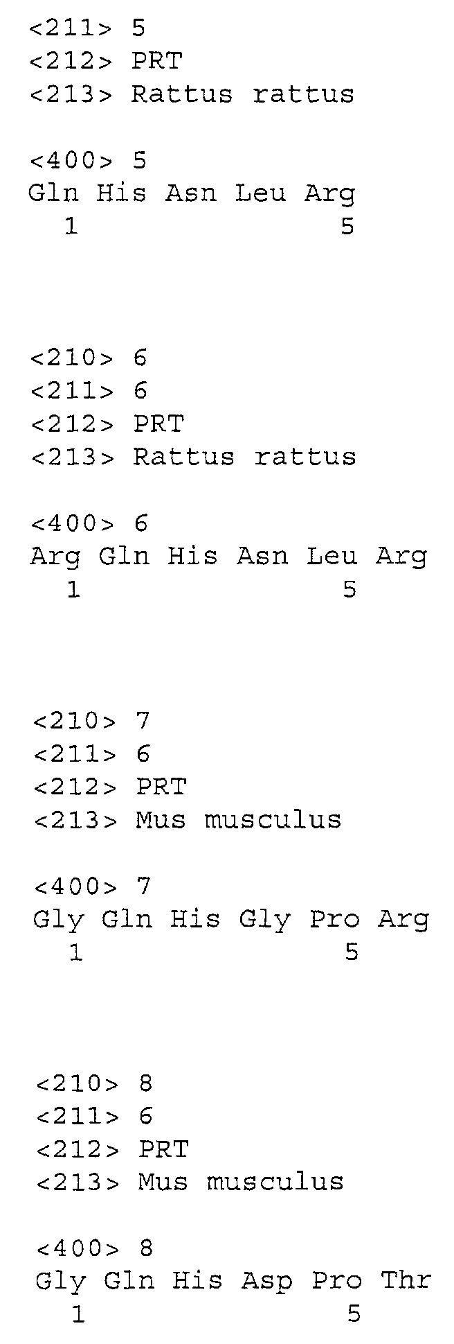
Claims (29)
- A therapeutic use of a SMR1-peptide or a pharmaceutically active amount of said SMR1-peptide, for the preparation of a therapeutic composition for preventing or treating diseases wherein a modulation of the activity of a membrane metallopeptidase is sought, in a mammal, specifically in a human.
- The use of claim 1 wherein said metallopeptidase is a membrane-zinc metallopeptidase.
- The use according to claim 2, for preventing or treating diseases wherein modulation of NEP-induced degradation of NEP-sensitive peptides is sought, in a mammal, specifically in human.
- The use according to claim 3, wherein said SMR1-peptide acts by inhibiting NEP.
- The use according to claims 1 to 4, wherein said SMR1-peptide acts as an analgesic agent.
- The use according to claims 1 to 4, wherein said SMR1-peptide acts as an antidiarrheal agent.
- The use according to claim 3, wherein said SMR1-peptide acts by inhibiting endogenous A II formation and BK and ANP inactivation.
- The use according to claim1 1 to 4 or 7, wherein said SMR1-peptide acts as an antihypertensive agent.
- The use according to claims 1 to 4 or 7, wherein said SMR1-peptide acts as a natriuretic agent.
- The use according to claims 1 to 4 or 7, wherein said SMR1-peptide acts as a diuretic agent.
- The use according to claim 1 for the prevention or treatment of atherosclerosis.
- The use according to claim 1, wherein the SMR1-peptide is an anti-tumor agent.
- The use according to claim 1 for the treatment of arthritis.
- The use according to claim 1 for the treatment of inflammatory bowel disease.
- The use according to claim 1 as an anti-infectious agent for treating infections such as bacterial or viral infections.
- The use according to claim 1 for controlling immuno-inflammatory responses.
- The use according to claims 1 to 16, wherein the therapeutic composition comprises a SMR1-peptide of formula (1)
X1QHX2X3X4 - The use according to claim 17, wherein the SMR1-peptide or one of its biologically active derivatives peptides comprises one or more amino acids in the D-form.
- The use according to claim 17, wherein the SMR1-peptide or one of its biologically active derivatives peptides further comprises a substituted or modified or added group for enhancing the bioavailability or enhancing the resistance to enzymatic degradation of said peptide or biologically active derivative thereof.
- A process for screening ligand molecules that specifically bind to the NEP binding site for the QHNPR pentapeptide, comprising the steps of:a) preparing a confluent target cell culture monolayer or preparing a target organ specimen or a tissue sample containing NEP binding sites for the QHNPR pentapeptide ;b) adding the candidate molecule to be tested in competition with half-saturating concentration of labeled pentapeptide ;c) incubating the cell culture, organ specimen or tissue sample of step a) in the presence of the labeled candidate molecule during a time sufficient and under conditions for the specific binding to take place ;d) quantifying the label specifically bound to the target cell culture, organ specimen or tissue sample in the presence of various concentrations of said candidate molecule.
- A process for determining the relative affinity of ligand molecules that specifically bind to the NEP binding site for the QHNPR pentapeptide comprising the steps a), b), c) and d) of the process of claim 20 for each candidate molecule and further comprising the step e) of comparing the affinity of each candidate molecule quantified in step d) to the one of the other candidate molecules.
- A process for determining the affinity of ligand molecules that specifically bind to the NEP binding site for the QHNPR pentapeptide, comprising the steps of :a) preparing a confluent target cell culture monolayer or preparing a target organ specimen or a tissue sample containing NEP binding sites for the QHNPR pentapeptide ;b) adding the candidate molecule which has previously been labeled with a radioactive or a nonradioactive label;c) incubating the cell culture, organ specimen or tissue sample of step a) in the presence of the labeled candidate molecule during a time sufficient under conditions for the specific binding to take place; andd) quantifying the label specifically bound to the target cell culture, organ specimen or tissue sample in the presence of various concentrations of labeled candidate molecule.
- A process for screening ligand molecules that possess an agonist biological activity on the NEP binding site of the QHNPR pentapeptide, comprising the steps of:a) preparing a confluent target cell culture monolayer or preparing a target organ specimen or a tissue sample containing NEP binding sites for the QHNPR pentapeptide ;b) incubating the cell culture, organ specimen or tissue sample of step a) at concentrations allowing measurement of NEP enzymatic activity under initial velocity conditions in the presence of the candidate molecule, a half-saturating concentration of QHNPR and a NEP substrate during a time sufficient for the hydrolysis activity of the NEP substrate to take place under initial velocity conditions ;c) quantifying the activity of the NEP present in the biological material of step a) by measuring the levels of NEP substrate hydrolysis, respectively in the presence or in the absence of the candidate ligand molecule and in the presence or in the absence of QHNPR.
- A process for screening ligand molecules that possess an antagonist biological activity on the NEP binding site of the QHNPR pentapeptide, comprising the steps of:a) preparing a confluent target cell culture monolayer or preparing a target organ specimen or a tissue sample containing NEP binding sites for the QHNPR pentapeptide ;b) incubating the cell culture, organ specimen or tissue sample of step a) at concentrations allowing measurement of NEP enzymatic activity under initial velocity conditions in the presence of a submaximal concentration of the XQHNPR peptide, specifically the QHNPR peptide and a NEP substrate, in the presence of the candidate molecule during a time sufficient for the hydrolysis of the NEP substrate to take place under velocity conditions ;c) quantifying the hydrolysis activity of the NEP present in the biological material of step a) by measuring the levels of NEP substrate hydrolysis, respectively in the presence or in the absence of the candidate ligand molecule and in the presence or in the absence of QHNPR.
- A maturation product of SMR1 protein or a biologically active derivative of the SMR1 protein or of its maturation products which has been obtained according to the method of claims 20 to 24, provided that said biologically active derivatives does not have the structure of formula (1) as defined in claim 17.
- A molecular complex comprising :the NEP receptor or the SMR1-binding site of the NEP receptor;a SMR1-peptide.
- A biologically active derivative of SMR1-peptide characterized by its capacity either to increase or to decrease a metallopeptidase activity or to prevent the normal interaction between the SMR1-peptide and said metallopeptidase.
- The biologically active derivative of claim 27 characterized in that said metallopeptidase is a membrane-zinc metallopeptidase.
- The biologically active derivative of claim 28 characterized in that said membrane-zinc metallopeptidase is NEP.
Priority Applications (27)
| Application Number | Priority Date | Filing Date | Title |
|---|---|---|---|
| EP00403670A EP1216707B1 (en) | 2000-12-22 | 2000-12-22 | Process for screening ligand molecules that specifically bind to the NEP binding site for the QHNPR pentapeptide |
| AT00403670T ATE288763T1 (en) | 2000-12-22 | 2000-12-22 | PROCESS FOR SCREENING MOLECULES THAT BIND SPECIFICALLY TO THE NEP BINDING SITE OF THE QHNPR PENTAPETIDE |
| DE60018085T DE60018085T2 (en) | 2000-12-22 | 2000-12-22 | Process for screening molecules that specifically bind to the NEP binding site of the QHNPR pentapeptide |
| PT00403670T PT1216707E (en) | 2000-12-22 | 2000-12-22 | NEW THERAPEUTIC UTILIZATIONS FOR A SMR-1-PEPTIDEO |
| ES00403670T ES2235806T3 (en) | 2000-12-22 | 2000-12-22 | MOLECULES SELECTION PROCEDURE BINDING TO SPECIFICALLY JOIN THE NEP UNION SITE FOR THE QHNPR PRESENT. |
| DK00403670T DK1216707T3 (en) | 2000-12-22 | 2000-12-22 | Method for screening ligand molecules that specifically bind to the NEP binding sites of the QHNPR pentapeptide |
| CA002431913A CA2431913A1 (en) | 2000-12-22 | 2001-12-24 | Modulation of smr1-nep interactions in cns disorders giving rise to mental disorders |
| CNB018212646A CN1229137C (en) | 2000-12-22 | 2001-12-24 | New therapeutic uses of SMR1 peptides |
| CN018212654A CN1217190C (en) | 2000-12-22 | 2001-12-24 | Modulation of smr1-nep interactions in cns disorders giving rise to mental disorders |
| EP01985459A EP1343519A2 (en) | 2000-12-22 | 2001-12-24 | Modulation of smr1-nep interactions in cns disorders giving rise to mental disorders |
| AU2002236100A AU2002236100A1 (en) | 2000-12-22 | 2001-12-24 | New therapeutic uses of smr1 peptides |
| CA2431916A CA2431916C (en) | 2000-12-22 | 2001-12-24 | New therapeutic uses of smr1 peptides |
| EP01985936A EP1343520B1 (en) | 2000-12-22 | 2001-12-24 | New therapeutic uses of smr1 peptides |
| PCT/IB2001/002819 WO2002051435A2 (en) | 2000-12-22 | 2001-12-24 | New therapeutic uses of smr1 peptides |
| ES01985936T ES2363708T3 (en) | 2000-12-22 | 2001-12-24 | NEW THERAPEUTIC USES OF PEPTIDES SMR1. |
| US10/451,073 US7423020B2 (en) | 2000-12-22 | 2001-12-24 | Treatment of mood disorders by administration of SMR1 |
| DE60143990T DE60143990D1 (en) | 2000-12-22 | 2001-12-24 | NEW THERAPEUTIC USE OF SMR-1 PEPTIDE |
| AT01985936T ATE497390T1 (en) | 2000-12-22 | 2001-12-24 | NEW THERAPEUTIC USE OF SMR-1 PEPTIDES |
| AU2002234806A AU2002234806A1 (en) | 2000-12-22 | 2001-12-24 | Modulation of smr1-nep interactions in cns disorders giving rise to mental disorders |
| JP2002552576A JP4347566B2 (en) | 2000-12-22 | 2001-12-24 | Modulation of SMR1-NEP interaction in diseases of the central nervous system that cause mental disorders |
| PCT/IB2001/002818 WO2002051434A2 (en) | 2000-12-22 | 2001-12-24 | Modulation of smr1-nep interactions in cns disorders giving rise to mental disorders |
| JP2002552577A JP4409831B2 (en) | 2000-12-22 | 2001-12-24 | Novel therapeutic use of SMR1 peptide |
| US10/451,064 US7153833B2 (en) | 2000-12-22 | 2001-12-24 | Therapeutic uses of SMR1 peptides |
| US11/594,105 US7625713B2 (en) | 2000-12-22 | 2006-11-08 | Process for identifying a ligand that binds to the NEP binding site for the SMR1 pentapeptide |
| JP2008149611A JP2009007352A (en) | 2000-12-22 | 2008-06-06 | New curative use of smr1-peptide |
| JP2008305315A JP2009149633A (en) | 2000-12-22 | 2008-11-28 | Modulation of smr1-nep interaction in central nervous system disorder giving rise to mental disorder |
| US12/580,701 US20100041072A1 (en) | 2000-12-22 | 2009-10-16 | Process for identifying a ligand that binds to the nep binding site for the smr1 pentapeptide |
Applications Claiming Priority (1)
| Application Number | Priority Date | Filing Date | Title |
|---|---|---|---|
| EP00403670A EP1216707B1 (en) | 2000-12-22 | 2000-12-22 | Process for screening ligand molecules that specifically bind to the NEP binding site for the QHNPR pentapeptide |
Publications (2)
| Publication Number | Publication Date |
|---|---|
| EP1216707A1 true EP1216707A1 (en) | 2002-06-26 |
| EP1216707B1 EP1216707B1 (en) | 2005-02-09 |
Family
ID=8174003
Family Applications (3)
| Application Number | Title | Priority Date | Filing Date |
|---|---|---|---|
| EP00403670A Expired - Lifetime EP1216707B1 (en) | 2000-12-22 | 2000-12-22 | Process for screening ligand molecules that specifically bind to the NEP binding site for the QHNPR pentapeptide |
| EP01985936A Expired - Lifetime EP1343520B1 (en) | 2000-12-22 | 2001-12-24 | New therapeutic uses of smr1 peptides |
| EP01985459A Withdrawn EP1343519A2 (en) | 2000-12-22 | 2001-12-24 | Modulation of smr1-nep interactions in cns disorders giving rise to mental disorders |
Family Applications After (2)
| Application Number | Title | Priority Date | Filing Date |
|---|---|---|---|
| EP01985936A Expired - Lifetime EP1343520B1 (en) | 2000-12-22 | 2001-12-24 | New therapeutic uses of smr1 peptides |
| EP01985459A Withdrawn EP1343519A2 (en) | 2000-12-22 | 2001-12-24 | Modulation of smr1-nep interactions in cns disorders giving rise to mental disorders |
Country Status (12)
| Country | Link |
|---|---|
| US (4) | US7153833B2 (en) |
| EP (3) | EP1216707B1 (en) |
| JP (4) | JP4409831B2 (en) |
| CN (2) | CN1217190C (en) |
| AT (2) | ATE288763T1 (en) |
| AU (2) | AU2002236100A1 (en) |
| CA (2) | CA2431913A1 (en) |
| DE (2) | DE60018085T2 (en) |
| DK (1) | DK1216707T3 (en) |
| ES (2) | ES2235806T3 (en) |
| PT (1) | PT1216707E (en) |
| WO (2) | WO2002051435A2 (en) |
Cited By (2)
| Publication number | Priority date | Publication date | Assignee | Title |
|---|---|---|---|---|
| EP1577320A1 (en) * | 2004-03-19 | 2005-09-21 | Institut Pasteur | Peptides derived from human BPLP protein, polynucleotides coding for said peptides and antibodies directed against said peptides |
| EP3009142A1 (en) | 2008-05-26 | 2016-04-20 | Institut Pasteur | Opiorphin for use as analgesic agent |
Families Citing this family (21)
| Publication number | Priority date | Publication date | Assignee | Title |
|---|---|---|---|---|
| US6589750B2 (en) * | 1997-02-20 | 2003-07-08 | Institut Pasteur | Therapeutic use of the SMR1 protein, the SMR1 maturation products, specifically the QHNPR pentapeptide as well as its biologically active derivatives |
| AU6154700A (en) | 1999-06-23 | 2001-01-31 | Centre National De La Recherche Scientifique (C.N.R.S.) | Therapeutic methods and compositions for the treatment of impaired interpersonal and behavioral disorders |
| EP1216707B1 (en) * | 2000-12-22 | 2005-02-09 | Institut Pasteur | Process for screening ligand molecules that specifically bind to the NEP binding site for the QHNPR pentapeptide |
| DE60327432D1 (en) * | 2002-05-29 | 2009-06-10 | Merck & Co Inc | USEFUL COMPOUNDS FOR THE TREATMENT OF ANTHRAX AND INHIBITION OF THE LETAL FACTOR |
| US7772188B2 (en) | 2003-01-28 | 2010-08-10 | Ironwood Pharmaceuticals, Inc. | Methods and compositions for the treatment of gastrointestinal disorders |
| BR122018074353B8 (en) | 2003-01-28 | 2021-07-27 | Ironwood Pharmaceuticals Inc | pharmaceutical composition, polypeptide and methods for producing them |
| JP2007537256A (en) * | 2004-05-11 | 2007-12-20 | メルク エンド カムパニー インコーポレーテッド | Process for the preparation of N-sulfonated amino acid derivatives |
| US20100003276A1 (en) * | 2004-12-07 | 2010-01-07 | Hermes Jeffery D | Methods for treating anthrax and inhibiting lethal factor |
| CN101772513B (en) | 2007-06-04 | 2013-11-13 | 协同医药品公司 | Agonists of guanylate cyclase useful for the treatment of gastrointestinal disorders, inflammation, cancer and other disorders |
| US8969514B2 (en) | 2007-06-04 | 2015-03-03 | Synergy Pharmaceuticals, Inc. | Agonists of guanylate cyclase useful for the treatment of hypercholesterolemia, atherosclerosis, coronary heart disease, gallstone, obesity and other cardiovascular diseases |
| WO2009124948A1 (en) * | 2008-04-07 | 2009-10-15 | Institut Pasteur | Opiorphin peptide derivatives as potent inhibitors of enkephalin - degrading ectopetidases |
| ES2522968T3 (en) | 2008-06-04 | 2014-11-19 | Synergy Pharmaceuticals Inc. | Guanylate cyclase agonists useful for the treatment of gastrointestinal disorders, inflammation, cancer and other disorders |
| JP2011528375A (en) | 2008-07-16 | 2011-11-17 | シナジー ファーマシューティカルズ インコーポレイテッド | Guanylate cyclase agonists useful for the treatment of gastrointestinal disorders, inflammation, cancer, and other disorders |
| CA2645452A1 (en) * | 2008-11-28 | 2010-05-28 | Institut Pasteur | Use of basic prolin-rich lacrimal gene products, such as opiorphin, as a biomarker |
| EP2373296B1 (en) | 2008-12-03 | 2016-08-03 | Synergy Pharmaceuticals Inc. | Formulations of guanylate cyclase c agonists and methods of use |
| US8506544B2 (en) | 2010-06-21 | 2013-08-13 | The Procter & Gamble Company | Disposable absorbent pant with efficient design and convenient single-section side stretch panels |
| US9616097B2 (en) | 2010-09-15 | 2017-04-11 | Synergy Pharmaceuticals, Inc. | Formulations of guanylate cyclase C agonists and methods of use |
| CA2828346C (en) | 2011-03-01 | 2021-01-26 | Synergy Pharmaceuticals Inc. | Process of preparing guanylate cyclase c agonists |
| AU2014235209B2 (en) | 2013-03-15 | 2018-06-14 | Bausch Health Ireland Limited | Guanylate cyclase receptor agonists combined with other drugs |
| JP2016514671A (en) | 2013-03-15 | 2016-05-23 | シナジー ファーマシューティカルズ インコーポレイテッド | Guanylate cyclase agonists and uses thereof |
| WO2014197720A2 (en) | 2013-06-05 | 2014-12-11 | Synergy Pharmaceuticals, Inc. | Ultra-pure agonists of guanylate cyclase c, method of making and using same |
Citations (2)
| Publication number | Priority date | Publication date | Assignee | Title |
|---|---|---|---|---|
| WO1990003981A1 (en) * | 1988-10-11 | 1990-04-19 | Institut Pasteur | Peptides and polypeptides from the sub-maxilary gland of the rat, corresponding monoclonal and polyclonal antibodies, corresponding hybridomas and applications of said products to the diagnosis, detection or for therapeutical purposes |
| WO1998037100A2 (en) * | 1997-02-20 | 1998-08-27 | Institut Pasteur | Therapeutic use of the smr1 protein and active derivatives thereof |
Family Cites Families (5)
| Publication number | Priority date | Publication date | Assignee | Title |
|---|---|---|---|---|
| US738778A (en) * | 1903-05-22 | 1903-09-15 | Cottrell C B & Sons Co | Sheet-delivery apparatus for printing and folding machinery. |
| ES2058169T3 (en) * | 1987-04-25 | 1994-11-01 | Hoechst Ag | ENKASTINAS: NEW GLYCOPEPTIDES WITH ENCEPHALINASE INHIBITING EFFECT, PROCEDURE FOR THEIR PREPARATION AND USE AS PHARMACEUTICAL PREPARATIONS. |
| US5550119A (en) * | 1995-03-02 | 1996-08-27 | Ciba-Geigy Corporation | Phosphono substituted tetrazole derivatives as ECE inhibitors |
| AU6154700A (en) | 1999-06-23 | 2001-01-31 | Centre National De La Recherche Scientifique (C.N.R.S.) | Therapeutic methods and compositions for the treatment of impaired interpersonal and behavioral disorders |
| EP1216707B1 (en) * | 2000-12-22 | 2005-02-09 | Institut Pasteur | Process for screening ligand molecules that specifically bind to the NEP binding site for the QHNPR pentapeptide |
-
2000
- 2000-12-22 EP EP00403670A patent/EP1216707B1/en not_active Expired - Lifetime
- 2000-12-22 ES ES00403670T patent/ES2235806T3/en not_active Expired - Lifetime
- 2000-12-22 PT PT00403670T patent/PT1216707E/en unknown
- 2000-12-22 AT AT00403670T patent/ATE288763T1/en active
- 2000-12-22 DK DK00403670T patent/DK1216707T3/en active
- 2000-12-22 DE DE60018085T patent/DE60018085T2/en not_active Expired - Lifetime
-
2001
- 2001-12-24 EP EP01985936A patent/EP1343520B1/en not_active Expired - Lifetime
- 2001-12-24 EP EP01985459A patent/EP1343519A2/en not_active Withdrawn
- 2001-12-24 JP JP2002552577A patent/JP4409831B2/en not_active Expired - Fee Related
- 2001-12-24 CA CA002431913A patent/CA2431913A1/en not_active Abandoned
- 2001-12-24 AU AU2002236100A patent/AU2002236100A1/en not_active Abandoned
- 2001-12-24 ES ES01985936T patent/ES2363708T3/en not_active Expired - Lifetime
- 2001-12-24 CA CA2431916A patent/CA2431916C/en not_active Expired - Lifetime
- 2001-12-24 AT AT01985936T patent/ATE497390T1/en active
- 2001-12-24 AU AU2002234806A patent/AU2002234806A1/en not_active Abandoned
- 2001-12-24 US US10/451,064 patent/US7153833B2/en not_active Expired - Lifetime
- 2001-12-24 DE DE60143990T patent/DE60143990D1/en not_active Expired - Lifetime
- 2001-12-24 US US10/451,073 patent/US7423020B2/en not_active Expired - Fee Related
- 2001-12-24 JP JP2002552576A patent/JP4347566B2/en not_active Expired - Fee Related
- 2001-12-24 WO PCT/IB2001/002819 patent/WO2002051435A2/en active Application Filing
- 2001-12-24 WO PCT/IB2001/002818 patent/WO2002051434A2/en active Application Filing
- 2001-12-24 CN CN018212654A patent/CN1217190C/en not_active Expired - Fee Related
- 2001-12-24 CN CNB018212646A patent/CN1229137C/en not_active Expired - Fee Related
-
2006
- 2006-11-08 US US11/594,105 patent/US7625713B2/en not_active Expired - Fee Related
-
2008
- 2008-06-06 JP JP2008149611A patent/JP2009007352A/en active Pending
- 2008-11-28 JP JP2008305315A patent/JP2009149633A/en active Pending
-
2009
- 2009-10-16 US US12/580,701 patent/US20100041072A1/en not_active Abandoned
Patent Citations (2)
| Publication number | Priority date | Publication date | Assignee | Title |
|---|---|---|---|---|
| WO1990003981A1 (en) * | 1988-10-11 | 1990-04-19 | Institut Pasteur | Peptides and polypeptides from the sub-maxilary gland of the rat, corresponding monoclonal and polyclonal antibodies, corresponding hybridomas and applications of said products to the diagnosis, detection or for therapeutical purposes |
| WO1998037100A2 (en) * | 1997-02-20 | 1998-08-27 | Institut Pasteur | Therapeutic use of the smr1 protein and active derivatives thereof |
Non-Patent Citations (1)
| Title |
|---|
| ROUGEOT CATHERINE ET AL: "Rodent submandibular gland peptide hormones and other biologically active peptides.", PEPTIDES (NEW YORK), vol. 21, no. 3, March 2000 (2000-03-01), pages 443 - 455, XP002179502, ISSN: 0196-9781 * |
Cited By (12)
| Publication number | Priority date | Publication date | Assignee | Title |
|---|---|---|---|---|
| EP1577320A1 (en) * | 2004-03-19 | 2005-09-21 | Institut Pasteur | Peptides derived from human BPLP protein, polynucleotides coding for said peptides and antibodies directed against said peptides |
| WO2005090386A1 (en) * | 2004-03-19 | 2005-09-29 | Institut Pasteur | Peptides derived from human bplp protein, polynucleotides coding for said peptides and antibodies directed against said peptides |
| US8017571B2 (en) | 2004-03-19 | 2011-09-13 | Institut Pasteur | Peptides derived from human BPLP protein |
| CN1953988B (en) * | 2004-03-19 | 2013-02-13 | 巴斯德研究院 | Peptides derived from human BPLP protein, polynucleotides coding for the peptides and antibodies directed against the peptides |
| CN103183726A (en) * | 2004-03-19 | 2013-07-03 | 巴斯德研究院 | Peptides derived from human BPLP protein, polynucleotides coding for said peptides and antibodies directed against said peptides |
| US8895251B2 (en) | 2004-03-19 | 2014-11-25 | Institut Pasteur | Method for detecting human BPLP protein or a maturation product thereof |
| EP2952521A1 (en) * | 2004-03-19 | 2015-12-09 | Institut Pasteur | Peptides derived from human BPLP protein, polynucleotides coding for said peptides and antibodies directed against said peptides |
| US9403871B2 (en) | 2004-03-19 | 2016-08-02 | Institut Pasteur | Methods for treating pain by administering peptides derived from human basic proline-rich lacrimal protein |
| US9714951B2 (en) | 2004-03-19 | 2017-07-25 | Institut Pasteur | Pharmaceutical compositions containing peptides derived from human BPLP protein |
| EP3009142A1 (en) | 2008-05-26 | 2016-04-20 | Institut Pasteur | Opiorphin for use as analgesic agent |
| EP3804744A1 (en) | 2008-05-26 | 2021-04-14 | Institut Pasteur | Opiorphin for use as analgesic agent |
| EP4218786A2 (en) | 2008-05-26 | 2023-08-02 | Institut Pasteur | Opiorphin for use as analgesic agent |
Also Published As
Similar Documents
| Publication | Publication Date | Title |
|---|---|---|
| EP1216707B1 (en) | Process for screening ligand molecules that specifically bind to the NEP binding site for the QHNPR pentapeptide | |
| US9714951B2 (en) | Pharmaceutical compositions containing peptides derived from human BPLP protein |
Legal Events
| Date | Code | Title | Description |
|---|---|---|---|
| PUAI | Public reference made under article 153(3) epc to a published international application that has entered the european phase |
Free format text: ORIGINAL CODE: 0009012 |
|
| AK | Designated contracting states |
Kind code of ref document: A1 Designated state(s): AT BE CH CY DE DK ES FI FR GB GR IE IT LI LU MC NL PT SE TR |
|
| AX | Request for extension of the european patent |
Free format text: AL;LT;LV;MK;RO;SI |
|
| 17P | Request for examination filed |
Effective date: 20021202 |
|
| AKX | Designation fees paid |
Designated state(s): AT BE CH CY DE DK ES FI FR GB GR IE IT LI LU MC NL PT SE TR |
|
| 17Q | First examination report despatched |
Effective date: 20030618 |
|
| GRAP | Despatch of communication of intention to grant a patent |
Free format text: ORIGINAL CODE: EPIDOSNIGR1 |
|
| RTI1 | Title (correction) |
Free format text: PROCESS FOR SCREENING LIGAND MOLECULES THAT SPECIFICALLY BIND TO THE NEP BINDING SITE FOR THE QHNPR PENTAPEPTIDE |
|
| GRAS | Grant fee paid |
Free format text: ORIGINAL CODE: EPIDOSNIGR3 |
|
| GRAA | (expected) grant |
Free format text: ORIGINAL CODE: 0009210 |
|
| AK | Designated contracting states |
Kind code of ref document: B1 Designated state(s): AT BE CH CY DE DK ES FI FR GB GR IE IT LI LU MC NL PT SE TR |
|
| REG | Reference to a national code |
Ref country code: GB Ref legal event code: FG4D |
|
| REG | Reference to a national code |
Ref country code: CH Ref legal event code: EP |
|
| REG | Reference to a national code |
Ref country code: CH Ref legal event code: NV Representative=s name: A. BRAUN, BRAUN, HERITIER, ESCHMANN AG PATENTANWAE |
|
| REG | Reference to a national code |
Ref country code: IE Ref legal event code: FG4D |
|
| REG | Reference to a national code |
Ref country code: SE Ref legal event code: TRGR |
|
| REF | Corresponds to: |
Ref document number: 60018085 Country of ref document: DE Date of ref document: 20050317 Kind code of ref document: P |
|
| REG | Reference to a national code |
Ref country code: GR Ref legal event code: EP Ref document number: 20050400607 Country of ref document: GR |
|
| REG | Reference to a national code |
Ref country code: DK Ref legal event code: T3 |
|
| REG | Reference to a national code |
Ref country code: PT Ref legal event code: SC4A Free format text: AVAILABILITY OF NATIONAL TRANSLATION Effective date: 20050506 |
|
| REG | Reference to a national code |
Ref country code: ES Ref legal event code: FG2A Ref document number: 2235806 Country of ref document: ES Kind code of ref document: T3 |
|
| ET | Fr: translation filed | ||
| PLBE | No opposition filed within time limit |
Free format text: ORIGINAL CODE: 0009261 |
|
| STAA | Information on the status of an ep patent application or granted ep patent |
Free format text: STATUS: NO OPPOSITION FILED WITHIN TIME LIMIT |
|
| 26N | No opposition filed |
Effective date: 20051110 |
|
| REG | Reference to a national code |
Ref country code: CH Ref legal event code: PFA Owner name: INSTITUT PASTEUR Free format text: INSTITUT PASTEUR#28, RUE DU DOCTEUR ROUX#75724 PARIS CEDEX 15 (FR) -TRANSFER TO- INSTITUT PASTEUR#28, RUE DU DOCTEUR ROUX#75724 PARIS CEDEX 15 (FR) |
|
| PGFP | Annual fee paid to national office [announced via postgrant information from national office to epo] |
Ref country code: IT Payment date: 20091222 Year of fee payment: 10 |
|
| PGFP | Annual fee paid to national office [announced via postgrant information from national office to epo] |
Ref country code: BE Payment date: 20100212 Year of fee payment: 10 |
|
| PGFP | Annual fee paid to national office [announced via postgrant information from national office to epo] |
Ref country code: NL Payment date: 20101116 Year of fee payment: 11 Ref country code: MC Payment date: 20101122 Year of fee payment: 11 Ref country code: IE Payment date: 20101129 Year of fee payment: 11 Ref country code: DK Payment date: 20101119 Year of fee payment: 11 Ref country code: AT Payment date: 20101117 Year of fee payment: 11 |
|
| PGFP | Annual fee paid to national office [announced via postgrant information from national office to epo] |
Ref country code: FI Payment date: 20101117 Year of fee payment: 11 Ref country code: LU Payment date: 20101123 Year of fee payment: 11 Ref country code: PT Payment date: 20101116 Year of fee payment: 11 |
|
| PGFP | Annual fee paid to national office [announced via postgrant information from national office to epo] |
Ref country code: TR Payment date: 20101221 Year of fee payment: 11 Ref country code: SE Payment date: 20101119 Year of fee payment: 11 Ref country code: GR Payment date: 20101123 Year of fee payment: 11 |
|
| PGFP | Annual fee paid to national office [announced via postgrant information from national office to epo] |
Ref country code: CY Payment date: 20101124 Year of fee payment: 11 |
|
| BERE | Be: lapsed |
Owner name: *INSTITUT PASTEUR Effective date: 20101231 |
|
| PGFP | Annual fee paid to national office [announced via postgrant information from national office to epo] |
Ref country code: ES Payment date: 20101223 Year of fee payment: 11 |
|
| PG25 | Lapsed in a contracting state [announced via postgrant information from national office to epo] |
Ref country code: BE Free format text: LAPSE BECAUSE OF NON-PAYMENT OF DUE FEES Effective date: 20101231 |
|
| PG25 | Lapsed in a contracting state [announced via postgrant information from national office to epo] |
Ref country code: IT Free format text: LAPSE BECAUSE OF NON-PAYMENT OF DUE FEES Effective date: 20101222 |
|
| REG | Reference to a national code |
Ref country code: PT Ref legal event code: MM4A Free format text: LAPSE DUE TO NON-PAYMENT OF FEES Effective date: 20120622 |
|
| REG | Reference to a national code |
Ref country code: NL Ref legal event code: V1 Effective date: 20120701 |
|
| REG | Reference to a national code |
Ref country code: DK Ref legal event code: EBP |
|
| PG25 | Lapsed in a contracting state [announced via postgrant information from national office to epo] |
Ref country code: MC Free format text: LAPSE BECAUSE OF NON-PAYMENT OF DUE FEES Effective date: 20111231 |
|
| REG | Reference to a national code |
Ref country code: SE Ref legal event code: EUG |
|
| PG25 | Lapsed in a contracting state [announced via postgrant information from national office to epo] |
Ref country code: FI Free format text: LAPSE BECAUSE OF NON-PAYMENT OF DUE FEES Effective date: 20111222 Ref country code: PT Free format text: LAPSE BECAUSE OF NON-PAYMENT OF DUE FEES Effective date: 20120622 |
|
| REG | Reference to a national code |
Ref country code: GR Ref legal event code: ML Ref document number: 20050400607 Country of ref document: GR Effective date: 20120704 |
|
| REG | Reference to a national code |
Ref country code: IE Ref legal event code: MM4A |
|
| PG25 | Lapsed in a contracting state [announced via postgrant information from national office to epo] |
Ref country code: CY Free format text: LAPSE BECAUSE OF NON-PAYMENT OF DUE FEES Effective date: 20111222 |
|
| PG25 | Lapsed in a contracting state [announced via postgrant information from national office to epo] |
Ref country code: IE Free format text: LAPSE BECAUSE OF NON-PAYMENT OF DUE FEES Effective date: 20111222 Ref country code: SE Free format text: LAPSE BECAUSE OF NON-PAYMENT OF DUE FEES Effective date: 20111223 |
|
| PG25 | Lapsed in a contracting state [announced via postgrant information from national office to epo] |
Ref country code: GR Free format text: LAPSE BECAUSE OF NON-PAYMENT OF DUE FEES Effective date: 20120704 |
|
| REG | Reference to a national code |
Ref country code: AT Ref legal event code: MM01 Ref document number: 288763 Country of ref document: AT Kind code of ref document: T Effective date: 20111222 |
|
| PG25 | Lapsed in a contracting state [announced via postgrant information from national office to epo] |
Ref country code: AT Free format text: LAPSE BECAUSE OF NON-PAYMENT OF DUE FEES Effective date: 20111222 Ref country code: DK Free format text: LAPSE BECAUSE OF NON-PAYMENT OF DUE FEES Effective date: 20120102 Ref country code: NL Free format text: LAPSE BECAUSE OF NON-PAYMENT OF DUE FEES Effective date: 20120701 |
|
| PG25 | Lapsed in a contracting state [announced via postgrant information from national office to epo] |
Ref country code: LU Free format text: LAPSE BECAUSE OF NON-PAYMENT OF DUE FEES Effective date: 20111222 |
|
| REG | Reference to a national code |
Ref country code: ES Ref legal event code: FD2A Effective date: 20130703 |
|
| PG25 | Lapsed in a contracting state [announced via postgrant information from national office to epo] |
Ref country code: ES Free format text: LAPSE BECAUSE OF NON-PAYMENT OF DUE FEES Effective date: 20111223 |
|
| PG25 | Lapsed in a contracting state [announced via postgrant information from national office to epo] |
Ref country code: TR Free format text: LAPSE BECAUSE OF NON-PAYMENT OF DUE FEES Effective date: 20111222 |
|
| REG | Reference to a national code |
Ref country code: CH Ref legal event code: PCAR Free format text: NEW ADDRESS: HOLBEINSTRASSE 36-38, 4051 BASEL (CH) |
|
| REG | Reference to a national code |
Ref country code: FR Ref legal event code: PLFP Year of fee payment: 16 |
|
| REG | Reference to a national code |
Ref country code: FR Ref legal event code: PLFP Year of fee payment: 17 |
|
| PGFP | Annual fee paid to national office [announced via postgrant information from national office to epo] |
Ref country code: CH Payment date: 20161222 Year of fee payment: 17 Ref country code: GB Payment date: 20161221 Year of fee payment: 17 |
|
| PGFP | Annual fee paid to national office [announced via postgrant information from national office to epo] |
Ref country code: FR Payment date: 20161229 Year of fee payment: 17 |
|
| PGFP | Annual fee paid to national office [announced via postgrant information from national office to epo] |
Ref country code: DE Payment date: 20161221 Year of fee payment: 17 |
|
| REG | Reference to a national code |
Ref country code: DE Ref legal event code: R119 Ref document number: 60018085 Country of ref document: DE |
|
| REG | Reference to a national code |
Ref country code: CH Ref legal event code: PL |
|
| GBPC | Gb: european patent ceased through non-payment of renewal fee |
Effective date: 20171222 |
|
| REG | Reference to a national code |
Ref country code: FR Ref legal event code: ST Effective date: 20180831 |
|
| PG25 | Lapsed in a contracting state [announced via postgrant information from national office to epo] |
Ref country code: DE Free format text: LAPSE BECAUSE OF NON-PAYMENT OF DUE FEES Effective date: 20180703 Ref country code: FR Free format text: LAPSE BECAUSE OF NON-PAYMENT OF DUE FEES Effective date: 20180102 |
|
| PG25 | Lapsed in a contracting state [announced via postgrant information from national office to epo] |
Ref country code: LI Free format text: LAPSE BECAUSE OF NON-PAYMENT OF DUE FEES Effective date: 20171231 Ref country code: GB Free format text: LAPSE BECAUSE OF NON-PAYMENT OF DUE FEES Effective date: 20171222 Ref country code: CH Free format text: LAPSE BECAUSE OF NON-PAYMENT OF DUE FEES Effective date: 20171231 |