EP1209167A1 - Method of producing a biosensor protein capable of regulating a fluorescence property of the modified green fluorescent protein therein, and the biosensor protein produced by the method - Google Patents
Method of producing a biosensor protein capable of regulating a fluorescence property of the modified green fluorescent protein therein, and the biosensor protein produced by the method Download PDFInfo
- Publication number
- EP1209167A1 EP1209167A1 EP01127775A EP01127775A EP1209167A1 EP 1209167 A1 EP1209167 A1 EP 1209167A1 EP 01127775 A EP01127775 A EP 01127775A EP 01127775 A EP01127775 A EP 01127775A EP 1209167 A1 EP1209167 A1 EP 1209167A1
- Authority
- EP
- European Patent Office
- Prior art keywords
- amino acid
- protein
- acid sequence
- fluorescent protein
- green fluorescent
- Prior art date
- Legal status (The legal status is an assumption and is not a legal conclusion. Google has not performed a legal analysis and makes no representation as to the accuracy of the status listed.)
- Withdrawn
Links
Images
Classifications
-
- C—CHEMISTRY; METALLURGY
- C12—BIOCHEMISTRY; BEER; SPIRITS; WINE; VINEGAR; MICROBIOLOGY; ENZYMOLOGY; MUTATION OR GENETIC ENGINEERING
- C12N—MICROORGANISMS OR ENZYMES; COMPOSITIONS THEREOF; PROPAGATING, PRESERVING, OR MAINTAINING MICROORGANISMS; MUTATION OR GENETIC ENGINEERING; CULTURE MEDIA
- C12N9/00—Enzymes; Proenzymes; Compositions thereof; Processes for preparing, activating, inhibiting, separating or purifying enzymes
- C12N9/10—Transferases (2.)
- C12N9/12—Transferases (2.) transferring phosphorus containing groups, e.g. kinases (2.7)
- C12N9/1205—Phosphotransferases with an alcohol group as acceptor (2.7.1), e.g. protein kinases
-
- C—CHEMISTRY; METALLURGY
- C07—ORGANIC CHEMISTRY
- C07K—PEPTIDES
- C07K14/00—Peptides having more than 20 amino acids; Gastrins; Somatostatins; Melanotropins; Derivatives thereof
- C07K14/435—Peptides having more than 20 amino acids; Gastrins; Somatostatins; Melanotropins; Derivatives thereof from animals; from humans
- C07K14/43504—Peptides having more than 20 amino acids; Gastrins; Somatostatins; Melanotropins; Derivatives thereof from animals; from humans from invertebrates
- C07K14/43595—Peptides having more than 20 amino acids; Gastrins; Somatostatins; Melanotropins; Derivatives thereof from animals; from humans from invertebrates from coelenteratae, e.g. medusae
-
- C—CHEMISTRY; METALLURGY
- C07—ORGANIC CHEMISTRY
- C07K—PEPTIDES
- C07K14/00—Peptides having more than 20 amino acids; Gastrins; Somatostatins; Melanotropins; Derivatives thereof
- C07K14/435—Peptides having more than 20 amino acids; Gastrins; Somatostatins; Melanotropins; Derivatives thereof from animals; from humans
- C07K14/46—Peptides having more than 20 amino acids; Gastrins; Somatostatins; Melanotropins; Derivatives thereof from animals; from humans from vertebrates
- C07K14/47—Peptides having more than 20 amino acids; Gastrins; Somatostatins; Melanotropins; Derivatives thereof from animals; from humans from vertebrates from mammals
- C07K14/4701—Peptides having more than 20 amino acids; Gastrins; Somatostatins; Melanotropins; Derivatives thereof from animals; from humans from vertebrates from mammals not used
- C07K14/4728—Calcium binding proteins, e.g. calmodulin
-
- C—CHEMISTRY; METALLURGY
- C07—ORGANIC CHEMISTRY
- C07K—PEPTIDES
- C07K2319/00—Fusion polypeptide
Definitions
- the present invention relates to a method of producing a biosensor protein capable of regulating a fluorescence property of green fluorescent protein. Furthermore, the present invention relates to a biosensor protein (hereinafter also referred to as a biosensor) produced by the above method and a biosensor gene encoding the biosensor protein. More specifically, the present invention relates to a calcium-sensing biosensor protein (hereinafter also referred to as a calcium sensor) produced by the above method and a biosensor gene encoding the calcium-sensing biosensor protein.
- a biosensor protein hereinafter also referred to as a biosensor
- a calcium-sensing biosensor protein hereinafter also referred to as a calcium sensor
- An object of the present invention is to provide a method of producing a biosensor protein capable of regulating a fluorescence property of green fluorescent protein, and to provide a biosensor protein produced by the method and a biosensor gene encoding the biosensor protein.
- an object of the present invention is to provide a calcium-sensing biosensor protein produced by the method and a biosensor gene encoding the calcium-sensing biosensor protein.
- the object of the present invention is to produce a biosensor protein (particularly a calcium-sensing biosensor protein) whose sensitivity to a substance to be detected is high enough to use practically and which makes it possible to measure easily. Further object of the present invention is to produce a biosensor protein (particularly a calcium-sensing biosensor protein) which is introduced into a specific cell easily and which makes it possible to measure without a specific detection device, a coenzyme, and so on.
- the present invention provides the means [1] to [19] described below.
- the biosensor protein of the present invention is made of a single-molecular green fluorescent protein.
- the biosensor protein may be also made of a derivative of green fluorescent protein.
- a derivative of green fluorescent protein means any derivative of green fluorescent protein including YFP (Yellow Fluorescent Protein), CFP (Cyan Fluorescent Protein), and BFP (Blue Fluorescent Protein).
- biosensor protein made of green fluorescent protein will be described as a representative example. Therefore, the present invention is not limited to the example.
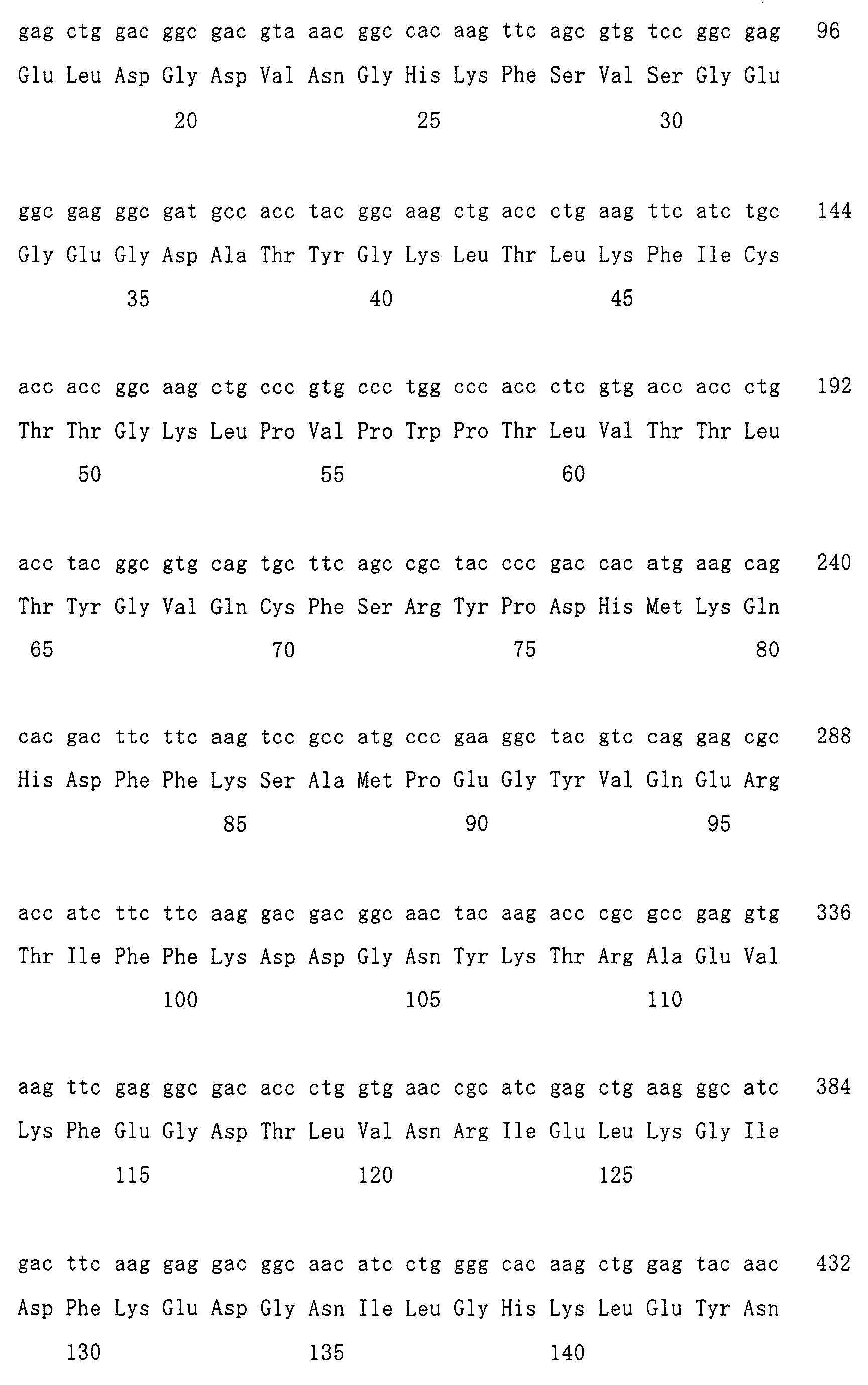
- Green fluorescent protein is a protein consisting of 238 amino acids.
- GFP used in the present invention is a recombinant GFP (EGFP) obtained from Clontech.
- the EGFP contains valine (gtg) (not a constituent of original GFP) which is inserted at the back of methionine of the N terminus of original GFP.
- the valine is designated as "amino acid No. 1' ".
- the valine (gtg)(amino acid No. 1') is omitted from the nucleotide sequence and amino acid sequence of the GFP in Sequence ID No: 1, for the sake of convenience.
- the n-th amino acid of green fluorescent protein or “the amino acid of n-th position of green fluorescent protein” corresponds to the position of the 1st to 238th amino acids in the amino acid sequence described in Sequence ID No: 1.
- biosensor proteins are actually produced using the aforementioned EGFP, which has valine (amino acid No. 1') at the back of methionine of the N terminus of GFP.
- GFP has a chromophore in the center of the inside of its molecular structure.
- GFP can be obtained by expressing a plasmid (e.g., pEGFP-N1 (Clontech)) containing GFP-encoding cDNA.
- the "hotspot amino acid residue” affecting the fluorescence property of GFP refers to an amino acid residue of GFP which acts as an indicator of a GFP modification site, in making a modified green fluorescent protein as described later.
- the structure of GFP is modified in the vicinity of the above hotspot amino acid residue, and thereby it becomes possible to produce a desired modified green fluorescent protein.
- Modification of GFP structure preferably means cleaving GFP in the vicinity of the predicted hotspot amino acid residue (preferably, at any position within 5 amino acids before and behind the hotspot amino acid residue), and eliminating the appropriate number of amino acids (preferably 1 to 10 amino acids) from the cleavage site.
- modification of GFP structure is not limited to the aforementioned modification, as long as it can produce a desired modified green fluorescent protein, and GFP may be modified by any modification process.
- the desired modified green fluorescent protein refers to a protein which is obtained by modifying the structure of GFP such that its fluorescence property can be reversibly changed, as described later.
- fluorescence property can be reversibly changed means that the conformation of the modified GFP reversibly changes by the presence or absence of a factor capable of causing a conformational change of the modified GFP (i.e., a biosensor-detecting factor), thereby altering the fluorescence property of the modified GFP in a reversible manner.
- a factor capable of causing a conformational change of the modified GFP i.e., a biosensor-detecting factor
- Such reversible change of the fluorescence property refers to a change detectable by a fluorescence microscope, a laser microscope or the like, and preferably a change detectable with the naked eye.
- a change in the fluorescence intensity may be employed.
- the fluorescence property refers to the property such as fluorescence intensity, fluorescence wavelength, a ratio of the fluorescence intensity, absorbance, absorption wavelength, or the like.
- the fluorescence intensity is used herein as the fluorescence property.
- the present invention is the first to find the concept of the hotspot amino acid residue. More specifically, the present inventor produced various types of biosensor proteins having a variety of modified GFPs, and thereby he found that the amino acid residue, which has a strong effect upon the fluorescence property of GFP, was present in GFP. The finding of the above amino acid residue led to the concept of the hotspot amino acid residue.
- the position of the hotspot amino acid residue can be predicted according to the performances of the obtained biosensor proteins. Also, the position of the hotspot amino acid residue can be predicted based on the crystal structure of GFP. More specifically, there is high possibility that the hotspot amino acid residue may be an amino acid which is bound to the chromophore of GFP via hydrogen bond. Therefore, the amino acid, which is bound to the chromophore via hydrogen bond, may be predicted as the hotspot amino acid residue on the basis of the crystal structure of GFP.
- the hotspot amino acid residue of GFP is an amino acid selected from the amino acids of 94th, 96th, 148th and 222nd positions of GFP.
- the amino acid residues of these positions are preferable as the hotspot amino acid residue, but it is not limited to these positions.
- the modified GFP preferably means those constructed by the steps below:
- GFP i.e., unmodified GFP
- original GFP original GFP
- the linker amino acids for connecting the N terminus and C terminus of original GFP preferably have an amino acid sequence consisting of 2 to 10 amino acid molecules.
- the linker amino acids more preferably have an amino acid sequence rich in glycine.
- Particularly preferable example of the linker amino acids includes Gly-Gly-Thr-Gly-Gly-Ser.
- the linker amino acid sequence is not restricted to this example.
- a modified GFP is prepared by cleaving original GFP between the n-th amino acid and the (n+1)th amino acid of GFP, removing the amino acids from the (n-a)th to the n-th position of GFP, and linking the original N-terminus and C-terminus of GFP with the linker amino acids
- the modified GFP has, in this order from the N-terminus, the (n+1)th to the 238th amino acids, the linker amino acids, and the 1st to the (n-a-1)th amino acids.
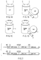
- a functional molecule is linked at a newly-created N-terminus and/or C-terminus of the modified GFP (see FIG. 1D).
- the modified GFP when the modification is performed by cleaving original GFP between the n-th amino acid and the (n+1)th amino acid of GFP, removing the amino acids from the (n-a)th to the n-th position of GFP, and leaving the original N terminus and C terminus unlinked, the modified GFP thus obtained consists of two discrete fragments, that is, one fragment of an amino acid sequence from the 1st to the (n-a-1)th position, and the other fragment of an amino acid sequence from the (n+1)th to the 238th position.
- the two discrete fragments may be linked by interposing a functional molecule between them in a later step, thereby forming a single-chain protein (see FIG. 1B). Examples of such a single-chain protein are biosensor proteins listed in Table 5 below. Even if the modified GFP does not have the structure of the single-chain protein, two discrete GFP fragments may work in a cell, like a modified GFP of the single chain.
- the vicinity of the hotspot amino acid residue (the n-th amino acid residue) at which original GFP is cleaved refers to, for example, the range of the (n+5)th to (n-5)th position, preferably the range of the (n+2)th to (n-2)th position.
- the number of amino acids to be removed (namely, the value of a+1 ) is, for example, 1-10, preferably 4.
- the various modified GFPs satisfying the aforementioned explanation of the modified GFP may be candidates for the biosensor protein of the present invention.
- the cleavage site in the vicinity of the hotspot amino acid residue and the number of amino acids to be removed may be appropriately set by one skilled in the art.
- the cleavage site and the number of amino acids to be removed may be varied as shown in Tables 1 and 2.
- the functional molecule which is linked to the modified GFP is not particularly limited, as long as it is capable of causing a conformational change itself by binding a factor acting on the functional molecule (i.e., a biosensor-detecting-factor) and it is capable of transmitting the conformational change to the modified GFP.
- a conformational change means a change of stereostructure (i.e., three-dimensional structure).
- the functional molecule used herein must be linked to the modified GFP at a position which it can transmit its conformational change to the modified GFP, thereby inducing the conformational change of the modified GFP.
- the functional molecule is preferably linked to the modified GFP at a position near the modified portion thereof. More specifically, the functional molecule is preferably linked to the cleavage site of original GFP via a linker molecule.
- the functional molecule may consist of a single molecule, or two or more molecules.
- the factor acting on the functional molecule i.e., biosensor-detecting-factor
- this conformational change induces a conformational change in the other functional molecule.
- each conformational change in two functional molecules can contribute to a conformational change of the modified GFP.
- the initial action of the factor on one functional molecule is transmitted to another functional molecule in turn, like signal transduction.
- each conformational change of all the functional molecules can act upon the modified GFP, causing a conformational change of the modified GFP.
- the functional molecule consists of two or more molecules
- conformational change each of the functional molecules can affect the structure of the modified GFP. Therefore, the effect of two or more functional molecules upon the modified GFP is larger than that of a single functional molecule. As a result, there is much possibility that the fluorescence property of the modified GFP may be drastically changed.
- the functional molecule to be connected to the modified GFP has a complete structure as expressed in vivo.
- the functional molecule may have a partial structure which includes only a binding site of the factor acting on the functional molecule, as long as it can play the aforementioned role of functional molecule.
- a preferable example of the functional molecule is a protein molecule.
- Specific examples of the functional molecule is a combination of calmodulin protein and myosin light chain kinase protein, and a combination of a CRE (Cyclic AMP Responsive Element) binding protein (CREB) and a CREB binding protein (CBP), but it is not limited to them.
- CRE Cyclic AMP Responsive Element binding protein
- CBP CREB binding protein binding protein
- calmodulin protein and a part of myosin light chain kinase protein capable of binding to calmodulin are employed as the functional molecules in the examples explained later.
- the fusion protein can function as a calcium sensor.
- FIGS. 1B and 1D show schematic structure of the fusion proteins, wherein the functional molecules (i.e., calmodulin protein (CaM) and a part of myosin light chain kinase (M13)) are linked to the modified GFP.
- the functional molecules i.e., calmodulin protein (CaM) and a part of myosin light chain kinase (M13)
- CaM calmodulin protein
- M13 myosin light chain kinase
- the modified GFP and the functional molecules are linked by linker molecules consisting of several amino acids.
- linker X the linker having an initiation codon (Met) at the N terminus of the fusion protein
- linker Y the linker connecting the N terminus side of the modified GFP and the functional molecule
- linker Z the linker connecting the C terminus side of the modified GFP and the functional molecule
- GGTGGS linker amino acids
- linkers are not necessarily limited in length and sequence, but preferably have a length of 0 to 10 amino acids, more preferably 2 amino acids.
- the linker Y preferably has a sequence of leucine-glutamic acid.
- Each linker is a portion for transmitting the conformational change of the functional molecule to the modified GFP. Therefore, the degree of the conformational change of the modified GFP is significantly affected depending upon length and sequence of each linker. A choice of length and sequence of each linker must be made depending upon the type of functional molecule to be linked. However, the modified GFP and the functional molecule may be directly linked without the linker.
- biosensor proteins are made by varying sequence of linker Y which links the modified GFP and the functional molecule (M13), and the resultant biosensor proteins are checked for their reactivity (See Tables 1 to 4).
- the amino acid of linker Y adjacent to the N terminal amino acid of the modified GFP is regarded as "the amino acid corresponding to the 148th position of original GFP". Furthermore, the amino acid of linker Y adjacent to "the amino acid corresponding to the 148th position of original GFP" is regarded as "the amino acid corresponding to the 147th position of original GFP”.
- the amino acid of linker Y located two amino acids away from the N terminal amino acid of the modified GFP is regarded as "the amino acid corresponding to the 148th position of original GFP".
- the amino acid corresponding to the 148th position of original GFP in the amino acid sequence of linker Y has an acidic side chain (Asp of G52, Glu of G18 and G79) or a side chain having a hydroxyl group (Ser of G3 and G17, Thr of G22, Tyr of G47), the functionally reactive biosensor may be formed.
- the amino acid corresponding to the 148th position of original GFP has a basic side chain (Lys of G62, Arg of G19 and G75)
- the reactivity of the biosensor tends to be reduced.
- the biosensor protein When “the amino acid corresponding to the 148th position of original GFP" has a hydroxyl group at the side chain, the biosensor protein further exhibits a photoisomerization property. Furthermore, “the amino acid corresponding to the 147th position of original GFP” also has an effect upon the reactivity of the biosensor protein. "The amino acid corresponding to the 147th position of original GFP” has an advantageous effect upon the reactivity of the biosensor in the order of Leu>Thr>Gly. However, when “the amino acid corresponding to the 147th position of original GFP" is Pro, it has little effect on the increase in the reactivity of Arg corresponding to the 148th position (See G19). As described above, it is possible to select an appropriate linker sequence by altering the linker sequence variously.
- the biosensor protein (i.e., the fusion protein) may be formed by a known genetic engineering procedure. More specifically, first, a fusion gene is made by separately preparing each gene fragment encoding each protein to be fused (that is, a gene encoding a modified GFP and a gene encoding a functional molecule) by polymerase chain reaction (PCR), and linking these fragments to each other. Then, the fusion protein is produced by introducing a plasmid comprising the above fusion gene into a desired cell and expressing the fusion gene.
- PCR polymerase chain reaction
- the factor affecting the conformation of a functional molecule constituting the fusion protein obtained in the step (B) it is possible to use a factor capable of causing the conformational change of the functional molecule directly by binding to the functional molecule.
- a molecule known to increase the intracellular level of the factor affecting the conformation of the functional molecule may be used.
- calcium ion may be used as the factor affecting the conformation of the functional molecules.
- Calcium ion can cause the conformational change of calmodulin by binding to calmodulin.
- a factor such as ATP, carbachol, caffeine, or thapsigargin, which is known to increase the calcium ion concentration in a cell expressing the fusion protein.
- cyclic AMP dependent protein kinase may be used, as the factor to confirm the function as a biosensor.
- Example of the method of reacting the fusion protein with the factor affecting the conformation of the functional molecule is as follows. First, cells (e.g., HEK 293 cells) producing the fusion protein of the modified GFP/functional molecules are placed in a chamber on stage of a laser microscope. Then, the factor is added to the cells. This method is preferable because the subsequent change in the fluorescence property of the fusion protein can be observed. When ATP or carbachol is used as the factor in practice, it is appropriate to suspend the factor in a perfusion solution (HBS) and add it at a concentration of 20-300 ⁇ M.
- HBS perfusion solution
- Step D Step of screening a fusion protein exhibiting a change in the fluorescence property by the reaction of the step (C), as a biosensor protein, from the various fusion proteins:
- a fusion protein exhibiting a change in the fluorescence property by reaction of the step (C) can be detected by using a fluorescence-detecting apparatus, such as a CCD camera attached to a laser microscope or a fluorescence microscope.
- a fluorescence-detecting apparatus such as a CCD camera attached to a laser microscope or a fluorescence microscope.
- the protein exhibiting a change in the fluorescence property is selected as the biosensor protein.
- the phrase "exhibiting a change in the fluorescence property” refers to exhibiting a change which is detectable by a fluorescence microscope, a laser microscope, or the like. More preferably, it refers to exhibiting a change sufficient to be observed by the naked eye.
- the biosensor protein screened in the step (D) is in a state where its fluorescence property can be reversibly changed depending upon the presence or absence of the biosensor-detecting-factor.
- the aforementioned biosensor protein which is modified so that the fluorescence property of GFP can be reversibly changed, may be said to be in a critical state capable of changing the fluorescence property.
- the biosensor protein when the fluorescence property is represented by the fluorescence intensity, the biosensor protein is in the critical state between a fluorescence-emitting state and a no-fluorescence-emitting state.
- the biosensor protein may emit no fluorescence or emit fluorescence at a low level of intensity.
- the biosensor protein in the critical state may emit fluorescence at a high level of intensity.
- the biosensor protein emits no fluorescence refers to the state where fluorescence cannot be detected by using an optical appliance.
- the biosensor protein emits fluorescence at a low level of intensity refers to the state where the fluorescence intensity is so low that the biosensor protein can transfer to a level of high fluorescence intensity by the action of the biosensor-detecting-factor, showing the above change amount (i.e., ⁇ F/F > 0.1 or more).
- the phrase "the biosensor protein emits fluorescence at a high level of intensity” refers to the state where the fluorescence intensity is so high that the biosensor protein can transfer to a level of low fluorescence intensity by the action of the biosensor-detecting-factor, showing the above change amount (i.e., ⁇ F/F > 0.1 or more).
- the biosensor protein When the biosensor protein is in a critical state and emits fluorescence at a high level of intensity, the biosensor protein can transfer to a no-fluorescence-emitting state or weak fluorescence-emitting state by a small conformational change thereof. Therefore, it may be said that the critical state is "the state where strong fluorescence can be barely emitted”. Further, when the biosensor protein is in a critical state and emits no fluorescence, the biosensor protein can transfer to a fluorescence-emitting state by a small conformational change thereof. Therefore, it may be said that the critical state is "the state where fluorescence cannot be barely emitted”.
- the biosensor protein when the biosensor protein is in a critical state and emits fluorescence at a low level of intensity, the biosensor protein can transfer to a strong fluorescence-emitting state by a small conformational change thereof. Therefore, it may be also said that the critical state is "the state where weak fluorescence can be barely emitted".
- the biosensor protein of the present invention is in a critical state. This is because the biosensor protein in a critical state can sensitively respond to the presence or absence of the sensor-detecting-factor to cause a reversible change of the conformation thereof, thereby changing the fluorescence property reversibly (e.g., moving back and forth between a fluorescence-emitting state and a no-fluorescence-emitting state reversibly). Therefore, when the biosensor protein is in the above critical state, it can be said that it is in a state where it can function as a biosensor.
- the biosensor protein of the present invention refers to that screened by the aforementioned method of producing a biosensor protein.
- the biosensor gene of the present invention refers to the gene encoding the biosensor protein of the present invention, and is used to express the biosensor protein in a cell.
- biosensor protein of the present invention is characterized by comprising (1) and (2) below:
- the modified GFP which is a constituent of the biosensor protein, is as defined above.
- the modified GFP is preferably formed by cleaving amino acid sequence of GFP in the vicinity of the hotspot amino acid residue, and eliminating some amino acids from the cleavage site, thereby modifying the molecular structure of GFP, and if necessary, linking the original N terminus and C terminus of GFP with an appropriate amino acid sequence.
- the functional molecule which is a constituent of the biosensor protein, is also as defined above.
- the functional molecule is a molecule capable of causing a conformational change itself by binding a factor acting upon the functional molecule (i.e., biosensor-detecting-factor), and transmitting its conformational change to the modified GFP.
- the modified GFP and the functional molecule are not particularly limited, as long as they satisfy the aforementioned definitions.
- the functional molecule must be linked to the modified GFP at a position where the functional molecule can transmit its conformational change to the modified GFP and cause a conformational change of the modified GFP. Therefore, it is preferable that the functional molecule is linked to the modified GFP in the vicinity of the modified portion of the modified GFP. More specifically, the functional molecule is preferably linked to the cleavage site of GFP via a linker molecule.
- the biosensor protein of the present invention is required to reversibly change its fluorescence property depending on the presence or absence of the biosensor-detecting-factor.
- the fluorescence property the fluorescence intensity is used in the present invention.
- the biosensor protein of the present invention is in a critical state between a fluorescence-emitting state and a no-fluorescence-emitting state, and can reversibly change its fluorescence property.
- the hotspot amino acid residue is preferably the amino acid residue of the 148th, 94th, 96th or 222nd position of GFP.
- the biosensor protein which is employed the amino acid of the 148th position of GFP as the hotspot amino acid residue, is characterized by comprising the following (1) and (2):
- biosensor protein which is employed the amino acid of the 148th position of GFP as the hotspot amino acid residue, is characterized by comprising the following sequences (a) to (h) sequentially from the N terminus:
- the linker X of sequence (a) is an arbitrary amino acid sequence containing methionine, preferably an amino acid sequence consisting of 1-10 amino acids.
- the linker X is more preferably the sequence of Met-Xaa-Xaa, wherein Xaa represents an arbitrary amino acid, and further preferably, the sequence of Met-Gly-Thr or Met-Val-Asp.
- the linker Y of sequence (c) is an arbitrary amino acid sequence, preferably an amino acid sequence consisting of 0-10 amino acids. More preferably, in the amino acid sequence of the linker Y, "the amino acid corresponding to the 147th position of GFP" is Leu, Thr, or Gly, and "the amino acid corresponding to the 148th position of GFP” is an amino acid having an acidic side chain or a side chain with a hydroxyl group therein. Further preferably, the linker Y is the sequence of Leu-Glu.
- the linker of sequence (e) is preferably an amino acid sequence consisting of 2-10 amino acids.
- the linker of sequence (e) is more preferably an amino acid sequence rich in an amino acid (e.g., glycine) which allows a main chain of a peptide to move with a large degree of freedom, rendering the main chain flexible.
- the linker of sequence (e) is the sequence of Gly-Gly-Thr-Gly-Gly-Ser.
- the linker Z of sequence (g) is an arbitrary amino acid sequence, preferably an amino acid sequence consisting of 0-10 amino acids.
- the linker Z is more preferably an amino acid sequence consisting of 2-3 amino acids, and further preferably the sequence of Thr-Arg.
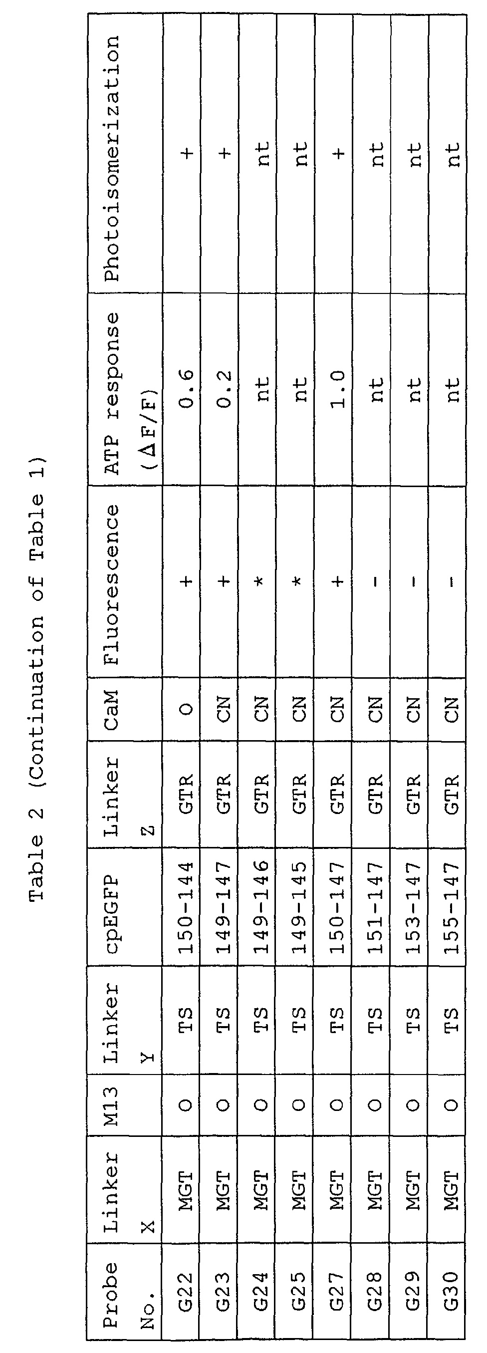
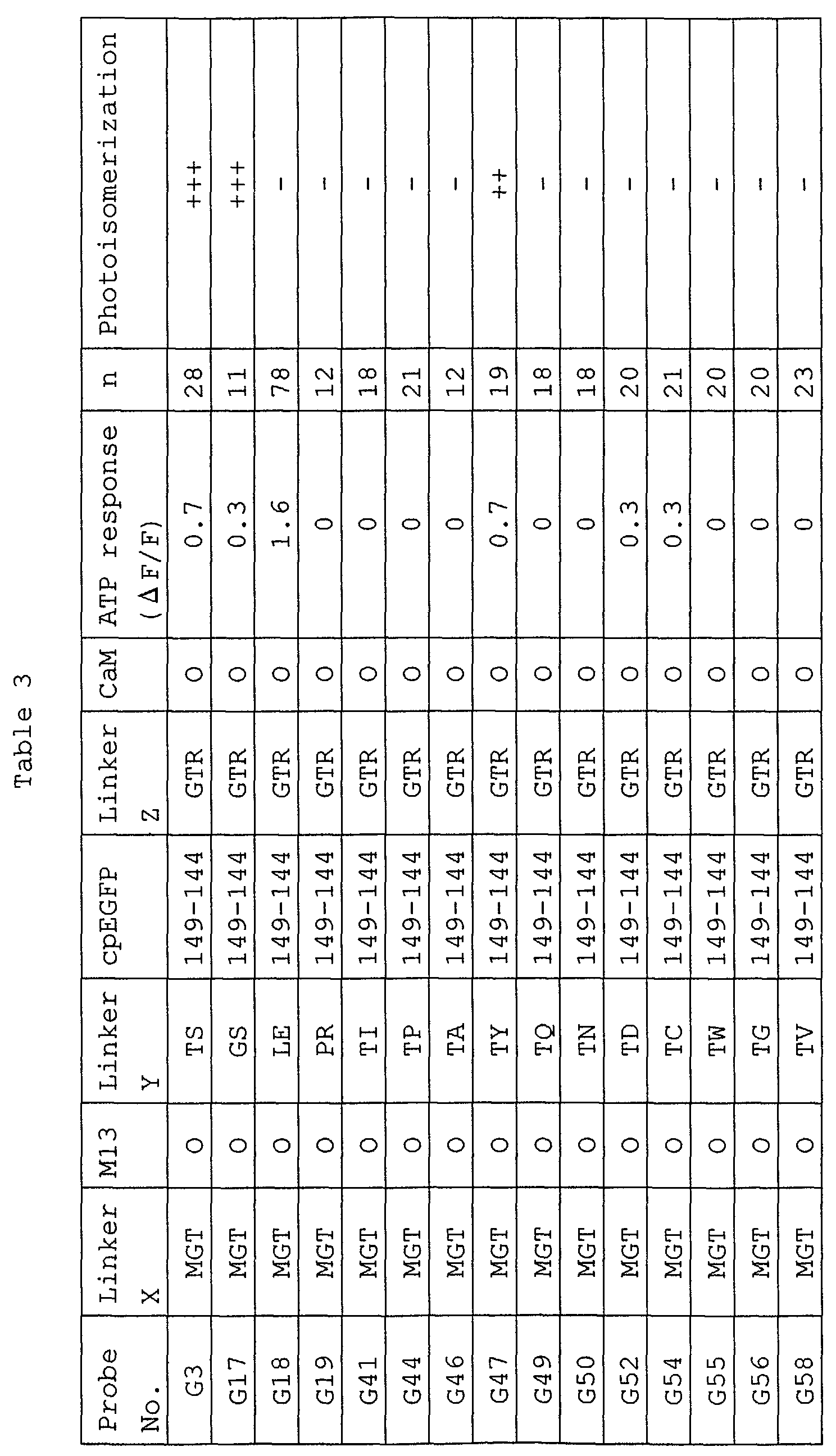
- Tables 1 and 2 list examples of the various biosensor proteins using the amino acid of the 148th position of GFP as the hotspot amino acid residue.
- Myosin light chain kinase (M13) and calmodulin (CaM) are used herein as the functional molecules.
- biosensor proteins listed on Tables 1 and 2 have the following amino acid sequences in this order from the N terminus:
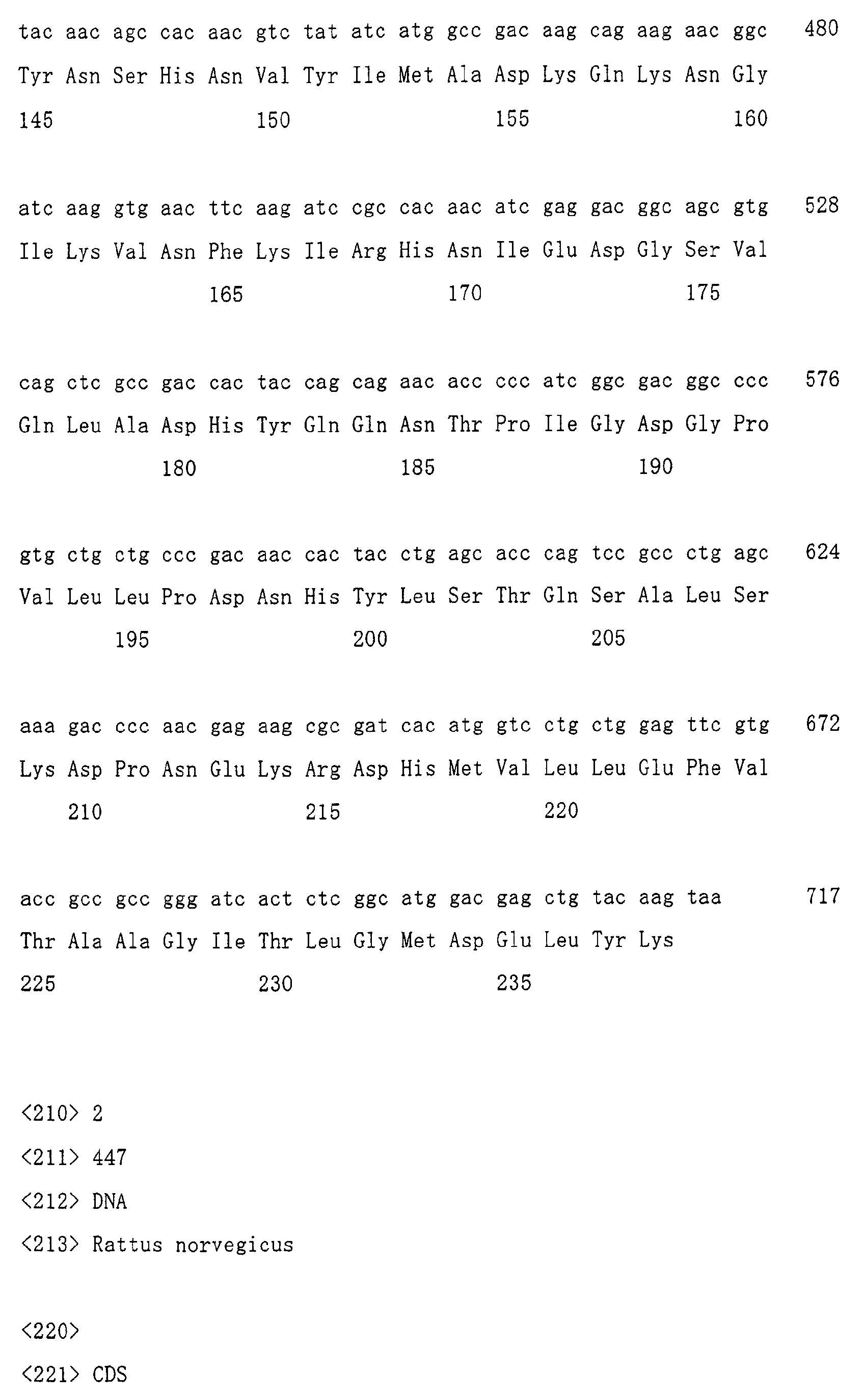
- the amino acid sequence of rat calmodulin used herein as a functional molecule is represented by Sequence ID No: 2.
- Each amino acid from the 1st to 148th position of the amino acid sequence represented by Sequence ID No: 2. corresponds to the "n-th amino acid of calmodulin protein" described in this text.
- methionine of the N terminus is omitted therefrom. This is the reason why methionine of the N terminus is cleaved out by post-translational modification, after translation of genetic code of calmodulin into amino acids.
- the partial amino acid sequence (M13) of myosin light chain kinase protein which is used as a functional molecule, is represented by Sequence ID No: 3.
- the nucleotide sequence encoding the M13 fragment is obtained by modifying the nucleotide sequence encoding M13 fragment of chicken (Guerriero,V et al., Biochemistry 25, 8372-8381 (1986)).
- the names of probe No. are given to a variety of biosensor proteins listed therein.
- the one-dimensional structure of the probe No. G3 (hereinafter, also referred to as "G3") is shown in FIG. 2.
- the G3 has, in this order from the N terminus, linker X (Met-Gly-Thr) ; a partial amino acid sequence of myosin light chain kinase (Ser-Ser-Arg-Arg-Lys-Trp-Asn-Lys-Thr-Gly-His-Ala-Val-Arg-Ala-Ile-Gly-Arg-Leu-Ser-Ser); linker Y (Thr-Ser); the modified GFP (the amino acid sequence of the 149-238th position of GFP, the linker amino acids (Gly-Gly-Thr-Gly-Gly-Ser), and the amino acid sequence of the 1-144th position of GFP, in this order from the N terminus); linker Z (Gly-Thr-Thr-G
- the number "149-144" in the column of cpEGFP means that the modified GFP has, in this order from the N terminus, the amino acid sequence of the 149-238th position of GFP, the linker amino acids (Gly-Gly-Thr-Gly-Gly-Ser), and the amino acid sequence of the 1-144th position of GFP.
- the symbol “O” in the column of CaM represents the amino acid sequence of the 2nd to 148th position of original rat calmodulin
- the symbol “CN” represents the amino acid sequence of the 2nd to 148th position of calmodulin mutant CaMCN (Persechini A et al., 1997, Cell Calcium 22, 209-216).
- the symbol “+” in the column of Fluorescence means that a biosensor protein emits weak fluorescence before ATP is added, whereas the symbol “-” means that a biosensor protein emits no fluorescence before ATP is added. Further, the symbol “*” represents that a biosensor protein emits weak fluorescence at a high intracellular calcium concentration of about 2 mM.
- the value “ ⁇ F/F” in the column of ATP response is a value (arbitrary unit) obtained by dividing " ⁇ F” by "F”, wherein “ ⁇ F” is a change amount of fluorescence intensity between before and after addition of ATP and "F” is a fluorescence intensity before addition of ATP, in the cell (HEK cell) producing a biosensor protein.
- the symbol “nt” represents “not tested”.
- the symbol “+” in the column of Photoisomerization means that a biosensor protein causes photoisomerization, and the symbol “-” means that a biosensor protein causes no photoisomerization.
- biosensor proteins are produced by varying the N terminus of cpEGFP within the amino acids of the 145-155th positions and varying the C terminus of cpEGFP within the amino acids of the 140-147th positions.
- the resultant biosensor proteins are checked for reactivity to ATP.
- the biosensor proteins having the N terminus starting from the amino acid of the 149 or 150th position exhibit a high reactivity to ATP.
- the biosensor proteins having the C terminus terminating at the amino acid of 140, 144, or 147th position exhibit a reactivity to ATP.
- the amino acid of the 148th position is included in linker Y. More specifically, when the N terminus of the modified GFP starts from the amino acid of the 149th position of original GFP, the amino acid of linker Y adjacent to the N terminal amino acid of the modified GFP may be regarded as "the amino acid corresponding to the 148th position of original GFP". Similarly, when the N terminus of the modified GFP starts from the amino acid of the 150th position of original GFP, the amino acid of linker Y located two amino acids away from the N terminal amino acid of the modified GFP may be regarded as "the amino acid corresponding to the 148th position of original GFP".
- the biosensor protein employing the amino acid of the 148th position of GFP as the hotspot amino acid residue is characterized by comprising the following (1) and (2):
- biosensor protein More specific example of the above more preferable biosensor protein is characterized by comprising the following sequences (a) to (h) sequentially from the N terminus:
- the linker X of sequence (a) is an arbitrary amino acid sequence containing methionine, preferably an amino acid sequence consisting of 1-10 amino acids.
- the linker X is more preferably the sequence of Met-Xaa-Xaa, wherein Xaa is an arbitrary amino acid, and further preferably, the sequence of Met-Gly-Thr or Met-Val-Asp.
- the linker Y of sequence (c) is an arbitrary amino acid sequence, preferably an amino acid sequence consisting of 0-10 amino acids. More preferably, the linker Y is an amino acid sequence consisting of 2 amino acids: one is "the amino acid corresponding to the 147th position of GFP", which is Leu, Thr, or Gly; and the other is "the amino acid corresponding to the 148th position of GFP", which is an amino acid having an acidic side chain or a side chain with a hydroxyl group therein. Further preferably, the linker Y is the sequence of Leu-Glu.
- the linker of sequence (e) is preferably an amino acid sequence consisting of 2-10 amino acids.
- the linker of sequence (e) is more preferably an amino acid sequence rich in an amino acid, such as glycine, which allows a main chain of a peptide to move with a large degree of freedom, rendering the main chain flexible.
- the linker of sequence (e) is the sequence of Gly-Gly-Thr-Gly-Gly-Ser.
- the linker Z of sequence (g) is an arbitrary amino acid sequence, preferably an amino acid sequence consisting of 0-10 amino acids.
- the linker Z is more preferably an amino acid sequence consisting of 2-3 amino acids, and further preferably the sequence of Thr-Arg.
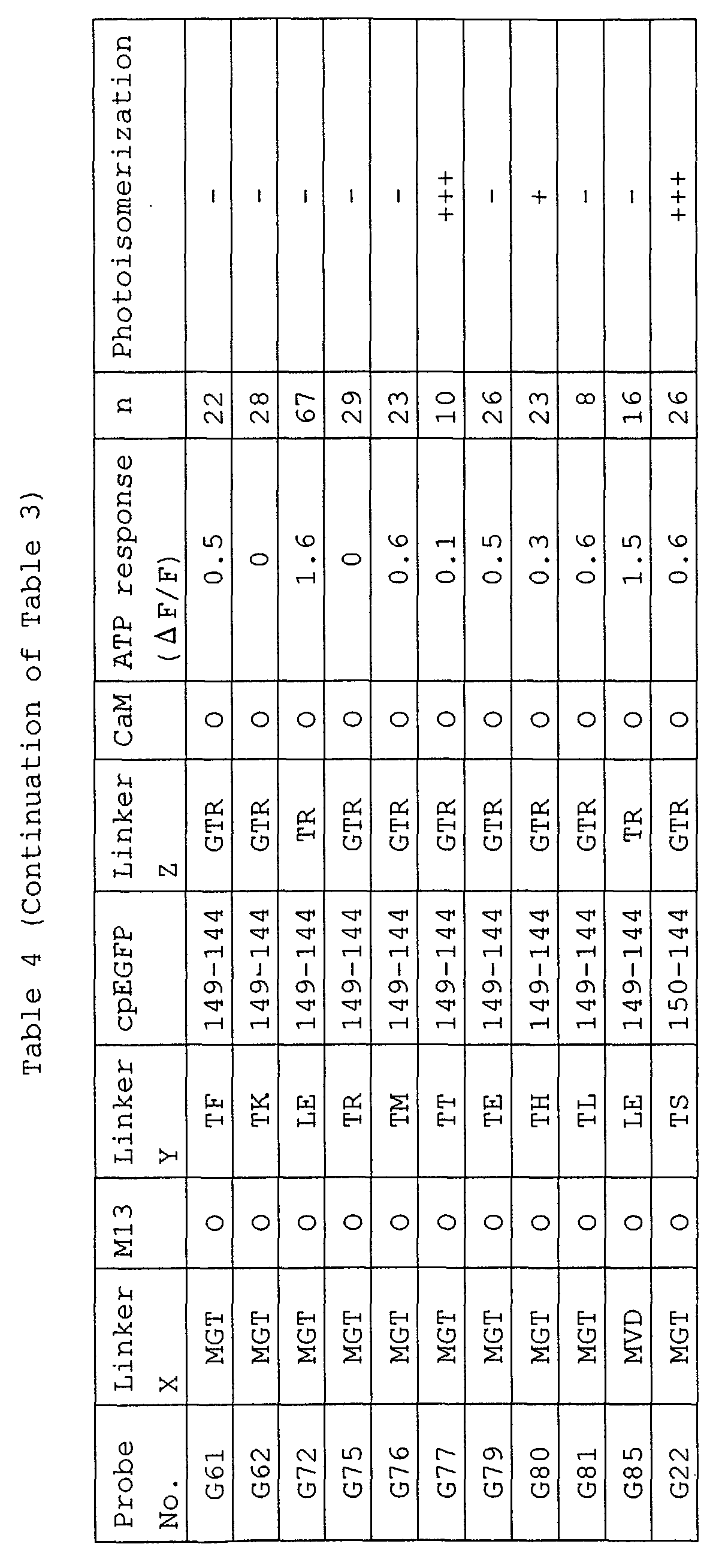
- biosensor proteins Various examples of the above more preferable biosensor proteins are listed in Tables 3 and 4 in the following.
- Myosin light chain kinase (M13) and calmodulin (CaM) are used herein as the functional molecules.
- the biosensor proteins listed on Tables 3 and 4 have, in this order from the N terminus, an amino acid sequence containing methionine (linker X); a partial amino acid sequence of myosin light chain kinase protein (M13); a variety of linker amino acid sequence (linker Y); a modified GFP (cpEGFP); a linker amino acid sequence (linker Z); and the amino acid sequence (CaM) of the 2nd to 148th position of calmodulin protein.
- the names of probe No. are given to various biosensor proteins listed therein.
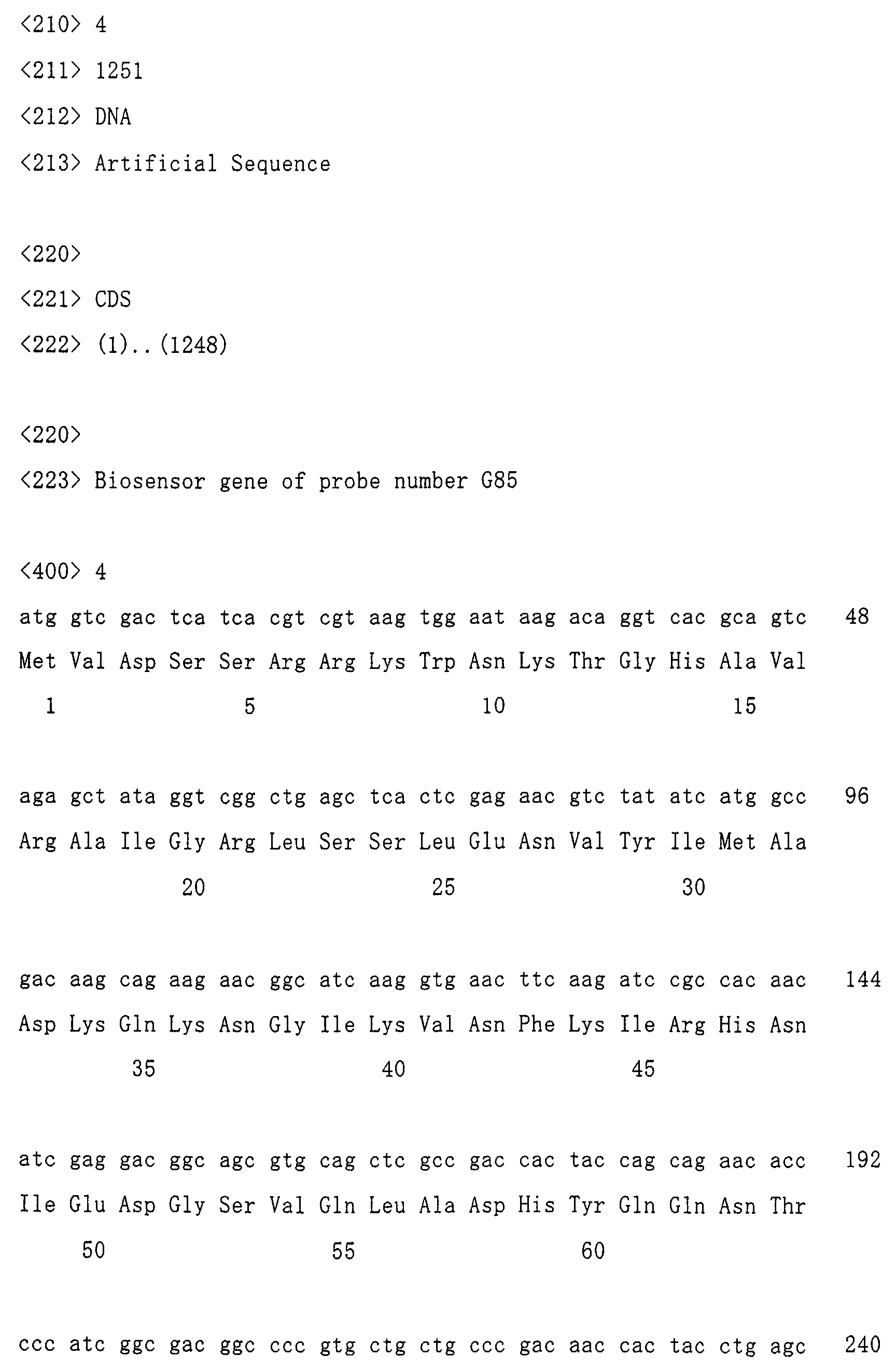
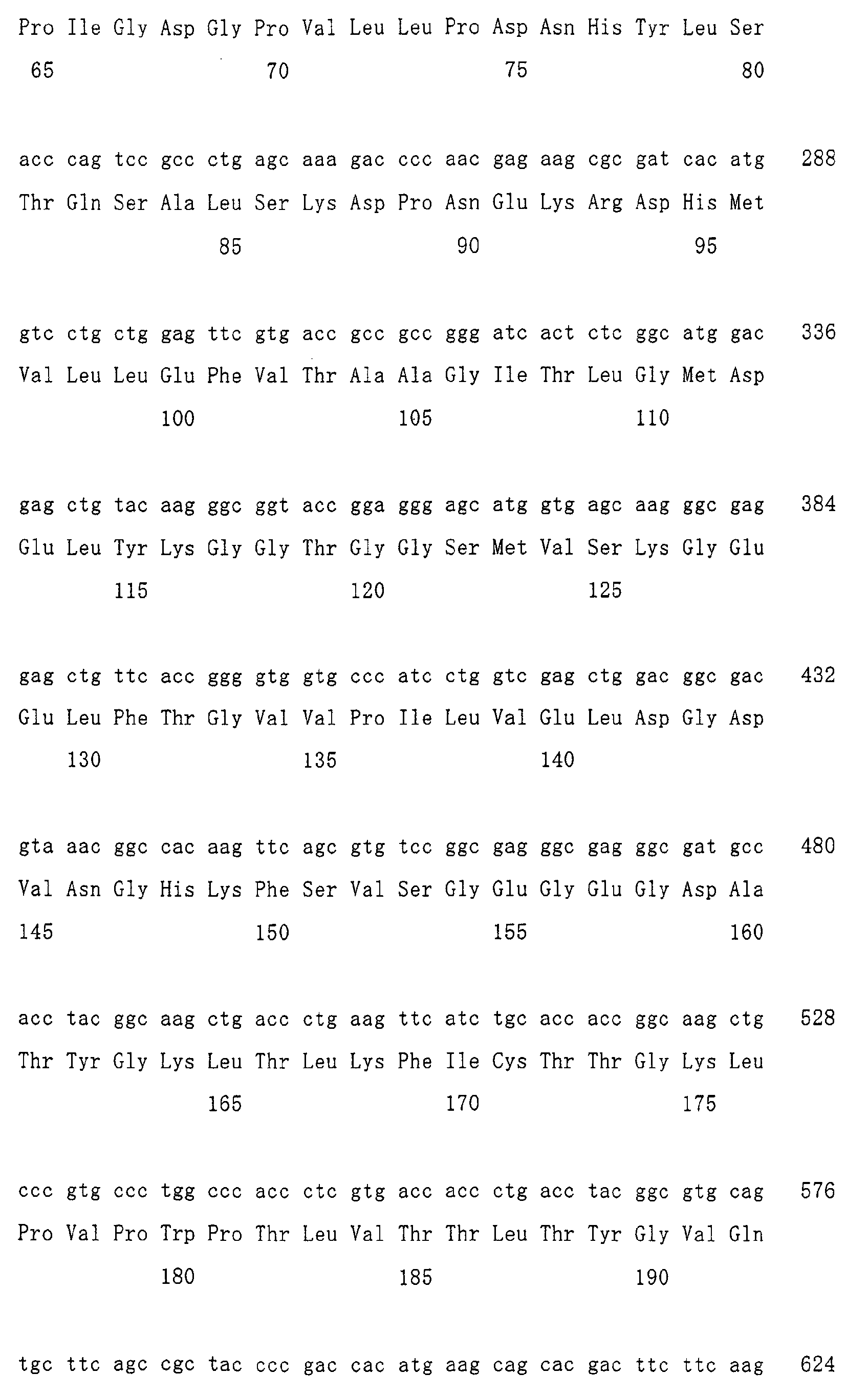
- the one-dimensional structure of the probe No. G85 (hereinafter, also referred to as "G85") is shown in FIG. 2.
- the G85 has, in this order from the N terminus, linker X (Met-Val-Asp); a partial amino acid sequence of myosin light chain kinase (Ser-Ser-Arg-Arg-Lys-Trp-Asn-Lys-Thr-Gly-His-Ala-Val-Arg-Ala-Ile-Gly-Arg-Leu-Ser-Ser); linker Y (Leu-Glu); the modified GFP (the amino acid sequence of the 149-238th position of GFP, the linker amino acids (Gly-Gly-Thr-Gly-Gly-Ser), and the amino acid sequence of the 1-144th position of GFP, in this order from the N terminus); linker Z (Thr-Arg); and the amino acid
- the number "149-144" in the column of cpEGFP means that the modified GFP has, in this order from the N terminus, the amino acid sequence from the 149-238th position of GFP, the linker amino acids (Gly-Gly-Thr-Gly-Gly-Ser), and the amino acid sequence of the 1-144th position of GFP.
- the symbol "O" in the column of CaM represents the amino acid sequence of the 2nd to 148th position of rat-inherent calmodulin.
- the value " ⁇ F/F” in the column of ATP response is a value (arbitrary unit) obtained by dividing " ⁇ F” by "F", wherein ⁇ F is a change amount of fluorescence intensity between before and after addition of ATP and "F” is a fluorescence intensity before addition of ATP, in the cell (HEK cell) producing a biosensor protein.
- the column “n” refers to the number of cells tested.
- the symbol “+” in the column of Photoisomerization means that a biosensor protein causes photoisomerization, whereas the symbol “-” means that a biosensor protein causes no photoisomerization.
- the biosensor proteins G18, G72, and G85 exhibit the very high reactivity, and show the value 1.5-1.6 in the change amount ( ⁇ F/F) of fluorescence to ATP.
- All of the biosensor proteins G18, G72, and G85 have the modified GFPs having the N terminus starting from the 149th amino acid of original GFP, and have glutamic acid (Glu) as "the amino acid corresponding to the 148th position of GFP" in linker Y.
- the biosensor protein G22 has the modified GFP having the N terminus starting from the 150th amino acid of original GFP, and has threonine as "the amino acid corresponding to the 148th position of GFP" in linker Y.
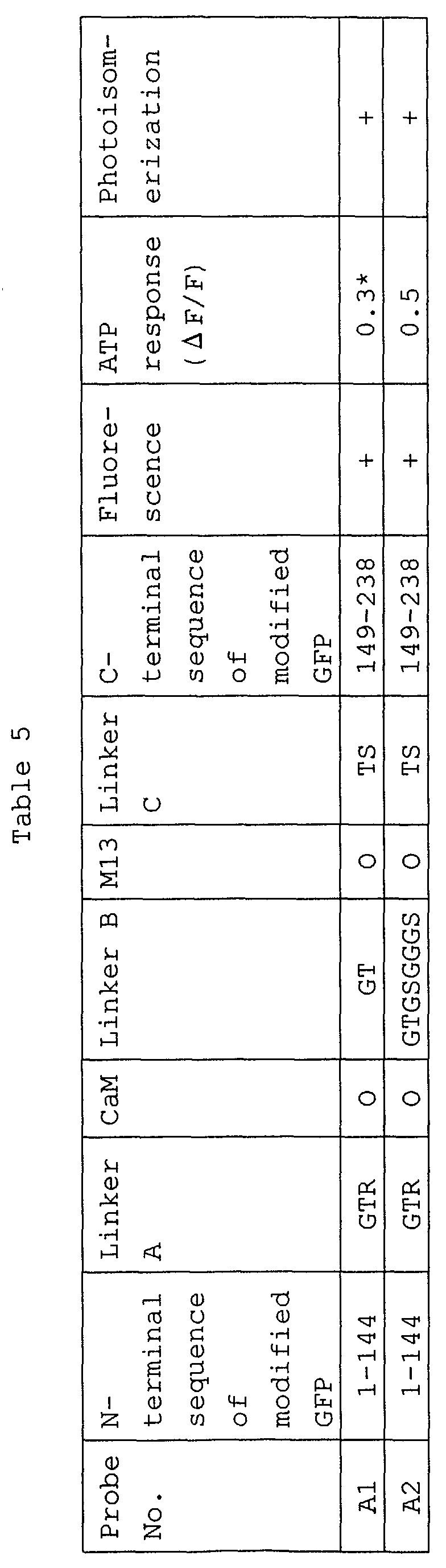
- biosensor proteins listed on Table 5 are prepared by cleaving GFP in the vicinity of the amino acid residue of the 148th position of GFP (which is the hotspot amino acid residue), and linking the functional molecules between the cleaved sites.
- biosensor proteins listed on Table 5 have the following sequences in this order from the N terminus:
- the amino acid sequences of the linkers A, B and C are represented by single-letter notation.
- the symbol “O” in the column of M13 represents the sequence of (Ser-Ser-Arg-Arg-Lys-Trp-Asn-Lys-Thr-Gly-His-Ala-Val-Arg-Ala-Ile-Gly-Arg-Leu-Ser-Ser).
- the symbol “O” in the column of CaM represents the amino acid sequence of the 2nd to 148th position of rat-inherent calmodulin.
- the symbol “+” in the column of Fluorescence means that a biosensor protein emits weak fluorescence before addition of ATP.
- the value " ⁇ F/F” in the column of ATP response is a value (arbitrary unit) obtained by dividing " ⁇ F” by "F", wherein " ⁇ F” is a change amount of fluorescence intensity between before and after addition of ATP and "F” is a fluorescence intensity before addition of ATP, in the cell (HEK cell) producing a biosensor protein.
- the symbol “*” in the column of ATP response represents that the biosensor protein responds to carbachol in the case of using carbachol instead of ATP.
- the symbol “+” in the column of Photoisomerization means that a biosensor protein causes photoisomerization.
- Table 5 shows the ATP reactivity of a biosensor group having the original N terminus and original C terminus of GFP.
- the biosensor group reacts to ATP, but the reactivity is low.
- the biosensor protein A2 having a longer linker B shows higher reactivity than A1.
- the reactivity of this biosensor group will be improved by examining the various sequences with respect to linkers A, B and C, and a biosensor protein having higher reactivity will be produced.
- biosensor proteins of the present invention include the biosensor proteins having calmodulin protein (or a part thereof) and myosin light chain kinase (or a part thereof) as the functional molecules, as described above.
- biosensor proteins of the present invention are not limited to them. In other words, the biosensor proteins of the present invention are not limited to those working as the aforementioned calcium sensor.
- Particularly preferable example of the calcium sensor protein of the present invention is characterized by comprising the following sequences (a) to (h), sequentially from the N terminus,
- the calcium sensor prepared in the present invention can be used when calcium concentration is measured inside or outside cells.
- calcium concentration can be measured by previously preparing a calcium sensor protein of the present invention by introducing a gene encoding the calcium sensor protein into E. coli or the like, and then mixing the resultant calcium sensor protein with a specimen.
- the intracellular calcium concentration may be measured by injecting directly the calcium sensor protein produced by using E. coli or the like into a desired cell to be measured for calcium concentration.
- the intracellular calcium concentration may be measured by introducing the calcium sensor gene into the desired cell to be measured for calcium concentration, and expressing the gene in the cell.
- the measurement of calcium concentration can be performed by irradiating light at a specific wavelength (e.g., excitation light at 488 nm) to a calcium sensor protein and detecting the property of the fluorescence emitted from the calcium sensor protein by means of an optical appliance (e.g., laser microscope).
- a specific wavelength e.g., excitation light at 488 nm
- an optical appliance e.g., laser microscope
- the biosensor protein of the present invention has almost the same property of fluorescence wavelength as that of EGFP (Clontech). Specifically, the biosensor protein of the present invention has a maximum excitation wavelength of 489 nm and a maximum emission wavelength of 509 nm.
- the biosensor gene refers to a gene encoding the biosensor protein of the present invention.
- the biosensor gene is prepared, in the form of a fusion gene, by making each gene fragment encoding individual constitutional parts (e.g., parts of a modified GFP and a functional molecule) of a biosensor protein by means of PCR, and linking the resultant each gene fragment.
- the biosensor gene thus prepared is introduced into an appropriate vector (pEGFP-N1, Clontech), and then the recombinant plasmid is further introduced into a cell. In this manner, the desired biosensor protein can be produced in an arbitrary cell.
- the biosensor protein of the present invention is required checking experimentally for the performance as a sensor in advance.
- the calcium sensor which was actually developed in the present invention was checked for the performance, by introducing the gene of the calcium sensor into cultured human embryonic kidney (HEK) 293 cells or mouse skeletal muscle primary cultured cells.
- HEK human embryonic kidney
- the calcium sensor of probe No. G85 is made by expressing the gene of the calcium sensor in the HEK 293 cells, and then a factor known to increase the calcium ion concentration in cells, such as ATP (0.1 mM) or carbachol (CCH) (0.1 mM), is added to the HEK 293 cells.
- a factor known to increase the calcium ion concentration in cells such as ATP (0.1 mM) or carbachol (CCH) (0.1 mM
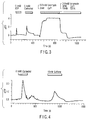
- the reaction profile of the calcium sensor G85 is shown in FIG. 3.
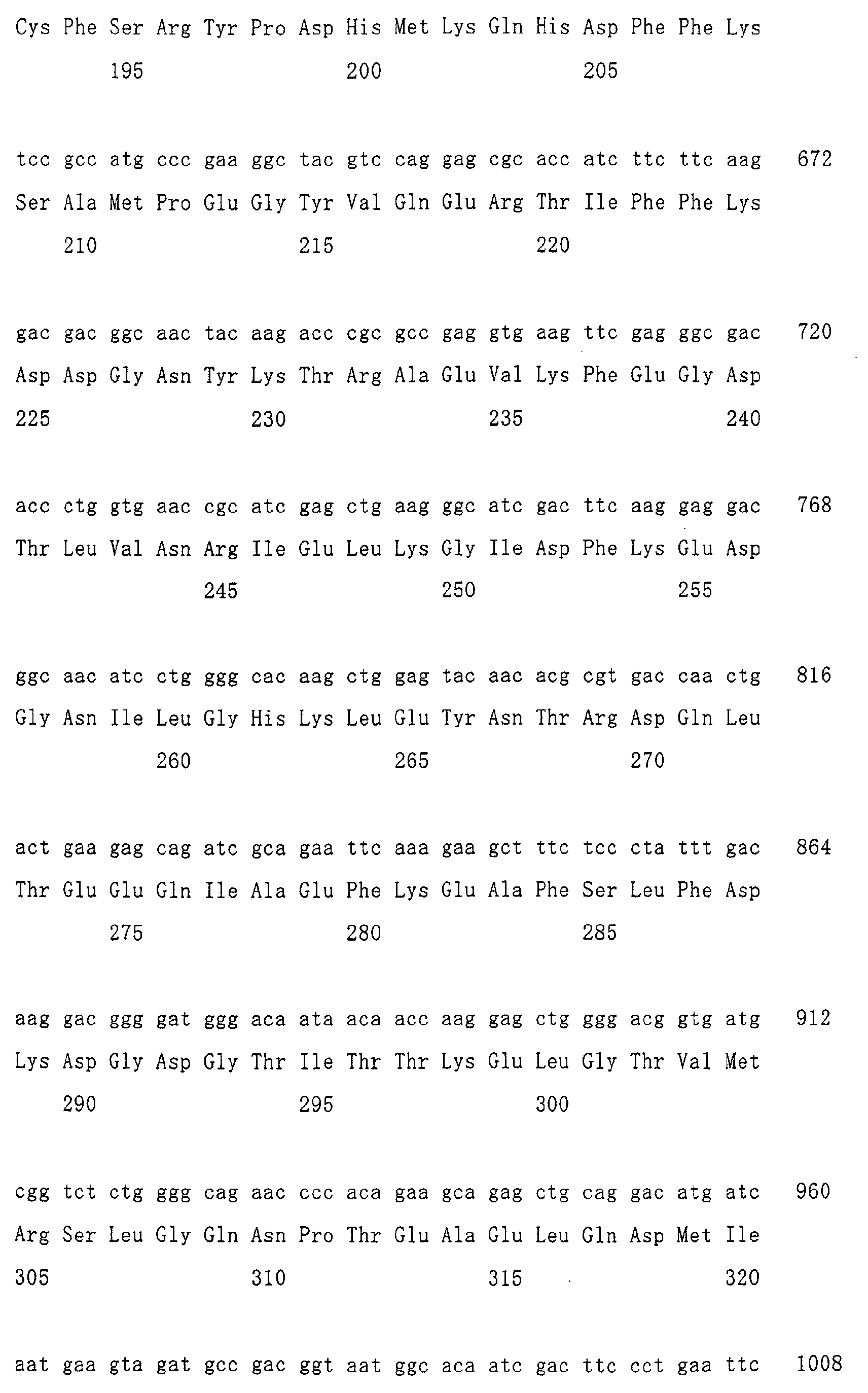
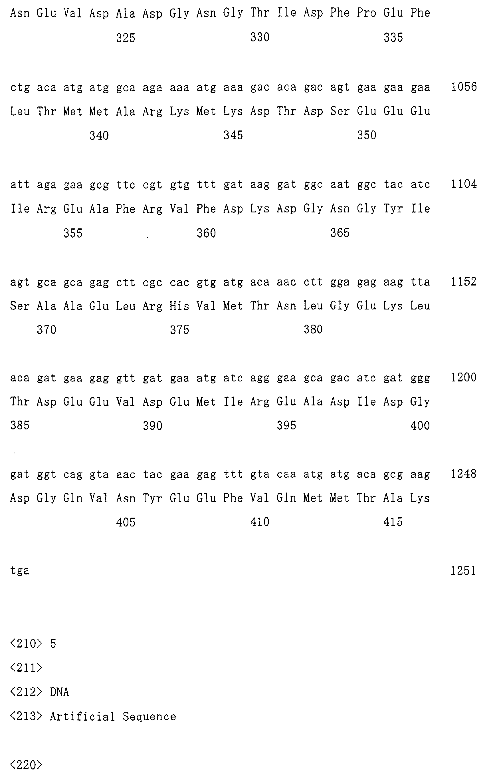
- the nucleotide sequence of the G85 is represented by Sequence ID No: 4
- the change amount ( ⁇ F/F) of fluorescence in response to ATP in the HEK 293 cells comes up to about 1.6. Such change amount can be captured by both fluorescence microscopy and naked eye.
- the change amount ( ⁇ F/F) of fluorescence in response to carbachol reaches about 2.2. Furthermore, it is observed that the fluorescence intensity of the calcium sensor reaches a maximum, when ionomycin (0.01 mM) and a calcium ion (2 mM) are added to increase the calcium concentration in the cells.
- a factor such as carbachol (0.1 mM) or caffeine (10 mM), which is known to increase the intracellular calcium ion concentration, is added to the skeletal muscle primary cultured cells expressing a calcium sensor (G85).
- the profile of the reaction is shown in FIG. 4.
- the change amount ( ⁇ F/F) of fluorescence in response to carbachol comes up to about 2.5, and the change amount ( ⁇ F/F) of fluorescence in response to caffeine reaches about 1.1, which is similar to the result of FIG. 3.
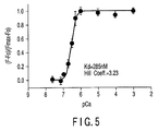
- the relationship between the calcium concentration and the fluorescence amount of the calcium sensor is shown in the graph of FIG. 5.
- the G85 is prepared by using E. coli.
- the calcium sensor protein of the present invention exhibits extremely high calcium sensitivity. This sensitivity is about 30 times as high as that of a calcium-sensitive protein consisting of a single GFP described in the column of "BACKGROUND OF THE INVENTION".
- the calcium sensor of the present invention has highly efficient performance as a sensor.
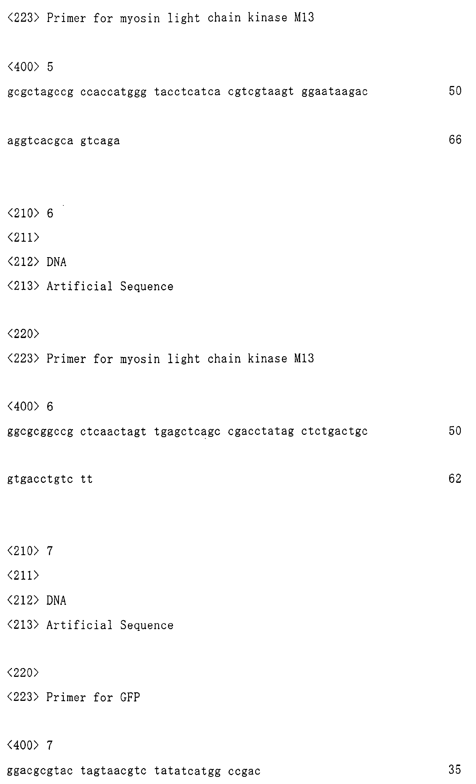
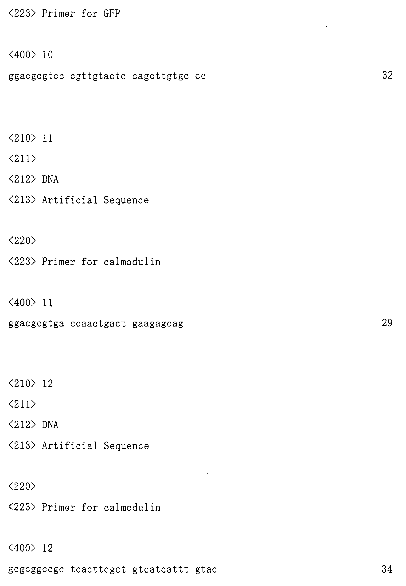
- the fragment M13 (Sequence ID No: 3) of smooth muscle myosin light chain kinase was prepared using the following synthetic primers which have been synthesized by Katayama Chemical. More specifically, PCR was performed using the following two primers as templates and primers, thereby synthesizing the fragment M13 of smooth muscle myosin light chain kinase.
- the reaction mixture obtained by PCR was subjected to agarose gel electrophoresis.
- a target PCR product was recovered from the gel, and digested with NheI and SpeI restriction enzymes, to obtain a PCR fragment 1.
- Modified GFP i.e., Circularly Permutated Enhanced GFP: cpEGFP
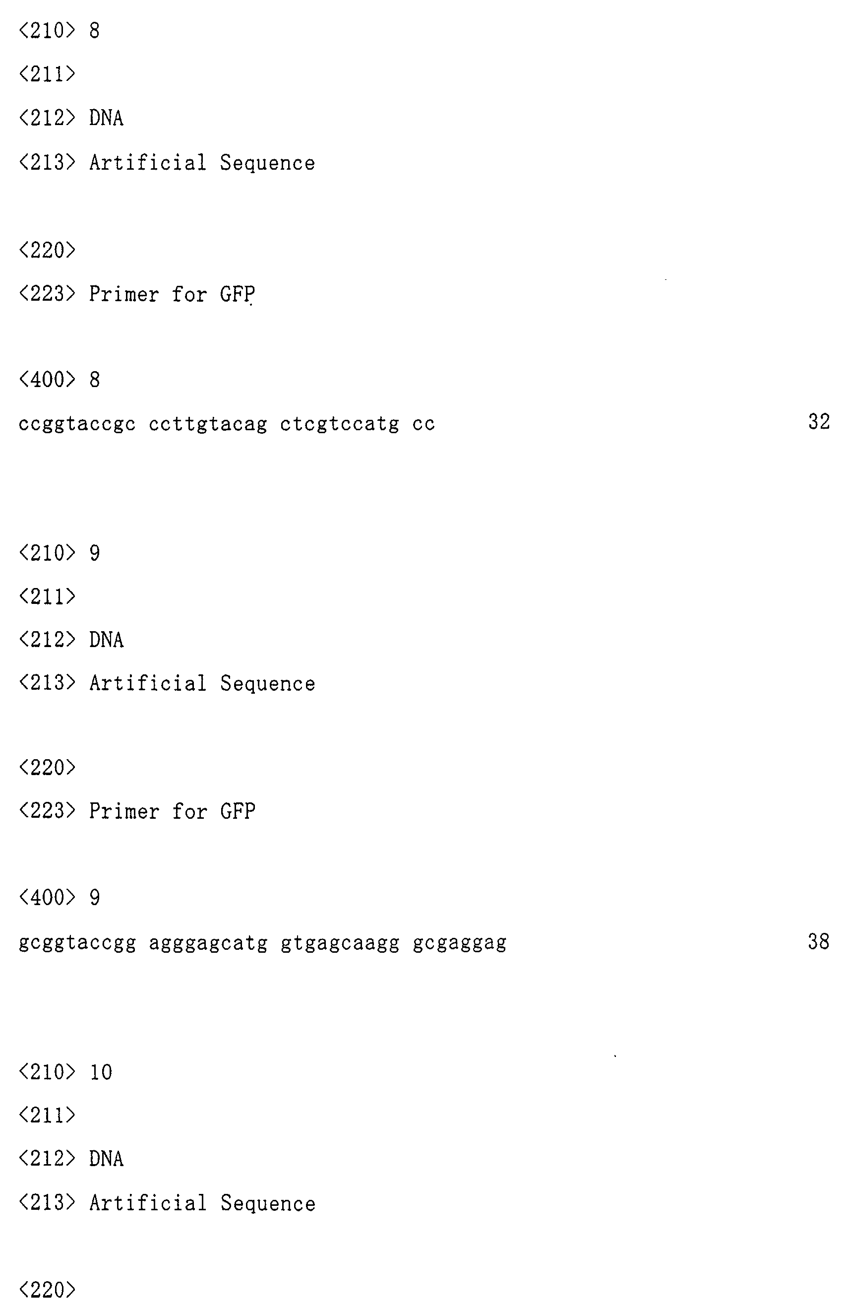
- PCR was performed using the following primers and a plasmid pEGFP-N1 (available from Clontech) containing GFP-encoding cDNA as a template.
- the reaction mixture obtained by PCR was subjected to agarose gel electrophoresis.
- a target PCR product was recovered from the gel, and digested with SpeI and KpnI restriction enzymes, to obtain a PCR fragment 2.
- PCR was performed using the following primers and a plasmid pEGFP-N1 as a template.
- the reaction mixture obtained by PCR was subjected to agarose gel electrophoresis.
- a target PCR product was recovered from the gel, and digested with KpnI and MluI restriction enzymes, to obtain a PCR fragment 3.
- PCR was performed using the following primers to rat calmodulin and a plasmid rCaM encoding a rat calmodulin cDNA (Sequence ID No: 2; Accession No. M19312 (Mori M, 2000, Biochemistry 39, 1316-1323)) as a template.
- the reaction mixture obtained by PCR was subjected to agarose gel electrophoresis.
- a target PCR product was recovered from the gel, and digested with MluI and NotI restriction enzymes, to obtain a PCR fragment 4.
- the four fragments obtained were linked stepwise with DNA ligase, and the ligated fragment was finally introduced into a pEGFP-N1 vector which was previously digested with NheI and NotI restriction enzymes.
- the primers used in each PCR were synthesized primers (Sawady and Katayama Chemical).
- the reaction temperature in each PCR was as follows.
- the recovery of DNA from gel was performed by using DNA Fragment Purification Kit MagExtractor (Toyobo) in accordance with the manual attached to the kit.
- the agarose gel electrophoresis was performed using 1% or 2% agarose gel (Agarose LE, Nakarai Tesque) and TAE buffer (4.98g/L Tris base (Nakarai Tesque), 1.142 mL/L glacial acetic acid (Nakarai Tesque), and 2 mL/L of 0.5 M EDTA (pH 8)(Dojin).
- the cleavage of DNA by restriction enzymes was performed using any of NheI, SpeI, MluI, and NotI (available from NEB) and buffer attached to the enzymes. Specifically, the enzymes (30 units for each), the buffer (10 ⁇ L), and water were added to 1-2 ⁇ g of DNA, to adjust the total amount of the reaction mixture to 100 ⁇ L, and then the resultant reaction mixture was reacted for 1-3 hours at 37°C. Thereafter, DNA was recovered by using DNA Fragment Purification Kit MagExtractor (Toyobo) in accordance with the manual attached to the kit.
- the ligation reaction was performed using a DNA Ligation kit (Takara) in accordance with the manual attached to the kit.
- E. coli HB101 (Takara) was used as a competent cell, and transformed by a calcium chloride method (Molecular Cloning A Laboratory Manual, 2nd Edition, by J.Sambrook, E.F.Fritsch, T.Maniatis, Cold Spring Harbor Laboratory Press, 1989).
- the recovery of the plasmid from E. coli was performed by using QIAprep Spin Miniprep kit (Qiagen) in accordance with the manual attached to the kit.
- cDNA that is, the plasmid recovered from E. coli
- HEK 293 cells American Type Culture Collection
- a Superfect transfection reagent Qiagen
- the HEK 293 cells used herein were previously incubated in Dulbecco's modified Eagle medium (Gibuco BRL) containing 10% fetal bovine serum, penicillin (30 units/mL), and streptomycin (30 mg/mL).
- the cDNA (that is, the plasmid recovered from E. coli) was introduced as follows: First, for HEK cells cultured in 3 cm-dish, 12.5 ⁇ L of Superfect transfection reagent and 80 ⁇ L of serum-free Dulbecco's modified Eagle medium were added to 3 ⁇ g of the cDNA (3 ⁇ L). The resultant reaction mixture was allowed to stand still at room temperature for 10 minutes. Thereafter, to the reaction mixture, 800 ⁇ L of serum-containing Dulbecco's modified Eagle medium was added. The resultant reaction mixture was added to the HEK cells which was previously washed once with a phosphate buffer, and then the HEK cells were incubated for 2 hours at 37°C.
- the incubated HEK cells were washed once with the phosphate buffer. Thereafter, the Dulbecco's modified Eagle medium containing 10% fetal bovine serum was added to the HEK cells in an appropriate amount (2 mL per 3cm-dish), and the cells were incubated at 28°C for 2-4 days. The obtained cells were used in experiments.
- the cover glass having the obtained cultured cells attached thererto was placed in a chamber on a stage of microscope, and refluxed with HBS solution (107 mM NaCl, 6 mM KCl, 1.2 mM MgSO4, 2 mM CaCl2, 1.2 mM KH2PO4, 11.5 mM glucose, and 20 mM HEPES pH 7.4 (purchased from Nakarai Tesque, Katayama Chemical, HEPES: 2-[4-(2-Hydroxyethyl)-1-piperazinyl]ethanesulfonic acid, Dojin)).
- HBS solution 107 mM NaCl, 6 mM KCl, 1.2 mM MgSO4, 2 mM CaCl2, 1.2 mM KH2PO4, 11.5 mM glucose, and 20 mM HEPES pH 7.4 (purchased from Nakarai Tesque, Katayama Chemical, HEPES: 2-[4-(2-Hydroxyeth
- adenosine 3'-phosphate ATP, Sigma
- carbachol 100 ⁇ M of carbachol (Sigma) was dissolved in HBS, and the resultant ATP or carbachol solution was supplied to the cells by reflux.
- the cells were excited at a wavelength of 488 nm, and the emitted fluorescence was recorded at a wavelength of 525 nm.
- the method of producing a biosensor protein of the present invention can be applied to the development of various types of biosensors. Also, it is expected that a high-performance biosensor can be developed in a short period by using the method of producing a biosensor protein of the present invention.
- the change amount in the signal intensity of the calcium sensor produced by using the method is sufficiently large to be observed by the naked eye.
- the calcium sensitivity of the calcium sensor is improved to 30 times as large as that of a conventional calcium sensor.
- the calcium sensor is a protein sensor, the sensor can be readily expressed in a cell by introducing the sensor-encoding gene into the cell.
- the present invention has the following advantages: A specific device and substance are not required for measurement, and a detector using an argon laser can be used. Also, a coenzyme is not required for measurement.
- the calcium sensor developed herein can be incorporated into a instrument for measuring a calcium concentration, by the use of the sensor protein itself.
- the animal, plant, tissue, organ, cell, or the like, to which the gene encoding a biosensor protein of the present invention is introduced can be used in developing pharmaceutical and agricultural products.
Landscapes
- Health & Medical Sciences (AREA)
- Chemical & Material Sciences (AREA)
- Life Sciences & Earth Sciences (AREA)
- Organic Chemistry (AREA)
- Zoology (AREA)
- Molecular Biology (AREA)
- Genetics & Genomics (AREA)
- Medicinal Chemistry (AREA)
- General Health & Medical Sciences (AREA)
- Biochemistry (AREA)
- Bioinformatics & Cheminformatics (AREA)
- Toxicology (AREA)
- Proteomics, Peptides & Aminoacids (AREA)
- Biophysics (AREA)
- Wood Science & Technology (AREA)
- Gastroenterology & Hepatology (AREA)
- Engineering & Computer Science (AREA)
- General Engineering & Computer Science (AREA)
- Tropical Medicine & Parasitology (AREA)
- Biomedical Technology (AREA)
- Biotechnology (AREA)
- Microbiology (AREA)
- Peptides Or Proteins (AREA)
- Investigating Or Analysing Materials By The Use Of Chemical Reactions (AREA)
- Preparation Of Compounds By Using Micro-Organisms (AREA)
- Measuring Or Testing Involving Enzymes Or Micro-Organisms (AREA)
Abstract
Description
the modified fluorescent protein being the protein obtained by cleaving amino acid sequence of green fluorescent protein or its derivative in the vicinity of the hotspot amino acid residue and modifying the structure of green fluorescent protein or its derivative, and
the functional molecules each being the molecules capable of transmitting their conformational changes to the modified fluorescent protein to cause a conformational change of the modified fluorescent protein, thereby altering the fluorescence property of the modified fluorescent protein;
(where X is an arbitrary number from 148 to 150, and Y is an arbitrary number from 140 to 147); and
the modified green fluorescent protein being the protein obtained by cleaving amino acid sequence of green fluorescent protein in the vicinity of the hotspot amino acid residue and modifying the structure of green fluorescent protein, and
the functional molecules each being the molecules capable of transmitting their conformational changes to the modified green fluorescent protein to cause a conformational change of the modified green fluorescent protein, thereby altering the fluorescence property of the modified green fluorescent protein:
The linker X is more preferably the sequence of Met-Xaa-Xaa, wherein Xaa represents an arbitrary amino acid, and further preferably, the sequence of Met-Gly-Thr or Met-Val-Asp.

linker X (Met-Gly-Thr) ;
a partial amino acid sequence of myosin light chain kinase (Ser-Ser-Arg-Arg-Lys-Trp-Asn-Lys-Thr-Gly-His-Ala-Val-Arg-Ala-Ile-Gly-Arg-Leu-Ser-Ser);
linker Y (Thr-Ser);
the modified GFP (the amino acid sequence of the 149-238th position of GFP, the linker amino acids (Gly-Gly-Thr-Gly-Gly-Ser), and the amino acid sequence of the 1-144th position of GFP, in this order from the N terminus);
linker Z (Gly-Thr-Arg); and
the amino acid sequence of the 2nd to 148th position of rat calmodulin.
an amino acid sequence containing methionine (linker X);
a partial amino acid sequence of myosin light chain kinase protein (M13);
a variety of linker amino acid sequence (linker Y);
a modified GFP (cpEGFP);
a linker amino acid sequence (linker Z); and
the amino acid sequence (CaM) of the 2nd to 148th position of calmodulin protein.
linker X (Met-Val-Asp);
a partial amino acid sequence of myosin light chain kinase (Ser-Ser-Arg-Arg-Lys-Trp-Asn-Lys-Thr-Gly-His-Ala-Val-Arg-Ala-Ile-Gly-Arg-Leu-Ser-Ser);
linker Y (Leu-Glu);
the modified GFP (the amino acid sequence of the 149-238th position of GFP, the linker amino acids (Gly-Gly-Thr-Gly-Gly-Ser), and the amino acid sequence of the 1-144th position of GFP, in this order from the N terminus);
linker Z (Thr-Arg); and
the amino acid sequence of the 2nd to 148th position of rat calmodulin.








| Template DNA | 1 µg |
| Two primers | 10 pmol for each |
| Reaction buffer (Stratagene) | 5 µL |
| Pfu enzyme (Stratagene) | 2 units |
| 2.5 mM dNTP (Takara) | 4 µL |
55°C, 30 seconds
72°C, 1 minute, 30 cycles


Claims (19)
- A method of producing a biosensor protein capable of regulating a fluorescence property of green fluorescent protein or its derivative by modifying the structure of green fluorescent protein or its derivative, comprising the steps of:(A) predicting a hotspot amino acid residue affecting a fluorescence property of green fluorescent protein or its derivative;(B) producing various fusion proteins which have the structure linked with a modified fluorescent protein and one or more functional molecules,
the modified fluorescent protein being the protein obtained by cleaving amino acid sequence of green fluorescent protein or its derivative in the vicinity of the hotspot amino acid residue and modifying the structure of green fluorescent protein or its derivative, and
the functional molecules each being the molecules capable of transmitting their conformational changes to the modified fluorescent protein to cause a conformational change of the modified fluorescent protein, thereby altering the fluorescence property of the modified fluorescent protein;(C) reacting the resultant various fusion proteins with a factor affecting the conformation of any of the functional molecules; and(D) screening a fusion protein exhibiting a change in the fluorescence property by the reaction of the step (C), as a biosensor protein, from the various fusion proteins. - The method according to claim 1, characterized in that the fluorescence property is fluorescence intensity.
- A biosensor protein comprising (1) and (2) below:(1) a modified fluorescent protein which is obtained by cleaving amino acid sequence of green fluorescent protein or its derivative in the vicinity of a hotspot amino acid residue which affects the fluorescence property, and modifying the structure of the green fluorescent protein or its derivative; and(2) one or more functional molecules which are capable of transmitting their conformational changes to the modified fluorescent protein to cause a conformational change of the modified fluorescent protein, thereby altering the fluorescence property of the modified fluorescent protein.
- The biosensor protein according to claim 3, characterized in that the fluorescence property is fluorescence intensity.
- The biosensor protein according to claim 3 or 4, characterized in that the hotspot amino acid residue corresponds to the 148th amino acid of amino acid sequence of green fluorescent protein.
- The biosensor protein according to claim 3 or 4, characterized in that the hotspot amino acid residue corresponds to the 94th amino acid of amino acid sequence of green fluorescent protein.
- The biosensor protein according to claim 3 or 4, characterized in that the hotspot amino acid residue corresponds to the 96th amino acid of amino acid sequence of green fluorescent protein.
- The biosensor protein according to claim 3 or 4, characterized in that the hotspot amino acid residue corresponds to the 222nd amino acid of amino acid sequence of green fluorescent protein.
- A biosensor protein comprising (1) and (2) below:(1) a modified green fluorescent protein having the following amino acid sequences (a) and (b) in this order from the N terminus:(a) an amino acid sequence of X-th to 238th position of green fluorescent protein; and(b) an amino acid sequence of 1st to Y-th position of green fluorescent protein,
(where X is an arbitrary number from 148 to 150, and Y is an arbitrary number from 140 to 147); and(2) one or more functional molecules which are capable of transmitting their conformational changes to the modified green fluorescent protein to cause a conformational change of the modified green fluorescent protein, thereby altering the fluorescence property of the modified green fluorescent protein. - A biosensor protein comprising the following sequences (a) to (h) sequentially from the N terminus:(a) an amino acid sequence containing methionine (linker X);(b) myosin light chain kinase protein or a partial amino acid sequence thereof;(c) an amino acid sequence (linker Y) for linking the above sequence (b) and the following sequence (d);(d) an amino acid sequence from X-th to 238th position of green fluorescent protein (where X is an arbitrary number from 148 to 150);(e) an amino acid sequence for linking the above sequence (d) and the following sequence (f);(f) an amino acid sequence from 1st to Y-th position of green fluorescent protein (where Y is an arbitrary number from 140 to 147);(g) an amino acid sequence (linker Z) for linking the above sequence (f) and the following sequence (h); and(h) calmodulin protein or a partial amino acid sequence thereof.
- A biosensor protein comprising (1) and (2) below:(1) a modified green fluorescent protein having the following amino acid sequences (a) and (b) in this order from the N terminus:(a) the amino acid sequence from the 149th to 238th position of green fluorescent protein, and(b) the amino acid sequence from the 1st to 144th position of green fluorescent protein; and(2) one or more functional molecules which are capable of transmitting their conformational changes to the modified green fluorescent protein to cause a conformational change of the modified green fluorescent protein, thereby altering the fluorescence property of the modified green fluorescent protein.
- A biosensor protein comprising the following sequences (a) to (h) sequentially from the N terminus:(a) an amino acid sequence containing methionine (linker X);(b) myosin light chain kinase protein or a partial amino acid sequence thereof;(c) an amino acid sequence (linker Y) for linking the above sequence (b) and the following sequence (d);(d) the amino acid sequence from the 149th to 238th position of green fluorescent protein;(e) an amino acid sequence for linking the above sequence (d) and the following sequence (f);(f) the amino acid sequence from the 1st to 144th position of green fluorescent protein;(g) an amino acid sequence (linker Z) for linking the above sequence (f) and the following sequence (h); and(h) calmodulin protein or a partial amino acid sequence thereof.
- The biosensor protein according to any one of claims 3-9, and 11, characterized in that the functional molecules are calmodulin protein or a partial amino acid sequence thereof, and myosin light chain kinase protein or a partial amino acid sequence thereof.
- A biosensor protein comprising the following sequences (a) to (h) sequentially from the N terminus:(a) Met-Gly-Thr or Met-Val-Asp (linker X);(b) a partial amino acid sequence of myosin light chain kinase protein (Ser-Ser-Arg-Arg-Lys-Trp-Asn-Lys-Thr-Gly-His-Ala-Val-Arg-Ala-Ile-Gly-Arg-Leu-Ser-Ser);(c) Leu-Glu (linker Y);(d) the amino acid sequence from the 149th to 238th position of green fluorescent protein;(e) Gly-Gly-Thr-Gly-Gly-Ser (linker amino acid sequence);(f) the amino acid sequence from the 1st to 144th position of green fluorescent protein;(g) Gly-Thr-Arg or Thr-Arg (linker Z); and(h) the amino acid sequence from the 2nd to 148th position of rat calmodulin protein.
- A biosensor protein comprising the following sequences (a) to (c) in this order from the N terminus:(a) the amino acid sequence from the 1st to 144th position of green fluorescent protein;(b) one or more functional molecules which are capable of transmitting their conformational changes to the modified green fluorescent protein to cause a conformational change of the modified green fluorescent protein, thereby altering the fluorescence property of the modified green fluorescent protein; and(c) the amino acid sequence from the 149th to 238th position of green fluorescent protein.
- The biosensor protein according to claim 15, characterized in that the functional molecules are calmodulin protein or a partial amino acid sequence thereof, and myosin light chain kinase protein or a partial amino acid sequence thereof.
- A biosensor protein comprising the following sequences (a) to (g) sequentially from the N terminus:(a) the amino acid sequence from the 1st to 144th position of green fluorescent protein;(b) an amino acid sequence (linker A) for linking the above sequence (a) and the following sequence (c);(c) calmodulin protein or a partial amino acid sequence thereof;(d) an amino acid sequence (linker B) for linking the above sequence (c) and the following sequence (e);(e) myosin light chain kinase protein or a partial amino acid sequence thereof;(f) an amino acid sequence (linker C) for linking the above sequence (e) and the following sequence (g); and(g) the amino acid sequence from the 149th to 238th position of green fluorescent protein.
- A biosensor protein comprising the following sequences (a) to (g) sequentially from the N terminus:(a) the amino acid sequence from the 1st to 144th position of green fluorescent protein;(b) Gly-Thr-Arg (linker A);(c) the amino acid sequence from the 2nd to 148th position of rat calmodulin protein;(d) Gly-Thr or Gly-Thr-Gly-Ser-Gly-Gly-Gly-Ser (linker B);(e) a partial amino acid sequence of myosin light chain kinase protein (Ser-Ser-Arg-Arg-Lys-Trp-Asn-Lys-Thr-Gly-His-Ala-Val-Arg-Ala-Ile-Gly-Arg-Leu-Ser-Ser);(f) Thr-Ser (linker C); and(g) the amino acid sequence from the 149th to 238th position of green fluorescent protein.
- A biosensor gene encoding the biosensor protein according to any one of claims 3-18.
Applications Claiming Priority (2)
| Application Number | Priority Date | Filing Date | Title |
|---|---|---|---|
| JP2000356047 | 2000-11-22 | ||
| JP2000356047A JP3650815B2 (en) | 2000-11-22 | 2000-11-22 | Method for producing biosensor protein capable of controlling fluorescence characteristics of green fluorescent protein, and biosensor protein produced by the method |
Publications (1)
| Publication Number | Publication Date |
|---|---|
| EP1209167A1 true EP1209167A1 (en) | 2002-05-29 |
Family
ID=18828357
Family Applications (1)
| Application Number | Title | Priority Date | Filing Date |
|---|---|---|---|
| EP01127775A Withdrawn EP1209167A1 (en) | 2000-11-22 | 2001-11-21 | Method of producing a biosensor protein capable of regulating a fluorescence property of the modified green fluorescent protein therein, and the biosensor protein produced by the method |
Country Status (3)
| Country | Link |
|---|---|
| US (1) | US6783958B2 (en) |
| EP (1) | EP1209167A1 (en) |
| JP (1) | JP3650815B2 (en) |
Cited By (3)
| Publication number | Priority date | Publication date | Assignee | Title |
|---|---|---|---|---|
| EP1877425A4 (en) * | 2005-05-03 | 2009-04-29 | Univ Georgia State Res Found | Fluorescent proteins and uses thereof |
| US7591040B2 (en) | 2003-12-18 | 2009-09-22 | Kimberly-Clark Worldwide, Inc. | Cleaning tool for removing larger and smaller sized particles |
| US11059870B2 (en) * | 2016-03-14 | 2021-07-13 | Industry-University Cooperation Foundation Hanyang University Erica Campus | Polypeptides with phase transition, triblock polypeptides of the polypeptide-calmodulin-polypeptide with multi-stimuli responsiveness, hydrogel of the triblock polypeptides, and its uses |
Families Citing this family (11)
| Publication number | Priority date | Publication date | Assignee | Title |
|---|---|---|---|---|
| JP2002253261A (en) * | 2001-03-05 | 2002-09-10 | Inst Of Physical & Chemical Res | Fluorescent protein |
| JP4214206B2 (en) * | 2002-12-10 | 2009-01-28 | 独立行政法人理化学研究所 | Fluorescent indicator using FRET |
| EP1900822B1 (en) | 2005-05-24 | 2016-02-03 | Suzuki, Miho | Method for simultaneous analysis of multiple biological reactions or changes in in vivo conditions |
| US7481994B2 (en) * | 2005-06-07 | 2009-01-27 | The Board Of Trustees Of The Leland Stanford Junior University | Calcium biosensor polypeptides and methods of use |
| US20090074750A1 (en) * | 2007-09-18 | 2009-03-19 | Onn Brandman | Regulator of basal cellular calcium concentration and methods of use |
| WO2011001885A1 (en) | 2009-06-30 | 2011-01-06 | 大塚製薬株式会社 | Bioassay method for antibody against thyroid-stimulating hormone receptor, measurement kit for the antibody, and novel genetically modified cell for use in the bioassay method or the measurement kit |
| JP5669080B2 (en) * | 2009-12-21 | 2015-02-12 | 国立大学法人埼玉大学 | Calcium sensor protein using a green fluorescent protein or its homologue substituted with an amino acid at a specific site |
| WO2012086756A1 (en) | 2010-12-24 | 2012-06-28 | 大塚製薬株式会社 | Bioassay method for detecting physiologically active substance |
| EP2688912B1 (en) * | 2011-03-23 | 2016-11-09 | Agency For Science, Technology And Research | Recombinant protein biosensors and a method for detecting the presence of an analyte molecule |
| CA2986446A1 (en) * | 2015-06-01 | 2016-12-08 | The University Of Queensland | Electrochemical biosensor |
| CN115991757B (en) * | 2022-07-15 | 2023-08-11 | 南宁师范大学 | Signal-enhanced fluorescent protein probe for detecting mercury ions and its preparation and application |
Citations (2)
| Publication number | Priority date | Publication date | Assignee | Title |
|---|---|---|---|---|
| WO2000027872A1 (en) * | 1998-11-11 | 2000-05-18 | Mitsubishi Chemical Corporation | Sensor protein and utilization thereof |
| WO2000071565A2 (en) * | 1999-05-21 | 2000-11-30 | The Regents Of The University Of California | Fluorescent protein indicators |
Family Cites Families (1)
| Publication number | Priority date | Publication date | Assignee | Title |
|---|---|---|---|---|
| US6376257B1 (en) * | 1997-04-24 | 2002-04-23 | University Of Rochester | Detection by fret changes of ligand binding by GFP fusion proteins |
-
2000
- 2000-11-22 JP JP2000356047A patent/JP3650815B2/en not_active Expired - Lifetime
-
2001
- 2001-11-21 US US09/989,025 patent/US6783958B2/en not_active Expired - Fee Related
- 2001-11-21 EP EP01127775A patent/EP1209167A1/en not_active Withdrawn
Patent Citations (3)
| Publication number | Priority date | Publication date | Assignee | Title |
|---|---|---|---|---|
| WO2000027872A1 (en) * | 1998-11-11 | 2000-05-18 | Mitsubishi Chemical Corporation | Sensor protein and utilization thereof |
| EP1132397A1 (en) * | 1998-11-11 | 2001-09-12 | Mitsubishi Chemical Corporation | Sensor protein and utilization thereof |
| WO2000071565A2 (en) * | 1999-05-21 | 2000-11-30 | The Regents Of The University Of California | Fluorescent protein indicators |
Non-Patent Citations (4)
| Title |
|---|
| BAIRD G. S. ET AL.: "Circular permutation and receptor insertion within green fluorescent proteins", PROC. NATL. ACAD. SCI. USA, vol. 96, no. 20, 28 September 1999 (1999-09-28), pages 11241 - 11246, XP002187230 * |
| DOI N ET AL: "Design of generic biosensors based on green fluorescent proteins with allosteric sites by directed evolution", FEBS LETTERS, ELSEVIER SCIENCE PUBLISHERS, AMSTERDAM, NL, vol. 453, no. 3, 25 June 1999 (1999-06-25), pages 305 - 307, XP004259893, ISSN: 0014-5793 * |
| NAKAI J. ET AL.: "A high signal-to-noise Ca2+ probe composed of a single green fluorescent protein", NATURE BIOTECHNOLOGY, vol. 19, no. 2, February 2001 (2001-02-01), pages 137 - 141, XP002187231 * |
| SCHARNAGL C. ET AL.: "Molecular Basis for pH Sensitivity and Proton Transfer in Green Fluorescent Protein: Protonation and Conformational Substates from Electrostatic Calculations", BIOPHYSICAL JOURNAL, BIOPHYS. SOC. (USA), vol. 77, no. 4, October 1999 (1999-10-01), pages 1839 - 1857, XP001027098 * |
Cited By (6)
| Publication number | Priority date | Publication date | Assignee | Title |
|---|---|---|---|---|
| US7591040B2 (en) | 2003-12-18 | 2009-09-22 | Kimberly-Clark Worldwide, Inc. | Cleaning tool for removing larger and smaller sized particles |
| EP1877425A4 (en) * | 2005-05-03 | 2009-04-29 | Univ Georgia State Res Found | Fluorescent proteins and uses thereof |
| US8541558B2 (en) | 2005-05-03 | 2013-09-24 | Georgia State University Research Foundation, Inc. | Fluorescent proteins and uses thereof |
| US11059870B2 (en) * | 2016-03-14 | 2021-07-13 | Industry-University Cooperation Foundation Hanyang University Erica Campus | Polypeptides with phase transition, triblock polypeptides of the polypeptide-calmodulin-polypeptide with multi-stimuli responsiveness, hydrogel of the triblock polypeptides, and its uses |
| US20210388042A1 (en) * | 2016-03-14 | 2021-12-16 | Industry-University Cooperation Foundation Hanyang University Erica Campus | Polypeptides with phase transition, triblock polypeptides of the polypeptide-calmodulin-polypeptide with multi-stimuli responsiveness, hydrogel of the triblock polypeptides, and its uses |
| US11866470B2 (en) | 2016-03-14 | 2024-01-09 | Industry-University Cooperation Foundation Hanyang University Erica Campus | Polypeptides with phase transition, triblock polypeptides of the polypeptide-calmodulin-polypeptide with multi-stimuli responsiveness, hydrogel of the triblock polypeptides, and its uses |
Also Published As
| Publication number | Publication date |
|---|---|
| US6783958B2 (en) | 2004-08-31 |
| JP3650815B2 (en) | 2005-05-25 |
| US20030068674A1 (en) | 2003-04-10 |
| JP2002153279A (en) | 2002-05-28 |
Similar Documents
| Publication | Publication Date | Title |
|---|---|---|
| US6900304B2 (en) | Emission ratiometric indicators of phosphorylation | |
| Shimozono et al. | Engineering FRET constructs using CFP and YFP | |
| US6783958B2 (en) | Method of producing a biosensor protein capable of regulating a fluorescence property of green fluorescent protein, and the biosensor protein produced by the method | |
| US8669074B2 (en) | Chimeric phosphorylation indicator | |
| WO2001046694A2 (en) | A bioluminescence resonance energy transfer (bret) fusion molecule and method of use | |
| EP2317318A1 (en) | Fragments of fluorescent proteins for protein fragment complementation assays | |
| AU2002312149A1 (en) | Emission ratiometric indicators of phosphorylation | |
| JP2008516607A (en) | Development of high sensitivity FRET sensor and its use | |
| EP1505073B1 (en) | Genetically encoded bioindicators of calcium-ions | |
| JP4214206B2 (en) | Fluorescent indicator using FRET | |
| JP6707472B2 (en) | Tag that activates and shifts fluorophores (FAST) | |
| US20090203035A1 (en) | Fluorescent proteins with increased photostability | |
| CA2432730A1 (en) | Humanized polynucleotide sequence encoding renilla mulleri green fluorescent protein | |
| Fischer et al. | Visualizing cytoskeleton dynamics in mammalian cells using a humanized variant of monomeric red fluorescent protein | |
| WO2002048174A2 (en) | Dimeric fluorescent polypeptides | |
| CA2949355A1 (en) | Genetically encoded sensors for imaging proteins and their complexes | |
| JP2005527210A (en) | Translocation-dependent complementarity for drug screening | |
| JP5283105B2 (en) | BRET expression system with high energy transfer efficiency | |
| CA2606876C (en) | Fluorescent proteins and uses thereof | |
| JP2003525605A (en) | Green fluorescent protein of Renilla reniformis and its mutants | |
| WO2002086505A2 (en) | Intracellular analysis | |
| US7935801B2 (en) | Teal fluorescent proteins | |
| RU2338785C2 (en) | Fluorescing proteins and chromoproteins from kinds hydrozoa which are not concerning to aequorea, and methods of their obtaining | |
| EP2802600B1 (en) | Method to identify ligands for sigma-1 receptors | |
| JP5669080B2 (en) | Calcium sensor protein using a green fluorescent protein or its homologue substituted with an amino acid at a specific site |
Legal Events
| Date | Code | Title | Description |
|---|---|---|---|
| PUAI | Public reference made under article 153(3) epc to a published international application that has entered the european phase |
Free format text: ORIGINAL CODE: 0009012 |
|
| 17P | Request for examination filed |
Effective date: 20011121 |
|
| AK | Designated contracting states |
Kind code of ref document: A1 Designated state(s): AT BE CH CY DE DK ES FI FR GB GR IE IT LI LU MC NL PT SE TR |
|
| AX | Request for extension of the european patent |
Free format text: AL;LT;LV;MK;RO;SI |
|
| AKX | Designation fees paid |
Designated state(s): AT BE CH CY DE DK ES FI FR GB GR IE IT LI LU MC NL PT SE TR |
|
| 17Q | First examination report despatched |
Effective date: 20030715 |
|
| RAP1 | Party data changed (applicant data changed or rights of an application transferred) |
Owner name: NATIONAL INSTITUTE OF NATURAL SCIENCES |
|
| STAA | Information on the status of an ep patent application or granted ep patent |
Free format text: STATUS: THE APPLICATION IS DEEMED TO BE WITHDRAWN |
|
| 18D | Application deemed to be withdrawn |
Effective date: 20050921 |