EP1182254A1 - Methods for separating, fractionating and analyzing gene of virus existing in water - Google Patents
Methods for separating, fractionating and analyzing gene of virus existing in water Download PDFInfo
- Publication number
- EP1182254A1 EP1182254A1 EP00912929A EP00912929A EP1182254A1 EP 1182254 A1 EP1182254 A1 EP 1182254A1 EP 00912929 A EP00912929 A EP 00912929A EP 00912929 A EP00912929 A EP 00912929A EP 1182254 A1 EP1182254 A1 EP 1182254A1
- Authority
- EP
- European Patent Office
- Prior art keywords
- virus
- genes
- separating
- virus particles
- gel
- Prior art date
- Legal status (The legal status is an assumption and is not a legal conclusion. Google has not performed a legal analysis and makes no representation as to the accuracy of the status listed.)
- Granted
Links
- 241000700605 Viruses Species 0.000 title claims abstract description 95
- 108090000623 proteins and genes Proteins 0.000 title claims abstract description 49
- 238000000034 method Methods 0.000 title claims abstract description 23
- XLYOFNOQVPJJNP-UHFFFAOYSA-N water Substances O XLYOFNOQVPJJNP-UHFFFAOYSA-N 0.000 title description 32
- 239000002245 particle Substances 0.000 claims description 42
- 239000004094 surface-active agent Substances 0.000 claims description 17
- 108091005804 Peptidases Proteins 0.000 claims description 13
- 102000035195 Peptidases Human genes 0.000 claims description 13
- 238000001962 electrophoresis Methods 0.000 claims description 10
- 230000002797 proteolythic effect Effects 0.000 claims description 4
- 239000000523 sample Substances 0.000 description 22
- 239000000499 gel Substances 0.000 description 17
- 239000000047 product Substances 0.000 description 13
- 239000000243 solution Substances 0.000 description 10
- 229920000936 Agarose Polymers 0.000 description 7
- 108020004414 DNA Proteins 0.000 description 7
- 238000001914 filtration Methods 0.000 description 7
- 239000012535 impurity Substances 0.000 description 6
- 239000011148 porous material Substances 0.000 description 6
- 108020004707 nucleic acids Proteins 0.000 description 5
- 102000039446 nucleic acids Human genes 0.000 description 5
- 150000007523 nucleic acids Chemical class 0.000 description 5
- 238000003906 pulsed field gel electrophoresis Methods 0.000 description 5
- 239000013535 sea water Substances 0.000 description 5
- 238000000926 separation method Methods 0.000 description 5
- 238000005199 ultracentrifugation Methods 0.000 description 5
- 102000016911 Deoxyribonucleases Human genes 0.000 description 4
- 108010053770 Deoxyribonucleases Proteins 0.000 description 4
- KCXVZYZYPLLWCC-UHFFFAOYSA-N EDTA Chemical compound OC(=O)CN(CC(O)=O)CCN(CC(O)=O)CC(O)=O KCXVZYZYPLLWCC-UHFFFAOYSA-N 0.000 description 4
- DBMJMQXJHONAFJ-UHFFFAOYSA-M Sodium laurylsulphate Chemical compound [Na+].CCCCCCCCCCCCOS([O-])(=O)=O DBMJMQXJHONAFJ-UHFFFAOYSA-M 0.000 description 4
- 238000000464 low-speed centrifugation Methods 0.000 description 4
- 238000002844 melting Methods 0.000 description 4
- 239000002773 nucleotide Substances 0.000 description 4
- 125000003729 nucleotide group Chemical group 0.000 description 4
- 229920002981 polyvinylidene fluoride Polymers 0.000 description 4
- 239000002244 precipitate Substances 0.000 description 4
- 108010067770 Endopeptidase K Proteins 0.000 description 3
- 101710163270 Nuclease Proteins 0.000 description 3
- 239000007984 Tris EDTA buffer Substances 0.000 description 3
- 238000004458 analytical method Methods 0.000 description 3
- 238000000354 decomposition reaction Methods 0.000 description 3
- 238000000746 purification Methods 0.000 description 3
- 239000000725 suspension Substances 0.000 description 3
- 241000894006 Bacteria Species 0.000 description 2
- VEXZGXHMUGYJMC-UHFFFAOYSA-N Hydrochloric acid Chemical compound Cl VEXZGXHMUGYJMC-UHFFFAOYSA-N 0.000 description 2
- TWRXJAOTZQYOKJ-UHFFFAOYSA-L Magnesium chloride Chemical compound [Mg+2].[Cl-].[Cl-] TWRXJAOTZQYOKJ-UHFFFAOYSA-L 0.000 description 2
- 108010083644 Ribonucleases Proteins 0.000 description 2
- 102000006382 Ribonucleases Human genes 0.000 description 2
- 238000002105 Southern blotting Methods 0.000 description 2
- 239000000872 buffer Substances 0.000 description 2
- 238000006243 chemical reaction Methods 0.000 description 2
- 239000013256 coordination polymer Substances 0.000 description 2
- 239000000706 filtrate Substances 0.000 description 2
- 238000005194 fractionation Methods 0.000 description 2
- 239000003349 gelling agent Substances 0.000 description 2
- 239000012528 membrane Substances 0.000 description 2
- 244000005700 microbiome Species 0.000 description 2
- 239000000203 mixture Substances 0.000 description 2
- 229920001343 polytetrafluoroethylene Polymers 0.000 description 2
- 239000004810 polytetrafluoroethylene Substances 0.000 description 2
- 238000010008 shearing Methods 0.000 description 2
- QKNYBSVHEMOAJP-UHFFFAOYSA-N 2-amino-2-(hydroxymethyl)propane-1,3-diol;hydron;chloride Chemical compound Cl.OCC(N)(CO)CO QKNYBSVHEMOAJP-UHFFFAOYSA-N 0.000 description 1
- 108090000565 Capsid Proteins Proteins 0.000 description 1
- 241000180097 Chattonella Species 0.000 description 1
- 241000206602 Eukaryota Species 0.000 description 1
- 241001465754 Metazoa Species 0.000 description 1
- 108091061960 Naked DNA Proteins 0.000 description 1
- 238000000636 Northern blotting Methods 0.000 description 1
- 239000002202 Polyethylene glycol Substances 0.000 description 1
- 241001510071 Pyrrhocoridae Species 0.000 description 1
- 238000010521 absorption reaction Methods 0.000 description 1
- 239000011543 agarose gel Substances 0.000 description 1
- 239000008280 blood Substances 0.000 description 1
- 210000004369 blood Anatomy 0.000 description 1
- 239000002734 clay mineral Substances 0.000 description 1
- 239000012141 concentrate Substances 0.000 description 1
- 238000007796 conventional method Methods 0.000 description 1
- 238000001816 cooling Methods 0.000 description 1
- 238000009396 hybridization Methods 0.000 description 1
- 239000010954 inorganic particle Substances 0.000 description 1
- 238000011835 investigation Methods 0.000 description 1
- 239000007788 liquid Substances 0.000 description 1
- 229910001629 magnesium chloride Inorganic materials 0.000 description 1
- 239000003550 marker Substances 0.000 description 1
- 239000000463 material Substances 0.000 description 1
- 238000013508 migration Methods 0.000 description 1
- 230000005012 migration Effects 0.000 description 1
- 238000002156 mixing Methods 0.000 description 1
- 239000004417 polycarbonate Substances 0.000 description 1
- 229920000515 polycarbonate Polymers 0.000 description 1
- 229920001223 polyethylene glycol Polymers 0.000 description 1
- -1 polytetrafluoroethylene Polymers 0.000 description 1
- 230000001376 precipitating effect Effects 0.000 description 1
- 102000004169 proteins and genes Human genes 0.000 description 1
- 229940024999 proteolytic enzymes for treatment of wounds and ulcers Drugs 0.000 description 1
- 239000012488 sample solution Substances 0.000 description 1
- 239000010802 sludge Substances 0.000 description 1
- 239000002689 soil Substances 0.000 description 1
- 238000007711 solidification Methods 0.000 description 1
- 230000008023 solidification Effects 0.000 description 1
- 238000000967 suction filtration Methods 0.000 description 1
- 239000006228 supernatant Substances 0.000 description 1
- 230000003612 virological effect Effects 0.000 description 1
- 239000002351 wastewater Substances 0.000 description 1
- 239000003643 water by type Substances 0.000 description 1
- 238000001262 western blot Methods 0.000 description 1
Images



Classifications
-
- C—CHEMISTRY; METALLURGY
- C12—BIOCHEMISTRY; BEER; SPIRITS; WINE; VINEGAR; MICROBIOLOGY; ENZYMOLOGY; MUTATION OR GENETIC ENGINEERING
- C12N—MICROORGANISMS OR ENZYMES; COMPOSITIONS THEREOF; PROPAGATING, PRESERVING, OR MAINTAINING MICROORGANISMS; MUTATION OR GENETIC ENGINEERING; CULTURE MEDIA
- C12N7/00—Viruses; Bacteriophages; Compositions thereof; Preparation or purification thereof
-
- C—CHEMISTRY; METALLURGY
- C12—BIOCHEMISTRY; BEER; SPIRITS; WINE; VINEGAR; MICROBIOLOGY; ENZYMOLOGY; MUTATION OR GENETIC ENGINEERING
- C12N—MICROORGANISMS OR ENZYMES; COMPOSITIONS THEREOF; PROPAGATING, PRESERVING, OR MAINTAINING MICROORGANISMS; MUTATION OR GENETIC ENGINEERING; CULTURE MEDIA
- C12N15/00—Mutation or genetic engineering; DNA or RNA concerning genetic engineering, vectors, e.g. plasmids, or their isolation, preparation or purification; Use of hosts therefor
- C12N15/09—Recombinant DNA-technology
- C12N15/10—Processes for the isolation, preparation or purification of DNA or RNA
- C12N15/1003—Extracting or separating nucleic acids from biological samples, e.g. pure separation or isolation methods; Conditions, buffers or apparatuses therefor
-
- C—CHEMISTRY; METALLURGY
- C12—BIOCHEMISTRY; BEER; SPIRITS; WINE; VINEGAR; MICROBIOLOGY; ENZYMOLOGY; MUTATION OR GENETIC ENGINEERING
- C12Q—MEASURING OR TESTING PROCESSES INVOLVING ENZYMES, NUCLEIC ACIDS OR MICROORGANISMS; COMPOSITIONS OR TEST PAPERS THEREFOR; PROCESSES OF PREPARING SUCH COMPOSITIONS; CONDITION-RESPONSIVE CONTROL IN MICROBIOLOGICAL OR ENZYMOLOGICAL PROCESSES
- C12Q1/00—Measuring or testing processes involving enzymes, nucleic acids or microorganisms; Compositions therefor; Processes of preparing such compositions
- C12Q1/70—Measuring or testing processes involving enzymes, nucleic acids or microorganisms; Compositions therefor; Processes of preparing such compositions involving virus or bacteriophage
- C12Q1/701—Specific hybridization probes
-
- C—CHEMISTRY; METALLURGY
- C12—BIOCHEMISTRY; BEER; SPIRITS; WINE; VINEGAR; MICROBIOLOGY; ENZYMOLOGY; MUTATION OR GENETIC ENGINEERING
- C12N—MICROORGANISMS OR ENZYMES; COMPOSITIONS THEREOF; PROPAGATING, PRESERVING, OR MAINTAINING MICROORGANISMS; MUTATION OR GENETIC ENGINEERING; CULTURE MEDIA
- C12N2710/00—MICROORGANISMS OR ENZYMES; COMPOSITIONS THEREOF; PROPAGATING, PRESERVING, OR MAINTAINING MICROORGANISMS; MUTATION OR GENETIC ENGINEERING; CULTURE MEDIA dsDNA viruses
- C12N2710/00011—Details
- C12N2710/00051—Methods of production or purification of viral material
Definitions
- the present invention relates to a method for separating genes from viruses existing in hydrosphere in an intact state, a method for fractionating thus separated genes, and a method for analyzing thus fractionated genes.
- the object of the present invention is to provide a method for separating genes from viruses existing in hydrosphere in an intact state, a method for fractionating thus separated genes, and a method for analyzing thus fractionated genes.
- virus DNA could be separated in an intact form by performing ultracentrifugation to concentrate virus particles in water samples, embedding the virus particles in a gel, followed by treating with a proteolytic enzyme and/or a surfactant. This led to the completion of the present invention.
- the present invention provides a method for separating virus genes comprising the steps of:
- the present invention further provides a method for fractionating virus genes comprising the steps of:
- the present invention provides a method for analyzing virus genes comprising the steps of:
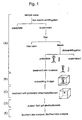
- Fig. 1 shows an embodiment of methods according to the present invention for separating, fractionating, and analyzing virus genes existing in hydrosphere.
- virus particles existing in sample water obtained from hydrosphere, are separated (step A).
- the virus particles thus obtained are embedded in a gel (step B).
- the embedded product is then treated with proteinase K, SDS or the like to decompose viral coats (step C), followed by the fractionation of virus genes (step D).
- step E the fractionated genes are analyzed by Southern blot analysis or Northern blot analysis. More specifically, such intact separation of virus genes can be carried out as follows.
- Hydrosphere refers to all aqueous environments including natural and artificial aqueous environments that could contain virus particles.
- a natural aqueous environment includes seas, lakes, and rivers.
- An artificial aqueous environment includes, but is not limited to, a culture solution, active sludge, wastewater, and blood.
- Sample water containing virus particles includes those derived from the above hydrosphere. For instance, sample water can be collected by recovering a water sample from a desired depth in seas and lakes onto a ship or onto land using Niskin water sampler, Bandon water sampler, and so on.
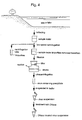
- Fig. 2 Various organisms as shown in Fig. 2 generally exist in seawater, lake water, and river water.
- the method of the present invention is directed to virus particles existing in these waters (hydrosphere).
- impurities are first removed from sample water obtained in (1) above.
- Impurities can be removed by, for example, low speed centrifugation and/or filtration.
- impurities refer to relatively large sample water components, for example, soil particles, clay mineral, fraction of animal tissue and plant texture, phytoplankton and zooplankton, and detritus.
- low speed centrifugation is carried out at about 6,000 x g for 10 to 15 min.
- Centrifuges that can be used for low speed centrifugation include Model HP-30 manufactured by Beckman and Model CR 26H manufactured by Hitachi, Ltd.
- filter paper for example, No. 3 manufactured by WATT MANN CO,. LTD
- the step of low speed centrifugation may be omitted.
- Filters that can be used for purification of viruses include those having pores with a diameter of 0.1 to 0.45 ⁇ m, preferably 0.20 to 0.22 ⁇ m.
- hydrophilic polyvinylidene difluoride membrane having a pore diameter of 0.22 ⁇ m (Model GVWP manufactured by Millipore) may be used. Pore diameter may be properly selected according to the size of organisms to be removed by filtration, for example, referring to Fig. 3. In general, most bacteria living in water can be removed by using a filter having a pore diameter of 0.20 to 0.22 ⁇ m.
- Filter materials include hydrophilic polyvinylidene difluoride (PVDF), hydrophilic polytetrafluoroethylene (PTFE), and polycarbonate, having low absorption to protein.
- PVDF polyvinylidene difluoride
- PTFE hydrophilic polytetrafluoroethylene
- polycarbonate having low absorption to protein.
- pressure is preferably applied.
- filtration pressure is not more than 100 mmHg, preferably not more than 30 mmHg.
- Virus particles from the filtrate obtained in (3) above can be separated by precipitating virus particles by ultracentrifugation.
- Ultracentrifugation is preferably carried out at 100,000 to 130,000 x g for 3 to 16 hr.
- Centrifuges that can be used for ultracentrifugation include Model CP 90 ⁇ manufactured by Hitachi, Ltd and Optima XL-100K manufactured by Beckman.
- the precipitate of virus particles obtained in (4) above sometimes contains a large quantity of naked nucleic acids, and nucleic acids absorbed and held on inorganic particles and detritus, which are derived from other organisms. These nucleic acids can be removed by treating them with nuclease. DNase is used to decompose DNA and RNase is used to decompose RNA. That is, the virus precipitate obtained in (4) above is suspended, for example, in a TM buffer or TE buffer to incubate with a proper amount of DNase or RNase. Adding an EDTA solution, an RNasin solution and the like can stop the reaction. In this way, a suspension of virus particles can be obtained which contains no naked nucleic acids therein.
- virus particles are immobilized by embedding them in a gel.
- the immobilized virus particles are treated with a proteolytic enzyme or a surfactant. This can prevent the breakage of genes.
- Embedding virus particles in the gel can be carried out by adding a gelling agent, for example a low-melting point agarose, to a virus suspension. That is, the nuclease treated virus suspension obtained in 1 above is heated to a temperature at which the low-melting point agarose does not solidify (for example 55°C).
- a gelling agent for example, a low-melting point agarose is then added thereto, followed by thorough mixing and cooling to solidification.
- the final concentration of the agarose is preferably about 1%.
- the coats of virus particles embedded in the gel in 2 above can be decomposed and removed by treating a gel embedded product with a virus coat decomposing solution containing, for example, a proteolytic enzyme and/or a surfactant.
- a proteolytic enzyme and/or a surfactant include proteinase K (manufactured by NIPPON GENE CO., LTD.).
- Surfactants that can be used herein include sodium dodecyl sulfate (SDS).
- SDS sodium dodecyl sulfate
- the product of virus coat decomposition obtained in 3 above is subjected to electrophoresis (for example pulsed-field gel electrophoresis), thereby fractionating nucleotides contained in the treated product by size. That is, gel, which has been subjected to virus coat decomposition, is sliced into a size small enough to fit in a well. The sliced gel is then applied to an electrophoresis gel having agarose or the like as a carrier. Subsequently, under pulsed-field gel electrophoresis conditions corresponding to fractionated size range, virus genes can be fractionated by size. The fractionated virus genes can be detected with the aid of a nucleic acid stainer or the like. Also, the separation of virus and a quantitative purification and fractionation of genes thereof enables the investigation of quantitative ratio of each size existing therein.
- electrophoresis for example pulsed-field gel electrophoresis
- the virus genes fractionated in 4 above can be analyzed, for example, by Southern blot analysis or Western blot analysis.
- cleaving of a nucleotide chain to some extent prior to blotting can enhance the blotting efficiency.
- Treating the fractionated gel with ultraviolet or a hydrochloric acid can cleave the nucleotide chain.
- the blotted genes can be analyzed in accordance with conventional methods regarding, for example, existence and quantity of specific genes by hybridization using a labeled probe.
- a synthetic probe when analyzing virus genes from a sample water collected from a sea area where a harmful red tide is likely to occur, a synthetic probe may be provided in accordance with the nucleotide sequence of genes of viruses associated with the decrease and increase of plankton causative of the red tide (for example, Chattonella virus) in order to investigate the existence, the quantity, and the like of the viruses.
- viruses associated with the decrease and increase of plankton causative of the red tide (for example, Chattonella virus) in order to investigate the existence, the quantity, and the like of the viruses.
- virus particles were separated from sample water collected from Tokyo Bay and Kasumigaura. Specifically, 500 ml of sample water was first centrifuged using a centrifuge equipped with an angle rotor (Model KR 200A manufactured by KUBOTA CORPORATION) at 6,000 x g for 10 min to remove impurities precipitated. Subsequently, the resultant supernatant was subjected to suction filtration with a PVDF membrane filter having a pore diameter of 0.22 ⁇ m (manufactured by IWAKI&CO., LTD).
- an angle rotor Model KR 200A manufactured by KUBOTA CORPORATION
- the resultant filtrate was subjected to ultracentrifugation with a centrifuge (Model CP 90 ⁇ manufactured by Hitachi, Ltd) at 125,000 x g for 3 hr.
- the precipitate was dissolved in 0.5 to 1.0 ml of TM buffer (10 mM Tris-10mM MgCl 2 (pH 7.5)) to prepare a virus solution.
- TM buffer 10 mM Tris-10mM MgCl 2 (pH 7.5)
- Example 1 Genes were separated from virus particles obtained in Example 1. To the virus particle-containing solution heated to 55°C was added an equal volume of 2% low-melting point agarose (55°C, heat dissolved, Agarose GB manufactured by NIPPON GENE CO., LTD.). The mixture was poured into a cast to solidify. The solidified virus embedded product was transferred to a sterilized tube and treated with a virus coat decomposing solution (1 mg/ml proteinase K, 1% SDS, 0.5 M EDTA (pH 8.2)) at 55°C for 15 to 18 hr. The virus embedded product was then rinsed with a TE buffer (10 mM Tris-HCl, 1 mM EDTA (pH 8.0)), and immersed in the TE buffer and stored at 4°C.
- a TE buffer (10 mM Tris-HCl, 1 mM EDTA (pH 8.0)
- Virus DNA was subjected to a pulsed-field gel electrophoresis with CHEF Mapper System (manufactured by Bio-Rad) to fractionate the virus DNA. That is, the virus embedded product obtained in Example 2 was thinly sliced. Each slice was measured for its weight on a wet basis, and applied to a previously prepared 1% agarose gel. Subsequently, electrophoresis was conducted at an initial switching time of 0.06 sec and a final switching time of 17.33 sec, at a voltage of 6 V/cm, for a migration time of 7 hr 53 min. After electrophresis, the product was stained with SYBR (SYBR Green) for 60 to 120 min.
- SYBR SYBR Green
- Fig. 5 shows a stained gel.
- each of lanes 1, 7, and 12 represents a migrated DNA size marker.
- lane 2 is a result of sample water collected from Tokyo Bay on September 24, 1997
- lanes 3 to 6 are results of sample water collected from Tokyo Bay on March 18, 1998.
- lane 3 shows a case wherein the sample water was collected from sea level of water-collection point T1; lane 4, 15 m below sea level of point T1; lane 5, sea level of water-collection point T2; and lane 6, 20 m below sea level of point T2.
- Lanes 8 to 11 are results of sample water collected from Tokyo Bay on August 19, 1998.
- lane 8 shows a case using sample water collected from sea level of water-collection point T1; lane 9, 15 m below sea level of point T1; lane 10, sea level of water-collection point T2; and lane 11, 20 m below sea level of point T2.
- lanes 13 to 15 are results of sample water collected from Kasumigaura.
- lane 13 shows a case using sample water collected on May 19, 1998; lane 14, on June 17, 1998; and lane 15, on July 14, 1998.
- DNA which has a molecular size exceeding 30 kbp and is considered to contain intact virus genes, has been fractionated.
- the present invention provides methods for separating, fractionating, and analyzing virus genes existing in hydrosphere in an intact state.
- the present invention is useful for an identification of kinds of viruses existing in hydrosphere and for an estimate of a quantitative ratio of viruses existing in hydrosphere.
Landscapes
- Chemical & Material Sciences (AREA)
- Life Sciences & Earth Sciences (AREA)
- Health & Medical Sciences (AREA)
- Engineering & Computer Science (AREA)
- Organic Chemistry (AREA)
- Wood Science & Technology (AREA)
- Zoology (AREA)
- Genetics & Genomics (AREA)
- Bioinformatics & Cheminformatics (AREA)
- Biomedical Technology (AREA)
- Biotechnology (AREA)
- General Engineering & Computer Science (AREA)
- General Health & Medical Sciences (AREA)
- Immunology (AREA)
- Biochemistry (AREA)
- Microbiology (AREA)
- Molecular Biology (AREA)
- Virology (AREA)
- Analytical Chemistry (AREA)
- Physics & Mathematics (AREA)
- Biophysics (AREA)
- Proteomics, Peptides & Aminoacids (AREA)
- Crystallography & Structural Chemistry (AREA)
- Medicinal Chemistry (AREA)
- Plant Pathology (AREA)
- Measuring Or Testing Involving Enzymes Or Micro-Organisms (AREA)
- Micro-Organisms Or Cultivation Processes Thereof (AREA)
Abstract
Description
Claims (3)
- A method for separating virus genes, comprising the steps of:(a) separating virus particles from hydrosphere;(b) embedding the virus particles obtained in step (a) in a gel; and(c) treating the embedded product obtained in step (b) with a proteolytic enzyme and/or a surfactant.
- A method for fractionating virus genes, comprising the steps of:(a) separating virus particles from hydrosphere;(b) embedding the virus particles obtained in step (a) in a gel;(c) treating the embedded product obtained in step (b) with a proteolytic enzyme and/or a surfactant; and(d) subjecting the proteolytic enzyme- and/or surfactant-treated product obtained in step (c) to electrophoresis.
- A method for analyzing virus genes, comprising the steps of:(a) separating virus particles from hydrosphere;(b) embedding the virus particles obtained in step (a) in a gel;(c) treating the embedded product obtained in step (b) with a proteolytic enzyme and/or a surfactant;(d) subjecting the proteolytic enzyme- and/or surfactant-treated product obtained in step (c) to electrophoresis; and(d) analyzing the electrophoresis gel obtained in step (d).
Applications Claiming Priority (3)
| Application Number | Priority Date | Filing Date | Title |
|---|---|---|---|
| JP14534199 | 1999-05-25 | ||
| JP14534199 | 1999-05-25 | ||
| PCT/JP2000/001968 WO2000071700A1 (en) | 1999-05-25 | 2000-03-29 | Methods for separating, fractionating and analyzing gene of virus existing in water |
Publications (3)
| Publication Number | Publication Date |
|---|---|
| EP1182254A1 true EP1182254A1 (en) | 2002-02-27 |
| EP1182254A4 EP1182254A4 (en) | 2002-08-21 |
| EP1182254B1 EP1182254B1 (en) | 2005-11-02 |
Family
ID=15382948
Family Applications (1)
| Application Number | Title | Priority Date | Filing Date |
|---|---|---|---|
| EP00912929A Expired - Lifetime EP1182254B1 (en) | 1999-05-25 | 2000-03-29 | Methods for separating, fractionating and analyzing gene of virus existing in water |
Country Status (6)
| Country | Link |
|---|---|
| US (1) | US6664063B1 (en) |
| EP (1) | EP1182254B1 (en) |
| JP (1) | JP3769604B2 (en) |
| AU (1) | AU3454200A (en) |
| DE (1) | DE60023690T2 (en) |
| WO (1) | WO2000071700A1 (en) |
Families Citing this family (1)
| Publication number | Priority date | Publication date | Assignee | Title |
|---|---|---|---|---|
| JP4195039B2 (en) * | 2006-02-16 | 2008-12-10 | 善之 辻本 | Sample pretreatment method and reagent |
Family Cites Families (5)
| Publication number | Priority date | Publication date | Assignee | Title |
|---|---|---|---|---|
| US4695548A (en) * | 1982-11-18 | 1987-09-22 | The Trustees Of Columbia University In The City Of New York | Gel inserts useful in electrophoresis |
| JPH0824582B2 (en) * | 1986-12-24 | 1996-03-13 | 株式会社島津製作所 | DNA purification device |
| US5457050A (en) * | 1993-11-30 | 1995-10-10 | Board Of Regents, The University Of Texas System | Agarose plug mold and processing chamber |
| JPH07163352A (en) * | 1993-12-13 | 1995-06-27 | Canon Inc | Method for purifying DNA |
| JPH0994098A (en) * | 1995-09-29 | 1997-04-08 | Unitika Ltd | Selective concentration of microorganism and microorganism examination using the concentration |
-
2000
- 2000-03-29 WO PCT/JP2000/001968 patent/WO2000071700A1/en not_active Ceased
- 2000-03-29 EP EP00912929A patent/EP1182254B1/en not_active Expired - Lifetime
- 2000-03-29 AU AU34542/00A patent/AU3454200A/en not_active Abandoned
- 2000-03-29 DE DE60023690T patent/DE60023690T2/en not_active Expired - Lifetime
- 2000-03-29 JP JP2000620077A patent/JP3769604B2/en not_active Expired - Lifetime
- 2000-05-29 US US09/979,503 patent/US6664063B1/en not_active Expired - Fee Related
Also Published As
| Publication number | Publication date |
|---|---|
| EP1182254A4 (en) | 2002-08-21 |
| WO2000071700A1 (en) | 2000-11-30 |
| EP1182254B1 (en) | 2005-11-02 |
| US6664063B1 (en) | 2003-12-16 |
| DE60023690D1 (en) | 2005-12-08 |
| DE60023690T2 (en) | 2006-07-20 |
| JP3769604B2 (en) | 2006-04-26 |
| AU3454200A (en) | 2000-12-12 |
Similar Documents
| Publication | Publication Date | Title |
|---|---|---|
| Fuhrman et al. | Extraction from natural planktonic microorganisms of DNA suitable for molecular biological studies | |
| US6548256B2 (en) | DNA isolation method and kit | |
| JP5112064B2 (en) | Kits and methods for removing contaminants from nucleic acids in environmental and biological samples | |
| Colombet et al. | Virioplankton ‘pegylation’: use of PEG (polyethylene glycol) to concentrate and purify viruses in pelagic ecosystems | |
| US7893251B2 (en) | Methods for selective isolation of nucleic acids from microbial cells present in samples containing higher eukaryotic cells and/or tissues | |
| US5972613A (en) | Methods of nucleic acid isolation | |
| EP1330518B1 (en) | Method for collecting and using nuclear mrna | |
| CA2296733A1 (en) | Methods for purifying viral nucleic acids using a hydrophilic polyvinylidine fluoride (pvdf) membrane | |
| Sun et al. | Efficient purification and concentration of viruses from a large body of high turbidity seawater | |
| JP2002515508A (en) | Method and apparatus for purifying nucleic acid | |
| Díez et al. | Pulsed-field gel electrophoresis analysis of virus assemblages present in a hypersaline environment | |
| KR20080066727A (en) | Extraction method of RNA and detection method of RNA | |
| US20030229222A1 (en) | Process of extracting nucleic acid and process of simultaneously carrying out extraction and purification of nucleic acid | |
| Matsuda et al. | First finding of urease‐positive thermophilic strains of Campylobacter in river water in the Far East, namely, in Japan and their phenotypic and genotypic characterization | |
| CN101268198A (en) | Methods for nucleic acid extraction and identification | |
| Prado et al. | Virome analysis in lakes of the South Shetland Islands, Antarctica-2020 | |
| US6664063B1 (en) | Methods for separating,fractionating and analyzing gene of virus existing in hydrosphere | |
| JP4165899B2 (en) | Method for collecting DNA from biological samples | |
| JPWO2000071700A1 (en) | Methods for isolating, fractionating, and analyzing the genes of viruses present in aquatic communities | |
| WO1994012657A1 (en) | Direct-phenol pcr, rt and rt-pcr methods | |
| JP3521079B2 (en) | DNA extraction and purification method and apparatus therefor | |
| EP2346985A1 (en) | Recovery and detection of microorganisms from mixed cellulose ester filtration supports by sequential treatment with methanol and acetone | |
| JP3517448B2 (en) | Method for recovering and purifying microorganism-derived DNA | |
| US20030228600A1 (en) | DNA isolation method and kit | |
| FR2974117A1 (en) | METHOD FOR QUANTIFICATION OF LIVING BACTERIA IN A LIQUID MEDIUM |
Legal Events
| Date | Code | Title | Description |
|---|---|---|---|
| PUAI | Public reference made under article 153(3) epc to a published international application that has entered the european phase |
Free format text: ORIGINAL CODE: 0009012 |
|
| 17P | Request for examination filed |
Effective date: 20011219 |
|
| AK | Designated contracting states |
Kind code of ref document: A1 Designated state(s): AT BE CH CY DE DK ES FI FR GB GR IE IT LI LU MC NL PT SE |
|
| RIC1 | Information provided on ipc code assigned before grant |
Free format text: 7C 12N 15/10 A, 7C 12N 7/02 B, 7C 12Q 1/68 B, 7G 01N 27/447 B, 7G 01N 33/18 B |
|
| A4 | Supplementary search report drawn up and despatched |
Effective date: 20020709 |
|
| AK | Designated contracting states |
Kind code of ref document: A4 Designated state(s): AT BE CH CY DE DK ES FI FR GB GR IE IT LI LU MC NL PT SE |
|
| 17Q | First examination report despatched |
Effective date: 20030827 |
|
| RBV | Designated contracting states (corrected) |
Designated state(s): DE FR GB |
|
| GRAP | Despatch of communication of intention to grant a patent |
Free format text: ORIGINAL CODE: EPIDOSNIGR1 |
|
| GRAS | Grant fee paid |
Free format text: ORIGINAL CODE: EPIDOSNIGR3 |
|
| GRAA | (expected) grant |
Free format text: ORIGINAL CODE: 0009210 |
|
| AK | Designated contracting states |
Kind code of ref document: B1 Designated state(s): DE FR GB |
|
| REG | Reference to a national code |
Ref country code: GB Ref legal event code: FG4D |
|
| REF | Corresponds to: |
Ref document number: 60023690 Country of ref document: DE Date of ref document: 20051208 Kind code of ref document: P |
|
| ET | Fr: translation filed | ||
| PLBE | No opposition filed within time limit |
Free format text: ORIGINAL CODE: 0009261 |
|
| STAA | Information on the status of an ep patent application or granted ep patent |
Free format text: STATUS: NO OPPOSITION FILED WITHIN TIME LIMIT |
|
| 26N | No opposition filed |
Effective date: 20060803 |
|
| PGFP | Annual fee paid to national office [announced via postgrant information from national office to epo] |
Ref country code: FR Payment date: 20100402 Year of fee payment: 11 |
|
| PGFP | Annual fee paid to national office [announced via postgrant information from national office to epo] |
Ref country code: GB Payment date: 20100322 Year of fee payment: 11 |
|
| PGFP | Annual fee paid to national office [announced via postgrant information from national office to epo] |
Ref country code: DE Payment date: 20100419 Year of fee payment: 11 |
|
| GBPC | Gb: european patent ceased through non-payment of renewal fee |
Effective date: 20110329 |
|
| REG | Reference to a national code |
Ref country code: FR Ref legal event code: ST Effective date: 20111130 |
|
| PG25 | Lapsed in a contracting state [announced via postgrant information from national office to epo] |
Ref country code: FR Free format text: LAPSE BECAUSE OF NON-PAYMENT OF DUE FEES Effective date: 20110331 Ref country code: DE Free format text: LAPSE BECAUSE OF NON-PAYMENT OF DUE FEES Effective date: 20111001 |
|
| REG | Reference to a national code |
Ref country code: DE Ref legal event code: R119 Ref document number: 60023690 Country of ref document: DE Effective date: 20111001 |
|
| PG25 | Lapsed in a contracting state [announced via postgrant information from national office to epo] |
Ref country code: GB Free format text: LAPSE BECAUSE OF NON-PAYMENT OF DUE FEES Effective date: 20110329 |