EP1181897A2 - Integrated fluoroscopic surgical navigation and imaging workstation with command protocol - Google Patents
Integrated fluoroscopic surgical navigation and imaging workstation with command protocol Download PDFInfo
- Publication number
- EP1181897A2 EP1181897A2 EP01307077A EP01307077A EP1181897A2 EP 1181897 A2 EP1181897 A2 EP 1181897A2 EP 01307077 A EP01307077 A EP 01307077A EP 01307077 A EP01307077 A EP 01307077A EP 1181897 A2 EP1181897 A2 EP 1181897A2
- Authority
- EP
- European Patent Office
- Prior art keywords
- imaging
- navigation
- subsystem
- message
- image
- Prior art date
- Legal status (The legal status is an assumption and is not a legal conclusion. Google has not performed a legal analysis and makes no representation as to the accuracy of the status listed.)
- Ceased
Links
Images

Classifications
-
- A—HUMAN NECESSITIES
- A61—MEDICAL OR VETERINARY SCIENCE; HYGIENE
- A61B—DIAGNOSIS; SURGERY; IDENTIFICATION
- A61B34/00—Computer-aided surgery; Manipulators or robots specially adapted for use in surgery
- A61B34/20—Surgical navigation systems; Devices for tracking or guiding surgical instruments, e.g. for frameless stereotaxis
-
- A—HUMAN NECESSITIES
- A61—MEDICAL OR VETERINARY SCIENCE; HYGIENE
- A61B—DIAGNOSIS; SURGERY; IDENTIFICATION
- A61B6/00—Apparatus or devices for radiation diagnosis; Apparatus or devices for radiation diagnosis combined with radiation therapy equipment
- A61B6/12—Arrangements for detecting or locating foreign bodies
-
- A—HUMAN NECESSITIES
- A61—MEDICAL OR VETERINARY SCIENCE; HYGIENE
- A61B—DIAGNOSIS; SURGERY; IDENTIFICATION
- A61B6/00—Apparatus or devices for radiation diagnosis; Apparatus or devices for radiation diagnosis combined with radiation therapy equipment
- A61B6/54—Control of apparatus or devices for radiation diagnosis
- A61B6/547—Control of apparatus or devices for radiation diagnosis involving tracking of position of the device or parts of the device
-
- A—HUMAN NECESSITIES
- A61—MEDICAL OR VETERINARY SCIENCE; HYGIENE
- A61B—DIAGNOSIS; SURGERY; IDENTIFICATION
- A61B90/00—Instruments, implements or accessories specially adapted for surgery or diagnosis and not covered by any of the groups A61B1/00 - A61B50/00, e.g. for luxation treatment or for protecting wound edges
- A61B90/36—Image-producing devices or illumination devices not otherwise provided for
-
- G—PHYSICS
- G16—INFORMATION AND COMMUNICATION TECHNOLOGY [ICT] SPECIALLY ADAPTED FOR SPECIFIC APPLICATION FIELDS
- G16H—HEALTHCARE INFORMATICS, i.e. INFORMATION AND COMMUNICATION TECHNOLOGY [ICT] SPECIALLY ADAPTED FOR THE HANDLING OR PROCESSING OF MEDICAL OR HEALTHCARE DATA
- G16H40/00—ICT specially adapted for the management or administration of healthcare resources or facilities; ICT specially adapted for the management or operation of medical equipment or devices
- G16H40/20—ICT specially adapted for the management or administration of healthcare resources or facilities; ICT specially adapted for the management or operation of medical equipment or devices for the management or administration of healthcare resources or facilities, e.g. managing hospital staff or surgery rooms
-
- A—HUMAN NECESSITIES
- A61—MEDICAL OR VETERINARY SCIENCE; HYGIENE
- A61B—DIAGNOSIS; SURGERY; IDENTIFICATION
- A61B34/00—Computer-aided surgery; Manipulators or robots specially adapted for use in surgery
- A61B34/20—Surgical navigation systems; Devices for tracking or guiding surgical instruments, e.g. for frameless stereotaxis
- A61B2034/2074—Interface software
-
- A—HUMAN NECESSITIES
- A61—MEDICAL OR VETERINARY SCIENCE; HYGIENE
- A61B—DIAGNOSIS; SURGERY; IDENTIFICATION
- A61B90/00—Instruments, implements or accessories specially adapted for surgery or diagnosis and not covered by any of the groups A61B1/00 - A61B50/00, e.g. for luxation treatment or for protecting wound edges
- A61B90/36—Image-producing devices or illumination devices not otherwise provided for
- A61B90/37—Surgical systems with images on a monitor during operation
- A61B2090/376—Surgical systems with images on a monitor during operation using X-rays, e.g. fluoroscopy
-
- A—HUMAN NECESSITIES
- A61—MEDICAL OR VETERINARY SCIENCE; HYGIENE
- A61B—DIAGNOSIS; SURGERY; IDENTIFICATION
- A61B6/00—Apparatus or devices for radiation diagnosis; Apparatus or devices for radiation diagnosis combined with radiation therapy equipment
- A61B6/44—Constructional features of apparatus for radiation diagnosis
- A61B6/4405—Constructional features of apparatus for radiation diagnosis the apparatus being movable or portable, e.g. handheld or mounted on a trolley
-
- A—HUMAN NECESSITIES
- A61—MEDICAL OR VETERINARY SCIENCE; HYGIENE
- A61B—DIAGNOSIS; SURGERY; IDENTIFICATION
- A61B6/00—Apparatus or devices for radiation diagnosis; Apparatus or devices for radiation diagnosis combined with radiation therapy equipment
- A61B6/44—Constructional features of apparatus for radiation diagnosis
- A61B6/4429—Constructional features of apparatus for radiation diagnosis related to the mounting of source units and detector units
- A61B6/4435—Constructional features of apparatus for radiation diagnosis related to the mounting of source units and detector units the source unit and the detector unit being coupled by a rigid structure
- A61B6/4441—Constructional features of apparatus for radiation diagnosis related to the mounting of source units and detector units the source unit and the detector unit being coupled by a rigid structure the rigid structure being a C-arm or U-arm
-
- G—PHYSICS
- G16—INFORMATION AND COMMUNICATION TECHNOLOGY [ICT] SPECIALLY ADAPTED FOR SPECIFIC APPLICATION FIELDS
- G16H—HEALTHCARE INFORMATICS, i.e. INFORMATION AND COMMUNICATION TECHNOLOGY [ICT] SPECIALLY ADAPTED FOR THE HANDLING OR PROCESSING OF MEDICAL OR HEALTHCARE DATA
- G16H30/00—ICT specially adapted for the handling or processing of medical images
- G16H30/20—ICT specially adapted for the handling or processing of medical images for handling medical images, e.g. DICOM, HL7 or PACS
-
- G—PHYSICS
- G16—INFORMATION AND COMMUNICATION TECHNOLOGY [ICT] SPECIALLY ADAPTED FOR SPECIFIC APPLICATION FIELDS
- G16H—HEALTHCARE INFORMATICS, i.e. INFORMATION AND COMMUNICATION TECHNOLOGY [ICT] SPECIALLY ADAPTED FOR THE HANDLING OR PROCESSING OF MEDICAL OR HEALTHCARE DATA
- G16H40/00—ICT specially adapted for the management or administration of healthcare resources or facilities; ICT specially adapted for the management or operation of medical equipment or devices
- G16H40/60—ICT specially adapted for the management or administration of healthcare resources or facilities; ICT specially adapted for the management or operation of medical equipment or devices for the operation of medical equipment or devices
- G16H40/67—ICT specially adapted for the management or administration of healthcare resources or facilities; ICT specially adapted for the management or operation of medical equipment or devices for the operation of medical equipment or devices for remote operation
Definitions
- the preferred embodiments of the present invention relate to surgical navigation systems and techniques.
- the preferred embodiments of the present invention relate to an integrated surgical navigation system and fluoroscopic X-ray system.
- Medical imaging techniques including X-ray, CAT (Computerized Axial Tomography), MRI (Magnetic Resonance Imaging), and ultrasound are well established. These techniques provide an examining physician with high resolution images useful for subsequent detailed study and diagnosis. Recently, however, surgical navigation techniques have been proposed that use pre-operative images for improving inter-operative visualization of patient anatomy. To that end, one or more of the pre-operative images are displayed for the surgeon during an operation, with a surgical tool superimposed on the image in the correct location.
- the navigational challenges associated with using pre-operative images during surgery include establishing a known coordinate system with respect to the patient, registering pre-operative images in the coordinate system, and tracking surgical tool movement through the coordinate system.
- navigation systems attempting to meet these challenges, were developed as separate and independent add-on systems to be connected to separate and independent imaging systems.
- the add-on navigation systems were designed as separate navigation units, and generally did not adhere to a standard or consistent communications protocol for communicating with the imaging system.
- prior navigation systems 1) required significant additional floorspace (in an already overcrowded operating room environment), 2) did not support high speed digital data transfer to the imaging system, and 3) had no bi-directional command and control interface with the imaging system.
- the lack of bi-directional command and control with the imaging system make it difficult, if not impossible, to ensure that position information from the navigation system actually corresponded to the moment in time that the image was acquired.
- tracking errors arise, for example, in a C-arm type X-ray system performing fluoroscopy, when the C-arm moves. The tracking error may arise during the time interval between the point in time at which image acquisition is finished and the point in time at which the navigation information was obtained. Any tracking error is undesirable in surgical navigation.
- the external output ports of prior imaging systems were generally limited to NTSC or PAL video outputs.
- An NTSC or PAL video output represents an immediate reduction in resolution and dynamic range in comparison with the original digital image read out of an X-ray detector (e.g., a 1024 x 1024 image).
- X-ray detector e.g., a 1024 x 1024 image.
- conventional navigation systems were limited to using a frame grabber connected to the imaging system output port to acquire lower resolution images for later surgical navigation.
- DICOM was used to transfer images between the imaging system and navigation system, the DICOM overhead limited throughput to as little as one image every twelve seconds.
- the present invention provides a medical diagnostic imaging system.
- the medical diagnostic imaging system includes an X-ray source and X-ray detector, sensors tracking a position of at least one of a surgical instrument and the X-ray detector, and an integrated imaging and navigation workstation.
- the integrated imaging and navigation workstation includes at least one processor executing fluoroscopic imaging based on an output of the X-ray detector and navigation tracking of positions of a surgical instrument and positions of an X-ray detector with respect to a coordinate system.
- the integrated imaging and navigation workstation also includes an input receiving surgical instrument tracking signals from the sensors, an input receiving detector tracking signals from the sensors, and a display for displaying fluoroscopic images with a displayed instrument superimposed.
- one or more relations of the displayed instrument with respect to the fluoroscopic image corresponds to the relation of the surgical instrument to the patient.
- the present invention also provides a diagnostic imaging system communication protocol.
- the communication protocol implements bi-directional communication between a medical imaging subsystem and a medical navigational subsystem.
- the communication protocol includes a set of navigation subsystem to imaging subsystem messages as well as a set of imaging subsystem to navigation subsystem messages.
- the imaging subsystem to navigation subsystem messages include a start imaging and end imaging message.
- the messages may include Ping response time messages, system configuration, file request, image request, and image reply messages, as examples.
- FIG 1 illustrates a block diagram of a medical diagnostic imaging system 100.
- the imaging system 100 includes an integrated imaging and navigation workstation 102 that in turn includes a navigation subsystem 104 and an imaging subsystem 106 (shown in Figure 1 as a fluoroscopic imaging subsystem).
- the navigation subsystem 104 includes a navigation computer 108 and a tracker module 110, while the imaging subsystem includes an imaging computer 112.
- the imaging system 100 further includes a workstation surgical tool tracking sensor input port 114, a workstation X-ray-detector fixture tracking sensor input port 116, a workstation dynamic reference frame transmitter control output port 118, and a workstation X-ray exposure output port 120 (that communicates with a foot switch activated exposure module 132).
- the X-ray detector fixture connects to an image intensifier (or solid state flat panel detector) on the C-arm 130.
- the tool sensor input port 114 allows the imaging system 100 to be coupled to a tool location sensor 123 on a medical (e.g., surgical) tool 122.
- the medical tool 122 may be, for example, an aspirating device such as that disclosed in U.S. Pat. No.
- the transmitter control output port 118 connects the imaging system 100 to the location transmitter 124.
- the detector tracking sensor input 116 allows the imaging system to receive tracking signal inputs from an X-ray detector position sensor 126 that detects the position of an image X-ray detector.
- the X-ray exposure output port 120 allows the imaging system 100 to communicate with the exposure module 132 and to read images from the X-ray detector.
- the exposure module 132 provides a physician with foot switch activated X-ray exposure control.
- the imaging system 100 also provides a navigation display 133 (attached to a movable display arm) for displaying images output by the navigation subsystem 104 as well as an imaging display 134 for displaying images output by the imaging subsystem 106.
- the navigation subsystem 104, imaging subsystem 106, and displays 133-134 are preferably placed in a self contained mobile cart system.
- the sensors 123 and 126 output pulsed signals representative of the sensor's location.
- the tracker module 110 implements coordinate determination from location pulses received via the detector tracking sensor input 116 and the tool sensor input 114 using the predetermined coordinate system typically associated with the location transmitter 124 and referenced to patient anatomy.
- the coordinates may include, for example, X, Y, and Z locations as well as roll, pitch, and yaw angles.
- the location transmitter 124 may be implemented, for example, as a field generator that includes three orthogonally disposed magnetic dipoles (e.g., current loops or electromagnets). Electromagnetic fields generated by each of the dipoles are distinguishable from one another in phase, frequency, time division multiplexing, and the like. The near-field characteristics of the electromagnetic fields may be used for coordinate determination as generally described, for example, in U.S. Pat. No. 4,054,881. Alternate embodiments of the location transmitter 124 may employ ultrasonic or optical fields. Alternatively, more than one location transmitter 124 may be used in a coordinate determination system based on triangulation of signals. Commercially available position detection units, including the 3 Space® FastrakTM system from Polhemus, Incorporated of Colchester, Vt. may also be used.
- 3 Space® FastrakTM system from Polhemus, Incorporated of Colchester, Vt.
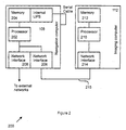
- the workstation 200 includes the navigation computer 108 and the imaging computer 112.
- the navigation computer includes a navigation processor 202, program / data memory 204, and a navigation network interface 206.
- the navigation network interface 206 implements a high speed digital communication interface (e.g., two or more 1024 x 1024 x 16 images per second).
- the images may be cropped in accordance with a circular blanking window to approximately 980 x 980 resolution.
- the navigation computer 108 receives full resolution digital images directly from the imaging computer 112 without the need for NTSC or PAL image grabbers, conversion to NTSC or PAL format, or the associated loss of resolution and dynamic range.
- the navigation network interface 206 may be, for example, a 100BaseT Ethernet network card.
- the navigation computer is preferably implemented as a Sun UltraSparc 10 running Solaris 2.6 with an uninterruptable power supply (UPS).
- a second network interface 208 may be provided in the navigation computer 108 for forwarding packets to other destinations.
- the imaging computer 112 includes the imaging processor 210, program/data memory 212, and imaging network interface 214.
- the imaging computer 112 is preferably implemented as an Intel x86 platform, for example, a PentiumTM processor based PC with 32-128 Megabytes of memory.
- the imaging network interface 214 is compatible with, and is generally implemented in the same manner as, the navigation network interface 206.
- the network interfaces 206, 208, 214 are used not only as a high speed digital communication bus, but also as a command and control bus that the imaging computer 112 and the navigation computer 108 may use to coordinate their functions (particularly image acquisition with coordinate determination as noted below).
- tool configuration updates may be performed using a floppy disk (or other storage media or TFPT protocol) interface in the imaging computer 112.
- the operating system and application software may be accomplished using a CDROM interface with keyboard and mouse control.
- Operating system and application software updates may be accomplished through the imaging network interface 214 using FTP, telnet, the Sun SparcTM pkgadd command, and the like.
- the network addresses assigned to the network interfaces 206, 208, and 214 may be reconfigured in a similar fashion using telnet, the vi editor, and ifconfig, as examples.
- the workstation 200 shown in Figure 2 provides a private network for the imaging computer 112 and the navigation computer 108 to communicate.
- the imaging computer 112 and the navigation computer 108 communicate, preferably, using a socket-based command protocol over TCP/IP.
- the private network is assigned an Internet Protocol (IP) address which is allocated strictly to private networks (e.g. 192.168.0.0/24) and is generally not routed over the Internet.
- IP Internet Protocol
- each workstation 200 shipped can use the same internal network address if desired.
- DICOM Ethernet traffic from the imaging computer 112 may pass through the navigation computer 108, which forwards the DICOM traffic to the second network interface 208.
- the workstation 200 may access external networks by using a Network Address Translation (NAT) at the navigation computer 108 to translate the IP address of the packets coming from the workstation 200 to a host IP address assigned to the system.
- NAT Network Address Translation
- a firewall may be implemented with an IP filter to prevent unnecessary external network traffic from reaching the private network between the Navigation Computer and Workstation.
- the firewall may be configured, for example, to block all inbound traffic from an external network except for ping, traceroute, FTP, and telnet commands.
- external outbound initiated traffic is allowed only for the duration of the required connection.
- the workstation 200 sets the IP address to "network.1” (e.g., 192.168.0.1) for the imaging computer 112 and the net mask to 255.255.255.0.
- the workstation 200 also sets the default route to "network.2" (e.g. 192.168.0.2, assigned to the navigation computer 108).
- the navigation network interface 206 is preferably assigned an IP address of "network 2" or 192.168.0.2.
- the second network interface 208 (connected to external networks) is assigned an IP address allocated by a third party.
- the imaging network interface 214 is preferably assigned an IP address of 192.168.0.1 and is translated to and from the third party specified IP address.
- the workstation 200 preferably sends the following to the navigation computer 108: Host IP address and sub-net mask, Gateway Address (e.g., obtained from DICOM store, print, and query screens), Date and Time, Language Configuration (e.g., English, French, German, Spanish, or Italian).
- Gateway Address e.g., obtained from DICOM store, print, and query screens
- Date and Time e.g., Date and Time
- Language Configuration e.g., English, French, German, Spanish, or Italian.
- the imaging computer 112 monitors the connection 215 through the network interfaces 206 and 214 for network protocol commands and responds synchronously.
- the imaging computer 112 may respond to Ping requests to check the connection between the imaging computer 112 and the navigation computer 108, Request Image requests (e.g., to request transfer of the left display image from imaging computer 112 without patient information), and Request Configuration requests.
- the imaging computer 112 generally sends network protocol commands to the navigation computer 108 asynchronously, including a Ping command to check a connection, Imaging Begin and End commands to delineate, for example, live X-ray exposure periods, a New Exam command to tell the navigation computer 108 that the current patient is no longer valid, and an Update Patient command to update the current patient information.
- responses from the imaging computer 112 are generated synchronous to a predetermined timing source, while original commands may be sent asynchronously. Additionally, the imaging computer 112 monitors for File Transfer commands and responds by retrieving the appropriate file and forwarding it to the navigation computer 108.
- the imaging computer 112 may display an error message such as "Navigation Computer not responding, Power Off, wait 10 seconds, then Power On". If communication fails between the imaging computer 112 and navigation computer 108 after a connection is established, the imaging computer 112 may timeout, for example, after 2 seconds, close the connection between the navigation computer 108 and the imaging computer 112, and display an error message such as "Communication Failure with Navigation Computer".
- the navigation computer 108 and the imaging computer 112 have different power-down strategies.
- the imaging computer 112 may be powered down at any time by removing the AC power without notifying the operating system of a shutdown.
- the navigation computer 108 preferably executes a shutdown procedure before removing AC power.
- the un-interruptible power supply (UPS) is used to hold power with battery backup while the navigation computer 108 shuts down.
- the UPS provides the power status on a serial port, which is used to signal the navigation computer 108 of a power loss.
- the navigation computer 108 may be booted simultaneously with the imaging computer 112, a UPS background (e.g., a daemon) process shall monitor the serial port for a power loss, including imaging computer 112 power switch status.
- a UPS background e.g., a daemon
- the navigation computer 108 Upon receiving a power loss signal, the navigation computer 108 initiates an operating system shutdown, following which the navigation computer 108 is powered off.
- the navigation computer 108 and the imaging computer 112 may be implemented using a single processor executing both navigation and imaging software.
- a multi-processor system under the coordination of an symmetric multiprocessing operating system (e.g., Windows NT) may be used to allow a single set of processors to execute the navigation and imaging software.
- an symmetric multiprocessing operating system e.g., Windows NT
- the navigation computer 108 and the imaging computer 112 may also be implemented as separate processing units, coupled together with the network interfaces 206, 214 and communicating with the diagnostic imaging system communication protocol set forth below.
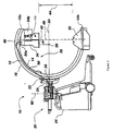
- FIG. 3 that figure shows an exemplary C-arm apparatus 10 that may be used with the medical diagnostic imaging system 100.
- the apparatus 10 includes a C-arm 12 having inner and outer circumferences 14 and 16, respectively, a back convex portion 40, and terminating in opposing upper and lower distal ends 18a and 18b.
- the C-arm 12 preferably has a uniformly circular C-shape, but may alternatively comprise any arc-shaped member.
- the C-arm 12 is held in a suspended position by support means such as structure, generally designated at 20, which includes a support arm 22 mounted upon a wheeled base 24.
- the support arm 22 provides for rotational movement of the C-arm 12 about an axis of lateral rotation 30, either by a bearing assembly between the support arm 22 and the C-arm 12, or by the support 22 itself being rotatably mounted with respect to the base 24.
- the apparatus 10 may also be provided with rotational capacity for rotational movement about an axis 60.
- the wheeled base 24 enables transport of the C-arm 12 from a first location to a second location.
- the wheels of the base operate as transporting means coupled to the support structure 20 for transporting the support arm 22 and the C-arm 12 from a first location to a second location. It is often highly advantageous to be able to move X-ray equipment from one room to another conveniently.
- the mobile nature of the apparatus 10 as provided by the wheeled base 24 offers the advantage of increased access by patients in many different rooms of a hospital, for example.
- the support arm 22 is slidably mounted to the outer circumference 16 of the C-arm 12 and the support structure 20 includes structure and mechanisms necessary to enable selective, sliding orbital motion of the C-arm about an axis of orbital rotation 26 to a selected position.
- the axis 26 preferably coincides with a center of curvature of the C-arm 12 and with the axis of lateral rotation 30. It will be appreciated that the sliding orbital motion causes the C-arm 12 to move through various sliding points of attachment 28 to the support arm 22. It will be appreciated that in practice, the support arm is essentially attached to the C-arm over an area 29 and not a single point, although a "point" may be a large area or a small site.
- the support structure 20 further includes mechanisms known in the art for laterally rotating the support arm 22 selectable amounts about an axis of lateral rotation 30 to a selected lateral position.
- the combination of sliding orbital motion and lateral rotation enables manipulation of the C-arm in two degrees of freedom, i.e. about two perpendicular axes. This provides a kind of spherical quality to the movability of the C-arm 12.
- the sliding orbital motion and lateral rotation enable an X-ray source 32 coupled to the C-arm to be moved to substantially any latitude/longitude point on a lower hemisphere of an imaginary sphere about which the C-arm is moveable.
- the apparatus 10 includes an X-ray source 32 and an image receptor 34 as known generally in the X-ray diagnostic art, mounted upon opposing locations, respectively, on the C-arm 12.
- the X-ray detector position sensor 126 is generally located inside the camera assembly of the image receptor 34 (which may further include registration fiducials and the like that may be used to compensate for image pincushioning and other effects imposed by the image receptor).
- the X-ray source 32 and the image receptor 34 (including a rear portion 34a, and a power supply 34b) may be referred to collectively as the X-ray source/image receptor 32/34.
- the image receptor 34 can be an image intensifier or the like.
- the orbital and laterally rotational manipulation of the C-arm enables selective positioning of the X-ray source/image receptor 32/34 with respect to the width and length of a patient located within interior free space 36 of the C-arm 12.
- the sliding orbital movement of the C-arm causes the X-ray source/image receptor 32/34 to move along respective arcuate movement paths.
- the image receptor 34 is preferably secured to the inner circumference 14 of the C-arm 12 and the X-ray source 32 may also be secured to said inner circumference 14.
- a high voltage cable assembly 50 supplies power to the X-ray source/image receptor 32/34.
- the mounted positions of the image receptor 34 and the C-arm 12 result in the axis of lateral rotation 30 substantially coinciding with the point of attachment 28 of the C-arm 12 to the support arm 22 for substantially any position of the C-arm 12.
- rotation of the support arm 22 does not introduce eccentric lateral moment-arm action so as to provide a more stabile, balanced support structure.
- the center of mass of the C-arm 12 coincides with the axis 30 for any position of the C-arm.
- An additional aspect of the C-arm includes the location of a power supply 34b of the image receptor 34.
- the image receptor 34 and the X-ray source 32 can be moved closer to the center of curvature 26, thereby reducing the distance 46 and thus improving overall balance of the apparatus 10. Balancing is enhanced by having a distance 46 between a line of alignment 48 and the intersection of the axes 26 and 30.
- the line of alignment 48 refers to alignment of a central beam produced by the X-ray source 32, and the image receptor 34. Note also that for a given desired distance 44 between the X-ray source/image receptor 32/34, a larger C-shape can be used for the C-arm 12 without substantially increasing the overall machine height.
- a diagnostic imaging system communication protocol is defined to implement bi-direction communication between the imaging computer 112 and the navigation computer 108.
- Tables 1 shows the messages provided for communication from the navigation computer 108 to the imaging computer 112.
- Tables 2 shows the messages provided for communication from the imaging computer 112 to the navigation computer 108.
- the message format 400 is used with the diagnostic imaging system protocol to implement, preferably, a bi-directional connection-oriented TCP-based data transfer protocol.
- the client i.e., message sender
- server i.e., message receiver
- the server thread preferably blocks (i.e., waits or loops without performing other actions) waiting for the next message which will indicate a command to perform.
- the server executes the command and returns a reply message (which may contain data) to the client.
- the server continues in a loop waiting for the next message.
- the communication protocol generally processes one particular message at a time. That is, at the moment when a particular message is sent, the sender does not send any other message until a corresponding reply message is received.
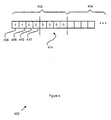
- the message format 400 includes a fixed-length header 402 followed by the data section 404 associated with the message (if any). The same message header format 400 is used for the initial message and the reply.
- the Code field 406 and Type field 408 are defined for each message below.
- the Flag field 410 generally uses bit 0 as a reply flag (1: reply, 0: otherwise), bit 1 as a request flag (1: request for data, 0: otherwise), and bit 2 as an Endian indicator (1: big, 0: little).
- the Status field 412 provides status codes (as additionally explained below), for example, (0: OK, non-0: ERROR).
- the Data Length field 414 provides an unsigned 4-byte length of data to follow in the data section 404.
- the Code field 406 of the message header 402 represents the message code that is common to both imaging and navigation computers.
- the Type field 408 represents a type that is a variation on the message code.
- the Flag field 410 represents the message flags that are used by the imaging and navigation computers to resolve the necessary action to be taken for each message. The specific cases of sending data versus requesting data are illustrated in Figures 5 and 6, respectively. For every message received, the returned message preferably has the same code as the sent message and the flags are incremented by one (reply flag is set high).
- the Status field 412 is used by the reply to indicate error conditions.
- the Data Length field 414 specifies the length of data following the header.
- the Data Length field 414 is encoded big endian (encoded with the most significant byte first, to the least significant last) or little endian (encoded with the least significant byte first, to the most significant last).
- the Endian flag (bit 2 in the Flag field 410) specifies the endian encoding used for the rest of the message. This includes the Data Length field 414 in the message header 402 and any fields over one byte in the data field 404 of the message 400. With the use of the endian flag, the image data can be sent in the "endian" native to a particular machine without byte swapping each pixel greater than one byte in an image.
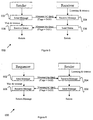
- FIG. 5 that figure illustrates a flow diagram 500 for sending data according to the diagnostic imaging system communication protocol.
- the sender sends data (after formatting the header according to the header format 400) to a receiver.
- the receiver (which has been waiting and listening for a message) receives the data at step 504.
- the receiver responds at step 506 by sending an appropriate reply to the sender (formatted according to the header format 400, with the Flag field set as noted above).
- the sender receives the reply and checks the status set by the receiver.
- a flow diagram 600 for receiving data according to the diagnostic imaging system communication protocol is shown.
- the sender sends a data request message (after formatting the header according to the header format 400) to the receiver.
- the receiver receives the message at step 604 and responds at step 508 by retrieving the requested data and sending an appropriate reply message (with the data) to the sender (formatted according to the header format 400, with the Flag field set as noted above).
- the sender receives the reply message and may process or otherwise use the data provided in the reply message.
- Event is distributed into four general classifications: Event, Update Patient, Request Image, and Request Configuration classifications as shown in Table 3 below.
- Message Description Code Flags Event Send an event, e.g., Fluoro, New Exam, Ping (no-data) 1 0
- Update Patient Imaging computer 112 sends patient information to Navigation computer 108 (data) 2 0
- Request Image Navigation computer 108 requests an image from Imaging computer 112 (data in reply) 3
- Request Configuration Navigation computer 108 requests configuration from Imaging computer 112 (data in reply) 4 2
- Exemplary messages for use with the communication protocol are set forth below. Note that the first 8 bytes (0-7) of the message constitute the header (code, type, flags, status, and the data length) as shown in Figure 4.
- Each field is a preferably string of ASCII characters prefixed by a byte indicating the length:
- Each field is preferably a string of ASCII characters prefixed by a byte indicating the length. All data fields combined will not exceed 512 bytes total.
- a magnification mode message is also defined to communicate the current magnification mode of an X-ray detector (e.g., an image intensifier).
- the magnification mode may specify one of a 12 inch, 9 inch, and 6 inch magnification mode for a 12 inch image intensifier or one of a 9 inch, 6 inch, and 4.5 inch magnification mode for a 9 inch image intensifier.
- the present imaging system allows navigation in several different magnification modes.
- the Image End message may be used to synchronize image acquisition and coordinate determination.
- the command and control information provided over the network interfaces 206, 208, 214 allows the imaging computer 112 to inform the navigation computer 108 that image acquisition just completed.
- the navigation computer 108 may obtain coordinate information from the x ray detector tracking sensor input port 116 to locate the image without intervening C-arm 130 movement and associated location error.
- the present integrated fluoroscopic surgical navigation and imaging workstation provides a high speed digital communication interface between the navigation subsystem and the imaging subsystem, as well as a bi-directional command and control interface.
- full resolution digital images are quickly transferred, and the navigation system is coordinated with the imaging system to maintain accurate tracking of surgical tools.
- the present command protocol provides a standard communication protocol that may be used by any supporting navigation system to communication with any supporting imaging system. Additional components required by "Add-on" type systems (e.g., carts, monitors, monitor arms, power supplies, cabling, and the like) are eliminated, resulting in less crowding in the operating environment.
Landscapes
- Health & Medical Sciences (AREA)
- Life Sciences & Earth Sciences (AREA)
- Engineering & Computer Science (AREA)
- Medical Informatics (AREA)
- Surgery (AREA)
- General Health & Medical Sciences (AREA)
- Biomedical Technology (AREA)
- Public Health (AREA)
- Veterinary Medicine (AREA)
- Heart & Thoracic Surgery (AREA)
- Molecular Biology (AREA)
- Animal Behavior & Ethology (AREA)
- Nuclear Medicine, Radiotherapy & Molecular Imaging (AREA)
- Pathology (AREA)
- Biophysics (AREA)
- Physics & Mathematics (AREA)
- High Energy & Nuclear Physics (AREA)
- Optics & Photonics (AREA)
- Radiology & Medical Imaging (AREA)
- Business, Economics & Management (AREA)
- General Business, Economics & Management (AREA)
- Oral & Maxillofacial Surgery (AREA)
- Robotics (AREA)
- Epidemiology (AREA)
- Primary Health Care (AREA)
- Apparatus For Radiation Diagnosis (AREA)
Abstract
Description


| Message | Description | Code | Flags | |
| Event | Send an event, e.g., Fluoro, New Exam, Ping (no-data) | 1 | 0 | |
| Update | Imaging computer | 112 sends patient information to Navigation computer 108 (data) | 2 | 0 |
| Request | Navigation computer | 108 requests an image from Imaging computer 112 (data in reply) | 3 | 2 |
| Request | Navigation computer | 108 requests configuration from Imaging computer 112 (data in reply) | 4 | 2 |
- Byte 0:
- Code (1)
- Byte 1:
- Type (see Table 4)
| Type | Description |
| 1 | Ping (no-operation) |
| 2 | Imaging (e.g., Fluoro) Begin |
| 3 | Imaging (e.g., Fluoro) End |
| 4 | New Exam |
- Byte 2:
- Flags (initial = 0, reply = 1)
- Byte 3:
- Status (0)
- Byte 4-7
- Data Length (0)
- Name: Update Patient
- Description: Send the patient data to the Navigation computer
- Message Direction: Imaging computer to Navigation computer
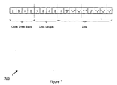
- Data Format: A concatenation of the patient fields where the first byte is the length of the field to follow. The patient fields are grouped, for example, in the following order: patient name, birth date, sex, patient ID, physician name, procedure, and accession number. The name encoding may be that used by the DICOM standard, Part 5, Section 6.2 Value Representation. For example, a patient name of "Jane Doe" would be sent as shown in Figure 7.
- Header:
- Byte 0:
- Code (2)
- Byte 1:
- Type (0)
- Byte 2:
- Flags (initial = 0, reply = 1)
- Byte 3:
- Status (initial = 0, reply = see Table 5)
Status Description 0 OK 1 ERROR - Byte 4-7
- Data Length (initial = data payload size in bytes, reply = 0)
- Data (initial message only):
- Bytes 8-end: Concatenation of the patient fields
- Name: Request Image (and Reply)
- Description: Request an image from the Workstation computer
- Message Direction: from the navigation computer to the imaging computer
- Data Format: An invalid image (test pattern, recalled image, or swap) is notified by an error in the reply message status. No data follows. A valid image is returned with a data payload.
- Header:
- Byte 0:
- Code (3)
- Byte 1:
- Type (see Table 6)
Type Description 1 Left Display, 980x980x8-bit (typically used) 2 Right Display, 980x980x8-bit 3 Filter Memory, 980x980x12-bit 4 Image Memory, 980x980x12-bit 5 Mask, 980x980x12-bit - Byte 2:
- Flags (initial = 2, reply = 3)
- Byte 3:
- Status (initial = 0, reply, see Table 7)
Status Description 0 OK - image valid 1 INVALID - image is invalid i.e. test pattern, recalled image, or swap - Byte 4-7
- Data Length (initial = 0, reply = data payload size in bytes)
- Data (reply only):
- Bytes 8-9:
- Image header length - offset from here,
byte 8, to the image data
- Bytes 10-11:
- Image width in pixels (16-bit unsigned integer)
- Bytes 12-13:
- Image height in pixels (16-bit unsigned integer)
- Byte 14:
- Bytes per pixel allocated, typically 1 or 2 (8-bit unsigned integer)
- Byte 15:
- Bits per pixel stored, assumes LSB at bit 0 (8-bit unsigned integer)
- Byte 16:
- Image flags that specify image features:
- Bit 0:
- Negation (1: yes, 0: no)
- Bit 1:
- Subtraction (1: yes, 0: no)
- Bit 2:
- Flip Backward (1: yes, 0: no)
- Bit 3:
- Flip Upside-down (1: yes, 0: no)
- Bits 4-5:
- Magnification Mode (10: 2x, 01: 1x, 00: norm)
- Bit 6-7:
- Reserved
- Byte 17:
- Reserved
- Bytes 18-19:
- Degrees of Rotation 0-360 (16-bit unsigned integer)
- Bytes start-end:
- Image data - The start is determined from Header Length (bytes 8-9).
- Name: Request Configuration (and Reply)
- Description: Request configuration information from the Workstation
- Message Direction: from the navigation computer to the imaging computer
- Data Format: See below.
- Header:
- Byte 0:
- Code (4)
- Byte 1:
- Type (0)
- Byte 2:
- Flags (initial = 2, reply = 3)
- Byte 3:
- Status (initial = 0, reply = see Table 8)
Status Description 0 OK 1 ERROR - Byte 4-7
- Data Length (initial = 0, reply = data payload size in bytes)
- Data (reply only):
- Bytes 8-end:
- Concatenation of the configuration fields
Claims (10)
- A medical diagnostic imaging system comprising:an X-ray source and X-ray detector;sensors (123, 126) tracking a position of at least one of a surgical instrument (122) and said X-ray detector;an integrated imaging and navigation workstation (102) including at least one processor (210) executing fluoroscopic imaging based on an output of said X-ray detector and navigation tracking of positions of a surgical instrument (122) and positions of an X-ray detector with respect to a coordinate system, said workstation comprising:an input (114) receiving surgical instrument tracking signals from said sensors; andan input (116) receiving detector tracking signals from said sensors; anda display (132) for displaying fluoroscopic images with a displayed instrument superimposed, a relation of the displayed instrument with respect to the fluoroscopic image corresponding to a relation of the surgical instrument to the patient.
- The medical diagnostic imaging system of claim 1, wherein the integrated imaging and navigation workstation (102) comprises a fluoroscopic imaging processor (210) for executing fluoroscopic imaging and a navigation processor (202) for executing navigation tracking.
- The medical diagnostic imaging system of claim 1, wherein the integrated imaging and navigation workstation (102) includes a single processor for executing both fluoroscopic imaging and navigation tracking.
- The medical diagnostic imaging system of claim 1, wherein the integrated imaging and navigation workstation (102) includes a multiprocessor system for executing both fluoroscopic imaging and navigation tracking.
- In a medical diagnostic imaging system, a communication protocol for providing bi-directional communication between a medical imaging subsystem (106) and a medical navigational subsystem (104), the communication protocol comprising:a plurality of navigation subsystem to imaging subsystem messages (400); anda Begin Imaging and an End Imaging message for synchronizing image acquisition with navigation coordinate determination, the Begin Imaging and End Imaging messages included in imaging subsystem to the navigation subsystem messages.
- The communication protocol of claim 5, wherein the communication protocol includes a magnification mode message specifying a magnification mode of an X-ray detector.
- The communication protocol of claim 5, wherein at least one of the navigation subsystem to imaging subsystem messages (400) and the imaging subsystem to navigation subsystem messages (400) include a Ping response time message.
- A method for communication in a bi-directional diagnostic imaging system between a medical imaging subsystem (106) and a medical navigational subsystem (104), the method comprising:realizing in a navigation subsystem a plurality of navigation subsystem to imaging subsystem messages (400);realizing in the imaging subsystem a Begin Imaging and an End Imaging message for synchronizing image acquisition with navigation coordinate determination, the Begin Imaging and End Imaging messages included in imaging subsystem to navigation subsystem messages (400); andformatting the End Imaging message according to a predetermined message header format (402) common to a plurality of the navigation subsystem to imaging subsystem messages (400) and the imaging subsystem to navigation subsystem messages (400); andtransmitting the End Imaging message from the imaging subsystem (106) to the navigation subsystem (104).
- The method of claim 8, further comprising:formatting an image request message according to the predetermined message header format (402);transmitting the image request message from the imaging subsystem to the navigation subsystem(104);formatting an image reply message according to the predetermined message header format (402); andtransmitting the image reply message with image data from the imaging subsystem (106) to the navigation subsystem (104).
- The method of claim 8, further comprising transmitting a magnification mode message specifying a magnification mode of an X-ray detector.
Applications Claiming Priority (2)
| Application Number | Priority Date | Filing Date | Title |
|---|---|---|---|
| US649071 | 2000-08-26 | ||
| US09/649,071 US6823207B1 (en) | 2000-08-26 | 2000-08-26 | Integrated fluoroscopic surgical navigation and imaging workstation with command protocol |
Publications (2)
| Publication Number | Publication Date |
|---|---|
| EP1181897A2 true EP1181897A2 (en) | 2002-02-27 |
| EP1181897A3 EP1181897A3 (en) | 2004-03-03 |
Family
ID=24603337
Family Applications (1)
| Application Number | Title | Priority Date | Filing Date |
|---|---|---|---|
| EP01307077A Ceased EP1181897A3 (en) | 2000-08-26 | 2001-08-20 | Integrated fluoroscopic surgical navigation and imaging workstation with command protocol |
Country Status (3)
| Country | Link |
|---|---|
| US (2) | US6823207B1 (en) |
| EP (1) | EP1181897A3 (en) |
| JP (1) | JP5337335B2 (en) |
Cited By (13)
| Publication number | Priority date | Publication date | Assignee | Title |
|---|---|---|---|---|
| WO2007062051A3 (en) * | 2005-11-23 | 2007-07-12 | Gen Electric | System for surgical navigation |
| WO2008109003A3 (en) * | 2007-03-01 | 2008-10-30 | Medtronic Navigation Inc | Method for localizing an imaging device with a surgical navigation system |
| EP2081124A3 (en) * | 2008-01-16 | 2011-01-12 | Karl Storz Imaging Inc. | Network based endoscopic surgical system |
| US8150495B2 (en) | 2003-08-11 | 2012-04-03 | Veran Medical Technologies, Inc. | Bodily sealants and methods and apparatus for image-guided delivery of same |
| US8483801B2 (en) | 2003-08-11 | 2013-07-09 | Veran Medical Technologies, Inc. | Methods, apparatuses, and systems useful in conducting image guided interventions |
| WO2014076141A3 (en) * | 2012-11-13 | 2014-10-23 | Trumpf Medizin Systeme Gmbh + Co. Kg | Medical control system |
| US9218664B2 (en) | 2005-09-13 | 2015-12-22 | Veran Medical Technologies, Inc. | Apparatus and method for image guided accuracy verification |
| WO2016114728A1 (en) | 2015-01-12 | 2016-07-21 | Ekin Efe Ozel Saglik Hizmetleri Medikal Teknoloji Ar-Ge Gida Insaat San.Tic.Ltd.Sti. | A device for dynamic controlling of the radiation level for radiation-based, real-time, medical-imaging systems |
| US9972082B2 (en) | 2012-02-22 | 2018-05-15 | Veran Medical Technologies, Inc. | Steerable surgical catheter having biopsy devices and related systems and methods for four dimensional soft tissue navigation |
| US10165928B2 (en) | 2010-08-20 | 2019-01-01 | Mark Hunter | Systems, instruments, and methods for four dimensional soft tissue navigation |
| US10617324B2 (en) | 2014-04-23 | 2020-04-14 | Veran Medical Technologies, Inc | Apparatuses and methods for endobronchial navigation to and confirmation of the location of a target tissue and percutaneous interception of the target tissue |
| US10624701B2 (en) | 2014-04-23 | 2020-04-21 | Veran Medical Technologies, Inc. | Apparatuses and methods for registering a real-time image feed from an imaging device to a steerable catheter |
| US11304630B2 (en) | 2005-09-13 | 2022-04-19 | Veran Medical Technologies, Inc. | Apparatus and method for image guided accuracy verification |
Families Citing this family (201)
| Publication number | Priority date | Publication date | Assignee | Title |
|---|---|---|---|---|
| US6344745B1 (en) * | 1998-11-25 | 2002-02-05 | Medrad, Inc. | Tapered birdcage resonator for improved homogeneity in MRI |
| US6823207B1 (en) * | 2000-08-26 | 2004-11-23 | Ge Medical Systems Global Technology Company, Llc | Integrated fluoroscopic surgical navigation and imaging workstation with command protocol |
| ATE258031T1 (en) * | 2002-09-12 | 2004-02-15 | Brainlab Ag | X-RAY IMAGE-ASSISTED NAVIGATION WITH ORIGINAL, TWO-DIMENSIONAL X-RAY IMAGES |
| US7212661B2 (en) * | 2003-02-14 | 2007-05-01 | Ge Medical Systems Information Technologies. Inc. | Image data navigation method and apparatus |
| US7596255B2 (en) * | 2003-11-26 | 2009-09-29 | Ge Medical Systems Information Technologies, Inc. | Image navigation system and method |
| JP2007538011A (en) * | 2004-05-07 | 2007-12-27 | メモリー・ファーマシューティカルズ・コーポレイション | 1H-indazole, benzothiazole, 1,2-benzisoxazole, 1,2-benzisothiazole, and chromone, and their preparation and use |
| EP3155966B1 (en) * | 2005-01-31 | 2018-07-04 | Mediguide Ltd. | A method and system for eliminating electromagnetic interference in a medical image, in real-time |
| US20060241396A1 (en) * | 2005-02-10 | 2006-10-26 | Fabian Carl E | Multi-modal detection of surgical sponges and implements |
| DE102005014286B4 (en) * | 2005-03-24 | 2006-12-21 | Siemens Ag | Diagnostic device with an X-ray system and a location system for catheters and methods for their operation |
| US8208988B2 (en) * | 2005-05-13 | 2012-06-26 | General Electric Company | System and method for controlling a medical imaging device |
| US20070263915A1 (en) * | 2006-01-10 | 2007-11-15 | Adi Mashiach | System and method for segmenting structures in a series of images |
| US20070160273A1 (en) * | 2006-01-10 | 2007-07-12 | Adi Maschiah | Device, system and method for modifying two dimensional data of a body part |
| US7283615B2 (en) * | 2006-02-08 | 2007-10-16 | Brent Morehead | On-demand multiple step fluoroscope control assembly |
| US8219178B2 (en) | 2007-02-16 | 2012-07-10 | Catholic Healthcare West | Method and system for performing invasive medical procedures using a surgical robot |
| US10653497B2 (en) | 2006-02-16 | 2020-05-19 | Globus Medical, Inc. | Surgical tool systems and methods |
| US10893912B2 (en) | 2006-02-16 | 2021-01-19 | Globus Medical Inc. | Surgical tool systems and methods |
| US10357184B2 (en) | 2012-06-21 | 2019-07-23 | Globus Medical, Inc. | Surgical tool systems and method |
| US20070236514A1 (en) * | 2006-03-29 | 2007-10-11 | Bracco Imaging Spa | Methods and Apparatuses for Stereoscopic Image Guided Surgical Navigation |
| US7331711B2 (en) * | 2006-05-02 | 2008-02-19 | Siemens Aktiengesellschaft | Method and foot switch control for fast angulation changes in an x-ray system |
| US8560047B2 (en) | 2006-06-16 | 2013-10-15 | Board Of Regents Of The University Of Nebraska | Method and apparatus for computer aided surgery |
| JP2008023220A (en) * | 2006-07-25 | 2008-02-07 | Ge Medical Systems Global Technology Co Llc | X-ray detector |
| US8040127B2 (en) * | 2006-08-15 | 2011-10-18 | General Electric Company | Multi-sensor distortion mapping method and system |
| US10016148B2 (en) * | 2006-09-27 | 2018-07-10 | General Electric Company | Method and apparatus for correction of multiple EM sensor positions |
| US20080144906A1 (en) * | 2006-10-09 | 2008-06-19 | General Electric Company | System and method for video capture for fluoroscopy and navigation |
| US7621169B2 (en) * | 2006-10-26 | 2009-11-24 | General Electric Company | Systems and methods for integrating a navigation field replaceable unit into a fluoroscopy system |
| US7831096B2 (en) * | 2006-11-17 | 2010-11-09 | General Electric Company | Medical navigation system with tool and/or implant integration into fluoroscopic image projections and method of use |
| US8727618B2 (en) * | 2006-11-22 | 2014-05-20 | Siemens Aktiengesellschaft | Robotic device and method for trauma patient diagnosis and therapy |
| US20080183064A1 (en) * | 2007-01-30 | 2008-07-31 | General Electric Company | Multi-sensor distortion detection method and system |
| EP2257220B1 (en) * | 2008-03-13 | 2017-06-14 | Liebel-Flarsheim Company LLC | Multi-function, foot-activated controller for imaging system |
| ES2659090T3 (en) | 2009-03-20 | 2018-03-13 | Orthoscan Incorporated | Mobile image capture device |
| TW201110941A (en) * | 2009-09-25 | 2011-04-01 | Accumis Inc | Image capture system for recording X-ray images in real-time |
| US8694075B2 (en) * | 2009-12-21 | 2014-04-08 | General Electric Company | Intra-operative registration for navigated surgical procedures |
| EP2566392A4 (en) | 2010-05-04 | 2015-07-15 | Pathfinder Therapeutics Inc | System and method for abdominal surface matching using pseudo-features |
| WO2012082799A1 (en) | 2010-12-13 | 2012-06-21 | Orthoscan, Inc. | Mobile fluoroscopic imaging system |
| WO2012131660A1 (en) | 2011-04-01 | 2012-10-04 | Ecole Polytechnique Federale De Lausanne (Epfl) | Robotic system for spinal and other surgeries |
| WO2012149548A2 (en) | 2011-04-29 | 2012-11-01 | The Johns Hopkins University | System and method for tracking and navigation |
| US9498231B2 (en) | 2011-06-27 | 2016-11-22 | Board Of Regents Of The University Of Nebraska | On-board tool tracking system and methods of computer assisted surgery |
| US11911117B2 (en) | 2011-06-27 | 2024-02-27 | Board Of Regents Of The University Of Nebraska | On-board tool tracking system and methods of computer assisted surgery |
| WO2013052187A2 (en) | 2011-06-27 | 2013-04-11 | Board Of Regents Of The University Of Nebraska | On-board tool tracking system and methods of computer assisted surgery |
| US10842461B2 (en) | 2012-06-21 | 2020-11-24 | Globus Medical, Inc. | Systems and methods of checking registrations for surgical systems |
| US11395706B2 (en) | 2012-06-21 | 2022-07-26 | Globus Medical Inc. | Surgical robot platform |
| US12594001B2 (en) | 2012-06-21 | 2026-04-07 | Globus Medical, Inc. | Apparatus for recording probe movement |
| US12262954B2 (en) | 2012-06-21 | 2025-04-01 | Globus Medical, Inc. | Surgical robotic automation with tracking markers |
| US11298196B2 (en) | 2012-06-21 | 2022-04-12 | Globus Medical Inc. | Surgical robotic automation with tracking markers and controlled tool advancement |
| US11317971B2 (en) | 2012-06-21 | 2022-05-03 | Globus Medical, Inc. | Systems and methods related to robotic guidance in surgery |
| US11607149B2 (en) | 2012-06-21 | 2023-03-21 | Globus Medical Inc. | Surgical tool systems and method |
| US12004905B2 (en) | 2012-06-21 | 2024-06-11 | Globus Medical, Inc. | Medical imaging systems using robotic actuators and related methods |
| US11864745B2 (en) | 2012-06-21 | 2024-01-09 | Globus Medical, Inc. | Surgical robotic system with retractor |
| US11589771B2 (en) | 2012-06-21 | 2023-02-28 | Globus Medical Inc. | Method for recording probe movement and determining an extent of matter removed |
| US10874466B2 (en) | 2012-06-21 | 2020-12-29 | Globus Medical, Inc. | System and method for surgical tool insertion using multiaxis force and moment feedback |
| US10799298B2 (en) | 2012-06-21 | 2020-10-13 | Globus Medical Inc. | Robotic fluoroscopic navigation |
| US10758315B2 (en) | 2012-06-21 | 2020-09-01 | Globus Medical Inc. | Method and system for improving 2D-3D registration convergence |
| US11045267B2 (en) | 2012-06-21 | 2021-06-29 | Globus Medical, Inc. | Surgical robotic automation with tracking markers |
| US20150032164A1 (en) | 2012-06-21 | 2015-01-29 | Globus Medical, Inc. | Methods for Performing Invasive Medical Procedures Using a Surgical Robot |
| US11974822B2 (en) | 2012-06-21 | 2024-05-07 | Globus Medical Inc. | Method for a surveillance marker in robotic-assisted surgery |
| US12472008B2 (en) | 2012-06-21 | 2025-11-18 | Globus Medical, Inc. | Robotic fluoroscopic navigation |
| US11786324B2 (en) | 2012-06-21 | 2023-10-17 | Globus Medical, Inc. | Surgical robotic automation with tracking markers |
| US10136954B2 (en) | 2012-06-21 | 2018-11-27 | Globus Medical, Inc. | Surgical tool systems and method |
| US10646280B2 (en) | 2012-06-21 | 2020-05-12 | Globus Medical, Inc. | System and method for surgical tool insertion using multiaxis force and moment feedback |
| US12310683B2 (en) | 2012-06-21 | 2025-05-27 | Globus Medical, Inc. | Surgical tool systems and method |
| US11896446B2 (en) | 2012-06-21 | 2024-02-13 | Globus Medical, Inc | Surgical robotic automation with tracking markers |
| US11857149B2 (en) | 2012-06-21 | 2024-01-02 | Globus Medical, Inc. | Surgical robotic systems with target trajectory deviation monitoring and related methods |
| US11963755B2 (en) | 2012-06-21 | 2024-04-23 | Globus Medical Inc. | Apparatus for recording probe movement |
| US12220120B2 (en) | 2012-06-21 | 2025-02-11 | Globus Medical, Inc. | Surgical robotic system with retractor |
| US11116576B2 (en) | 2012-06-21 | 2021-09-14 | Globus Medical Inc. | Dynamic reference arrays and methods of use |
| US10231791B2 (en) | 2012-06-21 | 2019-03-19 | Globus Medical, Inc. | Infrared signal based position recognition system for use with a robot-assisted surgery |
| US10350013B2 (en) | 2012-06-21 | 2019-07-16 | Globus Medical, Inc. | Surgical tool systems and methods |
| US12329593B2 (en) | 2012-06-21 | 2025-06-17 | Globus Medical, Inc. | Surgical robotic automation with tracking markers |
| WO2013192598A1 (en) | 2012-06-21 | 2013-12-27 | Excelsius Surgical, L.L.C. | Surgical robot platform |
| US11864839B2 (en) | 2012-06-21 | 2024-01-09 | Globus Medical Inc. | Methods of adjusting a virtual implant and related surgical navigation systems |
| US12465433B2 (en) | 2012-06-21 | 2025-11-11 | Globus Medical Inc. | Methods of adjusting a virtual implant and related surgical navigation systems |
| US12446981B2 (en) | 2012-06-21 | 2025-10-21 | Globus Medical, Inc. | System and method for surgical tool insertion using multiaxis force and moment feedback |
| US11253327B2 (en) | 2012-06-21 | 2022-02-22 | Globus Medical, Inc. | Systems and methods for automatically changing an end-effector on a surgical robot |
| US11857266B2 (en) | 2012-06-21 | 2024-01-02 | Globus Medical, Inc. | System for a surveillance marker in robotic-assisted surgery |
| US11793570B2 (en) | 2012-06-21 | 2023-10-24 | Globus Medical Inc. | Surgical robotic automation with tracking markers |
| US11399900B2 (en) | 2012-06-21 | 2022-08-02 | Globus Medical, Inc. | Robotic systems providing co-registration using natural fiducials and related methods |
| US10624710B2 (en) | 2012-06-21 | 2020-04-21 | Globus Medical, Inc. | System and method for measuring depth of instrumentation |
| WO2014106262A1 (en) | 2012-12-31 | 2014-07-03 | Mako Surgical Corp. | System for image-based robotic surgery |
| US10105149B2 (en) | 2013-03-15 | 2018-10-23 | Board Of Regents Of The University Of Nebraska | On-board tool tracking system and methods of computer assisted surgery |
| US9283048B2 (en) | 2013-10-04 | 2016-03-15 | KB Medical SA | Apparatus and systems for precise guidance of surgical tools |
| US9740821B2 (en) * | 2013-12-23 | 2017-08-22 | Biosense Webster (Israel) Ltd. | Real-time communication between medical devices over a DICOM network |
| EP3094272B1 (en) | 2014-01-15 | 2021-04-21 | KB Medical SA | Notched apparatus for guidance of an insertable instrument along an axis during spinal surgery |
| US10039605B2 (en) | 2014-02-11 | 2018-08-07 | Globus Medical, Inc. | Sterile handle for controlling a robotic surgical system from a sterile field |
| CN106659537B (en) | 2014-04-24 | 2019-06-11 | Kb医疗公司 | Surgical Instrument Holders for Use with Robotic Surgical Systems |
| US10828120B2 (en) | 2014-06-19 | 2020-11-10 | Kb Medical, Sa | Systems and methods for performing minimally invasive surgery |
| US10765438B2 (en) | 2014-07-14 | 2020-09-08 | KB Medical SA | Anti-skid surgical instrument for use in preparing holes in bone tissue |
| US10357257B2 (en) | 2014-07-14 | 2019-07-23 | KB Medical SA | Anti-skid surgical instrument for use in preparing holes in bone tissue |
| US9974525B2 (en) | 2014-10-31 | 2018-05-22 | Covidien Lp | Computed tomography enhanced fluoroscopic system, device, and method of utilizing the same |
| WO2016087539A2 (en) | 2014-12-02 | 2016-06-09 | KB Medical SA | Robot assisted volume removal during surgery |
| US20180263574A1 (en) * | 2014-12-10 | 2018-09-20 | Sparkbio S.R.L. | System for the capture and combined display of video and analog signals coming from electromedical instruments and equipment |
| US10013808B2 (en) | 2015-02-03 | 2018-07-03 | Globus Medical, Inc. | Surgeon head-mounted display apparatuses |
| US10555782B2 (en) | 2015-02-18 | 2020-02-11 | Globus Medical, Inc. | Systems and methods for performing minimally invasive spinal surgery with a robotic surgical system using a percutaneous technique |
| JP2017027423A (en) | 2015-07-24 | 2017-02-02 | ソニー株式会社 | Surgical system, surgical device, and surgical method |
| US10646298B2 (en) | 2015-07-31 | 2020-05-12 | Globus Medical, Inc. | Robot arm and methods of use |
| US10058394B2 (en) | 2015-07-31 | 2018-08-28 | Globus Medical, Inc. | Robot arm and methods of use |
| US10702226B2 (en) | 2015-08-06 | 2020-07-07 | Covidien Lp | System and method for local three dimensional volume reconstruction using a standard fluoroscope |
| US10674982B2 (en) | 2015-08-06 | 2020-06-09 | Covidien Lp | System and method for local three dimensional volume reconstruction using a standard fluoroscope |
| US10716525B2 (en) | 2015-08-06 | 2020-07-21 | Covidien Lp | System and method for navigating to target and performing procedure on target utilizing fluoroscopic-based local three dimensional volume reconstruction |
| US10080615B2 (en) | 2015-08-12 | 2018-09-25 | Globus Medical, Inc. | Devices and methods for temporary mounting of parts to bone |
| WO2017037127A1 (en) | 2015-08-31 | 2017-03-09 | KB Medical SA | Robotic surgical systems and methods |
| US10034716B2 (en) | 2015-09-14 | 2018-07-31 | Globus Medical, Inc. | Surgical robotic systems and methods thereof |
| US9771092B2 (en) | 2015-10-13 | 2017-09-26 | Globus Medical, Inc. | Stabilizer wheel assembly and methods of use |
| US11172895B2 (en) | 2015-12-07 | 2021-11-16 | Covidien Lp | Visualization, navigation, and planning with electromagnetic navigation bronchoscopy and cone beam computed tomography integrated |
| US10117632B2 (en) | 2016-02-03 | 2018-11-06 | Globus Medical, Inc. | Portable medical imaging system with beam scanning collimator |
| US11058378B2 (en) | 2016-02-03 | 2021-07-13 | Globus Medical, Inc. | Portable medical imaging system |
| US10448910B2 (en) | 2016-02-03 | 2019-10-22 | Globus Medical, Inc. | Portable medical imaging system |
| US11883217B2 (en) | 2016-02-03 | 2024-01-30 | Globus Medical, Inc. | Portable medical imaging system and method |
| US10842453B2 (en) | 2016-02-03 | 2020-11-24 | Globus Medical, Inc. | Portable medical imaging system |
| US10866119B2 (en) | 2016-03-14 | 2020-12-15 | Globus Medical, Inc. | Metal detector for detecting insertion of a surgical device into a hollow tube |
| EP3241518B1 (en) | 2016-04-11 | 2024-10-23 | Globus Medical, Inc | Surgical tool systems |
| US11051886B2 (en) | 2016-09-27 | 2021-07-06 | Covidien Lp | Systems and methods for performing a surgical navigation procedure |
| US11039893B2 (en) | 2016-10-21 | 2021-06-22 | Globus Medical, Inc. | Robotic surgical systems |
| EP3360502A3 (en) | 2017-01-18 | 2018-10-31 | KB Medical SA | Robotic navigation of robotic surgical systems |
| EP3351202B1 (en) | 2017-01-18 | 2021-09-08 | KB Medical SA | Universal instrument guide for robotic surgical systems |
| JP7583513B2 (en) | 2017-01-18 | 2024-11-14 | ケービー メディカル エスアー | Universal instrument guide for robotic surgical systems, surgical instrument system |
| US11071594B2 (en) | 2017-03-16 | 2021-07-27 | KB Medical SA | Robotic navigation of robotic surgical systems |
| US20180289432A1 (en) | 2017-04-05 | 2018-10-11 | Kb Medical, Sa | Robotic surgical systems for preparing holes in bone tissue and methods of their use |
| US11065069B2 (en) | 2017-05-10 | 2021-07-20 | Mako Surgical Corp. | Robotic spine surgery system and methods |
| US11033341B2 (en) | 2017-05-10 | 2021-06-15 | Mako Surgical Corp. | Robotic spine surgery system and methods |
| US10699448B2 (en) | 2017-06-29 | 2020-06-30 | Covidien Lp | System and method for identifying, marking and navigating to a target using real time two dimensional fluoroscopic data |
| US11135015B2 (en) | 2017-07-21 | 2021-10-05 | Globus Medical, Inc. | Robot surgical platform |
| EP3681397B1 (en) * | 2017-09-13 | 2022-06-08 | Varex Imaging Corporation | X-ray imaging component communication system and protocol |
| US10893843B2 (en) | 2017-10-10 | 2021-01-19 | Covidien Lp | System and method for identifying and marking a target in a fluoroscopic three-dimensional reconstruction |
| US11382666B2 (en) | 2017-11-09 | 2022-07-12 | Globus Medical Inc. | Methods providing bend plans for surgical rods and related controllers and computer program products |
| US12544109B2 (en) | 2017-11-09 | 2026-02-10 | Globus Medical, Inc. | Robotic rod benders and related mechanical and motor housings |
| US11794338B2 (en) | 2017-11-09 | 2023-10-24 | Globus Medical Inc. | Robotic rod benders and related mechanical and motor housings |
| JP6778242B2 (en) | 2017-11-09 | 2020-10-28 | グローバス メディカル インコーポレイティッド | Surgical robot systems for bending surgical rods, and related methods and equipment |
| US11134862B2 (en) | 2017-11-10 | 2021-10-05 | Globus Medical, Inc. | Methods of selecting surgical implants and related devices |
| US11364004B2 (en) | 2018-02-08 | 2022-06-21 | Covidien Lp | System and method for pose estimation of an imaging device and for determining the location of a medical device with respect to a target |
| US10905498B2 (en) | 2018-02-08 | 2021-02-02 | Covidien Lp | System and method for catheter detection in fluoroscopic images and updating displayed position of catheter |
| US20190254753A1 (en) | 2018-02-19 | 2019-08-22 | Globus Medical, Inc. | Augmented reality navigation systems for use with robotic surgical systems and methods of their use |
| US10573023B2 (en) | 2018-04-09 | 2020-02-25 | Globus Medical, Inc. | Predictive visualization of medical imaging scanner component movement |
| US11666368B2 (en) * | 2018-09-07 | 2023-06-06 | Cilag Gmbh International | Method for constructing and using a modular surgical energy system with multiple devices |
| US11337742B2 (en) | 2018-11-05 | 2022-05-24 | Globus Medical Inc | Compliant orthopedic driver |
| US11278360B2 (en) | 2018-11-16 | 2022-03-22 | Globus Medical, Inc. | End-effectors for surgical robotic systems having sealed optical components |
| US11744655B2 (en) | 2018-12-04 | 2023-09-05 | Globus Medical, Inc. | Drill guide fixtures, cranial insertion fixtures, and related methods and robotic systems |
| US11602402B2 (en) | 2018-12-04 | 2023-03-14 | Globus Medical, Inc. | Drill guide fixtures, cranial insertion fixtures, and related methods and robotic systems |
| US11918313B2 (en) | 2019-03-15 | 2024-03-05 | Globus Medical Inc. | Active end effectors for surgical robots |
| US11806084B2 (en) | 2019-03-22 | 2023-11-07 | Globus Medical, Inc. | System for neuronavigation registration and robotic trajectory guidance, and related methods and devices |
| US11382549B2 (en) | 2019-03-22 | 2022-07-12 | Globus Medical, Inc. | System for neuronavigation registration and robotic trajectory guidance, and related methods and devices |
| US11571265B2 (en) | 2019-03-22 | 2023-02-07 | Globus Medical Inc. | System for neuronavigation registration and robotic trajectory guidance, robotic surgery, and related methods and devices |
| US20200297357A1 (en) | 2019-03-22 | 2020-09-24 | Globus Medical, Inc. | System for neuronavigation registration and robotic trajectory guidance, robotic surgery, and related methods and devices |
| US11317978B2 (en) | 2019-03-22 | 2022-05-03 | Globus Medical, Inc. | System for neuronavigation registration and robotic trajectory guidance, robotic surgery, and related methods and devices |
| US11419616B2 (en) | 2019-03-22 | 2022-08-23 | Globus Medical, Inc. | System for neuronavigation registration and robotic trajectory guidance, robotic surgery, and related methods and devices |
| US11045179B2 (en) | 2019-05-20 | 2021-06-29 | Global Medical Inc | Robot-mounted retractor system |
| US11628023B2 (en) | 2019-07-10 | 2023-04-18 | Globus Medical, Inc. | Robotic navigational system for interbody implants |
| US11571171B2 (en) | 2019-09-24 | 2023-02-07 | Globus Medical, Inc. | Compound curve cable chain |
| US12396692B2 (en) | 2019-09-24 | 2025-08-26 | Globus Medical, Inc. | Compound curve cable chain |
| US11864857B2 (en) | 2019-09-27 | 2024-01-09 | Globus Medical, Inc. | Surgical robot with passive end effector |
| US11426178B2 (en) | 2019-09-27 | 2022-08-30 | Globus Medical Inc. | Systems and methods for navigating a pin guide driver |
| US12329391B2 (en) | 2019-09-27 | 2025-06-17 | Globus Medical, Inc. | Systems and methods for robot-assisted knee arthroplasty surgery |
| US12408929B2 (en) | 2019-09-27 | 2025-09-09 | Globus Medical, Inc. | Systems and methods for navigating a pin guide driver |
| US11890066B2 (en) | 2019-09-30 | 2024-02-06 | Globus Medical, Inc | Surgical robot with passive end effector |
| US11510684B2 (en) | 2019-10-14 | 2022-11-29 | Globus Medical, Inc. | Rotary motion passive end effector for surgical robots in orthopedic surgeries |
| US12133772B2 (en) | 2019-12-10 | 2024-11-05 | Globus Medical, Inc. | Augmented reality headset for navigated robotic surgery |
| US12220176B2 (en) | 2019-12-10 | 2025-02-11 | Globus Medical, Inc. | Extended reality instrument interaction zone for navigated robotic |
| US11992373B2 (en) | 2019-12-10 | 2024-05-28 | Globus Medical, Inc | Augmented reality headset with varied opacity for navigated robotic surgery |
| US12064189B2 (en) | 2019-12-13 | 2024-08-20 | Globus Medical, Inc. | Navigated instrument for use in robotic guided surgery |
| US11464581B2 (en) | 2020-01-28 | 2022-10-11 | Globus Medical, Inc. | Pose measurement chaining for extended reality surgical navigation in visible and near infrared spectrums |
| US11382699B2 (en) | 2020-02-10 | 2022-07-12 | Globus Medical Inc. | Extended reality visualization of optical tool tracking volume for computer assisted navigation in surgery |
| US12414752B2 (en) | 2020-02-17 | 2025-09-16 | Globus Medical, Inc. | System and method of determining optimal 3-dimensional position and orientation of imaging device for imaging patient bones |
| US11207150B2 (en) | 2020-02-19 | 2021-12-28 | Globus Medical, Inc. | Displaying a virtual model of a planned instrument attachment to ensure correct selection of physical instrument attachment |
| KR20220159392A (en) | 2020-03-27 | 2022-12-02 | 마코 서지컬 코포레이션 | Robotic spine surgery system and method using haptic interface |
| US11253216B2 (en) | 2020-04-28 | 2022-02-22 | Globus Medical Inc. | Fixtures for fluoroscopic imaging systems and related navigation systems and methods |
| US11153555B1 (en) | 2020-05-08 | 2021-10-19 | Globus Medical Inc. | Extended reality headset camera system for computer assisted navigation in surgery |
| US11510750B2 (en) | 2020-05-08 | 2022-11-29 | Globus Medical, Inc. | Leveraging two-dimensional digital imaging and communication in medicine imagery in three-dimensional extended reality applications |
| US11382700B2 (en) | 2020-05-08 | 2022-07-12 | Globus Medical Inc. | Extended reality headset tool tracking and control |
| US11317973B2 (en) | 2020-06-09 | 2022-05-03 | Globus Medical, Inc. | Camera tracking bar for computer assisted navigation during surgery |
| US12070276B2 (en) | 2020-06-09 | 2024-08-27 | Globus Medical Inc. | Surgical object tracking in visible light via fiducial seeding and synthetic image registration |
| US11382713B2 (en) | 2020-06-16 | 2022-07-12 | Globus Medical, Inc. | Navigated surgical system with eye to XR headset display calibration |
| US11877807B2 (en) | 2020-07-10 | 2024-01-23 | Globus Medical, Inc | Instruments for navigated orthopedic surgeries |
| US11793588B2 (en) | 2020-07-23 | 2023-10-24 | Globus Medical, Inc. | Sterile draping of robotic arms |
| US11737831B2 (en) | 2020-09-02 | 2023-08-29 | Globus Medical Inc. | Surgical object tracking template generation for computer assisted navigation during surgical procedure |
| US11523785B2 (en) | 2020-09-24 | 2022-12-13 | Globus Medical, Inc. | Increased cone beam computed tomography volume length without requiring stitching or longitudinal C-arm movement |
| US11911112B2 (en) | 2020-10-27 | 2024-02-27 | Globus Medical, Inc. | Robotic navigational system |
| US12076091B2 (en) | 2020-10-27 | 2024-09-03 | Globus Medical, Inc. | Robotic navigational system |
| US11941814B2 (en) | 2020-11-04 | 2024-03-26 | Globus Medical Inc. | Auto segmentation using 2-D images taken during 3-D imaging spin |
| US11717350B2 (en) | 2020-11-24 | 2023-08-08 | Globus Medical Inc. | Methods for robotic assistance and navigation in spinal surgery and related systems |
| US12070286B2 (en) | 2021-01-08 | 2024-08-27 | Globus Medical, Inc | System and method for ligament balancing with robotic assistance |
| US12150728B2 (en) | 2021-04-14 | 2024-11-26 | Globus Medical, Inc. | End effector for a surgical robot |
| US12178523B2 (en) | 2021-04-19 | 2024-12-31 | Globus Medical, Inc. | Computer assisted surgical navigation system for spine procedures |
| US12458454B2 (en) | 2021-06-21 | 2025-11-04 | Globus Medical, Inc. | Gravity compensation of end effector arm for robotic surgical system |
| US12484969B2 (en) | 2021-07-06 | 2025-12-02 | Globdus Medical Inc. | Ultrasonic robotic surgical navigation |
| US11857273B2 (en) | 2021-07-06 | 2024-01-02 | Globus Medical, Inc. | Ultrasonic robotic surgical navigation |
| US11439444B1 (en) | 2021-07-22 | 2022-09-13 | Globus Medical, Inc. | Screw tower and rod reduction tool |
| US12201375B2 (en) | 2021-09-16 | 2025-01-21 | Globus Medical Inc. | Extended reality systems for visualizing and controlling operating room equipment |
| US12238087B2 (en) | 2021-10-04 | 2025-02-25 | Globus Medical, Inc. | Validating credential keys based on combinations of credential value strings and input order strings |
| US12184636B2 (en) | 2021-10-04 | 2024-12-31 | Globus Medical, Inc. | Validating credential keys based on combinations of credential value strings and input order strings |
| US12602775B2 (en) | 2021-10-20 | 2026-04-14 | Globus Medical Inc. | Interpolation of medical images |
| US20230165639A1 (en) | 2021-12-01 | 2023-06-01 | Globus Medical, Inc. | Extended reality systems with three-dimensional visualizations of medical image scan slices |
| US11911115B2 (en) | 2021-12-20 | 2024-02-27 | Globus Medical Inc. | Flat panel registration fixture and method of using same |
| US12544146B2 (en) | 2022-02-11 | 2026-02-10 | Globus Medical, Inc. | Apparatus and method for removing circular trackers attached to a tracking array |
| US12103480B2 (en) | 2022-03-18 | 2024-10-01 | Globus Medical Inc. | Omni-wheel cable pusher |
| US12048493B2 (en) | 2022-03-31 | 2024-07-30 | Globus Medical, Inc. | Camera tracking system identifying phantom markers during computer assisted surgery navigation |
| US12394086B2 (en) | 2022-05-10 | 2025-08-19 | Globus Medical, Inc. | Accuracy check and automatic calibration of tracked instruments |
| US12588957B2 (en) | 2022-06-03 | 2026-03-31 | Mako Surgical Corp. | Surgical robotic system with compliance mechanism |
| US12161427B2 (en) | 2022-06-08 | 2024-12-10 | Globus Medical, Inc. | Surgical navigation system with flat panel registration fixture |
| US12226169B2 (en) | 2022-07-15 | 2025-02-18 | Globus Medical, Inc. | Registration of 3D and 2D images for surgical navigation and robotic guidance without using radiopaque fiducials in the images |
| US20240020840A1 (en) | 2022-07-15 | 2024-01-18 | Globus Medical, Inc. | REGISTRATION OF 3D and 2D IMAGES FOR SURGICAL NAVIGATION AND ROBOTIC GUIDANCE WITHOUT USING RADIOPAQUE FIDUCIALS IN THE IMAGES |
| US12318150B2 (en) | 2022-10-11 | 2025-06-03 | Globus Medical Inc. | Camera tracking system for computer assisted surgery navigation |
| US12502220B2 (en) | 2022-11-15 | 2025-12-23 | Globus Medical, Inc. | Machine learning system for spinal surgeries |
Family Cites Families (42)
| Publication number | Priority date | Publication date | Assignee | Title |
|---|---|---|---|---|
| JP4054402B2 (en) * | 1997-04-25 | 2008-02-27 | 株式会社東芝 | X-ray tomography equipment |
| DE3717871C3 (en) | 1987-05-27 | 1995-05-04 | Georg Prof Dr Schloendorff | Method and device for reproducible visual representation of a surgical intervention |
| JPH02503519A (en) * | 1987-05-27 | 1990-10-25 | サージカル ナビゲーション テクノロジース インコーポレーティッド(アン アフィリエイティッド カンパニー オブ ソファマー ダンネク グループ インコーポレーティッド) | Method and apparatus for reproducibly optically displaying surgical procedures |
| US5251127A (en) | 1988-02-01 | 1993-10-05 | Faro Medical Technologies Inc. | Computer-aided surgery apparatus |
| EP0326768A3 (en) | 1988-02-01 | 1991-01-23 | Faro Medical Technologies Inc. | Computer-aided surgery apparatus |
| JP2653210B2 (en) * | 1990-03-16 | 1997-09-17 | 天美 加藤 | Stereotactic brain surgery support device |
| US5091926A (en) * | 1990-03-26 | 1992-02-25 | Horton Jerry L | Head activated fluoroscopic control |
| EP0553246B1 (en) | 1990-10-19 | 2000-09-13 | St. Louis University | Surgical probe locating system for head use |
| JPH057576A (en) * | 1991-07-05 | 1993-01-19 | Toshiba Corp | X-ray diagnostic device |
| US5603318A (en) | 1992-04-21 | 1997-02-18 | University Of Utah Research Foundation | Apparatus and method for photogrammetric surgical localization |
| US5389101A (en) | 1992-04-21 | 1995-02-14 | University Of Utah | Apparatus and method for photogrammetric surgical localization |
| JPH05305073A (en) * | 1992-05-01 | 1993-11-19 | Olympus Optical Co Ltd | Position detection display device for insertion tool |
| US5611147A (en) | 1993-02-23 | 1997-03-18 | Faro Technologies, Inc. | Three dimensional coordinate measuring apparatus |
| EP1219259B1 (en) | 1993-04-22 | 2003-07-16 | Image Guided Technologies, Inc. | System for locating relative positions of objects |
| JPH07194614A (en) * | 1993-12-28 | 1995-08-01 | Shimadzu Corp | Position display device for surgical instruments |
| JPH07213536A (en) * | 1994-01-31 | 1995-08-15 | Shimadzu Corp | Position display device for surgical instruments |
| JPH07255744A (en) * | 1994-03-28 | 1995-10-09 | Shimadzu Corp | Position display device for surgical instruments |
| ATE252349T1 (en) | 1994-09-15 | 2003-11-15 | Visualization Technology Inc | SYSTEM FOR POSITION DETECTION USING A REFERENCE UNIT ATTACHED TO A PATIENT'S HEAD FOR USE IN THE MEDICAL FIELD |
| JP3135068B2 (en) * | 1994-09-15 | 2001-02-13 | ビジュアリゼイション テクノロジー インコーポレイテッド | Position tracking and image generation system for medical applications using a reference unit fixed to the patient's head |
| US5829444A (en) | 1994-09-15 | 1998-11-03 | Visualization Technology, Inc. | Position tracking and imaging system for use in medical applications |
| JP3552300B2 (en) * | 1994-09-30 | 2004-08-11 | 株式会社島津製作所 | Surgical instrument position display device |
| US5525905A (en) * | 1994-11-21 | 1996-06-11 | Picker International, Inc. | Patient handling system for use on multiple imaging systems |
| US5772594A (en) | 1995-10-17 | 1998-06-30 | Barrick; Earl F. | Fluoroscopic image guided orthopaedic surgery system with intraoperative registration |
| JPH09117445A (en) * | 1995-10-26 | 1997-05-06 | Hitachi Medical Corp | Medical x-ray system |
| US5891035A (en) * | 1996-09-25 | 1999-04-06 | Atl Ultrasound, Inc. | Ultrasonic diagnostic imaging system with data access and communications capability |
| JPH10201864A (en) * | 1997-01-22 | 1998-08-04 | Hitachi Medical Corp | Radiotherapy system |
| DE19703556A1 (en) * | 1997-01-31 | 1998-08-06 | Philips Patentverwaltung | Method and arrangement for determining the position in X-ray imaging |
| US5907395A (en) | 1997-06-06 | 1999-05-25 | Image Guided Technologies, Inc. | Optical fiber probe for position measurement |
| JPH119708A (en) * | 1997-06-25 | 1999-01-19 | Hitachi Medical Corp | Radiotherapy device |
| US5951475A (en) * | 1997-09-25 | 1999-09-14 | International Business Machines Corporation | Methods and apparatus for registering CT-scan data to multiple fluoroscopic images |
| DE19807884C2 (en) * | 1998-02-25 | 2003-07-24 | Achim Schweikard | Method for calibrating a recording device for determining spatial coordinates of anatomical target objects and device for carrying out the method |
| US20020023067A1 (en) * | 1998-06-12 | 2002-02-21 | Harry T. Garland | Integrating a primary record viewing system with a different secondary record viewing system |
| US6230043B1 (en) * | 1998-09-30 | 2001-05-08 | General Electric Company | Method and apparatus for capturing and automatically transferring an x-ray image to a remote location |
| US6285902B1 (en) | 1999-02-10 | 2001-09-04 | Surgical Insights, Inc. | Computer assisted targeting device for use in orthopaedic surgery |
| US6470207B1 (en) * | 1999-03-23 | 2002-10-22 | Surgical Navigation Technologies, Inc. | Navigational guidance via computer-assisted fluoroscopic imaging |
| US6725080B2 (en) * | 2000-03-01 | 2004-04-20 | Surgical Navigation Technologies, Inc. | Multiple cannula image guided tool for image guided procedures |
| US6535756B1 (en) * | 2000-04-07 | 2003-03-18 | Surgical Navigation Technologies, Inc. | Trajectory storage apparatus and method for surgical navigation system |
| US8538770B2 (en) * | 2000-08-01 | 2013-09-17 | Logical Images, Inc. | System and method to aid diagnoses using cross-referenced knowledge and image databases |
| US6823207B1 (en) * | 2000-08-26 | 2004-11-23 | Ge Medical Systems Global Technology Company, Llc | Integrated fluoroscopic surgical navigation and imaging workstation with command protocol |
| CA2334495A1 (en) * | 2001-02-06 | 2002-08-06 | Surgical Navigation Specialists, Inc. | Computer-aided positioning method and system |
| US6990220B2 (en) * | 2001-06-14 | 2006-01-24 | Igo Technologies Inc. | Apparatuses and methods for surgical navigation |
| CA2393101A1 (en) * | 2002-07-11 | 2004-01-11 | Martin Cyr | Apparatus, system and method of calibrating medical imaging systems |
-
2000
- 2000-08-26 US US09/649,071 patent/US6823207B1/en not_active Expired - Lifetime
-
2001
- 2001-08-20 EP EP01307077A patent/EP1181897A3/en not_active Ceased
- 2001-08-21 JP JP2001249846A patent/JP5337335B2/en not_active Expired - Lifetime
-
2003
- 2003-11-25 US US10/721,639 patent/US20040076259A1/en not_active Abandoned
Cited By (35)
| Publication number | Priority date | Publication date | Assignee | Title |
|---|---|---|---|---|
| US10470725B2 (en) | 2003-08-11 | 2019-11-12 | Veran Medical Technologies, Inc. | Method, apparatuses, and systems useful in conducting image guided interventions |
| US11426134B2 (en) | 2003-08-11 | 2022-08-30 | Veran Medical Technologies, Inc. | Methods, apparatuses and systems useful in conducting image guided interventions |
| US11154283B2 (en) | 2003-08-11 | 2021-10-26 | Veran Medical Technologies, Inc. | Bodily sealants and methods and apparatus for image-guided delivery of same |
| US8150495B2 (en) | 2003-08-11 | 2012-04-03 | Veran Medical Technologies, Inc. | Bodily sealants and methods and apparatus for image-guided delivery of same |
| US8483801B2 (en) | 2003-08-11 | 2013-07-09 | Veran Medical Technologies, Inc. | Methods, apparatuses, and systems useful in conducting image guided interventions |
| US9218663B2 (en) | 2005-09-13 | 2015-12-22 | Veran Medical Technologies, Inc. | Apparatus and method for automatic image guided accuracy verification |
| US10617332B2 (en) | 2005-09-13 | 2020-04-14 | Veran Medical Technologies, Inc. | Apparatus and method for image guided accuracy verification |
| US9218664B2 (en) | 2005-09-13 | 2015-12-22 | Veran Medical Technologies, Inc. | Apparatus and method for image guided accuracy verification |
| US11304629B2 (en) | 2005-09-13 | 2022-04-19 | Veran Medical Technologies, Inc. | Apparatus and method for image guided accuracy verification |
| US11304630B2 (en) | 2005-09-13 | 2022-04-19 | Veran Medical Technologies, Inc. | Apparatus and method for image guided accuracy verification |
| WO2007062051A3 (en) * | 2005-11-23 | 2007-07-12 | Gen Electric | System for surgical navigation |
| US10039613B2 (en) | 2007-03-01 | 2018-08-07 | Surgical Navigation Technologies, Inc. | Method for localizing an imaging device with a surgical navigation system |
| WO2008109003A3 (en) * | 2007-03-01 | 2008-10-30 | Medtronic Navigation Inc | Method for localizing an imaging device with a surgical navigation system |
| US8633975B2 (en) | 2008-01-16 | 2014-01-21 | Karl Storz Imaging, Inc. | Network based endoscopic surgical system |
| US8982199B2 (en) | 2008-01-16 | 2015-03-17 | Karl Storz Imaging, Inc. | Network based endoscopic surgical system |
| EP2081124A3 (en) * | 2008-01-16 | 2011-01-12 | Karl Storz Imaging Inc. | Network based endoscopic surgical system |
| US10165928B2 (en) | 2010-08-20 | 2019-01-01 | Mark Hunter | Systems, instruments, and methods for four dimensional soft tissue navigation |
| US11690527B2 (en) | 2010-08-20 | 2023-07-04 | Veran Medical Technologies, Inc. | Apparatus and method for four dimensional soft tissue navigation in endoscopic applications |
| US10264947B2 (en) | 2010-08-20 | 2019-04-23 | Veran Medical Technologies, Inc. | Apparatus and method for airway registration and navigation |
| US11109740B2 (en) | 2010-08-20 | 2021-09-07 | Veran Medical Technologies, Inc. | Apparatus and method for four dimensional soft tissue navigation in endoscopic applications |
| US10898057B2 (en) | 2010-08-20 | 2021-01-26 | Veran Medical Technologies, Inc. | Apparatus and method for airway registration and navigation |
| US10249036B2 (en) | 2012-02-22 | 2019-04-02 | Veran Medical Technologies, Inc. | Surgical catheter having side exiting medical instrument and related systems and methods for four dimensional soft tissue navigation |
| US9972082B2 (en) | 2012-02-22 | 2018-05-15 | Veran Medical Technologies, Inc. | Steerable surgical catheter having biopsy devices and related systems and methods for four dimensional soft tissue navigation |
| US12620101B2 (en) | 2012-02-22 | 2026-05-05 | Veran Medical Technologies, Llc | Systems, methods and devices for forming respiratory-gated point cloud for four dimensional soft tissue navigation |
| US10977789B2 (en) | 2012-02-22 | 2021-04-13 | Veran Medical Technologies, Inc. | Systems, methods and devices for forming respiratory-gated point cloud for four dimensional soft tissue navigation |
| US10460437B2 (en) | 2012-02-22 | 2019-10-29 | Veran Medical Technologies, Inc. | Method for placing a localization element in an organ of a patient for four dimensional soft tissue navigation |
| US10140704B2 (en) | 2012-02-22 | 2018-11-27 | Veran Medical Technologies, Inc. | Systems, methods and devices for forming respiratory-gated point cloud for four dimensional soft tissue navigation |
| US11830198B2 (en) | 2012-02-22 | 2023-11-28 | Veran Medical Technologies, Inc. | Systems, methods and devices for forming respiratory-gated point cloud for four dimensional soft tissue navigation |
| US11403753B2 (en) | 2012-02-22 | 2022-08-02 | Veran Medical Technologies, Inc. | Surgical catheter having side exiting medical instrument and related systems and methods for four dimensional soft tissue navigation |
| WO2014076141A3 (en) * | 2012-11-13 | 2014-10-23 | Trumpf Medizin Systeme Gmbh + Co. Kg | Medical control system |
| US10007408B2 (en) | 2012-11-13 | 2018-06-26 | Trumpf Medizin Systeme Gmbh + Co. Kg | Medical control systems |
| US10624701B2 (en) | 2014-04-23 | 2020-04-21 | Veran Medical Technologies, Inc. | Apparatuses and methods for registering a real-time image feed from an imaging device to a steerable catheter |
| US11553968B2 (en) | 2014-04-23 | 2023-01-17 | Veran Medical Technologies, Inc. | Apparatuses and methods for registering a real-time image feed from an imaging device to a steerable catheter |
| US10617324B2 (en) | 2014-04-23 | 2020-04-14 | Veran Medical Technologies, Inc | Apparatuses and methods for endobronchial navigation to and confirmation of the location of a target tissue and percutaneous interception of the target tissue |
| WO2016114728A1 (en) | 2015-01-12 | 2016-07-21 | Ekin Efe Ozel Saglik Hizmetleri Medikal Teknoloji Ar-Ge Gida Insaat San.Tic.Ltd.Sti. | A device for dynamic controlling of the radiation level for radiation-based, real-time, medical-imaging systems |
Also Published As
| Publication number | Publication date |
|---|---|
| US6823207B1 (en) | 2004-11-23 |
| JP2002209876A (en) | 2002-07-30 |
| US20040076259A1 (en) | 2004-04-22 |
| EP1181897A3 (en) | 2004-03-03 |
| JP5337335B2 (en) | 2013-11-06 |
Similar Documents
| Publication | Publication Date | Title |
|---|---|---|
| US6823207B1 (en) | Integrated fluoroscopic surgical navigation and imaging workstation with command protocol | |
| US8644570B2 (en) | System and method for automatic registration between an image and a subject | |
| CN1247156C (en) | Method and apparatus for obtaining and displaying computerized tomography images by fluoroscopic imaging system | |
| US8208988B2 (en) | System and method for controlling a medical imaging device | |
| US20070129629A1 (en) | System and method for surgical navigation | |
| JP2001258876A (en) | X-ray equipment and medical equipment | |
| US7632014B2 (en) | Large X-ray detector variable centering for angulation enhancement | |
| EP3675039B1 (en) | Integration of medical imaging and location tracking | |
| JP2002017732A (en) | Ultrasonic remote diagnosis system | |
| US20040138553A1 (en) | Method, apparatus, and facility for magnetic resonance imaging dual scanning | |
| JPH08263570A (en) | Medical image diagnostic device communication system and medical image diagnostic device | |
| KR100726022B1 (en) | 3D Surgical Space Measurement System and Method Using Multiple 2D Images | |
| CN207912699U (en) | A kind of medical image system | |
| EP4069088B1 (en) | Improved intraoral x-ray system | |
| CN208002889U (en) | Unilateral multiple degrees of freedom resets robot | |
| JP6502086B2 (en) | Real-time communication between medical devices by DICOM network | |
| JPS6221343A (en) | Picture communication system | |
| US12274574B2 (en) | Intraoral x-ray system | |
| JPH01288250A (en) | Operation supporter | |
| CN110942490A (en) | Data transmission method and device and electronic equipment | |
| CN219983038U (en) | A mixed reality surgical assistance system and surgical robot | |
| Ordas et al. | Interfacing proprietary hardware with the image-guided surgery toolkit (IGSTK): a case for the OpenIGTLink protocol | |
| CN118285914A (en) | Workstation, surgical device, control method for workstation, and control device for workstation | |
| WO2022146487A1 (en) | Improved intraoral x-ray system | |
| WO2025088433A1 (en) | System and method for navigation |
Legal Events
| Date | Code | Title | Description |
|---|---|---|---|
| PUAI | Public reference made under article 153(3) epc to a published international application that has entered the european phase |
Free format text: ORIGINAL CODE: 0009012 |
|
| AK | Designated contracting states |
Kind code of ref document: A2 Designated state(s): AT BE CH CY DE DK ES FI FR GB GR IE IT LI LU MC NL PT SE TR |
|
| AX | Request for extension of the european patent |
Free format text: AL;LT;LV;MK;RO;SI |
|
| RIC1 | Information provided on ipc code assigned before grant |
Ipc: 7A 61B 5/00 B Ipc: 7A 61B 19/00 A |
|
| PUAL | Search report despatched |
Free format text: ORIGINAL CODE: 0009013 |
|
| AK | Designated contracting states |
Kind code of ref document: A3 Designated state(s): AT BE CH CY DE DK ES FI FR GB GR IE IT LI LU MC NL PT SE TR |
|
| AX | Request for extension of the european patent |
Extension state: AL LT LV MK RO SI |
|
| 17P | Request for examination filed |
Effective date: 20040903 |
|
| AKX | Designation fees paid |
Designated state(s): DE FR |
|
| 17Q | First examination report despatched |
Effective date: 20041220 |
|
| STAA | Information on the status of an ep patent application or granted ep patent |
Free format text: STATUS: THE APPLICATION HAS BEEN REFUSED |
|
| 18R | Application refused |
Effective date: 20110408 |