EP1180680A1 - Dispositif d'imagerie de fluorescene en lumière polarisée - Google Patents
Dispositif d'imagerie de fluorescene en lumière polarisée Download PDFInfo
- Publication number
- EP1180680A1 EP1180680A1 EP01402118A EP01402118A EP1180680A1 EP 1180680 A1 EP1180680 A1 EP 1180680A1 EP 01402118 A EP01402118 A EP 01402118A EP 01402118 A EP01402118 A EP 01402118A EP 1180680 A1 EP1180680 A1 EP 1180680A1
- Authority
- EP
- European Patent Office
- Prior art keywords
- micro
- laser
- polarized
- fluorescence
- means comprise
- Prior art date
- Legal status (The legal status is an assumption and is not a legal conclusion. Google has not performed a legal analysis and makes no representation as to the accuracy of the status listed.)
- Withdrawn
Links
- 0 SC*1C(CC2C3)C2C3C1 Chemical compound SC*1C(CC2C3)C2C3C1 0.000 description 3
Images


Classifications
-
- G—PHYSICS
- G01—MEASURING; TESTING
- G01N—INVESTIGATING OR ANALYSING MATERIALS BY DETERMINING THEIR CHEMICAL OR PHYSICAL PROPERTIES
- G01N21/00—Investigating or analysing materials by the use of optical means, i.e. using sub-millimetre waves, infrared, visible or ultraviolet light
- G01N21/62—Systems in which the material investigated is excited whereby it emits light or causes a change in wavelength of the incident light
- G01N21/63—Systems in which the material investigated is excited whereby it emits light or causes a change in wavelength of the incident light optically excited
- G01N21/64—Fluorescence; Phosphorescence
- G01N21/6445—Measuring fluorescence polarisation
Definitions
- the invention relates to an imaging device. fluorescence in polarized light.
- Polarized fluorescence is used to highlight the movement of molecules and, hence, the size of molecules.
- the polarization of fluorescence light re-emitted by a molecule is in fact all the less modified compared to the polarization of the excitation light received by the molecule that the molecule is large.
- Polarized fluorescence has long been used to highlight movements parts of a polymer by marking the polymer with a fluorescent emissive substance.
- Polarized fluorescence as well as energy transfer, are also used for to separate molecules (cytometry for example).
- polarized fluorescence measurements are known from the prior art. Mention may be made, for example, of spectrophotometers fitted with polarization accessories. The spectra studied are then spectra with monochromators in front of which polarization filters are placed. White light is polarized vertically before reaching the sample and the fluorescence of the sample is analyzed alternately with vertical and then horizontal polarization.
- Well plate readers work either with a filtered white source lamp, either with lasers.
- Reading in the wells tends to depolarize the light (presence of a liquid-air meniscus). They follows that their polarization performances are limited.
- the fluorescence imaging device according to the invention does not have the disadvantages mentioned above.
- the invention relates to a device fluorescence imaging including first means for containing constituents to be analyzed, second means for lighting with light polarized the constituents to be analyzed and third means for reading fluorescent light emitted by the constituents under the action of polarized light.
- the first means consist of a structure of micro channels in parallel and the second means include at least one coupling device for guide the polarized light in the micro channels.
- the device according to the invention makes it possible to easily discriminate large molecules different. It is thus possible, for example, to discriminate a sequence of 16 to 20 nucleic acids marked with a fluorophore in a solution containing unlabeled oligonucleotides and fluorophores to the free state. This can be used in reactions of genotyping for the study of polymorphism.
- the polarization of light that illuminates the constituents to be analyzed is a polarization vertical.
- the device according to the invention makes it possible to make polarized fluorescence imaging of components distributed in N micro channels in parallel, N being a integer can, for example, be 100.
- the micro channels can either be engraved in a chip good quality glass or plastic holder optical or silicon, either be made of flexible capillaries.
- a coupling device allows to guide the light in the N micro channels in parallel and, thus, to obtain N fluorescent sections whose length 1 can be understood, for example, between 1 mm and 10 mm.
- the micro channels have a section of 200 ⁇ m and their pitch is 400 ⁇ m.
- the coupling device can be a cylindrical lens as shown in figure 1 or a diffraction grating as shown in figure 2.
- a cylindrical lens 2 lit by a laser source 1 makes it possible to constitute a thin plane of laser light 3 that enters the structure 4 of micro channels.
- laser source we means both laser and micro laser.
- a diffraction grating 5 illuminated by a light of wavelength ⁇ allows generate N distinct source points s1, s2, s3, ..., sN. Each source point is aligned with a micro channel. This latter configuration advantageously allows avoid parasitic diffusion problems and crosstalk problems between micro channels.
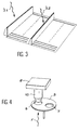
- Figure 3 shows a third example of device for coupling polarized light into a micro-channel structure according to the invention.
- the fluorescence of two separate markers is pictured.
- the structure of micro channels is then made, for example, of two elementary structures as represented in figure 2.
- a light of wavelength ⁇ 1 excites the markers contained in the micro channels of the first elementary structure and a wavelength light ⁇ 2 excites the markers contained in the micro channels of the second elementary structure.
- a structure as shown in Figure 3 is used, for example, in the reactions of genotyping for the study of polymorphism.
- a first marker is then chosen from fluoresceins (excitation wavelength ⁇ 1 substantially understood between 488nm and 514nm and emission wavelength substantially equal to 520nm) and the second marker is chosen from rhodamines (wavelength excitation ⁇ 2 substantially between 530nm and 550nm and substantially equal emission wavelength at 580nm).
- the first marker is FAM and the second marker is TAMRA.
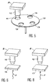
- Figure 4 shows a first example of polarized fluorescent light reading device according to the invention.
- Optics 10 and polarizing filters 6 and 7 allow the N micro channels to be imaged in parallel on a CCD camera (CCD for "Charge Coupled Device”).
- the polarizing filters 6 and 7 are mounted on a filter wheel 9.
- the fluorescence light F coming from the N micro channels is then detected.
- the image of the N micro channels is carried out, firstly, in a first direction of polarization, then, in the direction perpendicular to the first direction of polarization. We thus obtain, channel by channel, the two intensity values I
- Figure 5 shows a second example of polarized fluorescent light reading device according to the invention.
- two different markers are imaged. It can be, for example, R110 and Tamra as mentioned above.
- the device comprises a lens 10, a CCD camera 8 and four polarizing filters 11, 12, 13 and 14 mounted on a filter wheel 15. Filters 11 and 12 respectively filter the vertical polarization and the horizontal polarization of the fluorescent light coming from a first marker and the filters 13 and 14 respectively filter the vertical polarization and the horizontal polarization of the fluorescent light coming from the second marker.
- Tamra and I ⁇ Tamra are then successively measured by the camera 8.
- the filter wheel 15 is switched in synchronization with the two excitation laser beams (not shown in the figure) which successively illuminate the micro channels.
- Figure 6 shows a third example of polarized fluorescent light reading device according to the invention.
- the reading device comprises means for allowing, for each marker, the simultaneous measurement of the intensities I
- the vertical and horizontal polarizations are then separated and projected onto two distinct areas of the camera.
- the two polarizations which are contained in the fluorescent light F are separated by a birefringent crystal 16, for example a LiNbO 3 bar.
- a first image is then formed in a first color (first marker) and a second image is formed in a second color (second marker). The images are formed successively, following the switching of the excitation laser beams.
- the device can operate with a single color (in this case, a single marker is used in order to detect a type of nucleic acid) but also with three or even four colors (in this case, the corresponding number of markers is used).
- the fluorescence image obtained in a color is represented in FIG. 7.
- the pixels of the same pair have the respective intensities the intensities I
- Figure 8 shows a fourth example of fluorescent light reading device according to the invention.
- the reading device includes means for forming a fluorescence image of the chip, simultaneously in both polarizations and in both colors.
- the two colors are separated into two separate areas.
- the separation of colors is done by shifting the two beams laser which illuminate the structure of micro channels.
- a birefringent crystal for example a calcite crystal 17 is interposed between the structure micro channels and objective 10. Calcite crystal 17 allows polarization separation.
- each image Cj consists of a succession of pairs of pixels, each pair of pixels representing the same micro-channel area, the pixels of the same pair having the respective intensities the intensities I
Landscapes
- Health & Medical Sciences (AREA)
- Nuclear Medicine, Radiotherapy & Molecular Imaging (AREA)
- Physics & Mathematics (AREA)
- Life Sciences & Earth Sciences (AREA)
- Chemical & Material Sciences (AREA)
- Analytical Chemistry (AREA)
- Biochemistry (AREA)
- General Health & Medical Sciences (AREA)
- General Physics & Mathematics (AREA)
- Immunology (AREA)
- Pathology (AREA)
- Investigating, Analyzing Materials By Fluorescence Or Luminescence (AREA)
Abstract
Description
- "Fluorescence Anisotropy : Rapid, Quantitative Assay for Protein-DNA and Protein-Protein Interaction" de Tomasz Heyduk, Yuexing Ma, Hong Tang et Richard H. Ebright, Methods in Enzymology, vol. 274, et
- "DNA detection by strand displacement amplification and fluorescence polarization with signal enhancement using a DNA binding protein" de G.Terrance Walker, G.Preston Linn et James G. Nadeau, Nucleic Acids Research, 1996, vol 24, n°2.
- le brevet EP 0 382 433 B1 intitulé "Detection of nucleic acid séquences using fluorescence polarisation", et
- le brevet EP 0 678 581 A1 intitulé "Fluorescence polarization detection of nucleic acid amplification".
- la figure 1 représente un premier exemple de dispositif de couplage de lumière polarisée dans une structure de micro canaux selon l'invention ;
- la figure 2 représente un deuxième exemple de dispositif de couplage de lumière polarisée dans une structure de micro canaux selon l'invention ;
- la figure 3 représente un troisième exemple de dispositif de couplage de lumière polarisée dans une structure de micro canaux selon l'invention ;
- la figure 4 représente un premier exemple de dispositif de lecture de lumière fluorescente polarisée selon l'invention;
- la figure 5 représente un deuxième exemple de dispositif de lecture de lumière fluorescente polarisée selon l'invention,
- la figure 6 représente un troisième exemple de dispositif de lecture de lumière fluorescente polarisée selon l'invention ;
- la figure 7 représente, de façon symbolique, une image de fluorescence selon l'invention obtenue à l'aide d'un dispositif tel que le dispositif de la figure 6 ;
- la figure 8 représente un quatrième exemple de dispositif de lecture de lumière fluorescente polarisée selon l'invention ;
- la figure 9 représente, de façon symbolique, une image de fluorescence selon l'invention obtenue à l'aide d'un dispositif tel que le dispositif de la figure 8.
- taille de la barrette CCD : 1024 x 60 (24,6 x 1,44mm2) ;
- taille des pixels de détection de la barrette CCD : 24 x 24 µm2 ;
- micro canaux : largeur 200µm, pas 400µm, encombrement total selon l'axe des micro canaux 40mm, zone de fluorescence selon l'axe des micro canaux 1mm,
- grossissement 0,5 ;
- champ d'image sur la caméra # 30s ;
- nombre de pixels par micro canal : 4 x 20 ;
- ouverture numérique de l'optique : 0,1 avec une focale de 10mm ouverte à f/2
- puissance de la lumière laser par micro canal : 100µW.
Claims (12)
- Dispositif d'imagerie par fluorescence comprenant des premiers moyens pour contenir des constituants à analyser, des deuxièmes moyens pour éclairer par une lumière polarisée les constituants à analyser et des troisièmes moyens pour lire une lumière de fluorescence émise par les constituants sous l'action de la lumière polarisée, caractérisé en ce que les premiers moyens sont constitués d'une structure de micro canaux en parallèle (4) et en ce que les deuxièmes moyens comprennent au moins un dispositif de couplage (2, 5) pour guider la lumière polarisée dans les micro canaux.
- Dispositif selon la revendication 1, caractérisé en ce que les micro canaux sont gravés dans une puce support en verre ou en plastique présentant une bonne qualité optique ou en silicium.
- Dispositif selon la revendication 1, caractérisé en ce que les micro canaux sont des capillaires souples.
- Dispositif selon l'une quelconque des revendications précédentes caractérisé en ce que le dispositif de couplage comprend un réseau de diffraction (5).
- Dispositif selon l'une quelconque des revendications 1 à 3, caractérisé en ce que le dispositif de couplage comprend une lentille cylindrique (2).
- Dispositif selon l'une quelconque des revendications précédentes, caractérisé en ce que les deuxièmes moyens comprennent un laser ou un micro laser pour éclairer la totalité de la structure de micro canaux (4) et en ce que les troisièmes moyens comprennent un premier filtre polarisant (6, 11, 13) pour filtrer, dans un premier temps, une première composante de la lumière fluorescente polarisée selon une première direction et un deuxième filtre polarisant (7, 12, 14) pour filtrer, dans un deuxième temps, une deuxième composante de la lumière fluorescente polarisée selon une direction perpendiculaire à la première direction.
- Dispositif selon la revendication 6, caractérisé en ce qu'il comprend une roue à filtres (9, 15) pour commuter le premier filtre (6, 11, 13) et le deuxième filtre (7, 12, 14).
- Dispositif selon l'une quelconque des revendications 1 à 5, caractérisé en ce que les deuxièmes moyens comprennent un laser ou micro laser pour éclairer la totalité de la structure de micro canaux (4) et en ce que les troisièmes moyens comprennent un cristal biréfringent (16, 17) pour séparer la lumière de fluorescence émise selon deux composantes polarisées perpendiculairement l'une par rapport à l'autre.
- Dispositif selon l'une quelconque des revendications 6 à 8, caractérisé en ce que le laser ou le micro laser émet à une longueur d'onde (λ1) sensiblement comprise entre 488nm et 514nm ou à une longueur d'onde (λ2) sensiblement comprise entre 550nm et 580nm.
- Dispositif selon l'une quelconque des revendications 1 à 5, caractérisé en ce que les deuxièmes moyens comprennent un premier laser ou micro laser pour éclairer une première zone de la structure de micro canaux (4) et un deuxième micro laser pour éclairer, simultanément, une deuxième zone de la structure de micro canaux (4) et en ce que les troisièmes moyens comprennent un cristal biréfringent (16, 17) pour séparer la lumière de fluorescence émise selon deux composantes polarisées perpendiculairement l'une par rapport à l'autre.
- Dispositif selon la revendication 10, caractérisé en ce que le premier laser ou micro laser émet à une longueur d'onde (λ1) sensiblement comprise entre 488nm et 514nm et le deuxième micro laser émet à une longueur d'onde (λ2) sensiblement comprise entre 530nm et 550nm.
- Dispositif selon l'une quelconque des revendications 8 à 11, caractérisé en ce que le cristal biréfringent est un cristal de LiNbO3 ou un cristal de calcite.
Applications Claiming Priority (2)
| Application Number | Priority Date | Filing Date | Title |
|---|---|---|---|
| FR0010426A FR2812942B1 (fr) | 2000-08-08 | 2000-08-08 | Dispositif d'imagerie de fluorescence en lumiere polarisee |
| FR0010426 | 2000-08-08 |
Publications (1)
| Publication Number | Publication Date |
|---|---|
| EP1180680A1 true EP1180680A1 (fr) | 2002-02-20 |
Family
ID=8853393
Family Applications (1)
| Application Number | Title | Priority Date | Filing Date |
|---|---|---|---|
| EP01402118A Withdrawn EP1180680A1 (fr) | 2000-08-08 | 2001-08-06 | Dispositif d'imagerie de fluorescene en lumière polarisée |
Country Status (4)
| Country | Link |
|---|---|
| US (1) | US20020041378A1 (fr) |
| EP (1) | EP1180680A1 (fr) |
| JP (1) | JP2002072142A (fr) |
| FR (1) | FR2812942B1 (fr) |
Families Citing this family (21)
| Publication number | Priority date | Publication date | Assignee | Title |
|---|---|---|---|---|
| US7968287B2 (en) | 2004-10-08 | 2011-06-28 | Medical Research Council Harvard University | In vitro evolution in microfluidic systems |
| WO2007081385A2 (fr) | 2006-01-11 | 2007-07-19 | Raindance Technologies, Inc. | Dispositifs microfluidiques et leurs procédés d'utilisation dans la formation et le contrôle de nanoréacteurs |
| WO2007133710A2 (fr) | 2006-05-11 | 2007-11-22 | Raindance Technologies, Inc. | Dispositifs microfluidiques et leurs procédés d'utilisation |
| US9562837B2 (en) | 2006-05-11 | 2017-02-07 | Raindance Technologies, Inc. | Systems for handling microfludic droplets |
| GB2444961A (en) * | 2006-12-22 | 2008-06-25 | Thales Holdings Uk Plc | Imager with detector elements sensitive to radiation of different polarisations |
| WO2008097559A2 (fr) | 2007-02-06 | 2008-08-14 | Brandeis University | Manipulation de fluides et de réactions dans des systèmes microfluidiques |
| WO2008130623A1 (fr) | 2007-04-19 | 2008-10-30 | Brandeis University | Manipulation de fluides, composants fluidiques et réactions dans des systèmes microfluidiques |
| EP4047367A1 (fr) | 2008-07-18 | 2022-08-24 | Bio-Rad Laboratories, Inc. | Procedé de détection d'analytes cibles au moyens des bibliothèques de gouttelettes |
| US12038438B2 (en) | 2008-07-18 | 2024-07-16 | Bio-Rad Laboratories, Inc. | Enzyme quantification |
| WO2010111231A1 (fr) | 2009-03-23 | 2010-09-30 | Raindance Technologies, Inc. | Manipulation de gouttelettes microfluidiques |
| US10351905B2 (en) | 2010-02-12 | 2019-07-16 | Bio-Rad Laboratories, Inc. | Digital analyte analysis |
| US9399797B2 (en) | 2010-02-12 | 2016-07-26 | Raindance Technologies, Inc. | Digital analyte analysis |
| EP4435111A1 (fr) | 2010-02-12 | 2024-09-25 | Bio-Rad Laboratories, Inc. | Analyse numérique d'analyte |
| WO2012045012A2 (fr) | 2010-09-30 | 2012-04-05 | Raindance Technologies, Inc. | Dosages sandwich dans des gouttelettes |
| EP3859011A1 (fr) | 2011-02-11 | 2021-08-04 | Bio-Rad Laboratories, Inc. | Procédés permettant de former des gouttelettes mélangées |
| WO2012112804A1 (fr) | 2011-02-18 | 2012-08-23 | Raindance Technoligies, Inc. | Compositions et méthodes de marquage moléculaire |
| EP2714970B1 (fr) | 2011-06-02 | 2017-04-19 | Raindance Technologies, Inc. | Quantification d'enzyme |
| US8658430B2 (en) | 2011-07-20 | 2014-02-25 | Raindance Technologies, Inc. | Manipulating droplet size |
| US11901041B2 (en) | 2013-10-04 | 2024-02-13 | Bio-Rad Laboratories, Inc. | Digital analysis of nucleic acid modification |
| US9944977B2 (en) | 2013-12-12 | 2018-04-17 | Raindance Technologies, Inc. | Distinguishing rare variations in a nucleic acid sequence from a sample |
| US10647981B1 (en) | 2015-09-08 | 2020-05-12 | Bio-Rad Laboratories, Inc. | Nucleic acid library generation methods and compositions |
Citations (4)
| Publication number | Priority date | Publication date | Assignee | Title |
|---|---|---|---|---|
| US4122348A (en) * | 1976-05-21 | 1978-10-24 | Elscint, Ltd. | Method of and apparatus for classifying biological cells |
| WO1997010056A2 (fr) * | 1995-09-12 | 1997-03-20 | Becton Dickinson And Company | Dispositif et procede d'amplification et de dosage d'adn |
| WO1999040415A1 (fr) * | 1998-02-05 | 1999-08-12 | Novartis Ag | Procede et dispositif de mesure de la luminescence |
| EP0959343A1 (fr) * | 1998-05-22 | 1999-11-24 | C.S.E.M. Centre Suisse D'electronique Et De Microtechnique Sa | Capteur optique utilisant une réaction immunologique et un marqueur fluorescent |
Family Cites Families (4)
| Publication number | Priority date | Publication date | Assignee | Title |
|---|---|---|---|---|
| US5560811A (en) * | 1995-03-21 | 1996-10-01 | Seurat Analytical Systems Incorporated | Capillary electrophoresis apparatus and method |
| WO1996035940A1 (fr) * | 1995-05-12 | 1996-11-14 | Novartis Ag | Plateforme de detection et procede de detection parallele d'une pluralite d'analytes a l'aide de la luminescence excitee par evanescence |
| US6160618A (en) * | 1998-06-19 | 2000-12-12 | Board Of Regents, The University Of Texas System | Hyperspectral slide reader |
| US6455861B1 (en) * | 1998-11-24 | 2002-09-24 | Cambridge Research & Instrumentation, Inc. | Fluorescence polarization assay system and method |
-
2000
- 2000-08-08 FR FR0010426A patent/FR2812942B1/fr not_active Expired - Fee Related
-
2001
- 2001-07-27 US US09/915,514 patent/US20020041378A1/en not_active Abandoned
- 2001-08-03 JP JP2001236615A patent/JP2002072142A/ja not_active Withdrawn
- 2001-08-06 EP EP01402118A patent/EP1180680A1/fr not_active Withdrawn
Patent Citations (4)
| Publication number | Priority date | Publication date | Assignee | Title |
|---|---|---|---|---|
| US4122348A (en) * | 1976-05-21 | 1978-10-24 | Elscint, Ltd. | Method of and apparatus for classifying biological cells |
| WO1997010056A2 (fr) * | 1995-09-12 | 1997-03-20 | Becton Dickinson And Company | Dispositif et procede d'amplification et de dosage d'adn |
| WO1999040415A1 (fr) * | 1998-02-05 | 1999-08-12 | Novartis Ag | Procede et dispositif de mesure de la luminescence |
| EP0959343A1 (fr) * | 1998-05-22 | 1999-11-24 | C.S.E.M. Centre Suisse D'electronique Et De Microtechnique Sa | Capteur optique utilisant une réaction immunologique et un marqueur fluorescent |
Also Published As
| Publication number | Publication date |
|---|---|
| FR2812942B1 (fr) | 2002-10-31 |
| US20020041378A1 (en) | 2002-04-11 |
| FR2812942A1 (fr) | 2002-02-15 |
| JP2002072142A (ja) | 2002-03-12 |
Similar Documents
| Publication | Publication Date | Title |
|---|---|---|
| FR2812942A1 (fr) | Dispositif d'imagerie de fluorescence en lumiere polarisee | |
| US6320196B1 (en) | Multichannel high dynamic range scanner | |
| JP4783931B2 (ja) | 検出器の分光学的及び空間分解能を増大させるための方法 | |
| US6665072B2 (en) | Instantaneous dual band fluorescence detection systems | |
| US6455861B1 (en) | Fluorescence polarization assay system and method | |
| US7518728B2 (en) | Method and instrument for collecting fourier transform (FT) Raman spectra for imaging applications | |
| US7414725B2 (en) | Method and apparatus for a microscope image selector | |
| US7196339B2 (en) | Light-receiving unit and measuring apparatus including the same | |
| US20110068007A1 (en) | Multi-wavelength fluorescence detection system for multiplexed capillary electrophoresis | |
| Hanley et al. | Optical sectioning fluorescence spectroscopy in a programmable array microscope | |
| Ghosh et al. | Dynamic imaging of homo-FRET in live cells by fluorescence anisotropy microscopy | |
| EP3704471B1 (fr) | Appareil et procede de microscopie a fluorescence a super-resolution et de mesure de temps de vie de fluorescence | |
| US20130087718A1 (en) | Confocal fluorescence lifetime imaging system | |
| Naredi‐Rainer et al. | Confocal microscopy | |
| Cardullo | Theoretical principles and practical considerations for fluorescence resonance energy transfer microscopy | |
| Cardullo | Theoretical principles and practical considerations for fluorescence resonance energy transfer microscopy | |
| EP3724709B1 (fr) | Microscopie 3d spectrale en champ large | |
| JP5190773B2 (ja) | 創薬スクリーニング装置 | |
| Cardullo et al. | Fluorescence resonance energy transfer microscopy: theory and instrumentation | |
| Li et al. | A versatile optical microscope for time-dependent single-molecule and single-particle spectroscopy | |
| Elson et al. | Wide-field fluorescence lifetime imaging with optical sectioning and spectral resolution applied to biological samples | |
| EP2488854B1 (fr) | Procede et systeme d'imagerie par fonctionnalisation du substrat | |
| US7701572B2 (en) | System and method for chemical imaging of microarrays | |
| CN2811990Y (zh) | 高灵敏度光镊拉曼光谱仪 | |
| US6108094A (en) | Ultra-minute microscope for spectroscopy |
Legal Events
| Date | Code | Title | Description |
|---|---|---|---|
| PUAI | Public reference made under article 153(3) epc to a published international application that has entered the european phase |
Free format text: ORIGINAL CODE: 0009012 |
|
| AK | Designated contracting states |
Kind code of ref document: A1 Designated state(s): AT BE CH CY DE DK ES FI FR GB GR IE IT LI LU MC NL PT SE TR Kind code of ref document: A1 Designated state(s): DE GB IT |
|
| AX | Request for extension of the european patent |
Free format text: AL;LT;LV;MK;RO;SI |
|
| RIN1 | Information on inventor provided before grant (corrected) |
Inventor name: CAMPAGNOLO, RAYMOND Inventor name: PELTIE, PHILIPPE Inventor name: BERGER, MICHEL |
|
| 17P | Request for examination filed |
Effective date: 20020810 |
|
| AKX | Designation fees paid |
Free format text: AT BE CH LI |
|
| RBV | Designated contracting states (corrected) |
Designated state(s): DE GB IT |
|
| REG | Reference to a national code |
Ref country code: DE Ref legal event code: 8566 |
|
| STAA | Information on the status of an ep patent application or granted ep patent |
Free format text: STATUS: THE APPLICATION IS DEEMED TO BE WITHDRAWN |
|
| 18D | Application deemed to be withdrawn |
Effective date: 20060301 |