EP1147421B1 - Method for diagnosing endometriosis - Google Patents
Method for diagnosing endometriosis Download PDFInfo
- Publication number
- EP1147421B1 EP1147421B1 EP00901009A EP00901009A EP1147421B1 EP 1147421 B1 EP1147421 B1 EP 1147421B1 EP 00901009 A EP00901009 A EP 00901009A EP 00901009 A EP00901009 A EP 00901009A EP 1147421 B1 EP1147421 B1 EP 1147421B1
- Authority
- EP
- European Patent Office
- Prior art keywords
- cd45ro
- cd45ra
- hladr
- endometriosis
- markers
- Prior art date
- Legal status (The legal status is an assumption and is not a legal conclusion. Google has not performed a legal analysis and makes no representation as to the accuracy of the status listed.)
- Expired - Lifetime
Links
Images
Classifications
-
- G—PHYSICS
- G01—MEASURING; TESTING
- G01N—INVESTIGATING OR ANALYSING MATERIALS BY DETERMINING THEIR CHEMICAL OR PHYSICAL PROPERTIES
- G01N33/00—Investigating or analysing materials by specific methods not covered by groups G01N1/00 - G01N31/00
- G01N33/48—Biological material, e.g. blood, urine; Haemocytometers
- G01N33/50—Chemical analysis of biological material, e.g. blood, urine; Testing involving biospecific ligand binding methods; Immunological testing
- G01N33/53—Immunoassay; Biospecific binding assay; Materials therefor
- G01N33/574—Immunoassay; Biospecific binding assay; Materials therefor for cancer
- G01N33/57407—Specifically defined cancers
- G01N33/57442—Specifically defined cancers of the uterus and endometrial
-
- G—PHYSICS
- G01—MEASURING; TESTING
- G01N—INVESTIGATING OR ANALYSING MATERIALS BY DETERMINING THEIR CHEMICAL OR PHYSICAL PROPERTIES
- G01N2333/00—Assays involving biological materials from specific organisms or of a specific nature
- G01N2333/435—Assays involving biological materials from specific organisms or of a specific nature from animals; from humans
- G01N2333/705—Assays involving receptors, cell surface antigens or cell surface determinants
-
- G—PHYSICS
- G01—MEASURING; TESTING
- G01N—INVESTIGATING OR ANALYSING MATERIALS BY DETERMINING THEIR CHEMICAL OR PHYSICAL PROPERTIES
- G01N2800/00—Detection or diagnosis of diseases
- G01N2800/36—Gynecology or obstetrics
- G01N2800/364—Endometriosis, i.e. non-malignant disorder in which functioning endometrial tissue is present outside the uterine cavity
Definitions
- the invention relates to a method for the diagnosis of endometriosis using blood and endometrial leukocyte markers.
- Endometriosis is one of the most common gynecological disorders, affecting up to 15% of women within reproductive age. It is closely associated with severe pelvic pain, dysmenorrhea, dyspareunia, infertility and several other symptoms such as intraperitoneal bleeding, back pain, constipation and/or diarrhea. It is a major threat to physical, psychological and social integrity of the patients.
- Endometriosis is characterized by the implantation and growth of endometrial cells (which normally constitute the lining of the uterus) in extra-uterine sites such as the peritoneal cavity.
- endometrial cells which normally constitute the lining of the uterus
- the theory of retrograde menstruation is the most widely accepted to explain the presence of ectopic endometrial cells in the peritoneal cavity.
- this phenomenon occurs in most women and, thus, several other factors must be invoked to explain the implantation of endometrial cells and the subsequent development of endometriotic lesions. It is generally believed that initiation of endometriosis implies a complex cascade of events requiring several essential features.
- Retrogradely seeded endometrial cells must remain viable, be capable of adhering to the mesothelium and of proliferating. Local degradation of the extracellular matrix, as well as extensive vascularization, are also believed to play an essential role in promoting the invasion of the peritoneal cavity by endometrial cells. Furthermore, once implanted, ectopic endometrial cells must have the capacity to counteract the cytolytic action of the immune system. Indeed, this is supported by the observation of several immunological abnormalities in patients with endometriosis.
- endometriosis which can involve microlesions
- endometriosis can be hardly diagnosed by surgical methods, as they are unlikely to be detected by direct visualization. Indeed, several studies have reported microscopic endometriotic lesions that were not detected laparoscopically. Because the diagnosis of endometriosis by surgical procedures is difficult, costly and invasive, in some cases, several physicians and patients tend to avoid it or at least seriously delay it. Hence, the length of time between the onset of symptoms and the diagnosis can be as long as 8 to 12 years. The possibility to diagnose endometriosis at an early stage would certainly improve the efficacy of the treatments, and reduce dramatically the number of years during which patients endure acute or chronic pain.
- Imaging methods such as transvaginal ultrasound and magnetic resonance imaging have been designed for the diagnosis of endometriosis.
- these techniques can only be reliable for the detection of large (> 1 cm diameter) endometriomas lesions detected among a very small proportion of patients with endometriosis.
- the high cost of these techniques has limited their use for the diagnosis of endometriosis.
- Serum proteins such as CA-125 and placental protein-14 have been proposed as diagnostic markers for endometriosis. Elevated levels of CA-125 have been observed in serum, menstrual effluent and peritoneal fluid of patients with endometriosis. However, these markers, when used alone, are of very limited value for a diagnosis test. Indeed, these markers are not suitable for screening or diagnostic purposes because they provide poor sensitivity. Furthermore, levels of CA-125 and placental protein-14 vary according to several factors such as the assay, the stage of the disease and the menstrual cycle. Finally these markers are known to be modulated by conditions other than endometriosis.
- T lymphocyte subpopulations CD8+, CD45+/HLADR+, CD45+/CD3+/HLADR+ or CD3+/CD25+
- ICAM-1, LFA-1, CD2 or CD68+ cells were upregulated in the endometrium of patients with endometriosis compared with infertile controls.
- T lymphocyte subsets Klentzeris L.D., et al., Eur. J Obstet gynecol Reprod Biol., 63:41-47, 1995 ; Jones R.K., et al., Fertil Steril, 66:81-89, 1996 ).
- One aim of the present invention is to provide a less invasive, cheaper and reliable method that could allow detection of females suffering from endometriosis as early as possible.
- a method for determining likelihood of endometriosis in a sample of eutopic uterine endometrial tissues or a sample of blood of a female subject characterized in that it comprises the steps of:
- a method for determining endometriosis in a sample of eutopic uterine endometrial tissues or a sample of blood of a female subject comprising:
- the following symbol "/" is intended to mean a ratio between an expression in front of the symbol and another expression after the symbol.
- the present invention identifies a series of leukocyte subsets that can be used as markers in a diagnostic test for endometriosis. These leukocyte subsets are defined according to the expression of cell surface antigens. Several cell surface antigens may define the same population of cells, and thus they are included in the present invention.
- any other antibodies or molecules recognizing the same antigen or a different epitope, isoform, subunit, chain, glycosylation or phosphorylation form or an allelic variant of the same antigen, a member of the same complex, or an antigen with the same cell distribution is also included in the present invention.
- CD3+( ⁇ 40%) >40% 75% 73% CD3+CD8+ ( ⁇ 16%) CD13+CD45RO- ( ⁇ 21%) 21.
- CD3+ ( ⁇ 40%) >25% 71% 69% CD5+ ( ⁇ 40%) CD3+CD5+ ( ⁇ 37%) CD69+ ( ⁇ 33%) CD4-CD69+ ( ⁇ 35%) 22.
- CD3+ ( ⁇ 40%) >30% 68% 83% CD3+CD8+ ( ⁇ 13.5%) CD13+CD45RO- ( ⁇ 17.5%) 23.
- CD3+ ( ⁇ 40%) >30% 61% 86% CD3+CD8+ ( ⁇ 16%) CD13+CD45RO- ( ⁇ 17.5%) 24.
- CD13+CD45RO- ( ⁇ 17.5%) >70% 90% 54% CD4+CD45RA- ( ⁇ 16%) CD3+CD122- (42.5%) CD8+CD69- ( ⁇ 21%) 30.
- CD3+CD8+ ( ⁇ 16%) >70% 84% 60% CD13+CD45RO- ( ⁇ 17.5%) CD4+CD45RA- ( ⁇ 16%) CD3+CD45RO- ( ⁇ 30%) CD3-CD5- (>54%)
- Blood leukocyte markers 31.
- CD3-CD5+ (>14.5%) >55% 66% 60% CD3-CD45RA- (>14.5%) CD3-CD44+ (>13%) CD13+ (>17.5%) CD3-CD57-CD44- ( ⁇ 41.3%) 32.
- CD3-CD45RA- >17%) >22% 61% 64% CD20-CD44+ (>17%) CD20-HLADR+ (>20%) CD3-CD4-CD44+ (>40.5%) CD36-HLADR+ ( ⁇ 5.6%) 33.
- CD3-CD45RA- >14.5%
- CD3-CD45RO+ >19%) CD20-HLADR+ (>14.5%) Blood (in Italics) and endometrial leukocyte markers 34.
- CD57+ > 10%
- CD3-CD69+ > 17.5 %)
- CD3+ ⁇ 40%)
- CD4+ ⁇ 15.5%
- CD3+CD8+HLADR- ⁇ 35%)
- CD3-CD69+ >17.5%
- CD4+ ⁇ 15.5%
- CD3+CD8+HLADR- ⁇ 35%)
- CD4-CD36+ >14.4%
- CD3-CD69+ >17.5%
- CD8+ ⁇ 20%)
- CD13+ >29%)
- CD3+ ⁇ 40%)
- CD16+ >27%)
- CD69+( ⁇ 33%) CD5+ ⁇ 40%)
- CD3-CD45RA- >14.5%
- CD14+CD44+ >15%)
- CD8+ ⁇ 20%)
- CD5+ ⁇ 37%)
- CD3-CD20- >58%)
- CD3-HLADR- >54.5%
- CD3+ ( ⁇ 40%) 2 -7.9747 >.55 83% 79% 41 2.
- CD3-CD20- (>58.%) 9.5142 Interaction of 1 to 4 Constant 2.0516 Combination no. 2 1.
- CD3-CD5- (>60%) 5.8240 3. CD13+CD45RO- -1.9298 ( ⁇ 17.5%) -0.0262 Interaction 1 to 4 2.8385 Constant 2.7910 Combination no. 3 1.
- CD3+CD8+ ( ⁇ 16%) -0.1308 >.50 84% 72% 51 2.
- CD13+CD45RO- -2.6688 ( ⁇ 17.5%) -1.1778 3.
- Length of menstruation -1.8160 (>7days) -1.9656 3.
- CD13+CD20- ( ⁇ 21%) 10.3064 4.
- Pelvic pain 3 Constant 3.1984 Blood leukocyte markers Combination no. 1 1. CD14+CD44+ (>15%) 0.9298 >0.55 80% 70% 140 2.
- CD57+ (>10%) 0.7423 3.
- stage I-IV;I-II or III-IV endometrial and peripheral blood leukocyte subpopulations for which proportions were modulated in patients with endometriosis
- stage I-IV;I-II or III-IV endometrial and peripheral blood leukocyte subpopulations for which proportions were modulated in patients with endometriosis
- stage I-IV;I-II or III-IV endometrial and peripheral blood leukocyte subpopulations for which proportions were modulated in patients with endometriosis
- stage I-IV;I-II or III-IV endometriosis
- risk factors for endometriosis identified amongst personal information and menstrual characteristics were shown to be of significant value when use in combination with blood or endometrial leukocyte subsets in a predictive test for endometriosis.
- a cutoff point is established for the proportion of each combination of leukocyte markers in order to obtain the best discrimination between patients with endometriosis and controls.
- the proportion obtained for each marker is compared to the cutoff point.
- a positive test result gives a score of 1
- a negative test result gives a score of 0.
- the diagnostic value is obtained by adding the scores of all the markers of a particular combination and converting it in percentage.
- the final diagnostic value is then compared to a threshold value that was established to provide the best levels of sensibility and specificity.
- a positive diagnosis of endometriosis is given when the final diagnostic value exceeds the threshold value established for a particular combination of markers.
- a negative diagnosis of endometriosis is given when the final diagnostic value is lower than the threshold value (see Fig. 1).
- a method for determining endometriosis in a sample of eutopic uterine endometrial tissues or a sample of blood of a female subject comprising: a) measuring in said sample a quantitative level of at least two different surface antigens from blood leukocytes and/or eutopic endometrial leukocytes, said surface antigens from blood leukocytes and/or eutopic endometrial leukocytes being selected from the leukocyte marker combinations defined in the following Table: Leukocyte Marker Combinations Endometrial leukocyte markers CD4+, CD8+CD69-, and CD13+CD45RO- CD4+, CD8+CD69-, CD56+CD122-, CD3+CD45RA-, and CD13+CD45RO-, CD4+, CD8+CD69-, CD13-CD122+, and CD13+CD45RO- CD4+ CD8+CD69-, and D14+CD13
- P r ⁇ e c + B ⁇ 1 * ( marker 1 ) + B ⁇ 2 ( marker 2 ) + ... Bn marker n
- the probability of having endometriosis is then compared to a threshold value that provides the best discriminative value.
- a positive diagnosis of endometriosis is given when the P(r) value exceeds the threshold value established for a particular combination of markers.
- a negative diagnosis of endometriosis is given when the P(r) value is lower than the threshold value.
- a method for determining endometriosis in a sample of eutopic uterine endometrial tissues or a sample of blood of a female subject comprising: a) measuring in said sample a quantitative level of at least two different surface antigens from blood leukocytes and/or eutopic endometrial leukocytes, said surface antigens from blood leukocytes and/or eutopic endometrial leukocytes being selected from the leukocyte marker combinations defined in the following Table: Leukocyte Marker Combinations Endometrial leukocyte markers CD4+, CD8+CD69-, and CD13+CD45RO- CD4+, CD8+CD69-, CD56+CD122-, CD3+CD45RA-, and CD13+CD45RO-, CD4+, CD8+CD69-, CD13-CD122+, and CD13+CD45RO- CD9+ CD8+CD69-, and D14+CD13-CD
- 37 72 0.462 ⁇ 35 68 46 1.6 (0.7-3.5) CD3-CD4-CD45RO+ 41.3 ⁇ 18.1 43.4 ⁇ 20.9 43
- Cutoff points established for each individual marker are presented in Table 3, 4, 5, 6 and threshold value established for a particular marker combination are presented in Table 1. Any other cutoff points or threshold values providing a valuable diagnostic test for endometriosis are meant to be included in the present application.
- endometrial leukocyte markers Tables 1 and 2
- 7 combinations of blood leukocyte markers Table 1
- 4 combination of endometrial and blood leukocyte markers providing a diagnostic test with levels of sensibility and specificity up to 89 and 90%, respectively.
- the different marker combinations of the present invention may serve several important clinical needs. Hence in the general population, these markers could be used to evaluate the risk factor to develop endometriosis or to identify women with high likelihood of suffering of the disease. Furthermore in patients with endometriosis, these markers could serve to monitor the disease or to give a prognosis.
- Uterine endometrial tissues were obtained from 146 subjects undergoing laparoscopy or laparotomy.
- the experimental group was formed of 88 subjects with endometriosis stage I-IV confirmed by laparoscopy or laparotomy and the control group consist of 58 healthy subjects who underwent surgery for tubal ligation (or reanastomosis) and had no clinical evidence, nor family history of endometriosis.
- Table 7 gives details concerning the age, menstrual cycle and indication of laparoscopy or laparotomy for the subjects included in experimental and control groups.
- Endometrial biopsies were taken with a Pipet Curette (Milex) (approximately 0.5g of tissue). All samples were harvested in the secretory phase (day 14-28) of the menstrual cycle as confirmed by histological evaluation. The samples were collected into sterile RPMI-1640 medium (Gibco) supplemented with 2% heat-inactivated fetal calf serum (Bio-Media) and 1% penicillin-streptomycin and kept at 4°C until cell isolation.
- Milex Pipet Curette
- Blood samples were obtained from 172 subjects with endometriosis (stage I-IV) confirmed by laparascopy or laparotomy and from 132 healthy subjects with no evidence of endometriosis at surgery, and no family history of endometriosis. Blood samples (30 ml) were collected in heparin-tubes (Vacutainer TM , Becton Dickinson) and kept at 20°C until mononuclear cell separation. The age, menstrual dating and indication for laparoscopy of the subjects included in the study are given in Table 8.
- Endometrial tissue samples were mechanically disrupted with a Pyrex TM glass Broeck tissue grinder (Fisher) to obtain a single cell suspension.
- Stromal cell fraction was isolated by filtration through a 250 ⁇ m stainless steel sieve (Millipore) to retain the glandular fraction and was washed twice with 10 ml phosphate buffered saline (PBS) (Sigma) containing 1% BSA (Boehringer Mannheim), 0.1% sodium azide (Fisher) (thereafter called PBS washing buffer).
- PBS phosphate buffered saline
- HBSS Hank's Balanced Salt Solution
- Ficoll-PaqueTM Ficoll-PaqueTM
- Leukocytes were isolated at the interface of Ficoll and HBSS and they were washed in 50 ml of HBSS.
- Contaminating red blood cells were lysed with 6 ml of ammonium chloride solution (0.15M) (6 minutes at room temperature). The peripheral blood mononuclear cells were then washed twice in 10 ml PBS and resuspended in PBS washing buffer.
- Endometrial stromal cells or peripheral blood mononuclear cells were distributed in 5 ml tubes (1 to 1.5 x 10 6 cells/tube) or in 96 well plates (5 x 10 5 cells/well), respectively and incubated in the presence of 0.1 ⁇ g of human ⁇ -globulin for 5 minutes at room temperature. The cells were then incubated 30 minutes in the dark (at room temperature for endometrial cells and at 4°C for peripheral blood mononuclear cells) with a panel of 4 different mouse monoclonal antibodies (MAbs) in a total volume of 100 ⁇ l.
- MAbs mouse monoclonal antibodies
- the cell samples were stained with mouse anti-human CD45 MAbs conjugated to peridinin chlorophyl protein (PerCP) and with several sets of three different mouse MAbs labeled with distinct fluorochromes (fluorescein isothiocyanate - FITC-, phycoerythrin -PE or with phycoerythrin-texas red -ECD-) directed toward cell surface markers for specific cell populations such as T lymphocytes, B lymphocytes, NK cells, macrophages and/or activation markers (Table 9).
- fluorochromes fluorescein isothiocyanate - FITC-, phycoerythrin -PE or with phycoerythrin-texas red -ECD-
- TABLE 10 Main distribution of antigens Antigen Main Cell Distribution
- CD3 Expressed on all mature T cells associated with TCR complex ( ⁇ /b, ⁇ / ⁇ )
- CD4 Expressed on T helper lymphocytes. It can be also expressed on cells of the monocyte/macrophage lineage CD5 Found on all mature T lymphocytes and a subset of B lymphocytes CD8 Found on a subset of T lymphocytes called suppressor/cytotoxic T cells.
- CD13 Detected on most cells of myeloid origin polymorphonuclear cells or cells of the monocyte/macrophage lineage.
- CD14 Expressed strongly on the surface of monocytes Found on most tissue macrophages Weakly expressed on the surface of granulocytes and B lymphocytes Receptor for lipopolysacharride-(LPS) and LPS binding protein CD16 Expressed mainly on NK cells, monocytes macrophages and polymorphonuclear leukocytes Low affinity receptor for IgG CD 16b Found on granulocytes including polymorphonuclear cells (PMN) CD20 Present on all B lymphocytes CD36 Expressed on platelets, monocytes or macrophages, microvascular endothelial cells, mammary endothelial cells, during stages of erythroid cell development CD44 Widely expressed on the surface of most cell types.
- PMN polymorphonuclear cells
- Activation marker detected early after cell activation CD122 Expressed on NK cells B, T lymphocytes or monocytes/macrophages Component of the IL-15 receptor HLADR+ HLA class II molecule Found on antigen presenting cells or on other cells upon activation such as T cells.
- the immunofluorescence reactivity was carried out on a Coulter EPICS XLTM flow cytometer (Coulter Corporation, Hialeah, FL) equipped with an argon laser operating at 488 nm, 15 mW and detectors at 525, 575, 610, and 675 nm. Calibration of the flow cytometer parameters for forward scatter, side scatter and fluorescence were the same for all the samples.
- Cells expressing CD45 pan leukocyte antigen were gated using the Coulter system II software. The percentage of cells bearing markers for T, B lymphocytes, macrophages or NK cells and/or activation markers was evaluated within the CD45 positive populations only. A minimum of 6000 CD45+ cells were analyzed for each sample.
- a cutoff point was established for the proportion of the endometrial or blood leukocyte subpopulations identified as diagnostic markers. The value obtained for each marker is compared to the cutoff point (Fig. 1). A positive result was given when the proportion of a particular leukocyte subset fulfills the condition established by the cutoff point (for example ⁇ 40% for CD3+ cells). When these markers are used in combination, a positive result for each marker gives a score of 1, whereas a negative result gives a score of 0.
- a diagnosis of endometriosis is given, when the final diagnostic score obtained from adding the results of all the markers of a particular combination is higher than a predetermined threshold value.
- the levels of sensibility and/or specificity measured for the marker combination represents the number of positive test results obtained among the patients already confirmed with endometriosis and the number of negative test results among the subjects within the control group, respectively.
- the probability of having endometriosis is then compared to a threshold value that provides the best discriminative value.
- a positive diagnosis of endometriosis is given when the P(r) value exceeds the threshold value established for a particular combination of markers.
- a negative diagnosis of endometriosis is given when the P(r) value is lower than the threshold value.
- Endometrial and blood leukocyte subsets defined as good potential markers for the diagnosis of endometriosis are presented in Tables 3 and 4 respectively. Selection of these markers was done on the basis of a significative difference in the mean proportion of leukocyte subsets between patients with endometriosis (stage I-IV) and control groups. In addition, several endometrial and blood markers were also selected according to the area under the ROC curve, an indication of the discriminative value of the markers. The ROC curve allowed the determination of one or more cutoff proportion that best discriminate between patients with endometriosis (stage I-IV) and normal controls.
- Table 1 gives a series of 33 combinations in which endometrial or blood leukocyte markers are used in a diagnostic test for endometriosis. For each marker, a positive test result (as described above) gives a score of 1, whereas a negative test result gives a score of 0. The final diagnostic value obtained from adding the scores of all the markers of a particular combination is then compared to a threshold value, which is indicated in Table 1.
- a diagnosis of endometriosis is given, when the diagnostic value exceeds the threshold value established for each set of combination markers-
- leukocyte marker subsets in combination in this new method clearly improves the levels of sensibility and/or specificity for diagnosing endometriosis.
- Table 1 also provides 4 examples showing that blood leukocyte markers, when used in combination with endometrial markers, can also increase the predictive value of the diagnostic test.
- the present invention also demonstrates that logistic regression models can also be used to combine endometrial as well as blood leukocyte markers for the development of a predictive model of endometriosis (Table 2).
- these models need to be adjusted with risk factors associated with endometriosis such as the length of the menstrual cycle, the duration of menstruation, pain during intercourse and age. In some instances, these factors were shown to increase the predictive value of the model.
- the present invention identifies several examples of marker combinations, which give rise to diagnostic methods yielding improved levels of sensibility and specificity. Indeed, the different marker combinations of the present invention may serve different clinical applications including screening, diagnosis, monitoring and prognosis of endometriosis.
Landscapes
- Health & Medical Sciences (AREA)
- Life Sciences & Earth Sciences (AREA)
- Immunology (AREA)
- Engineering & Computer Science (AREA)
- Chemical & Material Sciences (AREA)
- Hematology (AREA)
- Urology & Nephrology (AREA)
- Biomedical Technology (AREA)
- Molecular Biology (AREA)
- Food Science & Technology (AREA)
- Analytical Chemistry (AREA)
- Microbiology (AREA)
- Biotechnology (AREA)
- Hospice & Palliative Care (AREA)
- Reproductive Health (AREA)
- Oncology (AREA)
- Medicinal Chemistry (AREA)
- Physics & Mathematics (AREA)
- Cell Biology (AREA)
- Biochemistry (AREA)
- General Health & Medical Sciences (AREA)
- General Physics & Mathematics (AREA)
- Pathology (AREA)
- Investigating Or Analysing Biological Materials (AREA)
- Measuring Or Testing Involving Enzymes Or Micro-Organisms (AREA)
- Devices That Are Associated With Refrigeration Equipment (AREA)
- Measurement Of Mechanical Vibrations Or Ultrasonic Waves (AREA)
- Measuring Pulse, Heart Rate, Blood Pressure Or Blood Flow (AREA)
Abstract
Description
- The invention relates to a method for the diagnosis of endometriosis using blood and endometrial leukocyte markers.
- Endometriosis is one of the most common gynecological disorders, affecting up to 15% of women within reproductive age. It is closely associated with severe pelvic pain, dysmenorrhea, dyspareunia, infertility and several other symptoms such as intraperitoneal bleeding, back pain, constipation and/or diarrhea. It is a major threat to physical, psychological and social integrity of the patients.
- Endometriosis is characterized by the implantation and growth of endometrial cells (which normally constitute the lining of the uterus) in extra-uterine sites such as the peritoneal cavity. Although the etiology and pathogenesis of endometriosis remain mainly unclear, the theory of retrograde menstruation is the most widely accepted to explain the presence of ectopic endometrial cells in the peritoneal cavity. However, this phenomenon occurs in most women and, thus, several other factors must be invoked to explain the implantation of endometrial cells and the subsequent development of endometriotic lesions. It is generally believed that initiation of endometriosis implies a complex cascade of events requiring several essential features. Retrogradely seeded endometrial cells must remain viable, be capable of adhering to the mesothelium and of proliferating. Local degradation of the extracellular matrix, as well as extensive vascularization, are also believed to play an essential role in promoting the invasion of the peritoneal cavity by endometrial cells. Furthermore, once implanted, ectopic endometrial cells must have the capacity to counteract the cytolytic action of the immune system. Indeed, this is supported by the observation of several immunological abnormalities in patients with endometriosis.
- At present, direct visualization of the endometriotic lesions under surgical procedures (laparascopy or laparotomy) is the golden standard and the only reliable method available to diagnose endometriosis. However, this method is highly invasive (i.e. surgery under general anesthesia), costly (i.e. direct cost and indirect cost due to convalescence) and requires a well-trained surgeon who has the ability to identify endometriotic lesions with a variety of appearances. The type of lesions, their size and their localization will determine the stage of the disease (stage I minimal, stage II mild, stage III moderate, stage IV severe). However, there is still no clear consensus on how these parameters correlate with the stage of the disease and the prognostic of endometriosis. In addition, early or minimal endometriosis (which can involve microlesions) can be hardly diagnosed by surgical methods, as they are unlikely to be detected by direct visualization. Indeed, several studies have reported microscopic endometriotic lesions that were not detected laparoscopically. Because the diagnosis of endometriosis by surgical procedures is difficult, costly and invasive, in some cases, several physicians and patients tend to avoid it or at least seriously delay it. Hence, the length of time between the onset of symptoms and the diagnosis can be as long as 8 to 12 years. The possibility to diagnose endometriosis at an early stage would certainly improve the efficacy of the treatments, and reduce dramatically the number of years during which patients endure acute or chronic pain.
- Imaging methods such as transvaginal ultrasound and magnetic resonance imaging have been designed for the diagnosis of endometriosis. However, these techniques can only be reliable for the detection of large (> 1 cm diameter) endometriomas lesions detected among a very small proportion of patients with endometriosis. Moreover, the high cost of these techniques has limited their use for the diagnosis of endometriosis.
- Serum proteins such as CA-125 and placental protein-14 have been proposed as diagnostic markers for endometriosis. Elevated levels of CA-125 have been observed in serum, menstrual effluent and peritoneal fluid of patients with endometriosis. However, these markers, when used alone, are of very limited value for a diagnosis test. Indeed, these markers are not suitable for screening or diagnostic purposes because they provide poor sensitivity. Furthermore, levels of CA-125 and placental protein-14 vary according to several factors such as the assay, the stage of the disease and the menstrual cycle. Finally these markers are known to be modulated by conditions other than endometriosis.
- High concentrations of antibodies to endometrial antigens were found in the serum of patients with endometriosis, and thus were proposed as markers for a diagnostic test (
International patent application publications WO 94/28021 WO 92/18535 - In
U.S. patent No. 5,478,725 , low levels of αvβ3 integrin expression in endometrial samples during the secretory phase of the menstrual cycle is described as a predictor of endometriosis in infertile but not in fertile patients with endometriosis. This observation was associated with milder form of endometriosis (stages I and II) only and, thus, is not useful to detect advanced stages of the disease. Moreover, this method yielded a specificity of 91% but a very low sensitivity (38%). - Taking into account that a number immunological abnormalities have been reported in patients with endometriosis, it is conceivable that the proportion of leukocyte populations and/or their activation status may be modulated during the course of the disease and, thus, may provide some diagnostic value. Previous flow cytometric studies have shown that some T lymphocyte subpopulations (CD8+, CD45+/HLADR+, CD45+/CD3+/HLADR+ or CD3+/CD25+) can be slightly modulated in the peritoneal fluid of subjects with endometriosis relative to normal controls (Oosterlyncck D.J., et al., Am J reprod. Immunol., 31: 25-31, 1994; Becker J.L., et al., Am J Reprod. Immunol., 34: 179-187, 1995; Wu M.Y., et al., Am. j. Reprod. Immunol. 35: 510-516, 1996). However, these observations have limited value for the diagnosis of endometriosis because peritoneal fluid collection is an invasive, non-conventional procedure. Proportions of leukocyte populations have also been studied in peripheral blood and endometrium of patients with endometriosis. Wu et al., (supra) have reported a modest but significant decrease in the proportion of CD3+ T lymphocytes expressing either CD69 or CD25 activation marker in the blood of patients with advanced endometriosis but not in patients with mild stage of endometriosis or normal controls. This difference was observed in advanced cases of endometriosis only and it was too modest to be used as a diagnostic marker. In contrast, Oosterlynck et al., (Oosterlyncck D.J., et al., Am J reprod. Immunol., 31: 25-31, 1994) and Ho et al. (Ho H.N., et al., Hum Reprod., 97: 2528-2533, 1997) reported no significant difference in term of T lymphocyte subpopulations when comparing endometriosis subjects with normal controls. These inconsistent results may be explained by the very low number of samples tested in these studies.
- Several studies have investigated whether leukocytes are also modulated in eutopic endometrium from patients with endometriosis. Results arising from these studies are contradictory, probably due to the fact that in most cases the methods used were only semi-quantitative and the number of samples tested were very low. For instance, by means of immunohistochemistry, Ota et al. (Ota H., et al., Am J Reprod. Immunol., 35: 477-482, 1996) have reported that the number of CD3+, CD4+, or CD8+ T lymphocytes, cells bearing adhesion molecules (i.e. ICAM-1, LFA-1, CD2) or CD68+ cells were upregulated in the endometrium of patients with endometriosis compared with infertile controls. In contrast, several other studies using similar techniques have reported no difference in the proportion of T lymphocyte subsets (Klentzeris L.D., et al., Eur. J Obstet gynecol Reprod Biol., 63:41-47, 1995; Jones R.K., et al., Fertil Steril, 66:81-89, 1996). In addition, a decrease in CD3 positive T cells has been shown by flow cytometry analysis but no difference in the proportion of CD4+, CD8+ stromal leukocytes in the endometrium of patients with endometriosis compared with fertile controls. When these observations are tentatively used in a diagnostic test, they give only low levels of sensibility and specificity because of a significant overlap between the groups.
- Therefore, the diagnostic methods presented in the literature so far do not solve the problems encountered with the diagnosis of endometriosis by surgical procedures. It thus remains imperative to be provided with a less invasive, cheaper and reliable method that could allow detection of females suffering from endometriosis as early as possible.
- One aim of the present invention is to provide a less invasive, cheaper and reliable method that could allow detection of females suffering from endometriosis as early as possible.
- In accordance with the present invention there is provided a method for determining likelihood of endometriosis in a sample of eutopic uterine endometrial tissues or a sample of blood of a female subject, characterized in that it comprises the steps of:
- a) measuring in said sample a quantitative level of at least two different selected surface antigens from blood leukocytes and/or eutopic endometrial leukocytes, as defined in
claim 1 ; - b) establishing a cutoff value for each leukocyte marker in the combination;
- c) comparing the proportion obtained in step b) for each leukocyte marker to a predetermined cutoff value, wherein a positive result gives a score of 1 whereas a negative result gives a score of 0;
- d) obtaining a diagnostic value by adding the scores of all the markers of the combination and converting it in percentage; and
- e) comparing the final diagnostic value to an established threshold value wherein a a positive diagnosis of endometriosis is given when the final diagnostic value exceeds the threshold value established for the combination of leukocyte markers whereas a negative diagnosis of endometriosis is given when the final diagnostic value is lower than the threshold value established for the combination of leukocyte markers.
- Further, in accordance with the present invention, there is also provided a method for determining endometriosis in a sample of eutopic uterine endometrial tissues or a sample of blood of a female subject, comprising:
- a) measuring in said sample a quantitative level of at least two different selected surface antigens from blood leukocytes and/or eutopic endometrial leukocytes, as defined in claim 3;
- b) establishing a predictive model for endometriosis by including each marker of said combination in a logistic regression equation, as defined in claim 3;
- c) comparing the probability of having endometriosis to a threshold value wherein a positive diagnosis of endometriosis is given when the probability of having endometriosis value exceeds the threshold value established for said combination of markers whereas a negative diagnosis of endometriosis is given when the probability of having endometriosis value is lower than the threshold value established for said combination of markers.
- For the purpose of the present invention, the following symbol "/" is intended to mean a ratio between an expression in front of the symbol and another expression after the symbol.
-
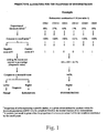
- Fig. 1 illustrates a predictive algorithm for the diagnosis of endometriosis.
- In accordance with the present invention, there is provided reliable diagnostic test for endometriosis that is less invasive and less costly than the actual surgical procedure accepted as the golden standard. An extensive study was undertaken by means of flow cytometric analysis, in which the proportion of several blood and endometrial leukocyte subsets was compared in patients with endometriosis and normal controls.
- The present invention identifies a series of leukocyte subsets that can be used as markers in a diagnostic test for endometriosis. These leukocyte subsets are defined according to the expression of cell surface antigens. Several cell surface antigens may define the same population of cells, and thus they are included in the present invention.
- Any other antibodies or molecules recognizing the same antigen or a different epitope, isoform, subunit, chain, glycosylation or phosphorylation form or an allelic variant of the same antigen, a member of the same complex, or an antigen with the same cell distribution is also included in the present invention.
- Further in accordance with the present invention, there is provided examples showing how at least two different surface antigens from blood and/or endometrial leukocytes can be used in combinations in a diagnostic test for endometriosis (Tables 1 and 2).
TABLE 1 Levels of sensibility and specificity provided by several examples of endometrial and/or blood marker combinations used as a diagnostic method for endometriosis Marker combination Threshold value 1 Specificity Sensibility Endometrial leukocyte markers 1. CD4+ (<17%)2 >67% 90% 67% CD8+CD69- (<21%) CD13+CD45RO- (<17.5%) 2. CD4+ (<15.5%) >60% 89% 65% CD8+CD69- (<21%) CD56+CD122- (>19%) CD3+CD45RA- (<35%) CD13+CD45RO- (<17.5%) 3. CD4+ (<17%) >67% 88% 65% CD8+CD69- (<21%) CD13-CD122+ (>28%) CD13+CD45RO- (<17.5%) 4. CD4+ (<17%) >67% 89% 63% CD8+CD69- (<21%) CD14+CD13-CD16b- (>14.5%) 5. CD3+CD16- (<40%) >55% 84% 62% CD13+CD45RO- (<13.5%) CD3+ (<40%) CD8+ (<20%) CD3+CD69+ (<15%) 6. CD3+ (<40%) >65% 84% 63% CD3+CD8+ (<16%) CD13+CD45RO- (<17.5%) CD3-CD20- (>56%) 7. CD3+CD8+ (<16%) >65% 81 % 65% CD13+CD45RO- (<17.5%) CD3+CD5+ (<37%) CD3+CD122- (<42.5%) CD3-CD20- (>56%) CD3+CD45RO- (<30%) 8. CD3+CD8+ (<16%) >60% 82% 64% CD13+CD45RO- (<17.5%) CD3+CD5+ (<37%) CD3+CD122- (<42.5%) 9. CD3+CD2O-CD5- (>7.7%) >60% 81% 66% CD4+CD13- (<20.5%) CD56-CD122- (<47%) 10. CD3+CD8+ (<16%) >60% 80% 65% CD13+CD45RO- (<17.5%) CD4+CD45RA- (<16%) CD3+CD45RO- (<30%) 11. CD3+ (<40%) >35% 79% 67% CD8+CD69- (<18%) CD3-CD4-CD45RO+ (>56%) Ratio CD13+/CD3+ (>0.675%) CD13+CD45RO- (<21 %) 12. CD3+CD8+ (<16%) >70% 81% 61% CD13+CD45RO- (<17.5%) CD3-CD5- (>54%) CD20-CD5+ (<44%) 13. CD8+ (<20%)2 >51% 81% 60% CD5+ (<37%) CD3-CD20- (>58%) CD3-HLADR- (>54.5%) 14. CD3+CD8+ (<16%) >60% 81% 60% CD13+CD45RO- (<17.5%) CD5+ (<40%) 15. CD4+ (<17%) >50% 76% 71 % CD13-CD122+ (>28%) CD8+CD69- (<19.5%) CD3+CD45RA- (<37%) 16. CD4+ (<15.5%) >35% 71% 78% CD8+CD69- (<21%) CD13-CD122+ (>28%) CD3+CD45RA-(<35%) CD13+CD45RO- (<17.5%) 17. CD4+ (<15.5%) >40% 70% 78% CD8+CD69- (<21 %) CD56-CD122- (<47%) CD3+CD45RA- (<35%) CD13+CD45RO- (<17.5%) 18. CD3+(<40%) >35% 72% 76% CD4+ (<17%) CD3+CD8+ (<16%) CD13+CD45RO- (<21%) CD3+CD5+ (<37%) 19. CD3+(<40%) >40% 74% 74% CD4+ (<17%) CD3+CD8+ (<16%) CD13+CD45RO- (<21%) 20. CD3+(<40%) >40% 75% 73% CD3+CD8+ (<16%) CD13+CD45RO- (<21%) 21. CD3+ (<40%) >25% 71% 69% CD5+ (<40%) CD3+CD5+ (<37%) CD69+ (<33%) CD4-CD69+ (<35%) 22. CD3+ (<40%) >30% 68% 83% CD3+CD8+ (<13.5%) CD13+CD45RO- (<17.5%) 23. CD3+ (<40%) >30% 61% 86% CD3+CD8+ (<16%) CD13+CD45RO- (<17.5%) 24. CD3+ (<40%) >22% 62% 80% CD3+CD8+ (<16%) CD13+CD45RO- (<17.5%) CD3-CD20- (>58%) CD56-CD16- (<46%) 25. CD3+CD16- (<47.5%) >45% 66% 79% CD3-CD4-CD45RO+ (>31.5%) CD3+ (<40%) CD8+ (<20%) CD3+CD69+ (<15%) CD13+CD45RO- (<17.5%) 26. CD3+ (<40%) >40% 66% 75% CD3-CD45RO+ (>15%) Ratio CD13+/CD3+ (>0.675%) CD3+CD8+ (<16%) CD8+CD69- (<21 %) 27. CD3+CD20-CD5- (>7.7%) >45% 61% 80% CD4+CD13- (<20.5%) CD56-CD122- (<47%) CD4+CD45RO- (<16%) 28. CD3+ (<40%) >20% 61% 86% CD3+CD8+ (<16%) CD13+CD45RO- (<17.5%) CD3-CD20- (>58%) 29. CD13+CD45RO- (<17.5%) >70% 90% 54% CD4+CD45RA- (<16%) CD3+CD122- (42.5%) CD8+CD69- (<21%) 30. CD3+CD8+ (<16%) >70% 84% 60% CD13+CD45RO- (<17.5%) CD4+CD45RA- (<16%) CD3+CD45RO- (<30%) CD3-CD5- (>54%) Blood leukocyte markers 31. CD3-CD5+ (>14.5%) >55% 66% 60% CD3-CD45RA- (>14.5%) CD3-CD44+ (>13%) CD13+ (>17.5%) CD3-CD57-CD44- (<41.3%) 32. CD3-CD45RA- (>17%) >22% 61% 64% CD20-CD44+ (>17%) CD20-HLADR+ (>20%) CD3-CD4-CD44+ (>40.5%) CD36-HLADR+ (<5.6%) 33. CD3-CD45RA- (>14.5%) >40% 62% 64% CD3-CD45RO+ (>19%) CD20-HLADR+ (>14.5%) Blood (in Italics) and endometrial leukocyte markers 34. CD57+ (>10%) >50% 76% 72% CD14+ (>10%) CD3-CD69+ (>17.5%) CD3+ (<40%) CD4+ (< 15.5%) CD3+CD8+HLADR- (<35%) 35. CD3-CD69+ (>17.5%) >33% 70% 79% CD3+ (<40%) CD4+ (<15.5%) CD3+CD8+HLADR- (<35%) 36. CD4-CD36+ (>14.4%) >43% 70% 74% CD3-CD69+ (>17.5%)- CD8+ (<20%) CD13+ (>29%) CD3+ (<40%) CD16+ (>27%) CD69+(<33%) CD5+ (<40%) 37. CD3-CD45RA- (>14.5%) >50% 73% 71% CD3-CD45RO+ (>19%) CD20-HLADR+ (>14.5%) CD14+CD44+ (>15%) CD8+ (<20%) CD5+ (<37%) CD3-CD20- (>58%) CD3-HLADR- (>54.5%) 1 Value above which a diagnosis of endometriosis is given.
2 Cutoff point established for each individual marker.TABLE 2 Examples of logistic regression models provided by endometrial or blood leukocyte markers for the identification of patients with endometriosis Marker combination B value Threshold value1 Specificity Sensibility Number of sample tested Endometrial leukocyte markers Combination no. 1 1. CD3+ (<40%)2 -7.9747 >.55 83% 79% 41 2. CD3-CD5- (>60%) 7.2921 3. CD13+CD45RO- -0.1410 (<17.5%) -1.6259 4. CD3-CD20- (>58.%) 9.5142 Interaction of 1 to 4 Constant = 2.0516 Combination no. 2 1. CD3+ (<40%) -6.7753 >.55 74% 73% 67 2. CD3-CD5- (>60%) 5.8240 3. CD13+CD45RO- -1.9298 (<17.5%) -0.0262 Interaction 1 to 42.8385 Constant = 2.7910 Combination no. 3 1. CD3+CD8+ (<16%) -0.1308 >.50 84% 72% 51 2. CD13+CD45RO- -2.6688 (<17.5%) -1.1778 3. CD3+CD5+ (<37%) Constant = 3.1417 Combination no. 4 1. CD3+ (<40%) -1.6965 >.50 78% 75% 81 2. Length of menstruation -1.8160 (>7days) -1.9656 3. CD13+CD20- (<21%) 10.3064 4. Pelvic pain3 Constant = 3.1984 Blood leukocyte markers Combination no. 1 1. CD14+CD44+ (>15%) 0.9298 >0.55 80% 70% 140 2. CD57+ (>10%) 0.7423 3. CD3-CD45RA- (>12%) -0.8147 4. CD14+ (>10%) 0.8629 Combination no. 2 1. CD14+ (>10%) 10.5891 >.50 65% 71% 125 2. CD57+ (>10%) 0.7326 3. CD3+CD69+ (>17.5%) 0.6899 4. CD3+HLADR+ (<4%) 1.2004 5. CD3-CD45RA- (>12%) -0.1137 Constant = -1.2062 Combination no.3 1. CD14+ (>10%) 1.1994 >.55 76% 75% 142 2. CD57+ (>10%) 0.8080 3. CD3+HLADR- (<4%) 1.3593 4. CD3-CD45RA- (>12%) -0.63 5. Pelvic pain 2.1506 6. Length of menstruation .7489 (>7d) Constant = -1.771 Combination no.4 1. CD14+ (>10%) .9727 >.50 71% 78% 141 2. CD57+ (>10%) .4489 3. CD3+CD69+ .8129 (>17.5%) 1.3368 4. CD3+HLADR- (<4%) -0.8805 5. CD3-CD45RA- (>12%) 2.1574 6. Pelvic pain 1.5164 7. Age (>40) Constant = -1.7686 1 Value above which a diagnosis of endometriosis is given.
2 Cutoff point established for each individual marker.
3 presence of pain at any time other than menstruation and intercourse - The predictive models for endometriosis were established according to the following equation:

- P(r) = probability of having endometriosis;
- c = constant established for a particular combination;
- B = coefficient of regression; and
- n = total number of markers in the combination.
- In the present invention, a series of endometrial and peripheral blood leukocyte subpopulations for which proportions were modulated in patients with endometriosis (stage I-IV;I-II or III-IV) compared with those of normal controls, have been identified. The novelty of the present invention is to use these leukocyte subpopulations in combination, as markers for the diagnosis of endometriosis. Moreover, risk factors for endometriosis identified amongst personal information and menstrual characteristics were shown to be of significant value when use in combination with blood or endometrial leukocyte subsets in a predictive test for endometriosis.
- Two methods were used for the combination of markers.
- A cutoff point is established for the proportion of each combination of leukocyte markers in order to obtain the best discrimination between patients with endometriosis and controls. The proportion obtained for each marker is compared to the cutoff point. A positive test result gives a score of 1, whereas a negative test result gives a score of 0. The diagnostic value is obtained by adding the scores of all the markers of a particular combination and converting it in percentage. The final diagnostic value is then compared to a threshold value that was established to provide the best levels of sensibility and specificity. A positive diagnosis of endometriosis is given when the final diagnostic value exceeds the threshold value established for a particular combination of markers. On the opposite, a negative diagnosis of endometriosis is given when the final diagnostic value is lower than the threshold value (see Fig. 1).
- Therefore, according to a first embodiment of the invention, there is provided a method .for determining endometriosis in a sample of eutopic uterine endometrial tissues or a sample of blood of a female subject, comprising:
a) measuring in said sample a quantitative level of at least two different surface antigens from blood leukocytes and/or eutopic endometrial leukocytes, said surface antigens from blood leukocytes and/or eutopic endometrial leukocytes being selected from the leukocyte marker combinations defined in the following Table:Leukocyte Marker Combinations Endometrial leukocyte markers CD4+, CD8+CD69-, and CD13+CD45RO- CD4+, CD8+CD69-, CD56+CD122-, CD3+CD45RA-, and CD13+CD45RO-, CD4+, CD8+CD69-, CD13-CD122+, and CD13+CD45RO- CD4+ CD8+CD69-, and D14+CD13-CD16b- CD3+CD16-, CD13+CD45RO-, CD3+, CD8+, and CD3+CD69+ CD3+, CD3+CD8+, CD13+CD45RO-, and CD3-CD20- CD3+CD8+, CD13+CD45RO-, CD3+CD5+, CD3+CD122-, CD3-CD20-, and CD3+CD45RO- CD3+CD8+, CD13+CD45RO-, CD3+CD5+, and CD3+CD122- CD3+CD20-CD5-, CD4+CD13-, and CD56-CD122- CD3+CD8+, CD13+CD45RO-, CD4+CD45RA-, and CD3+CD45RO- CD3+, CD8+CD69-, CD3-CD4-CD45RO+, ratio CD13+/CD3+, and CD13+CD45RO- CD3+CD8+, CD13+CD45RO-, CD3-CD5-, and CD20-CD5+ CD8+, CD5+, CD3-CD20-, and CD3-HLADR- CD3+CD8+, CD13+CD45RO-, and CD5+ CD4+, CD13-CD122+, CD8+CD69-, and CD3+CD45RA- CD4+, CD8+CD69-, CD13-CD122+, CD3+CD45RA-, and CD13+CD45RO- CD4+, CD8+CD69-, CD56-CD122-, CD3+CD45RA-, CD13+CD45RO- CD3+, CD4+, CD3+CD8+, CD13+CD45RO-, and CD3+CD5+ CD3+, CD4+, CD3+CD8+, and CD13+CD45RO- CD3+, CD3+CD8+, and CD13+CD45R0- CD3+, CD5+, CD3+CD5+, CD69+, and CD4-CD69+ CD3+, CD3+CD8+, and CD13+CD45RO- CD3+, CD3+CD8+, and CD13+CD45RO- CD3+, CD3+CD8+, CD13+CD45RO-, CD3-CD20-, and CD56-CD16- CD3+CD16-, CD3-CD4-CD45RO+, CD3+, CD8+, CD3+CD69+, and CD13+CD45RO- CD3+, CD3-CD45RO+, ratio CD13+/CD3+, CD3+CD8+, and CD8+CD69- CD3+CD20-CD5-, CD4+CD13-, CD56-CD122-, and CD4+CD45RO- CD3+, CD3+CD8+, CD13+CD45RO-, and CD3-CD20- CD13+CD45RO-, CD4+CD45RA-, CD3+CD122-, and CD8+CD69- CD3+CD8+, CD13+CD45RO-, CD4+CD45RA-, CD3+CD45RO-, and CD3-CD5- CD3+, CD3-CD5-, CD13+CD45RO-, and CD3-CD20- CD3+, CD3-CD5-, and CD13+CD45RO- CD3+CD8+, CD13+CD45RO-, and CD3+CD5+ CD3+ and CD13+CD20. Blood Leukocyte markers CD3-CD5+, CD3-CD45RA-, CD3-CD44+, CD13+, and CD3-CD57-CD44- CD3-CD45RA-, CD20-CD44+, CD20-HLADR+, CD3-CD4-CD44+, and CD36-HLADR+ CD3-CD45RA-, CD3-CD45RO+, and CD20-HLADR+ CD14+CD44+, CD57+, CD3-CD45RA, and CD14+ CD14+, CD57+, CD3+CD69+, CD3+HLADR+, and CD3-CD45RA- CD14+, CD57+, CD3+HLADR- and CD3-CD45RA- CD14+, CD57+, CD3+CD69+, CD3+HLADR-, CD3-CD45RA- Blood (in italics) and endometrial leukocyte markers CD57+, CD14+, CD3-CD69+, CD3+, CD4+, and CD3+CD8+HLADR- CD3-CD69+, CD3+, CD4+, and CD3+CD8+HLADR- CD4-CD36+, CD3-CD69+, CD8+, CD13+, CD3+, CD16+, CD69+, and CD5+ CD3-CD45RA-, CD3-CD45RO+, CD20-HLADR+, CD14+CD44+, CD8+, CD5+, CD3-CD20-, and CD3-HLADR-
b) establishing a cutoff value for the proportion of each leukocyte marker in the combination,
c) comparing the proportion obtained in step (b) for each leukocyte marker to a predetermined cutoff value wherein a positive result gives a score of 1 whereas a negative result gives a score of 0,
d) obtaining a diagnostic value by adding the scores of all the markers of the combination and converting it in percentage,
e) comparing the final diagnostic value to an established threshold value wherein a positive diagnosis of endometriosis is given when the final diagnostic value exceeds the threshold value established for the combination of leukocyte markers whereas a negative diagnosis of endometriosis is given when the final diagnostic value is lower than the threshold value established for the combination of leukocyte markers. - A predictive model for endometriosis is established by including each marker of a particular combination in the following logistic regression equation:

Where: - P(r) = probability of having endometriosis;
- c = constant established for a particular combination;
- B = coefficient of regression; and
- n = total number of markers in the combination.
- The probability of having endometriosis (P(r)) is then compared to a threshold value that provides the best discriminative value. A positive diagnosis of endometriosis is given when the P(r) value exceeds the threshold value established for a particular combination of markers. Alternatively, a negative diagnosis of endometriosis is given when the P(r) value is lower than the threshold value.
- Therefore, according to a second embodiment of the invention, there is provided a method for determining endometriosis in a sample of eutopic uterine endometrial tissues or a sample of blood of a female subject, comprising:
a) measuring in said sample a quantitative level of at least two different surface antigens from blood leukocytes and/or eutopic endometrial leukocytes, said surface antigens from blood leukocytes and/or eutopic endometrial leukocytes being selected from the leukocyte marker combinations defined in the following Table:Leukocyte Marker Combinations Endometrial leukocyte markers CD4+, CD8+CD69-, and CD13+CD45RO- CD4+, CD8+CD69-, CD56+CD122-, CD3+CD45RA-, and CD13+CD45RO-, CD4+, CD8+CD69-, CD13-CD122+, and CD13+CD45RO- CD9+ CD8+CD69-, and D14+CD13-CD16b- CD3+CD16-, CD13+CD45RO-, CD3+, CD8+, and CD3+CD69+ CD3+, CD3+CD8+, CD13+CD45RO-, and CD3-CD20- CD3+CD8+, CD13+CD45RO-, CD3+CD5+, CD3+CD122-, CD3-CD20-, and CD3+CD45RO- CD3+CD8+, CD13+CD45RO-, CD3+CD5+, and CD3+CD122- CD3+CD20-CD5-, CD4+CD13-, and CD56-CD122- CD3+CD8+, CD13+CD45RO-, CD4+CD45RA-, and CD3+CD45RO- CD3+, CD8+CD69-, CD3-CD4-CD45RO+, ratio CD13+/CD3+, and CD13+CD45RO- CD3+CD8+, CD13+CD45RO-, CD3-CD5-, and CD20-CD5+ CD8+, CD5+, CD3-CD20-, and CD3-HLADR- CD3+CD8+, CD13+CD45RO-, and CD5+ CD4+, CD13-CD122+, CD8+CD69-, and CD3+CD45RA- CD4+, CD8+CD69-, CD13-CD122+, CD3+CD45RA-, and CD13+CD45RO- CD4+, CD8+CD69-, CD56-CD122-, CD3+CD45RA-, CD13+CD45RO- CD3+, CD4+, CD3+CD8+, CD13+CD45RO-, and CD3+CD5+ CD3+, CD4+, CD3+CD8+, and CD13+CD45RO- CD3+, CD3+CD8+, and CD13+CD45RO- CD3+, CD5+, CD3+CD5+, CD69+, and CD4-CD69+ CD3+, CD3+CD8+, and CD13+CD45RO- CD3+, CD3+CD8+, and CD13+CD45RO- CD3+, CD3+CD8+, CD13+CD45RO-, CD3-CD20-, and CD56-CD16- CD3+CD16-, CD3-CD4-CD45RO+, CD3+, CD8+, CD3+CD69+, and CD13+CD45RO- CD3+, CD3-CD45RO+, ratio CD13+/CD3+, CD3+CD8+, and CD8+CD69- CD3+CD20-CD5-, CD4+CD13-, CD56-CD122-, and CD4+CD45RO- CD3+, CD3+CD8+, CD13+CD45RO-, and CD3-CD20- CD13+CD45RO-, CD4+CD45RA-, CD3+CD122-, and CD8+CD69- CD3+CD8+, CD13+CD45RO-, CD4+CD45RA-, CD3+CD45RO-, and CD3-CD5- CD3+, CD3-CD5-, CD13+CD45RO-, and CD3-CD20- CD3+, CD3-CD5-, and CD13+CD45RO- CD3+CD8+, CD13+CD45RO-, and CD3+CD5+ CD3+ and CD13+CD20. Blood Leukocyte markers CD3-CD5+, CD3-CD45RA-, CD3-CD44+, CD13+, and CD3-CD57-CD44- CD3-CD45RA-, CD20-CD44+, CD20-HLADR+, CD3-CD4-CD44+, and CD36-HLADR+ CD3-CD45RA-, CD3-CD45RO+, and CD20-HLADR+ CD14+CD44+, CD57+, CD3-CD45RA, and CD14+ CD14+, CD57+, CD3+CD69+, CD3+HLADR+, and CD3-CD45RA- CD14+, CD57+, CD3+HLADR-, CD3-CD45RA- CD14+, CD57+, CD3+CD69+, CD3+HLADR-, CD3-CD45RA- Blood (in italics) and endometrial leukocyte markers CD57+, CD14+, CD3-CD69+, CD3+, CD4+, and CD3+CD8+HLADR- CD3-CD69+, CD3+, CD4+, and CD3+CD8+HLADR- CD4-CD36+, CD3-CD69+, CD8+, CD13+, CD3+, CD16+, CD69+, and CD5+ CD3-CD45RA-, CD3-CD45RO+, CD20-HLADR+, CD14+CD44+, CD8+, CD5+, CD3-CD20-, and CD3-HLADR-
b) establishing a predictive model for endometriosis by including each marker of said combination in the following logistic regression equation :
- P (r) = probability of having endometriosis;
- c = constant established for a particular combination;
- B = coefficient of regression;
- n = total number of markers in the combination,
- In the present invention, there is reported a series of 101 endometrial CD45+ leukocyte populations and 96 blood mononuclear CD45+ leukocyte populations which were shown by flow cytometric analysis to be modulated in patients with endometriosis (stage I, II, III or IV) compared with normal controls and, thus are good candidate markers for the diagnosis of endometriosis (Tables 3, 4, 5, and 6). The innovative feature of the present invention is to use these markers in combination to increase their level of sensibility and specificity in the diagnostic test.
TABLE 3 Endometrial leukocyte populations proposed as good predictive markers for the identification of patients with endometriosis Mean proportion (% ± s.d.) of leukocyte subsets Number of samples tested Leukocyte Subsets Controls Endo stage I-IV P1 Control Endo area under ROC curve2 P 3 Cutoff point Specificity Sensitivity Odds ratio (CI)4 CD3+ 47.7±12.3 38.7±12.6 3.37×10⁻⁵ 58 88 0.703 3.3×10⁻⁵ <40 84 55 6.5 (2.9-14.9) CD4+ 18.3±5.6 15.7±5.9 0.008 55 88 0.632 0.008 <17 63 63 3.0 (1.5-6.2) <15.5 72 52 2.9 (1.4-6.0) CD5+ 45.1±11.6 36.3±12.3 1,6×10⁻⁴ 46 74 0.702 0.0002 <37 80 53 4.2 (1.8-9.7) <40 73 58 3.9 (1.7-8.8) CD8+ 24.3± 8.5 18.5±8.6 1.4×10⁻⁴ 54 87 0.688 0.00019 <20 74 62 4.8 (2.3-10.2) CD3+CD4+ 17.2±5.5 14.4 ±5.7 0.004 55 88 0.641 0.004 <15 67 57 2.7 (1.3-5.5) CD3+CD4- 29.9±9.6 23.6±9.1 1.2×10⁻⁴ 55 88 0.687 0.00017 <24 80 52 4.4 (2.0-9.6) CD3-CD4- 51.1 ±14.2 60.8 ± 12.6 4.1x10⁻⁵ 55 88 0.698 6.9x10⁻⁵ <61 83 50 5.1 (2.2-11.7) CD3+CD8+ 18.9± 7.5 13.7±7.7 1.1x10⁻⁴ 54 84 0.714 2.3 x10⁻⁵ <16 70 70 5.6 (2.6-11.8) <13.5 81 54 5.3 (2.3-11.9) CD3+CD8- 26.1±7.8 23.1±7.1 0.022 54 84 0.609 <23.5 68 51 2.2 (1.1-4.7) CD3-CD8- 49.6±12.0 58.2 ±13.0 1.3x10⁻⁴ 54 84 0.688 1.9 ×10⁻⁴ >53.5 70 63 4.0 (1.9-8.5) CD3+CD69+ 20.4± 9.6 15.5± 8.0 0.003 44 76 0.642 0.010 <15 67 53 2.4 (1.1-5.2) CD3+CD122- 41.4±10.0 34.4±12.4 0.011 29 53 0.669 0.012 <42.5 64 76 2.9 (1.1-7.5) CD3+HLADR- 38.1±10.3 30.6±12.3 2.8x10⁻⁴ 51 80 0.681 0.0005 <35 72 63 4.0 (1.8-8.5) CD3-HLADR- 46.4± 13.0 55.6 ±13.5 1.9×10⁻⁴ 51 80 0.693 0.0002 >54.5 80 51 4.1 (1.8-9.3) CD3+CD45RA+ 7.4±4.7 5.7 ±3.0 0.018 56 85 0.608 0.030 <4.9 77 40 2.2 (1.0-4.7) CD3+CD45RA- 40.3±11.2 32.7±12.0 2.5x10⁻⁴ 56 85 0.684 0.0002 <37 69 66 4.7 (2.2-9.7) <35 73 60 4.1 (1.9-8.5) CD3-CD45RA- 31.4±12.7 39.6±13.4 4.2x10⁻⁴ 56 85 0.667 8.2 x10⁻⁴ >32 51 69 2.3 (1.1-4.6) CD3+CD45RO- 31.0±11.1 25.0±9.8 0.002 50 73 0.661 0.002 <28 65 62 3.1 (1.4-6.6) <30 55 70 2.7 (1.3-5.7) CD3+CD16- 45.5±12.1 37.2±13.1 2.1x10⁻⁴ 57 83 0.680 0.0003 <38 80 49 4.5 (2.0-9.9) <40 75 58 4.2 (2.0-8.9) <47.5 40 80 2.6 (1.2-5.6) CD3+CD56- 46.5±12.2 38.5±12.8 3.2x10⁻⁴ 56 83 0.674 0.00053 <40 78 55 4.6 (2.1-9.9) CD3+CD5+ 41.9±11.4 33.3±12.3 2.1x10⁻⁴ 45 74 0.695 0.00036 <37 77 61 4.7 (2.1-10.9) CD3-CD5- 50.6±12.0 59.0±12.9 0.001 45 74 0.690 0.00052 >60 82 51 4.6 (1.9-11.2) >54 66 66 4.3 (1.9-9.6) CD4+CD69- 16.4± 4.8 13.8±5.1 0.012 37 72 0.648 0.012 <14 78 53 4.1 (1.6-10.1) CD4+CD45RA- 16.7 ±5.3 14.2 ±5.7 0.010 54 85 0.632 0.009 <16 62 66 3.3 (1.6-6.7) CD8+CD69- 24.0 ±7.9 18.9 ±8.3 0.007 30 59 0.687 0.004 <18 83 53 5.9 (1.9-17.6) <19.5 76 59 5.1 (1.9-13.9) <21 65 68 4.2 (1.6-10.7) CD8+H.LADR- 23.3±7.7 18.1±8.5 0.001 49 79 0.673 0.001 <18 77 54 4.1 (1.8-9.2) CD8-HLADR- 61.6±9.5 68.1 ±9.5 2.2x10⁻⁴ 49 79 0.675 0.0009 >61.5 52 77 3.8 (1.7-8.3) CD13-CD122+ 27.0 ±9.8 33.6 ± 18.7 0.031 32 58 0.605 >28 64 59 2.5 (1.0-6.2) CD13-CD122- 47.1 ±14.4 40.4± 15.0 0.043 32 58 0.635 0.035 <46 58 64 2.6 (1.1-6.2) CD20-CD5+ 44.6 ± 12.0 36.4 ± 12.6 0.001 41 66 0.681 0.002 <41 60 62 2.6 (1.2-5.7) <44 57 71 3.5 (1.5-7.9) CD20-CD5- 52.0± 12.8 60.9 ± 12.9 0.001 41 66 0.692 0.0009 >60 77 50 3.6 (1.5-8.6) CDS6-CD16+ 22.0±12.2 27.0 ±16.6 0.044 56 84 0.571 CD56-CD16- 51.6±12.2 42.8±13.5 1.2×10⁻⁴ 56 84 0.687 1.8 x 10⁻⁴ <46 71 57 3.3 (1.6-6.7) ratio CD3/CD45RO 1.5 ± 1.0 1.2 ± 0.7 0.020 51 80 0.626 0.015 CD14+CD13- 1.4 ± 0.9 2.3 ±1.8 0.041 21 36 0.639 CD3+CD20- 44.4 ± 11.2 36.9 ± 13.5 0.024 24 45 0.667 0.023 <40 78 58 4.1 (1.4-12.3) CD3-CD20- 52.3±11.5 61.0±14.9 0.016 24 45 0.669 0.022 >58 83 53 6.2 (1.8-21.2) >56 70 60 4.5 (1.4-13.5) CD3-CD4-CD45RA+ 40.1±13.7 34.9±15.1 0.046 51 79 0.618 0.023 CD3-CD4-CD45RA- 57.9 ±14.1 63.3±15.2 0.042 51 79 0.620 0,021 CD3+CD8+CD69- 40.4± 10.2 35.1± 11.7 0.039 29 56 0.635 0.042 <34.5 75 52 3.4 (1.2-9.2) CD3+CD8+HLADR- 39.5±9.2 33.6 ±11.5 0.003 48 74 0.665 0.002 <35 72 55 3.5 (1.6-7.7) CD3+CD8-HLADR- 43.1±7.5 46.3±10.9 48 74 0.603 >47 69 50 2.2 (1.0-4.7) CD3-CD8-HLADR- 76.7±8.9 80.3 ±7.9 0.021 48 74 0.597 CD14+CD13-CD16b- 19.8±16.4 33.4±22.9 0.026 19 36 0.703 0.014 >23 68 69 4.9 (1.4-16.3) >14.5 53 83 4.6 (1.4-15.6) CD4+CD14- 20.7±7.8 14.8±5.8 0.014 14 23 0.738 0.017 <16.6 79 65 6.9 (1.5-32.0) CD4-CD14- 75.7±8.1 81.0±5.7 0.025 14 23 0.711 0.033 >76 57 83 6.3 (1.4-28.7) CD4+HLADR- 16.0±5.7 12.3±4.1 0.018 14 30 0.715 0.023 <17 57 87 8.7 (1.9-38.6) CD13-CD69+ 54.2 ± 14.3 42.3 ± 18.2 0.039 14 30 0.705 0.030 <51 71 70 5.8 (1.4-23.6) CD13+CD45RO- 22.1 ± 8.8 15.5 ± 10.7 0.009 25 54 0.746 4.6 x 10⁻⁴ <17.5 76 70 7.5 (2.5-22.3) <21 52 80 4.2 (1.5-11.8) CD56-CD122- 49.0 ± 12.2 42.8 ± 15.8 29 51 0.631 <47 65 69 3.6 (1.4-9.3) CD3+CD69- 26.4 ± 7.0 23.3 ± 8.4 0.036 44 76 0.612 0.041 <27 57 67 2.2 (1.0-4.8) CD4+CD45RO- 14.9 ± 5.3 13.0 ± 6.2 47 74 0.615 0.034 <16 51 72 2.6 (1.2-5.6) CD56+CD122+ 3.5 ± 2.2 2.6 ± 1.7 29 51 0.651 0.025 <3.0 55 73 3.3 (1.3-8.5) CD3-CD56+CD16+ 8.2 ± 3.8 6.8 ± 4.0 0.049 53 78 0.615 0.026 <6.5 72 55 3.0 (1.4-6.2) CD3-CD56+CD122+ 3.6 ± 2.0 2.6 ± 1.8 0.033 27 49 0.638 0.048 <2.7 63 65 3.2 (1.2-8.5) CD14+CD13+ 3.8 ± 1.9 3.0 ± 2.6 21 36 0.672 0.032 <2.3 81 53 4.8 (1.3-16.9) CD3+CD20-CD5+ 93.0 ± 2.7 88.0 ± 13.8 24 40 0.659 0.034 <91.5 79 55 4.6 (1.4-14.9) CD4-CD13+CD16+ 31.0 ± 14.0 22.4 ± 18.9 16 36 0.699 0.023 CD69+ 41.8 ± 12.9 38.8 ± 17.8 43 78 0.557 <33 81 41 3.0 (1.2-7.4) ratio CD13/CD3 0.56 ± 0.54 0.78 * 0.71 46 78 0.596 >0.68 80 40 2.6 (1.1-6.1) CD3-CD20-CD5- 88.0 ± 6.9 90.7 ± 5.2 24 40 0.598 >84 37 90 5.4 (1.4-20.3) CD3+CD20-CD5- 5.1 ± 2.3 9.5 ± 13.6 24 40 0.683 0.015 >7.7 87 50 9.9 (2.1-48.1) CD4+CD13- 17.5 ± 6.9 15.8 ± 5.4 36 63 0.594 <20.5 42 86 4.3 (1.6-11.3) CD3+CD44- 41.7 ± 12.0 38.3 ± 13.6 31 56 0.596 <37.8 74 50 2.9 (1.1-7.5) CD56+ 26.2 ± 12.5 30.2 ± 17.2 57 87 0.562 >32 81 41 2.9 (1.3-6.5) CD13-CD45RO+ 21.4 ± 8.7 26.3 ± 11.3 25 54 0.625 >28 80 45 3.2 (1.0-9.8) CD56+CD69- 19.6 ± 12.9 24.3 ± 14.3 33 53 0.610 >26 85 40 3.4 (1.1-10.2) CD13-CD16+ 8.0 ± 7.5 6.7 ± 3.3 39 71 0.562 <6 72 51 2.6 (1.1-6.1) CD56+CD122- 21.8 ± 11.8 28.7 ± 17.3 29 51 0.621 >19 59 71 3.1 (1.2-7.9) >18 55 72 3.3 (1.3-8.5) CD3+CD4-CD69+ 37.2 ± 10.0 34.0 ± 13.8 34 66 0.572 <33.5 73 49 2.6 (1.1-6.4) CD4-CD13-CD16+ 8.5 ± 3.6 8.7 ± 10.8 16 36 0.642 <7.1 75 58 4.2 (1.1-15.6) CD4-CD13-CD16- 54.5 ± 13.0 62.2 ± 21.4 16 36 0.655 >65 81 56 5.4 (1.3-22.3) CD14+CD13+CD16b+ 11.0 ± 12.5 6.5 ± 6.2 19 36 0.616 <16 32 94 7.8 (1.4-43.9) CD56-CD122- 49.0 ± 12.2 42.8 ± 15.8 29 51 0.631 <47 65 68 3.6 (1.4-9.3) . CD4-CD69- 40.7 ± 12.9 46.4 ± 18.6 37 72 0.593 >47 76 47 2.8 (1.2-6.7) CD3-CD45RO+ 23.0 ± 12.5. 27.1 ± 16.3 50 73 0.556 >15 34 80 2.0 (0.9-4.5) CD4-CD69+ 39.3 ±11.8 37.7 ± 18.0 37 72 0.462 <35 68 46 1.6 (0.7-3.5) CD3-CD4-CD45RO+ 41.3 ± 18.1 43.4 ± 20.9 43 72 0.530 >31.5 33 71 1.2 (0.5-2.7) >56 81 28 1.6 (0.6-4.0) 1 P value (when ≤ 0.05) obtained in a student "t" test when mean proportion found in patients with endometriosis stage I-IV was compared to normal controls.
2 Discriminative value of each marker established by area under ROC curve.
3 P value (when ≤ 0.05), significance of area under ROC curve.
4 Confidence interval for odds ratio.TABLE 4 Peripheral blood leukocyte populations proposed as good predictive markers for the identification of patients with endometriosis Mean proportion (%± leukocyte subsets s.d.) of Number of samples tested Leukocyte Subsets Control Endo. Stage I-IV P1 Control Endo. Stage I-IV area under curve2 P3 Cutoff point Specificity Sensitivity Odds ratio (CI)4 CD3+ 66.6 ± 8.5 64.5 ± 8.7 0.032 132 172 0.570 0.037 CD8+ 17.3 ± 5.2 16.4 ± 4.8 129 172 0.549 CD13+ 16.0 ± 6.0 17.6 ± 6.5 0.039 122 155 0.575 0.032 >17.5 63 51 1.8 (1.1-2.9) CD14+ 11.8 ± 4.9 13.4 ± 6.0 0.020 124 167 0.575 0.029 >10 45 71 2.0 (1.3-3.3) CD20+ 5.7 ± 3.1 4.8 ± 2.3 0.006 124 162 0.582 0.017 <6 39 74 1.8 (1.1-3.0) CD36+ 15.7 ± 6.8 17.2 ± 7.3 112 140 0.560 >19 77 37 2.1 (1.2-3.6) CD44+ 17.1 ± 5.6 19.1 ± 6.5 0.009 113 148 0.585 0.018 >18.5 61 51 1.7 (1.0-2.7) CD57+ 80 ± 3.9 9.2 ± 4.9 0.023 114 148 0.569 >10 75 39 1.8 (1.1.3.1) CD69+ 19.4 ± 8.2 21.0 ± 7.1 109 144 0.590 0.014 >21.5 71 45 2.0 (1.2-3.4) CD122+ 29.2 ± 8.4 31.2 ± 11.7 122 166 0.567 >34 74 42 2.1 (1.2-3.4) CD3+CD5+ 66.6 ± 8.5 63.7 ± 10.4 0.017 115 146 0.586 0.017 <69 44 70 1.8 (1.1-2.9) CD3+CD45RA- 39.3 ± 9.5 37.2 ± 8.3 0.044 124 168 0.583 0.015 <42 40 72 1.7 (1.1-2.8) CD3+CD56- 65.3 ± 8.8 63.2 ± 8.6 0.035 126 169 0.571 0.037 <68 42 71 1.8 (1.1-2.9) CD3+CD57- 63.7 ± 8.3 60.7 ± 9.7 0.009 113 146 0.592 0.011 <67 40 77 2.3 (1.3-3.9) CD3+CD69- 60.1 ± 9.6 57.9 ± 9.2 107 141 0.584 0.023 CD3+CD122- 62.2 ± 8.4 59.6 ± 9.8 0.021 121 164 0.578 0.024 <58 69 42 1.6 (1.0-2.7) CD3+HLADR+ 3.9 ± 1.4 3.5 ± 1.2 0.006 121 154 0.601 0.004 <4 40 77 2.2 (1.3-3.7) CD3-CD5+ 15.5 ± 5.3 18.3 ± 8.4 0.003 115 146 0.619 0.001 >14.5 50 72 2.5 (1.5-4.2) CD3-CD16+ 23.3 ± 7.9 25.4 ± 8.7 0.036 123 166 0.572 0.037 CD3-CD44+ 13.1 ± 5.3 15.6 ± 6.3 0.001 111 143 0.615 0.002 >11.5 44 73 2.2 (1.4-3.7) CD3-CD57+ 4.2 ± 2.5 5.0 ± 3.1 0.023 113 146 0.573 0.044 CD3-CD69+ 14.2 ± 5.6 16.4 ± 6.5 0.006 107 141 0.602 0.006 >17.5 75 41 2.1 (1.2-3.6) CD3-CD45RO+ 16.0 ± 5.6 18.1 ± 6.8 0.006 117 148 0.595 0.008 >19 77 44 2.6 (1.5-4.5) CD3-CD4- 31.1 ± 9.5 33.3 ± 9.0 0.043 132 171 0.566 0.050 CD3-CD8- 33.4 ± 9.2 35.5 ± 10.0 122 166 0.568 0.050 CD3-CD45RA- 14.2 ± 5.2 16.2 ± 6.2 0.004 124 168 0.595 0.006 >14.5 61 60 2.3 (1.4-3.7) CD3-CD56- 21.6 ± 6.3 23.1 ± 7.0 126 169 0.562 >25 73 39 1.7 (1.0-2.8) CD4-CD13+ 14.5 ± 6.0 16.3 ± 6.8 0.030 108 131 0.586 0.023 >16.5 69 51 2.4 (1.4-4.1) CD4-CD36+ 13.8 ± 7.4 19.0 ± 6.2 0.037 13 21 0.771 0.009 >19 92 62 19.5 (2.1-179.9) CD4-CD69+ 16.0 ± 6.1 18.1 ± 6.5 0.021 94 120 0.603 0.009 >19 75 45 2.4 (1.3-4.3) CD4-CD45RO+ 23.4 ± 6.7 27.0 ± 7.7 0.043 27 50 0.620 CD4-CD45RA- 22.7 ± 6.5 24.4 ± 8.3 125 168 0.562 CD8-CD44+ 16.8 ± 5.5 18.9 ± 6.7 0.017 88 119 0.588 0.030 CD8-CD44- 66.4 ± 7.0 64.4 ± 6.6 0.039 88 119 0.584 0.040 <68 48 70 2.1 (1.2-3.7) CD13+CD44+ 13.3 5.3 15.4 ± 5.9 0.006 96 121 0.605 0.008 >11 38 76 1.9 (1.1-3.4) CD13+HLADR+ 13.0 ± 5.3 14.7 ± 6.1 0.024 108 135 0.581 0.031 >15.5 71 42 1.8 (1.1-3.1) CD13+CD16- 1.4 ± 0.9 3.7 ± 4.6 0.005 21 37 0.721 0.004 >2 76 57 4.2 (1.3-13.9) CD13-HLADR+ 8.2 ± 3.2 7.0 ± 3.0 0.005 108 135 0.619 0.001 <8 50 70 2.4 (1.4-4.0) CD13-CD44- 79.9 ± 6.2 76.9 ± 8.6 0.004 96 121 0.606 0.007 <82 39 76 2.0 (1.1-3.6) CD14+HLADR+ 11.0 ± 4.6 12.7 ± 5.9 0.009 110 147 0.587 0.017 >9.5 48 70 2.2 (1.3-3.6) CD14+CD44+ 10.7 ± 4.9 12.9 ± 5.7 0.003 85 123 0.612 0.006 >15 85 33 2.7 (1.3-5.4) CD14+CD45RO+ 12.2 ± 4.4 14.3 ± 6.1 0.004 102 118 0.586 0.028 CD14+CD16 0.7 ± 0.8 2.7 ± 5.4 0.023 23 41 0.603 CD14+CD122 0.4 ± 0.3 2.4 ± 5.2 0.029 14 34 0.648 >0.7 86 47 5.3 (1.0-27.5) CD14-HLADR+ 10.3 ± 3.3 9.1 ± 2.7 0.001 109 147 0.611 0.002 <8.5 69 47 2.0 (1.2-3.3) CD14-CD44- 83.5 ± 5.3 81.6 ± 6.3 0.023 85 123 0.588 0.032 <80 79 37 2.2 (1.2-4.2) CD20+HLADR+ 5.2 ± 2.9 4.3 ± 2.1 0.005 106 141 0.596 0.010 <5.5 39 75 1.9 (1.1-3.3) CD20+CD44- 5.0 ± 2.8 4.2 ± 2.2 0.033 95 126 0.571 <4 62 55 2.0 (1.2-3.4) CD20-CD44+ 14.4 ± 5.1 17.3 ± 6.2 0.0004 95 126 0.636 0.001 >17 74 47 2.5 (1.4-4.4) CD20-CD69+ 16.4 ± 4.3 21.0 ± 6.2 0.016 14 29 0.719 0.021 >16 57 76 4.2 (1.1-16.3) CD20-HLADR+ 15.4 ± 4.9 17.6 ± 6.9 0.004 106 141 0.591 0.014 >14.5 49 70 2.2 (1.3-3.7) CD20-CD44- 79.5 ± 5.1 77.5 ± 6.1 0.008 95 126 0.590 0.022 CD20-CD69- 79.4 ± 6.7 75.3 ± 6.5 0.065 14 29 0.711 0.027 <75.6 71 52 11.6 (2.3-57.0) CD36-HLADR+ 7.8 * 3.2 6.4 ± 2.0 0.0005 95 121 0.629 0.001 <5.6 77 40 2.2 (1.2-4.0) CD56-CD69+ 18.1 ± 4.2 21.8 ± 6.7 0.083 13 23 0.667 >20.5 85 52 6.0 (1.1-33.3) CD56-CD122+ 19.5 ± 5.7 21.4 ± 8.6 0.028 113 154 0.578 0.030 >23 74 40 2.0 (1.2-3.3) CD56-CD69- 67.9 ± 5.3 62.7 ± 9.4 0.075 13 23 0.671 0.093 CD56-CD122- 67.7 ± 8.3 64.7 ± 11.5 0.014 113 154 0.586 0.017 <64.5 67 51 2.1 (1.3-3.5) CD57-CD44- 74.3 ± 6.9 71.7 ± 7.7 0.015 84 117 0.602 0.014 <76 44 71 1.9 (1.1-3.5) CD3+CD57-HLADR+ 4.3 ± 1.5 4.0 ± 1.4 96 118 0.584 0.034 <3.7 72 43 1.9 (1.1-3.5) CD3-CD4-CD44+ 34.4 ± 11.0 38.1 ± 12.6 0.032 88 112 0.577 >40.5 69 45 1.8 (1.0-3.3) CD3-CD4-CD44- 58.3 ± 12.2 54.9 ± 12.7 88 112 0.572 CD3-CD56+CD16- 1.2 ± 0.6 1.4 ± 1.0 0.023 121 163 0.553 CD3-CD56-CD122- 23.4 ± 11.1 21.2 ± 11.4 113 148 0.581 0.024 CD3-CD57-CD44- 48.7 ± 10.1 44.5 ± 10.3 0.004 84 114 0.610 0.008 <41.3 80 41 2.8 (1.4-5.3) CD14+CD20+CD44- 0.2 ± 0.2 0.1 ± 0.1 0.036 65 75 0.613 0.037 CD14+CD20-CD44+ 91.8 ± 4.6 93.5 ± 3.5 0.016 65 75 0.614 0.021 >95 75 43 2.3 (1.1-4.7) CD14+CD20-CD44- 5.9 ± 4.5 4.6 ± 3.3 0.037 65 75 0.604 0.035 <3 75 43 2.3 (1.1-4.7) CD14-CD13-HLADR+ 8.7 ± 3.5 7.6 ± 2.6 0.021 77 82 0.600 0.029 <7 71 45 2.1 (1.1-4.0) Ratio CD13/CD3 0.25 ± 0.12 0.29 ± 0.14 0.025 121 153 0.583 0.018 >0.30 71 43 1.8 (1.1-3.0) Ratio CD13/CD8 1.04 ± 0.55 1.20 ± 0.69 0.039 114 153 0.574 0.039 Ratio CD14/CD3 0.19 ± 0.10 0.22 ± 0.12 0.011 123 165 0.575 0.028 >0.14 40 74 1.9 (1-1-3.1) Ratio CD14/CD8 0.78 ± 0.47 0.91 ± 0.55 0.040 116 164 0.574 0.036 1 P value (when ≤ 0.05) obtained in a student "t" test when mean proportion of leukocyte subsets was compared between patients with endometriosis (stage I-1V) and normal controls.
2 Discriminative value of each marker established by area under ROC curve.
3 P value (when ≤ 0.05), significance of area under ROC curve.
4 Confidence interval for odds ratio.


- Cutoff points established for each individual marker are presented in Table 3, 4, 5, 6 and threshold value established for a particular marker combination are presented in Table 1. Any other cutoff points or threshold values providing a valuable diagnostic test for endometriosis are meant to be included in the present application.
- In accordance with the present invention, there is provided a series of 34 different combinations of endometrial leukocyte markers (Tables 1 and 2), 7 combinations of blood leukocyte markers (Table 1) and 4 combination of endometrial and blood leukocyte markers providing a diagnostic test with levels of sensibility and specificity up to 89 and 90%, respectively. The different marker combinations of the present invention may serve several important clinical needs. Hence in the general population, these markers could be used to evaluate the risk factor to develop endometriosis or to identify women with high likelihood of suffering of the disease. Furthermore in patients with endometriosis, these markers could serve to monitor the disease or to give a prognosis.
- Uterine endometrial tissues were obtained from 146 subjects undergoing laparoscopy or laparotomy. The experimental group was formed of 88 subjects with endometriosis stage I-IV confirmed by laparoscopy or laparotomy and the control group consist of 58 healthy subjects who underwent surgery for tubal ligation (or reanastomosis) and had no clinical evidence, nor family history of endometriosis. Table 7 gives details concerning the age, menstrual cycle and indication of laparoscopy or laparotomy for the subjects included in experimental and control groups.
TABLE 7 Description of the experimental groups used in the analysis of endometrial leukocyte populations Percentage of patients* Menstrual cycle Indication of laparoscopy Experimental groups Number of subjects Mean age ±s.d. ES1 LS2* ligation or reanastomosis Hysterectomy and/or ovariectomy Diagnostic laparoscopy Other** Controls 58 34.2 ± 5.3 54.5% 45.5% 100% Endometriosis I-IV 88 34:4 ± 6.8 47.0% 53.0% 21.6% 22.7% 52.3% 3.4% Stage I-II 63 34.4±7.3 50.9% 49.1% 28.6% 22.2% 47.7% 1.5% Stage III- IV 25 34.4 ±5.4 36.4% 63.6% 4.0% 24.0% 64.0% 8.0% 1Early secretory (days 14-21)
2 Late secretory (days 22-28)
* % patients among control or endometriosis groups - Endometrial biopsies were taken with a Pipet Curette (Milex) (approximately 0.5g of tissue). All samples were harvested in the secretory phase (day 14-28) of the menstrual cycle as confirmed by histological evaluation. The samples were collected into sterile RPMI-1640 medium (Gibco) supplemented with 2% heat-inactivated fetal calf serum (Bio-Media) and 1% penicillin-streptomycin and kept at 4°C until cell isolation.
- Blood samples were obtained from 172 subjects with endometriosis (stage I-IV) confirmed by laparascopy or laparotomy and from 132 healthy subjects with no evidence of endometriosis at surgery, and no family history of endometriosis. Blood samples (30 ml) were collected in heparin-tubes (Vacutainer™, Becton Dickinson) and kept at 20°C until mononuclear cell separation. The age, menstrual dating and indication for laparoscopy of the subjects included in the study are given in Table 8.
TABLE 8 Description of the subjects included in the study of peripheral blood mononuclear leukocytes Percentage of patients** Menstrual cycle Indication of laparoscopy Experimental group Number of subjects Mean age ± s.d. Proliferative * * Secretory** ligation or reanastomosis Hysterectomy and/or ovariectomy Diagnostic laparoscopy Other Control 132 34.30 ± 5.5 43.8 % 56.2% 100% Endometriosis I-IV 172 36.40* 42.8% 57.2% 22.1% 33.7% 38.9% 5.3% Stage I-II 116 35.96 ± 6.39 41.0% 59.0% 31.1% 30.2% 37.1% 1.6% Stage III-IV 56 34.30 ± 5.5 46.2% 53.8% 3.6% 41.1% 42.9% 12.4% * P versus normal = 0.002
** % of patients amongst control or endometriosis groups - Endometrial tissue samples were mechanically disrupted with a Pyrex™ glass Broeck tissue grinder (Fisher) to obtain a single cell suspension. Stromal cell fraction was isolated by filtration through a 250 µm stainless steel sieve (Millipore) to retain the glandular fraction and was washed twice with 10 ml phosphate buffered saline (PBS) (Sigma) containing 1% BSA (Boehringer Mannheim), 0.1% sodium azide (Fisher) (thereafter called PBS washing buffer).
- Blood samples were diluted 1:1 with Hank's Balanced Salt Solution (HBSS) (Gibco), layered on an equal volume of Ficoll-Paque™ (Pharmacia Biotech) and centrifuged at 1500 rpm for 40 minutes at room temperature. Leukocytes were isolated at the interface of Ficoll and HBSS and they were washed in 50 ml of HBSS. Contaminating red blood cells were lysed with 6 ml of ammonium chloride solution (0.15M) (6 minutes at room temperature). The peripheral blood mononuclear cells were then washed twice in 10 ml PBS and resuspended in PBS washing buffer.
- Endometrial stromal cells or peripheral blood mononuclear cells were distributed in 5 ml tubes (1 to 1.5 x 10⁶ cells/tube) or in 96 well plates (5 x 10⁵ cells/well), respectively and incubated in the presence of 0.1 µg of human γ-globulin for 5 minutes at room temperature. The cells were then incubated 30 minutes in the dark (at room temperature for endometrial cells and at 4°C for peripheral blood mononuclear cells) with a panel of 4 different mouse monoclonal antibodies (MAbs) in a total volume of 100 µl. The cell samples were stained with mouse anti-human CD45 MAbs conjugated to peridinin chlorophyl protein (PerCP) and with several sets of three different mouse MAbs labeled with distinct fluorochromes (fluorescein isothiocyanate - FITC-, phycoerythrin -PE or with phycoerythrin-texas red -ECD-) directed toward cell surface markers for specific cell populations such as T lymphocytes, B lymphocytes, NK cells, macrophages and/or activation markers (Table 9).
TABLE 9 Description of mouse monoclonal antibodies used for immunophenotyping Specificity Clone Isotype Supplier Fluorochrome CD3 HIT3A mouse lgG2a Beckman/Coulter ECD CD4 SK3 mouse lgG1 Becton Dickinson PE CD5 BL1A mouse lgG2a Beckman/Coulter FITC CD8 SK1 mouse lgG1 Becton Dickinson PE CD13 SJ1D1 mouse lgG1 Beekman/Coulter RPE CD14 RM052 mouse lgG2a Beckman/Coulter PE CD16 NKP15 mouse lgG1 Becton Dickinson FITC CD16B 1d3 mouse igM Beckman/Coulter FITC CD20 H299 mouse lgG2a Beckman/Coulter RD1 CD36 SMf mouse IgM Sigma RPE CD44 L178 mouse lgG1 Becton Dickinson FITC CD45 2DI mouse IgG1 Becton Dickinson PerCP CD45-RA ALB11 mouse IgG1 Beckman/Coulter FITC CD45-RO UCHLI mouse IgG2a Beckman/Coulter FITC CD56 N901(NKH-1) mouse IgG1 Beckman/Coulter PE CD57 VC1.1 mouse IgM Sigma RPE CD69 L78 mouse IgG1 Becton Dickinson FITC CD122 2RB mouse IgG1 Beckman/Coulter FITC HLA-DR L243 mouse IgG2a Becton Dickinson FITC - Table 10 below lists the distribution of the antigens listed in Table 9.
TABLE 10 Main distribution of antigens Antigen Main Cell Distribution CD3 Expressed on all mature T cells associated with TCR complex (α/b, γ/δ) CD4 Expressed on T helper lymphocytes. It can be also expressed on cells of the monocyte/macrophage lineage CD5 Found on all mature T lymphocytes and a subset of B lymphocytes CD8 Found on a subset of T lymphocytes called suppressor/cytotoxic T cells. CD13 Detected on most cells of myeloid origin polymorphonuclear cells or cells of the monocyte/macrophage lineage. Member of metalloproteinase family CD14 Expressed strongly on the surface of monocytes Found on most tissue macrophages Weakly expressed on the surface of granulocytes and B lymphocytes Receptor for lipopolysacharride-(LPS) and LPS binding protein CD16 Expressed mainly on NK cells, monocytes macrophages and polymorphonuclear leukocytes Low affinity receptor for IgG CD 16b Found on granulocytes including polymorphonuclear cells (PMN) CD20 Present on all B lymphocytes CD36 Expressed on platelets, monocytes or macrophages, microvascular endothelial cells, mammary endothelial cells, during stages of erythroid cell development CD44 Widely expressed on the surface of most cell types. Including most leukocytes and epithelial cells. Family of core/link peptidoglycan CD45 Present on the surface of all leukocytes CD45RA Isoforms of CD45 Found on naive/resting T cells Also expressed on B lymphocytes and monocytes CD45RO Isoforms of CD45 expressed on memory/activated T cells also expressed on monocytes CD56 Marker for NK cells Can also be found on a population of T lymphocytes CD57 Found on a subset of cells with natural killer activity CD69 Expressed on activated leukocytes including T cells, B cells, NK cells, neutrophils, eosinophils and cells of the monocyte/macrophage lineage. Activation marker detected early after cell activation CD122 Expressed on NK cells B, T lymphocytes or monocytes/macrophages Component of the IL-15 receptor HLADR+ HLA class II molecule Found on antigen presenting cells or on other cells upon activation such as T cells. - Blood cells were washed twice with 0.15 ml of PBS washing buffer. Endometrial cell samples were incubated with a red blood cell lysing solution, (FACS™ Lysing Solution, Becton Dickinson) for 10 minutes at room temperature in the dark and washed with 3 ml of PBS washing buffer. Endometrial and blood cells were fixed in 1% paraformaldehyde (diluted in PBS) at a concentration of 1 x 10⁶ cells/ml and kept at 4°C in the dark until the immunofluorescence reactivity was determined by flow cytometry.
- The immunofluorescence reactivity was carried out on a Coulter EPICS XL™ flow cytometer (Coulter Corporation, Hialeah, FL) equipped with an argon laser operating at 488 nm, 15 mW and detectors at 525, 575, 610, and 675 nm. Calibration of the flow cytometer parameters for forward scatter, side scatter and fluorescence were the same for all the samples. Cells expressing CD45 pan leukocyte antigen were gated using the Coulter system II software. The percentage of cells bearing markers for T, B lymphocytes, macrophages or NK cells and/or activation markers was evaluated within the CD45 positive populations only. A minimum of 6000 CD45+ cells were analyzed for each sample.
- A cutoff point was established for the proportion of the endometrial or blood leukocyte subpopulations identified as diagnostic markers. The value obtained for each marker is compared to the cutoff point (Fig. 1). A positive result was given when the proportion of a particular leukocyte subset fulfills the condition established by the cutoff point (for example < 40% for CD3+ cells). When these markers are used in combination, a positive result for each marker gives a score of 1, whereas a negative result gives a score of 0. A diagnosis of endometriosis is given, when the final diagnostic score obtained from adding the results of all the markers of a particular combination is higher than a predetermined threshold value. The levels of sensibility and/or specificity measured for the marker combination represents the number of positive test results obtained among the patients already confirmed with endometriosis and the number of negative test results among the subjects within the control group, respectively.
- Endometrial and blood leukocyte markers can be used in combinations in logistic regression model:

Where: - P(r) = probability of having endometriosis
- c = constant established for a particular combination
- B = coefficient of regression
- n = total number of markers in the combination
- The probability of having endometriosis (P(r)) is then compared to a threshold value that provides the best discriminative value. A positive diagnosis of endometriosis is given when the P(r) value exceeds the threshold value established for a particular combination of markers. Alternatively, a negative diagnosis of endometriosis is given when the P(r) value is lower than the threshold value.
- Endometrial and blood leukocyte subsets defined as good potential markers for the diagnosis of endometriosis are presented in Tables 3 and 4 respectively. Selection of these markers was done on the basis of a significative difference in the mean proportion of leukocyte subsets between patients with endometriosis (stage I-IV) and control groups. In addition, several endometrial and blood markers were also selected according to the area under the ROC curve, an indication of the discriminative value of the markers. The ROC curve allowed the determination of one or more cutoff proportion that best discriminate between patients with endometriosis (stage I-IV) and normal controls. In an attempt to use these differences for identifying patients with endometriosis, a positive test result was given when the proportion measured for a particular leukocyte subset fulfills the condition established by the cutoff point (for example < 40% for CD3+ cells). The levels of specificity and sensibility were calculated for each marker used alone to diagnose endometriosis and are presented in Table 3 (for endometrial leukocyte markers) and Table 4 (for blood leukocyte markers). Moreover, a significant odds ratio calculated with a particular cutoff point gave an additional indication that the leukocyte markers selected in Tables 3 and 4 are associated with an increased risk to develop endometriosis and can, thus, be used for identifying women with high likelihood of suffering of endometriosis.
- The mean proportion of some endometrial (Table 5) and blood (Table 6) leukocyte subsets was found to be significantly modulated only in patients with stage I-II endometriosis or with stage III-IV endometriosis when compared to normal controls. Nevertheless, these markers remains good candidates for a diagnostic test for endometriosis, but their use may be limited to a specific stage of the disease.
- Several of the endometrial and blood leukocyte markers were found to be even more reliable as diagnostic markers when they are analyzed in combination with other markers. Table 1 gives a series of 33 combinations in which endometrial or blood leukocyte markers are used in a diagnostic test for endometriosis. For each marker, a positive test result (as described above) gives a score of 1, whereas a negative test result gives a score of 0. The final diagnostic value obtained from adding the scores of all the markers of a particular combination is then compared to a threshold value, which is indicated in Table 1.
- A diagnosis of endometriosis is given, when the diagnostic value exceeds the threshold value established for each set of combination markers- The use of leukocyte marker subsets in combination in this new method clearly improves the levels of sensibility and/or specificity for diagnosing endometriosis. Table 1 also provides 4 examples showing that blood leukocyte markers, when used in combination with endometrial markers, can also increase the predictive value of the diagnostic test.
- The present invention also demonstrates that logistic regression models can also be used to combine endometrial as well as blood leukocyte markers for the development of a predictive model of endometriosis (Table 2). In some cases, these models need to be adjusted with risk factors associated with endometriosis such as the length of the menstrual cycle, the duration of menstruation, pain during intercourse and age. In some instances, these factors were shown to increase the predictive value of the model.
- The present invention identifies several examples of marker combinations, which give rise to diagnostic methods yielding improved levels of sensibility and specificity. Indeed, the different marker combinations of the present invention may serve different clinical applications including screening, diagnosis, monitoring and prognosis of endometriosis.
c) comparing the probability of having endometriosis (P (r)) to a threshold value wherein a positive diagnosis of endometriosis is given when the P (r) value exceeds the threshold value established for said combination of markers whereas a negative diagnosis of endometriosis is given when the P (r) value is lower than the threshold value established for said combination of markers.
Claims (4)
- A method for determining endometriosis in a sample of eutopic uterine endometrial tissues or a sample of blood of a female subject, comprising:a) measuring in said sample a quantitative level of at least two different surface antigens from blood leukocytes and/or eutopic endometrial leukocytes, said surface antigens from blood leukocytes and/or eutopic endometrial leukocytes being selected from the leukocyte marker combinations defined in the following Table:
Leukocyte Marker Combinations Endometrial leukocyte markers CD4+, CD8+CD69-, and CD13+CD45RO- CD4+, CD8+CD69-, CD56+CD122-, CD3+CD45RA-, and CD13+CD45RO-, CD4+, CD8+CD69-, CD13-CD122+, and CD13+CD45RO- CD4+ CD8+CD69-, and D14+CD13-CD16b- CD3+CD16-, CD13+CD45RO-, CD3+, CD8+, and CD3+CD69+ CD3+, CD3+CD8+, CD13+CD45RO-, and CD3-CD20- CD3+CD8+, CD13+CD45RO-, CD3+CD5+, CD3+CD122-, CD3-CD20-, and CD3+CD45RO- CD3+CD8+, CD13+CD45RO-, CD3+CD5+, and CD3+CD122- CD3+CD20-CD5-, CD4+CD13-, and CD56-CD122- CD3+CD8+, CD13+CD45RO-, CD4+CD45RA-, and CD3+CD45RO- CD3+, CD8+CD69-, CD3-CD4-CD45RO+, ratio CD13+/CD3+, and CD13+CD45RO- CD3+CD8+, CD13+CD45RO-, CD3-CD5-, and CD20-CD5+ CD8+, CD5+, CD3-CD20-, and CD3-HLADR- CD3+CD8+, CD13+CD45RO-, and CD5+ CD4+, CD13-CD122+, CD8+CD69-, and CD3+CD45RA- CD4+, CD8+CD69-, CD13-CD122+, CD3+CD45RA-, and CD13+CD45RO- CD4+, CD8+CD69-, CD56-CD122-, CD3+CD45RA-, CD13+CD45RO- CD3+, CD4+, CD3+CD8+, CD13+CD45RO-, and CD3+CD5+ CD3+, CD4+, CD3+CD8+, and CD13+CD45RO- CD3+, CD3+CD8+, and CD13+CD45RO- CD3+, CD5+, CD3+CD5+, CD69+, and CD4-CD69+ CD3+, CD3+CD8+, and CD13+CD45RO- CD3+, CD3+CD8+, and CD13+CD45RO- CD3+, CD3+CD8+, CD13+CD45RO-, CD3-CD20-, and CD56-CD16- CD3+CD16-, CD3-CD4-CD45RO+, CD3+, CD8+, CD3+CD69+, and CD13+CD45RO- CD3+, CD3-CD45RO+, ratio CD13+/CD3+, CD3+CD8+, and CD8+CD69- CD3+CD20-CD5-, CD4+CD13-, CD56-CD122-, and CD4+CD45RO- CD3+, CD3+CD8+, CD13+CD45RO-, and CD3-CD20- CD13+CD45RO-, CD4+CD45RA-, CD3+CD122-, and CD8+CD69- CD3+CD8+, CD13+CD45RO-, CD4+CD45RA-, CD3+CD45RO-, and CD3-CD5- CD3+, CD3-CD5-, CD13+CD45RO-, and CD3-CD20- CD3+, CD3-CD5-, and CD13+CD45RO- CD3+CD8+, CD13+CD45RO-, and CD3+CD5+ CD3+ and CD13+CD20. Blood Leukocyte markers CD3-CD5+, CD3-CD45RA-, CD3-CD44+, CD13+, and CD3-CD57-CD44- CD3-CD45RA-, CD20-CD44+, CD20-HLADR+, CD3-CD4-CD44+, and CD36-HLADR+ CD3-CD45RA-, CD3-CD45RO+, and CD20-HLADR+ CD14+CD44+, CD57+, CD3-CD45RA, and CD14+ CD14+, CD57+, CD3+CD69+, CD3+HLADR+, and CD3-CD45RA- CD14+, CD57+, CD3+HLADR- and CD3-CD45RA- CD14+, CD57+, CD3+CD69+, CD3+HLADR-, CD3-CD45RA- Blood (in italics) and endometrial leukocyte markers CD57+, CD14+, CD3-CD69+, CD3+, CD4+, and CD3+CD8+HLADR- CD3-CD69+, CD3+, CD4+, and CD3+CD8+HLADR- CD4-CD36+, CD3-CD69+, CD8+, CD13+, CD3+, CD16+, CD69+, and CD5+ CD3-CD45RA-, CD3-CD45RO+, CD20-HLADR+, CD14+CD44+, CD8+, CD5+, CD3-CD20-, and CD3-HLADR- b) establishing a cutoff value for the proportion of each leukocyte marker in the combination,c) comparing the proportion obtained in step (b) for each leukocyte marker to a predetermined cutoff value wherein a positive result gives a score of 1 whereas a negative result gives a score of 0,d) obtaining a diagnostic value by adding the scores of all the markers of the combination and converting it in percentage,e) comparing the final diagnostic value to an established threshold value wherein a positive diagnosis of endometriosis is given when the final diagnostic value exceeds the threshold value established for the combination of leukocyte markers whereas a negative diagnosis of endometriosis is given when the final diagnostic value is lower than the threshold value established for the combination of leukocyte markers. - The method of claim 1, wherein said proportion of leukocytes marker is determined by using at least one antibody specific for said at least one specific population of leukocytes.
- A method for determining endometriosis in a sample of eutopic uterine endometrial tissues or a sample of blood of a female subject, comprising:a) measuring in said sample a quantitative level of at least two different surface antigens from blood leukocytes and/or eutopic endometrial leukocytes, said surface antigens from blood leukocytes and/or eutopic endometrial leukocytes being selected from the leukocyte marker combinations defined in the following Table:
Leukocyte Marker Combinations Endometrial leukocyte markers CD4+, CD8+CD69-, and CD13+CD45RO- CD4+, CD8+CD69-, CD56+CD122-, CD3+CD45RA-, and CD13+CD45RO-, CD4+, CD8+CD69-, CD13-CD122+, and CD13+CD45RO- CD4+ CD8+CD69-, and D14+CD13-CD16b- CD3+CD16-, CD13+CD45RO-, CD3+, CD8+, and CD3+CD69+ CD3+, CD3+CD8+, CD13+CD45RO-, and CD3-CD20- CD3+CD8+, CD13+CD45RO-, CD3+CD5+, CD3+CD122-, CD3-CD20-, and CD3+CD45RO- CD3+CD8+, CD13+CD45RO-, CD3+CD5+, and CD3+CD122- CD3+CD20-CD5-, CD4+CD13-, and CD56-CD122- CD3+CD8+, CD13+CD45RO-, CD4+CD45RA-, and CD3+CD45RO- CD3+, CD8+CD69-, CD3-CD4-CD45RO+, ratio CD13+/CD3+, and CD13+CD45RO- CD3+CD8+, CD13+CD45RO-, CD3-CD5-, and CD20-CD5+ CD8+, CD5+, CD3-CD20-, and CD3-HLADR- CD3+CDB+, CD13+CD45RO-, and CD5+ CD4+, CD13-CD122+, CD8+CD69-, and CD3+CD45RA- CD4+, CD8+CD69-, CD13-CD122+, CD3+CD45RA-, and CD13+CD45RO- CD4+, CD8+CD69-, CD56-CD122-, CD3+CD45RA-, CD13+CD45RO- CD3+, CD4+, CD3+CD8+, CD13+CD45RO-, and CD3+CD5+ CD3+, CD4+, CD3+CD8+, and CD13+CD45RO- CD3+, CD3+CD8+, and CD13+CD45RO- CD3+, CD5+, CD3+CD5+, CD69+, and CD4-CD69+ CD3+, CD3+CD8+, and CD13+CD45RO- CD3+, CD3+CD8+, and CD13+CD45RO- CD3+, CD3+CD8+, CD13+CD45RO-, CD3-CD20-, and CD56-CD16- CD3+CD16-, CD3-CD4-CD45RO+, CD3+, CD8+, CD3+CD69+, and CD13+CD45RO- CD3+, CD3-CD45RO+, ratio CD13+/CD3+, CD3+CD8+, and CD8+CD69- CD3+CD20-CD5-, CD4+CD13-, CD56-CD122-, and CD4+CD45RO- CD3+, CD3+CD8+, CD13+CD45RO-, and CD3-CD20- CD13+CD45RO-, CD4+CD45RA-, CD3+CD122-, and CD8+CD69- CD3+CD8+, CD13+CD45RO-, CD4+CD45RA-, CD3+CD45RO-, and CD3-CD5- CD3+, CD3-CD5-, CD13+CD45RO-, and CD3-CD20- CD3+, CD3-CD5-, and CD13+CD45RO- CD3+CD8+, CD13+CD45RO-, and CD3+CD5+ CD3+ and CD13+CD20- Blood Leukocyte markers CD3-CD5+, CD3-CD45RA-, CD3-CD44+, CD13+, and CD3-CD57-CD44- CD3-CD45RA-, CD20-CD44+, CD20-HLADR+, CD3-CD4-CD44+, and CD36-HLADR+ CD3-CD45RA-, CD3-CD45RO+, and CD20-HLADR+ CD14+CD44+, CD57+, CD3-CD45RA, and CD14+ CD14+, CD57+, CD3+CD69+, CD3+HLADR+, and CD3-CD45RA- CD14+, CD57+, CD3+HLADR-, CD3-CD45RA- CD14+, CD57+, CD3+CD69+, CD3+HLADR-, CD3-CD45RA- Blood (in italics) and endometrial leukocyte markers CD57+, CD14+, CD3-CD69+, CD3+, CD4+, and CD3+CD8+HLADR- CD3-CD69+, CD3+, CD4+, and CD3+CD8+HLADR- CD4-CD36+, CD3-CD69+, CD8+, CD13+, CD3+, CD16+, CD69+, and CD5+ CD3-CD45RA-, CD3-CD45RO+, CD20-HLADR+, CD14+CD44+, CD8+, CD5+, CD3-CD20-, and CD3-HLADR-  P (r) = probability of having endometriosis;c = constant established for a particular combination;B = coefficient of regression;n = total number of markers in the combination,comparing the probability of having endometriosis (P (r)) to a threshold value wherein a positive diagnosis of endometriosis is given when the P (r) value exceeds the threshold value established for said combination of markers whereas a negative diagnosis of endometriosis is given when the P (r) value is lower than the threshold value established for said combination of markers.
 - The method of claim 3, wherein said quantitative level is measured by using at least one antibody directed against said specific populations of leukocytes.
Applications Claiming Priority (3)
| Application Number | Priority Date | Filing Date | Title |
|---|---|---|---|
| US11703199P | 1999-01-25 | 1999-01-25 | |
| US117031P | 1999-01-25 | ||
| PCT/CA2000/000060 WO2000043789A1 (en) | 1999-01-25 | 2000-01-24 | Method and diagnostic kit for diagnosis of endometriosis |
Publications (2)
| Publication Number | Publication Date |
|---|---|
| EP1147421A1 EP1147421A1 (en) | 2001-10-24 |
| EP1147421B1 true EP1147421B1 (en) | 2007-09-26 |
Family
ID=22370639
Family Applications (1)
| Application Number | Title | Priority Date | Filing Date |
|---|---|---|---|
| EP00901009A Expired - Lifetime EP1147421B1 (en) | 1999-01-25 | 2000-01-24 | Method for diagnosing endometriosis |
Country Status (10)
| Country | Link |
|---|---|
| EP (1) | EP1147421B1 (en) |
| JP (1) | JP2002535658A (en) |
| AT (1) | ATE374370T1 (en) |
| AU (1) | AU2088800A (en) |
| BR (1) | BR0007702A (en) |
| CA (1) | CA2359200A1 (en) |
| DE (1) | DE60036536T2 (en) |
| ES (1) | ES2292424T3 (en) |
| NZ (1) | NZ513692A (en) |
| WO (1) | WO2000043789A1 (en) |
Families Citing this family (8)
| Publication number | Priority date | Publication date | Assignee | Title |
|---|---|---|---|---|
| CA2475298A1 (en) * | 2002-02-13 | 2003-08-21 | Kamran Shazand | Cd16b as a marker for the diagnosis of endometriosis |
| US20050100967A1 (en) * | 2003-07-11 | 2005-05-12 | Science & Technology Corporation @ Unm | Detection of endometrial pathology |
| US20060008876A1 (en) | 2004-07-07 | 2006-01-12 | Shami A S E | ME-5, ME-2, and EPP2: human protein antigens reactive with autoantibodies present in the serum of women suffering from endometriosis |
| WO2007031277A2 (en) * | 2005-09-14 | 2007-03-22 | Medizinische Universität Wien | Means and methods for diagnosing endometriosis |
| DE102012002929A1 (en) | 2012-02-14 | 2013-08-14 | Jürgen Lewald | Analyzing a peripheral blood sample of a female subject based on concentration of a steroid hormone that indicates endometriosis, comprising e.g. testosterone, progesterone, cortisol, dehydroepiandrosterone and androstenedione |
| WO2016073513A1 (en) * | 2014-11-03 | 2016-05-12 | Bioincept, Llc | Pif binding as a marker for immune dysregulation |
| AU2016317574A1 (en) | 2015-08-28 | 2018-03-29 | Bioincept, Llc | Compositions and methods for the treatment of neurodamage |
| US11096987B2 (en) | 2015-08-28 | 2021-08-24 | Bioincept, Llc | Mutant peptides and methods of treating subjects using the same |
Family Cites Families (3)
| Publication number | Priority date | Publication date | Assignee | Title |
|---|---|---|---|---|
| JPH03202769A (en) * | 1989-03-07 | 1991-09-04 | Adeza Biomedical Corp | Method and reagent for diagnosis of endometrium disease |
| US5618680A (en) * | 1994-12-28 | 1997-04-08 | Institut De Medecine De La Reproduction De Montreal | Use of ligands specific to major histocompatibility complex-class I antigens for diagnosing endometriosis |
| WO1998010291A1 (en) * | 1996-09-06 | 1998-03-12 | Osteometer Biotech A/S | Biochemical markers of the human endometrium |
-
2000
- 2000-01-24 EP EP00901009A patent/EP1147421B1/en not_active Expired - Lifetime
- 2000-01-24 WO PCT/CA2000/000060 patent/WO2000043789A1/en active IP Right Grant
- 2000-01-24 ES ES00901009T patent/ES2292424T3/en not_active Expired - Lifetime
- 2000-01-24 CA CA002359200A patent/CA2359200A1/en not_active Abandoned
- 2000-01-24 DE DE60036536T patent/DE60036536T2/en not_active Expired - Lifetime
- 2000-01-24 JP JP2000595159A patent/JP2002535658A/en active Pending
- 2000-01-24 BR BR0007702-0A patent/BR0007702A/en not_active IP Right Cessation
- 2000-01-24 AT AT00901009T patent/ATE374370T1/en not_active IP Right Cessation
- 2000-01-24 AU AU20888/00A patent/AU2088800A/en not_active Abandoned
- 2000-01-24 NZ NZ513692A patent/NZ513692A/en not_active IP Right Cessation
Non-Patent Citations (2)
| Title |
|---|
| MING-YIH WU ET AL: "The suppression of peritoneal cellular immunity in women with endometriosis could be restored after gonadotropin releasing hormone agonist treatment", AMERICAN JOURNAL OF REPRODUCTIVE IMMUNOLOGY, vol. 35, no. 6, 1996, pages 510 - 515 * |
| OTA H. ET AL: "Expression of gamma-delta T cells and adhesion molecules in endometriotic tissue in patients with endometriosis and adenomyosis", AMERICAN JOURNAL OF REPRODUCTIVE IMMUNOLOGY, vol. 35, no. 5, 1996, pages 477 - 482 * |
Also Published As
| Publication number | Publication date |
|---|---|
| DE60036536T2 (en) | 2008-06-19 |
| DE60036536D1 (en) | 2007-11-08 |
| NZ513692A (en) | 2001-09-28 |
| ES2292424T3 (en) | 2008-03-16 |
| WO2000043789A1 (en) | 2000-07-27 |
| EP1147421A1 (en) | 2001-10-24 |
| CA2359200A1 (en) | 2000-07-27 |
| ATE374370T1 (en) | 2007-10-15 |
| AU2088800A (en) | 2000-08-07 |
| JP2002535658A (en) | 2002-10-22 |
| BR0007702A (en) | 2002-09-10 |
Similar Documents
| Publication | Publication Date | Title |
|---|---|---|
| Bourdon et al. | Immunological changes associated with adenomyosis: a systematic review | |
| Gervasi et al. | Phenotypic and metabolic characteristics of monocytes and granulocytes in preeclampsia | |
| Sonneck et al. | Diagnostic and subdiagnostic accumulation of mast cells in the bone marrow of patients with anaphylaxis: monoclonal mast cell activation syndrome | |
| Gagné et al. | Development of a nonsurgical diagnostic tool for endometriosis based on the detection of endometrial leukocyte subsets and serum CA-125 levels | |
| Miguel et al. | Limitations of the criteria used to diagnose histologic endometritis in epidemiologic pelvic inflammatory disease research | |
| US6743595B1 (en) | Method and diagnostic kit for diagnosis of endometriosis | |
| Brooks et al. | Expression of the CD15 antigen (Lewis x) in breast cancer | |
| EP1151298B1 (en) | Methods and assay kits for detecting mononuclear cell phenotype | |
| Ricci et al. | Leukocyte detection in human semen using flow cytometry | |
| Vassiliadou et al. | Elevated expression of activation molecules by decidual lymphocytes in women suffering spontaneous early pregnancy loss | |
| EP1147421B1 (en) | Method for diagnosing endometriosis | |
| Vassiliadou et al. | Characterization of endometrial T lymphocyte subpopulations in spontaneous early pregnancy loss. | |
| Darmochwal‐Kolarz et al. | The Expressions of CD 200 and CD 200 R Molecules on Myeloid and Lymphoid Dendritic Cells in Pre‐Eclampsia and Normal Pregnancy | |
| Clark et al. | Contraception: Endometrial lymphomyeloid cells in abnormal uterine bleeding due to levonorgestrel (Norplant) | |
| Pavlov et al. | Characterization of cytokine production by human term placenta macrophages in vitro | |
| US20030219835A1 (en) | CD16b as a marker for the diagnosis of endometriosis | |
| Zargar et al. | Association of recurrent implantation failure and recurrent pregnancy loss with peripheral blood natural killer cells and interferon-gamma level | |
| Blackwell et al. | Unexplained fetal death is associated with changes in the adaptive limb of the maternal immune response consistent with prior antigenic exposure | |
| Lin et al. | Altered subgroups of regulatory T cells in patients with primary Sjögren's syndrome | |
| Esch et al. | Leukocytes infiltrating the submandibular glands of NOD mice express E-cadherin | |
| CA2206420A1 (en) | Use of ligands specific to major histo-compatibility complex-class i antigens for diagnosing endometriosis | |
| Ekström et al. | Presence of CD30+ and CD30L+ cells in human placenta and soluble CD30 levels in cord blood are independent of maternal atopy | |
| CN112708653B (en) | Detection method for predicting recurrent abortion and/or diagnosing cause of recurrent abortion by menstrual blood | |
| US6376201B2 (en) | Use of ligands specific to major histocompatibility complex-class I antigens for diagnosing endometriosis | |
| Lima et al. | Atopic Dermatitis-Like Non-Erythrodermic Leukemic Variant of CD3−/+ dim CD4+ Cutaneous T-Cell Lymphoma Preceded by Cutaneous Papular Xanthomatosis |
Legal Events
| Date | Code | Title | Description |
|---|---|---|---|
| PUAI | Public reference made under article 153(3) epc to a published international application that has entered the european phase |
Free format text: ORIGINAL CODE: 0009012 |
|
| 17P | Request for examination filed |
Effective date: 20010821 |
|
| AK | Designated contracting states |
Kind code of ref document: A1 Designated state(s): AT BE CH CY DE DK ES FI FR GB GR IE IT LI LU MC NL PT SE |
|
| AX | Request for extension of the european patent |
Free format text: AL;LT;LV;MK;RO;SI |
|
| 17Q | First examination report despatched |
Effective date: 20030310 |
|
| RAP1 | Party data changed (applicant data changed or rights of an application transferred) |
Owner name: SGF SANTE INC. |
|
| RAP1 | Party data changed (applicant data changed or rights of an application transferred) |
Owner name: DIAGNOSTIC PRODUCTS CORPORATION |
|
| GRAP | Despatch of communication of intention to grant a patent |
Free format text: ORIGINAL CODE: EPIDOSNIGR1 |
|
| RIC1 | Information provided on ipc code assigned before grant |
Ipc: G01N 33/574 20060101AFI20070307BHEP Ipc: G01N 33/68 20060101ALN20070307BHEP |
|
| RTI1 | Title (correction) |
Free format text: METHOD FOR DIAGNOSING ENDOMETRIOSIS |
|
| GRAS | Grant fee paid |
Free format text: ORIGINAL CODE: EPIDOSNIGR3 |
|
| GRAA | (expected) grant |
Free format text: ORIGINAL CODE: 0009210 |
|
| AK | Designated contracting states |
Kind code of ref document: B1 Designated state(s): AT BE CH CY DE DK ES FI FR GB GR IE IT LI LU MC NL PT SE |
|
| RAP1 | Party data changed (applicant data changed or rights of an application transferred) |
Owner name: SIEMENS MEDICAL SOLUTIONS DIAGNOSTICS |
|
| REG | Reference to a national code |
Ref country code: GB Ref legal event code: FG4D |
|
| REG | Reference to a national code |
Ref country code: CH Ref legal event code: EP |
|
| REF | Corresponds to: |
Ref document number: 60036536 Country of ref document: DE Date of ref document: 20071108 Kind code of ref document: P |
|
| REG | Reference to a national code |
Ref country code: IE Ref legal event code: FG4D |
|
| PG25 | Lapsed in a contracting state [announced via postgrant information from national office to epo] |
Ref country code: FI Free format text: LAPSE BECAUSE OF FAILURE TO SUBMIT A TRANSLATION OF THE DESCRIPTION OR TO PAY THE FEE WITHIN THE PRESCRIBED TIME-LIMIT Effective date: 20070926 |
|
| ET | Fr: translation filed | ||
| PG25 | Lapsed in a contracting state [announced via postgrant information from national office to epo] |
Ref country code: AT Free format text: LAPSE BECAUSE OF FAILURE TO SUBMIT A TRANSLATION OF THE DESCRIPTION OR TO PAY THE FEE WITHIN THE PRESCRIBED TIME-LIMIT Effective date: 20070926 |
|
| NLV1 | Nl: lapsed or annulled due to failure to fulfill the requirements of art. 29p and 29m of the patents act | ||
| REG | Reference to a national code |
Ref country code: ES Ref legal event code: FG2A Ref document number: 2292424 Country of ref document: ES Kind code of ref document: T3 |
|
| PG25 | Lapsed in a contracting state [announced via postgrant information from national office to epo] |
Ref country code: BE Free format text: LAPSE BECAUSE OF FAILURE TO SUBMIT A TRANSLATION OF THE DESCRIPTION OR TO PAY THE FEE WITHIN THE PRESCRIBED TIME-LIMIT Effective date: 20070926 |
|
| REG | Reference to a national code |
Ref country code: CH Ref legal event code: PL |
|
| PG25 | Lapsed in a contracting state [announced via postgrant information from national office to epo] |
Ref country code: GR Free format text: LAPSE BECAUSE OF FAILURE TO SUBMIT A TRANSLATION OF THE DESCRIPTION OR TO PAY THE FEE WITHIN THE PRESCRIBED TIME-LIMIT Effective date: 20071227 Ref country code: LI Free format text: LAPSE BECAUSE OF FAILURE TO SUBMIT A TRANSLATION OF THE DESCRIPTION OR TO PAY THE FEE WITHIN THE PRESCRIBED TIME-LIMIT Effective date: 20070926 Ref country code: CH Free format text: LAPSE BECAUSE OF FAILURE TO SUBMIT A TRANSLATION OF THE DESCRIPTION OR TO PAY THE FEE WITHIN THE PRESCRIBED TIME-LIMIT Effective date: 20070926 Ref country code: NL Free format text: LAPSE BECAUSE OF FAILURE TO SUBMIT A TRANSLATION OF THE DESCRIPTION OR TO PAY THE FEE WITHIN THE PRESCRIBED TIME-LIMIT Effective date: 20070926 |
|
| PG25 | Lapsed in a contracting state [announced via postgrant information from national office to epo] |
Ref country code: PT Free format text: LAPSE BECAUSE OF FAILURE TO SUBMIT A TRANSLATION OF THE DESCRIPTION OR TO PAY THE FEE WITHIN THE PRESCRIBED TIME-LIMIT Effective date: 20080226 |
|
| PG25 | Lapsed in a contracting state [announced via postgrant information from national office to epo] |
Ref country code: SE Free format text: LAPSE BECAUSE OF FAILURE TO SUBMIT A TRANSLATION OF THE DESCRIPTION OR TO PAY THE FEE WITHIN THE PRESCRIBED TIME-LIMIT Effective date: 20071226 |
|
| PG25 | Lapsed in a contracting state [announced via postgrant information from national office to epo] |
Ref country code: DK Free format text: LAPSE BECAUSE OF FAILURE TO SUBMIT A TRANSLATION OF THE DESCRIPTION OR TO PAY THE FEE WITHIN THE PRESCRIBED TIME-LIMIT Effective date: 20070926 |
|
| PLBE | No opposition filed within time limit |
Free format text: ORIGINAL CODE: 0009261 |
|
| STAA | Information on the status of an ep patent application or granted ep patent |
Free format text: STATUS: NO OPPOSITION FILED WITHIN TIME LIMIT |
|
| PG25 | Lapsed in a contracting state [announced via postgrant information from national office to epo] |
Ref country code: MC Free format text: LAPSE BECAUSE OF NON-PAYMENT OF DUE FEES Effective date: 20080131 |
|
| 26N | No opposition filed |
Effective date: 20080627 |
|
| PG25 | Lapsed in a contracting state [announced via postgrant information from national office to epo] |
Ref country code: IE Free format text: LAPSE BECAUSE OF NON-PAYMENT OF DUE FEES Effective date: 20080124 |
|
| PG25 | Lapsed in a contracting state [announced via postgrant information from national office to epo] |
Ref country code: CY Free format text: LAPSE BECAUSE OF FAILURE TO SUBMIT A TRANSLATION OF THE DESCRIPTION OR TO PAY THE FEE WITHIN THE PRESCRIBED TIME-LIMIT Effective date: 20070926 |
|
| PG25 | Lapsed in a contracting state [announced via postgrant information from national office to epo] |
Ref country code: LU Free format text: LAPSE BECAUSE OF NON-PAYMENT OF DUE FEES Effective date: 20080124 |
|
| PGFP | Annual fee paid to national office [announced via postgrant information from national office to epo] |
Ref country code: DE Payment date: 20140320 Year of fee payment: 15 |
|
| REG | Reference to a national code |
Ref country code: FR Ref legal event code: PLFP Year of fee payment: 16 |
|
| PGFP | Annual fee paid to national office [announced via postgrant information from national office to epo] |
Ref country code: IT Payment date: 20150128 Year of fee payment: 16 Ref country code: ES Payment date: 20150223 Year of fee payment: 16 |
|
| PGFP | Annual fee paid to national office [announced via postgrant information from national office to epo] |
Ref country code: FR Payment date: 20150114 Year of fee payment: 16 Ref country code: GB Payment date: 20150114 Year of fee payment: 16 |
|
| REG | Reference to a national code |
Ref country code: DE Ref legal event code: R119 Ref document number: 60036536 Country of ref document: DE |
|
| PG25 | Lapsed in a contracting state [announced via postgrant information from national office to epo] |
Ref country code: DE Free format text: LAPSE BECAUSE OF NON-PAYMENT OF DUE FEES Effective date: 20150801 |
|
| GBPC | Gb: european patent ceased through non-payment of renewal fee |
Effective date: 20160124 |
|
| REG | Reference to a national code |
Ref country code: FR Ref legal event code: ST Effective date: 20160930 |
|
| PG25 | Lapsed in a contracting state [announced via postgrant information from national office to epo] |
Ref country code: GB Free format text: LAPSE BECAUSE OF NON-PAYMENT OF DUE FEES Effective date: 20160124 |
|
| PG25 | Lapsed in a contracting state [announced via postgrant information from national office to epo] |
Ref country code: FR Free format text: LAPSE BECAUSE OF NON-PAYMENT OF DUE FEES Effective date: 20160201 |
|
| PG25 | Lapsed in a contracting state [announced via postgrant information from national office to epo] |
Ref country code: IT Free format text: LAPSE BECAUSE OF NON-PAYMENT OF DUE FEES Effective date: 20160124 |
|
| REG | Reference to a national code |
Ref country code: ES Ref legal event code: FD2A Effective date: 20170306 |
|
| PG25 | Lapsed in a contracting state [announced via postgrant information from national office to epo] |
Ref country code: ES Free format text: LAPSE BECAUSE OF NON-PAYMENT OF DUE FEES Effective date: 20160125 |