Technical Field
The present invention relates to a method of
detecting or quantifying a nucleic acid molecule
present in a specimen.
Background Art
Techniques for detecting a specific nucleic acid
molecule present in a biological specimen are of
importance in the fields of fundamental research and
clinical study. The techniques are particularly
important in analyzing proteins, which express and
function at specific organs, at a nucleic-acid level.
The techniques are also important in investigating
expression of proteins and how to control the
expression of proteins in the nervous system and the
immune system associating signal transduction system.
The techniques are extremely important in genetic
diagnosis since mutant genes associated with genetic
diseases, genes associated with cancer, and genes
associated with viruses can be identified by these
techniques.
A representative technique for detecting a nucleic
acid molecule is a hybridization method. In this
method, a target nucleic acid is hybridized with a
nucleic acid probe having a complementary sequence of
the target nucleic acid. Thereafter, the target
nucleic acid can be identified using the probe. A
drawback of this method resides in that it is difficult
to detect a target nucleic acid if it is present in low
numbers (e.g., about 1 to 1000 copies). Furthermore,
the specificity is low. Therefore, it is difficult to
recognize and detect analogous sequences
distinguishably.
In a device called a DNA chip, various types of
nucleic-acid probes are immobilized on a base.
Detection using the detection method employing the DNA
chip is much easier, compared to conventional
hybridization methods. However, the probes immobilized
on the base differ in optimal conditions, so that a
false positive hybridization reaction may take place.
This is a problem with this method. Furthermore, the
design of the probe to be immobilized on the base must
be changed in accordance with the nucleic acid to be
detected. This means that an appropriate DNA chip must
be prepared every time the nucleic acid to be detected
is changed. Whereas, in clinical tests, various types
of nucleic acids must be detected. Under these
circumstances, conventionally a wide variety of DNA
chips are required.
Conventional techniques, including the
aforementioned methods, are complicated since an
operation consists of many steps. In addition, a long
detection time and a well-trained operator are required.
Gene diagnosis is usually used as a definite
diagnosis. Therefore, a quick diagnosis is required
without the possibility of miss-diagnosis. However,
conventional methods do not fully satisfy these
requirements.
Disclosure of Invention
An object of the present invention is to provide a
method of detecting a target nucleic acid molecule from
a specimen, with a high specificity. According to the
present invention, it is possible to detect and
quantify only a specific nucleic acid accurately and
quickly from a specimen containing a plurality of types
of nucleic acid molecules.
Another object of the present invention is to
provide a method of detecting a target sequence which
is present few in number, thereby saving the number of
DNA chips required. A further object of the present
invention is to provide a method which can be easily
carried out in a few steps.
The aforementioned objects can be attained by the
present invention shown below.
There is provided a method of detecting or
quantifying a target nucleic acid having a
predetermined sequence in a specimen, comprising:
Two probes are employed to detect the target
nucleic acid in the method of the present invention.
Therefore, in this method, a stable hybridization can
be performed while preventing a non-specific binding.
Furthermore, the target nucleic acid can be identified
by detecting the probe (A+B). Therefore, the detection
can be made with a high sequence-specificity and a high
detection accuracy.
Furthermore, in the present invention, a target
nucleic acid is detected by detecting the flag. The
advantage of the present invention basically resides in
obtaining information regarding a target nucleic acid
by substituting the information with the flag, and then
detecting or analyzing the flag, as is explained later.
An operator can design the flag appropriately. Hence,
no matter what type of nucleic acid is used as a
detection target, a common DNA chip is indiscriminately
used for detection without preparing DNA chips varied
depending upon detection targets. It is therefore
possible to save the number of DNA chips required.
Furthermore, the number of probe types to be
immobilized on the DNA chip can be reduced. If the
design of the flag is modified, the detection
efficiency of a target nucleic acid can be tremendously
improved. Simultaneously, the detection step can be
simplified.
For example, if the flag is designed so as to have
a stable nucleic acid sequence, stable detection
results can be obtained. The flag may be constituted
of a plurality of units. In the case where a plurality
of units are used, it is possible to encode the
information of the target nucleic acid. A wide variety
of information items of a target nucleic acid can be
converted into codes which vary depending upon the
combination of the plural units contained in the flag
sequence. Furthermore, if the code is constructed with
DNA sequence, the DNA code itself can be amplified
(nucleotide sequence). If the DNA code uniform in
length and stability is amplified by using the same
primer pair, the quantification of DNA can be
excellently made compared to the conventional method of
amplifying a target nucleic acid itself. Therefore, a
target sequence can be accurately detected and
quantified, even if it is present in low numbers of
copies. If it is devised that the DNA code is
converted to a numerical value by using a specific
algorithm, a calculation based on a DNA molecular
reaction can be attained. By such a conversion, the
analysis of personal genetic information such as blood
types and SNPS can be simplified. In the present
invention, it is possible to read out the information
of a target nucleic acid from codes of the
aforementioned units.
Furthermore, a further object of the present
invention is to provide a method of simultaneously
detecting a plurality of types of target nucleic acids
from a specimen in a simple operation.
To attain the object, probes A1 to An (n is an
integer of 2 or more) as well as probes B1 to Bn (n is
an integer of 2 or more) are prepared with respect to
the corresponding target nucleic acids N1 to Nn (n is
an integer of 2 or more) and the aforementioned steps
are repeated in the same manner as above. According to
the present invention, individual target nucleic acids
can be simply and easily detected with a high
specificity. When a plurality of types of target
nucleic acids are detected, the use of a flag is
particularly useful.
Brief Description of Drawings
Preferable examples according to the method of the
present invention will be explained with reference to
the accompanying drawings below:
Best Mode for Carrying Out of the Invention
The term "nucleic acid" used herein refers to all
DNAs including cDNA, genomic DNA, and synthetic DNA;
and all RNAs including mRNA, total RNA, hnRNA and
synthetic RNA.
The term "target nucleic acid" refers to, but not
limited to a nucleic acid having an arbitrary sequence.
Examples of preferable "target nucleic acid" are
nucleic acids of causal genes for genetic diseases,
oncogenes, and virus-derived nucleic acids, which may
act as a marker for disorders.
The term "binding pair" is a pair of substances
capable of specifically binding to each other. For
example, avidin and biotin; streptoavidin and biotin;
and digoxigenin and a digoxigenin antibody. However,
the binding pair is not limited to these. Any pair of
substances may be used as long as they mutually bind to
each other. In the case of avidin and biotin, the
phrase "one of the binding pair" means either avidin or
biotin. The term "the other one of the binding pair"
means the remaining one. The term "binding molecule"
used herein can be interchangeably used with the term
"one of the binding pair" or "the other one of the
binding pair". Similarly, the term "a molecule having
a high affinity with the binding molecule" or the term
"a molecule specifically binding to the binding
molecule" may be interchangeably used with the term
"one of the binding pair" or "the other one of the
binding pair".
The term "specimen" used herein refers to, but not
limited to a body fluid including blood, urine, saliva,
and the like. Any specimen other than the body fluid
may be included. For example, when the specimen is a
solid matter, it may be converted into a liquid by an
appropriate method including the addition of an enzyme,
surfactant or an organic solvent.
The term "complementary sequence" used herein
refers to a sequence capable of hybridizing
specifically with a predetermined target nucleic acid
alone under appropriate conditions.
Example 1. Detection method
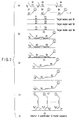
A detection method of the present invention will
be explained with reference to FIG. 1 (FIG. 1).
In the method of this example, two types of
nucleic acid probes (hereinafter, sometimes simply
referred to as "probe") shown below, a probe A1 and a
probe B2, are used (FIG. 1a).
The probe A1 is constituted of a sequence F' and a
binding molecule 4 bound to the sequence F'. The
sequence F' is complementary to a nucleotide sequence F
of a partial region of the target nucleic acid 3. The
binding molecule 4 used herein may directly bind to the
sequence F' or indirectly bind to the sequence F' via
some portion such as an arbitrary nucleotide sequence.
The portion except the nucleotide sequence F of the
probe A1 may be referred to as a "tag", "tag sequence
Tg" or "Tg". The arbitrary sequence, which is
responsible for the indirect binding, may be a
nucleotide sequence having any number of nucleotides.
More preferably, the sequence may be a non-complementary
sequence to the nucleotide sequence of
the target nucleic acid. Alternatively, the indirect
binding may be mediated by other means such as chemical
or physical means. The tag Tg is required to neither
bind to nor interact with any portion of the target
nucleic acid. The term "non-complementary" used herein
refers to a nucleotide sequence which does not
hybridize with a sequence of the target nucleic acid,
in particular, a sequence within an arbitral range for
use in detection.
The sequence F' used herein has a nucleotide of 1
or more. The sequence F' preferably has 15 or more of
nucleotides to attain stable hybridization.
The binding molecule 4 used herein may be either
one of the pair molecules specifically binding to each
other, as previously described.
The probe B2 consists of a sequence S' and a flag
(hereinafter, sometimes represented by "FL") bound to
the sequence S'. The sequence S' is complementary to a
nucleotide sequence S, which is a part of the target
nucleic acid 3. In this example, the flag consists of
a double-stranded sequence linking to the sequence S'
and the marker substance 5 bound to the double stranded
sequence. Generally, it is necessary that the flag
itself neither binds to nor interacts with the target
nucleic acid 3. Therefore, when the flag is a
nucleotide sequence, the entire length is not
necessarily formed of a double-strand nucleic acid.
The flag may be formed of a single-stranded sequence in
part or in its entirety as long as it neither binds to
nor interacts with the target nucleic acid.
Furthermore, the flag has any number of nucleotides.
Alternatively, the marker substance 5 may be bound to
the sequence S' by any means, as detailed in Example 3
set forth later.
When the flag is a double-stranded sequence, the
sequence may be formed of any types of nucleotides and
may have any number of nucleotides. However, the Tm of
the sequence must be sufficiently higher than those of
the sequence F and the sequence S. Particularly in the
case where the flag has a double-stranded sequence, the
flag sequence is not directly bound to the sequence S'.
More specifically, the flag sequence hybridizes with
the sequence which directly bounds to the sequence S'.
As the marker substance 5 used herein, any
substance is used as long as it is generally used as a
marker substance. Preferable examples of the marker
substance include fluorescent substances, luminescent
substances, radioisotope substances such as 32P, high
absorbency substances, high reflection substances, high
potential substances, magnetic substances, and pigments.
Particularly, fluorescent substances such as FITC are
preferable.
The sequence S' used herein has nucleotide(s) of 1
or more, preferably 15 or more.
Tg attached to the probe A1 and FL attached to the
probe B2 are preferably designed so as to be positioned
at ends at the farthest distance from each other when
both probes are bound to the desired regions of the
target nucleic acid 3, as shown in FIG. lb.
The probe A1 and probe B2, when they bind to the
desired regions of the target nucleic acid 3,
preferably have the nucleotides complementary to
adjacent nucleotides on the target nucleic acid 3, at
the close ends. This is advantageous in a ligation
step performed thereafter. However, this is not
limited thereto.
The sequences F and S of the probes A1 and B2 may
be appropriately selected so as to have the same Tm by
controlling the conditions including the GC content and
the combination of nucleotide bases.
The example is carried out as described below.
First, the probe A1 and probe B2 are prepared in
accordance with the target nucleic acid 3. They are
mixed, at an appropriate ratio, with the target nucleic
acid 3 (FIG. 1a).
Subsequently, the mixture is incubated under
arbitrary conditions suitable for hybridization for a
predetermined time period to perform hybridization
(FIG. 1b).
The hybridization is performed by any general
method. The preferable hybridization is performed by
allowing the mixture stand still at an appropriate high
temperature, for example, 95°C, effective in denaturing
a nucleic acid, for 5 minutes, whereby the
complimentary binding of the target nucleic acid is
dissociated to convert a double strand to a single
strand. In the case of the single-stranded target
nucleic acid, the single-strand target nucleic acid is
allowed to stand still at 70°C for 5 minutes, whereby it
can be converted into a secondary structure.
Subsequently, the single-strand is allowed to stand
still at an appropriate temperature effective in
rebinding of a complementary nucleotide sequence, for
example, 55°C for 15 minutes, the single-stranded target
nucleic acid is rebound to the complementary nucleotide
sequence. By the hybridization process mentioned above,
both the probes A1 and B2 bind to the same target
nucleic acid 3 to make a hybrid. Therefore, it is not
necessary that conditions including the type of buffer
and temperature are limited to those mentioned above.
Any conditions may be employed as long as the probes A1
and B2 are hybridized with the target nucleic acid 3
under the conditions.
Thereafter, the probe A1 is linked with the probe
B2 (FIG. 1c). In the present invention, the probe A1
and the probe B2 both being hybridized on the target
nucleic acid 3 are linked to each other, to form a
probe (A+B)7. This linkage is attained by formation of
the linkage portion 6.
This linkage is made by use of Taq DNA ligase such
as Thermus aquatius (Taq) DNA ligase manufactured by
New England Biolab. However, the agent used in this
linkage is not limited to this. Any agent may be used
as long as it is generally used in the linkage.
Furthermore, both probes may be linked with any
chemical means in place of the enzymatic means.
When the sequences F and S are not present on the
target nucleic acid 3 next to each other, a gap-filling
reaction may be performed by using DNA polymerase I and
a ligase. By this reaction, nucleotides may be
introduced in the gap between both proves. Any DNA
polymerase and ligase may be used as long as they are
generally used. Preferably, heat-resistance DNA
polymerase and ligase are preferable.
Subsequently, a probe (A+B)7 obtained in the above
is dissociated from a target nucleic acid 3 (FIG. 1d).
The dissociation may be performed by denaturation
treatment such as thermal denaturation. More
specifically, when the dissociation is performed by
thermal denaturation, the reaction may be made, but not
limited to, under physiological conditions at 85°C or
more, preferably 90°C or more. However, when the flag
sequence is a double strand, the dissociation may be
made of a temperature lower than the melting
temperature, Tm, of double strand.
Subsequently, a binding molecule 4, that is, one
of the binding pair, is allowed to bind to a molecule
having a high affinity with the binding molecule. In
this manner, a prove (A+B)7 is recovered (FIG. 1e).
Subsequently, the flag is denatured and converted into
a single chain. The single strand tagged with the
marker substance 5 is allowed to hybridize with a
sequence complementary to the single strand (FIG. 1g).
In this way, the single strand is recovered.
Thereafter, the marker substance 2 is detected or
quantified, whereby detection or quantification of the
target nucleic acid (FIG. 1g) is completed. The
detection may be performed by any general method
suitable for the marker substance used herein.
A single strand having the probe (A+B)7 and the
marker substance 5 bound thereto is separated by
Bound/Free separation (hereinafter, referred to as "B/F
separation"). Therefore, the accuracy in detection is
improved. The B/F separation is performed in
accordance with the B/F separation method generally
employed.
In this method, the B/F separation for the single
strand tagged with the marker substance 5 may be
preferably performed using a solid-phase carrier having
a nucleotide sequence complementary to the single
strand fixed on an appropriate support (FIG. 1g). The
B/F separation for the probe (A+B)7 can be preferably
performed by a solid-phase carrier having a molecule
capable of specifically binding to the binding molecule
4 immobilized on an appropriate support (FIG. 1e).
Examples of the support to be used include, but
not limited to, a substrate such as a silicon plate or
a glass plate, particles such as beads, a container
such as a test tube or a vial, a fiber, a tube
including a capillary, a filter, an affinity column, an
electrode and the like. It is preferable that the
material of the support and surface treatment be
appropriately selected depending upon how efficiently
the probe is fixed.
Since the flag has an artificially synthesized
sequence, the sequence may be designed such that it has
properties for improving the specificity of the
hybridization of the probe (A+B).
Example 2. Example of Detection Method
This method is also effective when a plurality of
target nucleic acids are simultaneously detected.
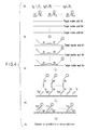
FIG. 2 shows the case where a plurality of target
nucleic acids N1, N2, N3 ··· Nn (hereinafter referred to
as "N1-Nn") where n is an integer of 2 or more) are
simultaneously detected.
Individual steps and the constitutions of
individual probes are the same as in Example 1 except
that a plurality of detection targets are used (FIG. 2,
a to e) .
To explain more specifically, target nucleic acids
N1-Nn, probes A1-An, and probes B1-Bn are mixed
(FIG. 2a). They are allowed to hybridize with the
target nucleic acids, respectively, and then the probes
A1-An are linked to the probes B1-Bn, respectively
(FIG. 2b). The resultant probes (A1+B1)-(An+Bn) are
dissociated from the target nucleic acids (not shown).
Then, the probes (A1+B1)-(An+Bn) dissociated are
recovered (FIG. 2c). Each of the flags is denatured
to obtain a single strand (FIG. 2d). The single strand
having a marker substance attached thereto is recovered
and the marker substance is detected or quantified
(FIG. 2e). The plurality types of target nucleic acids
can be simultaneously detected or quantified by using
the same marker substance. Therefore, this method made
it easier to detect a plurality of target nucleic acids
in fewer steps than those of conventional methods.
To distinguish a plurality of target nucleic acids
by using the same marker substance, the flags having
different sequences modified depending upon the target
nucleic acids may be used. Furthermore, as shown in
FIG. 2 (e), when complementary sequences, which are
used for recovering target single strands, are
immobilized to the substrate, they may be placed in
different regions depending upon the sequences. It may
be possible to modify the sequences of the flags
simultaneously with the immobilization of the
complementary sequences in different regions.
Alternatively, it is possible to use different
marker substances depending upon the target nucleic
acids. In this case, the modification of the flag and
the immobilization of the complementary sequences in
different regions are not necessarily performed.
If the GC contents of Fn' and Sn' sequences of a
probe (A1+B1), (A2+B2) ··· (An+Bn) [hereinafter,
referred to as "(A1+B1)-(An+Bn)"] and the types of
nucleotides are appropriately selected, optimal
hybridization conditions (e.g., temperature and salt
concentration) which differ between individual probes
can be overcome. When a plurality of types of flags
different in sequence are used, it. is preferable to use
the same (common) hybridization conditions even if the
types of flags differ. In this manner, detection
accuracy is improved. The detection accuracy is
further improved by increasing the common Tm value of
the flag sequences sufficiently higher than those of
the sequences Fn' and Sn'.
In contrast, in the conventional nucleic acid
detection method, in which a complementary sequence of
the target nucleic acid is immobilized, optimal
hybridization conditions for individual probes
immobilized differ in general. However, it is
impossible to set the conditions suitable for all
probes.
Example 3. Example of Detection Method
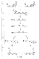
Referring to FIG. 3, another example of the
present invention will be explained. In the method of
this example, a plurality of target nucleic acids N1,
N2, N3 ··· Nn (Hereinafter, referred to as N1-Nn where n
is an integer of 2 or more) can be simultaneously
detected. Probes A1, A2, A3 ··· An (hereinafter
referred to as "A1-An" where n is an integer of 2 or
more) and probes B1, B2, B3 ··· Bn (hereinafter referred
to as "B1-Bn" where n is an integer of 2 or more) are
prepared corresponding to the target nucleic acids N1-Nn.
These nucleic acid probes have the characteristics
shown below (FIG. 3a).
The probes A1-An are formed of sequences F1' to
Fn', and tag Tg1 to Tgn (hereinafter referred to as
"Tg1-Tgn") bound to the sequences F1' to Fn',
respectively. The sequences F1' to Fn' are
complementary to nucleotide sequences F1 to Fn, which
are parts of the target nucleic acids N1-Nn,
respectively. The tag Tg1 to Tgn (hereinafter referred
to as "Tg1'-Tgn') of this example are nucleotide
sequences. Each of the sequences of the tags Tg1-Tgn
is required not to bind to and thus not to interact
with any portion of its target nucleic acid. The tags
Tg1 to Tgn used herein may be directly bound to
respective sequences F1' to Fn' or indirectly bound to
them via any intermediate portion, for example, a
nucleotide sequence. When the tags are indirectly
bound via an nucleotide sequence arbitrarily chosen,
the arbitrary sequence may be any nucleotide sequence
having any number of nucleotides. The nucleotide
sequence is preferably non-complementary to the
nucleotide sequence of a target nucleic acid. They may
be bound by a chemical or physical means.
The sequences F1' to Fn' used herein have
nucleotides different from each other. They may be
nucleotides of one or more. To attain stable
hybridization, the sequences preferably have nucleotide
of 15 or more.
As shown in FIG. 3a, the probes B1-Bn are formed
of sequences S1' to Sn' and arbitrary flags
(hereinafter referred to as "FL") bound to the
sequences S1' to Sn'. The sequences S1' to Sn' are
complementary to respective nucleotide sequences S1 to
Sn, which are parts of respective target nucleic acids.
In this example, the flags contain a marker substance.
The flags themselves do not bind to, and thus interact
with the target nucleic acids, in the same as in the
previous examples. The probes B1-Bn of this example
are constructed by binding the marker substance to the
sequences S1' to Sn' by an arbitrary means.
As the marker substance used herein, any substance
may be used as long as it is a marker substance
generally used, as set forth in Example 1. More
preferably, each of the sequences S1' to Sn' has
nucleotides of 15 or more.
When probe A1(An) and probe B1(Bn) are bound to
respective desired regions of the target nucleic acid,
as shown in FIG. 3b, the end of the probe A1(An) having
the tag attached thereto and the end of the probe
B1(Bn) having the flag attached thereto, are preferably
positioned at the farthest distance from each other
(FIG. 3b). Furthermore, when the probe A1(An) and
probe B1(Bn) are bonded to respective desired regions
of the target nucleic acid, the nucleotides positioned
at proximal ends of both probes are preferably
complementary to the nucleotides positioned next to
each other on the target molecule. This positioning is
advantageous in a ligation step performed later.
However, the positioning of the complementary
nucleotides is not limited thereto.
If the GC content and the nucleotide types of
sequences F1'-Fn' and S1'-Sn' contained in these probes
are appropriately selected, it is possible to overcome
the differences in optimal hybridization conditions of
individual probes. When a plurality of types of tag
sequences are used, it is preferable that they can be
hybridized well under the same (common) hybridization
conditions. Furthermore, if the common Tm value of the
tag sequences is set sufficiently higher than those of
the Fn' and Sn' sequences, the detection accuracy is
improved.
This example is carried out in the following
manner. First, probes A1-An and probes B1-Bn are
prepared in accordance with the target nucleic acids
N1-Nn, as mentioned above. Then, they are mixed with
the target nucleic acids N1-Nn, in an appropriate ratio
(FIG. 3a).
Then, the mixture is incubated for a predetermined
time under arbitrary conditions suitable for
hybridization. In this manner, hybridization is
performed (not shown).
The hybridization may be performed by any general
method. Preferable hybridization is performed as
follows. First, a target nucleic acid is allowed to
stand still at a high temperature, for example, 95°C,
which is effective in denaturing the target nucleic
acid. In the step, the complementary binding of the
target nucleic acid is denatured, with the result that
a double strand is dissociated into a single strand.
In the case where the target nucleic acid is a single
strand, the nucleic acid is allowed to stand still at
70°C for 5 minutes to denature its secondary structure.
Subsequently, the resultant reaction mixture is allowed
to stand still for 15 minutes at an appropriate
temperature, e.g., 55°C which is effective in rebinding
a complementary nucleotide sequence. As a result, the
single-stranded nucleic acid is rebound to the
complementary nucleotide sequence.
By the hybridization step, each set of probes
A1-An and probes B1-B2 bind to the same and
corresponding target nucleic acid to produce hybrids.
Accordingly, type of a buffer solution to be employed
and temperature condition are not limited to those
mentioned above. Any conditions may be employed as
long as the probes A1-An and probes B1-Bn can hybridize
with respective target nucleic acids.
Subsequently, the probes A1-An are bound the
corresponding probes B1-Bn (FIG. 3b). More
specifically, in the present invention, the probes
A1-An and probes B1-Bn both being hybridized on the
respective target nucleic acids N1-Nn are respectively
ligated, producing probes (A1+B1) to (An+Bn),
respectively. The ligation is attained by forming a
ligation portion. The ligation may be performed in the
same manner as Example 1.
Furthermore, in the case where the sequence F and
the sequence S are not placed next to each other on the
target nucleic acid, the gap between them may be filled
by use of DNA polymerase I and a ligase. In this way,
nucleic acid bases are introduced into the gap between
both probes. Any DNA polymerase I and ligase may be
used as long as they are generally used. Heat
resistant DNA polymerase and ligase are preferably used.
Subsequently, the probes (A1+B1) to (An+Bn)
obtained above are dissociated from the target nucleic
acids (FIG. 3c). The dissociation is performed by
denaturation treatment such as thermal denaturation.
For example, when the dissociation is performed by the
thermal denaturation, the reaction is performed under
physiological conditions at 85°C or more, preferably,
90°C or more. However, the conditions are not limited
thereto.
The probes (A1+B1) to (An+Bn) are recovered by
using the sequences Tg1' to Tgn' which hybridize with
complementary sequences Tg1 to Tgn (FIG. 3d).
Thereafter, the marker substance is detected or
quantified. In this manner, the target nucleic acids
can be detected or quantified (FIG. 3d). A detection
method varies depending upon the marker substance and
any detection method may be used as long as it is
generally used.
The complementary sequences Tg1' to Tgn' are
preferably fixed on a support. Examples of the support
to be used include, but not limited to, a substrate
such as a silicon plate or a glass plate, particles
such as beads, a container such as a test tube or a
vial, a fiber, a tube including a capillary, a filter,
an affinity column, an electrode and the like. It is
preferable that the material of the support and surface
treatment be appropriately selected depending upon how
efficiently the probe is fixed.
In Example 3, a plurality of target nucleic acids
are used. However, the method used herein is
applicable to a single target nucleic acid. If the
number of types of the marker substances are increased,
a larger number of target nucleic acids can be
identified.
Example 4
Referring now to FIG. 4, another example of the
present invention will be explained. In the method of
this example, a plurality of target nucleic acids N1,
N2 N3 ··· Nn (hereinafter, referred to as "N1-Nn" where
n is an integer of n or more) are simultaneously
detected. Probes A1, A2 A3 ··· An (hereinafter,
referred to as "A1-An" where n is an integer of 2 or
more) and probes B1, B2, B3 ··· Bn (hereinafter,
referred to as "B1-Bn" where n is an integer of 2 or
more) are prepared corresponding to the target nucleic
acids N1-Nn. These probes have the following
characteristics (FIG. 4a).
The probes A1-An is formed of sequences F1' to Fn'
and tags Tg1 to Tgn bound to the sequences F1' to Fn',
respectively. The sequences F1' to Fn' are
complementary to nucleotide sequences F1 to Fn, which
are part of the target nucleic acids N1-Nn,
respectively. Tags Tg1 to Tgn of this example are
nucleotide sequences which do not bind to any portion
of the corresponding target nucleic acids and do not
exhibit any interactions with the target nucleic acids.
Tags Tg1 to Tgn may directly bind to the respective
sequences F1' to Fn' and indirectly bind to them via an
intermediate portion such as a nucleotide sequence. In
the case of the indirect binding via an sequence
arbitrarily chosen, the sequence may consist of any
types of nucleotides and any number of nucleotides.
Preferably, the sequence is non-complementary to the
nucleotide sequence of the target nucleic acid.
Alternatively, they may bind to each other by a
chemical or physical means.
The sequences F1' to Fn' used herein are different
each other. They may be nucleotides of one or more.
To attain a stable hybridization, the sequences F1' to
Fn' preferably have nucleotides of 15 or more.
As shown in FIG. 4a, the probes B1-Bn have
sequences S1' to Sn' and flags bound to the sequences
S1' to Sn', respectively. The sequences S1' to Sn' are
complementary to nucleotide sequences S1 to Sn of parts
of the target nucleic acids, respectively. The flags
have nucleotide sequences FL to FLn and a marker
substance, respectively. As is the same as in Examples
mentioned above, the flags themselves do not bind to
the target nucleic acids and do not exhibit any
interactions with the target nucleic acids. The probes
B1-Bn are constructed by binding the marker substance
to the sequences S1' to Sn' by an arbitrary means.
As the marker substance used herein, any substance
may be used as long as it is generally used as a marker
substance, as described in Example 1.
The sequence S' used herein has nucleotides of 1
or more, preferably 15 or more.
When probe A1(An) and probe B1(Bn) are bound to
respective desired regions of the target nucleic acid,
as shown in FIG. 4b, the end of the probe A1(An) having
the tag attached thereto and the end of the probe
B1(Bn) having the flag attached thereto, are preferably
positioned at the farthest distance from each other
(FIG. 4b). Furthermore, when the probe A1(An) and
probe B1(Bn) are bonded to respective desired regions
of the target nucleic acid, the nucleotides positioned
at proximal ends of both probes are preferably
complementary to the nucleotides positioned next to
each other on the target molecule. This positioning is
advantageous in a ligation step performed later.
However, the positioning of the complementary
nucleotides is not limited thereto.
If the sequences F1'-Fn' and S1'-Sn' included in
the probes are constructed by appropriately setting the
GC content and the combination of nucleic acid bases,
hybridization of the probes can be appropriately
performed by overcoming different optimal hybridization
conditions of individual probes. When a plurality of
types of tag sequences and flag sequences are used, it
is preferable that they can be hybridized under the
same (common) hybridization conditions. Furthermore,
if the common Tm values of the tag sequences and flag
sequences is set sufficiently higher than those of the
sequences Fn' and Sn', the detection accuracy is
improved. Furthermore, if a plurality of flag
sequences are used with respect to a single tag
sequence, the number of the target nucleic acids
distinguishable from each other can be tremendously
improved.
This example can be carried out in the following
manner. First, the probes A1-An and probes B1-Bn are
prepared in accordance with the target nucleic acids
N1-Nn, as mentioned above. Then, the probes are mixed
with the target nucleic acids N1-Nn in an appropriate
ratio (FIG. 4a).
Subsequently, the mixture is incubated for a
predetermined time under arbitrary conditions suitable
for hybridization. In this manner, hybridization is
performed (not shown).
The hybridization may be performed in any general
method. Preferable hybridization is performed as
follows. First, a target nucleic acid is allowed to
stand still at a high temperature, for example, 95°C,
which is effective in denaturing the target nucleic
acid. In the step, the complementary binding of the
target nucleic acid is denatured, with the result that
a double strand is dissociated into a single strand.
Subsequently, the single-stranded nucleic acid is
allowed to stand still for 15 minutes at an appropriate
temperature, e.g., 55°C which is effective in rebinding
a complementary nucleotide sequence. As a result, the
single-stranded nucleic acid is rebound to the
complementary nucleotide sequence.
By the hybridization step, each set of probes Al-An
and probes B1-Bn bind to the same and corresponding
target nucleic acid to produce hybrids. Accordingly,
the type of a buffer solution to be employed and
temperature conditions are not limited to those
mentioned above. Any conditions may be employed as
long as the probes A1-An and probes B1-Bn can hybridize
with respective target nucleic acids.
Subsequently, the probes A1-An are bound the
corresponding probes B1-Bn (FIG. 4b). More
specifically, in the present invention, the probes A1-An
and probes B1-Bn, both being hybridized on the
respective target nucleic acids N1-Nn, are respectively
ligated, producing probes (A1+B1) to (An+Bn),
respectively. The ligation is attained by forming a
ligation portion. The ligation may be performed in the
same manner as Example 1.
Furthermore, in the case where the sequence F and
the sequence S are not placed next to each other on the
target nucleic acid, the gap between them may be filled
by use of DNA polymerase I and a ligase. In this way,
nucleic acid bases are introduced into the gap between
both probes. Any DNA polymerase I and ligase may be
used as long as they are generally used. Preferably,
heat resistant DNA polymerase and ligase are used.
Subsequently, the probes (A1+B1) to (An+Bn)
obtained above are dissociated from the target nucleic
acids (not shown). The dissociation is performed by
denaturation treatment such as thermal denaturation.
For example, when the dissociation is performed by the
thermal denaturation, the reaction is performed under
physiological conditions at 85°C or more, preferably,
90°C or more. However, the temperature conditions are
not limited thereto.
The probes (A1+B1) to (An+Bn) are recovered by
using the sequences Tg1' to Tgn' which hybridize with
complementary sequences Tg1 to Tgn (FIG. 4c).
Subsequently, the probes are dissociated by
denaturation. The dissociated probes are recovered by
hybridizing them respectively with the sequences FL' to
FLn' complementary to the sequences FL1 to FLn
(FIG. 4d).
Subsequently, respective marker substances are
detected or quantified to detect and quantify the
target nucleic acid (FIG. 4e). The detection method
varies depending upon the marker substance and any
detection method may be used as long as it is generally
used.
The complementary sequences Tg1' to Tgn' and FL'
to FLn' used herein are preferably fixed on a support.
Examples of the support to be used include, but not
limited to, a plate such as a silicon plate or a glass
plate, particles such as beads, a container such as a
test tube or a vial, a fiber, a tube including a
capillary, filter, an affinity column, an electrode and
the like. It is preferable that the material of the
support and surface treatment may be appropriately
selected depending upon how efficiently the probe is
fixed.
Example 5. Example of Detection method
Now, a further preferable example of the present
invention will be explained. If an appropriately
designed flag is employed in the following method, a
plurality of target nucleic acids can be efficiently
detected with a high accuracy.
The following two types of nucleic acid probes, a
probe A and a probe B, can be used in this method
(FIG. 5a).
The probe A is formed of a nucleotide sequence F'
and a binding molecule bound to the sequence F'. The
sequence F' is complementary to a nucleotide sequence F
which is a partial region of the target nucleic acid.
The binding molecule used herein is either one of
two substances having specifically a high affinity with
each other. For example, biotin, avidin, or
streptoavidin. Furthermore, the binding molecule may
bind directly to the sequence F' or may be indirectly
bind to the sequence F' via an arbitrary sequence. The
arbitrary sequence, which is responsible for the
indirect binding, may be any nucleotide sequence having
any number of nucleotides. More preferably, the
sequence is a non-complementary sequence to the
nucleotide sequence of the target nucleic acid.
The probe B is formed of a nucleotide sequence S'
and a flag. The nucleotide sequence S' is
complementary to a nucleotide sequence S which is a
partial region of the target nucleic acid. The flag is
a double strand. The double strand has an arbitrary
sequence consisting of a plurality of units. The flag
must not bind to the target nucleic acid and not
exhibit any interaction with the target nucleic acid.
The sequences F' and S' used herein have a
nucleotide of 1 or more, preferably 15 or more.
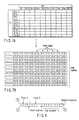
FIG. 7 shows a design of the unit. One of units
of the flag FL may contain 10 bases or more, more
preferably, about 15 bases. Although the flag FL has
any number of units, 4 units are preferable from the
view point of analysis. However, the number of units
is not limited to 4.
When a plurality of target nucleic acids are
simultaneously detected, the flag FL of a plurality of
types of units may be used. As an example, the flag FL
consists of 4 units, SD, D0, D1, ED will now be
explained. This flag is formed as follows. First, 22
types of units are designed. Then, two units are
selected from them as primers. One is defined as the
SD unit. The other one is defined as the ED unit.
Subsequently, the D0 and D1 units are selected by using
the remaining 20 units. If the flag is designed by
varying the combination of the two units depending upon
a type of nucleic acid, 100 types of nucleic sequences
can be detect (FIG. 7A).
The 22 types of units are preferably designed by
orthonormal nucleotide sequences. The Tm of the
orthonormal nucleotide sequences fall within almost the
same range, so that they do not make a stable hybrid
with the non-complementary sequences. They do not form
a stable secondary structure which prevents the
formation of a hybrid with the corresponding
complementary sequences. Therefore, the occurrence of
mis-hybridization will be reduced at the time of a
final detection operation, with the result that the
hybrid-formation speed can be increased. It is
therefore possible to improve the detection accuracy
and to reduce the detection time. Furthermore, if the
number and types of units are increased, 10000 types of
nucleic acid sequences can be detected.
Referring to FIG. 5, the method of this example
will be further explained. FIG. 5a shows a flag FL
consisting of 4 units, namely, SD unit, D0 unit, D1
unit, and ED unit. The SD unit serves as a primer in a
polymerase chain reaction (hereinafter, referred to as
a "PCR amplification" or "PCR reaction"). The DO and
D1 units are recognition units for recognizing the type
of target nucleic acid. The ED unit is another primer
sequence. Each of units serves as a reading frame in a
later step.
Detection is performed as follows. First, the
probe A and probe B are mixed with a target nucleic
acid (FIG. 5a). In this case, a plurality of target
nucleic acids different in sequence may be contained in
a specimen. For example, if there are 100 types or
less of target nucleic acids to be detected, the D0
unit is selected from 10 types of nucleic acids D0-1 to
D0-10, and D1 unit is selected from 10 types of nucleic
acids D1-1 o D1-10 (FIG. 7A).
Subsequently, the probe A, probe B, and target
nucleic acid(s) are incubated for a predetermined time
under the conditions suitable for hybridization
(FIG. 5b). The hybridization conditions are the same
as shown in Example 1.
Both the probes A and B hybridize with the same
target nucleic acid by the hybridization thus performed
(FIG. 5b).
Subsequently, the probes A and B, both being
hybridized with the same nucleic acid, are ligated to
each other (FIG. 5c). The conditions of ligation are
the same as shown in Example 1.
The Tm value of flag FL is preferably set at a
temperature higher than those of the sequences F' and
S'. If the Tm value is set in this manner, it is
possible to prevent denaturation of the flag, which
lowers the detection sensitivity, during the heating
and cooling operations at the time of hybridization,
ligation, and denaturation involved in this detection
method.
Subsequently, the information on the flag FL
obtained is subjected to the B/F separation. More
specifically, the binding molecule attached to the
probe (A+B) is captured on a solid-phase carrier via a
binding molecule to be paired up with the binding
molecule of the probe (A+B) (FIG. 5e).
Examples of the solid phase carrier are a plate,
beads, a container, a fiber, a tube, a filter, an
affinity column, an electrode and the like. Preferably,
beads are used.
The flag FL of the probe (A+B) bound to the
binding molecule is denatured into a single strand
(FIG. 5f), while keeping the binding molecule bound
thereto. The resultant single-stranded sequence FL'
present in a liquid phase is subjected to PCR
amplification (FIG. 5g). As described, the flag FL has
two primer sequences SD and ED previously arranged
therein. Therefore, the PCR reaction can be easily
performed by use of these primer sequences. At that
time, it is preferable that a binding molecule such as
biotin be bound to one of the primers for the PCR, for
example, the SD sequence. The detailed PCR conditions
used herein vary depending upon the flag design.
After completion of the PCR reaction, a PCR
product, that is, a double-stranded sequence, is
recovered by permitting the binding molecule to bind to
a solid-phase carrier immobilized (FIG. 5i). The
carrier immobilized is a substance capable of being
paired up with the binding molecule. Further,
denaturation is performed to remove the sequence FL'.
Only a single-stranded sequence FL is recovered by the
immobilized carrier (FIG. 5i).
Subsequently, the single-stranded flag sequence FL
attached on the immobilized carrier is analyzed as
follows. The immobilized carrier having the single-stranded
flag sequence FL bound thereto, is equally
divided into ten portions (in this case, D1 unit
consists of D1-1 to D1-10). To individual ten portions,
one of D1-1' to D1-10' labeled with a marker molecule
and all D0' sequences (D0-1' to D0-10') are added to
hybridize them with the flag sequence FL.
Subsequently, two nucleic acid molecules thus
hybridized are ligated to each other. The ligation
conditions and the definitions for the marker substance
are the same as previously mentioned. Thereafter, the
molecule ligated is denatured and recovered in a liquid
phase.
The nucleic acid molecules labeled with the marker
molecule are analyzed by hybridizing them with a DNA
chip or a DNA capillary on which the nucleic acid
molecules of D0-1 to D0-10 are previously immobilized.
Particularly, the DNA capillary is advantageous since
the nucleic acid molecules which have been divided into
D0-1 to D0-10 portions, can be simultaneously analyzed.
The analysis can be easily made by the DNA capillary.
As an example, the case of the flag FL which is
designed by using 10 types of sequences, D0-1 to D0-10,
and 10 types of sequences, D1-0 to D1-10 will now be
explained. Since the sequence D0-1 is immobilized at
position 1 of FIG. 7A, the nucleic acid molecule 63
ligated to a D1-1' molecule labeled with a marker
molecule, is hybridized with the sequence D0-1 at
position 1. Similarly, since a D0-n sequence is
immobilized to a corresponding position n of the column,
the nucleic acid molecule ligated to the D1' molecule
(i.e., a molecule corresponding to a row) is hybridized
with the position n of the column. If such a matrix
arrangement is applied to the DNA capillary described
later, the analysis can be readily performed (FIG. 7B).
In this example, 10 types of units are used.
However, the units are not limited to 10 types. More
than 10 types or less than 10 types may be used.
The "DNA capillary" used herein is a device for
detecting a target nucleic acid. Since a complementary
sequence to the target nucleic acid is bound inside the
device, the target nucleic acid can be detected by
binding the target nucleic acid to the complementary
sequence. As shown in FIG. 7B, if a plurality of DNA
capillaries having different probes (arranged at
hatched portions) are simultaneously used, a plurality
of target nucleic acids can be detected at the same
time.
In the method of this example, since an
orthonormal nucleotide sequence is used as each unit of
the flag sequence FL, hybridization can be uniformly
performed under the same conditions, e.g., a reaction
temperature. Therefore, the detection can be made with
a high accuracy while preventing mis-hybridization.
Furthermore, since numerous analyses can be
performed simultaneously under the same conditions, the
time required for the detection can be reduced.
According to the method of the present invention,
complicated genomic information expressed by a
nucleotide sequence of DNA can be converted into
numerical values. Moreover, if calculation is made by
using a DNA molecular reaction, analysis of various
types of information and complicated genetic
information mutually associated can be easily made. In
addition, after a nucleic acid is encoded to a nucleic
acid code, the nucleic acid code can be amplified
easily. Therefore, even if the target sequences are
present in low copy numbers, the target sequences can
be accurately and quantitatively detected. Furthermore,
it is possible to compress numerous data by encoding
the sequence. Therefore, a number of detections can be
made by using a fewer number of devices, such as a DNA
chip or a capillary array.
The term "encode reaction" used herein is referred
to converting a nucleotide sequence into codes based on
orthonormal nucleotide sequences. The "encode
reaction" includes the steps of FIGS. 5a to 5f.
The term "decode reaction" used herein is referred
to reading the code converted in the above and thereby
reproducing original information. The step of FIG. 5j
corresponds to the "decode reaction".
In Example 5, a single type of target nucleic acid
is detected. If a plurality of types of flag sequences
are designed, a plurality of types of target nucleic
acids can be simultaneously detected by the same steps.
An application example of Example 5 is shown in
FIG. 6. In the example of FIG. 6, flag sequences
including D01-D11 to D0n-D1n (n is an integer) are
designed in accordance with the target nucleic acids to
be detected. The flag sequences thus designed are
treated in the same manner as in steps of FIGS. 5a to
5f and further subjected to the decode reaction
described below (FIG. 6). To explain more specifically,
the single-stranded sequence FL' obtained is subjected
to a PCR reaction using two primer sequences SD and ED
(FIG. 6g). In the steps of FIGS. 6g to 6i, a probe n
is representatively shown instead of showing a
plurality of probes. However, it should be interpreted
that 1 to n probes are present. In this case, it is
preferable that a binding molecule such as biotin
should be bound to the SD sequence. The specific
conditions for PCR in this case vary depending upon the
design of a flag FL. The obtained double strand is
recovered by using a substance capable of making a pair
with the binding molecule (FIG. 6h). After the pair is
denatured and washed to obtain a single strand
(FIG. 6i). Subsequently, the primers complementary
with respective D11-D1n sequences of each FL sequence
and tagged with a marker substance, are hybridized with
each FL sequence and extended (FIG. 6j).
The resultant sequences D11'-D1n' obtained are
detected by using a chip or a capillary array to which
the sequences D01-D1n are immobilized (FIG. 6k). The
detection can be performed by the following method. A
probe A is ligated to a probe B. The B/F dissociation
is performed by using a binding substance attached to
the probe B or using a Tgn sequence. By this method,
the obtained FL can be directly detected without using
PCR. In this case, the sequence to be bound to a
sequence Sn' may be formed of two units. In this case,
the D0n sequence and D1n sequence may be immobilized to
the DNA chip or the capillary array. In this case,
detection can be made by previously attaching different
marker substances depending upon FL sequences.
Alternatively, after the FL sequences bind to desired
regions of the DNA chip, a marker substance may be
attached to the FL sequences.
Example 6
To demonstrate that the method of the present
invention is useful in practice, the following
experiment was performed. The sequence used herein and
how to bind the sequence are shown in FIG. 8.
(1) Target sequence and modified sequence
A sequence r-32-f1 was used as a target sequence
(FIG. 9). A modified sequence different from the
target sequence in sequence was used to confirm the
specificity of the detection method. The modified
sequence is designated as f1-neg-r-32AB (FIG. 9).
(2) Probe and detection sequence
A nucleotide b-16A was used as a probe A (FIG. 9).
The symbol b of the probe A represents biotin. A
nucleotide P-16B-48 was used as a probe B (FIG. 9).
The probe B includes a flag FL. The symbol P
represents a phosphorus group. The complementary
sequence to the FL sequence of a probe B is a detection
sequence, which is designated as fl-r-48 (FIG. 9).
(3) Experimental method
The target sequence was tried to be detected by
using the aforementioned probes. First, a probe B
solution containing 500 pmol of probe B in a TE buffer
solution of pH 8.0 was mixed with a solution for a
detection sequence in an equal amount. The resultant
mixture was incubated at 95°C for one minute and
decreased in temperature to 25°C for 7 minutes.
Consequently, a double-strand probe B was obtained.
The double-stranded probe B obtained, the target
sequence, and the probe A were mixed. The ligation
reaction was performed using a Tag DNA ligase
(manufactured by New England Biolab) of 20U in 100 µL
of a ligantion buffer solution. The ligation reaction
was performed by gradually reducing temperature from
70°C to 55°C over 5 minutes to anneal the probes A, B
and the target sequence. A ligation reaction was
performed by adding TagDNA ligase at 55°C for 15 minutes.
Thereafter, the temperature of the reaction is reduced
to room temperature. After the ligation reaction, the
specimen was added to magnetic beads on which avidin is
immobilized. The resultant mixture was subjected to
pre-cold wash at 30°C to remove the target sequence
unreacted. Further, cold wash was performed at the
same temperature as above to remove the target sequence.
After the supernatant was removed, 100 µL of TE buffer
solution was added. The resultant solution was
subjected to hot wash at 70°C to recover the detection
sequence.
(4) Experimental results
The experimental results will be described below.
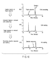
FIG. 10 shows the results of capillary electrophoresis
of the sequences obtained from the supernatants in
individual washing steps. It was demonstrated that the
supernatant recovered in the prewash contained a target
sequence unused in the ligation reaction. It was also
demonstrated that the supernatant recovered from the
cold wash contained a target nucleotide sequence
employed in the ligation reaction. It was further
demonstrated that the supernatant recovered from the
hot wash contained a detection sequence. More
specifically, DNA having a length corresponding to the
length of nucleotides of the detection sequence was
detected.
FIG. 11 shows the relationship between the
concentration of the target sequence and the
concentration of the detection sequence recovered.
More specifically, the graph of FIG. 11 shows the
recovery rates (%) of the detection sequence obtained
by varying the concentration of the target sequence to
be added to 0, 5, 10, 20, 25 and 50 nM in the
aforementioned method. As is apparent from the graph,
as the concentration of the target sequence increases,
the recovery rate of the detection sequence increases.
This fact is one of the proofs demonstrating that the
detection method of this example is useful.
The specificity of the method of this example was
investigated and the results are shown in FIG. 12.
More specifically, the results show the recovery rate
of the detection sequence when the modified sequence
partially different from the target sequence in
nucleotide sequence, is present in a detection system.
In the aforementioned method, the modified sequence is
added in amounts of 0.05, 0.5 and 1 µM simultaneously
with the target sequence. As a result, the recovery
rate of the detection sequence was not adversely
affected by the presence of the modified sequence, as
shown in FIG. 12. Therefore, it was demonstrated that
the target sequence can be specifically detected by the
detection method of this example.
Example 7. Experiment
The following experiment was performed to
demonstrate, in particular, the specificity and
quantitative properties of the encode reaction and the
decode reaction of the method of the present invention.
The sequence used herein is shown in FIG. 13A. The
positional relationship between individual probes and
the element constituting the probes is shown in
FIG. 13B.
7-1 Specificity of encode reaction
(1) Experimental method
First, a probe BIATP (S'IGTP+SD+D0IGTP+D1IGTP+ED,
0.3 nM) for IGTP, a probe BLRG-47
(S'LRG-47+SD+D0LRG-47+D1IGTP+ED, 0.3 nM) for LRG-47,
a probe AIGTP (0.3 nM) for IGTP, and an IGTP gene
(0.3 nM - 0.003 nM) serving as a target gene, were
mixed in a ligation buffer solution, which is supplied
together with Taq DNA ligase (TaqDNA ligase,
manufactured by New England Biolab). The temperature
of the solution mixture obtained was reduced from 70°C
at a rate of 3°C/minute. When the temperature reached
55°C, a ligase (20 units) was added to make a total
volume of 30 µL. Thereafter, a ligation reaction was
performed for 15 minutes, 10 mg/mL of dynabeads M-280
streptoavidin (1 µL) were added and mixed for
15 minutes. After the supernatant was removed, 20 µL
of Tris EDTA buffer solution (hereinafter, simply
referred to as "TE") was added. The resultant mixture
was washed at 60°C to clean probes A and B unreacted and
the target nucleic acid sequence (cold wash or roomtemperature
wash). The washing was performed twice.
After the supernatant was removed, 20 µL of TE was
added. The flag sequence encoded was recovered at 95°C.
Similarly, the encode reaction using LRG-47 was
evaluated by ligating probes A and B responsive to the
target gene LRG-47 gene. In the encode reaction, the
probe ligated was subjected to PCR using the SD
sequence and ED sequence as primers. Measurement was
performed by capillary electrophoresis for single-strand.
(2) Experimental results.
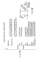
FIG. 15 is a scheme schematically showing steps of
the experiment described above. The detection results
are shown below. FIGS. 14A and 14B are the results
obtained by capillary electrophoresis. As shown in
FIGS. 14A and 14B, specific PCR amplification of
individual FL sequences can be attained by the encode
reaction of the present invention. As a result,
specific PCR products are obtained.
7-2. Quantitative properties of the PCR
amplification after the encode reaction
(1) Experimental method
The uniformity and quantification property of the
PCR performed after the encode reaction were confirmed
by using the following method. The experiment was
performed with respect to the flag sequences for
detecting IGTP and LRG-47. The flag sequences for
detecting IGTP and LRG-47 include both SD sequence and
ED sequence. PCR reactions of both the flag sequences
for IGTP and LRG-47 were performed by use of the SD
sequence and ED sequence. The amounts of strands of
both sequences added to the PCR reaction fall within
100 pM to 10 fM. The cycle of the PCR reaction
consisting of 94°C/30 seconds, 65°C/30 seconds,
65°C/60 seconds, and 72°C/30 seconds, was repeated
25 times.
(2) Experimental results
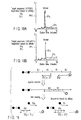
FIG. 17 is a scheme schematically showing steps of
the experiment mentioned above. The detection results
are shown below. FIG. 15 are the results obtained by
capillary electrophoresis. As shown in FIG. 16, the
amount of PCR amplification after the encode reaction
of the present invention varies depending upon the
concentration of the target sequence.
7-3. Specificity and Quantitative properties of
the decode reaction
(1) Experimental method
The sequences used herein are the same as
mentioned above. More specifically, an
SD+D0IGTP+D1IGTP+ED sequence added with biotin
(hereinafter, also referred to as "b-code 1") and SD+
D0LRG-47+D1LRG-47+ED (hereinafter, also referred to as
"b-code 2") were subjected to a decode reaction.
0.2 µL of the b-code 1 and b-code 2 (concentration:
100 µM) were mixed with a TE buffer (49.8 µL)
containing 1M NaCl and 40 µL of dynabeads M-280
streptoavidin (10 mg/mL). The resultant solution was
stirred for 15 minutes, washed, at room temperature,
and then, the supernatant was removed. To the obtained
beads, a TE buffer solution of 50 µL was added, and the
mixture was washed at room temperature. After the
supernatant was removed, a further 50 µL of TE buffer
solution was added to the beads obtained. The
resultant beads were washed at 80°C and the supernatant
was removed. Thereafter, the resultant beads was
washed at room temperature with a ligation buffer and
the supernatant was removed. Subsequently, a reverse
strand relative to D1 and reverse strands relative to
D0IGTP and D0LRG-47 were added to the resultant beads
to a final concentration of 400 nm, individually. The
temperature of the resultant mixtures was raised to 95°C
to convert the reverse strand into a single-strand
reverse strand. The temperature was further reduced at
a rate of 7°C per minute. Subsequently, Taq ligase of
20 units were added at 60°C to the resultant mixture,
the ligation reaction was performed for 15 minutes.
After the supernatant was removed, 50 µL of the TE
buffer was added to the reaction solution. The
resultant mixture was washed at room temperature.
Thereafter, the supernatant was removed, 50 µL of the
TE buffer was again added. The temperature of the
reaction solution was increased to 80°C and the
supernatant was recovered. In this manner, the probes
ligated was recovered. The streptoavidin beads (40 µL)
were added to the supernatant obtained and further a TE
buffer solution (12.5 µL) containing 1M NaCl was added,
and stirred for 15 minutes. Thereafter, the
supernatant was recovered at 80°C, an aliquot (20 µL)
of the supernatant was added to each of two tubes. The
probe (b-1) having the sequence corresponding to DOIGTP
having biotin added thereto and the probe (b-2) having
the sequence corresponding to D0LRG-47 having biotin
added thereto were added to each of two tubes in an
amount of 100 µmol. To the resultant solution, further
16 µL of TE buffer solution containing 2M NaCl was
added. The resultant mixture was increased to a
temperature to 95°C. Thereafter, the temperature of the
mixture was reduced to 25°C at a rate of 10°C per minute.
In this manner, hybridization was performed. To this
solution, 40 µL of the streptoavidin was added and the
solution mixture was stirred for 15 minutes.
Thereafter, the supernatant was removed and 40 µL of TE
buffer solution was added to the resultant mixture.
The mixture was washed at 40°C. Subsequently, the
supernatant was removed and the TE buffer solution
(40 µL) was further added, and the temperature of the
resultant mixture was increased to 70°C. The
supernatant was recovered, and each supernatant was
subjected to capillary electrophoresis for a single
strand.
(2) The experimental results
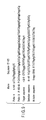
FIG. 19 is a scheme schematically the steps of the
experiment described above. The detection results are
shown below. FIGS. 18A and 18B are the results
obtained by capillary electrophoresis. The decode of
the present invention was performed by ligating a PCR
product amplified by the encode reaction with D1 and
labeled D2 sequences corresponding to respective genes.
The probes ligated were detected by D1IGTP and D1LRG-47
labeled with biotin. As a result, it was demonstrated
that the product corresponding to each of genes can be
specifically detected, as shown in FIG. 17.