TECHNICAL FIELD
This invention relates to diagnostic methods and a
diagnostic aid for judging the onset and progress of diseases
associated with a change in the coupling factor (CF6) level in
the blood, diseases in association with the excess or shortage
of PGI2 or diseases associated with the accentuation or
attenuation of cPLA2 function, which may be referred to as cPLA2
hyperfunction or cPLA2 hypofunction, and a pharmaceutical
composition for the treatment of these diseases. The present
invention further relates to a vector containing a DNA encoding
CF6 or a polypeptide which is a part thereof, a transformant
having been transformed by this vector, a method of efficiently
producing CF6 and a partial polypeptide thereof, an antibody
reacting specifically with CF6, a method for producing the
antibody, and a method of assaying CF6.
BACKGROUND ART
Prostanoids produced via the arachidonate cascade
(prostaglandin (PG), thromboxane (TG), leukotriene (LT), etc.)
are physiologically active substances which are essential in
maintaining the homeostasis in vivo. Prostacyclin (PGI2),
which is a prostanoid is synthesized in hemoendothelial cells
under the action of PGI2 synthase on PGH2 having been formed
from arachidonic acid by cyclooxygenase and prostaglandin H2
(PGH2) synthase.
It is known that the PGI
2 has the following
pharmacological effects:
As described above, the biosynthesis pathway of PGI2
and the enzymes participating in its biosynthesis
(arachidonate cascade) have been identified and thus the
physiological activities of PGI2 have been clarified to a
certain extent. However, the mechanism of regulating the
PGI2 production in diseases still remains unknown.
Regarding the regulation of the PGI2 production, on
the other hand, it has been suggested from the results of
experiments with the use of spontaneously hypertensive rats
(SHR), that an endogenous PGI2 production inhibitor may be
present in SHR (Osanai, T. et al., Jpn. Circ. J. 54, 507-514,
1990; Falardeau, P. et al., Prostaglandins, 29, 621-628,
1985). Although this endogenous PGI2 production
inhibitor has not yet been discussed sufficiently, the
present inventors successfully isolated and purified from a
SHR heart a PGI2 production inhibitory factor secreted by
smooth muscle cells originating in SHR mesenteric artery.
Further, they determined the structure of this factor and
clarified that it is rat CF6 (hereinafter referred to as
rCF6) which is one of the subunits of H+ ATP synthase
(Referential Example 1).
Mammalian H+ ATP synthase is present in the
mitochondrial inner membrane and consists of at least 14
subunits. CF6, which is one of these subunits, consists of
76 amino acids. CF6 is synthesized as a peptide having 32
amino acids containing a mitochondrial transit sequence at
the N-end (Higuchi, T. et al., Biochem. Biophys. Res.
Commun. 178, 793-799, 1991).
The present inventors have established a method
comprising expressing a chimeric protein from which rCF6
can be cut off by using factor Xa with the use of
Escherichia coli as a host, and cutting off rCF6 from the
chimeric protein followed by purification (Referential
Example 2).
In addition, the present inventors have disclosed
that the action point of the inhibition of the PGI2
production by CF6 resides in the PLA2 inhibition
(Referential Example 3), in particular, in the cPLA2
inhibition (Referential Example 4) (Osanai, T. et al., J.
Biol. Chem. 273, 31778-31778, 1998). However, the
relationships between CF6, which is an intracellular
peptide, and diseases have not been known so far. Moreover,
it still remains unknown as to how CF6 acts on
hemoendothelium in vivo, namely, whether or not CF6 is
present in the blood.
On the other hand, it has been clarified the
following facts with respect to the functions of cPLA
2.
Based on these facts, it is estimated that cPLA2
inhibitors are usable as potent remedies for inflammation
or remedies for brain infarction, etc. However, there has
been found hitherto neither an efficacious remedy nor a
method of screening a cPLA2 inhibitor on the basis of the
effects of CF6.
As described above, the biosynthesis and
physiological effects of PGI2, the physiological effects of
cPLA2 in the arachidonate cascade and the relations among
PGI2, cPLA2 and CF6 have been gradually clarified. However,
causes of diseases, in which PGI2 located at the end of the
arachidonate cascade and cPLA2 located at the initial point
thereof participate, have not been sufficiently clarified
yet.
DISCLOSURE OF THE INVENTION
The present inventors have clarified that CF6 is
present in rat blood and human blood and that CF6 is
produced in hemoendothelial cells. They have also
disclosed that the blood rCF6 level is elevated with an
increase in blood pressure in the case of hypertension with
the shortage of PGI2; that the blood pressure further
increases by administering rCF6 in this state; and that an
anti-rCF6 antibody exerts a hypotensive effect in the case
of hypertension with a high rCF6 level. Moreover, they
have found that the blood CF6 level is elevated in the case
of human acute heart infarction, namely, the blood CF6
level changes in human diseases too, thereby having
completed the present invention.
Under the above-described circumstances, the present
invention relates to a pharmaceutical composition for the
prevention or the treatment of diseases caused by changes
in the blood CF6 level, diseases associated with the excess
or shortage of PGI2 and diseases associated with the
accentuation or attenuation of cPLA2 function, and
diagnostic methods and a diagnostic aid for judging the
onset and progress of diseases based on the control of the
blood CF6 level.
Accordingly, the present invention provides a
pharmaceutical composition for the prevention or the
treatment of diseases associated with an increase or a
decrease in the CF6 level, diseases associated with an
increase or a decrease in the PGI2 level and diseases
associated with the accentuation or attenuation of the
cPLA2 function. The pharmaceutical composition comprises,
as the active ingredient, CF6 activators (for example, CF6,
CF6 secretion-promoting substances and CF6 agonists) or CF6
inhibitors (for example, CF6 secretion-inhibiting
substances and CF6 antagonists). The present invention
also provides a method of measuring the blood CF6 level.
The present invention still further provides a diagnostic
method and a diagnostic aid for diseases associated with an
increase or a decrease in the CF6 level.
Moreover, the present invention provides CF6
secretion accelerating substances, CF6 secretion inhibitory
substances, CF6 agonists and CF6 antagonists which are the
active ingredients of the pharmaceutical composition for
the prevention or the treatment of the diseases as cited
above. In addition, the present invention provides an
anti-CF6 antibody which is usable as a reagent in the
method of measuring the blood CF6 level or as a diagnostic
aid in the diagnostic methods. The present invention
further provides a vector containing a DNA encoding CF6, a
transformant having been transformed by this vector, and a
method of producing a CF6-like peptide. According to the
present invention, an antibody reacting specifically with
CF6 or a partial polypeptide thereof and a method for
producing the same are provided.
BRIEF DESCRIPTION OF THE DRAWINGS
Fig. 1 is a diagram which illustrates how to
construct a vector for expressing a chimeric protein from
which rCF6 or human CF6 (hCF6) can be cut off with
enterokinase from a rat liver cDNA library or a human
kidney cDNA library.
Fig. 2 shows the C18-HPLC elution profile of a
solution wherein the rCF6-containing chimeric protein thus
expressed is treated with enterokinase.
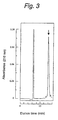
Fig. 3 shows the C18-HPLC elution profile of partial
purified rCF6.
Fig. 4 shows the C18-HPLC elution profile of a
solution wherein the hCF6-containing chimeric protein thus
expressed is treated with enterokinase.
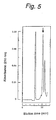
Fig. 5 shows the C18-HPLC elution profile of partial
purified rCF6.
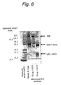
Fig. 6 provides SDS-PAGE patterns of an antiserum
prepared by administering to rabbits an immune antigen
derived from rCF6, and of antibodies purified by the
immunoaffinity chromatograpy using an antigen peptide as
the affinty ligand.
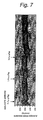
Fig. 7 provides a chart which shows the effect of the
anti-rCF6 antibody on the blood pressure of SHR (16 weeks
of age).
Fig. 8 provides a graph which shows the effects of
the anti-rCF6 antibody on rats (SHR and WKY, 16 weeks of
age).
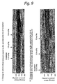
Fig. 9 provides a chart which shows the effects of
the preadministration of the anti-rCF6 antibody on the
decrease in blood pressure of rats (SHR, 16 weeks of age)
caused by bradykinin.
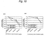
Fig. 10 provides a chart which shows the effects of
the preadministration of the anti-rCF6 antibody on the
decrease in blood pressure of rats (SHR and WKY, 16 weeks
of age) caused by bradykinin.
Fig. 11 provides a chart which shows the effects of
rCF6 on the blood pressure of rats (SHR and WKY, 16 weeks
of age).
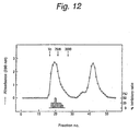
Fig. 12 provides a chart which shows the
fractionation with Sephadex G-25 of a boiling water-extract
of SHR heart.
Fig. 13 provides a chart obtained by analyzing the
active fraction in the above Fig. 12 by HPLC.
Fig. 14 provides a chart obtained by analyzing the
active fraction in the above Fig. 13 by HPLC.
Fig. 15 is a diagram which shows how to construct a
vector for expressing a protein containing rCF6 having the
recognition sequence of the factor Xa added to the N-terminus
from a rat aorta cDNA library.
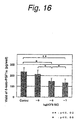
Fig. 16 provides a graph which shows the inhibition
of the PGI2 production by rCF6.
Fig. 17 provides a graph which shows the inhibition
of production by rCF6 in the presence of bradykinin.
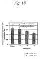
Fig. 18 provides a graph which shows the effect of
rCF6 on the release of arachidonic acid from human
umbilical venal endothelial cells (HUVEC).
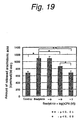
Fig. 19 provides a graph which shows the effect of
rCF6 on the effect of bradykinin of releasing arachidonic
acid from HUVEC.
Fig. 20 provides a graph which shows the effects of
various PLA2 inhibitors and rCF6 on the release of
arachidonic acid from HUVEC.
Fig. 21 provides a graph which shows the effects of
various PLA2 inhibitors and rCF6 on the effect of
bradykinin of releasing arachidonic acid from HUVEC.
Fig. 22 provides a graph which shows the effects of
rCF6 on the blood pressure of rats (SHR and WKY, 16 weeks
of age).
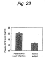
Fig. 23 provides a graph which shows a comparison of
the blood hCF6 levels of normal subjects and patients with
acute heart infarction.
Fig. 24 provides a graph which shows changes in the
blood hCF6 level of patients with acute heart infarction.
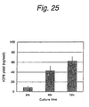
Fig. 25 provides a graph which shows changes in the
hCF6 level in a liquid culture of HUVEC.
MODE FOR CARRYING OUT THE INVENTION
(Production of CF6)
In the present invention the rat CF6 and the human
CF6, which are to be used as the active ingredients in the
above-described pharmaceutical composition for the
prevention or the treatment, as a reagent or a diagnostic
aid in the method of measuring the blood CF6 level, or as a
reagent or a diagnostic aid in the diagnostic methods for
diseases associated with changes in the blood CF6 level,
can be efficiently produced by using enterokinase as a
cutting enzyme compared with the conventional method
(Referential Example 2). Enterokinase is an enzyme which
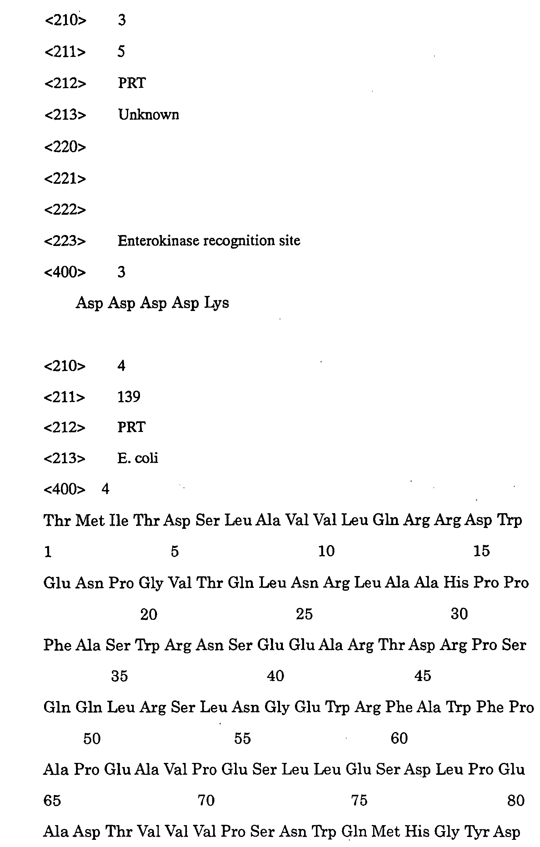
recognizes a sequence Asp-Asp-Asp-Asp-Lys (SEQ ID NO:3) and
cuts the peptide bond at the C-end thereof. To obtain CF6,
a chimeric protein carrying a protective peptide at the N-terminus
of the enterokinase recognition sequence and CF6
or a partial peptide thereof at the C-terminus is obtained
by a conventional gene recombination technique and then CF6
is cut off therefrom with enterokinase. As the chimeric
protein, use can be made, for example, of a peptide wherein
CF6 is bonded via the recognition sequence of enterokinase
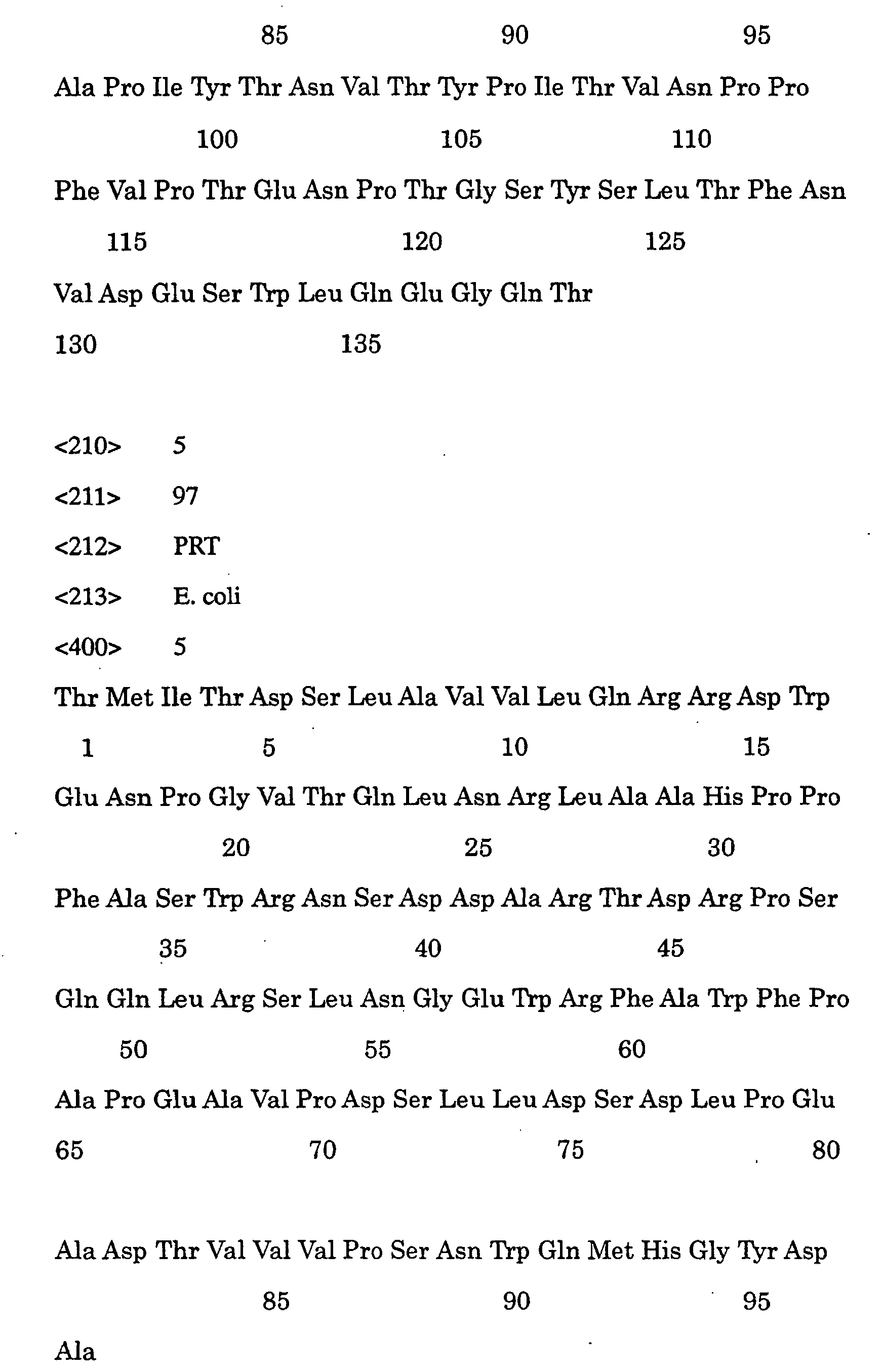
to the C-terminus of a peptide consisting of 139 N-terminal
amino acids of E. coli β-galactosidase (cysteine residues
at the 76- and 122-positions have been substituted by
serine: SEQ ID NO:4). Due to a large accumulation of the
insoluble fraction of E. coli, this chimeric protein can be
efficiently expressed and purified. After solubilizing the
insoluble chimeric protein in a urea solution, CF6 can be
cut off from the chimeric protein by treating with
enterokinase. The thus cut off CF6 can be purified by
subjecting to HPLC employing a C18 column. An expression
vector can be constructed and a host can be transformed
each by a conventional method well known by a person
skilled in the art (Maniatitis et al., "Molecular Cloning:
A Laboratory Manual", Cold Spring Harbor Laboratory, 1989).
(Partial polypeptide)
The term "partial polypeptide" as used herein means a
polypeptide corresponding to a part of CF6 which has an
effect of inhibiting the production of PGI2 from human
umbilical venal endothelial cells (HUVEC). Also,
polypeptides formed by the modification of a polypeptide
corresponding to a part of CF6 by the deletion, addition,
substitution or combination thereof of one to several amino
acids and having an effect of inhibiting the production of
PGI2 from HUVEC fall within this category.
(Production of CF6 antibody)
An antibody immunologically specific to CF6, which is
to be used as the active ingredient in the above-described
pharmaceutical composition for the prevention or the
treatment or as a reagent or a diagnostic aid in the method
of measuring the blood CF6 level or the diagnostic methods
in the present invention, can be obtained by sensitizing an
animal (preferably a non-human animal) with the use of CF6,
its fragment or an analog thereof as an immunogen by a
conventional experimental method. In case of preparing a
monoclonal antibody, use can be made of, for example, the
hybridoma method (Kohler, G. et al. Nature, 256:495-497,
1975) whereby an antibody produced by continuous cell line
culture is presented. A single-stranded antibody against
CF6 can be produced by applying a technique to be used in
producing a single-stranded antibody (US Patent 4946778).
Also, a human-type antibody can be expressed by using a
transgenic mouse having a human antibody production system
(Green LL. et al. Nat Genet, 7, 13-21, 1994). In this
specification, an example of the production of a polyclonal
antibody, wherein rabbits were immunized with an antigen
obtained by synthesizing a peptide having a cysteine added
to the N-terminus of an rCF6- or hCF6-origin fragment
consisting of about 20 amino acids and bonding keyhole
limpet hemocyanin thereto via the N-terminal cysteine, is
presented (Example 2).
(Method of measuring CF6 level)
According to the present invention, CF6 in a
sample can be assayed by the RIA method or the ELISA
method with the use of the antibody as described
above (Radioimmunoassay: Kitagawa p.79-88; Enzyme
Immunoassay:p.88-99, Shin-seikagaku Jikken Koza 12, Bunshi
Menekigaku III, Kogen, Kotai, Hotai, ed. by Nippon
Seikagaku Gakkai, 1992). As an example thereof, an
antagonistic inhibition system (RIA method) wherein
quantification is carried out by inhibiting the reaction
between a 125I-labeled antigen with an antibody by an
antigen in a sample is presented in this specification
(Example 2).
(Method of screening CF6 secretion accelerating substance
or CF6 secretion inhibitory substance)
According to the present invention, it is also
possible to obtain a CF6 secretion accelerating substance
or a CF6 secretion inhibitory substance by using the
above-described method of measuring the CF6 level. More
particularly speaking, a test substance is added to a
liquid culture of cells capable of secreting CF6 (for
example, HUVEC) and the amount of CF6 secreted into the
liquid culture is measured by an assay system with the use
of the anti-CF6 antibody (for example, the RIA method as
described above). Thus, a CF6 secretion accelerating
substance or a CF6 secretion inhibitory substance can be
obtained (Example 7). It is still preferable to use cells
causative of diseases as the cells capable of secreting CF6.
(Method of screening CF6 agonist or CF6 antagonist)
According to the present invention, it is also
possible to obtain a method of detecting a CF6 agonist or a
CF6 antagonist.
More particularly speaking, a test substance is added
together with CF6 to a liquid culture of cells wherein the
PGI2 production is inhibited by CF6 (for example, HUVEC or
smooth muscle cells originating in rat mesenteric artery).
Then 6-keto-PGF1α, which is the stable metabolite of PGI2,
secreted in the liquid culture is quantitated. Thus, a CF6
antagonist causing an increase in the 6-keto-PGF1α amount
or a CF6 agonist causing a decrease in the 6-keto-PGF1α
amount can be obtained. Alternatively, an antagonist or an
agonist can be detected by adding a test substance together
with CF6 to a liquid culture of HUVEC in the presence of
labeled arachidonic acid and measuring arachidonic acid
thus released into the medium. It is still preferable to
use cells causative of diseases as the cells producing PGI2.
(Method of identifying CF6-binding receptor)
The present invention also provides a method of
identifying a CF6-binding receptor. A CF6-binding receptor
can be identified by standard receptor-binding methods
commonly known in the art with the use of CF6. Although
these methods include the ligand binding method and the
crosslinking assay method, the present invention is not
restricted thereto. In such a method, CF6 is fused with a
peptide sequence appropriate for radioisotope (for example,
125I) labeling, chemical modification (for example,
biotinylation), detection or purification and then
incubated with a membrane fraction of cells such as HUVEC.
The labeled CF6 is also usable in the method of identifying a
CF6 agonist or a CF6 antagonist in competition with the binding
of CF6 to its receptor with the use of binding assay. Standard
methods for carrying out such assays are well known in the art.
(Examples of diseases)
The term "diseases associated with the excess of CF6" as
used herein means diseases wherein the function of CF6 in the
blood is accentuated to a level undesirable for a living body.
Namely, these diseases are not always restricted to those
wherein the blood CF6 level is higher than the level of normal
persons. Namely, diseases associated with the shortage of PGI2
and diseases associated with the attenuation of the cPLA2
function or the cPLA2 hypofunction fall within this category.
Examples thereof include diseases associated with accentuated
platelet agglutination, diseases associated with peripheral
circulatory failure caused by inhibited vasodilatation, heart
infarction, angina pectoris, heart failure, pulmonary
hypertension, hypertension, cerebrovascular disorder,
arteriosclerosis obliterans, arteriosclerosis, hyperlipemia,
diabetes, bronchial disease, stomach ulcer, eclampsia of
pregnancy, hemolytic-uremic syndrome and thrombotic
thrombocytopenic purpura.
The term "diseases associated with the shortage of CF6"
as used herein means diseases wherein the function of CF6 in
the blood is attenuated to a level undesirable for living body.
Namely, these diseases are not always
restricted to those wherein the blood CF6 level is lower than
the level of normal persons. Namely, diseases associated with
the excess of PGI2 and diseases associated with the
accentuation of the cPLA2 function or cPLA2 hyperfunction fall
within this category. Examples thereof include brain
infarction, acute pancreatitis, asthma, ARDS and rheumatoid
arthritis. (Provision of pharmaceutical compositions for
treatment)
According to the present invention, the pharmaceutical
composition for the treatment of the above described diseases
associated with the excess or shortage of the blood CF6 level
can be provided.
In case where the CF6 activity is in excess, the CF6
activity can be lowered by administering antagonists or CF6
secretion inhibitory substances in an amount efficacious in
inhibiting the action of CF6 on ligands, substrates, receptors,
enzymes, etc. These antagonists or CF6 secretion inhibitory
substances can be obtained by the screening method as described
above.
To treat abnormal conditions associated with the shortage
of CF6, on the other hand, the CF6 activity can be elevated by
administering agonists or CF6 secretion accelerating substances
in an amount efficacious in promoting the action of CF6 on
ligands, substrates, receptors, enzymes, etc". These agonists or
CF6 secretion accelerating substances can be obtained by the
screening method as described above. (Method of producing
preparations)
Medicinal preparations of the CF6 secretion
accelerating substances, CF6 secretion inhibitory
substances, CF6 agonists or CF6 antagonists obtained by
using the screening method according to the present
invention can be produced by conventional methods. For
example, these preparations may be used in the dosage form
of solid preparations such as optionally coated tablets,
capsules, granules or fine powder; injections, eye drops or
nasal drops obtained by dissolving in water or other
pharmaceutically acceptable liquid vehicles; or external
preparations such as ointments or patches.
These preparations can be produced in a conventional
manner with the use of additives commonly employed in the
art. In case of the production of solid preparations for
oral administration such as tablets, capsules, granules and
fine powder, use can be made as additives of: (1) fillers
such as lactose, starch and crystalline cellulose; (2)
binders such as hydroxypropylcellulose and polyvinyl
pyrrolidone; (3) disintegrating agents such as starch and
croscarmellose sodium; (4) plasticizers such as macrogol
and triethyl citrate; (5) lubricants such as magnesium
stearate and talc; (6) coating bases such as
hydroxypropylmethylcellulose and Eudragit; (7) taste
correctives such as sucrose and mannitol; and odor
correctives, coloring agents, etc.
In case of the production of injections, eye drops
and nasal drops, use can be made as additives of: (1)
tonicity agents such as sodium chloride, D-mannitol and D-sorbitol;
(2) pH controlling agents such as hydrochloric
acid and citric acid; (3) buffers such as sodium citrate,
sodium acetate and boric acid; (4) soothing agents such as
procaine hydrochloride; and stabilizers, surfactants, etc.
Considering the stability, etc. of the active ingredient,
these preparations may be either dissolved or suspended
just before using or in the form of solutions.
In case of the production of external preparations
such as ointments and patches, use can be made as additives
of: (1) bases such as liquid paraffin, vaseline and
hydrophilic ointment; (2) emulsifiers such as polysorbate
80 and tragacanth; (3) preservatives such as sodium
benzoate and propyl parahydroxybenzoate; (4) soothing
agents such as procaine hydrochloride; and stabilizers,
surfactants, etc.
The preparations thus prepared are packaged in an
appropriate manner depending on the dosage form. Examples
of the package styles include bottling, folding, PTP-packaging,
ampul-filling and vialling. The preparations
thus obtained can be administered to, for example, mammals
such as rat, rabbit, sheep, pig, cow, cat, dog, monkey,
humans, etc. The administration dose varies depending on
the indication, symptom, administration route, etc. In
case of a human adult weighing 60 kg, the daily dose
generally ranges from about 0.01 to 300 mg, preferably from
about 0.1 to 100 mg. In case of other animals, the
administration dose may be calculated by converting the
bodyweight into a corresponding fraction of 60 kg.
(Diagnostic methods)
The present invention also provides methods of
diagnosing diseases associated with an increase or a
decrease in the CF6 level which comprises measuring the CF6
level in collected blood samples. As described above, the
CF6 level can be measured by the RIA method or the ELISA
method with the use of antibody.
(CF6 gene or mRNA)
According to the present invention, it is possible to
use a CF6 gene or mRNA usable as a diagnostic or a
diagnostic aid for the underexpression or hyperexpression
of CF6, changes in the localization of CF6 or diseases
caused thereby.
Individuals having a mutated CF6 gene can be detected
by various methods for measuring DNA sequences. The
nucleic acids to be employed in the diagnosis can be
obtained from the sample cells present in, for example,
blood, urine, saliva, biopsy specimens or autopsy specimens.
Genomic DNA may be either employed as such or amplified
prior to the analysis by the PCR method or the like. mRNA
or cDNA may be used in the same manner. Thus, a deletion
or insertion can be detected based on the difference in the
amplification product size from the normal genotype. A
point mutation can be identified by hybridizing the
amplified DNA with CF6 DNA having the normal DNA nucleotide
sequence. Completely matched sequences can be distinguished
from sequences of mismatched double strands by digesting
with RNase or by measuring difference in melting
temperature. Also, a difference of DNA sequences can be
detected based on a change in the mobility in gel when a
DNA fragment is electrophoresed alone or together with a
denaturing substance. It is also possible to detect the
difference by directly determining the DNA sequence. A
change in the sequence at a specific position can be
clarified by a nuclease protective assay, for example,
protection with RNase and S1 or by the chemical cutting
method. Alternatively, it is also possible to construct
an array of oligonucleotide probes containing the CF6
nucleotide sequence or fragments thereof and carry out, for
example, an effective screening of a genetic mutation. This
array method, which has been widely known, is applicable
over a broad scope and is usable in studying various
problems in the field of molecular genetics such as gene
expression, genetic linkage and genetic change.
[Examples]
Now, the invention will be illustrated in greater
detail by reference to the following Examples. However, it
is to be understood that these Examples merely provide
modes for carrying out the present invention and the
invention is not to be construed as being limited thereto.
Unless otherwise noted, technical terms and abbreviations
are employed in these Examples in accordance with the
common usage in the art. The plasmids, E. coli and
fundamental expression procedures employed in common in
these Examples will be illustrated below for reference. To
confirm and illustrate the prior art providing the basis of
the present invention and the related art of the present
invention, Referential Examples will be also given.
(Plasmid)
pG97S4DhCT[G] is a plasmid capable of expressing a
chimeric protein wherein hCT[G] (a peptide having Gly
added to the C-terminus at the 32-position of human
calcitonin) is bonded via a sequence Glu-Phe-Glu to a
peptide consisting of the amino acids from the N-terminus
to the 97-position of β-galactosidase (called βGal-97S4D
represented by SEQ ID NO:5 wherein Cys at the 76-position
is substituted by Ser and Glu at the 40-, 41- 71- and 75-positions
are substituted by Asp) by an E. coli lactose
operon promoter. A transformant obtained by using this
plasmid can be selected depending on resistance to
tetracycline. A chimeric protein with βGal-97S4D can be
expressed by transferring an EcoRI-SalI DNA fragment
consisting of the DNA domain encoding a target peptide and
the coding frame. An E. coli W3110 strain containing this
plasmid was named Escherichia coli SBM323 and deposited
with National Institute of Bioscience and Human Technology,
Agency of Industrial Science and Technology under the
accession number FERMBP-3503 on August 8, 1991.
pGP#19RI is a plasmid wherein a gene encoding the
chimeric protein βGal-97S4D/hCT[G] of pG97S4DhCT[G] is
substituted by another gene encoding a chimeric protein
βGal-139S(SEQ ID NO:4)/hProPTH. A chimeric protein with
βGal-139S can be expressed by transferring an EcoRI-SalI
DNA fragment consisting of the DNA domain encoding a target
peptide and the coding frame. This plasmid pGP#19RI can be
constructed by inserting the EcoRI site into the DNA domain
encoding the bond between the protective peptide βGal-139S
and ProPTH of a plasmid pGP#19 (unexamined published
Japanese application (Kokai) No.9-296000; Fig. 4).
A plasmid pCRII was purchased from Invitrogen Co. in
the state of allowing direct linkage to DNA fragments
having been amplified by the PCR method with the use of Taq
DNA polymerase. A transformant obtained by using this
plasmid pCRII can be selected depending on resistance to
ampicillin.
(E. coli and media)
The E. coli JM109 strain was purchased form Toyobo
Co., Ltd. and employed in preparing the plasmids and
expressing the chimeric proteins.
The media employed were an LB medium (0.5% (w/v) of
yeast extract, 1% (w/v) of trypton, 0.5% (w/v) of NaCl) and
an SB medium (2% (w/v) of yeast extract, 1% (w/v) of
trypton, 0.5% (v/v) of glycerol, 1.2% (w/v) of K2HPO4, 0.3%
(w/v) of KH2PO4).
(Fundamental experimental procedures)
Unless otherwise stated in particular, the following
experimental procedures were employed in Examples. DNA
nucleotide sequences were determined by using the dideoxy
method with a DNA Sequencer 373 (Applied Biosystems). To
cleave DNA with restriction enzymes, the reaction was
performed for 1 hour using the enzymes in an amount 3 to 10
times more than the amount specified by the manufacturer.
Plasmid structures were analyzed in 20 µL of a liquid
reaction mixture by using 0.5 to 1 µg of DNA. Preparation
of DNA was carried out in 50 to 100 µL of a liquid reaction
mixture by using 3 to 10 µg of DNA. The reaction
conditions (temperature, buffer, etc.) were set up in
accordance with the manufacturer's instruction. The
obtained DNA fragments were separated by agarose gel
electrophoresis and subjected to analysis or preparation.
Samples for the agarose gel electrophoresis were
prepared by adding 1.5 times by volume as much a
colorant solution (15% (w/v) aqueous solution of Ficoll
containing 0.25% (w/v) of Bromophenol Blue) to liquid
reaction mixtures. As the buffer for the agarose gel
electrophoresis, use was made of a TAE buffer (10 mM of
tris, 20 mM of acetic acid, 2 mM of EDTA·Na, pH 6.8). In
analyzing the plasmid structures and preparing the DNA
fragments, migration was carried out by using Mupid-2
(COSMO BIO) at 100 V for 1 hour. The gel was stained with
an aqueous solution of ethidium bromide (0.5 µg/ml) for 20
minutes and then irradiated with ultraviolet light to
detect DNA bands. The concentration of the.agarose gel was
adjusted to 1.0, 1.5 and 2.0% (w/v) depending on the size
of the DNA fragment to be fractionated. DNA in the agarose
gel was extracted from the gel by using SUPREC-01 (Takara
Shuzo Co., Ltd.). DNA solutions were treated with phenol
and then subjected to precipitation from ethanol. Ligation
reactions were carried out by using 0.05 to 1 µg of DNA
fragments with the use of a TAKARA Ligation Kit (Takara
Shuzo Co., Ltd.).
E. coli was transformed by the calcium chloride
method, wherein competent cells of JM109 strain were
employed, and transformants were selected depending on the
resistance to drugs (ampicillin or tetracycline).
The SDS polyacrylamide gel electrophoresis (SDS-PAGE)
was performed in accordance with Laemmli's method (Laemmli
et al. Nature 227, 680-685, 1970). Namely, 1/4 times by
volume as much 4xSDS sample buffer (375 mM of tris
hydrochloride (pH 6.8), 30% (v/v) of glycerol, 7% (w/v) of
SDS, 15% (v/v) of 2-mercaptoethanol and 0.1% (w/v) of
Bromophenol Blue) was added to a sample and heated to 95°C
for 5 minutes. Then 10 µL of the reaction mixture was
subjected to SDS-polyacrylamide gel (55 mm x 85 mm x 1 mm,
Tefco) and electrophoresed at 20 mA for 80 minutes. After
the completion of the electrophoresis, the gel was stained
with a staining solution (10% (v/v) of acetic acid, 40%
(v/v) of methanol and 0.25% (w/v) of Coomassie Brilliant
Blue R250).
In the analysis and purification of peptides by HPLC
with the use of a C18 column, use was made of an HPLC LC10A
(Shimadzu Corporation) connected to an YMC0ODS-A302 column
(diameter: 4.6 mm x 150 mm), an YMC-ODS-A323 column
(diameter: 10 mm x 250 mm) (YMC K.K.) and an Inertsil ODS-2
C18 column (diameter: 4.6 mm x 250 mm) (GL Science K.K.).
Development was carried out under concentration gradient of
a solution A (0.1% (v/v) trifluoroacetic acid (TFA)) and a
solution B (0.092% (v/v) TFA/80% (v/v) acetonitrile, or
0.092% (v/v) TFA/60% (v/v) acetonitrile) at a flow rate of
1 ml/min or 2.5 ml/min. The peak area of the absorbance at
210 nm was measured.
DNA oligomers were synthesized by Greiner Japan or
Lifetech Oriental.
The activity of inhibiting the PGI2 production was
evaluated by measuring the stable metabolite of PGI2 (6-keto-PGF1α)
secreted into the medium of smooth muscle cells
collected from SHR mesenteric artery or human umbilical
venal endothelial cells (HUVEC). Namely, the smooth muscle
cells collected from SHR mesenteric artery were subcultured
in a Dulbecco's modified Eagle medium (DMEM) containing 10%
(v/v) of fetal calf serum until these cells attained the
confluent state. HUVEC were proliferated in a synthetic
medium (HuMedia, Kurabo Industries) containing 2% (v/v) of
fetal calf serum, 10 ng/ml of EGF, 5 ng/ml of bFGF, 1 µg/ml
of hydrocortisone and 10 µg/ml of heparin. The smooth
muscle cells collected from SHR mesenteric artery or HUVEC
were incubated for 30 minutes in the DMEM medium containing
the sample to be analyzed and 6-keto-PGF1α secreted into
the medium during the incubation period was determined.
Blood pressure was measured in the open manner by
inserting catheters directly into the aorta of rats
anesthetized with 75 mg/kg of ketamine and 15 mg/kg of
xylazine hydrochloride. Namely, two polyethylene catheters
filled with physiological saline containing 20 U/ml of
heparin were inserted respectively into the left carotid
artery and the left femoral vein. Then the catheter
inserted into the left carotid artery was connected to a
pressure detector (Carrier Amplifier AP-601G, Nihon Kohden
Co.). Each sample was dissolved in 200 µL of physiological
saline and then administered via the vena cava.
The instruments and reagents were each used in
accordance with the manufacturer's instruction.
Fundamental gene manipulation procedures were carried out
by reference to Maniatitis et al., "Molecular Cloning: A
Laboratory Manual", Cold Spring Harbor Laboratory, 1989).
[Example 1] Preparation of CF6 by chimeric protein
expression method-1
Next, a method of producing CF6 by using enterokinase
as an enzyme for cutting it off from a chimeric protein
will be described in detail.
(Preparation of rCF6 cDNA and hCF6 cDNA)
rCF6 cDNA was obtained by the PCR method with the use
of rat liver cDNA (Quick Clone cDNA Library, Clontech) as a
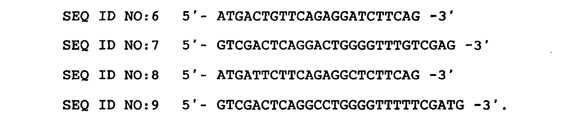
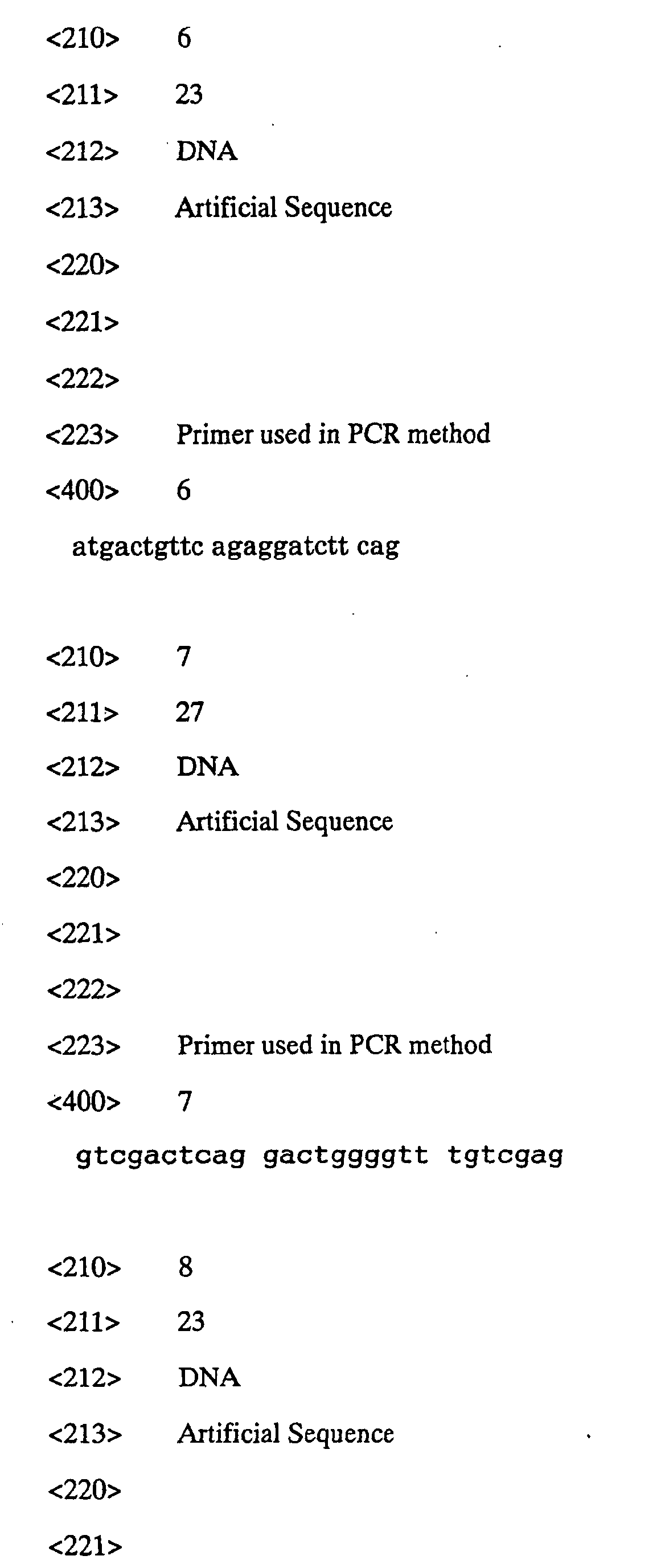
template and rCF6-01 (SEQ ID NO:6) and rCF6-02 (SEQ ID
NO:7) as primers. Namely, by using KOD (Toyobo Co., Ltd.)
as a thermostable DNA polymerase, a liquid reaction mixture
comprising 1 µL of the rat liver cDNA library, 5 µL of 10 x
reaction buffer, 7.5 µL of 2 mM of dNTP mix, 0.5 µL of
rCF6-01, 0.5 µL of rCF6-02 and 35 µL of sterilized water
was subjected to the reaction for 30 cycles with each cycle
consisting of 30 seconds at 96°C, 1 minute at 56°C and 40
seconds at 74°C. Thus rCF6 cDNA was obtained. Similarly,
hCF6 cDNA was obtained by the PCR method with the use of
human kidney cDNA (Quick Clone cDNA Library, Clontech) as a
template and hCF6-01 (SEQ ID NO:8) and hCF6-02 (SEQ ID
NO:9) as primers. The reaction conditions were as in the
case of the rat.
The rCF6 cDNA and hCF6 cDNA thus obtained were each
electrophoresed on a 1.5% (w/v) agarose gel, collected by
using SUPREC-01 (Takara Shuzo Co., Ltd.) and then used as a
template for constructing a chimeric protein gene from
which rCF6 or hCF6 could be cut off respectively with
enterokinase.
(Preparation of rCF6 and hCF6 having enterokinase
recognition sequence added thereto)
To construct the chimeric protein gene from which
rCF6 or hCF6 could be cut off with enterokinase, a DNA
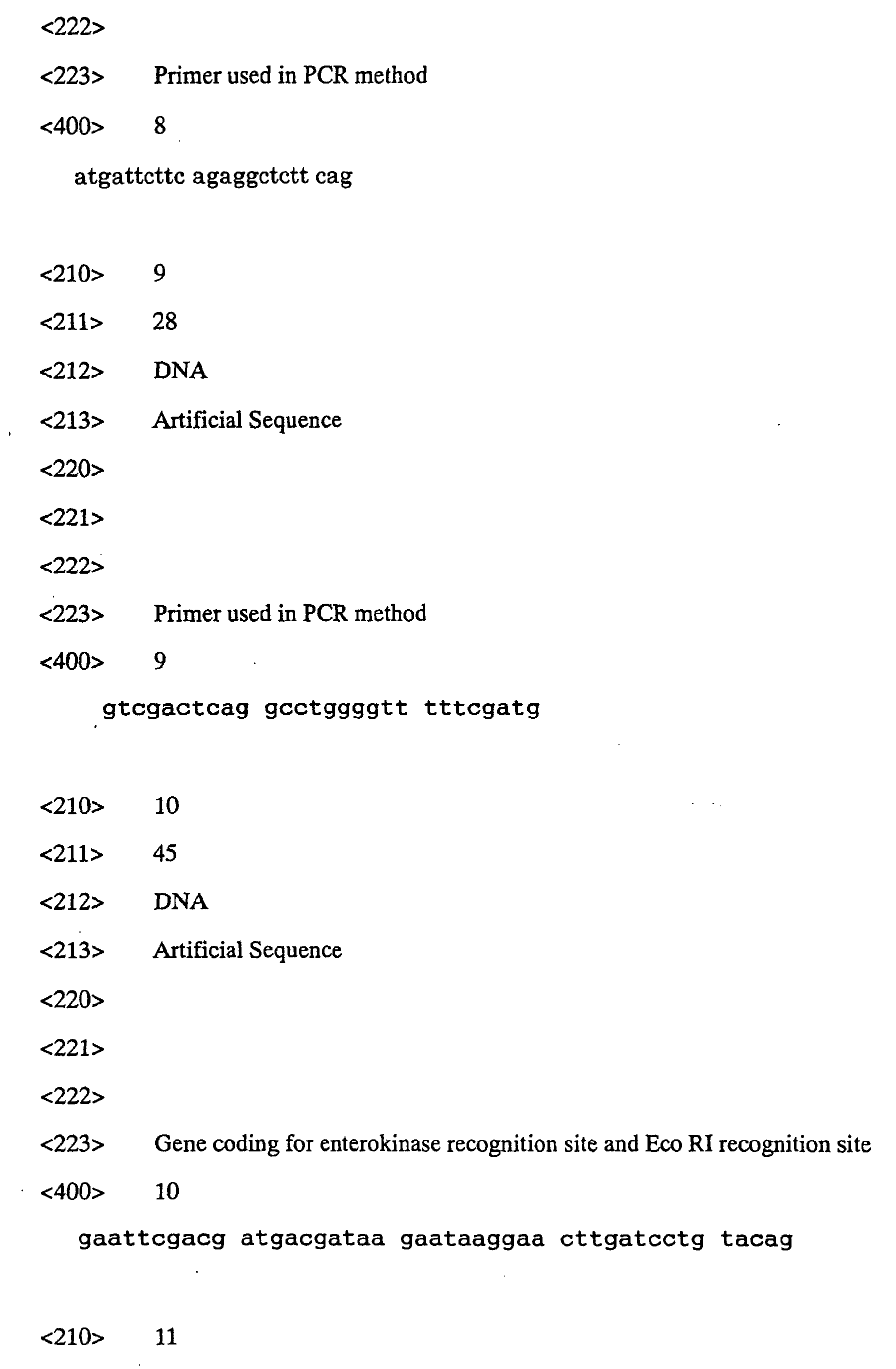
oligomer having a gene sequence encoding Asp-Asp-Asp-Asp-Lys
and a restriction enzyme EcoRI recognition site in the
5' side of a gene encoding the N-terminal side of rCF6 or
hCF6 was synthesized (rCF6E: SEQ ID NO:10 and hCF6E: SEQ ID
NO:11).
The DNA sequence of SEQ ID NO:10 was transferred by
the PCR method with the use of the rCF6 cDNA as a template
and rCF6E (SEQ ID NO:10) and rCH6-02 (SEQ ID NO:7) as
primers. As the thermostable DNA polymerase in PCR, use
was made of Taq DNA Polymerase (Pharmacia). Namely, a
liquid reaction mixture comprising 1 µL of rCF6 cDNA, 5 µL
of 10 x reaction buffer, 8 µL of 1.25 mM of dNTP mix, 0.5
µL of rCF6-01, 0.5 µL of rCF6-02 and 25 µL of sterilized
water was subjected to the reaction for 8 cycles with each
cycle consisting of 1 minutes at 96°C, 1 minute at 60°C and
30 seconds at 72°C. Thus rCF6 cDNA having the enterokinase
recognition sequence and the restriction enzyme EcoRI
recognition sequence in the 5'-side was obtained. The DNA
fragment thus amplified was cloned into pCRII (pCRrCF6EK)
and the nucleotide sequence was confirmed. Similarly, the
DNA sequence of SEQ ID NO:11 was transferred by the PCR
method with the use of the hCF6 cDNA as a template and
hCF6E (SEQ ID NO:11) and hCF6-02 (SEQ ID NO:9) as primers.
The reaction conditions were as in the case of the rat.
The DNA fragment thus amplified was cloned into pCRII
(pCRhCF6EK) and the nucleotide sequence was confirmed.
(Construction of chimeric protein-expression vectors)
The plasmids pCRrCF6EK and pCRhCF6EK were cut with
restriction enzymes EcoRI and SalI to give EcoRI-SalI DNA
fragments rCF6EK and hCF6EK each consisting of 0.3 kb.
These DNA fragments have DNA nucleotide sequences encoding
proteins containing rCF6 or hCF6 having an enterokinase
recognition sequence Asp-Asp-Asp-Asp-Lys (SEQ ID NO:3)
bonded to the N-terminus and a restriction enzyme EcoRI
site in the upstream thereof and another restriction enzyme
SalI site in the downstream thereof. The plasmid pGP#19RI
was cut with the restriction enzymes EcoRI and SalI and
then a DNA fragment of about 3.5 kb containing the vector
part was prepared. This DNA fragment was ligated to rCF6EK
or hCF6KE, thereby giving plasmids pG139SrCF6EK and
pG139ShCF6EK respectively.
(Expression and purification of chimeric proteins)
The E. coli JM109 strain was transformed by using the
plasmids pG139SrCF6EK and pG139ShCF6EK, thereby giving
JM109[pG139SrCF6EK] and JM109[pG139ShCF6EK] respectively.
JM109[pG139SrCF6EK] and JM109[pG139ShCF6EK] were each
cultured in an SB medium containing 10 µg/mL of
tetracycline under aeration and agitation at 37°C with the
use of a 3 L jar fermenter. 8 hours thereafter, IPTG was
added to give a final concentration of 1 mM and the culture
was continued overnight. 3 L of the liquid culture was
centrifuged at 6,000 rpm at 4°C for 10 minutes by using a
centrifuge 20PR-52D (Hitachi, Ltd.) and then the cells were
collected. The cells were suspended in 20 mM tris
hydrochloride (pH 7.0) containing 0.5% (w/v) of Triton X-100,
1 mM EDTA and disrupted by ultrasonication. Then the
disrupted cells were centrifuged at 6,000 rpm at 4°C for 15
minutes by using a centrifuge 05PR-22 (Hitachi, Ltd.).
Thus, the chimeric protein was collected in the precipitate
fraction as an inclusion body. The dissolution and
centrifugation of the precipitate fraction were repeated
thrice to thereby give a partially purified chimeric
protein. The hCF6 chimeric protein was prepared in the
same manner.
(Cutting off of hCF6 and rCF6 from chimeric proteins and
purification)
As a method of cutting off rCF6 from a chimeric
protein, the method with the use of factor Xa as a cutting
enzyme may be cited (Referential Example 2). To solubilize
the chimeric protein and treat it with the factor Xa in
this method, it is necessary to use at least 2 M of urea.
At this urea concentration, the activity of the factor Xa
is lowered to 1/10. Therefore, it is necessary in this
case to use a large amount of the factor Xa, i.e., 1/60
times as much as the chimeric protein. In addition, the
factor Xa shows only a low substrate specificity in the
presence of urea. As a result, the cutting efficiency from
the chimeric protein was as low as about 8%.
Therefore, attempts were made to elevate the CF6
cutting efficiency by using enterokinase as a cutting
enzyme. First, 4 mg of the crude rCF6 chimeric protein was
solubilized in 1.75 mL of a 10 M urea solution. Then, the
obtained solution was centrifuged to thereby eliminate
insoluble matters. To the supernatant, 0.5 mL of 10 x EK
buffer (200 mM of tris hydrochloride, (pH 8.0), 500 mM of
NaCl, 20 mM of CaCl2) and 30 U (about 3.4 µg, corresponding
to about 1/1200 of the substrate) of enterokinase
(Stratagene) were added and the total volume of the mixture
was adjusted to 5 mL with distilled water. After reacting
at 22°C overnight, the liquid reaction mixture was poured
into SepPak 18 (Waters) to thereby adsorb the peptides.
The peptides thus adsorbed were eluted with an 80% (v/v)
acetonitrile solution containing 0.1% (v/v) of TFA. The
eluate was freeze-dried and then dissolved in 0.1% (v/v)
TFA. A 1/10 portion thereof was subjected to HPLC
connected to YMC-ODS-A302 (diameter: 4.6 mm x 150 mm).
Then a peak eluted about 14.2 minutes after loading the
sample was collected as an rCF6 fraction (Fig. 2). The
same procedure was repeated 10 times and the fractions thus
eluted were combined, thereby giving partially purified
rCF6.
This partially purified rCF6 fraction was freeze-dried,
then dissolved in 0.1% TFA and then purified by HPLC
connected to YMC-ODS-AM-323 (Fig. 3, %B:40-43/18 min, flow
rate: 2.5 ml/min). The peak eluted after about 14.2
minutes was collected and freeze-dried to give an rCF6
specimen. The structure of rCF6 was confirmed based on the
facts that it showed a single peak in mass spectrometry
with the use of a mass spectrometer MALDI-TOFMS (Voyager
Elite: Nippon PerSeptive), that its molecular weight
roughly agreed with the theoretical value, and that the 10
amino acids at the N-terminus were identical with those of
rCF6, when analyzed by using an amino acid sequence
analyzer PPSQ-10 (Shimadzu Corporation).
hCF6 was cut off from the chimeric protein and
purified as in the case of rCF6. The hCF6 was eluted after
about 18.0 minutes in the first HPLC (Fig. 14). In the
chromatographic re-purification, it was eluted after about
10.5 minutes (Fig. 5, elution concentration
gradient, %B:45-48/18 min). The structure was confirmed as
in the case of rCF6.
About 210 µg and about 290 µg of hCF6 and rCF6 were
respectively obtained from 400 mg portions of the chimeric
proteins and the cutting efficiencies were about 15% and
about 20% respectively. Namely, the cutting efficiencies
were elevated 2 to 2.5-fold, compared with the case where
the factor Xa was employed as the cutting enzyme (yield:
8%).
[Example 2] Assay of CF6 in rat blood
(Preparation of antirat CF6 rabbit antibody (anti-rCF6
antibody))
A peptide consisting of 20 amino acids in total,
wherein cysteine was added to the N-terminus of the 19 C-terminal
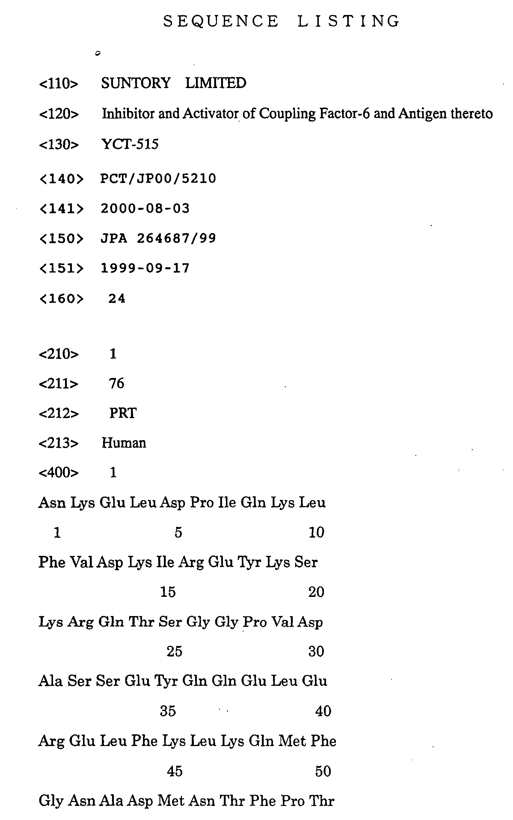
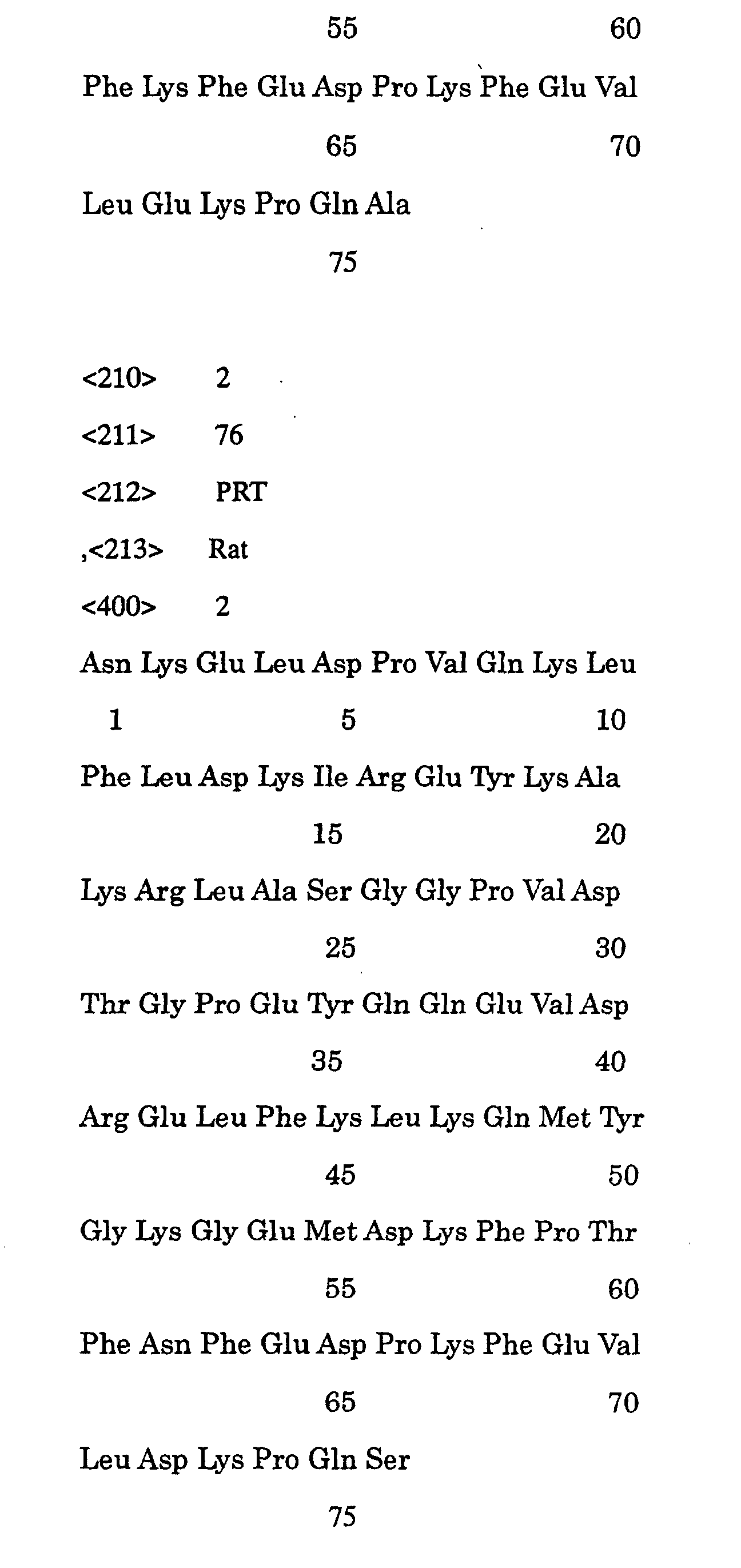
amino acids of rCF6 (SEQ ID NO:2) (i.e.,
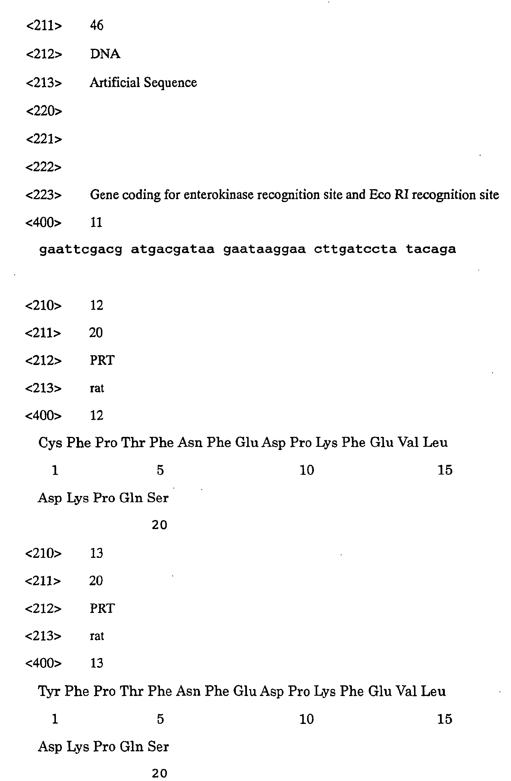
(CFPTFNFEDPKFEVLDKPQS:C-rCF6 (58-76):SEQ ID NO:12), was
synthesized by the Fmoc solid phase method and purified by
HPLC with the use of a C18 column. Then an antigen for
immunization was prepared by bonding keyhole limpet
hemocyanin (KLH) to the peptide thus synthesized via the
N-terminal cysteine.
As animals, two female New Zealand white
rabbits were employed. At the priming, the antigen
emulsified with complete Freund's adjuvant (CFA) was
subcutaneously administered at 4 positions of the dorsal
part of the rabbits. 2, 6 and 8 weeks thereafter, the
antigen emulsified with incomplete Freund's adjuvant (IFA)
was administered in the same manner to thereby immunize the
animals. Then all of the blood samples of the animals were
collected and thus antisera were obtained. The antibody
titers of the antisera were measured by ELISA with the
immobilization of the antigen. The antibody titers of the
rabbit sera before the immunization were each 50 or less,
while the antibody titers of the antisera of the immunized
rabbits were 59,600 and 81,400 respectively. The antibody
was purified from the antisera by the immunoaffinity
chromatograpy using an antigen peptide as the affinity
ligand (Fig. 6). The antibody thus purified was
concentrated, dialyzed against a PBS solution containing
0.1% (w/v) of NaN3 and then stored at -80°C until it was
used.
(Setting up of method of measuring rCF6 level)
A peptide consisting of 20 amino acid in total,
wherein tyrosine was added to the N-terminus of the 19
C-terminal amino acids of rCF6 (SEQ ID NO:2) (i.e.,
(YFPTFNFEDPKFEVLDKPQS : Y-rCF6 (58-76):SEQ ID NO:13) , was
synthesized by the Fmoc solid phase method and purified by
HPLC with the use of a C18 column. Y-rCF6(58-76) was
labeled with radioactive iodine (125I-labeling) by the
lactoperoxidase method. The 125I-labeled Y-rCF6(58-76) was
purified by HPLC with the use of a C18 column and then
bovine serum albumin was added to give a final
concentration of 0.1% (w/v). Then it was stored at -80°C
until it was used.
As the reaction system, use was made of an
antagonistic system wherein the reaction between the
125I-labeled antigen and the antibody was quantified by
inhibiting by the antigen in a sample. Namely, 100 µL of a
sample containing 0.25 to 32 ng of rCF6 was mixed with 100
µL of the rCF6 antibody (diluted 2500-fold) and reacted for
12 hours. Then 100 µL (20,000 cpm) of the 125I-labeled Y-rCF6(58-76)
was added thereto and the mixture was further
reacted overnight. Next, 1 mL of antirabbit IgG goat serum
(Japan Immunoresearch Laboratories Co., Ltd.) diluted 60-fold
with RIA buffer (50 mM of Na2HPO4, 80 mM of NaCl, 25
mM of EDTA·2Na, 0.05% (w/v) of NaN3, 0.5% (w/v) of bovine
serum albumin, pH 7.4) containing 100 µL of 200 µg/mL
rabbit IgG and 10% (w/v) of polyethylene glycol 6000 was
added and the resultant mixture was allowed to stand for 1
hour. Then it was centrifuged at 3,000 rpm for 30 minutes
and the supernatant was eliminated. The 125I level in the
supernatant was measured with a gamma counter. These
reactions were all carried out at 4°C. As a result, dose-dependent
radioactivity was observed in the precipitate
within the range of 0.25 to 16 ng/100 µL. Thus, a method
of measuring the rCF6 level in a sample could be set up.
(Assay of rCF6 in rat blood)
By using the assay method as described above,
measurement was made of rCF6 in the blood of male
spontaneously hypertensive rats (SHR) and rats, 4 weeks of
age, with normal blood pressure (WKY), and SHR and WKY of
16 weeks of age.
Each group had 5 animals. The blood of each animal
was collected by using a cylinder containing a 1% (w/v)
EDTA·2Na solution in 1/10 dose. The blood thus obtained
was centrifuged under cooling to extract the plasma. The
plasma was stored at -80°C until the assay. 0.2 ml of 2 N
hydrochloric acid was added to 0.8 ml of the plasma and the
mixture was allowed to stand at 4°C for 10 minutes. Next,
it was centrifuged at 4°C at 10,000 rpm for 30 minutes.
The supernatant was added to a SepPak C18 column and washed
with 5 ml portions of 0.1% (v/v) TFA thrice. After eluting
from the column with 2 ml of a 60% (v/v) acetonitrile
solution containing 0.1% (v/v) of TFA, the solvent was
removed in vacuo and the residue was dissolved in 0.2 to
0.4 ml of RIA buffer to give a sample for RIA which was
then used in the assay method with the use of the anti-rCF6
antibody as described above.
As the results of the assay of rCF6 in the plasma of
the rats of each group, every group showed a plasma rCF6
level exceeding 200 pg/mL. At the age of 4 weeks, no
difference was observed between the plasma rCF6 levels of
SHR and WKY. At the age of 16 weeks, the plasma rCF6 level
of SHR was significantly increased and thus higher than
that of WKY (p<0.05) (Table 1).
Thus, it is clarified for the first time that rCF6 is
present in rat blood. It is also found out that the blood
rCF6 level increases with aging (i.e., the onset of
hypertension) in the spontaneously hypertensive model (SHR)
suffering from the shortage of PGI2. That is to say, it is
clarified that the onset of hypertension caused by the
shortage of PGI2 correlates to the blood CF6 level.
[Table 1]
| Plasma rCF6 level (pg/ml) |
| Age |
WKY |
SHR |
| 4 weeks |
414±123 |
216±108 |
| 16 weeks |
294±126 |
2310±813 |
| Expressed in mean ± standard error. |
(Molecules in anti-rCF6 antibody-reactive fraction of RIA
sample by HPLC)
Anti-rCF6 antibody was prepared by using an rCF6
fragment (58-76) as an antigen. Thus, the amount of CF6
measured in the above-described RIA system included the
peptides containing the C-terminal fragment originating in
rCF6. The RIA sample was fractionated by HPLC and the
reactivity of anti-rCF6 antibody of each fraction was
examined. The RIA sample was adsorbed by SepPak C18 and
eluted with a 60% (v/v) acetonitrile solution containing
0.1% (v/v) of TFA. The sample thus eluted was freeze-dried,
dissolved in 0.1% (v/v) TFA and separated and fractionated
by HPLC connected to an Inertsil ODS-2 C18 column
(diameter:4.6 mm x 250 mm, GL Science K.K.) by washing with
a 20% (v/v) acetonitrile solution containing 0.1% (v/v) of
TFA for 5 minutes or longer and then eluting under linear
gradient of acetonitrile (20-60%) for 30 minutes. Thus, it
was confirmed that 90% or more of the antibody reactivity
was eluted in the same fraction as rCF6 in the assay system
with the use of the antid-rCF6 antibody as described above.
These results indicate that 90% or more of molecules
reactive to the anti-rCF6 antibody present in the blood are
not peptides containing the C-terminal fragment originating
in rCF6 but rCF6 per se.
[Example 3] Preparation of anti-hCF6 rabbit antibody (anti-hCF6
antibody) and construction of hCF6 assay system
Antisera against hCF6 were obtained as in Example 2
but using as an antigen for immunization a peptide
consisting of 19 amino acid wherein cysteine was added to
the N-terminus of the N-terminal amino acids (10-27) of
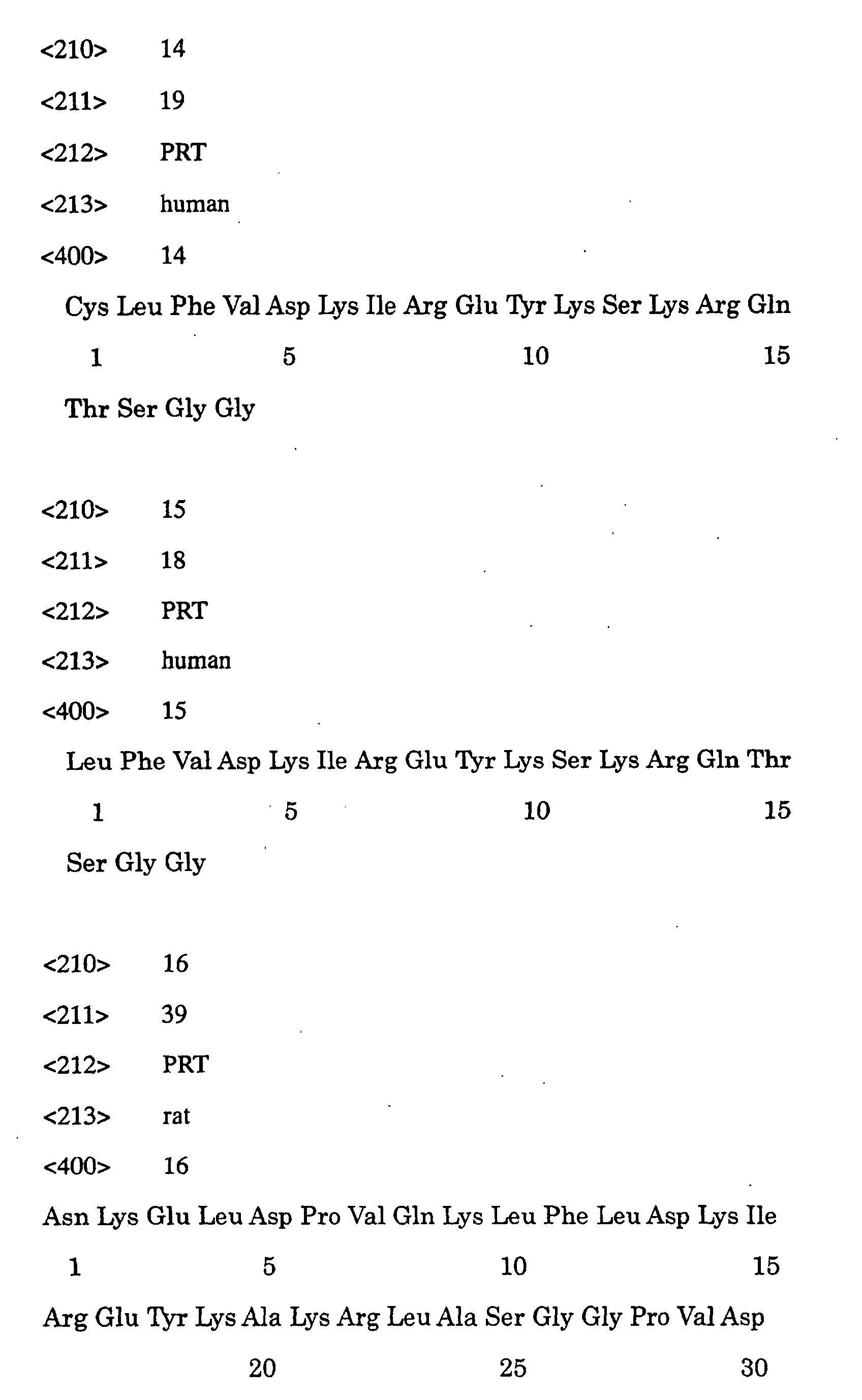
hCF6 (SEQ ID NO:1) (i.e., CLFVDKIREYKSKRQTSGG:C-hCF6 (10-27),
SEQ ID NO:14) bonded to KLH. The antibody titers of
the immunized rabbits were 123,000 and 121,300. The anti-hCF6
antibody was purified as in Example 2. The hCF6 assay
system was constructed as in the rCF6 assay system but
using a peptide consisting of the N-terminal amino acids
(10-27) of hCF6 (SEQ ID NO:2) (LFVDKIREYKSKRQTSGG:hCF6(10-27),
SEQ ID NO:15) as the 125I-labeled peptide and hCF6 as
the standard.
[Example 4] Effects of anti-rCF6 antibody on rat blood
pressure
(Effects of anti-rCF6 antibody on rat blood pressure)
SHR and WKY each 16 weeks of age were anesthetized
and the anti-rCF6 antibody (0.3, 1.0 and 3.0 µg/kg) was
administered by bolus injection into the femoral vein.
Then the arterial blood pressure was measured in the open
manner from the carotid artery. As a result, the average
arterial blood pressure of SHR was lowered in the dose-dependent
manner by administering the anti-rCF6 antibody
(Figs. 7 and 8). In WKY, on the other hand, no significant
change in the blood pressure was observed due to the
administration of the anti-rCF6 antibody (Fig. 8). The
administration of a nonspecific rabbit antibody (IgG: 10
µg/kg) did not affect the arterial blood pressures of SHR
and WKY.
(Effects of the preadministration of anti-rCF6 antibody on
decrease in rat blood pressure due to bradykinin)
Effects of the anti-rCF6 antibody on the effect of
bradykinin of lowering rat blood pressure were examined.
SHR and WKY each 16 weeks of age were anesthetized and
bradykinin (0.3, 1.0 and 3.0 µg/kg) was cumulatively
administered by bolus injection into the femoral vein.
Then the arterial blood pressure was measured in the open
manner from the carotid artery. Next, the anti-rCF6
antibody (3.0 µg/kg) was administered by bolus injection
into the femoral vein and then bradykinin was administered
in the same manner. The blood pressure reaction was
observed.
As a result, the blood pressures of SHR and WKY were
dose-dependently lowered by the administration of
bradykinin (Figs. 9-1 and 10). In SHR, the administration
of the anti-rCF6 antibody potentiated the effect of
bradykinin of lowering blood pressure (Figs. 9-2 and 10),
while no effect was observed in WKY (Fig. 10).
As described above, the anti-rCF6 antibody exerted a
hypotensive effect exclusively in the hypertensive state
with an increased rCF6 level. Thus, it is indicated that
the inhibition of CF6 is efficacious in ameliorating
symptoms of diseases caused by the excess of CF6 in the
blood.
[Example 5] Effects of the administration of rCF6 on rat
blood pressure
Effects of rCF6 on rat blood pressure were examined.
SHR or WKY 15 weeks of age were anesthetized and rCF6 (0.3,
1.0 and 3.0 µg/kg) was administered by bolus injection into
the femoral vein. Then the arterial blood pressure was
measured in an open manner from the carotid artery. As a
result, the average arterial blood pressures of SHR and WKY
were dose-dependently increased by the administration of
rCF6. A remarkable effect was observed in SHR compared with
WKY (Figs. 11 and 22).
Namely, a more remarkable effect of CF6 was observed
on SHR having a higher blood CF6 level. This fact
indicates that SHR is more sensitive to the effect of CF6.
Considering this result and the hypotensive effect of
the anti-rCF6 antibody on SHR observed in Example 4, it is
expected that CF6 agonists and CF6 secretion inhibitory
drugs would be usable as radical remedies for hypertension
which is a disease associated with the excess of CF6 in the
blood. Namely, it is indicated that the control of blood
CF6 level is efficacious as a method of treating diseases
associated with abnormal CF6 level in the blood.
[Example 6] Plasma hCF6 levels of normal subjects and
patients with acute heart infarction
Blood samples of 6 normal subjects and 18 patients
with acute heart infarction were collected from the vein
with the use of a cylinder containing a 1% (w/v) EDTA·2Na
solution in 1/10 dose. All of the patients with acute
heart infarction had been successfully treated by
percutaneous transluminal coronary angioplasty (PTCA). The
blood samples were collected on the day of the onset and
the days 2, 3, 5, 7 and 14. Each blood sample thus
obtained was centrifuged under cooling to extract the
plasma. The plasma was stored at -80°C until assaying.
A sample for assaying hCF6 was prepared from the
plasma in the following manner. Namely, 0.2 ml of 2 N
hydrochloric acid was added to 0.8 ml of the plasma and the
mixture was allowed to stand at 4°C for 10 minutes. Next,
it was centrifuged at 4°C at 10,000 rpm for 30 minutes.
The supernatant was added to a SepPak C18 column and washed
with 5 ml portions of 0.1% (v/v) TFA thrice. After eluting
from the column with 2 ml of a 60% (v/v) acetonitrile
solution containing 0.1% (v/v) of TFA, the solvent was
removed in vacuo and the residue was dissolved in 0.2 to
0.4 ml of RIA buffer to give a sample for RIA.
The hCF6 level was measured as in the case of rCF6
(Example 3) but using recombinant hCF6 as a standard and
the anti-hCF6 antibody.
CF6 was detected at the concentration of 15.2±0.5
ng/ml (mean ± standard error) in the normal subject plasma.
It is thus confirmed that CF6, which is a protein present
in the mitochondrial inner membrane, is present not only in
rat blood but also in human blood (Fig. 23). The CF6 level
in the plasma of the patients with acute heart infarction
showed a peak on the day of the onset or the day 2 and a
high plasma CF6 level of 22.1±1.3 ng/ml was observed (Fig.
23). The plasma CF6 levels on the day 1 and day 2 were
respectively 20.2±1.4 ng/ml and 17.7±1.5 ng/ml which were
significantly higher than the concentration on day 14
(13.7±0.7 ng/ml) (Fig. 24: Fisher's PLSD: p<0.001 and
p=0.01). The plasma CF6 levels on the 7 and day 14 were
respectively 13.7±0.6 ng/ml and 13.7±0.7 ng/ml, i.e.,
lowered to the level of the normal subjects.
Thus, it is clarified that the blood CF6 level
increases in patients with acute heart infarction. It is
therefore suggested that the measurement of the CF6 level
might be useful as a diagnostic method for diseases
associated with changes in the CF6 level such as acute
heart infarction.
[Example 7] Release of CF6 into HUVEC culture medium
HUVEC in the confluent state were cultured in a fetal
calf serum-free 199 medium in a Petri dish (diameter: 10
cm). After 24, 48 and 72 hours, the culture medium was
sampled (n=4 at each point) and hCF6 was quantitated. The
culture medium was treated as in Example 6 and the hCF6
level was measured. Thus, it was found out that the CF6
level in the culture medium increased with the passage of
time, thus indicating that hCF6 was released from HUVEC
(Fig. 25).
Accordingly, CF6 secretion accelerating substances or
CF6 secretion inhibitory substances can be obtained by
adding candidate compounds to a liquid culture of HUVEC and
comparing the amount of CF6 secreted into the culture with
a control.
[Referential Example 1] Identification of prostacyclin
production inhibitor
A prostacyclin production inhibitor was identified as
CF6 by the following method.
The hearts (152 g) of 187 SHR 16 to 20 weeks of age
were allowed to stand in boiling water for 10 minutes.
Then peptides were extracted therefrom with 1 N acetic acid.
The extract was fractionated in 0.5 ml portions by using
Sephadex G-25 (1.5 x 30 cm) (flow rate: 3 ml/min, Fig. 12).
The active fractions (19-21) were separated by HPLC
connected to an Inertsil ODS-2 C18 (diameter: 4.6 mm x 250
mm) column (GL Science K.K.). After washing with a 20%
(v/v) acetonitrile solution containing 0.1% (v/v) of
purified TFA for 5 minutes or longer, it was eluted with
acetonitrile under a linear gradient (20-60%) for 30
minutes (flow rate: 1 ml/min, Fig. 13). The activity was
evaluated by assaying 6-keto-PGF1α secreted into the medium
obtained from smooth muscle cells originating in SHR
mesenteric artery. The purified peptide (Fig. 14) was
sequenced as follows. The sequence from the N-terminus to
the 39-position was identified by using an amino acid
sequence analyzer (PPSQ-10, Shimadzu Corporation) as
NKELDPVQKLFLDKIREYKA KRLASGGPVDTGPEYQQEV (1- to 39-amino
acids in SEQ ID NO:2, SEQ ID NO:16). By purifying the
fragment digested with AsnN by HPLC connected to YMC-ODS-A302
(diameter: 4.6 mm x 150 mm), the subsequent sequences
were identified as DRELFKLKQMYGKGEM (40- to 55-amino acids
in SEQ ID NO:2, SEQ ID NO:17), DKFPTFNFE (56- to 64-amino
acids in SEQ ID NO:2, SEQ ID NO:18), DPKFEVL (65- to 71-amino
acids in SEQ ID NO:2, SEQ ID NO:19) and DKPQS (72- to
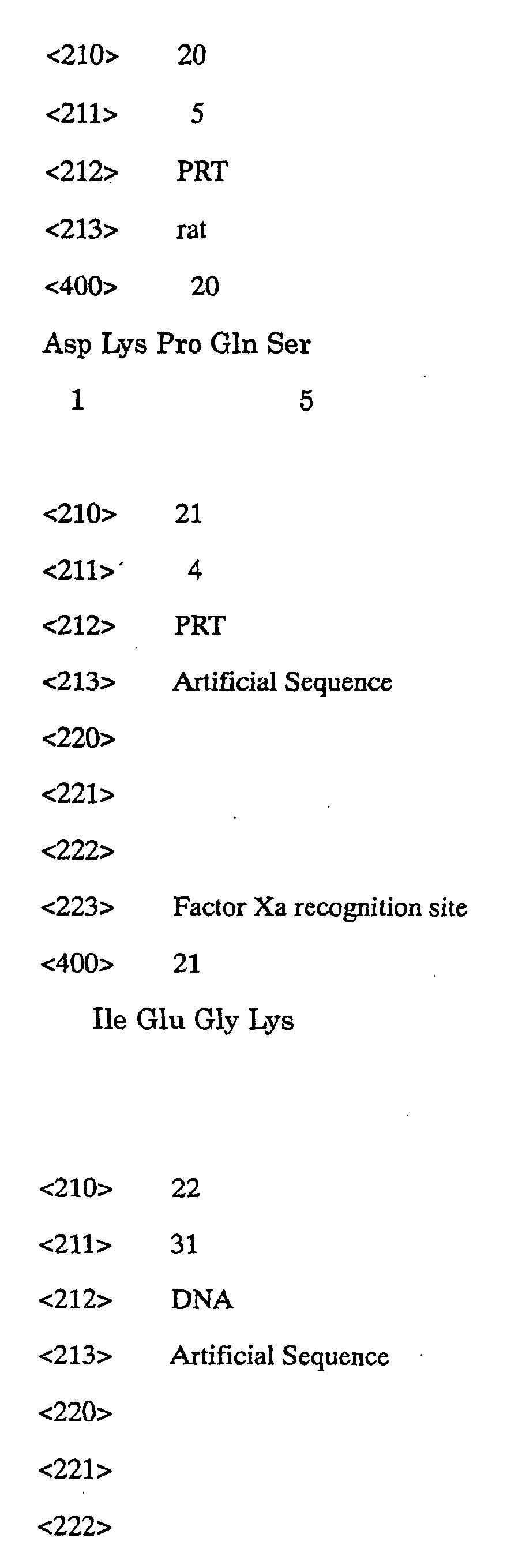
76-amino acids in SEQ ID NO:2, SEQ ID NO:20). These
sequences completely agreed with rCF6 (SEQ ID NO:2) which
is a subunit of rat H+ ATP synthase.
[Referential Example 2] Preparation of CF6 by chimeric
protein expression method-2
A method of producing rCF6 (SEQ ID NO:2) with the use
of factor Xa as a cutting enzyme from a chimeric protein
will be illustrated.
(Preparation of rCF6 DNA)
A DNA fragment containing a gene encoding a rCF6
protein having a cofactor Xa recognition sequence Ile Glu
Gly Lys (SEQ ID NO:21) added to the N-terminus was obtained
by PCR. A rat aorta cDNA library (Clontech) having been
treated at 100°C for 1 minute was used as a template, while
CF03 (SEQ ID NO:22) and CF04 (SEQ ID NO:23) were used as
primers.
The DNA fragment was cloned into pCRII (pCRIIrCF6Xa)
and then the DNA nucleotide sequence was determined to
thereby confirm the structure.
The plasmid pCRIIrCF6Xa was cut with restriction
enzymes EcoRI and SalI to give a 0.3 kb EcoRI-SalI DNA
fragment. This DNA fragment had a DNA nucleotide sequence
encoding a protein wherein a sequence Glu-Phe-Gly-Leu-Ile-Glu-Gly-Lys
(SEQ ID NO:24) having the factor Xa recognition
sequence Ile-Glu-Gly-Lys bonded to the C-terminus of a
sequence Glu-Phe-Gly-Leu originating in the plasmid pCRII
DNA was added to the N-terminus of rCF6 and had the
restriction enzyme EcoRI site in the upstream thereof and
another restriction enzyme SalI site in the downstream
thereof. The plasmid pG97S4DhCT[G] was cut with the
restriction enzymes EcoRI and SalI and a DNA fragment of
about 3.5 kb containing the vector part was prepared. Then
it was ligated to the 0.3 kb EcoRI-SalI DNA fragment to
thereby give a plasmid pG97SDrCF6Xa (Fig. 15).
(Expression and purification of rCF6 chimeric protein)
By using the plasmid pG97SDrCF6Xa, E. coli JM109
strain was transformed to give JM109[pG97SDrCF6Xa]. The
obtained JM109[pG97SDrCF6Xa] was inoculated into an LB
medium containing 10 µg/mL of tetracycline and cultured at
37°C for 6 hours. Then the liquid culture was inoculated
into an SB medium containing 10 µg/mL of tetracycline and 5
mM of MIPTG to give a final turbidity (OD 660 nm) of 0.1
and cultured at 37°C for 16 hours.
The liquid culture was centrifuged at 6,000 rpm at
4°C for 10 minutes (20PR-52D, Hitachi, Ltd.) and the cells
were collected. The cells were suspended in 20 mM tris
hydrochloride (pH 8.0) containing 1 mM of EDTA·Na and
disrupted with a French press (10,000 psi; twice).
The disrupted cell suspension was centrifuged at
8,000 rpm at 4°C (05PR-22, Hitachi Ltd.). The precipitate
was suspended in 30 ml of 20 mM tris hydrochloride (pH 8.0)
containing 0.5% (w/w) of Triton X-100 and centrifuged at
3,000 rpm at 4°C for 15 minutes. Then, precipitate was
collected. This procedure was repeated 4 times to thereby
give a partially purified chimeric protein.
(Cutting out of rCF6 from chimeric protein and
purification)
To cut out rCF6 from the chimeric protein, 50 µg/mL
of the factor Xa (Takara Shuzo Co., Ltd.) was used at a
concentration of 3 mg/mL of the chimeric protein and
reacted in 50 mM tris hydrochloride (pH 8.0) containing 2 M
of urea, 100 mM of NaCl and 1 mM solution of CaCl2 at 37°C
for 4 hours. The liquid reaction mixture was adsorbed by a
SepPak C18 column and washed with 0.1% (v/v) of
trifluoroacetic acid. Then the peptide was collected in an
80% (v/v) acetonitrile solution containing 0.1% (v/v) of
trifluoroacetic acid. The eluate was freeze-dried and then
dissolved in 0.1% (v/v) TFA. rCF6 was purified by HPLC
connected to YMC-ODS-A302 (%B: 35-50/30 minutes, eluted
after 14.5 minutes). The structure of rCF6 was confirmed
as in Example 1.
In this method, however, rCF6 was cut off from the
chimeric protein only at a low efficiency (about 8%).
Therefore, it seems necessary to develop a more efficient
method of producing CF6 in order to carry out in vivo
experiments, etc. wherein large amounts of CF6 are required.
[Referential Example 3] Inhibition of the production of
prostacyclin by CF6
HUVEC were cultured at 37°C for 30 minutes in the
DMEM containing 1, 10 and 100 nM of rCF6 prepared in
Referential Example 2 and thus the inhibition of the PGI2
production by CF6 was examined. The production of PGI2 was
measured with the guidance of the amount of 6-keto-PGF1α,
which is a stable metabolite of PGI2, secreted into the
medium. As a result, CF6 dose-dependently inhibited the
PGI2 production (Fig. 16). The same test was carried out
in the presence of 1 µM of bradykinin. Thus, it was found
out that CF6 dose-independently inhibited the increase in
the PGI2 production due to bradykinin (Fig. 17).
Further, 10 nM of CF6 inhibited the increase in the
PGI2 production due to 2 µg/ml of ionomycin but not the
increase therein due to 10 ng/ml of arachidonic acid and 10
ng/ml of PGH2.
PGI2 is produced starting with a phospholipid.
Namely, arachidonic acid cut off from the phospholipid by
PLA2 is converted into PGH2 by cyclooxygenase. It is
further converted by PGI2 synthase. In this reaction,
ionomycin promotes the activation of PLA2, i.e., the
cutting off of arachidonic acid from the phospholipid.
Namely, it has been found out that CF6 does not
inhibit the production of PGI2 from arachidonic acid or
PGH2 which is the downstream product in the arachidonate
cascade but inhibits the PGI2 production based on the
activation of PLA2. It is thus suggested that the PGI2
production-inhibiting effect of CF6 is based on the
inhibition of PLA2.
[Referential Example 4] Effect of CF6 of inhibiting release
of arachidonic acid
In Referential Example 3, it is indicated that the
PGI2 production-inhibiting effect of CF6 is based on the
inhibition of PLA2. On the other hand, PLA2 releasing
arachidonic acid from a fatty acid is roughly classified
into Ca2+-dependent cytoplasmic PLA2 (cPLA2), Ca2+-dependent
secretory PLA2 (sPLA2) and Ca2+-independent PLA2 (iPLA2)
depending on the structure and biochemical properties. By
using respective inhibitors, it was examined which PLA2 was
inhibited by CF6.
HUVEC were cultured for 24 hours in the presence of
[3H]-labeled arachidonic acid. Then the medium was
replaced by DMEM and the culture was continued in the
presence of 1, 10 and 100 mM of CF6 at 37°C for an
additional 30 minutes. Then the release of the [3H]-labeled
arachidonic acid into the liquid culture was
examined. As a result, CF6 dose-dependently inhibited the
release of the [3H]-labeled arachidonic acid (Fig. 18).
The same test was carried out in the presence of 1 µM of
bradykinin. Thus, it was found out that CF6 dose-dependently
inhibited the effect of bradykinin of promoting
the release of [3H]-labeled arachidonic acid (Fig. 19).
Next, the effects of a cPLA2 inhibitor (40 µM of
AACOCF3: arachidonic acid 3-fluoromethyl ketone), an sPLA2
inhibitor (1 µM of OPC; oleyloxyethylphosphorylcholine) and
an iPLA2 inhibitor (1 µM of BEL: bromoenolcholine) of
inhibiting the release of arachidonic acid form HUVEC were
examined. As a result, it was found out that, in HUVEC,
the cPLA2 inhibitor alone inhibited the release of
arachidonic acid. Namely, the PLA2 participating in the
release of arachidonic acid is cPLA2 (Figs. 20 and 21).
cPLA2 shows a high specificity to arachidonic acid-containing
phospholipids and selectively releases
arachidonic acid at a Ca2+ concentration of µM order. As
the results of experiments with the use of mice with the
hyperexpression of c PLA2 and mice lacking cPLA2, it has
been clarified that cPLA2 plays an important role not only
in the metabolism of arachidonic acid in the immediate
phase but in the delayed phase of arachidonic acid which is
important in inflammatory reactions. Therefore, it is
expected that the inhibition of cPLA2 induces a strong
inflammatory reaction (Uozumi N., et al. 390 618-622, 1997).
INDUSTRIAL APPLICABILITY
The present invention provides an antibody specific
to CF6 and diagnostic methods for pathological conditions
in which CF6 participates. The present invention also
provides a method of treating diseases such as hypertension
on the basis of the inhibition of CF6 in the blood whereby
normal blood pressure is not lowered. Moreover, the
diagnostic methods according to the present invention
contribute to the provision of methods of treating
pathological conditions associated with an increase or
decrease in CF6 in the blood.