This invention is directed to a method for providing digitized
radiographic images using a radiographic film that can be directly viewed or
further manipulated using digitization. In addition, the radiographic film used in
this invention also has what is known as "visually adaptive contrast" because it
can provide higher contrast than normal in the higher density regions of an image.
Over one hundred years ago, W.C. Roentgen discovered X-radiation
by the inadvertent exposure of a silver halide photographic element. In
1913, Eastman Kodak Company introduced its first product specifically intended
to be exposed by X-radiation (X-rays). Today, radiographic silver halide films
account for the overwhelming majority of medical diagnostic images. Such films
provide viewable black-and-white images upon imagewise exposure followed by
processing with the suitable wet developing and fixing photochemicals.
In medical radiography an image of a patient's anatomy is produced
by exposing the patient to X-rays and recording the pattern of penetrating X-radiation
using a radiographic film containing at least one radiation-sensitive
silver halide emulsion layer coated on a transparent support. X-radiation can be
directly recorded by the emulsion layer where only low levels of exposure are
required. Because of the potential harm of exposure to the patient, an efficient
approach to reducing patient exposure is to employ one or more phosphor-containing
intensifying screens in combination with the radiographic film (usually
both in the front and back of the film). An intensifying screen absorbs X-rays and
emits longer wavelength electromagnetic radiation that the silver halide emulsions
more readily absorb.
Another technique for reducing patient exposure is to coat two
silver halide emulsion layers on opposite sides of the film support to form a "dual
coated" radiographic film so the film can provide suitable images with less
exposure. Of course, a number of commercial products provide assemblies of
both dual coated films in combination with two intensifying screens to allow the
lowest possible patient exposure to X-rays. Typical arrangements of film and
screens are described in considerable detail for example in US-A-4,803,150
(Dickerson et al), US-A-5,021,327 (Bunch et al) and US-A-5,576,156
(Dickerson).
One important component of the films described in these patents is
a microcrystalline dye located in a silver halide emulsion layer or antihalation
layer that reduces "crossover" (exposure of an emulsion from light emitted by an
intensifying screen on the opposite of the film support) to less than 10%.
Crossover results in reduced image sharpness. These microcrystalline dyes are
readily decolorized during the wet processing cycle so they are not visible in the
resulting image.
Radiographic films that can be rapidly wet processed (that is,
processed in an automatic processor within 90 seconds and preferably less than 45
seconds) are also described in the noted US-A-5,576,156. Typical processing
cycles include contacting with a black-and-white developing composition,
desilvering with a fixing composition, and rinsing and drying. Films processed in
this fashion are then ready for image viewing. In recent years, there has been an
emphasis in the industry for more rapidly processing such films to increase
equipment productivity and to enable medical professionals to make faster and
better medical decisions.
As could be expected, image quality and workflow productivity
(that is processing time) are of paramount importance in choosing a radiographic
imaging system [radiographic film and intensifying screen(s)]. One problem with
known systems is that these requirements are not necessarily mutually inclusive.
Some film/screen combinations provide excellent image quality but cannot be
rapidly processed. Other combinations can be rapidly processed but image quality
may be diminished. Both features are not readily provided at the same time.
In addition, the characteristic graphical plots [density vs. log E
(exposure)] that demonstrate a film's response to a patient's attenuation of X-ray
absorption indicate that known films do not generally provide desired sensitivity at
the highest image densities where important pathology might be present.
Traditionally, such characteristic sensitometric "curves" are S-shaped. That is the
lower to midscale curve shape is similar to but inverted in comparison with the
midscale to upper scale curve shape. Thus, these curves tend to be symmetrical
about a density midpoint.
Another concern in the industry is the need to have radiographic
films that as accurately as possible show all gradations of density differences
against all backgrounds. It is well known that the typical response of the human
eye to determining equal differences in density against a background of increasing
density is not linear. In other words, typically it is more different for the human
eye to see an object against a dark background than it is to see an object against a
lighter background. Therefore, when an object is imaged (for example using X-rays,
with or without intensifying screens) at the higher densities of the
sensitometric curves, it is less readily apparent to the human eye when the
radiographic film is being viewed. Obviously, this is not a desirable situation
when medical images are being viewed and used for important diagnostic
purposes.
In order to compensate for this nonlinearity of response by the
human eye, it would be desirable to somehow increase radiographic film contrast
only at the higher densities without changing contrast or other properties at lower
densities. The result of such a modification would be a unique sensitometric curve
shape where the contrast is higher than normal in the higher density regions. Such
a curve shape is considered as providing "visually adaptive contrast" (VAC).
While this type of sensitometry sounds like a simple solution to a
well known problem, achieving it in complicated radiographic film/screen systems
is not simple and is not readily apparent from what is already known in the art.
Moreover, one cannot predict that even if VAC is obtained with a particular
radiographic film, other necessary image properties and rapid processability may
be adversely affected.
Recent digital technologies in the photographic industry offer
advantages in that they can enable the user to manipulate the images after wet
processing by scanning to create a digital representation of the image. One of
these advantages is the ability to readjust the exposure by automatic tone scaling
to correct for either over- or underexposure. This is particularly useful in
radiography where a patient is not available to have a second X-ray image taken
(for example, the patient may be too ill), as in intensive care facilities. A problem
with known digital modalities is that they do not provide the high image quality
that high performance film/screen imaging assemblies are capable of.
In addition, while photographic film scanners are available today,
and films scanning and digitization is common, existing radiographic films do not
work will with known film scanning equipment. One limitation is that the
scanned film must have sufficient exposure latitude so that information can be
recovered digitally even if the film is over- or underexposed. It is thus necessary
that the film exhibits sufficient contrast in the both the toe and shoulder regions of
a characteristic sensitometric density vs. log E curve to capture image information.
Early attempts to accomplish this are described in US-A-4,755,447
(Kitts, Jr.). The films described in this patent may be suitable for recovering
information even if over- or underexposed, but their contrast was too low if
directly viewed. In addition, films at a high density level are less useful because
the image signals are dominated by electronic noise (see for example, Bunch et al,
Applied Optics, Vol. 27, No. 16, pp. 3468-3474, 1988).
With these constraints in mind, there is a need for a means to
provide high quality images that can be directly viewed as well as electronically
scanned and digitized for further manipulation.
The present invention provides a solution to the noted problems
with a method for providing a radiographic image comprising:
Thus, the present invention provides means for obtaining
radiographic images using a radiographic film (for example as part of a
film/intensifying screen assembly) to give the medical professional a greater
ability to see an object against a dark (or high density) background. Therefore,
when an object is imaged using the film of this invention at the higher densities,
the object is more readily apparent to the human eye.
In order to compensate for the nonlinearity of response by the
human eye and the limitations of the film digitizer (i.e. scanner), the radiographic
film contrast has been increased only at the higher densities without changing
contrast or other properties at lower densities. The result of such a modification is
a unique sensitometric curve shape where the contrast is higher than normal in the
higher density regions. Thus, the films used in this invention are considered as
providing "visually adaptive contrast" (VAC) as we have defined it.
In addition, all other desirable sensitometric properties are
maintained, crossover is desirably low, and the films can be rapidly processed in
conventional processing equipment and compositions. Because the wet
processing time can be shortened, the time needed for image digitization can be
more readily accommodated within overall image formation.
More importantly, the present invention provides wide latitude
(that is, wide dynamic range) in radiographic imaging and thus enables the user to
correct over- or under-exposures by digital scanning and electronic manipulation.
The films can also be directly viewed if desired. However, once the image is in
digital form, exposure compensation can be carried out using conventional tone
scaling algorithms without any loss in information. The particular films used in
this invention are suitable for scanning in conventional scanning devices. Due to
the high upper scale contrast, the images obtained by this method are less
susceptible to film granularity at high densities.
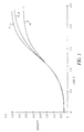
FIG. 1 is graphical representation of characteristic density vs. log E
(exposure) for Films A, B and C of the Example described below.
FIG. 2 is a graphical representation of gamma (contrast) vs. log E
(exposure) for Films A, B and C of the Example described below.
The term "contrast" as herein employed indicates the average
contrast (also referred to as γ) derived from a characteristic curve of a radiographic
element using as a first reference point (1) a density (D1) of 0.25 above minimum
density and as a second reference point (2) a density (D2) of 2.0 above minimum
density, where contrast is ΔD (i.e. 1.75) ÷ Δlog10E (log10E2- log10E1), E1 and
E2 being the exposure levels at the reference points (1) and (2).
"Lower scale contrast" is the slope of the characteristic curve
measured between of a density of 0.85 to the density achieved by shifting -0.3 log
E units.
"Upper scale contrast" is the slope of the characteristic curve
measured between a density of 1.5 above Dmin to 2.85 above Dmin.
"Dynamic range " refers to the range of exposures over which
useful images can be obtained.
The term "fully forehardened" is employed to indicate the
forehardening of hydrophilic colloid layers to a level that limits the weight gain of
a radiographic film to less than 120% of its original (dry) weight in the course of
wet processing. The weight gain is almost entirely attributable to the ingestion of
water during such processing.
The term "rapid access processing" is employed to indicate dry-to-dry
processing of a radiographic film in 45 seconds or less. That is, 45 seconds or
less elapse from the time a dry imagewise exposed radiographic film enters a wet
processor until it emerges as a dry fully processed film.
In referring to grains and silver halide emulsions containing two
or more halides, the halides are named in order of ascending concentrations.
The term "equivalent circular diameter" (ECD) is used to define
the diameter of a circle having the same projected area as a silver halide grain.
The term "aspect ratio" is used to define the ratio of grain ECD to
grain thickness.
The term "coefficient of variation" (COV) is defined as 100 times
the standard deviation (a) of grain ECD divided by the mean grain ECD.
The term "tabular grain" is used to define a silver halide grain
having two parallel crystal faces that are clearly larger than any remaining crystal
faces and having an aspect ratio of at least 2. The term "tabular grain emulsion"
refers to a silver halide emulsion in which the tabular grains account for more
than 50% of the total grain projected area.
The term "covering power" is used to indicate 100 times the ratio
of maximum density to developed silver measured in mg/dm2.
The term "rare earth" is used to refer to elements having an
atomic number of 39 or 57 to 71.
The term "front" and "back" refer to locations nearer to and
further from, respectively, the source of X-radiation than the support of the film.
The term "dual-coated" is used to define a radiographic film
having silver halide emulsion layers disposed on both the front- and backsides of
the support.
The radiographic films useful in this invention include a flexible
support having disposed on both sides thereof: two or more silver halide
emulsion layers and optionally one or more non-radiation sensitive hydrophilic
layer(s). The silver halide emulsions in the various layers can be the same or
different, and can comprise mixtures of various silver halide emulsions in or
more of the layers.
In preferred embodiments, the film has the same silver halide
emulsion on both sides of the support, and closest to the support. The emulsion
layers disposed farther from the support can also have the same silver halide
emulsions. It is also preferred that the films have a protective overcoat
(described below) over the silver halide emulsions on each side of the support.
The support can take the form of any conventional radiographic
element support that is X-radiation and light transmissive. Useful supports for
the films of this invention can be chosen from among those described in
Research Disclosure, September 1996, Item 38957 XV. Supports and Research
Disclosure, Vol. 184, August 1979, Item 18431, XII. Film Supports. Research
Disclosure is published by Kenneth Mason Publications, Ltd., Dudley House, 12
North Street, Emsworth, Hampshire P010 7DQ England.
The support is a transparent film support. In its simplest possible
form the transparent film support consists of a transparent film chosen to allow
direct adhesion of the hydrophilic silver halide emulsion layers or other
hydrophilic layers. More commonly, the transparent film is itself hydrophobic and
subbing layers are coated on the film to facilitate adhesion of the hydrophilic
silver halide emulsion layers. Typically the film support is either colorless or blue
tinted (tinting dye being present in one or both of the support film and the subbing
layers). Referring to Research Disclosure, Item 38957, Section XV Supports,
cited above, attention is directed particularly to paragraph (2) that describes
subbing layers, and paragraph (7) that describes preferred polyester film supports.
In the more preferred embodiments, at least one non-light sensitive
hydrophilic layer is included with the two or more silver halide emulsion layers on
each side of the film support. This layer may be called an interlayer or overcoat,
or both.
The silver halide emulsion layers comprise one or more types of
silver halide grains responsive to X-radiation. Silver halide grain compositions
particularly contemplated include those having at least 80 mol% bromide
(preferably at least 98 mol% bromide) based on total silver. Such emulsions
include silver halide grains composed of, for example, silver bromide, silver
iodobromide, silver chlorobromide, silver iodochlorobromide, and silver
chloroiodobromide. Iodide is generally limited to no more than 3 mol% (based on
total silver) to facilitate more rapid processing. Preferably iodide is limited to no
more than 2 mol% (based on total silver) or eliminated entirely from the grains.
The silver halide grains in each silver halide emulsion unit (or silver halide
emulsion layers) can be the same or different, or mixtures of different types of
grains.
The silver halide grains useful in this invention can have any
desirable morphology including, but not limited to, cubic, octahedral,
tetradecahedral, rounded, spherical or other non-tabular morphologies, or be
comprised of a mixture of two or more of such morphologies. Preferably, the
grains are tabular grains and the emulsions are tabular grain emulsions in each
silver halide emulsion layer.
In addition, different silver halide emulsion layers can have silver
halide grains of the same or different morphologies as long as at least 50% of the
grains are tabular grains. For cubic grains, the grains generally have an ECD of at
least 0.8 µm and less than 3 µm (preferably from 0.9 to 1.4 µm). The useful ECD
values for other non-tabular morphologies would be readily apparent to a skilled
artisan in view of the useful ECD values provided for cubic and tabular grains.
Generally, the average ECD of tabular grains used in the films is
greater than 0.9 µm and less than 4.0 µm, and preferably greater than 1 and less
than 3 µm. Most preferred ECD values are from 1.6 to 4.5 µm. The average
thickness of the tabular grains is generally at least 0.1 and no more than 0.3 µm,
and preferably at least 0.12 and no more than 0.18 µm.
It may also be desirable to employ silver halide grains that exhibit a
coefficient of variation (COV) of grain ECD of less than 20% and, preferably, less
than 10%. In some embodiments, it may be desirable to employ a grain
population that is as highly monodisperse as can be conveniently realized.
Generally, at least 50% (and preferably at least 90%) of the silver
halide grain projected area in each silver halide emulsion layer is provided by
tabular grains having an average aspect ratio greater than 5, and more preferably
greater than 10. The remainder of the silver halide projected area is provided by
silver halide grains having one or more non-tabular morphologies.
Tabular grain emulsions that have the desired composition and
sizes are described in greater detail in the following patents:
US-A-4,414,310 (Dickerson), US-A-4,425,425 (Abbott et al), US-A-4,425,426
(Abbott et al), US-A-4,439,520 (Kofron et al), US-A-4,434,226
(Wilgus et al), US-A-4,435,501 (Maskasky), US-A-4,713,320 (Maskasky), US-A-4,803,150
(Dickerson et al), US-A-4,900,355 (Dickerson et al), US-A-4,994,355
(Dickerson et al), US-A-4,997,750 (Dickerson et al), US-A-5,021,327 (Bunch et
al), US-A-5,147,771 (Tsaur et al), US-A-5,147,772 (Tsaur et al), US-A-5,147,773
(Tsaur et al), US-A-5,171,659 (Tsaur et al), US-A-5,252,442 (Dickerson et al),
US-A-5,370,977 (Zietlow), US-A-5,391,469 (Dickerson), US-A-5,399,470
(Dickerson et al), US-A-5,411,853 (Maskasky), US-A-5,418,125 (Maskasky), US-A-5,494,789
(Daubendiek et al), US-A-5,503,970 (Olm et al), US-A-5,536,632
(Wen et al), US-A-5,518,872 (King et al), US-A-5,567,580 (Fenton et al), US-A-5,573,902
(Daubendiek et al), US-A-5,576,156 (Dickerson), US-A-5,576,168
(Daubendiek et al), US-A-5,576,171 (Olm et al), and US-A-5,582,965 (Deaton et
al). The patents to Abbott et al, Fenton et al, Dickerson and Dickerson et al are
also cited to show conventional radiographic film features in addition to gelatino-vehicle,
high bromide (≥ 80 mol% bromide) tabular grain emulsions and other
features useful in the present invention.
A variety of silver halide dopants can be used, individually and in
combination, to improve contrast as well as other common properties, such as
speed and reciprocity characteristics. A summary of conventional dopants to
improve speed, reciprocity and other imaging characteristics is provided by
Research Disclosure, Item 38957, cited above, Section I. Emulsion grains and
their preparation, sub-section D. Grain modifying conditions and adjustments,
paragraphs (3), (4) and (5).
A general summary of silver halide emulsions and their preparation
is provided by Research Disclosure, Item 38957, cited above, Section I. Emulsion
grains and their preparation. After precipitation and before chemical sensitization
the emulsions can be washed by any convenient conventional technique using
techniques disclosed by Research Disclosure, Item 38957, cited above, Section III.
Emulsion washing.
The emulsions can be chemically sensitized by any convenient
conventional technique as illustrated by Research Disclosure, Item 38957, Section
IV. Chemical Sensitization. Sulfur, selenium or gold sensitization (or any
combination thereof) are specifically contemplated. Sulfur sensitization is
preferred, and can be carried out using for example, thiosulfates, thiosulfonates,
thiocyanates, isothiocyanates, thioethers, thioureas, cysteine or rhodanine. A
combination of gold and sulfur sensitization is most preferred.
Instability that increases minimum density in negative-type
emulsion coatings (that is fog) can be protected against by incorporation of
stabilizers, antifoggants, antikinking agents, latent-image stabilizers and similar
addenda in the emulsion and contiguous layers prior to coating. Such addenda are
illustrated by Research Disclosure, Item 38957, Section VII. Antifoggants and
stabilizers, and Item 18431, Section II. Emulsion Stabilizers, Antifoggants and
Antikinking Agents.
It may also be desirable that one or more silver halide emulsion
layers include one or more covering power enhancing compounds adsorbed to
surfaces of the silver halide grains. A number of such materials are known in the
art, but preferred covering power enhancing compounds contain at least one
divalent sulfur atom that can take the form of a -S- or =S moiety. Such
compounds include, but are not limited to, 5-mercapotetrazoles, dithioxotriazoles,
mercapto-substituted tetraazaindenes, and others described in US-A-5,800,976
(Dickerson et al) for the teaching of the sulfur-containing covering power
enhancing compounds. Such compounds are generally present at concentrations
of at least 20 mg/silver mole, and preferably of at least 30 mg/silver mole. The
concentration can generally be as much as 2000 mg/silver mole and preferably as
much as 700 mg/silver mole.
The silver halide emulsion layers and other hydrophilic layers on
both sides of the support of the radiographic film generally contain conventional
polymer vehicles (peptizers and binders) that include both synthetically prepared
and naturally occurring colloids or polymers. The most preferred polymer
vehicles include gelatin or gelatin derivatives alone or in combination with other
vehicles. Conventional gelatino-vehicles and related layer features are disclosed
in Research Disclosure, Item 38957, Section II. Vehicles, vehicle extenders,
vehicle-like addenda and vehicle related addenda. The emulsions themselves can
contain peptizers of the type set out in Section II, paragraph A. Gelatin and
hydrophilic colloid peptizers. The hydrophilic colloid peptizers are also useful as
binders and hence are commonly present in much higher concentrations than
required to perform the peptizing function alone. The preferred gelatin vehicles
include alkali-treated gelatin, acid-treated gelatin or gelatin derivatives (such as
acetylated gelatin, deionized gelatin, oxidized gelatin and phthalated gelatin).
Cationic starch used as a peptizer for tabular grains is described in US-A-5,620,840
(Maskasky) and US-A-5,667,955 (Maskasky). Both hydrophobic and
hydrophilic synthetic polymeric vehicles can be used also. Such materials include,
but are not limited to, polyacrylates (including polymethacrylates), polystyrenes
and polyacrylamides (including polymethacrylamides). Dextrans can also be used.
Examples of such materials are described for example in US-A-5,876,913
(Dickerson et al).
The silver halide emulsion layers (and other hydrophilic layers) in
the radiographic films are generally fully hardened using one or more
conventional hardeners. Thus, the amount of hardener in each silver halide
emulsion and other hydrophilic layer is generally at least 1.5% and preferably at
least 2%, based on the total dry weight of the polymer vehicle in each layer.
Conventional hardeners can be used for this purpose, including but
not limited to formaldehyde and free dialdehydes such as succinaldehyde and
glutaraldehyde, blocked dialdehydes, α-diketones, active esters, sulfonate esters,
active halogen compounds, s-triazines and diazines, epoxides, aziridines, active
olefins having two or more active bonds, blocked active olefins, carbodiimides,
isoxazolium salts unsubstituted in the 3-position, esters of 2-alkoxy-N-carboxydi-hydroquinoline,
N-carbamoyl pyridinium salts, carbamoyl oxypyridinium salts,
bis(amidino) ether salts, particularly bis(amidino) ether salts, surface-applied
carboxyl-activating hardeners in combination with complex-forming salts,
carbamoylonium, carbamoyl pyridinium and carbamoyl oxypyridinium salts in
combination with certain aldehyde scavengers, dication ethers, hydroxylamine
esters of imidic acid salts and chloroformamidinium salts, hardeners of mixed
function such as halogen-substituted aldehyde acids (e.g., mucochloric and
mucobromic acids), onium-substituted acroleins, vinyl sulfones containing other
hardening functional groups, polymeric hardeners such as dialdehyde starches,
and copoly(acrolein-methacrylic acid).
On each side of the radiographic film, the minimal total level of
silver is generally at least 15 mg/dm2. In addition, the total coverage of polymer
vehicle per side (that is, all layers on that side) is generally no more than 35
mg/dm2, and preferably no more than 30 and generally at least 20 mg/dm2. The
amounts of silver and polymer vehicle on the two sides of the support can be the
same or different. These amounts refer to dry weights.
The radiographic films generally include a surface protective
overcoat on each side of the support that is typically provided for physical
protection of the emulsion layers. Each protective overcoat can be sub-divided
into two or more individual layers. For example, protective overcoats can be sub-divided
into surface overcoats and interlayers (between the overcoat and silver
halide emulsion layers). In addition to vehicle features discussed above the
protective overcoats can contain various addenda to modify the physical properties
of the overcoats. Such addenda are illustrated by Research Disclosure, Item
38957, Section IX. Coating physical property modifying addenda, A. Coating aids,
B. Plasticizers and lubricants, C. Antistats, and D. Matting agents. Interlayers that
are typically thin hydrophilic colloid layers can be used to provide a separation
between the emulsion layers and the surface overcoats. It is quite common to
locate some emulsion compatible types of protective overcoat addenda, such as
anti-matte particles, in the interlayers. The overcoat on at least one side of the
support can also include a blue toning dye or a tetraazaindene (such as 4-hydroxy-6-methyl-1,3,3a,7-tetraazaindene)
if desired.
The protective overcoat is generally comprised of a hydrophilic
colloid vehicle, chosen from among the same types disclosed above in connection
with the emulsion layers. In conventional radiographic films protective overcoats
are provided to perform two basic functions. They provide a layer between the
emulsion layer and the surface of the element for physical protection of the
emulsion layer during handling and processing. Secondly, they provide a
convenient location for the placement of addenda, particularly those that are
intended to modify the physical properties of the radiographic film. The
protective overcoats of the films of this invention can perform both these basic
functions.
The various coated layers of radiographic films useful in this
invention can also contain tinting dyes to modify the image tone to transmitted or
reflected light. These dyes are not decolorized during processing and may be
homogeneously or heterogeneously dispersed in the various layers. Preferably,
such non-bleachable tinting dyes are in a silver halide emulsion layer.
An essential feature of the radiographic films useful in this
invention is the presence of one or more microcrystalline particulate dyes in the
first and third silver halide emulsion layers (that is, those emulsion layers closest
to and on opposing sides of the support). The presence of such dyes reduces
crossover during film use in radiographic assemblies to less than 15%, preferably
less than 10% and more preferably less than 5%. The amount in the film to
achieve this result will vary on the particular dye(s) used, as well as other factors,
but generally the amount of particulate dye is at least 0.5 mg/dm2, and preferably
at least 1 mg/dm2, and up to 2 mg/dm2.
The particulate dyes generally provide optical densities of at least
0.5, and preferably at least 1. Examples of useful particulate dyes and teaching of
their synthesis are described in US-A-5,021,327 (noted above, Cols. 11-50) and
US-A-5,576,156 (noted above, Cols. 6-7), for description of the dyes. Preferred
particulate dyes are nonionic polymethine dyes that include the merocyanine,
oxonol, hemioxonol, styryl and arylidene dyes. These dyes are nonionic in the pH
range of coating, but ionic under the alkaline pH of wet processing. A particularly
useful dye is 1-(4'-carboxyphenyl)-4-(4'-dimethylaminobenzylidene)-3-ethoxycarbonyl-2-pyrazolin-5-one
(identified as Dye XOC-1 herein).
The dye can be added directly to the hydrophilic colloid as a
particulate solid or it can be converted to a particulate solid after it has been added
to the hydrophilic colloid, as described in US-A-5,021,327 (Col. 49).
In addition to being present in particulate form and satisfying the
optical density requirements described above, the dyes useful in the practice of
this invention must be substantially decolorized during wet processing. The term
"substantially decolorized" is used to mean that the density contributed to the
image after processing is no more than 0.1, and preferably no more than 0.05,
within the visible spectrum.
The films described herein exhibit an upper scale contrast (USC) of
at least 2, and preferably at least 3. In addition, the ratio of USC to LSC is at least
1.2 and preferably at least 1.6. These features provide what is described above as
visually adaptive contrast (VAC). This attribute is similar to "perceptually
linearized contrast" or visually optimized tone scale as described for example by
Lee et al, SPIE Vol. 3036, pp. 118-129, 1997.
Preferred films useful in this invention comprise a dual coated
radiographic film comprising a light transmissive support and having disposed on
each side thereof:
The radiographic imaging assemblies useful in the present
invention are composed of a radiographic film as described herein and intensifying
screens adjacent the front and back of the radiographic film. The screens are
typically designed to absorb X-rays and to emit electromagnetic radiation having a
wavelength greater than 300 nm. These screens can take any convenient form
providing they meet all of the usual requirements for use in radiographic imaging,
as described for example in US-A-5,021,327 (noted above). A variety of such
screens are commercially available from several sources, including by not limited
to, LANEX™, X-SIGHT™ and InSight™ Skeletal screens available from
Eastman Kodak Company. The front and back screens can be appropriately
chosen depending upon the type of emissions desired, the photicity desired,
whether the films are symmetrical or assymmetrical, film emulsion speeds, and
crossover.
Exposure and processing of the radiographic films can be
undertaken in any convenient conventional manner. The exposure and processing
techniques of US-A-5,021,327 and 5,576,156 (both noted above), are typical for
processing radiographic films. Other processing compositions (both developing
and fixing compositions) are described in US-A-5,73 8,979 (Fitterman et al), US-A-5,866,309
(Fitter-man et al), US-A-5,871,890 (Fitterman et al), US-A-5,935,770
(Fitterman et al), US-A-5,942,378 (Fitterman et al). The processing compositions
can be supplied as single- or multi-part formulations, and in concentrated form or
as more diluted working strength solutions.
It is particularly desirable that the films described herein be
processed within 90 seconds, and preferably from 30 to 60 seconds, including
developing, fixing and any washing (or rinsing). Such processing can be carried
out in any suitable processing equipment including but not limited to, a Kodak X-OMATTM
RA 480 processor that can utilize Kodak Rapid Access processing
chemistry. Other "rapid access processors" are described for example in US-A-3,545,971
(Barnes et al) and EP-A-0 248,390 (Akio et al). Preferably, the black-and-white
developing compositions used during processing are free of any
photographic film (for example, gelatin) hardeners, such as glutaraldehyde.
Since rapid access processors employed in the industry vary in their
specific processing cycles and selections of processing compositions, the preferred
radiographic films satisfying the requirements of the present invention are
specifically identified as those that are capable of dry-to-dry processing according
to the following reference conditions:
| Development | 11.1 seconds at 35°C, |
| Fixing | 9.4 seconds at 35°C, |
| Washing | 7.6 seconds at 35°C, |
| Drying | 12.2 seconds at 55-65°C. |
Any additional time is taken up in transport between processing step. Typical
black-and-white developing and fixing compositions are described in the Example
below.
Radiographic kits useful in the practice of the present invention can
include one or more samples of radiographic film, one or more intensifying
screens used in the radiographic imaging assemblies, and/or one or more suitable
processing compositions. Preferably, the kit includes all of these components.
The processed films described herein can be scanned immediately
after black-and-white development or after partial of complete fixing. Preferably,
both development and fixing are carried out prior to scanning.
The resulting black-and-white image is scanned point-by-point,
line-by-line or frame-by-frame using any suitable scanning device (such as the
Lumiscan 75 available from Lumisys) to produce "density representative signals".
The digital record so produced can then be read into any convenient memory
medium (for example, an optical disk, computer hard drive, or magnetic storage
media) or transmitted to other medical institutions or users for future digital
manipulation, or immediate use (for example diagnosis) to correct any under- or
overexposures. A user would know how much exposure correction to make by
looking at the image and adjusting the exposure levels. Alternatively, an example
of software useful for this purpose is RV-2000 DICOM Viewing Software
available from Lumisys.
The corrected digital signals (that is, the digital records) can also be
forwarded to a suitable output device to provide a display image or stored for
future use. The output device may take a number of forms such as a CRT display,
CD disk, magnetic storage devices or other type of storage or output device readily
apparent to one skilled in the art.
Besides digital manipulation, the digital images can be used to
change physical characteristics of the image, such as "windowing" and "leveling"
(used in computed tomography scanning) or other manipulations known in the art.
The following example is provided for illustrative purposes, and is
not meant to be limiting in any way.
Example:
Radiographic Film A (Control):
Radiographic Film A was a dual coated having silver halide
emulsions on both sides of a blue-tinted 178 µm transparent poly(ethylene
terephthalate) film support. Each silver halide emulsion layer contained a green-sensitized
mixture of two different high aspect ratio tabular silver bromide
emulsions. The emulsions were chemically sensitized with sodium thiosulfate,
potassium tetrachloroaurate, sodium thiocyanate and potassium selenocyanate,
and spectrally sensitized with 400 mg/Ag mole of anhydro-5,5-dichloro-9-ethyl-3,3'-bis(3-sulfopropyl)oxacarbocyanine
hydroxide, followed by 300 mg/Ag mole
of potassium iodide.
Radiographic Film A had the following layer arrangement on each
side of the film support:
The noted layers were prepared from the following formulations.
| Overcoat Formulation | Coverage (mg/dm2) |
| Gelatin vehicle | 3.4 |
| Methyl methacrylate matte beads | 0.14 |
| Carboxymethyl casein | 0.57 |
| Colloidal silica (LUDOX AM) | 0.57 |
| Polyacrylamide | 0.57 |
| Chrome alum | 0.025 |
| Resorcinol | 0.058 |
| Whale oil lubricant | 0.15 |
| Interlayer Formulation | Coverage (mg/dm2) |
| Gelatin vehicle | 3.4 |
| AgI Lippmann emulsion (0.08 µm) | 0.11 |
| Carboxymethyl casein | 0.57 |
| Colloidal silica (LUDOX AM) | 0.57 |
| Polyacrylamide | 0.57 |
| Chrome alum | 0.025 |
| Resorcinol | 0.058 |
| Nitron | 0.044 |
| High Contrast Emulsion Layer Formulation | Coverage (mg/dm2) |
| T-grain emulsion (AgBr 2.7 x 0.13 µm) | 9.5 |
| T-grain emulsion (AgBr 2.0 x 0.10 µm) | 14.2 |
| Gelatin vehicle | 21.5 |
| 4-hydroxy-6-methyl-1,3,3a,7-tetraazaindene | 2.1 g/Ag mole |
| Potassium nitrate | 1.8 |
| Ammonium hexachloropalladate | 0.0022 |
| Maleic acid hydrazide | 0.0087 |
| Sorbitol | 0.53 |
| Glycerin | 0.57 |
| Potassium bromide | 0.14 |
| Resorcinol | 0.44 |
| Bisvinylsulfonylmethylether | 2.4% based on total gelatin in all layers |
| Crossover Control Emulsion Layer Formulation | Coverage (mg/dm2) |
| Magenta microcrystalline filter dye (XOC-1) | 2.5 |
| Gelatin | 6.7 |
| Low Contrast Emulsion Layer Formulation | Coverage (mg/dm2) |
| T-grain emulsion (AgBr 3.6 x 0.13 µm) | 7.8 |
| T-grain emulsion (AgBr 1.2 x 0.13 µm) | 10.1 |
| Gelatin vehicle | 21.5 |
| 4-hydroxy-6-methyl-1,3,3a,7-tetraazaindene | 2.1 g/Ag mole |
| Potassium nitrate | 1.8 |
| Ammonium hexachloropalladate | 0.0022 |
| Maleic acid hydrazide | 0.0087 |
| Sorbitol | 0.53 |
| Glycerin | 0.57 |
| Potassium bromide | 0.14 |
| Resorcinol | 0.44 |
| Bisvinylsulfonylmethylether | 2.4% based on total gelatin in all layers |
Radiographic Film B has the following layer arrangement and
formulations. The layers on each side of the support were identical.
| Overcoat Formulation | Coverage (mg/dm2) |
| Gelatin vehicle | 3.4 |
| Methyl methacrylate matte beads | 0.14 |
| Carboxymethyl casein | 0.57 |
| Colloidal silica (LUDOX AM) | 0.57 |
| Polyacrylamide | 0.57 |
| Chrome alum | 0.025 |
| Resorcinol | 0.058 |
| Whale oil lubricant | 0.15 |
| Interlayer Formulation | Coverage (mg/dm2) |
| Gelatin vehicle | 3.4 |
| AgI Lippmann emulsion (0.08 µm) | 0.11 |
| Carboxymethyl casein | 0.57 |
| Colloidal silica (LUDOX AM) | 0.57 |
| Polyacrylamide | 0.57 |
| Chrome alum | 0.025 |
| Resorcinol | 0.058 |
| Nitron | 0.044 |
| Emulsion Layer Formulation | Coverage (mg/dm2) |
| T-grain emulsion (AgBr 3.7 x 0.13 µm) | 3.2 |
| T-grain emulsion (AgBr 2.0 x 0.10 µm) | 9.9 |
| T-grain emulsion (AgBr 1.2 x 0.13 µm) | 4.1 |
| Gelatin vehicle | 23.7 |
| 4-hydroxy-6-methyl-1,3,3a,7-tetraazaindene | 2.1 g/Ag mole |
| Potassium nitrate | 1.8 |
| Ammonium hexachloropalladate | 0.0022 |
| Maleic acid hydrazide | 0.0087 |
| Sorbitol | 0.53 |
| Glycerin | 0.57 |
| Potassium bromide | 0.14 |
| Resorcinol | 0.44 |
| Bisvinylsulfonylmethylether | 2.4% based on total gelatin in all layers |
Radiographic Film C (Invention):
Radiographic Film C is within the present invention and had the
following layer arrangement and formulations on both sides of the film support:
| Overcoat Formulation | Coverage (mg/dm2) |
| Gelatin vehicle | 3.4 |
| Methyl methacrylate matte beads | 0.14 |
| Carboxymethyl casein | 0.57 |
| Colloidal silica (LUDOX AM) | 0.57 |
| Polyacrylamide | 0.57 |
| Chrome alum | 0.025 |
| Resorcinol | 0.058 |
| Whale oil lubricant | 0.15 |
| Interlayer Formulation | Coverage (mg/dm2) |
| Gelatin vehicle | 3.4 |
| AgI Lippmann emulsion (0.08 µm) | 0.11 |
| Carboxymethyl casein | 0.57 |
| Colloidal silica (LUDOX AM) | 0.57 |
| Polyacrylamide | 0.57 |
| Chrome alum | 0.025 |
| Resorcinol | 0.058 |
| Nitron | 0.044 |
| Upper Emulsion Layer Formulation | Coverage (mg/dm2) |
| T-grain emulsion (AgBr 3.7 x 0.13 µm) | 5.4 |
| T-grain emulsion (AgBr 2.0 x 0.10 µm) | 5.4 |
| Gelatin vehicle | 12 |
| 4-hydroxy-6-methyl-1,3,3a,7-tetraazaindene | 2.1 g/Ag mole |
| Potassium nitrate | 0.83 |
| Ammonium hexachloropalladate | 0.001 |
| Maleic acid hydrazide | 0.0044 |
| Sorbitol | 0.24 |
| Glycerin | 0.26 |
| Potassium bromide | 0.06 |
| Resorcinol | 0.2 |
| Bottom Emulsion Formulation | Coverage (mg/dm2) |
| T-grain emulsion (AgBr 2.0 x 0.10 µm) | 11.2 |
| Gelatin | 12 |
| Magenta microcrystalline dye (XOC-1) | 1.08 |
| 4-hydroxy-6-methyl-1,3,3a,7-tetraazaindene | 2.1 g/Ag mole |
| Potassium nitrate | 1.1 |
| Ammonium hexachloropalladate | 0.0013 |
| Maleic acid hydrazide | 0.0053 |
| Sorbitol | 0.32 |
| Glycerin | 0.35 |
| Potassium bromide | 0.083 |
| Resorcinol | 0.26 |
| Bisvinylsulfonylmethlyether | 2.4 % based on total gelatin in all layers |
Samples of Radiographic Films A, B and C were exposed through a
graduated density step tablet using a MacBeth sensitometer for 1/50 second and a
500 watt General Electric DMX projector lamp calibrated to 2650°K filtered with
a Coming C4010 filter.
Processing of the exposed film samples for sensitometric
evaluation was carried out using a processor commercially available under the
trademark KODAK RP X-OMAT film Processor M6A-N. Development was
carried out using the following black-and-white developing composition:
| Hydroquinone | 30 g |
| Phenidone | 1.5 g |
| Potassium hydroxide | 21 g |
| NaHCO3 | 7.5 g |
| K2SO3 | 44.2 g |
| Na2S2O5 | 12.6 g |
| Sodium bromide | 35 g |
| 5-Methylbenzotriazole | 0.06 g |
| Glutaraldehyde | 4.9 g |
| Water to 1 liter, pH 10 |
The film samples were in contact with the developer in each
instance for less than 90 seconds. Fixing for all experiments in this example was
carried out using KODAK RP X-OMAT LO Fixer and Replenisher fixing
composition (available from Eastman Kodak Company).
Rapid processing has evolved over the last several years as a way to
increase productivity in busy hospitals without compromising image quality or
sensitometric response. Where 90 second processing times were once the standard,
below 40 seconds processing is becoming the standard in medical radiography.
One such example of a rapid processing system is the commercially available
KODAK Rapid Access (RA) processing system that includes a line of X-ray
sensitive films available as T-MAT-RA radiographic films that feature fully
forehardened emulsions in order to maximize film diffusion rates and minimize
film drying. Processing chemistry for this process is also available. As a result of
the film being fully forehardened, glutaraldehyde (a common hardening agent) can
be removed from the developer solution, resulting in ecological and safety
advantages (see KODAK KWIK Developer below). The developer and fixer
designed for this system are Kodak X-OMAT RA/30 chemicals. A commercially
available processor that allows for the rapid access capability is the Kodak X-OMAT
RA 480 processor. This processor is capable of running in 4 different
processing cycles. "Extended" cycle is for 160 seconds, and is used for
mammography where longer than normal processing results in higher speed and
contrast. "Standard" cycle is 82 seconds, "Rapid Cycle" is 55 seconds and
"KWIK/RA" cycle is 40 seconds (see KODAK KWIK Developer below). A
proposed new "Super KWIK" cycle is intended to be 30 seconds (see KODAK
Super KWIK Developer below). The two KWIK cycles (30 & 40 seconds) use the
RA/30 chemistries while the longer time cycles use standard RP X-OMAT
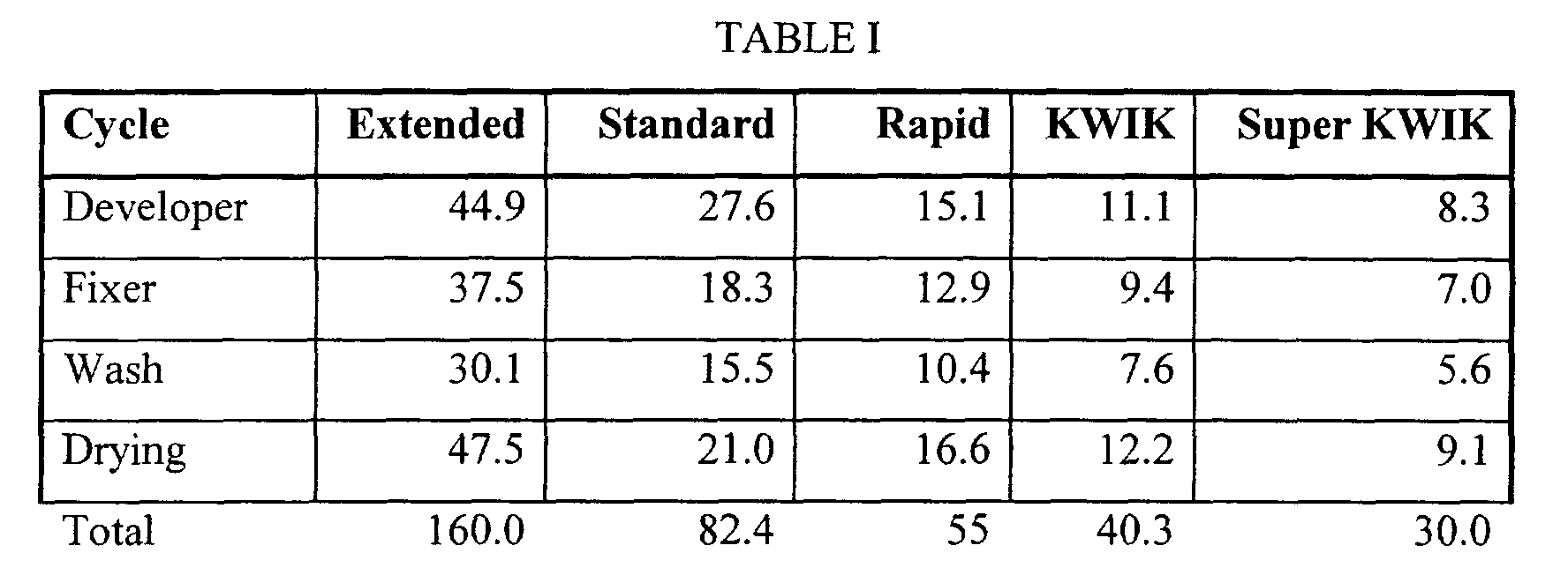
chemistry. The following Table I shows typical processing times (seconds) for
these various processing cycles.
The black-and-white developer useful for the KODAK KWIK
cycle contained the following components:
| Hydroquinone | 32 g |
| 4-Hydroxymethyl-4-methyl-1-phenyl-3-pyrazolidone | 6 g |
| Potassium bromide | 2.25 g |
| 5-Methylbenzotriazole | 0.125 g |
| Sodium sulfite | 160 g |
| Water to 1 liter, pH 10.35 |
The black-and-white developer used for the KODAK Super KWIK
cycle contained the following components:
| Hydroquinone | 30 g |
| 4-Hydroxymethyl-4-methyl-1-phenyl-3-pyrazolidone | 3 g |
| Phenylmercaptotetrazole | 0.02 g |
| 5-Nitroindazole | 0.02 g |
| Glutaraldehyde | 4.42 g |
| Diethylene glycol | 15 g |
| Sodium bicarbonate | 7.5 g |
| VERSENEX 80 | 2.8 g |
| Potassium sulfite | 71.48 g |
| Sodium sulfite | 11.75 g |
| Water to 1 liter, pH 10.6 |
The "% Drying" was determined by feeding an exposed film
flashed to result in a density of 1.0 into an X-ray processing machine. As the film
just exits the drier section, the processing machine was stopped and the film was
removed. Roller marks from the processing machine can be seen on the film
where the film has not yet dried. Marks from 100% of the rollers in the drier
indicate the film has just barely dried. Values less than 100% indicate the film has
dried partway into the drier. The lower the value the better the film is for drying.
"Crossover" measurements were obtained by determining the
density of the silver developed in each of the silver halide emulsion layers, in the
silver halide emulsion layer adjacent the intensifying screen, and in the non-adjacent
silver halide emulsion layer separated from the film support. By plotting
the density produced by each silver halide emulsion layer versus the steps of a
conventional aluminum step wedge (a measure of exposure), a characteristic
sensitometric curve was generated for each silver halide emulsion layer. A higher
density was produced for a given exposure in the silver halide emulsion layer
adjacent the film support. Thus, the two sensitometric curves were offset in speed.
At three different density levels in the relatively straight-line portions of the
sensitometric curves between the toe and shoulder regions of the curves, the
difference in speed (Δ log E) between the two sensitometric curves was measured.
These differences were then averaged and used in the following equation to
calculate the % crossover:
% Crossover = 1antilog(Δ logE) + 1 x 100
Screen Exposures:
Radiographic film/intensifying screen imaging assemblies were
prepared by placing a screen on both sides of each radiographic Film A, B or C.
Each assembly was exposed to 70 KVp X-radiation, varying either current
(milliAmperes) or time, using a 3-phase Picker Medical (Model VTX-650) X-ray
unit containing filtration up to 3 mm of aluminum. Sensitometric gradations in
exposure were achieved by using a 21-increment (0.1 log E) aluminum step wedge
of varying thickness.
The data in the following Table II show a relative comparison of
the three imaging assemblies A, B and C using radiographic Films A, B and C,
respectively. Films A (Control) and C (Invention) provided high resolution
imaging assemblies as can be seen by the crossover values of less than 10%. Both
Films B and C could be rapidly processed. However, only Film C could be both
rapidly processed and exhibit low crossover.
In addition, Film C exhibited a unique sensitometric curve shape in
that the upper scale contrast was significantly higher than the lower scale contrast.
Film B that is a conventional radiographic film has a typical characteristic curve
shape wherein the lower scale and upper scale contrasts are similar in shape. Film
A exhibited a higher upper scale contrast than Film B, and the ratio of upper scale
contrast to lower scale contrast was greater than 1.0, but it could not be rapidly
processed.
Thus, only Film C provides all of the desired properties: low
crossover in radiographic imaging assemblies, a ratio of upper scale contrast to
lower scale contrast significantly greater than 1.0, and rapid processability. Thus,
this radiographic film would demonstrate visually adaptive contrast because it
would record information at higher densities with greater reliability and can be
better viewed using conventional light boxes.
These results are also apparent from FIGS. 1 and 2 in which Curves
A, B and C represent sensitometric data for Films A, B and C respectively.
| Film | Speed | Contrast | % Cross- over | Drying (KWIK Cycle) | LSC | USC | Ratio USC/LSC |
| Control A |
| | 0 | 2.4 | 3 | >100% | 1.88 | 2.83 | 1.5 |
| Control B | + 0.1 | 2.3 | 30 | 50% | 1.87 | 1.57 | 0.8 |
| Invention C | + 0.6 | 2.4 | 7 | 50% | 1.80 | 3.41 | 1.9 |
As noted previously, one of the benefits of the present invention is
that the radiographic films used in the imaging method have sufficient dynamic
range that they can be scanned and the images digitized for further manipulation.
TABLE III below shows gamma values for each of the noted radiographic imaging
assemblies A, B and C (comprising Films A, B and C, respectively) at different
densities. All 3 assemblies provide comparable gamma values up to a density of
1.0. However, at higher densities, the values begin to diverge.
Film B begins to lose contrast above a density of 2.0 where useful
information can still be recorded. As a result, if Film B is scanned and digitized, it
would be very difficult to readjust the exposure level using tone scaling algorithms
since the gamma decreases at higher densities. In addition, Film B is difficult to
"read" using conventional means because the information at higher densities
cannot readily be ascertained by the human eye. Having the gamma value
decrease worsens the problem.
Film A provides an improvement over Film B in the respect, but it
cannot be rapidly processed. Thus, it limits productivity and the ability to view
the image quickly. This is particularly important in intensive care facilities.
Film C provides the greatest exposure latitude (dynamic range),
maintaining a high gamma (at least 2.5) up to a density of 2.5 and beyond.
Densities of 2.5 or higher are common in radiographic images that are overexposed.
Thus, Film C is suitable for scanning and digitization because of its
wide dynamic range for capturing information even when improperly exposed.
But it can also be viewed conventional without scanning if desired. The present
invention provides flexibility and choices for the medical practitioner so the best
medical care can be provided.
Film B was also susceptible to electronic noise during digitization
due to its lower upper scale contrast, but Film C exhibited reduced susceptibility
to this problem due to its increased upper scale contrast.