This application claims priority, under 35 U.S.C. § 119(e), from U.S.
provisional application 60/185,617, filed February 29, 2000, and from U.S. provisional
application 60/234,500, filed September 22, 2000.
Field of the Invention
The invention is directed to novel osteoregulin polypeptides, which play a role
in regulating bone homeostasis, adiposity, and the calcification of atherosclerotic
plaques. The invention also features screening assays to identify modulators of
osteoregulin activity as well as methods of treating mammals for diseases or
disorders associated with osteoregulin activity.
Background of the Invention
Bone is a highly dynamic tissue that undergoes continual processes of
remodeling and modeling (Parfitt, J. Cellular Biochemistry 55: 273-86, 1994). In the
growing skeleton, the amount of mineralized bone formed exceeds the amount lost
through resorption, whereas in the mature adult, bone loss and bone formation are
equivalent, thereby preserving the integrity of the skeleton. Under certain conditions
such as aging, postmenopausal estrogen deficiency, or prolonged steroid treatment,
the amount of bone formed is not sufficient to compensate for the quantity lost by
resorption. Over time, this imbalance results in reduced bone mass and
compromises the structural integrity of the skeleton.
Bone remodeling is a very complex process of tightly coordinated action by the
bone resorbing osteoclasts and the bone forming osteoblasts. Osteoblasts are derived
from a mesenchymal cell lineage and are responsible for the formation of new bone
matrix in their differentiated state (Wlodarski, Clinical Orthopaedics & Related Research
276: 93, 1990). In addition, factors produced by osteoblasts regulate the formation of
osteoclasts and osteoclastic bone resorbing activity in response to endocrine signals
such as parathyroid hormone and vitamin D. It has been postulated that the bone loss
associated with aging is a result of a defect in the osteoblast cell lineage (Rodriguez et
al., J. of Cellular Biochemistry 75: 414-423, 1999; Erdmann et al., Mechanisms of Aging
& Development 110: 73-85, 1999; Roholl et al., J. of Bone & Mineral Research 9: 355-66,
1994; Katzburg et al., Bone 25: 667-673, 1999). Either the mesenchymal precursor
population is insufficient or has lost the capacity to proliferate and differentiate into
sufficient numbers of functioning osteoblasts.
Osteoblasts progress through a 3 stage process of differentiation: proliferation,
matrix maturation, and mineralization (Aubin, Journal of Cellular Biochemistry 72: 396-410,
1999; Stein and Lian, Endocrine Reviews 14: 424-42, 1993; Malaval et al., J. of
Cellular Biochemistry 74: 616-27, 1999). During this differentiation process, a well
characterized temporal and spatial expression pattern of extracellular bone matrix
proteins and other genes occurs (Malaval et al., J. of Cellular Biochemistry 74: 616-27,
1999; Owen et al., J. of Cellular Physiology 143: 420-30, 1990; Ingram et al., J. of Bone
& Mineral Research 8: 1019-29, 1993). The bone matrix is composed primarily of Type
I collagen which forms the extracellular structural component for the deposition of
mineral forming hydroxyapatite. Osteoblasts also secrete non-collagenous proteins into
the extracellular matrix (Robey, Connective Tissue Research 35: 131-6, 1996; Boskey,
Connective Tissue Research 35: 357-63, 1996). The non-collagenous proteins include
proteoglycans, sulfated glycoproteins, highly phosphorylated RGD-motif proteins, and
proteins modified to contain gla amino acid residues. Examples include biglycan,
osteonectin, bone sialoprotein, osteopontin, and osteocalcin. The exact function of
many of these proteins have not yet been delineated although most evidence supports
their role in promoting mineralization events (Robey, Connective Tissue Research 35:
131-6, 1996). An exception is the gla-peptide, osteocalcin, which has been shown to be
a negative regulator of bone formation by gene knockout technology (Ducy et al.,
Nature 382: 448-52, 1996).
The progression of osteoblast differentiation has been modeled in cell culture
using primary calvarial cells or bone marrow cells. Bone marrow contains pluripotent
stem cells of the adipocytic, osteoblastic, fibroblastic, and hematopoetic cell lineage
(Owen et al., J. of Cellular Physiology 143: 420-30, 1990; Beresford, J. of Cell Science
102: 341-51, 1992; Herbertson and Aubin, Bone 21: 491-500, 1997). Bone marrow
from rats, mice, and humans has been shown to contain osteoprogenitor cells that
proliferate and can be induced to differentiate into osteoblastic cells. Rat and human
cultures require a differentiation agent such as dexamethasone whereas mouse-derived
cells can differentiate in the absence of dexamethasone (Rickard et al., J. of Bone &
Mineral Research 11: 312-24, 1996; Maniatopoulos et al., Cell & Tissue Research 254:
317-30, 1988; Chen et al., Endocrinology 112: 1739-45, 1983). In vitro differentiated
osteoblasts display the capacity to secrete noncollagenous proteins into the
extracellular matrix in a temporally regulated manner which may indicate a regulatory
function for each of these proteins in the mineralization process (Yao et al., J. of Bone &
Mineral Research 9: 231-40, 1994). Furthermore, differentiated bone marrow cultures
have the capacity to facilitate the deposition of matrix and the formation of
hydroxyapatite mineral when grown in the presence of a phosphate source such as β-glycerophosphate.
These properties have made the differentiation of bone marrow cells
a useful model to investigate the mechanisms of bone remodeling and osteoblast
function.
Osteoporosis accounts for approximately 700,000 fractures per year in the
United States alone, and osteoporotic fractures are linked to significant death and
morbidity in the aged population. Therefore, there is a clear need to further
understand the process of bone remodeling, both in normal and pathological states,
in order to develop therapeutic agents to prevent, reduce, or reverse bone loss
associated with osteoporosis or other bone-related disorders.
Summary of the Invention
We sequenced a novel cDNA transcript expressed specifically in rat osteoblasts
and osteocytes that encodes a 45 kDa polypeptide; and we have also identified the
mouse and human forms. Our characterization revealed the protein to be a secreted,
RGD motif containing protein with a limited homology to dmp1, an extracellular matrix
protein present in bone and teeth (Roholl et al., J. of Bone & Mineral Research 9: 355-66,
1994; Katzburg et al., Bone 25: 667-673, 1999). Thus, we have designated this
mammalian protein "osteoregulin." Further studies of osteoregulin expression patterns
and function (as further described in the detailed description) have confirmed that
osteoregulin plays an important role in controlling bone homeostasis, adipose
regulation, and the calcification of atherosclerotic plaques.
The invention features novel osteoregulin polypeptides, the nucleotide
sequences that encode them, expression vectors containing these osteoregulin
sequences, and transgenic hosts which have been genetically modified to express
the osteoregulins of the invention.
Another feature of the invention is non-human mammals and animal cells that
have been genetically-modified to disrupt one or both copies of an endogenous
osteoregulin gene. Studies of genetically-modified mice that are homozygous or
heterozygous for the osteoregulin gene disruption demonstrate that the absence or
reduction of osteoregulin gene expression results in increased bone mass, increased
bone mineralization, increased bone formation, and an increase in adiposity in
females. These phenotypes indicate that osteoregulin functions as a negative
regulator of bone mass/density and adiposity. This role in bone homeostasis is
further supported by the significant expression of osteoregulin in bone tissue as well
as osteoregulin's similarities to other proteins that play a role in regulating bone
function.
Given the discovered function of osteoregulins as negative regulators of bone
formation, bone density, bone mineralization, as well as its role in adiposity and
plaque calcification, the present invention also features screening assays to identify
agents that modulate osteoregulin activity or gene expression. Such agents are
useful for administering to mammals, preferably humans, for the treatment of bone
disorders, such as osteoporosis, to stimulate bone repair or regeneration, and to treat
disorders related to adiposity or the calcification of atherosclerotic plaques.
In its first aspect, the invention features an isolated or purified polypeptide, or
a heterologous polypeptide, wherein the polypeptide has osteoregulin activity and
contains: the amino acid sequence of SEQ ID NO: 2, 4, 6, 8, 34, or 46; an amino acid
sequence encoded by a polynucleotide which will hybridize under moderately, more
preferably highly stringent conditions with a probe having the complement of the
coding sequence shown in SEQ ID NO: 1, 3, 5, 7, 33, or 45; or an amino acid
sequence having at least 50%, more preferably 60%, more preferably 70%, more
preferably 80%, more preferably 90% identity, most preferably, 95% identity, to an
amino acid sequence containing SEQ ID NO: 2, 4, 6, 8, 34, or 46. Preferably, the
polypeptide is mammalian and/or naturally occurring.
In a second, related aspect, the invention features an isolated or purified
polynucleotide containing: the coding sequence of SEQ ID NO: 1, 3, 5, 7, 33, or 45; a
sequence encoding any of the polypeptides above, preferably SEQ ID NO: 2, 4, 6, 8,
34, or 46; or a sequence which hybridizes, under moderately, more preferably highly
stringent conditions, to a probe, preferably to the full length of a probe, having the
complement of any of the above-mentioned polynucleotides. Preferably, the
sequence hybridizes to the complement of the coding sequence shown in SEQ ID
NO: 1, 3, 5, 7, 33, or 45, or the sequence is present in a mammalian cDNA library.
The invention also features an antibody that selectively binds to a polypeptide
of the invention, preferably SEQ ID NO: 2, 4, 6, 8, 34, or 46 a vector containing a
polynucleotide of the invention, preferably SEQ ID NO: 1, 3, 5, 7, 33, or 45 and a
transgenic host expressing a polynucleotide of the invention, preferably the coding
sequence shown in SEQ ID NO: 1, 3, 5, 7, 33, or 45. The preferred host is a
transfected mammalian host cell or a transgenic mammal (e.g., mouse, rat, pig,
sheep, monkey).
Another aspect of the invention is a genetically-modified, non-human
mammal, wherein the modification results in a functionally disrupted osteoregulin
gene. The mammal may be heterozygous for the modification, or homozygous for
the modification. Preferably, the mammal is a rodent, more preferably, a mouse. In
a related aspect, the invention also provides a genetically-modified animal cell,
wherein the modification comprises a functionally disrupted osteoregulin gene. The
cell is heterozygous or homozygous for the modification. Preferably, the animal cell
is an embryonic stem (ES) cell or an ES-like cell, the cell is human, or the cell is
murine.
A method of screening for an agent that modulates osteoregulin activity (e.g.,
affecting the regulation of bone mass, bone density, adiposity, vascular flexibility,
and/or atherosclerotic plaque calcification) is an additional feature of the invention,
the method comprises contacting an agent with an osteoregulin polypeptide and
measuring the activity of the osteoregulin, wherein a difference between the
osteoregulin activity in the presence of the agent and in the absence of the agent is
indicative that the agent modulates the activity.
Also featured is a method of screening for an agent that modulates any of the
above-mentioned osteoregulin activities by regulating osteoregulin expression, the
method comprises contacting an agent with a cell containing a nucleotide sequence
containing an osteoregulin gene regulatory element (e.g., an osteoregulin promoter
sequence) operably linked to a coding sequence, and measuring the expression of
the coding sequence, wherein a difference between the expression in the presence
of the agent and in the absence of the agent is indicative that the agent modulates
osteoregulin expression.
The invention also provides a method of treating a mammal to regulate bone
mass and/or density, adiposity, vascular flexibility, and/or atherosclerotic plaque
calcification, the method comprises administering an osteoregulin modulator to the
mammal. Preferably, the modulator is an osteoregulin antagonist and is
administered to increase bone mass and/or bone density, the modulator is an
osteoregulin agonist and is administered to reduce adiposity; the modulator is an
osteoregulin antagonist and is administered to increase atherosclerotic plaque
stability by increasing plaque calcification (e.g., to prevent stroke); or the modulator is
an osteoregulin agonist wherein the agonist is administered to increase vascular
flexibility by decreasing atherosclerotic plaque calcification.
An additional aspect of the invention features a method for producing a
polypeptide of claim 3 or 4. The method comprises (a) culturing a cell under
conditions suitable for expression of the polypeptide, wherein said cell comprises a
heterologous polynucleotide that encodes said polypeptide and said polynucleotide is
operably linked to a promoter sequence; and (b) recovering said polypeptide.
Preferably, the polynucleotide comprises SEQ ID NO: 1, 3, 5, or 7, or, in the case
where the cell is a bacterial cell, a preferred polynucleotide comprises SEQ ID NO:
33 or SEQ ID NO: 45, or rat or mouse nucleic acid sequence encoding an
osteoregulin polypeptide lacking the N-terminal signal sequence.
Those skilled in the art will fully understand the terms used herein in the
description and the appendant claims to describe the present invention.
Nonetheless, unless otherwise provided herein, the following terms are as described
immediately below.
"Osteoregulin activity" is the negative regulation of bone mineralization, bone
density, and/or bone formation. Thus, an osteoregulin functions to decrease bone
area, bone mineral content, bone mineral density, and/or bone density.
"Osteoregulin activity" also includes the negative regulation of the calcification of
atherosclerotic plaques and the stimulation of increased adiposity. A polypeptide
with "osteoregulin activity" exhibits at least one of these activities.
An osteoregulin "agonist" refers to a molecule which intensifies or mimics the
biological activity of an osteoregulin. Agonists may include proteins, nucleic acids,
carbohydrates, small molecules, or any other compound or composition which
increases the activity of an osteoregulin either by increasing the amount of
osteoregulin present in a cell or by increasing the signaling of an osteoregulin
polypeptide in its signal transduction pathway.
An "allelic variant" is an alternative form of the gene encoding an
osteoregulin. Allelic variants may result from at least one mutation in the nucleic acid
sequence and may result in altered mRNAs or in polypeptides whose structure or
function may or may not be altered. A gene may have none, one, or many allelic
variants of its naturally occurring form. Common mutational changes which give rise
to allelic variants are generally ascribed to naturally-occurring deletions, additions, or
substitutions of nucleotides. Each of these types of changes may occur alone, or in
combination with the others, one or more times in a given sequence.
"Altered" nucleic acid sequences encoding an osteoregulin include those
sequences with deletions, insertions, or substitutions of different nucleotides,
resulting in a polypeptide with at least one functional characteristic of an
osteoregulin. Included within this definition are polymorphisms which may or may not
be readily detectable using a particular oligonucleotide probe of the polynucleotide
encoding an osteoregulin. The encoded protein may also be "altered," and may
contain deletions, insertions, or substitutions of amino acid residues which produce a
silent change and result in a functionally equivalent osteoregulin. Deliberate amino
acid substitutions may be made on the basis of similarity in polarity, charge,
solubility, hydrophobicity, hydrophilicity, and/or the amphipathic nature of the
residues, as long as the biological or immunological activity of the osteoregulin is
retained. For example, negatively charged amino acids may include aspartic acid
and glutamic acid, and positively charged amino acids may include lysine and
arginine. Amino acids with uncharged polar side chains having similar hydrophilicity
values may include: asparagine and glutamine; and serine and threonine. Amino
acids with uncharged side chains having similar hydrophilicity values may include:
leucine, isoleucine, and valine; glycine and alanine; and phenylalanine and tyrosine.
"Amplification" relates to the production of additional copies of a nucleic acid
sequence. It is generally carried out using polymerase chain reaction (PCR)
technologies well known in the art.
An osteoregulin "antagonist" refers to a molecule which inhibits or attenuates
the biological activity of an osteoregulin. Antagonists may include proteins such as
anti-osteoregulin antibodies, nucleic acids, carbohydrates, small molecules, or any
other compound or composition which decreases the activity of an osteoregulin either
by reducing the amount of osteoregulin present in a cell, or by decreasing the
signaling of an osteoregulin polypeptide in its signal transduction pathway.
The term "antibody" refers to intact immunoglobulin molecules as well as to
fragments thereof, such as Fab, F(ab')2, and Fv fragments, which are capable of
binding an epitopic determinant. Antibodies that bind osteoregulin polypeptides can
be prepared using intact polypeptides or using fragments containing small peptides
of interest as the immunizing antigen. The polypeptide or oligopeptide used to
immunize an animal (e.g., a mouse, a rat, or a rabbit) can be derived from the
translation of RNA, or synthesized chemically, and can be conjugated to a carrier
protein if desired. Commonly used carriers that are chemically coupled to peptides
include bovine serum albumin, thyroglobulin, and keyhole limpet hemocyanin. The
coupled peptide is then used to immunize the animal.
The term "biologically active" refers to a protein having structural, regulatory,
or biochemical functions of a naturally occurring molecule. Likewise,
"immunologically active" refers to the capability of the natural, recombinant, or
synthetic osteoregulin, or of any oligopeptide thereof, to induce a specific immune
response in appropriate animals or cells and to bind with specific antibodies.
A "composition" comprising a given polynucleotide or amino acid sequence
refers broadly to any composition containing the given polynucleotide or amino acid
sequence. The composition may comprise a dry formulation or an aqueous solution.
"Conservative amino acid substitutions" are those substitutions that, when
made, least interfere with the properties of the original protein, i.e., the structure and
especially the function of the protein is conserved and not significantly changed by
such substitutions. Table A below shows amino acids which may be substituted for
an original amino acid and which are regarded as conservative amino acid
substitutions.
| Conservative Amino Acid Substitutions |
| Original Residue | Conservative Substitution |
| Ala | Gly, Ser |
| Arg | His, Lys |
| Asn | Asp, Gln, His |
| Asp | Asn, Glu |
| Cys | Ala, Ser |
| Gln | Asn, Glu, His |
| Glu | Asp, Gln, His |
| Gly | Ala |
| His | Asn, Arg, Gln, Glu |
| lle | Leu, Val |
| Leu | lle, Val |
| Lys | Arg, Gln, Glu |
| Met | Leu, lle |
| Phe | His, Met, Leu, Trp, Tyr |
| Ser | Cys, Thr |
| Thr | Ser, Val |
| Trp | Phe, Tyr |
| Tyr | His, Phe, Trp |
| Val | Ile, Leu, Thr |
Conservative amino acid substitutions generally maintain (a) the structure of
the polypeptide backbone in the area of the substitution, for example, as a beta sheet
or alpha helical conformation, (b) the charge or hydrophobicity of the molecule at the
site of the substitution, and/or (c) the bulk of the side chain.
The term "derivative" refers to the chemical modification of a polypeptide
sequence, or a polynucleotide sequence. Chemical modifications of a polynucleotide
sequence can include, for example, replacement of hydrogen by an alkyl, acyl,
hydroxyl, or amino group. A derivative polynucleotide encodes a polypeptide which
retains at least one biological or immunological function of the natural molecule. A
derivative polypeptide is one modified by glycosylation, pegylation, or any similar
process that retains at least one biological or immunological function of the
polypeptide from which it was derived.
A "fragment" is a unique portion of an osteoregulin or the polynucleotide
encoding an osteoregulin which is identical in sequence to but shorter in length than
the parent sequence. A fragment used as a probe, primer, antigen, therapeutic
molecule, or for other purposes, may be at least 5, 10, 15, 20, 25, 30, 40, 50, 60, 75,
100, 150, 250 or at least 500 contiguous nucleotides or amino acid residues in
length. Fragments may be preferentially selected from, or lack, certain regions of a
molecule. For example, an osteoregulin polypeptide fragment may be the N-terminal
signal sequence of an osteoregulin, such as amino acids 1-16 of SEQ ID NO: 2, 4, 6,
or 8, or a fragment may contain an RGD motif.
The term "identity" refers to a degree of complementarity. There may be
partial similarity or complete identity. The word "similarity" may substitute for the
word "identity." A partially complementary sequence that at least partially inhibits an
identical sequence from hybridizing to a target nucleic acid is referred to as
"substantially similar." The inhibition of hybridization of the completely
complementary sequence to the target sequence may be examined using a
hybridization assay (Southern or Northern blot, solution hybridization, and the like)
under conditions of reduced stringency. A substantially similar sequence or
hybridization probe will compete for and inhibit the binding of a completely similar
(identical) sequence to the target sequence under conditions of reduced stringency.
This is not to say that conditions of reduced stringency are such that non-specific
binding is permitted. Rather, reduced stringency conditions require that the binding
of two sequences to one another be a specific (i.e., a selective) interaction. The
absence of non-specific binding may be tested by the use of a second target
sequence which lacks even a partial degree of complementarity (e.g., less than about
30% similarity or identity). In the absence of non-specific binding, the substantially
similar sequence or probe will not hybridize to the second non-complementary target
sequence.
The phrases "percent identity" and "% identity," as applied to polynucleotide
sequences, refer to the percentage of residue matches between at least two
polynucleotide sequences aligned using a standardized algorithm. Such an
algorithm may insert, in a standardized and reproducible way, gaps in the sequences
being compared in order to optimize alignment between two sequences, and
therefore achieve a more meaningful comparison of the two sequences. Percent
identity between polynucleotide sequences may be determined using the default
parameters of the CLUSTAL V algorithm as incorporated into the MEGALIGN version
3.12e sequence alignment program. This program is part of the LASERGENE
software package, a suite of molecular biological analysis programs (DNASTAR,
Madison WI). CLUSTAL V is described in Higgins and Sharp CABIOS 5:151-153,
1989 and in Higgins et al. CABIOS 8:189-19, 1992. For pairwise alignments of
polynucleotide sequences, the default parameters are set as follows: Ktuple=2, gap
penalty=5, window=4, and "diagonals saved"=4. The "weighted" residue weight table
is selected as the default. Percent identity is reported by CLUSTAL V as the "percent
similarity" between aligned polynucleotide sequence pairs. Alternatively, a suite of
commonly used and freely available sequence comparison algorithms is provided by
the National Center for Biotechnology Information (NCBI) Basic Local Alignment
Search Tool (BLAST) (Altschul et al., J. Mol. Biol. 215:403-410, 1990), which is
available from several sources, including the NCBI, Bethesda, MID, and at
http://www.ncbi.nim.nih.lzov/13LAST/. The BLAST software suite includes various
sequence analysis programs including "blastn," that is used to align a known
polynucleotide sequence with other polynucleotide sequences from a variety of
databases. Also available is a tool called "BLAST 2 Sequences" that is used for
direct pairwise comparison of two nucleotide sequences. "BLAST 2 Sequences" can
be accessed and used interactively at http:/www.ncbi.nlm. nih.lzov/gorf/bl2.html.
The "BLAST 2 Sequences" tool can be used for both blastn and blastp (discussed
below). BLAST programs are commonly used with gap and other parameters set to
default settings. For example, to compare two nucleotide sequences, one may use
blastn with the "BLAST 2 Sequences" tool Version 2.0.9 (May-07-1999) set at default
parameters. Such default parameters may be, for example Matrix: BLOSUM62
Rewardfor match: 1 Penaltyfor mismatch: -2 Open Gap: 5 and Extension Gap.- 2
penalties Gap x drop-off.- 50 Expect: 10 Word Size: I Filter: on. Percent identity may
be measured over the length of an entire defined sequence, for example, as defined
by a particular SEQ ID number, or may be measured over a shorter length, for
example, over the length of a fragment taken from a larger, defined sequence, for
instance, a fragment of at least 20, at least 30, at least 40, at least 50, at least 70, at
least 100, or at least 200 contiguous nucleotides. Such lengths are exemplary only,
and it is understood that any fragment length disclosed by the sequences shown
herein may be used to describe a length over which percentage identity may be
measured. Nucleic acid sequences that do not show a high degree of identity may
nevertheless encode similar amino acid sequences due to the degeneracy of the
genetic code. It is understood that changes in a nucleic acid sequence can be made
using this degeneracy to produce multiple nucleic acid sequences encompassed by
the invention that all encode substantially the same osteoregulin protein.
The phrases "percent identity" and "% identity," as applied to polypeptide
sequences, refer to the percentage of residue matches between at least two
polypeptide sequences aligned using a standardized algorithm. Methods of
polypeptide sequence alignment are well known. Some alignment methods take into
account conservative amino acid substitutions. Such conservative substitutions,
explained in more detail above, generally preserve the hydrophobicity and acidity at
the site of substitution, thus preserving the structure and function of the polypeptide.
Percent identity between polypeptide sequences may be determined using the
default parameters of the CLUSTAL V algorithm as incorporated into the MegAlign®
sequence alignment program (DNASTAR, Madison, WI). For pairwise alignments of
polypeptide sequences using CLUSTAL V, the default parameters are set as follows:
Ktuple=I, gap penalty-3, window-5, and "diagonals saved"=5. The PAM250 matrix is
selected as the default residue weight table. As with polynucleotide alignments, the
percent identity is reported by CLUSTAL V as the "percent similarity" between
aligned polypeptide sequence pairs.
Alternatively the NCBI BLAST software suite may be used. For example, for
a pairwise comparison of two polypeptide sequences, one may use the "BLAST 2
Sequences" tool Version 2.0.9 (May-07-1999) with blastp set at default parameters.
Such default parameters may be, for example, Matrix: BLOSUM62 Open Gap: II and
Extension Gap: I penalties Gap x drop-off: 50 Expect: 10 Word Size: 3 Filter: on.
Percent identity may be measured over the length of an entire defined polypeptide
sequence, for example, as defined by a particular SEQ ID number (e.g., 2, 4, 6, 8, or
10), or may be measured over a shorter length, for example, over the length of a
fragment taken from a larger, defined polypeptide sequence, for instance, a fragment
of at least 15, at least 20, at least 30, at least 40, at least 50, at least 70 or at least
100 contiguous residues. Such lengths are exemplary only, and it is understood that
any fragment length supported by the sequences shown herein, in the tables, figures
or Sequence Listing, may be used to describe a length over which percentage
identity may be measured.
By a "host" is meant a transgenic cell (e.g., mammalian, bacterial, insect) or
an animal (e.g., non-human mammal) that is transfected with, and capable of
expressing, a heterologous polynucleotide.
"Human artificial chromosomes" (HACs) are linear microchromosomes which
may contain DNA sequences of about 6 kb to 10 Mb in size, and which contain all of
the elements required for stable mitotic chromosome segregation and maintenance.
The term "humanized antibody" refers to an originally non-human antibody
molecule in which the amino acid sequence in the non-antigen binding regions has
been altered so that the antibody more closely resembles a human antibody, and still
retains its original binding ability.
"Hybridization" refers to the process by which a polynucleotide strand anneals
with a complementary strand through base pairing under defined hybridization
conditions. Specific hybridization is an indication that two nucleic acid sequences
share a high degree of identity. Specific hybridization complexes form under
permissive annealing conditions and remain hybridized after the "washing" step(s).
The washing step(s) is particularly important in determining the stringency of the
hybridization process, with more stringent conditions allowing less non-specific
binding, i.e., binding between pairs of nucleic acid strands that are not perfectly
matched. Permissive conditions for annealing of nucleic acid sequences are
routinely determinable by one of ordinary skill in the art and may be consistent
among hybridization experiments, whereas wash conditions may be varied among
experiments to achieve the desired stringency, and therefore hybridization specificity.
Permissive annealing conditions occur, for example, at 68°C in the presence of about
6X SSC, about 1% (w/v) SDS, and about 100 pg/ml denatured salmon sperm DNA.
Generally, stringency of hybridization is expressed, in part, with reference to
the temperature under which the wash step is carried out. Generally, such wash
temperatures are selected to be about 5°C to 20°C lower than the thermal melting
point (Tm) for the specific sequence at a defined ionic strength and pH. The Tm is
the temperature (under defined ionic strength and pH) at which 50% of the target
sequence hybridizes to a perfectly matched probe. An equation for calculating Tm
and conditions for nucleic acid hybridization are well known and can be found in
Sambrook et al., 1989, Molecular Cloning: A Laboratory Manual, 2nd ed., vol. 1-3,
Cold Spring Harbor Press, Plainview, NY; specifically see volume 2, chapter 9.
High stringency conditions for hybridization between polynucleotides of the
present invention include wash conditions of about 55-68°C in the presence of about
0.2-1.0X SSC and about 0.1% SDS, for I hour.
In general, hybridization reactions can be carried out at temperatures of about
65°C, 60°C, 55°C, or 42°C may be used. SSC concentration may be varied from
about 0.1 to 2X SSC, with SDS being present at about 0.1%. Typically, blocking
reagents are used to block non-specific hybridization. Such blocking reagents
include, for instance, denatured salmon sperm DNA at about 100-200 pg/ml.
Organic solvent, such as formamide at a concentration of about 35-50% v/v, may
also be used under particular circumstances, such as for RNA:DNA hybridizations.
Useful variations on these wash conditions will be readily apparent to those of
ordinary skill in the art. Hybridization, particularly under high stringency conditions, is
suggestive of evolutionary similarity between the nucleotides. Such similarity is
strongly indicative of a similar role for the nucleotides and their encoded
polypeptides.
The term "hybridization complex" refers to a complex formed between two
nucleic acid sequences by virtue of the formation of hydrogen bonds between
complementary bases. A hybridization complex may be formed between sequences
present in solution or formed between one nucleic acid sequence present in solution
and another nucleic acid sequence immobilized on a solid support (e.g., paper,
membranes, filters, chips, pins or glass slides).
By "isolated or purified" is meant changed from the natural state "by the hand
of man." If a polynucleotide or polypeptide exists in nature, then it is "isolated or
purified" if it is changed and/or removed from its original environment. For example,
an "isolated or purified" polynucleotide is separated from other polynucleotide
sequence with which it is associated in nature. For example, a cDNA sequence is
removed from intronic sequence normally associated with the coding sequence
contained in the cDNA. Such a sequence may be introduced into another cell for
recombinant expression. However, polynucleotide sequences as found in cDNA
libraries are excluded from what is meant by "isolated or purified." An "isolated or
purified" polypeptide is separated from at least one cellular component with which it
is associated in nature. Preferably, the polypeptide is at least 60% free, more
preferably, at least 75% free, and, most preferably, at least 90% from from other
components.
"Operably linked" refers to the situation in which a first nucleic acid sequence
is placed in a functional relationship with the second nucleic acid sequence. For
example, a promoter is operably linked to a coding sequence if the promoter
functions to regulate transcription or expression of the coding sequence. Generally,
operably linked DNA sequences may be in close proximity or contiguous and, where
necessary to join two protein coding regions, in the same reading frame.
"Peptide nucleic acid" (PNA) refers to an antisense molecule or anti- gene
agent which comprises an oligonucleotide of at least about 5 nucleotides in length
linked to a peptide backbone of amino acid residues ending in lysine. The terminal
lysine confers solubility to the composition. PNAs preferentially bind complementary
single stranded DNA or RNA and stop transcript elongation, and may be pegylated to
extend their lifespan in the cell.
"Polynucleotide" generally refers to any RNA (e.g., mRNA), RNA-like, DNA
(e.g., cDNA or genomic), or DNA like sequences, including, without limit, single-stranded,
double-stranded, and triple-stranded sequence, sense or antisense
strands, sequence generated using nucleotide analogs, hybrid molecules comprising
RNA and DNA, and RNA or DNA containing modified bases. The polynucleotide can
be naturally-occurring or synthesized.
The term "polypeptide" refers to an amino acid sequence, oligopeptide,
peptide, polypeptide, or protein sequence, or a fragment of any of these, and to
naturally occurring or synthetic molecules. It includes amino acid sequences
modified either by natural processes, such as post-translational processing, or by
chemical modifications well known in the art (see, e.g., Proteins - Structure and
Molecular Properties, Ed. Creighton, W.H. Freeman and Co., New York, NY, 2nd Ed,
1993; Posttranslational covalent Modification of Proteins, Ed. Johnson, Academic
Press, New York, NY, 1983; Seifter et al., Meth. Enzymol., 182: 626-46, 1990; and
Rattan et al., Ann. NY Acad. Sci. 663: 48-62, 1992).
"Probes" refer to nucleic acid sequences encoding osteoregulins, their
complements, or fragments thereof, which are used to detect identical, allellic or
related nucleic acid sequences. Probes are isolated oligonucleotides or
polynucleotides attached to a detectable label or reporter molecule. Typical labels
include radioactive isotopes, ligands, chemiluminescent agents, and enzymes.
"Primers" are short nucleic acids, usually DNA oligonucleotides, which may
be annealed to a target polynucleotide by complementary base-pairing. The primer
may then be extended along the target DNA strand by a DNA polymerase enzyme.
Primer pairs can be used for amplification (and identification) of a nucleic acid
sequence, e.g., by the polymerase chain reaction (PCR).
Probes and primers as used in the present invention typically comprise at
least 15 contiguous nucleotides of a known sequence. In order to enhance
specificity, longer probes and primers may also be employed, such as probes and
primers that comprise at least 20, 25, 30, 40, 50, 60, 70, 80, 90, 100, or at least 150
consecutive nucleotides of the disclosed nucleic acid sequences. Probes and
primers may be considerably longer than these examples, and it is understood that
any length disclosed by the specification, including the tables, figures, and Sequence
Listing, may be used.
Methods for preparing and using probes and primers are described in the
literature (e.g., Sambrook et al., Molecular Cloning: A Laboratory Manual, 2nd ed.,
vol. 1-3, Cold Spring Harbor Press, Plainview, NY 1989; Ausubel et al., Current
Protocols in Molecular Biology, Greene Publ. Assoc. & Wiley-Intersciences, New
York, NY 1987; and Innis et al., PCR Protocols-A Guide to Methods and Application,
Academic Press, San Diego, CA 1990). PCR primer pairs can be derived from a
known sequence, for example, by using computer programs intended for that
purpose (e.g., Primer, Version 0.5, 1991, Whitehead Institute for Biomedical
Research, Cambridge, MA).
Oligonucleotides for use as primers are selected using software known in the
art for such purpose. For example, OLIGO 4.06 software is useful for the selection of
PCR primer pairs and for the analysis of oligonucleotides and larger polynucleotides
of up to 5,000 nucleotides from an input polynucleotide sequence of up to 32
kilobases. Similar primer selection programs have incorporated additional features
for expanded capabilities. For example, the PrimOU primer selection program
(available to the public from the Genome Center at University of Texas South West
Medical Center, Dallas TX) is capable of choosing specific primers from megabase
sequences and is thus useful for designing primers on a genome-wide scope. The
Primer3 primer selection program (available to the public from the Whitehead
Institute/MIT Center for Genome Research, Cambridge MA) allows the user to input
a "mispriming library," in which sequences to avoid as primer binding sites are user-specified.
Primer3 is useful, in particular, for the selection of oligonucleotides for
microarrays. (The source code for the latter two primer selection programs may also
be obtained from their respective sources and modified to meet the user's specific
needs.) The PrimeGen program (available to the public from the UK Human Genome
Mapping Project Resource Centre, Cambridge UK) designs primers based on
multiple sequence alignments, thereby allowing selection of primers that hybridize to
either the most conserved or least conserved regions of aligned nucleic acid
sequences. Hence, this program is useful for identification of both unique and
conserved oligonucleotides and polynucleotide fragments. The oligonucleotides and
polynucleotide fragments identified by any of the above selection methods are useful
in hybridization technologies, for example, as PCR or sequencing primers,
microarray elements, or specific probes to identify fully or partially complementary
polynucleotides in a sample of nucleic acids. Methods of oligonucleotide selection
are not limited to those described above.
A "substitution" refers to the replacement of one or more amino acids or
nucleotides by different amino acids or nucleotides, respectively.
A "variant" of a particular nucleic acid sequence is defined as a nucleic acid
sequence having at least 40% sequence identity to the particular nucleic acid
sequence over a certain length of one of the nucleic acid sequences using blastn
with the "BLAST 2 Sequences" tool Version 2.0.9, set at default parameters. Such a
pair of nucleic acids may show, for example, at least 50%, at least 60%, at least
70%, at least 80%, at least 85%, at least 90%, at least 95% or at least 98% or
greater sequence identity over a certain defined length. A variant may be described
as, for example, an "allelic" (as defined above), "splice," "species," or "polymorphic"
variant. A splice variant may have significant identity to a reference molecule, but will
generally have a greater or lesser number of polynucleotides due to alternate splicing
of exons during mRNA processing. The corresponding polypeptide may possess
additional functional domains or lack domains that are present in the reference
molecule. Species variants are polynucleotide sequences that vary from one species
to another. The resulting polypeptides generally will have significant amino acid
identity relative to each other. A polymorphic variant is a variation in the
polynucleotide sequence of a particular gene between individuals of a given species.
Polymorphic variants also may encompass "single nucleotide polymorphisms"
(SNPs) in which the polynucleotide sequence varies by one nucleotide base. The
presence of SNPs may be indicative of, for example, a certain population, a disease
state, or a propensity for a disease state.
"Transformation" or"transfection" describes a process of genetic modification
by which heterologous (i.e., foreign or exogenous) DNA enters and renders a
recipient cell capable of expressing the heterologous DNA. Transformation may
occur according to various methods well known in the art, for the insertion of foreign
nucleic acid sequences into a prokaryotic or eukaryotic host cell. The method is
selected based on the type of host cell being transformed and may include, but is not
limited to, viral infection, electroporation, heat shock, lipofection, and particle
bombardment. The terms "transformed cells" or "transfected cells" include stably
transformed cells in which the inserted DNA is capable of replication either as an
autonomously replicating plasmid or as part of the host chromosome, as well as
transiently transformed or transfected cells which express the inserted DNA or RNA
for limited periods of time. All of such transformed or transfected cells are referred to
as "transgenic."
A non-human mammal or an animal cell that is "genetically-modified" is
heterozygous or homozygous for a modification that is introduced into the non-human
mammal or animal cell, or into a progenitor non-human mammal or animal
cell, by genetic engineering. The standard methods of genetic engineering that are
available for introducing the modification include homologous recombination, viral
vector gene trapping, irradiation, chemical mutagenesis, and the transgenic
expression of a nucleotide sequence encoding antisense RNA alone or in
combination with catalytic ribozymes. Preferred methods for genetic modification to
disrupt a gene are those which modify an endogenous gene by inserting a "foreign
nucleic acid sequence" into the gene locus, e.g., by homologous recombination or
viral vector gene trapping. A "foreign nucleic acid sequence" is an exogenous
sequence that is non-naturally occurring in the gene. This insertion of foreign DNA
can occur within any region of the osteoregulin gene, e.g., in an enhancer, promoter,
regulator region, noncoding region, coding region, intron, or exon. The most
preferred method of genetic engineering for gene disruption is homologous
recombination, in which the foreign nucleic acid sequence is inserted in a targeted
manner either alone or in combination with a deletion of a portion of the endogenous
gene sequence.
By an osteoregulin gene that is "functionally disrupted" is meant an
osteoregulin gene that is genetically modified such that the cellular activity of the
osteoregulin polypeptide encoded by the disrupted gene is decreased or eliminated
in cells that normally express a wild type version of the osteoregulin gene. When the
genetic modification effectively eliminates all wild type copies of the osteoregulin
gene in a cell (e.g., the genetically-modified, non-human mammal or animal cell is
homozygous for the osteoregulin gene disruption or the only wild type copy of
osteoregulin gene originally present is now disrupted), then the genetic modification
results in a reduction in osteoregulin polypeptide activity as compared to an
appropriate control cell that expresses the wild type osteoregulin gene. This
reduction in osteoregulin polypeptide activity results from either reduced osteoregulin
gene expression (i.e., osteoregulin mRNA levels are effectively reduced and produce
reduced levels of osteoregulin polypeptide) and/or because the disrupted
osteoregulin gene encodes a mutated polypeptide with reduced function or stability
as compared to a wild type osteoregulin polypeptide. Preferably, the activity of
osteoregulin polypeptide in the genetically-modified, non-human mammal or animal
cell is reduced to 50% or less of wild type levels, more preferably, to 25% or less,
and, even more preferably, to 10% or less of wild type levels. Most preferably, the
osteoregulin gene disruption results in non-detectable osteoregulin activity.
By a "genetically-modified, non-human mammal" containing a functionally
disrupted osteoregulin gene is meant a non-human mammal that is originally
produced, for example, by creating a blastocyst or embryo carrying the desired
genetic modification and then implanting the blastocyst or embryo in a foster mother
for in utero development. The genetically-modified blastocyst or embryo can be
made, in the case of mice, by implanting a genetically-modified embryonic stem (ES)
cell into a mouse blastocyst or by aggregating ES cells with tetraploid embryos.
Alternatively, various species of genetically-modified embryos can be obtained by
nuclear transfer. In the case of nuclear transfer, the donor cell is a somatic cell or a
pluripotent stem cell, and it is engineered to contain the desired genetic modification
that functionally disrupts the osteoregulin gene. The nucleus of this cell is then
transferred into a fertilized or parthenogenetic oocyte that is enucleated, the embryo
is reconstituted, and developed into a blastocyst. A genetically-modified blastocyst
produced by either of the above methods is then implanted into a foster mother
according to standard methods well known to those skilled in the art. A "genetically-modified,
non-human mammal" includes all progeny of the mammals created by the
methods described above, provided that the progeny inherit at least one copy of the
genetic modification that functionally disrupts the osteoregulin gene. It is preferred
that all somatic cells and germline cells of the genetically-modified mammal contain
the modification. Preferred non-human animals that are genetically-modified to
contain a disrupted osteoregulin gene include rodents, such as mice and rats, cats,
dogs, rabbits, guinea pigs, hamsters, sheep, pigs, and ferrets.
By a "genetically-modified animal cell" containing a functionally disrupted
osteoregulin gene is meant an animal cell, including a human cell, created by genetic
engineering to contain a functionally disrupted osteoregulin gene, as well as daughter
cells that inherit the disrupted osteoregulin gene. These cells may be genetically-modified
in culture according to any standard method known in the art. As an
alternative to genetically modifying the cells in culture, non-human mammalian cells
may also be isolated from a genetically-modified, non-human mammal that contains
an osteoregulin gene disruption. The animal cells of the invention may be obtained
from primary cell or tissue preparations as well as culture-adapted, tumorigenic, or
transformed cell lines. These cells and cell lines are derived, for example, from
endothelial cells, epithelial cells, islets, neurons and other neural tissue-derived cells,
mesothelial cells, osteocytes, lymphocytes, chondrocytes, hematopoietic cells,
immune cells, cells of the major glands or organs (e.g., testicle, liver, lung, heart,
stomach, pancreas, kidney, and skin), muscle cells (including cells from skeletal
muscle, smooth muscle, and cardiac muscle), exocrine or endocrine cells,
fibroblasts, and embryonic and other totipotent or pluripotent stem cells (e.g., ES
cells, ES-like cells, and embryonic germline (EG) cells, and other stem cells, such as
progenitor cells and tissue-derived stem cells). The preferred genetically-modified
cells are ES cells, more preferably, mouse or rat ES cells, and, most preferably,
human ES cells.
By an "ES cell" or an "ES-like cell" is meant a pluripotent stem cell derived
from an embryo, from a primordial germ cell, or from a teratocarcinoma, that is
capable of indefinite self renewal as well as differentiation into cell types that are
representative of all three embryonic germ layers.
By "modulates" is meant increases or decreases (including a complete
elimination).
Other features and advantages of the invention will be apparent from the
following detailed description and from the claims. While the invention is described in
connection with specific embodiments, it will be understood that other changes and
modifications that may be practiced are also part of this invention and are also within
the scope of the appendant claims. This application is intended to cover any
equivalents, variations, uses, or adaptations of the invention that follow, in general,
the principles of the invention, including departures from the present disclosure that
come within known or customary practice within the art, and that are able to be
ascertained without undue experimentation. Additional guidance with respect to
making and using nucleic acids and polypeptides is found in standard textbooks of
molecular biology, protein science, and immunology (see, e.g., Davis et al., Basic
Methods in Molecular Biology, Elsevier Sciences Publishing, Inc., New York,
NY,1986; Hames et al., Nucleic Acid Hybridization, IL Press, 1985; Molecular
Cloning, Sambrook et al., Current Protocols in Molecular Biology, Eds. Ausubel et al.,
John Wiley and Sons; Current Protocols in Human Genetics, Eds. Dracopoli et al.,
John Wiley and Sons; Current Protocols in Protein Science, Eds. John E. Coligan et
al., John Wiley and Sons; and Current Protocols in Immunology, Eds. John E.
Coligan et al., John Wiley and Sons). All publications mentioned herein are
incorporated by reference in their entireties.
Description of the Figures
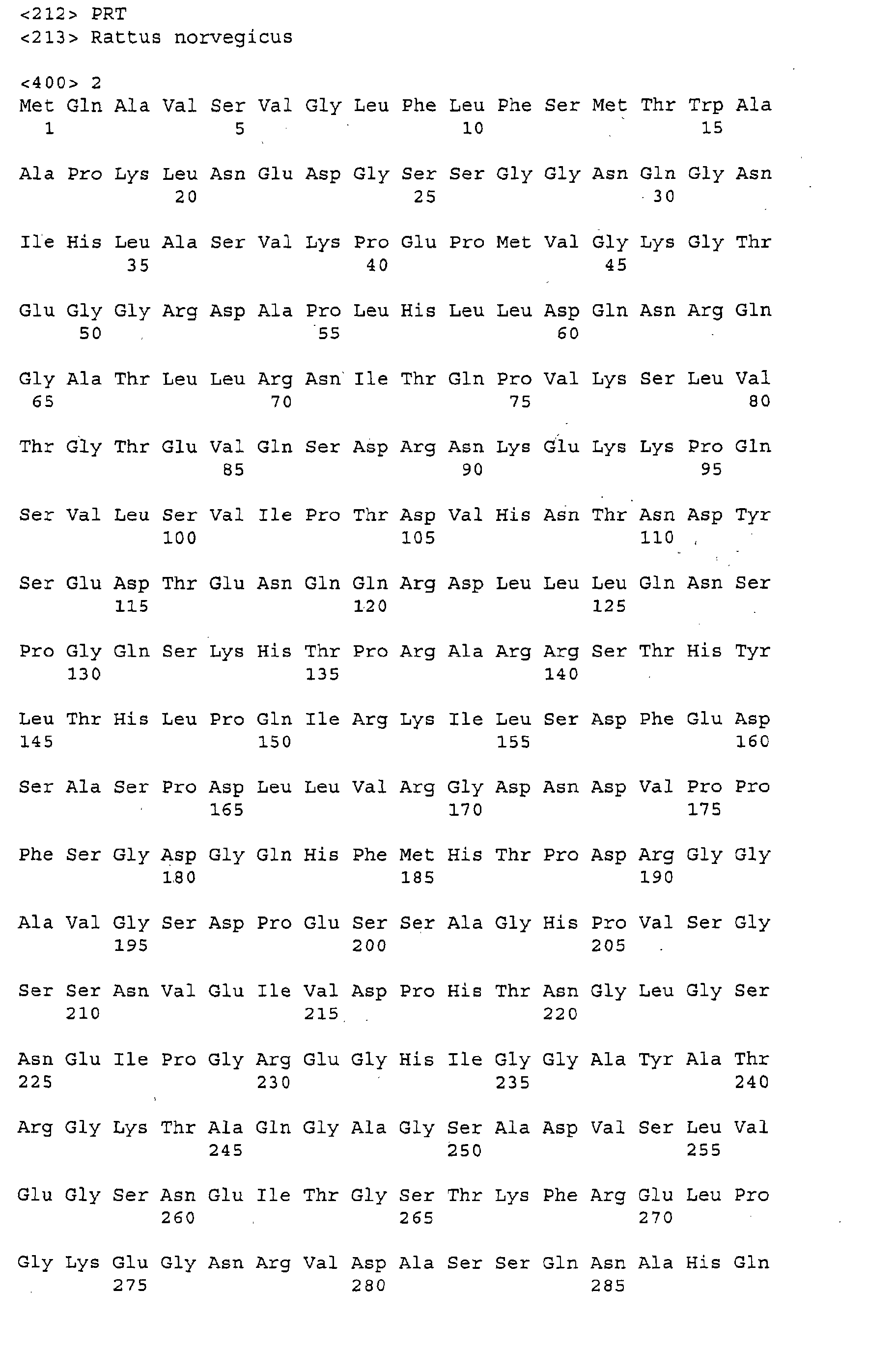
Figure 1A shows the nucleotide sequence encoding rat osteoregulin (SEQ ID
NO: 1). Figure 1B shows the predicted amino acid sequence (SEQ ID NO: 2). A signal
sequence is underlined.
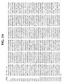
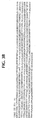
Figure 2A details the nucleotide sequence encoding mouse osteoregulin (SEQ
ID NO: 3). Figure 2B describes the predicted amino acid sequence (SEQ ID NO: 4).
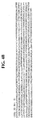
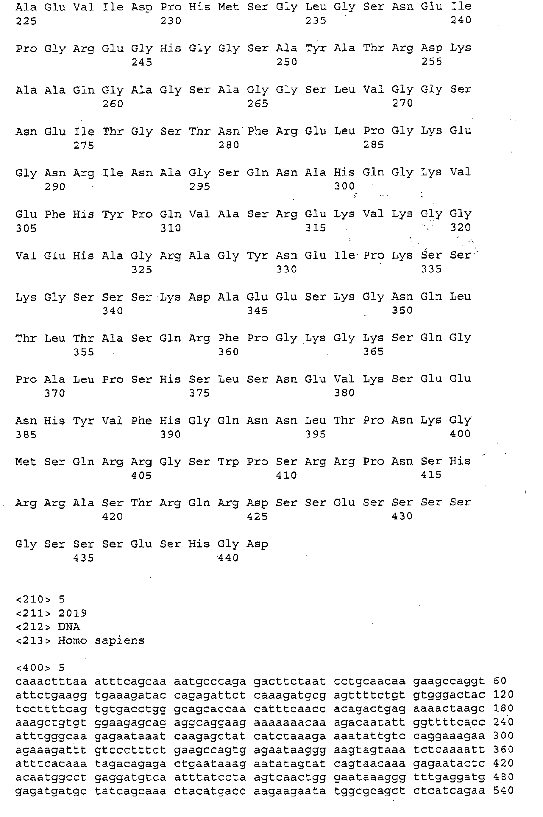
Figure 3A shows a nucleotide sequence encoding a human osteoregulin (SEQ
ID NO: 5). Figure 3B shows the predicted amino acid sequence (SEQ ID NO: 6).
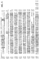
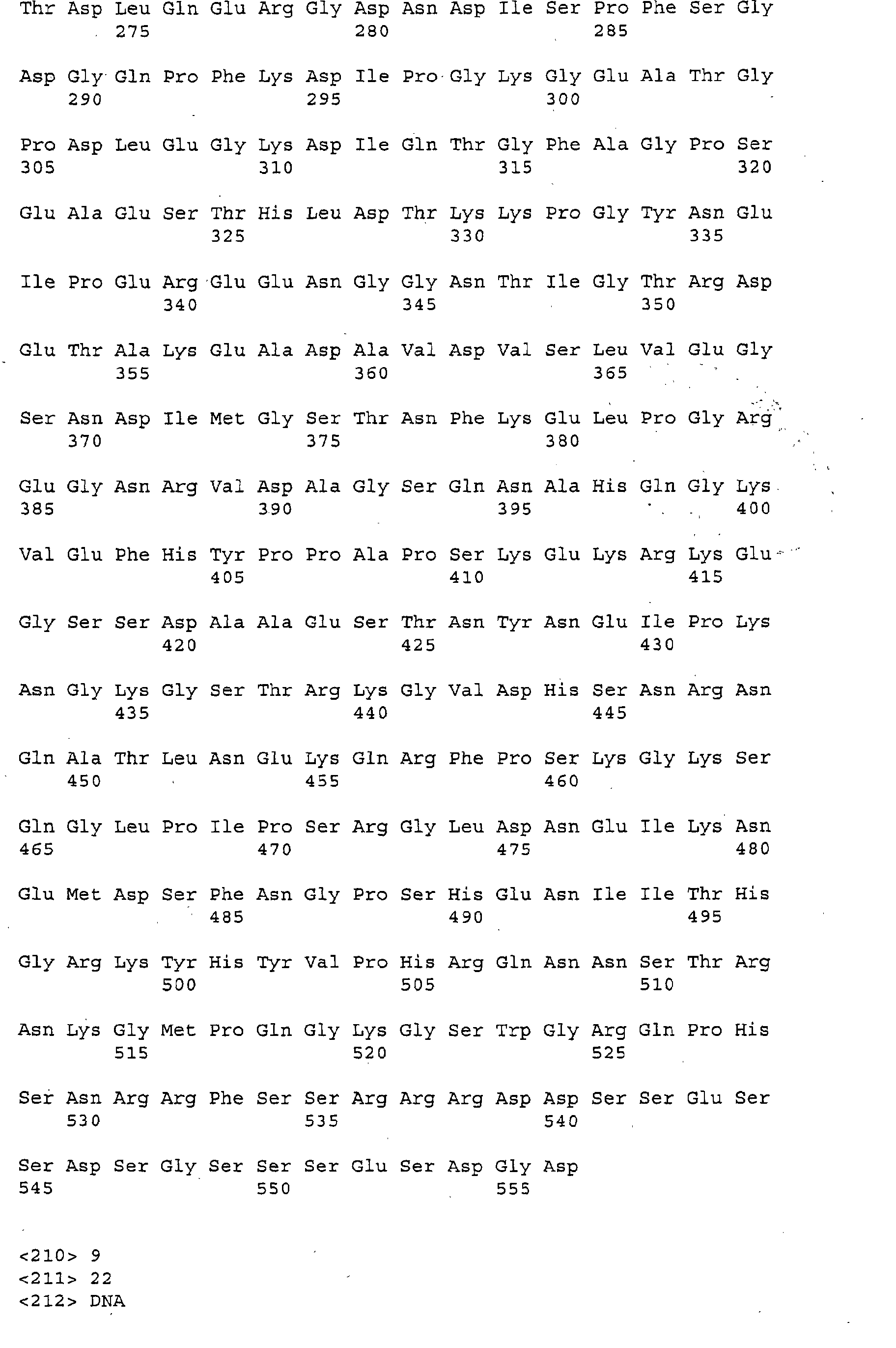
Figure 4A describes another nucleotide sequence encoding a human
osteoregulin (SEQ ID NO: 7). Figure 4B describes the predicted amino acid sequence
(SEQ ID NO: 8).
Figure 5 is a schematic showing the alignment between rat, mouse, and human
osteoregulin polypeptides. A conserved RGD motif is underlined.
Figure 6 is a Northern blot showing osteoregulin mRNA increase during
differentiation of rat bone marrow cell cultures into osteoblastic cells by dexamethasone
treatment. 20 µg of total RNA from either control or dexamethasone treated rat bone
marrow cell cultures was subjected to Northern blot analysis at the indicated time
points. Filters were probed sequentially for alkaline phosphatase (AP), bone
sialoprotein (BSP), osteocalcin (Oc), osteonectin (On), osteopontin (Op) and the control
18S rRNA (18S).
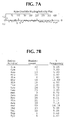
Figure 7A is a hydrophathy plot of the rat osteoregulin polypeptide calculated
according to Kyte-Doolittle algorithm, indicating a hydrophobic leader sequence at the
amino terminus of the predicted amino acid sequence. Figure 7B describes the amino
acid composition of the osteoregulin polypeptide, as predicted after proteolytic
processing between Ala16 and Ala17.
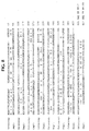
Figure 8 shows the alignment between rat osteoregulin amino acid sequence
and the dentin and bone matrix protein DMP1/AG1, showing weak homology. The rat
osteoregulin primary amino acid sequence was aligned with the first 447 amino acids of
rat DMP (SEQ ID NO: 43).
Figure 9 depicts the highly specific bone tissue osteoregulin mRNA expression
pattern in rat. Figure 9A shows a multiple tissue Northern blot containing 2 µg poly A+
RNA from, heart, lane 1; brain, lane 2; spleen, lane 3; lung, lane 4; liver, lane 5; skeletal
muscle, lane 6; kidney, lane 7; and testis, lane 8. The blot was probed with a cDNA for
osteoregulin followed by actin as a control. Figure 9B shows a Northern blot analysis of
20 µg total RNA from pituitary, lane 9; tibial shaft, lane 10; uterus, lane 11; marrow, lane
12; tibial metaphysis, lane 13; intestine, lane 14; aorta, lane 15; RBMC + vehicle, lane
16; RBMC + dexamethasone, lane 17; brown fat, lane 18; and white fat, lane 19. The
filter was probed for osteoregulin followed by an 18S control.
Figure 10 depicts osteoregulin mRNA being highly expressed in rat tibial shaft,
metaphysis and growth plate. Northern blot analysis of 20 µg of total RNA from marrow,
lane 1; periosteum, lane 2; tibial shaft, lane 3; tibial metaphysis, lane 4; femoral neck,
lane 5; tibial growth plate, lane 6; and calvaria, lane 7. The blot was probed
sequentially for osteoregulin (OR), alkaline phosphatase (AP), bone sialoprotein (BSP),
osteocalcin (Oc), osteonectin (On), osteopontin (Op), tartrate-resistant alkaline
phosphatase (TRAP) and 18S rRNA (18S) as an internal control.
Figure 11 is a Northern blot showing osteoregulin expression in immortalized
osteoblastic cell lines. 20 µg of total RNA from UMR106 and ROS17/2.8 cells lane 1,
time 0; lane 2, vehicle 1 day; lane 3, 10 nM dexamethasone 1 day; lane 4, 10 nM
dexamethasone/50 µg/ml ascorbic acid 1 day; lane 5, vehicle 14 days; lane 6, 10 nM
dexamethasone 14 days; lane 7, 10 nM dexamethasone/50 µg/ml ascorbic acid 14
days. The identical filter was sequentially probed for osteoregulin and 18S rRNA as an
internal control.
Figure 12 is a Northern blot showing that the induction of osteoregulin mRNA
differs from that of osteocalcin mRNA in UMR106 cells. Lane 1, 20 ug of total RNA
from UMR106 cells treated with 10 nM dexamethasone for 2 days; lane 2, 10 nM
dexamethasone for 3 days; lane 3, 10 nM dexamethasone for 10 days; lane 4, vehicle
for 14 days; lane 5, 50 µg/ml ascorbic acid for 14 days; lane 6, 10 nM dexamethasone
for 14 days; lane 7, 10 mM BGP for 14 days; lane 8, 10 nM dexamethasone and 50
µg/ml ascorbic acid for 14 days; lane 9, 10 nM dexamethasone, 10 mM BGP and 50
µg/ml ascorbic acid for 14 days was sequentially probed for the indicated messages.
Alkaline phosphatase (AP), osteocalcin (Oc), osteonectin (On), osteopontin (Op), and
18S rRNA (18S) as an internal control.
Figure 13A is a Northern blot analysis of osteoregulin expression in various
mouse tissues which indicates bone specific expression of the osteoregulin gene.
Figure 13B (upper panel) shows a Southern hybridization blot of reverse transcription
(RT)-PCR of assorted mouse tissues with primers specific for osteoregulin. In addition
to bone, the osteoregulin message is detected at very low levels in the embryo, testes,
brown and white fat, and the aorta. The lower panel represents actin primer RT-PCR
product for each RNA sample.
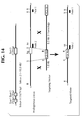
Figure 14 shows the genomic organization and the strategy for targeted
disruption of the murine osteoregulin gene. Exon sizes and intron/exon boundaries of
the mouse osteoregulin gene are diagrammed. Recombination of the targeting vector
with the osteoregulin locus results in the replacement of the third and largest exon with
the neomycin selectable marker gene. Successful targeting was detected with both
external (probe 1) and internal (probe 2) probes. X=Xba1, Bg=Bgl II, Sp=Spel, M=Msc
I, B=Bam HI.
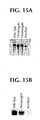
Figure 15A is a Southern blot analysis of tail DNAs to assess the targeted
disruption of the osteoregulin locus in mice. Tail DNA was digested with Bam HI and
Bgl I and hybridized with Probe 2. A 10 kb band is detected at the wild type allele and a
9 kb band is detected at the targeted allele. Figure 15B is a Southern hybridization blot
of RT-PCR of RNA prepared from bone of wild type, heterozygous, and knockout
animals. Note the reduced message in the heterozygote lane and the absence of
message in the knockout lane.
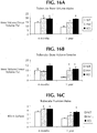
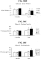
Figure 16 is a chart showing the effects of osteoregulin gene disruption on bone
phenotype on mice aged 4 months and 1 year, as determined by quantitative
histomorphometry (* = p<0.05). Wild type (WT); heterozygote (Het); knockout (KO)
(i.e., homozygote).
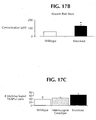
Figure 17A is a graph quantitating mineralized nodules by Von Kossa staining of
primary bone marrow cultures. Figure 17B is a graph depicting Alizarin Red staining of
primary calvarial cultures. Figure 17C is a graph quantitating osteoclastogenesis, as
determined by tartrate resistant acid phosphatase (TRAP) cytochemical stain of bone
marrow cultures. TRAP positive cells with three or more discrete nuclei were counted
(*=p<0.05).
Figure 18 is a Southern blot indicating hybridization of a rat osteoregulin probe
to human genomic DNA. Mouse (M); Rat (R); Human (H).
Figure 19 shows Southern blot hybridization of rat cDNA to human BAC clone
33735 (B).
Figure 20 is a Southern blot detailing the subcloning of hybridizing HindIII
fragment from human BAC clone 22725. Clones 2, 4, 6, and 12 were considered to
have the insert of interest and were subjected to sequence analysis.
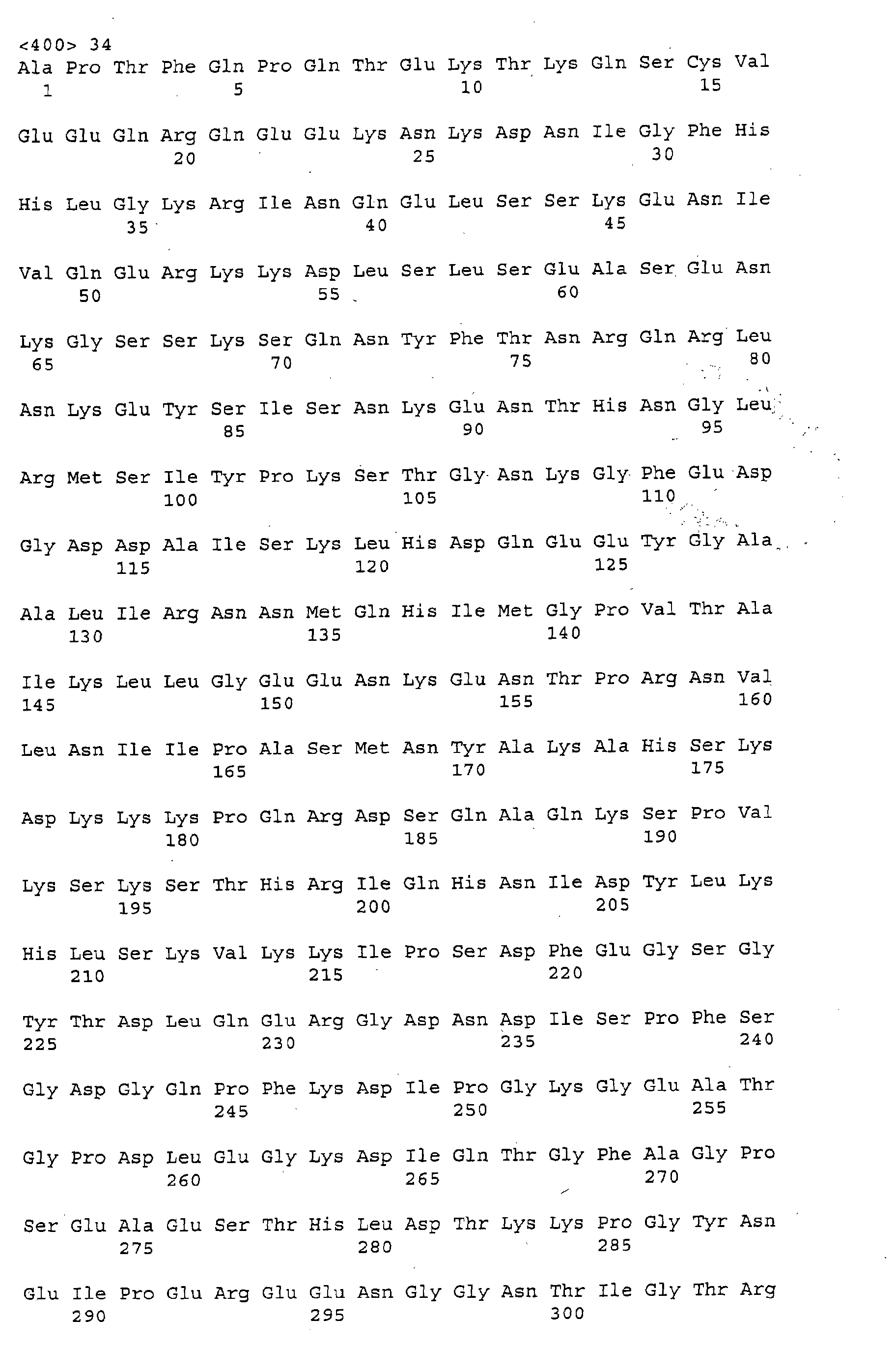
Figure 21A shows the amino acid sequence for a human osteoregulin
polypeptide which lacks an N-terminal signal sequence (SEQ ID NO: 34). The amino
acid sequence is encoded by SEQ ID NO: 33. Fig. 21B shows the amino acid
sequence for a second human osteoregulin polypeptide which lacks an N-terminal
signal sequence (SEQ ID NO: 46). The amino acid sequence is encoded by SEQ ID
NO: 45.
Figure 22 is a Northern blot showing the tissue distribution of human
osteoregulin expression. Other tissues not shown, but negative for expression,
include prostate, testes, and ovary.
Figure 23 shows osteoregulin tissue expression by Southern blot analysis of
RT-PCR.
Detailed Description
The invention is directed to new mammalian proteins, designated
osteoregulins, the polynucleotides encoding the osteoregulins, genetically-modified
non-human mammals and animal cells containing a disrupted osteoregulin gene, and
host cells transfected with, and expressing an exogenous osteoregulin protein.
We have discovered that osteoregulins play a role in regulating bone density,
bone mineralization, and the level of adiposity. Accordingly, additional features of the
invention provide, first, for screening assays that identify agents that modulate
osteoregulin activity and/or expression, and, second, for the use of agents that
regulate osteoregulin activity or expression to treat or prevent bone or adipose tissue
related disorders.
Osteoregulin Amino Acid Sequences and Nucleotide Sequences
The invention encompasses osteoregulin polypeptide sequences, for
example, rat osteoregulin (Fig. IB, SEQ ID NO: 2), mouse osteoregulin (Fig. 2B, SEQ
ID NO: 4), and human osteoregulin (Figs. 3B, 4B, 21A and 21B, SEQ ID NOs: 6, 8,
34, and 46, respectively), and any variant, homologue, fragment, or derivative
thereof.
All of the disclosed osteoregulin sequences are rich in serine, glycine, and
charged amino acids, and include a charged amino terminal hydrophobic signal
sequence that targets the polypeptides for secretion. An RGD sequence, an element
involved in cell-matrix interactions via integrin binding and signaling is also conserved
in all sequences. Comparison of these sequences revealed 67% identity between
the rat and mouse osteoregulins and 45% identity between human osteoregulin and
either rat or mouse osteoregulin. As shown in Fig. 5, the identity between the
sequences was distributed throughout the entire length of the sequences. Preferred
variants, homologues, fragments, or derivatives of the above disclosed osteoregulin
sequences have a conserved RGD sequence (Fig. 5, underlined).
These osteoregulins share structural features common to a class of
extracellular matrix phosphoglycoproteins that includes osteopontin, dentin,
sialophosphoprotein, dentin matrix protein 1, and bone sialoprotein II. All of the
above-mentioned related phosphoglycoproteins are expressed in bone or dentin and
play roles in regulating mineralization (D'Souza et al., J. Bone Min. Res. 12: 2040-49,
1997; Robey, Conn. Tiss. Res. 35: 131-36, 1996; and Boskey, Conn. Tiss. Res.
35: 357-63, 1996).
The invention is also directed to nucleic acid sequences encoding an
osteoregulin polypeptide or encoding any variant, homologue, derivative, or fragment
thereof. Examples include the coding sequences and the full length cDNA-derived
sequences for rat osteoregulin (Fig 1A and SEQ ID NO: 1), mouse osteoregulin (Fig.
2A, and SEQ ID NO: 3), and two splice variants for human osteoregulins (Fig. 3A and
SEQ ID NO: 5; Fig. 4A and SEQ ID NO: 7). Also encompassed by the invention are
polynucleotide sequences that are capable of hybridizing to the polynucleotide
sequences of the invention under various conditions of moderate and high stringency
(see e.g., Wahl and Berger, Methods Enzymol. 152: 399-407, 1987, and Kimmel,
Methods Enzymol. 152: 507-11, 1987).
The nucleic acid sequences encoding osteoregulin may be extended utilizing
a partial nucleotide sequence and employing various PCR-based methods known in
the art to detect upstream sequences, such as promoters and regulatory elements.
For example, one method which may be employed, restriction-site PCR, uses
universal and nested primers to amplify unknown sequence from genomic DNA
within a cloning vector. (See, e.g., Sarkar, G., PCR Methods Applic. 2: 318-322,
1993.) Another method, inverse PCR, uses primers that extend in divergent
directions to amplify unknown sequence from a circularized template. The template
is derived from restriction fragments comprising a known genomic locus and
surrounding sequences (see, e.g., Triglia et al., Nucleic Acids Res. 16: 8186, 1988).
A third method, capture PCR, involves PCR amplification of DNA fragments adjacent
to known sequences in human and yeast artificial chromosome DNA (see, e.g.,
Lagerstrom et al., PCR Methods Applic. 1: 111-119, 1991). In this method, multiple
restriction enzyme digestions and ligations may be used to insert an engineered
double-stranded sequence into a region of unknown sequence before performing
PCR.
In another embodiment of the invention, a polynucleotide of the invention may
be cloned in recombinant DNA molecules that direct expression of osteoregulin, its
homologues, variants, derivatives, or fragments thereof, in appropriate host cells.
The nucleotide sequences of the present invention can be engineered using methods
generally known in the art in order to alter osteoregulin-encoding sequences for a
variety of purposes including, but not limited to, modification of the cloning,
processing, and/or expression of the gene product.
DNA shuffling by random fragmentation and PCR reassembly of gene
fragments and synthetic oligonucleotides may be used to engineer the nucleotide
sequences. For example, oligonucleotide-mediated site-directed mutagenesis may
be used to introduce mutations that create new restriction sites, alter glycosylation
patterns, change codon preference, produce splice variants, and so forth. In another
embodiment, sequences encoding osteoregulin may be synthesized, in whole or in
part, using chemical methods well known in the art (see, e.g., Caruthers et al.,
Nucleic Acids Symp. Ser. 7: 215-223, 1980; and Horn et al., Nucleic Acids Symp.
Ser. 7: 225-232, 1980). Alternatively, osteoregulin itself or a fragment thereof may
be synthesized using chemical methods. For example, peptide synthesis can be
performed using various solid- phase techniques (see, e.g., Roberge et al., Science
269: 202-204, 1995). Automated synthesis may be achieved using the ABI 431A
peptide synthesizer (Perkin-Elmer, Norwalk, CT). Additionally, the amino acid
sequence of osteoregulin, or any part thereof, may be altered during direct synthesis
and/or combined with sequences from other proteins, or any part thereof, to produce
a variant polypeptide. The peptide may be substantially purified by preparative high
performance liquid chromatography (see, e.g., Chiez and Regnier, Methods
Enzymol. 182: 392-421, 1990). The composition of the synthetic peptides may be
confirmed by amino acid analysis or by sequencing (see, e.g., Creighton, Proteins,
Structures and Molecular Properties, WH Freeman, New York, NY 1984).
Osteoregulin-Encoding Expression Vectors
In order to express a biologically active osteoregulin, the nucleotide
sequences encoding osteoregulin or derivatives thereof may be inserted into an
appropriate expression vector, i.e., a vector which contains the necessary elements
for transcriptional and translational control of the inserted coding sequence in a
suitable host. These elements include regulatory sequences, such as enhancers,
constitutive and inducible promoters, and 5' and 3' untranslated regions derived from
the vector and/or from the polynucleotide sequences encoding osteoregulin. Such
elements may vary in their strength and specificity. Specific initiation signals may
also be used to achieve more efficient translation of sequences encoding
osteoregulin. Such signals include the ATG initiation codon and adjacent sequences,
e.g. the Kozak sequence. In cases where sequences encoding osteoregulin and its
initiation codon and upstream regulatory sequences are inserted into the appropriate
expression vector, no additional transcriptional or translational control signals may be
needed. However, in cases where only coding sequence, or a fragment thereof, is
inserted, exogenous translational control signals including an in-frame ATG initiation
codon should be provided by the vector. Exogenous translational elements and
initiation codons may be of various origins, both natural and synthetic. The efficiency
of expression may be enhanced by the inclusion of enhancers appropriate for the
particular host cell system used (see, e.g., Scharf et al., Results Probl. Cell Differ. 20:
125-162, 1994). Methods which are well known to those skilled in the art may be
used to construct expression vectors containing sequences encoding osteoregulin
and appropriate transcriptional and translational control elements. These methods
include in vitro recombinant DNA techniques, synthetic techniques, and in vivo
genetic recombination. (See, e.g., Sambrook et al., Molecular Cloning, Laboratory
Manual, Cold Spring Harbor Press, Plainview NY, ch. 4, 8, and 16-17, 1989; Ausubel
et al., Current Protocols in Molecular Biology, John Wiley & Sons, New York, NY,
chs. 9, 13, and 16, 1995). A variety of expression vector/host systems may be
utilized to contain and express sequences encoding osteoregulin. These include, but
are not limited to, microorganisms such as bacteria transformed with recombinant
bacteriophage, plasmid, or cosmid DNA expression vectors; yeast transformed with
yeast expression vectors; insect cell systems infected with viral expression vectors
(e.g., baculovirus), plant cell systems transformed with viral expression vectors (e.g.,
cauliflower mosaic virus, CaNW, or tobacco mosaic virus, TMV), with bacterial
expression vectors (e.g., Ti or pBR322 plasmids), or animal cell systems. The
invention is not limited by the host cell employed.
In bacterial systems, a number of cloning and expression vectors may be
selected depending upon the use intended for polynucleotide sequences encoding
osteoregulin. For example, routine cloning, subcloning, and propagation of
polynucleotide sequences encoding osteoregulin can be achieved using a
multifunctional E.coli vector such as pBlueScript (Stratagene, La Jolla, CA) or
pSport1 plasmid (Life Technologies, Gaithersburg, MD). Ligation of sequences
encoding osteoregulin into the vector's multiple cloning site disrupts the lacZ gene,
allowing a colorimetric screening procedure for identification of transformed bacteria
containing recombinant molecules. In addition, these vectors may be useful for in
vitro transcription, dideoxy sequencing, single strand rescue with helper phage, and
creation of nested deletions in the cloned sequence (see, e.g., Van Heeke and
Schuster, J. Biol. Chem. 264: 5503-5509, 1989). When large quantities of
osteoregulin are needed, e.g. for the production of antibodies, vectors which direct
high level expression of osteoregulin may be used. For example, vectors containing
the strong, inducible T5 or T7 bacteriophage promoter may be used.
Host Cells Transformed with Osteoregulin-Encoding Sequences
Yeast expression systems maybe used for production of osteoregulin. A
number of vectors containing constitutive or inducible promoters, such as alpha
factor, alcohol oxidase, or PGH promoters, may be used in the yeast Saccharomyces
cerevisiae or Pichia pastoris. In addition, such vectors direct either the secretion or
intracellular retention of expressed proteins and enable integration of foreign
sequences into the host genome for stable propagation (see, e.g., Ausubel, 1995;
Bitter et al., Methods Enzymol. 153: 516-544, 1987; and Scorer et al., BioTechnology
12: 181-184, 1994). Plant systems may also be used for expression of osteoregulin.
Transcription of sequences encoding osteoregulin may be driven by viral promoters,
e.g., the 35S and 19S promoters of CaMV used alone or in combination with the
omega leader sequence from TMV (Takamatsu, EMBO J. 6: 307-311, 1987).
Alternatively, plant promoters such as the small subunit of RUBISCO or heat shock
promoters may be used. (see, e.g., Coruzzi et al., EMBO J. 3: 1671-1680, 1984;
Broglie et al., Science 224: 838-843, 1984; and Winter et al., Results Probl. Cell
Differ. 17: 85-105, 1991). These constructs can be introduced into plant cells by
direct DNA transformation or pathogen-mediated transfection (see, e.g., The McGraw
Hill Yearbook of Science and Technology, McGraw Hill, New York NY, pp. 191-196,
1992).
In mammalian cells, a number of viral-based expression systems may be
utilized. In cases where an adenovirus is used as an expression vector, sequences
encoding osteoregulin may be ligated into an adenovirus transcription/translation
complex consisting of the late promoter and tripartite leader sequence. Insertion in a
non-essential E1 or E3 region of the viral genome may be used to obtain infective
virus that express osteoregulin in infected host cells (see, e.g., Logan and Shenk,
Proc. Natl. Acad. Sci. USA 81: 3655-3659, 1984). In addition, transcription
enhancers, such as the Rous sarcoma virus (RSV) enhancer, may be used to
increase expression in mammalian host cells. SV40 or EBV-based vectors may also
be used for high-level protein expression.
Human artificial chromosomes (HACs) may also be employed to deliver larger
fragments of DNA than can be contained in and expressed from a plasmid. HACs of
about 6 kb to 10 Mb are constructed and delivered via conventional delivery methods
(liposomes, polycationic amino polymers, or vesicles) for therapeutic purposes (see,
e.g., Harrington et al., Nat. Genet. 15: 345-355, 1997). For long term production of
recombinant proteins in mammalian systems, stable expression of osteoregulin in
cell lines is preferred. For example, sequences encoding osteoregulin can be
transformed into cell lines using expression vectors which may contain viral origins of
replication and/or endogenous expression elements and a selectable marker gene on
the same or on a separate vector. Following the introduction of the vector, cells may
be allowed to grow for about 1 to 2 days in enriched media before being switched to
selective media. The purpose of the selectable marker is to confer resistance to a
selective agent, and its presence allows growth and recovery of cells which
successfully express the introduced sequences. Resistant clones of stably
transformed cells may be propagated using tissue culture techniques appropriate to
the cell type.
Any number of selection systems may be used to recover transformed cell
lines. These include, but are not limited to, the herpes simplex virus thymidine
kinase and adenine phosphoribosyltransferase genes, for use in tk-, and apr- cells,
respectively (see, e.g., Wigler et al., Cell 11: 223-232, 1997; Lowy et al., Cell 22:
817-823, 1980). Also, antimetabolite, antibiotic, or herbicide resistance can be used
as the basis for selection. For example, dhfr confers resistance to methotrexate; neo
confers resistance to the aminoglycosides neomycin and G-418; and als and pat
confer resistance to chlorsulfuron and phosphinotricin acetyltransferase, respectively
(see, e.g., Wigler et al., Proc. Natl. Acad. Sci. USA 77: 3567-3570, 1980; Colbere-Garapin
et al., J. Mol. Biol. 150: 1-14, 1981). Additional selectable genes have been
described, e.g., trpB and hisD, which alter cellular requirements for metabolites (see,
e.g., Hartman and Mulligan, Proc. Natl. Acad. Sci. USA 85: 8047-8051, 1988).
Visible markers, e.g., anthocyanins, green fluorescent proteins (GFP; Clontech, Palo
Alto, CA), β-glucuronidase and its substrate β-glucuronide, or luciferase and its
substrate luciferin may be used. These markers can be used not only to identify
transformants, but also to quantify the amount of transient or stable protein
expression attributable to a specific vector system (see, e.g., Rhodes, Methods Mol.
Biol. 55: 121-131, 1995). Although the presence/absence of marker gene expression
suggests that the gene of interest is also present, the presence and expression of the
gene may need to be confirmed. For example, if the sequence encoding
osteoregulin is inserted within a marker gene sequence, transformed cells containing
sequences encoding osteoregulin can be identified by the absence of marker gene
function. Alternatively, a marker gene can be placed in tandem with a sequence
encoding osteoregulin under the control of a single promoter. Expression of the
marker gene in response to induction or selection usually indicates expression of the
tandem gene as well.
In general, host cells that contain the nucleic acid sequence encoding
osteoregulin and that express osteoregulin may be identified by a variety of
procedures known to those of skill in the art. These procedures include, but are not
limited to, DNA-DNA or DNA-RNA hybridizations, PCR amplification, and protein
bioassay or immunoassay techniques which include membrane, solution, or chip
based technologies for the detection and/or quantification of nucleic acid or protein
sequences.
Immunological methods for detecting and measuring the expression of
osteoregulin using either specific polyclonal or monoclonal antibodies are known in
the art. Examples of such techniques include enzyme-linked immunosorbent assays
(ELISAs), radioimmunoassays (RIAs), and fluorescence activated cell sorting
(FACS). A two-site, monoclonal-based immunoassay utilizing monoclonal antibodies
reactive to two non-interfering epitopes on osteoregulin is preferred, but a
competitive binding assay may be employed. These and other assays are well
known in the art (see, e.g., Hampton, Serological Methods, A Laboratory Manual,
APS Press, St. Paul, MN, Sect. IV, 1990; Coligan et al., Current Protocols in
Immunology, Greene Pub. Associates and Wiley-Interscience, New York NY, 1997;
and Pound, Immunochemical Protocols, Humana Press, Totowa, NJ, 1998). A wide
variety of labels and conjugation techniques are known by those skilled in the art and
may be used in various nucleic acid and amino acid assays.
Means for producing labeled hybridization or PCR probes for detecting
sequences related to polynucleotides encoding osteoregulin include oligolabeling,
nick translation, end-labeling, or PCR amplification using a labeled nucleotide.
Alternatively, the sequences encoding osteoregulin, or any fragments thereof,
may be cloned into a vector for the production of an mRNA probe. Such vectors are
known in the art, are commercially available, and may be used to synthesize RNA
probes in vitro by addition of an appropriate RNA polymerase such as T7, T3, or SP6
and labeled nucleotides. These procedures may be conducted using a variety of
commercially available kits (e.g., Amersham Pharmacia Biotech, Piscataway, NJ,
Promega, Madison, WI, and US Biochemical, Cleveland OH). Suitable reporter
molecules or labels which may be used for ease of detection include radionuclides,
enzymes, fluorescent, chemiluminescent, or chromogenic agents, as well as
substrates, cofactors, inhibitors, magnetic particles, and the like.
Host cells transformed with nucleotide sequences encoding osteoregulin may
be cultured under conditions suitable for the expression and recovery of the protein
from cell culture. The protein produced by a transformed cell may be secreted or
retained intracellularly depending on the sequence and/or the vector used. As will be
understood by those of skill in the art, expression vectors containing polynucleotides
which encode osteoregulin may be designed to contain signal sequences which
direct secretion of osteoregulin through a prokaryotic or eukaryotic cell membrane.
In addition, a host cell strain may be chosen for its ability to modulate expression of
the inserted sequences or to process the expressed protein in the desired fashion.
Such modifications of the polypeptide include, but are not limited to, acetylation,
carboxylation, glycosylation, phosphorylation, lipidation, and acylation. Post-translational
processing which cleaves a "prepro" or "pro" form of the protein may
also be used to specify protein targeting, folding, and/or activity.
Different host cells which have specific cellular machinery and characteristic
mechanisms for post-translational activities (e.g., CHO, HeLa, MDCK, HEK293, and
W138) are available from the American Type Culture Collection (ATCC, Manassas,
VA) and may be chosen to ensure the correct modification and processing of the
foreign protein. In another embodiment of the invention, natural, modified, or
recombinant nucleic acid sequences encoding osteoregulin may be ligated to a
heterologous sequence resulting in translation of a fusion protein in any of the
aforementioned host systems. For example, a chimeric osteoregulin protein
containing a heterologous moiety that can be recognized by a commercially available
antibody may facilitate the screening of peptide libraries for inhibitors of osteoregulin
activity. Heterologous protein and peptide moieties may also facilitate purification of
fusion proteins using commercially available affinity matrices. Such moieties include,
but are not limited to, glutathione S-transferase (GST), maltose binding protein
(MBP), thioredoxin (Trx), calmodulin binding peptide (CBP), 6-His, FLAG, c-myc, and
hemagglutinin (HA). GST, MBP, Trx, CBP, and 6-His enable purification of their
cognate fusion proteins on immobilized glutathione, maltose, phenylarsine oxide,
calmodulin, and metal-chelate resins, respectively. FLAG, c-myc, and hemagglutinin
(HA) enable immunoaffinity purification of fusion proteins using commercially
available monoclonal and polyclonal antibodies that specifically recognize these
epitope tags. A fusion protein may also be engineered to contain a proteolytic
cleavage site located between the osteoregulin encoding sequence and the
heterologous protein sequence, so that osteoregulin may be cleaved away from the
heterologous moiety following purification. Methods for fusion protein expression and
purification are discussed in Ausubel (1995, supra, ch. 10). A variety of commercially
available kits may also be used to facilitate expression and purification of fusion
proteins.
In a further embodiment of the invention, synthesis of radiolabeled
osteoregulin may be achieved in vitro using the TNT rabbit reticulocyte lysate or
wheat germ extract system (Promega). These systems couple transcription and
translation of protein-coding sequences operably associated with the T7, T3, or SP6
promoters. Translation takes place in the presence of a radiolabeled amino acid
precursor, for example, 35S-methionine.
Fragments of osteoregulin may be produced not only by recombinant means,
but also by direct peptide synthesis using solid-phase techniques (see, e.g.,
Creighton, supra, pp. 55-60). Protein synthesis may be performed by manual
techniques or by automation. Automated synthesis may be achieved, for example,
using the ABI® 431A peptide synthesizer (Perkin-Elmer). Various fragments of
osteoregulin may be synthesized separately and then combined to produce the full
length molecule.
Genetically-Modified Non-human Mammals and Animal Cells Containing a Disrupted
Osteoregulin Gene
1. Genetically-Modified Non-human Mammals and Animal Cells
The genetically-modified, non-human mammals and genetically-modified
animal cells, including human cells, of the invention are heterozygous or
homozygous for a modification that functionally disrupts the osteoregulin gene. The
animal cells may be derived by genetically engineering cells in culture, or, in the case
of non-human mammalian cells, the cells may be isolated from genetically-modified,
non-human mammals.
The osteoregulin gene locus is functionally disrupted by one of the several
techniques for genetic modification known in the art, including chemical mutagenesis
(Rinchik, Trends in Genetics 7: 15-21, 1991, Russell, Environmental & Molecular
Mutagenesis 23 (Suppl. 24) 23-29, 1994), irradiation (Russell, supra), transgenic
expression of osteoregulin gene antisense RNA, either alone or in combination with a
catalytic RNA ribozyme sequence (Luyckx et al., Proc. Natl. Acad. Sci. 96: 12174-79,
1999; Sokol et al., Transgenic Research 5: 363-71, 1996; Efrat et al., Proc. Natl.
Acad. Sci. USA 91: 2051-55, 1994; Larsson et al., Nucleic Acids Research 22: 2242-48,
1994) and, as further discussed below, the disruption of the osteoregulin gene by
the insertion of a foreign nucleic acid sequence into the osteoregulin gene locus.
Preferably, the foreign sequence is inserted by homologous recombination or by the
insertion of a viral vector. Most preferably, the method of osteoregulin gene
disruption is homologous recombination and includes a deletion of a portion of the
endogenous osteoregulin gene sequence.
The integration of the foreign sequence functionally disrupts the osteoregulin
gene through one or more of the following mechanisms: by interfering with the
osteoregulin gene transcription or translation process (e.g., by interfering with
promoter recognition, or by introducing a transcription termination site or a
translational stop codon into the osteoregulin gene); or by distorting the osteoregulin
gene coding sequence such that it no longer encodes a osteoregulin polypeptide with
normal function (e.g., by inserting a foreign coding sequence into the osteoregulin
gene coding sequence, by introducing a frameshift mutation or amino acid(s)
substitution, or, in the case of a double crossover event, by deleting a portion of the
osteoregulin gene coding sequence that is required for expression of a functional Y
protein).
To insert a foreign sequence into a osteoregulin gene locus in the genome of
a cell, the foreign DNA sequence is introduced into the cell according to a standard
method known in the art such as electroporation, calcium-phosphate precipitation,
retroviral infection, microinjection, biolistics, liposome transfection, DEAE-dextran
transfection, or transferrinfection (see, e.g., Neumann et al., EMBO J. 1: 841-845,
1982; Potter et al., Proc. Natl. Acad. Sci USA 81: 7161-65, 1984; Chu et al., Nucleic
Acids Res. 15: 1311-26, 1987; Thomas and Capecchi, Cell 51: 503-12, 1987; Baum
et al., Biotechniques 17: 1058-62, 1994; Biewenga et al., J. Neuroscience Methods
71: 67-75, 1997; Zhang et al., Biotechniques 15: 868-72, 1993; Ray and Gage,
Biotechniques 13: 598-603, 1992; Lo, Mol. Cell. Biol. 3: 1803-14, 1983; Nickoloff et
al., Mol. Biotech. 10: 93-101, 1998; Linney et al., Dev. Biol. (Orlando) 213: 207-16,
1999; Zimmer and Gruss, Nature 338: 150-153, 1989; and Robertson et al., Nature
323: 445-48, 1986). The preferred method for introducing foreign DNA into a cell is
electroporation.
2. Homologous Recombination
The method of homologous recombination targets the osteoregulin gene
for disruption by introducing a osteoregulin gene targeting vector into a cell
containing a osteoregulin gene. The ability of the vector to target the osteoregulin
gene for disruption stems from using a nucleotide sequence in the vector that is
homologous to the osteoregulin gene. This homology region facilitates hybridization
between the vector and the endogenous sequence of the osteoregulin gene. Upon
hybridization, the probability of a crossover event between the targeting vector and
genomic sequences greatly increases. This crossover event results in the integration
of the vector sequence into the osteoregulin gene locus and the functional disruption
of the osteoregulin gene.
General principles regarding the construction of vectors used for targeting
are reviewed in Bradley et al. (Biotechnol. 10: 534, 1992). Two different types of
vector can be used to insert DNA by homologous recombination: an insertion vector
or a replacement vector. An insertion vector is circular DNA which contains a region
of osteoregulin gene homology with a double stranded break. Following hybridization
between the homology region and the endogenous osteoregulin gene, a single
crossover event at the double stranded break results in the insertion of the entire
vector sequence into the endogenous gene at the site of crossover.
The more preferred vector to use for homologous recombination is a
replacement vector, which is colinear rather than circular. Replacement vector
integration into the osteoregulin gene requires a double crossover event, i.e. crossing
over at two sites of hybridization between the targeting vector and the osteoregulin
gene. This double crossover event results in the integration of vector sequence that
is sandwiched between the two sites of crossover into the osteoregulin gene and the
deletion of the corresponding endogenous osteoregulin gene sequence that originally
spanned between the two sites of crossover (see, e.g., Thomas and Capecchi et al.,
Cell 51: 503-12, 1987; Mansour et al., Nature 336: 348-52, 1988; Mansour et al.,
Proc. Natl. Acad. Sci. USA 87: 7688-7692, 1990; and Mansour, GATA 7: 219-227,
1990).
A region of homology in a targeting vector is generally at least 100
nucleotides in length. Most preferably, the homology region is at least 1-5 kilobases
(kb) in length. Although there is no demonstrated minimum length or minimum
degree of relatedness required for a homology region, targeting efficiency for
homologous recombination generally corresponds with the length and the degree of
relatedness between the targeting vector and the osteoregulin gene locus. In the
case where a replacement vector is used, and a portion of the endogenous
osteoregulin gene is deleted upon homologous recombination, an additional
consideration is the size of the deleted portion of the endogenous osteoregulin gene.
If this portion of the endogenous osteoregulin gene is greater than 1 kb in length,
then a targeting cassette with regions of homology that are longer than 1 kb is
recommended to enhance the efficiency of recombination. Further guidance
regarding the selection and use of sequences effective for homologous
recombination is described in the literature (see, e.g., Deng and Capecchi, Mol. Cell.
Biol. 12: 3365-3371, 1992; Bollag et al., Annu. Rev. Genet. 23: 199-225, 1989; and
Waldman and Liskay, Mol. Cell. Biol. 8: 5350-5357, 1988).
A wide variety of cloning vectors may be used as vector backbones in the
construction of the osteoregulin gene targeting vectors of the present invention,
including pBluescript-related plasmids (e.g., Bluescript KS+11), pQE70, pQE60,
pQE-9, pBS, pD10, phagescript, phiX174, pBK Phagemid, pNH8A, pNH16a,
pNH18Z, pNH46A, ptrc99a, pKK223-3, pKK233-3, pDR540, and pRIT5 PWLNEO,
pSV2CAT, pXT1, pSG (Stratagene), pSVK3, PBPV, PMSG, and pSVL, pBR322 and
pBR322-based vectors, pBM9, pBR325, pKH47, pBR328, pHC79, phage Charon 28,
pKB11, pKSV-10, pK19 related plasmids, pUC plasmids, and the pGEM series of
plasmids. These vectors are available from a variety of commercial sources (e.g.,
Boehringer Mannheim Biochemicals, Indianapolis, IN; Qiagen, Valencia, CA;
Stratagene, La Jolla, CA; Promega, Madison, WI; and New England Biolabs, Beverly,
MA). However, any other vectors, e.g. plasmids, viruses, or parts thereof, may be
used as long as they are replicable and viable in the desired host. The vector may
also comprise sequences which enable it to replicate in the host whose genome is to
be modified. The use of such a vector can expand the interaction period during
which recombination can occur, increasing the efficiency of targeting (see Molecular
Biology, ed. Ausubel et al, Unit 9.16, Fig. 9.16.1).
The specific host employed for propagating the targeting vectors of the
present invention is not critical. Examples include E. coli K12 RR1 (Bolivar et al.,
Gene 2: 95, 1977), E. coli K12 HB101 (ATCC No. 33694), E. coli MM21 (ATCC No.
336780), E. coli DH1 (ATCC No. 33849), E. coli strain DH5α, and E. coli STBL2.
Alternatively, hosts such as C. cerevisiae or B. subtilis can be used. The above-mentioned
hosts are available commercially (e.g., Stratagene, La Jolla, CA; and Life
Technologies, Rockville, MD).
To create the targeting vector, a osteoregulin gene targeting construct is
added to an above-described vector backbone. The osteoregulin gene targeting
constructs of the invention have at least one osteoregulin gene homology region. To
make the osteoregulin gene homology regions, a osteoregulin genomic or cDNA
sequence is used as a basis for producing polymerase chain reaction (PCR) primers.
These primers are used to amplify the desired region of the osteoregulin sequence
by high fidelity PCR amplification (Mattila et al., Nucleic Acids Res. 19: 4967, 1991;
Eckert and Kunkel 1: 17, 1991; and U.S. Pat. No. 4,683, 202). The genomic
sequence is obtained from a genomic clone library or from a preparation of genomic
DNA, preferably from the animal species that is to be targeted for osteoregulin gene
disruption.
Preferably, the targeting constructs of the invention also include an
exogenous nucleotide sequence encoding a positive marker protein. The stable
expression of a positive marker after vector integration confers an identifiable
characteristic on the cell without compromising cell viability. Therefore, in the case of
a replacement vector, the marker gene is positioned between two flanking homology
regions so that it integrates into the osteoregulin gene following the double crossover
event.
It is preferred that the positive marker protein is a selectable protein; the
stable expression of such a protein in a cell confers a selectable phenotypic
characteristic, i.e., the characteristic enhances the survival of the cell under
otherwise lethal conditions. Thus, by imposing the selectable condition, one can
isolate cells that stably express the positive selectable marker-encoding vector
sequence from other cells that have not successfully integrated the vector sequence
on the basis of viability. Examples of positive selectable marker proteins (and their
agents of selection) include neo (G418 or kanomycin), hyg (hygromycin), hisD
(histidinol), gpt (xanthine), ble (bleomycin), and hprt (hypoxanthine) (see, e.g.,
Capecchi and Thomas, U.S. Pat. No. 5,464,764, and Capecchi, Science 244: 1288-92,
1989). Other positive markers that may also be used as an alternative to a
selectable marker include reporter proteins such as β-galactosidase, firefly luciferase,
or GFP (see, e.g., Current Protocols in Cytometry, Unit 9.5, and Current Protocols in
Molecular Biology, Unit 9.6, John Wiley & Sons, New York, NY, 2000).
The above-described positive selection step does not distinguish between
cells that have integrated the vector by targeted homologous recombination at the
osteoregulin gene locus versus random, non-homologous integration of vector
sequence into any chromosomal position. Therefore, when using a replacement
vector for homologous recombination, it is also preferred to include a nucleotide
sequence encoding a negative selectable marker protein. Expression of a negative
selectable marker causes a cell expressing the marker to lose viability when exposed
to a certain agent (i.e., the marker protein becomes lethal to the cell under certain
selectable conditions). Examples of negative selectable markers (and their agents of
lethality) include herpes simplex virus thymidine kinase (gancyclovir or 1,2-deoxy-2-fluoro-α-d-arabinofuransyl-5-iodouracil),
Hprt (6-thioguanine or 6-thioxanthine), and
diphtheria toxin, ricin toxin, and cytosine deaminase (5-fluorocytosine).
The nucleotide sequence encoding the negative selectable marker is
positioned outside of the two homology regions of the replacement vector. Given this
positioning, cells will only integrate and stably express the negative selectable
marker if integration occurs by random, non-homologous recombination; homologous
recombination between the osteoregulin gene and the two regions of homology in the
targeting construct excludes the sequence encoding the negative selectable marker
from integration. Thus, by imposing the negative condition, cells that have integrated
the targeting vector by random, non-homologous recombination lose viability.
The above-described combination of positive and negative selectable
markers is preferred because a series of positive and negative selection steps can be
designed to more efficiently select only those cells that have undergone vector
integration by homologous recombination, and, therefore, have a potentially
disrupted osteoregulin gene. Further examples of positive-negative selection
schemes, selectable markers, and targeting constructs are described, for example, in
U.S. Pat. No. 5,464,764, WO 94/06908, and Valancius and Smithies, Mol. Cell. Biol.
11: 1402, 1991.
In order for a marker protein to be stably expressed upon vector integration,
the targeting vector may be designed so that the marker coding sequence is operably
linked to the endogenous osteoregulin gene promoter upon vector integration.
Expression of the marker is then driven by the osteoregulin gene promoter in cells
that normally express osteoregulin gene. Alternatively, each marker in the targeting
construct of the vector may contain its own promoter that drives expression
independent of the osteoregulin gene promoter. This latter scheme has the
advantage of allowing for expression of markers in cells that do not typically express
the osteoregulin gene (Smith and Berg, Cold Spring Harbor Symp. Quant. Biol. 49:
171, 1984; Sedivy and Sharp, Proc. Natl. Acad. Sci. (USA) 86: 227, 1989; Thomas
and Capecchi, Cell 51: 503, 1987).
Exogenous promoters that can be used to drive marker gene expression
include cell-specific or stage-specific promoters, constitutive promoters, and inducible
or regulatable promoters. Non-limiting examples of these promoters include the
herpes simplex thymidine kinase promoter, cytomegalovirus (CMV)
promoter/enhancer, SV40 promoters, PGK promoter, PMC1-neo, metallothionein
promoter, adenovirus late promoter, vaccinia virus 7.5K promoter, avian beta globin
promoter, histone promoters (e.g., mouse histone H3-614), beta actin promoter,
neuron-specific enolase, muscle actin promoter, and the cauliflower mosaic virus 35S
promoter (see generally, Sambrook et al., Molecular Cloning, Vols. I-III, Cold Spring
Harbor Laboratory Press, Cold Spring Harbor, NY, 1989, and Current Protocols in
Molecular Biology, John Wiley & Sons, New York, NY, 2000; Stratagene, La Jolla,
CA).
To confirm whether cells have integrated the vector sequence into the
targeted osteoregulin gene locus, primers or genomic probes that are specific for the
desired vector integration event can be used in combination with PCR or Southern
blot analysis to identify the presence of the desired vector integration into the
osteoregulin gene locus (Erlich et al., Science 252: 1643-51, 1991; Zimmer and
Gruss, Nature 338: 150, 1989; Mouellic et al., Proc. Natl. Acad. Sci. (USA) 87: 4712,
1990; and Shesely et al., Proc. Natl. Acad. Sci. (USA) 88: 4294, 1991).
3. Gene Trapping
Another method available for inserting a foreign nucleic acid sequence into
the osteoregulin gene locus to functionally disrupt the osteoregulin gene is gene
trapping. This method takes advantage of the cellular machinery present in all
mammalian cells that splices exons into mRNA to insert a gene trap vector coding
sequence into a gene in a random fashion. Once inserted, the gene trap vector
creates a mutation that may functionally disrupt the trapped osteoregulin gene. In
contrast to homologous recombination, this system for mutagenesis creates largely
random mutations. Thus, to obtain a genetically-modified cell that contains a
functionally disrupted osteoregulin gene, cells containing this particular mutation
must be identified and selected from a pool of cells that contain random mutations in
a variety of genes.
Gene trapping systems and vectors have been described for use in
genetically modifying murine cells and other cell types (see, e.g., Allen et al., Nature
333: 852-55, 1988; Bellen et al., Genes Dev. 3: 1288-1300, 1989; Bier et al., Genes
Dev. 3: 1273-1287, 1989; Bonnerot et al., J. Virol. 66: 4982-91, 1992; Brenner et al.,
Proc. Nat. Acad. Sci. USA 86: 5517-21, 1989; Chang et al., Virology 193: 737-47,
1993; Friedrich and Soriano, Methods Enzymol. 225: 681-701, 1993; Friedrich and
Soriano, Genes Dev. 5: 1513-23, 1991; Goff, Methods Enzymol. 152: 469-81, 1987;
Gossler et al., Science 244: 463-65, 1989; Hope, Develop. 113: 399-408, 1991; Kerr
et al., Cold Spring Harb. Symp. Quant. Biol. 2: 767-776, 1989; Reddy et al., J. Virol.
65: 1507-1515, 1991; Reddy et al., Proc. Natl. Acad. Sci. U.S.A. 89: 6721-25, 1992;
Skarnes et al., Genes Dev. 6: 903-918, 1992; von Melchner and Ruley, J. Virol. 63:
3227-3233, 1989; and Yoshida et al., Transgen. Res. 4: 277-87, 1995).
Promoter trap, or 5', vectors contain, in 5' to 3' order, a splice acceptor
sequence followed by an exon, which is typically characterized by a translation
initiation codon and open reading frame and/or an internal ribosome entry site. In
general, these promoter trap vectors do not contain promoters or operably linked
splice donor sequences. Consequently, after integration into the cellular genome of
the host cell, the promoter trap vector sequence intercepts the normal splicing of the
upstream gene and acts as a terminal exon. Expression of the vector coding
sequence is dependent upon the vector integrating into an intron of the disrupted
gene in the proper reading frame. In such a case, the cellular splicing machinery
splices exons from the trapped gene upstream of the vector coding sequence
(Zambrowicz et al., WO 99/50426).
An alternative method for producing an effect similar to the above-described
promoter trap vector is a vector that incorporates a nested set of stop codons present
in, or otherwise engineered into, the region between the splice acceptor of the
promoter trap vector and the translation initiation codon or polyadenylation sequence.
The coding sequence can also be engineered to contain an independent ribosome
entry site (IRES) so that the coding sequence will be expressed in a manner largely
independent of the site of integration within the host cell genome. Typically, but not
necessarily, an IRES is used in conjunction with a nested set of stop codons.
Another type of gene trapping scheme uses a 3' gene trap vector. This type
of vector contains, in operative combination, a promoter region, which mediates
expression of an adjoining coding sequence, the coding sequence, and a splice
donor sequence that defines the 3' end of the coding sequence exon. After
integration into a host cell genome, the transcript expressed by the vector promoter is
spliced to a splice acceptor sequence from the trapped gene that is located
downstream of the integrated gene trap vector sequence. Thus, the integration of
the vector results in the expression of a fusion transcript comprising the coding
sequence of the 3' gene trap cassette and any downstream cellular exons, including
the terminal exon and its polyadenylation signal. When such vectors integrate into a
gene, the cellular splicing machinery splices the vector coding sequence upstream of
the 3' exons of the trapped gene. One advantage of such vectors is that the
expression of the 3' gene trap vectors is driven by a promoter within the gene trap
cassette and does not require integration into a gene that is normally expressed in
the host cell (Zambrowicz et al., WO 99/50426). Examples of transcriptional
promoters and enhancers that may be incorporated into the 3' gene trap vector
include those discussed above with respect to targeting vectors.
The viral vector backbone used as the structural component for the promoter
or 3' gene trap vector may be selected from a wide range of vectors that can be
inserted into the genome of a target cell. Suitable backbone vectors include, but are
not limited to, herpes simplex virus vectors, adenovirus vectors, adeno-associated
virus vectors, retroviral vectors, lentiviral vectors, pseudorabies virus, alpha-herpes
virus vectors, and the like. A thorough review of viral vectors, in particular, viral
vectors suitable for modifying nonreplicating cells and how to use such vectors in
conjunction with the expression of an exogenous polynucleotide sequence, can be
found in Viral Vectors: Gene Therapy and Neuroscience Applications, Eds. Caplitt
and Loewy, Academic Press, San Diego, 1995.
Preferably, retroviral vectors are used for gene trapping. These vectors can
be used in conjunction with retroviral packaging cell lines such as those described in
U.S. Patent No. 5,449,614. Where non-murine mammalian cells are used as target
cells for genetic modification, amphotropic or pantropic packaging cell lines can be
used to package suitable vectors (Ory et al., Proc. Natl. Acad. Sci., USA 93: 11400-11406,
1996). Representative retroviral vectors that can be adapted to create the
presently described 3' gene trap vectors are described, for example, in U.S. Pat. No.
5,521,076.
The gene trapping vectors may contain one or more of the positive marker
genes discussed above with respect to targeting vectors used for homologous
recombination. Similar to their use in targeting vectors, these positive markers are
used in gene trapping vectors to identify and select cells that have integrated the
vector into the cell genome. The marker gene may be engineered to contain an
independent ribosome entry site (IRES) so that the marker will be expressed in a
manner largely independent of the location in which the vector has integrated into the
target cell genome.
Given that gene trap vectors will integrate into the genome of infected host
cells in a fairly random manner, a genetically-modified cell having a disrupted
osteoregulin gene must be identified from a population of cells that have undergone
random vector integration. Preferably, the genetic modifications in the population of
cells are of sufficient randomness and frequency such that the population represents
mutations in essentially every gene found in the cell's genome, making it likely that a
cell with a disrupted osteoregulin gene will be identified from the population (see
Zambrowicz et al., WO 99/50426; Sands et al., WO 98/14614).
Individual mutant cell lines containing a disrupted osteoregulin gene are
identified in a population of mutated cells using, for example, reverse transcription
and polymerase chain reaction (PCR) to identify a mutation in a osteoregulin gene
sequence. This process can be streamlined by pooling clones. For example, to find
an individual clone containing a disrupted osteoregulin gene, RT-PCR is performed
using one primer anchored in the gene trap vector and the other primer located in the
osteoregulin gene sequence. A positive RT-PCR result indicates that the vector
sequence is encoded in the osteoregulin gene transcript, indicating that osteoregulin
gene has been disrupted by a gene trap integration event (see, e.g., Sands et al.,
WO 98/14614).
4. Temporal, Spatial, and Inducible Osteoregulin Gene Disruptions
In certain embodiments of the present invention, a functional disruption of the
endogenous osteoregulin gene occurs at specific developmental or cell cycle stages
(temporal disruption) or in specific cell types (spatial disruption). In other
embodiments, the osteoregulin gene disruption is inducible when certain conditions
are present. A recombinase excision system, such as a Cre-Lox system, may be
used to activate or inactivate the osteoregulin gene at a specific developmental
stage, in a particular tissue or cell type, or under particular environmental conditions.
Generally, methods utilizing Cre-Lox technology are carried out as described by
Torres and Kuhn, Laboratory Protocols for Conditional Gene Targeting, Oxford
University Press, 1997. Methodology similar to that described for the Cre-Lox
system can also be employed utilizing the FLP-FRT system. Further guidance
regarding the use of recombinase excision systems for conditionally disrupting genes
by homologous recombination or viral insertion is provided, for example, in U.S. Pat.
No. 5,626,159, U.S. Pat. No. 5,527,695, U.S. Pat. No. 5,434,066, WO 98/29533,
Orban et al., Proc. Nat. Acad. Sci. USA 89: 6861-65, 1992; O'Gorman et al., Science
251: 1351-55, 1991; Sauer et al., Nucleic Acids Research 17: 147-61, 1989;
Barinaga, Science 265: 26-28, 1994; and Akagi et al., Nucleic Acids Res. 25: 1766-73,
1997. More than one recombinase system can be used to genetically modify a
non-human mammal or animal cell.
When using homologous recombination to disrupt the osteoregulin gene in a
temporal, spatial, or inducible fashion, using a recombinase system such as the Cre-Lox
system, a portion of the osteoregulin gene coding region is replaced by a
targeting construct comprising the osteoregulin gene coding region flanked by loxP
sites. Non-human mammals and animal cells carrying this genetic modification
contain a functional, loxP-flanked osteoregulin gene. The temporal, spatial, or
inducible aspect of the osteoregulin gene disruption is caused by the expression
pattern of an additional transgene, a Cre recombinase transgene, that is expressed
in the non-human mammal or animal cell under the control of the desired spatially-regulated,
temporally-regulated, or inducible promoter, respectively. A Cre
recombinase targets the loxP sites for recombination. Therefore, when Cre
expression is activated, the LoxP sites undergo recombination to excise the
sandwiched osteoregulin gene coding sequence, resulting in a functional disruption
of the osteoregulin gene (Rajewski et al., J. Clin. Invest. 98: 600-03, 1996; St.-Onge
et al., Nucleic Acids Res. 24: 3875-77, 1996; Agah et al., J. Clin. Invest. 100: 169-79,
1997; Brocard et al., Proc. Natl. Acad. Sci. USA 94: 14559-63, 1997; Feil et al., Proc.
Natl. Acad. Sci. USA 93: 10887-90, 1996; and Kühn et al., Science 269: 1427-29,
1995).
A cell containing both a Cre recombinase transgene and loxP-flanked
osteoregulin gene can be generated through standard transgenic techniques or, in
the case of genetically-modified, non-human mammals, by crossing genetically-modified,
non-human mammals wherein one parent contains a loxP flanked
osteoregulin gene and the other contains a Cre recombinase transgene under the
control of the desired promoter. Further guidance regarding the use of recombinase
systems and specific promoters to temporally, spatially, or conditionally disrupt the
osteoregulin gene is found, for example, in Sauer, Meth. Enz. 225: 890-900, 1993,
Gu et al., Science 265: 103-06, 1994, Araki et al., J. Biochem. 122: 977-82, 1997,
Dymecki, Proc. Natl. Acad. Sci. 93: 6191-96, 1996, and Meyers et al., Nature
Genetics 18: 136-41, 1998.
An inducible disruption of the osteoregulin gene can also be achieved by
using a tetracycline responsive binary system (Gossen and Bujard, Proc. Natl. Acad.
Sci. USA 89: 5547-51, 1992). This system involves genetically modifying a cell to
introduce a Tet promoter into the endogenous osteoregulin gene regulatory element
and a transgene expressing a tetracycline-controllable repressor (TetR). In such a
cell, the administration of tetracycline activates the TetR which, in turn, inhibits
osteoregulin gene expression and, therefore, functionally disrupts the osteoregulin
gene (St.-Onge et al., Nucleic Acids Res. 24: 3875-77, 1996, U.S. Patent No.
5,922,927).
The above-described systems for temporal, spatial, and inducible disruptions
of the osteoregulin gene can also be adopted when using gene trapping as the
method of genetic modification, for example, as described, in WO 98/29533.
5. Creating Genetically-Modified, Non-human Mammals and Animal Cells
The above-described methods for genetic modification can be used to
functionally disrupt a osteoregulin gene in virtually any type of somatic or stem cell
derived from an animal. Genetically-modified animal cells of the invention include,
but are not limited to, mammalian cells, including human cells, and avian cells.
These cells may be derived from genetically engineering any animal cell line, such as
culture-adapted, tumorigenic, or transformed cell lines, or they may be isolated from
a genetically-modified, non-human mammal carrying the desired osteoregulin genetic
modification.
The cells may be heterozygous or homozygous for the disrupted osteoregulin
gene. To obtain cells that are homozygous for the osteoregulin gene disruption
(osteoregulin-/-), direct, sequential targeting of both alleles can be performed. This
process can be facilitated by recycling a positive selectable marker. According to this
scheme the nucleotide sequence encoding the positive selectable marker is removed
following the disruption of one allele using the Cre-Lox P system. Thus, the same
vector can be used in a subsequent round of targeting to disrupt the second
osteoregulin gene allele (Abuin and Bradley, Mol. Cell. Biol. 16: 1851-56, 1996;
Sedivy et al., T.I.G. 15: 88-90, 1999; Cruz et al., Proc. Natl. Acad. Sci. (USA) 88:
7170-74, 1991; Mortensen et al., Proc. Natl. Acad. Sci. (USA) 88: 7036-40, 1991; te
Riele et al., Nature (London) 348: 649-651, 1990).
An alternative strategy for obtaining ES cells that are osteoregulin-/- is the
homogenotization of cells from a population of cells that is heterozygous for the
osteoregulin gene disruption (osteoregulin+/-). The method uses a scheme in which
osteoregulin+/- targeted clones that express a selectable drug resistance marker are
selected against a very high drug concentration; this selection favors cells that
express two copies of the sequence encoding the drug resistance marker and are,
therefore, homozygous for the osteoregulin gene disruption (Mortensen et al., Mol.
Cell. Biol. 12: 2391-95, 1992). In addition, genetically-modified animal cells can be
obtained from genetically-modified osteoregulin-/- non-human mammals that are
created by mating non-human mammals that are osteoregulin+/- in germline cells, as
further discussed below.
Following the genetic modification of the desired cell or cell line, the
osteoregulin gene locus can be confirmed as the site of modification by PCR analysis
according to standard PCR or Southern blotting methods known in the art (see, e.g.,
U.S. Pat. No. 4,683,202; and Erlich et al., Science 252: 1643, 1991). Further
verification of the functional disruption of the osteoregulin gene may also be made if
osteoregulin gene messenger RNA (mRNA) levels and/or osteoregulin polypeptide
levels are reduced in cells that normally express the osteoregulin gene. Measures of
osteoregulin gene mRNA levels may be obtained by using reverse transcriptase
mediated polymerase chain reaction (RT-PCR), Northern blot analysis, or in situ
hybridization. The quantification of osteoregulin polypeptide levels produced by the
cells can be made, for example, by standard immunoassay methods known in the
art. Such immunoassays include, but are not limited to, competitive and non-competitive
assay systems using techniques such as RIAs (radioimmunoassays),
ELISAs (enzyme-linked immunosorbent assays), "sandwich" immunoassays,
immunoradiometric assays, gel diffusion precipitin reactions, immunodiffusion
assays, in situ immunoassays (using, for example, colloidal gold, enzymatic, or
radioisotope labels), Western blots, 2-dimensional gel analysis, precipitation
reactions, immunofluorescence assays, protein A assays, and
immunoelectrophoresis assays.
Preferred genetically-modified animal cells are embryonic stem (ES) cells and
ES-like cells. These cells are derived from the preimplantation embryos and
blastocysts of various species, such as mice (Evans et al., Nature 129:154-156,
1981; Martin, Proc. Natl. Acad. Sci., USA, 78: 7634-7638, 1981), pigs and sheep
(Notanianni et al., J. Reprod. Fert. Suppl., 43: 255-260, 1991; Campbell et al., Nature
380: 64-68,1996) and primates, including humans (Thomson et al., U.S. Patent No.
5,843,780, Thomson et al., Science 282: 1145-1147, 1995; and Thomson et al.,
Proc. Natl. Acad. Sci. USA 92: 7844-7848, 1995).
These types of cells are pluripotent. That is, under proper conditions, they
differentiate into a wide variety of cell types derived from all three embryonic germ
layers: ectoderm, mesoderm and endoderm. Depending upon the culture conditions,
a sample of ES cells can be cultured indefinitely as stem cells, allowed to
differentiate into a wide variety of different cell types within a single sample, or
directed to differentiate into a specific cell type, such as macrophage-like cells,
neuronal cells, cardiomyocytes, chondrocytes, adipocytes, smooth muscle cells,
endothelial cells, skeletal muscle cells, keratinocytes, and hematopoietic cells, such
as eosinophils, mast cells, erythroid progenitor cells, or megakaryocytes. Directed
differentiation is accomplished by including specific growth factors or matrix
components in the culture conditions, as further described, for example, in Keller et
al., Curr. Opin. Cell Biol. 7: 862-69, 1995, Li et al., Curr. Biol. 8: 971, 1998, Klug et
al., J. Clin. Invest. 98: 216-24, 1996, Lieschke et al., Exp. Hematol. 23: 328-34, 1995,
Yamane et al., Blood 90: 3516-23, 1997, and Hirashima et al., Blood 93: 1253-63,
1999.
The particular embryonic stem cell line that is used for genetic modification is
not critical; exemplary murine ES cell lines include AB-1 (McMahon and Bradley, Cell
62:1073-85, 1990), E14 (Hooper et al., Nature 326: 292-95, 1987), D3 (Doetschman
et al., J. Embryol. Exp. Morph. 87: 27-45, 1985), CCE (Robertson et al, Nature 323:
445-48, 1986), RW4 (Genome Systems, St. Louis, MO), and DBA/1lacJ (Roach et
al., Exp. Cell Res. 221: 520-25, 1995). Genetically-modified murine ES cells may be
used to generate genetically-modified mice, according to published procedures
(Robertson, 1987, Teratocarcinomas and Embryonic Stem Cells: A Practical
Approach, Ed. E. J. Robertson, Oxford: IRL Press, pp. 71-112, 1987; Zjilstra et al.,
Nature 342: 435-438, 1989; and Schwartzberg et al., Science 246: 799-803, 1989).
Following confirmation that the ES cells contain the desired functional
disruption of the osteoregulin gene, these ES cells are then injected into suitable
blastocyst hosts for generation of chimeric mice according to methods known in the
art (Capecchi, Trends Genet. 5: 70, 1989). The particular mouse blastocysts
employed in the present invention are not critical. Examples of such blastocysts
include those derived from C57BL6 mice, C57BL6 Albino mice, Swiss outbred mice,
CFLP mice, and MFI mice. Alternatively ES cells may be sandwiched between
tetraploid embryos in aggregation wells (Nagy et al., Proc. Natl. Acad. Sci. USA90:
8424-8428, 1993).
The blastocysts or embryos containing the genetically-modified ES cells are
then implanted in pseudopregnant female mice and allowed to develop in utero
(Hogan et al., Manipulating the Mouse Embryo: A Laboratory Manual, Cold Spring
Harbor Laboratory press, Cold Spring Harbor, NY 1988; and Teratocarcinomas and
Embryonic Stem Cells: A Practical Approach, E.J. Robertson, ed., IRL Press,
Washington, D.C., 1987). The offspring born to the foster mothers may be screened
to identify those that are chimeric for the osteoregulin gene disruption. Generally,
such offspring contain some cells that are derived from the genetically-modified
donor ES cell as well as other cells from the original blastocyst. In such
circumstances, offspring may be screened initially for mosaic coat color, where a
coat color selection strategy has been employed, to distinguish cells derived from the
donor ES cell from the other cells of the blastocyst. Alternatively, DNA from tail
tissue of the offspring can be used to identify mice containing the genetically-modified
cells.
The mating of chimeric mice that contain the osteoregulin gene disruption in
germ line cells produces progeny that possess the osteoregulin gene disruption in all
germ line cells and somatic cells. Mice that are heterozygous for the osteoregulin
gene disruption can then be crossed to produce homozygotes (see, e.g., U.S. Pat.
No. 5,557,032, and U.S. Pat. No. 5,532,158).
An alternative to the above-described ES cell technology for transferring a
genetic modification from a cell to a whole animal is to use nuclear transfer. This
method can be employed to make other genetically-modified, non-human mammals
besides mice, for example, sheep (McCreath et al., Nature 29: 1066-69, 2000;
Campbell et al., Nature 389: 64-66, 1996; and Schnieke et al., Science 278: 2130-33,
1997) and calves (Cibelli et al., Science 280: 1256-58, 1998). Briefly, somatic cells
(e.g., fibroblasts) or pluripotent stem cells (e.g., ES-like cells) are selected as nuclear
donors and are genetically-modified to contain a functional disruption of the
osteoregulin gene. When inserting a DNA vector into a somatic cell to mutate the
osteoregulin gene, it is preferred that a promoterless marker be used in the vector
such that vector integration into the osteoregulin gene results in expression of the
marker under the control of the osteoregulin gene promoter (Sedivy and Dutriaux,
T.I.G. 15: 88-90, 1999; McCreath et al., Nature 29: 1066-69, 2000). Nuclei from
donor cells which have the appropriate osteoregulin gene disruption are then
transferred to fertilized or parthenogenetic oocytes that are enucleated (Campbell et
al., Nature 380: 64, 1996; Wilmut et al., Nature 385: 810, 1997). Embryos are
reconstructed, cultured to develop into the morula/blastocyst stage, and transferred
into foster mothers for full term in utero development.
The present invention also encompasses the progeny of the genetically-modified,
non-human mammals and genetically-modified animal cells. While the
progeny are heterozygous or homozygous for the genetic modification that
functionally disrupts the osteoregulin gene, they may not be genetically identical to
the parent non-human mammals and animal cells due to mutations or environmental
influences that may occur in succeeding generations at other loci besides that of the
original genetic disruption of the osteoregulin gene.
6 "Humanized" Non-human Mammals and Animal Cells
The genetically-modified non-human mammals and animal cells (non-human)
of the invention containing a disrupted endogenous osteoregulin gene can be further
modified to express the human osteoregulin sequence (referred to herein as
"humanized"). A preferred method for humanizing cells involves replacing the
endogenous osteoregulin sequence with nucleic acid sequence encoding the human
osteoregulin sequence by homologous recombination. The vectors are similar to
those traditionally used as targeting vectors with respect to the 5' and 3' homology
arms and positive/negative selection schemes. However, the vectors also include
sequence that, after recombination, either substitutes the human osteoregulin coding
sequence for the endogenous sequence, or effects base pair changes, exon
substitutions, or codon substitutions that modify the endogenous sequence to encode
the human osteoregulin. Once homologous recombinants have been identified, it is
possible to excise any selection-based sequences (e.g., neo) by using Cre or Flp-mediated
site directed recombination (Dymecki, Proc. Natl. Acad. Sci. 93: 6191-96,
1996).
When substituting the human osteoregulin sequence for the endogenous
sequence, it is preferred that these changes are introduced directly downstream of
the endogenous translation start site. This positioning preserves the endogenous
temporal and spatial expression patterns of the osteoregulin gene. The human
sequence can be the full length human cDNA sequence with a polyA tail attached at
the 3' end for proper processing or the whole genomic sequence (Shiao et al.,
Transgenic Res. 8: 295-302, 1999). Further guidance regarding these methods of
genetically modifying cells and non-human mammals to replace expression of an
endogenous gene with its human counterpart is found, for example, in Sullivan et al.,
J. Biol. Chem. 272: 17972-80, 1997, Reaume et al., J. Biol. Chem. 271: 23380-88,
1996, and Scott et al., U.S. Pat. No. 5,777,194).
Another method for creating such "humanized" organisms is a two step
process involving the disruption of the endogenous gene followed by the introduction
of a transgene encoding the human sequence by pronuclear microinjection into the
knock-out embryos.
Screening Methods
The invention provides methods (also referred to herein as "screening
assays") for identifying modulators that stimulate or inhibit osteoregulin activity. Such
modulators include, e.g., test compounds, agents, proteins, peptides,
peptidomimetics, peptoids, small molecules, and other chemical entities. Therefore,
agents thus identified can be used to regulate bone growth/formation, bone density,
bone mineralization, and/or adiposity in a therapeutic protocol. Such therapy may be
useful, for example, to treat or prevent osteoporosis or to stimulate bone repair or
regeneration.
The test compounds used for screening may be selected individually or
obtained from a compound library. Such libraries include biological libraries, peptoid
libraries (libraries of molecules having the functions of peptides, but with novel, non-peptide
backbones which are resistant to enzymatic degradation yet remain
bioactive) (see, e.g., Zuckermann, J. Med. Chem. 37:2678-85, 1994), spatially
addressable parallel solid phase or solution phase libraries, synthetic library methods
requiring deconvolution, the "one-bead one-compound" library method, and synthetic
library methods using affinity chromatography selection.
Examples of methods for the synthesis of molecular libraries can be found in
the art, for example, in: DeWitt et al., Proc. Natl. Acad. Sci. (USA) 90:6909 (1993);
Erd et al., Proc. Natl. Acad. Sci. (USA) 91: 11422, 1994; Zuckermann et al., J. Med.
Chem., 37:2678,1994; Cho et al., Science, 261:1303, 1995; Carrell et al., Angew.
Chem. Int. Ed. Engl. 33:2061, 1994; and in Gallop et al., J. Med. Chem. 37:1233,
1994.
Libraries of compounds may be presented in solution (e.g., Houghten,
Biotechniques, 13:412-421, 1992), or on beads (Lam, Nature 354:82-841, 1991), on
chips (Fodor, Nature 364:555-556, 1993), bacteria and spores (Ladner U.S. Patent
No. 5,223,409), plasmids (Cull et al., Proc. Natl. Acad. Sci. USA. 89:1865-1869,
1992) or on phage (Scott et al., Science 249:386-390, 1990; Devlin, Science
249:404-406, 1990; Cwirla et al., Proc. Natl. Acad. Sci. (USA) 87:6378-6382, 1990;
Felici, J. Mol. Biol. 222:301-310, 1991; Ladner, supra).
In a preferred embodiment, the effect of an agent in modulating the cellular
activity of an osteoregulin polypeptide is measured in a cell type that plays a role in
mediating calcium homeostasis and/or bone density, for example, cells derived from
kidney, intestine, parathyroid, C cells, thyroid, hypothalamus, and bone. Preferred
cells are osteoblast cell lines (available from the American Type Culture Collection,
Manassas, VA), such as SaOS, UMR, ros, MC3T3L1, U2OS, MG-63, HOS5, and
primary osteoblast cultures derived from marrow or calvarial preparations. Femur
and tibia bone marrow cells are harvested by centrifugation, and plated at a density
of 15 x 106 cells per 100mM diameter plate in alpha-minimal essential medium with
10% fetal bovine serum and 50 µg/ml gentamycin (Gibco BRL, Gaithersburg, MD).
Calvarial preparations are prepared as described, for example, in Owen et al., J. Cell.
Physiol. 143: 420-30, 1990.
The effects of osteoregulin may be studied in cultured cells or in vivo.
Osteoregulin protein, obtained from a natural source or from recombinant expression
in a bacterial or mammalian host cell, can be applied to the cells, or administered to a
mammal, in the presence or absence of the test agent. Alternatively, the
osteoregulin protein could be administered to the test cells or mammals by genetic
modification, such that an exogenous osteoregulin coding sequence is expressed in
the cultured cells or mammals using methodology based, for example, on
transgenics, naked DNA delivery, or viral vector insertion.
An agent is identified as an inhibitor or enhancer of osteoregulin activity, for
example, if the agent reduces or increases, respectively, osteoregulin-mediated
changes in bone structure or bone mineralization. Such changes may be assessed,
for example, by Von Kossa staining (Von Kossa, Bietr. Anat. 29: 163, 1901), by
histomorphometry (e.g., Simmons et al., J. Pharmacol. & Exp. Therap. 286: 341-44,
1998), by measuring bone cell proliferation, and/or by a quantifying marker of
osteoblast activation, such as alkaline phosphatase, osteocalcin, or osteopontin.
Histomorphometric values can be analyzed using computer-aided image analysis
(Bioquant II, M Biometrics, Nashville, TN).
In another embodiment, the invention provides an assay for identifying an
agent that modulates the expression of an osteoregulin protein. The method uses
cells capable of expressing an osteoregulin gene (e.g., an osteoblast cell line). The
amount of endogenous osteoregulin gene expression can be measured or the
amount of transgene expression can be measured in a cell transformed with a
transgene containing a regulatory element(s) of an osteoregulin gene (e.g. an
osteoregulin promoter) operably linked to a nucleic acid sequence encoding a
polypeptide (e.g., a reporter protein), such that the osteoregulin gene regulatory
element controls expression of the coding sequence.
For example, a genomic DNA fragment spanning from 13 bp upstream of the
osteoregulin ATG start site to approximately 400-10000 base pairs upstream of the
start site is cloned into a vector (e.g., pGL3, Promega, Madison, WI) such that the
osteoregulin regulatory elements included in the genomic fragment control
expression of a reporter gene such as luciferase. This vector is then transfected into
cells that normally express an osteoregulin gene. Agents that increase or decrease
osteoregulin expression are identified by an increased or decreased level,
respectively, of luciferase in these transfected cells. In preferred embodiments, the
osteoregulin gene promoter is human or mouse.
Examples
Unless otherwise stated, methods for identifying and characterizing mouse
and human osteoregulin were as described for identifying and characterizing rat
osteoregulin.
Rat Osteoregulin
1. Osteoregulin Isolation
The rat osteoregulin gene was isolated from a pool of genes with induced
expression during osteoblast differentiation. Primary rat bone marrow cells (RBMC)
were isolated and differentiated into osteoblastic cells in the presence of 10 nM
dexamethasone (Rickard et al., Developmental Biology 161: 218-28, 1994; Leboy et
al., J. of Cellular Physiology 146: 370-8, 1991). Consistent with earlier literature reports
(Maniatopoulos et al., Cell & Tissue Research 254: 317-30, 1988; Rickard et al.,
Developmental Biology 161: 218-28, 1994; Leboy et al., J. of Cellular Physiology 146:
370-8, 1991), this culture system required dexamethasone treatment to potentiate
differentiation into osteoblastic cells as evidenced by induction of osteoblast marker
genes (Figure 6) or the ability to detect mineralization by Von Kossa staining when a
phosphate source such as β-glycerophosphate was included in the culture medium.
Primary rat bone marrow cells (RBMC) were isolated and differentiated into
osteoblastic cells as previously described (Rickard et al., Developmental Biology 161:
218-28, 1994; Leboy et al., J. of Cellular Physiology 146: 370-8, 1991). Marrow was
isolated from the tibia and femur of 3 month old Sprague Dawley female rats and placed
into culture medium Minimum Essential Medium (MEM) supplemented with 15% fetal
calf serum, 2 mM glutamine, 0.1 mg/ml gentamicin, 100 U/ml penicillin and 100 µg/ml
streptomycin; Gibco/BRL, Gaithersburg, MD). Isolated marrow cells were filtered
through 100 µm mesh, washed 3 times with culture medium and plated into 10 cm
tissue culture dishes at a density of 1 x 10⁶ cells/cm2. Cells were allowed to attach for 4
days undisturbed at 37°C with 5% CO2. Cells were grown until 90% confluent at which
time they were treated with 50 µg/ml ascorbate and 10 nM dexamethasone in culture
medium. Medium was changed 3 times per week after the initial attachment period. To
assess the ability of these cell cultures to mineralize, cultures were maintained in 50
µg/ml ascorbate/10 nM dexamethasone/10 mM β-glycerophosphate and stained by Von
Kossa staining.
Von Kossa staining visualizes the mineralization of calveria and bone marrow
cultures by silver nitrate staining. The media was removed from cells and the plate
rinsed once with phosphate buffered saline (PBS). Cells were fixed with cold methanol
(-20°C) for 10 minutes. Plates were then stained with 5% AgNO3 in direct sunlight for 5
minutes. Plates were then rinsed with water and examined for mineralization.
A cDNA library enriched for genes induced during osteoblastic differentiation
was constructed by subtractive hybridization of cDNA from dexamethasone treated
RBMC versus cDNA from vehicle treated cultures (Wang and Brown, Proceedings of
the National Academy of Sciences of the United States of America 88: 11505-9, 1991;
Rivera-Gonzalez et al., J. of Steroid Biochemistry & Molecular Biology 64: 13-24, 1998).
Briefly, 5 µg of poly A+ mRNA from untreated cultures and 5 µg from dexamethasone
treated cultures were used to generate cDNA using Superscript II reverse transcriptase
following conditions specified by the manufacturer (Gibco BRL, Gaithersburg, MD). The
PCR product after 5 rounds of subtractive hybridization was ligated to BamHI
digested/alkaline phosphatase treated pBSSK+ (Stratagene, La Jolla, CA). Colonies
were screened by hybridization to round 5 probe as previously described (Rivera-Gonzalez
et al., J. of Steroid Biochemistry & Molecular Biology 64: 13-24, 1998).
After 5 rounds of subtractive hybridization, clones were analyzed by colony
screening using round 5 subtracted cDNA as a probe. This step constituted
hybridization of the library against itself. Under these conditions, approximately 70% of
the clones yielded a positive hybridization signal. These results suggest that non-hybridizing
clones represented rare messages within the population that were not
efficiently subtracted. Only hybridizing colonies were chosen for further analysis. A
total of 153 of the hybridizing clones were randomly selected and sequenced. The
insert size ranged from 150-450 bp. The sequence data were assembled and
compared to known genes in GenBank with the BLAST algorithm (Altschul et al., J. of
Molecular Biology 215: 403-10, 1990). Regulation was determined by Northern blot
analysis of 20 µg total RNA from vehicle or dexamethasone treated RBMC cultures. A
summary of the analysis of these 153 clones is shown in Table 1.
| Summary of identities, frequencies and regulation of clones isolated from dexamethasone treated subtractive library |
| Fragment identity | Frequency | Regulation |
| Osteopontin | 32 | + |
| Osteoregulin | 26 | + |
| Bone sialoprotein | 12 | + |
| Cystatin | 7 | + |
| Mucofibrase | 3 | ND |
| Alpha 2-macroglobulin | 1 | ND |
| Procollagen |
| | 1 | ND |
| Prostacylin synthase |
| | 1 | ND |
| 28S RNA |
| | 1 | NR |
| Osteocalcin |
| | 1 | + |
| Clone 28 | 8 | NS |
| Clone |
| 26 | 7 | NS |
| Clone 68 | 4 | ND |
| Clone 63 | 3 | ND |
| Clone 74 | 2 | NS |
| Clone 140 | 2 | NR |
| Clone 126 | 2 | NS |
| Clone 90 | 2 | ND |
| Single isolates | 38 |
| Results are indicated as follows: + = up regulated by DEX; NR = not regulated, NS = no signal detected; ND = not determined. |
As an indicator of the efficiency of the subtraction, there was very little
contamination with ribosomal RNA (1/153) or with mitochondrial genes (0/153). The
identification of genes previously reported to be induced during osteoblast differentiation
(Owen et al., J. of Cellular Physiology 143: 420-30, 1990) such as osteopontin, bone
sialoprotein and osteocalcin, confirmed that the subtractive library contained osteoblast
marker genes.
Plasmid DNA was isolated from hybridizing colonies and sequenced.
Sequencing of purified DNA was performed on an ABI 373 automated sequencing
apparatus with cycle sequencing using BigDye Terminator (BDT) Taq FS chemistry (PE
Biosystems, Foster City, CA) according to the manufacturer's protocol with the following
modifications: 1) half-reaction BDT reactions (50% BDT, 50% ABI 5X Buffer) contained
5% DMSO; 2) cycle sequencing thermal profile with hot start: 95°C for 1 min for 1 cycle,
98°C for 45 s, 50°C for 10 s, 60°C for 4 min for one cycle, followed by 98°C for 15 s,
50°C for 10 s, 60°C for 4 m for a total of 30 cycles (MJ Tetrad Thermal Cycler, MJ
Research, Watertown, MA). Sequencing reactions were purified over gel filtration
cartridges (Edge Biosystems, Gaithersburg, MD), and subjected to electrophoresis for
18 h at 2500 V, 40 mA on gels of 5.75% Page-Plus (Amresco Inc., Solon, OH)/ 6M
Urea, 1X TBE, mobility file 4% and ABI 100 computerized base calling.
To clone a full length cDNA, a cDNA library was constructed from mRNA
isolated from dexamethasone treated RBMC using the pSPORT1 vector system as
described by the manufacturer (Gibco/BRL) (Wang and Brown, Proceedings of the
National Academy of Sciences of the United States of America 88: 11505-9, 1991;
Rivera-Gonzalez et al., J. of Steroid Biochemistry & Molecular Biology 64: 13-24, 1998).
The cDNA library was plated, transferred to Hybond N membranes (Amersham,
Arlington Heights, IL), denatured, crosslinked with a Stratalinker (Stratagene), and
hybridized according to standard methods (Sambrook et al.,
Molecular Cloning:A
Laboratory Manual, Cold Spring Harbor Laboratory Press, Cold Spring Harbor, New
York, 1989). Hybridization probes were obtained by PCR using oligonucleotides:
The amplified cDNA fragment was gel purified and labeled by random priming
reaction using [32P]-dCTP (Ready-to-Go Mix, Pharmacia, Piscataway, NJ).
Approximately one million colonies were screened by hybridization with 3 different [32P]-labeled
fragments from the subtractive cDNA library. Twenty eight positive clones were
identified and sequenced.
The longest clone, 1664 bp named 51-35, hybridized to all 3 probes and was
sequenced on both strands to confirm the sequence. The first nucleotide of the longest
clone corresponds to base 17 in the full length SEQ ID NO: 1 shown in Figure 1.
The 5'-end of the mRNA was.determined by RNA ligase mediated rapid
amplification of cDNA ends (RACE) as described previously (Frohman, PCR Methods
and Applications, Vol. 4: S40-S58 (Cold Spring Harbor Laboratory Press, Cold Spring
Harbor, New York, 1994). 50 µg total RNA from dexamethasone treated RBMC was
dephosphorylated with calf intestinal alkaline phosphatase (Boehringer Mannheim,
Indianapolis, IN) and the 5' cap was removed using tobacco acid pyrophosphatase
(Epicentre Technologies, Madison, WI). The RNA linker generated from the plasmid
pGbx-1 (Gift from Dr. Michael Frohman, Department of Pharmacology, SUNY, Stony
Brook, NY) was ligated to the decapped RNA using T4 RNA ligase (Epicentre
Technologies, Madison, WI). The RNA was converted to cDNA by using Superscript II
reverse transcriptase and poly dT(Robey, Connective Tissue Research 35: 131-6, 1996;
Boskey, Connective Tissue Research 35: 357-63, 1996; Ducy et al., Nature 382: 448-52,
1996; Beresford, J. of Cell Science 102: 341-51, 1992; Herbertson and Aubin, Bone
21: 491-500, 1997; Rickard et al., J. of Bone & Mineral Research 11: 312-24, 1996;
Maniatopoulos et al., Cell & Tissue Research 254: 317-30, 1988) primer (Gibco/BRL).
PCR was used to amplify the 5'-end of the cDNA using primer pairs NRC-1-288A & 44A
and NRC-1-288B & 44B in the first and second round, respectively. A single PCR
product of ∼ 420 bp was obtained in the second round PCR. A control sample in which
the pyrophosphatase step was omitted did not yield a PCR product, indicating that the
420 bp product likely resulted from full length mRNA that had been 5'-capped. The
second round PCR fragment was isolated and ligated using a TA Cloning Kit
(Invitrogen, Carlsbad, CA). Insert containing plasmids were sequenced.
44A: 5'-GTTAGGTAGTGCGTGCTTCG-3' (SEQ ID NO: 11) 44B: 5'-GGACATCTGTTGGAATTACGC-3' (SEQ ID NO: 12) pGbx-1 RNA linker primers: NRC-1-288A: 5'-CCAAGACTCACTGGGTACTGC-3' (SEQ ID NO: 13) NRC-2-288B: 5'-CTAGAGGGGCCTGTTGAACC-3' (SEQ ID NO: 14)
2. Osteoregulin Characterization
A Kyte-Doolittle hydrophobicity plot demonstrated that the sequence contained a
hydrophobic leader sequence followed by a hydrophilic protein (Figure 7A). Although
the predicted amino acid sequence lacked the N-terminal positively-charged region (N-region)
characteristic of a signal sequence for a secreted protein, it does contain an
amino terminal hydrophobic region (H-region) characteristic of a signal sequence as
described by von Heijne (von Heijne, Nucleic Acids Research 14: 4683-90, 1986).
Analysis of the amino acid sequence with PSORTII, a program to predict the subcellular
localization sites of proteins from their amino acid sequences indicates that this is likely
to be an extracellular protein (Nakai and Kanehisa, Genomics 14: 897-911, 1992;
McGeoch, Virus Research 3: 271-86, 1985). The predicted site of cleavage would be
between Ala16 and Ala17. Assuming the actual cleavage occurs after Ala16, the
secreted protein would be 419 amino acids in length with a calculated molecular weight
of 44744.47 Da.
The predicted amino acid composition of the processed protein is shown in
Figure 7B. The sequence is rich in serine, glycine and charged residues. It would be a
basic protein having a calculated pl of 8.67 with a net charge of +5.13 at neutral pH in
the absence of post-translational modification. There are numerous potential
phosphorylation sites including 9 consensus casein kinase II sites. Phosphorylation
sites offer the potential for post-translational modification to increase the acidic
character of the protein. Finally, sequence analysis reveals one consensus RGD
integrin recognition motif characteristic of some extracellular matrix proteins (Figure 1).
Comparison of the osteoregulin cDNA sequence to Genbank using the BLAST
1.4 algorithm (Altschul et al., J. of Molecular Biology 215: 403-10, 1990) showed little
primary sequence homology to any known genes in the database. Weak homology was
found to the protein DMP1 (also known as AG1) (George, J. of Biological Chemistry
268: 12624-30, 1993). The primary sequence homology is shown in the alignment in
Figure 8. Although the homology at the primary sequence level is relatively weak (22%
identity, 37% similarity, Expect probability 3 x 10⁶) there are general similarities that
support the relatedness of these two proteins. Osteoregulin shares properties with
DMP1 including the fact that both genes are highly charged, serine-rich, and contain a
hydrophobic leader sequence. Osteoregulin is also analogous to DMP1 in that both
genes exhibit a highly tissue restricted expression. DMP1 is most highly expressed in
mature ondontoblasts, the tooth forming cells analogous to the bone forming
osteoblasts that express osteoregulin mRNA (MacDougall et al., J. of Bone & Mineral
Research 13: 422-31, 1998).
3. Expression Versus Other Osteoblast Markers
Northern blots analysis was used to detect gene expression changes over 21
days in culture in the differentiating RBMC (Figure 9). RNA was isolated from cell
culture with Trizol reagent (Gibco/BRL) according to a protocol modified for
proteoglycan-rich sources (Chomczynski and Mackey, BioTechniques 19, 942-945,
1995). Cells were homogenized in Trizol and extracted after the addition of chloroform.
The aqueous phase was precipitated using 0.25 volume isopropanol and 0.25 volume
1.2 M NaCI / 0.8 M sodium citrate. The RNA was further purified by chloroform/sodium
docecyl sulfate (SDS)/potassium acetate extraction as modified from Salvatori et al.
(Salvatori et al., BioTechniques 13: 510-512, 1992). The RNA was resuspended in 0.6
ml of H2O and 0.07 ml of 20% SDS was added. Chloroform (0.3 ml) was added and
mixed. Next, potassium acetate/glacial acetic acid (0.3 ml) was added, the sample was
mixed and centrifuged 5 minutes 14,000xg. The aqueous layer was precipitated with
0.75 ml isopropanol, washed with 70% ethanol and resuspended in H2O.
RNA was isolated from rat tissues using guanidine isothiocyanate extraction
followed by a cesium chloride gradient (Sambrook et al., Molecular Cloning:A
Laboratory Manual, Cold Spring Harbor Laboratory Press, Cold Spring Harbor, New
York, 198925). Tissues were frozen in liquid nitrogen. Pools of tissues were
homogenized in RNA extraction buffer (4 M guanidine isothiocyanate, 0.03 M sodium
acetate, 0.4 gm/ml cesium chloride) and Sarkosyl was added to 0.5% (w/v). The
homogenate was layered over a 3 ml pad of 5.7 M cesium chloride/0.03 M sodium
acetate and centrifuged at 100,000 X g for 14-16 hours. Total RNA was recovered and
dissolved in H2O.
For the Northern blots, 20 µg of total RNA was separated by electrophoresis
through a 0.9% agarose-formaldehyde gel in 1X MOPS-acetate buffer (pH=7.0,
transferred to Nytran membranes (Schleicher and Schuell, Keene, NH) and crosslinked.
Filters were prehybridized at 42°C in buffer containing 50% deionized
formamide, 6X SSC (1X SSC = 0.15 M NaCI, 0.015 M Na citrate pH 7), 7.5X Denhardts
solution (1X Denhardts = 0.02 gm/ml each of Ficoll, polyvinylpyrolidone and bovine
serum albumin), 0.5% sodium dodecyl sulfate (SDS) and 100 µg/ml heat-denatured
sonicated salmon sperm DNA. Hybridization was performed at 42°C in fresh
prehybridization buffer containing 1.5 X 10⁶ cpm/ml cDNA probe labeled with [32P]-dCTP
using a random oligonucleotide priming kit (Pharmacia Biotech, Piscataway, NJ).
Following hybridization for 16-20 hours, the filters were washed 3 times in 0.3X
SSC/0.1% SDS at room temperature followed by a 0.1X SSC/0.1% SDS wash at 55°C
for 30 minutes. Blots were exposed to a phosphoimaging screen and then filmed for
varying times. Signals were quantified by phosphoimage analysis on a Fuji BAS 2000
(Fuji Medical Systems USA Inc., Stamford, CT). Different cDNA probes were used on
the identical filters after stripping of the filters overnight in prehybridization buffer at
57°C prior to adding the next radiolabeled cDNA probe.
Each cDNA probe was obtained by RT-PCR from either rat tibia or
ROS 17/2.8
cell RNA. Specific probes regions are:
As expected based on earlier literature, (Maniatopoulos et al., Cell & Tissue
Research 254: 317-30, 1988; Rickard et al., Developmental Biology 161: 218-28, 1994;
Leboy et al., J. of Cellular Physiology 146: 370-8, 1991) RBMC cultured in the absence
of dexamethasone do not express mRNA of bone sialoprotein or osteocalcin, although
they do express low levels of alkaline phosphatase, osteonectin and osteopontin.
Dexamethasone induced differentiation, as indicated by increased osteopontin, alkaline
phosphatase, osteocalcin and bone sialoprotein mRNA expression. Osteoregulin
mRNA was first detectable at 6 days and increased throughout the 21 days.
Osteoregulin expression thus correlated with the increased differentiation toward the
osteoblastic cell type. Temporally, osteoregulin expression was most similar to the
expression of bone sialoprotein and osteocalcin in this culture system. These mRNAs
are considered markers of mature osteoblasts, with expression occurring late in the
differentiation program of osteoblasts (Aubin, Journal of Cellular Biochemistry 72: 396-410,
1999; Owen et al., J. of Cellular Physiology 143: 420-30, 1990; Rickard et al.,
Developmental Biology 161: 218-28, 1994).
4. Osteoregulin Tissue Distribution
Osteoregulin was undetectable in polyA+ RNA from heart, brain, spleen, lung,
liver, skeletal muscle, kidney and testis by Northern blot analysis (Figure 9A).
Hybridization of the identical filter with an actin probe demonstrated that the filter
contained intact mRNA (Figure 9A). In addition, 20 µg total RNA from pituitary, tibial
shaft, uterus, marrow, tibial metaphysis, intestine, aorta, RBMC +/- dexamethasone,
brown fat and white fat were probed for osteoregulin expression (Figure 9B).
Osteoregulin was expressed in tibial shaft and metaphysis as well as dexamethasone
treated RBMC but was not detectable in other tissues. Based on these results,
expression of osteoregulin was highly specific to bone.
In order to further characterize the expression of osteoregulin in bone, total RNA
from various bone compartments was subjected to Northern blot analysis (Figure 10).
Expression of osteoregulin was shown to be highest in tibial shaft, tibial metaphysis and
tibial growth plate with lower expression seen in femoral neck and calvaria.
Osteoregulin mRNA was not detectable in bone marrow or periosteum by this method.
The expression of osteoregulin differed from that of other bone markers. Most notably,
osteoregulin mRNA was expressed highly in the tibial midshaft. This sample
represented primarily cortical bone. Other osteoblast markers such as alkaline
phosphatase, osteopontin and osteonectin are expressed only at low levels in the
cortical bone of tibial shaft. Similarly the osteoclast marker, tartrate-resistant alkaline
phosphatase, is detectable only at low levels in the tibial shaft. Even in comparison to
osteocalcin and bone sialoprotein, osteoregulin had a significantly different distribution.
Osteoregulin has roughly equal expression in the tibial shaft versus tibial metaphysis
and tibial growth plate, whereas osteocalcin and bone sialoprotein are less abundant in
the tibial shaft versus the metaphysis or growth plate.
5. Expression in Osteoblastic Cell Lines
Although RBMC cultures consist predominantly of osteoblastic cells after
dexamethasone treament, they did contain multiple cell types. The RNA derived from
bone compartments was similarly derived from multiple cell types present in bone. In
order to strengthen the evidence for osteoregulin expression in osteoblastic cells, we
determined the expression pattern of osteoregulin in the well-characterized osteoblastic
cell lines, UMR106 and ROS17/2.8 cells. UMR-106 cells were obtained from ATCC
(American Type Culture Collection, Manassas, VA) and maintained in DMEM/F12
media supplemented with 10% heat inactivated fetal bovine serum, and gentamycin 50
µg/ml (Gibco/BRL). Cells were plated in 10 cm tissue culture dishes and grown until
80% confluent. Cells were then switched into either control media containing 10 nM
dexamethasone, or 10 nM dexamethasone and 50 µg/ml ascorbic acid. Cells were
refed with fresh media 3 times per week and were extracted for RNA at either 24 h or 14
days using Trizol reagent as descibed below. ROS 17/2.8 cells were provided by
Barbara Kream (The University of Connecticut Health Center, Division of Endocrinology
and Metabolism, Farmington, CT) and were plated and treated in an identical manner
as the UMR-106 cell cultures.
Cells were cultured in the presence of dexamethasone or vehicle for either 1 or
14 days. Total RNA was extracted and Northern blot analysis was performed (Figure
11). In UMR106 cells, osteoregulin mRNA was detectable at a low level on day 1 in the
presence of dexamethasone or dexamethasone/ascorbic acid. By day 14, mRNA
expression was increased in the presence of dexamethasone and was highest at day
14 in dexamethasone/ascorbic acid. ROS17/2.8 cells cultured in the presence of
dexamethasone or dexamethasone/ascorbic acid showed higher levels of expression at
day 1 versus day 14. Clearly, osteoregulin mRNA was expressed in these osteoblastic
cell lines.
The expression of osteoregulin mRNA in UMR106 cells under various
differentiation conditions was compared to other osteoblast markers in UMR cells
(Figure 12). Consistent with Figure 7, osteoregulin mRNA was induced by
dexamethasone/ascorbic acid and to a lesser extent by dexamethasone alone.
Osteocalcin and alkaline phosphatase were also induced by dexamethasone or
dexamethasone/ascorbic acid. Although the pattern of osteoregulin induction
resembled that observed for osteocalcin, differences were noted. That is, osteocalcin
was induced over the length of culture even in the presence of ascorbic acid alone. In
contrast, osteoregulin mRNA was not induced after 14 days with only ascorbic acid.
Dexamethasone treatment was required for induction of osteoregulin.
6. Expression Vectors
A mammalian expression vector for rat osteoregulin was created by PCR
amplification of the coding region (primer 36393.65E: 5'-AAGGTGAACGACACCAGAGAGC-3'
(SEQ ID NO: 15) and
36392.65F: 5'-TGACCAAGTCTGGTACCTTGC-3') (SEQ ID NO: 16). The PCR
fragment was cloned using a TA Cloning Kit (Invitrogen, Carlsbad, CA). The EcoRI
digested insert from pCR2.1 was then subcloned in the correct orientation to the CMV
mammalian expression vector pcDNA3 (Invitrogen, Carlsbad, CA).
For bacterial expression of osteoregulin protein, the complete coding region of
osteoregulin was PCR amplified and subcloned to pCR2.1 using a TA Cloning Kit
(Invitrogen, Carlsbad, CA) using primers (36318.170A
5'-GCCGGATCCATGCAGGCTGTGTCTGTTGGAC-3' (SEQ ID NO: 17) and
36318.170B 5'-GCCGAATTCCAGGACCAGAGCTGGGAAC-3' (SEQ ID NO: 18). The
correct orientation insert in pCR2.1 (Invitrogen) was subcloned using the BamHI site
incorporated into the PCR primer and the NotI site of pCR2.1 in-frame into the
BamHI/NotI sites of the bacterial expression plasmid pET-28a (Novagen, Madison, WI).
This construct created a fusion protein with a (His)6-Tag and T7-Tag at the amino
terminus. A GST-fusion protein was created by subcloning the BamHI/EcoRI fragment
in the E. coli expression construct pGEX-2TK (Pharmacia, Piscataway, NJ).
The GST fusion protein expression was induced by isopropyl-β-D-galactose
(IPTG) in the bacterial strain BL-21 (DE3) (Novagen) as suggested by the vector
manufacturer (Pharmacia, Piscataway, NJ). The majority of the osteoregulin fusion
protein was present in the insoluble inclusion bodies. The recombinant protein was
solubilized from the inclusion bodies with 8 M urea/0.5 M Tris HCI (pH 8)/0.5 M NaCl/1
mM EDTA/1 mM DTT.
The pET28a His-Tag osteoregulin fusion protein was expressed in the bacterial
strain BL-21 (DE3) (Novagen, Madison, WI) and purified from inclusion bodies on Ni-NTA
Superflow resin as described by the manufacturer (Qiagen, Valencia , CA). The
protein was eluted from the NTA resin in 8 M urea/0.1 M NaPO4/0.01 M Tris HCI pH 4.5.
7. Immunohistochemistry
Affinity purified anti-osteoregulin polyclonal antibodies were generated and used
to localize osteoregulin protein expression in normal adult bone tissue by
immunohistochemistry. High titer polyclonal antisera were generated by immunization
of rabbits by Research Genetics (Huntsville, AL). A polyacrylamide gel slice containing
about 20 mg of highly purified GST-osteoregulin fusion protein was used as the
immunogen. This material was obtained by SDS-PAGE purification of GST-osteoregulin
fusion protein that was solubilized from the inclusion bodies.
The His-Tag osteoregulin fusion protein expressed in pET28a was renatured by
exhaustively dialyzing versus 0.2 M NaHCO3/0.5 M NaCI pH 8.3 at 4°C. The dialyzed
protein was then coupled to cyanogen bromide activated Sepharose 4B at 4°C
overnight as suggested by the manufacturer (Pharmacia). Approximately 5 mg
protein/ml of resin was coupled.
Osteoregulin antibody was affinity purified as described (Harlow and Lane,
Antibodies: A Laboratory Manual, Cold Spring Harbor Laboratories Press, Cold Spring
Harbor, NY, 1988). Briefly, 5 ml of antiserum was diluted in 10 mM Tris HCI (pH 7.5)
and applied by batch to resin coupled to osteoregulin as above. The column was
washed extensively with 10 mM Tris HCI (pH 7.5) and then with 500 mM NaCl, 10 mM
Tris HCI (pH 7.5). Acid sensitive interactive antibodies were eluted with 100 mM glycine
(pH 2.5) and collected in a tube containing one bed volume of 1M Tris-HCI (pH 8).
Base sensitive antibodies were eluted using 100 mM triethylamine (pH 11.5) collected
into a tube containing 1M Tris HCI (pH 8). The two fractions were dialyzed extensively
against PBS (pH 8.0) containing 0.02% sodium azide and tested for reactivity by
Western analysis.
The binding of the affinity purified antibody was characterized by Western blot
analysis on protein extracts from cells that had been transfected with expression vectors
expressing osteoregulin. Samples were separated by electrophoresis on Novex (San
Diego, CA) 10% Bis-Tris gels and transferred using a semi-dry transfer system to
nitrocellulose. Blots were blocked using 1% Western Blocking Reagent from Roche
(Mannheim, Germany) for 1 hour and primary antibody diluted in 0.5% block was added
for 1 hour. After washing, a goat anti-rabbit peroxidase secondary antibody (Roche)
was diluted into 0.5% block and applied to the blot for 1 hour. After washing, signal was
detected and quantitated using an ECL detection kit from Amersham (Buckinghamshire,
England) following the manufacturer's instructions.
Adult rat tibias were fixed in 4% paraformaldehyde and decalcified in 10%
EDTA. Bones were embedded using standard techniques into paraffin and 5 micron
sections were placed onto silanated slides (Digene, Beltsville, MD) and heated
overnight at 50°C. Slides were rehydrated and endogenous peroxidase activity was
blocked by incubating sections in 3% H2O2 in methanol. Antigenic sites were exposed
by pepsin digestion at 20,000 units in 0.01 M HCI. Sections were blocked in 1%
BSA/PBS and incubated overnight at 4°C in a humidified chamber with primary antibody
diluted at 1:500 in 0.1% BSA/PBS or rabbit IgG (Vector Laboratory Inc., Burlingame,
CA) as a control. Secondary antibody, consisting of anti-rabbit IgG Fab fragments
linked to horseradish peroxidase (Amersham Life Sciences), was diluted at 1:100 in
0.1% BSA/PBS and placed onto tibia sections for 1 hour at room temperature.
Detection was performed using a DAB substrate chromagen system (Vector Laboratory
Inc.) according to the manufacturer's instructions.
To assess the ability of the tibia and femur cells to mineralize, cultures were
maintained in 50 µg/ml sodium ascorbate/10 nM dexamethasone/10 mM β-glycerophosphate
and stained by Von Kossa staining to assess mineral deposition.
Marrow was isolated from the tibia and femur of 3 month old Sprague Dawley female
rats and placed into Minimum Essential Medium (MEM) culture medium supplemented
with 15% fetal calf serum, 2 mM glutamine, 0.1 mg/ml gentamicin, 100 U/ml penicillin
and 100 µg/ml streptomycin; Gibco/BRL, Gaithersburg, MD). Isolated marrow cells
were filtered through 100 µm mesh, washed 3X with culture medium and plated into 10
cm tissue culture dishes at a density of 1 x 10⁶ cells/cm2. Cells were allowed to attach
for 4 days undisturbed at 37°C with 5% CO2. Cells were grown until 90% confluent at
which time they were treated with 50 µg/ml sodium ascorbate and 10 nM
dexamethasone in culture medium. Medium was changed 3 times per week after the
initial attachment period.
Specific staining was detected in osteocytic cells within both trabecular and
cortical surfaces indicating expression in highly differentiated osteoblasts. There was
no specific staining detected in osteoclasts, chondrocytes or periosteal cells. Also, cells
of the hematopoetic lineage contained in the marrow compartment were negative for
osteoregulin.
Mouse Osteoregulin
1. Isolation and Characterization
A complete mouse cDNA with a high degree of homology to rat osteoregulin
was cloned (SEQ ID NO: 3, Figure 1) using low stringency hybridization and PCR. A
1255 bp probe encoding most of the rat osteoregulin cDNA sequence excluding the
3'-UTR was obtained by digestion of the rat osteoregulin cDNA library clone at a Sall
site within plasmid pSPORT (Invitrogen) and the internal Xhol site within the cDNA.
A mouse 129 genomic lambda library was plated and transferred to Hybond N+ filters
according to standard protocols (Sambrook et al., Molecular Cloning: A Laboratory
Manual, Cold Spring Harbor Laboratory Press, Cold Spring Harbor, New York, 1989).
Filters were hybridized overnight at 65°C with 1 x 10⁶ cpm/ml in 5X SSC/5X
Denhardts/5% dextran sulfate/0.5% SDS/500 µg/ml salmon sperm DNA. The filters
were washed 2 times 20 minutes in 2X SSC/0.1% SDS at 25°C, 2 times 20 minutes
in 2X SSC/0.1% SDS at 65°C and a final 20 minutes wash in 1X SSC/0.1% SDS at
65°C. Positive plaques were purified by two additional rounds of hybridization
screening. Efforts focused on an approximately 15 kB lambda clone that hybridized
to the probe.
The 5'-end of the mRNA was determined by RNA ligase mediated RACE as
described above. 50 µg total RNA from mouse tibia was dephosphorylated with calf
intestinal alkaline phosphatase (Boehringer Mannheim, Indianapolis, IN) and the 5' cap
was removed using tobacco acid pyrophosphatase (Epicentre Technologies, Madison,
WI). The RNA linker generated from the plasmid pGbx-1 (Gift from Dr. Frohman,
Department of Pharmacology, SUNY, Stony Brook, NY) was ligated to the decapped
RNA using T4 RNA ligase (Epicentre Technologies, Madison, WI). The RNA was
converted to cDNA by using Superscript II reverse transcriptase and poly dT(Robey,
Connective Tissue Research 35: 131-6, 1996; Boskey, Connective Tissue Research 35:
357-63, 1996; Ducy et al., Nature 382: 448-52, 1996; Beresford, J. of Cell Science 102:
341-51, 1992; Herbertson and Aubin, Bone 21: 491-500, 1997; Rickard et al., J. of
Bone & Mineral Research 11: 312-24, 1996; Maniatopoulos et al., Cell & Tissue
Research 254: 317-30, 198812-18) primer (Gibco/BRL). PCR was used to amplify the
5'-end of the cDNA using primer pairs NRC-1-288A & 36387.233B and NRC-1-288B &
36387.233A in the first and second round, respectively. A single PCR product of
approximately 550 bp was obtained in the second round PCR. A control sample in
which the pyrophosphatase step was omitted did not yield a PCR product, indicating
that the approximately 550 bp product likely resulted from full length mRNA that had
been 5'-capped. The second round PCR fragment was isolated and ligated using a TA
Cloning Kit (Invitrogen, Carlsbad, CA). Insert containing plasmids were sequenced.
The 3'-end of the mRNA was determined using a 3' RACE kit as suggested by
the manufacturer (Gibco/BRL). 5 µg mouse tibia total RNA was reverse transcribed
using the AP primer provided. The PCR reaction used primer 36387.233E and the
AUAP primer provided. Two fragments close in size were cloned with the TA Cloning
Kit (Invitrogen) and sequenced. The sequence of the 2 fragments were identical except
for a short stretch of additional sequence at the 3'-end.
The cDNA was 1741 bp in length, encoding a 441 amino acid protein.
Alignment with the rat amino acid sequence demonstrated approximately 70%
identity (Figure 5). Importantly, key structural features were conserved. Both
proteins were rich in serine, glycine and charged amino acids and included an amino
terminal hydrophobic region that appears to be a signal sequence targeting the
peptide for secretion. The RGD sequence, an element potentially involved in cell-matrix
interactions through integrin binding and signaling, was also conserved
(Figure 1).
2. Tissue Distribution
Mouse osteoregulin mRNA was expressed in primary mouse calverial and bone
marrow cell cultures stimulated to mineralize. Bone marrow was harvested by
centrifugation from the femur and tibia. Cell pellets were triturated and passed through
a 100 µm filter. Cells were counted and plated at a density of 4 X 10⁵ cells/cm2. Cells
were grown in MEM alpha medium supplemented with 10% FBS and 50ug/ml
gentamycin. The media was changed on day 7. On day 10, cells were either
stimulated to form mineralized nodules with MEM-alpha media (+FBS and gentamycin)
supplemented with β-glycerophosphate (10 mM) and L-ascorbic acid (50 µg/ml) or left
unstimulated and fed the standard MEM-alpha medium (+ FBS and gentamycin) without
added supplementation. The medium was changed every 3 days, and aliquots of cells
were harvested for RNA or Van Kossa staining at several timepoints.
Whole calvaria were dissected from postnatal day 3 mouse pups. Calveria were
rinsed twice with cold PBS and digested in 10 ml enzyme cocktail. Cocktail included
0.2mg/ml Collagenase P (Boehringer Mannheim, Indianapolis, IN) dissolved in serum-free
MEM (Gibco BRL) and 0.25% trypsin solution (Gibco BRL, Cat. No. 15090) After
15 minutes, the cocktail was removed and discarded, the calveria rinsed with PBS and
new cocktail added. After 30 minutes, cells were filtered through a 70 µm filter and 4 ml
of FBS added to neutralize proteases. Cells were then washed in PBS twice,
resuspended in MEM-alpha (10% FBS and 50 µg/ml gentamycin) and cell number
counted. Calverial cells were plated at 2 X 10⁴ cells /cm2. Cells were fed every 3 days
until nearly confluent. Calverial cultures were then stimulated to differentiate with MEM-alpha
(10% FBS and 50 µg/ml gentamycin) supplemented with 50 µg/ml L-ascorbic acid
and 10 mM β-glycerophosphate. Cells were harvested for RNA or Van Kossa staining
at designated timepoints as described above.
Osteoregulin mRNA appeared at 7 days of ascorbic acid/β-glycerol phosphate
treatment in calverial cultures and reached a plateau by 14 days. Osteoregulin
expression was detected after 7 days in bone marrow cultures and reached a plateau
by 21 days. This pattern of expression suggested osteoregulin was induced in the
osteoblast lineage as the cells proceeded down their differentiation program. This
pattern directly paralleled what has been observed previously for the rat mRNA.
The mRNA tissue distribution by Northern blot analysis indicated that
osteoregulin was highly bone-specific in mouse (Figure 13). A high level of
osteoregulin message was detected in mRNA prepared from mouse tibia, but all
other tissues are negative, even when polyA+ mRNA was probed. Using the very
sensitive technique of Southern analysis of cDNA amplified by RT-PCR, low levels of
osteoregulin mRNA were detected in brown fat, white fat, testes, and aorta.
Expression of osteoregulin in the aorta appeared to be significant, especially under
pathophysiological conditions where vascular calcification can occur (Schinke et al.,
Annals of Medicine 30: 538-41, 1998).
The predominant expression of osteoregulin in bone tissue was further
confirmed by expression of a reporter gene under the regulatory control of
osteoregulin gene promoter elements. Two murine fragments 450 bp and 4.5 Kb
long, representing sequence adjacent to and upstream of the osteoregulin ATG start
site, were cloned into the PGL3 basic vector (Promega) in operable linkage with a
luciferase reporter gene sequence. Vectors encoding each of these upstream
fragments were transfected into separate samples of the bone cell line UMR as well
as a human embryonic kidney cell line HEK293. Both upstream fragments directed
expression of luciferase in the bone cell line, and luciferase expression was
increased following dexamethasone administration. By contrast, luciferase activity
was not increased in the kidney cell line above control levels. Therefore, either the
450 bp or 4.5 kb fragment contains a functional expression element(s) that regulates
expression in a physiologically relevant, tissue-specific pattern.
To clone the 450 bp upstream fragment, the following Hind III fragment of the
mouse osteoregulin promoter was cloned into the Hind III site of the pGL3 vector (a
potential CBFA site is underlined; a potential CCAAT box is highlighted in bold).
AGCTTCTCTAATGTCCGCCCTATGGTAGGTGCTCAAAGCATCATTGGTGTCTGA
ATAAGATTCAGATACAATTAAGGTTGTACATTCTCTAAATAAAACAACAAAACTAC
TTTTCACTTATAAATTTGTTGGTTTACAAATATTTCCTACTTACACGTATGCAACC
CATTCACCCAGTCCTAGTTAATAGTTCTAAAAATCAGTTCTAATTATTTTGCAACA
TAATGTCCAAACTGAGATATTTATATTTTTTTTCTGTTTATTCTACTTTCTGGAACA
TTTGATAGCAGCTTTCCACCACAAAACATTTTTAGATTAAAATTACCATACCCCCT
GTGGTCATCTGTGGGCATTTGTGACAGACTCCGTGCTGGTACTGAGTGAACCAT
GCTGATTGGACATCGGGGGCTCTCACAAACTTTAAATTTCAGCAAATGCCCAGA
GACTAAGCCCGAAGAAGCCAAGCT (SEQ ID NO: 43)
The 4.5 kb upstream fragment of the mouse osteoregulin promoter included
SEQ ID NO: 44 as well as additional 4.1 kb sequence upstream of SEQ ID NO: 43.
This 4.5 kb fragment was cloned into the pGL3 vector by partial digest of a 20 Kb
Spe fragment sucloned from the mouse BAC clone #17535.
3. Expression Vectors
For mammalian expression, sequence information obtained from the genomic
lambda clone was used; 5'-and 3'-RACE primers were designed to amplify 1340 bp
containing the coding region of the mouse osteoregulin cDNA. (36393.44C:
TTTCCTGAAGGTGAATGACG-3' (SEQ ID NO: 23) & 36393.44H: 5'-CTAGTCACCATGACTCTCACTAG-3')
(SEQ ID NO: 24). A 1340 bp fragment was
generated by PCR, cloned to pCR2.1 using a TA Cloning Kit (Invitrogen, Carlsbad,
CA) and sequenced. The EcoRI digested insert from pCR2.1 was then subcloned in
the correct orientation to the CMV mammalian expression vector pcDNA3
(Invitrogen, Carlsbad, CA).
For bacterial expression of osteoregulin protein, the complete coding region of
osteoregulin was PCR amplified and subcloned to pCR2.1 using a TA Cloning Kit
(Invitrogen, Carlsbad, CA) using primers (41766.188A
5'-GCCGGATCCATGACGCCAGAGGGCC-3' (SEQ ID NO: 25) and 36393.44H 5'-CTAGTCACCATGACTCTCACTAG-3')
(SEQ ID NO: 26). The insert in pCR2.1 was
subcloned using the BamHI site incorporated into the PCR primer and the EcoRI site
site of pCR2.1 in-frame into the BamHI/EcoRI sites of the bacterial expression pGEX-6P
(Pharmacia). This construct created a fusion protein with GST at the amino terminus
which can be cleaved with PreScission Protease (Pharmacia). The GST fusion protein
expression was induced by IPTG in the bacterial strain BL-21 (DE3) (Novagen) as
suggested by the vector manufacturer (Pharmacia). The majority of the osteoregulin
fusion protein was present in the insoluble inclusion bodies. The fusion protein was
solubilized from the inclusion bodies with 8 M urea/0.5 M TrisHCl (pH 8)/0.5 M NaCl/1
mM EDTA/1 mM DTT. Approximately 16 mg of 8 M urea soluble protein was further
purified by SDS-PAGE. The osteoregulin-GST protein was identified by staining with
Coomassie Blue for five minutes; the gel band was excised.
In a preferred method to reduce the proteolytic degradation that occurred within
the host bacteria, we also generated an E. coli expression construct that omitted the
largely hydrophobic stretch of 26 amino acids present at the amino terminus. A region
of osteoregulin cDNA was PCR amplified and subcloned to pCR2.1 using a TA Cloning
Kit (Invitrogen, Carlsbad, CA) using primers (41766.252A 5'-GGATCCATGCCGAATGAAGACAGGA-3'
(SEQ ID NO: 27) and 36393.44H 5'-CTAGTCACCATGACTCTCACTAG-3')
(SEQ ID NO: 28). The pCR2.1 insert was
further subcloned using the BamHI site incorporated into the PCR primer and the NotI
site of pCR2.1 into the BamHI/NotI sites of the bacterial expression pGEX-6P
(Pharmacia). This expression construct was called m51-35delta-pGEX. Mouse
osteoregulin-GST fusion protein was expressed by induction with IPTG in the bacterial
strain BL-21 (DE3) (Novagen). The bacterial pellet was lysed by freeze thawing and
sonication in lysis buffer (50 mM Tris-HCI pH 7.5/100 mM NaCl/0.5% Triton X100/1 mM
EDTA/ 5mM DTT plus 1 complete protease inhibitor tablet (Boehringer Mannheim) per
40 ml). The soluble protein was purified on GST-Sepharose and cleaved with
PreScission™ Protease by the on-resin protocol as suggested by manufacturer
(Pharmacia). The isolated protein was further purified by cation exchange
chromatography on a HiTrap™ SP column (Pharmacia). Highly purified osteoregulin
protein was eluted from the SP column at about 0.4 M NaCI.
4. Immunohistochemistry
High titer polyclonal antisera was generated by immunization of rabbits by
Zymed Laboratories Inc. (South San Francisco, CA). The initial immunogen was an
SDS PAGE gel slice containing full length osteoregulin fused to GST expressed from
the pGEX-6P vector. The immunogen for the final boost was the soluble, highly purified
osteoregulin protein lacking the GST and amino terminal leader sequence that was
obtained after purification on the HiTrap SP cation exchange column. The murine anti-osteoregulin
antibodies were affinity purified as described for rat antibodies.
The expression of osteoregulin at the protein level was determined by
immunohistochemical analysis of longitudinal tibia sections. Mouse osteoregulin
protein was expressed in osteoblasts and osteocytes, again consistent with the rat
osteoregulin expression pattern. There was no specific staining detected in
osteoclasts, chondrocytes or periosteal cells. Also, cells of the hematopoetic lineage
contained in the marrow compartment were negative for osteoregulin.
5. Genomic Organization and Targeting of the Osteoregulin Gene
The genomic DNA encoding mouse osteoregulin was cloned and mapped using
a BAC clone. A 124 bp probe for the 5'-end of the cDNA was generated by PCR with
primers
The mouse osteoregulin gene was comprised of three exons, two small 76 and
78 bp exons separated from the third 1597 bp exon by 10.5 kb (Figure 14).
Fluorescence in situ hybridization (FISH) analysis was conducted by GenomeSystems
Inc. according to their protocols and determined that the osteoregulin gene is located on
mouse chromosome 5, region 5E3-E5. Briefly, DNA from the mouse BAC clone was
labeled with digoxigenin dUTP by nick translation. Labeled probe was combined with
sheared mouse DNA and hybridized to normal metaphase chromosomes derived from
mouse embryo fibroblast cells in a solution containing 50% formamide, 10% dextran
sulfate and 2X SSC. Specific hybridization signals were detected by incubating the
hybridized slides in flouresceinated anti-digoxigenin antibodies followed by counter
staining with DAPI.
The initial FISH experiment resulted in specific labeling of a medium sized
chromosome which was believed to be chromosome 5 on the basis of DAPI staining. A
second experiment was conducted in which a probe which is specific for the
centromeric region of chromosome 5 was cohybridized with the mouse BAC clone. This
experiment resulted in the specific labeling of the centromere and the middle portion of
chromosome 5. Measurements of 10 specifically labeled chromosomes 5 demonstrated
that the osteoregulin gene is located at a position which is 62% of the distance from the
heterochromatic-euchromatic boundary to the telomere of chromosome 5, an area that
corresponds to band 5E3-E5. A total of 80 metaphase cells were analyzed with 72
exhibiting specific labeling.
A targeting vector was constructed to delete the third and largest exon of
osteoregulin and replace it with the neomycin selectable marker gene (Figure 14).
Standard gene mapping and sublconing methods were used to map the genomic locus
of osteoregulin and construct a targeting vector (Sambrook et al., Molecular Cloning:A
Laboratory Manual, Cold Spring Harbor Laboratory Press, Cold Spring Harbor, New
York, 1989). The following restriction enzymes were used for mapping and generating
a targeting construct of the osteoregulin locus: Not I, Xba I, Spe I, Bam HI, and Xho I
(New England Biolabs, Beverly, MA). Restriction mapping, Southern hybridization and
sequence data confirmed the localization of the third exon of osteoregulin within a 3 kb
Xba/Spe genomic restriction fragment. Genomic DNA fragments from the osteoregulin
locus were cloned into the JNS2 targeting plasmid (Dombrowicz et al., Cell 75: 969-76,
1993). A 1.8 kb Not I/Xba1 genomic fragment of 5' homology was cloned into the NotI
and Xba1 sites of pBluescript (Stratagene) and then removed with NotI and Xhol to
clone into the NotI and Xhol sites of JNS2, 5' of the neomycin selectable marker gene.
An 8 kb SpeI/XbaI fragment of 3' homology was cloned into the Xba I site of JNS2 in a
position 3' to the neo cassette. Successful recombination of this targeting vector with
the osteoregulin locus results in the replacement of a 3.0 kb fragment region of the
osteoregulin gene with the neomycin selectable marker gene. Recombination of this
targeting vector with the osteoregulin locus results in the introduction of a Bam HI site to
the locus via the neomycin gene insertion. Targeting events in embryonic stem cells
were detected by Southern blot hybridization using a 500 bp Xba I/Bam HI probe
homologous to a region 3' of the targeting homology. Following Bam HI digestion of ES
cell DNA, a 15 kb hybridizing band was detected in the untargeted allele, and an 8 kb
hybridizing band was detected in the targeted allele. This deletion removed the DNA
encoding Met27 through the end of the translated sequence including the final Asp441
of SEQ ID NO: 4.
The E14Tg2a embryonic stem (ES) cell line derived from the 129sj mouse strain
was used for gene targeting of the osteoregulin locus (Hooper et al., Nature 326: 292-5,
1987). Pluripotent ES cells were maintained in culture on a mitomyocin C treated
primary embryonic fibroblast (PEF) feeder layer in stem cell medium (SCML) which
consisted of D-MEM (Life Technologies, Inc., Gaithersburg, MD, Catalog No. LTI
#10829-018) supplemented with 15% ES cell qualified fetal calf serum (Life
Technologies, Inc., Catalog No. LTI #10439-024), 0.1 mM 2-mercaptoethanol (Sigma,
St. Louis, MO, #M-7522), 0.2 mM L-glutamine (Life Technologies, Inc., Catalog No. LTI
#25030-081), 0.1 mM MEM non-essential amino acids (Life Technologies, Inc., Catalog
No. LTI # 11140-050), 1000 u/ml recombinant murine leukemia inhibitory factor
(ESGRO, Life Technologies, Inc., Catalog No. LTI#13275-029) and
penicillin/streptomycin (Life Technologies, Inc., Catalog No. LTI #15140-122).
Electroporation of 1X10⁷ cells in SCML and 25 µg linearized targeting vector was
carried out using a BTX Electro Cell Manipulator 600 (BTX, Inc., San Diego, CA) at a
voltage of 280 v, a capacitance of 50 µf and a resistance of 360 ohms.
Positive/negative selection began 24 hours after electroporation in SCML which
contained 200 µg/ml G418 (positive) and 2 µM gancyclovir (negative). Resistant
colonies were picked with a micropipette following 8-10 days of selection. Expansion
and screening of resistant ES cell colonies was performed as described in Mohn and
Koller (Mohn, DNA Cloning 4 (ed. Hames), 143-184 Oxford University Press, New
York, 1995). Out of 70 neomycin and gancyclovir resistant clones, four were positive for
the desired recombination event, resulting in a targeting frequency of 1 in 17.
Preparation of cells for injection was performed as described in Mohn and Koller
(Mohn, DNA Cloning 4 (ed. Hames), 143-184 Oxford University Press, New York,
1995). Ten to fifteen targeted ES cells were introduced into the blastocoel of C57BL/6
embryos and the embryos were allowed to continue development by reintroducing them
into a pseudopregnant foster mother. Chimeras showing greater than 90% contribution
of ES cells based on the coat color were mated with C57BL/6 mice. A 850 bp Spe
l/Msc I probe, homologous to a region directly 3' of the targeted mutation was used to
genotype agouti offspring by Southern blot analysis. Digestion of tail DNA with Bam
HI/BgI I double digest resulted in a 10 kb hybridizing band in the wild type allele and a 9
kb hybridizing band in the targeted allele.
RNA was prepared from the following mouse tissues: aorta, tibia (wild type,
knockout, and heterozygous), 14.5 day embryo, brain, heart, intestine, kidney, liver,
lung, skin, spleen, and testes. The RNA was converted to cDNA by using Superscript II
reverse transcriptase and poly dT(12-18) primer (Gibco/BRL). PCR was used to amplify
an internal 483 bp cDNA using primer pairs:
Heterozygote intercrosses produced litters with Mendelian ratios of wild type,
heterozygous and homozygous mutant pups. Northern analysis of mRNA prepared
from tibia of wild type, heterozygous and knockout animals revealed no osteoregulin
message in knockout animals, and reduced expression in heterozygous animals.
Southern analysis confirmed the disruption of the osteoregulin gene (Figure 15).
Immunohistochemistry of tibia sections revealed osteoregulin protein in osteocytes in
wild type animals. These bone sections were negative for osteoregulin protein in
homozygous mutant animals.
6. Phenotypic Analysis of Genetically-Modified Mice
Mice heterozygous and homozygous for the osteoregulin mutations appeared
fertile and, in general, suffer from no obvious, severe pathology. In order to
determine if ostoregulin mutation has an effect on the bone physiology of mice, we
examined groups of at least twelve animals from each sex and genotype at four
months and 1 year of age and analyzed bone parameters by DEXA, pQCT, and
histomorphometry.
Animals were maintained on a 12-hr light/12-hr dark cycle and were provided
food and water ad libitum. Ten and two days prior to sacrifice, mice were given
subcutaneous injection of the fluorochrome calcein at 10 mg/kg (Sigma Chemical). On
the day of sacrifice, animals were anesthetized and scanned by Dual Energy X-Ray
Absorptiometry (DEXA). Animals were then euthanized by cervical dislocation and the
hindlimbs removed and placed in 70% ethanol, for later peripheral Quantitative
Computed Tomography (pQCT) and histomorphometric analysis as described below.
DEXA analysis involves the quantitation of an X-ray image of a live animal to
estimate bone, fat, and lean mass. Animals were anesthetized and scanned for whole
body bone area, mineral content and bone density measurements by DEXA (Hologic
4500, Hologic, Bedford, MA) equipped with rodent whole body scan software (Hagiwara
et al., Bone & Mineral 22: 57-68, 1993). The scan field size was 15X8 cm, resolution
was 0.0254 X 0.0127 cm and scan speed was 7.25 mm/sec.
The comparison of whole body bone area, bone mineral content, and bone
mineral density in wild type versus heterozygous and homozygous (knockouts)
mutant animals indicates that disruption of the osteoregulin gene leads to increased
bone in heterozygous and homozygous mutant animals (Table 2). In female mice,
there is an 11% increase in bone area in heterozygotes and 15% in homozygotes.
The increase in bone mineral content is 14% and 18%, respectively. In males, there
is also a significant increase in these parameters. An even more pronounced
increase in trabecular bone was seen in heterozygote and knockout animals at one
year of age.
| Dual X-ray absorptiometry (DEXA) analysis. *Difference between mutant and wild type is statistically significant (p<0.05) |
| Sex/Genotype | Bone Area | | Bone Mineral Content | Bone Mineral Density |
| | Mean | SD | Mean | SD | Mean | SD |
| Female |
| Wild type | 8.1106 | 1.283 | 0.6652 | 0.1041 | 0.0821 | 0.0032 |
| Heterozygote | 9.0227* | 1.0282 | 0.7556* | 0.0974 | 0.0836 | 0.0028 |
| Knockout | 9.3294* | 1.2293 | 0.7860* | 0.1183 | 0.0842 | 0.0041 |
| Male |
| Wild type | 9.7497 | .9368 | 0.7819 | 0.0846 | 0.0816 | 0.0033 |
| Heterozygote | 10.3789* | .8695 | 0.8397* | 0.0964 | 0.0808 | 0.0043 |
| Knockout | 10.0141 | 1.1489 | 0.8340 | 0.1019 | 0.0833* | 0.003 |
In addition to examining bone parameters of osteoregulin knockout mice, we
also used DEXA analysis to measure body fat. In female knockout and heterozygous
animals, there were significant increases in total body weight, fat content, and
percent body fat (Table 3). In addition, mesenteric, gonadal and kidney fat pads
were removed and weighed independently (Table 4). Knockout and heterozygote
females had significantly heavier white fat pads. These data may indicate a mild
obesity phenotype associated with osteoregulin mutation in female mice.
| Body Mass Parameters by Dual X-ray absorptiometry (DEXA) analysis. *Difference between mutant and wild type is statistically significant (p<0.05) |
| Sex/Genotype | Bone Content | Mineral | Fat Content | Fat % | Lean | Total Weight (g) |
| | Mean | SD | Mean | SD | Mean | SD | Mean | SD | Mean | SD |
| Female |
| Wild Type | 0.6652 | 0.1041 | 3.308 | 1.723 | 11.7 | 5.23 | 22.6 | 3.38 | 26.5 | 4.84 |
| Heterozygote | 0.7556* | 0.0974 | 5.7* | 3.06 | 18.5* | 8.2 | 23.3 | 1.7 | 29.8* | 3.14 |
| Knockout | 0.7860* | 0.1183 | 5.87* | 3.29 | 18.7* | 8.7 | 23.4 | 2.4 | 30.1* | 4.27 |
| Male |
| Wild Type | 0.7819 | 0.0846 | 5.96 | 4.11 | 16.8 | 9.21 | 27.2 | 2.4 | 33.9 | 3.87 |
| Heterozygote | 0.8397* | 0.0964 | 6.73 | 3.05 | 17.81 | 7 | 29.6* | 2.8 | 37.2* | 3.36 |
| Knockout | 0.8340 | 0.1019 | 5.62 | 2.9 | 15.7 | 6.97 | 28.4 | 2.11 | 34.9 | 3.27 |
| Fat Pat Mass (in grams). *Difference between mutant and wild type is statistically significant (p<0.05) |
| Sex/Genotype | Mesenteric Fat Pad | Gonadal Fat Pad | Kidney Fat Pad | Scapular Brown Fat |
| | Mean | SD | Mean | SD | Mean | SD | Mean | SD |
| Female |
| Wild Type | 0.28 | 0.12 | 0.2675 | 0.165 | 0.0992 | 0.0623 | 0.091 | 0.022 |
| Heterozygote | 0.41* | 0.13 | 0.473* | 0.215 | 0.17* | 0.074 | 0.100 | 0.018 |
| Knockout | 0.36 | 0.18 | 0.475* | 0.275 | 0.164* | 0.097 | 0.105 | 0.019 |
| Male |
| Wild Type | 0.295 | 0.122 | 0.287 | 0.207 | 0.0635 | 0.0578 | 0.086 | 0.0547 |
| Heterozygote | 0.279 | 0.101 | 0.326 | 0.163 | 0.0738 | 0.0541 | 0.086 | 0.035 |
| Knockout | 0.344 | 0.144 | 0.318 | 0.147 | 0.075 | 0.054 | 0.0836 | 0.0342 |
Higher resolution morphology of the proximal tibia of wild type, heterozygous,
and knockout mice was measured in transverse plane by pQCT. Bone density was
determined with a Stratec XCT 960 M (Norland Medical Systems, Fort Atkinson, WI) as
described previously (Ballica et al., J. of Bone & Mineral Research 14: 1067-74, 1999).
One millimeter thick slices located 3 mm proximal to the distal end of distal femoral
metaphyses were scanned. Analysis was performed using XMICE version 5.1 software
(Norland Medical Systems, Fort Atkinson, WI).
There was significant higher trabecular content and trabecular density in both
male and female homozygous mutant mice compared to wild type. In addition,
heterozygous males had significantly greater trabecular bone content and density. Male
heterozygous and homozygous mice also demonstrated significantly increased cortical
bone area, total bone content and total bone area compared to wild type (Table 5).
| pQCT analysis of proximal tibia. *Difference between mutant and wild type is statistically significant (p<0.05). |
| Sex/Genotyp e | Total Content | Bone | Total Area | Bone | Trabecular Content | Trabecular Density | Trabecular Area |
| | Mean | SD | Mean | SD | Mean | SD | Mean | SD | Mean | SD |
| Female |
| Wildtype | 1.817 | 0.26 | 3.27 | .32 | 0.3067 | 0.0711 | 176.1 | 35.3 | 1.737 | 0.15 |
| Heterozygote | 1.859 | 0.19 | 3.35 | .30 | 0.3380 | 0.0659 | 189.6 | 32.0 | 1.778 | 0.19 |
| Knockout | 1.883 | 0.29 | 3.47 | .42 | 0.3969 | 0.0782 | 211.6* | 35.3 | 1.881 | 0.24 |
| Male |
| Wild type | 1.559 | 0.21 | 3.88 | .46 | 0.2922 | 0.0669 | 216.8 | 28.04 | 1.338 | 0.18 |
| Heterozygote | 1.816* | 0.27 | 4.32* | .56 | 0.359* | 0.0717 | 242.1* | 29.8 | 1.477 | 0.20 |
| Knockout | 1.71* | 0.21 | 4.18* | .36 | 0.362* | 0.0636 | 251.3* | 30.9 | 1.437 | 0.15 |
To further characterize the increase in trabecular bone in osteoregulin mutant
animals, trabecular morphology of bone sections was examined by bone
histomorphometry as previously reported. (Simmons et al., J. of Pharmacology &
Experimental Therapeutics 286: 341-4, 1998). Histomorphometric analysis of calcein
double-labeled sections of the tibial metaphysis allows the measurement of bone
composition at the cellular/microscopic level, and the calcein double label allows for
the quantitation of bone growth within a two-week period. Following pQCT analysis,
proximal tibiae were dehydrated and embedded in methyl methacrylate and 4 and 10
µm longitudinal sections prepared with a Reichert-Jung Polycut S microtome (Leica,
Deerfield, II). The 4 µm sections were stained with modified Masson's Trichrome
stain and static histologic methaphyseal bone parameters were quantified using a
computer-aided image analysis system (Bioquant II, M Biometrics, Nashville, TN).
Bone parameters, including trabecular bone volume, trabecular number and
thickness, and osteoclast number were determined by standard procedures (Parfitt et
al., J. of Bone & Mineral Research 2: 595-610, 1987). Percent labeled perimeter,
mineral apposition and bone formation rates/bone volume were determined on
unstained 10 µm sections via quantitation of calcein fluorochrome signal.
At 4 months of age, a 50% increase was observed in trabecular bone volume
in female homozygotes compared to wild type (Figure 16). In males, there is 32%
greater trabecular bone volume in homozygotes. Male heterozygotes also
demonstrated a significantly higher trabecular bone volume.
The effect of the osteoregulin gene disruption was more pronounced at 1
year, with 2-fold to 3-fold more trabecular bone volume in male and female
knockouts, respectively, as compared to wild types. The increased trabecular bone
volume was reflective of both increased trabecular number and increased trabecular
thickness (Figure 16).
It was highly noteworthy that heterozygous animals exhibited the phenotypic
effect of increased bone. It indicates that the loss of one allele and the resulting
reduced gene expression caused significant phenotypic effects in bones. Of
additional note, both the heterozygous and knockout mice apparently exhibited less
aging associated bone loss than wild type animals. The male and female 1 year old
groups possessed approximately 40% and 70%, respectively, less trabecular bone
volume versus the matched 4 month old groups. By contrast, the knockout males
exhibited no trabecular bone loss at 1 year; knockout female bone volume was
reduced by only 15% in the 1 year old mice (Figure 16). Thus, the reduction in
osteoregulin expression and, therefore, activity, protects against age-related bone
loss.
The net increase in trabecular bone volume observed in the heterozygous
and knockout animals could theoretically arise from either 1) decreased osteoclastic
bone resorption, 2) increased osteoblast mediated bone formation or 3) a
combination of osteoblast and osteoclast effects. Histomorphometric quantitation
revealed no significant differences in osteoclast number or osteoclast surface.
Dynamic histomorphometry measurements utilizing calcein double-labeled bones to
quantitate the bone formation rate within a 10 day labeling interval demonstrated no
difference in mineral apposition rate. A statistically significant increase in bone
formation rate per bone volume referent was observed in both male and female mice
at 1 year. However, when normalized to the greater amount of trabecular bone
present in the mutant animals, there was no difference in bone formation rate per
bone surface. Therefore, histomorphometric analysis did not indicate whether
osteoregulin ablation increased bone through an osteoblast effect, an osteoclast
effect or some combination.
7. Ex Vivo Culture
In order to further examine the cellular mechanisms behind the increased
trabecular bone volume in heterozygote and knockout animals, we examined the in
vitro mineralization and osteoclastogenesis potential of cultures derived from wild
type and mutant animals. Primary cultures of bone marrow cells from all three
genotypes were grown in the presence of sodium ascorbate and β-glycerophosphate
to induce osteoblast differentiation and promote bone nodule formation. After three
weeks of growth, mineralization was detected by Von Kossa stain and quantitated by
image analysis. Heterozygote- and knockout-derived cultures gave rise to
significantly more bone nodules than those from wild type bone marrow, suggesting
the presence of more osteoblastic precursor cells in the marrow analysis. In addition,
the mineralized area per nodule was increased in knockout cultures (Fig. 17A). A
second culture system derived from mouse calvarial bone of 3-day-old mice
produced analogous results. In this more highly osteoblast enriched system,
knockout calvarial cultures produced 60% more mineralized matrix as assayed by
quantitation of solubilized Alizarin Red stain (Fig. 17B). In addition, this experiment
indicated that the osteoblastic cells from knockout mice exhibited increased
mineralization potential even at only 3 days after birth.
In order to determine if a deficiency in the formation or activity of osteoclasts
contributed to the increased bone phenotype, bone marrow cultures from wild type,
heterozygous and knockout animals were established under osteoclast promoting
conditions. The ability of marrow cultures to form multinucleated tartrate-resistent
alkaline phosphatase (TRAP) positive cells was unimpaired (Figure 17C). In fact,
knockout and heterozygote bone marrow cultures exhibited a trend toward higher
numbers of TRAP positive multinucleated cells than wild type derived cultures.
No differences,were seen in mineral apposition rate or bone formation rate
during the two-week labeling period. This may indicate that the events leading to the
increased bone volume in the mutant animals preceded the labeling period.
8. Statistical Methods
Statistical significance of in vivo parameters was determined by two-tailed
student's t-test.
Human Osteoregulin
1. Isolation
We have identified two splice variants of human osteoregulin. The nucleic
acid and amino acid sequences of one variant are shown in Fig. 3A (SEQ ID NO: 5)
and Fig. 3B (SEQ ID NO: 6), respectively. The nucleic acid and amino acid
sequences of the second human splice variant are shown in Fig. 4A (SEQ ID NO: 7)
and 4B (SEQ ID NO: 8), respectively. These two polypeptide variants lacking an N-terminal
signal sequence are shown in Fig. 21A (SEQ ID NO: 34) and in Fig. 21B
(SEQ ID NO: 46) and are encoded by the nucleic acid sequences of SEQ ID NO: 33
and 45, respectively. The initial steps in identifying the human osteoregulins involved
hybridizing a rat cDNA probe of SEQ ID NO: 1 to human genomic DNA. This rat
cDNA probe was 32P-CTP labeled (Prime-It® II Random Primer Labeling Kit, Cat.
No. 300385, Stratagene, La Jolla, CA). Human, rat, and mouse genomic DNAs (10
µg) (Cat. Nos. 6550-1, 6750-1, and 6650-1, respectively, Clontech, Palo Alto, CA)
were digested with either EcoR1 or PST1 restriction enzymes (New England Biolabs,
Beverly, MA) overnight at 37 C. The samples of digested DNA were then separated
by electrophoresis on a 0.8% agarose/1% TAE gel overnight at 35 V. The gel was
denatured by two 30 min washes in "Solution A" (1.5 M NaCI, 500 mM NaOH) and
then neutralized in "Solution B" (1M ammonium acetate, 200 mM NaOH). The DNA
in the gel was then transferred to a Hybond N nylon membrane (Cat. No. RPN303N,
Amersham Pharmacia Biotech, Buckhamshire, England) by capillary transfer using
Solution B overnight. The DNA was then crosslinked to the membrane using a
Stratalinker ("Autocrosslink" setting, Stratagene). Hybridization was performed in 10
ml of a standard solution (Quickhybe® solution, Cat. No. 201221, Stratagene) in
roller bottles at 60 C for one hour. The membrane was wrapped in plastic wrap and
exposed to X-ray film for two days. The film revealed a single human gene that
hybridized to the rat probe (Fig. 18).
A human genomic BAC library was then screened (Genome Systems, Inc.,
Palo Alto, CA) using the rat osteoregulin cDNA probe. BAC clone DNA (2.5 µg) and
rat genomic DNA (10 µg) samples were digested with Bam HI, Eco RI, Eco RV, Hind
III, Pst I, and Spe I, and the samples were then subjected to electrophoresis and
probe hybridization as described above. The hybridization results are shown in Fig.
19. The hybridizing fragment of the Hind III human BAC clone library was the same
size as the hybridizing fragment in the human genomic DNA sample.
The Hind III fragment of the BAC clone 22725 was electrophoresed using a
1.5% Low Melt Agarose/TAE gel, and fragments between 2-3 kb were isolated by
LiCI purification. The insert DNA was cloned into pBluescript (Cat No. 212205,
Stratagene), digested with Hind III and phosphatase treated. Following overnight T4
DNA ligase treatment (Clontech), 1 µl of ligation reaction sample was used to
transform Dh5-alpha competent E. coli cells (Gibco BRL). Tranformed colonies were
white and AMP-resistant. These colonies were selected, cultured in 5 ml overnight,
and DNA from these cells was purified (Qiagen Robot® miniprep, Qiagen, Valencia,
CA). Clones were then Hind III digested, electrophoresed in 1% agarose/TAE gel,
transferred to nylon and hybridized to the full length rat cDNA probe as previously
described. The results of this hybridization are shown in Fig. 20. Clones 2, 4, 6, and
12 were selected for subsequent sequence analysis (Genome Systems, Inc.), which
revealed the osteoregulin cDNA fragment of SEQ ID NO:33 (Figure 21A) encoding
an osteoregulin polypeptide of SEQ ID NO: 34 (Fig. 21 B, identical to amino acids 38-526
of SEQ ID NO: 6) which lacks the secretory signal sequence.
Determinations of the full length human osteoregulin sequences of SEQ ID
NOs 5 and 7 were achieved by electronic analysis of a Celera database by
conducting a BLAST search using the human sequence of the BAC clone (SEQ ID
NO: 9). The identified human sequence (Celera clone Contig|41787504) was
compared to the mouse osteoregulin sequence (SEQ ID NO: 3) using a Genewise
program. This comparison confirmed that the identified genomic sequence encoded
the same human sequence as the BAC clone and also contained additional upstream
coding exons.
Alignment of the reverse complement of the Celera genomic sequence with
the mouse osteoregulin cDNA sequence (SEQ ID NO: 3) using the MEGALIGN®
program (DNASTAR, Inc., Madison, WI) revealed homology in the 5' untranslated
region of the sequences. Based upon sequence alignment between the mouse
cDNA sequence and the human genomic sequence, PCR primers were designed to
amplify the 5' end of the human cDNA sequence. 3' primers were designed based
on the 3' terminal exon of the BAC clone. Total RNA from human osteoblast SaOS
cells was reverse transcribed using SuperScript® according to manufacturer's
recommended protocol (Gibco BRL, Gaithersburg, MD). High fidelity first round PCR
(Boehringer Mannheim) was performed with primer pair
2. Characterization
The human osteoregulin sequence of SEQ ID NO: 6 was aligned to rat and
mouse sequences using MegAlign (DNASTAR) using either Clustal or One pair
analysis. The sequences were then analyzed manually as shown in Fig. 5. By
Clustal analysis, the human SEQ ID NO: 6 was 44.6% identical to mouse and 44.7%
identical to rat. Homology was conserved throughout the length of the polypeptides.
As with rat and mouse, the RGD motif and the serine-rich tails were conserved in
human osteoregulin.
The genomic structure of the human osteoregulin gene is shown in Table 6.
| Genomic Structure of Human Osteoregulin |
| base pairs Celera Clone position hOR cONA position amino acid |
| position |
| Exon 1: | 82 | 7760-7842 | 1-83 |
| Intron 1: | 1689 |
| Exon 2: | 65 | 9531-9596 | 84-149 | 1-18 |
| Intron 2: | 3866 |
| Exon 3: | 53 | 13462-13515 | 150-203 | 19-37 |
| Intron 3: | 6291 |
| Exon 4: | 1815 | 19807-21622 | 204-2019 | 38-526 |
| Exon 3A | 93 | 14514-14607 | | 38-68 |
Chromosomal localization of the human osteoregulin gene was determined by
FISH analysis of BAC clone 22725 (Genome Systems), demonstrating colocalization
to 4q22.
Purified DNA from clone 22725 was labeled with digoxigenin dUTP by nick
translation. Labeled probe was combined with sheared human DNA and hybridized
to metaphase chromosomes derived from PHA stimulated peripheral blood
lymphocytes in a solution containing 50% formamide, 10% dextran sulfate and
2XSSC. Specific hybridization signals were detected by incubating the hybridized
slides in fluoresceinated antidigoxigenin antibodies followed by counterstaining with
DAPI for one-color experiments. Probe detection for two color experiments was
accomplished by incubating the slides in fluoresceinated antidigoxigenin antibodies
as well as avidin Texas red followed by counterstaining with DAPI. The initial
experiment resulted in the specific labeling of the long arm of a group B
chromosome, which was believed to be chromosome 4 on the basis of size,
morphology and banding pattern. A second experiment was conducted in which a
biotin labeled probe, which is specific for the centromere of chromosome 4, was cohybridized
with clone 22725. This experiment resulted in the specific labeling of the
centromere in red and the long arm in green of chromosome 4. Measurements of 10
specifically labeled chromosomes 4 demonstrated that clone 22725 is located at a
position which is 28% of the distance from the centromere to the telomere of
chromosome arm 4q, an area which corresponds to band 4q22 (Cytogenet. Cell
Genet. 65:206-219, 1994). A total of 80 metaphase cells were analyzed with 73
exhibiting specific labeling.
3. Tissue Distribution
Northern blot analysis of expression revealed expression in brain, bone
marrow, and placenta, as demonstrated in Figure 22. Tissues that demonstrated no
osteoregulin mRNA included prostate, testes, and ovary. Human MTN northern blots
were obtained commercially (Cat. Nos. 7780-1, 7759-1, 7757-1, Clontech, Palo Alto,
CA). Membranes were hybridized with Quikhybe® (Stratagene, La Jolla, CA) as
described above for Southern blots using a probe to exon 4 of human osteoregulin
amplified from SAOS cell line cDNA at 68°C for one hour. Blots were washed with
2XSSC/0.1% SDS at RT and at 60°C, then exposed to film for one week.
Reverse transcription PCR and subsequent Southern analysis of PCR
products revealed human osteoregulin expression in brain, the human bone cell line
SaOS, placenta, adrenal gland, ovary, peripheral blood lymphocytes, bone marrow,
and fetal brain, as shown in Fig. 23. 24 human tissues were screened for
osteoregulin expression by rtPCR using cDNAs included in the Origene Rapid-Scan®
Human cDNA panel (cat. no. HSCA-101, Origene Technologies, Rockville, MD) and
cDNA prepared from the human SAOS bone cell line. Primers (5'-AGATGCTGTTGATGTCAGCC-3')
(SEQ ID NO 41) and (5'-AAGACCACGAGAAGGAATGG-3')
(SEQ ID NO: 42) were used to amplify a 115 bp
product. 20 µl of PCR product was electrophoresed on a 1% agarose/TAE gel,
transferred to membrane, and hybridized as described above with a probe generated
by the same primer pair from the human SaOS bone cell line.