EP1127582A2 - Use of cladribine on a stent to prevent restenosis - Google Patents
Use of cladribine on a stent to prevent restenosis Download PDFInfo
- Publication number
- EP1127582A2 EP1127582A2 EP01301671A EP01301671A EP1127582A2 EP 1127582 A2 EP1127582 A2 EP 1127582A2 EP 01301671 A EP01301671 A EP 01301671A EP 01301671 A EP01301671 A EP 01301671A EP 1127582 A2 EP1127582 A2 EP 1127582A2
- Authority
- EP
- European Patent Office
- Prior art keywords
- cladribine
- stent
- restenosis
- angioplasty
- cells
- Prior art date
- Legal status (The legal status is an assumption and is not a legal conclusion. Google has not performed a legal analysis and makes no representation as to the accuracy of the status listed.)
- Withdrawn
Links
- PTOAARAWEBMLNO-KVQBGUIXSA-N Cladribine Chemical compound C1=NC=2C(N)=NC(Cl)=NC=2N1[C@H]1C[C@H](O)[C@@H](CO)O1 PTOAARAWEBMLNO-KVQBGUIXSA-N 0.000 title claims abstract description 44
- 208000037803 restenosis Diseases 0.000 title claims abstract description 44
- 229960002436 cladribine Drugs 0.000 title claims abstract description 40
- 239000003814 drug Substances 0.000 claims description 11
- 230000002265 prevention Effects 0.000 claims description 10
- 230000001225 therapeutic effect Effects 0.000 claims description 10
- 229940079593 drug Drugs 0.000 claims description 9
- 238000011282 treatment Methods 0.000 claims description 7
- 238000004519 manufacturing process Methods 0.000 claims 1
- 238000002513 implantation Methods 0.000 abstract description 7
- 230000001028 anti-proliverative effect Effects 0.000 abstract description 6
- 239000011248 coating agent Substances 0.000 abstract description 5
- 238000000576 coating method Methods 0.000 abstract description 5
- 229940034982 antineoplastic agent Drugs 0.000 abstract description 2
- 239000002246 antineoplastic agent Substances 0.000 abstract description 2
- 238000013459 approach Methods 0.000 abstract description 2
- 238000010348 incorporation Methods 0.000 abstract description 2
- KKJUPNGICOCCDW-UHFFFAOYSA-N 7-N,N-Dimethylamino-1,2,3,4,5-pentathiocyclooctane Chemical compound CN(C)C1CSSSSSC1 KKJUPNGICOCCDW-UHFFFAOYSA-N 0.000 abstract 1
- 210000000329 smooth muscle myocyte Anatomy 0.000 description 22
- 238000002399 angioplasty Methods 0.000 description 19
- 210000004027 cell Anatomy 0.000 description 18
- 239000003795 chemical substances by application Substances 0.000 description 10
- 230000035755 proliferation Effects 0.000 description 10
- HTTJABKRGRZYRN-UHFFFAOYSA-N Heparin Chemical compound OC1C(NC(=O)C)C(O)OC(COS(O)(=O)=O)C1OC1C(OS(O)(=O)=O)C(O)C(OC2C(C(OS(O)(=O)=O)C(OC3C(C(O)C(O)C(O3)C(O)=O)OS(O)(=O)=O)C(CO)O2)NS(O)(=O)=O)C(C(O)=O)O1 HTTJABKRGRZYRN-UHFFFAOYSA-N 0.000 description 8
- 230000004663 cell proliferation Effects 0.000 description 8
- 229960002897 heparin Drugs 0.000 description 8
- 229920000669 heparin Polymers 0.000 description 8
- 238000000034 method Methods 0.000 description 8
- 210000004351 coronary vessel Anatomy 0.000 description 7
- 230000008569 process Effects 0.000 description 7
- 230000001154 acute effect Effects 0.000 description 6
- 230000008901 benefit Effects 0.000 description 6
- 210000002889 endothelial cell Anatomy 0.000 description 6
- 108010038512 Platelet-Derived Growth Factor Proteins 0.000 description 5
- 102000010780 Platelet-Derived Growth Factor Human genes 0.000 description 5
- 206010020718 hyperplasia Diseases 0.000 description 5
- 230000005764 inhibitory process Effects 0.000 description 5
- 210000000265 leukocyte Anatomy 0.000 description 5
- IAKHMKGGTNLKSZ-INIZCTEOSA-N (S)-colchicine Chemical compound C1([C@@H](NC(C)=O)CC2)=CC(=O)C(OC)=CC=C1C1=C2C=C(OC)C(OC)=C1OC IAKHMKGGTNLKSZ-INIZCTEOSA-N 0.000 description 4
- 208000007536 Thrombosis Diseases 0.000 description 4
- 210000001772 blood platelet Anatomy 0.000 description 4
- 230000002401 inhibitory effect Effects 0.000 description 4
- 230000003902 lesion Effects 0.000 description 4
- 230000009467 reduction Effects 0.000 description 4
- 230000009885 systemic effect Effects 0.000 description 4
- 102000004190 Enzymes Human genes 0.000 description 3
- 108090000790 Enzymes Proteins 0.000 description 3
- 102000010834 Extracellular Matrix Proteins Human genes 0.000 description 3
- 108010037362 Extracellular Matrix Proteins Proteins 0.000 description 3
- 102000003974 Fibroblast growth factor 2 Human genes 0.000 description 3
- 108090000379 Fibroblast growth factor 2 Proteins 0.000 description 3
- 238000010171 animal model Methods 0.000 description 3
- 210000001367 artery Anatomy 0.000 description 3
- 230000015572 biosynthetic process Effects 0.000 description 3
- 210000004204 blood vessel Anatomy 0.000 description 3
- 230000001684 chronic effect Effects 0.000 description 3
- 230000004087 circulation Effects 0.000 description 3
- 229940088598 enzyme Drugs 0.000 description 3
- 230000004054 inflammatory process Effects 0.000 description 3
- 239000003112 inhibitor Substances 0.000 description 3
- 230000005865 ionizing radiation Effects 0.000 description 3
- 230000005012 migration Effects 0.000 description 3
- 238000013508 migration Methods 0.000 description 3
- 210000001616 monocyte Anatomy 0.000 description 3
- 230000000144 pharmacologic effect Effects 0.000 description 3
- 230000004044 response Effects 0.000 description 3
- 210000001519 tissue Anatomy 0.000 description 3
- 102000004008 5'-Nucleotidase Human genes 0.000 description 2
- 239000005541 ACE inhibitor Substances 0.000 description 2
- 108020000948 Antisense Oligonucleotides Proteins 0.000 description 2
- 108091003079 Bovine Serum Albumin Proteins 0.000 description 2
- 108010033174 Deoxycytidine kinase Proteins 0.000 description 2
- 102100029588 Deoxycytidine kinase Human genes 0.000 description 2
- 229930012538 Paclitaxel Natural products 0.000 description 2
- 208000027418 Wounds and injury Diseases 0.000 description 2
- 238000009825 accumulation Methods 0.000 description 2
- 230000009471 action Effects 0.000 description 2
- 230000004913 activation Effects 0.000 description 2
- OIRDTQYFTABQOQ-KQYNXXCUSA-N adenosine Chemical compound C1=NC=2C(N)=NC=NC=2N1[C@@H]1O[C@H](CO)[C@@H](O)[C@H]1O OIRDTQYFTABQOQ-KQYNXXCUSA-N 0.000 description 2
- 229940044094 angiotensin-converting-enzyme inhibitor Drugs 0.000 description 2
- 239000000074 antisense oligonucleotide Substances 0.000 description 2
- 238000012230 antisense oligonucleotides Methods 0.000 description 2
- 230000003143 atherosclerotic effect Effects 0.000 description 2
- 210000004369 blood Anatomy 0.000 description 2
- 239000008280 blood Substances 0.000 description 2
- 230000017531 blood circulation Effects 0.000 description 2
- 229960001338 colchicine Drugs 0.000 description 2
- 230000001472 cytotoxic effect Effects 0.000 description 2
- 210000002744 extracellular matrix Anatomy 0.000 description 2
- 239000012894 fetal calf serum Substances 0.000 description 2
- 230000004927 fusion Effects 0.000 description 2
- 239000003102 growth factor Substances 0.000 description 2
- 239000001963 growth medium Substances 0.000 description 2
- 102000043827 human Smooth muscle Human genes 0.000 description 2
- 108700038605 human Smooth muscle Proteins 0.000 description 2
- 208000014674 injury Diseases 0.000 description 2
- 230000003834 intracellular effect Effects 0.000 description 2
- 230000007246 mechanism Effects 0.000 description 2
- 239000002609 medium Substances 0.000 description 2
- 230000004048 modification Effects 0.000 description 2
- 238000012986 modification Methods 0.000 description 2
- 229960001592 paclitaxel Drugs 0.000 description 2
- 108010000685 platelet-derived growth factor AB Proteins 0.000 description 2
- FYPMFJGVHOHGLL-UHFFFAOYSA-N probucol Chemical compound C=1C(C(C)(C)C)=C(O)C(C(C)(C)C)=CC=1SC(C)(C)SC1=CC(C(C)(C)C)=C(O)C(C(C)(C)C)=C1 FYPMFJGVHOHGLL-UHFFFAOYSA-N 0.000 description 2
- 229960003912 probucol Drugs 0.000 description 2
- 108010043671 prostatic acid phosphatase Proteins 0.000 description 2
- 230000002829 reductive effect Effects 0.000 description 2
- 230000001105 regulatory effect Effects 0.000 description 2
- 230000000284 resting effect Effects 0.000 description 2
- 210000003705 ribosome Anatomy 0.000 description 2
- 210000002460 smooth muscle Anatomy 0.000 description 2
- RCINICONZNJXQF-MZXODVADSA-N taxol Chemical compound O([C@@H]1[C@@]2(C[C@@H](C(C)=C(C2(C)C)[C@H](C([C@]2(C)[C@@H](O)C[C@H]3OC[C@]3([C@H]21)OC(C)=O)=O)OC(=O)C)OC(=O)[C@H](O)[C@@H](NC(=O)C=1C=CC=CC=1)C=1C=CC=CC=1)O)C(=O)C1=CC=CC=C1 RCINICONZNJXQF-MZXODVADSA-N 0.000 description 2
- 238000002560 therapeutic procedure Methods 0.000 description 2
- 239000003053 toxin Substances 0.000 description 2
- 231100000765 toxin Toxicity 0.000 description 2
- 108700012359 toxins Proteins 0.000 description 2
- 239000001226 triphosphate Substances 0.000 description 2
- 230000002792 vascular Effects 0.000 description 2
- PUDHBTGHUJUUFI-SCTWWAJVSA-N (4r,7s,10s,13r,16s,19r)-10-(4-aminobutyl)-n-[(2s,3r)-1-amino-3-hydroxy-1-oxobutan-2-yl]-19-[[(2r)-2-amino-3-naphthalen-2-ylpropanoyl]amino]-16-[(4-hydroxyphenyl)methyl]-13-(1h-indol-3-ylmethyl)-6,9,12,15,18-pentaoxo-7-propan-2-yl-1,2-dithia-5,8,11,14,17-p Chemical compound C([C@H]1C(=O)N[C@H](CC=2C3=CC=CC=C3NC=2)C(=O)N[C@@H](CCCCN)C(=O)N[C@H](C(N[C@@H](CSSC[C@@H](C(=O)N1)NC(=O)[C@H](N)CC=1C=C2C=CC=CC2=CC=1)C(=O)N[C@@H]([C@@H](C)O)C(N)=O)=O)C(C)C)C1=CC=C(O)C=C1 PUDHBTGHUJUUFI-SCTWWAJVSA-N 0.000 description 1
- UENGBOCGGKLVJJ-UHFFFAOYSA-N 2-chloro-1-(2,4-difluorophenyl)ethanone Chemical class FC1=CC=C(C(=O)CCl)C(F)=C1 UENGBOCGGKLVJJ-UHFFFAOYSA-N 0.000 description 1
- 101710169336 5'-deoxyadenosine deaminase Proteins 0.000 description 1
- 102000055025 Adenosine deaminases Human genes 0.000 description 1
- 208000036490 Arterial inflammations Diseases 0.000 description 1
- 206010003162 Arterial injury Diseases 0.000 description 1
- 239000000592 Artificial Cell Substances 0.000 description 1
- 239000005528 B01AC05 - Ticlopidine Substances 0.000 description 1
- 239000002126 C01EB10 - Adenosine Substances 0.000 description 1
- 229940127291 Calcium channel antagonist Drugs 0.000 description 1
- 229930105110 Cyclosporin A Natural products 0.000 description 1
- PMATZTZNYRCHOR-CGLBZJNRSA-N Cyclosporin A Chemical compound CC[C@@H]1NC(=O)[C@H]([C@H](O)[C@H](C)C\C=C\C)N(C)C(=O)[C@H](C(C)C)N(C)C(=O)[C@H](CC(C)C)N(C)C(=O)[C@H](CC(C)C)N(C)C(=O)[C@@H](C)NC(=O)[C@H](C)NC(=O)[C@H](CC(C)C)N(C)C(=O)[C@H](C(C)C)NC(=O)[C@H](CC(C)C)N(C)C(=O)CN(C)C1=O PMATZTZNYRCHOR-CGLBZJNRSA-N 0.000 description 1
- 108010036949 Cyclosporine Proteins 0.000 description 1
- 230000006820 DNA synthesis Effects 0.000 description 1
- 108010038218 Dietary Fish Proteins Proteins 0.000 description 1
- 102100031780 Endonuclease Human genes 0.000 description 1
- 108010042407 Endonucleases Proteins 0.000 description 1
- 102000009123 Fibrin Human genes 0.000 description 1
- 108010073385 Fibrin Proteins 0.000 description 1
- BWGVNKXGVNDBDI-UHFFFAOYSA-N Fibrin monomer Chemical compound CNC(=O)CNC(=O)CN BWGVNKXGVNDBDI-UHFFFAOYSA-N 0.000 description 1
- 108010007267 Hirudins Proteins 0.000 description 1
- 102000007625 Hirudins Human genes 0.000 description 1
- 235000003332 Ilex aquifolium Nutrition 0.000 description 1
- 241000209027 Ilex aquifolium Species 0.000 description 1
- 108010021625 Immunoglobulin Fragments Proteins 0.000 description 1
- 102000008394 Immunoglobulin Fragments Human genes 0.000 description 1
- 206010061218 Inflammation Diseases 0.000 description 1
- 102000008070 Interferon-gamma Human genes 0.000 description 1
- 108010074328 Interferon-gamma Proteins 0.000 description 1
- 208000031481 Pathologic Constriction Diseases 0.000 description 1
- 108010035030 Platelet Membrane Glycoprotein IIb Proteins 0.000 description 1
- 102000012338 Poly(ADP-ribose) Polymerases Human genes 0.000 description 1
- 108010061844 Poly(ADP-ribose) Polymerases Proteins 0.000 description 1
- 229920000776 Poly(Adenosine diphosphate-ribose) polymerase Polymers 0.000 description 1
- 206010038563 Reocclusion Diseases 0.000 description 1
- 102000000505 Ribonucleotide Reductases Human genes 0.000 description 1
- 108010041388 Ribonucleotide Reductases Proteins 0.000 description 1
- 241000011102 Thera Species 0.000 description 1
- 108090000190 Thrombin Proteins 0.000 description 1
- 102000003938 Thromboxane Receptors Human genes 0.000 description 1
- 108090000300 Thromboxane Receptors Proteins 0.000 description 1
- GSNOZLZNQMLSKJ-UHFFFAOYSA-N Trapidil Chemical compound CCN(CC)C1=CC(C)=NC2=NC=NN12 GSNOZLZNQMLSKJ-UHFFFAOYSA-N 0.000 description 1
- GLNADSQYFUSGOU-GPTZEZBUSA-J Trypan blue Chemical compound [Na+].[Na+].[Na+].[Na+].C1=C(S([O-])(=O)=O)C=C2C=C(S([O-])(=O)=O)C(/N=N/C3=CC=C(C=C3C)C=3C=C(C(=CC=3)\N=N\C=3C(=CC4=CC(=CC(N)=C4C=3O)S([O-])(=O)=O)S([O-])(=O)=O)C)=C(O)C2=C1N GLNADSQYFUSGOU-GPTZEZBUSA-J 0.000 description 1
- 208000024248 Vascular System injury Diseases 0.000 description 1
- 208000012339 Vascular injury Diseases 0.000 description 1
- 206010072810 Vascular wall hypertrophy Diseases 0.000 description 1
- 229960005305 adenosine Drugs 0.000 description 1
- GFFGJBXGBJISGV-UHFFFAOYSA-N adenyl group Chemical class N1=CN=C2N=CNC2=C1N GFFGJBXGBJISGV-UHFFFAOYSA-N 0.000 description 1
- 230000001464 adherent effect Effects 0.000 description 1
- 230000008485 antagonism Effects 0.000 description 1
- 239000003529 anticholesteremic agent Substances 0.000 description 1
- 229940127226 anticholesterol agent Drugs 0.000 description 1
- 239000003146 anticoagulant agent Substances 0.000 description 1
- 229940127219 anticoagulant drug Drugs 0.000 description 1
- 238000003782 apoptosis assay Methods 0.000 description 1
- 230000006907 apoptotic process Effects 0.000 description 1
- 108010055460 bivalirudin Proteins 0.000 description 1
- 229960001500 bivalirudin Drugs 0.000 description 1
- OIRCOABEOLEUMC-GEJPAHFPSA-N bivalirudin Chemical compound C([C@@H](C(=O)N[C@@H](CCC(O)=O)C(=O)N[C@@H](CCC(O)=O)C(=O)N[C@@H]([C@@H](C)CC)C(=O)N1[C@@H](CCC1)C(=O)N[C@@H](CCC(O)=O)C(=O)N[C@@H](CCC(O)=O)C(=O)N[C@@H](CC=1C=CC(O)=CC=1)C(=O)N[C@@H](CC(C)C)C(O)=O)NC(=O)[C@H](CC(O)=O)NC(=O)CNC(=O)[C@H](CC(N)=O)NC(=O)CNC(=O)CNC(=O)CNC(=O)CNC(=O)[C@H]1N(CCC1)C(=O)[C@H](CCCNC(N)=N)NC(=O)[C@H]1N(CCC1)C(=O)[C@H](N)CC=1C=CC=CC=1)C1=CC=CC=C1 OIRCOABEOLEUMC-GEJPAHFPSA-N 0.000 description 1
- 210000000601 blood cell Anatomy 0.000 description 1
- 210000001185 bone marrow Anatomy 0.000 description 1
- 210000002798 bone marrow cell Anatomy 0.000 description 1
- 230000015556 catabolic process Effects 0.000 description 1
- 230000010261 cell growth Effects 0.000 description 1
- 239000006285 cell suspension Substances 0.000 description 1
- 230000019522 cellular metabolic process Effects 0.000 description 1
- 230000008859 change Effects 0.000 description 1
- 229960001265 ciclosporin Drugs 0.000 description 1
- 238000003776 cleavage reaction Methods 0.000 description 1
- KNHUKKLJHYUCFP-UHFFFAOYSA-N clofibrate Chemical compound CCOC(=O)C(C)(C)OC1=CC=C(Cl)C=C1 KNHUKKLJHYUCFP-UHFFFAOYSA-N 0.000 description 1
- 238000012790 confirmation Methods 0.000 description 1
- 238000007887 coronary angioplasty Methods 0.000 description 1
- 230000001086 cytosolic effect Effects 0.000 description 1
- 231100000433 cytotoxic Toxicity 0.000 description 1
- SUYVUBYJARFZHO-RRKCRQDMSA-N dATP Chemical compound C1=NC=2C(N)=NC=NC=2N1[C@H]1C[C@H](O)[C@@H](COP(O)(=O)OP(O)(=O)OP(O)(O)=O)O1 SUYVUBYJARFZHO-RRKCRQDMSA-N 0.000 description 1
- SUYVUBYJARFZHO-UHFFFAOYSA-N dATP Natural products C1=NC=2C(N)=NC=NC=2N1C1CC(O)C(COP(O)(=O)OP(O)(=O)OP(O)(O)=O)O1 SUYVUBYJARFZHO-UHFFFAOYSA-N 0.000 description 1
- 230000006378 damage Effects 0.000 description 1
- 230000034994 death Effects 0.000 description 1
- 231100000517 death Toxicity 0.000 description 1
- 238000006731 degradation reaction Methods 0.000 description 1
- 230000001419 dependent effect Effects 0.000 description 1
- 230000008021 deposition Effects 0.000 description 1
- 229960002768 dipyridamole Drugs 0.000 description 1
- 230000000694 effects Effects 0.000 description 1
- 210000002472 endoplasmic reticulum Anatomy 0.000 description 1
- 230000003511 endothelial effect Effects 0.000 description 1
- 238000005516 engineering process Methods 0.000 description 1
- 230000002708 enhancing effect Effects 0.000 description 1
- 229960001123 epoprostenol Drugs 0.000 description 1
- KAQKFAOMNZTLHT-VVUHWYTRSA-N epoprostenol Chemical compound O1C(=CCCCC(O)=O)C[C@@H]2[C@@H](/C=C/[C@@H](O)CCCCC)[C@H](O)C[C@@H]21 KAQKFAOMNZTLHT-VVUHWYTRSA-N 0.000 description 1
- 210000003743 erythrocyte Anatomy 0.000 description 1
- 230000007717 exclusion Effects 0.000 description 1
- 229950003499 fibrin Drugs 0.000 description 1
- 235000021323 fish oil Nutrition 0.000 description 1
- 239000012634 fragment Substances 0.000 description 1
- 239000012737 fresh medium Substances 0.000 description 1
- 230000012010 growth Effects 0.000 description 1
- 230000036541 health Effects 0.000 description 1
- 239000002634 heparin fragment Substances 0.000 description 1
- 229940006607 hirudin Drugs 0.000 description 1
- WQPDUTSPKFMPDP-OUMQNGNKSA-N hirudin Chemical compound C([C@@H](C(=O)N[C@@H](CCC(O)=O)C(=O)N[C@@H](CCC(O)=O)C(=O)N[C@@H]([C@@H](C)CC)C(=O)N1[C@@H](CCC1)C(=O)N[C@@H](CCC(O)=O)C(=O)N[C@@H](CCC(O)=O)C(=O)N[C@@H](CC=1C=CC(OS(O)(=O)=O)=CC=1)C(=O)N[C@@H](CC(C)C)C(=O)N[C@@H](CCC(N)=O)C(O)=O)NC(=O)[C@H](CC(O)=O)NC(=O)CNC(=O)[C@H](CC(O)=O)NC(=O)[C@H](CC(N)=O)NC(=O)[C@H](CC=1NC=NC=1)NC(=O)[C@H](CO)NC(=O)[C@H](CCC(N)=O)NC(=O)[C@H]1N(CCC1)C(=O)[C@H](CCCCN)NC(=O)[C@H]1N(CCC1)C(=O)[C@@H](NC(=O)CNC(=O)[C@H](CCC(O)=O)NC(=O)CNC(=O)[C@@H](NC(=O)[C@@H](NC(=O)[C@H]1NC(=O)[C@H](CCC(N)=O)NC(=O)[C@H](CC(N)=O)NC(=O)[C@H](CCCCN)NC(=O)[C@H](CCC(O)=O)NC(=O)CNC(=O)[C@H](CC(O)=O)NC(=O)[C@H](CO)NC(=O)CNC(=O)[C@H](CC(C)C)NC(=O)[C@H]([C@@H](C)CC)NC(=O)[C@@H]2CSSC[C@@H](C(=O)N[C@@H](CCC(O)=O)C(=O)NCC(=O)N[C@@H](CO)C(=O)N[C@@H](CC(N)=O)C(=O)N[C@H](C(=O)N[C@H](C(NCC(=O)N[C@@H](CCC(N)=O)C(=O)NCC(=O)N[C@@H](CC(N)=O)C(=O)N[C@@H](CCCCN)C(=O)N2)=O)CSSC1)C(C)C)NC(=O)[C@H](CC(C)C)NC(=O)[C@H]1NC(=O)[C@H](CC(C)C)NC(=O)[C@H](CC(N)=O)NC(=O)[C@H](CCC(N)=O)NC(=O)CNC(=O)[C@H](CO)NC(=O)[C@H](CCC(O)=O)NC(=O)[C@H]([C@@H](C)O)NC(=O)[C@@H](NC(=O)[C@H](CC(O)=O)NC(=O)[C@@H](NC(=O)[C@H](CC=2C=CC(O)=CC=2)NC(=O)[C@@H](NC(=O)[C@@H](N)C(C)C)C(C)C)[C@@H](C)O)CSSC1)C(C)C)[C@@H](C)O)[C@@H](C)O)C1=CC=CC=C1 WQPDUTSPKFMPDP-OUMQNGNKSA-N 0.000 description 1
- 238000000338 in vitro Methods 0.000 description 1
- 230000028709 inflammatory response Effects 0.000 description 1
- 238000001802 infusion Methods 0.000 description 1
- 230000000977 initiatory effect Effects 0.000 description 1
- 229960003130 interferon gamma Drugs 0.000 description 1
- 238000007918 intramuscular administration Methods 0.000 description 1
- 238000002608 intravascular ultrasound Methods 0.000 description 1
- 238000001990 intravenous administration Methods 0.000 description 1
- 229960002437 lanreotide Drugs 0.000 description 1
- 108010021336 lanreotide Proteins 0.000 description 1
- 230000000670 limiting effect Effects 0.000 description 1
- 230000007774 longterm Effects 0.000 description 1
- 230000000527 lymphocytic effect Effects 0.000 description 1
- 210000002540 macrophage Anatomy 0.000 description 1
- 239000002207 metabolite Substances 0.000 description 1
- 229910052751 metal Inorganic materials 0.000 description 1
- 239000002184 metal Substances 0.000 description 1
- 230000001617 migratory effect Effects 0.000 description 1
- 150000004712 monophosphates Chemical class 0.000 description 1
- 208000031225 myocardial ischemia Diseases 0.000 description 1
- MRWXACSTFXYYMV-FDDDBJFASA-N nebularine Chemical compound O[C@@H]1[C@H](O)[C@@H](CO)O[C@H]1N1C2=NC=NC=C2N=C1 MRWXACSTFXYYMV-FDDDBJFASA-N 0.000 description 1
- 150000002875 norsteroids Chemical class 0.000 description 1
- 239000002777 nucleoside Substances 0.000 description 1
- -1 nucleoside triphosphate Chemical class 0.000 description 1
- 239000008188 pellet Substances 0.000 description 1
- 230000002093 peripheral effect Effects 0.000 description 1
- 239000002831 pharmacologic agent Substances 0.000 description 1
- 230000026731 phosphorylation Effects 0.000 description 1
- 238000006366 phosphorylation reaction Methods 0.000 description 1
- 239000000106 platelet aggregation inhibitor Substances 0.000 description 1
- 229920000642 polymer Polymers 0.000 description 1
- 230000003389 potentiating effect Effects 0.000 description 1
- 230000005522 programmed cell death Effects 0.000 description 1
- 230000002062 proliferating effect Effects 0.000 description 1
- 108090000623 proteins and genes Proteins 0.000 description 1
- 239000002212 purine nucleoside Substances 0.000 description 1
- 108020003175 receptors Proteins 0.000 description 1
- 102000005962 receptors Human genes 0.000 description 1
- 108091006091 regulatory enzymes Proteins 0.000 description 1
- 210000003935 rough endoplasmic reticulum Anatomy 0.000 description 1
- 210000003752 saphenous vein Anatomy 0.000 description 1
- 230000007017 scission Effects 0.000 description 1
- 229940121356 serotonin receptor antagonist Drugs 0.000 description 1
- 230000005783 single-strand break Effects 0.000 description 1
- 230000027849 smooth muscle hyperplasia Effects 0.000 description 1
- 238000001228 spectrum Methods 0.000 description 1
- 229910001220 stainless steel Inorganic materials 0.000 description 1
- 239000010935 stainless steel Substances 0.000 description 1
- 230000036262 stenosis Effects 0.000 description 1
- 208000037804 stenosis Diseases 0.000 description 1
- 150000003431 steroids Chemical class 0.000 description 1
- 238000007920 subcutaneous administration Methods 0.000 description 1
- 239000013589 supplement Substances 0.000 description 1
- 230000002459 sustained effect Effects 0.000 description 1
- 238000003786 synthesis reaction Methods 0.000 description 1
- 238000007910 systemic administration Methods 0.000 description 1
- 231100000057 systemic toxicity Toxicity 0.000 description 1
- DOMXUEMWDBAQBQ-WEVVVXLNSA-N terbinafine Chemical compound C1=CC=C2C(CN(C\C=C\C#CC(C)(C)C)C)=CC=CC2=C1 DOMXUEMWDBAQBQ-WEVVVXLNSA-N 0.000 description 1
- 229960002722 terbinafine Drugs 0.000 description 1
- 229940124597 therapeutic agent Drugs 0.000 description 1
- 229960004072 thrombin Drugs 0.000 description 1
- 230000001732 thrombotic effect Effects 0.000 description 1
- 229960005001 ticlopidine Drugs 0.000 description 1
- PHWBOXQYWZNQIN-UHFFFAOYSA-N ticlopidine Chemical compound ClC1=CC=CC=C1CN1CC(C=CS2)=C2CC1 PHWBOXQYWZNQIN-UHFFFAOYSA-N 0.000 description 1
- 231100000816 toxic dose Toxicity 0.000 description 1
- 229960000363 trapidil Drugs 0.000 description 1
- 235000011178 triphosphate Nutrition 0.000 description 1
- VBEQCZHXXJYVRD-GACYYNSASA-N uroanthelone Chemical compound C([C@@H](C(=O)N[C@H](C(=O)N[C@@H](CS)C(=O)N[C@@H](CC(N)=O)C(=O)N[C@@H](CS)C(=O)N[C@H](C(=O)N[C@@H]([C@@H](C)CC)C(=O)NCC(=O)N[C@@H](CC=1C=CC(O)=CC=1)C(=O)N[C@@H](CO)C(=O)NCC(=O)N[C@@H](CC(O)=O)C(=O)N[C@@H](CCCNC(N)=N)C(=O)N[C@@H](CS)C(=O)N[C@@H](CCC(N)=O)C(=O)N[C@@H]([C@@H](C)O)C(=O)N[C@@H](CCCNC(N)=N)C(=O)N[C@@H](CC(O)=O)C(=O)N[C@@H](CC(C)C)C(=O)N[C@@H](CCCNC(N)=N)C(=O)N[C@@H](CC=1C2=CC=CC=C2NC=1)C(=O)N[C@@H](CC=1C2=CC=CC=C2NC=1)C(=O)N[C@@H](CCC(O)=O)C(=O)N[C@@H](CC(C)C)C(=O)N[C@@H](CCCNC(N)=N)C(O)=O)C(C)C)[C@@H](C)O)NC(=O)[C@H](CO)NC(=O)[C@H](CC(O)=O)NC(=O)[C@H](CC(C)C)NC(=O)[C@H](CO)NC(=O)[C@H](CCC(O)=O)NC(=O)[C@@H](NC(=O)[C@H](CC=1NC=NC=1)NC(=O)[C@H](CCSC)NC(=O)[C@H](CS)NC(=O)[C@@H](NC(=O)CNC(=O)CNC(=O)[C@H](CC(N)=O)NC(=O)[C@H](CC(C)C)NC(=O)[C@H](CS)NC(=O)[C@H](CC=1C=CC(O)=CC=1)NC(=O)CNC(=O)[C@H](CC(O)=O)NC(=O)[C@H](CC=1C=CC(O)=CC=1)NC(=O)[C@H](CO)NC(=O)[C@H](CO)NC(=O)[C@H]1N(CCC1)C(=O)[C@H](CS)NC(=O)CNC(=O)[C@H]1N(CCC1)C(=O)[C@H](CC=1C=CC(O)=CC=1)NC(=O)[C@H](CO)NC(=O)[C@@H](N)CC(N)=O)C(C)C)[C@@H](C)CC)C1=CC=C(O)C=C1 VBEQCZHXXJYVRD-GACYYNSASA-N 0.000 description 1
- 210000005167 vascular cell Anatomy 0.000 description 1
- 210000003556 vascular endothelial cell Anatomy 0.000 description 1
- 210000004509 vascular smooth muscle cell Anatomy 0.000 description 1
- 239000013598 vector Substances 0.000 description 1
- 230000035899 viability Effects 0.000 description 1
- PJVWKTKQMONHTI-UHFFFAOYSA-N warfarin Chemical compound OC=1C2=CC=CC=C2OC(=O)C=1C(CC(=O)C)C1=CC=CC=C1 PJVWKTKQMONHTI-UHFFFAOYSA-N 0.000 description 1
- 229960005080 warfarin Drugs 0.000 description 1
Images

Classifications
-
- A—HUMAN NECESSITIES
- A61—MEDICAL OR VETERINARY SCIENCE; HYGIENE
- A61L—METHODS OR APPARATUS FOR STERILISING MATERIALS OR OBJECTS IN GENERAL; DISINFECTION, STERILISATION OR DEODORISATION OF AIR; CHEMICAL ASPECTS OF BANDAGES, DRESSINGS, ABSORBENT PADS OR SURGICAL ARTICLES; MATERIALS FOR BANDAGES, DRESSINGS, ABSORBENT PADS OR SURGICAL ARTICLES
- A61L31/00—Materials for other surgical articles, e.g. stents, stent-grafts, shunts, surgical drapes, guide wires, materials for adhesion prevention, occluding devices, surgical gloves, tissue fixation devices
- A61L31/14—Materials characterised by their function or physical properties, e.g. injectable or lubricating compositions, shape-memory materials, surface modified materials
- A61L31/16—Biologically active materials, e.g. therapeutic substances
-
- A—HUMAN NECESSITIES
- A61—MEDICAL OR VETERINARY SCIENCE; HYGIENE
- A61F—FILTERS IMPLANTABLE INTO BLOOD VESSELS; PROSTHESES; DEVICES PROVIDING PATENCY TO, OR PREVENTING COLLAPSING OF, TUBULAR STRUCTURES OF THE BODY, e.g. STENTS; ORTHOPAEDIC, NURSING OR CONTRACEPTIVE DEVICES; FOMENTATION; TREATMENT OR PROTECTION OF EYES OR EARS; BANDAGES, DRESSINGS OR ABSORBENT PADS; FIRST-AID KITS
- A61F2/00—Filters implantable into blood vessels; Prostheses, i.e. artificial substitutes or replacements for parts of the body; Appliances for connecting them with the body; Devices providing patency to, or preventing collapsing of, tubular structures of the body, e.g. stents
- A61F2/82—Devices providing patency to, or preventing collapsing of, tubular structures of the body, e.g. stents
- A61F2/86—Stents in a form characterised by the wire-like elements; Stents in the form characterised by a net-like or mesh-like structure
- A61F2/90—Stents in a form characterised by the wire-like elements; Stents in the form characterised by a net-like or mesh-like structure characterised by a net-like or mesh-like structure
- A61F2/91—Stents in a form characterised by the wire-like elements; Stents in the form characterised by a net-like or mesh-like structure characterised by a net-like or mesh-like structure made from perforated sheets or tubes, e.g. perforated by laser cuts or etched holes
- A61F2/915—Stents in a form characterised by the wire-like elements; Stents in the form characterised by a net-like or mesh-like structure characterised by a net-like or mesh-like structure made from perforated sheets or tubes, e.g. perforated by laser cuts or etched holes with bands having a meander structure, adjacent bands being connected to each other
-
- A—HUMAN NECESSITIES
- A61—MEDICAL OR VETERINARY SCIENCE; HYGIENE
- A61P—SPECIFIC THERAPEUTIC ACTIVITY OF CHEMICAL COMPOUNDS OR MEDICINAL PREPARATIONS
- A61P41/00—Drugs used in surgical methods, e.g. surgery adjuvants for preventing adhesion or for vitreum substitution
-
- A—HUMAN NECESSITIES
- A61—MEDICAL OR VETERINARY SCIENCE; HYGIENE
- A61P—SPECIFIC THERAPEUTIC ACTIVITY OF CHEMICAL COMPOUNDS OR MEDICINAL PREPARATIONS
- A61P43/00—Drugs for specific purposes, not provided for in groups A61P1/00-A61P41/00
-
- A—HUMAN NECESSITIES
- A61—MEDICAL OR VETERINARY SCIENCE; HYGIENE
- A61P—SPECIFIC THERAPEUTIC ACTIVITY OF CHEMICAL COMPOUNDS OR MEDICINAL PREPARATIONS
- A61P9/00—Drugs for disorders of the cardiovascular system
- A61P9/10—Drugs for disorders of the cardiovascular system for treating ischaemic or atherosclerotic diseases, e.g. antianginal drugs, coronary vasodilators, drugs for myocardial infarction, retinopathy, cerebrovascula insufficiency, renal arteriosclerosis
-
- A—HUMAN NECESSITIES
- A61—MEDICAL OR VETERINARY SCIENCE; HYGIENE
- A61F—FILTERS IMPLANTABLE INTO BLOOD VESSELS; PROSTHESES; DEVICES PROVIDING PATENCY TO, OR PREVENTING COLLAPSING OF, TUBULAR STRUCTURES OF THE BODY, e.g. STENTS; ORTHOPAEDIC, NURSING OR CONTRACEPTIVE DEVICES; FOMENTATION; TREATMENT OR PROTECTION OF EYES OR EARS; BANDAGES, DRESSINGS OR ABSORBENT PADS; FIRST-AID KITS
- A61F2/00—Filters implantable into blood vessels; Prostheses, i.e. artificial substitutes or replacements for parts of the body; Appliances for connecting them with the body; Devices providing patency to, or preventing collapsing of, tubular structures of the body, e.g. stents
- A61F2/82—Devices providing patency to, or preventing collapsing of, tubular structures of the body, e.g. stents
- A61F2/86—Stents in a form characterised by the wire-like elements; Stents in the form characterised by a net-like or mesh-like structure
- A61F2/90—Stents in a form characterised by the wire-like elements; Stents in the form characterised by a net-like or mesh-like structure characterised by a net-like or mesh-like structure
- A61F2/91—Stents in a form characterised by the wire-like elements; Stents in the form characterised by a net-like or mesh-like structure characterised by a net-like or mesh-like structure made from perforated sheets or tubes, e.g. perforated by laser cuts or etched holes
- A61F2/915—Stents in a form characterised by the wire-like elements; Stents in the form characterised by a net-like or mesh-like structure characterised by a net-like or mesh-like structure made from perforated sheets or tubes, e.g. perforated by laser cuts or etched holes with bands having a meander structure, adjacent bands being connected to each other
- A61F2002/91533—Stents in a form characterised by the wire-like elements; Stents in the form characterised by a net-like or mesh-like structure characterised by a net-like or mesh-like structure made from perforated sheets or tubes, e.g. perforated by laser cuts or etched holes with bands having a meander structure, adjacent bands being connected to each other characterised by the phase between adjacent bands
- A61F2002/91541—Adjacent bands are arranged out of phase
-
- A—HUMAN NECESSITIES
- A61—MEDICAL OR VETERINARY SCIENCE; HYGIENE
- A61F—FILTERS IMPLANTABLE INTO BLOOD VESSELS; PROSTHESES; DEVICES PROVIDING PATENCY TO, OR PREVENTING COLLAPSING OF, TUBULAR STRUCTURES OF THE BODY, e.g. STENTS; ORTHOPAEDIC, NURSING OR CONTRACEPTIVE DEVICES; FOMENTATION; TREATMENT OR PROTECTION OF EYES OR EARS; BANDAGES, DRESSINGS OR ABSORBENT PADS; FIRST-AID KITS
- A61F2/00—Filters implantable into blood vessels; Prostheses, i.e. artificial substitutes or replacements for parts of the body; Appliances for connecting them with the body; Devices providing patency to, or preventing collapsing of, tubular structures of the body, e.g. stents
- A61F2/82—Devices providing patency to, or preventing collapsing of, tubular structures of the body, e.g. stents
- A61F2/86—Stents in a form characterised by the wire-like elements; Stents in the form characterised by a net-like or mesh-like structure
- A61F2/90—Stents in a form characterised by the wire-like elements; Stents in the form characterised by a net-like or mesh-like structure characterised by a net-like or mesh-like structure
- A61F2/91—Stents in a form characterised by the wire-like elements; Stents in the form characterised by a net-like or mesh-like structure characterised by a net-like or mesh-like structure made from perforated sheets or tubes, e.g. perforated by laser cuts or etched holes
- A61F2/915—Stents in a form characterised by the wire-like elements; Stents in the form characterised by a net-like or mesh-like structure characterised by a net-like or mesh-like structure made from perforated sheets or tubes, e.g. perforated by laser cuts or etched holes with bands having a meander structure, adjacent bands being connected to each other
- A61F2002/9155—Adjacent bands being connected to each other
- A61F2002/91558—Adjacent bands being connected to each other connected peak to peak
-
- A—HUMAN NECESSITIES
- A61—MEDICAL OR VETERINARY SCIENCE; HYGIENE
- A61F—FILTERS IMPLANTABLE INTO BLOOD VESSELS; PROSTHESES; DEVICES PROVIDING PATENCY TO, OR PREVENTING COLLAPSING OF, TUBULAR STRUCTURES OF THE BODY, e.g. STENTS; ORTHOPAEDIC, NURSING OR CONTRACEPTIVE DEVICES; FOMENTATION; TREATMENT OR PROTECTION OF EYES OR EARS; BANDAGES, DRESSINGS OR ABSORBENT PADS; FIRST-AID KITS
- A61F2250/00—Special features of prostheses classified in groups A61F2/00 - A61F2/26 or A61F2/82 or A61F9/00 or A61F11/00 or subgroups thereof
- A61F2250/0058—Additional features; Implant or prostheses properties not otherwise provided for
- A61F2250/0067—Means for introducing or releasing pharmaceutical products into the body
- A61F2250/0068—Means for introducing or releasing pharmaceutical products into the body the pharmaceutical product being in a reservoir
-
- A—HUMAN NECESSITIES
- A61—MEDICAL OR VETERINARY SCIENCE; HYGIENE
- A61L—METHODS OR APPARATUS FOR STERILISING MATERIALS OR OBJECTS IN GENERAL; DISINFECTION, STERILISATION OR DEODORISATION OF AIR; CHEMICAL ASPECTS OF BANDAGES, DRESSINGS, ABSORBENT PADS OR SURGICAL ARTICLES; MATERIALS FOR BANDAGES, DRESSINGS, ABSORBENT PADS OR SURGICAL ARTICLES
- A61L2300/00—Biologically active materials used in bandages, wound dressings, absorbent pads or medical devices
- A61L2300/40—Biologically active materials used in bandages, wound dressings, absorbent pads or medical devices characterised by a specific therapeutic activity or mode of action
- A61L2300/416—Anti-neoplastic or anti-proliferative or anti-restenosis or anti-angiogenic agents, e.g. paclitaxel, sirolimus
Definitions
- This invention describes the delivery of cladribine either systemically or locally, particularly from an intravascular stent, directly from micropores in the stent body or mixed or bound to a polymer coating applied on stent, to inhibit neointimal tissue proliferation and thereby prevent restenosis.
- This invention given either systemically or locally also facilitates the performance of the stent in inhibiting restenosis.
- Percutaneous translumenal coronary angioplasty (PTCA) to open the obstructed artery was performed in over 550,000 patients in the U.S. and 945,000+ patients worldwide in 1996 (Lemaitre et al., 1996).
- PTCA percutaneous translumenal coronary angioplasty
- a major limitation of this technique is the problem of post-PTCA closure of the vessel, both immediately after PTCA (acute occlusion) and in the long term (restenosis): 30% of patients with subtotal lesions and 50% of patients with chronic total lesions will go on to restenosis after angioplasty.
- restenosis is a significant problem in patients undergoing saphenous vein bypass graft.
- the mechanism of acute occlusion appears to involve several factors and may result from vascular recoil with resultant closure of the artery and/or deposition of blood platelets along the damaged length of the newly opened blood vessel followed by formation of a fibrin/red blood cell thrombus.
- Restenosis after angioplasty is a more gradual process and involves initial formation of a subcritical thrombosis with release from adherent platelets of cell derived growth factors with subsequent proliferation of intimal smooth muscle cells resulting in vascular hyperplasia. It is important to note that both thrombosis and myointimal cell proliferation contribute to the restenotic process.
- heparin and heparin fragments (Clowes and Kamovsky, 265 Nature, 25-626, (1977); Guyton, J.R. et al. 46 Circ. Res., 625-634, (1980); Clowes, A.W. and Clowes, M.M., 52 Lab. Invest., 611-616, (1985); Clowes, A.W. and Clowes, M.M., 58 Circ.
- agents with antiproliferative activity may have therapeutic utility in reducing intimal hyperplasia.
- the 7E3 humanized monoclonal antibody fragment to the platelet GP IIb/IIIa receptor is still under study but has not shown promising results for the reduction in restenosis following angioplasty and stenting ().
- Other agents which have also been unsuccessful in the prevention of restenosis include the calcium channel antagonists, prostacyclin mimetics, angiotensin converting enzyme inhibitors, serotonin receptor antagonists, and antiproliferative agents.
- antiproliferative (or anti-restenosis) concentrations may exceed the known toxic concentrations of these agents so that levels sufficient to produce smooth muscle inhibition may not be reached (Mak and Topol, 1997; Lang et al ., 1991; Popma et al ., 1991).
- Stents Unlike systemic pharmacologic therapy, stents have proven useful in partially preventing restenosis. Stents, such as seen in layout in Figure 4, are balloon-expandable slotted metal tubes (usually, but not limited to, stainless steel), which when expanded within the lumen of an angioplastied coronary artery, provide structural support to the arterial wall. This support is helpful in maintaining an open path for blood flow.
- stents increased angiographic success after PTCA, increased the stenosed blood vessel lumen and reduced (but did not eliminate) the incidence of restenosis at 6 months (Serruys et al., 1994; Fischman et al., 1994).
- heparin coated stents appear to possess the same benefit of reduction in stenosis diameter at follow-up as was observed with non-heparin coated stents. Heparin coating also appears to have the added benefit of producing a reduction in sub-acute thrombosis after stent implantation (Serruys et al., 1996).
- sustained mechanical expansion of a stenosed coronary artery with a stent has been shown to provide some measure of restenosis prevention
- coating of stents with heparin has demonstrated both the feasibility and the clinical usefulness of delivering drugs locally, at the site of injured tissue.
- Cladribine (2-CdA) is the 2-chloro-2'-deoxy derivative of the purine nucleoside, adenosine.
- 2-CdA is resistant to degradation by adenosine deaminase, one of two intracellular adenine nucleotide regulatory enzymes, found in most cells. The other enzyme, 5'-nucleotidase, is present in variable amounts in different cell types (Carson et al., 1983). After initial phosphorylation to its monophosphate derivative by the intracellular enzyme, deoxycytidine kinase, 2-CdA is converted to a 5'-triphosphate (2-CdATP) which accumulates in levels which may be 50-fold greater than normal dATP levels.
- 2-CdATP is incorporated into DNA which results in single strand breaks. Breaks in DNA results in the activation of poly (ADP-ribose) polymerase which in tum leads to a depletion of NAD, ATP and a disruption of cell metabolism (Carson et al., 1986; Seto et al., 1985). Further activation of a Ca 2+ /Mg 2+ -dependent endonuclease results in cleavage of the DAMAGED DNA into fragments leading to programmed cell death (apoptosis).
- 2CdA can be cytotoxic to both resting and dividing cells (Beutler, 1992).
- cladribine The cytotoxic action of cladribine has been shown for both leukocytes and monocytes (Carrera et al., J. Clin. Invest. 86:1480-1488, 1990; Carson, D.A., et al., Blood 62:737-743, 1983), cell types known to play a role in the inflammatory process which accompanies restenosis. Additionally, data presented herein demonstrate that cladribine also possesses an ability to inhibit smooth muscle cell proliferation, an action previously unknown for cladribine (see Example 1).
- cladribine may possess a unique spectrum of therapeutic action comprising, 1) prevention of the leukocyte accumulation known to occur at sites of arterial injury and inflammation as well as 2) the prevention of smooth muscle hyperplasia which results from angioplasty and stent implantation.
- the current invention comprises an approach to solving the clinical problem of restenosis, which involves the administration of the antineoplastic agent, cladribine, to patients undergoing PTCA or stent implantation.
- cladribine is administered to patients systemically, either subcutaneously, intramuscular or intravenously.
- a therapeutic effect could be achieved with, but not limited to, a dose of 90 ug/kg/day for 7 days by continuous intravenous infusion.
- a therapeutic effect could be achieved with, but not limited to, a dose of 140 ug/kg/day for 5 days by subcutaneous administration.
- cladribine is bound to the surface of a stent by means of incorporation within either a biodegradable or biostable polymeric coating.
- cladribine could be incorporated into a stent constructed with a grooved reservoir.
- Stents are metallic slotted tubular devices which, 1) provide structural support for arteries which become dilated and injured during the process of angioplasty, and 2) at least partially limit the extent of restenosis after angioplasty.
- delivery of a cladribine-containing stent to a coronary artery injured during the process of angioplasty would provide the added therapeutic benefit of limiting the degree of local smooth muscle cell proliferation, enhancing the restenosis-limiting action of the stent.
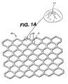
- Figures 1 and 1A are top views and section views of a stent containing reservoirs as described in the present invention.
- implantation of a coronary stent in conjunction with balloon angioplasty is highly effective in treating acute vessel closure and may reduce the risk of restenosis.
- Intravascular ultrasound studies suggest that coronary stenting effectively prevents vessel constriction and that most of the late luminal loss after stent implantation is due to plaque growth, probably related to neointimal hyperplasia.
- the late luminal loss after coronary stenting is almost two times higher than that observed after conventional balloon angioplasty.
- an agent which prevents inflammation and the proliferation of SMC combined with a stent may provide the most efficacious treatment for post-angioplasty restenosis (Bauters et al., 1996).
- a stent in conjunction with systemic cladribine treatment or local delivery of cladribine is an attractive treatment. Either systemic or local delivery of cladribine from a stent has the following advantages:
- any stent 10 having strut 12 can be modified to have a certain reservoir 30. Each of these reservoirs can be "open” or “closed” as desired. These reservoirs can hold the drug to be delivered.
- Figure 1a shows a stent 10 with a reservoir 45 created at the apex 14 of struts 12. Of course, this reservoir 45 is intended to be useful to deliver cladribine or any other drug at a specific point of flexibility of the stent. Accordingly, this concept can be useful for "second” or "third” generation-type stents.
- the reservoir size in the stent struts must be kept at a size of about 0.1 mm to about 1 mm depth, and 7 mm to 15 mm length, or enough to hold at least a therapeutic amount of the drug. Then, it should be possible to adequately apply the drug dosage at the desired location and in the desired amount.
- cladribine To assess the ability of cladribine to prevent cell proliferation, human smooth muscle or endothelial cells (Clonetics, Walkersville, MD) were seeded at a density of 2000 cells/cm 2 (approximately 3600 cells/well) into each well of 12-well plates and cultured with 1.5 ml of growth medium containing 5% fetal calf serum (FCS). After 24 hours, the growth medium was changed and fresh medium containing 10 ng/ml platelet-derived growth factor AB (PDGF AB; LIFE Technologies), as well as various concentrations of cladribine (0.001 - 10,000 nM) were added with triplicate wells. Medium was replaced with fresh cladribine-containing medium after 3 days. On day six, cells were detached by trypsinization to yield a cell suspension, lightly centrifuged to pellet and then counted manually using a Neubauer hemacytometer system. Clee viability was assessed by trypan blue exclusion.
- FCS fetal
- Table 1 provides the percent inhibition of the various tested concentrations of cladribine on human smooth muscle and endothelial cells in culture.
- Cladribine produced a concentration-related decrease in the proliferation of both smooth muscle and endothelial cells in this model system.
- IC 50 values concentration required to produce a reduction in proliferation to 50% of the vehicle-treated cell count
- Cladribine was thus approximately twice as potent as an inhibitor of smooth muscle cells as it was as an inhibitor of endothelial cells. Both IC 50 values are within the range of inhibitory concentrations reported for cladribine on human monocytes (Carrera et al., J. Clin. Invest.
- cladribine known to be effective at inhibiting peripheral leukemic blood cell proliferation and bone marrow cells are also effective at inhibiting proliferating vascular smooth muscle and endothelial cells. Cladribine may therefore be therapeutically useful for inhibition of the intimal smooth muscle cell proliferation which accompanies stent implantation. Inhibition of human vascular cell proliferation with cladribine.
- Cladribine (nM) Control Vehicle 0.001 0.01 0.1 1 10 100 1000 10,000 SMC 100 108 - 104 86 85 54 58 12 -4 EC 100 100 100 90 79 75 59 57 35 10 Values represent % of PDGF-stimulated increase in cell count. Each % is the mean of triplicate determinations. SMC, smooth muscle cells; EC, endothelial cells.
Landscapes
- Health & Medical Sciences (AREA)
- Engineering & Computer Science (AREA)
- Life Sciences & Earth Sciences (AREA)
- Biomedical Technology (AREA)
- Public Health (AREA)
- General Health & Medical Sciences (AREA)
- Animal Behavior & Ethology (AREA)
- Veterinary Medicine (AREA)
- Vascular Medicine (AREA)
- Heart & Thoracic Surgery (AREA)
- Chemical & Material Sciences (AREA)
- Cardiology (AREA)
- Medicinal Chemistry (AREA)
- Transplantation (AREA)
- Optics & Photonics (AREA)
- Bioinformatics & Cheminformatics (AREA)
- Oral & Maxillofacial Surgery (AREA)
- Physics & Mathematics (AREA)
- Nuclear Medicine, Radiotherapy & Molecular Imaging (AREA)
- General Chemical & Material Sciences (AREA)
- Chemical Kinetics & Catalysis (AREA)
- Organic Chemistry (AREA)
- Pharmacology & Pharmacy (AREA)
- Surgery (AREA)
- Molecular Biology (AREA)
- Epidemiology (AREA)
- Urology & Nephrology (AREA)
- Pharmaceuticals Containing Other Organic And Inorganic Compounds (AREA)
- Media Introduction/Drainage Providing Device (AREA)
Abstract
Description
- In the normal arterial wall, smooth muscle cells (SMC) proliferate at a low rate (<O.I%/day; ref). SMC in vessel wall exists in a 'contractile' phenotype characterized by 80-90% of the cell cytoplasmic volume occupied with the contractile apparatus. Endoplasmic reticulum, Golgi, and free ribosomes are few and located in the perinuclear region. Extracellular matrix surrounds SMC and is rich in heparin-like glycosylaminoglycans which are believed to be responsible for maintaining SMC in the contractile phenotypic state (Campbell and Campbell, 1985).
- Upon pressure expansion of an intracoronary balloon catheter during angioplasty, smooth muscle cells within the arterial wall become injured, initiating a thrombotic and inflammatory response. Cell derived growth factors such as platelet derived growth factor (PDGF), basic fibroblast growth factor (bFGF), epidermal growth factor (EGF), thrombin, etc. released from platelets (i.e., PDGF) adhering to the DAMAGED arterial luminal surface, invading macrophages and/or leukocytes, or directly from SMC (i.e., bFGF) provoke a proliferation and migratory response in medial SMC. These cells undergo a phenotypic change from the contractile phenotyope to a 'synthetic' phenotype characterized by only few contractile filament bundles but extensive rough endoplasmic reticulum, Golgi and free ribosomes. Proliferation/migration usually begins within 1-2 days post-injury and peaks at 2 days in the media, rapidly declining THEREAFTER (Campbell and Campbell, 1987; Clowes and Schwartz, 1985).
- Daughter synthetic cells migrate to the intimal layer of arterial smooth muscle and continue to proliferate and begin to secrete significant amounts of extracellular matrix proteins. Proliferation and migration continues until the damaged luminal endothelial layer regenerates at which time proliferation ceases within the intima, usually within 7-14 days postinjury. The remaining increase in intimal thickening which occurs over the next 3-6 months is due to an increase in extracellular matrix rather than cell number. Thus, SMC migration and proliferation is an acute response to vessel injury while intimal hyperplasia is a more chronic response. (Liu et al., 1989).
- prevention of vessel constriction with the stent;
- prevention of leukocyte and monocyte accumulation and smooth muscle cell proliferation at the site of vascular injury with cladribine
- higher tissue concentrations would be achievable than would occur with systemic administration
- reduced systemic toxicity
- single treatment/ease of administration
| Inhibition of human vascular cell proliferation with cladribine. | ||||||||||
| Cladribine (nM) | ||||||||||
| Control | Vehicle | 0.001 | 0.01 | 0.1 | 1 | 10 | 100 | 1000 | 10,000 | |
| SMC | 100 | 108 | - | 104 | 86 | 85 | 54 | 58 | 12 | -4 |
| EC | 100 | 100 | 100 | 90 | 79 | 75 | 59 | 57 | 35 | 10 |
| Values represent % of PDGF-stimulated increase in cell count. Each % is the mean of triplicate determinations. SMC, smooth muscle cells; EC, endothelial cells. |
Claims (3)
- In combination:a stent for the delivery of drugs to a lumen of a patient; anda therapeutic dosage amount of cladribine coated to said stent.
- A stent comprising:
a plurality of struts, said struts expansible within the lumen of the body, and at least one of said struts containing a reservoir therein said reservoir filled with a therapeutic dosage amount of cladribine. - The use of cladribine in the manufacture of a medicament for the prevention or treatment of restenosis in a patient, wherein said cladribine is administered to said patient subcutaneously, intramuscularly, intravenously, or on the strut of a stent, preferably in a dosage of at least 40nM.
Applications Claiming Priority (2)
| Application Number | Priority Date | Filing Date | Title |
|---|---|---|---|
| US51243200A | 2000-02-25 | 2000-02-25 | |
| US512432 | 2000-02-25 |
Publications (2)
| Publication Number | Publication Date |
|---|---|
| EP1127582A2 true EP1127582A2 (en) | 2001-08-29 |
| EP1127582A3 EP1127582A3 (en) | 2001-09-19 |
Family
ID=24039054
Family Applications (1)
| Application Number | Title | Priority Date | Filing Date |
|---|---|---|---|
| EP01301671A Withdrawn EP1127582A3 (en) | 2000-02-25 | 2001-02-23 | Use of cladribine on a stent to prevent restenosis |
Country Status (5)
| Country | Link |
|---|---|
| US (1) | US20030088312A1 (en) |
| EP (1) | EP1127582A3 (en) |
| JP (1) | JP2001293094A (en) |
| AU (1) | AU780539B2 (en) |
| CA (1) | CA2337565A1 (en) |
Cited By (8)
| Publication number | Priority date | Publication date | Assignee | Title |
|---|---|---|---|---|
| DE10150995A1 (en) * | 2001-10-08 | 2003-04-10 | Biotronik Mess & Therapieg | Implant e.g. a stent, comprises a decomposable substance which allows contact between the cell proliferation inhibitor and the stent surroundings only after a specified time |
| DE10216971A1 (en) * | 2002-04-16 | 2003-10-30 | Lothar Sellin | Medical implant, e.g. stent, has drug combined directly or by drug release system with biocompatible e.g. hemocompatible surface coating e.g. of carbon, silicon carbide or pyrolytic carbon |
| EP1570871A1 (en) * | 2004-02-18 | 2005-09-07 | Cordis Corporation | Local vascular delivery of cladribine in combination with rapamycin to prevent restenosis following vascular injury |
| DE202008006190U1 (en) | 2008-05-06 | 2008-07-17 | Sellin, Lothar | restenosis prophylaxis |
| DE202008007347U1 (en) | 2008-05-31 | 2008-10-16 | Orlowski, Benjamin Daniel | Copper stent |
| DE102008008263A1 (en) | 2008-02-08 | 2009-08-13 | Thomas Kuczera | Percutaneous transluminal coronary angioplasty catheter with a coating, useful e.g. to treat restenosis, comprises amino-/carboxyl-functional group, oligonucleotides, microbodies, C-type natriuretic peptide, proteins and/or oligopeptide |
| DE102009059070A1 (en) | 2008-12-19 | 2010-07-01 | Lothar Sellin | Medical device e.g. percutaneous transluminal coronary angioplasty catheter, for direct application of C-type natriuretic peptide on blood vessel wall, has base body with coating, where device consists of C-type natriuretic peptide |
| DE202015002048U1 (en) | 2015-03-16 | 2015-09-24 | Han Bock-Sun | implant |
Families Citing this family (50)
| Publication number | Priority date | Publication date | Assignee | Title |
|---|---|---|---|---|
| US7713297B2 (en) | 1998-04-11 | 2010-05-11 | Boston Scientific Scimed, Inc. | Drug-releasing stent with ceramic-containing layer |
| WO2003002243A2 (en) | 2001-06-27 | 2003-01-09 | Remon Medical Technologies Ltd. | Method and device for electrochemical formation of therapeutic species in vivo |
| USD516723S1 (en) | 2004-07-06 | 2006-03-07 | Conor Medsystems, Inc. | Stent wall structure |
| US8840660B2 (en) | 2006-01-05 | 2014-09-23 | Boston Scientific Scimed, Inc. | Bioerodible endoprostheses and methods of making the same |
| US8089029B2 (en) | 2006-02-01 | 2012-01-03 | Boston Scientific Scimed, Inc. | Bioabsorbable metal medical device and method of manufacture |
| US20070224235A1 (en) | 2006-03-24 | 2007-09-27 | Barron Tenney | Medical devices having nanoporous coatings for controlled therapeutic agent delivery |
| US8187620B2 (en) | 2006-03-27 | 2012-05-29 | Boston Scientific Scimed, Inc. | Medical devices comprising a porous metal oxide or metal material and a polymer coating for delivering therapeutic agents |
| US8048150B2 (en) | 2006-04-12 | 2011-11-01 | Boston Scientific Scimed, Inc. | Endoprosthesis having a fiber meshwork disposed thereon |
| US8815275B2 (en) | 2006-06-28 | 2014-08-26 | Boston Scientific Scimed, Inc. | Coatings for medical devices comprising a therapeutic agent and a metallic material |
| JP2009542359A (en) | 2006-06-29 | 2009-12-03 | ボストン サイエンティフィック リミテッド | Medical device with selective covering |
| CA2659761A1 (en) | 2006-08-02 | 2008-02-07 | Boston Scientific Scimed, Inc. | Endoprosthesis with three-dimensional disintegration control |
| WO2008033711A2 (en) | 2006-09-14 | 2008-03-20 | Boston Scientific Limited | Medical devices with drug-eluting coating |
| CA2663250A1 (en) | 2006-09-15 | 2008-03-20 | Boston Scientific Limited | Bioerodible endoprostheses and methods of making the same |
| DE602007011114D1 (en) | 2006-09-15 | 2011-01-20 | Boston Scient Scimed Inc | BIODEGRADABLE ENDOPROTHESIS WITH BIOSTABILES INORGANIC LAYERS |
| US8052744B2 (en) | 2006-09-15 | 2011-11-08 | Boston Scientific Scimed, Inc. | Medical devices and methods of making the same |
| JP2010503494A (en) | 2006-09-15 | 2010-02-04 | ボストン サイエンティフィック リミテッド | Biodegradable endoprosthesis and method for producing the same |
| CA2663762A1 (en) | 2006-09-18 | 2008-03-27 | Boston Scientific Limited | Endoprostheses |
| US7981150B2 (en) | 2006-11-09 | 2011-07-19 | Boston Scientific Scimed, Inc. | Endoprosthesis with coatings |
| CA2674195A1 (en) | 2006-12-28 | 2008-07-10 | Boston Scientific Limited | Bioerodible endoprostheses and methods of making same |
| US8070797B2 (en) | 2007-03-01 | 2011-12-06 | Boston Scientific Scimed, Inc. | Medical device with a porous surface for delivery of a therapeutic agent |
| US8431149B2 (en) | 2007-03-01 | 2013-04-30 | Boston Scientific Scimed, Inc. | Coated medical devices for abluminal drug delivery |
| US8067054B2 (en) | 2007-04-05 | 2011-11-29 | Boston Scientific Scimed, Inc. | Stents with ceramic drug reservoir layer and methods of making and using the same |
| US7976915B2 (en) | 2007-05-23 | 2011-07-12 | Boston Scientific Scimed, Inc. | Endoprosthesis with select ceramic morphology |
| US7942926B2 (en) | 2007-07-11 | 2011-05-17 | Boston Scientific Scimed, Inc. | Endoprosthesis coating |
| US8002823B2 (en) | 2007-07-11 | 2011-08-23 | Boston Scientific Scimed, Inc. | Endoprosthesis coating |
| US9284409B2 (en) | 2007-07-19 | 2016-03-15 | Boston Scientific Scimed, Inc. | Endoprosthesis having a non-fouling surface |
| US7931683B2 (en) | 2007-07-27 | 2011-04-26 | Boston Scientific Scimed, Inc. | Articles having ceramic coated surfaces |
| US8815273B2 (en) | 2007-07-27 | 2014-08-26 | Boston Scientific Scimed, Inc. | Drug eluting medical devices having porous layers |
| US8221822B2 (en) | 2007-07-31 | 2012-07-17 | Boston Scientific Scimed, Inc. | Medical device coating by laser cladding |
| JP2010535541A (en) | 2007-08-03 | 2010-11-25 | ボストン サイエンティフィック リミテッド | Coating for medical devices with large surface area |
| US8052745B2 (en) | 2007-09-13 | 2011-11-08 | Boston Scientific Scimed, Inc. | Endoprosthesis |
| US20090076591A1 (en) * | 2007-09-19 | 2009-03-19 | Boston Scientific Scimed, Inc. | Stent Design Allowing Extended Release of Drug and/or Enhanced Adhesion of Polymer to OD Surface |
| US8029554B2 (en) | 2007-11-02 | 2011-10-04 | Boston Scientific Scimed, Inc. | Stent with embedded material |
| US8216632B2 (en) | 2007-11-02 | 2012-07-10 | Boston Scientific Scimed, Inc. | Endoprosthesis coating |
| US7938855B2 (en) | 2007-11-02 | 2011-05-10 | Boston Scientific Scimed, Inc. | Deformable underlayer for stent |
| US7833266B2 (en) * | 2007-11-28 | 2010-11-16 | Boston Scientific Scimed, Inc. | Bifurcated stent with drug wells for specific ostial, carina, and side branch treatment |
| WO2009099935A2 (en) * | 2008-02-01 | 2009-08-13 | Boston Scientific Scimed, Inc. | Drug-coated medical devices for differential drug release |
| JP5581311B2 (en) | 2008-04-22 | 2014-08-27 | ボストン サイエンティフィック サイムド,インコーポレイテッド | MEDICAL DEVICE HAVING INORGANIC MATERIAL COATING AND MANUFACTURING METHOD THEREOF |
| WO2009132176A2 (en) | 2008-04-24 | 2009-10-29 | Boston Scientific Scimed, Inc. | Medical devices having inorganic particle layers |
| US7998192B2 (en) | 2008-05-09 | 2011-08-16 | Boston Scientific Scimed, Inc. | Endoprostheses |
| US8236046B2 (en) | 2008-06-10 | 2012-08-07 | Boston Scientific Scimed, Inc. | Bioerodible endoprosthesis |
| WO2009155328A2 (en) | 2008-06-18 | 2009-12-23 | Boston Scientific Scimed, Inc. | Endoprosthesis coating |
| US7951193B2 (en) * | 2008-07-23 | 2011-05-31 | Boston Scientific Scimed, Inc. | Drug-eluting stent |
| US7985252B2 (en) | 2008-07-30 | 2011-07-26 | Boston Scientific Scimed, Inc. | Bioerodible endoprosthesis |
| US8382824B2 (en) | 2008-10-03 | 2013-02-26 | Boston Scientific Scimed, Inc. | Medical implant having NANO-crystal grains with barrier layers of metal nitrides or fluorides |
| US8231980B2 (en) | 2008-12-03 | 2012-07-31 | Boston Scientific Scimed, Inc. | Medical implants including iridium oxide |
| US8267992B2 (en) | 2009-03-02 | 2012-09-18 | Boston Scientific Scimed, Inc. | Self-buffering medical implants |
| US8071156B2 (en) | 2009-03-04 | 2011-12-06 | Boston Scientific Scimed, Inc. | Endoprostheses |
| US8287937B2 (en) | 2009-04-24 | 2012-10-16 | Boston Scientific Scimed, Inc. | Endoprosthese |
| WO2011119573A1 (en) | 2010-03-23 | 2011-09-29 | Boston Scientific Scimed, Inc. | Surface treated bioerodible metal endoprostheses |
Family Cites Families (7)
| Publication number | Priority date | Publication date | Assignee | Title |
|---|---|---|---|---|
| US5424296A (en) * | 1993-04-15 | 1995-06-13 | The Scripps Research Institute | 2-Halo-2'-deoxyadenosines as therapeutic agents against malignant astrocytoma |
| US6273913B1 (en) * | 1997-04-18 | 2001-08-14 | Cordis Corporation | Modified stent useful for delivery of drugs along stent strut |
| CA2294247C (en) * | 1997-07-01 | 2004-10-26 | Atherogenics, Inc. | Antioxidant enhancement of therapy for hyperproliferative conditions |
| US6800616B2 (en) * | 1998-02-26 | 2004-10-05 | Supergen, Inc. | Treatment of HIV infections |
| WO1999058126A1 (en) * | 1998-05-11 | 1999-11-18 | The Endowment For Research In Human Biology, Inc. | Use of neomycin for treating angiogenesis-related diseases |
| US6531477B1 (en) * | 1998-10-13 | 2003-03-11 | Dupont Pharmaceuticals Company | 6-substituted pyrazolo [3,4-d] pyrimidin-4-ones useful as cyclin dependent kinase inhibitors |
| US6239118B1 (en) * | 1999-10-05 | 2001-05-29 | Richard A. Schatz | Method for preventing restenosis using a substituted adenine derivative |
-
2001
- 2001-02-22 AU AU23177/01A patent/AU780539B2/en not_active Ceased
- 2001-02-22 CA CA002337565A patent/CA2337565A1/en not_active Abandoned
- 2001-02-23 JP JP2001049364A patent/JP2001293094A/en not_active Withdrawn
- 2001-02-23 EP EP01301671A patent/EP1127582A3/en not_active Withdrawn
-
2002
- 2002-11-12 US US10/292,299 patent/US20030088312A1/en not_active Abandoned
Cited By (12)
| Publication number | Priority date | Publication date | Assignee | Title |
|---|---|---|---|---|
| DE10150995A1 (en) * | 2001-10-08 | 2003-04-10 | Biotronik Mess & Therapieg | Implant e.g. a stent, comprises a decomposable substance which allows contact between the cell proliferation inhibitor and the stent surroundings only after a specified time |
| US6849089B2 (en) | 2001-10-08 | 2005-02-01 | Biotronik Mess-Und Therapiegeraete Gmbh & Co Ingenieurbuero Berlin | Implant with proliferation-inhibiting substance |
| DE10216971A1 (en) * | 2002-04-16 | 2003-10-30 | Lothar Sellin | Medical implant, e.g. stent, has drug combined directly or by drug release system with biocompatible e.g. hemocompatible surface coating e.g. of carbon, silicon carbide or pyrolytic carbon |
| EP1570871A1 (en) * | 2004-02-18 | 2005-09-07 | Cordis Corporation | Local vascular delivery of cladribine in combination with rapamycin to prevent restenosis following vascular injury |
| US7806924B2 (en) | 2004-02-18 | 2010-10-05 | Cordis Corporation | Implantable structures for local vascular delivery of cladribine in combination with rapamycin for restenosis |
| DE102008008263A1 (en) | 2008-02-08 | 2009-08-13 | Thomas Kuczera | Percutaneous transluminal coronary angioplasty catheter with a coating, useful e.g. to treat restenosis, comprises amino-/carboxyl-functional group, oligonucleotides, microbodies, C-type natriuretic peptide, proteins and/or oligopeptide |
| DE202008006190U1 (en) | 2008-05-06 | 2008-07-17 | Sellin, Lothar | restenosis prophylaxis |
| DE102008063889A1 (en) | 2008-05-06 | 2009-11-26 | Lothar Sellin | Medical device and method for its manufacture |
| DE202008007347U1 (en) | 2008-05-31 | 2008-10-16 | Orlowski, Benjamin Daniel | Copper stent |
| DE102009059070A1 (en) | 2008-12-19 | 2010-07-01 | Lothar Sellin | Medical device e.g. percutaneous transluminal coronary angioplasty catheter, for direct application of C-type natriuretic peptide on blood vessel wall, has base body with coating, where device consists of C-type natriuretic peptide |
| DE202015002048U1 (en) | 2015-03-16 | 2015-09-24 | Han Bock-Sun | implant |
| DE102016003023A1 (en) | 2015-03-16 | 2016-09-22 | Bock-Sun Han | implant |
Also Published As
| Publication number | Publication date |
|---|---|
| AU780539B2 (en) | 2005-03-24 |
| JP2001293094A (en) | 2001-10-23 |
| EP1127582A3 (en) | 2001-09-19 |
| AU2317701A (en) | 2001-08-30 |
| US20030088312A1 (en) | 2003-05-08 |
| CA2337565A1 (en) | 2001-08-25 |
Similar Documents
| Publication | Publication Date | Title |
|---|---|---|
| AU780539B2 (en) | Use of cladribine on a stent to prevent restenosis | |
| US8029561B1 (en) | Drug combination useful for prevention of restenosis | |
| US6776796B2 (en) | Antiinflammatory drug and delivery device | |
| US7300662B2 (en) | Drug/drug delivery systems for the prevention and treatment of vascular disease | |
| US20020007215A1 (en) | Drug/drug delivery systems for the prevention and treatment of vascular disease | |
| US20020005206A1 (en) | Antiproliferative drug and delivery device | |
| US20020007213A1 (en) | Drug/drug delivery systems for the prevention and treatment of vascular disease | |
| US20020007214A1 (en) | Drug/drug delivery systems for the prevention and treatment of vascular disease | |
| US20100268329A1 (en) | Drug/Drug Delivery Systems For The Prevention And Treatment Of Vascular Disease | |
| AU2001262957A1 (en) | Drug combinations useful for prevention of restenosis | |
| AU2001263113A1 (en) | Drug/drug delivery systems for the prevention and treatment of vascular disease | |
| AU2001261579A1 (en) | Delivery systems for treatment of vascular disease | |
| AU2001263112A1 (en) | Delivery systems for the prevention and treatment of vascular disease | |
| AU2001261580A1 (en) | Delivery devices for treatment of vascular disease | |
| AU2001259774A1 (en) | Delivery devices for treatment of vascular disease | |
| CA2508065C (en) | Antiproliferative drug and delivery device | |
| US8236048B2 (en) | Drug/drug delivery systems for the prevention and treatment of vascular disease |
Legal Events
| Date | Code | Title | Description |
|---|---|---|---|
| PUAI | Public reference made under article 153(3) epc to a published international application that has entered the european phase |
Free format text: ORIGINAL CODE: 0009012 |
|
| PUAL | Search report despatched |
Free format text: ORIGINAL CODE: 0009013 |
|
| AK | Designated contracting states |
Kind code of ref document: A2 Designated state(s): AT BE CH CY DE DK ES FI FR GB GR IE IT LI LU MC NL PT SE TR |
|
| AX | Request for extension of the european patent |
Free format text: AL;LT;LV;MK;RO;SI |
|
| AK | Designated contracting states |
Kind code of ref document: A3 Designated state(s): AT BE CH CY DE DK ES FI FR GB GR IE IT LI LU MC NL PT SE TR |
|
| AX | Request for extension of the european patent |
Free format text: AL;LT;LV;MK;RO;SI |
|
| 17P | Request for examination filed |
Effective date: 20020225 |
|
| AKX | Designation fees paid |
Free format text: AT BE CH CY DE DK ES FI FR GB GR IE IT LI LU MC NL PT SE TR |
|
| 17Q | First examination report despatched |
Effective date: 20040426 |
|
| STAA | Information on the status of an ep patent application or granted ep patent |
Free format text: STATUS: THE APPLICATION IS DEEMED TO BE WITHDRAWN |
|
| 18D | Application deemed to be withdrawn |
Effective date: 20041109 |