EP1123383B1 - La proteine tensioactive d servant au diagnostic et au traitement de l'emphyseme pulmonaire - Google Patents
La proteine tensioactive d servant au diagnostic et au traitement de l'emphyseme pulmonaire Download PDFInfo
- Publication number
- EP1123383B1 EP1123383B1 EP99958659A EP99958659A EP1123383B1 EP 1123383 B1 EP1123383 B1 EP 1123383B1 EP 99958659 A EP99958659 A EP 99958659A EP 99958659 A EP99958659 A EP 99958659A EP 1123383 B1 EP1123383 B1 EP 1123383B1
- Authority
- EP
- European Patent Office
- Prior art keywords
- mice
- lung
- emphysema
- surfactant
- protein
- Prior art date
- Legal status (The legal status is an assumption and is not a legal conclusion. Google has not performed a legal analysis and makes no representation as to the accuracy of the status listed.)
- Expired - Lifetime
Links
Images


Classifications
-
- C—CHEMISTRY; METALLURGY
- C07—ORGANIC CHEMISTRY
- C07K—PEPTIDES
- C07K14/00—Peptides having more than 20 amino acids; Gastrins; Somatostatins; Melanotropins; Derivatives thereof
- C07K14/435—Peptides having more than 20 amino acids; Gastrins; Somatostatins; Melanotropins; Derivatives thereof from animals; from humans
- C07K14/785—Alveolar surfactant peptides; Pulmonary surfactant peptides
-
- A—HUMAN NECESSITIES
- A61—MEDICAL OR VETERINARY SCIENCE; HYGIENE
- A61P—SPECIFIC THERAPEUTIC ACTIVITY OF CHEMICAL COMPOUNDS OR MEDICINAL PREPARATIONS
- A61P11/00—Drugs for disorders of the respiratory system
-
- C—CHEMISTRY; METALLURGY
- C07—ORGANIC CHEMISTRY
- C07K—PEPTIDES
- C07K14/00—Peptides having more than 20 amino acids; Gastrins; Somatostatins; Melanotropins; Derivatives thereof
- C07K14/435—Peptides having more than 20 amino acids; Gastrins; Somatostatins; Melanotropins; Derivatives thereof from animals; from humans
- C07K14/46—Peptides having more than 20 amino acids; Gastrins; Somatostatins; Melanotropins; Derivatives thereof from animals; from humans from vertebrates
- C07K14/47—Peptides having more than 20 amino acids; Gastrins; Somatostatins; Melanotropins; Derivatives thereof from animals; from humans from vertebrates from mammals
- C07K14/4701—Peptides having more than 20 amino acids; Gastrins; Somatostatins; Melanotropins; Derivatives thereof from animals; from humans from vertebrates from mammals not used
- C07K14/4726—Lectins
-
- A—HUMAN NECESSITIES
- A61—MEDICAL OR VETERINARY SCIENCE; HYGIENE
- A61K—PREPARATIONS FOR MEDICAL, DENTAL OR TOILETRY PURPOSES
- A61K48/00—Medicinal preparations containing genetic material which is inserted into cells of the living body to treat genetic diseases; Gene therapy
Definitions
- the present invention relates generally to the field of biologically active proteins. More specifically the present invention relates to SP-D proteins involved in pulmonary surfactant homeostasis and structure, and alveolar structure in the lungs and SP-D (-/-) null mice.
- Pulmonary surfactant is essential for normal lung mechanics and gas exchange in the lung. Pulmonary surfactant is produced by type II epithelial cells and is made up of a phospholipid component which confers the ability of surfactant to lower surface tension in the lung. In addition, there are proteins associated with the surfactant called collectins which are collagenous, lectin domain-containing polypeptides. Two of these, designated surfactant protein A (SP-A) and surfactant protein D (SP-D), are likely involved in surfactant structure and function and host defense. Both quantitative and qualitative deficiencies in pulmonary surfactant are associated with neonatal respiratory distress, adult respiratory distress syndrome, congenital deficiencies of surfactant protein B, and allergic asthma.
- SP-A surfactant protein A
- SP-D surfactant protein D
- deficiency in pulmonary surfactant may contribute to the increased susceptibility of some individuals to microbial challenge, especially in the setting of inadequate or impaired specific immunity.
- These disorders as well as some disorders associated with increased risk of pneumonia may also be associated with acquired defects or deficiency in collectin function.
- Alveolar surfactant pools are regulated at multiple levels including intracellular synthesis, secretion, re-uptake and degradation of these components by alveolar macrophages. The synthesis and clearance of surfactant phospholipids and proteins is further influenced by developmental, mechanical, and humoral stimuli that serve to maintain steady-state surfactant concentrations after birth.
- the role of the collectins in surfactant and normal lung function has been extensively investigated.
- the collectin family of C-type lectins includes a number of molecules with known host defense functions.
- SP-A and SP ⁇ D also C-type lectins, bind influenza and herpes simplex viruses as well as gram positive and gram-negative bacteria and various fungi. By binding they enhance uptake by alveolar macrophages and neutrophils.
- Various cellular binding sites for SP-A and SP-D have been identified on alveolar macrophages or, in the case of SP-A, on type II epithelial cells.
- SP-A-deficient mice are susceptible to infections by group B streptococcus, Pseudomonas aeruginosa, Respiratory syncytial virus, adenovirus, and mycoplasma in vivo.
- group B streptococcus Pseudomonas aeruginosa
- Respiratory syncytial virus adenovirus
- mycoplasma mycoplasma in vivo.
- Collectins may also participate in the recognition or clearance of other complex organic materials, such as pollens and dust mite allergens.
- no human diseases have been associated with specific deficiencies in SP-A or SP-D.
- SP-D is a 43 kilodalton protein that has been proposed to play a role in host defense in the lung. Its cDNA and gene have been sequenced in various mammals including humans. SP-D shares considerable structural homology with other C-type lectins, including surfactant protein A (SP-A), conglutinin, bovine collectin-43, and mannose binding protein. In vitro studies and its close structural relationship to a mammalian Ca 2+ -dependent lectin family (particularly shared structural motifs) support its role in host defense.
- SP-A surfactant protein A
- conglutinin conglutinin
- bovine collectin-43 bovine collectin-43
- mannose binding protein mannose binding protein
- SP-D is synthesized primarily and at relatively high concentrations by Type II epithelial cells and nonciliated bronchiolar epithelial cells in the lung but may also be expressed in the gastrointestinal tract, heart, kidney, pancreas, genitourinary tract and mesentery cells.
- SP-D binds to the surface of organisms via its lectin domain (or sugar binding domain) which leads to binding, aggregation, opsonization and, in some instances, activation of killing by phagocytes in vitro.
- SP-D binds to lipopolysaccharide, various bacteria, fungi and viruses, including influenza virus. It also binds to both alveolar macrophages and polymorphonuclear cells. It may possibly play a role in surfactant phospholipid homeostasis, including the effects of SP-A on phospholipid metabolism by Type II cells in vitro, however, this is controversial and the precise role of SP-D in vivo is still unclear.
- Botas et al (Proc. Natl Acad Sci USA, 1998, Vol. 95, pp 11869 - 11874 ), discloses the generation of SP-D deficient mice.
- Crouch et al J. Biol. Chem, 1993, 5, 268(4), pp 2976 - 2983 ) describes the genetic organisation of SP-D.
- WO 91 00871 discloses the preparation of stable lung surfactant compositions.
- WO 94 23582 A discloses adenoviral vectors encoding SP-A, SP-B and SP-C.
- Johansson et al discusses the structural and functional roles of SP-A, SP-B, SP-C and SP-D.
- Van Iwaarden et al discloses that rat SP-D enhances the production of oxygen radicals by rat alveolar macrophages.
- the present invention provides an SP-D(-/-) mouse which can be used as a model for emphysema.
- SP-D protein was involved in lung lipid homeostasis.
- an SP-D null mouse would have the symptoms of emphysema.
- an isolated mammalian SP-D protein or vectors expressing the mammalian SP-D protein in the manufacture of an aerosol formulation for the treatment of emphysema.
- the emphysema has developed after chronic lung injury.
- the SP-D protein is administered intra-tracheally.
- the SP-D protein is expressed from an adenoviral vector.
- SP ⁇ D (-/-) knockout mouse to identify the role of SP-D in normal lung function and development and to demonstrate the temporal progression of postnatal airspace enlargement and spontaneous inflammatory changes in the lungs of these mice.
- SP-D (-/-) mice develop progressive pulmonary emphysema, associated with chronic inflammation and increased oxidant production by alveolar macrophages.
- the lung abnormalities make this mouse an excellent model for emphysema. Because there are very few existing therapies for treatment of emphysema, the most common being lung volume reduction surgery, the model is urgently needed.
- Example 1 describes the steps required to produce the SP-D (-/-) mouse.
- Table 1 Comparison of Body Weights, Lung Volumes, and Volume-to-Body Weight Rations (Mean ⁇ SE) AGE BODY WEIGHTS (g) LUNG VOLUMES (ml) LV:BW (Ml/g x 10 2 ) SP-D(-/-) SP-D(+/+) SP-D(-/-) SP-D(+/+) SP-D (-/-) SP-D(+/+) 2 day 1.8 ⁇ 0.1* 3.4 ⁇ 0.1 NO ND ND 5 day 3.7 ⁇ 0.3 4.6 ⁇ 0.2 ND ND ND ND ND ND ND ND ND ND ND ND ND ND ND ND ND ND ND ND ND ND ND ND ND ND ND ND ND ND ND ND ND ND ND ND ND ND ND ND ND
- SP-D mice were generated by targeted gene inactivation. Integration of a pGKneo targeting vector containing sequences from exon 2 of the SP-D gene generated a deletion of the second exon of the SP-D gene, which included removal of the initiating methionine and translation initiation sequences.
- the mouse SP-D gene sequence of Exons 1 and 2 can be found under Genbank accession No. AF047741.
- the targeting vector was designed using pGKneo by first subcloning a 5.1-kb blunt ended Kpnl-tailed HindIII genomic fragment encoding intron 2 through exon 6 into a Kpnl site between the neomycin-resistance cassette and the thymidine kinase cassette. Subsequently, a 1.5-kb genomic Pstl fragment containing a portion of intron I was tailed with Xhol linkers and cloned into an Xhol site 5' from the neomycin-resistance cassette Eight of 104 ES clones surviving the double selection process were correctly targeted as determined by both 5' and 3' PCR analyses.
- Clone 93 a highly undifferentiated and proliferative clone, was expanded and injected into C57/BI6 blastocysts generating chimeric males. Chimeric males were bred to NIH Swiss Black females. A female bearing the targeted gene was obtained and bred to NIH Swiss Black males to generate normal SP-D (-/-) and SP-D ( ⁇ ) mice. The distribution of genotypes from heterozygotic matings followed a Mendelian pattern, with 30 (+/+), 45 (+/-), and 25% (-/-) of 115 offspring, indicating that there were no obvious abnormalities in survival related to SP ⁇ D alleles.
- SP-D(-/-) mice survive and breed normally in the vivarium under barrier containment facilities at Children's Hospital Medical Center, Cincinnati, Ohio. Mice have been viral free as assessed by serology. No serological evidence of viral infection in SP-D(-/-) mice was detected at necropsy.
- DNA from tail clips was digested with Bam HI and probed with a PCR product derived from genomic mouse DNA, containing exon 2 and part of intron 2, and with the G418 resistance cDNA clone. This demonstrated a simultaneous loss of exon 2 with appearance of sequences encoding G418 resistance in SP-D ( ⁇ ) and SP-D (-/-) mice.
- RNA blot analysis was conducted with total lung RNA from null, normal, and heterozygotic animals. The results showed approximately 50% reduction in the intensity of the SP-D hybridization band in heterozygous animals with a total absence of normally sized SP-D mRNA in null animals. After prolonged exposure, a diffuse mRNA band approximately 150 nucleotides smaller than the normal SP-D mRNA was detected. By scanning densitometry, this band represents less than 5% of the intensity of the normal SP-D transcript from heterozygous animals.
- examples 2 through 5 describe the other abnormalities or changes found in SP ⁇ D (-/-) mice.
- Example 2 demonstrates the effect on phospholipid levels. Alveolar and tissue phospholipid levels, specifically phosphatidylcholine pool levels, were markedly increased while total brochoalveolar lavage (BAL) protein levels remained unchanged.
- BAL brochoalveolar lavage
- SP-D has a role in the regulation of SP-A production. Since SP-A is involved in host defense in the lungs, SP-D can affect host defense in two ways. By up-regulation of SP-A production and by direct interaction with immune and microbial cells.
- Surfactant in alveolar was can be separated into large aggregate (heavy, dense) and small aggregate (light, visicular) fractions by centrifugation. Alveolar washes were centrifuged at 40,000 x g over 0.8 M sucrose cushion for 15 min. The large aggregate surfactant then was collected from the interface, diluted with normal saline and centrifuged again at 40,000 x g for 15 min. The supernatant from the first 40,000 x g centrifugation that contains small aggregate surfactant is concentrated at 4°C by ultrafiltration using a 300,000 molecular weight retention filter (Minitan, Miliore Corp., Bedford, MA) or centrifugal concentrators (Amicon Corp., Danvers, MA). The small aggregate surfactant is diluted with 50 ml normal saline and ultrafiltered 3 times to remove soluble proteins.
- Lung tissue from SP-D (+/+) and SP-D (-/-) mice were sacrificed at 2 weeks, 3 weeks and 6 weeks. Animals were weighed, anesthetized with a 4:1:1 mixture of ketamine, acepromazine and xylazine, and exsanguinated by severing the inferior vena cava and descending aorta. The trachea was cannulated, and the lungs were collapsed by piercing the diaphragm. The lungs were inflation-fixed at 25 cm of water pressure with 4% paraformaldehyde in phosphate buffered saline (PBS) for 1 minute.
- PBS phosphate buffered saline
- the trachea was tied off as the cannula was removed in order to maintain the fixative in the inflated lung.
- Excised lungs and heart were allowed to equilibrate in cold fixative until they had sunk to the bottom of the container. Lung and heart volumes were then determined by fluid displacement. Each lobe was measured along its longest axis, bisected perpendicularly to the long axis, and processed into paraffin blocks. Five micron sections were cut in series throughout the length of each lobe, loaded onto polysine-coated slides, and stained with hematoxylin and eosin, Masson's trichrome stain for collagen, or ocein for elastin.
- BALT bronchial-associated lymphocytic tissue
- Macrophage size was estimated from the diameter of fixed and stained macrophages from cytospin preparations sedimented onto glass slides at 1500 X g for 2 min. Mean diameter of macrophages from (+/+) was 11.75 ⁇ 1.75 ⁇ m compared with (-/-) mice 18.75 ⁇ 7.25 ⁇ m.
- Abnormally large macrophages defined as those with a diameter of twice normal, comprised 22.4 ⁇ 0.6% of the macrophages from (-/-) mice compared with 18 ⁇ 1.0% from (+/+) mice.
- Numbers and morphology of alveolar macrophages were not different in SP-D (+/-) mice.
- Ultrastructural characteristics of type II cells were similar in SP-D (-/-) compared with SP-D (+/+) mice.
- the morphology of the alveolar macrophages is consistent with that of activated "foam" cells, known to be associated with inflammation.
- Type II cells are routinely isolated in this laboratory using the following method. Mice are anesthetized by intraperitoneal injection and pentobarbital (50 mg/ml 3.25 ml/kg body weight). After opening the abdominal cavity, mice are exsanguinated by severing the inferior vena cava. The trachea is exposed, cannulated with a 20 gauge luer stub adaptor, and secured by a suture. The chest plate is removed and lungs perfused with 10-20 ml sterile saline via the pulmonary artery until visually free of blood.
- Dispase Cold Research, Inc., Bedford, MA
- Dispase Collaborative Research, Inc., Bedford, MA
- 1% low melt agarose warmed to 45° C.
- Lungs are immediately covered with ice and incubated for 2 minutes to set the agarose.
- Lungs are dissected out, put in a culture tube containing an additional 1 ml Dispase, and incubated for 45 minutes at room temperature.
- Lungs are next transferred to a 60 mm culture dish containing 100 U/ml DNAase 1 (Sigma, St. Louis, MO) in 7 ml DMEM (Gibco BRL, Gaithersburgh, MD). The tissue is gently teased away from the airways and swirled for 5 minutes.
- Cells are then placed on ice until being filtered.
- the cell suspension is successively filtered through 100 m and 40 m cell strainers, and then through 25 m nylon gauze (Tetko, Briarcliff Manor, NY).
- Cells are pelleted for 7 min at 130 x g at 4°C and resuspended in 10 ml DMEM with 10% FBS (Intergen Co., Purchase, NY).
- Crude cell suspensions are added to 100 mm culture dishes that were previously coated with CD-45 and CD-32 antibodies (Pharmigen. San Diego, CA) and incubated for 102 hours at 37° in the presence of 5% CO 2 . Plates are removed from the incubator and gently "panned" to free settled type II cells.
- the cell suspension is centrifuged at 130 x g at 4°C and resuspended in 10 ml DMEM with 10% FBS (Intergen Co., Purchase, NY). Crude cell suspensions are added to 100 mm culture dishes that were previously coated with CD-45 and CD-32 antibodies (Pharmigen. San Diego, CA) and incubated for 102 hours at 37°C in the presence of 5% CO 2 , Plates are removed from the incubator and gently "panned" to free settled type II cells. The cell suspension is centrifuged at 130 x g for 7 minutes and cells are resuspended in DMEM containing 10% FBS.
- Morphometric measurements were performed on mice at 5 days (0.5 weeks), 14 days (2 weeks) and 17 days (2.5 weeks), 3 and 6 weeks, and 6 to 7 months of age, the overall proportion (% fractional area) of respiratory parenchyma and airspace was determined using a point counting method. Measurements were performed on sections taken at intervals throughout the left, right upper, or right lower lobes. Slides were viewed using a 20x objective, and the images (fields) were transferred by video camera to a computer screen using MetaMorph imaging software (Universal Imaging Corp., West Chester, PA).
- Ten fields per section were analyzed to gather the data.
- the x and y coordinates for each field measured were selected using a random number generator.
- the overall percent reduction in parenchyma at 7 months of age in the SP-D ( ⁇ / ⁇ ) mice was 32% of control values, while the percent increase in airspace in the SP ⁇ D (-/-) mice was 27% of control values.
- BrdU labeling indices were relatively low, and no changes in BrdU labeling of respiratory parenchymal cells or alveolar macrophages were observed in the lungs from SP-D (-/-) mice compared to controls.
- mice were injected with sodium pentobarbital and placed in a chamber containing 100% oxygen to ensure complete collapse of alveoli by oxygen absorption. Mice were killed by exsanguination, the trachea cannulated and connected to a syringe linked to a pressure sensor via a three way connector (Mouse Pulmonary Testing System, TSS Incorporated, Cincinnati, 0H). After opening the diaphragm, lungs were inflated in 75 ⁇ l increments every 10 seconds to a maximum pressure of 28 cm of water and then deflated.
- TSS Incorporated Cincinnati, 0H
- Pressure-volume curves were generated for each animal, determining lung volumes (divided by body weight) at 10, 5, and 0 cm of water during the deflation curve.
- pressure-volume curves were generated in 5-6 mice at 12 weeks of age. Lung volumes associated with the deflation limbs of pressure-volume curves were significantly greater for 12 week old SP ⁇ D (-/-) mice compared age-matched to SP ⁇ D (+/+) mice at 10 cm H 2 O and at the maximum pressure of 28 cm H 2 O (*p ⁇ 0.05).
- SP-D is very likely to be involved in the regulation of alveolar remodeling in the lungs. Because abnormalities and airspace remodeling is a defining characteristic of emphysema, the SP-D (-/-) mouse is an ideal model for emphysema.
- TNF- ⁇ Tumor necrosis factor alpha
- IL-1 ⁇ interleukin-1 ⁇
- IL-6 interleukin-6
- MIP macrophage inflammatory protein
- Alveolar macrophages were collected by bronchoalveolar lavage with 1 ml of dye-free RPM1 media (Gibco, Grand Island, NY) times three. Bronchoalveolar lavage fluid (BALF) from 8-10 mice was pooled to provide sufficient numbers of macrophages for analysis. The lavage was centrifuged at 1200 RPM for 10 minutes and one million macrophages were resuspended in PBS.
- BALF Bronchoalveolar lavage fluid
- Hydrogen peroxide production by macrophages was measured using a commercially available assay (Bioxytech H 2 O 2 -560 assay, OXIS International, Portland, OR), based on the oxidation of ferrous ions (Fe 2+ ) to ferric ions (Fe 3+ ) by hydrogen peroxide under acidic conditions. Methods followed the manufacturer's recommendations. Hydrogen peroxide production was determined after activation with 100 ng/ml phorbol myristate acetate (PMA) or without stimulation.
- PMA phorbol myristate acetate
- Mouse lavage samples were centrifuged (100,000 x g, 1 hour) in a SW-28 rotor (Beckman, Palo Alto, CA). The supernatants were concentrated using Centricon-30 filtration units (Amicon, Inc., Beverly, MA). Samples (200 ⁇ g protein) were electrophoresed under nonreducing conditions (laemmli) into 10% Zymogram, gelatin and casein gels (Novex, San Diego, CA).
- Metalloproteinase 2 and 9 mRNA's were quantitated by Northern blot analyses of total lung mRNA from wild type and SP-D (-/-) mice using [ 32 P]-labeled cDNA probes (Chemicon International, Inc., Temecula, CA).
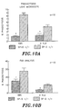
- lung homogenates from SP-D (-/-) mice did not contain inflammatory levels of the pro-inflammatory cytokines TNF- ⁇ , IL-1 ⁇ , IL-6 or MIP-2, although basal levels of IL-1 ⁇ were increased significantly, Figure 3 .
- oxidant production as assessed by measuring hydrogen peroxide production by alveolar macrophages isolated from SP ⁇ D ( ⁇ / ⁇ ) mice, was increased 10 fold, Figure 4 . Hydrogen peroxide and superoxide production is a measure of macrophage activation, particularly the microbicidal activation.
- metalloproteinase activities were estimated by degradation of gelatin substrates after SDS-PAGE of BALF supernatants isolated from SP-D (-/-) and SP-D (+/+) mice. Bands of activity consistent with metalloproteinases -2 and 9 were readily detected in both genotypes, but were not altered in BALF from SP-D (-/-) mice. Likewise, the abundance of metalloproteinasese -2 and 9 mRNA's were similar in whole lung RNA samples from SP-D (-/-) and SP-D (+/+) mice as assessed by Northern blot analysis.
- the SP-D (-/-) mouse conclusively demonstrates a remarkable and surprising role for SP-D in regulation of surfactant homeostasis, the structure of alveolar surfactant in the lung, regulation of SP-A expression, or plays a critical inhibitory role in oxidant, hydrogen peroxide production in the lung. Therefore, its levels are important for suppression of ongoing oxidant production and injury and the regulation of alveolar remodeling.
- Example 7 will summarize the results for the mouse model of emphysema.
- SP-D deficiency caused inflammation increased oxidant production by isolated alveolar macrophages, emphysema, and localized fibrosis in gene-inactivated SP-D (-/-) mice.
- the timing and progressive nature of these pulmonary abnormalities support the conclusion that alveolar enlargement in SP-D (-/-) mice is caused by alveolar remodeling associated with chronic inflammation, rather than with development abnormalties occurring during alveologenesis.
- the present findings are consistent with an important and unanticipated role of SP-D in the modulation of pulmonary inflammation and oxidant production and suggest that changes in the regulation or function SP-D may play a role in the pathologic processes leading emphysema following chronic lung injury.
- Enlarged airspaces were generally associated with focal accumulation of large, foamy, alveolar macrophages, although there was some heterogeneity in both localization and extent of inflammatory infiltrates and remodeling in older mice While focal accumulation of alveolar macrophages in lungs of SP-D (-/-) mice were observed as early as 2 weeks of age, macrophage morphology remained normal at this time. Abnormal alveolar macrophage morphology, consisting of enlarged foamy cells, was noted by 3 weeks of age and was coincident with enlargement of alveolar structures thereafter. Previous studies demonstrated increased numbers of enlarged alveolar macrophages in SP-D (-/-) mice by 8 weeks of age.
- the present findings do support an important role for SP ⁇ D in the modulation of alveolar macrophage activation and oxidant production, leading to emphysema and fibrosis, macrophage infiltration and lung remodeling in SP-D (-/-) mice were associated with modest but significant differences in inflammatory levels of various pro-inflammatory mediators, including IL-1b, MIP-2, but not TNF- ⁇ and IL-6, but rather with markedly increased hydrogen peroxide production by isolated alveolar macrophages. Although basal levels of IL- ⁇ 1 were significantly increased in SP-D (-/-) mice, IL- ⁇ 1 was not increased to levels typically detected in severe inflammation.
- SP-D While increased IL-1 ⁇ and hydrogen peroxide production were observed in SP-D (-/-) mice, it remains unclear whether the pulmonary abnormalities seen in these mice were directly mediated by cytokine or oxidant-induced injury. Although SP-D has been proposed to play an important role in host defense, there was no histologic or serologic evidence of infection in the SP-D (-/-) colony.
- Enhanced hydrogen peroxide production and increased numbers of alveolar macrophages found in the lungs of SP-D (-/-) mice support the concept that SP-D plays a critical anti-inflammatory role in the lung and regulates hydrogen peroxide production by alveolar macrophages in vivo.
- Relationships between oxidant injury and the development of emphysema and pulmonary fibrosis are well established in numerous animal and genetic models. For example, neonatal exposure to hyperoxia caused alveolar remodeling and fibrosis in newborn mice. Since activation of metalloproteinases has been associated with oxidant injury and emphysema, metalloproteinase activities were assessed in BALF from the SP ⁇ D (-/-) mice.
- protease activity consistent with metalloproteinase -2 and -9 were readily detected by zymography, no consistent changes in the activities of these proteinases or their mRNAs were detected in SP-D ( ⁇ / ⁇ ) mice. It is still possible, however, that increased, localized tissue concentrations of metalloproteinases and/or alterations in other proteases or antiproteases may be associated with SP-D deficiency. Deficiencies in antiproteases, as well as smoking and oxidant injury from oxidizing toxicants (e.g., bleomycin or paraquat), have all been associated with emphysema or pulmonary fibrosis in human lung.
- oxidizing toxicants e.g., bleomycin or paraquat
- surfactant phospholipid content was increased in SP-D (-/-) mice and was associated with increased numbers of large, foamy, alveolar macrophages
- increased phospholipid content alone is not likely to be sufficient to cause the alveolar remodeling observed in SP-D (-/-) mice.
- the overall effect of surfactant phospholipids appears to be anti-inflammatory, altering phagocytosis, oxidant production, and cytokine release, and inhibiting lymphocyte proliferation, immunoglobulin production, and expression of adhesion molecules.
- SP-D (-/-) mice developed severe and progressive emphysema. Alveolar remodeling and macrophage abnormalities were apparent as early as 3 weeks of age, while mild, focal, pulmonary fibrosis was observed at 6 to 7 months of age, demonstrating a role for SP-D in the regulation of inflammation and alveolar remodeling.
- the present study also demonstrated an unexpected role for SP-D in the regulation of hydrogen peroxide production by alveolar macrophages in vivo, which may contribute to the development of emphysema in the lungs of SP-D (-/-) mice. Whether SP-D deficiency contributes to ongoing inflammation or to the development of emphysema and fibrosis found in various human chronic lung diseases, including those caused by smoking and other oxidants, remains to be determined.
- Example 6 provides a sample framework for testing pharmaceuticals, protein preparations, or genetic manipulations for the treatment of emphysema.
- a number of doses or concentrations of protein or pharmaceutical diluted in an appropriate buffer is administered to SP-D (-/-) mice intratracheally. Protein and pharmaceutical is purified as appropriate for in vivo use.
- Recombinant adenovirus or other genetic vectors containing the gene of interest is administered as follows. SP-D (-/-) mice are immunosuppressed to block specifically T cell-mediated immune responses, and treated with an adenoviral construct designed to express the gene of interest in transduced cells. Mice are injected intraperitoneally with H57 antibody 3 days prior to receiving the adenoviral construct. H57 alters immune recognition at the T cell receptor and decreases splenic and lung T and B lymphocytes.
- Intratracheal inoculation involves anesthetizing with isofluorane, and an anterior midline incision is made to expose the trachea. A 30-gauge needle attached to a tuberculin syringe is inserted into the trachea, and a 100 ⁇ l inoculum of protein or pharmaceutical is dispersed into the lungs. The incision is closed with one drop of Nexaband. Nonpyogenic PBS is injected intratracheally as a control.
- lungs To determine the effects of the protein or pharmaceutical on the lung structure lungs are inflation fixed and sections evaluated by electron microscopy. Lungs from treated and untreated mice are inflated via a tracheal cannula at 20 cm of pressure with 4% paraformaldehyde and removed en bloc from the thorax. Lungs are dehydrated and embedded in paraffin. Tissue sections (5 m) are stained with hematoxylin and eosin.
- Macrophage number is determined by staining with SP-B antiserum. Macrophage size is estimated from the diameter of fixed and stained macrophages from cytospin preparations sedimented onto glass slides at 1500 x g for 2 min.
- Surfactant composition and ultrastructure is analyzed as follows: The structure of surfactant is analyzed by isolating large aggregates from pooled alveolar lavage of SP-D (-/-) treated and untreated mice and examined by EM (see protocol below). For alveolar lavage phospholipid composition analysis, two to four samples consisting of the pooled lavage from two to three mice are evaluated for the relative abundance of phosphatidylcholine, phosphatidylethanolamine, phosphatidylglycerol, phosphatidylinositol, sphingomyelin, and lyso-bis-phosphatidic acid.
- the supernatant from the first 40,000 x g centrifugation that contains small aggregate surfactant is concentrated at 4°C by ultrafiltration using a 300,000 molecular weight retention filter (Minitan, Miliore Corp., Bedford, MA) or centrifugal concentrators (Amicon Corp., Danvers, MA).
- the small aggregate surfactant is diluted with 50 ml normal saline and ultrafiltered 3 times to remove soluble proteins.
- SP-D is an obvious choice as a treatment for or prevention of emphysema. It is also an obvious treatment for other types of pulmonary disease since many of these diseases are characterized by aberrant surfactant production. In addition, its affect on SP-A and its possible role in host defense makes it a useful tool to augment immune function in the lungs.
- the feasibility of gene transfer to the respiratory epithelium is very promising as a treatment for various pulmonary diseases.
- a variety of viral and non-viral-based vectors have been developed to transfer genes to cells of the airways, including recombinant adenoviral vectors. These vectors are particularly promising for use in respiratory treatment because they have the potential of being aerosolized.
- Example 9 is an experiment using purified mouse SP-D protein for treatment of emphysema in SP-D(-/-) mice.
- Example 10 is an experiment using adenovirus to express rat SP-D for treatment of emphysema in SP-D(-/-) mice.
- Example 11 provides a sample framework for the use of SP-D peptide, or vectors expressing SP-D for the prevention and treatment of these diseases.
- Emphysema is used as an exemplary pulmonary disease.
- Adenovirus is used as an exemplary vector.
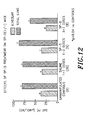
- SP-D(-/-) mice were treated with purified mouse SP ⁇ D, purified as outlined below. Saturated PC levels were analyzed in alveolar lavage and total lung lavage. Repeated doses intratracheally at 24 hour intervals resulted in partial correction of lipid accumulation after 3 to 7 doses, see Figure 12 .
- the half life of SP-D in the airway was determined as 13 hours in mouse (see Figure 13 ) (the technique is outlined below); therefore, the SP-D deficiency can be treated by replacement of SP-D protein at a reasonable interval by aerosol or particulate inhaler or surfactant mixtures.
- BAL Mouse bronchoalveolar lavage (BAL) fluid from GMCSF and SP-A double null mutant mice was collected, frozen, and pooled for later purification of SP-D.
- Maltosyl-agarose (Sigma) was packed in a gravity flow column (10 x 80 mm) and equilibrated with buffer containing 20 mM Tris-HCl, pH 7.4, 10 mM calcium chloride, 0.02% (W/V) sodium azide (TCB).
- the BAL was made 20 mM with respect to Tris-HCl, and 10 mM with respect to EDTA, ph 7.4 and stirred for one hour at room temperature.
- the turbid solution was centrifuged at 10,000 X g for 40 minutes at 4 degrees C.
- the supernatant was made 20 mM with respect to calcium chloride and readjusted to pH 7.4 before loading on the maltosyl-agarose column.
- the column was washed to background absorbence with TCB followed by washing with TCB containing 1.0 M Sodium Chloride.
- the SP-D which has a specific requirement for calcium in binding to maltose was eluted with 50 mM manganese chloride, 20 mM Tris-HCl, 0.02% (W/V) sodium azide, pH 7.4.
- the fractions containing SP-D were determined by SDS polyacrylamide gel electrophoresis or by direct ELISA, pooled, and dialysed against three changes of 20 mM Tris-HCl, 100 mM sodium Chloride, 5 mM EDTA pH 7.4. This protocol was adapted from Strong, Peter; Kishore, Uday; Morgan, Cliff; Bernal, Andres Lopez; Singh, Mamta; and Reid, Kenneth B.M.; Journal of Immunological Methods 220 (1998) 139-149 . Treatment of mice with surfactant components. We have successfully used a technique for oral blind intubation using 26 g feeding tubes in mice under anesthesia with isoflurane for repetitively treating mice with SP-D daily for up to 7 days without problems. This approach avoids surgery and permits the type of experiments proposed for SP-D replacement and treatment with mutant SP-D proteins.
- GM-CSF deficiency causes a 48 fold increase in SP-D, and the GM-CSF (-/-) x SP-A(-/-) cross has similarly elevated SP-D but no SP-A.
- Plasmid pAvS6a-rSPD has a RSV promoter, a rSPC cDNA, an SV40 poly A signal and an Ad5 sequence (9.24-17.34 mu). Not I linearized pAvS6a-rSPD was co-transfected into 293 cells with Cla I digested large fragment of adenovirual DNA Ad d1327, which has E3 region (78.5-84.7 mu) deleted. After homologous recombination, individual plaques were analyzed by Western blot assay to determine rSPD protein expression. One rSPD positive clone was subject to one round of plaque purification. The Ad-rSPD adenovirus has deletions in E1 and E3 regions and is replication deficient.
- Ad-rSPD adenovirus After amplification in 293 cells, the purified Ad-rSPD adenovirus was produced through two rounds of CsCl gradient ultracentrifugation. The adenovirus expressing SP-D was able to correct some lipid abnormalites by intratracheal administration. Therefore, this remains a very positive possibility for treatment of emphysema and many other SP-D deficiency illnesses as well as various other forms of pulmonary injury and deficiency.
- Adenovirus was administered by intratracheal injection of 5 X 10 8 PFU of virus.
- Levels of SP-D protein were measured 1 week after administration to detect uptake and expression of the vector.
- Four mice were tested and SP-D (-/-) mice receiving no treatment are used as a control.
- SP-D -/- mice receiving no treatment are used as a control.
- a number of tests are performed as follows.
- lungs are inflation fixed and sections evaluated by electron microscopy.
- Lungs are inflated via a tracheal cannula at 20 cm of pressure with 4% paraformaldehyde and removed en bloc from the thorax. Lungs are dehydrated and embedded in paraffin. Tissue sections (5 m) are stained with hematoxylin and eosin.
- Macrophage number is determined by staining with SP-B antiserum. Macrophage size is estimated from the diameter of fixed and stained macrophages from cytospin preparations sedimented onto glass slides at 1500 x g for 2 min.
- Surfactant composition and ultrastructure are analyzed as follows: the structure of surfactant is analyzed by isolating large aggregates from pooled alveolar lavage of SP-D (-/-) treated and untreated mice and examined by EM.
- alveolar lavage phospholipid composition analysis two to four samples consisting of the pooled lavage from two to three mice are evaluated for the relative abundance of phosphatidylcholine, phosphatidylethanolamine, phosphatidylglycerol, phosphatidylinositol, sphingomyelin, and lyso-bis-phosphatidic acid.
- Incorporation of ( 3 H)choline into total lung Sat-PC is evaluated to determine total phospholipid concentration.
- treatment can be tested on other appropriate mammals.
- SP-D and SP-A have specific interactions with various microorganisms in vitro, modifying pulmonary inflammation in vitro by altering cytokine and free radical production.
- the role of SP-D in bacterial clearance and inflammatory response of the lung was evaluated in vivo using a mouse model of SP-D deficiency.
- SP-A-deficient mice are known to be more susceptible to infections.
- a number of in vitro studies have shown a possible role for SP-D in host defense in addition to its role in up-regulating SP ⁇ A.
- Examples 8-11 outline sample protocols for testing SP-D as a therapy in the, bacterially, or fungally infected SP-D (-/-) mice as well as in the SP-A (-/-) mice.
- Examples 12-14 are experiments showing the role of SP ⁇ D in the response to bacterial, fungal, and viral infection.
- Example 13 is an experiment showing the effect of infecting SP-D(-/-) mice with Respiratory Syncytial Virus.
- SP-D deficient mice SP-D -/- were intratracheally infected with Group B streptococcus (GBS) or Hemophilus influenzaa (Hflu) to assess clearance compared to wild type mice.
- Group A Streptococcus was administered at 10 4 CFU.
- Pulmonary inflammation was also assessed by analysis of BAL fluid for total cells ( Figures 5 , 6 , and 7 ), cytokine levels in lung homogenates ( Figure 8 ), oxygen radical production by alveolar macrophages ( Figure 11 ) and Nitrite levels in BAL ( Figure 9 ).
- SP-D -/- mice cleared the bacteria similarly to wild type mice (see Figures 5 and 6 ). Infection with GBS and Hflu resulted in significantly greater total cells in the BAL fluid of the SP-D -/- mice compared to wild type mice ( figure 7 ). Selective alterations of cytokine levels were detected in SP-D -/- mice. Tumor necrosis factor (TNF-) and interleukin (IL)-6 levels were greater in lung homogenates from SP-D -/- mice early after infection with GBS or Hflu ( Figure 8 ).
- TNF- tumor necrosis factor
- IL-6 interleukin-6
- Macrophage inflammatory protein-2 (MIP-2), a neutrophil chemoattractant, was significantly greater in lung homogenates from SP-A -/- mice after Hflu but not GBS infection ( Figure 8 ). Macrophages from SP-D -/- mice generated significantly greater superoxide and hydrogen peroxide compared to wild type mice ( Figure 11 ).
- BAL nitrite levels were increase in SP-D (-/-) mice as compared to wildtype mice.
- Nitric oxide production was measured as nitrite in BALF.
- Nitric oxide plays a role in host defense by contributing to bacterial killing.
- Nitric oxide reacts with superoxide to form peroxynitrite which is a potent bacteriocidal agent.
- phagocytosis was evaluated using light microscopy and flow cytometry. SP-D(-/-) mice showed significantly reduced phagocytosis of bacteria as compared to wildtype.
- Example 13 the SP-D(-/-) mice were infected with Respiratory Syncytial Virus.
- Host defense mechanisms have evolved to maintain the lung clear of microbial pathogens including innate mediators of bacterial and viral clearance and acquired immune responses.
- SP-D(-/-) mice were intratracheally infected with respiratory syncytial virus (RSV), a common respiratory pathogen in children.
- RSV respiratory syncytial virus
- Viral titers and lung inflammation were assessed in SP-D (-/-) mice and wild type mice.
- RSV titers in lung homogenates were similar between SP ⁇ D (-/-) and wild type mice 3 and 5 days after administration.
- significantly increased numbers of inflammatory cells were found in BAL fluid from SP-D (-/-) mice with a greater percentage of PMNs compared to wild type mice, 3 and 5 days after RSV infection.
- lung inflammation assessed by histology, 5 days after RSV infection was greater in SP-D (-/-) compared to wild type mice.
- Pro-inflammatory cytokines including TNF-a, IL-1, IL-6 and MIP-2 were greater in lung homogenates from SP-D (-/-) mice 3 and 5 days after RSV infection.
- SP-D (-/-) mice had efficient viral clearance from the lung however demonstrated greater inflammatory responses following RSV infection than wild type mice.
- the mouse is infected as follows: an appropriate prototype of a fungal pathogen is used.
- the infectious agent is purified as appropriate and suspended in appropriate buffer and administered intratracheally with or without SP-D into the SP-D (-/-) mouse (as in Examples 12 and 13).
- the fungal prototype is administered at an appropriate dose.
- SP-D (-/-) and SP-D (+/+) mice are used to test the effect of SP-D on susceptibility of mice to infection.
- SP-D (-/-) mice with or without SP-D protein is used to test SP-D as a therapy for infection. Clearance of infection is evaluated as in Examples 12 and 13 and as follows:
- Fungal clearance is determined by purifying lung and spleen homogenates at 6, 24, and 48 hours after inoculation of the animals with infectious agent or infectious agent with SP ⁇ D. Bacterial clearance from the lungs is determined after varying SP ⁇ D concentrations appropriately. Quantitative cultures are also determined for the SP ⁇ D (+/-) mice a to determine if 50% reduction in SP-D provides sufficient endogenous SP-D for bacterial or viral clearance.
- SP-D in normal function and development of the lung is clearly demonstrated by the SP-D (-/-) null mouse. Therefore, agents that regulate production, expression, or the action of SP-D are important future pharmaceuticals and experimental aids for identifying further such pharmaceuticals. Many techniques for identifying such agents would suggest themselves to one having ordinary skill in the art. Examples 15 and 16 outline a sample protocol for two of these techniques. Example 17 shows that IL-4 markedly increases SP-D levels in vivo and could thus be used to treat various pulmonary diseases with or without the addition of SP-D.
- a one-hybrid technique is set up using the SP-D promoter to identify proteins that up-regulate expression of SP ⁇ D. These proteins are then tested on the SP ⁇ D (-/-) mouse for efficacy in treating emphysema and other pulmonary diseases and infections as in Example 8.
- a two-hybrid technique is set up to identify proteins that interact directly with the SP-D protein. These proteins are then be tested on the SP-D (-/-) mouse for efficacy in treating emphysema and other pulmonary diseases and infections as in Example 8.
- IL-4 increases SP-D levels in vivo
- mice that express IL-4 in Clara cells develop chronic airway inflammation and an alveolar proteinosis-like syndrome.
- CCSP-IL-4 Clara cells
- Alveolar saturated phosphatidylcholine (Sat PC) pools were increased 6.5 fold and lung tissue Sat PC pools were increased 4.8 fold in the IL-4 transgenic mice (see Figure 15 ).
- SP-D was increased approximately 90 fold in the IL-4 mice compared to wild type mice and was associated with 2.8 fold increased SP ⁇ D mRNA (see Figure 15 ).
- SP-D is important in normal lung function and development.
- SP-D (-/-) mice are a model for emphysema. This then suggests that mutations in the gene or alleles of the gene for SP-D have a profound effect on pulmonary disease susceptibility. Therefore, a method to identify mutations or alleles, and mutant protein identifies individuals at risk for emphysema, pulmonary infections, and a number of other respiratory diseases.
- Example 18 and 19 are sample protocols for these diagnostic techniques.
- Mutations in the SP-D gene are likely involved in the symptoms and etiology of emphysema. Therefore, mutations are identified by sequence analysis of a statistically significant number of patients. These mutations are used to produce a diagnostic test. Mutations in the SP-D gene are detected in the following ways: PCR analysis of the SP-D gene using appropriate primers is performed. Resulting PCR fragments are analyzed by SSCP and sequenced to determine mutation or allele. Alternatively, differential hybridization of genomic DNA or cDNA is used to detect mutations.
- Monoclonal or polyclonal antibodies which specifically recognize mutant SP-D protein or an allele of SP-D associated with emphysema or other pulmonary diseases are produced. These antibodies are then used to set up an enzyme-linked immunoassay for susceptibility to these pulmonary diseases.
- the antibodies of Example 20 can be used for this assay.
- Example 20 presents a protocol for the purification of polyclonal or further purification of monoclonal antibodies using transgenic technology.
- This antisera was reacted overnight with a solid phased lung homogenate from a null mutant mouse which does not produce any SP-D protein.
- the antisera was reacted against whole lung lavage after absorption showing reactivities only against SP-D.
- This antisera was also evaluated in immunohistochemistry experiments which demonstrated very low reactivities to lung sections from SP-D null mutant mice and very specific type II cell reactivities in normal control mice. This technique greatly enhances the ability to prepare highly specific antibodies with high titers and eliminates the need to use blocking agents when using absorbed antibodies.
Claims (4)
- Utilisation d'une protéine SP-D isolée de mammifère, ou de vecteurs exprimant la protéine SP-D de mammifère, dans la production d'une formulation en aérosol pour le traitement de l'emphysème.
- Utilisation suivant la revendication 1, dans laquelle ledit emphysème est apparu après une lésion pulmonaire chronique.
- Utilisation suivant l'une quelconque des revendications précédentes, dans laquelle ladite protéine SP-D est apte à l'administration intra-trachéale.
- Utilisation suivant l'une quelconque des revendications précédentes, dans laquelle ladite protéine SP-D est exprimée à partir d'un vecteur adénoviral.
Priority Applications (2)
| Application Number | Priority Date | Filing Date | Title |
|---|---|---|---|
| EP08012802.8A EP1995315B1 (fr) | 1998-10-20 | 1999-10-20 | Protéine D tensioactive pour la prévention et le diagnostic d'emphysème pulmonaire |
| DK08012802.8T DK1995315T3 (da) | 1998-10-20 | 1999-10-20 | Tensidprotein D til forebyggelse og diagnosticering af lungeemfysem |
Applications Claiming Priority (3)
| Application Number | Priority Date | Filing Date | Title |
|---|---|---|---|
| US10494198P | 1998-10-20 | 1998-10-20 | |
| US104941P | 1998-10-20 | ||
| PCT/US1999/024675 WO2000023569A1 (fr) | 1998-10-20 | 1999-10-20 | La proteine tensioactive d servant au diagnostic et au traitement de l'emphyseme pulmonaire |
Related Child Applications (1)
| Application Number | Title | Priority Date | Filing Date |
|---|---|---|---|
| EP08012802.8A Division EP1995315B1 (fr) | 1998-10-20 | 1999-10-20 | Protéine D tensioactive pour la prévention et le diagnostic d'emphysème pulmonaire |
Publications (3)
| Publication Number | Publication Date |
|---|---|
| EP1123383A1 EP1123383A1 (fr) | 2001-08-16 |
| EP1123383A4 EP1123383A4 (fr) | 2002-07-17 |
| EP1123383B1 true EP1123383B1 (fr) | 2008-07-23 |
Family
ID=22303258
Family Applications (2)
| Application Number | Title | Priority Date | Filing Date |
|---|---|---|---|
| EP99958659A Expired - Lifetime EP1123383B1 (fr) | 1998-10-20 | 1999-10-20 | La proteine tensioactive d servant au diagnostic et au traitement de l'emphyseme pulmonaire |
| EP08012802.8A Expired - Lifetime EP1995315B1 (fr) | 1998-10-20 | 1999-10-20 | Protéine D tensioactive pour la prévention et le diagnostic d'emphysème pulmonaire |
Family Applications After (1)
| Application Number | Title | Priority Date | Filing Date |
|---|---|---|---|
| EP08012802.8A Expired - Lifetime EP1995315B1 (fr) | 1998-10-20 | 1999-10-20 | Protéine D tensioactive pour la prévention et le diagnostic d'emphysème pulmonaire |
Country Status (11)
| Country | Link |
|---|---|
| EP (2) | EP1123383B1 (fr) |
| JP (1) | JP4807902B2 (fr) |
| AT (1) | ATE402255T1 (fr) |
| AU (1) | AU767774B2 (fr) |
| BR (1) | BR9914645A (fr) |
| CA (2) | CA2749553C (fr) |
| DE (1) | DE69939179D1 (fr) |
| DK (1) | DK1995315T3 (fr) |
| ES (2) | ES2488692T3 (fr) |
| IL (2) | IL142639A0 (fr) |
| WO (1) | WO2000023569A1 (fr) |
Families Citing this family (10)
| Publication number | Priority date | Publication date | Assignee | Title |
|---|---|---|---|---|
| US20030221199A1 (en) * | 1998-10-20 | 2003-11-27 | Whitsett Jeffrey A. | Surfactant protein D for the prevention and diagnosis of pulmonary emphysema |
| US8933032B2 (en) | 1998-10-20 | 2015-01-13 | Children's Hospital Medical Center | Surfactant protein D for the treatment of disorders associated with lung injury |
| US6838428B2 (en) | 1998-10-20 | 2005-01-04 | Children's Hospital Medical Center | Surfactant protein D for the prevention and diagnosis of pulmonary emphysema |
| EP1440083B1 (fr) | 2001-10-25 | 2013-01-02 | Medical Research Council | Molecules |
| KR20040105838A (ko) * | 2002-04-01 | 2004-12-16 | 지티씨바이오쎄라퓨틱스,인크. | 폐 질환의 치료 방법 |
| SE0203591D0 (sv) * | 2002-12-04 | 2002-12-04 | Siemens Elema Ab | Förfarande för beredning av ett mediakament och en medicinsk anordning |
| CA2628472C (fr) * | 2005-11-03 | 2015-01-27 | Children's Hospital Medical Center | Proteine d surfactante pour la prevention et le traitement des infections pulmonaires et de la sepsis |
| US8883730B2 (en) * | 2006-09-29 | 2014-11-11 | Council Of Scientific And Industrial Research | Human lung surfactant protein, SP-D, modulates eosinophil activation and survival and enhances phagocytosis of apoptotic bosinophils |
| GB201402909D0 (en) | 2014-02-19 | 2014-04-02 | Univ Southampton | Treating infection |
| WO2016199146A1 (fr) | 2015-06-09 | 2016-12-15 | B. G. Negev Technologies And Applications Ltd | Système de libération contrôlée pour l'administration pulmonaire de protéine de surfactant d |
Citations (1)
| Publication number | Priority date | Publication date | Assignee | Title |
|---|---|---|---|---|
| WO1991000871A1 (fr) * | 1989-07-11 | 1991-01-24 | Genentech, Inc. | Compositions de surfactant et methodes y relatives |
Family Cites Families (1)
| Publication number | Priority date | Publication date | Assignee | Title |
|---|---|---|---|---|
| EP0701401A4 (fr) * | 1993-04-08 | 1997-07-16 | Genetic Therapy Inc | Vecteurs adenoviraux renfermant l'adn codant une proteine surfactant des poumons |
-
1999
- 1999-10-20 BR BR9914645-2A patent/BR9914645A/pt not_active Application Discontinuation
- 1999-10-20 DE DE69939179T patent/DE69939179D1/de not_active Expired - Lifetime
- 1999-10-20 DK DK08012802.8T patent/DK1995315T3/da active
- 1999-10-20 AU AU15980/00A patent/AU767774B2/en not_active Expired
- 1999-10-20 WO PCT/US1999/024675 patent/WO2000023569A1/fr active IP Right Grant
- 1999-10-20 ES ES08012802.8T patent/ES2488692T3/es not_active Expired - Lifetime
- 1999-10-20 IL IL14263999A patent/IL142639A0/xx unknown
- 1999-10-20 CA CA2749553A patent/CA2749553C/fr not_active Expired - Lifetime
- 1999-10-20 EP EP99958659A patent/EP1123383B1/fr not_active Expired - Lifetime
- 1999-10-20 CA CA2347248A patent/CA2347248C/fr not_active Expired - Lifetime
- 1999-10-20 ES ES99958659T patent/ES2311307T3/es not_active Expired - Lifetime
- 1999-10-20 JP JP2000577281A patent/JP4807902B2/ja not_active Expired - Lifetime
- 1999-10-20 AT AT99958659T patent/ATE402255T1/de not_active IP Right Cessation
- 1999-10-20 EP EP08012802.8A patent/EP1995315B1/fr not_active Expired - Lifetime
-
2001
- 2001-04-17 IL IL142639A patent/IL142639A/en not_active IP Right Cessation
Patent Citations (1)
| Publication number | Priority date | Publication date | Assignee | Title |
|---|---|---|---|---|
| WO1991000871A1 (fr) * | 1989-07-11 | 1991-01-24 | Genentech, Inc. | Compositions de surfactant et methodes y relatives |
Non-Patent Citations (3)
| Title |
|---|
| JOHANSSON J ET AT: "The Proteins of the Surfactant System", EUR RESP JOUR, vol. 7, 1994, pages 372 - 391 * |
| VAN IWAARDEN ET AL: "Rat Surfactant Protein D Enhances the Production of Oxygen Radicals by Rat alveolar macrophages", BIOCHEM J, vol. 286, 1992, pages 5 - 8 * |
| WANG J Y ET AL: "'INHIBITORY EFFECT OF PULMONARY SURFACTANT PROTEINS A AND D ON ALLERGEN-INDUCED LYMPHOCYTE PROLIFERATION AND HISTAMINE RELEASE IN CHILDREN WITH ASTHMA", AM J RESPIR CRIT CARE MED, vol. 158, August 1998 (1998-08-01), pages 510 - 518 * |
Also Published As
| Publication number | Publication date |
|---|---|
| CA2749553A1 (fr) | 2000-04-27 |
| WO2000023569A1 (fr) | 2000-04-27 |
| BR9914645A (pt) | 2002-02-05 |
| CA2347248A1 (fr) | 2000-04-27 |
| JP4807902B2 (ja) | 2011-11-02 |
| AU1598000A (en) | 2000-05-08 |
| CA2347248C (fr) | 2011-09-20 |
| ATE402255T1 (de) | 2008-08-15 |
| DE69939179D1 (de) | 2008-09-04 |
| ES2488692T3 (es) | 2014-08-28 |
| CA2749553C (fr) | 2015-09-15 |
| IL142639A (en) | 2009-09-01 |
| ES2311307T3 (es) | 2009-02-01 |
| AU767774B2 (en) | 2003-11-27 |
| EP1123383A1 (fr) | 2001-08-16 |
| EP1995315A3 (fr) | 2010-06-23 |
| IL142639A0 (en) | 2002-03-10 |
| EP1995315A2 (fr) | 2008-11-26 |
| WO2000023569A9 (fr) | 2002-08-29 |
| EP1995315B1 (fr) | 2014-05-14 |
| DK1995315T3 (da) | 2014-07-28 |
| JP2002527102A (ja) | 2002-08-27 |
| EP1123383A4 (fr) | 2002-07-17 |
| WO2000023569A8 (fr) | 2003-11-06 |
Similar Documents
| Publication | Publication Date | Title |
|---|---|---|
| US6838428B2 (en) | Surfactant protein D for the prevention and diagnosis of pulmonary emphysema | |
| US9370555B2 (en) | Surfactant protein D for the treatment of disorders associated with lung injury | |
| Melton et al. | SP-B deficiency causes respiratory failure in adult mice | |
| Sorensen et al. | Surfactant protein A and surfactant protein D variation in pulmonary disease | |
| Korfhagen et al. | Altered surfactant function and structure in SP-A gene targeted mice. | |
| Pryhuber | Regulation and function of pulmonary surfactant protein B | |
| EP1123383B1 (fr) | La proteine tensioactive d servant au diagnostic et au traitement de l'emphyseme pulmonaire | |
| Nogee | Genetics of the hydrophobic surfactant proteins | |
| US20030221199A1 (en) | Surfactant protein D for the prevention and diagnosis of pulmonary emphysema | |
| CA2628472C (fr) | Proteine d surfactante pour la prevention et le traitement des infections pulmonaires et de la sepsis | |
| WO2005046720A2 (fr) | Diagnostics, pronostic et traitement de maladies pulmonaires | |
| AU2001257386C1 (en) | Surfactant protein D for the prevention and diagnosis of pulmonary emphysema | |
| AU2001257386B2 (en) | Surfactant protein D for the prevention and diagnosis of pulmonary emphysema | |
| AU2001257386A1 (en) | Surfactant protein D for the prevention and diagnosis of pulmonary emphysema | |
| AU2002360332A1 (en) | Surfactant protein D for the prevention and diagnosis of pulmonary emphysema | |
| Gupta et al. | Pulmonary Collectins in Diagnosis and Prevention of Lung Diseases | |
| Stockley | Molecular Biology of the Lung: Emphysema and infection | |
| Poelma | Surfactant Uptake by Alveolar Cells: Factors affecting lipid uptake in vivo and in vitro |
Legal Events
| Date | Code | Title | Description |
|---|---|---|---|
| PUAI | Public reference made under article 153(3) epc to a published international application that has entered the european phase |
Free format text: ORIGINAL CODE: 0009012 |
|
| 17P | Request for examination filed |
Effective date: 20010514 |
|
| AK | Designated contracting states |
Kind code of ref document: A1 Designated state(s): AT BE CH CY DE DK ES FI FR GB GR IE IT LI LU MC NL |
|
| AX | Request for extension of the european patent |
Free format text: AL;LT;LV;MK;RO;SI |
|
| RIC1 | Information provided on ipc code assigned before grant |
Free format text: 7C 12N 15/00 A, 7A 01K 67/027 B, 7C 07K 14/785 B, 7A 61K 49/00 B, 7C 07K 16/18 B, 7G 01N 33/50 B |
|
| A4 | Supplementary search report drawn up and despatched |
Effective date: 20020604 |
|
| AK | Designated contracting states |
Kind code of ref document: A4 Designated state(s): AT BE CH CY DE DK ES FI FR GB GR IE IT LI LU MC NL |
|
| RIC1 | Information provided on ipc code assigned before grant |
Free format text: 7C 12N 15/00 A, 7A 01K 67/027 B, 7C 07K 14/785 B, 7A 61K 49/00 B, 7C 07K 16/18 B, 7G 01N 33/50 B, 7A 61K 38/17 B |
|
| 17Q | First examination report despatched |
Effective date: 20031010 |
|
| GRAP | Despatch of communication of intention to grant a patent |
Free format text: ORIGINAL CODE: EPIDOSNIGR1 |
|
| RBV | Designated contracting states (corrected) |
Designated state(s): AT BE CH CY DE DK ES FI FR GB GR IE IT LI LU MC NL PT SE |
|
| GRAS | Grant fee paid |
Free format text: ORIGINAL CODE: EPIDOSNIGR3 |
|
| GRAA | (expected) grant |
Free format text: ORIGINAL CODE: 0009210 |
|
| AK | Designated contracting states |
Kind code of ref document: B1 Designated state(s): AT BE CH CY DE DK ES FI FR GB GR IE IT LI LU MC NL PT SE |
|
| REG | Reference to a national code |
Ref country code: GB Ref legal event code: FG4D |
|
| REG | Reference to a national code |
Ref country code: CH Ref legal event code: EP |
|
| REG | Reference to a national code |
Ref country code: IE Ref legal event code: FG4D |
|
| REF | Corresponds to: |
Ref document number: 69939179 Country of ref document: DE Date of ref document: 20080904 Kind code of ref document: P |
|
| NLV1 | Nl: lapsed or annulled due to failure to fulfill the requirements of art. 29p and 29m of the patents act | ||
| PG25 | Lapsed in a contracting state [announced via postgrant information from national office to epo] |
Ref country code: NL Free format text: LAPSE BECAUSE OF FAILURE TO SUBMIT A TRANSLATION OF THE DESCRIPTION OR TO PAY THE FEE WITHIN THE PRESCRIBED TIME-LIMIT Effective date: 20080723 |
|
| REG | Reference to a national code |
Ref country code: ES Ref legal event code: FG2A Ref document number: 2311307 Country of ref document: ES Kind code of ref document: T3 |
|
| PG25 | Lapsed in a contracting state [announced via postgrant information from national office to epo] |
Ref country code: FI Free format text: LAPSE BECAUSE OF FAILURE TO SUBMIT A TRANSLATION OF THE DESCRIPTION OR TO PAY THE FEE WITHIN THE PRESCRIBED TIME-LIMIT Effective date: 20080723 Ref country code: AT Free format text: LAPSE BECAUSE OF FAILURE TO SUBMIT A TRANSLATION OF THE DESCRIPTION OR TO PAY THE FEE WITHIN THE PRESCRIBED TIME-LIMIT Effective date: 20080723 |
|
| PG25 | Lapsed in a contracting state [announced via postgrant information from national office to epo] |
Ref country code: BE Free format text: LAPSE BECAUSE OF FAILURE TO SUBMIT A TRANSLATION OF THE DESCRIPTION OR TO PAY THE FEE WITHIN THE PRESCRIBED TIME-LIMIT Effective date: 20080723 |
|
| PG25 | Lapsed in a contracting state [announced via postgrant information from national office to epo] |
Ref country code: DK Free format text: LAPSE BECAUSE OF FAILURE TO SUBMIT A TRANSLATION OF THE DESCRIPTION OR TO PAY THE FEE WITHIN THE PRESCRIBED TIME-LIMIT Effective date: 20080723 |
|
| PG25 | Lapsed in a contracting state [announced via postgrant information from national office to epo] |
Ref country code: MC Free format text: LAPSE BECAUSE OF NON-PAYMENT OF DUE FEES Effective date: 20081031 |
|
| PLBE | No opposition filed within time limit |
Free format text: ORIGINAL CODE: 0009261 |
|
| REG | Reference to a national code |
Ref country code: CH Ref legal event code: PL |
|
| STAA | Information on the status of an ep patent application or granted ep patent |
Free format text: STATUS: NO OPPOSITION FILED WITHIN TIME LIMIT |
|
| 26N | No opposition filed |
Effective date: 20090424 |
|
| REG | Reference to a national code |
Ref country code: IE Ref legal event code: MM4A |
|
| PG25 | Lapsed in a contracting state [announced via postgrant information from national office to epo] |
Ref country code: LI Free format text: LAPSE BECAUSE OF NON-PAYMENT OF DUE FEES Effective date: 20081031 Ref country code: IE Free format text: LAPSE BECAUSE OF NON-PAYMENT OF DUE FEES Effective date: 20081020 Ref country code: CH Free format text: LAPSE BECAUSE OF NON-PAYMENT OF DUE FEES Effective date: 20081031 |
|
| PG25 | Lapsed in a contracting state [announced via postgrant information from national office to epo] |
Ref country code: SE Free format text: LAPSE BECAUSE OF FAILURE TO SUBMIT A TRANSLATION OF THE DESCRIPTION OR TO PAY THE FEE WITHIN THE PRESCRIBED TIME-LIMIT Effective date: 20081023 |
|
| PG25 | Lapsed in a contracting state [announced via postgrant information from national office to epo] |
Ref country code: LU Free format text: LAPSE BECAUSE OF NON-PAYMENT OF DUE FEES Effective date: 20081020 |
|
| PG25 | Lapsed in a contracting state [announced via postgrant information from national office to epo] |
Ref country code: CY Free format text: LAPSE BECAUSE OF FAILURE TO SUBMIT A TRANSLATION OF THE DESCRIPTION OR TO PAY THE FEE WITHIN THE PRESCRIBED TIME-LIMIT Effective date: 20080723 |
|
| PG25 | Lapsed in a contracting state [announced via postgrant information from national office to epo] |
Ref country code: GR Free format text: LAPSE BECAUSE OF FAILURE TO SUBMIT A TRANSLATION OF THE DESCRIPTION OR TO PAY THE FEE WITHIN THE PRESCRIBED TIME-LIMIT Effective date: 20081024 |
|
| PG25 | Lapsed in a contracting state [announced via postgrant information from national office to epo] |
Ref country code: PT Free format text: LAPSE BECAUSE OF FAILURE TO SUBMIT A TRANSLATION OF THE DESCRIPTION OR TO PAY THE FEE WITHIN THE PRESCRIBED TIME-LIMIT Effective date: 20080723 |
|
| REG | Reference to a national code |
Ref country code: FR Ref legal event code: PLFP Year of fee payment: 17 |
|
| REG | Reference to a national code |
Ref country code: FR Ref legal event code: PLFP Year of fee payment: 18 |
|
| REG | Reference to a national code |
Ref country code: FR Ref legal event code: PLFP Year of fee payment: 19 |
|
| REG | Reference to a national code |
Ref country code: FR Ref legal event code: PLFP Year of fee payment: 20 |
|
| PGFP | Annual fee paid to national office [announced via postgrant information from national office to epo] |
Ref country code: DE Payment date: 20181029 Year of fee payment: 20 |
|
| PGFP | Annual fee paid to national office [announced via postgrant information from national office to epo] |
Ref country code: ES Payment date: 20181102 Year of fee payment: 20 Ref country code: IT Payment date: 20181023 Year of fee payment: 20 Ref country code: FR Payment date: 20181025 Year of fee payment: 20 Ref country code: GB Payment date: 20181029 Year of fee payment: 20 |
|
| REG | Reference to a national code |
Ref country code: DE Ref legal event code: R071 Ref document number: 69939179 Country of ref document: DE |
|
| REG | Reference to a national code |
Ref country code: GB Ref legal event code: PE20 Expiry date: 20191019 |
|
| PG25 | Lapsed in a contracting state [announced via postgrant information from national office to epo] |
Ref country code: GB Free format text: LAPSE BECAUSE OF EXPIRATION OF PROTECTION Effective date: 20191019 |
|
| REG | Reference to a national code |
Ref country code: ES Ref legal event code: FD2A Effective date: 20201204 |
|
| PG25 | Lapsed in a contracting state [announced via postgrant information from national office to epo] |
Ref country code: ES Free format text: LAPSE BECAUSE OF EXPIRATION OF PROTECTION Effective date: 20191021 |