EP1122535A2 - Interrogation of changes in the contents of a sealed container - Google Patents
Interrogation of changes in the contents of a sealed container Download PDFInfo
- Publication number
- EP1122535A2 EP1122535A2 EP01200347A EP01200347A EP1122535A2 EP 1122535 A2 EP1122535 A2 EP 1122535A2 EP 01200347 A EP01200347 A EP 01200347A EP 01200347 A EP01200347 A EP 01200347A EP 1122535 A2 EP1122535 A2 EP 1122535A2
- Authority
- EP
- European Patent Office
- Prior art keywords
- biosensor
- receptor
- sensor device
- primary container
- fluorescent
- Prior art date
- Legal status (The legal status is an assumption and is not a legal conclusion. Google has not performed a legal analysis and makes no representation as to the accuracy of the status listed.)
- Withdrawn
Links
- 239000012528 membrane Substances 0.000 claims abstract description 102
- 239000011148 porous material Substances 0.000 claims abstract description 100
- 230000008859 change Effects 0.000 claims abstract description 47
- 230000003287 optical effect Effects 0.000 claims abstract description 29
- 230000004044 response Effects 0.000 claims abstract description 23
- 239000000463 material Substances 0.000 claims description 53
- 108020003175 receptors Proteins 0.000 claims description 43
- 102000005962 receptors Human genes 0.000 claims description 43
- 230000005284 excitation Effects 0.000 claims description 22
- 239000000758 substrate Substances 0.000 claims description 20
- 150000002632 lipids Chemical class 0.000 claims description 11
- 102000004190 Enzymes Human genes 0.000 claims description 10
- 108090000790 Enzymes Proteins 0.000 claims description 10
- 230000000975 bioactive effect Effects 0.000 claims description 10
- 229920000642 polymer Polymers 0.000 claims description 9
- 238000000926 separation method Methods 0.000 claims description 9
- 230000004888 barrier function Effects 0.000 claims description 8
- 230000001580 bacterial effect Effects 0.000 claims description 7
- 102000004169 proteins and genes Human genes 0.000 claims description 6
- 108090000623 proteins and genes Proteins 0.000 claims description 6
- 238000004458 analytical method Methods 0.000 claims description 5
- 230000002255 enzymatic effect Effects 0.000 claims description 5
- 230000005764 inhibitory process Effects 0.000 claims description 5
- 239000000232 Lipid Bilayer Substances 0.000 claims description 4
- 238000003776 cleavage reaction Methods 0.000 claims description 4
- 239000012530 fluid Substances 0.000 claims description 4
- 230000007017 scission Effects 0.000 claims description 4
- 230000003595 spectral effect Effects 0.000 claims description 4
- 238000012546 transfer Methods 0.000 claims description 4
- 230000009471 action Effects 0.000 claims description 3
- 239000002131 composite material Substances 0.000 claims description 2
- 239000002158 endotoxin Substances 0.000 claims description 2
- 108091027981 Response element Proteins 0.000 claims 7
- 230000003628 erosive effect Effects 0.000 claims 1
- 239000002904 solvent Substances 0.000 claims 1
- 239000004033 plastic Substances 0.000 abstract description 26
- 229920003023 plastic Polymers 0.000 abstract description 26
- 239000000523 sample Substances 0.000 abstract description 21
- 238000000034 method Methods 0.000 abstract description 15
- 230000007613 environmental effect Effects 0.000 abstract description 5
- 238000011156 evaluation Methods 0.000 abstract description 5
- 230000000717 retained effect Effects 0.000 abstract description 5
- 238000007689 inspection Methods 0.000 abstract description 3
- 239000011324 bead Substances 0.000 description 48
- MHMNJMPURVTYEJ-UHFFFAOYSA-N fluorescein-5-isothiocyanate Chemical compound O1C(=O)C2=CC(N=C=S)=CC=C2C21C1=CC=C(O)C=C1OC1=CC(O)=CC=C21 MHMNJMPURVTYEJ-UHFFFAOYSA-N 0.000 description 32
- 238000001514 detection method Methods 0.000 description 31
- 239000003446 ligand Substances 0.000 description 24
- 241000894006 Bacteria Species 0.000 description 18
- 210000004027 cell Anatomy 0.000 description 16
- 238000005259 measurement Methods 0.000 description 14
- 150000002500 ions Chemical class 0.000 description 11
- 229960002684 aminocaproic acid Drugs 0.000 description 10
- 210000003743 erythrocyte Anatomy 0.000 description 10
- 239000000243 solution Substances 0.000 description 10
- 238000009825 accumulation Methods 0.000 description 9
- 239000007787 solid Substances 0.000 description 8
- 210000001772 blood platelet Anatomy 0.000 description 7
- XLYOFNOQVPJJNP-UHFFFAOYSA-N water Substances O XLYOFNOQVPJJNP-UHFFFAOYSA-N 0.000 description 7
- 229920002307 Dextran Polymers 0.000 description 6
- 108091005804 Peptidases Proteins 0.000 description 6
- 239000004365 Protease Substances 0.000 description 6
- 239000012491 analyte Substances 0.000 description 6
- 238000013459 approach Methods 0.000 description 6
- 210000004369 blood Anatomy 0.000 description 6
- 239000008280 blood Substances 0.000 description 6
- 239000011159 matrix material Substances 0.000 description 6
- 239000000203 mixture Substances 0.000 description 6
- 108010090804 Streptavidin Proteins 0.000 description 5
- 238000002835 absorbance Methods 0.000 description 5
- 239000006285 cell suspension Substances 0.000 description 5
- 238000004519 manufacturing process Methods 0.000 description 5
- 238000005070 sampling Methods 0.000 description 5
- 239000000725 suspension Substances 0.000 description 5
- 210000000601 blood cell Anatomy 0.000 description 4
- 238000011109 contamination Methods 0.000 description 4
- 238000010168 coupling process Methods 0.000 description 4
- 238000005859 coupling reaction Methods 0.000 description 4
- 239000004005 microsphere Substances 0.000 description 4
- 150000003839 salts Chemical class 0.000 description 4
- 239000003053 toxin Substances 0.000 description 4
- 231100000765 toxin Toxicity 0.000 description 4
- 108700012359 toxins Proteins 0.000 description 4
- 238000003466 welding Methods 0.000 description 4
- 102000035195 Peptidases Human genes 0.000 description 3
- 102100037486 Reverse transcriptase/ribonuclease H Human genes 0.000 description 3
- 239000002253 acid Substances 0.000 description 3
- 125000003178 carboxy group Chemical group [H]OC(*)=O 0.000 description 3
- 230000008878 coupling Effects 0.000 description 3
- 230000001419 dependent effect Effects 0.000 description 3
- 238000013461 design Methods 0.000 description 3
- 238000009792 diffusion process Methods 0.000 description 3
- 230000000694 effects Effects 0.000 description 3
- 238000000684 flow cytometry Methods 0.000 description 3
- 235000013305 food Nutrition 0.000 description 3
- 238000010348 incorporation Methods 0.000 description 3
- 210000002381 plasma Anatomy 0.000 description 3
- 125000006853 reporter group Chemical group 0.000 description 3
- 230000035440 response to pH Effects 0.000 description 3
- 230000035945 sensitivity Effects 0.000 description 3
- 238000001228 spectrum Methods 0.000 description 3
- 230000001954 sterilising effect Effects 0.000 description 3
- 238000004659 sterilization and disinfection Methods 0.000 description 3
- 230000000007 visual effect Effects 0.000 description 3
- YBJHBAHKTGYVGT-ZKWXMUAHSA-N (+)-Biotin Chemical compound N1C(=O)N[C@@H]2[C@H](CCCCC(=O)O)SC[C@@H]21 YBJHBAHKTGYVGT-ZKWXMUAHSA-N 0.000 description 2
- 229920002972 Acrylic fiber Polymers 0.000 description 2
- 101710092462 Alpha-hemolysin Proteins 0.000 description 2
- 231100000699 Bacterial toxin Toxicity 0.000 description 2
- 239000002028 Biomass Substances 0.000 description 2
- 102000016359 Fibronectins Human genes 0.000 description 2
- 108010067306 Fibronectins Proteins 0.000 description 2
- WQZGKKKJIJFFOK-GASJEMHNSA-N Glucose Natural products OC[C@H]1OC(O)[C@H](O)[C@@H](O)[C@@H]1O WQZGKKKJIJFFOK-GASJEMHNSA-N 0.000 description 2
- 239000004793 Polystyrene Substances 0.000 description 2
- 230000003466 anti-cipated effect Effects 0.000 description 2
- 239000000688 bacterial toxin Substances 0.000 description 2
- 239000012620 biological material Substances 0.000 description 2
- 230000000903 blocking effect Effects 0.000 description 2
- 150000001718 carbodiimides Chemical class 0.000 description 2
- 150000007942 carboxylates Chemical class 0.000 description 2
- 150000001768 cations Chemical class 0.000 description 2
- 239000003795 chemical substances by application Substances 0.000 description 2
- 239000002975 chemoattractant Substances 0.000 description 2
- 239000011248 coating agent Substances 0.000 description 2
- 238000000576 coating method Methods 0.000 description 2
- 230000007423 decrease Effects 0.000 description 2
- 238000005516 engineering process Methods 0.000 description 2
- MURGITYSBWUQTI-UHFFFAOYSA-N fluorescin Chemical compound OC(=O)C1=CC=CC=C1C1C2=CC=C(O)C=C2OC2=CC(O)=CC=C21 MURGITYSBWUQTI-UHFFFAOYSA-N 0.000 description 2
- 239000008103 glucose Substances 0.000 description 2
- 238000001802 infusion Methods 0.000 description 2
- 230000002401 inhibitory effect Effects 0.000 description 2
- JVTAAEKCZFNVCJ-UHFFFAOYSA-N lactic acid Chemical compound CC(O)C(O)=O JVTAAEKCZFNVCJ-UHFFFAOYSA-N 0.000 description 2
- 229920000609 methyl cellulose Polymers 0.000 description 2
- 239000001923 methylcellulose Substances 0.000 description 2
- 230000000813 microbial effect Effects 0.000 description 2
- 239000003068 molecular probe Substances 0.000 description 2
- 229920000728 polyester Polymers 0.000 description 2
- 229920002223 polystyrene Polymers 0.000 description 2
- 239000004800 polyvinyl chloride Substances 0.000 description 2
- 229920000915 polyvinyl chloride Polymers 0.000 description 2
- 230000006337 proteolytic cleavage Effects 0.000 description 2
- 230000004043 responsiveness Effects 0.000 description 2
- 238000001179 sorption measurement Methods 0.000 description 2
- 241000894007 species Species 0.000 description 2
- 238000003860 storage Methods 0.000 description 2
- 229920002994 synthetic fiber Polymers 0.000 description 2
- MPLHNVLQVRSVEE-UHFFFAOYSA-N texas red Chemical compound [O-]S(=O)(=O)C1=CC(S(Cl)(=O)=O)=CC=C1C(C1=CC=2CCCN3CCCC(C=23)=C1O1)=C2C1=C(CCC1)C3=[N+]1CCCC3=C2 MPLHNVLQVRSVEE-UHFFFAOYSA-N 0.000 description 2
- RNAMYOYQYRYFQY-UHFFFAOYSA-N 2-(4,4-difluoropiperidin-1-yl)-6-methoxy-n-(1-propan-2-ylpiperidin-4-yl)-7-(3-pyrrolidin-1-ylpropoxy)quinazolin-4-amine Chemical compound N1=C(N2CCC(F)(F)CC2)N=C2C=C(OCCCN3CCCC3)C(OC)=CC2=C1NC1CCN(C(C)C)CC1 RNAMYOYQYRYFQY-UHFFFAOYSA-N 0.000 description 1
- 241000193755 Bacillus cereus Species 0.000 description 1
- CURLTUGMZLYLDI-UHFFFAOYSA-N Carbon dioxide Chemical compound O=C=O CURLTUGMZLYLDI-UHFFFAOYSA-N 0.000 description 1
- 102000008186 Collagen Human genes 0.000 description 1
- 108010035532 Collagen Proteins 0.000 description 1
- 241000588724 Escherichia coli Species 0.000 description 1
- 102000004157 Hydrolases Human genes 0.000 description 1
- 108090000604 Hydrolases Proteins 0.000 description 1
- 102000004310 Ion Channels Human genes 0.000 description 1
- 102000007547 Laminin Human genes 0.000 description 1
- 108010085895 Laminin Proteins 0.000 description 1
- KDXKERNSBIXSRK-UHFFFAOYSA-N Lysine Natural products NCCCCC(N)C(O)=O KDXKERNSBIXSRK-UHFFFAOYSA-N 0.000 description 1
- 239000004472 Lysine Substances 0.000 description 1
- 241000219470 Mirabilis Species 0.000 description 1
- BAWFJGJZGIEFAR-NNYOXOHSSA-N NAD zwitterion Chemical compound NC(=O)C1=CC=C[N+]([C@H]2[C@@H]([C@H](O)[C@@H](COP([O-])(=O)OP(O)(=O)OC[C@@H]3[C@H]([C@@H](O)[C@@H](O3)N3C4=NC=NC(N)=C4N=C3)O)O2)O)=C1 BAWFJGJZGIEFAR-NNYOXOHSSA-N 0.000 description 1
- 241000607715 Serratia marcescens Species 0.000 description 1
- 241000194017 Streptococcus Species 0.000 description 1
- HCHKCACWOHOZIP-UHFFFAOYSA-N Zinc Chemical compound [Zn] HCHKCACWOHOZIP-UHFFFAOYSA-N 0.000 description 1
- 238000010521 absorption reaction Methods 0.000 description 1
- 230000004308 accommodation Effects 0.000 description 1
- NIXOWILDQLNWCW-UHFFFAOYSA-N acrylic acid group Chemical group C(C=C)(=O)O NIXOWILDQLNWCW-UHFFFAOYSA-N 0.000 description 1
- 238000013019 agitation Methods 0.000 description 1
- 150000001450 anions Chemical class 0.000 description 1
- 238000012865 aseptic processing Methods 0.000 description 1
- 230000009286 beneficial effect Effects 0.000 description 1
- WQZGKKKJIJFFOK-VFUOTHLCSA-N beta-D-glucose Chemical compound OC[C@H]1O[C@@H](O)[C@H](O)[C@@H](O)[C@@H]1O WQZGKKKJIJFFOK-VFUOTHLCSA-N 0.000 description 1
- 230000033228 biological regulation Effects 0.000 description 1
- 239000012472 biological sample Substances 0.000 description 1
- 229960002685 biotin Drugs 0.000 description 1
- 235000020958 biotin Nutrition 0.000 description 1
- 239000011616 biotin Substances 0.000 description 1
- 239000010836 blood and blood product Substances 0.000 description 1
- 229940125691 blood product Drugs 0.000 description 1
- 238000004364 calculation method Methods 0.000 description 1
- 229910052799 carbon Inorganic materials 0.000 description 1
- 229910002092 carbon dioxide Inorganic materials 0.000 description 1
- 125000002057 carboxymethyl group Chemical group [H]OC(=O)C([H])([H])[*] 0.000 description 1
- 230000015556 catabolic process Effects 0.000 description 1
- 230000001413 cellular effect Effects 0.000 description 1
- 230000019522 cellular metabolic process Effects 0.000 description 1
- 239000001913 cellulose Substances 0.000 description 1
- 229920002678 cellulose Polymers 0.000 description 1
- 239000003153 chemical reaction reagent Substances 0.000 description 1
- 229920001436 collagen Polymers 0.000 description 1
- 239000003086 colorant Substances 0.000 description 1
- 230000021615 conjugation Effects 0.000 description 1
- 239000000356 contaminant Substances 0.000 description 1
- 238000012937 correction Methods 0.000 description 1
- 230000003247 decreasing effect Effects 0.000 description 1
- 238000006731 degradation reaction Methods 0.000 description 1
- 238000002405 diagnostic procedure Methods 0.000 description 1
- 125000001664 diethylamino group Chemical group [H]C([H])([H])C([H])([H])N(*)C([H])([H])C([H])([H])[H] 0.000 description 1
- LOKCTEFSRHRXRJ-UHFFFAOYSA-I dipotassium trisodium dihydrogen phosphate hydrogen phosphate dichloride Chemical compound P(=O)(O)(O)[O-].[K+].P(=O)(O)([O-])[O-].[Na+].[Na+].[Cl-].[K+].[Cl-].[Na+] LOKCTEFSRHRXRJ-UHFFFAOYSA-I 0.000 description 1
- BFMYDTVEBKDAKJ-UHFFFAOYSA-L disodium;(2',7'-dibromo-3',6'-dioxido-3-oxospiro[2-benzofuran-1,9'-xanthene]-4'-yl)mercury;hydrate Chemical compound O.[Na+].[Na+].O1C(=O)C2=CC=CC=C2C21C1=CC(Br)=C([O-])C([Hg])=C1OC1=C2C=C(Br)C([O-])=C1 BFMYDTVEBKDAKJ-UHFFFAOYSA-L 0.000 description 1
- 238000004090 dissolution Methods 0.000 description 1
- 238000009713 electroplating Methods 0.000 description 1
- 238000011067 equilibration Methods 0.000 description 1
- 230000001747 exhibiting effect Effects 0.000 description 1
- 238000002474 experimental method Methods 0.000 description 1
- 239000007850 fluorescent dye Substances 0.000 description 1
- 102000034287 fluorescent proteins Human genes 0.000 description 1
- 108091006047 fluorescent proteins Proteins 0.000 description 1
- 239000011521 glass Substances 0.000 description 1
- 239000003102 growth factor Substances 0.000 description 1
- 230000036571 hydration Effects 0.000 description 1
- 238000006703 hydration reaction Methods 0.000 description 1
- 230000001900 immune effect Effects 0.000 description 1
- 239000012770 industrial material Substances 0.000 description 1
- 238000012994 industrial processing Methods 0.000 description 1
- 230000000977 initiatory effect Effects 0.000 description 1
- 230000003993 interaction Effects 0.000 description 1
- 229920000831 ionic polymer Polymers 0.000 description 1
- 239000004310 lactic acid Substances 0.000 description 1
- 235000014655 lactic acid Nutrition 0.000 description 1
- 230000004807 localization Effects 0.000 description 1
- 230000033001 locomotion Effects 0.000 description 1
- 238000004020 luminiscence type Methods 0.000 description 1
- 210000004698 lymphocyte Anatomy 0.000 description 1
- 230000007246 mechanism Effects 0.000 description 1
- 230000002503 metabolic effect Effects 0.000 description 1
- 239000002207 metabolite Substances 0.000 description 1
- 239000011325 microbead Substances 0.000 description 1
- 239000012982 microporous membrane Substances 0.000 description 1
- 238000012544 monitoring process Methods 0.000 description 1
- 108091008685 nuclear receptors type I Proteins 0.000 description 1
- 108091008686 nuclear receptors type II Proteins 0.000 description 1
- 102000027507 nuclear receptors type II Human genes 0.000 description 1
- 108091008732 nuclear receptors type III Proteins 0.000 description 1
- 102000027495 nuclear receptors type III Human genes 0.000 description 1
- 108091008728 nuclear receptors type IV Proteins 0.000 description 1
- 150000007523 nucleic acids Chemical class 0.000 description 1
- 102000039446 nucleic acids Human genes 0.000 description 1
- 108020004707 nucleic acids Proteins 0.000 description 1
- 235000015205 orange juice Nutrition 0.000 description 1
- 239000002245 particle Substances 0.000 description 1
- 239000007793 ph indicator Substances 0.000 description 1
- 239000002953 phosphate buffered saline Substances 0.000 description 1
- 150000003904 phospholipids Chemical class 0.000 description 1
- 230000002028 premature Effects 0.000 description 1
- 230000008569 process Effects 0.000 description 1
- 108090000765 processed proteins & peptides Proteins 0.000 description 1
- 238000012545 processing Methods 0.000 description 1
- 230000002829 reductive effect Effects 0.000 description 1
- 230000001105 regulatory effect Effects 0.000 description 1
- 238000012552 review Methods 0.000 description 1
- 230000011664 signaling Effects 0.000 description 1
- 239000007790 solid phase Substances 0.000 description 1
- 238000002764 solid phase assay Methods 0.000 description 1
- 238000004611 spectroscopical analysis Methods 0.000 description 1
- 239000012086 standard solution Substances 0.000 description 1
- 230000008685 targeting Effects 0.000 description 1
- 238000012360 testing method Methods 0.000 description 1
- 238000011282 treatment Methods 0.000 description 1
- 238000011179 visual inspection Methods 0.000 description 1
- 229910052725 zinc Inorganic materials 0.000 description 1
- 239000011701 zinc Substances 0.000 description 1
Images






Classifications
-
- G—PHYSICS
- G01—MEASURING; TESTING
- G01N—INVESTIGATING OR ANALYSING MATERIALS BY DETERMINING THEIR CHEMICAL OR PHYSICAL PROPERTIES
- G01N21/00—Investigating or analysing materials by the use of optical means, i.e. using sub-millimetre waves, infrared, visible or ultraviolet light
- G01N21/62—Systems in which the material investigated is excited whereby it emits light or causes a change in wavelength of the incident light
- G01N21/63—Systems in which the material investigated is excited whereby it emits light or causes a change in wavelength of the incident light optically excited
- G01N21/64—Fluorescence; Phosphorescence
- G01N21/6428—Measuring fluorescence of fluorescent products of reactions or of fluorochrome labelled reactive substances, e.g. measuring quenching effects, using measuring "optrodes"
-
- G—PHYSICS
- G01—MEASURING; TESTING
- G01N—INVESTIGATING OR ANALYSING MATERIALS BY DETERMINING THEIR CHEMICAL OR PHYSICAL PROPERTIES
- G01N21/00—Investigating or analysing materials by the use of optical means, i.e. using sub-millimetre waves, infrared, visible or ultraviolet light
- G01N21/62—Systems in which the material investigated is excited whereby it emits light or causes a change in wavelength of the incident light
- G01N21/63—Systems in which the material investigated is excited whereby it emits light or causes a change in wavelength of the incident light optically excited
- G01N21/64—Fluorescence; Phosphorescence
- G01N21/645—Specially adapted constructive features of fluorimeters
- G01N2021/6484—Optical fibres
-
- G—PHYSICS
- G01—MEASURING; TESTING
- G01N—INVESTIGATING OR ANALYSING MATERIALS BY DETERMINING THEIR CHEMICAL OR PHYSICAL PROPERTIES
- G01N21/00—Investigating or analysing materials by the use of optical means, i.e. using sub-millimetre waves, infrared, visible or ultraviolet light
- G01N21/75—Systems in which material is subjected to a chemical reaction, the progress or the result of the reaction being investigated
- G01N21/77—Systems in which material is subjected to a chemical reaction, the progress or the result of the reaction being investigated by observing the effect on a chemical indicator
- G01N2021/7769—Measurement method of reaction-produced change in sensor
- G01N2021/7786—Fluorescence
-
- G—PHYSICS
- G01—MEASURING; TESTING
- G01N—INVESTIGATING OR ANALYSING MATERIALS BY DETERMINING THEIR CHEMICAL OR PHYSICAL PROPERTIES
- G01N21/00—Investigating or analysing materials by the use of optical means, i.e. using sub-millimetre waves, infrared, visible or ultraviolet light
- G01N21/75—Systems in which material is subjected to a chemical reaction, the progress or the result of the reaction being investigated
- G01N21/77—Systems in which material is subjected to a chemical reaction, the progress or the result of the reaction being investigated by observing the effect on a chemical indicator
- G01N2021/7796—Special mountings, packaging of indicators
-
- G—PHYSICS
- G01—MEASURING; TESTING
- G01N—INVESTIGATING OR ANALYSING MATERIALS BY DETERMINING THEIR CHEMICAL OR PHYSICAL PROPERTIES
- G01N21/00—Investigating or analysing materials by the use of optical means, i.e. using sub-millimetre waves, infrared, visible or ultraviolet light
- G01N21/75—Systems in which material is subjected to a chemical reaction, the progress or the result of the reaction being investigated
- G01N21/77—Systems in which material is subjected to a chemical reaction, the progress or the result of the reaction being investigated by observing the effect on a chemical indicator
- G01N21/78—Systems in which material is subjected to a chemical reaction, the progress or the result of the reaction being investigated by observing the effect on a chemical indicator producing a change of colour
- G01N21/80—Indicating pH value
-
- Y—GENERAL TAGGING OF NEW TECHNOLOGICAL DEVELOPMENTS; GENERAL TAGGING OF CROSS-SECTIONAL TECHNOLOGIES SPANNING OVER SEVERAL SECTIONS OF THE IPC; TECHNICAL SUBJECTS COVERED BY FORMER USPC CROSS-REFERENCE ART COLLECTIONS [XRACs] AND DIGESTS
- Y10—TECHNICAL SUBJECTS COVERED BY FORMER USPC
- Y10S—TECHNICAL SUBJECTS COVERED BY FORMER USPC CROSS-REFERENCE ART COLLECTIONS [XRACs] AND DIGESTS
- Y10S435/00—Chemistry: molecular biology and microbiology
- Y10S435/808—Optical sensing apparatus
-
- Y—GENERAL TAGGING OF NEW TECHNOLOGICAL DEVELOPMENTS; GENERAL TAGGING OF CROSS-SECTIONAL TECHNOLOGIES SPANNING OVER SEVERAL SECTIONS OF THE IPC; TECHNICAL SUBJECTS COVERED BY FORMER USPC CROSS-REFERENCE ART COLLECTIONS [XRACs] AND DIGESTS
- Y10—TECHNICAL SUBJECTS COVERED BY FORMER USPC
- Y10S—TECHNICAL SUBJECTS COVERED BY FORMER USPC CROSS-REFERENCE ART COLLECTIONS [XRACs] AND DIGESTS
- Y10S436/00—Chemistry: analytical and immunological testing
- Y10S436/805—Optical property
Definitions
- This invention relates to analyte enclosures to be used in conjunction with sensing devices.
- this invention relates to evaluation of the contents of a sealed primary container by means of an integral sensor which is separated from the contents of the sealed primary container yet provides information on quality of the contents of the primary container without breaking the sealed system.
- the background for this invention is presented in the context of human blood cells, because of their great societal importance and especially because of limitations of current procedures used for their storage.
- the invention could have been presented in a context of food storage and processing, production of valuable molecules by biofermentation, or industrial processing such as electroplating or acid/base treatment of products.
- blood is obtained from donors and fractionated into components including erythrocytes, platelets, lymphocytes, and plasma. Erythrocytes and platelets are separately packaged as "units", which then are held at appropriate temperature and distributed for later transfusion into patients. Each unit is aseptically placed into a previously sterilized plastic bag and sealed therein. Despite careful attention to aseptic processing, there is a probability that a given unit of blood cells will be contaminated with microbes.
- a device and method of the present invention allows evaluation of the contents of a sealed primary container by means of an integral sensor which is separated from the contents of the sealed primary container yet provides information on quality of the contents of the primary container without breaking the sealed system.
- the integral sensor device includes a biosensor retained within a plastic construct by a gated-pore membrane. Pores in the membrane open in response to an environmental change in the primary container allowing the contents of the primary container to contact the biosensor. Status of the contents of the primary container can be determined by inspection of the biosensor, visually or via a fiberoptic probe, through the optical window of the plastic construct.
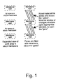
- Fig. 1 is a schematic representation of a stock membrane and an occluded membrane according to the present invention
- Fig. 2 is a schematic representation of the functioning of six classes of biosensors
- Fig. 3 is a schematic representation of a device according to the present invention for visual detection of a signal from a biosensor
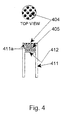
- Fig. 4 is a schematic representation of a device according to the present invention for monitoring a change within a closed primary container, when visual detection of changes in the biosensor or configuration of the device is inappropriate;
- Fig. 5 is a schematic representation of a device according to the present invention with an outer opaque element
- Fig. 6 is a graph of measured light intensity vs. concentration as measured in the device of Fig. 5;
- Fig. 7 is a graph of measured light intensity vs. ratio of labeled beads to plain beads as measured in the device of Fig. 5;
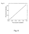
- Fig. 8 is a graph of measured light intensity vs. available fluorescin as measured in the device of Fig. 5;
- Fig. 9 is a graph of measured light intensity vs. pH as measured in the device of Fig. 5, showing pH responsiveness of FITC;
- Fig. 10 is a graph of measured light intensity vs. pH as measured in the device of Fig. 5, showing pH non-responsiveness of phycoerythrin-cyanin-5 strepavidin;
- Fig. 11 is a graph of the ratio of two wavelengths of emitted light vs. pH as measured in the device of Fig. 5.
- Primary container in this disclosure, we use the term "primary container” in a broad sense, to include any container for cells, foods, or industrial materials.
- Integral sensor ⁇ In this disclosure, we use the term “integral sensor” to designate one of many devices as disclosed herein.
- An “integral sensor” typically is incorporated into a primary container, prior to sterilization if appropriate, and separates a biosensor (see below) from the contents of the sealed primary container yet provides information on quality of the contents of the container without breaking the sealed system.
- Gated-pore membrane ⁇ in this disclosure, we use the term "gated-pore membrane" in a broad sense, to include any separation barrier containing one or more pores which have been occluded as taught in U.S. Patent No. 5,261,870, pending patents, or other procedures. Such a separation barrier has one or more pores which initially are closed, but which will open in response to a change in the local environment to an extent sufficient to allow passage of ions or molecules, as appropriate. The occluding material might erode, dissolve, or change three-dimensional form, depending on design of the gated-pore membrane.
- Gate in reference to one or more pores through a separation barrier.
- the gate is "closed” if the pore is occluded to prevent passage of ions or molecules.
- the gate is "open” if the occluding material has eroded, dissolved or changed three-dimensional form sufficiently to allow passage of one or more species of selected ions or molecules.
- Sensor compartment in reference to a component of an integral sensor, with said component formed in part from a gated-pore membrane and in part from a plastic construct.
- the sensor compartment contains and positions a biosensor where it can become accessable to ions, molecules, or cells entering from the primary container and also positions that biosensor where it can be interrogated in a consistent manner through a special optical window or, alternatively, through a portion of the primary container.
- Biosensor in reference to a component of a device, with said component designed to respond to a molecular change(s) in the local environment in a known and consistent manner with a response or signal that can be detected or measured using procedures known to those skilled in the art.
- the change in local environment, or microenvironment might be ionic, molecular or cellular in nature.
- Receptor ⁇ in this disclosure, we use the term "receptor” in reference to any detector molecule, either synthetic or natural in origin, or antibody incorporated into a biosensor. A receptor has reasonable affinity and specificity for one or more "ligands” (see below).
- Ligand ⁇ in this disclosure, we use the term "ligand" in reference to a specific ion(s), molecule(s), or cell(s) accumulating or disappearing within a primary container over time. Accumulation of ligand within the primary container will result in a change in the proportion of unoccupied receptor to ligand-receptor complexes, and a change in characteristics the of a biosensor.
- the present invention consists of devices and methods which allow evaluation of the contents of a sealed primary container by means of an integral sensor which is separated from the contents of the sealed primary container yet provides information on quality of the contents of the primary container without breaking the sealed system.
- the integral sensor device might be a hollow cylinder or a shallow construct.
- One end or face of the device is a gated-pore membrane whose pores normally are occluded, by one of many approaches, forming one end of a sensor compartment, containing a biosensor appropriate for the task, with the other end of the sensor compartment formed by an optical window recessed in from the end opposite to the gated-pore membrane or formed by a wall of the primary container.
- the integral sensor device is fabricated separately from the primary container and incorporated into the primary container during final fabrication, before sterilization. Certain embodiments of the device are therefore capable of aseptic operation.
- the status of the contents of a primary container for blood cells, other cells, foods, or industrial products can be determined by inspection, visually or via a fiberoptic probe through the optical window of a plastic construct incorporated into said primary container at fabrication, of the biosensor retained within said plastic construct by a gated-pore membrane, the pores in which opened in response to an environmental change in said primary container allowing the contents of said primary container to contact and cause a change in said biosensor.
- the gated-pore membrane opens and allows continuous exposure of the biosensor, within the sensor compartment, to the contents of the primary container. Changes within the primary container affect the signal from the biosensor, which can be quantified at any instant in time by viewing the optical window of the device with an appropriate sensor, as above, providing a measure of quality without opening the primary container.
- the gated-pore membrane remains closed until a predetermined change occurs within the primary container which causes the gated-pore membrane to open allowing fluid to enter the sensor compartment and contact the biosensor, which then responds with an appropriate signal.
- Changes in the primary container that can be detected include, but are not limited to, a decrease or increase in pH away from a threshold value or accumulation of one or more members of a preselected class of molecules, including toxins produced by bacteria, above a threshold value.
- a great range in utility is possible because, depending on the device and method, both the material(s) occluding the gated-pore membrane and material(s) forming the biosensor can be varied independently or in combination.
- a predetermined change in contents of the primary container can be evidenced by opening of the gated-pore membrane and/or a change in the signal from the biosensor.
- a number of other embodiments or uses would be obvious to one skilled in the art, and the examples herein illustrate but do not set the limits of this invention.
- Conventional membranes with pores of known size can be fabricated into special gated-pore membranes, and then fabricated into a container or other device (U.S. Patent No. 5,261,870).
- the gated-pore membrane can provide a closed container which opens only when predefined conditions are met, and the material occluding or otherwise closing the pores is altered so that the gates previously blocking passage of molecules through the pores is opened.
- An almost infinite combination of membranes, pore diameters, and occluding materials is envisioned in US Patent 5,261,870 and other disclosures.
- the conditions on one side of the gated-pore membrane and not mechanical, electrical or other interventions determine when the pores open and allow passage of molecules through the membrane into the sensor compartment.
- Biosensors can be designed to respond to many molecular changes in the environment.
- the color change of a pH indicator in response to proton concentration or certain dipsticks to glucose concentration are two common and simple examples.
- Biosensors sensitive to one or another molecular stimulus can be incorporated into beads or micro-beads, and frequently can be designed to change color or emit light of a given wavelength when exposed to light of an appropriate wavelength (i.e., fluorescence). These or other changes can be monitored with a variety of detectors, ranging from the human eye to fiberoptic electronic devices with a digital readout.
- the gated-pore membrane would: (a) retain the biosensor in place; and (b) determine when, if ever, the biosensor received the stimulus driving the response.
- a device could be a plastic cylinder, series of concentric cylinders, shallow construct, or of any other shape so long as it positioned the biosensor in a manner conveniently accessible to the detection device. If desirable, the device might include a clear optical window, through which a separate detector could view the biosensor, but would have opaque walls to minimize stray light from falling on the sensor or detector.
- the optical window, and the sensor compartment located more than 1 cm from the open end of the construct (the end opposite from that with the gated-pore membrane).
- a portion of the wall of the primary container can serve as the optical window to view a biosensor contained in a shallow construct.
- the gated-pore membrane is fabricated using an occluding polymer such as methylcellulose, or other polymers or mixtures taught in U.S. Patent No. 5,261,870 with the intent that the pores would remain occluded until a cell suspension had been placed into the primary container incorporating the device.
- a predetermined interval e.g., 5 to 15 minutes
- the occluding material would erode or dissolve, the gates open, and free exchange of molecules across the membrane would occur by diffusion and other forces.
- the biosensor then would start to sample the contents of the primary container, limited only by diffusion within the sensor compartment, diffusion across the gated-pore membrane, and correctness of the sampling site of the external face of the gated-pore membrane. Occasional or continuous movement of the primary container or its contents might facilitate accurate sampling. Changes in the biosensor might be detected visually or via a fiberoptic probe and appropriate instrumentation.
- the biosensor provides specificity for detection of molecular changes within the primary container, and the gated-pore membrane retains the biosensor and prevents premature sampling.
- the gated-pore membrane is fabricated with a special occluding material selected to respond to a specific stimulus and not the mere presence of water.
- This stimulus opening the gated-pores might be a decline in pH within the primary container, possibly caused by normal cellular metabolism or especially that of contaminating microbes, or accumulation of a secreted bacterial toxin.
- the gated-pore membrane remains closed after the cell suspension or product is placed into the container and opens only if and when the triggering stimulus occurs.
- the gated-pores open allowing water to pass through the membrane and into the detector compartment, carrying along whatever ions or molecules that might be in the water.
- the sensor would change from contact with water, for example from a green to a white color discernable by the human eye, or in response to specific molecules or ions entering the detector chamber with the water.
- visual inspection for a green rather than a white color before use might be sufficient to preclude a decreased pH associated with microbial contamination.
- the material occluding the gated-pore membrane completely block the pores or micropores through the membrane, so long as the molecules to which the biosensor responds cannot gain access through the membrane to the biosensor.
- Utility of such devices is defined by the combination of membrane stock plus materials plugging the pores to form the gated-pore membrane, precision or repeatability in opening of the occluded pores, nature of the biosensor, and sensitivity of the detector plus biosensor to detect the monitored event.
- the magnitude of the change to trigger a detected response can be adjusted either via the occluding material or the biosensor.
- response of the detector is not dependent on mechanical or electrical forces, although readout might require an instrument. With appropriate instrumentation, signals in a remote or isolated area can be detected.
- Membrane stock used to form the gated-pore membrane can be any of those commercially available, e.g., fibril membranes with a "haystack” structure, microporous membranes with a “sponge-like” structure, and track-etched or capillary-pore membranes with a “tunnel” structure. Although others have used membranes, either preformed or formed in place, to position biosensors against a fiberoptic or other detector, gated-pore membranes have not been used for this purpose. Gated-pore membranes for this application could be prepared using microporous membrane stock. However, characteristics unique to capillary-pore membrane stock frequently make this the material of choice to form gated-pore membranes for use in biosensor detectors as described herein. Alternatively, one might use a stock membrane which incorporates the ability to change in response to pH (Maeda et al., 1984; Kinoshita et al., 1994).
- Materials to occlude preformed pores in a stock membrane and, thereby, form a gated-pore membrane might be selected from members of the classes listed below, but these are illustrative and not inclusive.
- a generic resultant gated-pore membrane is illustrated in Fig. 1.
- occluding material within the pores is the functional element rather than residual occluding material that might be retained on the membrane faces around the pores.
- Many of the potentially useful materials have been used for other applications, but none has been used to fill preformed pores in a membrane to control access of material on one side of the membrane to a biosensor positioned behind the membrane where accessible to a detector. All materials applicable for this application are considered as under the scope of this invention, recognizing that in some instances the material might be patented.
- the term "receptor” is any detector molecule, either synthetic or natural in origin, or antibody bound on a solid substrate, termed the biosensor, and the term "ligand” is a specific ion(s), molecule(s), or cell(s) accumulating or disappearing within a primary container over time.
- the receptor has reasonable affinity and specificity for the ligand. Accumulation of ligand within the primary container will result in a change in the proportion of unoccupied receptor to ligand-receptor complexes. Appropriate selection of the ligand and receptor molecules allows detection of this change in ratio of unoccupied vs.
- occupied receptors by an altered spectroscopic property from either of the separate ligand or receptor molecules or their complexes.
- a signal moiety might be conjugated to the receptor and provide amplified evidence of the status as unoccupied or occupied.
- the change in ratio of unoccupied vs occupied receptors might be amplified by a property intrinsic to the solid substrate.
- biosensors appropriate for this device, insofar as they undergo a change in signal character, upon exposure to the change in microenvironment to be detected, which can be detected or measured by any form of spectroscopy (e.g., luminescence, fluorescence, absorbance, infrared, magnetic, light scatter, etc.). In certain cases, the material or cells stored can provide the requisite signal.
- spectroscopy e.g., luminescence, fluorescence, absorbance, infrared, magnetic, light scatter, etc.
- the material or cells stored can provide the requisite signal.
- use of special biosensors increases the nature of changes that can be monitored. Many of the potentially useful biosensors have been used for other applications, but none has been positioned in a viewable compartment formed in part by a gated-pore membrane initiating access of the molecules, ions or bacteria to be detected and formed in part by an optical window allowing detection of the signal resulting from the biosensor.
- Biosensors often contain two active elements; namely, the detector material(s
- the solid substrate can have any three-dimensional shape; include impervious or permeable materials; include smooth, microporous or complex surfaces; etc.
- This patent is not limited by the nature or shape of the solid substrate.
- this invention is not limited by the method used to attach the detector molecules to the solid substrate.
- Detector molecules can be attached to the surface of the biosensor (e.g., plastic or glass bead) by simple adsorption, direct covalent attachment, or via an affinity tag (Anonymous 1999a, 1999b; Nolan et al., 1999; Song and Swanson, 1999).
- the solid surface might be pre-coated with lipids and then lipophilic ligand-detector molecules partitioned therein (Nolan et al., 1999). If not in a combination molecule with the detector material, the signal material must respond to the change in status of the detector material (occupied vs unoccupied) and the change in the signal must be a change in a characteristic absorption or emission property within the electromagnetic spectrum. An absorbance or fluorescent signal is most common.
- biosensor appropriate for this application is considered as under the scope of this invention, recognizing that in some instances the biosensor might be patented. In this discussion we will consider a bead-shaped biosensor, but all shapes are claimed. Appropriate concepts for biosensors might be considered in several classes (shown schematically in Fig. 2; for easy reference, given designations herein) which include, but are not limited to:
- Detection of a change intrinsic to the contents of a container or via a special biosensor requires an optical probe (Maroze et al., 1999), either an integral and fixed fiberoptic probe or a separate and removable fiberoptic probe.
- a fiberoptic probe integral to the container to be analyzed might read an inherent spectral property of materials within the bag.
- an integral fiberoptic probe might indirectly monitor changes in internal contents via reading a change in an optical property of a biosensor element when the material of interest reaches an appropriate concentration in the immediate environment of the biosensor.
- the biosensor is immobilized by covalent bonding to the face of the fiberoptic bundle or the biosensor is restrained close to the fiberoptic bundle with a perm-selective membrane.
- Such systems have been used to monitor anions, cations, biomass, glucose, lactic acid, NAD(P)H, O 2 , CO 2 and pH (Agayn and Walt, 1993; Kar and Arnold, 1995; Marose et al., 1998; Spichiger-Keller, 1997; Tartakovsky et al., 1996; Urbano et al., 1984; Vaccari et al., 1994; Weigl et al., 1994; Xu et al., 1998). These approaches cannot be used with a closed container.
- a device to interrogate changes in a closed primary container logically might include an immobilized biosensor, sampling a specific attribute of the contents of said closed primary container, with said biosensor protected in a separate closed sub-container within said device, with said biosensor retained in front of an optical window, with said optical window accessible to a removable fiberoptic detector without opening either said primary container or said sub-container, and with said device capable of facile incorporation into said primary container at the time of manufacture before sterilization.
- Solution of this hitherto unattainable goal is the subject of this invention. This is achieved by combination of existing technology on gated-pore membranes; bioactive polymers, proteins or lipid films; and biosensors in a novel manner and adding flexibility offered by new materials and configurations in novel devices. Clearly, several elements are combined in a novel manner to provide such devices.
- biosensors as above, can be positioned in front of an optical window, accessible to a removable fiberoptic detector, while protected in a separate closed sub-container within the primary container. They are illustrative of the many uses of the invention, although the invention encompasses other biosensors, other membranes with open pores or gated-pores, other configurations, and other combinations of functional elements in the device. Examples of devices include reference to welding of a gated-pore membrane to a plastic construct. The invention is not limited by the plastic(s) used to form the device.
- the preferred welding process will depend on the plastic used to fabricate the construct, and the invention includes any method to assemble the device or incorporate it into the primary container, such as ultrasonic and radio-frequency welding or other methods known to those skilled in the art. Examples illustrate the variety of materials that can be used to form gated-pore membranes or serve as biosensors, in one or more types of devices, and the invention is not limited in respect to specific compositions or analytes.
- a simple device can be used to monitor one of a variety of changes within a closed primary container. It is formed from a shallow, plastic construct (301) with an external flange (301a) on one side and a gated-pore membrane (304) welded to the other side. After filling the construct with a biosensor (305) providing a signal detectable by a human eye (e.g., plastic beads that change color when bathed in an aqueous fluid), the construct is welded to a convenient location on the wall of the primary container (306).
- This device depends on selectivity in response of the gated-pore membrane as the primary mechanism regulating change in a simple biosensor. When thus positioned, changes in the signal from the biosensor can be detected visually through a portion (307) of the primary container, without opening the primary container.
- FIG. 4 Another device (Fig. 4) can be used to monitor one of a variety of changes within a closed primary container, when visual detection of changes in the biosensor or the configuration of the device in Example 1 is inappropriate.
- This device can be of any convenient shape and formed from any plastic, but a cylindrical plastic construct formed from clear acrylic plastic is favored for some applications, and a clear optical window transmitting the portion of the spectrum of interest is essential.
- the device of Fig. 4 is a cylindrical acrylic construct (411) with a biosensor compartment formed by an optical window (412), a gated-pore membrane (404) responding to any aqueous environment, and the walls of the construct.
- the gated-pore membrane is welded to the surface (411a) of a recess in the end of the construct.
- This device uses the gated-pore membrane simply to protect and isolate the biosensor until the primary container is filled, for example with a cell suspension, after which the gates open in ⁇ 30 minutes and selectivity in response to a predetermined analyte and intensity of signal are properties of the biosensor and amount of material impinging thereon.
- a third device incorporates an opaque outer plastic element (515), with accommodation (515a) to weld a gated-pore membrane (504) to this part of the device, positioned around and bonded to an inner element (511) which is a clear plastic construct similar to the device of Example 2 and contains a biosensor (505) capable of detecting the desired change within the primary container.
- This device can be of any convenient shape and formed from any plastic, but a cylindrical concentric construct is favored for some applications.
- the outer opaque element (515) might be of colored polyvinyl chloride to shield the biosensor from stray light and facilitate welding the device into a polyvinyl chloride blood bag, and the inner element of clear plastic so that the optical window (512) will transmit the portion of the spectrum of interest.
- This device can be used to monitor one of a variety of changes within a closed primary container, when stray light might affect detection of the signal from the biosensor, by positioning the tip of a separate fiberoptic probe (not shown), as known to those skilled in the art, against the optical window (512).
- This device uses a gated-pore membrane to protect and isolate the biosensor until the primary container is filled and for a predetermined interval (e.g., 0.5, 2 or 5 days) thereafter or until a predetermined threshold of a specific change in contents of the primary container is reached (e.g., concentration of first analyte), after which the gates open and the biosensor begins to monitor changes in concentration of the same molecule or ion triggering the gated-pore membrane.
- the biosensor can be used to monitor concentration of a second analyte with intensity of the signal from the biosensor dependent on properties of the biosensor and amount of second analyte impinging thereon.
- Beads suitable for use as an "optical bench” were prepared by covalent attachment of dextran (#1820; Molecular Probes) containing both FITC and lysine to 10 micrometer diameter carboxylate beads (Polysciences, Inc), using standard carbodiimide coupling chemistry. This provided an immobilized fluorophore, known by those versed in the art to be responsive to pH through the FITC function and suitable for analysis of pH. Localization of the FITC at a sufficient distance from the bead surface, by means of the dextran, eliminated problems related to the well-known surface effects on fluorescence.
- the biosensor cavity was filled with the bead suspension (505), and the membrane (504) attached. Beads were allowed to settle onto the optical window (512).
- response time could be reduced substantially by agitation of the primary container, or by design of the integral sensor to increase the amount of gated-pore membrane enclosing the biosensor cavity and thus reduce time for protons or other ligands to enter and interact with the biosensor.
- Samples to be analyzed might contain components that will interfere with direct fluorescence measurements.
- human plasma can contain variable amounts of "yellow” materials that could absorb incident light at the excitation wavelength.
- Optical density at 480 nm was measured for 13 different samples of human blood plasma (cell free), and absorbance averaged 0.554 (standard deviation was 0.428; range from 0.12 to 1.30), but was much lower at >500 nm.
- this wide a range in absorbance could affect the light available for excitation of a biosensor incorporating, for example, FITC as the signal moiety.
- a second detector bead system could be constructed so that it shared excitation wavelength with the primary bead (B-1; e.g., FITC), but emitted at a distinctly different wavelength than B-1 and was unaffected by the ligand altering response of the biosensor of bead B-1 (e.g., protons in case of FITC).
- B-1 primary bead
- B-2 e.g., protons in case of FITC
- a general bead coating was developed by covalently attaching biotin-dextran (D-1956; Molecular Probes) to 10 micrometer diameter carboxylate beads (Polysciences, Inc.), using standard carbodiimide coupling chemistry. This provided a common attachment site on the beads for avidin-streptavidin conjugates. Assuming beads of biosensor B-1 used FITC, materials suitable for service as B-2 should be excited (absorb) at 480 nm, emit at a wavelength distinct from 535 nm, and have a response independent of pH.
- Examples include reagents available from Coulter Diagnostics such as: Steptavidin-PC5 (streptavidin conjugated to phycoerythrin-cyanin 5; excitation 486-580; emission 660-680) and EDC (strepavidin conjugated to phycoerythrin-Texas Red7-x; excitation 486-580; emission 610-630). It is well-known to those versed in the art that many other combinations can be used to correct for effects on either excitation or emission wavelengths. We used the PC-5 system and evaluated freedom from a pH-driven shift in emission. As evident in Fig.
- the pH sensitive biosensor (B-1) was FITC dextran attached to a polystyrene support (as in Example 6) and the pH insensitive biosensor (B-2) was EDC-streptavidin attached to biotin/dextran polystyrene (see Example 7).
- the same fiberoptic fluorometer (S2000-FL; Ocean Optic, Inc.) was used to interrogate the device, as pH of the salts solution was changed as in Example 6.
- the system was excited at 480 nm and emission read with both the 535 nm and ⁇ 640 nm filters.
- Resultant data (Fig. 11) show excellent linearity of response in the range of interest to blood banks, pH 6.0 to 7.0.
- a device fabricated as in Figs. 4 or 5, can use a Class-5 biosensor (see Fig. 2) to detect accumulation of a protease secreted by bacteria within a closed primary container.
- Active elements of the integral detector would be as detailed above, except that plastic beads would be coated with "doped” lipid to provide a Class-5 biosensor.
- the lipid would be "doped” with an enzyme substrate synthesized with fluorescent moieties ("a” and "b”; inhibiting each other's emission because of proximity) incorporated on either side of the proteolytic cleavage site.
- fluorescent moieties a
- b fluorescent moieties
- biosensor suitable for attachment to an avidin-streptavidin coated bead is: biotin-(6-aminohexanoicacid)-(6-aminohexanoicacid)-(6-aminohexanoicacid)-Lys(Tamara)-(6-aminohexanoicacid)-Ala-Phe-Glu-Ala-Leu-(6-aminohexanoicacid)BCys(Texas Red)-Gly-COOH.
- the "-Ala-Phe-Glu-Ala-Leu-" sequence should be useful for detecting proteases known to be secreted from a variety of bacteria including Bacillus cereus, Escherichia coli, Poteus mirabilis, Serratia marcescens, Staphyloccous aureus, and Streptococcus species. Other similar substrates, exhibiting appropriate susceptibility to hydrolases, can be designed as appropriate, using assumptions known to those skilled in the art.
- a device fabricated as in Figs. 4 or 5, can use a Class-6 biosensor (see Fig. 2) to accumulate bacteria and increase sensitivity for detection of a protease secreted by bacteria within a closed primary container.
- Active elements of the integral detector would be as detailed above, except that plastic beads "doped” lipid would be used to fabricate a Class-6 biosensor.
- a material binding the bacteria of interest e.g., fibronectin, laminin, type-4 collagen
- the coating of this biosensor also would include an enzyme substrate synthesized with fluorescent moieties ("b" and "c”; inhibiting each other's emission because of proximity) incorporated on either side of the proteolytic cleavage site.
- the enzyme substrate might be: biotin-(6-aminohexanoicacid)-(6-aminohexanoicacid)-(6-aminohexanoicacid)-Lys(Tamara)-(6-aminohexanoicacid)-Ala-Phe-Glu-Ala-Leu-(6-aminohexanoicacid)BCys(Texas Red)-Gly-COOH.
- Streptococus pyrogenes are known to bind to the fibronectin serving as receptor "a” and secrete proteases that can cleave the -Ala-Phe-Glu-Ala-Leu- peptide integral to receptor "b” and "c", removing the proximity inhibition to their fluorescence. Consequently, as Streptococus pyrogenes, or other bacteria, proliferated in the primary container they would accumulate on the biosensor and the fluorescent signal detectable on probing with a fiberoptic fluorometer would reflect the extent of bacterial contamination in the primary container.
Landscapes
- Health & Medical Sciences (AREA)
- Immunology (AREA)
- Chemical & Material Sciences (AREA)
- Physics & Mathematics (AREA)
- Analytical Chemistry (AREA)
- Optics & Photonics (AREA)
- Nuclear Medicine, Radiotherapy & Molecular Imaging (AREA)
- Life Sciences & Earth Sciences (AREA)
- Chemical Kinetics & Catalysis (AREA)
- Biochemistry (AREA)
- General Health & Medical Sciences (AREA)
- General Physics & Mathematics (AREA)
- Pathology (AREA)
- Investigating Or Analysing Materials By The Use Of Chemical Reactions (AREA)
- Apparatus Associated With Microorganisms And Enzymes (AREA)
- Measuring Or Testing Involving Enzymes Or Micro-Organisms (AREA)
- Investigating, Analyzing Materials By Fluorescence Or Luminescence (AREA)
Abstract
Description
Biosensors often contain two active elements; namely, the detector material(s) and the signal material(s), which frequently are combined within a combination molecule.
- Class-1:
- A bead coated with a fluorescent receptor targeting an epitope or ligand characteristic of, or secreted by, bacteria of interest and which fluoresces only when its conformation is altered by binding of the ligand. Detection of bound ligand is via fluorescence when the biosensor is illuminated with light of an appropriate wavelength.
- Class-2:
- A bead coated with lipid in which are positioned appropriate fluorochrome-receptor complexes, anchored therein, which fluoresce only when their conformation is altered by binding of the ligand. Detection of bound bacterial products is via fluorescence when the biosensor is illuminated with light of an appropriate wavelength.
- Class-3:
- A bead coated with lipid or otherwise prepared in which are positioned low concentrations of two different fluorescent receptors each binding the ligand, so that concurrent binding of a ligand molecule by both receptors brings them in closer proximity and enables charge-coupling excitation to produce light of a unique wavelength when the biosensor is illuminated with light of an appropriate wavelength. In this case, appropriate external excitation causes only one receptor to fluoresce and although that light is not detected it does excite, by fluorescence resonance transfer, the second receptor and causes it to emit light of a unique wavelength detectable by the fiberoptic probe. Because fluorescence of the second receptor is dependent on proximity to the first, achieved only by both binding to ligand, such systems can be very specific.
- Class-4:
- A bead coated with lipid or otherwise prepared to accept a mixed-matrix, in which one receptor-A is designed to bind a certain class or type of cell, which after binding continues to release material serving as the ligand bound by the nearby second receptor-B, causing a rapid shift from unoccupied to occupied receptor-B molecules and a detectable spectral change. Positioning a chemoattractant on the biosensor could further enhance sensitivity of a mixed-matrix surface.
- Class-5:
- A bead coated with lipid or otherwise prepared to accept a detection substrate-fluorochrome which can serve as a substrate for a specific class of enzymes secreted by bacteria or other cells. In this biosensor, the substrate is designed to contain two fluorescent groups with one on either side of the enzymatic cleavage site in close proximity while the molecule is intact but substantially farther apart after the substrate is cleaved. Local action of the enzyme on the substrate releases the fluorescent inhibition caused by close proximity, and the detector molecule will fluoresce when illuminated with light of an appropriate wavelength. Bark and Kahn (2000) provided examples.
- Class-6:
- A bead coated with a mixed matrix combining elements of Class-4 and Class-5. As in Class-4, receptor-A binds the cell of interest, without or with help from a chemoattractant, so that the cell continues to release an enzyme in close proximity to a substrate incorporating the fluorescent reporter B-C, as in Class-5. The enzyme from the localized cell (e.g., a bacterium) acts on the substrate and this releases the fluorescent inhibition caused by close proximity, so that the detector molecule will fluoresce when illuminated with light of an appropriate wavelength.
- Fig.1
- Schematic representation of a gated-pore membrane prepared using a microporous or capillary-track membrane and unique material to occlude the pores (e.g., as in U.S. Patent No. 5,026,342).
- Fig. 2
- Schematic representation of changes in a receptor-fluorescent reporter group upon change from unoccupied receptor to occupied receptor-ligand complex, listed by biosensor class as detailed in the specification. A reporter group that is non-fluorescent is designated with a lower-case letter and a reporter group that can fluoresce when excited with light of appropriate wavelength is designated with an upper-case letter.
- Fig. 3
- Cross-section through a simple boat-shaped device as described in Example 1. The capillary-pore membrane (302) was processed with material (303) to plug the pores and make a gated-pore membrane (304). This membrane is bonded to a plastic construct (301) to form a cavity which then was filled with a biosensor (305) before the device was bonded via a flange (301a) to the primary container (306).
- Fig. 4
- Longitudinal section through a device, and also a top view, as described in Example 2. The biosensor-compartment in a cylindrical construct (411) was filled with a biosensor (405). A gated-pore membrane (404) was bonded to a recessed surface (411a) to seal the biosensor in the device. The device then can be incorporated into the seam of a primary container (not shown) so that the optical window (412) can be interrogated with a fiberoptic probe.
- Fig. 5
- Longitudinal section through a device, and also a top view, as described in Example 3. An optically clear cylindrical construct (511) was surrounded by an opaque outer concentric construct (515). The biosensor compartment was filled with a biosensor (505). A gated-pore membrane (504) was bonded to a recessed surface (515a) of the opaque outer concentric construct (515) to seal the biosensor in the device. The device then can be incorporated into the seam of a primary container (not shown) so that the optical window (512) can be interrogated with a fiberoptic probe.
- Fig. 6
- Intensity of fluorescence from a solution of fluorescein isothiocyanate (FITC) in a device as a function of concentration of FITC. Device as in Fig. 5; measurements with fiberoptic detector using excitation at 480 nm and detection at 535 nm (thousands of fluorescent units).
- Fig. 7
- Intensity of fluorescence from beads in a device as a function of the ratio of beads labeled with fluorescein isothiocyanate (FITC) to plain beads. Device as in Fig. 5; measurements with fiberoptic detector using excitation at 480 nm and detection at 535 nm (thousands of fluorescent units).
- Fig.8
- Intensity of fluorescence from beads in a device as a function of their known content of fluorescein isothiocyanate (FITC). Known contents of FITC were equivalent to 0.00, 0.22 and 1.20 million molecules of FITC in solution. Device as in Fig. 5; measurements with fiberoptic detector using excitation at 480 nm and detection at 535 nm (millions of fluorescent units).
- Fig. 9
- Intensity of fluorescence from beads, previously labeled with fluorescein isothiocyanate (FITC), in a device as a function of pH of the salts solution around the device and around the beads therein. Device as in Fig. 5; measurements with fiberoptic detector using excitation at 480 nm and detection at 535 nm (thousands of fluorescent units).
- Fig. 10
- Minimal effect of pH of a salts solution on fluorescence from phycoerythrin-cyanin-5 strepavidin attached to beads. Device as in Fig. 5; measurements with fiberoptic detector using excitation at 480 nm and detection at ≥640 nm (thousands of fluorescent units).
- Fig. 11
- Ratio of emissions from beads responsive to pH and non-responsive to pH
can be used to normalize data for irregularities of response or color of the
medium. The device contained a mixture of beads labeled with
fluorescein isothiocyanate (pH responsive; Fig. 9) and phycoerythrin-cyanin-5
strepavidin (pH non-responsive; Fig. 10). The ratio of emission
at 535 nm and ≥640 nm was calculated as a function of pH of the salts
solution around the device and around the beads therein. Device as in
Fig. 5; measurements with fiberoptic detector using excitation at 480 nm
and detection at 535 nm and ≥640 nm (raw values ranged between 75,000
and 135,000 relative fluorescent units).
The invention is further illustrated in the following examples:
Claims (19)
- A sensor device, comprising:a biosensor;a biosensor enclosure having an interior and an exterior, enclosing the biosensor, the biosensor enclosure having a surface allowing external viewing of the biosensor; anda separation barrier forming a portion of the biosensor enclosure, the separation barrier having at least one pore allowing fluid communication between the interior and exterior of the biosensor enclosure.
- The sensor device of claim 1, wherein the separation barrier separates the interior of the biosensor enclosure from a primary container.
- The sensor device of claim 1, wherein the at least one pore is occluded with a responsive material.
- The sensor device of claim 3, wherein the responsive material is selected from the group consisting of cellulosics, non-cellulosic non-protein polymers, protein polymers, lipid bilayers, and lipid-containing composites.
- The sensor device of claim 3, wherein the responsive material exhibits a response selected from the group consisting of eroding, dissolving, and changing three-dimensional form.
- The sensor device of claim 5, wherein the response results from a change selected from the group consisting of a change in solvent concentration, a change in pH, a change in temperature, bacterial action, endotoxin action, and enzymic action.
- The sensor device of claim 1, wherein the surface allowing external sensing permits optical sensing of the biosensor.
- The sensor device of claim 1, wherein the biosensor enclosure is partially surrounded by an opaque material.
- The sensor device of claim 1, wherein the separation barrier is constructed from a material selected from the group consisting of fibril membranes, microporous membranes, and capillary-pore membranes.
- The sensor device of claim 1, wherein the biosensor is comprised of:a substrate; andat least one bioactive response element.
- The sensor device of claim 10, wherein the bioactive response element is a fluorescent receptor.
- The sensor device of claim 10, wherein the bioactive response element is a fluorochrome-receptor complex.
- The sensor device of claim 10, wherein the bioactive response element is a combination of a first fluorescent receptor and a second fluorescent receptor, the second receptor emitting detectable light of a unique wavelength on excitation by fluorescence resonance transfer by the first fluorescent receptor.
- The sensor device of claim 10, wherein the bioactive response element is a combination of a first receptor and a second receptor, the first receptor binding a cell and the second receptor undergoing a detectable spectral change in response to material released by the cell bound to the first receptor.
- The sensor device of claim 10, wherein the bioactive response element is a combination of two inhibited fluorescent groups linked by an enzymatic cleavage site, and wherein enzymatic action cleaves the enzymatic cleavage site and releases the fluorescent inhibition.
- The sensor device of claim 10, wherein the bioactive response element is a combination of a first receptor and a second receptor, the first receptor binding a cell capable of releasing an enzyme and the second receptor being an inhibited fluorescent group wherein the enzyme releases the fluorescent inhibition.
- The sensor device of claim 2, wherein the primary container need not be opened for analysis.
- The sensor device of claim 1, wherein the device is capable of aseptic operation.
- The sensor device of claim 1, wherein the external sensing is remote sensing.
Applications Claiming Priority (4)
| Application Number | Priority Date | Filing Date | Title |
|---|---|---|---|
| US17916400P | 2000-01-31 | 2000-01-31 | |
| US179164P | 2000-01-31 | ||
| US77227201A | 2001-01-29 | 2001-01-29 | |
| US772272 | 2001-01-29 |
Publications (2)
| Publication Number | Publication Date |
|---|---|
| EP1122535A2 true EP1122535A2 (en) | 2001-08-08 |
| EP1122535A3 EP1122535A3 (en) | 2004-09-22 |
Family
ID=26875065
Family Applications (1)
| Application Number | Title | Priority Date | Filing Date |
|---|---|---|---|
| EP01200347A Withdrawn EP1122535A3 (en) | 2000-01-31 | 2001-01-31 | Interrogation of changes in the contents of a sealed container |
Country Status (2)
| Country | Link |
|---|---|
| US (2) | US7407796B2 (en) |
| EP (1) | EP1122535A3 (en) |
Cited By (5)
| Publication number | Priority date | Publication date | Assignee | Title |
|---|---|---|---|---|
| WO2006023725A3 (en) * | 2004-08-19 | 2006-05-18 | Blood Cell Storage Inc | FLUORESCENT pH DETECTOR SYSTEM AND RELATED METHODS |
| WO2008006153A1 (en) * | 2006-07-11 | 2008-01-17 | Paul Nigel Brockwell | Medical indicator method and device |
| US8183052B2 (en) | 2004-08-19 | 2012-05-22 | Blood Cell Storage, Inc. | Methods and apparatus for sterility testing |
| US8497134B2 (en) | 2004-08-19 | 2013-07-30 | Blood Cell Storage, Inc. | Fluorescent detector systems for the detection of chemical perturbations in sterile storage devices |
| US9040307B2 (en) | 2011-05-27 | 2015-05-26 | Blood Cell Storage, Inc. | Fluorescent pH detector system and related methods |
Families Citing this family (75)
| Publication number | Priority date | Publication date | Assignee | Title |
|---|---|---|---|---|
| US6036924A (en) | 1997-12-04 | 2000-03-14 | Hewlett-Packard Company | Cassette of lancet cartridges for sampling blood |
| US6391005B1 (en) | 1998-03-30 | 2002-05-21 | Agilent Technologies, Inc. | Apparatus and method for penetration with shaft having a sensor for sensing penetration depth |
| US8641644B2 (en) | 2000-11-21 | 2014-02-04 | Sanofi-Aventis Deutschland Gmbh | Blood testing apparatus having a rotatable cartridge with multiple lancing elements and testing means |
| US20040243093A1 (en) * | 2001-01-10 | 2004-12-02 | Ron Berenson | System for growth, analysis, storage, validation and distribution of cells and tissues used for biomedical purposes |
| WO2002100461A2 (en) | 2001-06-12 | 2002-12-19 | Pelikan Technologies, Inc. | Method and apparatus for improving success rate of blood yield from a fingerstick |
| US7682318B2 (en) | 2001-06-12 | 2010-03-23 | Pelikan Technologies, Inc. | Blood sampling apparatus and method |
| AU2002348683A1 (en) | 2001-06-12 | 2002-12-23 | Pelikan Technologies, Inc. | Method and apparatus for lancet launching device integrated onto a blood-sampling cartridge |
| US9226699B2 (en) | 2002-04-19 | 2016-01-05 | Sanofi-Aventis Deutschland Gmbh | Body fluid sampling module with a continuous compression tissue interface surface |
| US9427532B2 (en) | 2001-06-12 | 2016-08-30 | Sanofi-Aventis Deutschland Gmbh | Tissue penetration device |
| US7537571B2 (en) | 2001-06-12 | 2009-05-26 | Pelikan Technologies, Inc. | Integrated blood sampling analysis system with multi-use sampling module |
| US8337419B2 (en) | 2002-04-19 | 2012-12-25 | Sanofi-Aventis Deutschland Gmbh | Tissue penetration device |
| US7041068B2 (en) | 2001-06-12 | 2006-05-09 | Pelikan Technologies, Inc. | Sampling module device and method |
| WO2002100460A2 (en) | 2001-06-12 | 2002-12-19 | Pelikan Technologies, Inc. | Electric lancet actuator |
| US7344507B2 (en) | 2002-04-19 | 2008-03-18 | Pelikan Technologies, Inc. | Method and apparatus for lancet actuation |
| CA2448902C (en) | 2001-06-12 | 2010-09-07 | Pelikan Technologies, Inc. | Self optimizing lancing device with adaptation means to temporal variations in cutaneous properties |
| US9795747B2 (en) | 2010-06-02 | 2017-10-24 | Sanofi-Aventis Deutschland Gmbh | Methods and apparatus for lancet actuation |
| US7981056B2 (en) | 2002-04-19 | 2011-07-19 | Pelikan Technologies, Inc. | Methods and apparatus for lancet actuation |
| US7344894B2 (en) | 2001-10-16 | 2008-03-18 | Agilent Technologies, Inc. | Thermal regulation of fluidic samples within a diagnostic cartridge |
| AU2003231749A1 (en) | 2002-04-19 | 2003-11-03 | Pelikan Technologies, Inc. | Device and method for variable speed lancet |
| US8267870B2 (en) | 2002-04-19 | 2012-09-18 | Sanofi-Aventis Deutschland Gmbh | Method and apparatus for body fluid sampling with hybrid actuation |
| US8784335B2 (en) | 2002-04-19 | 2014-07-22 | Sanofi-Aventis Deutschland Gmbh | Body fluid sampling device with a capacitive sensor |
| US7582099B2 (en) | 2002-04-19 | 2009-09-01 | Pelikan Technologies, Inc | Method and apparatus for penetrating tissue |
| US9248267B2 (en) | 2002-04-19 | 2016-02-02 | Sanofi-Aventis Deustchland Gmbh | Tissue penetration device |
| US8702624B2 (en) | 2006-09-29 | 2014-04-22 | Sanofi-Aventis Deutschland Gmbh | Analyte measurement device with a single shot actuator |
| US9795334B2 (en) | 2002-04-19 | 2017-10-24 | Sanofi-Aventis Deutschland Gmbh | Method and apparatus for penetrating tissue |
| US7892185B2 (en) | 2002-04-19 | 2011-02-22 | Pelikan Technologies, Inc. | Method and apparatus for body fluid sampling and analyte sensing |
| US7892183B2 (en) | 2002-04-19 | 2011-02-22 | Pelikan Technologies, Inc. | Method and apparatus for body fluid sampling and analyte sensing |
| US7291117B2 (en) | 2002-04-19 | 2007-11-06 | Pelikan Technologies, Inc. | Method and apparatus for penetrating tissue |
| US7371247B2 (en) | 2002-04-19 | 2008-05-13 | Pelikan Technologies, Inc | Method and apparatus for penetrating tissue |
| US7909778B2 (en) | 2002-04-19 | 2011-03-22 | Pelikan Technologies, Inc. | Method and apparatus for penetrating tissue |
| US7232451B2 (en) | 2002-04-19 | 2007-06-19 | Pelikan Technologies, Inc. | Method and apparatus for penetrating tissue |
| US7331931B2 (en) | 2002-04-19 | 2008-02-19 | Pelikan Technologies, Inc. | Method and apparatus for penetrating tissue |
| US7901362B2 (en) | 2002-04-19 | 2011-03-08 | Pelikan Technologies, Inc. | Method and apparatus for penetrating tissue |
| US7297122B2 (en) | 2002-04-19 | 2007-11-20 | Pelikan Technologies, Inc. | Method and apparatus for penetrating tissue |
| US7976476B2 (en) | 2002-04-19 | 2011-07-12 | Pelikan Technologies, Inc. | Device and method for variable speed lancet |
| US7410468B2 (en) | 2002-04-19 | 2008-08-12 | Pelikan Technologies, Inc. | Method and apparatus for penetrating tissue |
| US8579831B2 (en) | 2002-04-19 | 2013-11-12 | Sanofi-Aventis Deutschland Gmbh | Method and apparatus for penetrating tissue |
| US8221334B2 (en) | 2002-04-19 | 2012-07-17 | Sanofi-Aventis Deutschland Gmbh | Method and apparatus for penetrating tissue |
| US8360992B2 (en) | 2002-04-19 | 2013-01-29 | Sanofi-Aventis Deutschland Gmbh | Method and apparatus for penetrating tissue |
| US7674232B2 (en) | 2002-04-19 | 2010-03-09 | Pelikan Technologies, Inc. | Method and apparatus for penetrating tissue |
| US7563232B2 (en) | 2002-04-19 | 2009-07-21 | Pelikan Technologies, Inc. | Method and apparatus for penetrating tissue |
| US7244265B2 (en) | 2002-04-19 | 2007-07-17 | Pelikan Technologies, Inc. | Method and apparatus for penetrating tissue |
| US7485128B2 (en) | 2002-04-19 | 2009-02-03 | Pelikan Technologies, Inc. | Method and apparatus for penetrating tissue |
| US7374544B2 (en) | 2002-04-19 | 2008-05-20 | Pelikan Technologies, Inc. | Method and apparatus for penetrating tissue |
| US7229458B2 (en) | 2002-04-19 | 2007-06-12 | Pelikan Technologies, Inc. | Method and apparatus for penetrating tissue |
| US7226461B2 (en) | 2002-04-19 | 2007-06-05 | Pelikan Technologies, Inc. | Method and apparatus for a multi-use body fluid sampling device with sterility barrier release |
| US9314194B2 (en) | 2002-04-19 | 2016-04-19 | Sanofi-Aventis Deutschland Gmbh | Tissue penetration device |
| US7491178B2 (en) | 2002-04-19 | 2009-02-17 | Pelikan Technologies, Inc. | Method and apparatus for penetrating tissue |
| US7648468B2 (en) | 2002-04-19 | 2010-01-19 | Pelikon Technologies, Inc. | Method and apparatus for penetrating tissue |
| US7524293B2 (en) | 2002-04-19 | 2009-04-28 | Pelikan Technologies, Inc. | Method and apparatus for penetrating tissue |
| US7717863B2 (en) | 2002-04-19 | 2010-05-18 | Pelikan Technologies, Inc. | Method and apparatus for penetrating tissue |
| US7547287B2 (en) | 2002-04-19 | 2009-06-16 | Pelikan Technologies, Inc. | Method and apparatus for penetrating tissue |
| US7141058B2 (en) | 2002-04-19 | 2006-11-28 | Pelikan Technologies, Inc. | Method and apparatus for a body fluid sampling device using illumination |
| US8574895B2 (en) | 2002-12-30 | 2013-11-05 | Sanofi-Aventis Deutschland Gmbh | Method and apparatus using optical techniques to measure analyte levels |
| EP2238892A3 (en) | 2003-05-30 | 2011-02-09 | Pelikan Technologies Inc. | Apparatus for body fluid sampling |
| ES2490740T3 (en) | 2003-06-06 | 2014-09-04 | Sanofi-Aventis Deutschland Gmbh | Apparatus for blood fluid sampling and analyte detection |
| WO2006001797A1 (en) | 2004-06-14 | 2006-01-05 | Pelikan Technologies, Inc. | Low pain penetrating |
| US7604592B2 (en) | 2003-06-13 | 2009-10-20 | Pelikan Technologies, Inc. | Method and apparatus for a point of care device |
| EP1671096A4 (en) | 2003-09-29 | 2009-09-16 | Pelikan Technologies Inc | METHOD AND APPARATUS FOR AN IMPROVED SAMPLING INTERFERENCE DEVICE |
| EP1680014A4 (en) | 2003-10-14 | 2009-01-21 | Pelikan Technologies Inc | METHOD AND APPARATUS PROVIDING A VARIABLE USER INTERFACE |
| WO2005065414A2 (en) | 2003-12-31 | 2005-07-21 | Pelikan Technologies, Inc. | Method and apparatus for improving fluidic flow and sample capture |
| US7822454B1 (en) | 2005-01-03 | 2010-10-26 | Pelikan Technologies, Inc. | Fluid sampling device with improved analyte detecting member configuration |
| US8828203B2 (en) | 2004-05-20 | 2014-09-09 | Sanofi-Aventis Deutschland Gmbh | Printable hydrogels for biosensors |
| EP1765194A4 (en) | 2004-06-03 | 2010-09-29 | Pelikan Technologies Inc | METHOD AND APPARATUS FOR MANUFACTURING A DEVICE FOR SAMPLING LIQUIDS |
| US9775553B2 (en) | 2004-06-03 | 2017-10-03 | Sanofi-Aventis Deutschland Gmbh | Method and apparatus for a fluid sampling device |
| WO2006039800A1 (en) | 2004-10-13 | 2006-04-20 | Mcmaster University | Operating environment analysis techniques for wireless communication systems |
| DE102004054349A1 (en) * | 2004-11-09 | 2006-05-11 | Heuft Systemtechnik Gmbh | Checking the integrity of products in containers |
| US8652831B2 (en) | 2004-12-30 | 2014-02-18 | Sanofi-Aventis Deutschland Gmbh | Method and apparatus for analyte measurement test time |
| GB0622689D0 (en) * | 2006-11-14 | 2006-12-27 | Thermo Electron Bremen Gmbh | Method of operating a multi-reflection ion trap |
| EP2265324B1 (en) | 2008-04-11 | 2015-01-28 | Sanofi-Aventis Deutschland GmbH | Integrated analyte measurement system |
| US9375169B2 (en) | 2009-01-30 | 2016-06-28 | Sanofi-Aventis Deutschland Gmbh | Cam drive for managing disposable penetrating member actions with a single motor and motor and control system |
| US8965476B2 (en) | 2010-04-16 | 2015-02-24 | Sanofi-Aventis Deutschland Gmbh | Tissue penetration device |
| WO2013052940A1 (en) | 2011-10-06 | 2013-04-11 | Nant Holdings Ip, Llc | Sensor equipped medicinal container |
| US10330218B2 (en) | 2015-02-09 | 2019-06-25 | President And Fellows Of Harvard College | Fluid-based gating mechanism with tunable multiphase selectivity and antifouling behavior |
| CN107770566B (en) * | 2017-11-03 | 2019-12-13 | 中广热点云科技有限公司 | Bandwidth allocation method of P2P overlay network |
Family Cites Families (27)
| Publication number | Priority date | Publication date | Assignee | Title |
|---|---|---|---|---|
| US4200110A (en) * | 1977-11-28 | 1980-04-29 | United States Of America | Fiber optic pH probe |
| US4513607A (en) * | 1981-10-20 | 1985-04-30 | John Coupal | Analytical filtration technique |
| DE3588124T2 (en) * | 1984-06-13 | 1997-02-20 | Applied Research Systems | Device used in chemical test methods |
| US4666672A (en) * | 1985-04-08 | 1987-05-19 | University Of California | Optrode for sensing hydrocarbons |
| US5114864A (en) * | 1986-06-25 | 1992-05-19 | Trustees Of Tufts College | Fiber optic sensors, apparatus, and detection methods using fluid erodible controlled release polymers for delivery of reagent formulations |
| US4743629A (en) * | 1987-07-02 | 1988-05-10 | Becton, Dickinson And Company | Crosslinked polyetherurethane membranes useful in blood electrolyte sensors |
| US4816130A (en) * | 1987-07-02 | 1989-03-28 | Becton, Dickinson And Company | Blood electrolyte sensors including crosslinked polyetherurethane membranes |
| US4785814A (en) * | 1987-08-11 | 1988-11-22 | Cordis Corporation | Optical probe for measuring pH and oxygen in blood and employing a composite membrane |
| US5164796A (en) * | 1988-03-15 | 1992-11-17 | Akzo N.V. | Apparatus and method for detection of microorganisms |
| US5261870A (en) * | 1988-10-26 | 1993-11-16 | Biopore, Inc. | Separation barrier with plugged pores |
| DK0429907T3 (en) * | 1989-11-21 | 1994-10-03 | Bayer Ag | Optical biosensor |
| US5328847A (en) * | 1990-02-20 | 1994-07-12 | Case George D | Thin membrane sensor with biochemical switch |
| WO1992019764A1 (en) * | 1991-05-08 | 1992-11-12 | Baxter Diagnostics Inc. | Method and apparatus to detect bacterial contamination of transfusable blood |
| DK13492D0 (en) * | 1992-02-04 | 1992-02-04 | Bo Holte | METHOD AND APPARATUS FOR INDICATING THE EXISTENCE OF CO2 |
| DE4208645A1 (en) * | 1992-03-18 | 1993-09-23 | Bayer Ag | OPTICAL SOLID PHASE BIOSENSOR BASED ON FLUORESCENT COLOR-MARGINED POLYIONIC LAYERS |
| WO1994010553A1 (en) * | 1992-10-23 | 1994-05-11 | Optex Biomedical, Inc. | Fibre-optic probe for the measurement of fluid parameters |
| US5605809A (en) * | 1994-10-28 | 1997-02-25 | Oncoimmunin, Inc. | Compositions for the detection of proteases in biological samples and methods of use thereof |
| US5571684A (en) * | 1994-11-07 | 1996-11-05 | Litmus Concepts, Inc. | Assay for proline iminopeptidase and other hydrolytic activities |
| US5814449A (en) * | 1996-05-28 | 1998-09-29 | University Of Pittsburgh | Homogenous affinity assay for quantitative drug and metabolite determination |
| DE69716105T2 (en) * | 1996-11-08 | 2003-05-22 | Valtion Teknillinen Tutkimuskeskus, Espoo | PACKAGING FOR POTATORY FOOD |
| US6327410B1 (en) * | 1997-03-14 | 2001-12-04 | The Trustees Of Tufts College | Target analyte sensors utilizing Microspheres |
| US6023540A (en) * | 1997-03-14 | 2000-02-08 | Trustees Of Tufts College | Fiber optic sensor with encoded microspheres |
| US5976827A (en) * | 1997-12-12 | 1999-11-02 | Akzo Nobel, N.V. | Sensor device for detecting microorganisms and method therefor |
| US6210910B1 (en) * | 1998-03-02 | 2001-04-03 | Trustees Of Tufts College | Optical fiber biosensor array comprising cell populations confined to microcavities |
| US6315767B1 (en) * | 1998-08-19 | 2001-11-13 | Gambro, Inc. | Cell storage maintenance and monitoring system |
| WO2002036230A2 (en) * | 2000-11-01 | 2002-05-10 | The Penn State Research Foundation | pH DEPENDENT MEMBRANE DIFFUSION |
| US6790672B2 (en) * | 2001-02-19 | 2004-09-14 | Board Of Regents The University Of Texas System | Encoded molecular sieve particle-based sensors |
-
2001
- 2001-01-31 EP EP01200347A patent/EP1122535A3/en not_active Withdrawn
-
2004
- 2004-05-06 US US10/840,178 patent/US7407796B2/en not_active Expired - Fee Related
-
2008
- 2008-07-28 US US12/180,985 patent/US20090023203A1/en not_active Abandoned
Cited By (12)
| Publication number | Priority date | Publication date | Assignee | Title |
|---|---|---|---|---|
| WO2006023725A3 (en) * | 2004-08-19 | 2006-05-18 | Blood Cell Storage Inc | FLUORESCENT pH DETECTOR SYSTEM AND RELATED METHODS |
| US7608460B2 (en) | 2004-08-19 | 2009-10-27 | Blood Cell Storage, Inc. | Fluorescent pH detector system and related methods |
| US7968346B2 (en) | 2004-08-19 | 2011-06-28 | Blood Cell Storage, Inc. | Fluorescent pH detector system and related methods |
| CN101036046B (en) * | 2004-08-19 | 2012-02-22 | 血细胞保存公司 | Fluorescence pH detector system and related methods |
| AU2005277258B2 (en) * | 2004-08-19 | 2012-03-29 | Blood Cell Storage, Inc | Fluorescent pH detector system and related methods |
| US8148167B2 (en) | 2004-08-19 | 2012-04-03 | Blood Cell Storage, Inc. | Fluorescent pH detector system and related methods |
| US8183052B2 (en) | 2004-08-19 | 2012-05-22 | Blood Cell Storage, Inc. | Methods and apparatus for sterility testing |
| US8497134B2 (en) | 2004-08-19 | 2013-07-30 | Blood Cell Storage, Inc. | Fluorescent detector systems for the detection of chemical perturbations in sterile storage devices |
| US9217170B2 (en) | 2004-08-19 | 2015-12-22 | Blood Cell Storage, Inc. | Fluorescent detector systems for the detection of chemical perturbations in sterile storage devices |
| US10156578B2 (en) | 2004-08-19 | 2018-12-18 | Blood Cell Storage, Inc. | Fluorescent detector systems for the detection of chemical perturbations in sterile storage devices |
| WO2008006153A1 (en) * | 2006-07-11 | 2008-01-17 | Paul Nigel Brockwell | Medical indicator method and device |
| US9040307B2 (en) | 2011-05-27 | 2015-05-26 | Blood Cell Storage, Inc. | Fluorescent pH detector system and related methods |
Also Published As
| Publication number | Publication date |
|---|---|
| US7407796B2 (en) | 2008-08-05 |
| US20040206658A1 (en) | 2004-10-21 |
| EP1122535A3 (en) | 2004-09-22 |
| US20090023203A1 (en) | 2009-01-22 |
Similar Documents
| Publication | Publication Date | Title |
|---|---|---|
| US7407796B2 (en) | Interrogation of changes in the contents of a sealed container | |
| US10156578B2 (en) | Fluorescent detector systems for the detection of chemical perturbations in sterile storage devices | |
| JP3121596B2 (en) | Microorganism detection equipment | |
| KR970003150B1 (en) | Apparatus and device for detecting microorganisms | |
| US8183052B2 (en) | Methods and apparatus for sterility testing | |
| AU2005277258B2 (en) | Fluorescent pH detector system and related methods | |
| JP2002533675A (en) | Method and apparatus for selective detection of biological material | |
| AU2005329068A1 (en) | Ultrasensitive sensor and rapid detection of analytes | |
| Borisov et al. | Fluorescent poly (styrene-block-vinylpyrrolidone) nanobeads for optical sensing of pH | |
| Seo et al. | Polydiacetylene liposome microarray toward facile measurement of platelet activation in whole blood | |
| JPS6132621B2 (en) | ||
| Wang et al. | Rapid detection of Listeria monocytogenes using quantum dots and nanobeads‐based optical biosensor | |
| JP2001509008A (en) | Rapid microbiological assays | |
| CA2333346C (en) | Interrogation of changes in the contents of a sealed container | |
| US20160168615A1 (en) | Compositions and methods for detecting pathogens | |
| US20230067926A1 (en) | Systems and methods for monitoring platelet activation | |
| WO2007124492A1 (en) | Fluorescent detector systems for the detection of chemical perturbations in sterile storage devices | |
| Ilhan et al. | Optical Based Transducers for Biosensors | |
| WO1998004743A9 (en) | Polymeric assemblies for sensitive colorimetric assays | |
| GB2427689A (en) | Radiometric fluorescently labelled carrier particles and detection methods | |
| JP2008508526A (en) | Detection of microorganisms using fluorescence-based devices | |
| Kocincová | New pH sensitive sensor materials. Luminescent fiber-optic dual sensors for non-invasive and simultaneous measurement of pH and pO2 (dissolved oxygen) in biological systems | |
| McShane | Photoluminescent Nanosensors | |
| AU2006238861A1 (en) | Multiple-valent opsonophagocytic assay selection panel arrays and uses therefor |
Legal Events
| Date | Code | Title | Description |
|---|---|---|---|
| PUAI | Public reference made under article 153(3) epc to a published international application that has entered the european phase |
Free format text: ORIGINAL CODE: 0009012 |
|
| AK | Designated contracting states |
Kind code of ref document: A2 Designated state(s): AT BE CH CY DE DK ES FI FR GB GR IE IT LI LU MC NL PT SE TR |
|
| AX | Request for extension of the european patent |
Free format text: AL;LT;LV;MK;RO;SI |
|
| PUAL | Search report despatched |
Free format text: ORIGINAL CODE: 0009013 |
|
| AK | Designated contracting states |
Kind code of ref document: A3 Designated state(s): AT BE CH CY DE DK ES FI FR GB GR IE IT LI LU MC NL PT SE TR |
|
| AX | Request for extension of the european patent |
Extension state: AL LT LV MK RO SI |
|
| RIC1 | Information provided on ipc code assigned before grant |
Ipc: 7A 61J 1/00 B Ipc: 7G 01N 31/22 B Ipc: 7G 01N 21/64 A |
|
| 17P | Request for examination filed |
Effective date: 20050322 |
|
| AKX | Designation fees paid |
Designated state(s): AT BE CH CY DE DK ES FI FR GB GR IE IT LI LU MC NL PT SE TR |
|
| 17Q | First examination report despatched |
Effective date: 20080311 |
|
| STAA | Information on the status of an ep patent application or granted ep patent |
Free format text: STATUS: THE APPLICATION IS DEEMED TO BE WITHDRAWN |
|
| 18D | Application deemed to be withdrawn |
Effective date: 20141013 |