The invention relates to therapeutic preparations of
K-252a derivatives for eye diseases and use thereof for treatments of
eye diseases.
Background Art
The retina has a function to receive lights from outside
and plays an important role in optic functions. It has a structure
consisting of ten layers including the pigment layer of the retina, the
inner plexiform layer, the layer of gangliocyte, the layer of nerve fiber
and the like to form a tissue with the depth of 0.1 - 0.5 mm. The
inner plexiform layer contains neurocyte called amacrine cell which
forms synapse in combination with gangliocytic process. This neurocyte
is thought to act as a light detector since it shows superior
responses at the starting point and the terminating point of irradiation
of lights. The layer of gangliocyte contains neurocytes which
bodies lie in the inner deepest part of the retina and deeply relates to
movement vision, periscope, color perception, form perception and
the like. Moreover, the blood vessels of the retina, that are the
bifurcated blood vessels originated from the central retinal arteries
and veins, run in the layer of nerve fiber and play a role to supply
oxygen and nutrient with the optic nerve.
Recently, a notion of so-called "neuro-protection" becomes
popular, which says that since in the case of glaucoma, disorders in
retina circulation and axonal transport in the retinal nerve ultimately
lead to dropping out of nerve fiber caused by death of gangliocytes,
promoting the disease to visual field disturbance, developing a way of
treatment to prevent or minimize death of gangliocyte will lead to an
ultimate treatment of glaucoma (GANKA, 40, 251 - 273 (1998)).
Actually, the papers have been publicized which show that disorders
of the layer of retinal gangliocyte and the optic disc were observed
even 45 minutes after ischemia in ischemic rats with ocular hypertension
(Graefes Arch. Clin. Exp. Ophthalmol., 234, 445 - 451 (1996)) and
that in rabbits with methyl cellulose-triggered ocular hypertension
the density of retinal gangliocyte significantly decreased and the
density of glia cell significantly increased after the rabbits had suffered
from ocular hypertension for 10 days, and that then it was
confirmed that there was a correlation between the extend of dropping
out of gangliocyte and the size of the cell (Graefes Arch. Clin.
Exp. Ophthalmol., 234, S209 - S213 (1996)).
When vascular occlusion or hematostenosis occurs in the
blood vessels of the retina by an element such as convulsion, clot,
embolus, arterial sclerosis and the like, retina circulation is disturbed
and supply of oxygen and nutrient to the retina or the optic nerve is
blocked. Circulatory disturbance of the retina occupies an especially
important position among retinal diseases. Representative examples
of the conditions accompanied by circulation disturbance of the retina
are retinal vascular occlusion in which the retinal veins or the retinal
arteries cause occlusion or stenosis, diabetic retinopathy in which
even detachment of the retina likely occurs, and ischemic optic
neuropathy in which disorders of optic functions appear. Moreover,
due to this circulatory disturbance of the retina, supply of oxygen or
nutrient becomes insufficient, leading to death of retinal neurocytes.
This death of retinal neurocytes is thought to deeply participate in
some hereditary retinal diseases such as macular degeneration,
pigmentary retinal dystrophy, Leber's disease and the like.
And it has been elucidated that apoptosis, that is one form
of programmed cell death, shall participate in various forms of pathology
of eye diseases. For example, the fact that apoptosis occurs
in retinal neurocytes is reported in the cases of retinal disorders
caused by ischemia - reperfusion (J. Ocul. Pharmacol. Ther., 11,
253 - 259 (1995)), detachment of the retina (Arc. Ophthalmol., 113,
880 - 886 (1995)), retinal degeneration (Proc. Natl. Acad. Sci. USA,
91, 974 - 978 (1994), Invest. Ophthalmol. Vis. Sci., 35, 2693 - 2699
(1994)), light-induced retinal degeneration (Invest. Ophthalmol. Vis.
Sci., 37, 775 - 782 (1996)), glaucoma (Invest. OphthalmoL Vis. Sci., 36,
774 - 786 (1995), Exp. Eye Res., 61, 33 - 44 (1995)) and the like. That
is, although there are various causes, it is highly possible that the
resulting disorder in optic functions is caused by apoptosis occurring
in neurocytes which construct an information network for optic
perception.
Then, if there is a drug which has an effect to protect
retinal gangliocyte, it is expected to be effective for the treatment of
retinal diseases represented by retinal vascular occlusion, diabetic
retinopathy, ischemic optic neuropathy, macular degeneration,
pigmentary retinal dystrophy, Leber's disease as well as eye diseases
such as glaucoma and the like.
On the other hand, International Patent Application WO
94/02488 discloses K-252a derivatives having a strong effect to
promote choline acetyl transferase activity of the spinal cord and
being effective for treatments of neurocytic degeneration such as
Alzheimer's disease, amyotrophic lateral sclerosis (ALS), Parkinson's
disease, cerebral ischemia and the like. Moreover, there are also
other reports showing that these derivatives suppress apoptosis by
the motor neuron (J. Neurosci., 18, 104 - 111 (1998)) and that these
derivatives suppress over-production of tumor necrosis factor - α and
interleukin - 1β (in a brochure for International Patent Application
WO 97/49406).
However, no report has been publicized which studies
these derivatives in ophthalmologic field.
Disclosure of the Invention
It has been very interesting challenge to find out therapeutic
agents for retinal diseases represented by retinal vascular occlusion,
diabetic retinopathy, ischemic optic neuropathy, macular degeneration,
pigmentary retinal dystrophy, Leber's disease as well as eye
diseases such as glaucoma and the like which have peculiar characteristics
in their action mechanisms.
As the inventors put their focus on known drugs used as
therapeutic agents for degeneration of neurocyte from the view point
of nerve protection and investigated the effects thereof on retinal
gangliocyte, it was confirmed that K-252a derivatives protect retinal
neurocyte from disorders and it was found that the derivatives are
effective as therapeutic agents for retinal diseases represented by
retinal vascular occlusion, diabetic retinopathy, ischemic optic neuropathy,
macular degeneration, pigmentary retinal dystrophy, Leber's
disease as well as other eye diseases such as glaucoma and the like.
The invention relates to use of K-252a derivatives (it is
referred to as the compounds hereinafter) represented by the following
general formula (I) for therapeutic agents of optical diseases such
as therapeutic agents of glaucoma, therapeutic agents of retinal
diseases or the like as well as for remedies of the said diseases.
General formula (I):
wherein,
More specifically, according to the invention, medical
formulations to treat or prevent eye diseases are provided, which
comprise the compound represented by general formula (I) in an
amount effective to treat or prevent the eye diseases, as well as
vehicles or additives.
As another embodiment of the invention, remedies or
preventive methods comprising administering to a patient which
needs a remedy or prevention of an eye disease the compound represented
by general formula (I) in an amount sufficient to the said
remedy or prevention are provided.
As further embodiment of the invention, use of the compound
represented by general formula (I) as an active ingredient
contained in a medical formulation to treat or prevent an eye disease
is provided.
Best Mode for Carrying Out the Invention
In the context of the invention, the lower alkyl means
straight or branched alkyl with 1 - 6 carbon atom(s) such as methyl,
ethyl, propyl, butyl, hexyl, isopropyl, isobutyl, tert-butyl or the like.
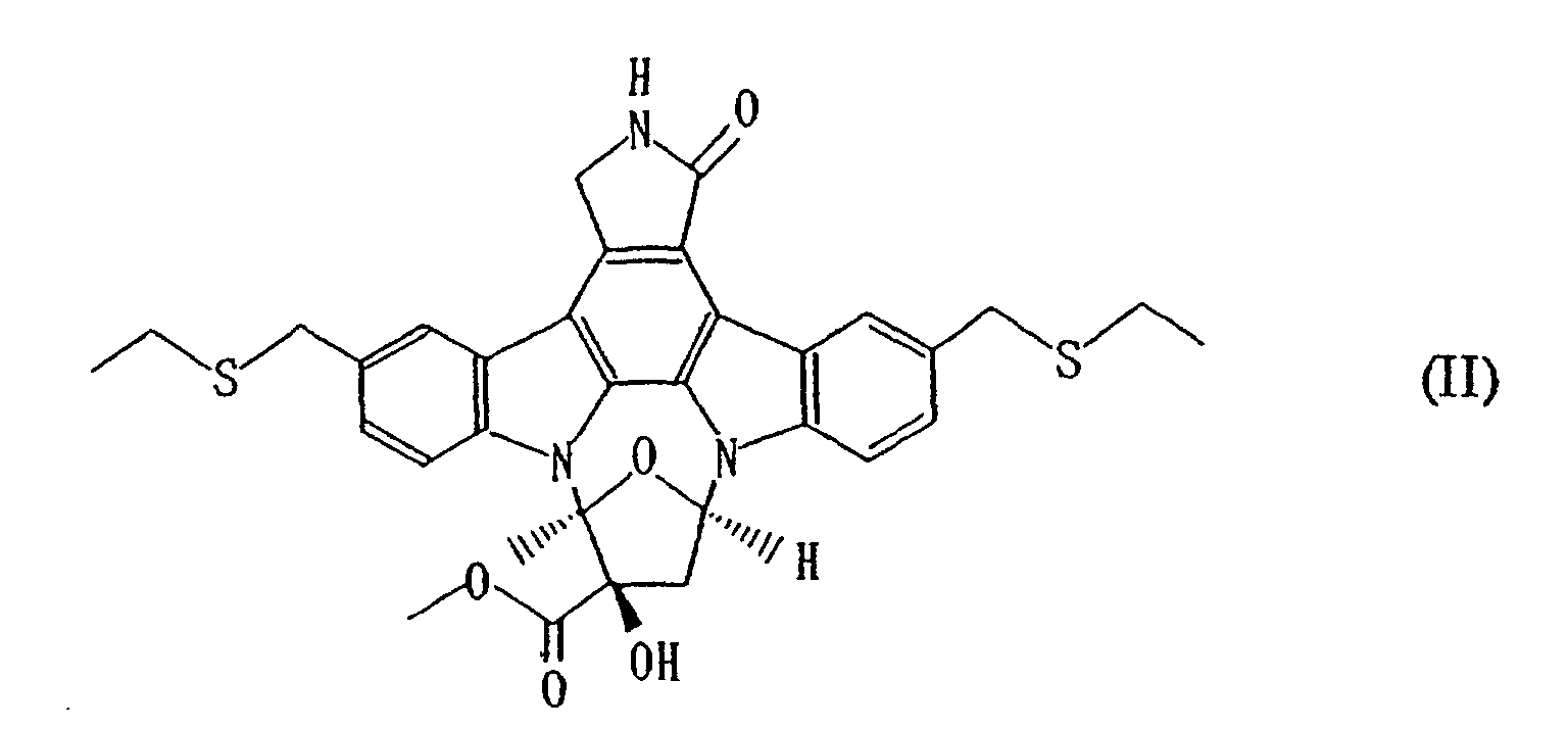
As a preferable example of the compound, a compound in
which both R1 and R2 represent an ethyl group and both R3 and R4
represent a methyl group can be mentioned (it is referred to as Compoud
A hereinafter), with the specified structure of the most preferable
example represented by the following formula (II).
Whereas the detailed effects of the compounds on retinal
neurocytes will be explained in the section of Pharmacological Tests
described hereinafter, it was confirmed that the compounds suppress
the decline in the number of the cells in the layer of retinal ganglyocyte
when the above-descrived effects were examined using the
retina subjected to an ischemia - reperfusion treatment and the eyes
treated with kainic acid.
The route of administration of the compounds can be
either parenteral or oral. As dosage forms for parenteral administration,
eye drop, injection, nose drop and the like can be mentioned
whereas as for oral administration, tablet, capsule, powder and the
like can be mentioned, all of which can be formulated using vehicles or
additives as well as techniques routinely used in the art. For example,
in the case of an eye drop, as the vehicles or additives, isotonizing
agents such as sodium chloride, concentrated glycerin and the like,
buffering agents such as sodium phosphate, sodium acetate and the
like, surfactants such as polyoxyethylene sorbitan mono-oleate (it is
referred to as Polysorbate 80 hereinafter), polyoxyl stearate 40,
polyoxyethylene hydrogenated castor oil and the like, stabilization
agents such as sodium citrate, sodium edetate and the like, as well as
preservatives such as benzalkonium chloride, parabens and the like
can be used when required to formulate the composition, and pH of
the formulation should be within the range acceptable to ophthalmologic
formulations with the range of pH 4 - 8 being preferable.
The dose can be suitably selected depending on the conditions
and age of the patient, dosage form and the like, and in the case
of eye drops, it is appropriate to administer an eye drop of 0.01 - 10
% (w / v) once or a couple of times a day and in the case of injections,
it is usually appropriate to administer 0.0001 - 1 mg of the compound
once a day or in divided doses. Moreover, in the case of oral drugs, it
may be usually appropriate to administer 10 µg - 1 g of the compound
once a day or in divided doses.
The results from the Pharmacological Tests are illustrated
hereinafter, but it should be understood that these examples are
intended for better understanding of the invention and are not to
limit the extent of the invention.
Examples
Pharmacological Tests:
In order to study usefulness of the compounds, the effects
of the compounds on the retina subjected to the ischemia - reperfusion
treatment and on the retina from the eyes treated with kainic
acid were examined.
1. Effects on the retina subjected to the ischemia - reperfusion
treatment
It has been reported that brain-derived neurotrophic
factor (it is referred to as BDNF hereinafter) which is a neurotrophic
factor has a protective effect against gangliocytic death caused by
retinal ischemia (Invest. Ophthalmol. Vis. Sci. 35, 907 - 915 (1994)).
Thus, according to the method described in this reference,
the effects of the compounds on retinal gangliocyte in a retinal ischemia
- reperfusion model were studied.
(Method of Experiment)
Under anesthesia with a mixture of Ketalar and Celactal,
the conjunctiva of a male Spraugue - Dawley rat (300 - 450 g) was
dissected to expose the optic nerve and then the retina was subjected
to an ischemic state by ligating the optic nerve with a suture. Sixty
minutes after the ischemia, the suture was taken out to reperfuse the
retina with the blood by allowing the circulation. Seven days after the
reperfusion, the eye bulb was enucleated and then immobilized with
paraformaldehyde, and after embedding it in paraffin, the transverse
paraffin sections of the eye bulb with 3 µm of the thickness were
prepared by slicing the eye bulb beginning from the point of exposure
of the optic nerve with 60 µm of each interval. These sections were
stained with hematoxylin - eosin and photographs were taken for the
section in which the retinal part at the distance of approximately 1
mm from the optic disc to either direction, left or right, is included to
measure the number of the cells contained in the layer of gangliocyte.
Here, the compound was dissolved into polyethylene glycol
660 hydroxystearate and injected in the volume of 1 µl into the
hyaloid body two day before the creation of the ischemic state.
(Results)
In Table 1, as one example of the experimental results, the
number of the cells in the layer of gangliocyte in the case in which a
solution of Compound A prepared at the concentration of 3 mg / ml
was injected (the amount injected: 3 µg / eye) is shown. Also, the
results from the experiments in which neither of the ischemia-reperfusion
treatment nor the injection of the compound was conducted
(it is referred to as Normal Group (1) hereinafter) as well as
those in which the ischemia - reperfusion treatment was conducted
and then only polyethylene glycol 660 hydroxystearate without the
compound was injected (it is referred to as Control Group (1) hereinafter)
are shown.
| | Number of the cells in the layer of gangliocyte (cells/mm) |
| Normal Group (1) (5) | 38.8 |
| Control Group (1) (7) | 20.5 |
| Group with the injection of Compound A (3 µg/eye) (10) | 27.5 |
| Note: The figures in the table show the averages for the cases studied, the numbers of which are indicated within the right parentheses. |
As shown in Table 1, when 3 µg of compound A was injected,
it was confirmed that the compound suppressed the decline in
the number of the cells in the layer of gangliocyte caused by the ischemia
- reperfusion treatment and the number of the cells was recovered
to the level of 70.9 % of that for Normal Group (1).
From this fact, Compound A was proved to have a superior
protective effect on neurocyte disturbance of the retina caused by the
ischemia - reperfusion treatment.
2. Effects on the retina from the eye treated with kainic acid
It has been reported that when kainic acid, that is a neurotoxin,
is injected into the hyaloid body, the inner plexiform layer
becomes thinner because of synaptic collapse caused by cell death of
amacrine cell or the number of the cells contained in the layer of
gangliocyte decreases because of apoptosis happening to those cells
(Neurochem. Int., 31, 251 - 260 (1997)).
Then, according to the method described in this reference,
the effects of the compound on the retinal disturbance induced by
injecting kainic acid into the hyaloid body were studied.
(Method of Experiment)
To a male Sprague - Dawley rat (180 - 250 g) under anesthesia
with pentobarbital, a solution of kainic acid in polyethylene
glycol 660 hydroxystearate (1 mmol/L) was injected into the hyaloid
bodies in both of the eyes in the amount of 5 µl under observation
with a stereoscopic microscopy. Seven days after the kainic acid
treatment, the eye bulbs were enucleated, and immobilized with
glutaraldehyde - paraformaldehyde, and after embedding them in
paraffin, the transverse paraffin sections of the eye bulbs with 3 µm
of the thickness were prepared by slicing the eye bulbs beginning
from the point of exposure of the optic nerve with 60 µm of each
interval. These sections were stained with hematoxylin - eosin and
photographs were taken for the sections in which the retinal part at
the distance of approximately 1 mm from the optic disc to either
direction, left or right, is included to measure the thickness of the
inner plexiform layer and the number of the cells contained in the
layer of gangliocyte.
The compound was dissolved into polyethylene glycol 660
hydroxystearate and then injected into the hyaloid bodies two days
before, at the same time as and one day after the kainic acid treatment
in the volume of 5 µl each.
(Results)
In Table 2, as examples of the results from the experiments,
the thickness of the inner plexiform layer and the numbers of
the cells in the layer of gangliocyte for the cases in which a solution of
Compound A prepared at the concentration of 6 µg / ml (the amount
injected: 30 ng
/ eye) and of 600 µg / ml (the amount injected: 3 µg /
eye) were injected are shown. Also, the results from the experiments
in which neither the treatment with kainic acid nor the injection of
the compound was conducted, but only the injection of polyethylene
glycol 660 hydroxystearate was conducted (it is referred to as Normal
Group (2) hereinafter), as well as those in which the treatment with
kainic acid was conducted, and only polyethylene glycol 660
hydroxystearate without the compound was injected (it is referred to
as Control Group (2) hereinafter) are shown.
| | Thickness of the inner plexiform layer (µm) | Number of the cells in the layer of gangliocyte (cells/mm) |
| Normal Group (2) | 47.5 | 64.2 |
| Control Group (2) | 20.8 | 36.8 |
| Group with the injection of Compound A (30 ng/eye) | 23.8 | 42.3 |
| Group with the injection of Compound A (3 µg/eye) | 28.8 | 51.8 |
| Note: The figures contained in the table show the averages of the five eyes studied. |
As shown in Table 2, when 3 µg of Compound A was injected,
thinning propensity of the inner plexiform layer and the
decline in the number of the cells in the layer of gangliocyte caused by
kainic acid were suppressed. Especially, for the number of the cells
in the layer of gangliocyte, the cell number was confirmed to be
recovered to the level of 80.7% of that for Normal group (2).
From this fact, Compound A was proved to have a superior
protective effect against neurocyte disturbance of the retina caused
by the kainic acid treatment.
Industrial Applicability
The compound was found to protect the retina against
neurocyte disturbance of the retina subjected to the ischemia-reperfusion
treatment and the kainic acid treatment as well as be
effective as a therapeutic agent for retinal diseases represented by
retinal vascular occlusion, diabetic retinopathy, ischemic optic neuropathy,
macular degeneration, pigmentary retinal dystrophy, Leber's
disease as well as other eye diseases such as glaucoma and the like.
Therefore, the invention has an applicability in drug
manufacturing industry or health care industry.


 R1 represents a lower alkyl group,R2 represents a lower alkyl group,R3 represents a hydrogen atom or a lower alkyl group, andR4 represents a hydrogen atom or a lower alkyl group, as the active ingredient.