BACKGROUND OF THE INVENTION
This invention relates to the field of nucleic acid sequence detection. The
invention relates to a method of detecting any type of mutation at a predetermined nucleic
acid base site of interest. The present invention is directed to a method called shifted
termination analysis, also known as specific termination assay, or shifted terminator
alignment which can all be abbreviated as STA.
Practical applications for the inventive method includes genetic disease diagnoses,
infectious disease diagnoses, forensic techniques, paternity determinations, and genome
mapping, wherein the site of the mutation to be detected is known.
In the past decade, genes implicated in inherited susceptibility to form cancer
have been identified and many cancer-related mutations were characterized. Diagnostic
tests for these mutations can provide a more accurate estimate of an individual's risk of
developing cancer. Early diagnosis of a cancer related mutation is one of the goals of the
invention.
There are four major types of gene mutations. The first is point mutations caused
by a single nucleotide substitution in a normal DNA sequence. In most cases, this
mutation causes a frame shift in the coding strand which results in termination of normal
protein synthesis. A point mutation in APC gene as found in family adenopolyposis
(FAP) patients is a typical example (Kinzler et al., Science 253, 661-665 (1991); Joslyn
et al., Cell 66, 601-613 (1991); Nishisho et al., Science 253, 665-669 (1991)). The
second is insertion mutation in which a single or multiple nucleotides are inserted into a
normal DNA sequence. The third is deletion mutation, wherein a single nucleotide or
multiple nucleotides are deleted from a normal DNA sequence. Both insertion and
deletion types of mutations can cause severe changes such as frame shift, early
termination of protein synthesis, and addition or deletion of one or multiple amino acids.
The fourth is gene translocation, which occurs when a fragment of a gene is incorporated
into another gene. The Philadelphia chromosome seen in chronic myeloid leukemia
patients is an example of this phenomenon (Konopka et al., Cell 37 1035 (1984)).
Changes in protein structure cause a series of disorders in a cell that can lead to the onset
of cancer.
Detection of one mutated DNA among thousands of normal DNA is difficult.
Chemical or enzymatic DNA sequencing method to directly read the DNA sequence of
an isolated species, has been used as the most thorough method in research laboratory in
analyzing and identifying gene mutations. However, clinical application of such
sequencing method is impractical as it is limited by the level of professional skill that is
required to perform the assays, its labor intensiveness, high cost associated with
procurement of the apparati and reagents in carrying out the sequencing reactions, as well
as the long duration required to complete the project. Finally, another disadvantage of the
sequencing method is that the procedure requires a large amount of DNA template, which
is difficult to obtain from a 10 ml blood specimen that is usually collected from a patient.
Examples of conventional mutation detection techniques include: restriction
fragment length polymorphism (RFLP) (Botstein et al., Am. J. Hum. Genet., 32, 314-331
(1980), and White et al., Scientific American, 258:40-48 (1988)); single-strand
conformational polymorphism (SSCP) (Howell et al., Am. J. Hum. Genet., 55, 203-206
(1994)); allele-specific oligonucleotide hybridization. (Studencki et al., Am. J. Hum.
Genet., 37, 42-51 (1985), and Saiki et al., Nature, 324, 163-166); oligonucleotide-ligation
assay (Landgrun et al., Science, 241, 1077-1080 (1990)); and allele-specific PCR
(ASPCR) (Wu et al., Proc. Natl. Acad.Sci., 86, 2757-2760 (1989), and Okayama et al., J.
Lab. Clin. Med. 114, 105-113 (1989)).
Some of these techniques are suitable for detecting only point mutations. Some of
the other techniques can be used to detect only insertions or deletions that may for
example, destroy or build up restriction enzyme cleavage sites, but are not suitable for
detecting single base mutations. For example, point mutations that do not affect the
enzyme cleavage site are missed by such methods as RFLP. Other techniques require
optimization of a special probe hybridization condition. In addition, all of the above
mentioned techniques require special laboratory equipment such as gel electrophoresis
apparatus and hybridization equipment, time and labor.
Some primer extension based methods for detecting mutations are also known
(Mohan et al., Proc. Natl. Acad. Sci. USA, 88, 1143-1147 (1991), Prezant et al., Hum.
Mutation 1, 159-164 (1992), Fahy et al., Nucleic Acid Research, 25, 3102-3109 (1997),
and U. S. Patent Nos. 5,846,710 (1998) and 5,888,819 (1998)). Such methods include:
primer extension with a thionucleotide; primer extension from oligonucleotide primer
flanking the mutated nucleotide with labeled nucleotide complementary to the mutated
nucleotide base; and primer extension with labeled dideoxynucleotide terminator
complementary to the mutant base.
These primer extension based mutation detection methods are fast, facile to
perform, and can be potentially applied to clinical use. However, there are at least two
weaknesses with these methods. First, all of these techniques are based on incorporating
only one labeled-nucleotide in the primer extension strand. Incorporating only one type
of labeled-nucleotide chosen from A, C, G, T or U , or labeled-dideoxynucleotide permits
the detection of only the specific point mutation that is specific to the nucleotide base that
is complementary to the labeled nucleotide that is used in the assay.
Hypothetically, when a different type or nature of mutation occurs at the same
position, for example, if A is changed to C or GT, or TCT, with innumerable other
permutations, these known methods require that at least three separate tests be conducted
with labeled G, C, A. Alternatively, one test assay can be carried out that uses
differentially labeled nucleotide combined with gel analysis and special marker detection
system to detect the G, C, A mutants separately. However, carrying out three separate
tests requires more than three times the blood sample that is generally obtained from
patients. Such a high volume of blood sample or complicated gel analysis procedure not
only increases the cost of the test, and the time and labor involved, but more
importantly, the probability of error is increased because of chances of mislabeling of
tubes and the numerous steps that are required to carry out these assays. Therefore, these
primer extension based methods are not convenient or suitable for screening a large
sample number or for carrying out routine tests at a clinic.
Second, the sensitivity of these primer extension based assays need improvement.
Because the primer extended strand obtained in these tests carries only one labeled
nucleotide or labeled dideoxynucleotide, the signals generated are varied and their
strength depends on what kind of chemical label was used. But in general, the signal is
weak.
Thus, there is a need in the mutation detection field for a rapid, low-cost, non-labor
intensive, and clinically applicable technique that is able to detect any type of
mutation occurring at a nucleic acid base at a specific predetermined position, and yet
provides a strong and accurate detection signal.
SUMMARY OF THE INVENTION
The present invention has met the herein before described need.
Even though the present invention shares some of the advantageous features
associated with general primer extension based methods, such as the simplicity in design
for testing for a mutation at a particular site, the present invention provides a method that
overcomes the drawbacks associated with primer extension based methods as described
above. The invention has wide applicability for detecting and identifying all types of
mutations. It is cost-effective, timesaving, and less labor intensive than conventional
methods.
Some of the key advantages of the invention over the above described methods
are: 1) capability of detecting all types of mutations in only one reaction tube without
necessarily employing gel electrophoretic size separation methods; 2) high degree of
detection sensitivity by way of strong signal emitted due to incorporation of multiple
labeled-nucleotides into the primer extension strand; and 3) high degree of accuracy
because two or three different types of nucleotide or nucleotide analogue markers can be
inserted into the primer extension strand at same time. These advantageous features
provide an opportunity to use the invention to routinely test for the presence of a genetic
mutation in any clinic based on this simple inventive procedure. The invention is also
easily adaptable to automation for screening a large number of samples.
The invention relates to a method for detecting any mutation occurring at a
predetermined nucleotide (target base) in a known nucleic acid sequence in a single
reaction. The inventive method uses a primer extension analysis to detect the mutation.
Preferably, the primer is complementary to and sequence-specifically hybridizes with the
nucleic acid of interest at the position immediately adjacent to the predetermined
nucleotide base to form a duplex, so that the target base in the nucleic acid of interest is
an unpaired base immediately downstream of the 3' end of the primer. The primer
extension reaction reagent includes one type of unlabeled terminator nucleotide (or
optionally, no corresponding nucleotide base) along with three types of labeled (or
optionally, differentially labeled or unlabeled) non-terminator nucleotides, wherein the
terminator nucleotide is complementary to the target base at the predetermined position
of the nucleic acid of interest. The labeled non-terminator nucleotides are not
complementary to the target base. The incorporation of the terminator nucleotide into the
3' end of the primer complementary to the target base in the nucleic acid of interest will
terminate the primer extension reaction without further incorporation of any labeled non-terminator
nucleotides. If the target base was changed due to a mutation, a labeled non-terminator
sequence-dependently incorporates into the primer. Thus, any labeled signal
detected in the primer indicates that a mutation has occurred at the predetermined nucleic
acid base site.
An object of the invention is to provide a method for detecting or quantifying a
target nucleic acid in a sample comprising:
In a preferred embodiment, in step (b), the nucleic acid base of interest is
immediately adjacent to the nucleotide base to be identified at the predetermined
position, and the nucleotide base to be identified is an unpaired base at a predetermined
position immediately downstream of the 3' end of the duplex. In a preferred
embodiment, in the method in step (d), the duplex from step (c) is contacted with at least
one labeled non-terminator, and at least one unlabeled terminator. Alternatively, in step
(d), the duplex from step (c) is contacted with non-terminators, wherein each non-terminator
is labeled with same or different detectable label.
In another preferred embodiment, the method above can be practiced, wherein the
template-dependent enzyme is E. coli DNA polymerase I or the "Klenow fragment"
thereof, T4 DNA polymerase, T7 DNA polymerase T. aquaticus DNA polymerase, a
retroviral reverse transcriptase, or combinations thereof.
In other preferred embodiments, the nucleic acid of the invention is a
deoxyribonucleic acid, a ribonucleic acid, or a copolymer of deoxyribonucleic acid and
ribonucleic acid. The primer is an oligodeoxyribonucleotide, an oligoribonucleotide, or a
copolymer of deoxyribonucleic acid and ribonucleic acid. The template is a
deoxyribonucleic acid, the primer is an oligodeoxyribonucleotide, oligoribonucleotide, or
a copolymer of deoxyribonucleotides and ribonucleotides, and the template-dependent
enzyme is a DNA polymerase. The template is preferably a ribonucleic acid, the primer is
an oligodeoxyribonucleotide, oligoribonucleotide, or a copolymer of
deoxyribonucleotides and ribonucleotides, and the template-dependent enzyme is a
reverse transcriptase. Preferably, the template is a deoxyribonucleic acid, the primer is an
oligoribonucleotide, and the enzyme is an RNA polymerase. Preferably, the template is a
ribonucleic acid, the primer is an oligoribonucleotide, and the template-dependent
enzyme is an RNA replicase.
In the above method, in step (d), the duplex from step (c) is contacted with at
least one labeled non-terminator, and at least one terminator that is labeled differently
from the non-terminator. In addition, in step (e), the label signal of the incorporated
labeled non-terminator and at least one terminator that is labeled differently from the
non-terminator are detected.
According to the method of the invention, the nucleic acid of interest has been
synthesized enzymatically in vivo, synthesized enzymatically in vitro, or synthesized non-enzymatically.
In another embodiment of the method of the invention, the
oligonucleotide primer has been synthesized enzymatically in vivo, synthesized
enzymatically in vitro, or synthesized non-enzymatically. In addition, the oligonucleotide
primer can comprise one or more moieties that permit affinity separation of the primer
from the unincorporated reagent and/or the nucleic acid of interest.
In particular, as a preferred embodiment, the oligonucleotide primer comprises
biotin which permits affinity separation of the primer from the unincorporated reagent
and/or nucleic acid of interest via binding of the biotin to streptavidin which is attached
to a solid support. In another embodiment of the invention, the sequence of the
oligonucleotide primer comprises a DNA sequence that permits affinity separation of the
primer from the unincorporated reagent and/or the nucleic acid of interest via base
pairing to a complementary sequence present in a nucleic acid attached to a solid support.
In yet another embodiment of the invention, the nucleic acid of interest comprises one or
more moieties that permit affinity separation of the nucleic acid of interest from the
unincorporated reagent and/or the primer. The nucleic acid of interest can comprise biotin
which permits affinity separation of the nucleic acid of interest from the unincorporated
reagent and/or the primer via binding of the biotin to streptavidin which is attached to a
solid support.
In a method of the invention, the sequence of the nucleic acid of interest
comprises a DNA sequence that permits affinity separation of the nucleic acid of interest
from the unincorporated reagent and/or the primer via base pairing to a complementary
sequence present in a nucleic acid attached to a solid support. The oligonucleotide primer
can be labeled with a detectable marker. The oligonucleotide primer can be labeled with a
detectable marker that is different from any detectable marker present in the reagent or
attached to the nucleic acid of interest. The nucleic acid of interest can be labeled with a
detectable marker. The nucleic acid of interest is preferably labeled with a detectable
marker that is different from any detectable marker present in the reagent or attached to
the primer.
In another embodiment of the invention, the nucleic acid of interest comprises
non-natural nucleotide analogs. The non-natural nucleotide analogs comprise
deoxyinosine or 7-deaza-2'-deoxyguanosine. The nucleic acid of interest can be
synthesized by the polymerase chain reaction.
In another method of the invention, the sample comprises genomic DNA from an
organism, RNA transcripts thereof, or cDNA prepared from RNA transcripts thereof. The
sample can comprise extragenomic DNA from an organism, RNA transcripts thereof, or
cDNA prepared from RNA transcripts thereof. In the method of the invention the primer
can be preferably separated from the nucleic acid of interest after the primer extension
reaction in step (d) above by using appropriate denaturing conditions. Preferably, the
denaturing conditions comprise heat, alkali, formamide, urea, glyoxal, enzymes, and
combinations thereof. Even more preferably, the denaturing conditions comprise
treatment with 0.2N NaOH.
The method of the invention can be practiced using nucleic acid from any
organism, including plant, microorganism, virus, or bird. The organism can be a
vertebrate or invertebrate. The organism is preferably a mammal. Even more preferably,
the mammal is a human being. The mammal can be also a horse, dog, cow, cat, pig, or
sheep.
These and other objects of the invention will be more fully understood from the
following description of the invention, the referenced drawings attached hereto and the
claims appended hereto.
BRIEF DESCRIPTION OF THE DRAWINGS
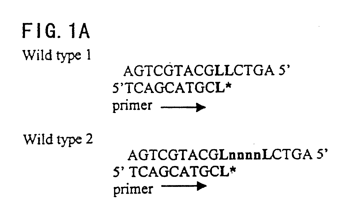
FIGS. 1A-1C. A schematic drawing of a preferred embodiment of the mutation detection
method of the invention is shown. "L" represents the wild-type nucleotide, which can
include A, G, C, T, or U. "L*" represents an unlabeled terminator such as a dideoxy
nucleotide that is complementary to L. "M" represents a mutation at site L, and the
mutant nucleotide can include A, G, C, T, or U. "W" represents a complementary
nucleotide to M, and can include A, G, C, T, or U labeled with a detectable marker. "n"
represents one or multiple nucleotides or nucleotide analogues, including A, G, C, T, and
U. "y" represents a nucleotide or nucleotide analogue, including A, G, C, T, or U, labeled
with a detectable marker and complementary to M or n.
FIG. 2.1 µl of the STA reaction mixture was applied to a thin layer chromatography strip
and then the strip was subjected to solvent containing 1 M NaCl and 1 M HCl. The strip
was then dried at room temperature for 10 min and exposed to Kodak film for 30
minutes, and the film was developed by auto-film developer. The templates used STA
test are marked under each strip. The top arrow indicates the free nucleotide front and the
arrow at the bottom indicates the primer extended strand incorporated with [-32P]
dCTP.
DETAILED DESCRIPTION OF THE INVENTION
As used herein, "nucleic acid" or "nucleotide" can be a deoxyribonucleic acid, a
ribonucleic acid, or a copolymer of deoxyribonucleic acid and ribonucleic acid. The
sample of nucleic acids can be natural or synthetic. The sample of nucleic acid can be
naturally occurring nucleic acid, and can be obtained from any organism. Some examples
of organisms to which the method of the present invention is applicable include plants,
microorganisms, viruses, birds, vertebrates, invertebrates, mammals, human beings,
horses, dogs, cows, cats, pigs, or sheep.The target nucleic acid can occur naturally, or can
be synthesized enzymatically in vivo, synthesized enzymatically in vitro, or synthesized
non-enzymatically.
The sample containing the nucleic acid or acids of interest can comprise genomic
DNA from an organism, RNA transcripts thereof, or cDNA prepared from RNA
transcripts thereof. The sample containing the nucleic acid or acids of interest can also
comprise extragenomic DNA from an organism, RNA transcripts thereof, or cDNA
prepared from RNA transcripts thereof. Also, the nucleic acid or acids of interest can be
synthesized by the polymerase chain reaction.
The nucleic acid of interest can comprise non-natural nucleotide analogs such as
deoxyinosine or 7-deaza-2-deoxyguanosine. These analogues destabilize DNA duplexes
and could allow a primer annealing and extension reaction to occur in a double-stranded
sample without completely separating the strands.
The nucleic acid of interest can comprise one or more moieties that permit affinity
separation of the nucleic acid of interest from the unincorporated reagent and/or the
primer. For example, the nucleic acid of interest can comprise biotin which permits
affinity separation of the nucleic acid of interest from the unincorporated reagent and/or
the primer via binding of the biotin to avidin and its analogue which is attached to a solid
support. The sequence of the nucleic acid of interest can comprise a DNA sequence that
permits affinity separation of the nucleic acid of interest from the unincorporated reagent
and/or the primer via base pairing to a complementary sequence present in a nucleic acid
attached to a solid support. The nucleic acid of interest can be labeled with a detectable
marker; this detectable marker can be different from any detectable marker present in the
reagent or attached to the primer.
In this regard, the term "normal nucleotide" or "normal base" is defined as the
wild-type or previously known standard nucleotide base from which a mutation is sought
to be identified at the base site. By "standard nucleotide base", it includes any known
base, which may include wild-type or a known mutant base so long as the base is known
and it is desired to know its variant. Thus, as an example, normal base can be a known
wild-type base for which a mutation is sought at the position. Inversely, the known base
can be a known mutant for which the presence of a wild-type base is sought at the
position. Alternatively, the known normal base can be a known mutant for which another
mutant variant base is sought. Therefore, the method of the invention can be applied to
any known sequence that can be used to determine the presence of any other base variant
at the site.
As used herein, the term "primer" or "oligonucleotide primer" refers to an
oligonucleotide which is capable of acting as a point of initiation of synthesis when
placed under conditions that allow for synthesis of a primer extension product which is
complementary to a nucleic acid (template) strand, in the presence of various factors such
as for example, nucleotides and enzymes such as DNA polymerase, and at a suitable
temperature and pH.
The term "primer" is alternatively defined as any nucleic acid fragment obtained
from any source. For example, the primer can be produced by fragmenting larger nucleic
acid fragments such as genomic DNA, cDNA or DNA that has been obtained through
PCR. In other words, the nature of the primer is not limited by how the primer is
obtained, whether it be by fragmenting naturally or synthetically occurring nucleic acid
or by synthesizing the nucleic acid primer. Furthermore, the primer can be
oligodeoxyribonucleotide, a copolymer of oligodeoxyribonucleotides, an
oligoribonucleotides, a copolymer of ribonucleotides, or a copolymer of
deoxyribonucleotides and ribonucleotides. The primer can be either natural or synthetic.
The oligonucleotide primer can be synthesized either enzymatically in vivo,
enzymatically in vitro, or non-enzymatically in vitro. The primer can be labeled with a
detectable marker; this detectable marker can be different from any detectable marker
present in the reagent or attached to the nucleic acid of interest. In addition, the primer
must possess sequence corresponding to the flanking sequence at a specific position of
interest adjacent to, and upstream of, the nucleotide base to be identified.
In addition, the primer must be capable of hybridizing or annealing with
nucleotides present in the nucleic acid of interest. One way to accomplish the desired
hybridization is to have the template-dependent primer be substantially complementary or
fully complementary to the known base sequence.
The oligonucleotide primer can comprise one or more moieties that permit
affinity separation of the primer from the unincorporated reagent and/or the nucleic acid
of interest. Such affinity moeties include, but are not limited to, digitonin, magnetic
beads, and ligands, such as protein ligands, including antibodies. Preferably, the moiety is
biotin. In the case of using biotin, the primer comprising biotin permits affinity separation
of the primer from the unincorporated reagent and/or nucleic acid of interest via binding
of the biotin to avidin and its analogue which is attached to a solid support. The sequence
of the oligonucleotide primer can comprise a DNA sequence that permits affinity
separation of the primer from the unincorporated reagent and/or the nucleic acid of
interest via base pairing to a complementary sequence present in a nucleic acid attached
to a solid support.
As used herein, the term "primer extension reaction" refers to the reaction
conditions in which the template-dependent nucleic acid synthesis reaction is carried out.
The conditions for the occurrence of the template-dependent, primer extension reaction
can be created, in part, by the presence of a suitable template-dependent enzyme. Some
of the suitable template-dependent enzymes are DNA polymerases. The DNA
polymerase can be of several types. The DNA polymerase must, however, be primer and
template dependent. For example, E. coli DNA polymerase I or the "Klenow fragment"
thereof, T4 DNA polymerase, T7 DNA polymerase ("Sequenase"), T. aquaticus DNA
polymerase, or a retroviral reverse transcriptase can be used. RNA polymerases such as
T3 or T7 RNA polymerase could also be used in some protocols. Depending upon the
polymerase, different conditions must be used, and different temperature ranges may be
required for the hybridization and extension reactions.
As used herein, the term "primer extension strand" includes the strand that is
formed opposite the template in a duplex after the primer has been added. Preferably, the
extension of the primer has terminated by the binding of the terminator to the template.
As used herein, the term "template" is defined as a nucleic acid, including double
strand DNA, single strand DNA and RNA, or any modification thereof, and can be any
length or sequence.
As used herein, the term "terminator" or "chain terminator" is meant to refer to a
nucleic acid base, such as A, G, C, T or U, or an analogue that effectively terminates the
primer extension reaction when it is incorporated into the primer extension strand
opposite the template strand. Preferably, the terminator is a dideoxynucleotide. Also
preferably, the terminator is either unlabeled or is labeled so that it is distinguished from
the label on the non-terminator. Also as used herein, when the term "terminator" or
"chain terminator" are referred to in the singular, it does not mean that a single nucleotide
molecule is used. Rather, the singular form of the term "terminator" refers to the type of
nucleotide, nucleic acid base or nucleic acid analogue that is used in the assay. For
example, if the terminator is ddA, then all of the ddA's in the aggregate are referred to in
the singular form, and not just a single molecule of ddA. Alternatively, the "terminator"
may be the absence of the specific type of nucleotide so that primer extension is stopped
by the lack of the specific nucleotide at the locus. For example, if it is desired that the
primer extension reaction be stopped opposite a "C" on the template strand, the non-terminating
bases A, T and G should be included in the primer extension reaction
mixture, but not "G", which is the complement of "C". Thus, the absence of the
complementary base will cause termination of the primer extension reaction with a
similar result as adding a dideoxy terminator nucleotide, for example.
As used herein, the term "non-terminator" or "non-chain terminator" includes a
nucleotide base that does not terminate the extension reaction when it is incorporated
into the primer extension strand. Preferably, at least one non-terminator in the primer
extension reaction is labeled. Also as used herein, when the term "non-terminator" or
"non-chain terminator"are referred to in the singular, it does not mean that a single
nucleotide molecule is used. Rather, the singular form of the term "non-terminator" refers
to the type of nucleotide, nucleic acid base or nucleic acid analogue that is used in the
assay. For example, if the terminator is G, then all of the G's in the aggregate are referred
to in the singular form, and not just a single molecule of G.
As used herein, the term "mutant" or "mutation" indicates any base on the
template strand that is different from the wild-type or normal base. The mutation that can
be detected using the method of the instant invention can be any type of mutation at all,
including, single base mutation, insertion, deletion, or gene translocation, so long as the
base on the template directly opposite to the base immediately 3' to the annealed primer is
affected.
As used herein, the term "label" refers to any molecule that is linked to the
terminator or non-terminator nucleotide to provide a detectable signal. The label may be
radioactive, chemiluminescent, protein ligand such as an antibody, or if a fluorescent
group is used, a different fluorescent group may be used for each type of non-terminating
nucleotide base. These fluorescent tags would have the property of having
spectroscopically distinguishable emission spectra.
Alternatively, the method of determining the level of incorporation of a nucleotide
base in the primer extension product can be measured by mass spectrometry techniques
as exemplified in U.S. Patent No. 5,885,775, which is incorporated herein by reference in
its entirety.
As used herein, the phrase "high stringency hybridization conditions" refers to
nucleic hybridization conditions, such as but not limited to a wash condition of 0.1XSSC,
at 42°C. Hybridization conditions generally can be found in general Molecular Biology
protocol books, such as Ausubel et al., Current Protocols in Molecular Biology Greene and
Wiley, pub. (1994), which is incorporated herein by reference in its entirety.
As used herein, "thin layer chromatography (TLC)" can be carried out in paper
medium based on cellulose products, but can be made of any substance that allows for
molecules to be finely divided and formed into a uniform layer. This substance includes, but
is not limited to, inorganic substances such as silica gel, aluminum oxide, diatomaceous
earth or magnesium silicate. Organic substances include, but are not limited to, cellulose,
polyamide, or polyethylene powder. Thin layer chromatography methods are described
generally in Chemical protocol books, such as generally set forth in Freifelder, Physical
Biochemistry - Applications to Biochemistry and Molecular Biology, second ed., published
by Freeman and Co. (1982), which is incorporated herein by reference in its entirety,
especially Chapter 8, which discusses chromatographic techniques, and in particular thin
layer chromatography at pages 229-232.
It can be appreciated by a person of skill in the art that the terminator can be
labeled with a different label from the non-terminator, which can then be used to
differentiate between incorporation of terminator or non-terminator in the primer
extension strand. The terminator exemplified as being the absence of the particular type
of nucleotide in the present application only for purposes of simplicity of illustration, but
this illustration should not be construed to limit the claims in any way. Differentially
labeled or unlabeled terminator is also encompassed by the invention, so long as the label
on the terminator is different from the label on the non-terminator.
It can also be appreciated by a person of skill in the art that so long as the
sequence of the template is at least partially known, a primer can be designed that binds
to the template strand such that the binding of the primer on the template strand can
occur. It can also be appreciated by a person of skill in the art that the method of the
invention can be practiced by using several primers in one or more assay tube.
A feature of the method of the invention is that strong signal can be generated if
the non-terminators are uniformly labeled because of the additive signal effect achieved
by the incorporation of several labeled non-terminators incorporated in the primer
extension strand when there is a mutation at the predetermined site. This generates an
advantageous signal strength over conventional mutation detection methods that
incorporate only a single label per each primer extension strand. Accuracy is enhanced
when signals are observed from using different labels specific to various terminators or
non-terminators.
The following examples are offered by way of illustration of the present
invention, and not by way of limitation.
EXAMPLES
Example 1
Sequence of human APC gene was selected as a target sequence for the STA test
of the invention. Oligonucleotides corresponding to the wild-type APC sequence 4317-4347
and three different types of mutations were synthesized and used as templates. The
primers employed in the STA test are listed in Table 1.
STA: Each STA reaction was performed in 20 µl buffer (10 mM Tris-HCl, pH7.5,
50 mM KCl, and 5 mM MgCl
2) containing 50ng of template oligonucleotide, 1 µM
primer, 2 units of DNA Polymerase, 1 µl of [α-
32P]-labeled dCTP (250 µCi/ml, 3000
Ci/mmol Dupont-New England Nuclear), dATP, dTTP, and 1µl of non-labeled dd GTP.
The mixture was incubated at 37 °C for 30 minutes and heated at 100 °C for 3 minute. 1 µl
of the reaction mixture was applied to a TI strip, which is a strip of thin layer
chromatography (TRIM USA, MD). The strips were extended for 10 minutes with
solution containing 1M HCl and 1M NaCl. The primers were completely separated from
unincorporated nucleotide on the TI strip by this procedure. The labeled primer was
.visualized by autoradiography and the radioactivity was counted by scintillation counter
(Beckman LS 5000). The autoradiogram is shown in Fig. 2, and then counting the results
for the corresponding autoradiogram is shown in Table 2.
| Template | Total Counts | Labeled Primer |
| No template | 76,675 | 95 |
| Oapc-w | 79,599 | 117 |
| Oapc-p | 82,584 | 4,821 |
| Oapc-i | 75,376 | 8,602 |
| Oapc-d | 100,634 | 6,571 |
The counts shown in Table 2, right column represents the amount of [α-32P]-dCTP
incorporated into the primer extension Oapc-w, a wild type oligonucleotide, is
serves as the template. The first unpaired nucleotide after primer annealing is C, which is
complementarily matched by the terminator ddG. When the reaction of template-dependent
primer extension was started, the terminator ddG was soon incorporated into
3' end of the primer as a first extended nucleotide and the further incorporation of labeled
nucleotides was blocked by the bound ddG. As a result, the primer has been extended by
only one nucleotide base, which is the terminator nucleotide. Since it is not possible for
other nucleotide bases to bind to the template after the terminator has bound, the primer
extension reaction has stopped. The radioactive count in the Oapc-w sample showed that
the count was similar to the sample without any template, i.e., background control.
In comparision, Oapc-p is an oligonucleotide with a point mutation. The template
was created by replacing the wild-type C to the mutant T at the first unpaired nucleotide
at the 3' end of the wild-type template. In this case, the dATP instead of the terminator
ddG was complementarily matched to the mutated nucleotide T, when the primer
extension reaction was started. The primer extension reaction stopped after the terminator
ddG was incorporated into the position opposite the C residue on the template strand.
In the mutant oligonucleotide encompassing an insertion mutation, Oapc-i in
Tables 1 and 2, the first unpaired nucleotide is T, which is not complementary to the
terminator ddG. In this case, the primer is extended by the binding of dATP opposite it on
the primer extension strand, and then the primer is further elongated by the addition of
[α-32P]-dCTP, dATP, two [α-32P]-dCTP's and dATP. Sequential incorporation of ddG by
the nucleotide polymerase first encountering C terminates the elongation process. The
final result is that three [α-32P]-dCTPs were incorporated into the primer extension
strand.
As in the insertion mutant, the oligonucleotide deletion mutant, Oapc-d (Tables 1
and 2), was assayed using the inventive STA reagent and method. The primer was
extended by incorporating in order, two [α-32P]-dCTP, dATP, and terminated with ddG.
Thus, two [α-32P]-CTP's were incorporated into the primer extension strand. These
results provide strong evidence that the inventive STA method can detect all types of
mutations. The STA method can identify the presence of any type of mutation by
performing only one test.
Particularly in the deletion and insertion mutations (Oapc-I and Oapc-d) multiple
labeled nucleotides were incorporated into the primer extension strand. This multiple
labeling dramatically enhanced the detection sensitivity. In addition, the test sensitivity
can be further increased by using different nucleotides labeled with the same detection
marker. For example, all of the non-terminators can be labeled, such as, [α-32P]-CTP, [α-32P]-ATP,
[α-32P]-TTP, in extending the primer.
The multiple labeling also provides an opportunity to label the non-terminator
nucleotides with different detectable markers to distinguish each non-terminator
nucleotide base. For example, the nucleotides can be labeled with different fluorescent
dyes, the primer is then extended carrying the different fluorescent labels. Detection of
the different signals at the same time will increase the accuracy of the STA test. These
advanced multiple labeling features associated with the inventive STA reagent and
method provides greater mutant detection sensitivity and accuracy over methods known
in the art.
Example 2
PCR products of human APC gene were used as a test sample. A fragment of
APC gene was PCR amplified by using standard PCR protocol. Human APC cDNA was
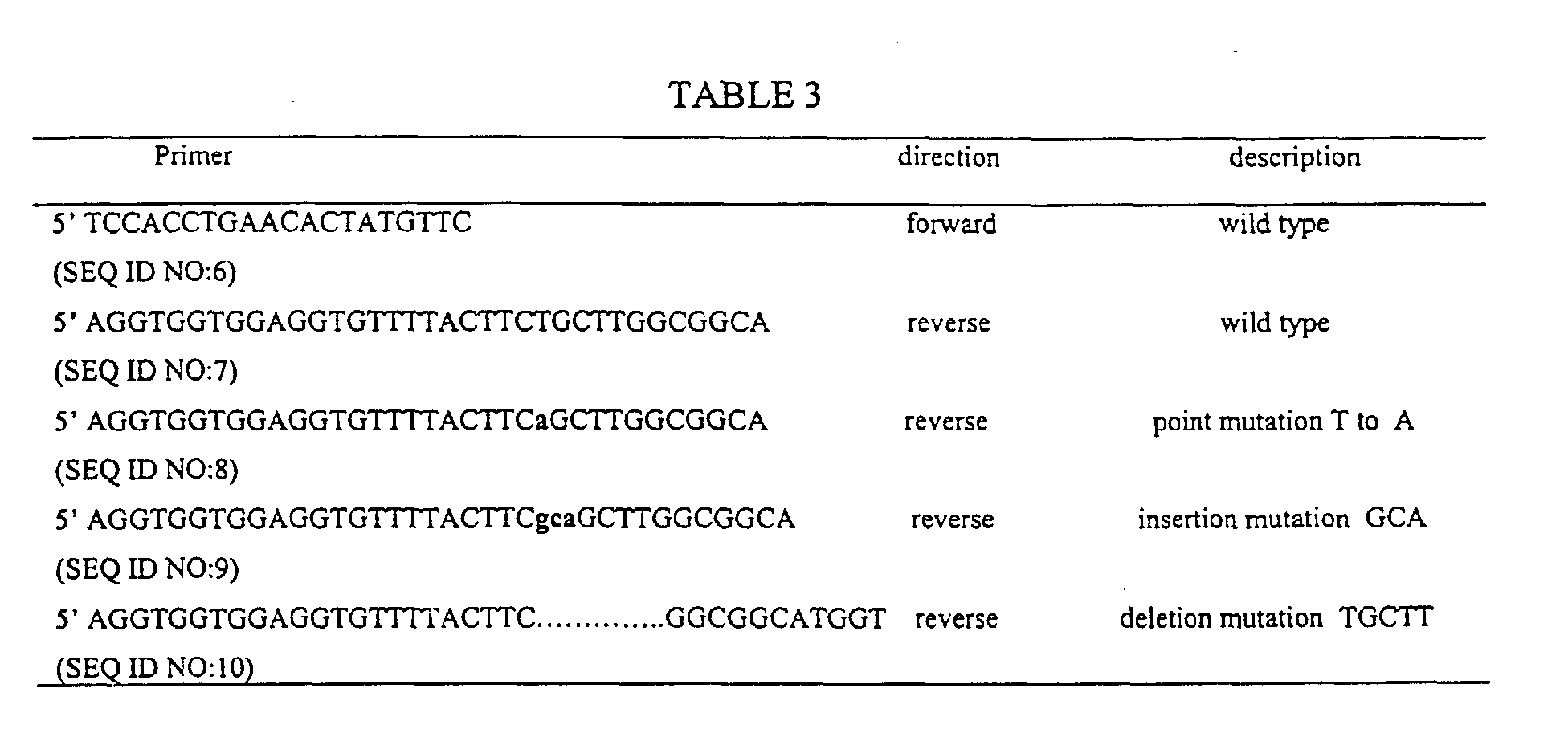
used as a template. The primers used for PCR are listed in Table 3.
Four different PCR products size about 200 bp were generated by combination of
the primers. They are APC-w: wild-type; APC-p: having a point mutation; APC-i: having
an insertion mutation and APC-d: having a deletion mutation. The PCR products were
applied to 1% agarose gel to remove the template and free nucleotides. The products
were then purified by Qiax DNA purification Kit (Qiagen). The STA primer is designed
as 5' -AGGTGGTGGAGGTGTTTTACTTC - 3' (SEQ ID NO: 11) and STA reactions
were performed in a total volume of 20 µl in a buffer containing 10 mM Tris-HCl, pH
8.3, 50 mM KCl, 2 mM MgCl
2, 0.05 pmol double strand PCR product, 5 pmol primers,
20pM of dATP, dGTP, 1 µCi of [α-
32P]-labeled CTP, 20µM non-labeled dideoxy TTP
and 2 units of Taq DNA Polymerase. Twenty cycles of 94 °C for 20 s, 55 °C for 1 min
were performed in a thermocycler (Perkin Elmer, GeneAmp 9600). 1 pl of STA product
was applied to Trim Strip, a thin layer chromatography strip made by TRIM Corporation
in Japan, and radioactivity was counted as described in Example 1. The results are
shown in Table 4.
| | Total Counts | Labeled Primer |
| No template | 182,245 | 343 |
| Papc-w | 208,271 | 595 |
| Papc-p | 197,494 | 5,568 |
| Papc-i | 176,984 | 10,372 |
| Papc-d | 195,570 | 12,010 |
The primer in all three types of mutant samples has been extended with [α-32P]
dCTP. The intensity of the strength of the signal generated by the primer extension
incorpoating the label correlates well with the number of labeled nucleotides that are
carried in the primer extension strand.
Example 3
The inventive STA reagent and method was applied to RNA fragments of human
APC gene. The PCR products of human APC gene in Example 2 were ligated into TA
cloning Vector 3.1 (TA cloning kit, Invitrogen). Four vectors were constructed and they
are listed in Table 5.
| Vector | Insert | description | Name of RNA products |
| Tapc-w | APC-w | Wild type | Rapc-w |
| Tapc-p | APC-p | Point Mutation | Rapc-p |
| Tapc-I | APC-i | Insertion Mutation | Rapc-I |
| Tapc-d | APC-d | Deletion Mutation | Rapc-d |
RNA species corresponding to each vector was synthesized using an
in vitro RNA
synthesis kit (Promega, WI). The RNA was synthesized at 37 °C for 1 hour in buffer
containing 2µg of vector and T7 Polymerase. The reaction was stopped by adding LiCl
and 100% ethanol. After incubation at -20 °C for 15 min., the RNA was precipitated by
spinning in a centrifuge at 14,000g for 15 min., and the purified RNA was resuspended in
RNase-free water. 5µg RNA was mixed with the STA primer described in Example 2 in
a total volume of 10 µl buffer containing 10 mM Tris-HCl pH 7.6, 50 mM NaCl, and 10
mM KCI. The mixture was heat denatured at 65 °C for 3 minutes followed by quenching
on ice for 2 minutes. STA reaction was performed as described in Example 1, the buffer
containing 1 µl of [α-
32P]-labeled dCTP (250 µCi/ml, 3000 Ci/mmol Dupont-New
England Nuclear), 10 µM dATP, dGTP, and 10 µM of non-labeled ddTTP, and 20 units
of reverse transcriptase. After incubating at 40 °C for 15 minutes, the reaction was
stopped by heating at 100 °C for 2 minutes. 1 µl reaction product was applied to Trim
Strip and the radioactivity was counted as described in Example 1. The results are shown
in Table 6.
| sample | Total Counts | Labeled Primer |
| No template | 167,496 | 690 |
| Rapc-w | 172,734 | 435 |
| Rapc-p | 166,979 | 1,745 |
| Rapc-i | 170,801 | 7,348 |
| Rapc-d | 174,888 | 7,360 |
All of the above steps involve chemistries, manipulations, and protocols that have
been, or are amenable to being, automated. Thereby, incorporation of the preferred mode
of practice of this invention into the operation of a suitably programmed robotic
workstation should result in significant cost savings and increases in productivity for
virtually any diagnostic procedure that depends on the detection of specific nucleotide
sequences or sequence differences in nucleic acids derived from biological samples.
All of the references cited herein are incorporated by reference in their entirety.
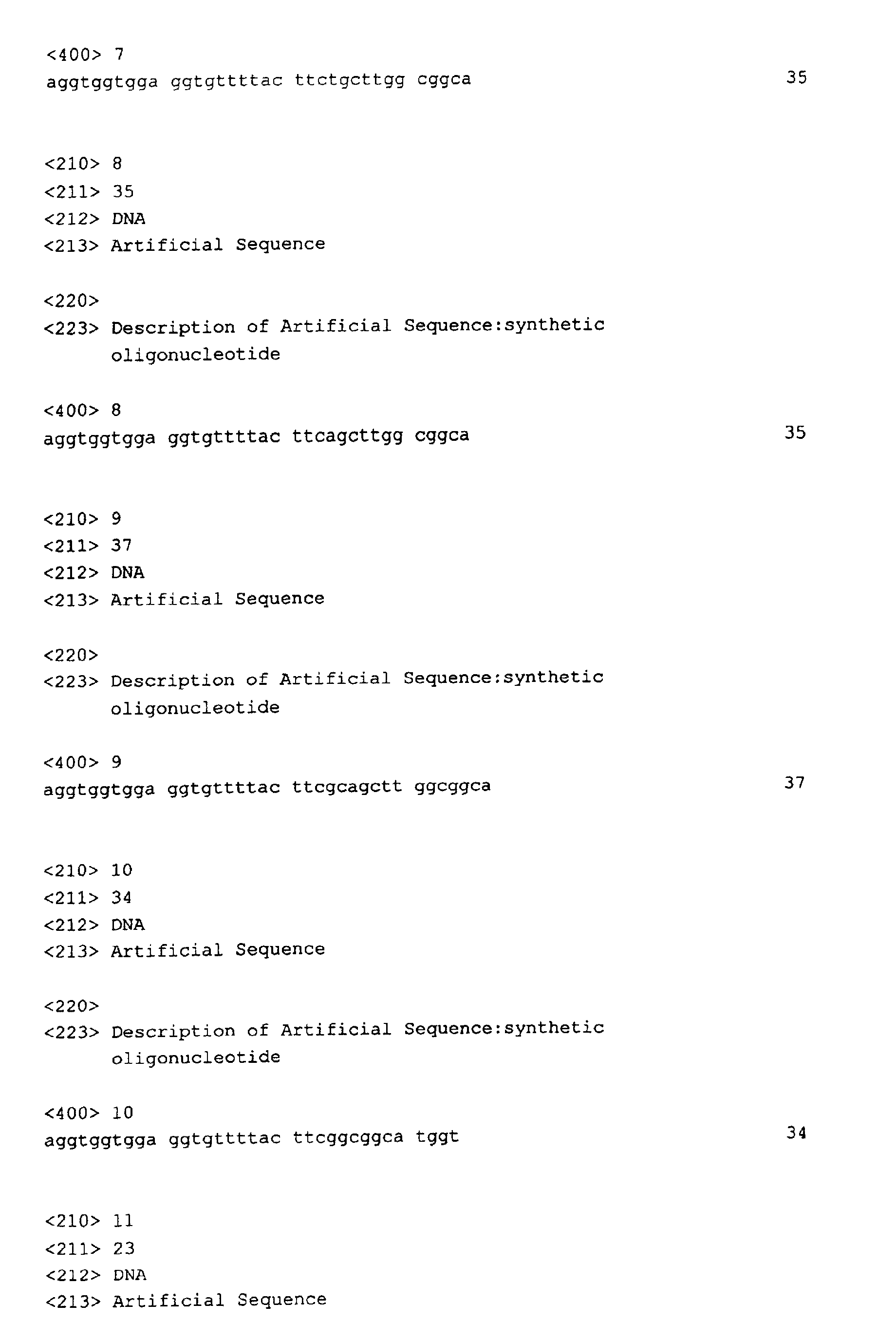
Annex to the application documents - subsquently filed sequences listing