EP1095278B1 - Differential diagnosis of neurodegeneration - Google Patents
Differential diagnosis of neurodegeneration Download PDFInfo
- Publication number
- EP1095278B1 EP1095278B1 EP99934543A EP99934543A EP1095278B1 EP 1095278 B1 EP1095278 B1 EP 1095278B1 EP 99934543 A EP99934543 A EP 99934543A EP 99934543 A EP99934543 A EP 99934543A EP 1095278 B1 EP1095278 B1 EP 1095278B1
- Authority
- EP
- European Patent Office
- Prior art keywords
- tau
- amyloid
- disease
- antibodies
- neurological
- Prior art date
- Legal status (The legal status is an assumption and is not a legal conclusion. Google has not performed a legal analysis and makes no representation as to the accuracy of the status listed.)
- Expired - Lifetime
Links
- 230000004770 neurodegeneration Effects 0.000 title claims abstract description 55
- 238000003748 differential diagnosis Methods 0.000 title claims abstract description 49
- 210000001175 cerebrospinal fluid Anatomy 0.000 claims abstract description 151
- 230000000926 neurological effect Effects 0.000 claims abstract description 101
- 238000000034 method Methods 0.000 claims abstract description 92
- 102000003802 alpha-Synuclein Human genes 0.000 claims abstract description 81
- 108090000185 alpha-Synuclein Proteins 0.000 claims abstract description 81
- 101150004420 RAB3A gene Proteins 0.000 claims abstract description 71
- 108010057722 Synaptosomal-Associated Protein 25 Proteins 0.000 claims abstract description 65
- 102000004183 Synaptosomal-Associated Protein 25 Human genes 0.000 claims abstract description 65
- 238000001514 detection method Methods 0.000 claims abstract description 64
- 238000011895 specific detection Methods 0.000 claims abstract description 61
- 238000011002 quantification Methods 0.000 claims abstract description 46
- 210000001124 body fluid Anatomy 0.000 claims abstract description 34
- 239000010839 body fluid Substances 0.000 claims abstract description 34
- 239000013068 control sample Substances 0.000 claims abstract description 3
- 102000013498 tau Proteins Human genes 0.000 claims description 128
- 108010026424 tau Proteins Proteins 0.000 claims description 128
- 208000024827 Alzheimer disease Diseases 0.000 claims description 107
- 102000013455 Amyloid beta-Peptides Human genes 0.000 claims description 96
- 108010090849 Amyloid beta-Peptides Proteins 0.000 claims description 96
- 102100023206 Neuromodulin Human genes 0.000 claims description 65
- 101710144282 Neuromodulin Proteins 0.000 claims description 65
- 239000003550 marker Substances 0.000 claims description 63
- 239000000523 sample Substances 0.000 claims description 58
- 108090000623 proteins and genes Proteins 0.000 claims description 51
- 102000004169 proteins and genes Human genes 0.000 claims description 50
- 206010012289 Dementia Diseases 0.000 claims description 43
- 108010022181 Phosphopyruvate Hydratase Proteins 0.000 claims description 39
- 102000012288 Phosphopyruvate Hydratase Human genes 0.000 claims description 39
- 238000011282 treatment Methods 0.000 claims description 36
- 238000002512 chemotherapy Methods 0.000 claims description 30
- 208000018737 Parkinson disease Diseases 0.000 claims description 27
- 108090000765 processed proteins & peptides Proteins 0.000 claims description 27
- 208000009829 Lewy Body Disease Diseases 0.000 claims description 26
- 201000002832 Lewy body dementia Diseases 0.000 claims description 26
- 102000004196 processed proteins & peptides Human genes 0.000 claims description 17
- 210000003478 temporal lobe Anatomy 0.000 claims description 17
- 230000008878 coupling Effects 0.000 claims description 13
- 238000010168 coupling process Methods 0.000 claims description 13
- 238000005859 coupling reaction Methods 0.000 claims description 13
- 208000032839 leukemia Diseases 0.000 claims description 10
- 239000007853 buffer solution Substances 0.000 claims description 9
- 150000001875 compounds Chemical class 0.000 claims description 9
- 230000008105 immune reaction Effects 0.000 claims description 9
- 238000009007 Diagnostic Kit Methods 0.000 claims description 8
- 238000012544 monitoring process Methods 0.000 claims description 6
- 208000003174 Brain Neoplasms Diseases 0.000 claims description 5
- 230000008859 change Effects 0.000 claims description 5
- 230000001225 therapeutic effect Effects 0.000 claims description 4
- 230000002792 vascular Effects 0.000 claims description 4
- 238000003556 assay Methods 0.000 abstract description 24
- 235000018102 proteins Nutrition 0.000 description 49
- 238000003745 diagnosis Methods 0.000 description 24
- 208000037265 diseases, disorders, signs and symptoms Diseases 0.000 description 24
- 210000004556 brain Anatomy 0.000 description 23
- 210000000225 synapse Anatomy 0.000 description 21
- 230000001900 immune effect Effects 0.000 description 20
- 201000010099 disease Diseases 0.000 description 18
- 230000026683 transduction Effects 0.000 description 18
- 238000010361 transduction Methods 0.000 description 18
- 210000003169 central nervous system Anatomy 0.000 description 16
- 238000011156 evaluation Methods 0.000 description 15
- 208000012902 Nervous system disease Diseases 0.000 description 13
- FEWOUVRMGWFWIH-ILZZQXMPSA-N amyloid-beta polypeptide 40 Chemical compound C([C@@H](C(=O)N[C@@H](C)C(=O)N[C@@H](CCC(O)=O)C(=O)N[C@@H](CC(O)=O)C(=O)N[C@H](C(=O)NCC(=O)N[C@@H](CO)C(=O)N[C@@H](CC(N)=O)C(=O)N[C@@H](CCCCN)C(=O)NCC(=O)N[C@@H](C)C(=O)N[C@H](C(=O)N[C@@H]([C@@H](C)CC)C(=O)NCC(=O)N[C@@H](CC(C)C)C(=O)N[C@@H](CCSC)C(=O)N[C@@H](C(C)C)C(=O)NCC(=O)NCC(=O)N[C@@H](C(C)C)C(=O)N[C@@H](C(C)C)C(O)=O)[C@@H](C)CC)C(C)C)NC(=O)[C@H](CC=1C=CC=CC=1)NC(=O)[C@@H](NC(=O)[C@H](CC(C)C)NC(=O)[C@H](CCCCN)NC(=O)[C@H](CCC(N)=O)NC(=O)[C@H](CC=1N=CNC=1)NC(=O)[C@H](CC=1N=CNC=1)NC(=O)[C@@H](NC(=O)[C@H](CCC(O)=O)NC(=O)[C@H](CC=1C=CC(O)=CC=1)NC(=O)CNC(=O)[C@H](CO)NC(=O)[C@H](CC(O)=O)NC(=O)[C@H](CC=1N=CNC=1)NC(=O)[C@H](CCCNC(N)=N)NC(=O)[C@H](CC=1C=CC=CC=1)NC(=O)[C@H](CCC(O)=O)NC(=O)[C@H](C)NC(=O)[C@@H](N)CC(O)=O)C(C)C)C(C)C)C1=CC=CC=C1 FEWOUVRMGWFWIH-ILZZQXMPSA-N 0.000 description 12
- 238000004458 analytical method Methods 0.000 description 12
- 239000000427 antigen Substances 0.000 description 12
- 102000036639 antigens Human genes 0.000 description 12
- 108091007433 antigens Proteins 0.000 description 12
- 230000008569 process Effects 0.000 description 12
- 208000028564 B-cell non-Hodgkin lymphoma Diseases 0.000 description 11
- 241000588724 Escherichia coli Species 0.000 description 11
- 201000004810 Vascular dementia Diseases 0.000 description 11
- 238000009593 lumbar puncture Methods 0.000 description 11
- 230000000626 neurodegenerative effect Effects 0.000 description 11
- 210000002966 serum Anatomy 0.000 description 11
- 238000011160 research Methods 0.000 description 10
- 238000003118 sandwich ELISA Methods 0.000 description 10
- 241000283973 Oryctolagus cuniculus Species 0.000 description 9
- 150000001413 amino acids Chemical class 0.000 description 9
- 230000004069 differentiation Effects 0.000 description 9
- 230000014509 gene expression Effects 0.000 description 9
- 230000001537 neural effect Effects 0.000 description 9
- 208000019553 vascular disease Diseases 0.000 description 9
- 201000010374 Down Syndrome Diseases 0.000 description 8
- 206010021143 Hypoxia Diseases 0.000 description 8
- FBOZXECLQNJBKD-ZDUSSCGKSA-N L-methotrexate Chemical compound C=1N=C2N=C(N)N=C(N)C2=NC=1CN(C)C1=CC=C(C(=O)N[C@@H](CCC(O)=O)C(O)=O)C=C1 FBOZXECLQNJBKD-ZDUSSCGKSA-N 0.000 description 8
- 235000001014 amino acid Nutrition 0.000 description 8
- -1 irradiation Substances 0.000 description 8
- 239000000047 product Substances 0.000 description 8
- 206010028980 Neoplasm Diseases 0.000 description 7
- 208000015914 Non-Hodgkin lymphomas Diseases 0.000 description 7
- 102000011070 Rab3 Human genes 0.000 description 7
- 108050001276 Rab3 Proteins 0.000 description 7
- 238000006243 chemical reaction Methods 0.000 description 7
- 239000000499 gel Substances 0.000 description 7
- 206010027175 memory impairment Diseases 0.000 description 7
- 201000003793 Myelodysplastic syndrome Diseases 0.000 description 6
- 208000025966 Neurological disease Diseases 0.000 description 6
- FAPWRFPIFSIZLT-UHFFFAOYSA-M Sodium chloride Chemical compound [Na+].[Cl-] FAPWRFPIFSIZLT-UHFFFAOYSA-M 0.000 description 6
- 102000019355 Synuclein Human genes 0.000 description 6
- 108050006783 Synuclein Proteins 0.000 description 6
- 208000029028 brain injury Diseases 0.000 description 6
- 239000000872 buffer Substances 0.000 description 6
- 239000003795 chemical substances by application Substances 0.000 description 6
- 238000004587 chromatography analysis Methods 0.000 description 6
- 208000035475 disorder Diseases 0.000 description 6
- 102000004963 gamma-Synuclein Human genes 0.000 description 6
- 108090001121 gamma-Synuclein Proteins 0.000 description 6
- 238000003018 immunoassay Methods 0.000 description 6
- 238000011534 incubation Methods 0.000 description 6
- 238000000746 purification Methods 0.000 description 6
- 230000035945 sensitivity Effects 0.000 description 6
- 239000000758 substrate Substances 0.000 description 6
- 208000011580 syndromic disease Diseases 0.000 description 6
- 238000012360 testing method Methods 0.000 description 6
- 238000002965 ELISA Methods 0.000 description 5
- 241000699670 Mus sp. Species 0.000 description 5
- 108010088373 Neurofilament Proteins Proteins 0.000 description 5
- 102000008763 Neurofilament Proteins Human genes 0.000 description 5
- 210000003719 b-lymphocyte Anatomy 0.000 description 5
- 210000004369 blood Anatomy 0.000 description 5
- 239000008280 blood Substances 0.000 description 5
- 230000006931 brain damage Effects 0.000 description 5
- 231100000874 brain damage Toxicity 0.000 description 5
- 201000011510 cancer Diseases 0.000 description 5
- 239000012634 fragment Substances 0.000 description 5
- 230000006870 function Effects 0.000 description 5
- 230000006698 induction Effects 0.000 description 5
- 238000002347 injection Methods 0.000 description 5
- 239000007924 injection Substances 0.000 description 5
- 210000000265 leukocyte Anatomy 0.000 description 5
- 210000004558 lewy body Anatomy 0.000 description 5
- 210000005044 neurofilament Anatomy 0.000 description 5
- 210000002569 neuron Anatomy 0.000 description 5
- 230000003961 neuronal insult Effects 0.000 description 5
- 238000001959 radiotherapy Methods 0.000 description 5
- 230000009467 reduction Effects 0.000 description 5
- 230000007470 synaptic degeneration Effects 0.000 description 5
- 102000004899 14-3-3 Proteins Human genes 0.000 description 4
- UHDGCWIWMRVCDJ-CCXZUQQUSA-N Cytarabine Chemical compound O=C1N=C(N)C=CN1[C@H]1[C@@H](O)[C@H](O)[C@@H](CO)O1 UHDGCWIWMRVCDJ-CCXZUQQUSA-N 0.000 description 4
- 208000000172 Medulloblastoma Diseases 0.000 description 4
- 108010029485 Protein Isoforms Proteins 0.000 description 4
- 102000001708 Protein Isoforms Human genes 0.000 description 4
- 208000006011 Stroke Diseases 0.000 description 4
- QAOWNCQODCNURD-UHFFFAOYSA-N Sulfuric acid Chemical compound OS(O)(=O)=O QAOWNCQODCNURD-UHFFFAOYSA-N 0.000 description 4
- 230000000903 blocking effect Effects 0.000 description 4
- 210000005013 brain tissue Anatomy 0.000 description 4
- 210000004899 c-terminal region Anatomy 0.000 description 4
- 229960000684 cytarabine Drugs 0.000 description 4
- 230000034994 death Effects 0.000 description 4
- 230000003247 decreasing effect Effects 0.000 description 4
- 230000001419 dependent effect Effects 0.000 description 4
- 238000011161 development Methods 0.000 description 4
- 230000018109 developmental process Effects 0.000 description 4
- 208000015181 infectious disease Diseases 0.000 description 4
- 230000002503 metabolic effect Effects 0.000 description 4
- 230000035772 mutation Effects 0.000 description 4
- 102000013415 peroxidase activity proteins Human genes 0.000 description 4
- 108040007629 peroxidase activity proteins Proteins 0.000 description 4
- 230000002829 reductive effect Effects 0.000 description 4
- 102000003137 synaptotagmin Human genes 0.000 description 4
- 108060008004 synaptotagmin Proteins 0.000 description 4
- 238000002560 therapeutic procedure Methods 0.000 description 4
- 238000005406 washing Methods 0.000 description 4
- 238000001262 western blot Methods 0.000 description 4
- 241000283707 Capra Species 0.000 description 3
- PEDCQBHIVMGVHV-UHFFFAOYSA-N Glycerine Chemical compound OCC(O)CO PEDCQBHIVMGVHV-UHFFFAOYSA-N 0.000 description 3
- 208000017604 Hodgkin disease Diseases 0.000 description 3
- 208000010747 Hodgkins lymphoma Diseases 0.000 description 3
- 101000617536 Homo sapiens Presenilin-1 Proteins 0.000 description 3
- 102100040243 Microtubule-associated protein tau Human genes 0.000 description 3
- 101710115937 Microtubule-associated protein tau Proteins 0.000 description 3
- 206010028923 Neonatal asphyxia Diseases 0.000 description 3
- 208000037212 Neonatal hypoxic and ischemic brain injury Diseases 0.000 description 3
- 102100022033 Presenilin-1 Human genes 0.000 description 3
- 108010010469 Qa-SNARE Proteins Proteins 0.000 description 3
- 108010005730 R-SNARE Proteins Proteins 0.000 description 3
- 201000000582 Retinoblastoma Diseases 0.000 description 3
- 102000002215 Synaptobrevin Human genes 0.000 description 3
- 102000050389 Syntaxin Human genes 0.000 description 3
- 210000001744 T-lymphocyte Anatomy 0.000 description 3
- 239000007983 Tris buffer Substances 0.000 description 3
- 206010044688 Trisomy 21 Diseases 0.000 description 3
- 230000003321 amplification Effects 0.000 description 3
- 102000003799 beta-Synuclein Human genes 0.000 description 3
- 108090000182 beta-Synuclein Proteins 0.000 description 3
- 210000004027 cell Anatomy 0.000 description 3
- 238000012512 characterization method Methods 0.000 description 3
- DQLATGHUWYMOKM-UHFFFAOYSA-L cisplatin Chemical compound N[Pt](N)(Cl)Cl DQLATGHUWYMOKM-UHFFFAOYSA-L 0.000 description 3
- 229960004316 cisplatin Drugs 0.000 description 3
- 239000011248 coating agent Substances 0.000 description 3
- 238000000576 coating method Methods 0.000 description 3
- 239000002299 complementary DNA Substances 0.000 description 3
- 230000002596 correlated effect Effects 0.000 description 3
- 239000003246 corticosteroid Substances 0.000 description 3
- 229960001334 corticosteroids Drugs 0.000 description 3
- 230000006378 damage Effects 0.000 description 3
- 238000010790 dilution Methods 0.000 description 3
- 239000012895 dilution Substances 0.000 description 3
- 229940079593 drug Drugs 0.000 description 3
- 239000003814 drug Substances 0.000 description 3
- 230000000694 effects Effects 0.000 description 3
- 210000004408 hybridoma Anatomy 0.000 description 3
- 230000006872 improvement Effects 0.000 description 3
- 230000001939 inductive effect Effects 0.000 description 3
- 230000003834 intracellular effect Effects 0.000 description 3
- 230000009545 invasion Effects 0.000 description 3
- 230000007774 longterm Effects 0.000 description 3
- 238000005259 measurement Methods 0.000 description 3
- 201000006417 multiple sclerosis Diseases 0.000 description 3
- 230000002981 neuropathic effect Effects 0.000 description 3
- 238000003199 nucleic acid amplification method Methods 0.000 description 3
- 208000033300 perinatal asphyxia Diseases 0.000 description 3
- 230000026731 phosphorylation Effects 0.000 description 3
- 238000006366 phosphorylation reaction Methods 0.000 description 3
- 239000013612 plasmid Substances 0.000 description 3
- CPTBDICYNRMXFX-UHFFFAOYSA-N procarbazine Chemical compound CNNCC1=CC=C(C(=O)NC(C)C)C=C1 CPTBDICYNRMXFX-UHFFFAOYSA-N 0.000 description 3
- 229960000624 procarbazine Drugs 0.000 description 3
- 201000009410 rhabdomyosarcoma Diseases 0.000 description 3
- 239000012723 sample buffer Substances 0.000 description 3
- 238000011896 sensitive detection Methods 0.000 description 3
- 239000011780 sodium chloride Substances 0.000 description 3
- 238000002415 sodium dodecyl sulfate polyacrylamide gel electrophoresis Methods 0.000 description 3
- 239000006228 supernatant Substances 0.000 description 3
- 210000001519 tissue Anatomy 0.000 description 3
- LENZDBCJOHFCAS-UHFFFAOYSA-N tris Chemical compound OCC(N)(CO)CO LENZDBCJOHFCAS-UHFFFAOYSA-N 0.000 description 3
- OGWKCGZFUXNPDA-XQKSVPLYSA-N vincristine Chemical compound C([N@]1C[C@@H](C[C@]2(C(=O)OC)C=3C(=CC4=C([C@]56[C@H]([C@@]([C@H](OC(C)=O)[C@]7(CC)C=CCN([C@H]67)CC5)(O)C(=O)OC)N4C=O)C=3)OC)C[C@@](C1)(O)CC)CC1=C2NC2=CC=CC=C12 OGWKCGZFUXNPDA-XQKSVPLYSA-N 0.000 description 3
- 229960004528 vincristine Drugs 0.000 description 3
- OGWKCGZFUXNPDA-UHFFFAOYSA-N vincristine Natural products C1C(CC)(O)CC(CC2(C(=O)OC)C=3C(=CC4=C(C56C(C(C(OC(C)=O)C7(CC)C=CCN(C67)CC5)(O)C(=O)OC)N4C=O)C=3)OC)CN1CCC1=C2NC2=CC=CC=C12 OGWKCGZFUXNPDA-UHFFFAOYSA-N 0.000 description 3
- 102100025573 1-alkyl-2-acetylglycerophosphocholine esterase Human genes 0.000 description 2
- HRPVXLWXLXDGHG-UHFFFAOYSA-N Acrylamide Chemical compound NC(=O)C=C HRPVXLWXLXDGHG-UHFFFAOYSA-N 0.000 description 2
- 102000009027 Albumins Human genes 0.000 description 2
- 108010088751 Albumins Proteins 0.000 description 2
- 208000037259 Amyloid Plaque Diseases 0.000 description 2
- 206010073478 Anaplastic large-cell lymphoma Diseases 0.000 description 2
- 240000003291 Armoracia rusticana Species 0.000 description 2
- 235000011330 Armoracia rusticana Nutrition 0.000 description 2
- 108010024976 Asparaginase Proteins 0.000 description 2
- 206010003497 Asphyxia Diseases 0.000 description 2
- DLGOEMSEDOSKAD-UHFFFAOYSA-N Carmustine Chemical compound ClCCNC(=O)N(N=O)CCCl DLGOEMSEDOSKAD-UHFFFAOYSA-N 0.000 description 2
- 208000006332 Choriocarcinoma Diseases 0.000 description 2
- 208000035473 Communicable disease Diseases 0.000 description 2
- AOJJSUZBOXZQNB-TZSSRYMLSA-N Doxorubicin Chemical compound O([C@H]1C[C@@](O)(CC=2C(O)=C3C(=O)C=4C=CC=C(C=4C(=O)C3=C(O)C=21)OC)C(=O)CO)[C@H]1C[C@H](N)[C@H](O)[C@H](C)O1 AOJJSUZBOXZQNB-TZSSRYMLSA-N 0.000 description 2
- 102000004190 Enzymes Human genes 0.000 description 2
- 108090000790 Enzymes Proteins 0.000 description 2
- 208000035366 Familial hemophagocytic lymphohistiocytosis Diseases 0.000 description 2
- GHASVSINZRGABV-UHFFFAOYSA-N Fluorouracil Chemical compound FC1=CNC(=O)NC1=O GHASVSINZRGABV-UHFFFAOYSA-N 0.000 description 2
- 208000000527 Germinoma Diseases 0.000 description 2
- 102000053171 Glial Fibrillary Acidic Human genes 0.000 description 2
- 101710193519 Glial fibrillary acidic protein Proteins 0.000 description 2
- 101000891579 Homo sapiens Microtubule-associated protein tau Proteins 0.000 description 2
- 101001104083 Homo sapiens Rabphilin-3A Proteins 0.000 description 2
- 108060003951 Immunoglobulin Proteins 0.000 description 2
- 108010002350 Interleukin-2 Proteins 0.000 description 2
- 208000032004 Large-Cell Anaplastic Lymphoma Diseases 0.000 description 2
- 241001465754 Metazoa Species 0.000 description 2
- 102000029749 Microtubule Human genes 0.000 description 2
- 108091022875 Microtubule Proteins 0.000 description 2
- 241000699666 Mus <mouse, genus> Species 0.000 description 2
- 238000005481 NMR spectroscopy Methods 0.000 description 2
- 206010029350 Neurotoxicity Diseases 0.000 description 2
- 239000000020 Nitrocellulose Substances 0.000 description 2
- 229910019142 PO4 Inorganic materials 0.000 description 2
- 208000037273 Pathologic Processes Diseases 0.000 description 2
- 239000004743 Polypropylene Substances 0.000 description 2
- 239000004365 Protease Substances 0.000 description 2
- 102100040040 Rabphilin-3A Human genes 0.000 description 2
- 229920002684 Sepharose Polymers 0.000 description 2
- UIIMBOGNXHQVGW-UHFFFAOYSA-M Sodium bicarbonate Chemical compound [Na+].OC([O-])=O UIIMBOGNXHQVGW-UHFFFAOYSA-M 0.000 description 2
- 102000001435 Synapsin Human genes 0.000 description 2
- 108050009621 Synapsin Proteins 0.000 description 2
- 102000004874 Synaptophysin Human genes 0.000 description 2
- 108090001076 Synaptophysin Proteins 0.000 description 2
- 206010044221 Toxic encephalopathy Diseases 0.000 description 2
- DTQVDTLACAAQTR-UHFFFAOYSA-N Trifluoroacetic acid Chemical compound OC(=O)C(F)(F)F DTQVDTLACAAQTR-UHFFFAOYSA-N 0.000 description 2
- 208000027418 Wounds and injury Diseases 0.000 description 2
- 238000002835 absorbance Methods 0.000 description 2
- 230000001154 acute effect Effects 0.000 description 2
- 238000001042 affinity chromatography Methods 0.000 description 2
- 230000004075 alteration Effects 0.000 description 2
- 238000000540 analysis of variance Methods 0.000 description 2
- 230000003542 behavioural effect Effects 0.000 description 2
- 230000015572 biosynthetic process Effects 0.000 description 2
- 238000007413 biotinylation Methods 0.000 description 2
- 230000006287 biotinylation Effects 0.000 description 2
- 238000004820 blood count Methods 0.000 description 2
- 210000000133 brain stem Anatomy 0.000 description 2
- 239000005018 casein Substances 0.000 description 2
- BECPQYXYKAMYBN-UHFFFAOYSA-N casein, tech. Chemical compound NCCCCC(C(O)=O)N=C(O)C(CC(O)=O)N=C(O)C(CCC(O)=N)N=C(O)C(CC(C)C)N=C(O)C(CCC(O)=O)N=C(O)C(CC(O)=O)N=C(O)C(CCC(O)=O)N=C(O)C(C(C)O)N=C(O)C(CCC(O)=N)N=C(O)C(CCC(O)=N)N=C(O)C(CCC(O)=N)N=C(O)C(CCC(O)=O)N=C(O)C(CCC(O)=O)N=C(O)C(COP(O)(O)=O)N=C(O)C(CCC(O)=N)N=C(O)C(N)CC1=CC=CC=C1 BECPQYXYKAMYBN-UHFFFAOYSA-N 0.000 description 2
- 235000021240 caseins Nutrition 0.000 description 2
- 230000001684 chronic effect Effects 0.000 description 2
- 238000003759 clinical diagnosis Methods 0.000 description 2
- 238000010367 cloning Methods 0.000 description 2
- 230000001149 cognitive effect Effects 0.000 description 2
- 230000000295 complement effect Effects 0.000 description 2
- 239000000470 constituent Substances 0.000 description 2
- 238000012217 deletion Methods 0.000 description 2
- 230000037430 deletion Effects 0.000 description 2
- 238000013461 design Methods 0.000 description 2
- 238000002405 diagnostic procedure Methods 0.000 description 2
- 235000014113 dietary fatty acids Nutrition 0.000 description 2
- 238000005516 engineering process Methods 0.000 description 2
- 229940088598 enzyme Drugs 0.000 description 2
- 206010015037 epilepsy Diseases 0.000 description 2
- 230000007717 exclusion Effects 0.000 description 2
- 239000000284 extract Substances 0.000 description 2
- 229930195729 fatty acid Natural products 0.000 description 2
- 239000000194 fatty acid Substances 0.000 description 2
- 150000004665 fatty acids Chemical class 0.000 description 2
- 210000005153 frontal cortex Anatomy 0.000 description 2
- 102000037865 fusion proteins Human genes 0.000 description 2
- 108020001507 fusion proteins Proteins 0.000 description 2
- 201000003115 germ cell cancer Diseases 0.000 description 2
- 210000005046 glial fibrillary acidic protein Anatomy 0.000 description 2
- 230000036541 health Effects 0.000 description 2
- 208000014752 hemophagocytic syndrome Diseases 0.000 description 2
- 125000000487 histidyl group Chemical group [H]N([H])C(C(=O)O*)C([H])([H])C1=C([H])N([H])C([H])=N1 0.000 description 2
- 102000057063 human MAPT Human genes 0.000 description 2
- JYGXADMDTFJGBT-VWUMJDOOSA-N hydrocortisone Chemical compound O=C1CC[C@]2(C)[C@H]3[C@@H](O)C[C@](C)([C@@](CC4)(O)C(=O)CO)[C@@H]4[C@@H]3CCC2=C1 JYGXADMDTFJGBT-VWUMJDOOSA-N 0.000 description 2
- 230000002267 hypothalamic effect Effects 0.000 description 2
- 238000002649 immunization Methods 0.000 description 2
- 230000003053 immunization Effects 0.000 description 2
- 102000018358 immunoglobulin Human genes 0.000 description 2
- 238000001727 in vivo Methods 0.000 description 2
- 230000000977 initiatory effect Effects 0.000 description 2
- 208000014674 injury Diseases 0.000 description 2
- 239000002198 insoluble material Substances 0.000 description 2
- 238000012423 maintenance Methods 0.000 description 2
- 230000003211 malignant effect Effects 0.000 description 2
- 238000013507 mapping Methods 0.000 description 2
- 239000012528 membrane Substances 0.000 description 2
- 229960000485 methotrexate Drugs 0.000 description 2
- 210000004688 microtubule Anatomy 0.000 description 2
- 210000000478 neocortex Anatomy 0.000 description 2
- 230000002887 neurotoxic effect Effects 0.000 description 2
- 231100000228 neurotoxicity Toxicity 0.000 description 2
- 230000007135 neurotoxicity Effects 0.000 description 2
- 229920001220 nitrocellulos Polymers 0.000 description 2
- 210000004940 nucleus Anatomy 0.000 description 2
- 230000003287 optical effect Effects 0.000 description 2
- 230000008520 organization Effects 0.000 description 2
- 230000009054 pathological process Effects 0.000 description 2
- NBIIXXVUZAFLBC-UHFFFAOYSA-K phosphate Chemical compound [O-]P([O-])([O-])=O NBIIXXVUZAFLBC-UHFFFAOYSA-K 0.000 description 2
- 239000010452 phosphate Substances 0.000 description 2
- 229920001155 polypropylene Polymers 0.000 description 2
- 125000002924 primary amino group Chemical group [H]N([H])* 0.000 description 2
- 210000004129 prosencephalon Anatomy 0.000 description 2
- 208000020016 psychiatric disease Diseases 0.000 description 2
- 230000002441 reversible effect Effects 0.000 description 2
- 201000000980 schizophrenia Diseases 0.000 description 2
- 239000000243 solution Substances 0.000 description 2
- 238000003860 storage Methods 0.000 description 2
- 208000024891 symptom Diseases 0.000 description 2
- XLYOFNOQVPJJNP-UHFFFAOYSA-N water Substances O XLYOFNOQVPJJNP-UHFFFAOYSA-N 0.000 description 2
- KISWVXRQTGLFGD-UHFFFAOYSA-N 2-[[2-[[6-amino-2-[[2-[[2-[[5-amino-2-[[2-[[1-[2-[[6-amino-2-[(2,5-diamino-5-oxopentanoyl)amino]hexanoyl]amino]-5-(diaminomethylideneamino)pentanoyl]pyrrolidine-2-carbonyl]amino]-3-hydroxypropanoyl]amino]-5-oxopentanoyl]amino]-5-(diaminomethylideneamino)p Chemical compound C1CCN(C(=O)C(CCCN=C(N)N)NC(=O)C(CCCCN)NC(=O)C(N)CCC(N)=O)C1C(=O)NC(CO)C(=O)NC(CCC(N)=O)C(=O)NC(CCCN=C(N)N)C(=O)NC(CO)C(=O)NC(CCCCN)C(=O)NC(C(=O)NC(CC(C)C)C(O)=O)CC1=CC=C(O)C=C1 KISWVXRQTGLFGD-UHFFFAOYSA-N 0.000 description 1
- UAIUNKRWKOVEES-UHFFFAOYSA-N 3,3',5,5'-tetramethylbenzidine Chemical compound CC1=C(N)C(C)=CC(C=2C=C(C)C(N)=C(C)C=2)=C1 UAIUNKRWKOVEES-UHFFFAOYSA-N 0.000 description 1
- YNTLVCDWTWUMDV-UHFFFAOYSA-N 4-(4-aminophenyl)-n,2,3-trimethylaniline Chemical compound CC1=C(C)C(NC)=CC=C1C1=CC=C(N)C=C1 YNTLVCDWTWUMDV-UHFFFAOYSA-N 0.000 description 1
- 102000012440 Acetylcholinesterase Human genes 0.000 description 1
- 108010022752 Acetylcholinesterase Proteins 0.000 description 1
- 208000000044 Amnesia Diseases 0.000 description 1
- 101710137189 Amyloid-beta A4 protein Proteins 0.000 description 1
- 101710151993 Amyloid-beta precursor protein Proteins 0.000 description 1
- 102100022704 Amyloid-beta precursor protein Human genes 0.000 description 1
- 208000002109 Argyria Diseases 0.000 description 1
- 208000025324 B-cell acute lymphoblastic leukemia Diseases 0.000 description 1
- 208000003950 B-cell lymphoma Diseases 0.000 description 1
- 208000011691 Burkitt lymphomas Diseases 0.000 description 1
- 208000004434 Calcinosis Diseases 0.000 description 1
- 102100025570 Cancer/testis antigen 1 Human genes 0.000 description 1
- 101800005309 Carboxy-terminal peptide Proteins 0.000 description 1
- 206010008096 Cerebral atrophy Diseases 0.000 description 1
- 108010007718 Chromogranins Proteins 0.000 description 1
- 102000007345 Chromogranins Human genes 0.000 description 1
- 108091026890 Coding region Proteins 0.000 description 1
- 108020004705 Codon Proteins 0.000 description 1
- 206010010071 Coma Diseases 0.000 description 1
- 201000009343 Cornelia de Lange syndrome Diseases 0.000 description 1
- 208000010859 Creutzfeldt-Jakob disease Diseases 0.000 description 1
- 108020004414 DNA Proteins 0.000 description 1
- 108010014303 DNA-directed DNA polymerase Proteins 0.000 description 1
- 102000016928 DNA-directed DNA polymerase Human genes 0.000 description 1
- 208000003471 De Lange Syndrome Diseases 0.000 description 1
- 206010011953 Decreased activity Diseases 0.000 description 1
- 208000012184 Diffuse Brain injury Diseases 0.000 description 1
- 206010013033 Diplegia Diseases 0.000 description 1
- 101100326341 Drosophila melanogaster brun gene Proteins 0.000 description 1
- 201000011240 Frontotemporal dementia Diseases 0.000 description 1
- 108700042658 GAP-43 Proteins 0.000 description 1
- WQZGKKKJIJFFOK-GASJEMHNSA-N Glucose Natural products OC[C@H]1OC(O)[C@H](O)[C@@H](O)[C@@H]1O WQZGKKKJIJFFOK-GASJEMHNSA-N 0.000 description 1
- 206010053759 Growth retardation Diseases 0.000 description 1
- 208000035895 Guillain-Barré syndrome Diseases 0.000 description 1
- 208000002250 Hematologic Neoplasms Diseases 0.000 description 1
- 208000036066 Hemophagocytic Lymphohistiocytosis Diseases 0.000 description 1
- 241000282412 Homo Species 0.000 description 1
- 101000856237 Homo sapiens Cancer/testis antigen 1 Proteins 0.000 description 1
- 101001002634 Homo sapiens Interleukin-1 alpha Proteins 0.000 description 1
- 101000652315 Homo sapiens Synaptosomal-associated protein 25 Proteins 0.000 description 1
- 101000611183 Homo sapiens Tumor necrosis factor Proteins 0.000 description 1
- 108010001336 Horseradish Peroxidase Proteins 0.000 description 1
- 206010061216 Infarction Diseases 0.000 description 1
- 206010061218 Inflammation Diseases 0.000 description 1
- 102000014150 Interferons Human genes 0.000 description 1
- 108010050904 Interferons Proteins 0.000 description 1
- 108090001005 Interleukin-6 Proteins 0.000 description 1
- 238000012313 Kruskal-Wallis test Methods 0.000 description 1
- 241001484259 Lacuna Species 0.000 description 1
- 208000026139 Memory disease Diseases 0.000 description 1
- 201000009906 Meningitis Diseases 0.000 description 1
- 206010049567 Miller Fisher syndrome Diseases 0.000 description 1
- 208000019022 Mood disease Diseases 0.000 description 1
- 208000001089 Multiple system atrophy Diseases 0.000 description 1
- 208000021642 Muscular disease Diseases 0.000 description 1
- 102000006386 Myelin Proteins Human genes 0.000 description 1
- 108010083674 Myelin Proteins Proteins 0.000 description 1
- 206010028570 Myelopathy Diseases 0.000 description 1
- 201000009623 Myopathy Diseases 0.000 description 1
- HRNLUBSXIHFDHP-UHFFFAOYSA-N N-(2-aminophenyl)-4-[[[4-(3-pyridinyl)-2-pyrimidinyl]amino]methyl]benzamide Chemical compound NC1=CC=CC=C1NC(=O)C(C=C1)=CC=C1CNC1=NC=CC(C=2C=NC=CC=2)=N1 HRNLUBSXIHFDHP-UHFFFAOYSA-N 0.000 description 1
- 102000003729 Neprilysin Human genes 0.000 description 1
- 108090000028 Neprilysin Proteins 0.000 description 1
- 108091028043 Nucleic acid sequence Proteins 0.000 description 1
- 108700026244 Open Reading Frames Proteins 0.000 description 1
- 239000002033 PVDF binder Substances 0.000 description 1
- 108090000526 Papain Proteins 0.000 description 1
- 102000057297 Pepsin A Human genes 0.000 description 1
- 108090000284 Pepsin A Proteins 0.000 description 1
- 108091005804 Peptidases Proteins 0.000 description 1
- 102000035195 Peptidases Human genes 0.000 description 1
- 206010036105 Polyneuropathy Diseases 0.000 description 1
- 108010050254 Presenilins Proteins 0.000 description 1
- 102000015499 Presenilins Human genes 0.000 description 1
- 208000028017 Psychotic disease Diseases 0.000 description 1
- 108020004511 Recombinant DNA Proteins 0.000 description 1
- 102000007056 Recombinant Fusion Proteins Human genes 0.000 description 1
- 108010008281 Recombinant Fusion Proteins Proteins 0.000 description 1
- 241000269319 Squalius cephalus Species 0.000 description 1
- 108010055170 Synaptotagmin I Proteins 0.000 description 1
- 102100036417 Synaptotagmin-1 Human genes 0.000 description 1
- 108010006785 Taq Polymerase Proteins 0.000 description 1
- 239000004098 Tetracycline Substances 0.000 description 1
- FOCVUCIESVLUNU-UHFFFAOYSA-N Thiotepa Chemical compound C1CN1P(N1CC1)(=S)N1CC1 FOCVUCIESVLUNU-UHFFFAOYSA-N 0.000 description 1
- 101710120037 Toxin CcdB Proteins 0.000 description 1
- 230000002159 abnormal effect Effects 0.000 description 1
- 230000005856 abnormality Effects 0.000 description 1
- 238000009825 accumulation Methods 0.000 description 1
- OIPILFWXSMYKGL-UHFFFAOYSA-N acetylcholine Chemical compound CC(=O)OCC[N+](C)(C)C OIPILFWXSMYKGL-UHFFFAOYSA-N 0.000 description 1
- 229960004373 acetylcholine Drugs 0.000 description 1
- 229940022698 acetylcholinesterase Drugs 0.000 description 1
- 238000001261 affinity purification Methods 0.000 description 1
- 229960002684 aminocaproic acid Drugs 0.000 description 1
- DZHSAHHDTRWUTF-SIQRNXPUSA-N amyloid-beta polypeptide 42 Chemical compound C([C@@H](C(=O)N[C@@H](C)C(=O)N[C@@H](CCC(O)=O)C(=O)N[C@@H](CC(O)=O)C(=O)N[C@H](C(=O)NCC(=O)N[C@@H](CO)C(=O)N[C@@H](CC(N)=O)C(=O)N[C@@H](CCCCN)C(=O)NCC(=O)N[C@@H](C)C(=O)N[C@H](C(=O)N[C@@H]([C@@H](C)CC)C(=O)NCC(=O)N[C@@H](CC(C)C)C(=O)N[C@@H](CCSC)C(=O)N[C@@H](C(C)C)C(=O)NCC(=O)NCC(=O)N[C@@H](C(C)C)C(=O)N[C@@H](C(C)C)C(=O)N[C@@H]([C@@H](C)CC)C(=O)N[C@@H](C)C(O)=O)[C@@H](C)CC)C(C)C)NC(=O)[C@H](CC=1C=CC=CC=1)NC(=O)[C@@H](NC(=O)[C@H](CC(C)C)NC(=O)[C@H](CCCCN)NC(=O)[C@H](CCC(N)=O)NC(=O)[C@H](CC=1N=CNC=1)NC(=O)[C@H](CC=1N=CNC=1)NC(=O)[C@@H](NC(=O)[C@H](CCC(O)=O)NC(=O)[C@H](CC=1C=CC(O)=CC=1)NC(=O)CNC(=O)[C@H](CO)NC(=O)[C@H](CC(O)=O)NC(=O)[C@H](CC=1N=CNC=1)NC(=O)[C@H](CCCNC(N)=N)NC(=O)[C@H](CC=1C=CC=CC=1)NC(=O)[C@H](CCC(O)=O)NC(=O)[C@H](C)NC(=O)[C@@H](N)CC(O)=O)C(C)C)C(C)C)C1=CC=CC=C1 DZHSAHHDTRWUTF-SIQRNXPUSA-N 0.000 description 1
- 206010002026 amyotrophic lateral sclerosis Diseases 0.000 description 1
- 238000000137 annealing Methods 0.000 description 1
- 239000002246 antineoplastic agent Substances 0.000 description 1
- 238000013459 approach Methods 0.000 description 1
- 208000037849 arterial hypertension Diseases 0.000 description 1
- 210000003050 axon Anatomy 0.000 description 1
- 230000008335 axon cargo transport Effects 0.000 description 1
- 230000007844 axonal damage Effects 0.000 description 1
- 230000003376 axonal effect Effects 0.000 description 1
- 230000028600 axonogenesis Effects 0.000 description 1
- 230000001580 bacterial effect Effects 0.000 description 1
- 230000005549 barrier dysfunction Effects 0.000 description 1
- 210000004227 basal ganglia Anatomy 0.000 description 1
- 230000006399 behavior Effects 0.000 description 1
- 230000008499 blood brain barrier function Effects 0.000 description 1
- 230000017531 blood circulation Effects 0.000 description 1
- 210000001772 blood platelet Anatomy 0.000 description 1
- 210000004204 blood vessel Anatomy 0.000 description 1
- 210000001218 blood-brain barrier Anatomy 0.000 description 1
- 238000009835 boiling Methods 0.000 description 1
- 238000010322 bone marrow transplantation Methods 0.000 description 1
- 230000003925 brain function Effects 0.000 description 1
- 201000008273 brain germinoma Diseases 0.000 description 1
- 210000000974 brodmann area Anatomy 0.000 description 1
- UDSAIICHUKSCKT-UHFFFAOYSA-N bromophenol blue Chemical compound C1=C(Br)C(O)=C(Br)C=C1C1(C=2C=C(Br)C(O)=C(Br)C=2)C2=CC=CC=C2S(=O)(=O)O1 UDSAIICHUKSCKT-UHFFFAOYSA-N 0.000 description 1
- 230000002308 calcification Effects 0.000 description 1
- 230000015556 catabolic process Effects 0.000 description 1
- 230000030833 cell death Effects 0.000 description 1
- 230000003915 cell function Effects 0.000 description 1
- 230000007910 cell fusion Effects 0.000 description 1
- 230000003727 cerebral blood flow Effects 0.000 description 1
- 210000003710 cerebral cortex Anatomy 0.000 description 1
- 230000002490 cerebral effect Effects 0.000 description 1
- 208000026106 cerebrovascular disease Diseases 0.000 description 1
- 239000003153 chemical reaction reagent Substances 0.000 description 1
- 201000002797 childhood leukemia Diseases 0.000 description 1
- 230000001713 cholinergic effect Effects 0.000 description 1
- 230000019771 cognition Effects 0.000 description 1
- 238000009096 combination chemotherapy Methods 0.000 description 1
- 238000002591 computed tomography Methods 0.000 description 1
- 230000009260 cross reactivity Effects 0.000 description 1
- XUJNEKJLAYXESH-UHFFFAOYSA-N cysteine Natural products SCC(N)C(O)=O XUJNEKJLAYXESH-UHFFFAOYSA-N 0.000 description 1
- 230000003436 cytoskeletal effect Effects 0.000 description 1
- 210000000172 cytosol Anatomy 0.000 description 1
- 229940127089 cytotoxic agent Drugs 0.000 description 1
- 230000007423 decrease Effects 0.000 description 1
- 230000006735 deficit Effects 0.000 description 1
- 230000003412 degenerative effect Effects 0.000 description 1
- 238000006731 degradation reaction Methods 0.000 description 1
- 230000001934 delay Effects 0.000 description 1
- 206010012601 diabetes mellitus Diseases 0.000 description 1
- 238000000502 dialysis Methods 0.000 description 1
- 239000003085 diluting agent Substances 0.000 description 1
- 230000008034 disappearance Effects 0.000 description 1
- 230000009429 distress Effects 0.000 description 1
- 238000009826 distribution Methods 0.000 description 1
- 230000003291 dopaminomimetic effect Effects 0.000 description 1
- 229960004679 doxorubicin Drugs 0.000 description 1
- 230000004064 dysfunction Effects 0.000 description 1
- 210000003372 endocrine gland Anatomy 0.000 description 1
- 230000006862 enzymatic digestion Effects 0.000 description 1
- 230000002255 enzymatic effect Effects 0.000 description 1
- 210000003743 erythrocyte Anatomy 0.000 description 1
- 238000002474 experimental method Methods 0.000 description 1
- 238000000605 extraction Methods 0.000 description 1
- 208000029944 familial hemophagocytic lymphohistiocytosis type 1 Diseases 0.000 description 1
- 230000001605 fetal effect Effects 0.000 description 1
- 238000009093 first-line therapy Methods 0.000 description 1
- 239000012530 fluid Substances 0.000 description 1
- 238000005194 fractionation Methods 0.000 description 1
- 210000001652 frontal lobe Anatomy 0.000 description 1
- 238000002825 functional assay Methods 0.000 description 1
- 230000004927 fusion Effects 0.000 description 1
- 238000001502 gel electrophoresis Methods 0.000 description 1
- 239000008103 glucose Substances 0.000 description 1
- 231100000001 growth retardation Toxicity 0.000 description 1
- 201000010235 heart cancer Diseases 0.000 description 1
- 208000019622 heart disease Diseases 0.000 description 1
- 208000024348 heart neoplasm Diseases 0.000 description 1
- 238000004128 high performance liquid chromatography Methods 0.000 description 1
- 210000001320 hippocampus Anatomy 0.000 description 1
- 235000014304 histidine Nutrition 0.000 description 1
- 230000003054 hormonal effect Effects 0.000 description 1
- 102000053886 human SNAP25 Human genes 0.000 description 1
- 210000003917 human chromosome Anatomy 0.000 description 1
- 229960000890 hydrocortisone Drugs 0.000 description 1
- HOMGKSMUEGBAAB-UHFFFAOYSA-N ifosfamide Chemical compound ClCCNP1(=O)OCCCN1CCCl HOMGKSMUEGBAAB-UHFFFAOYSA-N 0.000 description 1
- 229960001101 ifosfamide Drugs 0.000 description 1
- 239000000367 immunologic factor Substances 0.000 description 1
- 230000007574 infarction Effects 0.000 description 1
- 230000002458 infectious effect Effects 0.000 description 1
- 230000004054 inflammatory process Effects 0.000 description 1
- 238000003780 insertion Methods 0.000 description 1
- 230000037431 insertion Effects 0.000 description 1
- 230000003993 interaction Effects 0.000 description 1
- 230000002452 interceptive effect Effects 0.000 description 1
- 229940079322 interferon Drugs 0.000 description 1
- 238000007917 intracranial administration Methods 0.000 description 1
- 238000007912 intraperitoneal administration Methods 0.000 description 1
- 238000007913 intrathecal administration Methods 0.000 description 1
- 238000002642 intravenous therapy Methods 0.000 description 1
- 230000000302 ischemic effect Effects 0.000 description 1
- 238000002955 isolation Methods 0.000 description 1
- 230000000366 juvenile effect Effects 0.000 description 1
- 238000002372 labelling Methods 0.000 description 1
- 210000001821 langerhans cell Anatomy 0.000 description 1
- 208000016344 lissencephaly with cerebellar hypoplasia Diseases 0.000 description 1
- 210000002751 lymph Anatomy 0.000 description 1
- 210000004698 lymphocyte Anatomy 0.000 description 1
- 239000006166 lysate Substances 0.000 description 1
- 230000036210 malignancy Effects 0.000 description 1
- 238000004519 manufacturing process Methods 0.000 description 1
- 230000006984 memory degeneration Effects 0.000 description 1
- 208000023060 memory loss Diseases 0.000 description 1
- 230000003340 mental effect Effects 0.000 description 1
- 108020004999 messenger RNA Proteins 0.000 description 1
- 230000037353 metabolic pathway Effects 0.000 description 1
- 230000004060 metabolic process Effects 0.000 description 1
- 238000010369 molecular cloning Methods 0.000 description 1
- 238000003032 molecular docking Methods 0.000 description 1
- 229940126619 mouse monoclonal antibody Drugs 0.000 description 1
- 210000005012 myelin Anatomy 0.000 description 1
- 210000000653 nervous system Anatomy 0.000 description 1
- 208000015122 neurodegenerative disease Diseases 0.000 description 1
- 210000002682 neurofibrillary tangle Anatomy 0.000 description 1
- 230000009251 neurologic dysfunction Effects 0.000 description 1
- 208000015015 neurological dysfunction Diseases 0.000 description 1
- 238000010984 neurological examination Methods 0.000 description 1
- 230000016273 neuron death Effects 0.000 description 1
- 230000014511 neuron projection development Effects 0.000 description 1
- 230000003955 neuronal function Effects 0.000 description 1
- 230000007514 neuronal growth Effects 0.000 description 1
- 231100000189 neurotoxic Toxicity 0.000 description 1
- 230000003957 neurotransmitter release Effects 0.000 description 1
- 201000003077 normal pressure hydrocephalus Diseases 0.000 description 1
- 238000002577 ophthalmoscopy Methods 0.000 description 1
- 238000005457 optimization Methods 0.000 description 1
- 230000002018 overexpression Effects 0.000 description 1
- 230000008557 oxygen metabolism Effects 0.000 description 1
- 229940055729 papain Drugs 0.000 description 1
- 235000019834 papain Nutrition 0.000 description 1
- 230000036961 partial effect Effects 0.000 description 1
- 239000002245 particle Substances 0.000 description 1
- 230000001575 pathological effect Effects 0.000 description 1
- 230000007310 pathophysiology Effects 0.000 description 1
- 229940111202 pepsin Drugs 0.000 description 1
- 230000009984 peri-natal effect Effects 0.000 description 1
- 210000002856 peripheral neuron Anatomy 0.000 description 1
- 208000033808 peripheral neuropathy Diseases 0.000 description 1
- 238000011458 pharmacological treatment Methods 0.000 description 1
- NMHMNPHRMNGLLB-UHFFFAOYSA-N phloretic acid Chemical compound OC(=O)CCC1=CC=C(O)C=C1 NMHMNPHRMNGLLB-UHFFFAOYSA-N 0.000 description 1
- 230000007505 plaque formation Effects 0.000 description 1
- 229920002401 polyacrylamide Polymers 0.000 description 1
- 230000007824 polyneuropathy Effects 0.000 description 1
- 229920002981 polyvinylidene fluoride Polymers 0.000 description 1
- 238000002600 positron emission tomography Methods 0.000 description 1
- 239000002243 precursor Substances 0.000 description 1
- 229960005205 prednisolone Drugs 0.000 description 1
- OIGNJSKKLXVSLS-VWUMJDOOSA-N prednisolone Chemical compound O=C1C=C[C@]2(C)[C@H]3[C@@H](O)C[C@](C)([C@@](CC4)(O)C(=O)CO)[C@@H]4[C@@H]3CCC2=C1 OIGNJSKKLXVSLS-VWUMJDOOSA-N 0.000 description 1
- 238000002360 preparation method Methods 0.000 description 1
- 210000000063 presynaptic terminal Anatomy 0.000 description 1
- 238000012545 processing Methods 0.000 description 1
- 230000000750 progressive effect Effects 0.000 description 1
- 230000002035 prolonged effect Effects 0.000 description 1
- 238000011321 prophylaxis Methods 0.000 description 1
- 235000004252 protein component Nutrition 0.000 description 1
- 230000006337 proteolytic cleavage Effects 0.000 description 1
- 230000005855 radiation Effects 0.000 description 1
- 230000002285 radioactive effect Effects 0.000 description 1
- 102000005962 receptors Human genes 0.000 description 1
- 230000008929 regeneration Effects 0.000 description 1
- 238000011069 regeneration method Methods 0.000 description 1
- 230000000717 retained effect Effects 0.000 description 1
- 206010039073 rheumatoid arthritis Diseases 0.000 description 1
- 102220005373 rs33938574 Human genes 0.000 description 1
- 102220020469 rs397508332 Human genes 0.000 description 1
- 238000005070 sampling Methods 0.000 description 1
- 238000012216 screening Methods 0.000 description 1
- 230000028327 secretion Effects 0.000 description 1
- 238000002603 single-photon emission computed tomography Methods 0.000 description 1
- 229910000030 sodium bicarbonate Inorganic materials 0.000 description 1
- CMZUMMUJMWNLFH-UHFFFAOYSA-N sodium metavanadate Chemical compound [Na+].[O-][V](=O)=O CMZUMMUJMWNLFH-UHFFFAOYSA-N 0.000 description 1
- 210000004989 spleen cell Anatomy 0.000 description 1
- 239000000126 substance Substances 0.000 description 1
- 210000003523 substantia nigra Anatomy 0.000 description 1
- 238000003786 synthesis reaction Methods 0.000 description 1
- 230000009885 systemic effect Effects 0.000 description 1
- 230000008685 targeting Effects 0.000 description 1
- 230000002123 temporal effect Effects 0.000 description 1
- 229930101283 tetracycline Natural products 0.000 description 1
- 229960002180 tetracycline Drugs 0.000 description 1
- 235000019364 tetracycline Nutrition 0.000 description 1
- 150000003522 tetracyclines Chemical class 0.000 description 1
- 229960001196 thiotepa Drugs 0.000 description 1
- 230000001173 tumoral effect Effects 0.000 description 1
- 210000002700 urine Anatomy 0.000 description 1
- 230000002861 ventricular Effects 0.000 description 1
- 230000003612 virological effect Effects 0.000 description 1
- 238000011179 visual inspection Methods 0.000 description 1
- 238000010626 work up procedure Methods 0.000 description 1
- 229910000166 zirconium phosphate Inorganic materials 0.000 description 1
- DGVVWUTYPXICAM-UHFFFAOYSA-N β‐Mercaptoethanol Chemical compound OCCS DGVVWUTYPXICAM-UHFFFAOYSA-N 0.000 description 1
Images















Classifications
-
- G—PHYSICS
- G01—MEASURING; TESTING
- G01N—INVESTIGATING OR ANALYSING MATERIALS BY DETERMINING THEIR CHEMICAL OR PHYSICAL PROPERTIES
- G01N33/00—Investigating or analysing materials by specific methods not covered by groups G01N1/00 - G01N31/00
- G01N33/48—Biological material, e.g. blood, urine; Haemocytometers
- G01N33/50—Chemical analysis of biological material, e.g. blood, urine; Testing involving biospecific ligand binding methods; Immunological testing
- G01N33/68—Chemical analysis of biological material, e.g. blood, urine; Testing involving biospecific ligand binding methods; Immunological testing involving proteins, peptides or amino acids
- G01N33/6893—Chemical analysis of biological material, e.g. blood, urine; Testing involving biospecific ligand binding methods; Immunological testing involving proteins, peptides or amino acids related to diseases not provided for elsewhere
- G01N33/6896—Neurological disorders, e.g. Alzheimer's disease
-
- G—PHYSICS
- G01—MEASURING; TESTING
- G01N—INVESTIGATING OR ANALYSING MATERIALS BY DETERMINING THEIR CHEMICAL OR PHYSICAL PROPERTIES
- G01N2800/00—Detection or diagnosis of diseases
- G01N2800/28—Neurological disorders
-
- G—PHYSICS
- G01—MEASURING; TESTING
- G01N—INVESTIGATING OR ANALYSING MATERIALS BY DETERMINING THEIR CHEMICAL OR PHYSICAL PROPERTIES
- G01N2800/00—Detection or diagnosis of diseases
- G01N2800/28—Neurological disorders
- G01N2800/2835—Movement disorders, e.g. Parkinson, Huntington, Tourette
-
- G—PHYSICS
- G01—MEASURING; TESTING
- G01N—INVESTIGATING OR ANALYSING MATERIALS BY DETERMINING THEIR CHEMICAL OR PHYSICAL PROPERTIES
- G01N2800/00—Detection or diagnosis of diseases
- G01N2800/52—Predicting or monitoring the response to treatment, e.g. for selection of therapy based on assay results in personalised medicine; Prognosis
Definitions
- the present invention relates to the field of diagnosis of neurodegeneration.
- the present invention relates to new methods for the differential diagnosis of neurodegeneration, making use of a combination assay detecting different neurological markers in body fluids.
- the present description also relates to new methods for the detection of Rab3a, SNAP25 or ⁇ -synuclein in cerebrospinal fluid and to the use of these methods in a combination assay for differential diagnosis of neurodegeneration.
- Neurodegeneration is a feature of several neurological disorders. Neurodegeneration may involve axonal damage, gradually evolving neuronal death; abnormalities in neurotransmitter release or receptor function: destruction of myelin; alterations in CNS blood flow, blood/brain barrier dysfunction and/or altered oxygen metabolism: difficulties in other CNS metabolic pathways and/or various other, often unknown, aspects that may cause a malfunctioning of the CNS.
- Today different diseases have been associated with different aspects af neuronal malfunctioning (for an overview see Wilson et al., 1991).
- Alzheimer's disease for example, is the most important of all neurodegenerative diseases in which death and disappearance of nerve cells in the cerebral cortex are involved.
- Frontal temporal lobe dementia is the second most common type of primary degenerative dementia and accounts for approximately 3-10% of all patients with dementia (Brun, 1993; Knopman, 1993).
- the clinical picture is characterized by the presence of a predominating frontal lobe syndrome (Sjögren, 1997), which also can be observed in other disorders like affective disorders and schizophrenia (Abbruzzese et al. 1997).
- Lewy Body disease is an illness that presents with progressive dementia or psychosis. Parkinsonian signs, which may be absent or mild at the onset, eventually become common and rigidity is usually severe.
- Lewy bodies are found profusely in the brainstem, basal forebrain, hypothalamic nuclei and neocortex Parkinson disease is a type of Lewy Body disease occurring in the middle or late life, with very gradual progression and a prolonged course. It can be considered as an example of neuronal system disease, involving mainly the nigrostriatal dopaminergic system. Cerebrovascular disease, on the other hand, is caused by one of several pathologic processes involving the blood vessels of the brain. It is the third leading cause of death after heart disease and cancer in developed countries and has an overall prevalence of 794 per 100 000. Five percent of the population over 65 is affected by stroke, an acute neurologic injury occurring as a result of one of these pathologic processes.
- Neurodegeneration can also be induced by exposure to certain chemical compounds (table 1), irradiation, chemotherapy or hypoxic-ischemic events.
- table 1 chemical compounds
- the long-term complications of the treatment (or prophylaxis) of childhood leukemia and brain tumor include behavioral changes, poor school performance, memory loss, intellectual decline, growth retardation, hormonal disturbances, and abnormal CT scans (cerebral atrophy, ventricular dilatation, intracerebral calcifications). Delays in intellectual development (deficits in IQ, memory, attention, visuospatial ability) (Fletcher et al., 1988) or declines in cognitive functioning in leukemia survivors (Ochs et al., 1991) were observed after radiation or chemotherapy without cranial irradiation, respectively.
- Lewy Body dementia is more susceptible to acetylcholinesterase treatment, differentiation of Lewy Body dementia from Alzheimer's disease is essential for optimization of treatment (Levy et al., 1994; Perry et al., 1994; Wilcock et al., 1994).
- frontal temporal lobe dementia is often misdiagnosed as other types of dementia or other psychiatric disorders since the symptoms of frontal temporal lobe dementia can also be observed in other disorders.
- vascular disease and Alzheimer's disease and also here the risk of misdiagnosing is evident.
- pharmacological treatment of vascular disease is possible, an early correct diagnosis is crucial.
- microtubule-associated protein tau is a major protein component of paired helical filaments (PHF) and neurofibrillar tangles (11FT) (Brion et al., 1985; Delacourte and Defossez, 1986; Grundke-Iqbal et al., 1986; Kosik et al., 1986; Wood et al., 1986; Kondo et al., 1988).
- PHF paired helical filaments
- 11FT neurofibrillar tangles
- the diversity of the isoforms is generated from a single gene on human chromosome 17 by alternative mRNA splicing (Himmler, 1989; Goedert et al., 1989; Andreadis et al., 1992).
- the most striking feature of tau protein, as deduced from molecular cloning, is a stretch of 31 or 32 amino acids, occurring in the carboxy-terminal part of the molecule, which can be repeated either 3 or 4 times.
- NSE neuron-specific enolase
- ⁇ -amyloid Two major forms of ⁇ -amyloid have been identified, ⁇ -amyloid (1-40) and ⁇ -amyloid (1-42) .
- ⁇ -amyloid (1-42) is a major constituent of the neuritic plaques of Alzheimer's disease, Down's syndrome and normal aged brains. It might be neurotoxic and it is known to increase the vulnerability of neurons to other insults. In addition, low concentrations of soluble amyloid can induce cholinergic hypoactivity that is not dependent on concurrent neurotoxicity. Acetylcholine plays a critical role in cognitive processes (Auld et al., 1998).
- GAP-43 also called neuromodulin or B-50, is a nervous tissue specific protein, primarily localized to the axons and presynaptic terminals.
- GAP-43 is considered to play a major role in neuronal growth, neurite formation, and in regeneration and neuronal sprouting (Skene and Willard, 1981; Basi, 1987; Benowitz et al., 1989; Mercken et al., 1992a).
- Synapse proteins have different roles in synapse function. Proteins such as synapsin are important in determining the amount of vesicles available for fusion, while Rab3a and rabphilin are important in targeting the vesicles to the membrane.
- the docking process is determined by a molecular complex of synaptobrevin, SNAP25, Sec and syntaxin, while it is believed that CSP and synaptotagmin play an important role in the Ca 2+ -dependent release of the content of the vesicle.
- Alpha-synuclein is abundant in synapses of the substantia nigra and basal ganglia, and belongs to a family of proteins including ⁇ -synuclein and ⁇ -synuclein.
- Such intracellular markers that are present and stable in body fluids and that reflect the metabolic state of neurons in the central nervous system, might be useful in early recognition of neurodegeneration, even before clinical signs are present.
- a biochemical index of neuronal function that could be used, possibly in combination which other diagnostic methods, would be of a great help to improve the clinical diagnostic accuracy and therapeutic monitoring of neurodegeneration.
- Alzheimer's disease is characterized by abundant extracellular senile plaques, intracellular tangles, and synapse loss.
- Tau and ⁇ -amyloid (1-42) are essential components of respectively these tangles and plaques, the two diagnostic structures in the neuropathological examination of AD.
- Both tau and ⁇ -amyloid (1-42) have been detected in cerebrospinal fluid (CSF) and it is now well-established that CSF-tau and CSF- ⁇ -amyloid (1-42) can be used as neurological markers for Alzheimer's disease, although it is not yet known how changes in CSF levels relate to the pathophysiology of Alzheimer's disease.
- CSF cerebrospinal fluid
- CSF-tau is increased in Alzheimer patients compared to age-matched controls and relates to the number of tangles in the brain, while ⁇ -amyloid (1-42) is reduced in Alzheimer's disease.
- ⁇ -amyloid (1-42) is probably not related to plaque formation as it is found reduced in dementia without senile, diffuse plaques such as frontal lobe dementia.
- studies on brain tissue suggest a relationship for plaques and certainly tangles to the degree of dementia, the levels of CSF-tau and CSF- ⁇ -amyloid (1-42) are not consistently related to the degree of dementia, as defined by the Mini-Mental State, and overlap with other types of dementia occurs as well.
- ⁇ -amyloid (1-40) as a neurological marker in addition to tau and ⁇ -amyloid (1-42) (Shoji et al., 1998) is not improving the diagnostic assay for Alzheimer's disease as the level of ⁇ -amyloid (1-40) in the Alzheimer's disease patients does not change compared to the normal controls (Motter et al., 1995).
- Studies on brain GAP-43 have suggested decreased levels in the frontal cortex in Alzheimer's disease but increased levels in other regions (Coleman et al., 1992). No study has been performed on GAP-43 in body fluids of patients with dementia disorders. Another important structural alteration in brains of AD patients is synapse loss.
- ⁇ -synuclein Two mutations in ⁇ -synuclein were detected in two rare forms of Familial Parkinson disease and ⁇ -synuclein was characterized as a major component in lewy bodies. Lewy body formation in vivo may result from synuclein accumulation, which may be the consequence of a reduction in the fast axonal transport or overexpression of synuclein (Jensen et al., 1998). As synapse proteins appear to be one of the major correlates of the degree of dementia (Terry et al., 1991), it would be useful to provide methods for the detection and quantitation of synapse proteins in body fluids. The presence and quantification of synapse proteins in CSF has not yet been fully explored.
- Chromogranin has already been used as a marker for synapse loss in CSF, but a reduction was only shown in 'pure' or type I Alzheimer's disease (Blennow et al., 1995). Shortly thereafter synaptotagmin I was shown to be present in CSF (Davidsson et al., 1996). In this study it was demonstrated that synaptotagmin is selectively reduced in the left hippocampal formation and Brodmann area 9 of the frontal cortex of Alzheimer's disease patients. Based on pools of CSF it was suggested that this reduction could also be present in CSF, but it has not been quantified. Davidsson et al. (1996) were not able to detect Rab3a and synaptophysin in CSF.
- the aim of the present invention is to provide methods for specific detection, quantification and/or differential diagnosis of neurodegeneration in an individual. It is another aim of the present invention to provide a method for a more specific detection, quantification and/or differential diagnosis of Alzheimer's disease in an individual. It is another aim of the present invention to provide a method for a more specific detection, quantification and/or differential diagnosis of Lewy Body Disease in an individual. It is another aim of the present invention to provide a method for a more specific detection, quantification and/or differential diagnosis of Parkinson disease in an individual. It is another aim of the present invention to provide a method for a more specific detection, quantification and/or differential diagnosis of frontal temporal lobe dementia in an individual.
- the present invention relates to methods for specific detection, quantification and/or differential diagnosis of neurodegeneration in an individual. These methods involve the determination of the level of at least three neurological markers in one or more body fluid samples of said individual, whereby the type and degree of neurodegeneration is reflected by a quantitative change in the level of all of said neurological markers compared to a control sample. As can be seen from the present examples, by use of at least three neurological markers a more specific and sensitive detection of a number of neurodegenerative conditions was obtained. It is also apparent that by use of at least three neurological markers it became possible to discriminate between a number of neurodegenerative conditions that could not be differentiated on the basis of clinical diagnosis.
- neurodegeneration and neurodegenerative condition used in the present application stand for the same and are used interchangeable throughout the application. These terms include any condition of the brain that is associated with a neuronal malfunctioning.
- diseases associated with neurodegeneration are cited in Wilson et al. (1991). They include Alzheimer's disease, stroke (Focal brain injury), diffuse brain injury, vascular disease, Parkinson disease, Lewy Body Disease, Creutzfeld Jacob Disease, Frontal temporal lobe dementia, Guilain Barré Syndrome, Multiple Sclerosis, Normal Pressure Hydrocephalus, Amyotrophic Lateral Sclerosis, Schizophrenia, Depression, Neurolathyrisme, Epilepsy and Asphyxia.
- stroke Fecal brain injury
- diffuse brain injury vascular disease
- Parkinson disease Lewy Body Disease
- Creutzfeld Jacob Disease Frontal temporal lobe dementia
- Guilain Barré Syndrome Multiple Sclerosis
- Normal Pressure Hydrocephalus Amyotrophic Lateral Sclerosis
- Schizophrenia Depression
- Neurolathyrisme Epilepsy and Asphyxia.
- Neurodegeneration also includes any kind of brain damage or any condition of the brain that is associated with a neuronal malfunctioning and which is caused by a specific inducing agent.
- the neurodegenerative condition to be specifically detected, quantified and/or differentially diagnosed is chosen from the group consisting of Alzheimer's disease, Lewy Body Disease, Parkinson disease and frontal temporal lobe dementia.
- Lewy Body Disease is used for any disease showing lewy bodies in the brainstem, basal forebrain, hypothalamic nuclei and/or neocortex.
- Lewy Body Disease includes Parkinson disease, multiple system atrophy and Lewy Body dementia.
- the neurodegenerative condition to be specifically detected, quantified and/or differentially diagnosed is induced by hypoxic-ischemic events, chemotherapy, radiotherapy, or by exposure to chemical compounds or irradiation. More particularly, neurodegeneration can be induced by chemotherapy or radiotherapy during the treatment of leukemia or brain tumor.
- this list of neurodegenerative conditions is not complete. Other conditions in which a malfunctioning of the brain occurs, are included as well.
- the expression "specific detection of neurodegeneration" as used in the present invention means that a higher sensitivity and specificity is obtained for the association of a certain disease or a certain cause of neurological disorder with a certain neurodegenerative condition than would be obtained when less than three neurological markers were used for diagnosis.
- the expression "quantification of neurodegeneration” as used in the present invention means that the degree of neuronal malfunctioning due to a certain neurodegenerative condition is determined.
- the expression “differential diagnosis of neurodegeneration” as used in the present invention refers to the discrimination between various neurodegenerative conditions in this way that a certain disease or a certain cause of neurological disorder is associated with a certain neurodegenerative condition.
- the specific detection, quantification and/or differential diagnosis of neurodegeneration in an individual is accomplished by the detection of at least three different neurological markers in one or more body fluid samples of said individual; making use of an immuno-assay comprising the steps of:
- the antibodies used in a method of the present invention may be labeled by an appropriate label of the enzymatic, fluorescent, or radioactive type.
- at least one of the neurological markers to be detected in a method as described above is chosen from the group consisting of tau, phospho-tau, ⁇ -amyloid (1-42) , ⁇ -amyloid (1-40) , neuromodulin, neuron-specific enolase and/or synapse proteins. Any possible combination of 3, 4, 5, 6, 7, 8 or more markers of which one is chosen from the above group can be used for the specific detection, quantification and/or differential diagnosis of neurodegeneration in an individual. It is clear that also more than one (i.e.
- one, more preferably two, most preferably three of the neurological markers to be detected in a method as described above are chosen from the following groups:
- the present invention also relates to a diagnostic kit for the specific detection, quantification and/or differential diagnosis of neurodegeneration in an individual, comprising at least three antibodies each recognizing a different neurological marker in one or more body fluid samples of said individual.
- the present invention relates to a diagnostic kit for the specific detection, quantification and/or differential diagnosis of neurodegeneration in an individual, comprising at least a support such as a microtiterplate with, together or in separate wells, at least three antibodies each recognizing a different neurological marker in one or more body fluid samples of said individual.
- the present invention also relates to a kit for the specific detection, quantification and/or differential diagnosis of neurodegeneration in an individual, comprising:
- Animal Antigen Estimated titer in direct coating 312 (m3) 5 ⁇ g ⁇ -synuclein protein >512000 312 (m2) 5 ⁇ g ⁇ -synuclein protein 100000 309 (m2) 1°: 50 ⁇ g ⁇ -synuclein 2°: 5 ⁇ g peptide (IGP1463) 512000 (titer of 2000 on peptide) 309 (m4) 1°: 50 ⁇ g ⁇ -synuclein 2°: 5 ⁇ g peptide (IGP1463) 128000 313 (m2) 1°: 50 ⁇ g protein 2°: 5 ⁇ g protein 2500000 308 (m4) 50 ⁇ g protein 250000 Levels of intracellular proteins in cerebrospinal fluid (mean ⁇ SD) .
- PCR products were re-amplified in order to generate sufficient amount of PCR product, polished with T4 DNA polymerase, cut with XbaI and ligated in NcoI-blunted, xbaI cut pIGRHISA (Innogenetics, Gent, Belgium; Cat No 2075). This resulted in pIGRHISARab3a (Innogenetics, Gent, Belgium; Cat No 3008) for Rab3a and in pIGRH6SNAP25a (Innogenetics, Gent, Belgium; Cat No 2941) for SNAP25.
- the ligated product was transformed into DH1( ⁇ ) (Bachmann, 1987) and Tetracycline resistant colonies were analyzed for the presence of an insert.
- Rab3a and SNAP25 were expressed in E . coli using a PL based expression system, pIGRHISARab3a and pIGRH6SNAP25a, respectively.
- the correct plasmid was transformed into MC1061 pACI (Wertman et al., 1986) with a thermosensitive cI repressor.
- a coumassie stainable band around 25 kDa was visible on a 12.5% acrylamide gel, indicating a reasonable expression level.
- the recombinant proteins were made as a fusion protein containing 6 additional histidine residues to allow rapid purification over NiIMAC columns. More than 10 mg of recombinant Rab3 a and SNAP25 were purified to at least 95% homogeneity using 3 liter of heat-induced E . coli (Hochuli, 1988; Van Gelder et al., 1993).
- Antibodies to recombinant Rab3a and SNAP25 were raised in rabbits. 50 ⁇ g of purified protein was injected intraperitoneal in two rabbits (100 ⁇ g/rabbit). Injections were done every 4 weeks and titers were determined in ELISA. The antibodies were characterized on brain extracts. Results for the Rab3a immunoreactivity are shown in figure 1. Tissue samples from Alzheimer's disease and control patients were prepared by rapidly homogenizing in 5 volumes of 1% SDS, 1% sodium vanadate, 10 mM Tris pH 7.4 and boiling in a water bath for 5 minutes. Homogenates were centrifuged (12000g, room temperature) for 5 minutes to remove insoluble material.
- Purified recombinant antigen was dialyzed overnight in 0.3 M NaHCO 3 , pH 8.6. The OD 280 values were determined before and after dialysis to estimate the amount of protein.
- a Mini-Leak-Medium KEM-EN-TEC Biozyme, Vancouver, BC, Canada; Cat No 10127, Lot No 60232-5) was used to immunopurify the antibodies according to instructions provided by the manufacturer. Part of the antibodies were biotinylated (Amersham, Place Little Chalfont Buckinghamshire, UK; Cat No RPN 2202). Purification and labeling was monitored by silver staining and Western blot (results not shown).
- a sandwich ELISA based on a monoclonal Rab3a or SNAP25 antibody as capturing antibody and a biotinylated immuno-affinity-purified polyclonal antibody as detector antibody was developed. Maxisorp microtiterplates were coated with Affini Pure Goat anti-Mouse IgG (Jackson Immuno Research Laboratories, Inc., West Grove, Pennsylvania, USA; Cat No 115-035-144) 1% BSA (Clinical Grade 98% fatty acid free, ICN, Biomedical Research Products, Costa Mesa, CA, USA; Cat No 105033, Lot No 6p384) in PBS was used as blocking buffer.
- biotinylated rabbit antibodies were then detected via horse-radish labeled streptavidine (Immuno Research Laboratories, Inc., West Grove, Pennsylvania, USA; Cat No 016-030-084) at a dilution of 1/2000. Coupled peroxidase was detected via TMB, H 2 O 2 substrate solution. Reaction was stopped after 30 min with 2N H 2 SO 4 and absorbance was measured at 450 nm. Based on this ELISA as low as 10 pg/ml Rab3a could be detected. The assay for SNAP25 was based on the same principle.
- Example 2 Presence and detection of ⁇ -synuclein in cerebrospinal fluid
- ⁇ -synuclein, ⁇ -synuclein and ⁇ -synuclein open reading frames were amplified from a human brain cDNA library (HL5018; Clontech, Palo Alto, CA, USA) using primers based on published sequence data ( ⁇ -synuclein: accession number L08850; ⁇ -synuclein: accession number S69965; ⁇ -synuclein: accession number AF010126).
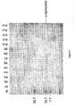
- CSF was pooled from several patients. 200 ml pooled CSF was separated according to isoelectric point via a Rotophor. Subsequently, the resulting fractions were run on a SDS-PAGE and immunoblotted with the monoclonal antibody from Transduction Labs (Lexington, KY, USA). Two immuno-reactive bands were detected in the 19 kDa range and with a pI ranging from 4 to 6 (Fig. 4). The lower band could be a C-terminal truncated form, since deletion of some negative charged amino acids (6E in the last 20 amino acids) causes a shift in pI towards the more basic end. Based on this immunoreactivity the concentration range of ⁇ -synuclein was estimated in the pg/ml range.
- Alpha-synuclein was purified from 3 liters of induced E. coli cultures resulting in purification of more than 10 mg ⁇ -synuclein (more than 90% pure estimated from coumassie and silverstained gels).
- the protein was injected into mice following several immunization schemes (Table 3). After 4 injections the titer to ⁇ -synuclein was evaluated in a coating ELISA. Six mice had a titer above 100000 (titer defined as serum dilution resulting in OD value twice the background).
- spleen cells were retrieved from animal 312 (m3) and used for cell fusion mainly according the procedure as described by Kölher and Milstein (1975).
- hybridoma's were tested for the presence of specific antibodies in a direct coating assay. Subsequently, they were retested on dot-blot of E. coli lysates containing ⁇ -, ⁇ -, ⁇ -synuclein and deletion mutants from ⁇ -synuclein in order to select for hybridomas that produce antibodies that recognize a different epitope on the synuclein protein.
- a Mini-Leak-Medium (KEM-EN-TEC Biozyme, Vancouver, BC, Canada; Cat No 10127, Lot No 60232-5) is used to immunopurify the antibodies according to instructions provided by the manufacturer.
- Biotinylation is performed according to well-established procedures (Bonhard et al., 1984) using D-Biotinoyl-eta-aminocaproic acid N-Hydroxysuccinimide Ester (Boehringer-Mannheim, Brussels, Belgium; Cat No 1008960).
- a sandwich ELISA based on an ⁇ -synuclein antibody as capturing antibody and one of the biotinylated monoclonal antibodies as detector antibody is developed. Maxisorp microtiterplates are coated with Affini Pure Goat anti-Mouse IgG (Jackson Immuno Research Laboratories, Inc., West Grove, Pennsylvania, USA; Cat No 115-035-144). 1% BSA (Clinical Grade 98% fatty acid free; ICN, Biomedical Research Products, Costa Mesa, CA, USA; Cat No 105033, Lot No 6p384) + 1% mice serum in PBS is used as blocking buffer. Anti- ⁇ -synuclein (Transduction Labs, Lexington, KY, USA) diluted 1/1000 in blocking buffer is added.
- biotinylated anti- ⁇ -synuclein monoclonal antibody is added in the presence of 1% mice antibodies at a concentration chosen for optimal background-signal ratio.
- the biotinylated rabbit antibodies are then detected via horse-radish labeled streptavidine (Jackson Immuno Research Laboratories, Inc., West Grove, Pennsylvania, USA; Cat No 016-030-084) at a dilution of 1/2000.
- Coupled peroxidase is detected via TMB, H 2 O 2 substrate solution. Reaction is stopped after 30 min with 2N H 2 SO 4 and absorbance is measured at 450 nm.
- Example 3 Detection of other neurological markers in cerebrospinal fluid
- Total tau was measured with the tau antigen test, using AT120 as capturing antibody and biotinylated HT7-BT2 as detector antibody (INNOTEST hTau antigen, Innogenetics, Gent, Belgium).
- Monoclonal antibody AT120 reacts equally well with both normal and hyperphosphorylated human tau protein (Vandermeeren et al., 1993)
- monoclonal antibody HT7 also reacts equally well with both normal and hyperphosphorylated human tau protein, while monoclonal antibody BT2 preferentially recognizes normal tau (Goedert et al., 1994).
- Affinity purified tau protein prepared as described previously (Mercken et al., 1992b), was used as standard.
- Phospho-tau was measured with a sandwich ELISA, using as HT7 as capturing antibody and biotinylated AT270 as detector antibody (INNOTEST phospho-tau(181), Innogenetics, Gent, Belgium). AT270 specifically recognizes phospho-tau (International application published under WO 95/17429).
- ⁇ -amyloid( 1-42) concentrations were measured with the Innotest ⁇ -amyloid (1-42) (Innogenetics, Gent, Belgium).
- the assay is a sandwich-type ELISA, in which a first monoclonal antibody, 21F12 (specific for the carboxy-terminus of amyloid), is used as capturing antibody and biotinylated 3D6 (specific for the amino-terminus), is used as detector antibody.
- 21F12 monoclonal antibody
- 3D6 specific for the amino-terminus
- 21F12 antibody was suspended in 10 mM Tris-10 mM NaCl and coated onto Nunc Maxisorbs microtiter plates overnight at 4°C. After one wash step, plates were blocked for 2 hr at 25°C with PBS-0.1 % casein. The test was performed by simultaneous incubation (one hour, 25°C) of 75 ⁇ l biotinylated 3D6 and 25 ⁇ l CSF or standard. After several wash steps, the amount of bound antibody was verified by adding 100 ⁇ l HRP-streptavidine (RDI, Flanders, New York, NY, USA). Incubation was continued for 30 min. at 25°C. Then, 100 ⁇ l of 0.42 mM 3,5,3',5'-tetramethylbenzidine was added as peroxidase substrate. The reaction was stopped after 30 min with 50 ⁇ l 0.9N H 2 SO 4 .
- ⁇ -amyloid (1-40) concentrations were measured using a C-terminal specific affinity purified polyclonal antibody from Quality Controlled Biochemicals (QCB, Hopkinton, MA, USA) as capturing antibody and 3D6 (Citron et al., 1997; Johnson-Wood et al., 1997) as detector antibody.
- Nunc maxisorps microtitre plates were coated for 2 hrs at 25°C with 5 ⁇ g/ml affinity purified goat anti rabbit IgG (H+L) (Jackson Immuno Research Laboratories, Inc., West Grove, Pennsylvania, USA; Cat. No 111-005-144) in 10 mM Tris-10 mM NaCl buffer.
- the amount of antibody bound was verified by adding 100 ⁇ l SV-AP (Gibco, Rockville, MD, USA; Cat No JK-4410). Incubation was continued for one hour at 25°C. After a final wash step (five times), 100 ⁇ l of TMB, dissolved in substrate buffer, was added as peroxidase substrate. The reaction was stopped after 30 min. with 50 ⁇ l 0.9N H 2 SO 4 .
- Neuromodulin was also measured with a sandwich-type ELISA, using two epitope-specific monoclonal antibodies (NM2, NM4; Oestreicher et al., 1994).
- NM2 was selected as capturing antibody
- biotinylated NM4 as detector antibody.
- Recombinant neuromodulin was used as standard.
- NSE measurements were based on a sandwich ELISA using an anti-NSE monoclonal antibody, 2E7, as capturing antibody, and the peroxidase-labelled anti-NSE monoclonal antibody, 10Cl, as detector antibody.
- Purified NSE from human brain was used as standard (Vanmechelen et al., 1997).
- Protein concentrations were determined with the BCA protein reagent (Pierce, Rockford, Illinois, USA).
- Example 4 Combination assay, making use of CSF-Rab3a, CSF-SNAP25, CSF-tau and CSF-beta-amyloid (1-42) as neurological markers for the specific detection of Alzheimer's disease and the differentiation of Alzheimer's disease versus age-matched controls
- the Alzheimer's disease (AD) group included 32 patients, 15 men and 17 women, with a mean age ⁇ SD of 75.0 ⁇ 6.6 years.
- the vascular dementia (VAD) group existed of 20 patients, 10 men and 10 women, with a mean age ⁇ SD of 81.0 ⁇ 7.0 years.
- the control group contained 18 individuals, 7 men and 11 women, with a mean age ⁇ SD of 67.5 ⁇ 5.5 years. Diagnosis of probable AD was made by exclusion, in accordance with the NINCDS-ADRDA criteria (McKhann et al., 1984).
- VAD was diagnosed in patients with transitory ischemaemic attacks and/or stroke episodes in relation to the evolution of dementia and/or CT finding of large infarcts and/or multiple lacunas, and/or history of or clinical findings of severe vascular diseases, such as arterial hypertension or diabetes mellitus with complications.
- the control group consisted of individuals without histories, symptoms or signs of psychiatric or neurological disease, malignant disease, or systemic disorders (e.g. rheumatoid arthritis, infectious disease). In individuals over 60 years of age, the cognitive status was examined using the Mini-Mental state examination (Folstein et al., 1975). Individuals with scores below 28 were not included. The study was approved by the Ethics Committee of the University of Göteborg (Göteborg, Sweden). The patients (or their nearest relatives) and the individuals of the control group gave their informed consent to participate in the study.
- likelihood ratios were determined (Table 5). Since Rab3a and SNAP25 levels are correlated, one can only use either Rab3 a or SNAP25 in combination with tau and ⁇ -amyloid (1-42) (Fig. 8). Thus likelihood-ratios for combinations of Rab3a and SNAP25 were not determined.
- Example 5 Combination assay, making use of CSF-tau, CSF-neuromodulin and CSF-neuron-specific enolase as neurological markers for the diagnosis of chemotherapy-induced neuronal damage in children treated for leukemia
- ACL anaplastic large cell lymphoma
- VHR very high risk
- This group includes 5 children with medulloblastoma (3 staging, 1 during treatment, 1 follow up), 3 children with Hodgkin's disease (staging), 3 children with rhabdomyosarcoma (2 staging, 1 during treatment), 2 children with myelodysplastic syndrome (MDS) (staging), 2 children with Langerhans cell histiocytis (LCH/HLH, staging), 1 child with juvenile chronic myelomic leukemia (CML) (during treatment), choriocarcinoma (follow up), or germinoma (staging).
- MDS myelodysplastic syndrome
- CML juvenile chronic myelomic leukemia
- the supernatant was stored at -70°C for subsequent analysis.
- the number of freeze/thaw cycles was restricted to a minimum.
- Routine CSF measurement included cytology, protein concentration, glucose, etc. Since normality for all neurological marker data, independent of the diagnostic groups, was rejected, non-parametric statistics were used for the analysis.
- the Kruskal-Wallis test was used to investigate group differences regarding the effect variables (tau, neuromodulin, protein, etc.).
- the Wilcoxon signed ranks test matched-pairs was used to check for differences between the first diagnostic LP and subsequent LPs. Pearson's correlation was used to investigate possible covariates. Analyses were done with Prism software v2.01 (Graphpad Software Inc., San Diego, CA, USA), Systat version 7 (SPSS, Chicago, IL, USA).
- One CSF sample was available from a child who was treated in another center and who suffered from manifest neurotoxicity after treatment with MTX, and who had diplegia.
- the tau and neuromodulin levels in this child exceeded the highest standard used (1500 pg/ml for tau, 8000 pg/ml for neuromodulin), reflecting gross neuronal degeneration.
- Figure 14a shows the evolution of tau in individual patients with AML.
- Patients 11, 12 and 23 did not have a significant increase in tau during treatment (Table 7c).
- Patient 12 from whom LPs were taken at days 1 and 8, had an aggressive disease and died early after bone marrow transplantation. From one out of two patients with Down's syndrome, longitudinal data were available, and this patient had tau levels raising slightly above 300 pg/ml, in contrast to the evolution of tau levels in the CSF of patient 11 and 23.
- Patient 69 with evidence of CNS invasion at diagnosis, had a tremendous increase of tau and neuromodulin (Fig. 14b) in the CSF. This child is still under treatment, and is in complete remission at the moment.
- Example 6 Combination assay for the diagnosis of brain damage resulting from perinatal asphyxia
- Perinatal asphyxia may be associated with neuronal damage.
- CSF neurological markers may complement clinical data in the evaluation of hypoxic-ischemic events (Garcia-Alix, 1994). It has become increasingly evident that modified brain metabolic activity is reflected by changes in components in the CSF. The perinatal levels of the CSF markers and the distribution of changes due to aspyxia are evaluated.
- Example 7 Combination assay, making use of CSF-neuromodulin, CSF-tau and CSF- ⁇ -amyloid (1-42) as neurological markers for the specific detection of Alzheimer's disease and for the differentiation of Alzheimer's disease versus control subjects
- CSF was routinely sampled as part of the neurological examination. All tests were performed on the remainder of the CSF. ⁇ -amyloid (1-42) was only measured in CSF samples which were properly stored in polypropylene tubes and frozen only once.
- the levels of tau, ⁇ -amyloid (1-42) and neuromodulin (GAP-43) in the CSF of these patients were determined. Average CSF levels for each group are shown in table 8. Individual levels of ⁇ -amyloid (1-42 ), tau and neuromodulin are show in figures 15a, 15b and 15c, respectively.
- CSF-tau and CSF- ⁇ -amyloid (1-42) were significantly altered in the AD patients. Also, CSF-neuromodulin was significantly increased in AD. In contrast, in the aged memory-impaired patients, no significant effect in CSF-neuromodulin was observed, while CSF-tau level was significantly raised compared to the control subjects. Three of the 7 patients with memory-impairment had CSF-tau levels above the maximum defined by the control group (280 pg/ml). Due to improper storage, CSF. ⁇ -amyloid (1-42) levels could only be determined in 3 patients with memory impairment. In two of the three patients, levels below 300 pg/ml were found. One of these patients had also an elevated CSF-tau level.
- the other patient had a CSF-tau level of 278 pg/ml, i.e. just below the maximum defined by the control group.
- CSF-tau level 278 pg/ml
- an increase of CSF-tau level of 327 pg/ml was observed, together with a ⁇ -amyloid (1-42) level below 300 pg/ml.
- This analysis on a few samples of memory impaired individuals indicate that the combined use of tau, ⁇ -amyloid (1-42) and neuromodulin is a better indicator for the presence of Alzheimer's disease.
- a larger number of samples per group is being analyzed (at least 50 samples per diagnostic group).
- Example 8 Combination assay, making use of CSF-neuromodulin, CSF-beta-amyloid (1-42) and CSF-tau as neurological markers for the specific detection of Alzheimer's disease, to differentiate Alzheimer's disease versus control subjects , for the specific detection of Parkinson disease, to differentiate Parkinson disease versus control subjects and to differentiate between Alzheimer's disease and Parkinson disease.
- CSF was obtained from 60 patients with Alzheimer's disease, 23 patients with Parkinson disease and 32 age-mached controls. The levels of tau, ⁇ -amyloid (1-42) and neuromodulin (GAP-43) in the CSF of these patients were determined. Average CSF levels for each group are shown in table 9.
- the variables tau/neuromodulin and ⁇ -amyloid (1-42) were selected and correctly classified 55 of the 58 Alzheimer's disease patients (sensitivity 94.8%, CI 85.6%-98.9%) and 30 of the 31 control patients (specificity 96.8%, CI 83.3%-99.9%) (Fig. 17). Further analysis of a larger number of samples will enable to demonstrate a statistically significant improvement of the diagnosis of Alzheimer's disease by use of these three markers.
- ⁇ -amyloid (1-42) and neuromodulin were selected to differentiate Alzheimer's disease patients from Parkinson disease patients with a sensitivity of 94.8% (CI 85.6%-98.9%) for the Alzheimer's disease patients and a specificity of 78.3% (CI 56.3%-92.5%) for the Parkinson disease patients. Further analysis of a larger number of samples will enable to demonstrate a statistically significant improvement of the differentiation between Alzheimer's disease and Parkinson disease by use of these three markers.
- Example 9 Use of CSF- ⁇ -synuclein as a neurological marker to differentiate between Lewy Body dementia and Alzheimer's disease
- CSF is obtained from 30-40 patients with Alzheimer's disease, 10-20 patients with Lewy Body dementia and 10-20 age-mached controls.
- the concentration of ⁇ -synuclein in the CSF of the different patients and control groups is quantified.
- a significant different mean value for the ⁇ -synuclein level in the Alzheimer's disease group versus the mean value for the ⁇ -synuclein level in the Lewy Body dementia group allows us to differentiate between both types of dementia. Differentiation between Alzheimer's disease and Lewy Body dementia is further improved by combining the quantification of ⁇ -synuclein with the quantification of at least 2 other neurological markers (such as tau and ⁇ -amyloid (1-42) .
- other neurological markers such as tau and ⁇ -amyloid (1-42)
- Example 10 Combination assay, making use of CSF-beta-amyloid (1-42) .
- CSF-tau and CSF-phospho-tau as neurological markers for the specific detection of Frontal Temporal Lobe dementia and to differentiate Frontal Temporal Lobe dementia versus other dementia
- CSF is obtained from 30-40 patients with Frontal Temporal Lobe dementia and 10-20 age-mached controls.
- the level of ⁇ -amyloid (1-42) , tau and phospho-tau in the CSF of the different patients with Frontal Temporal Lobe dementia and in the control patients is quantified.
- the mean values for the level of tau and phospho-tau in the patients with Frontal Temporal Lobe dementia is significantly increased compared to mean values for the level of tau and phospho-tau in the control patients.
- the patients with Frontal Temporal Lobe dementia show a decreased level of ⁇ -amyloid (1-42) compared to the control patients.
- Example 11 Combination assay, making use of CSF-beta-amyloid (1-42) , CSF-tau and plasma-beta-amyloid (1-42) as neurological markers for the specific detection of vascular problems in Alzheimer's disease and to differentiate vascular disease from other forms of Alzheimer's disease
- CSF and plasma is obtained from 30-40 patients with vascular disease, from 30-40 patients with other forms of Alzheimer's disease and from 10-20 age-mached controls.
- the level of tau and ⁇ -amyloid (1-42) in the CSF and the level of ⁇ -amyloid (1-42) in the plasma of the different patients with vascular disease, with other forms of Alzheimer's disease and in the control patients is quantified.
- a quantitatively different level of plasma- ⁇ -amyloid (1-42) , CSF-tau and CSF- ⁇ -amyloid (1-42) enables the differentiation between the vascular disease patients and the patients with other forms of Alzheimer's disease.
Landscapes
- Life Sciences & Earth Sciences (AREA)
- Health & Medical Sciences (AREA)
- Engineering & Computer Science (AREA)
- Biomedical Technology (AREA)
- Hematology (AREA)
- Chemical & Material Sciences (AREA)
- Urology & Nephrology (AREA)
- Molecular Biology (AREA)
- Immunology (AREA)
- Biotechnology (AREA)
- Analytical Chemistry (AREA)
- Microbiology (AREA)
- Proteomics, Peptides & Aminoacids (AREA)
- Neurosurgery (AREA)
- Neurology (AREA)
- Food Science & Technology (AREA)
- Medicinal Chemistry (AREA)
- Physics & Mathematics (AREA)
- Cell Biology (AREA)
- Biochemistry (AREA)
- General Health & Medical Sciences (AREA)
- General Physics & Mathematics (AREA)
- Pathology (AREA)
- Investigating Or Analysing Biological Materials (AREA)
- Measurement And Recording Of Electrical Phenomena And Electrical Characteristics Of The Living Body (AREA)
- Eye Examination Apparatus (AREA)
- Investigating Or Analysing Materials By The Use Of Chemical Reactions (AREA)
- Measuring Or Testing Involving Enzymes Or Micro-Organisms (AREA)
- Peptides Or Proteins (AREA)
Abstract
Description
Also frontal temporal lobe dementia is often misdiagnosed as other types of dementia or other psychiatric disorders since the symptoms of frontal temporal lobe dementia can also be observed in other disorders.
There is no clear-cut difference between vascular disease and Alzheimer's disease and also here the risk of misdiagnosing is evident. As pharmacological treatment of vascular disease is possible, an early correct diagnosis is crucial.
Also recognition and treatment of brain damage caused by inducing agents such as certain chemical compounds, irradiation, chemotherapy or hypoxic-ischemic events, remains a frequent and important clinical problem for most neurologists. In the cases where clinical diagnosis is doubtful, definitive diagnosis can only be done irrevocably by neuropathologic examination. As such, an accurate and differential diagnosis of neurodegeneration is only possible post mortem.
Therefore, a method for the early detection of neurological disorders in patients and for the monitoring of neurological changes induced by different agents, would be of great help for determining whether exposure to the inducing agent can be continued, whether appropriate doses and drugs are being used in individual patients and for starting the right treatments.
The microtubule-associated protein tau, for example, is a major protein component of paired helical filaments (PHF) and neurofibrillar tangles (11FT) (Brion et al., 1985; Delacourte and Defossez, 1986; Grundke-Iqbal et al., 1986; Kosik et al., 1986; Wood et al., 1986; Kondo et al., 1988). Tau protein exists in different isoforms, of which 4 to 6 are found in adult brain but only 1 isoform is detected in fetal brain. The diversity of the isoforms is generated from a single gene on
The gamma-subunit of neuron-specific enolase (NSE) is a major constituent of neuronal cytosol (Kato et al., 1981). NSE represent 3% of total soluble brain protein. In adults, it is considered to be useful in evaluating active neuronal damage of ischemic, infectious, or tumoral origin (Garcia et al., 1994). NSE in serum of children has not been studied. Nara et al. (1988) showed that a high NSE level in cerebrospinal fluid or serum is correlated with poor outcome and death in comatose children. However, increased serum NSE is not necessarily of CNS origin. Several tissues, including peripheral neurons, endocrine glands, lymphocytes, red blood cells, and platelets contain NSE (Kaiser, 1989), which may compromise the use of this marker alone.
β-amyloid, a 40-43 amino acids long peptide, is derived via proteolytic cleavage from a large precursor protein, called amyloid precursor protein or APP. Amyloid is produced during metabolism of normal cells. The amyloid peptide exhibits a high degree of heterogeneity. Two major forms of β-amyloid have been identified, β-amyloid(1-40) and β-amyloid(1-42). β-amyloid(1-42) is a major constituent of the neuritic plaques of Alzheimer's disease, Down's syndrome and normal aged brains. It might be neurotoxic and it is known to increase the vulnerability of neurons to other insults. In addition, low concentrations of soluble amyloid can induce cholinergic hypoactivity that is not dependent on concurrent neurotoxicity. Acetylcholine plays a critical role in cognitive processes (Auld et al., 1998). The development of high affinity monoclonal antibodies specifically recognizing well-defined epitopes of the peptide has lead to a simple test for the β-amyloid(1-42) peptide in unconcentrated cerebrospinal fluid (Citron et al., 1997; Johnson-Wood et al., 1997). This test may also prove to be of value in monitoring long-term effects of drugs, irradiation, or chemical substances interfering with APP processing.
Growth associated protein-43 (GAP-43), also called neuromodulin or B-50, is a nervous tissue specific protein, primarily localized to the axons and presynaptic terminals. GAP-43 is considered to play a major role in neuronal growth, neurite formation, and in regeneration and neuronal sprouting (Skene and Willard, 1981; Basi, 1987; Benowitz et al., 1989; Mercken et al., 1992a). Synapse proteins have different roles in synapse function. Proteins such as synapsin are important in determining the amount of vesicles available for fusion, while Rab3a and rabphilin are important in targeting the vesicles to the membrane. The docking process is determined by a molecular complex of synaptobrevin, SNAP25, Sec and syntaxin, while it is believed that CSP and synaptotagmin play an important role in the Ca2+-dependent release of the content of the vesicle. Alpha-synuclein is abundant in synapses of the substantia nigra and basal ganglia, and belongs to a family of proteins including α-synuclein and γ-synuclein.
Such intracellular markers that are present and stable in body fluids and that reflect the metabolic state of neurons in the central nervous system, might be useful in early recognition of neurodegeneration, even before clinical signs are present. A biochemical index of neuronal function that could be used, possibly in combination which other diagnostic methods, would be of a great help to improve the clinical diagnostic accuracy and therapeutic monitoring of neurodegeneration.
Studies on brain GAP-43 have suggested decreased levels in the frontal cortex in Alzheimer's disease but increased levels in other regions (Coleman et al., 1992). No study has been performed on GAP-43 in body fluids of patients with dementia disorders.
Another important structural alteration in brains of AD patients is synapse loss. In fact, recent studies measuring the amount of tangles, plaques and synapse loss suggest that synapse loss is the major correlate of the degree of dementia (Terry et al., 1991). In the latter study reduction of synaptophysin immunoreactivity was observed. A similar reduction has been documented for other synapse proteins: synaptotagmin, Rab3a, synaptobrevin and syntaxin (Blennow et al., 1996; Davidsson et al., 1996; Shimohama et al., 1997; Ferrer et al., 1998). Also for several forms of Parkinson disease there are strong indications that synapse proteins play a pathological role.
Two mutations in α-synuclein were detected in two rare forms of Familial Parkinson disease and α-synuclein was characterized as a major component in lewy bodies. Lewy body formation in vivo may result from synuclein accumulation, which may be the consequence of a reduction in the fast axonal transport or overexpression of synuclein (Jensen et al., 1998). As synapse proteins appear to be one of the major correlates of the degree of dementia (Terry et al., 1991), it would be useful to provide methods for the detection and quantitation of synapse proteins in body fluids. The presence and quantification of synapse proteins in CSF has not yet been fully explored. Chromogranin has already been used as a marker for synapse loss in CSF, but a reduction was only shown in 'pure' or type I Alzheimer's disease (Blennow et al., 1995). Shortly thereafter synaptotagmin I was shown to be present in CSF (Davidsson et al., 1996). In this study it was demonstrated that synaptotagmin is selectively reduced in the left hippocampal formation and
It is another aim of the present invention to provide a method for a more specific detection, quantification and/or differential diagnosis of Alzheimer's disease in an individual.
It is another aim of the present invention to provide a method for a more specific detection, quantification and/or differential diagnosis of Lewy Body Disease in an individual.
It is another aim of the present invention to provide a method for a more specific detection, quantification and/or differential diagnosis of Parkinson disease in an individual.
It is another aim of the present invention to provide a method for a more specific detection, quantification and/or differential diagnosis of frontal temporal lobe dementia in an individual.
It is another aim of the present invention to provide a method for the differentiation of Alzheimer's disease versus Parkinson disease.
It is another aim of the present invention to provide a method for the differentiation of Alzheimer's disease versus Lewy Body dementia.
It is another aim of the present invention to provide a method for the specific detection or quantification of vascular problems in Alzheimer's disease and for the differential diagnosis of different forms of Alzheimer's disease.
It is another aim of the present invention to provide a method for the diagnosis of neurodegeneration induced by chemotherapy or by exposure to chemical compounds or irradiation.
It is another aim of the present invention to provide a method for the diagnosis of neurodegeneration induced by chemotherapy in individuals treated for leukemia or brain tumor.
It is another aim of the present description to provide a new method for the detection of the synapse protein Rab3a in cerebrospinal fluid.
It is another aim of the present description to provide a new method for the detection of Rab3a in cerebrospinal fluid that allows a more specific detection, quantification and/or differential diagnosis of neurodegeneration in an individual.
It is another aim of the present description to provide a new method for the detection of Rab3a in cerebrospinal fluid that allows a more specific detection, quantification and/or differential diagnosis of Alzheimer's disease.
It is another aim of the present description to provide a new method for the detection of the synapse protein α-synuclein in cerebrospinal fluid.
It is another aim of the present description to provide a new method for the detection of α-synuclein in cerebrospinal fluid that allows a more specific detection, quantification and/or differential diagnosis of neurodegeneration in an individual.
It is another aim of the present description to provide a new method for the detection of α-synuclein in cerebrospinal fluid that allows a more specific detection or quantification of Alzheimer's disease and/or Lewy Body Disease and/or that allows the differential diagnosis of Alzheimer's disease versus Lewy Body Disease.
It is another aim of the present invention to provide a diagnostic kit for performing a method as described above.
It is another aim of the present invention to provide a method for therapeutic monitoring and/or determination of the effectiveness of a certain treatment.
As can be seen from the present examples, by use of at least three neurological markers a more specific and sensitive detection of a number of neurodegenerative conditions was obtained. It is also apparent that by use of at least three neurological markers it became possible to discriminate between a number of neurodegenerative conditions that could not be differentiated on the basis of clinical diagnosis.
The term's neurodegeneration and neurodegenerative condition used in the present application stand for the same and are used interchangeable throughout the application. These terms include any condition of the brain that is associated with a neuronal malfunctioning. Various diseases associated with neurodegeneration are cited in Wilson et al. (1991). They include Alzheimer's disease, stroke (Focal brain injury), diffuse brain injury, vascular disease, Parkinson disease, Lewy Body Disease, Creutzfeld Jacob Disease, Frontal temporal lobe dementia, Guilain Barré Syndrome, Multiple Sclerosis, Normal Pressure Hydrocephalus, Amyotrophic Lateral Sclerosis,
Schizophrenia, Depression, Neurolathyrisme, Epilepsy and Asphyxia. However, this list is not complete. Other diseases known to be associated with neuronal malfunctioning are included as well. Neurodegeneration also includes any kind of brain damage or any condition of the brain that is associated with a neuronal malfunctioning and which is caused by a specific inducing agent.
In a preferred embodiment of the present invention, the neurodegenerative condition to be specifically detected, quantified and/or differentially diagnosed is chosen from the group consisting of Alzheimer's disease, Lewy Body Disease, Parkinson disease and frontal temporal lobe dementia. Lewy Body Disease is used for any disease showing lewy bodies in the brainstem, basal forebrain, hypothalamic nuclei and/or neocortex. Lewy Body Disease includes Parkinson disease, multiple system atrophy and Lewy Body dementia.
In another preferred embodiment of the present invention, the neurodegenerative condition to be specifically detected, quantified and/or differentially diagnosed is induced by hypoxic-ischemic events, chemotherapy, radiotherapy, or by exposure to chemical compounds or irradiation. More particularly, neurodegeneration can be induced by chemotherapy or radiotherapy during the treatment of leukemia or brain tumor.
However, this list of neurodegenerative conditions is not complete. Other conditions in which a malfunctioning of the brain occurs, are included as well.
The expression "specific detection of neurodegeneration" as used in the present invention means that a higher sensitivity and specificity is obtained for the association of a certain disease or a certain cause of neurological disorder with a certain neurodegenerative condition than would be obtained when less than three neurological markers were used for diagnosis.
The expression "quantification of neurodegeneration" as used in the present invention means that the degree of neuronal malfunctioning due to a certain neurodegenerative condition is determined. The expression "differential diagnosis of neurodegeneration" as used in the present invention refers to the discrimination between various neurodegenerative conditions in this way that a certain disease or a certain cause of neurological disorder is associated with a certain neurodegenerative condition.
The specific detection, quantification and/or differential diagnosis of neurodegeneration in an individual is accomplished by the detection of at least three different neurological markers in one or more body fluid samples of said individual; making use of an immuno-assay comprising the steps of:
- bringing one or more body fluid samples obtained from said individual into contact with at least three different antibodies (primary antibodies or capturing antibodies) recognizing each a different neurological marker in the body fluid samples, under conditions being suitable for producing an antigen-antibody complex; and
- detecting the immunological binding of said antibodies to said body fluid samples;
- inference on the level of said neurological markers in said body fluids, the type and degree of neurodegeneration being reflected in the quantitative changes in the level of all of said neurological markers compared to control samples.
- a) a secondary antibody (or detector antibody)
- that can be a monoclonal antibody recognizing a specific epitope of the antigen-antibody complex but not recognizing the primary antibody alone, or
- which can be a polyclonal antibody recognizing a specific epitope of the antigen-antibody complex but not recognizing the primary antibody alone, with said polyclonal antibody being preferably purified by immunoaffinity chromatography using immobilized neurological marker or neurological marker-primary antibody complex;
- b) a marker either for specific tagging or coupling with said secondary antibody, with said marker being any possible marker known to the person skilled in the art;
- c) appropriate buffer solutions for carrying out the immunological reaction between the antibodies and the body fluid samples, between the secondary antibody and the neurological marker-primary antibody complex and/or the bound secondary antibody and the marker on the other hand; and
- d) possibly also, for standardization purposes, purified proteins or synthetic peptides reactive with the antibodies used for detection of the neurological markers.
The term "epitope" refers to that portion of the antigen-antibody complex that is specifically bound by an antibody-combining site. Epitopes may be determined by any of the techniques known in the art or may be predicted by a variety of computer prediction models known in the art.
The expression "recognizing", ''reacting with", "immunological binding" or "producing an antigen-antibody complex" as used in the present invention is to be interpreted that binding between the antigen and antibody occurs under all conditions that respect the immunological properties of the antibody and the antigen.
The term body fluids refers to all fluids that are present in the human body including but not limited to blood, lymph, urine and cerebrospinal fluid (CSF).
In a specific embodiment, the present invention relates to a method as described above wherein the body fluid sample is chosen from the group consisting of a cerebrospinal fluid sample and a blood sample. The blood sample can include the whole sample as taken from the patient. More preferably the blood sample includes a plasma sample or a serum sample.
In the methods of the present invention it is also possible to detect the same marker in two different body fluids (in combination with the detection of at least one other marker) or to detect the same marker in three different body fluids. For example, two neurological markers are detected in cerebrospinal fluid and one of these neurological markers is also detected in plasma. As shown in the example section, also detection of the same marker in two different body fluids leads to a more specific and sensitive detection and to a better differential diagnosis of neurodegeneration compared to detection of this maker in only one body fluid.
The neurological markers that are detected in the method of the present invention can be any protein associated with certain types of neuronal cells or cell function, of which the level in one or more body fluids under conditions of neurodegeneration is indicative for the disease process or the cause of neurological disorder. Some neurological markers are elevated and others are reduced in one or more body fluids under a certain neurological condition. Any possible combination of 3, 4, 5, 6, 7, 8 or more neurological markers that have an altered level in a certain body fluid under a certain neurological condition can be used for the specific detection, quantification and/or differential diagnosis of said neurological condition in an individual.
Possible neurological markers used for specific detection, quantification and/or differential diagnosis of neurodegeneration include: tau, neuron-specific enolase (NSE), beta-amyloid(1-42), beta-amyloid(1-40), neuromodulin, synapse proteins (such as Rab3a, SNAP25, α-synuclein, synapsin, synaptotagmin, synaptobrevin, syntaxin, rabphilin, n-sec, cystein string protein and others), glial fibrillary acidic protein (GFAP), S100, IL6, TNF, IL1, IL2, neurofilament (NF), myelin basic protein (MBP) and 14-3-3. However, this list is not complete. Other neurological markers that are indicative for a certain disease process or cause of neurological disorder can be used as well. The behavior of some of these neurological markers in body fluids of patients suffering from different neurological diseases and brain damage is shown in table 2.
Any monoclonal or polyclonal antibody prepared or present in the art that specifically recognizes one of the above-mentioned neurological markers, can be used for the detection of the neurological marker. Antibodies specifically recognizing tau include Alz50 (Ghanbari et al., 1990), Ab423 (Harrington et al., 1991), AT8 (International application published under WO 93/08302), AT120 (Vandermeeren et al., 1993); AT180 and AT270 (International application published under WO 95/17429) and AT100 (International application published under WO 96/04309). But also other antibodies known in the art specifically recognizing tau can be used. Antibodies that specifically recognize NSE include 10Cl and 2E7 and others from Innogenetics (Gent, Belgium), commercially available antibodies such as can be obtained from Dako (Glostrup, Denmark; Cat No BBS/NC/VI-H14), from Biogenex (San Ramon, CA, USA; Cat Nos MA055-5C and AM055-5M), from RDI (Flanders, NJ, USA; Cat No RDI-TRK4N6), from Roche Diagnostic Systems (Basel, Switzerland; Cat No 07 34373), from Immunosource (Brussels, Belgium; Cat Nos CLA 73/5 and CR7041M) and from Cortex Biochem (San Leandro, CA, USA; Cat No CR7047). This list of antibodies recognizing NSE is not complete and other antibodies, commercially available or described in the art, that recognize NSE, can be used as well. Antibodies that specifically recognize β-amyloid include 2H3, 8E5 (Johnson-Wood et al., 1997), 10H3 (Majocha et al., 1992; Friedland et al., 1994), 2G3 (Citron et al., 1996), BA-27 and BC-05 (Suzuki et al., 1994), BNT77 (Asami-Odaka et al., 1995), 369.2B (König et al., 1996), 22C11 (Lannfelt et al., 1995), 6E10 (Kim et al., 1990) and AMY-33 (Stern et al., 1990). But any other antibodies known in the art specifically recognizing β-amyloid can be used as well. Antibodies that specifically recognize neuromodulin include NM2 (Oestreicher et al., 1994), NM4 (Six et al., 1992), NM1, NM3, NM6, NM7 and NM8 (Mercken et al., 1992a). But any other antibody known in the art that specifically recognizes neuromodulin can be used as well. Antibodies specifically recognizing Rab3a include commercially available antibodies such as can be obtained from Transduction Labs (Lexington, KY, USA; Cat No R35520). Also other antibodies commercially available or described in the art that recognize Rab3a can be used. Antibodies specifically recognizing SNAP25 include commercially available antibodies such as can be obtained from Serotec (Oxford, UK; Cat No SP12), from Sternberger Monoclonals Inc. (Distributed by Affinity Research Products Lim., Mamhead, Exeter, UK; Cat No SMI-81), from Chemicon (Temecula, CA, USA; Cat No MAB33 1) and from Transduction Labs (Lexington, KY, USA; Cat No S35020). This list of antibodies recognizing SNAP25 is not complete and other antibodies commercially available or described in the art that recognize SNAP25 can be used as well. Antibodies specifically recognizing α-synuclein include commercially available antibodies such as can be obtained from Transduction Labs (Lexington, KY, USA; Cat No S63320). Also other antibodies commercially available or described in the art that recognize α-synuclein can be used. Also for the specific detection of other synapse proteins that can possibly be used as neurological markers, various antibodies are commercially available and/or known in the art. Antibodies specifically recognizing
This list of antibodies recognizing 14-3-3 is not complete and other antibodies commercially available or described in the art that recognize 14-3-3 can be used as well. Antibodies specifically recognizing neurofilament include commercially available antibodies such as can be obtained from Innogenetics (Gent, Belgium; Cat Nos M-011 and M-005) and from Alexis (Läufelfingen, Switzerland; Cat Nos BC-4000-A-L001 and BC-4010-A-L001). This list of antibodies recognizing neurofilament is not complete and other antibodies commercially available or described in the art that recognize neurofilament can be used as well.
Also fragments derived from these monoclonal antibodies such as Fab, F(ab)'2, ssFv ("single chain variable fragment") and other antibody like constructs that retain the variable region of the antibody, providing they have retained the original binding properties, can be used in a method of the present invention. Such fragments are commonly generated by, for instance, enzymatic digestion of the antibodies with papain, pepsin, or other proteases. It is well known to the person skilled in the art that monoclonal antibodies, or fragments thereof, can be modified for various uses. Also miniantibodies and multivalent antibodies such as diabodies, triabodies, tetravalent antibodies and peptabodies can be used in a method of the invention. The preparation and use of these fragments and multivalent antibodies has been described extensively in International Patent Application WO 98/29442.
The monoclonal antibodies used in a method of the invention may be humanized versions of the mouse monoclonal antibodies made by means of recombinant DNA technology, departing from the mouse and/or human genomic DNA sequences coding for H and L chains or from cDNA clones coding for H and L chains. Alternatively the monoclonal antibodies used in a method of the invention may be human monoclonal antibodies. The term "humanized antibody" means that at least a portion of the framework regions of an immunoglobulin is derived from human immunoglobulin sequences.
In a specific embodiment of the invention, at least one of the neurological markers to be detected in a method as described above, is chosen from the group consisting of tau, phospho-tau, β-amyloid(1-42), β-amyloid(1-40), neuromodulin, neuron-specific enolase and/or synapse proteins. Any possible combination of 3, 4, 5, 6, 7, 8 or more markers of which one is chosen from the above group can be used for the specific detection, quantification and/or differential diagnosis of neurodegeneration in an individual. It is clear that also more than one (i.e. 2, 3, 4, 5, 6, 7 or more) or even all neurological markers to be used in a method of the present invention can be chosen from the above group.
In a more specific embodiment of the invention, one, more preferably two, most preferably three of the neurological markers to be detected in a method as described above, are chosen from the following groups:
- tau, β-amyloid(1,42), and neuromodulin; or
- tau, neuron-specific enolase and neuromodulin; or
- tau, phospho-tau and β-amyloid(1-42); or
In another more specific embodiment of the invention, at least one of the neurological markers to be detected in a method as described above, is a synapse protein chosen from the group consisting of Rab3a, SNAP25 and α-synuclein. Any possible combination of 3, 4, 5, 6, 7, 8 or more markers of which one is chosen from the above group of synapse proteins can be used for the specific detection, quantification and/or differential diagnosis of neurodegeneration in an individual.
Accordingly, the present description also relates to a new method for the detection of Rab3 a in cerebrospinal fluid, comprising at least the following steps:
- bringing a cerebrospinal fluid sample obtained from an individual into contact with a monoclonal antibody (primary antibody or capturing antibody) recognizing Rab3a under conditions being suitable for producing an antigen-antibody complex; and
- detecting the immunological binding of said antibody to said cerebrospinal fluid sample.
Any antibody that allows specific detection of Rab3a in cerebrospinal fluid can be used in this new method. A preferred monoclonal antibody for use in the method of the invention can be obtained from Transduction Labs (Lexington, KY, USA; Cat No R35520).
Advantageously, the monoclonal antibody used in the invention is in an immobilized state on a suitable support. Alternatively, the present process may be put into practice by using any other immunoassay format known to the person skilled in the art.
The process for the detection of the immunological binding can then be carried out by bringing together said antigen-antibody complex formed by the antigen and the antibody recognizing Rab3 a with:
- a) a secondary antibody (or detector antibody)
- which can be a monoclonal antibody recognizing an epitope of the antigen-antibody complex but not recognizing the primary antibody alone; or
- which can be a polyclonal antibody recognizing an epitope of the antigen-antibody complex but not recognizing the primary antibody alone, with said polyclonal antibody being preferably purified by immunoaffinity chromatography using immobilized Rab3a or Rab3a-primary antibody complex.
- b) a marker either for specific tagging or coupling with said secondary antibody, with said marker being any possible marker known to the person skilled in the art;
- c) appropriate buffer solutions for carrying out the immunological reaction between the antibodies and the cerebrospinal fluid sample, between the secondary antibody and the neurological marker-primary antibody complex and/or between the bound second antibody and the marker; and
- d) possibly also, for standardization purposes, purified proteins or synthetic peptides reactive with the antibodies that recognize Rab3a.
Advantageously, the secondary antibody itself carries a marker or a group for direct or indirect coupling with a marker.
The present description also relates to a new method for the detection of SNAP25 in cerebrospinal fluid comprising at least the following steps:
- bringing a cerebrospinal fluid sample obtained from an individual into contact with a monoclonal antibody (primary antibody or capturing antibody) recognizing SNAP25 under conditions being suitable for producing an antigen-antibody complex; and
- detecting the immunological binding of said antibody to said cerebrospinal fluid sample.
Any antibody that allows specific detection of SNAP25 in cerebrospinal fluid can be used in this new method. A preferred monoclonal antibody for use in the method of the invention can be obtained from Serotec (Oxford, UK; Cat No SP12), from Sternberger Monoclonals Inc. (Distributed by Affinity Research Products Lim., Mamhead, Exeter, UK; Cat No SMI-81), from Chemicon (Temecula, CA, USA; Cat No MAB331) or from Transduction Labs (Lexington, KY, USA; Cat No S3S020).
Advantageously, the monoclonal antibody used in the invention is in an immobilized state on a suitable support. Alternatively, the present process may be put into practice by using any other immunoassay format known to the person skilled in the art.
The process for the detection of the immunological binding can then be carried out by bringing together said antigen-antibody complex formed by the antigen and the antibody recognizing SNAP25 with:
- a) a secondary antibody (or detector antibody)
- which can be a monoclonal antibody recognizing an epitope of the antigen-antibody complex but not recognizing the primary antibody alone; or
- which can be a polyclonal antibody recognizing an epitope of the antigen-antibody complex but not recognizing the primary antibody alone, with said polyclonal antibody being preferably purified by immuno-affinity chromatography using immobilized SNAP25 or SNAP25-primary antibody complex.
- b) a marker either for specific tagging or coupling with said secondary antibody, with said marker being any possible marker known to the person skilled in the art;
- c) appropriate buffer solutions for carrying out the immunological reaction between the antibodies and the cerebrospinal fluid sample, between the secondary antibody and the neurological marker-primary antibody complex, and/or the bound secondary antibody and the marker; and
- d) possibly also, for standardization purposes, purified proteins or synthetic peptides reactive with the antibodies that recognize SNAP25.
Advantageously, the secondary antibody itself carries a marker or a group for direct or indirect coupling with a marker
The present description also relates to a new method for the detection of α-synuclein in cerebrospinal fluid comprising at least the following steps:
- bringing a cerebrospinal fluid sample obtained from an individual into contact with a monoclonal antibody (primary antibody or capturing antibody) recognizing α-synuclein under conditions being suitable for producing an antigen-antibody complex; and
- detecting the immunological binding of said antibody to said cerebrospinal fluid sample.
As the presence of α-synuclein in cerebrospinal fluid had never been reported before, it was even doubtful if any α-synuclein would be present in CSF. Firstly, the present inventors were able to show that α-synuclein is present in cerebrospinal fluid. In addition, an accurate method was developed for the quantitative detection of α-synuclein in cerebrospinal fluid. The present inventors also showed that α-synuclein is altered under certain neurodegenerative conditions, including but not limited to Lewy Body Disease.
Any antibody that allows specific detection of α-synuclein in cerebrospinal fluid can be used in this new method. A preferred monoclonal antibody for use in the method of the invention can be obtained from Transduction Labs (Lexington, KY, USA; Cat No R35520).
Advantageously, the monoclonal antibody used in the invention is in an immobilized state on a suitable support. Possibly this immobilized state can be a microtiter plate, coated or not coated with anti-IgG. Alternatively, the present process may be put into practice by using any other immunoassay format known to the person skilled in the art.
The process for the detection of the immunological binding can then be carried out by bringing together said antigen-antibody complex formed by the antigen and the antibody recognizing α-synuclein with:
- a) a secondary antibody (or detector antibody)
- which can be a monoclonal antibody recognizing an epitope of the antigen-antibody complex but not recognizing the primary antibody alone; or
- which can be a polyclonal antibody recognizing an epitope of the antigen-antibody complex but not recognizing the primary antibody alone, with said polyclonal antibody being preferably purified by immuno-affinity chromatography using immobilized α-synuclein or α-synuclein -primary antibody complex.
- b) a marker either for specific tagging or coupling with said secondary antibody, with said marker being any possible marker known to the person skilled in the art;
- c) appropriate buffer solutions for carrying out the immunological reaction between the antibodies and the cerebrospinal fluid sample, between the secondary antibody and the neurological marker-primary antibody complex, and/or the bound secondary antibody and the marker; and
- d) possibly also, for standardization purposes, purified proteins or synthetic peptides reactive with the antibodies that recognize α-synuclein.
In a preferred embodiment these methods for the detection of Rab3a, SNAP25 and/or α-synuclein can be used in combination with a method for detection of one or more other neurological markers in order to specifically detect, quantify and/or differential diagnose neurodegeneration in an individual.
In an even more preferred embodiment, these methods for the detection of Rab3a, SNAP25 and/or α-synuclein can be used in combination with a method for detection of one or more neurological markers chosen from the group consisting of tau, phospho-tau, β-amyloid(1-42), β-amyloid(1-40), neuromodulin, neuron-specific enolase (NSE).
More particularly, the neurological markers used for the specific detection, quantification and/or differential diagnosis of neurodegeneration can be chosen from one of the following groups:
- tau, phospho-tau, NSE, β-amyloid(1-42), β-amyloid(1-40), neuromodulin or Rab3a; or
- tau, phospho-tau, NSE, β-amyloid(1-42), β-amyloid(1-40), neuromodulin or SNAP25; or
- tau, phospho-tau, NSE, β-amyloid(1-42), β-amyloid(1-40), neuromodulin or α-synuclein.
In another embodiment, the methods for the detection of Rab3a, SNAP25 and/or α-synuclein can be used together for the specific detection, quantification and/or differential diagnosis of neurodegeneration. Accordingly the present invention relates to a method wherein two or three of the neurological markers are chosen from the group consisting of Rab3a, SNAP25 and α-synuclein.
A very specific embodiment relates to a method as described above for the specific detection or quantification of Alzheimer's disease and/or Lewy Body Disease and/or for the differential diagnosis of Alzheimer's disease versus Lewy Body Disease, wherein:
- at least the level of α-synuclein is determined in a cerebrospinal fluid sample; and/or
- the level of tau, β-amyloid(1-42) and α-synuclein is determined in a cerebrospinal fluid sample.
- the level of tau, β-amyloid(1-42) and Rab3a is determined in a cerebrospinal fluid sample; or
- the level of tau, β-amyloid(1-42) and SNAP25 is determined in a cerebrospinal fluid sample.
Another very specific embodiment relates to a method for the specific detection or quantification of neurodegeneration induced by chemotherapy, exposure to chemical compounds and/or irradiation wherein the level of tau, neuron-specific enolase and neuromodulin is determined in a cerebrospinal fluid sample.
Another very specific embodiment relates to a method for the specific detection or quantification of neurodegeneration induced by chemotherapy, exposure to chemical compounds and/or irradiation in an individual treated for leukemia or brain tumor wherein the level of tau, neuron-specific enolase and neuromodulin is determined in a cerebrospinal fluid sample.
Another very specific embodiment relates to a method for the specific detection or quantification of neurodegeneration induced by prenatal asphyxia wherein at least three neurological markers are detected.
Another very specific embodiment relates to a method for the specific detection or quantification of Frontal Temporal Lobe dementia and/or for the differential diagnosis of Frontal Temporal Lobe dementia versus other dementia wherein the level of tau, phospho-tau and β-amyloid(1-42) is determined in a cerebrospinal fluid sample.
Another very specific embodiment relates to a method for the specific detection or quantification of vascular problems in Alzheimer's disease, for the differential diagnosis of different forms of Alzheimer's disease and/or for the differential diagnosis of Alzheimer's disease versus other dementia, wherein at least the level of:
- tau and β-amyloid(1-42) is determined quantitatively in a cerebrospinal fluid sample and the level of β-amyloid(1-42) is determined quantitatively in a plasma sample; or
- phospho-tau and β-amyloid(1-42) is determined quantitatively in a cerebrospinal fluid sample and the level of β-amyloid(1-42) is determined quantitatively in a plasma sample; or
- tau and phospho-tau is determined quantitatively in a cerebrospinal fluid sample and the level of β-amyloid(1-42) is determined quantitatively in a plasma sample; or
- tau, phospho-tau and β-amyloid(1-42) is determined quantitatively in a cerebrospinal fluid sample.
The present invention also relates to a kit for the specific detection, quantification and/or differential diagnosis of neurodegeneration in an individual, comprising:
- a support such as a microtiterplate comprising, together or in separate wells, at least three antibodies (primary antibodies or capturing antibodies) each recognizing a different neurological marker;
- secondary antibodies (detector antibodies), each recognizing one of the neurological
marker-primary antibody complexes:
- which can be a monoclonal antibody being capable of forming an immunological complex with an epitope of the neurological marker-primary antibody complex but not with the primary antibody alone; or
- which can be a polyclonal antibody being capable of forming an immunological complex with epitopes of the neurological marker-primary antibody complex but not with the primary antibody alone, with said polyclonal antibody being preferably purified by immunoaffinity chromatography using immobilized neurological marker or neurological marker-primary antibody complex;
- possibly, a marker either for specific tagging or coupling with said secondary antibodies;
- possibly, appropriate buffer solutions for carrying out the immunological reaction between the primary antibodies and the body fluid sample, between the secondary antibodies and the neurological marker-primary antibody complexes and/or between the bound secondary antibodies and the marker;
- possibly, for standardization purposes, purified proteins or synthetic peptides that are specifically recognized by the antibodies of the kit, used for the detection of the neurological marker.
More particularly the present invention relates to a diagnostic kit as described above comprising at least antibodies that specifically recognize:
- α-synuclein; or
- tau, β-amyloid(1-42) and α-synuclein; or
- tau, β-amyloid(1-42) and Rab3a; or
- tau, β-amyloid(1-42) and SNAP25; or
- tau, β-amyloid(1-42) and neuromodulin; or
- tau, neuron-specific enolase and neuromodulin; or
- tau, phospho-tau and β-amyloid(1-42); or
- tau and β-amyloid(1-42);
The present description also relates to a kit for the detection of Rab3a in cerebrospinal fluid, comprising at least a support such as a microtiterplate comprising a monoclonal antibody recognizing Rab3a.
More particularly, the present description relates to a kit for the detection of Rab3a in
cerebrospinal fluid, comprising:
- at least a support such as a microtiterplate comprising a monoclonal antibody recognizing Rab3a (primary antibody or capturing antibody);
- a secondary antibody (or detector antibody)
- which can be a monoclonal antibody being capable of forming an immunological complex with an epitope of the Rab3a-primary antibody complex but not with the primary antibody alone; or
- which can be a polyclonal antibody being capable of forming an immunological complex with an epitope of the Rab3a-primary antibody complex but not with the primary antibody alone, with said polyclonal antibody being preferably purified by immunoaffinity chromatography using immobilized Rab3a or Rab3a-primary antibody complex;
- possibly, a marker either for specific tagging or coupling with said secondary antibody;
- possibly, appropriate buffer solutions for carrying out the immunological reaction between the antibodies and the cerebrospinal fluid sample, between the secondary antibody and the Rab3a-primary antibody complex and/or between the bound secondary antibody and the marker;
- possibly, for standardization purposes, purified proteins or synthetic peptide that are
The present invention also relates to a kit for the detection of SNAP25 in cerebrospinal fluid, comprising at least a monoclonal antibody recognizing SNAP25.
The present description also relates to a kit for the detection of SNAP25 in cerebrospinal fluid, comprising at least a support such as a microtiterplate comprising a monoclonal antibody recognizing SNAP25.
More particularly, the present description relates to a kit for the detection of SNAP25 in cerebrospinal fluid, comprising:
- at least a support such as a microtiterplate comprising a monoclonal antibody recognizing SNAP25 (primary antibody or capturing antibody);
- a secondary antibody (or detector antibody)
- which can be a monoclonal antibody being capable of forming an immunological complex with an epitope of the SNAP25-primary antibody complex but not with the primary antibody alone, or
- which can be a polyclonal antibody being capable of forming an immunological complex with an epitope of the SNAP25-primary antibody complex but not with the primary antibody alone, with said polyclonal antibody being preferably purified by immunoaffinity chromatography using immobilized SNAP25 or SNAP25-primary antibody complex;
- possibly, a marker either for specific tagging or coupling with said secondary antibody;
- possibly, appropriate buffer solutions for carrying out the immunological reaction between the antibodies and the cerebrospinal fluid sample, between the secondary antibody and the SNAP25-primary antibody complex and/or between the bound secondary antibody and the marker;
- possibly, for standardization purposes, purified proteins or synthetic peptides that are specifically recognized by the antibodies of the kit, used for the detection of SNAP25.
The present description also relates to a kit for the detection of α-synuclein in cerebrospinal fluid, comprising at least a support such as a microtiterplate comprising a monoclonal antibody recognizing α-synuclein.
More particularly, the present description relates to a kit for the detection of α-synuclein in cerebrospinal fluid, comprising:
- at least a support such as a microtiterplate comprising a monoclonal antibody recognizing α-synuclein (primary antibody or capturing antibody) directly linked to the microtiterplate, possibly by an anti-IgG antibody;
- a secondary antibody (or detector antibody)
- which can be a monoclonal antibody being capable of forming an immunological complex with an epitope of the α-synuclein-primary antibody complex but not with the primary antibody alone, or
- which can be a polyclonal antibody being capable of forming an immunological complex with an epitope of the α-synuclein-primary antibody complex but not with the primary antibody alone, with said polyclonal antibody being preferably purified by immunoaffinity chromatography using immobilized α-synuclein or α-synuclein-primary antibody complex;
- possibly, a marker either for specific tagging or coupling with said secondary antibody;
- possibly, appropriate buffer solutions for carrying out the immunological reaction between the antibodies and the cerebrospinal fluid sample, between the secondary antibody and the α-synuclein-primary antibody complex and/or between the bound secondary antibody and the marker;
- possibly, for standardization purposes, purified proteins or synthetic peptides that are specifically recognized by the antibodies of the kit, use for the detection of α-synuclein.
The following examples merely serve to illustrate the present invention.
| Neurological complications of chemotherapeutics (partial list). | |||||
| Encephalopatics | Cerebral syndromes | Myelopathy | Peripheral neuropathy | Myopathy | Stroke-like syndromes |
| BCNU | cytarabine | IT Methotrexate | cisplatin | corticosteroids | L-asparaginase |
| cisplatin | 5-F-uracil | cytarabine | vincristine | mtx | |
| cytarabine | procarbazine | thiotepa | (cytarabine) | (ic) BCNU | |
| 5-F-uracil | acc.IT vincristine | (procarbazine) | (ic) cisplatin | ||
| ifosfamide | acc.IT doxorubicin | ||||
| L-asparaginase | |||||
| methotrexate | |||||
| procarbazine | |||||
| corticosteroids | |||||
| biological | |||||
| response | |||||
| modifiers: IL-2, | |||||
| Interferon |

| Titer obtained after immunization of mice with α-synuclein. | ||
| Animal Antigen | Estimated titer in direct coating | |
| 312 (m3) | 5 µg α-synuclein protein | >512000 |
| 312 (m2) | 5 µg α-synuclein protein | 100000 |
| 309 (m2) | 1°: 50 µg α- |
512000 (titer of 2000 on peptide) |
| 309 (m4) | 1°: 50 µg α- |
128000 |
| 313 (m2) | 1°: 50 |
2500000 |
| 308 (m4) | 50 µg protein | 250000 |
| Levels of intracellular proteins in cerebrospinal fluid (mean ± SD). | |||
| Markers | AD | VAD | Controls |
| n = 32 | n=20 | n=11 | |
| Age | 75 ± 7 | 81 ±7 | 68 ± 5 |
| Rab3a (pg/ml) | 16 ± 5 | 16 ± 4 | 23 ± 4 |
| SNAP25 (AU) | 164 ± 12 | 169 ± 12 | 183 ± 10 |
| tau (pg/ml) | 829 ± 440* | 512 ± 209 | 225 ± 128 |
| β-amyloid(1-42) (pg/ml) | 310 ± 115 | 343 ± 107 |
| Likelihood ratio's for each marker separately and for combinations of markers. | ||||
| cut-off | Sensitivity | Specificity | Likelihood Ratio | |
| Rab3a | 230 mOD | 90.4% | 81.8% | 5.0 |
| SNAP25 | 170 mOD | 69.2% | 91.7% | 8.3 |
| tau | 262 pg/nd | 92.9% | 71.4% | 3.3 |
| β-amyloid(1-42) | 629 pg/ml | 98.5% | 28.6% | 1.4 |
| tau-β-amyloid(1.42) | 91.5% | 79.6% | 4.5 | |
| Tau-Rab3a | 83.9% | 94.8% | 16.2 | |
| Tau-SNAP25 | 64.3% | 97.6% | 27.0 | |
| β-amyloid(1-42)-Rab3a | 89.1% | 87.0% | 6.9 | |
| β-amyloid(1-42)-SNAP25 | 68.2% | 94.0% | 11.5 | |
| Tau-β-amyloid(1-42)-Rab3a | 82.7% | 96.3% | 22.3 | |
| Tau-β-amyloid(1-42)-SNAP25 | 63.3% | 98.3% | 37.2 |


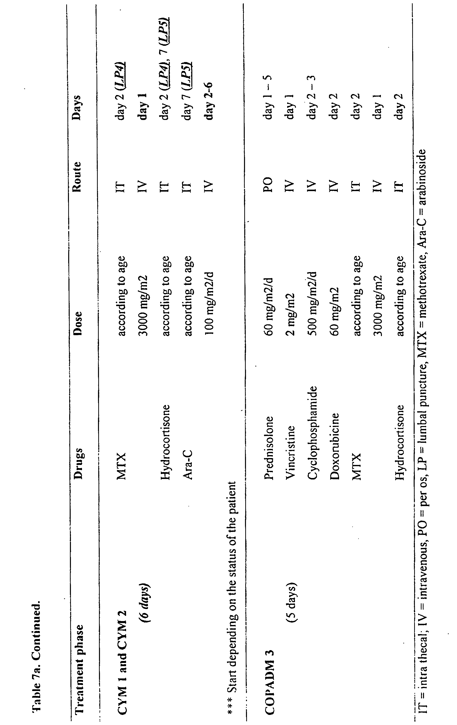


| Average cerebrospinal fluid levels of tau, β-amyloid(1-42) and neuromodulin for 4 groups of patients as described in example 7. | ||||||
| Group | n | M/F | Age | tau | β-amyloid (1-42) | NM |
| Control | 70 | 36/34 | 45 ± 16 | 120 ± 78 | 466 ± 168 | 504 ± 283 |
| |
7 | 05/02 | 61 ± 10 | 253 ± 139 | 184, 184, 550 | 518 ± 168 |
| AD | 27 | 16/11 | 71 ± 12 | 316 ± 186 | 244 ± 74 | 927 391 |
| |
5 | 02/03 | 68 ± 5 | 235 ± 82 | 272,413 | 737 ± 514 |

The PCR products were re-amplified in order to generate sufficient amount of PCR product, polished with T4 DNA polymerase, cut with XbaI and ligated in NcoI-blunted, xbaI cut pIGRHISA (Innogenetics, Gent, Belgium; Cat No 2075). This resulted in pIGRHISARab3a (Innogenetics, Gent, Belgium; Cat No 3008) for Rab3a and in pIGRH6SNAP25a (Innogenetics, Gent, Belgium; Cat No 2941) for SNAP25. The ligated product was transformed into DH1(λ) (Bachmann, 1987) and Tetracycline resistant colonies were analyzed for the presence of an insert. Inserts were sequenced and the plasmids containing the correct sequence (Zahraoui et al., 1989; Zhao et al., 1994) were further used. For Rab3 a several PCR artifacts were present. The correct sequence was assembled from two clones.
Direct evidence that both proteins are present in CSF was obtained by extraction of albumin and IgG from 50 ml CSF and fractionation on column. The fractions were dried and dissolved in sample buffer, run on a 12% acrylamide gel and blotted. The PVDF membranes were probed with a Rab3a and SNAP25 monoclonal antibody. Immunoreactive bands of the expected molecular weight and pI value were detected.
Phospho-tau was measured with a sandwich ELISA, using as HT7 as capturing antibody and biotinylated AT270 as detector antibody (INNOTEST phospho-tau(181), Innogenetics, Gent, Belgium). AT270 specifically recognizes phospho-tau (International application published under WO 95/17429).
Samples were only taken in the course of scheduled lumbar punctions (LPs) for staging or treatment for malignancy. Different groups of patients with hematological malignancies were enrolled in the present study (Table 6). A first group included 9 B-cell non-Hodgkin's lymphoma patients (B-NHL), treated according to the United Kingdom Children Cancers group (UKCCSG 9602) NHL protocol (Table 7a). In this group, four patients had B-cell lymphomas, 3 patients had Burkitt's lymphoma, and 2 patients had anaplastic large cell lymphoma (ALCL). Eight of these patients were studied longitudinally. A second and largest group consisted of 42 patients with non-B-cell acute lymphoblastic leukemia/non Hodgkin's lymphoma (NB ALL/NHL), treated according to the 'European Organization for Research and Treatment of Cancer' (EORTC) protocol 58881 (Table 7b). In this second group, 18 children had CD10(+) blasts or common NB ALL, two patients had Down syndrome (DS), 1 patient had the Brachmann-de Lange syndrome, 3 patients had common B-cell blasts, 1 patient had common T-cell blasts, 2 patients had pro-B-cell blasts, 9 patients had pre-B-cell blasts, and 8 patients had T-cell blasts. Thirty-eight children had leukemia, 5 patients had non-Hodgkin's lymphoma stage II (1 patient), Stage III (3 patients) or Stage IV (1 patient), of which one patient had overt CNS involvement (CNS+), defined according to the study protocol with malignant cells in the CSF, eye funduscopy and contrast captation. Five patients were considered as very high risk (VHR) patients according to the criteria defined in the treatment protocol (2 patients with T-cell blast and 3 patients with corticoid-resistance). Twenty-seven of the patients within this group could be followed longitudinally. A third patient group consisted of 6 children with acute myelomic leukemia (AML), of which 1 patient had CNS involvement and two patients had Down syndrome. There were 3 patients with M0, and 1 patient each with M1, M2 or M7 phenotype. All these patients were treated according to the EORTC 58921 protocol (Table 7c) and followed longitudinally. The other patients consisted of a heterogeneous group of children (n=18) in which for clinical reasons a lumbar puncture was performed for diagnosis of infection and staging, during or after their treatment. This group includes 5 children with medulloblastoma (3 staging, 1 during treatment, 1 follow up), 3 children with Hodgkin's disease (staging), 3 children with rhabdomyosarcoma (2 staging, 1 during treatment), 2 children with myelodysplastic syndrome (MDS) (staging), 2 children with Langerhans cell histiocytis (LCH/HLH, staging), 1 child with juvenile chronic myelomic leukemia (CML) (during treatment), choriocarcinoma (follow up), or germinoma (staging). A large group of healthy newborns was not available, since taking cerebrospinal fluid from these patients would be unethical. As controls children suspected of having meningitis but with negative findings (n=4), localized retinoblastoma (n=1) and familial hemophagocytic lymphohistiocytosis (n=1) were enrolled. Parental informed concent was obtained.
Lumbar punctures were performed for routine analysis either at baseline for diagnostic work-up or just prior to the IT administration of chemotherapy. Five ml of CSF was collected in different polypropylene tubes. One sample was centrifuged immediately at 1500 rpm for 2 minutes to eliminate cells and other insoluble material. The supernatant was stored at -70°C for subsequent analysis. The number of freeze/thaw cycles was restricted to a minimum. Routine CSF measurement included cytology, protein concentration, glucose, etc.
Since normality for all neurological marker data, independent of the diagnostic groups, was rejected, non-parametric statistics were used for the analysis. The Kruskal-Wallis test was used to investigate group differences regarding the effect variables (tau, neuromodulin, protein, etc.). The Wilcoxon signed ranks test, matched-pairs was used to check for differences between the first diagnostic LP and subsequent LPs. Pearson's correlation was used to investigate possible covariates. Analyses were done with Prism software v2.01 (Graphpad Software Inc., San Diego, CA, USA), Systat version 7 (SPSS, Chicago, IL, USA).
Tau levels at diagnosis were analyzed for each subgroup of patients (Fig. 9). Tau levels at diagnosis ranged from 66 to 1500 pg/ml. The data for tau, neuromodulin, β-amyloid(1-42), β-amyloid(1-40), serum LDH and CSF white blood cell count (Fig. 10) show no obvious difference in patient groups with and without Down syndrome or between diagnositc groups. In addition, no correlation was found between the tau level and WBC or LDH levels. No significant correlation was found between the tau concentration and the age of the children. The two patients with MDS, patients with overt CNS invasion (CNS+), but not children with AML had markedly enhanced levels of tau at diagnosis. Also, three patients entering the hospital with rised intracranial pressure due to medulloblastoma in the fossa posterior, from which CSF was taken for stageing, had very high CSF-tau concentrations (823,1397,1500). However, 7/21 children with non-B ALL/NHL, 1/4 non-B ALL/NHL patients with very high risk criteria, 2/4 patients with AML and 2/8 patients with B-cell NHL had a level of tau above 312 pg/ml, although using classical diagnostic procedures, CNS invasion was not detected.
For the 27 patients with non-B ALL/NHL or nine patients with B-cell NHL (data not shown), tau levels at diagnosis did not correlate with tumor burden, as reflected by the white blood cell count (p=0.935,n=40) or serum LDH (p=0.855, n=39). At LP1, there was a highly significant correlation between tau and neuromodulin [r=0.793; 95% CI: 0.658-0.928, n=50) indicating that the secretion of both proteins are interrelated at some point. No correlation was seen between tau and β-amyloid(1-42) (p=0.1032, n=52).
One Down's syndrome-non-B ALL patient had a tau concentration at diagnosis of 70 pg/ml. While the mean increase of tau in all non-B ALL patients was 250% (95% CI = 130% - 370%, n=13) at
One particular patient was treated with prednisolone for 8 days without IT MTX at
One CSF sample was available from a child who was treated in another center and who suffered from manifest neurotoxicity after treatment with MTX, and who had diplegia. The tau and neuromodulin levels in this child exceeded the highest standard used (1500 pg/ml for tau, 8000 pg/ml for neuromodulin), reflecting gross neuronal degeneration.
Claims (5)
- A method for specific detection, quantification and/or differential diagnosis of neurodegeneration in an individual comprising the step of determining the level of at least three neurological markers in one or more body fluid sample(s) from said individual, or determining the level of the same marker in two different body fluid samples in combination with the detection of at least one other marker in one or more body fluid sample(s) by means of antibodies specifically recognizing said neurological markers, whereby the type and degree of neurodegeneration is reflected by a quantitative change in the level of all of said neurological markers compared to a control sample, said method further characterized in that:for the specific detection or quantification of Alzheimer's disease and/or Lewy Body disease and/or for the differential diagnosis of Alzheimer's disease versus Lewy Body disease, the level of tau, β-amyloid(1-42) and α-synuclein is determined in a cerebrospinal fluid sample from said individual;for the specific detection or quantification of Alzheimer's disease and/or for the differential diagnosis of Alzheimer's disease versus other dementiathe level of tau, β-amyloid(1-42) and Rab3a is determined in a cerebrospinal fluid sample from said individual; orthe level of tau, β-amyloid(1-42) and SNAP25 is determined in a cerebrospinal fluid sample from said individual;for the specific detection or quantification of Alzheimer's disease and/or Parkinson's disease and/or for the differential diagnosis of Alzheimer's disease versus Parkinson's disease, the level of tau, β-amyloid(1-42) and neuromodulin is determined in a cerebrospinal fluid sample from said individual;for the specific detection or quantification of neurodegeneration induced by chemotherapy and/or exposure to chemical compounds and/or irradiation, the level of tau, neuron-specific enolase and neuromodulin is determined in a cerebrospinal fluid sample from said individual;for the specific detection or quantification of Frontal Temporal Lobe dementia and/or for the differential diagnosis of Frontal Temporal Lobe dementia versus other dementia, the level of tau, phospho-tau and β-amyloid(1-42) is determined in a cerebrospinal fluid sample from said individual;for the specific detection or quantification of vascular problems in Alzheimer's disease, for the differential diagnosis of different forms of Alzheimer's disease and/or for the differential diagnosis of Alzheimer's disease versus other dementia,the level tau and β-amyloid(1-42) is determined quantitatively in a cerebrospinal fluid sample and the level of β-amyloid(1-42) is determined quantitatively in a plasma sample from said individual; orthe level of phospho-tau and β-amyloid(1-42) is determined quantitatively in a cerebrospinal fluid sample and the level of β-amyloid(1-42) is determined quantitatively in a plasma sample form said individual; orthe level of tau and phospho-tau is determined quantitatively in a cerebrospinal fluid sample and the level of β-amyloid(1-42) is determined quantitatively in a plasma sample from said individual; orthe level of tau, phospho-tau and β-amyloid(1-42) is determined quantitatively in a cerebrospinal fluid sample from said patient.
- A method according to claim 1 further characterized in that the individual, whose neurodegeneration induced by chemotherapy, exposure to chemical compounds and/or irradiation is detected or quantified, is suffering from leukemia or brain tumor.
- A diagnostic kit for the specific detection, quantification and/or differential diagnosis of neurodegeneration in an individual comprising at least 3 antibodies each recognizing a different neurological marker and recognizing one of the following combinations of neurological markers:tau, β-amyloid(1-42), α-synuclein;tau, β-amyloid(1-42), Rab3a;tau, β-amyloid(1-42), SNAP25;tau, β-amyloid(1-42), neuromodulin;tau, neuron-specific enolase, neuromodulin; ortau, phospho-tau, β-amyloid(1-42); or comprising at least 2 antibodies each recognizing a different neurological marker of which one antibody recognizes a neurological marker in 2 different body fluids, and recognizing one of the following combinations of neurological markers:tau, β-amyloid(1-42); orphospho-tau, β-amyloid(1-42).
- A diagnostic kit according to claim 3, for the specific detection, quantification and/or differential diagnosis of neurodegeneration in an individual comprising:a support comprising, together or separately, at least 3 antibodies (primary antibodies or capturing antibodies) each recognizing a different neurological marker, or comprising at least 2 antibodies (primary antibodies or capturing antibodies) each recognizing a different neurological marker of which one antibody recognizes a neurological marker in 2 different body fluids;secondary antibodies (detector antibodies), each recognizing one of the neurological marker-primary antibody complexes;possibly, a marker either for specific tagging or coupling with said secondary antibodies;possibly, appropriate buffer solutions for carrying out the immunological reaction between the primary antibodies and the body fluid sample, between the secondary antibodies and the neurological marker-primary antibody complexes and/or between the bound secondary antibodies and the marker;possibly, for standardization purposes, purified proteins or synthetic peptides that are specifically recognized by the antibodies of the kit, used for the detection of the neurological marker.
- Use of a method or a diagnostic kit according to any of claims 1 to 4, for therapeutic monitoring and/or determination of the effectiveness of a certain treatment.
Priority Applications (1)
| Application Number | Priority Date | Filing Date | Title |
|---|---|---|---|
| EP99934543A EP1095278B1 (en) | 1998-07-03 | 1999-06-29 | Differential diagnosis of neurodegeneration |
Applications Claiming Priority (8)
| Application Number | Priority Date | Filing Date | Title |
|---|---|---|---|
| EP98870148 | 1998-07-03 | ||
| EP98870148 | 1998-07-03 | ||
| EP98870236 | 1998-11-03 | ||
| EP98870236 | 1998-11-03 | ||
| EP99870069 | 1999-04-09 | ||
| EP99870069 | 1999-04-09 | ||
| EP99934543A EP1095278B1 (en) | 1998-07-03 | 1999-06-29 | Differential diagnosis of neurodegeneration |
| PCT/EP1999/004483 WO2000002053A2 (en) | 1998-07-03 | 1999-06-29 | Differential diagnosis of neurodegeneration |
Publications (2)
| Publication Number | Publication Date |
|---|---|
| EP1095278A2 EP1095278A2 (en) | 2001-05-02 |
| EP1095278B1 true EP1095278B1 (en) | 2005-12-07 |
Family
ID=27239769
Family Applications (1)
| Application Number | Title | Priority Date | Filing Date |
|---|---|---|---|
| EP99934543A Expired - Lifetime EP1095278B1 (en) | 1998-07-03 | 1999-06-29 | Differential diagnosis of neurodegeneration |
Country Status (11)
| Country | Link |
|---|---|
| EP (1) | EP1095278B1 (en) |
| JP (3) | JP2002519702A (en) |
| CN (1) | CN1316055A (en) |
| AT (1) | ATE312349T1 (en) |
| AU (1) | AU754062B2 (en) |
| BR (1) | BR9911291A (en) |
| CA (1) | CA2329523A1 (en) |
| DE (1) | DE69928809T2 (en) |
| DK (1) | DK1095278T3 (en) |
| ES (1) | ES2255280T3 (en) |
| WO (1) | WO2000002053A2 (en) |
Cited By (2)
| Publication number | Priority date | Publication date | Assignee | Title |
|---|---|---|---|---|
| RU2441240C1 (en) * | 2010-10-05 | 2012-01-27 | Государственное образовательное учреждение высшего профессионального образования "Московский государственный медико-стоматологический университет" Министерства здравоохранения и социального развития Российской Федерации | Diagnostic technique for axonal demyelinating polyneuropathies |
| EP3542159A4 (en) * | 2016-11-16 | 2020-09-16 | Nanosomix Inc. | QUANTIFICATION OF SUBPOPULATIONS OF EXOSOMES AND DIAGNOSIS OF NEURODEGENERATIVE DISEASES |
Families Citing this family (38)
| Publication number | Priority date | Publication date | Assignee | Title |
|---|---|---|---|---|
| DE10041478A1 (en) | 2000-08-24 | 2002-03-14 | Sanol Arznei Schwarz Gmbh | New pharmaceutical composition |
| WO2002075317A2 (en) * | 2001-03-15 | 2002-09-26 | Novartis Ag | Screening for drugs against disorders associated with schizophrenia |
| WO2003044051A1 (en) * | 2001-11-20 | 2003-05-30 | Atgen Co., Ltd. | Novel peptides conferring environmental stress resistance and fusion proteins including said peptides |
| GB0203446D0 (en) * | 2002-02-14 | 2002-04-03 | Univ Lancaster | Detection and/or monitoring of synuclein-related diseases |
| US9034337B2 (en) | 2003-10-31 | 2015-05-19 | Prothena Biosciences Limited | Treatment and delay of outset of synucleinopathic and amyloidogenic disease |
| WO2005001483A2 (en) * | 2003-06-27 | 2005-01-06 | Georg-August-Universität Göttingen Stiftung Öffentlichen Rechts | Methods for diagnisis and differential diagnosis of dementia disorders |
| WO2005047860A2 (en) | 2003-11-08 | 2005-05-26 | Elan Pharmaceuticals, Inc. | Antibodies to alpha-synuclein |
| PT2211183E (en) * | 2003-11-19 | 2013-05-31 | Satoris Inc | Method for diagnosis and monitoring of alzheimer`s disease |
| US20060094064A1 (en) * | 2003-11-19 | 2006-05-04 | Sandip Ray | Methods and compositions for diagnosis, stratification, and monitoring of alzheimer's disease and other neurological disorders in body fluids |
| DE10361258A1 (en) | 2003-12-24 | 2005-07-28 | Schwarz Pharma Ag | Use of substituted 2-aminotetralins for the preventive treatment of Parkinson's disease |
| AT500321B1 (en) * | 2003-12-30 | 2006-02-15 | Red Bull Gmbh | Early, prenatal diagnosis of Down syndrome, comprises detecting abnormal levels of specific proteins in fetal cells from amniotic fluid or blood, or in brain tissue |
| DE102004014841B4 (en) | 2004-03-24 | 2006-07-06 | Schwarz Pharma Ag | Use of rotigotine for the treatment and prevention of Parkinson-Plus syndrome |
| AU2005323173A1 (en) * | 2004-12-01 | 2006-07-13 | The Curators Of The University Of Missouri | Modulator of alpha-synuclein toxicity |
| ES2441253T3 (en) * | 2006-11-17 | 2014-02-03 | Friedrich-Alexander-Universität Erlangen-Nürnberg | Procedure to differentially diagnose dementias |
| PL2118300T3 (en) * | 2007-02-23 | 2015-11-30 | Prothena Biosciences Ltd | Prevention and treatment of synucleinopathic and amyloidogenic disease |
| EP2020602A1 (en) * | 2007-08-02 | 2009-02-04 | Araclon Biotech | High sensitivty immunoassays and kits for the determination of peptides and proteins of biological interest |
| JP2009052933A (en) * | 2007-08-24 | 2009-03-12 | Rakuno Gakuen | Detecting method and diagnosing kit of prion disease |
| WO2009039586A2 (en) * | 2007-09-28 | 2009-04-02 | Powmri Limited | Biomarkers for parkinson's disease |
| WO2009086306A1 (en) | 2007-12-21 | 2009-07-09 | Whitehead Institute For Biomedical Research | Modulators of alpha-synuclein toxicity |
| DK2282758T3 (en) * | 2008-04-29 | 2019-02-25 | Bioarctic Ab | ANTIBODIES AND VACCINES FOR USE IN THERAPEUTIC AND DIAGNOSTIC PROCEDURES FOR ALPHA-SYNUCLEIN-RELATED DISORDERS |
| US20110159527A1 (en) * | 2008-06-16 | 2011-06-30 | Michael Gebhard Schlossmacher | Methods and kits for diagnosing neurodegenerative disease |
| US20120190652A1 (en) * | 2009-09-16 | 2012-07-26 | United Arab Emirates University | Diagnostic agent for parkinson's disease |
| CA2784919A1 (en) | 2010-01-26 | 2011-07-26 | Banyan Biomarkers, Inc. | Compositions and methods relating to argininosuccinate synthetase |
| EP2539366B1 (en) | 2010-02-26 | 2017-11-08 | BioArtic Neuroscience AB | Protofibril-binding antibodies and their use in therapeutic and diagnostic methods for parkinson's disease, dementia with lewy bodies and other alpha-synucleinopathies |
| CN102918397A (en) * | 2010-04-01 | 2013-02-06 | 班扬生物标记公司 | Markers and assays for detection of neurotoxicity |
| GB201011420D0 (en) * | 2010-07-06 | 2010-08-18 | United Arab Emirates Universit | Method for diagnosis |
| UA118441C2 (en) * | 2012-10-08 | 2019-01-25 | Протена Біосаєнсиз Лімітед | Antibodies recognizing alpha-synuclein |
| WO2015111430A1 (en) | 2014-01-21 | 2015-07-30 | 株式会社 島津製作所 | Measurement method for app cleavage peptides |
| EP4040157A1 (en) | 2014-05-22 | 2022-08-10 | Shimadzu Corporation | Surrogate biomarker for evaluating intracerebral amyloid beta peptide accumulation and method for analysis thereof |
| BR112017004883A2 (en) * | 2014-09-10 | 2017-12-05 | Abbvie Deutschland | rgma fragment-based diagnostic assay |
| EP4424704A3 (en) | 2015-09-16 | 2024-12-25 | Shimadzu Corporation | Multiplex biomarker for use in evaluation of state of accumulation of amyloid b in brain, and analysis method for said evaluation |
| EP3242134A1 (en) * | 2016-05-04 | 2017-11-08 | Euroimmun Medizinische Labordiagnostika AG | Assay for the diagnosis of a neurological disease |
| US10364286B2 (en) * | 2016-12-22 | 2019-07-30 | H. Lundbeck A/S | Monoclonal anti-alpha-synuclein antibodies for preventing tau aggregation |
| GB201814807D0 (en) * | 2018-09-12 | 2018-10-24 | Univ Newcastle | Dementia Biomarkers |
| EP3660509B1 (en) * | 2018-11-29 | 2022-03-09 | Hugel Inc. | A cell-based method for determining an activity of botulinum toxin |
| GB202005732D0 (en) * | 2020-04-20 | 2020-06-03 | Synpromics Ltd | Regulatory nucleic acid sequences |
| HUE072262T2 (en) | 2020-06-26 | 2025-11-28 | Bioarctic Ab | Alpha-synuclein protofibril-binding antibodies |
| WO2022115705A2 (en) * | 2020-11-30 | 2022-06-02 | Enigma Biointelligence, Inc. | Non-invasive assessment of alzheimer's disease |
-
1999
- 1999-06-29 AT AT99934543T patent/ATE312349T1/en active
- 1999-06-29 EP EP99934543A patent/EP1095278B1/en not_active Expired - Lifetime
- 1999-06-29 BR BR9911291-4A patent/BR9911291A/en not_active IP Right Cessation
- 1999-06-29 DK DK99934543T patent/DK1095278T3/en active
- 1999-06-29 CN CN99810286.5A patent/CN1316055A/en active Pending
- 1999-06-29 JP JP2000558397A patent/JP2002519702A/en active Pending
- 1999-06-29 AU AU50290/99A patent/AU754062B2/en not_active Ceased
- 1999-06-29 ES ES99934543T patent/ES2255280T3/en not_active Expired - Lifetime
- 1999-06-29 DE DE69928809T patent/DE69928809T2/en not_active Expired - Lifetime
- 1999-06-29 WO PCT/EP1999/004483 patent/WO2000002053A2/en not_active Ceased
- 1999-06-29 CA CA002329523A patent/CA2329523A1/en not_active Abandoned
-
2005
- 2005-11-10 JP JP2005326503A patent/JP2006091025A/en active Pending
-
2009
- 2009-10-28 JP JP2009247559A patent/JP4580455B2/en not_active Expired - Lifetime
Cited By (2)
| Publication number | Priority date | Publication date | Assignee | Title |
|---|---|---|---|---|
| RU2441240C1 (en) * | 2010-10-05 | 2012-01-27 | Государственное образовательное учреждение высшего профессионального образования "Московский государственный медико-стоматологический университет" Министерства здравоохранения и социального развития Российской Федерации | Diagnostic technique for axonal demyelinating polyneuropathies |
| EP3542159A4 (en) * | 2016-11-16 | 2020-09-16 | Nanosomix Inc. | QUANTIFICATION OF SUBPOPULATIONS OF EXOSOMES AND DIAGNOSIS OF NEURODEGENERATIVE DISEASES |
Also Published As
| Publication number | Publication date |
|---|---|
| DE69928809D1 (en) | 2006-01-12 |
| AU5029099A (en) | 2000-01-24 |
| JP4580455B2 (en) | 2010-11-10 |
| JP2002519702A (en) | 2002-07-02 |
| EP1095278A2 (en) | 2001-05-02 |
| ATE312349T1 (en) | 2005-12-15 |
| BR9911291A (en) | 2001-12-04 |
| CA2329523A1 (en) | 2000-01-13 |
| DE69928809T2 (en) | 2006-08-31 |
| JP2010019864A (en) | 2010-01-28 |
| WO2000002053A3 (en) | 2000-02-24 |
| ES2255280T3 (en) | 2006-06-16 |
| WO2000002053A2 (en) | 2000-01-13 |
| CN1316055A (en) | 2001-10-03 |
| JP2006091025A (en) | 2006-04-06 |
| AU754062B2 (en) | 2002-10-31 |
| DK1095278T3 (en) | 2006-04-18 |
| HK1036834A1 (en) | 2002-01-18 |
Similar Documents
| Publication | Publication Date | Title |
|---|---|---|
| AU754062B2 (en) | Differential diagnosis of neurodegeneration | |
| US20040014142A1 (en) | Differential diagnosis of neurodegeneration | |
| Sjögren et al. | The cerebrospinal fluid levels of tau, growth-associated protein-43 and soluble amyloid precursor protein correlate in Alzheimer’s disease, reflecting a common pathophysiological process | |
| Blennow et al. | CSF total tau, Aβ42 and phosphorylated tau protein as biomarkers for Alzheimer’s disease | |
| Hampel et al. | Core biological marker candidates of Alzheimer’s disease–perspectives for diagnosis, prediction of outcome and reflection of biological activity | |
| Mollenhauer et al. | Biological confounders for the values of cerebrospinal fluid proteins in Parkinson's disease and related disorders | |
| Jensen et al. | Increased cerebrospinal fluid tau in patients with Alzheimer's disease | |
| US6670137B2 (en) | Differential diagnosis of neurological diseases | |
| JP2021523357A (en) | Diagnostic and therapeutic methods based on site-specific taurine oxidation | |
| US20110159527A1 (en) | Methods and kits for diagnosing neurodegenerative disease | |
| Sobów et al. | Amyloid-beta and tau proteins as biochemical markers of Alzheimer's disease | |
| Mulder et al. | CSF markers related to pathogenetic mechanisms in Alzheimer's disease | |
| Wang et al. | Synaptic vesicle glycoprotein 2 A in serum is an ideal biomarker for early diagnosis of Alzheimer’s disease | |
| HK1040429B (en) | Tau as a marker for early cns damage | |
| AU2003200041B2 (en) | Differential Diagnosis of Neurodegeneration | |
| Mehta et al. | Biological markers of Alzheimer's disease | |
| HK1036834B (en) | Differential diagnosis of neurodegeneration | |
| JP4851052B2 (en) | Differential diagnosis of neurological diseases | |
| US9618522B2 (en) | Diagnostic testing in dementia and methods related thereto | |
| Mehta | Amyloid beta and tau proteins in Alzheimer’s disease and Down syndrome | |
| Mehta | Amyloid beta and tau proteins in | |
| Nguyen | Analytical improvements in the detection of amyloid-β peptides in cerebrospinal fluid by mass spectrometry | |
| Hirota et al. | Short title p-tau biomarkers represent Aβ pathology | |
| EP4619416A1 (en) | Methods to detect csf mtbr-tau and uses thereof | |
| HK40130133A (en) | Methods to detect csf mtbr-tau and uses thereof |
Legal Events
| Date | Code | Title | Description |
|---|---|---|---|
| PUAI | Public reference made under article 153(3) epc to a published international application that has entered the european phase |
Free format text: ORIGINAL CODE: 0009012 |
|
| 17P | Request for examination filed |
Effective date: 20010123 |
|
| AK | Designated contracting states |
Kind code of ref document: A2 Designated state(s): AT BE CH CY DE DK ES FI FR GB GR IE IT LI LU MC NL PT SE |
|
| AX | Request for extension of the european patent |
Free format text: AL PAYMENT 20010123;LT PAYMENT 20010123;LV PAYMENT 20010123;MK PAYMENT 20010123;RO PAYMENT 20010123;SI PAYMENT 20010123 |
|
| 17Q | First examination report despatched |
Effective date: 20030711 |
|
| GRAP | Despatch of communication of intention to grant a patent |
Free format text: ORIGINAL CODE: EPIDOSNIGR1 |
|
| GRAS | Grant fee paid |
Free format text: ORIGINAL CODE: EPIDOSNIGR3 |
|
| GRAA | (expected) grant |
Free format text: ORIGINAL CODE: 0009210 |
|
| AK | Designated contracting states |
Kind code of ref document: B1 Designated state(s): AT BE CH CY DE DK ES FI FR GB GR IE IT LI LU MC NL PT SE |
|
| AX | Request for extension of the european patent |
Extension state: AL LT LV MK RO SI |
|
| REG | Reference to a national code |
Ref country code: GB Ref legal event code: FG4D |
|
| REG | Reference to a national code |
Ref country code: CH Ref legal event code: EP |
|
| REG | Reference to a national code |
Ref country code: IE Ref legal event code: FG4D |
|
| REF | Corresponds to: |
Ref document number: 69928809 Country of ref document: DE Date of ref document: 20060112 Kind code of ref document: P |
|
| REG | Reference to a national code |
Ref country code: SE Ref legal event code: TRGR |
|
| REG | Reference to a national code |
Ref country code: DK Ref legal event code: T3 |
|
| LTIE | Lt: invalidation of european patent or patent extension |
Effective date: 20051207 |
|
| REG | Reference to a national code |
Ref country code: GR Ref legal event code: EP Ref document number: 20060400852 Country of ref document: GR |
|
| REG | Reference to a national code |
Ref country code: ES Ref legal event code: FG2A Ref document number: 2255280 Country of ref document: ES Kind code of ref document: T3 |
|
| ET | Fr: translation filed | ||
| PLBE | No opposition filed within time limit |
Free format text: ORIGINAL CODE: 0009261 |
|
| STAA | Information on the status of an ep patent application or granted ep patent |
Free format text: STATUS: NO OPPOSITION FILED WITHIN TIME LIMIT |
|
| REG | Reference to a national code |
Ref country code: HK Ref legal event code: GR Ref document number: 1036834 Country of ref document: HK |
|
| 26N | No opposition filed |
Effective date: 20060908 |
|
| PGFP | Annual fee paid to national office [announced via postgrant information from national office to epo] |
Ref country code: MC Payment date: 20140612 Year of fee payment: 16 Ref country code: IE Payment date: 20140625 Year of fee payment: 16 |
|
| PGFP | Annual fee paid to national office [announced via postgrant information from national office to epo] |
Ref country code: PT Payment date: 20140625 Year of fee payment: 16 Ref country code: LU Payment date: 20140624 Year of fee payment: 16 Ref country code: GR Payment date: 20140624 Year of fee payment: 16 |
|
| PGFP | Annual fee paid to national office [announced via postgrant information from national office to epo] |
Ref country code: CY Payment date: 20140602 Year of fee payment: 16 |
|
| REG | Reference to a national code |
Ref country code: FR Ref legal event code: PLFP Year of fee payment: 17 |
|
| REG | Reference to a national code |
Ref country code: PT Ref legal event code: MM4A Free format text: LAPSE DUE TO NON-PAYMENT OF FEES Effective date: 20151229 |
|
| PG25 | Lapsed in a contracting state [announced via postgrant information from national office to epo] |
Ref country code: MC Free format text: LAPSE BECAUSE OF NON-PAYMENT OF DUE FEES Effective date: 20150630 |
|
| PG25 | Lapsed in a contracting state [announced via postgrant information from national office to epo] |
Ref country code: LU Free format text: LAPSE BECAUSE OF NON-PAYMENT OF DUE FEES Effective date: 20150629 Ref country code: CY Free format text: LAPSE BECAUSE OF NON-PAYMENT OF DUE FEES Effective date: 20150629 Ref country code: PT Free format text: LAPSE BECAUSE OF NON-PAYMENT OF DUE FEES Effective date: 20151229 |
|
| REG | Reference to a national code |
Ref country code: IE Ref legal event code: MM4A |
|
| PG25 | Lapsed in a contracting state [announced via postgrant information from national office to epo] |
Ref country code: IE Free format text: LAPSE BECAUSE OF NON-PAYMENT OF DUE FEES Effective date: 20150629 |
|
| PG25 | Lapsed in a contracting state [announced via postgrant information from national office to epo] |
Ref country code: GR Free format text: LAPSE BECAUSE OF NON-PAYMENT OF DUE FEES Effective date: 20160112 |
|
| REG | Reference to a national code |
Ref country code: GR Ref legal event code: ML Ref document number: 20060400852 Country of ref document: GR Effective date: 20160112 |
|
| REG | Reference to a national code |
Ref country code: FR Ref legal event code: PLFP Year of fee payment: 18 |
|
| REG | Reference to a national code |
Ref country code: FR Ref legal event code: PLFP Year of fee payment: 19 |
|
| REG | Reference to a national code |
Ref country code: FR Ref legal event code: PLFP Year of fee payment: 20 |
|
| PGFP | Annual fee paid to national office [announced via postgrant information from national office to epo] |
Ref country code: CH Payment date: 20180621 Year of fee payment: 20 Ref country code: NL Payment date: 20180620 Year of fee payment: 20 Ref country code: DE Payment date: 20180625 Year of fee payment: 20 Ref country code: FI Payment date: 20180621 Year of fee payment: 20 |
|
| PGFP | Annual fee paid to national office [announced via postgrant information from national office to epo] |
Ref country code: BE Payment date: 20180620 Year of fee payment: 20 Ref country code: FR Payment date: 20180620 Year of fee payment: 20 Ref country code: AT Payment date: 20180621 Year of fee payment: 20 |
|
| PGFP | Annual fee paid to national office [announced via postgrant information from national office to epo] |
Ref country code: SE Payment date: 20180620 Year of fee payment: 20 |
|
| PGFP | Annual fee paid to national office [announced via postgrant information from national office to epo] |
Ref country code: IT Payment date: 20180627 Year of fee payment: 20 Ref country code: ES Payment date: 20180720 Year of fee payment: 20 Ref country code: GB Payment date: 20180620 Year of fee payment: 20 |
|
| PGFP | Annual fee paid to national office [announced via postgrant information from national office to epo] |
Ref country code: DK Payment date: 20180620 Year of fee payment: 20 |
|
| REG | Reference to a national code |
Ref country code: DE Ref legal event code: R071 Ref document number: 69928809 Country of ref document: DE |
|
| REG | Reference to a national code |
Ref country code: DK Ref legal event code: EUP Effective date: 20190629 |
|
| REG | Reference to a national code |
Ref country code: NL Ref legal event code: MK Effective date: 20190628 |
|
| REG | Reference to a national code |
Ref country code: BE Ref legal event code: MK Effective date: 20190629 |
|
| REG | Reference to a national code |
Ref country code: CH Ref legal event code: PL |
|
| REG | Reference to a national code |
Ref country code: GB Ref legal event code: PE20 Expiry date: 20190628 |
|
| REG | Reference to a national code |
Ref country code: SE Ref legal event code: EUG |
|
| REG | Reference to a national code |
Ref country code: AT Ref legal event code: MK07 Ref document number: 312349 Country of ref document: AT Kind code of ref document: T Effective date: 20190629 |
|
| PG25 | Lapsed in a contracting state [announced via postgrant information from national office to epo] |
Ref country code: GB Free format text: LAPSE BECAUSE OF EXPIRATION OF PROTECTION Effective date: 20190628 |
|
| REG | Reference to a national code |
Ref country code: ES Ref legal event code: FD2A Effective date: 20201204 |
|
| PG25 | Lapsed in a contracting state [announced via postgrant information from national office to epo] |
Ref country code: ES Free format text: LAPSE BECAUSE OF EXPIRATION OF PROTECTION Effective date: 20190630 |