EP1059540A2 - Method and apparatus for efficient MRI tissue differentiation - Google Patents
Method and apparatus for efficient MRI tissue differentiation Download PDFInfo
- Publication number
- EP1059540A2 EP1059540A2 EP00304184A EP00304184A EP1059540A2 EP 1059540 A2 EP1059540 A2 EP 1059540A2 EP 00304184 A EP00304184 A EP 00304184A EP 00304184 A EP00304184 A EP 00304184A EP 1059540 A2 EP1059540 A2 EP 1059540A2
- Authority
- EP
- European Patent Office
- Prior art keywords
- pulse
- tissue
- suppression
- fat
- spectrally selective
- Prior art date
- Legal status (The legal status is an assumption and is not a legal conclusion. Google has not performed a legal analysis and makes no representation as to the accuracy of the status listed.)
- Ceased
Links
- 238000000034 method Methods 0.000 title claims abstract description 44
- 230000004069 differentiation Effects 0.000 title claims abstract description 8
- 230000001629 suppression Effects 0.000 claims abstract description 141
- 230000005415 magnetization Effects 0.000 claims abstract description 50
- 238000012546 transfer Methods 0.000 claims abstract description 47
- 238000002595 magnetic resonance imaging Methods 0.000 claims abstract description 38
- XLYOFNOQVPJJNP-UHFFFAOYSA-N water Substances O XLYOFNOQVPJJNP-UHFFFAOYSA-N 0.000 claims abstract description 29
- 230000004044 response Effects 0.000 claims abstract description 22
- 230000000694 effects Effects 0.000 claims description 30
- 238000003384 imaging method Methods 0.000 claims description 18
- 238000013461 design Methods 0.000 claims description 13
- 230000007704 transition Effects 0.000 claims description 9
- 238000004590 computer program Methods 0.000 claims description 5
- 230000001747 exhibiting effect Effects 0.000 claims description 5
- 210000004351 coronary vessel Anatomy 0.000 abstract description 42
- 230000000747 cardiac effect Effects 0.000 abstract description 15
- 238000002583 angiography Methods 0.000 abstract description 11
- 210000004165 myocardium Anatomy 0.000 abstract description 4
- 210000001519 tissue Anatomy 0.000 description 79
- 210000004369 blood Anatomy 0.000 description 15
- 239000008280 blood Substances 0.000 description 15
- 230000002107 myocardial effect Effects 0.000 description 15
- 230000001965 increasing effect Effects 0.000 description 14
- 229920001817 Agar Polymers 0.000 description 10
- 238000005481 NMR spectroscopy Methods 0.000 description 10
- 239000008272 agar Substances 0.000 description 10
- 230000005284 excitation Effects 0.000 description 8
- 238000010521 absorption reaction Methods 0.000 description 7
- 210000003127 knee Anatomy 0.000 description 5
- 241000894007 species Species 0.000 description 5
- 238000012800 visualization Methods 0.000 description 5
- 238000001208 nuclear magnetic resonance pulse sequence Methods 0.000 description 4
- 230000009467 reduction Effects 0.000 description 4
- 238000001228 spectrum Methods 0.000 description 4
- 238000013459 approach Methods 0.000 description 3
- 210000004204 blood vessel Anatomy 0.000 description 3
- 210000003205 muscle Anatomy 0.000 description 3
- 238000009738 saturating Methods 0.000 description 3
- 239000000126 substance Substances 0.000 description 3
- 210000000988 bone and bone Anatomy 0.000 description 2
- 230000015556 catabolic process Effects 0.000 description 2
- 208000029078 coronary artery disease Diseases 0.000 description 2
- 230000034994 death Effects 0.000 description 2
- 231100000517 death Toxicity 0.000 description 2
- 238000006731 degradation reaction Methods 0.000 description 2
- 230000000593 degrading effect Effects 0.000 description 2
- 238000005259 measurement Methods 0.000 description 2
- 230000007246 mechanism Effects 0.000 description 2
- 241001465754 Metazoa Species 0.000 description 1
- 210000000709 aorta Anatomy 0.000 description 1
- 210000001367 artery Anatomy 0.000 description 1
- 230000005540 biological transmission Effects 0.000 description 1
- 210000001715 carotid artery Anatomy 0.000 description 1
- 239000002131 composite material Substances 0.000 description 1
- 230000001010 compromised effect Effects 0.000 description 1
- 238000010586 diagram Methods 0.000 description 1
- 238000002592 echocardiography Methods 0.000 description 1
- 230000002708 enhancing effect Effects 0.000 description 1
- 238000002474 experimental method Methods 0.000 description 1
- 210000002082 fibula Anatomy 0.000 description 1
- 210000004013 groin Anatomy 0.000 description 1
- 229920002521 macromolecule Polymers 0.000 description 1
- 238000012423 maintenance Methods 0.000 description 1
- 239000011159 matrix material Substances 0.000 description 1
- 230000003278 mimic effect Effects 0.000 description 1
- 230000000737 periodic effect Effects 0.000 description 1
- 230000002093 peripheral effect Effects 0.000 description 1
- 230000002688 persistence Effects 0.000 description 1
- 238000012545 processing Methods 0.000 description 1
- 229920006395 saturated elastomer Polymers 0.000 description 1
- 238000004088 simulation Methods 0.000 description 1
- 230000003595 spectral effect Effects 0.000 description 1
- 238000010561 standard procedure Methods 0.000 description 1
Images


Classifications
-
- G—PHYSICS
- G01—MEASURING; TESTING
- G01R—MEASURING ELECTRIC VARIABLES; MEASURING MAGNETIC VARIABLES
- G01R33/00—Arrangements or instruments for measuring magnetic variables
- G01R33/20—Arrangements or instruments for measuring magnetic variables involving magnetic resonance
- G01R33/44—Arrangements or instruments for measuring magnetic variables involving magnetic resonance using nuclear magnetic resonance [NMR]
- G01R33/446—Multifrequency selective RF pulses, e.g. multinuclear acquisition mode
-
- G—PHYSICS
- G01—MEASURING; TESTING
- G01R—MEASURING ELECTRIC VARIABLES; MEASURING MAGNETIC VARIABLES
- G01R33/00—Arrangements or instruments for measuring magnetic variables
- G01R33/20—Arrangements or instruments for measuring magnetic variables involving magnetic resonance
- G01R33/44—Arrangements or instruments for measuring magnetic variables involving magnetic resonance using nuclear magnetic resonance [NMR]
- G01R33/48—NMR imaging systems
- G01R33/483—NMR imaging systems with selection of signals or spectra from particular regions of the volume, e.g. in vivo spectroscopy
- G01R33/4838—NMR imaging systems with selection of signals or spectra from particular regions of the volume, e.g. in vivo spectroscopy using spatially selective suppression or saturation of MR signals
Definitions
- the present invention relates generally to magnetic resonance imaging (MRI), and more particularly to a method and apparatus for efficient MRI tissue differentiation using an RF pulse designed to provide a frequency response combining a magnetization transfer contrast and fat suppression simultaneously.
- MRI magnetic resonance imaging
- Coronary artery disease is currently the leading cause of death in western hemisphere countries. Therefore, the visualization of the coronary arteries is an important step in preventing deaths due to coronary artery disease.
- coronary artery visualization is accomplished using x-ray contrast angiography, a highly invasive procedure.
- x-ray angiography a catheter is inserted into the artery through the groin area of a patient in order to accomplish x-ray angiography. It would be advantageous to acquire angiographic images of the coronary arteries without having to require such an invasive procedure.
- Magnetic resonance angiography is a non-invasive alternative to x-ray angiography that has been successfully used to image carotid arteries, the aorta, and other peripheral vessels.
- coronary arteries are small, tortuous vessels, usually no more than 1-5 mm. in diameter.
- Successful visualization of coronary arteries using conventional MRA is hampered because coronary arteries are often surrounded by pericardial fat and myocardium, especially the more distal segments.
- a high degree of fat suppression can be achieved using standard chemical saturation pulses.
- suppression of the myocardial signal without an accompanying reduction in the blood signal, has heretofore been a challenging problem.
- myocardial signal is a significant attribute to the contrast-to-noise ratio (CNR) of the coronary arteries
- suppression of myocardium is essential for reliable visualization of more complete portions of the left anterior descending (LAD) coronary arteries, as well as the distal segments of the right coronary artery.
- MTC magnetization transfer contrast
- MTC effects have been found to adequately suppress myocardial signals in human and animal hearts and can be produced by using either a binomial, zero-degree on-resonance excitation, or an off-resonance spectrally selective excitation.
- the off-resonance irradiation utilizes RF pulses of high B 1 amplitude and are set to at least 1 kHz from the water resonance frequency.
- the large resonance offset avoids undesired saturation of spins with long T 2 times in the imaged volume.
- Continuous wave MTC results in too high of a specific absorption rate (SAR) to the patient and requires an ancillary RF amplifier on conventional whole-body MR imaging scanners, and is therefore not widely used or desirable.
- SAR specific absorption rate
- the off-resonant MTC technique is most effectively achieved using a train of Gaussian pulses of fixed bandwidth, for example, 500 Hz, at 1500 Hz off-resonance.
- the entire duration of the MTC pulse composite is typically about 200-300 ms.
- MRA using a cardiac-gated 3D fast-gradient recalled echo sequence, several problems arise.
- the specific absorption rate of these pulses is very high, thereby limiting its use. It also prohibits use of continuous RF excitation, which is another mechanism for suppressing myocardial signals that can be used in conjunction with magnetization transfer based methods.
- the present invention relates to a system and method for differentiating tissue types in MR imaging that can efficiently discriminate fat tissue from proteinated tissues, and each from water-based tissue, that solves the aforementioned problems.
- the present invention combines fat suppression and magnetization transfer (MT) in an RF pulse to overcome the aforementioned problems.
- MT fat suppression and magnetization transfer
- Two basic approaches are described.
- the pulse can be designed to induce an MT effect by increasing the flip angle based on the amount of MT effect and fat suppression desired.
- increasing the flip angle, or the effective B 1 , of a fat saturation pulse that increases the MTC effect is done at the cost of degrading fat suppression at certain flip angles and care must be taken so as to not exceed the specific absorption rate desirable for a patient.
- Another approach includes modulating an RF pulse with a sinusoidal function, such as that provided in a Hadamard excitation pulse.
- the frequency response of the resultant pulse is the frequency response of the original RF pulse shifted in frequency to both sides of the carrier.
- the spacing of the pulses is determined by the frequency of the modulation function.
- a method of differentiating tissue in NMR imaging includes the steps of creating a spectrally selective suppression pulse having an RF pulse profile designed to produce a frequency response with high fat suppression and selecting a spectrally selective suppression amplitude to produce a magnetization transfer contrast between two tissue types.
- the method also includes applying the spectrally selective suppression pulse with a flip angle selected to optimize fat suppression and magnetization transfer contrast saturation simultaneously.
- an MRI apparatus for MR angiography is disclosed that is capable of efficient tissue differentiation that includes an MRI system having a number of gradient coils positioned about a bore of a magnet to impress a polarizing magnetic field and an RF transceiver system having an RF modulator controlled by a pulse control module to transmit RF signals to an RF coil assembly to acquire MR images.
- the MRI apparatus also includes a computer programmed to construct a desired RF pulse based on input design criteria that includes desired bandwidth, desired percentage in-band (pass band) ripple, and desired percentage out-of-band (stop-band) ripple, and a desired flip angle so that the desired RF pulse has low stop and pass band ripple and high transition band slope.
- the computer is also programmed to select an RF pulse frequency for the desired RF pulse proportional to a fat tissue resonance frequency and to select an RF pulse amplitude of the desired RF pulse to obtain both optimal fat suppression and to obtain a substantial MTC effect between proteinated tissue and water tissue.
- the system then transmits the desired RF pulse to the pulse control module to transmit RF signals to the RF coil assembly to acquire MR images having fat suppression and MTC effects between proteinated tissue and water tissue simultaneously.
- the acquired MR images enhance the contrast between the proteinated tissue and the water tissue, while at the same time suppressing fat tissue.
- a computer system for use with an MRI apparatus comprising a computer programmed from a computer readable storage medium having thereon a computer program programmed to create a spectrally selective suppression pulse having an RF pulse profile designed to produce a frequency and amplitude response with high fat suppression and to select a spectrally selective suppression amplitude to produce a magnetization transfer contrast between two tissue types.
- the computer is also programmed to apply the spectrally selective suppression pulse with a flip angle selected to optimize fat suppression and magnetization transfer contrast saturation simultaneously for use by the MRI apparatus.
- a nuclear magnetic resonance (NMR) imaging system 8 of a type suitable for the practice of the invention includes a computer 10 which controls gradient coil power amplifiers 14 through a pulse control module 12.
- the pulse control module 12 and the gradient amplifiers 14 together produce the proper gradient waveforms Gx, Gy, and Gz, for either a spin echo, a gradient recalled echo pulse sequence, a fast spin echo, or other type of pulse sequences.
- the gradient waveforms are connected to gradient coils 16 which are positioned around the bore of the magnet 34 so that gradients Gx, Gy, and Gz are impressed along their respective axes on the polarizing magnetic field Bo from magnet 34.
- the pulse control module 12 also controls a radio frequency synthesizer 18 which is part of an RF transceiver system, portions of which are enclosed by dashed line block 36.
- the pulse control module 12 also controls an RF modulator 20 which modulates the output of the radio frequency synthesizer 18.
- the resultant RF signals amplified by power amplifier 22 and applied to RF coil 26 through transmit/receive switch 24, are used to excite the nuclear spins of the imaged object (not shown).
- the NMR signals from the excited nuclei of the imaged object are picked up by the RF coil 26 and presented to preamplifier 28 through transmit/receive switch 24, to be amplified and then processed by a quadrature phase detector 30.
- the detected signals are digitized by a high speed A/D converter 32 and applied to computer 10 for processing to produce NMR images of the object.
- Computer 10 also controls shimming coil power supplies 38 to power shimming coil assembly 40.
- the present invention includes a method and system for MR imaging that differentiates water-based tissue from proteinated tissue while simultaneously suppressing fat tissue, which is particularly useful in MR angiography (MRA), but is applicable to any MR imaging of any portion of the body in which enhanced contrast between blood tissue and the surrounding tissue is poor.
- MRA MR angiography
- the present invention combines magnetization transfer contrast techniques with a fat suppression pulse to produce an MR image with enhanced tissue differentiation.
- coronary arteries are small, tortuous vessels, typically between 1-5 mm in diameter, the visualization of such vessels using MRA is hampered because they are often surrounded by pericardial fat and also embedded in myocardium.
- a high degree of fat suppression is achieved by using a standard method for spectral chemical saturation, while suppression of the myocardial signal is achieved by combining a magnetization transfer contrast (MTC) pulse with the fat suppression pulse to avoid a concomitant reduction in the blood signal.
- MTC magnetization transfer contrast
- a specially designed RF pulse with requisite features must be used.
- Magnetization transfer imaging alters the contrast in MR images by saturating the short T 2 species present in tissue.
- Off-resonance continuous wave and pulsed short T 2 selective saturation alters the relaxation mechanisms governing tissue contrast, thus causing some spins close to the saturated species to experience longer T 2 and T 1 times.
- the continuous wave MT technique requires the application of lower power off-resonance irradiation.
- the pulsed MT technique reduces the overall specific absorption rate by applying periodic RF pulses, avoids the need for a secondary RF transmitter, and significantly reduces the total average RF power transmitted. Therefore, the pulsed MT technique is preferred.
- the power requited by a radio frequency pulse depends on the difference between the transmit and the resonance frequencies.
- the distance traversed by the magnetization which is a product of the effective flip angle, increases as the transmission frequency moves off-resonance.
- the effective flip angle is calculated from the pulse field strength and the transmit frequency difference, and is given by: where ⁇ transmit is the transmit frequency, ⁇ 0 is the resonance frequency, B 1 is the pulse field strength, t is the transmit time, ⁇ is the gyomagnetic ratio, a physical constant and ⁇ effective is the effective flip angle. Therefore, by transmitting close to resonance, a substantial reduction in absorbed RF power is accomplished.
- the use of such pulses provides efficient saturation of short T 2 species. Applying these binomial RF pulses slightly off-resonance, and in particular, at the fat resonance frequency, effectively suppresses fat signals while providing an adequate MT effect. Two such techniques are disclosed.
- a fat suppression pulse transmitted at off-resonant frequency of ⁇ 220 Hz at 1.5 Telsa can be made to induce an MT effect by increasing the effective flip angle.
- By increasing the flip angle, or effectively the B 1 , of the fat saturation pulse increases the MT contrast effect with only slight degradation of the fat suppression. It has been found that acceptable MTC effects can be achieved at flip angles of approximately 200° or more using the fat saturation pulse.
- An example of such a pulse is disclosed in Fig. 2 which shows an RF pulse profile oh spectrally selective suppression pulse 50 according to the present invention.
- the pulse is a real-valued pulse, and in a preferred embodiment, has the following design parameters: a pulse width of approximately 8 ms, approximately 400 total points, a flip angle of approximately 200° , a 1.0% or less pass band and stop band ripple, a bandwidth of approximately 180 Hz, and is an inversion, minimum phase pulse.
- the flip angle is set to increase the MTC saturation at least 15% while exhibiting a frequency response with fat suppression of at least 85%.
- Such a pulse will reduce signal intensity of proteinated tissue to less than 80% of its maximum level to enhance contrast between proteinated tissue and water tissue, and suppress a majority of fat tissue.
- the flip angle can be set between 100° and 1,000° depending on the image to be acquired, the strength of the magnet in the MR apparatus, the amount of fat suppression desired, as well as what level of specific absorption rate is allowable.
- the bandwidth of the pulse would likewise depend on the aforementioned factors and can range between 100 Hz and 200 Hz.
- the pulse width can range from 8 ms to 32 ms for most applications.
- the transition slopes 52, 54 of Fig. 2 are preferably set to a maximum slope (i.e., with as short a frequency span as possible) to avoid saturating any on-resonance protons.
- the aforementioned requirements provide a well-defined pulse having good transition bandwidth and minimal pass and stop band ripple so as to avoid saturating the on-resonance frequency protons.
- the pulse is brought in further and further, on the frequency spectrum, the more important a well-shaped pulse is in order to avoid saturation of on-resonance protons.
- the frequency response 56 of the spectrally selective suppression pulse of Fig. 1 is shown.
- the longitudinal magnetization (M z ) is plotted as a function of frequency and shows that at the on-resonance frequency protons are minimally affected by the magnetization.
- Fig. 4 shows a plot of saturation, measured as a percentage, for a phantom having agar, fat, and CuSO 4 -doped water as a function of RF saturation/suppression pulse flip angle or pulse amplitude.
- agar simulates proteinated tissue and is used to mimic the tissue that exhibits MTC effects. It is noted that the agar plot 58 exhibits a pronounced MTC saturation effect as the RF pulse amplitude increases.
- the plot for fat tissue 60 shows a broad minimum for fat suppression that permits the RF suppression pulse to be overdriven and allow increased MTC saturation of the simulated proteinated tissue 58 while only minimally degrading the fat suppression.
- the RF pulse is applied using a flip angle of at least 100° to provide at least a 16% MTC effect on the agar while providing fat suppression of approximately 90%. Only a 1% saturation of the on-resonance water signal 62 was observed at a flip angle of 100°.
- the broad minimum suppression of fat tissue 60 allows the flip angle to be increased to provide a corresponding increase in MTC saturation 58. It is noted that increasing the MTC saturation to between 22% and 29%, allows the maintenance of fat suppression at 88% - 90%. With the increased RF amplitude, the longer T 2 species begin to exhibit increased saturation and there is a negligible degradation in the doped water saturation 62 at the higher flip angles.
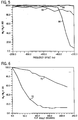
- Fig. 5 shows a plot of saturation of the agar 64, the fat tissue 66 and the doped water 68 as a function of frequency offset with the suppression flip angle set at 110°. It is noted that the persistence of agar saturation as compared to fat or water saturation, at higher frequency offsets is indicative of the MTC effect. It is therefore evident from Fig. 5 that the saturation effect observed is due to magnetization transfer and not to a direct saturation of the agar since the agar and water signals are not equally affected by the suppression pulse. In fact, at a frequency offset of approximately -500Hz, both fat and doped water remain relatively unaffected, while the agar exhibited an 8% saturation level.
- Fig. 6 shows actual data points obtained from an MRI of a normal knee of a volunteer patient that shows similar results as depicted in the aforementioned phantom measurements.
- Fig. 6 is a plot of saturation as a function of RF suppression flip angle in muscle 70 and in the trabecular bone 72, which is fatty marrow. It is noted that the same acquisition parameters were used to acquire the signal intensity in the human knee of Fig. 6 as that for the phantom depicted in Figs. 4 and 5.
- a 60-70% MTC saturation was obtained while maintaining fat suppression at 85-90%. It is evident that overdriving the spectrally selective suppression RF pulse by approximately 50%, an additional 17% MTC saturation can be obtained.
- the spectrally selective suppression pulse applied was an SLR pulse (a pulse designed using the Shinnar-LeRoux algorithm, Pauley J., Le Roux P., Parameter Relations for the Shinnar-Le Roux Selective Excitation Pulse Design Algorithm , IEEE Trans. Med. Imaging, March 1991, Vol. 10: 53-65) that is widely known in the industry.
- the pulse was applied in a steady state fast gradient recalled echo pulse sequence.
- the 8 ms pulse was applied once per RF excitation and increased the minimum sequence by about 12 ms, including crusher gradients.
- the amplitude of the spectrally selective suppression pulse was phase-cycled in a positive (+), negative (-), negative (-), positive (+) fashion to reduce spurious echoes from forming in the data acquisition window.
- the other pulse design parameters were previously disclosed.
- the phantom used for plotting Figs. 4 and 5 contained agar, a proteinated tissue substitute, fat tissue, and CuSO 4 -doped water.
- the pulse sequence parameters were set at 5 mm. thick sections, 24 cm. field-of-view, a 256 x 128 acquisition matrix, ⁇ 32 kH receiver bandwidth, 30° flip angle, and TE/TR of 2.9 and 19.9 ms, respectively. All experiments were performed in a head coil.
- the MTC saturation effect was measured as a function of the flip angle of the spectrally selective suppression RF pulse and is a function of the frequency offset at a fixed flip angle for the saturation pulse.
- the measurements obtained on the knee were obtained in a head coil with the same acquisition parameters as that of the phantom studies. However, the images acquired were in the coronal plane. The signal intensities were measured in the muscle and in the trabecular bone of the fibula just below the knee for the fat signal. The experimental results using the phantoms indicated an MTC saturation effect of 20% - 30% at the fat resonance of ⁇ 220 Hz.
- the present invention includes a method of differentiating tissue in NMR imaging having the steps of creating a spectrally selective suppression pulse having an RF pulse profile designed to produce a frequency response with high fat suppression and selecting a spectrally selective suppression amplitude to produce a magnetization transfer contrast between two tissue types -- proteinated tissue and water tissue.
- the method also includes applying the spectrally selective suppression pulse with a flip angle selected to optimize fat suppression and magnetization transfer contrast saturation of the two tissue types simultaneously.
- the method results in a magnetization transfer contrast saturation of at least 15% while maintaining fat suppression of at least 80%.
- the flip angle is set in excess of 100° and less than 1,000°.
- the spectrally selective suppression pulse should have a bandwidth of between 100 Hz and 200 Hz and have a pulse width of approximately 8-32 ms. It is important to minimize the stop and pass band ripple, preferably to less than 1.0%.
- the pulse is preferably applied at the fat resonance frequency of approximately ⁇ 220 Hz. In order to avoid saturation of on-resonance protons, the pulse should be designed with minimum transition bandwidth.
- the invention also includes an MRI apparatus capable of efficient tissue differentiation that includes an MRI system having a plurality of gradient coils positioned about a bare of a magnet to impress a polarizing magnetic field and an RF transceiver system, including an RF modulator controlled by a pulse control module to transmit RF signals to an RF coil assembly to acquire MR images.
- the MRI apparatus includes a computer program to construct a desired RF pulse based on input design criteria that includes a desired bandwidth, a desired percentage in-band (pass-band) ripple, and desired percentage out-band (stop-band) ripple, so that the desired RF pulse has low stop and pass band ripple and minimal transition bandwidth.
- the computer is further programmed to select an RF pulse frequency for the desired RF pulse proportional to a fat tissue resonance frequency and select an RF pulse amplitude of the desired RF pulse to maximize a magnetization transfer contrast effect between proteinated tissue and water tissue.
- the computer is also programmed to transmit the desired RF pulse to the pulse control module to transmit RF signals to the RF coil assembly to acquire MR images having fat suppression and magnetization transfer contrast effect between proteinated tissue and water tissue simultaneously.
- the resulting acquired MR images enhance the contrast between proteinated tissue and water tissue, while at the same time substantially suppressing fat tissue.
- the pulse design is as previously described.
- the present invention also includes a computer system for use with an MRI apparatus comprising a computer programmed from a computer readable storage medium having thereon a computer program programmed to create a spectrally selective suppression pulse having an RF pulse profile designed to produce a frequency response with high fat suppression and select a spectrally selective suppression amplitude to produce a magnetization transfer contrast between two tissue types, and apply the spectrally selective suppression pulse with a flip angle selected to optimize fat suppression and magnetization transfer contrast saturation simultaneously for the MRI apparatus.
- the computer system is implemented using the same pulse design criteria as previously described.
- An alternate embodiment of the present invention includes modulating an RF pulse similar to that as shown in Fig. 2, with a sinusoidal function in that at least two spectrally selected suppression pulses are applied simultaneously but with a proportional reduction in the RF amplitude, each applied at the same absolute frequency value but on opposite sides of the frequency spectrum.
- the frequency response of the resultant pulse is a frequency response of the original single RF pulse, but replicated on the other side of the on-resonance carrier frequency, the spacing of which is determined by the frequency of the modulation function.
- An example of such a pulse is the known Hadamard encoded pulse, which results in excitation on both sides of the transmitter frequency. This results in an effective MTC effect equivalent to 2 ⁇ ,if the fat saturation pulse is ⁇ in the aforementioned embodiment.
- the invention includes the alternative step of applying at least two of the spectrally selective suppression pulses simultaneously and distributed evenly on each side of a resonance frequency, each having a proportional amount of total RF power.
- the method includes applying only two spectrally selective suppression pulses simultaneously at off-resonance frequency, each having one-half of the total RF power applied, and wherein one of the spectrally selective suppression pulses is applied at a desired fat suppression frequency on a positive side of the resonance frequency and the other is applied at the desired fat suppression frequency on a negative side of the resonance frequency.
- a specific implementation of the aforementioned invention is to enhance blood vessel visibility in an MRI by producing a magnetization transfer contrast between blood in a vessel and myocardial tissue located about the vessel while suppressing associated fat about the vessel to thereby producing an improved MR image of the blood vessel.
- the blood vessel is a coronary artery and the MR image is an MR angiograph.
- the step of applying the spectrally selective suppression pulse is further defined as applying the spectrally selective suppression pulse to a patient in imaging the heart such that an MR angiogram of the patient's coronary arteries is acquired, and further includes the step of reconstructing an image of the patient's coronary arteries wherein the image reconstructed suppresses fat and simultaneously enhances contrast between blood in the coronary artery and myocardial about the coronary artery.
- the computer of the MRI apparatus of the present invention is also programmed, in a preferred embodiment, to enhance cardiac vessel visibility in an MRI by producing a magnetization transfer contrast between blood in a cardiac vessel and myocardial tissue located about the cardiac vessel while suppressing associated fat about the cardiac vessel to thereby produce an MR angiograph of the coronary arteries.
- the computer is programmed to apply the spectrally selective suppression pulse to a patient's coronary arteries to acquire an MR image of the patient's coronary arteries, and to reconstruct an image of the patient's coronary arteries wherein the resulting image reconstructed adequately suppresses fat and enhances contrast between blood in the coronary artery and myocardial tissue located about the coronary artery, simultaneously.
Landscapes
- Physics & Mathematics (AREA)
- High Energy & Nuclear Physics (AREA)
- Condensed Matter Physics & Semiconductors (AREA)
- General Physics & Mathematics (AREA)
- Magnetic Resonance Imaging Apparatus (AREA)
Abstract
Description
- The present invention relates generally to magnetic resonance imaging (MRI), and more particularly to a method and apparatus for efficient MRI tissue differentiation using an RF pulse designed to provide a frequency response combining a magnetization transfer contrast and fat suppression simultaneously.
- Coronary artery disease is currently the leading cause of death in western hemisphere countries. Therefore, the visualization of the coronary arteries is an important step in preventing deaths due to coronary artery disease. Currently, coronary artery visualization is accomplished using x-ray contrast angiography, a highly invasive procedure. In x-ray angiography, a catheter is inserted into the artery through the groin area of a patient in order to accomplish x-ray angiography. It would be advantageous to acquire angiographic images of the coronary arteries without having to require such an invasive procedure.
- Magnetic resonance angiography (MRA) is a non-invasive alternative to x-ray angiography that has been successfully used to image carotid arteries, the aorta, and other peripheral vessels. However, coronary arteries are small, tortuous vessels, usually no more than 1-5 mm. in diameter. Successful visualization of coronary arteries using conventional MRA is hampered because coronary arteries are often surrounded by pericardial fat and myocardium, especially the more distal segments. Under known methods, a high degree of fat suppression can be achieved using standard chemical saturation pulses. However, suppression of the myocardial signal, without an accompanying reduction in the blood signal, has heretofore been a challenging problem. Since the myocardial signal is a significant attribute to the contrast-to-noise ratio (CNR) of the coronary arteries, suppression of myocardium is essential for reliable visualization of more complete portions of the left anterior descending (LAD) coronary arteries, as well as the distal segments of the right coronary artery.
- A technique known as magnetization transfer contrast (MTC) has been commonly employed in imaging sequences to improve image contrast but has heretofore been employed only in limited manner in MRA of the coronary arteries because of various limitations. The MTC technique relies on the fact that a fraction of the water in biological systems is bound to large macromolecules. This results in lowering-of their tumbling rates and consequently, their relaxation times. The "bound pool" is in chemical exchange with the "free pool", therefore resulting in transfer of magnetization. Saturation of the bound pool will then result in saturation of the free pool. MTC effects have been found to adequately suppress myocardial signals in human and animal hearts and can be produced by using either a binomial, zero-degree on-resonance excitation, or an off-resonance spectrally selective excitation. In the past, the off-resonance irradiation utilizes RF pulses of high B1 amplitude and are set to at least 1 kHz from the water resonance frequency. The large resonance offset avoids undesired saturation of spins with long T2 times in the imaged volume. However, the further off-resonance the irradiation is, the higher the RF power level is required for observable MTC effects. Continuous wave MTC results in too high of a specific absorption rate (SAR) to the patient and requires an ancillary RF amplifier on conventional whole-body MR imaging scanners, and is therefore not widely used or desirable.
- The off-resonant MTC technique is most effectively achieved using a train of Gaussian pulses of fixed bandwidth, for example, 500 Hz, at 1500 Hz off-resonance. The entire duration of the MTC pulse composite is typically about 200-300 ms. In MRA using a cardiac-gated 3D fast-gradient recalled echo sequence, several problems arise. First, the specific absorption rate of these pulses is very high, thereby limiting its use. It also prohibits use of continuous RF excitation, which is another mechanism for suppressing myocardial signals that can be used in conjunction with magnetization transfer based methods. However, when such RF pulses are applied at a time-delay from the start of the R-wave in the R-R interval, until mid-diastole, the resulting pulses are quite long and consequently not practical for use in interleaved or segmented 3D fast gradient recalled echo sequence, hybrid sequences, where multiple k-space lines are acquired in the same R-R interval, or any other fast image acquisition sequence.
- It would therefore be desirable to have a method and apparatus capable of imaging coronary arteries with a lower effective specific absorption rate using the advantages of the MTC effect without the disadvantages while simultaneously suppressing fat tissue to provide enhanced visibility of tissue where the contrast between blood and the surrounding tissue is typically poor.
- The present invention relates to a system and method for differentiating tissue types in MR imaging that can efficiently discriminate fat tissue from proteinated tissues, and each from water-based tissue, that solves the aforementioned problems.
- In order to accomplish efficient MR angiography of coronary arteries, or MR jmages in any portion of the body where contrast between water-based tissue and the surrounding tissue is typically poor, the present invention combines fat suppression and magnetization transfer (MT) in an RF pulse to overcome the aforementioned problems. Two basic approaches are described. In one approach, by overdriving a fat saturation pulse that is off-resonant, the pulse can be designed to induce an MT effect by increasing the flip angle based on the amount of MT effect and fat suppression desired. However, increasing the flip angle, or the effective B1, of a fat saturation pulse that increases the MTC effect is done at the cost of degrading fat suppression at certain flip angles and care must be taken so as to not exceed the specific absorption rate desirable for a patient.
- Another approach includes modulating an RF pulse with a sinusoidal function, such as that provided in a Hadamard excitation pulse. The frequency response of the resultant pulse is the frequency response of the original RF pulse shifted in frequency to both sides of the carrier. The spacing of the pulses is determined by the frequency of the modulation function. Using a Hadamard encoded pulse for fat saturation provides excitation on both sides of the transmitter frequency which provides an effective MTC effect equivalent to twice that of an equivalent single fat saturation pulse with MTC. Thus, using a Hadamard pulse, the same flip angle can be maintained for fat suppression and yet double the available MTC effect.
- Therefore, in accordance with one aspect of the invention, a method of differentiating tissue in NMR imaging includes the steps of creating a spectrally selective suppression pulse having an RF pulse profile designed to produce a frequency response with high fat suppression and selecting a spectrally selective suppression amplitude to produce a magnetization transfer contrast between two tissue types. The method also includes applying the spectrally selective suppression pulse with a flip angle selected to optimize fat suppression and magnetization transfer contrast saturation simultaneously.
- In accordance with another aspect of the invention, an MRI apparatus for MR angiography is disclosed that is capable of efficient tissue differentiation that includes an MRI system having a number of gradient coils positioned about a bore of a magnet to impress a polarizing magnetic field and an RF transceiver system having an RF modulator controlled by a pulse control module to transmit RF signals to an RF coil assembly to acquire MR images. The MRI apparatus also includes a computer programmed to construct a desired RF pulse based on input design criteria that includes desired bandwidth, desired percentage in-band (pass band) ripple, and desired percentage out-of-band (stop-band) ripple, and a desired flip angle so that the desired RF pulse has low stop and pass band ripple and high transition band slope. These design parameters are needed to ensure uniform suppression of the fat signal within the pass-band and minimize saturation of the blood and tissue signal in the stop-band. The computer is also programmed to select an RF pulse frequency for the desired RF pulse proportional to a fat tissue resonance frequency and to select an RF pulse amplitude of the desired RF pulse to obtain both optimal fat suppression and to obtain a substantial MTC effect between proteinated tissue and water tissue. The system then transmits the desired RF pulse to the pulse control module to transmit RF signals to the RF coil assembly to acquire MR images having fat suppression and MTC effects between proteinated tissue and water tissue simultaneously. The acquired MR images enhance the contrast between the proteinated tissue and the water tissue, while at the same time suppressing fat tissue.
- In accordance with yet another aspect of the invention, a computer system is disclosed for use with an MRI apparatus comprising a computer programmed from a computer readable storage medium having thereon a computer program programmed to create a spectrally selective suppression pulse having an RF pulse profile designed to produce a frequency and amplitude response with high fat suppression and to select a spectrally selective suppression amplitude to produce a magnetization transfer contrast between two tissue types. The computer is also programmed to apply the spectrally selective suppression pulse with a flip angle selected to optimize fat suppression and magnetization transfer contrast saturation simultaneously for use by the MRI apparatus.
- The invention will now be described in greater detail, by way of example, with reference to the drawings, in which:
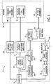
- Fig. 1 is a schematic block diagram of an NMR imaging system for use with the present invention.
- Figs. 2 is a graphical representation of a spectrally selective suppression pulse according to the present invention.
- Fig. 3 is a graphical representation of the frequency response of the spectrally selective suppression pulse of Fig. 2.
- Fig. 4 is a graphical representation of saturation of three tissue types as a function of RF suppression pulse flip angle as acquired from the NMR imaging system of Fig. 1, using an RF pulse according to Fig. 2, as received from a phantom.
- Fig. 5 is a graphical representation of saturation of three tissue types as a function of frequency offset acquired similarly as Fig. 4.
- Fig. 6 is a graphical representation of saturation of two tissue types as a function of RF suppression flip angle as acquired from the NMR imaging system of Fig. 1, using an RF pulse according to Fig. 2, as received from a human subject.
-
- Referring to Fig. 1, a nuclear magnetic resonance (NMR)
imaging system 8 of a type suitable for the practice of the invention includes acomputer 10 which controls gradientcoil power amplifiers 14 through apulse control module 12. Thepulse control module 12 and thegradient amplifiers 14 together produce the proper gradient waveforms Gx, Gy, and Gz, for either a spin echo, a gradient recalled echo pulse sequence, a fast spin echo, or other type of pulse sequences. The gradient waveforms are connected togradient coils 16 which are positioned around the bore of themagnet 34 so that gradients Gx, Gy, and Gz are impressed along their respective axes on the polarizing magnetic field Bo frommagnet 34. - The
pulse control module 12 also controls aradio frequency synthesizer 18 which is part of an RF transceiver system, portions of which are enclosed bydashed line block 36. Thepulse control module 12 also controls anRF modulator 20 which modulates the output of theradio frequency synthesizer 18. The resultant RF signals, amplified bypower amplifier 22 and applied toRF coil 26 through transmit/receiveswitch 24, are used to excite the nuclear spins of the imaged object (not shown). - The NMR signals from the excited nuclei of the imaged object are picked up by the
RF coil 26 and presented to preamplifier 28 through transmit/receiveswitch 24, to be amplified and then processed by aquadrature phase detector 30. The detected signals are digitized by a high speed A/D converter 32 and applied tocomputer 10 for processing to produce NMR images of the object.Computer 10 also controls shimmingcoil power supplies 38 to powershimming coil assembly 40. - The present invention includes a method and system for MR imaging that differentiates water-based tissue from proteinated tissue while simultaneously suppressing fat tissue, which is particularly useful in MR angiography (MRA), but is applicable to any MR imaging of any portion of the body in which enhanced contrast between blood tissue and the surrounding tissue is poor. The present invention combines magnetization transfer contrast techniques with a fat suppression pulse to produce an MR image with enhanced tissue differentiation. In particular, since coronary arteries are small, tortuous vessels, typically between 1-5 mm in diameter, the visualization of such vessels using MRA is hampered because they are often surrounded by pericardial fat and also embedded in myocardium. In the present invention, a high degree of fat suppression is achieved by using a standard method for spectral chemical saturation, while suppression of the myocardial signal is achieved by combining a magnetization transfer contrast (MTC) pulse with the fat suppression pulse to avoid a concomitant reduction in the blood signal. This results in a significantly increased contrast-to-noise ratio of the coronary arteries allowing the use of MRA in place of the highly invasive x-ray angiography. In order to maintain good fat suppression and MTC effect, while avoiding direct saturation of the on-resonance (water) spins, a specially designed RF pulse with requisite features must be used.
- Magnetization transfer imaging alters the contrast in MR images by saturating the short T2 species present in tissue. Off-resonance continuous wave and pulsed short T2 selective saturation alters the relaxation mechanisms governing tissue contrast, thus causing some spins close to the saturated species to experience longer T2 and T1 times. The continuous wave MT technique requires the application of lower power off-resonance irradiation. The pulsed MT technique reduces the overall specific absorption rate by applying periodic RF pulses, avoids the need for a secondary RF transmitter, and significantly reduces the total average RF power transmitted. Therefore, the pulsed MT technique is preferred.
- The power requited by a radio frequency pulse depends on the difference between the transmit and the resonance frequencies. The distance traversed by the magnetization, which is a product of the effective flip angle, increases as the transmission frequency moves off-resonance. The effective flip angle is calculated from the pulse field strength and the transmit frequency difference, and is given by:where ωtransmit is the transmit frequency, ω0 is the resonance frequency, B1 is the pulse field strength, t is the transmit time, γ is the gyomagnetic ratio, a physical constant and αeffective is the effective flip angle. Therefore, by transmitting close to resonance, a substantial reduction in absorbed RF power is accomplished. The use of such pulses provides efficient saturation of short T2 species. Applying these binomial RF pulses slightly off-resonance, and in particular, at the fat resonance frequency, effectively suppresses fat signals while providing an adequate MT effect. Two such techniques are disclosed.

- In the first technique, assuming a 1.5 T MR imaging apparatus, a fat suppression pulse transmitted at off-resonant frequency of ± 220 Hz at 1.5 Telsa (or 3.4 ppm off-resonance) can be made to induce an MT effect by increasing the effective flip angle. By increasing the flip angle, or effectively the B1, of the fat saturation pulse, increases the MT contrast effect with only slight degradation of the fat suppression. It has been found that acceptable MTC effects can be achieved at flip angles of approximately 200° or more using the fat saturation pulse. An example of such a pulse is disclosed in Fig. 2 which shows an RF pulse profile oh spectrally
selective suppression pulse 50 according to the present invention. The pulse is a real-valued pulse, and in a preferred embodiment, has the following design parameters: a pulse width of approximately 8 ms, approximately 400 total points, a flip angle of approximately 200° , a 1.0% or less pass band and stop band ripple, a bandwidth of approximately 180 Hz, and is an inversion, minimum phase pulse. In general, the flip angle is set to increase the MTC saturation at least 15% while exhibiting a frequency response with fat suppression of at least 85%. Such a pulse will reduce signal intensity of proteinated tissue to less than 80% of its maximum level to enhance contrast between proteinated tissue and water tissue, and suppress a majority of fat tissue. - In accordance with the present invention, the flip angle can be set between 100° and 1,000° depending on the image to be acquired, the strength of the magnet in the MR apparatus, the amount of fat suppression desired, as well as what level of specific absorption rate is allowable. The bandwidth of the pulse would likewise depend on the aforementioned factors and can range between 100 Hz and 200 Hz. The pulse width can range from 8 ms to 32 ms for most applications. The transition slopes 52, 54 of Fig. 2, are preferably set to a maximum slope (i.e., with as short a frequency span as possible) to avoid saturating any on-resonance protons. The aforementioned requirements provide a well-defined pulse having good transition bandwidth and minimal pass and stop band ripple so as to avoid saturating the on-resonance frequency protons. As one skilled in the art will readily recognize, as the pulse is brought in further and further, on the frequency spectrum, the more important a well-shaped pulse is in order to avoid saturation of on-resonance protons.
- Referring to Fig. 3, the
frequency response 56 of the spectrally selective suppression pulse of Fig. 1 is shown. The longitudinal magnetization (Mz) is plotted as a function of frequency and shows that at the on-resonance frequency protons are minimally affected by the magnetization. - Fig. 4 shows a plot of saturation, measured as a percentage, for a phantom having agar, fat, and CuSO4-doped water as a function of RF saturation/suppression pulse flip angle or pulse amplitude. As is known, agar simulates proteinated tissue and is used to mimic the tissue that exhibits MTC effects. It is noted that the
agar plot 58 exhibits a pronounced MTC saturation effect as the RF pulse amplitude increases. The plot forfat tissue 60 shows a broad minimum for fat suppression that permits the RF suppression pulse to be overdriven and allow increased MTC saturation of the simulatedproteinated tissue 58 while only minimally degrading the fat suppression. Nominally, the RF pulse is applied using a flip angle of at least 100° to provide at least a 16% MTC effect on the agar while providing fat suppression of approximately 90%. Only a 1% saturation of the on-resonance water signal 62 was observed at a flip angle of 100°. The broad minimum suppression offat tissue 60 allows the flip angle to be increased to provide a corresponding increase inMTC saturation 58. It is noted that increasing the MTC saturation to between 22% and 29%, allows the maintenance of fat suppression at 88% - 90%. With the increased RF amplitude, the longer T2 species begin to exhibit increased saturation and there is a negligible degradation in the dopedwater saturation 62 at the higher flip angles. - Fig. 5 shows a plot of saturation of the
agar 64, thefat tissue 66 and the dopedwater 68 as a function of frequency offset with the suppression flip angle set at 110°. It is noted that the persistence of agar saturation as compared to fat or water saturation, at higher frequency offsets is indicative of the MTC effect. It is therefore evident from Fig. 5 that the saturation effect observed is due to magnetization transfer and not to a direct saturation of the agar since the agar and water signals are not equally affected by the suppression pulse. In fact, at a frequency offset of approximately -500Hz, both fat and doped water remain relatively unaffected, while the agar exhibited an 8% saturation level. - Fig. 6 shows actual data points obtained from an MRI of a normal knee of a volunteer patient that shows similar results as depicted in the aforementioned phantom measurements. Fig. 6 is a plot of saturation as a function of RF suppression flip angle in
muscle 70 and in thetrabecular bone 72, which is fatty marrow. It is noted that the same acquisition parameters were used to acquire the signal intensity in the human knee of Fig. 6 as that for the phantom depicted in Figs. 4 and 5. The same broad minimum of thefat suppression plot 72 and the same increase in the MTC saturation in themuscle 70, as the flip angle was increased, was found in a scan of the knee as was found in the simulation using the aforementioned phantom. In particular, a 60-70% MTC saturation was obtained while maintaining fat suppression at 85-90%. It is evident that overdriving the spectrally selective suppression RF pulse by approximately 50%, an additional 17% MTC saturation can be obtained. - These results indicate that by slightly overdriving the spectrally selective suppression RF pulse used for fat suppression, a significant MTC effect can be obtained. A further MTC saturation of up to 60% was realized when the RF suppression pulse was driven to just less than 1.5 times the nominal suppression flip angle. However, there is a trade off in that overdriving the spectrally selected suppression RF pulse too far can cause increased suppression of the longer T2 species within the imaged volume. Fat suppression was not compromised with the suggested increase in the suppression RF pulse flip angle.
- It is noted that in all the aforementioned examples, the spectrally selective suppression pulse applied was an SLR pulse (a pulse designed using the Shinnar-LeRoux algorithm, Pauley J., Le Roux P., Parameter Relations for the Shinnar-Le Roux Selective Excitation Pulse Design Algorithm, IEEE Trans. Med. Imaging, March 1991, Vol. 10: 53-65) that is widely known in the industry. The pulse was applied in a steady state fast gradient recalled echo pulse sequence. The 8 ms pulse was applied once per RF excitation and increased the minimum sequence by about 12 ms, including crusher gradients. The amplitude of the spectrally selective suppression pulse was phase-cycled in a positive (+), negative (-), negative (-), positive (+) fashion to reduce spurious echoes from forming in the data acquisition window. The other pulse design parameters were previously disclosed.
- As mentioned, the phantom used for plotting Figs. 4 and 5 contained agar, a proteinated tissue substitute, fat tissue, and CuSO4-doped water. The pulse sequence parameters were set at 5 mm. thick sections, 24 cm. field-of-view, a 256 x 128 acquisition matrix, ± 32 kH receiver bandwidth, 30° flip angle, and TE/TR of 2.9 and 19.9 ms, respectively. All experiments were performed in a head coil. The MTC saturation effect was measured as a function of the flip angle of the spectrally selective suppression RF pulse and is a function of the frequency offset at a fixed flip angle for the saturation pulse. Similarly, the measurements obtained on the knee were obtained in a head coil with the same acquisition parameters as that of the phantom studies. However, the images acquired were in the coronal plane. The signal intensities were measured in the muscle and in the trabecular bone of the fibula just below the knee for the fat signal. The experimental results using the phantoms indicated an MTC saturation effect of 20% - 30% at the fat resonance of ± 220 Hz.
- Accordingly, the present invention includes a method of differentiating tissue in NMR imaging having the steps of creating a spectrally selective suppression pulse having an RF pulse profile designed to produce a frequency response with high fat suppression and selecting a spectrally selective suppression amplitude to produce a magnetization transfer contrast between two tissue types -- proteinated tissue and water tissue. The method also includes applying the spectrally selective suppression pulse with a flip angle selected to optimize fat suppression and magnetization transfer contrast saturation of the two tissue types simultaneously. Preferably, the method results in a magnetization transfer contrast saturation of at least 15% while maintaining fat suppression of at least 80%. Depending on the aforementioned design parameters, the flip angle is set in excess of 100° and less than 1,000°. The spectrally selective suppression pulse should have a bandwidth of between 100 Hz and 200 Hz and have a pulse width of approximately 8-32 ms. It is important to minimize the stop and pass band ripple, preferably to less than 1.0%. The pulse is preferably applied at the fat resonance frequency of approximately ± 220 Hz. In order to avoid saturation of on-resonance protons, the pulse should be designed with minimum transition bandwidth.
- The invention also includes an MRI apparatus capable of efficient tissue differentiation that includes an MRI system having a plurality of gradient coils positioned about a bare of a magnet to impress a polarizing magnetic field and an RF transceiver system, including an RF modulator controlled by a pulse control module to transmit RF signals to an RF coil assembly to acquire MR images. The MRI apparatus includes a computer program to construct a desired RF pulse based on input design criteria that includes a desired bandwidth, a desired percentage in-band (pass-band) ripple, and desired percentage out-band (stop-band) ripple, so that the desired RF pulse has low stop and pass band ripple and minimal transition bandwidth. The computer is further programmed to select an RF pulse frequency for the desired RF pulse proportional to a fat tissue resonance frequency and select an RF pulse amplitude of the desired RF pulse to maximize a magnetization transfer contrast effect between proteinated tissue and water tissue. The computer is also programmed to transmit the desired RF pulse to the pulse control module to transmit RF signals to the RF coil assembly to acquire MR images having fat suppression and magnetization transfer contrast effect between proteinated tissue and water tissue simultaneously.
- The resulting acquired MR images enhance the contrast between proteinated tissue and water tissue, while at the same time substantially suppressing fat tissue. The pulse design is as previously described.
- The present invention also includes a computer system for use with an MRI apparatus comprising a computer programmed from a computer readable storage medium having thereon a computer program programmed to create a spectrally selective suppression pulse having an RF pulse profile designed to produce a frequency response with high fat suppression and select a spectrally selective suppression amplitude to produce a magnetization transfer contrast between two tissue types, and apply the spectrally selective suppression pulse with a flip angle selected to optimize fat suppression and magnetization transfer contrast saturation simultaneously for the MRI apparatus. The computer system is implemented using the same pulse design criteria as previously described.
- An alternate embodiment of the present invention includes modulating an RF pulse similar to that as shown in Fig. 2, with a sinusoidal function in that at least two spectrally selected suppression pulses are applied simultaneously but with a proportional reduction in the RF amplitude, each applied at the same absolute frequency value but on opposite sides of the frequency spectrum. The frequency response of the resultant pulse is a frequency response of the original single RF pulse, but replicated on the other side of the on-resonance carrier frequency, the spacing of which is determined by the frequency of the modulation function. An example of such a pulse is the known Hadamard encoded pulse, which results in excitation on both sides of the transmitter frequency. This results in an effective MTC effect equivalent to 2 ,if the fat saturation pulse is in the aforementioned embodiment.
- Accordingly, the invention includes the alternative step of applying at least two of the spectrally selective suppression pulses simultaneously and distributed evenly on each side of a resonance frequency, each having a proportional amount of total RF power. In a more specific application, the method includes applying only two spectrally selective suppression pulses simultaneously at off-resonance frequency, each having one-half of the total RF power applied, and wherein one of the spectrally selective suppression pulses is applied at a desired fat suppression frequency on a positive side of the resonance frequency and the other is applied at the desired fat suppression frequency on a negative side of the resonance frequency. These alternative method steps are equivalently applicable to the apparatus as well in that the computer can equivalently be programmed to accomplish these alternative steps.
- A specific implementation of the aforementioned invention is to enhance blood vessel visibility in an MRI by producing a magnetization transfer contrast between blood in a vessel and myocardial tissue located about the vessel while suppressing associated fat about the vessel to thereby producing an improved MR image of the blood vessel. In a preferred embodiment, the blood vessel is a coronary artery and the MR image is an MR angiograph. The step of applying the spectrally selective suppression pulse is further defined as applying the spectrally selective suppression pulse to a patient in imaging the heart such that an MR angiogram of the patient's coronary arteries is acquired, and further includes the step of reconstructing an image of the patient's coronary arteries wherein the image reconstructed suppresses fat and simultaneously enhances contrast between blood in the coronary artery and myocardial about the coronary artery.
- The computer of the MRI apparatus of the present invention is also programmed, in a preferred embodiment, to enhance cardiac vessel visibility in an MRI by producing a magnetization transfer contrast between blood in a cardiac vessel and myocardial tissue located about the cardiac vessel while suppressing associated fat about the cardiac vessel to thereby produce an MR angiograph of the coronary arteries. Further, in a preferred embodiment, the computer is programmed to apply the spectrally selective suppression pulse to a patient's coronary arteries to acquire an MR image of the patient's coronary arteries, and to reconstruct an image of the patient's coronary arteries wherein the resulting image reconstructed adequately suppresses fat and enhances contrast between blood in the coronary artery and myocardial tissue located about the coronary artery, simultaneously.
- For the sake of good order, various features of the invention are set out in the following clauses:
- 1. A method of differentiating tissue in NMR imaging comprising the steps of creating a spectrally selective suppression pulse having an RF pulse profile designed to produce a frequency response with high fat suppression; selecting a spectrally selective suppression amplitude to produce a magnetization transfer contrast between two tissue types; and applying the spectrally selective suppression pulse with a flip angle selected to optimize fat suppression and magnetization transfer contrast saturation simultaneously.
- 2. The method of
clause 1 further comprising the step of setting the flip angle to increase the magnetization transfer contrast saturation at least 150/a while exhibiting a frequency response with fat suppression of at least 80%. - 3. The method of
clause 1 further comprising the step of setting the flip angle in excess of 100° and less than 1,000°. - 4. The method of
clause 1 further comprising the step of optimally designing a spectrally selective suppression pulse with a bandwidth of between 100 and 200 Hz and having a pulse width of approximately 8-32 ms. - 5. The method of
clause 4 wherein the step of optimally designing a spectrally selective suppression pulse further includes minimizing stop band ripple. - 6. The method of
clause 4 wherein the step of optimally designing a spectrally selective suppression pulse further includes setting a pass band ripple to less than 1 % of the spectrally selective suppression pulse. - 7. The method of
clause 1 further comprising the step of applying the spectrally selective suppression pulse centered on ± 220 Hz resonance offset from the carrier frequency at 1.5 Tesla, which is an offset of 3.4 ppm at any field strength. - 8. The method of
clause 4 wherein the step of optimally designing a spectrally selective suppression pulse further includes a pulse design having minimal outer band ripple and maximum transition band slope. - 9. The method of
clause 1 further comprising the step of applying at least two of the spectrally selective suppression pulses simultaneously and distributed evenly on each side of a resonance frequency, each having a proportional amount of total RF power. - 10. The method of clause 9 wherein only two spectrally selective suppression pulses are applied simultaneously at off-resonance frequencies, each having one-half of the total RF power, and wherein one of the spectrally selective suppression pulses is applied at a desired fat suppression frequency on a positive side of the resonance frequency and another is applied at the desired fat suppression frequency on-a negative side of the resonance frequency.
- 11. The method of
clause 1 further comprising the step of enhancing vessel visibility in an MRI by producing a magnetization transfer contrast between blood in a vessel and myocardial tissue located about the vessel while suppressing associated fat about the vessel to thereby produce an improved MR image of the vessel. - 12. The method of clause 11 wherein the vessel is a coronary artery and the MR image is an MR angiogram.
- 13. The method of
clause 1 wherein the step of applying the spectrally selective suppression pulse is further defined as applying the spectrally selective suppression pulse to a patient's coronary arteries to acquire an MR angiograph of the patient's coronary arteries, and the method further comprises the step of reconstructing an image of the patient's coronary arteries, wherein the image reconstructed suppresses fat and simultaneously enhances contrast between blood in the coronary artery and myocardial about the coronary artery. - 14. An MRI apparatus capable of efficient tissue differentiation comprising:
- a magnetic resonance imaging (MRI) system having a plurality of gradient coils positioned about a bore of a magnet to impress a polarizing magnetic field and an RF transceiver system having an RF modulator controlled by a pulse control module to transmit RF signals to an RF coil assembly to acquire MR images; and
- a computer programmed to:
- construct a desired RF pulse based on input design criteria including desired bandwidth, desired percentage in-band ripple, and desired percentage out-band ripple, so that the desired RF pulse has low stop and pass band ripple and high transition band slope;
- select an RF pulse frequency for the desired RF pulse proportional to a fat tissue resonance frequency;
- select an RF pulse amplitude of the desired RF pulse to maximize a magnetization transfer contrast effect between proteinated tissue and water tissue; and
- transmit the desired RF pulse to the pulse control module to transmit RF
signals to the RF coil assembly to acquire
MR images 20 having fat suppression and magnetization transfer contrast effect between proteinated tissue and water tissue simultaneously, such that the acquired MR images enhance a contrast between proteinated tissue and water tissue, and suppresses fat tissue. - 15. The MRI apparatus of
clause 14 wherein the computer is further programmed to construct the desired RF pulse based on whether a simultaneous sinusoidal RF pulse train is desired. - 16. The MRI apparatus of
clause 14 wherein the computer is further programmed to set a flip angle to increase the magnetization transfer contrast saturation at least 15% while exhibiting a frequency response having fat suppression of at least 80%. - 17. The MRI apparatus of
clause 16 wherein the computer is further programmed to set the flip angle in excess of 100° and less than 1,000°. - 18. The MRI apparatus of
clause 14 wherein the computer is further programmed to: - optimally design a spectrally selective suppression pulse with a bandwidth of between 100 Hz and 200 Hz and a pulse width of approximately 8-32 ms; and
- apply the spectrally selective suppression pulse centered on ± 220 Hz resonance offset from the carrier frequency at 1.5 Tesla, which is an offset of 3.4 ppm at any field strength.
- 19. The MRI apparatus of
clause 14 wherein the computer is further programmed to apply two spectrally selective suppression pulses simultaneously at off resonance frequencies, each having one-half of the total RF power wherein one of the spectrally selective suppression pulses is applied at a desired fat suppression frequency on a positive side of the resonance frequency and another is applied at the desired fat suppression frequency on a negative side of the resonance frequency. - 20. The MRI apparatus of
clause 14 wherein the computer is further programmed to enhance cardiac vessel visibility in an MRI by producing a magnetization transfer contrast between blood in a cardiac vessel and myocardial tissue located about the cardiac vessel while suppressing associated fat about the cardiac vessel to thereby produce an MR angiograph of the cardiac vessel. - 21. The MRI apparatus of
clause 14 wherein the computer is further programmed to apply the spectrally selective suppression pulse to a patient's coronary arteries to acquire an MR image of the patient's coronary arteries, and to reconstruct an image of the patient's coronary arteries, wherein the image reconstructed suppresses fat and enhances contrast between blood in the coronary artery and myocardial about the coronary artery simultaneously. - 22. A computer system for use with an MRI apparatus comprising a computer
programmed from a computer readable storage medium having thereon a computer
program programmed to:
- create a spectrally selective suppression pulse having an RF pulse profile, designed to produce a frequency response with high fat suppression;
- select a spectrally selective suppression amplitude to produce a magnetization transfer contrast between two tissue types; and
- apply the spectrally selective suppression pulse with a flip angle selected to
optimize fat suppression and magnetization
transfer contrast saturation 10 simultaneously for an MRI apparatus. - 23. A computerized system of
clause 22 wherein the computer is further programmed to increase the magnetization transfer contrast saturation in a patient image at least 15 % while simultaneously providing fat saturation of at least 80%. - 24. The computerized system of
clause 22 wherein the computer is further programmed to set a flip angle in excess of 100° and less than 1,000°. - 25. The computerized system of
clause 22 wherein the computer is further programmed to apply the spectrally selective suppression pulse at ± 220 Hz resonance offset from the carrier frequency at 1.5 Tesla, which is an offset of 3.4 ppm at any field strength. - 26. The computerized system of
clause 22 wherein the computer is further programmed to apply a spectrally selective suppression pulse with minimal outer band ripple and maximum transition band slope. - 27. The computerized system of
clause 22 wherein the computer is further programmed to: - apply two spectrally selective suppression pulses, one on a positive side of the frequency spectrum and one on a negative side of the frequency spectrum, both at a same absolute frequency value; and
- select a lower spectrally selective suppression amplitude for each spectrally selective suppression such that a specific absorption rate to a patient is no more than that of applying a single spectrally selective suppression pulse while increasing the magnetization transfer contrast saturation.
- 28. The computerized system of
clause 22 wherein the computer is further programmed to enhance cardiac vessel visibility in an MRI by producing a magnetization transfer contrast between blood in a cardiac vessel and myocardial tissue located about the cardiac vessel while suppressing associated fat about the cardiac vessel and to produce an MR angiogram of the cardiac vessel. - 29. The computerized system of
clause 22 wherein the computer is further programmed to apply the spectrally selective suppression pulse to a patient's coronary arteries to acquire an MR image of the patient's coronary arteries, and to reconstruct an image of the patient's coronary arteries, wherein the image reconstructed suppresses fat and enhances contrast between blood in the coronary artery and myocardial about the coronary artery simultaneously. -
Claims (10)
- A method of differentiating tissue in NMR imaging comprising the steps of:creating a spectrally selective suppression pulse (50) having an RIF pulse profile designed to produce a frequency response with high fat suppression (60);selecting a spectrally selective suppression amplitude to produce a magnetization transfer contrast between two tissue types (58, 62); andapplying the spectrally selective suppression pulse (50) with a flip angle selected to optimize fat suppression and magnetization transfer contrast saturation simultaneously.
- The method of claim 1, further comprising the step of setting the flip angle to increase the magnetization transfer contrast saturation at least 15% while exhibiting a frequency response with fat suppression of at least 80%.
- The method of claim 1 or 2, further comprising the step of setting the flip angle in excess of 100° and less than 1,000°.
- The method of claim 1, 2 or 3, further comprising the step of optimally designing a spectrally selective suppression pulse (50) with a bandwidth of between 100 and 200 Hz and having a pulse width of approximately 8-32 ms.
- An MRI apparatus capable of efficient tissue differentiation comprising:a magnetic resonance imaging (MRI) system (8) having a plurality of gradient coils (16) positioned about a bore of a magnet (34) to impress a polarizing magnetic field and an RF transceiver system (36) having an RF modulator (20) controlled by a pulse control module (12) to transmit RF signals to an RF coil assembly (26) to acquire MR images; anda computer programmed (10) to:construct a desired RF pulse (50) based on input design criteria including desired bandwidth, desired percentage in-band ripple, and desired percentage out-band ripple, so that the desired RF pulse (50) has low stop and pass band ripple and high transition band slope;select an RF pulse frequency for the desired RF pulse (50) proportional to a fat tissue resonance frequency (66);select an RF pulse amplitude of the desired RF pulse (50) to maximize a magnetization transfer contrast effect between proteinated tissue (58) and water tissue (62); andtransmit the desired RF pulse (50) to the pulse control module (12) to transmit RF signals to the RF coil assembly (26) to acquire MR images having fat suppression (60) and magnetization transfer contrast effect between proteinated tissue (58) and water tissue (62) simultaneously, such that the acquired MR images enhance a contrast between proteinated tissue (58) and water tissue (62), and suppresses fat tissue (60).
- The MRI apparatus of claim 5 wherein the computer (10) is further programmed to construct the desired RF pulse (50) based on whether a simultaneous sinusoidal RF pulse train is desired.
- The MRI apparatus of claim 5 or 6 wherein the computer (10) is further programmed to set a flip angle to increase the magnetization transfer contrast saturation at least 15% while exhibiting a frequency response having fat suppression of at least 80%.
- A computer system for use with an MRI apparatus comprising a computer (10) programmed from a computer readable storage medium having thereon a computer program programmed to:create a spectrally selective suppression pulse (50) having an RIF pulse profile designed to produce a frequency response (56) with high fat suppression (60);select a spectrally selective suppression amplitude to produce a magnetization transfer contrast between two tissue types (58, 62); andapply the spectrally selective suppression pulse (50) with a flip angle selected to optimize fat suppression (60) and magnetization transfer contrast saturation (58) simultaneously for an MRI apparatus.
- A computerized system of claim 8 wherein the computer (10) is further programmed to increase the magnetization transfer contrast saturation (58) in a patient image at least 15 % while simultaneously providing fat saturation (60) of at least (80)%.
- The computerized system of claim 8 or 9 wherein the computer (10) is further programmed to set a flip angle in excess of 100° and less than 1,000°.
Applications Claiming Priority (2)
| Application Number | Priority Date | Filing Date | Title |
|---|---|---|---|
| US09/313,107 US6265875B1 (en) | 1999-05-17 | 1999-05-17 | Method and apparatus for efficient MRI tissue differentiation |
| US313107 | 1999-05-17 |
Publications (2)
| Publication Number | Publication Date |
|---|---|
| EP1059540A2 true EP1059540A2 (en) | 2000-12-13 |
| EP1059540A3 EP1059540A3 (en) | 2002-07-10 |
Family
ID=23214414
Family Applications (1)
| Application Number | Title | Priority Date | Filing Date |
|---|---|---|---|
| EP00304184A Ceased EP1059540A3 (en) | 1999-05-17 | 2000-05-17 | Method and apparatus for efficient MRI tissue differentiation |
Country Status (3)
| Country | Link |
|---|---|
| US (1) | US6265875B1 (en) |
| EP (1) | EP1059540A3 (en) |
| JP (1) | JP4675455B2 (en) |
Cited By (4)
| Publication number | Priority date | Publication date | Assignee | Title |
|---|---|---|---|---|
| DE10245155B4 (en) * | 2001-10-04 | 2008-04-03 | Ge Medical Systems Global Technology Company Llc, Waukesha | Magnetic resonance imaging apparatus |
| US8320647B2 (en) | 2007-11-20 | 2012-11-27 | Olea Medical | Method and system for processing multiple series of biological images obtained from a patient |
| EP3236277A1 (en) * | 2016-04-18 | 2017-10-25 | Centre Hospitalier Universitaire Vaudois (CHUV) | Differentiated tissue excitation by mri using binomial off-resonance 1-1 rf pulses |
| US11187768B2 (en) | 2017-04-06 | 2021-11-30 | King's College London | Controlled excitation and saturation of magnetisation transfer systems |
Families Citing this family (21)
| Publication number | Priority date | Publication date | Assignee | Title |
|---|---|---|---|---|
| US6818199B1 (en) | 1994-07-29 | 2004-11-16 | James F. Hainfeld | Media and methods for enhanced medical imaging |
| US6998841B1 (en) * | 2000-03-31 | 2006-02-14 | Virtualscopics, Llc | Method and system which forms an isotropic, high-resolution, three-dimensional diagnostic image of a subject from two-dimensional image data scans |
| DE10221850B4 (en) * | 2002-05-16 | 2007-04-05 | Siemens Ag | A method of designing a selective RF pulse and selective RF pulse |
| AU2003273242A1 (en) * | 2002-08-27 | 2004-03-19 | Xavier Golay (Nmi) | Microvascular blood volume magnetic resonance imaging |
| US20040175329A1 (en) * | 2003-03-07 | 2004-09-09 | Fisher John Steele | Method for continuous visualization of a body lumen |
| US7803352B2 (en) * | 2003-03-07 | 2010-09-28 | John Steele Fisher | Method for continuous visualization of a blood clot or plaque in body lumen |
| US7256579B2 (en) * | 2003-11-24 | 2007-08-14 | The Board Of Trustees Of The Leland Stanford Junior University | Method for imaging biological matter using an adiabatic pulse |
| US9265439B2 (en) | 2006-01-19 | 2016-02-23 | The Johns Hopkins University | Method of assessing central arterial stiffness using MRI, method of assessing vascular function including arterial stiffness, applications program and media embodying same |
| US8457711B2 (en) * | 2007-02-01 | 2013-06-04 | Beth Israel Deaconess Medical Center, Inc. | Magnetic resonance imaging of coronary venous structures |
| US20100201361A1 (en) * | 2007-05-03 | 2010-08-12 | Edelman Robert R | System and method for passive catheter tracking with magnetic resonance imaging |
| US8228060B2 (en) * | 2007-06-25 | 2012-07-24 | General Electric Company | Method and apparatus for generating a flip angle schedule for a spin echo train pulse sequence |
| US8150126B2 (en) * | 2007-09-26 | 2012-04-03 | Siemens Aktiengesellschaft | Method and system for scale-based vessel enhancement in X-ray angiography |
| JP5508697B2 (en) * | 2007-10-04 | 2014-06-04 | 株式会社東芝 | MRI equipment |
| WO2009126289A2 (en) * | 2008-04-09 | 2009-10-15 | Beth Israel Deaconess Medical Center, Inc. | Positive magnetic resonance imaging contrast methods and apparatus using chemical exchange saturation transfer |
| JP5460264B2 (en) * | 2009-11-24 | 2014-04-02 | 株式会社日立メディコ | Magnetic resonance imaging apparatus and operation control method of magnetic resonance imaging apparatus |
| US20120274322A1 (en) | 2011-04-27 | 2012-11-01 | Sangwoo Lee | Magnetic resonance imaging apparatus |
| US9339239B2 (en) * | 2013-09-10 | 2016-05-17 | Ohio State Innovation Foundation | Methods and devices for optimization of magnetic resonance imaging protocols |
| JP6496547B2 (en) * | 2014-12-25 | 2019-04-03 | ジーイー・メディカル・システムズ・グローバル・テクノロジー・カンパニー・エルエルシー | Magnetic resonance equipment |
| US10444311B2 (en) | 2015-03-11 | 2019-10-15 | Ohio State Innovation Foundation | Methods and devices for optimizing magnetic resonance imaging protocols |
| WO2017013801A1 (en) * | 2015-07-23 | 2017-01-26 | 株式会社日立製作所 | Magnetic resonance imaging device |
| KR101877104B1 (en) * | 2015-12-11 | 2018-07-10 | (의료)길의료재단 | Water suppression method using control of central frequency of excitation signal band and control of receiver bandwidth in magnetic resonance spectroscopy |
Family Cites Families (11)
| Publication number | Priority date | Publication date | Assignee | Title |
|---|---|---|---|---|
| JPS63212337A (en) * | 1987-02-28 | 1988-09-05 | 株式会社島津製作所 | Water/fat separation imaging method |
| US5270652A (en) * | 1992-05-20 | 1993-12-14 | North American Philips Corporation | MR method and apparatus employing magnetization transfer contrast inducing fat-selective RF pulse |
| US5677626A (en) * | 1993-04-27 | 1997-10-14 | Kabushiki Kaisha Toshiba | System for magnetic resonance imaging |
| JP3386509B2 (en) * | 1993-04-27 | 2003-03-17 | 株式会社東芝 | MR imaging method and magnetic resonance imaging apparatus |
| US5572126A (en) * | 1994-07-28 | 1996-11-05 | University Of Pennsylvania | Reduced power selective excitation RF pulses |
| JP3307114B2 (en) * | 1994-09-30 | 2002-07-24 | 株式会社島津製作所 | MR imaging device |
| US5619138A (en) * | 1995-08-21 | 1997-04-08 | National Research Council Of Canada | Method of providing an RF pulse for use in NMR |
| US5821752A (en) * | 1996-07-15 | 1998-10-13 | General Electric Company | Real-time RF pulse construction for NMR measurement sequences |
| DE19628951C2 (en) * | 1996-07-18 | 2000-08-31 | Juergen Hennig | Magnetic resonance imaging method for the time-resolved display of pulsatile vessels (projection angiography) |
| JP3112866B2 (en) * | 1997-08-13 | 2000-11-27 | ジーイー横河メディカルシステム株式会社 | MR imaging method and MRI apparatus |
| JPH11244256A (en) * | 1998-03-03 | 1999-09-14 | Toshiba Corp | Magnetic resonance diagnostic equipment |
-
1999
- 1999-05-17 US US09/313,107 patent/US6265875B1/en not_active Expired - Fee Related
-
2000
- 2000-05-16 JP JP2000142749A patent/JP4675455B2/en not_active Expired - Fee Related
- 2000-05-17 EP EP00304184A patent/EP1059540A3/en not_active Ceased
Non-Patent Citations (1)
| Title |
|---|
| None * |
Cited By (6)
| Publication number | Priority date | Publication date | Assignee | Title |
|---|---|---|---|---|
| DE10245155B4 (en) * | 2001-10-04 | 2008-04-03 | Ge Medical Systems Global Technology Company Llc, Waukesha | Magnetic resonance imaging apparatus |
| US8320647B2 (en) | 2007-11-20 | 2012-11-27 | Olea Medical | Method and system for processing multiple series of biological images obtained from a patient |
| US9123100B2 (en) | 2007-11-20 | 2015-09-01 | Olea Medical | Method and system for processing multiple series of biological images obtained from a patient |
| EP3236277A1 (en) * | 2016-04-18 | 2017-10-25 | Centre Hospitalier Universitaire Vaudois (CHUV) | Differentiated tissue excitation by mri using binomial off-resonance 1-1 rf pulses |
| US10578695B2 (en) | 2016-04-18 | 2020-03-03 | Centre Hospitalier Universitaire Vaudois | Differentiated tissue excitation in MRI |
| US11187768B2 (en) | 2017-04-06 | 2021-11-30 | King's College London | Controlled excitation and saturation of magnetisation transfer systems |
Also Published As
| Publication number | Publication date |
|---|---|
| JP4675455B2 (en) | 2011-04-20 |
| US6265875B1 (en) | 2001-07-24 |
| EP1059540A3 (en) | 2002-07-10 |
| JP2000350713A (en) | 2000-12-19 |
Similar Documents
| Publication | Publication Date | Title |
|---|---|---|
| US6265875B1 (en) | Method and apparatus for efficient MRI tissue differentiation | |
| Botnar et al. | 3D coronary vessel wall imaging utilizing a local inversion technique with spiral image acquisition | |
| Mosher et al. | A DANTE tagging sequence for the evaluation of translational sample motion | |
| DE102015203385B4 (en) | Method for generating motion information to an at least partially moving examination area and magnetic resonance system and hybrid imaging modality | |
| US7542793B2 (en) | MR-guided breast tumor ablation and temperature imaging system | |
| EP1060706B1 (en) | Magnetic resonance imaging device | |
| US5229717A (en) | Simultaneous two-contrast fast spin echo NMR imaging | |
| JP3373563B2 (en) | Magnetic resonance imaging system | |
| US6377834B1 (en) | Real time in vivo measurement of temperature changes with contrast enhanced NMR imaging | |
| EP0636342B1 (en) | Magnetic resonance imaging apparatus | |
| US5251628A (en) | Variable ECG delay in fast pulse sequence scans | |
| EP2199815A1 (en) | MR imaging with CEST contrast enhancement | |
| US8483466B2 (en) | Magnetic resonance imaging apparatus and blood vessel image acquiring method | |
| EP0471501A2 (en) | NMR methods and apparatus for angiography | |
| US5682891A (en) | MR imaging apparatus using a devised MTC pulse waveform | |
| DE4428503A1 (en) | Diffusion-weighted imaging with magnetic resonance | |
| DE69225831T2 (en) | Nuclear magnetic resonance method and arrangement for motion monitoring on a part of an object on the basis of stimulated echoes | |
| De Martino et al. | Spin echo functional MRI in bilateral auditory cortices at 7 T: an application of B1 shimming | |
| Törmänen et al. | Image technique optimization in MR imaging of a titanium alloy joint prosthesis | |
| CN1698537B (en) | Time resolved contrast-enhanced mr projection imaging of the coronary arteries | |
| Zhu et al. | Ultrafast B1 mapping with RF‐prepared 3D FLASH acquisition: correcting the bias due to T1‐induced k‐space filtering effect | |
| Mao et al. | Fat tissue and fat suppression | |
| JPH07116144A (en) | Magnetic resonance imaging method and apparatus | |
| JP2016041243A (en) | Magnetic resonance imaging device | |
| US20040133098A1 (en) | Quadruple inversion recovery for quantitative contrast-enhanced black blood imaging |
Legal Events
| Date | Code | Title | Description |
|---|---|---|---|
| PUAI | Public reference made under article 153(3) epc to a published international application that has entered the european phase |
Free format text: ORIGINAL CODE: 0009012 |
|
| AK | Designated contracting states |
Kind code of ref document: A2 Designated state(s): AT BE CH CY DE DK ES FI FR GB GR IE IT LI LU MC NL PT SE |
|
| AX | Request for extension of the european patent |
Free format text: AL;LT;LV;MK;RO;SI |
|
| PUAL | Search report despatched |
Free format text: ORIGINAL CODE: 0009013 |
|
| AK | Designated contracting states |
Kind code of ref document: A3 Designated state(s): AT BE CH CY DE DK ES FI FR GB GR IE IT LI LU MC NL PT SE |
|
| AX | Request for extension of the european patent |
Free format text: AL;LT;LV;MK;RO;SI |
|
| 17P | Request for examination filed |
Effective date: 20030110 |
|
| AKX | Designation fees paid |
Designated state(s): DE NL |
|
| 17Q | First examination report despatched |
Effective date: 20050126 |
|
| STAA | Information on the status of an ep patent application or granted ep patent |
Free format text: STATUS: THE APPLICATION HAS BEEN REFUSED |
|
| 18R | Application refused |
Effective date: 20061103 |