EP1055735A2 - Method for gene analysis by hybridisation in the presence of a double-stranded DNA-binding protein - Google Patents
Method for gene analysis by hybridisation in the presence of a double-stranded DNA-binding protein Download PDFInfo
- Publication number
- EP1055735A2 EP1055735A2 EP00110932A EP00110932A EP1055735A2 EP 1055735 A2 EP1055735 A2 EP 1055735A2 EP 00110932 A EP00110932 A EP 00110932A EP 00110932 A EP00110932 A EP 00110932A EP 1055735 A2 EP1055735 A2 EP 1055735A2
- Authority
- EP
- European Patent Office
- Prior art keywords
- nucleic acid
- dna
- hybridization
- double
- stranded dna
- Prior art date
- Legal status (The legal status is an assumption and is not a legal conclusion. Google has not performed a legal analysis and makes no representation as to the accuracy of the status listed.)
- Withdrawn
Links
- 238000009396 hybridization Methods 0.000 title claims abstract description 100
- 108090000623 proteins and genes Proteins 0.000 title claims abstract description 67
- 238000000034 method Methods 0.000 title claims abstract description 66
- 238000004458 analytical method Methods 0.000 title claims abstract description 43
- 102000022788 double-stranded DNA binding proteins Human genes 0.000 title claims abstract description 36
- 101710149498 Double-stranded DNA-binding protein Proteins 0.000 title claims abstract description 31
- 101710135007 Histone-like protein p6 Proteins 0.000 title claims abstract description 31
- 239000000523 sample Substances 0.000 claims abstract description 123
- 102000039446 nucleic acids Human genes 0.000 claims abstract description 98
- 108020004707 nucleic acids Proteins 0.000 claims abstract description 98
- 150000007523 nucleic acids Chemical class 0.000 claims abstract description 98
- 239000000758 substrate Substances 0.000 claims abstract description 23
- 230000000295 complement effect Effects 0.000 claims abstract description 15
- 101000844752 Saccharolobus solfataricus (strain ATCC 35092 / DSM 1617 / JCM 11322 / P2) DNA-binding protein 7d Proteins 0.000 claims description 36
- 102000004169 proteins and genes Human genes 0.000 claims description 21
- 125000003729 nucleotide group Chemical group 0.000 claims description 20
- 239000002773 nucleotide Substances 0.000 claims description 19
- 241000894006 Bacteria Species 0.000 claims description 12
- 238000001514 detection method Methods 0.000 claims description 12
- 108020004414 DNA Proteins 0.000 claims description 8
- 241000205091 Sulfolobus solfataricus Species 0.000 claims description 8
- 238000012360 testing method Methods 0.000 claims description 5
- 241000205101 Sulfolobus Species 0.000 claims description 3
- 125000003275 alpha amino acid group Chemical group 0.000 claims description 3
- 108091034117 Oligonucleotide Proteins 0.000 description 30
- 239000012528 membrane Substances 0.000 description 30
- 238000000018 DNA microarray Methods 0.000 description 18
- 239000000872 buffer Substances 0.000 description 16
- FAPWRFPIFSIZLT-UHFFFAOYSA-M Sodium chloride Chemical compound [Na+].[Cl-] FAPWRFPIFSIZLT-UHFFFAOYSA-M 0.000 description 14
- JLCPHMBAVCMARE-UHFFFAOYSA-N [3-[[3-[[3-[[3-[[3-[[3-[[3-[[3-[[3-[[3-[[3-[[5-(2-amino-6-oxo-1H-purin-9-yl)-3-[[3-[[3-[[3-[[3-[[3-[[5-(2-amino-6-oxo-1H-purin-9-yl)-3-[[5-(2-amino-6-oxo-1H-purin-9-yl)-3-hydroxyoxolan-2-yl]methoxy-hydroxyphosphoryl]oxyoxolan-2-yl]methoxy-hydroxyphosphoryl]oxy-5-(5-methyl-2,4-dioxopyrimidin-1-yl)oxolan-2-yl]methoxy-hydroxyphosphoryl]oxy-5-(6-aminopurin-9-yl)oxolan-2-yl]methoxy-hydroxyphosphoryl]oxy-5-(6-aminopurin-9-yl)oxolan-2-yl]methoxy-hydroxyphosphoryl]oxy-5-(6-aminopurin-9-yl)oxolan-2-yl]methoxy-hydroxyphosphoryl]oxy-5-(6-aminopurin-9-yl)oxolan-2-yl]methoxy-hydroxyphosphoryl]oxyoxolan-2-yl]methoxy-hydroxyphosphoryl]oxy-5-(5-methyl-2,4-dioxopyrimidin-1-yl)oxolan-2-yl]methoxy-hydroxyphosphoryl]oxy-5-(4-amino-2-oxopyrimidin-1-yl)oxolan-2-yl]methoxy-hydroxyphosphoryl]oxy-5-(5-methyl-2,4-dioxopyrimidin-1-yl)oxolan-2-yl]methoxy-hydroxyphosphoryl]oxy-5-(5-methyl-2,4-dioxopyrimidin-1-yl)oxolan-2-yl]methoxy-hydroxyphosphoryl]oxy-5-(6-aminopurin-9-yl)oxolan-2-yl]methoxy-hydroxyphosphoryl]oxy-5-(6-aminopurin-9-yl)oxolan-2-yl]methoxy-hydroxyphosphoryl]oxy-5-(4-amino-2-oxopyrimidin-1-yl)oxolan-2-yl]methoxy-hydroxyphosphoryl]oxy-5-(4-amino-2-oxopyrimidin-1-yl)oxolan-2-yl]methoxy-hydroxyphosphoryl]oxy-5-(4-amino-2-oxopyrimidin-1-yl)oxolan-2-yl]methoxy-hydroxyphosphoryl]oxy-5-(6-aminopurin-9-yl)oxolan-2-yl]methoxy-hydroxyphosphoryl]oxy-5-(4-amino-2-oxopyrimidin-1-yl)oxolan-2-yl]methyl [5-(6-aminopurin-9-yl)-2-(hydroxymethyl)oxolan-3-yl] hydrogen phosphate Chemical class Cc1cn(C2CC(OP(O)(=O)OCC3OC(CC3OP(O)(=O)OCC3OC(CC3O)n3cnc4c3nc(N)[nH]c4=O)n3cnc4c3nc(N)[nH]c4=O)C(COP(O)(=O)OC3CC(OC3COP(O)(=O)OC3CC(OC3COP(O)(=O)OC3CC(OC3COP(O)(=O)OC3CC(OC3COP(O)(=O)OC3CC(OC3COP(O)(=O)OC3CC(OC3COP(O)(=O)OC3CC(OC3COP(O)(=O)OC3CC(OC3COP(O)(=O)OC3CC(OC3COP(O)(=O)OC3CC(OC3COP(O)(=O)OC3CC(OC3COP(O)(=O)OC3CC(OC3COP(O)(=O)OC3CC(OC3COP(O)(=O)OC3CC(OC3COP(O)(=O)OC3CC(OC3COP(O)(=O)OC3CC(OC3COP(O)(=O)OC3CC(OC3CO)n3cnc4c(N)ncnc34)n3ccc(N)nc3=O)n3cnc4c(N)ncnc34)n3ccc(N)nc3=O)n3ccc(N)nc3=O)n3ccc(N)nc3=O)n3cnc4c(N)ncnc34)n3cnc4c(N)ncnc34)n3cc(C)c(=O)[nH]c3=O)n3cc(C)c(=O)[nH]c3=O)n3ccc(N)nc3=O)n3cc(C)c(=O)[nH]c3=O)n3cnc4c3nc(N)[nH]c4=O)n3cnc4c(N)ncnc34)n3cnc4c(N)ncnc34)n3cnc4c(N)ncnc34)n3cnc4c(N)ncnc34)O2)c(=O)[nH]c1=O JLCPHMBAVCMARE-UHFFFAOYSA-N 0.000 description 14
- 239000004677 Nylon Substances 0.000 description 13
- 238000006243 chemical reaction Methods 0.000 description 13
- 239000011521 glass Substances 0.000 description 13
- 229920001778 nylon Polymers 0.000 description 13
- 239000000243 solution Substances 0.000 description 13
- 238000002372 labelling Methods 0.000 description 11
- UIIMBOGNXHQVGW-UHFFFAOYSA-M Sodium bicarbonate Chemical compound [Na+].OC([O-])=O UIIMBOGNXHQVGW-UHFFFAOYSA-M 0.000 description 10
- KRKNYBCHXYNGOX-UHFFFAOYSA-N citric acid Chemical compound OC(=O)CC(O)(C(O)=O)CC(O)=O KRKNYBCHXYNGOX-UHFFFAOYSA-N 0.000 description 9
- DBMJMQXJHONAFJ-UHFFFAOYSA-M Sodium laurylsulphate Chemical compound [Na+].CCCCCCCCCCCCOS([O-])(=O)=O DBMJMQXJHONAFJ-UHFFFAOYSA-M 0.000 description 8
- 239000002585 base Substances 0.000 description 8
- 230000014509 gene expression Effects 0.000 description 8
- 238000000746 purification Methods 0.000 description 8
- 238000003786 synthesis reaction Methods 0.000 description 8
- TWRXJAOTZQYOKJ-UHFFFAOYSA-L Magnesium chloride Chemical compound [Mg+2].[Cl-].[Cl-] TWRXJAOTZQYOKJ-UHFFFAOYSA-L 0.000 description 7
- 230000015572 biosynthetic process Effects 0.000 description 7
- 239000011780 sodium chloride Substances 0.000 description 7
- XLYOFNOQVPJJNP-UHFFFAOYSA-N water Chemical compound O XLYOFNOQVPJJNP-UHFFFAOYSA-N 0.000 description 7
- 239000003298 DNA probe Substances 0.000 description 6
- 239000007983 Tris buffer Substances 0.000 description 6
- LENZDBCJOHFCAS-UHFFFAOYSA-N tris Chemical compound OCC(N)(CO)CO LENZDBCJOHFCAS-UHFFFAOYSA-N 0.000 description 6
- 102000053602 DNA Human genes 0.000 description 5
- 208000026350 Inborn Genetic disease Diseases 0.000 description 5
- WCUXLLCKKVVCTQ-UHFFFAOYSA-M Potassium chloride Chemical compound [Cl-].[K+] WCUXLLCKKVVCTQ-UHFFFAOYSA-M 0.000 description 5
- 210000004027 cell Anatomy 0.000 description 5
- 108091013637 double-stranded DNA binding proteins Proteins 0.000 description 5
- 208000016361 genetic disease Diseases 0.000 description 5
- FDGQSTZJBFJUBT-UHFFFAOYSA-N hypoxanthine Chemical class O=C1NC=NC2=C1NC=N2 FDGQSTZJBFJUBT-UHFFFAOYSA-N 0.000 description 5
- 238000012544 monitoring process Methods 0.000 description 5
- 235000017557 sodium bicarbonate Nutrition 0.000 description 5
- 229910000030 sodium bicarbonate Inorganic materials 0.000 description 5
- 208000035473 Communicable disease Diseases 0.000 description 4
- 239000000020 Nitrocellulose Substances 0.000 description 4
- XUIMIQQOPSSXEZ-UHFFFAOYSA-N Silicon Chemical compound [Si] XUIMIQQOPSSXEZ-UHFFFAOYSA-N 0.000 description 4
- 239000007850 fluorescent dye Substances 0.000 description 4
- 229920001220 nitrocellulos Polymers 0.000 description 4
- 230000035484 reaction time Effects 0.000 description 4
- 229910052710 silicon Inorganic materials 0.000 description 4
- 239000010703 silicon Substances 0.000 description 4
- 239000000126 substance Substances 0.000 description 4
- LWIHDJKSTIGBAC-UHFFFAOYSA-K tripotassium phosphate Chemical compound [K+].[K+].[K+].[O-]P([O-])([O-])=O LWIHDJKSTIGBAC-UHFFFAOYSA-K 0.000 description 4
- 108020003215 DNA Probes Proteins 0.000 description 3
- 102000052510 DNA-Binding Proteins Human genes 0.000 description 3
- 101710096438 DNA-binding protein Proteins 0.000 description 3
- LFQSCWFLJHTTHZ-UHFFFAOYSA-N Ethanol Chemical compound CCO LFQSCWFLJHTTHZ-UHFFFAOYSA-N 0.000 description 3
- PEDCQBHIVMGVHV-UHFFFAOYSA-N Glycerine Chemical compound OCC(O)CO PEDCQBHIVMGVHV-UHFFFAOYSA-N 0.000 description 3
- VEXZGXHMUGYJMC-UHFFFAOYSA-N Hydrochloric acid Chemical compound Cl VEXZGXHMUGYJMC-UHFFFAOYSA-N 0.000 description 3
- 238000012300 Sequence Analysis Methods 0.000 description 3
- HEMHJVSKTPXQMS-UHFFFAOYSA-M Sodium hydroxide Chemical compound [OH-].[Na+] HEMHJVSKTPXQMS-UHFFFAOYSA-M 0.000 description 3
- 239000007984 Tris EDTA buffer Substances 0.000 description 3
- 238000002835 absorbance Methods 0.000 description 3
- 125000003277 amino group Chemical group 0.000 description 3
- 238000000376 autoradiography Methods 0.000 description 3
- 239000008367 deionised water Substances 0.000 description 3
- 229910021641 deionized water Inorganic materials 0.000 description 3
- 238000003745 diagnosis Methods 0.000 description 3
- 229910001629 magnesium chloride Inorganic materials 0.000 description 3
- 125000006239 protecting group Chemical group 0.000 description 3
- 230000005855 radiation Effects 0.000 description 3
- 230000035945 sensitivity Effects 0.000 description 3
- 125000002221 trityl group Chemical group [H]C1=C([H])C([H])=C([H])C([H])=C1C([*])(C1=C(C(=C(C(=C1[H])[H])[H])[H])[H])C1=C([H])C([H])=C([H])C([H])=C1[H] 0.000 description 3
- YBJHBAHKTGYVGT-ZKWXMUAHSA-N (+)-Biotin Chemical compound N1C(=O)N[C@@H]2[C@H](CCCCC(=O)O)SC[C@@H]21 YBJHBAHKTGYVGT-ZKWXMUAHSA-N 0.000 description 2
- JKMHFZQWWAIEOD-UHFFFAOYSA-N 2-[4-(2-hydroxyethyl)piperazin-1-yl]ethanesulfonic acid Chemical compound OCC[NH+]1CCN(CCS([O-])(=O)=O)CC1 JKMHFZQWWAIEOD-UHFFFAOYSA-N 0.000 description 2
- FPQQSJJWHUJYPU-UHFFFAOYSA-N 3-(dimethylamino)propyliminomethylidene-ethylazanium;chloride Chemical compound Cl.CCN=C=NCCCN(C)C FPQQSJJWHUJYPU-UHFFFAOYSA-N 0.000 description 2
- LOJNBPNACKZWAI-UHFFFAOYSA-N 3-nitro-1h-pyrrole Chemical compound [O-][N+](=O)C=1C=CNC=1 LOJNBPNACKZWAI-UHFFFAOYSA-N 0.000 description 2
- OZFPSOBLQZPIAV-UHFFFAOYSA-N 5-nitro-1h-indole Chemical compound [O-][N+](=O)C1=CC=C2NC=CC2=C1 OZFPSOBLQZPIAV-UHFFFAOYSA-N 0.000 description 2
- 241000203069 Archaea Species 0.000 description 2
- 239000007995 HEPES buffer Substances 0.000 description 2
- UGQMRVRMYYASKQ-UHFFFAOYSA-N Hypoxanthine nucleoside Natural products OC1C(O)C(CO)OC1N1C(NC=NC2=O)=C2N=C1 UGQMRVRMYYASKQ-UHFFFAOYSA-N 0.000 description 2
- CSNNHWWHGAXBCP-UHFFFAOYSA-L Magnesium sulfate Chemical compound [Mg+2].[O-][S+2]([O-])([O-])[O-] CSNNHWWHGAXBCP-UHFFFAOYSA-L 0.000 description 2
- JOCBASBOOFNAJA-UHFFFAOYSA-N N-tris(hydroxymethyl)methyl-2-aminoethanesulfonic acid Chemical compound OCC(CO)(CO)NCCS(O)(=O)=O JOCBASBOOFNAJA-UHFFFAOYSA-N 0.000 description 2
- 241000205098 Sulfolobus acidocaldarius Species 0.000 description 2
- 101500011469 Sulfolobus acidocaldarius (strain ATCC 33909 / DSM 639 / JCM 8929 / NBRC 15157 / NCIMB 11770) DNA-binding protein 7a Proteins 0.000 description 2
- 101500011470 Sulfolobus acidocaldarius (strain ATCC 33909 / DSM 639 / JCM 8929 / NBRC 15157 / NCIMB 11770) DNA-binding protein 7b Proteins 0.000 description 2
- 101000844753 Sulfolobus acidocaldarius (strain ATCC 33909 / DSM 639 / JCM 8929 / NBRC 15157 / NCIMB 11770) DNA-binding protein 7d Proteins 0.000 description 2
- 101000844750 Sulfolobus acidocaldarius (strain ATCC 33909 / DSM 639 / JCM 8929 / NBRC 15157 / NCIMB 11770) DNA-binding protein 7e Proteins 0.000 description 2
- QAOWNCQODCNURD-UHFFFAOYSA-N Sulfuric acid Chemical compound OS(O)(=O)=O QAOWNCQODCNURD-UHFFFAOYSA-N 0.000 description 2
- 239000007994 TES buffer Substances 0.000 description 2
- ISAKRJDGNUQOIC-UHFFFAOYSA-N Uracil Chemical compound O=C1C=CNC(=O)N1 ISAKRJDGNUQOIC-UHFFFAOYSA-N 0.000 description 2
- ZKHQWZAMYRWXGA-KNYAHOBESA-N [[(2r,3s,4r,5r)-5-(6-aminopurin-9-yl)-3,4-dihydroxyoxolan-2-yl]methoxy-hydroxyphosphoryl] dihydroxyphosphoryl hydrogen phosphate Chemical compound C1=NC=2C(N)=NC=NC=2N1[C@@H]1O[C@H](COP(O)(=O)OP(O)(=O)O[32P](O)(O)=O)[C@@H](O)[C@H]1O ZKHQWZAMYRWXGA-KNYAHOBESA-N 0.000 description 2
- LPQOADBMXVRBNX-UHFFFAOYSA-N ac1ldcw0 Chemical compound Cl.C1CN(C)CCN1C1=C(F)C=C2C(=O)C(C(O)=O)=CN3CCSC1=C32 LPQOADBMXVRBNX-UHFFFAOYSA-N 0.000 description 2
- 125000003172 aldehyde group Chemical group 0.000 description 2
- 150000001413 amino acids Chemical class 0.000 description 2
- 125000000129 anionic group Chemical group 0.000 description 2
- 230000001580 bacterial effect Effects 0.000 description 2
- 125000003178 carboxy group Chemical group [H]OC(*)=O 0.000 description 2
- 238000005119 centrifugation Methods 0.000 description 2
- 210000000349 chromosome Anatomy 0.000 description 2
- OPTASPLRGRRNAP-UHFFFAOYSA-N cytosine Chemical compound NC=1C=CNC(=O)N=1 OPTASPLRGRRNAP-UHFFFAOYSA-N 0.000 description 2
- 238000002405 diagnostic procedure Methods 0.000 description 2
- VHJLVAABSRFDPM-QWWZWVQMSA-N dithiothreitol Chemical compound SC[C@@H](O)[C@H](O)CS VHJLVAABSRFDPM-QWWZWVQMSA-N 0.000 description 2
- MHMNJMPURVTYEJ-UHFFFAOYSA-N fluorescein-5-isothiocyanate Chemical compound O1C(=O)C2=CC(N=C=S)=CC=C2C21C1=CC=C(O)C=C1OC1=CC(O)=CC=C21 MHMNJMPURVTYEJ-UHFFFAOYSA-N 0.000 description 2
- UYTPUPDQBNUYGX-UHFFFAOYSA-N guanine Chemical compound O=C1NC(N)=NC2=C1N=CN2 UYTPUPDQBNUYGX-UHFFFAOYSA-N 0.000 description 2
- XLYOFNOQVPJJNP-ZSJDYOACSA-N heavy water Substances [2H]O[2H] XLYOFNOQVPJJNP-ZSJDYOACSA-N 0.000 description 2
- 230000002458 infectious effect Effects 0.000 description 2
- 238000013507 mapping Methods 0.000 description 2
- 239000000463 material Substances 0.000 description 2
- 229910000402 monopotassium phosphate Inorganic materials 0.000 description 2
- 230000035772 mutation Effects 0.000 description 2
- 238000001668 nucleic acid synthesis Methods 0.000 description 2
- 239000008363 phosphate buffer Substances 0.000 description 2
- -1 polypropylene Polymers 0.000 description 2
- 239000001103 potassium chloride Substances 0.000 description 2
- 235000011164 potassium chloride Nutrition 0.000 description 2
- 229910000160 potassium phosphate Inorganic materials 0.000 description 2
- 235000011009 potassium phosphates Nutrition 0.000 description 2
- 238000002360 preparation method Methods 0.000 description 2
- 238000011160 research Methods 0.000 description 2
- 239000011347 resin Substances 0.000 description 2
- 229920005989 resin Polymers 0.000 description 2
- 238000012163 sequencing technique Methods 0.000 description 2
- 238000003756 stirring Methods 0.000 description 2
- RWQNBRDOKXIBIV-UHFFFAOYSA-N thymine Chemical compound CC1=CNC(=O)NC1=O RWQNBRDOKXIBIV-UHFFFAOYSA-N 0.000 description 2
- 239000003656 tris buffered saline Substances 0.000 description 2
- 238000005406 washing Methods 0.000 description 2
- DGVVWUTYPXICAM-UHFFFAOYSA-N β‐Mercaptoethanol Chemical compound OCCS DGVVWUTYPXICAM-UHFFFAOYSA-N 0.000 description 2
- VGONTNSXDCQUGY-RRKCRQDMSA-N 2'-deoxyinosine Chemical compound C1[C@H](O)[C@@H](CO)O[C@H]1N1C(N=CNC2=O)=C2N=C1 VGONTNSXDCQUGY-RRKCRQDMSA-N 0.000 description 1
- QKNYBSVHEMOAJP-UHFFFAOYSA-N 2-amino-2-(hydroxymethyl)propane-1,3-diol;hydron;chloride Chemical compound Cl.OCC(N)(CO)CO QKNYBSVHEMOAJP-UHFFFAOYSA-N 0.000 description 1
- UJBCLAXPPIDQEE-UHFFFAOYSA-N 5-prop-1-ynyl-1h-pyrimidine-2,4-dione Chemical compound CC#CC1=CNC(=O)NC1=O UJBCLAXPPIDQEE-UHFFFAOYSA-N 0.000 description 1
- MSSXOMSJDRHRMC-UHFFFAOYSA-N 9H-purine-2,6-diamine Chemical compound NC1=NC(N)=C2NC=NC2=N1 MSSXOMSJDRHRMC-UHFFFAOYSA-N 0.000 description 1
- 229930024421 Adenine Natural products 0.000 description 1
- GFFGJBXGBJISGV-UHFFFAOYSA-N Adenine Chemical compound NC1=NC=NC2=C1N=CN2 GFFGJBXGBJISGV-UHFFFAOYSA-N 0.000 description 1
- 108091003079 Bovine Serum Albumin Proteins 0.000 description 1
- UXVMQQNJUSDDNG-UHFFFAOYSA-L Calcium chloride Chemical compound [Cl-].[Cl-].[Ca+2] UXVMQQNJUSDDNG-UHFFFAOYSA-L 0.000 description 1
- 101710141836 DNA-binding protein HU homolog Proteins 0.000 description 1
- 241000588724 Escherichia coli Species 0.000 description 1
- 241000206602 Eukaryota Species 0.000 description 1
- 229930010555 Inosine Natural products 0.000 description 1
- UGQMRVRMYYASKQ-KQYNXXCUSA-N Inosine Chemical compound O[C@@H]1[C@H](O)[C@@H](CO)O[C@H]1N1C2=NC=NC(O)=C2N=C1 UGQMRVRMYYASKQ-KQYNXXCUSA-N 0.000 description 1
- 239000007836 KH2PO4 Substances 0.000 description 1
- JLVVSXFLKOJNIY-UHFFFAOYSA-N Magnesium ion Chemical compound [Mg+2] JLVVSXFLKOJNIY-UHFFFAOYSA-N 0.000 description 1
- 241000202974 Methanobacterium Species 0.000 description 1
- 241000203353 Methanococcus Species 0.000 description 1
- 101710174628 Modulating protein YmoA Proteins 0.000 description 1
- 229910004619 Na2MoO4 Inorganic materials 0.000 description 1
- 108020005187 Oligonucleotide Probes Proteins 0.000 description 1
- 239000004743 Polypropylene Substances 0.000 description 1
- 229920001213 Polysorbate 20 Polymers 0.000 description 1
- 241000205160 Pyrococcus Species 0.000 description 1
- 239000002262 Schiff base Substances 0.000 description 1
- 150000004753 Schiff bases Chemical class 0.000 description 1
- 108020004682 Single-Stranded DNA Proteins 0.000 description 1
- 229930006000 Sucrose Natural products 0.000 description 1
- CZMRCDWAGMRECN-UGDNZRGBSA-N Sucrose Chemical compound O[C@H]1[C@H](O)[C@@H](CO)O[C@@]1(CO)O[C@@H]1[C@H](O)[C@@H](O)[C@H](O)[C@@H](CO)O1 CZMRCDWAGMRECN-UGDNZRGBSA-N 0.000 description 1
- 239000002253 acid Substances 0.000 description 1
- 150000007513 acids Chemical class 0.000 description 1
- 229960000643 adenine Drugs 0.000 description 1
- BFNBIHQBYMNNAN-UHFFFAOYSA-N ammonium sulfate Chemical compound N.N.OS(O)(=O)=O BFNBIHQBYMNNAN-UHFFFAOYSA-N 0.000 description 1
- 229910052921 ammonium sulfate Inorganic materials 0.000 description 1
- 230000003321 amplification Effects 0.000 description 1
- 238000003491 array Methods 0.000 description 1
- 238000003149 assay kit Methods 0.000 description 1
- 239000011616 biotin Substances 0.000 description 1
- 229960002685 biotin Drugs 0.000 description 1
- 235000020958 biotin Nutrition 0.000 description 1
- 239000008280 blood Substances 0.000 description 1
- 210000004369 blood Anatomy 0.000 description 1
- 229940098773 bovine serum albumin Drugs 0.000 description 1
- 239000001110 calcium chloride Substances 0.000 description 1
- 229910001628 calcium chloride Inorganic materials 0.000 description 1
- 229940041514 candida albicans extract Drugs 0.000 description 1
- 239000006285 cell suspension Substances 0.000 description 1
- 239000003638 chemical reducing agent Substances 0.000 description 1
- 239000013611 chromosomal DNA Substances 0.000 description 1
- 230000000052 comparative effect Effects 0.000 description 1
- 238000009833 condensation Methods 0.000 description 1
- 238000007796 conventional method Methods 0.000 description 1
- 229940104302 cytosine Drugs 0.000 description 1
- 239000005547 deoxyribonucleotide Substances 0.000 description 1
- 125000002637 deoxyribonucleotide group Chemical group 0.000 description 1
- VGONTNSXDCQUGY-UHFFFAOYSA-N desoxyinosine Natural products C1C(O)C(CO)OC1N1C(NC=NC2=O)=C2N=C1 VGONTNSXDCQUGY-UHFFFAOYSA-N 0.000 description 1
- 235000013681 dietary sucrose Nutrition 0.000 description 1
- 230000004069 differentiation Effects 0.000 description 1
- 239000003085 diluting agent Substances 0.000 description 1
- BNIILDVGGAEEIG-UHFFFAOYSA-L disodium hydrogen phosphate Chemical compound [Na+].[Na+].OP([O-])([O-])=O BNIILDVGGAEEIG-UHFFFAOYSA-L 0.000 description 1
- FVJFRFUSHCIRKP-UHFFFAOYSA-N disodium;hydrogen borate Chemical compound [Na+].[Na+].OB([O-])[O-] FVJFRFUSHCIRKP-UHFFFAOYSA-N 0.000 description 1
- 238000010494 dissociation reaction Methods 0.000 description 1
- 239000012153 distilled water Substances 0.000 description 1
- 229940079593 drug Drugs 0.000 description 1
- 239000003814 drug Substances 0.000 description 1
- 238000010195 expression analysis Methods 0.000 description 1
- 238000011049 filling Methods 0.000 description 1
- 238000001215 fluorescent labelling Methods 0.000 description 1
- 239000012634 fragment Substances 0.000 description 1
- 229960003786 inosine Drugs 0.000 description 1
- BAUYGSIQEAFULO-UHFFFAOYSA-L iron(2+) sulfate (anhydrous) Chemical compound [Fe+2].[O-]S([O-])(=O)=O BAUYGSIQEAFULO-UHFFFAOYSA-L 0.000 description 1
- 229910000359 iron(II) sulfate Inorganic materials 0.000 description 1
- 229910001425 magnesium ion Inorganic materials 0.000 description 1
- 229910052943 magnesium sulfate Inorganic materials 0.000 description 1
- 230000014759 maintenance of location Effects 0.000 description 1
- 238000012986 modification Methods 0.000 description 1
- 230000004048 modification Effects 0.000 description 1
- 238000010369 molecular cloning Methods 0.000 description 1
- 235000019796 monopotassium phosphate Nutrition 0.000 description 1
- 230000009871 nonspecific binding Effects 0.000 description 1
- 238000003199 nucleic acid amplification method Methods 0.000 description 1
- 238000007899 nucleic acid hybridization Methods 0.000 description 1
- 244000052769 pathogen Species 0.000 description 1
- PJNZPQUBCPKICU-UHFFFAOYSA-N phosphoric acid;potassium Chemical compound [K].OP(O)(O)=O PJNZPQUBCPKICU-UHFFFAOYSA-N 0.000 description 1
- 239000000256 polyoxyethylene sorbitan monolaurate Substances 0.000 description 1
- 235000010486 polyoxyethylene sorbitan monolaurate Nutrition 0.000 description 1
- 229920001155 polypropylene Polymers 0.000 description 1
- GNSKLFRGEWLPPA-UHFFFAOYSA-M potassium dihydrogen phosphate Chemical compound [K+].OP(O)([O-])=O GNSKLFRGEWLPPA-UHFFFAOYSA-M 0.000 description 1
- 239000000047 product Substances 0.000 description 1
- 230000036632 reaction speed Effects 0.000 description 1
- 238000005215 recombination Methods 0.000 description 1
- 230000006798 recombination Effects 0.000 description 1
- 230000035939 shock Effects 0.000 description 1
- 235000020183 skimmed milk Nutrition 0.000 description 1
- 239000012279 sodium borohydride Substances 0.000 description 1
- 229910000033 sodium borohydride Inorganic materials 0.000 description 1
- 239000011684 sodium molybdate Substances 0.000 description 1
- TVXXNOYZHKPKGW-UHFFFAOYSA-N sodium molybdate (anhydrous) Chemical compound [Na+].[Na+].[O-][Mo]([O-])(=O)=O TVXXNOYZHKPKGW-UHFFFAOYSA-N 0.000 description 1
- 229910052911 sodium silicate Inorganic materials 0.000 description 1
- 230000000087 stabilizing effect Effects 0.000 description 1
- 229960004793 sucrose Drugs 0.000 description 1
- 239000006228 supernatant Substances 0.000 description 1
- 239000012134 supernatant fraction Substances 0.000 description 1
- 239000004094 surface-active agent Substances 0.000 description 1
- 229940113082 thymine Drugs 0.000 description 1
- 238000013519 translation Methods 0.000 description 1
- 229940035893 uracil Drugs 0.000 description 1
- 239000012138 yeast extract Substances 0.000 description 1
Classifications
-
- C—CHEMISTRY; METALLURGY
- C12—BIOCHEMISTRY; BEER; SPIRITS; WINE; VINEGAR; MICROBIOLOGY; ENZYMOLOGY; MUTATION OR GENETIC ENGINEERING
- C12Q—MEASURING OR TESTING PROCESSES INVOLVING ENZYMES, NUCLEIC ACIDS OR MICROORGANISMS; COMPOSITIONS OR TEST PAPERS THEREFOR; PROCESSES OF PREPARING SUCH COMPOSITIONS; CONDITION-RESPONSIVE CONTROL IN MICROBIOLOGICAL OR ENZYMOLOGICAL PROCESSES
- C12Q1/00—Measuring or testing processes involving enzymes, nucleic acids or microorganisms; Compositions therefor; Processes of preparing such compositions
- C12Q1/68—Measuring or testing processes involving enzymes, nucleic acids or microorganisms; Compositions therefor; Processes of preparing such compositions involving nucleic acids
- C12Q1/6813—Hybridisation assays
- C12Q1/6827—Hybridisation assays for detection of mutation or polymorphism
-
- C—CHEMISTRY; METALLURGY
- C12—BIOCHEMISTRY; BEER; SPIRITS; WINE; VINEGAR; MICROBIOLOGY; ENZYMOLOGY; MUTATION OR GENETIC ENGINEERING
- C12Q—MEASURING OR TESTING PROCESSES INVOLVING ENZYMES, NUCLEIC ACIDS OR MICROORGANISMS; COMPOSITIONS OR TEST PAPERS THEREFOR; PROCESSES OF PREPARING SUCH COMPOSITIONS; CONDITION-RESPONSIVE CONTROL IN MICROBIOLOGICAL OR ENZYMOLOGICAL PROCESSES
- C12Q1/00—Measuring or testing processes involving enzymes, nucleic acids or microorganisms; Compositions therefor; Processes of preparing such compositions
- C12Q1/68—Measuring or testing processes involving enzymes, nucleic acids or microorganisms; Compositions therefor; Processes of preparing such compositions involving nucleic acids
- C12Q1/6813—Hybridisation assays
- C12Q1/6816—Hybridisation assays characterised by the detection means
-
- C—CHEMISTRY; METALLURGY
- C12—BIOCHEMISTRY; BEER; SPIRITS; WINE; VINEGAR; MICROBIOLOGY; ENZYMOLOGY; MUTATION OR GENETIC ENGINEERING
- C12Q—MEASURING OR TESTING PROCESSES INVOLVING ENZYMES, NUCLEIC ACIDS OR MICROORGANISMS; COMPOSITIONS OR TEST PAPERS THEREFOR; PROCESSES OF PREPARING SUCH COMPOSITIONS; CONDITION-RESPONSIVE CONTROL IN MICROBIOLOGICAL OR ENZYMOLOGICAL PROCESSES
- C12Q1/00—Measuring or testing processes involving enzymes, nucleic acids or microorganisms; Compositions therefor; Processes of preparing such compositions
- C12Q1/68—Measuring or testing processes involving enzymes, nucleic acids or microorganisms; Compositions therefor; Processes of preparing such compositions involving nucleic acids
- C12Q1/6813—Hybridisation assays
- C12Q1/6834—Enzymatic or biochemical coupling of nucleic acids to a solid phase
- C12Q1/6837—Enzymatic or biochemical coupling of nucleic acids to a solid phase using probe arrays or probe chips
Definitions
- the present invention relates to a method for gene analysis by hybridization, and more specifically, it relates to a novel method for gene analysis preferably utilized for gene analysis by hybridization using a DNA chip and the like, which method can be used for the analysis of nucleic acids, such as nucleotide sequence determination of nucleic acids, gene diagnosis of infectious diseases or genetic diseases, and monitoring of genome DNA expression.
- DNA chip or DNA array (DNA microarray or DNA macroarray) is utilized, which comprises nucleic acids such as DNA and/or RNA immobilized on a substrate.
- membranes made of resin such as nylon membranes and polypropylene membranes, nitrocellulose membranes, glass plates or silicon plates are utilized.
- detection of hybridization is performed by not using radioisotopes, but using, for example, fluorescent substances, it is preferable to utilize glass plates or silicon plates which contain no fluorescent substance.
- hybridization velocity is the major factor affecting the speed of gene analysis.
- the hybridization time required for gene analysis utilizing usual DNA chips or DNA arrays is 1 to 5 hours for gene sequence analysis [J.G. Hacia et al., Nature Genetics, 14, 441-447 (1996)], or 6 to 12 hours for gene sequence analysis [M. Shena et al., Proc. Natl. Acad. Sci. USA, Vol. 93, pp. 10614-10619 (1996)], and this has been a major problem in realizing gene analysis of higher speed utilizing the aforementioned method.
- the object of the present invention is to increase the speed of gene analysis by hybridization utilizing a probe nucleic acid.
- DNA microarray or DNA macroarray which consists of a substrate having an immobilized probe nucleic acid, in the presence of a double-stranded DNA-binding protein derived from a thermophilic bacterium. That is, it is considered that a double-stranded DNA-binding protein was bound to a double-stranded DNA in a DNA hybridization system in an equilibrated state to forward the reaction in a single direction (complementary double-stranded DNA forming direction), and that the heat resistance of the protein enabled the reaction at high temperatures and thereby realized higher speed and high-throughput of the gene analysis.
- the present invention has been accomplished on the basis of these findings.
- the present invention provides a method for gene analysis comprising the step of detecting hybridization between a probe nucleic acid and a sample nucleic acid containing a target sequence that has a sequence complementary to that of the probe nucleic acid, wherein either the probe nucleic acid or the sample nucleic acid is immobilized on a substrate, at least one of the probe nucleic acid and the sample nucleic acid is DNA, and the hybridization is caused in the presence of a double-stranded DNA-binding protein.
- the present invention also provides the aforementioned method for gene analysis wherein the sample nucleic acid is DNA.
- the present invention further provides the aforementioned method for gene analysis wherein the double-stranded DNA-binding protein is derived from a hyperthermophilic bacterium.
- the present invention further provides the aforementioned method for gene analysis wherein the double-stranded DNA-binding protein is derived from an archaebacterium.
- the present invention further provides the aforementioned method for gene analysis wherein the double-stranded DNA-binding protein is derived from a bacterium belonging to the genus Sulfolobus .
- the present invention further provides the aforementioned method for gene analysis wherein the double-stranded DNA-binding protein is derived from Sulfolobus solfataricus .
- the present invention further provides the aforementioned method for gene analysis wherein the double-stranded DNA-binding protein is Sso7d protein derived from Sulfolobus solfataricus.
- the present invention further provides the aforementioned method for gene analysis wherein the double-stranded DNA-binding protein is a protein having homology of 75% or more to the protein represented by the amino acid sequence of SEQ ID NO: 9.
- the present invention further provides the aforementioned method for gene analysis wherein the sample nucleic acid is labeled.
- the present invention further provides the aforementioned method for gene analysis wherein amount of the sample nucleic acid containing the target sequence is analyzed based on intensity of hybridization signal.
- the present invention further provides the aforementioned method for gene analysis wherein detecting hybridization is performed by using a plurality of probe nucleic acids and then polymorphism in the target sequence is detected based on the result of detection of hybridization.
- the present invention further provides the aforementioned method for gene analysis wherein detecting hybridization is performed by using a plurality of probe nucleic acids and then nucleotide sequence of the sample nucleic acid is determined based on the result of detection of hybridization.
- the present invention further provides a test kit for detection of hybridization between a probe nucleic acid and a sample nucleic acid containing a target sequence that has a sequence complementary to that of the probe nucleic acid, which comprises at least a double-stranded DNA-binding protein.
- double-staranded DNA-binding protein refers to a protein which binds to chromosome of eucaryote or that of prokaryote strongly and concerns retention of higher-order structure of chromosome. That is, it comprises a protein having function to stabilize a complementary double-staranded DNA.
- sample nucleic acid refers to a nucleic acid which is a subject of analysis such as nucleotide sequence determination or expression analysis, and it may be either DNA or RNA.
- the term "probe nucleic acid” refers to a nucleic acid which is utilized for detection of a target gene in a sample nucleic acid, and which may be either DNA or RNA.
- the probe nucleic acid include a probe containing an oligonucleotide comprising usual base of nucleic acid that is A (adenine), T (thymine), G (guanine), C (cytosine) and U(uracil), and a probe consisting of a DNA fragment amplified by PCR and having a length of approximately 50-2,000 nucleotides.
- the length of the probe nucleic acid is not particularly limited so long as it is a length hybridizable with the sample nucleic acid. Those having a length of 6-90 nucleotides, preferably 8-30 nucleotides, are usually used. However, nucleic acids of either longer or shorter than these lengths may also be used.
- probe nucleic acid there may further be used a probe nucleic acid containing an oligonucleotide which comprises a modified nucleotide, for example, hypoxanthines such as inosine (Japanese Patent Laid-open (Kokai) No. 8-70900(U.S. Patent No. 5,738,993)), 5-nitroindole, 3-nitropyrrole (Japanese Patent Laid-open (Kokai) No. 10-262675), 2-aminoadenine, 5-(1-propynyl)uracil (Tetrahedron Letters, Vol.33, p. 5307-5310) and so forth.
- hypoxanthines such as inosine (Japanese Patent Laid-open (Kokai) No. 8-70900(U.S. Patent No. 5,738,993)), 5-nitroindole, 3-nitropyrrole (Japanese Patent Laid-open (Kokai) No. 10-
- a non-specific region may be ligated to at least one of the ends of the sequence region substantially complementary to the target sequence in the sample nucleic acid. Ligation of such a non-specific region enables increase of hybridization sensitivity as well as facilitates differentiation between a complementary hybrid and a hybrid having a mismatch, in particular, end mismatch.
- the non-specific region consists of at least one nucleotide which has a base that can form a base pair with a nucleotide constituting normal nucleic acids, but is different from those of the nucleotides constituting normal nucleic acids, or an oligomer thereof.
- a base is preferably one that can associate with any of bases constituting normal nucleic acid equally, and strength of such association is preferably weaker than that of the base pairs constituting a specific pairing.
- hypoxanthine, 5-nitroindole, 3-nitropyrrole and so forth can be mentioned.
- deoxyinosine which is a deoxyribonucleotide having hypoxanthine as a base.
- the number of nucleotides or oligomers thereof constituting the non-specific region is not particularly limited. However, it is preferably 2-20, more preferably 2-8.
- the location for ligation of the non-specific region to the specific region is not particularly limited as well, and it may be either of the 5' end and 3' end of the specific region. Both of the 5' end and 3' end of the specific region may be each ligated with a non-specific region. Among these cases, the latter case is particularly preferred. While the ratio of lengths of the specific region and the non-specific region may vary depending on the length of the specific region or the GC content, the length of the specific region is preferably equal to or longer than that of the non-specific region.
- the gene analysis can be performed with higher precision.
- the aforementioned probe nucleic acid and the sample nucleic acid can easily be synthesized by using a commercially available DNA synthesizer.
- the probe nucleic acid is utilized to detect a sample nucleic acid comprising a nucleotide sequence complementary to that of the probe nucleic acid.
- This complementary sequence to the probe nucleic acid is referred to as a "target sequence" in the present invention.
- the method of the present invention is characterized in that the hybridization between a probe nucleic acid and a sample nucleic acid is performed in the presence of a double-stranded DNA-binding protein.
- the hybridization may be performed in the same manner as conventional methods for gene analysis by hybridization, except for the use of the double-stranded DNA-binding protein.
- at least one of the aforementioned probe nucleic acid or the sample nucleic acid should be DNA.
- the probe nucleic acid or the sample nucleic acid should be immobilized on a substrate, and the hybridization between the probe nucleic acid and a sample nucleic acid should be performed on the substrate.
- the sample nucleic acid should be labeled to detect whether or not the hybridization has occurred.
- the method used for labeling is not particularly limited, and for example, it may include methods utilizing radioisotopes, fluorescent dyes, biotin and so forth.
- a substrate for hybridization various kinds of materials, for example, membranes made of resin such as nylon membranes and nitrocellulose membranes, glass, silicon, etc are usually used.
- the substrate is not limited to these materials, and any substrate can be utilized so long as DNA and RNA can be immobilized on it in some way.
- the substrate on which a probe nucleic acid is immobilized is also referred to as a "DNA chip” or a “DNA array (DNA microarray or DNA macroarray)" for convenience.
- DNA chip or a “DNA array (DNA microarray or DNA macroarray)
- DNA array DNA microarray or DNA macroarray
- this does not mean that the nucleic acid to be immobilized must be DNA, as it may also be RNA.
- Those comprising membranes such as nylon membrane, nitrocellulose membrane and so forth having an immobilized probe nucleic acid are generally referred to as "DNA (macro)array", and those comprising a rigid substrate composed of glass, silicon etc. having an immobilized probe nucleic acid are sometimes referred to as "DNA chip” or "DNA (micro)array". In the present invention, all of these may be used.
- the method for immobilization of the probe nucleic acid or the sample nucleic acid onto the substrate there can be mentioned the method utilizing synthesis of nucleic acids directly onto a substrate [A.C. Pease et al., Proc. Natl. Acad. Sci. USA, 91, 5022-5026 (1994)], the methods comprising immobilization of synthesized nucleic acids [Z. Guo et al., Nucl. Acids Res., 22, 5456-5465 (1994)] or PCR products [M. Shena et al., Proc. Natl. Acad. Sci. USA, vol. 93, pp. 10614-10619 (1996)] and so forth onto a substrate.
- the synthesis of a probe nucleic acid or a sample nucleic acid can be performed according to a standard protocol. In the synthesis, it is preferable to synthesize them with a cycle in which trityl groups, which are usually used as a protective group for the 5' end, are not removed, when the following purification method is used.
- the synthesized nucleic acid is preferably purified by using Poros Oligo R3 (produced by PerSeptive Biosystems) or the like.
- concentrations of the probe nucleic acid or the sample nucleic acid are adjusted to predetermined concentrations prior to immobilization onto the substrate or hybridization thereof.
- these nucleic acids are concentrated to dryness, and then suspended in a 0.5 M sodium hydrogencarbonate buffer (pH 8.4), TE buffer or the like, and they are quantified based on absorbance at 260 nm and adjusted to 1 nmol/ ⁇ l.
- Nucleic acids can be immobilized onto the substrate, for example, by bonding amino groups of amino-modified oligonucleotides to a nylon membrane having anionic carboxyl groups on its surface at a high density through amide bonds, as described below.
- nitrocellulose membranes As for the substrate, nitrocellulose membranes, glass and so forth can also be utilized. It is particularly preferable to utilize glass when the sample DNA will be labeled with a fluorescent substance.
- the RI method utilizing [ ⁇ - 32 P] ATP was used in the example described later, but either the fluorescent method or the biotin-avidin method may be used as well.
- the fluorescent substance include Cy3, Cy5, FITC (fluorescein isothiocyanate) and so forth.
- the labeling method in addition to the 5' end labeling method, the random primer labeling method, the nick translation method and so forth can be utilized as well.
- thermostable double-stranded DNA-binding protein which is derived from a hyperthermophilic bacterium (bacterium which can grow at temperatures of 90°C or higher). Hyperthermophilic bacteria are assumed to have some double-stranded DNA-binding protein because, when double-stranded DNA is separated from genome of hyperthermophilic bacteria and heated to 100°C, the DNA becomes single-stranded DNA.
- archaebacteria such as Methanobacterium, Methanococcus, Archaeglobus, Pyrococcus and so forth contain double-stranded DNA-binding proteins such as histone-like proteins and HU protein.
- Sso7d protein derived from Sulfolobus solfataricus A. Guagliardi et al., J. Mol. Biol., Vol. 267, p.841-848 (1997)] ,Sac7d and Sac7e proteins derived from Sulfolobus acidocaldarius [J.G. McAfee et al., Biochemistry, Vol. 34, p. 10063-10077 (1955)] Sac7a and Sac7b proteins derived from Sulfolobus acidocaldarius [Kimura, M.
- the Sso7d protein is a protein consisting of 63 amino acids [H. Baumann et al., Nature Structural Biology, Vol. 1, p. 808-819 (1994)].
- a protein consisting of the 63 amino acids or a protein having a homology thereto of 75% or more in amino acid sequence are particularly preferably used as the double-stranded DNA-binding protein.
- analysis of the homology is carried out by using the Lipman-Pearson method.
- double-stranded DNA-binding proteins have a function of stabilizing the double strands complementarily hybridized, and it is considered that they can maintain the double strands acceleratedly hybridized at high temperatures as it is without causing re-dissociation thereof. It is considered that a protein having such a function can be utilized in the present invention like the specifically aforementioned protein. Any special conditions are not particularly required for the expression of the aforementioned function of the double-stranded DNA-binding protein, and ordinary hybridization conditions with the presence of dithiothreitol(DTT), magnesium ions and so forth are sufficient.
- DTT dithiothreitol
- the double-stranded DNA-binding protein is preferably purified.
- the purification method may be a conventional purification method for proteins, and for example, purification can be performed by the method of Bauman (Nature Structural Biology, Vol. 1, p.808-819 (1994)) and so forth.
- the purification of the Sso7d protein can be performed by the following procedure.
- the Sulfolobus solfataricus strain DSM 1618 strain IFO 15331
- the obtained supernatant fraction is fractionated by using a MonoQ (produced by Pharmacia) column, and the target fraction is concentrated.
- the fraction is fractionated by using a Superose 6 column, and the target fraction is dialyzed and fractionated by using a MonoS column to perform the purification.
- Sso7d protein obtained from Sulfolobus solfataricus is shown in the working examples mentioned hereinafter. It can also be purified from bacterial cells such as Escherichia coli cells in which the protein is highly produced thanks to a Sso7d protein gene introduced by a gene recombination technique.
- the hybridization can be performed in the same manner as the usual nucleic acid hybridization, except that the hybridization is performed in the presence of a double-stranded DNA-binding protein.
- the hybridization can be performed as follows. First reducing agents such as dithiothreitol(DTT) and 2-mercaptoethanol, bovine serum albumin(BSA) and skim milk which prevent protein from non-specific biding to vessel and stabilize protein are added, and further protein accessory factors such as magnesium chloride(MgCl 2 ), salt-condensation regulators such as sodium chloride(NaCl) and potassium chloride(KCl) and so forth are added as required into buffer such as Tris buffer, phosphate buffer, citric acid byffer, TES buffer, HEPES buffer or the like.
- the double-stranded DNA biding protein is then added to the solution.
- the aforementioned oligonucleotide-immobilized nylon membrane (DNA (macro)array) and a labeled sample DNA are hybridized preferably for 1-20 minutes within a range of 40-120°C.
- Sso7d is used as the double-staranded DNA binding protein, to a Tris buffer, 0.1-100 mM of DTT, 0.1-100 mM of MgCl 2 and 1-100 ⁇ g/ ⁇ l of BSA (all of the concentrations are final concentrations) are added, and the Sso7d protein is added to the solution within a range of 0.001-10%.
- the oligonucleotide-immobilized nylon membrane (DNA (macro)array) and a labeled sample DNA are hybridized preferably for 1-15 minutes within a range of 40-70°C.
- the membrane was washed with buffer such as Tris buffer, phosphate buffer, citric acid byffer, TES buffer, HEPES buffer or the like for 1-10 minutes within a range of 10-50°C, and then dried.
- buffer such as Tris buffer, phosphate buffer, citric acid byffer, TES buffer, HEPES buffer or the like for 1-10 minutes within a range of 10-50°C, and then dried.
- surfactant such as sodium dodecyl sulfate (SDS) is preferable because it can prevent from non-specific binding and step down backgraound.
- the membrane should be washed with citric acid buffer such as SSC, more preferably buffer added with SDS within a range of 0.001-0.05% as required to SSC, for 3-10 minuites within a range of 10-40 °C, and then air-dried.
- citric acid buffer such as SSC
- SDS a buffer added with SDS within a range of 0.001-0.05% as required to SSC, for 3-10 minuites within a range of 10-40 °C, and then air-dried.
- radiation dose or the like of each dot on the dried nylon membrane can be measured by autoradiography etc. to calculate the hybridization strength.
- Detection of hybridization can be measured by a method suitable for each of various labeling methods.
- Examples of the application of the nucleotide sequence analysis according to the present invention include: DNA nucleotide sequence determination [Genomics, Vol. 4, p. 114-128 (1989)], diagnosis of infectious and genetic diseases etc. [J.G. Hacia et al., Nature Genetics, 14, 441-447 (1996)], mapping of giant genome DNA [BioTechniques, vol.17, No.2, p.328-336 (1994)], single-nucleotidepolymorphisms (SNPs) [Wang et al. Science, Vol. 280, p. 1077-1082 (1998)], the amplification of genes or analysis of deleted regions by CGH (comparative genomic hybridization) method [D. Pinkel et al. Nat. Genet. Vol. 20, p. 207-211 (1998)], gene expression monitoring [M. Shena et al., Proc. Natl. Acad. Sci. USA, Vol. 93, pp.10614-10619 (1996)] and so forth.
- a method of detecting presence of a causative factor by extracting DNA from subject's blood etc. preparing a DNA probe based on a sequence specific to each of various pathogens [A. Troesch et al., J. Clin. Microbiol., Vol.37, p.49-55(1999)], and performing hybridization reaction according to the present invention to perform gene analysis for the extracted DNA.
- a method of detecting presence or absence of a mutation in a gene by preparing an oligonucleotide based on a sequence specific to a causative gene of genetic disease [M. J. Kozal et al., Nature Medicine, Vol.2, p.753-759(1996)], and performing hybridization of the oligonucleotide with chromosomal DNA obtained from a subject according to the present invention to perform gene analysis.
- the giant genome DNA mapping is an essential technique for the genome DNA analysis project and so forth. By performing hybridization of many DNA probes prepared by the method of the present invention with genes of a genome bank, the location of each clone on the genome can be determined.
- test kit for using to performe the aforementioned gene analysis can be prepared by using a double-stranded DNA-binding protein.
- a test kit is constituted by components similar to those of ordinary test kits for gene analysis utilizing hybridization except for the use of the double-stranded DNA-binding protein. That is, the test kit of the present invention comprises at least a double-stranded DNA-binding protein and, as optional components, washing solution, diluent, hybridization solution and so forth.
- the oligonucleotides shown in Table 1 were synthesized by using a DNA synthesizer (apparatus name: Expedite 8909) manufactured by PerSeptive Biosystems.
- Immobilization of the oligonucleotides may be facilitated by modifying their 5' or 3' ends with 5' amino-modified C6 (produced by Glen Research) or the like.
- the oligonucleotide numbers used below corresponds to SEQ ID NOS in SEQUENCE LISTING.
- the oligonucleotide (3) is a sample DNA to be analyzed
- the oligonucleotide (1) is a DNA probe completely complementary to the oligonucleotide (3) (completely matched)
- the oligonucleotide (2) is a DNA probe complementary to the oligonucleotide (3), but containing one nucleotide mismatch in the internal region.
- oligonucleotides and the sample DNA were performed in accordance with a standard protocol (Nucleic Acid Synthesis System User's Guide, PerSeptive Biosystems) by using a cycle in which the trityl groups were not removed, which were the protective groups of the 5' ends.
- the synthesized DNAs were purified by using Poros Oligo R3 (produced by PerSeptive Biosystems).
- the oligonucleotides (1) and (2), and the sample DNA (3) were concentrated to dryness, and then suspended in a 0.5 M sodium hydrogencarbonate buffer (pH 8.4) for (1) and (2), or TE buffer for (3), and they are quantified based on absorbance at 260 nm and adjusted to 1 nmol/ ⁇ l.
- Immobilization of the oligonucleotides on a substrate was attained by bonding amino groups of the amino-modified oligonucleotides to a nylon membrane containing anionic carboxyl groups on its surface at a high density through amide bonds, as described below.
- a Biodyne C (trade mark, produced by Pall) membrane was rinsed with 0.1 N HCl to acidify it, and immersed in 20% EDC (1-ethyl-3-dimethylaminopropylcarbodiimide hydrochloride) at room temperature for 15-30 minutes.
- the membrane was lightly rinsed with deionized water and 0.5 M sodium hydrogencarbonate buffer (pH 8.4), then mounted on a dot blot apparatus (produced by Bio-Rad) and allowed to react with the amino-modified oligonucleotide (1) or (2), which was suspended in a 0.5 M sodium hydrogencarbonate buffer (pH 8.4), at room temperature for 15 minutes.
- the membrane was washed with TBS (Tris-buffered saline)/0.1% Tween-20, then treated with 0.1 N NaOH for 10 minutes, lightly rinsed with deionized water, and air-dried.
- TBS Tris-buffered saline
- the 5' end was radioactively-labeled with [ ⁇ - 32 P] ATP.
- the reaction was performed by using a DNA 5' end labeling kit (MEGALABEL, produced by Takara Shuzo).
- Cultivation was performed at 75°C under an aerobic condition in a medium (1 g of yeast extract, 2.3 g of (NH 4 ) 2 SO 4 , 0.08 g of CaCl 2 2H 2 O, 0.25 g of MgSO 4 7H 2 O, 0.28 g of KH 2 PO 4 , 0.3 g of Na 2 SiO 3 9H 2 O, 0.03 g of Na 2 MoO 4 2H 2 O, 0.02 g of FeSO 4 7H 2 O in 1 L of distilled water, adjusted to pH 3.0 with sulfuric acid) added with 10 g/L of saccharose, using a membrane fermenter.
- the cells were given with heat shock at 88°C for 90 minutes, and harvested by centrifugation.
- the cells (100 g) were dissolved in Buffer A (10 mM Tris buffer, pH 8.8, 20 mM NaCl, 10% glycerol), and then disrupted with a French press. The disrupted cell suspension was centrifuged, and the obtained supernatant was dialyzed against Buffer A. Then, the dialysate was applied to a MonoQ column (produced by Pharmacia) and equilibrated with Buffer A. The Sso7d protein was obtained in the outflow fraction. This fraction was concentrated with Amicon, applied to a Superose 6 column and equilibrated with Buffer B (30 mM Tris-HCl buffer, pH 7.4, 200 mM NaCl).
- Buffer A 10 mM Tris buffer, pH 8.8, 20 mM NaCl, 10% glycerol
- the fractions containing Sso7d were collected and dialyzed against Buffer C (50 mM potassium phosphate, pH 6.0, 50 mM NaCl), and then applied to a MonoS column (produced by Pharmacia). The column was equilibrated with Buffer C and eluted with a gradient using Buffer D (50 mM potassium phosphate, pH 8.0, 1 M NaCl) to obtain the Sso7d protein at a 25% concentration of Buffer D.
- Buffer C 50 mM potassium phosphate, pH 6.0, 50 mM NaCl
- the hybridization signal was evaluated by measuring radiation dose of each dot on the air-dried nylon membrane by autoradiography to calculate hybridization signal strength. The results are shown in Table 2.
- the hybridization signal strength is represented with relative values based on the hybridization signal strength of the probe oligonucleotide (1) when Sso7d was added, which is taken as 100.
- hybridization between the probe DNA immobilized on the substrate (on the DNA array) and the sample DNA was not substantially detected for both of the completely matched probe (1) and the mismatched probe (2).
- the Sso7d protein was added to Buffer E at a concentration of 70 ⁇ g/ml, 50 ⁇ g/ml, 25 ⁇ g/ml or 10 ⁇ g/ml, and the nylon membrane on which the completely matched probe in Table 1, the probe oligonucleotide (1), was immobilized, and the radioactively-labeled sample DNA were hybridized at 60°C for 3 minutes in each solution.
- the membrane was washed with a solution of 1 x SSC/0.03% SDS buffer at 30°C for 5 minutes, and then air-dried.
- the hybridization signal was evaluated by measuring radiation dose of each dot on the air-dried nylon membrane by autoradiography to calculate hybridization signal strength. The results are shown in Table 3.
- the hybridization signal strength is represented with relative values based on the hybridization signal strength obtained when Sso7d was added at a concentration of 70 ⁇ g/ml, which is taken as 100.
- the completely matched probe strongly hybridized to the sample DNA on the DNA array at a Sso7d protein concentration of 50 ⁇ g/ml or more. However, at the concentrations of 25 ⁇ g/ml and 10 ⁇ g/ml, the hybridization signal strength was weak. These results show that the hybridization signal sensitivity was increased by addition of Sso7d.
- a nylon membrane on which the completely matched probe, the oligonucleotide (1), was immobilized, and a radioactively-labeled sample DNA were allowed to hybridize at 60°C under the same condition as the above (E) (a), except that the hybridization reaction time was changed.
- the hybridization signal strength was measured at 3, 10 and 30 minutes after the start of the hybridization.
- the hybridization signal strength did not vary with the reaction time.
- the hybridization had been almost fully completed within 3 minutes of reaction time, thus showing that the hybridization reaction speed was increased with the addition of the Sso7d protein.
- the oligonucleotides shown in Table 4 were synthesized by using a DNA synthesizer (apparatus name: Expedite 8909) manufactured by PerSeptive Biosystems. Immobilization of the oligonucleotides may be facilitated by modifying their 5' or 3' ends with 3' amino-modified C6 (produced by Glen Research) or the like. As for the end of the sample DNA, its 5' end may be labeled with fluorescence using Cy5 Amidite (produced by Amersham Pharmacia Biotech).
- Cy5 Amidite produced by Amersham Pharmacia Biotech.
- the oligonucleotide numbers used below corresponds to SEQ ID NOS in SEQUENCE LISTING.
- the oligonucleotide (8) is a sample DNA to be analyzed
- the oligonucleotides (4) and (6) are DNA probes completely complementary to the oligonucleotide (8) (completely matched)
- the oligonucleotides (5) and (7) are DNA probes complementary to the oligonucleotide (8), but containing one nucleotide mismatch in the intermediary region.
- the syntheses of the aforementioned oligonucleotides and the sample DNA were performed in accordance with a standard protocol (Nucleic Acid Synthesis System User's Guide, PerSeptive Biosystems) by using a cycle in which the trityl groups were not removed, which were the protective groups of the 5' ends.
- the synthesized DNA were purified by using Poros Oligo R3 (produced by PerSeptive Biosystems).
- the oligonucleotides (4) to (8) and the sample DNA were concentrated to dryness, and then suspended in a 0.5 M sodium hydrogencarbonate buffer (pH 8.4) for (4) to (7), or TE buffer for (8), and they are quantified based on absorbance at 260 nm and adjusted to 100 pmol/ ⁇ l.
- Immobilization of the oligonucleotides was performed as follows. First, solutions of the oligonucleotide probe nucleic acids (4) to (7) were each spotted onto a Silylateds Slide of TeleChem (slide glass having aldehyde groups on its surface) using a GTMASS Stamping apparatus produced by Nippon Laser & Electronics Lab. Then, according to the protocol of TeleChem, the nucleic acid was bonded on the slide glass through terminal covalent bonds (a Schiff base was formed by amino group and aldehyde group) as follows. First, the slide glass was mounted on a slide rack, and washed twice with 0.2% SDS at 25°C for 2 minutes with sufficient stirring in a beaker.
- the membrane was washed with 1 x SSC/0.03% SDS buffer at 25°C for 5 minutes, rinsed with 0.2 x SSC, and then further rinsed with 0.5 x SSC. After the washing solution was removed by centrifugation, the slide glass was air-dried.
- the hybridization signal was evaluated by measuring amount of fluorescent dye of each spot on the air-dried slide glass by using ScanArray 3000 produced by Genaral Scanning to calculate the hybridization signal strength. The results are shown in Table 5. The results are represented with relative values based on the hybridization signal strength of the probe DNA (4) obtained with the addition of Sso7d, which is taken as 100.
- hybridization between the probe DNA immobilized on the substrate (on the DNA array) and the sample DNA was not substantially detected under the aforementioned conditions.
- gene analysis by hybridization utilizing a probe nucleic acid can be quickly performed with high precision and high sensitivity.
Landscapes
- Chemical & Material Sciences (AREA)
- Organic Chemistry (AREA)
- Life Sciences & Earth Sciences (AREA)
- Zoology (AREA)
- Wood Science & Technology (AREA)
- Proteomics, Peptides & Aminoacids (AREA)
- Health & Medical Sciences (AREA)
- Engineering & Computer Science (AREA)
- Microbiology (AREA)
- Immunology (AREA)
- Physics & Mathematics (AREA)
- Molecular Biology (AREA)
- Biotechnology (AREA)
- Biophysics (AREA)
- Analytical Chemistry (AREA)
- Biochemistry (AREA)
- Bioinformatics & Cheminformatics (AREA)
- General Engineering & Computer Science (AREA)
- General Health & Medical Sciences (AREA)
- Genetics & Genomics (AREA)
- Measuring Or Testing Involving Enzymes Or Micro-Organisms (AREA)
Abstract
Description
- The present invention relates to a method for gene analysis by hybridization, and more specifically, it relates to a novel method for gene analysis preferably utilized for gene analysis by hybridization using a DNA chip and the like, which method can be used for the analysis of nucleic acids, such as nucleotide sequence determination of nucleic acids, gene diagnosis of infectious diseases or genetic diseases, and monitoring of genome DNA expression.
- The analysis of genes by a hybridization technique utilizing a substrate (DNA chip or DNA array) having immobilized probe nucleic acids is widely utilized, for example, for nucleotide sequence determination, gene diagnosis of infectious and genetic diseases, monitoring expression of genome gene and so forth. For instance, SBH [Sequencing By Hybridization, R. Drmanac et al., Science, 260, 1649 (1993)], i.e., nucleotide sequencing via hybridization, is expected to be put into practical use as a high-speed and low cost method. Further, the method for detecting mutations contained in genes using DNA chips [J.G. Hacia et al., Nature Genetics, 14,411-447(1996)] and the method of monitoring gene expression patterns using DNA chips [M. Schena et al., Science, 270,467-470(1995)] are drawing attention as methods enabling quick analysis of significant amounts of gene expression.
- For these analyses of genes, an article called DNA chip or DNA array (DNA microarray or DNA macroarray) is utilized, which comprises nucleic acids such as DNA and/or RNA immobilized on a substrate.
- As a substrate on which DNA is immobilized, membranes made of resin such as nylon membranes and polypropylene membranes, nitrocellulose membranes, glass plates or silicon plates are utilized. When detection of hybridization is performed by not using radioisotopes, but using, for example, fluorescent substances, it is preferable to utilize glass plates or silicon plates which contain no fluorescent substance.
- However, when hybridization is performed on a DNA chip or DNA array (DNA microarray or DNA macroarray), the hybridization velocity is the major factor affecting the speed of gene analysis. The hybridization time required for gene analysis utilizing usual DNA chips or DNA arrays (DNA microarrays or DNA macroarrays) is 1 to 5 hours for gene sequence analysis [J.G. Hacia et al., Nature Genetics, 14, 441-447 (1996)], or 6 to 12 hours for gene sequence analysis [M. Shena et al., Proc. Natl. Acad. Sci. USA, Vol. 93, pp. 10614-10619 (1996)], and this has been a major problem in realizing gene analysis of higher speed utilizing the aforementioned method.
- The object of the present invention is to increase the speed of gene analysis by hybridization utilizing a probe nucleic acid.
- According to the present invention it was found that gene analysis speed can be increased by performing hybridization on a DNA chip or DNA array (DNA microarray or DNA macroarray), which consists of a substrate having an immobilized probe nucleic acid, in the presence of a double-stranded DNA-binding protein derived from a thermophilic bacterium. That is, it is considered that a double-stranded DNA-binding protein was bound to a double-stranded DNA in a DNA hybridization system in an equilibrated state to forward the reaction in a single direction (complementary double-stranded DNA forming direction), and that the heat resistance of the protein enabled the reaction at high temperatures and thereby realized higher speed and high-throughput of the gene analysis. The present invention has been accomplished on the basis of these findings.
- That is, the present invention provides a method for gene analysis comprising the step of detecting hybridization between a probe nucleic acid and a sample nucleic acid containing a target sequence that has a sequence complementary to that of the probe nucleic acid, wherein either the probe nucleic acid or the sample nucleic acid is immobilized on a substrate, at least one of the probe nucleic acid and the sample nucleic acid is DNA, and the hybridization is caused in the presence of a double-stranded DNA-binding protein.
- The present invention also provides the aforementioned method for gene analysis wherein the sample nucleic acid is DNA.
- The present invention further provides the aforementioned method for gene analysis wherein the double-stranded DNA-binding protein is derived from a hyperthermophilic bacterium.
- The present invention further provides the aforementioned method for gene analysis wherein the double-stranded DNA-binding protein is derived from an archaebacterium.
- The present invention further provides the aforementioned method for gene analysis wherein the double-stranded DNA-binding protein is derived from a bacterium belonging to the genus Sulfolobus.
- The present invention further provides the aforementioned method for gene analysis wherein the double-stranded DNA-binding protein is derived from Sulfolobus solfataricus.
- The present invention further provides the aforementioned method for gene analysis wherein the double-stranded DNA-binding protein is Sso7d protein derived from Sulfolobus solfataricus.
- The present invention further provides the aforementioned method for gene analysis wherein the double-stranded DNA-binding protein is a protein having homology of 75% or more to the protein represented by the amino acid sequence of SEQ ID NO: 9.
- The present invention further provides the aforementioned method for gene analysis wherein the sample nucleic acid is labeled.
- The present invention further provides the aforementioned method for gene analysis wherein amount of the sample nucleic acid containing the target sequence is analyzed based on intensity of hybridization signal.
- The present invention further provides the aforementioned method for gene analysis wherein detecting hybridization is performed by using a plurality of probe nucleic acids and then polymorphism in the target sequence is detected based on the result of detection of hybridization.
- The present invention further provides the aforementioned method for gene analysis wherein detecting hybridization is performed by using a plurality of probe nucleic acids and then nucleotide sequence of the sample nucleic acid is determined based on the result of detection of hybridization.
- The present invention further provides a test kit for detection of hybridization between a probe nucleic acid and a sample nucleic acid containing a target sequence that has a sequence complementary to that of the probe nucleic acid, which comprises at least a double-stranded DNA-binding protein.
- The present invention will be explained in detail hereafter.
- In the present invention, the term "double-staranded DNA-binding protein" refers to a protein which binds to chromosome of eucaryote or that of prokaryote strongly and concerns retention of higher-order structure of chromosome. That is, it comprises a protein having function to stabilize a complementary double-staranded DNA.
- In the present invention, the term "sample nucleic acid" refers to a nucleic acid which is a subject of analysis such as nucleotide sequence determination or expression analysis, and it may be either DNA or RNA.
- In the present invention, the term "probe nucleic acid" refers to a nucleic acid which is utilized for detection of a target gene in a sample nucleic acid, and which may be either DNA or RNA. Examples of the probe nucleic acid include a probe containing an oligonucleotide comprising usual base of nucleic acid that is A (adenine), T (thymine), G (guanine), C (cytosine) and U(uracil), and a probe consisting of a DNA fragment amplified by PCR and having a length of approximately 50-2,000 nucleotides. The length of the probe nucleic acid is not particularly limited so long as it is a length hybridizable with the sample nucleic acid. Those having a length of 6-90 nucleotides, preferably 8-30 nucleotides, are usually used. However, nucleic acids of either longer or shorter than these lengths may also be used.
- As the probe nucleic acid, there may further be used a probe nucleic acid containing an oligonucleotide which comprises a modified nucleotide, for example, hypoxanthines such as inosine (Japanese Patent Laid-open (Kokai) No. 8-70900(U.S. Patent No. 5,738,993)), 5-nitroindole, 3-nitropyrrole (Japanese Patent Laid-open (Kokai) No. 10-262675), 2-aminoadenine, 5-(1-propynyl)uracil (Tetrahedron Letters, Vol.33, p. 5307-5310) and so forth.
- In the probe nucleic acid, as described in Japanese Patent Laid-open (Kokai) No. 8-70900 (U.S. Patent No. 5,738,993), a non-specific region may be ligated to at least one of the ends of the sequence region substantially complementary to the target sequence in the sample nucleic acid. Ligation of such a non-specific region enables increase of hybridization sensitivity as well as facilitates differentiation between a complementary hybrid and a hybrid having a mismatch, in particular, end mismatch.
- The non-specific region consists of at least one nucleotide which has a base that can form a base pair with a nucleotide constituting normal nucleic acids, but is different from those of the nucleotides constituting normal nucleic acids, or an oligomer thereof. Such a base is preferably one that can associate with any of bases constituting normal nucleic acid equally, and strength of such association is preferably weaker than that of the base pairs constituting a specific pairing. As a specific example of such a base, hypoxanthine, 5-nitroindole, 3-nitropyrrole and so forth can be mentioned. As a specific example of such a nucleotide, there can be mentioned deoxyinosine, which is a deoxyribonucleotide having hypoxanthine as a base.
- The number of nucleotides or oligomers thereof constituting the non-specific region is not particularly limited. However, it is preferably 2-20, more preferably 2-8. The location for ligation of the non-specific region to the specific region is not particularly limited as well, and it may be either of the 5' end and 3' end of the specific region. Both of the 5' end and 3' end of the specific region may be each ligated with a non-specific region. Among these cases, the latter case is particularly preferred. While the ratio of lengths of the specific region and the non-specific region may vary depending on the length of the specific region or the GC content, the length of the specific region is preferably equal to or longer than that of the non-specific region.
- By providing a non-specific region in the probe nucleic acid as described above, the gene analysis can be performed with higher precision.
- The aforementioned probe nucleic acid and the sample nucleic acid can easily be synthesized by using a commercially available DNA synthesizer.
- The probe nucleic acid is utilized to detect a sample nucleic acid comprising a nucleotide sequence complementary to that of the probe nucleic acid. This complementary sequence to the probe nucleic acid is referred to as a "target sequence" in the present invention.
- The method of the present invention is characterized in that the hybridization between a probe nucleic acid and a sample nucleic acid is performed in the presence of a double-stranded DNA-binding protein. The hybridization may be performed in the same manner as conventional methods for gene analysis by hybridization, except for the use of the double-stranded DNA-binding protein. In addition, in the method of the present invention, at least one of the aforementioned probe nucleic acid or the sample nucleic acid should be DNA.
- In the method of the present invention, it is preferable that the probe nucleic acid or the sample nucleic acid should be immobilized on a substrate, and the hybridization between the probe nucleic acid and a sample nucleic acid should be performed on the substrate. In this case, it is preferable that the sample nucleic acid should be labeled to detect whether or not the hybridization has occurred. The method used for labeling is not particularly limited, and for example, it may include methods utilizing radioisotopes, fluorescent dyes, biotin and so forth.
- As a substrate for hybridization, various kinds of materials, for example, membranes made of resin such as nylon membranes and nitrocellulose membranes, glass, silicon, etc are usually used. However, in the present invention, the substrate is not limited to these materials, and any substrate can be utilized so long as DNA and RNA can be immobilized on it in some way.
- In this specification, the substrate on which a probe nucleic acid is immobilized is also referred to as a "DNA chip" or a "DNA array (DNA microarray or DNA macroarray)" for convenience. However, as was described earlier, this does not mean that the nucleic acid to be immobilized must be DNA, as it may also be RNA. Those comprising membranes such as nylon membrane, nitrocellulose membrane and so forth having an immobilized probe nucleic acid are generally referred to as "DNA (macro)array", and those comprising a rigid substrate composed of glass, silicon etc. having an immobilized probe nucleic acid are sometimes referred to as "DNA chip" or "DNA (micro)array". In the present invention, all of these may be used.
- As the method for immobilization of the probe nucleic acid or the sample nucleic acid onto the substrate, there can be mentioned the method utilizing synthesis of nucleic acids directly onto a substrate [A.C. Pease et al., Proc. Natl. Acad. Sci. USA, 91, 5022-5026 (1994)], the methods comprising immobilization of synthesized nucleic acids [Z. Guo et al., Nucl. Acids Res., 22, 5456-5465 (1994)] or PCR products [M. Shena et al., Proc. Natl. Acad. Sci. USA, vol. 93, pp. 10614-10619 (1996)] and so forth onto a substrate.
- For example, the synthesis of a probe nucleic acid or a sample nucleic acid can be performed according to a standard protocol. In the synthesis, it is preferable to synthesize them with a cycle in which trityl groups, which are usually used as a protective group for the 5' end, are not removed, when the following purification method is used. The synthesized nucleic acid is preferably purified by using Poros Oligo R3 (produced by PerSeptive Biosystems) or the like.
- It is preferable to adjust the concentrations of the probe nucleic acid or the sample nucleic acid to predetermined concentrations prior to immobilization onto the substrate or hybridization thereof. For example, these nucleic acids are concentrated to dryness, and then suspended in a 0.5 M sodium hydrogencarbonate buffer (pH 8.4), TE buffer or the like, and they are quantified based on absorbance at 260 nm and adjusted to 1 nmol/µl.
- Nucleic acids can be immobilized onto the substrate, for example, by bonding amino groups of amino-modified oligonucleotides to a nylon membrane having anionic carboxyl groups on its surface at a high density through amide bonds, as described below.
- As for the substrate, nitrocellulose membranes, glass and so forth can also be utilized. It is particularly preferable to utilize glass when the sample DNA will be labeled with a fluorescent substance.
- As the method for labeling and detection of nucleic acid, the RI method utilizing [γ-32P] ATP was used in the example described later, but either the fluorescent method or the biotin-avidin method may be used as well. Examples of the fluorescent substance include Cy3, Cy5, FITC (fluorescein isothiocyanate) and so forth. As for the labeling method, in addition to the 5' end labeling method, the random primer labeling method, the nick translation method and so forth can be utilized as well.
- Methods of the aforementioned synthesis of nucleic acid, hybridization, the labeling of nucleic acids and so forth are described in references well known to those skilled in the art, for example, Maniatis, T. et al., "Molecular Cloning, A Laboratory Manual, Second Edition", Cold Spring Harbor Laboratory Press (1989)) and so forth.
- Although various kinds of double-stranded DNA-binding proteins are known as double-stranded DNA-binding proteins, it is preferable to utilize a thermostable double-stranded DNA-binding protein which is derived from a hyperthermophilic bacterium (bacterium which can grow at temperatures of 90°C or higher). Hyperthermophilic bacteria are assumed to have some double-stranded DNA-binding protein because, when double-stranded DNA is separated from genome of hyperthermophilic bacteria and heated to 100°C, the DNA becomes single-stranded DNA.
- Further, among hyperthermophilic bacteria, archaebacteria such as Methanobacterium, Methanococcus, Archaeglobus, Pyrococcus and so forth contain double-stranded DNA-binding proteins such as histone-like proteins and HU protein.
- Other than those, as double-stranded DNA-binding proteins derived from archaebacteria, there have been known proteins derived from Sulfolobus bacteria such as Sso7d protein derived from Sulfolobus solfataricus [A. Guagliardi et al., J. Mol. Biol., Vol. 267, p.841-848 (1997)] ,Sac7d and Sac7e proteins derived from Sulfolobus acidocaldarius [J.G. McAfee et al., Biochemistry, Vol. 34, p. 10063-10077 (1955)], Sac7a and Sac7b proteins derived from Sulfolobus acidocaldarius [Kimura, M. et al., FEBS Letts., Vol. 176, p. 176-178(1984); Choli, T. et al., J. Biol. Chem., Vol.263, p.7087-7093(1998)] and so forth. However, it is more preferable to utilize the Sso7d protein derived from Sulfolobus solfataricus.
- As shown in SEQ ID NO: 9, the Sso7d protein is a protein consisting of 63 amino acids [H. Baumann et al., Nature Structural Biology, Vol. 1, p. 808-819 (1994)]. In the present invention, a protein consisting of the 63 amino acids or a protein having a homology thereto of 75% or more in amino acid sequence are particularly preferably used as the double-stranded DNA-binding protein.
- In the present invention, analysis of the homology is carried out by using the Lipman-Pearson method.
- As software for analysis, "Genetyx" produced by Software Development Co., LTD. is used.
- The result of analysis by using the method is shown as follows.
- Sso7d vs. Sac7a : 84.5%
- Sso7d vs. Sac7b : 87.5%
- Sso7d vs. Sac7d : 84.5%
- Sso7d vs. Sac7e : 83.3%
-
- These double-stranded DNA-binding proteins have a function of stabilizing the double strands complementarily hybridized, and it is considered that they can maintain the double strands acceleratedly hybridized at high temperatures as it is without causing re-dissociation thereof. It is considered that a protein having such a function can be utilized in the present invention like the specifically aforementioned protein. Any special conditions are not particularly required for the expression of the aforementioned function of the double-stranded DNA-binding protein, and ordinary hybridization conditions with the presence of dithiothreitol(DTT), magnesium ions and so forth are sufficient.
- Further, the double-stranded DNA-binding protein is preferably purified. The purification method may be a conventional purification method for proteins, and for example, purification can be performed by the method of Bauman (Nature Structural Biology, Vol. 1, p.808-819 (1994)) and so forth.
- Specifically, the purification of the Sso7d protein, for example, can be performed by the following procedure. First, the Sulfolobus solfataricus strain DSM 1618 (strain IFO 15331) is cultured, and then the obtained bacterial cells are disrupted by a French press or the like and centrifuged. The obtained supernatant fraction is fractionated by using a MonoQ (produced by Pharmacia) column, and the target fraction is concentrated. The fraction is fractionated by using a Superose 6 column, and the target fraction is dialyzed and fractionated by using a MonoS column to perform the purification.
- An example of purification of the Sso7d protein obtained from Sulfolobus solfataricus is shown in the working examples mentioned hereinafter. It can also be purified from bacterial cells such as Escherichia coli cells in which the protein is highly produced thanks to a Sso7d protein gene introduced by a gene recombination technique.
- The hybridization can be performed in the same manner as the usual nucleic acid hybridization, except that the hybridization is performed in the presence of a double-stranded DNA-binding protein. Specifically the hybridization can be performed as follows. First reducing agents such as dithiothreitol(DTT) and 2-mercaptoethanol, bovine serum albumin(BSA) and skim milk which prevent protein from non-specific biding to vessel and stabilize protein are added, and further protein accessory factors such as magnesium chloride(MgCl2), salt-condensation regulators such as sodium chloride(NaCl) and potassium chloride(KCl) and so forth are added as required into buffer such as Tris buffer, phosphate buffer, citric acid byffer, TES buffer, HEPES buffer or the like. The double-stranded DNA biding protein is then added to the solution. In this hybridization solution, the aforementioned oligonucleotide-immobilized nylon membrane (DNA (macro)array) and a labeled sample DNA are hybridized preferably for 1-20 minutes within a range of 40-120°C. When Sso7d is used as the double-staranded DNA binding protein, to a Tris buffer, 0.1-100 mM of DTT, 0.1-100 mM of MgCl2 and 1-100 µg/µl of BSA (all of the concentrations are final concentrations) are added, and the Sso7d protein is added to the solution within a range of 0.001-10%. In this hybridization solution, the oligonucleotide-immobilized nylon membrane (DNA (macro)array) and a labeled sample DNA are hybridized preferably for 1-15 minutes within a range of 40-70°C.
- After the hybridization reaction, the membrane was washed with buffer such as Tris buffer, phosphate buffer, citric acid byffer, TES buffer, HEPES buffer or the like for 1-10 minutes within a range of 10-50°C, and then dried. In this case, adding a small amount of surfactant such as sodium dodecyl sulfate (SDS) is preferable because it can prevent from non-specific binding and step down backgraound. When Sso7d is used as the double-staranded DNA binding protein, after the hybridization reaction, it is preferable that the membrane should be washed with citric acid buffer such as SSC, more preferably buffer added with SDS within a range of 0.001-0.05% as required to SSC, for 3-10 minuites within a range of 10-40 °C, and then air-dried. As for the hybridization signal, radiation dose or the like of each dot on the dried nylon membrane can be measured by autoradiography etc. to calculate the hybridization strength.
- Detection of hybridization can be measured by a method suitable for each of various labeling methods. In the gene expression monitoring method, it is preferable to use fluorescent labeling because this method enables simultaneous detection of the expression strength for a plurality of sample nucleic acids by labeling them with a plurality of fluorescent dyes each having a different detection wavelength.
- Examples of the application of the nucleotide sequence analysis according to the present invention include: DNA nucleotide sequence determination [Genomics, Vol. 4, p. 114-128 (1989)], diagnosis of infectious and genetic diseases etc. [J.G. Hacia et al., Nature Genetics, 14, 441-447 (1996)], mapping of giant genome DNA [BioTechniques, vol.17, No.2, p.328-336 (1994)], single-nucleotidepolymorphisms (SNPs) [Wang et al. Science, Vol. 280, p. 1077-1082 (1998)], the amplification of genes or analysis of deleted regions by CGH (comparative genomic hybridization) method [D. Pinkel et al. Nat. Genet. Vol. 20, p. 207-211 (1998)], gene expression monitoring [M. Shena et al., Proc. Natl. Acad. Sci. USA, Vol. 93, pp.10614-10619 (1996)] and so forth.
- As a diagnostic method for infectious diseases, there can be mentioned, for example, a method of detecting presence of a causative factor by extracting DNA from subject's blood etc., preparing a DNA probe based on a sequence specific to each of various pathogens [A. Troesch et al., J. Clin. Microbiol., Vol.37, p.49-55(1999)], and performing hybridization reaction according to the present invention to perform gene analysis for the extracted DNA.
- As a diagnostic method for genetic diseases, there can be mentioned, for example, a method of detecting presence or absence of a mutation in a gene by preparing an oligonucleotide based on a sequence specific to a causative gene of genetic disease [M. J. Kozal et al., Nature Medicine, Vol.2, p.753-759(1996)], and performing hybridization of the oligonucleotide with chromosomal DNA obtained from a subject according to the present invention to perform gene analysis.
- The giant genome DNA mapping is an essential technique for the genome DNA analysis project and so forth. By performing hybridization of many DNA probes prepared by the method of the present invention with genes of a genome bank, the location of each clone on the genome can be determined.
- Further, a test kit for using to performe the aforementioned gene analysis can be prepared by using a double-stranded DNA-binding protein. Such a test kit is constituted by components similar to those of ordinary test kits for gene analysis utilizing hybridization except for the use of the double-stranded DNA-binding protein. That is, the test kit of the present invention comprises at least a double-stranded DNA-binding protein and, as optional components, washing solution, diluent, hybridization solution and so forth.
- The present invention will be explained more specifically with reference to the following examples. However, the present invention is no way limited by these examples.
- The oligonucleotides shown in Table 1 were synthesized by using a DNA synthesizer (apparatus name: Expedite 8909) manufactured by PerSeptive Biosystems.
- Immobilization of the oligonucleotides may be facilitated by modifying their 5' or 3' ends with 5' amino-modified C6 (produced by Glen Research) or the like. The oligonucleotide numbers used below corresponds to SEQ ID NOS in SEQUENCE LISTING.
- The oligonucleotide (3) is a sample DNA to be analyzed, the oligonucleotide (1) is a DNA probe completely complementary to the oligonucleotide (3) (completely matched), and the oligonucleotide (2) is a DNA probe complementary to the oligonucleotide (3), but containing one nucleotide mismatch in the internal region.

- The synthesis of the aforementioned oligonucleotides and the sample DNA were performed in accordance with a standard protocol (Nucleic Acid Synthesis System User's Guide, PerSeptive Biosystems) by using a cycle in which the trityl groups were not removed, which were the protective groups of the 5' ends. The synthesized DNAs were purified by using Poros Oligo R3 (produced by PerSeptive Biosystems).
- The oligonucleotides (1) and (2), and the sample DNA (3) were concentrated to dryness, and then suspended in a 0.5 M sodium hydrogencarbonate buffer (pH 8.4) for (1) and (2), or TE buffer for (3), and they are quantified based on absorbance at 260 nm and adjusted to 1 nmol/µl.
- Immobilization of the oligonucleotides on a substrate was attained by bonding amino groups of the amino-modified oligonucleotides to a nylon membrane containing anionic carboxyl groups on its surface at a high density through amide bonds, as described below.
- A Biodyne C (trade mark, produced by Pall) membrane was rinsed with 0.1 N HCl to acidify it, and immersed in 20% EDC (1-ethyl-3-dimethylaminopropylcarbodiimide hydrochloride) at room temperature for 15-30 minutes. The membrane was lightly rinsed with deionized water and 0.5 M sodium hydrogencarbonate buffer (pH 8.4), then mounted on a dot blot apparatus (produced by Bio-Rad) and allowed to react with the amino-modified oligonucleotide (1) or (2), which was suspended in a 0.5 M sodium hydrogencarbonate buffer (pH 8.4), at room temperature for 15 minutes.
- The membrane was washed with TBS (Tris-buffered saline)/0.1% Tween-20, then treated with 0.1 N NaOH for 10 minutes, lightly rinsed with deionized water, and air-dried.
- As for the labeling of the sample DNA, the 5' end was radioactively-labeled with [γ-32P] ATP. The reaction was performed by using a DNA 5' end labeling kit (MEGALABEL, produced by Takara Shuzo).
- Cultivation was performed at 75°C under an aerobic condition in a medium (1 g of yeast extract, 2.3 g of (NH4)2SO4, 0.08 g of CaCl2 2H2O, 0.25 g of MgSO4 7H2O, 0.28 g of KH2PO4, 0.3 g of Na2SiO3 9H2O, 0.03 g of Na2MoO4 2H2O, 0.02 g of FeSO4 7H2O in 1 L of distilled water, adjusted to pH 3.0 with sulfuric acid) added with 10 g/L of saccharose, using a membrane fermenter. The cells were given with heat shock at 88°C for 90 minutes, and harvested by centrifugation.
- The cells (100 g) were dissolved in Buffer A (10 mM Tris buffer, pH 8.8, 20 mM NaCl, 10% glycerol), and then disrupted with a French press. The disrupted cell suspension was centrifuged, and the obtained supernatant was dialyzed against Buffer A. Then, the dialysate was applied to a MonoQ column (produced by Pharmacia) and equilibrated with Buffer A. The Sso7d protein was obtained in the outflow fraction. This fraction was concentrated with Amicon, applied to a Superose 6 column and equilibrated with Buffer B (30 mM Tris-HCl buffer, pH 7.4, 200 mM NaCl). The fractions containing Sso7d were collected and dialyzed against Buffer C (50 mM potassium phosphate, pH 6.0, 50 mM NaCl), and then applied to a MonoS column (produced by Pharmacia). The column was equilibrated with Buffer C and eluted with a gradient using Buffer D (50 mM potassium phosphate, pH 8.0, 1 M NaCl) to obtain the Sso7d protein at a 25% concentration of Buffer D.
- 70 µg/ml of Sso7d protein was added to Buffer E (20 mM Tris buffer, pH 7.5, 2 mM DTT, 5 mM MgCl2, 10 µg/µl of BSA), and the aforementioned oligonucleotide-immobilized nylon membrane and the radioactively-labeled sample DNA were allowed to hybridize at 60°C for 3 minutes in the buffer. As a control, hybridization reaction was also performed in the same manner without adding the Sso7d protein.
- After the hybridization reaction, the membrane was washed with 1 x SSC/0.03% SDS buffer at 30°C for 5 minutes and air-dried. The hybridization signal was evaluated by measuring radiation dose of each dot on the air-dried nylon membrane by autoradiography to calculate hybridization signal strength. The results are shown in Table 2. The hybridization signal strength is represented with relative values based on the hybridization signal strength of the probe oligonucleotide (1) when Sso7d was added, which is taken as 100.

- In the hybridization systems not added with Sso7d, hybridization between the probe DNA immobilized on the substrate (on the DNA array) and the sample DNA was not substantially detected for both of the completely matched probe (1) and the mismatched probe (2).
- On the other hand, when the Sso7d protein was added to the hybridization systems, only the completely matched probe hybridized with the sample DNA on the DNA array. In comparison with the hybridization signal strength of the completely matched probe, the hybridization signal strength of the mismatch probe was considerably weaker, and these were clearly distinguishable.
- The Sso7d protein was added to Buffer E at a concentration of 70 µg/ml, 50 µg/ml, 25 µg/ml or 10 µg/ml, and the nylon membrane on which the completely matched probe in Table 1, the probe oligonucleotide (1), was immobilized, and the radioactively-labeled sample DNA were hybridized at 60°C for 3 minutes in each solution.
- After the hybridization reaction, the membrane was washed with a solution of 1 x SSC/0.03% SDS buffer at 30°C for 5 minutes, and then air-dried. The hybridization signal was evaluated by measuring radiation dose of each dot on the air-dried nylon membrane by autoradiography to calculate hybridization signal strength. The results are shown in Table 3.
- In the table, the hybridization signal strength is represented with relative values based on the hybridization signal strength obtained when Sso7d was added at a concentration of 70 µg/ml, which is taken as 100.
No. Addition of Sso7d Hybridization signal Strength (1) 70 µg/ml 100 (2) 50 µg/ml 100 (3) 25 µg/ml 40 (4) 10 µg/ml 10 - The completely matched probe strongly hybridized to the sample DNA on the DNA array at a Sso7d protein concentration of 50 µg/ml or more. However, at the concentrations of 25 µg/ml and 10 µg/ml, the hybridization signal strength was weak. These results show that the hybridization signal sensitivity was increased by addition of Sso7d.
- A nylon membrane on which the completely matched probe, the oligonucleotide (1), was immobilized, and a radioactively-labeled sample DNA were allowed to hybridize at 60°C under the same condition as the above (E) (a), except that the hybridization reaction time was changed. The hybridization signal strength was measured at 3, 10 and 30 minutes after the start of the hybridization.
- The hybridization signal strength did not vary with the reaction time. The hybridization had been almost fully completed within 3 minutes of reaction time, thus showing that the hybridization reaction speed was increased with the addition of the Sso7d protein.
- The oligonucleotides shown in Table 4 were synthesized by using a DNA synthesizer (apparatus name: Expedite 8909) manufactured by PerSeptive Biosystems. Immobilization of the oligonucleotides may be facilitated by modifying their 5' or 3' ends with 3' amino-modified C6 (produced by Glen Research) or the like. As for the end of the sample DNA, its 5' end may be labeled with fluorescence using Cy5 Amidite (produced by Amersham Pharmacia Biotech). The oligonucleotide numbers used below corresponds to SEQ ID NOS in SEQUENCE LISTING.
- The oligonucleotide (8) is a sample DNA to be analyzed, the oligonucleotides (4) and (6) are DNA probes completely complementary to the oligonucleotide (8) (completely matched), and the oligonucleotides (5) and (7) are DNA probes complementary to the oligonucleotide (8), but containing one nucleotide mismatch in the intermediary region.
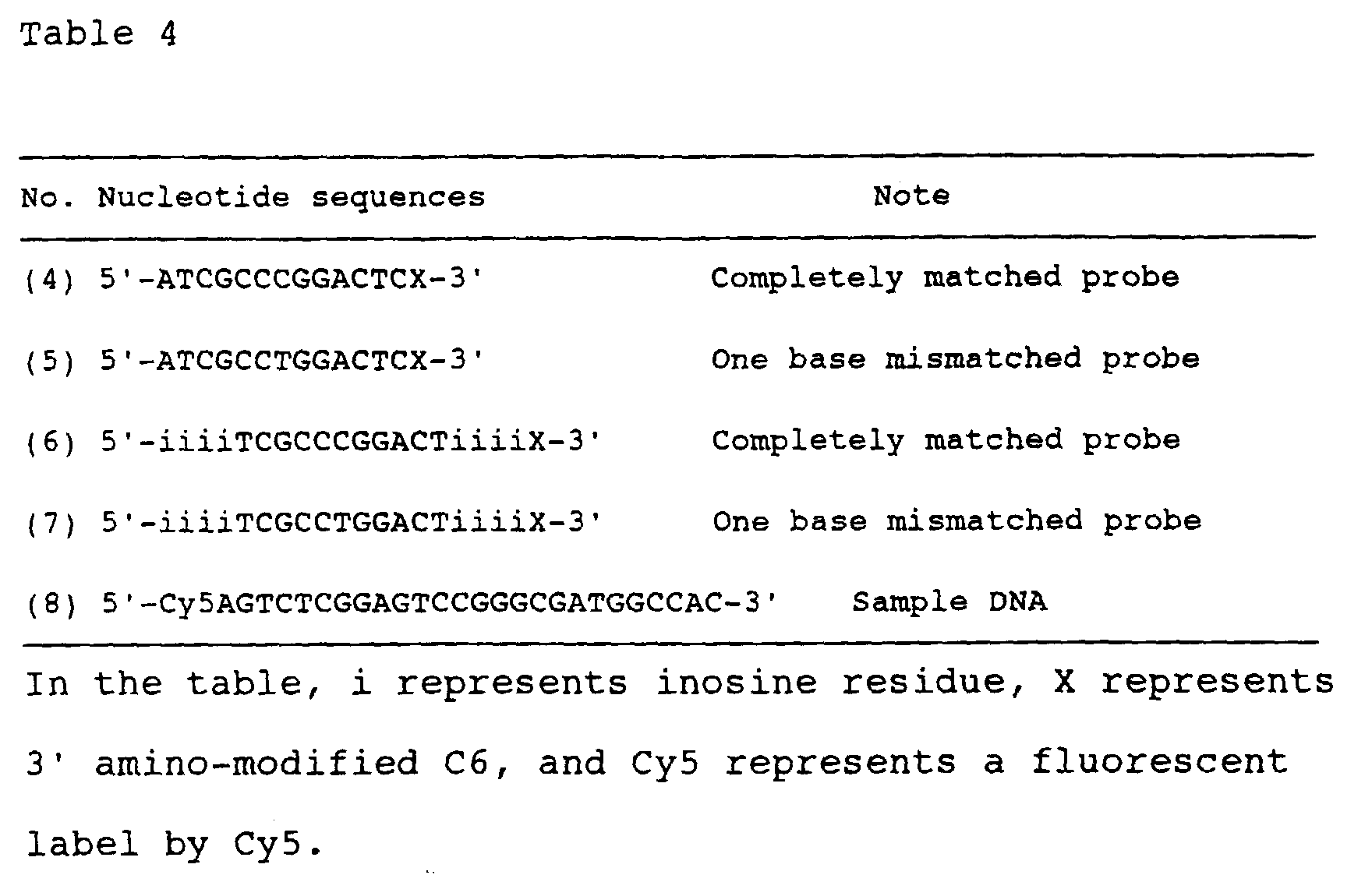
- The syntheses of the aforementioned oligonucleotides and the sample DNA were performed in accordance with a standard protocol (Nucleic Acid Synthesis System User's Guide, PerSeptive Biosystems) by using a cycle in which the trityl groups were not removed, which were the protective groups of the 5' ends. The synthesized DNA were purified by using Poros Oligo R3 (produced by PerSeptive Biosystems).
- The oligonucleotides (4) to (8) and the sample DNA were concentrated to dryness, and then suspended in a 0.5 M sodium hydrogencarbonate buffer (pH 8.4) for (4) to (7), or TE buffer for (8), and they are quantified based on absorbance at 260 nm and adjusted to 100 pmol/µl.
- Immobilization of the oligonucleotides was performed as follows. First, solutions of the oligonucleotide probe nucleic acids (4) to (7) were each spotted onto a Silylateds Slide of TeleChem (slide glass having aldehyde groups on its surface) using a GTMASS Stamping apparatus produced by Nippon Laser & Electronics Lab. Then, according to the protocol of TeleChem, the nucleic acid was bonded on the slide glass through terminal covalent bonds (a Schiff base was formed by amino group and aldehyde group) as follows. First, the slide glass was mounted on a slide rack, and washed twice with 0.2% SDS at 25°C for 2 minutes with sufficient stirring in a beaker. Then, it was washed twice with sterilized water at 25°C for 2 minutes with sufficient stirring, and further treated with sterilized water at 98°C for 2 minutes. The slide glass was air-dried at room temperature for 5 minutes, then transferred into a sodium hydrogenborate solution [prepared by dissolving 1 g of NaBH4 in 300 ml of PBS buffer (prepared by dissolving 8 g of sodium chloride, 0.2 g of potassium chloride, 1.44 g of disodium hydrogenphosphate, and 0.24 g of potassium dihydrogenphosphate in deionized water, adjusting it to pH 7.4 with hydrochloric acid, and filling it up to 1000 ml) and 100 ml of ethanol], treated in the solution at 25°C for 5 minutes, washed three times with 0.2% SDS at 25°C (room temperature) for 1 minute, finally washed with sterilized water at 25°C for 1 minute, and air-dried.
- 70 µg/ml of the Sso7d protein and 100 pmol/ml of the sample DNA (8) were added to Buffer E (20 mM Tris buffer, pH 7.5, 2 mM DTT, 5 mM MgCl2, 10 µg/ml of BSA), and the aforementioned oligonucleotide-immobilized slide glass (DNA microarray) and the fluorescently labeled sample DNA were allowed to hybridize at 60°C for 6 minutes in the solution. As a control, hybridization reaction was also performed in the same manner without adding the Sso7d protein.
- After the hybridization reaction, the membrane was washed with 1 x SSC/0.03% SDS buffer at 25°C for 5 minutes, rinsed with 0.2 x SSC, and then further rinsed with 0.5 x SSC. After the washing solution was removed by centrifugation, the slide glass was air-dried.
- The hybridization signal was evaluated by measuring amount of fluorescent dye of each spot on the air-dried slide glass by using ScanArray 3000 produced by Genaral Scanning to calculate the hybridization signal strength. The results are shown in Table 5. The results are represented with relative values based on the hybridization signal strength of the probe DNA (4) obtained with the addition of Sso7d, which is taken as 100.
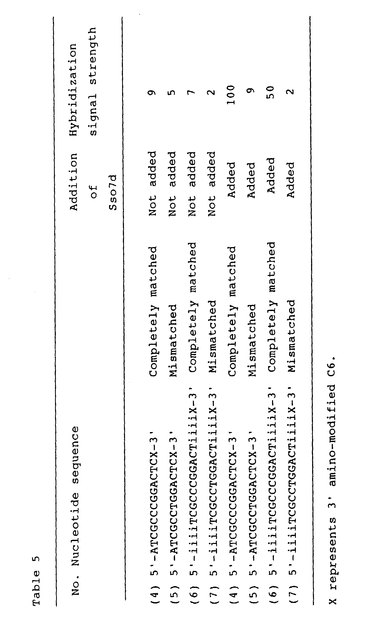
- In the hybridization systems not added with Sso7d, hybridization between the probe DNA immobilized on the substrate (on the DNA array) and the sample DNA was not substantially detected under the aforementioned conditions.
- On the other hand, when the Sso7d protein was added to the hybridization systems, only the completely matched probes hybridized with the sample DNA on the DNA array. In comparison with the hybridization signal strength of the completely matched probes, the hybridization signal strength of the mismatched probes was considerably weaker, and these were clearly distinguishable.
- According to the present invention, gene analysis by hybridization utilizing a probe nucleic acid can be quickly performed with high precision and high sensitivity.
- Having thus described the present invention, it will be obvious that several aspects of the invention may be modified in various ways. Such variations are not to be regarded as departures from the spirit and scope of the invention, and all such modifications would be obvious to those skilled in the arts, and are intended to be included within the scope of the following claims.
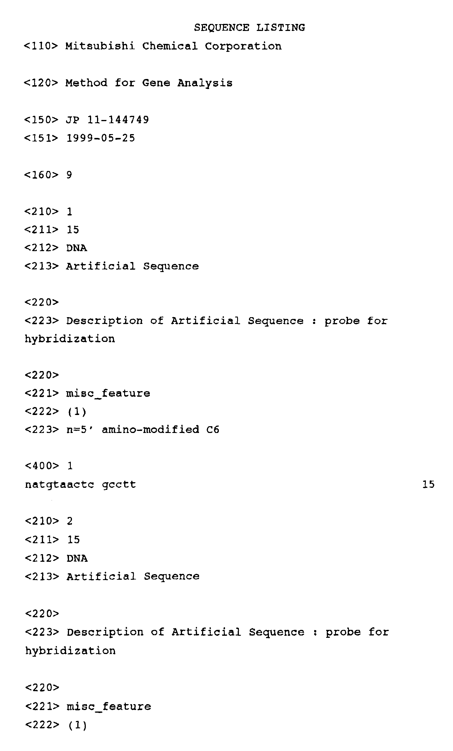
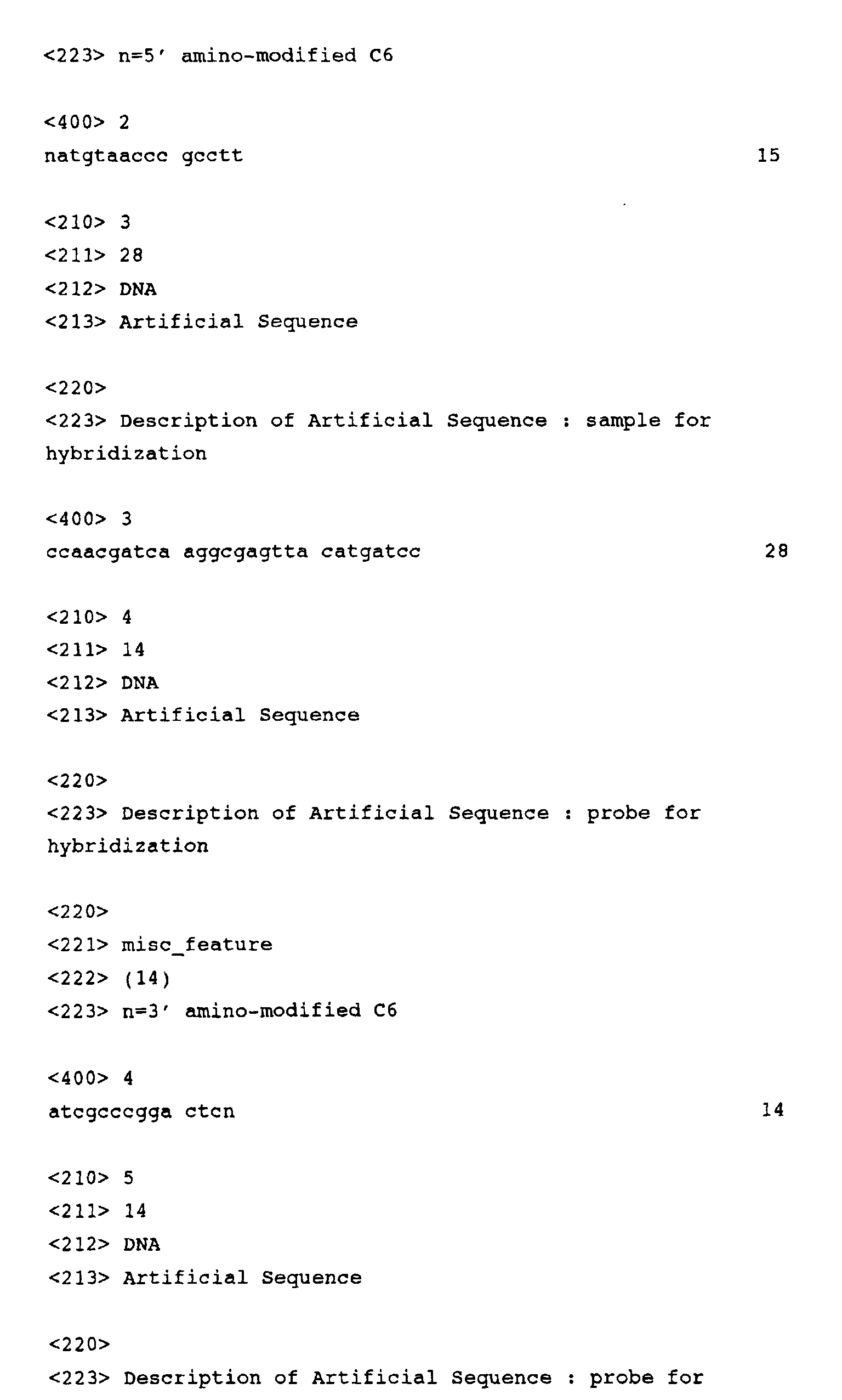
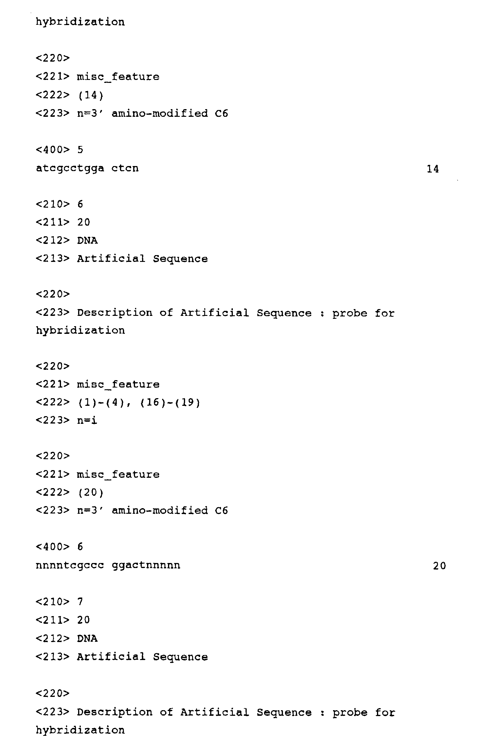

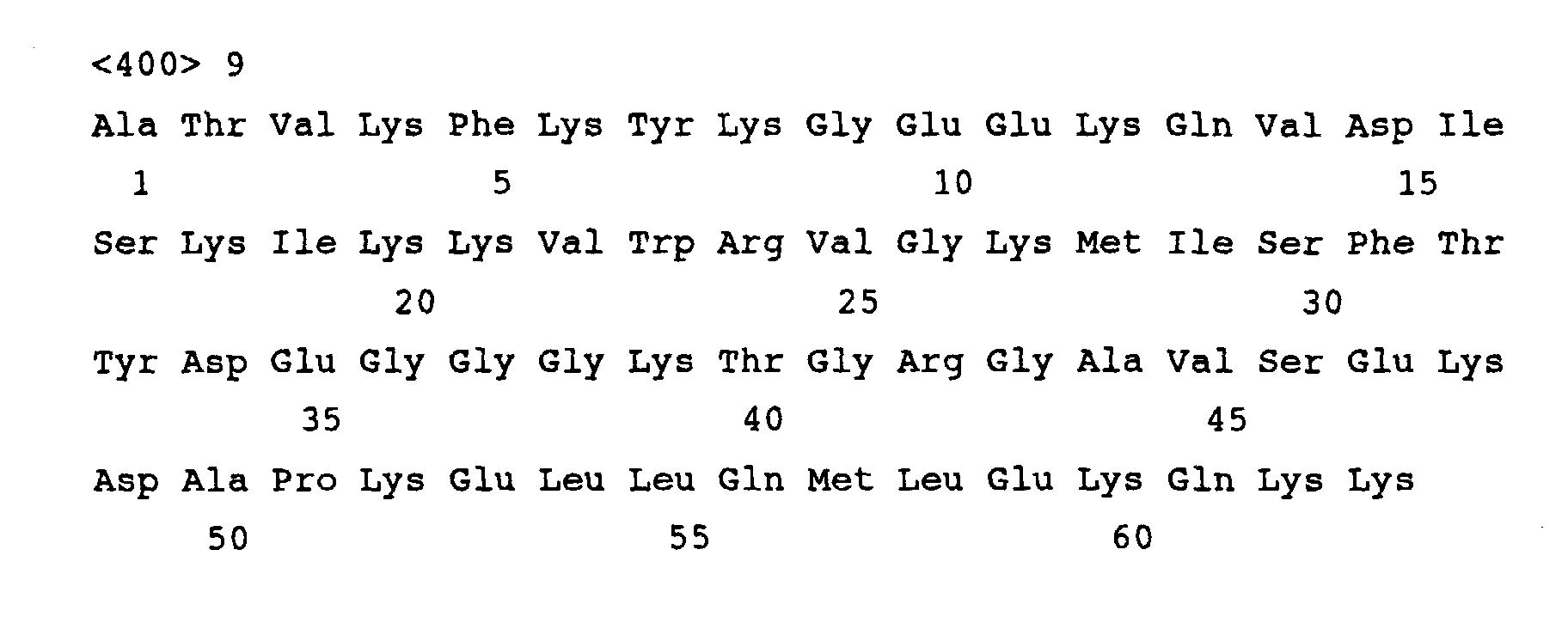
Claims (13)
- A method for gene analysis comprising the step of detecting hybridization between a probe nucleic acid and a sample nucleic acid containing a target sequence that has a sequence complementary to that of the probe nucleic acid, wherein either the probe nucleic acid or the sample nucleic acid is immobilized on a substrate, at least one of the probe nucleic acid and the sample nucleic acid is DNA, and the hybridization is caused in the presence of a double-stranded DNA-binding protein.
- The method of claim 1, wherein the sample nucleic acid is DNA.
- The method of claim 1 or 2, wherein the double-stranded DNA-binding protein is derived from a hyperthermophilic bacterium.
- The method of claim 3, wherein the double-stranded DNA-binding protein is derived from an archaebacterium.
- The method of claim 4, wherein the double-stranded DNA-binding protein is derived from a bacterium belonging to the genus Sulfolobus.
- The method of claim 5, wherein the double-stranded DNA-binding protein is derived from Sulfolobus solfataricus.
- The method of claim 6, wherein the double-stranded DNA-binding protein is the Sso7d protein derived from Sulfolobus solfataricus.
- The method of any one of claims 1 to 7, wherein the double-stranded DNA-binding protein is a protein having homology of 75% or more to the protein represented by the amino acid sequence of SEQ ID NO: 9.
- The method of any one of claims 1 to 8, wherein the sample nucleic acid is labeled.
- The method of any one of claims 1 to 9, wherein amount of the sample nucleic acid containing the target sequence is analyzed based on intensity of hybridization signal.
- The method of any one of claims 1 to 9, wherein detecting hybridization is performed by using a plurality of probe nucleic acids and then polymorphism in the target sequence is detected based on the result of detection of hybridization.
- The method of any one of claims 1 to 9, wherein detecting hybridization is performed by using a plurality of probe nucleic acids and then the nucleotide sequence of the sample nucleic acid is determined based on the result of detection of hybridization.
- A test kit for the detection of hybridization between a probe nucleic acid and a sample nucleic acid containing a target sequence that has a sequence complementary to that of the probe nucleic acid, which comprises at least a double-stranded DNA-binding protein.
Applications Claiming Priority (2)
| Application Number | Priority Date | Filing Date | Title |
|---|---|---|---|
| JP14474999 | 1999-05-25 | ||
| JP14474999 | 1999-05-25 |
Publications (2)
| Publication Number | Publication Date |
|---|---|
| EP1055735A2 true EP1055735A2 (en) | 2000-11-29 |
| EP1055735A3 EP1055735A3 (en) | 2003-02-12 |
Family
ID=15369488
Family Applications (1)
| Application Number | Title | Priority Date | Filing Date |
|---|---|---|---|
| EP00110932A Withdrawn EP1055735A3 (en) | 1999-05-25 | 2000-05-25 | Method for gene analysis by hybridisation in the presence of a double-stranded DNA-binding protein |
Country Status (2)
| Country | Link |
|---|---|
| US (1) | US20030022162A1 (en) |
| EP (1) | EP1055735A3 (en) |
Cited By (1)
| Publication number | Priority date | Publication date | Assignee | Title |
|---|---|---|---|---|
| US7943299B2 (en) * | 2002-04-08 | 2011-05-17 | Eppendorf Ag | Method for determining nucleotide sequences |
Families Citing this family (4)
| Publication number | Priority date | Publication date | Assignee | Title |
|---|---|---|---|---|
| EP1836319B1 (en) * | 2005-01-06 | 2012-09-19 | Life Technologies Corporation | Polypeptides having nucleic acid binding activity and methods for nucleic acid amplification |
| US20070059713A1 (en) * | 2005-09-09 | 2007-03-15 | Lee Jun E | SSB-DNA polymerase fusion proteins |
| CN113454235A (en) * | 2019-02-21 | 2021-09-28 | 豪夫迈·罗氏有限公司 | Improved nucleic acid target enrichment and related methods |
| CN114561449B (en) * | 2022-02-28 | 2022-10-14 | 深圳大学 | Northern blot hybridization solution free of prehybridization and application thereof |
Family Cites Families (3)
| Publication number | Priority date | Publication date | Assignee | Title |
|---|---|---|---|---|
| WO1991006679A1 (en) * | 1989-10-24 | 1991-05-16 | Stratagene | An improved method for hybridizing nucleic acids using single-stranded nucleic acid binding protein |
| US5547843A (en) * | 1992-07-17 | 1996-08-20 | Associated Universities, Inc. | Method for promoting specific alignment of short oligonucleotides on nucleic acids |
| US5989879A (en) * | 1996-10-24 | 1999-11-23 | Yale University | Compositions and methods using eukaryotic Rad52 |
-
2000
- 2000-05-23 US US09/576,715 patent/US20030022162A1/en not_active Abandoned
- 2000-05-25 EP EP00110932A patent/EP1055735A3/en not_active Withdrawn
Non-Patent Citations (1)
| Title |
|---|
| None |
Cited By (1)
| Publication number | Priority date | Publication date | Assignee | Title |
|---|---|---|---|---|
| US7943299B2 (en) * | 2002-04-08 | 2011-05-17 | Eppendorf Ag | Method for determining nucleotide sequences |
Also Published As
| Publication number | Publication date |
|---|---|
| US20030022162A1 (en) | 2003-01-30 |
| EP1055735A3 (en) | 2003-02-12 |
Similar Documents
| Publication | Publication Date | Title |
|---|---|---|
| CA1340121E (en) | Process for amplifying, detecting and/or cloning nucleic acid sequences | |
| EP0972081B1 (en) | Method of nucleic acid amplification | |
| Guo et al. | Enhanced discrimination of single nucleotide polymorphisms by artificial mismatch hybridization | |
| EP0237362B1 (en) | Process for detecting specific nucleotide variations and genetic polymorphisms present in nucleic acids and kits therefor | |
| AU770217B2 (en) | Ligation assembly and detection of polynucleotides on solid-support | |
| US6197563B1 (en) | Kits for amplifying and detecting nucleic acid sequences | |
| JP3010738B2 (en) | Hybridization and amplification methods for nucleic acids | |
| EP1358352B1 (en) | Solid support assay systems and methods utilizing non-standard bases | |
| US6514736B1 (en) | Kits for amplifying and detecting nucleic acid sequences | |
| US6361940B1 (en) | Compositions and methods for enhancing hybridization and priming specificity | |
| US6284497B1 (en) | Nucleic acid arrays and methods of synthesis | |
| US7553643B2 (en) | Method for amplifying target nucleic acid sequences using a primer comprising an AP endonuclease-cleavable linker | |
| CZ229997A3 (en) | Enzyme of thermally stable dna polymerase, process of its preparation and a kit containing thereof | |
| JPH0723800A (en) | Nucleic acid detection method | |
| KR20030074797A (en) | Method of detecting nucleotide polymorphism | |
| JPWO2002062993A1 (en) | Amplified nucleic acid and immobilized product thereof | |
| WO2001004357A2 (en) | Generic sbe-fret protocol | |
| AU4599797A (en) | Compositions and methods for enhancing hybridization specificity | |
| US20030022162A1 (en) | Method for gene analysis | |
| JP2001037489A (en) | Gene analysis method | |
| EP0952228A2 (en) | Compositions and methods for enhancing hybridization specificity | |
| JP2001518787A (en) | Nucleic acid hybridization detection system and its preparation and application | |
| JP2002171992A (en) | Target nucleic acid detection method and kit | |
| HK1155784B (en) | Method of nucleic acid sequencing |
Legal Events
| Date | Code | Title | Description |
|---|---|---|---|
| PUAI | Public reference made under article 153(3) epc to a published international application that has entered the european phase |
Free format text: ORIGINAL CODE: 0009012 |
|
| AK | Designated contracting states |
Kind code of ref document: A2 Designated state(s): AT BE CH CY DE DK ES FI FR GB GR IE IT LI LU MC NL PT SE |
|
| AX | Request for extension of the european patent |
Free format text: AL;LT;LV;MK;RO;SI |
|
| PUAL | Search report despatched |
Free format text: ORIGINAL CODE: 0009013 |
|
| AK | Designated contracting states |
Designated state(s): AT BE CH CY DE DK ES FI FR GB GR IE IT LI LU MC NL PT SE |
|
| AX | Request for extension of the european patent |
Extension state: AL LT LV MK RO SI |
|
| RIC1 | Information provided on ipc code assigned before grant |
Ipc: 7G 01N 33/50 B Ipc: 7C 12Q 1/68 A |
|
| AKX | Designation fees paid | ||
| STAA | Information on the status of an ep patent application or granted ep patent |
Free format text: STATUS: THE APPLICATION IS DEEMED TO BE WITHDRAWN |
|
| 18D | Application deemed to be withdrawn |
Effective date: 20030813 |