EP1047771B1 - Method of refolding angiostatin - Google Patents
Method of refolding angiostatin Download PDFInfo
- Publication number
- EP1047771B1 EP1047771B1 EP99901279A EP99901279A EP1047771B1 EP 1047771 B1 EP1047771 B1 EP 1047771B1 EP 99901279 A EP99901279 A EP 99901279A EP 99901279 A EP99901279 A EP 99901279A EP 1047771 B1 EP1047771 B1 EP 1047771B1
- Authority
- EP
- European Patent Office
- Prior art keywords
- concentration
- during
- present
- seq
- angiostatin
- Prior art date
- Legal status (The legal status is an assumption and is not a legal conclusion. Google has not performed a legal analysis and makes no representation as to the accuracy of the status listed.)
- Expired - Lifetime
Links
- 108010079709 Angiostatins Proteins 0.000 title claims abstract description 103
- 102000012936 Angiostatins Human genes 0.000 title claims abstract description 93
- FZCSTZYAHCUGEM-UHFFFAOYSA-N aspergillomarasmine B Natural products OC(=O)CNC(C(O)=O)CNC(C(O)=O)CC(O)=O FZCSTZYAHCUGEM-UHFFFAOYSA-N 0.000 title claims abstract description 89
- 238000000034 method Methods 0.000 title claims abstract description 77
- 230000003381 solubilizing effect Effects 0.000 claims abstract description 4
- 108090000623 proteins and genes Proteins 0.000 claims description 83
- 230000014509 gene expression Effects 0.000 claims description 34
- 238000005063 solubilization Methods 0.000 claims description 24
- 230000007928 solubilization Effects 0.000 claims description 24
- 101500025915 Homo sapiens Angiostatin Proteins 0.000 claims description 15
- XSQUKJJJFZCRTK-UHFFFAOYSA-N Urea Chemical compound NC(N)=O XSQUKJJJFZCRTK-UHFFFAOYSA-N 0.000 claims description 14
- 239000004202 carbamide Substances 0.000 claims description 14
- SLXKOJJOQWFEFD-UHFFFAOYSA-N 6-aminohexanoic acid Chemical compound NCCCCCC(O)=O SLXKOJJOQWFEFD-UHFFFAOYSA-N 0.000 claims description 9
- 229960002684 aminocaproic acid Drugs 0.000 claims description 9
- 229960003067 cystine Drugs 0.000 claims description 9
- RWSXRVCMGQZWBV-WDSKDSINSA-N glutathione Chemical compound OC(=O)[C@@H](N)CCC(=O)N[C@@H](CS)C(=O)NCC(O)=O RWSXRVCMGQZWBV-WDSKDSINSA-N 0.000 claims description 9
- 229960000789 guanidine hydrochloride Drugs 0.000 claims description 9
- PJJJBBJSCAKJQF-UHFFFAOYSA-N guanidinium chloride Chemical compound [Cl-].NC(N)=[NH2+] PJJJBBJSCAKJQF-UHFFFAOYSA-N 0.000 claims description 9
- 108010024636 Glutathione Proteins 0.000 claims description 8
- XUJNEKJLAYXESH-UHFFFAOYSA-N cysteine Natural products SCC(N)C(O)=O XUJNEKJLAYXESH-UHFFFAOYSA-N 0.000 claims description 8
- 235000018417 cysteine Nutrition 0.000 claims description 8
- 230000003647 oxidation Effects 0.000 claims description 8
- 238000007254 oxidation reaction Methods 0.000 claims description 8
- 238000004007 reversed phase HPLC Methods 0.000 claims description 8
- 230000027455 binding Effects 0.000 claims description 7
- 238000004587 chromatography analysis Methods 0.000 claims description 7
- 239000003638 chemical reducing agent Substances 0.000 claims description 6
- LEVWYRKDKASIDU-IMJSIDKUSA-N cystine group Chemical group C([C@@H](C(=O)O)N)SSC[C@@H](C(=O)O)N LEVWYRKDKASIDU-IMJSIDKUSA-N 0.000 claims description 6
- 230000008569 process Effects 0.000 claims description 6
- 239000003795 chemical substances by application Substances 0.000 claims description 5
- 239000003446 ligand Substances 0.000 claims description 5
- DGVVWUTYPXICAM-UHFFFAOYSA-N β‐Mercaptoethanol Chemical compound OCCS DGVVWUTYPXICAM-UHFFFAOYSA-N 0.000 claims description 5
- JJMDCOVWQOJGCB-UHFFFAOYSA-N 5-aminopentanoic acid Chemical compound [NH3+]CCCCC([O-])=O JJMDCOVWQOJGCB-UHFFFAOYSA-N 0.000 claims description 4
- 229920002684 Sepharose Polymers 0.000 claims description 4
- BTCSSZJGUNDROE-UHFFFAOYSA-N gamma-aminobutyric acid Chemical compound NCCCC(O)=O BTCSSZJGUNDROE-UHFFFAOYSA-N 0.000 claims description 4
- 238000004191 hydrophobic interaction chromatography Methods 0.000 claims description 4
- 238000004255 ion exchange chromatography Methods 0.000 claims description 4
- LEVWYRKDKASIDU-QWWZWVQMSA-N D-cystine Chemical compound OC(=O)[C@H](N)CSSC[C@@H](N)C(O)=O LEVWYRKDKASIDU-QWWZWVQMSA-N 0.000 claims description 3
- BWGNESOTFCXPMA-UHFFFAOYSA-N Dihydrogen disulfide Chemical compound SS BWGNESOTFCXPMA-UHFFFAOYSA-N 0.000 claims description 3
- 238000012258 culturing Methods 0.000 claims description 3
- YPZRWBKMTBYPTK-BJDJZHNGSA-N glutathione disulfide Chemical compound OC(=O)[C@@H](N)CCC(=O)N[C@H](C(=O)NCC(O)=O)CSSC[C@@H](C(=O)NCC(O)=O)NC(=O)CC[C@H](N)C(O)=O YPZRWBKMTBYPTK-BJDJZHNGSA-N 0.000 claims description 3
- XDOLZJYETYVRKV-UHFFFAOYSA-N 7-Aminoheptanoic acid Chemical compound NCCCCCCC(O)=O XDOLZJYETYVRKV-UHFFFAOYSA-N 0.000 claims description 2
- 108010053070 Glutathione Disulfide Proteins 0.000 claims description 2
- XUJNEKJLAYXESH-REOHCLBHSA-N L-Cysteine Chemical compound SC[C@H](N)C(O)=O XUJNEKJLAYXESH-REOHCLBHSA-N 0.000 claims description 2
- 241001465754 Metazoa Species 0.000 claims description 2
- 230000002708 enhancing effect Effects 0.000 claims description 2
- 229960003692 gamma aminobutyric acid Drugs 0.000 claims description 2
- YPZRWBKMTBYPTK-UHFFFAOYSA-N oxidized gamma-L-glutamyl-L-cysteinylglycine Natural products OC(=O)C(N)CCC(=O)NC(C(=O)NCC(O)=O)CSSCC(C(=O)NCC(O)=O)NC(=O)CCC(N)C(O)=O YPZRWBKMTBYPTK-UHFFFAOYSA-N 0.000 claims description 2
- 125000003396 thiol group Chemical group [H]S* 0.000 claims description 2
- 210000003000 inclusion body Anatomy 0.000 abstract description 35
- 241000894006 Bacteria Species 0.000 abstract description 5
- 150000007523 nucleic acids Chemical class 0.000 abstract description 3
- 108020004707 nucleic acids Proteins 0.000 abstract description 2
- 102000039446 nucleic acids Human genes 0.000 abstract description 2
- 210000004027 cell Anatomy 0.000 description 65
- 108020004414 DNA Proteins 0.000 description 61
- 102000004169 proteins and genes Human genes 0.000 description 61
- 235000018102 proteins Nutrition 0.000 description 55
- 241000588724 Escherichia coli Species 0.000 description 43
- 239000013612 plasmid Substances 0.000 description 39
- 239000002299 complementary DNA Substances 0.000 description 30
- 210000002889 endothelial cell Anatomy 0.000 description 23
- 239000012634 fragment Substances 0.000 description 23
- 102000013566 Plasminogen Human genes 0.000 description 22
- 108010051456 Plasminogen Proteins 0.000 description 22
- 229960000268 spectinomycin Drugs 0.000 description 21
- UNFWWIHTNXNPBV-WXKVUWSESA-N spectinomycin Chemical compound O([C@@H]1[C@@H](NC)[C@@H](O)[C@H]([C@@H]([C@H]1O1)O)NC)[C@]2(O)[C@H]1O[C@H](C)CC2=O UNFWWIHTNXNPBV-WXKVUWSESA-N 0.000 description 21
- 206010028980 Neoplasm Diseases 0.000 description 19
- 150000001413 amino acids Chemical group 0.000 description 17
- 239000002609 medium Substances 0.000 description 15
- 101000605403 Homo sapiens Plasminogen Proteins 0.000 description 13
- 235000001014 amino acid Nutrition 0.000 description 13
- 230000035772 mutation Effects 0.000 description 13
- 239000013604 expression vector Substances 0.000 description 12
- 230000012010 growth Effects 0.000 description 12
- 108090000765 processed proteins & peptides Proteins 0.000 description 12
- 230000015572 biosynthetic process Effects 0.000 description 11
- 230000001580 bacterial effect Effects 0.000 description 10
- 239000002253 acid Substances 0.000 description 9
- 230000033115 angiogenesis Effects 0.000 description 9
- 239000000872 buffer Substances 0.000 description 9
- 238000005119 centrifugation Methods 0.000 description 9
- 238000006243 chemical reaction Methods 0.000 description 9
- 238000000338 in vitro Methods 0.000 description 9
- 229920001184 polypeptide Polymers 0.000 description 9
- 102000004196 processed proteins & peptides Human genes 0.000 description 9
- 108091008146 restriction endonucleases Proteins 0.000 description 9
- 239000013598 vector Substances 0.000 description 9
- 108020004705 Codon Proteins 0.000 description 8
- 108091028043 Nucleic acid sequence Proteins 0.000 description 8
- 150000007513 acids Chemical class 0.000 description 8
- 238000007792 addition Methods 0.000 description 8
- 201000011510 cancer Diseases 0.000 description 8
- 230000005764 inhibitory process Effects 0.000 description 8
- 230000005012 migration Effects 0.000 description 8
- 238000013508 migration Methods 0.000 description 8
- 229960000210 nalidixic acid Drugs 0.000 description 8
- MHWLWQUZZRMNGJ-UHFFFAOYSA-N nalidixic acid Chemical compound C1=C(C)N=C2N(CC)C=C(C(O)=O)C(=O)C2=C1 MHWLWQUZZRMNGJ-UHFFFAOYSA-N 0.000 description 8
- 239000002773 nucleotide Substances 0.000 description 8
- 125000003729 nucleotide group Chemical group 0.000 description 8
- 238000000746 purification Methods 0.000 description 8
- 238000002415 sodium dodecyl sulfate polyacrylamide gel electrophoresis Methods 0.000 description 8
- 238000001712 DNA sequencing Methods 0.000 description 7
- 230000003115 biocidal effect Effects 0.000 description 7
- 238000012512 characterization method Methods 0.000 description 7
- 238000002135 phase contrast microscopy Methods 0.000 description 7
- 239000000047 product Substances 0.000 description 7
- 241000283690 Bos taurus Species 0.000 description 6
- 239000003155 DNA primer Substances 0.000 description 6
- HEMHJVSKTPXQMS-UHFFFAOYSA-M Sodium hydroxide Chemical compound [OH-].[Na+] HEMHJVSKTPXQMS-UHFFFAOYSA-M 0.000 description 6
- 238000003556 assay Methods 0.000 description 6
- 238000010276 construction Methods 0.000 description 6
- 238000000855 fermentation Methods 0.000 description 6
- 230000004151 fermentation Effects 0.000 description 6
- 238000003752 polymerase chain reaction Methods 0.000 description 6
- 239000013615 primer Substances 0.000 description 6
- 230000037432 silent mutation Effects 0.000 description 6
- 108020004635 Complementary DNA Proteins 0.000 description 5
- 208000006552 Lewis Lung Carcinoma Diseases 0.000 description 5
- 206010027476 Metastases Diseases 0.000 description 5
- 241000699670 Mus sp. Species 0.000 description 5
- 102000016387 Pancreatic elastase Human genes 0.000 description 5
- 108010067372 Pancreatic elastase Proteins 0.000 description 5
- 108010076504 Protein Sorting Signals Proteins 0.000 description 5
- 239000007864 aqueous solution Substances 0.000 description 5
- 230000010261 cell growth Effects 0.000 description 5
- 210000000805 cytoplasm Anatomy 0.000 description 5
- 239000003398 denaturant Substances 0.000 description 5
- 230000000694 effects Effects 0.000 description 5
- 238000004128 high performance liquid chromatography Methods 0.000 description 5
- 230000002401 inhibitory effect Effects 0.000 description 5
- 238000010232 migration assay Methods 0.000 description 5
- 238000000527 sonication Methods 0.000 description 5
- 239000007989 BIS-Tris Propane buffer Substances 0.000 description 4
- UXVMQQNJUSDDNG-UHFFFAOYSA-L Calcium chloride Chemical compound [Cl-].[Cl-].[Ca+2] UXVMQQNJUSDDNG-UHFFFAOYSA-L 0.000 description 4
- 102100024785 Fibroblast growth factor 2 Human genes 0.000 description 4
- 108090000379 Fibroblast growth factor 2 Proteins 0.000 description 4
- 102000007056 Recombinant Fusion Proteins Human genes 0.000 description 4
- 108010008281 Recombinant Fusion Proteins Proteins 0.000 description 4
- FAPWRFPIFSIZLT-UHFFFAOYSA-M Sodium chloride Chemical compound [Na+].[Cl-] FAPWRFPIFSIZLT-UHFFFAOYSA-M 0.000 description 4
- HHKZCCWKTZRCCL-UHFFFAOYSA-N bis-tris propane Chemical compound OCC(CO)(CO)NCCCNC(CO)(CO)CO HHKZCCWKTZRCCL-UHFFFAOYSA-N 0.000 description 4
- 239000001110 calcium chloride Substances 0.000 description 4
- 229910001628 calcium chloride Inorganic materials 0.000 description 4
- 238000005516 engineering process Methods 0.000 description 4
- 239000012091 fetal bovine serum Substances 0.000 description 4
- 239000000203 mixture Substances 0.000 description 4
- 239000008188 pellet Substances 0.000 description 4
- 230000035755 proliferation Effects 0.000 description 4
- 238000012163 sequencing technique Methods 0.000 description 4
- 239000000243 solution Substances 0.000 description 4
- IAZDPXIOMUYVGZ-UHFFFAOYSA-N Dimethylsulphoxide Chemical compound CS(C)=O IAZDPXIOMUYVGZ-UHFFFAOYSA-N 0.000 description 3
- KCXVZYZYPLLWCC-UHFFFAOYSA-N EDTA Chemical compound OC(=O)CN(CC(O)=O)CCN(CC(O)=O)CC(O)=O KCXVZYZYPLLWCC-UHFFFAOYSA-N 0.000 description 3
- WQZGKKKJIJFFOK-GASJEMHNSA-N Glucose Natural products OC[C@H]1OC(O)[C@H](O)[C@@H](O)[C@@H]1O WQZGKKKJIJFFOK-GASJEMHNSA-N 0.000 description 3
- OKKJLVBELUTLKV-UHFFFAOYSA-N Methanol Chemical compound OC OKKJLVBELUTLKV-UHFFFAOYSA-N 0.000 description 3
- 241001529936 Murinae Species 0.000 description 3
- 241000699666 Mus <mouse, genus> Species 0.000 description 3
- 101100301239 Myxococcus xanthus recA1 gene Proteins 0.000 description 3
- 108010073929 Vascular Endothelial Growth Factor A Proteins 0.000 description 3
- 102000005789 Vascular Endothelial Growth Factors Human genes 0.000 description 3
- 108010019530 Vascular Endothelial Growth Factors Proteins 0.000 description 3
- 229960000723 ampicillin Drugs 0.000 description 3
- AVKUERGKIZMTKX-NJBDSQKTSA-N ampicillin Chemical compound C1([C@@H](N)C(=O)N[C@H]2[C@H]3SC([C@@H](N3C2=O)C(O)=O)(C)C)=CC=CC=C1 AVKUERGKIZMTKX-NJBDSQKTSA-N 0.000 description 3
- 239000002870 angiogenesis inducing agent Substances 0.000 description 3
- 230000002491 angiogenic effect Effects 0.000 description 3
- 230000001772 anti-angiogenic effect Effects 0.000 description 3
- 230000012292 cell migration Effects 0.000 description 3
- 150000001875 compounds Chemical class 0.000 description 3
- 239000008103 glucose Substances 0.000 description 3
- 238000001727 in vivo Methods 0.000 description 3
- 238000002955 isolation Methods 0.000 description 3
- 210000004962 mammalian cell Anatomy 0.000 description 3
- 238000004519 manufacturing process Methods 0.000 description 3
- 108010082117 matrigel Proteins 0.000 description 3
- 239000012528 membrane Substances 0.000 description 3
- 230000009401 metastasis Effects 0.000 description 3
- 238000010369 molecular cloning Methods 0.000 description 3
- 230000017854 proteolysis Effects 0.000 description 3
- 239000011347 resin Substances 0.000 description 3
- 229920005989 resin Polymers 0.000 description 3
- 239000000523 sample Substances 0.000 description 3
- 239000001488 sodium phosphate Substances 0.000 description 3
- 229910000162 sodium phosphate Inorganic materials 0.000 description 3
- 230000009466 transformation Effects 0.000 description 3
- RYFMWSXOAZQYPI-UHFFFAOYSA-K trisodium phosphate Chemical compound [Na+].[Na+].[Na+].[O-]P([O-])([O-])=O RYFMWSXOAZQYPI-UHFFFAOYSA-K 0.000 description 3
- 108091003079 Bovine Serum Albumin Proteins 0.000 description 2
- 206010006187 Breast cancer Diseases 0.000 description 2
- 208000026310 Breast neoplasm Diseases 0.000 description 2
- 238000007399 DNA isolation Methods 0.000 description 2
- ZHNUHDYFZUAESO-UHFFFAOYSA-N Formamide Chemical compound NC=O ZHNUHDYFZUAESO-UHFFFAOYSA-N 0.000 description 2
- 108010010803 Gelatin Proteins 0.000 description 2
- 241000238631 Hexapoda Species 0.000 description 2
- ZDXPYRJPNDTMRX-VKHMYHEASA-N L-glutamine Chemical compound OC(=O)[C@@H](N)CCC(N)=O ZDXPYRJPNDTMRX-VKHMYHEASA-N 0.000 description 2
- 229930182816 L-glutamine Natural products 0.000 description 2
- 102000008300 Mutant Proteins Human genes 0.000 description 2
- 108010021466 Mutant Proteins Proteins 0.000 description 2
- 102000004211 Platelet factor 4 Human genes 0.000 description 2
- 108090000778 Platelet factor 4 Proteins 0.000 description 2
- 239000012614 Q-Sepharose Substances 0.000 description 2
- 238000011579 SCID mouse model Methods 0.000 description 2
- 240000004808 Saccharomyces cerevisiae Species 0.000 description 2
- IQFYYKKMVGJFEH-XLPZGREQSA-N Thymidine Chemical compound O=C1NC(=O)C(C)=CN1[C@@H]1O[C@H](CO)[C@@H](O)C1 IQFYYKKMVGJFEH-XLPZGREQSA-N 0.000 description 2
- 102100039037 Vascular endothelial growth factor A Human genes 0.000 description 2
- 238000002835 absorbance Methods 0.000 description 2
- 239000004037 angiogenesis inhibitor Substances 0.000 description 2
- 238000011122 anti-angiogenic therapy Methods 0.000 description 2
- 230000004663 cell proliferation Effects 0.000 description 2
- 238000001516 cell proliferation assay Methods 0.000 description 2
- 238000010367 cloning Methods 0.000 description 2
- 238000011161 development Methods 0.000 description 2
- 230000018109 developmental process Effects 0.000 description 2
- 230000029087 digestion Effects 0.000 description 2
- LOKCTEFSRHRXRJ-UHFFFAOYSA-I dipotassium trisodium dihydrogen phosphate hydrogen phosphate dichloride Chemical compound P(=O)(O)(O)[O-].[K+].P(=O)(O)([O-])[O-].[Na+].[Na+].[Cl-].[K+].[Cl-].[Na+] LOKCTEFSRHRXRJ-UHFFFAOYSA-I 0.000 description 2
- 201000010099 disease Diseases 0.000 description 2
- 208000037265 diseases, disorders, signs and symptoms Diseases 0.000 description 2
- VHJLVAABSRFDPM-QWWZWVQMSA-N dithiothreitol Chemical compound SC[C@@H](O)[C@H](O)CS VHJLVAABSRFDPM-QWWZWVQMSA-N 0.000 description 2
- 229940079593 drug Drugs 0.000 description 2
- 239000003814 drug Substances 0.000 description 2
- 238000002330 electrospray ionisation mass spectrometry Methods 0.000 description 2
- 230000010595 endothelial cell migration Effects 0.000 description 2
- 238000002474 experimental method Methods 0.000 description 2
- 238000001502 gel electrophoresis Methods 0.000 description 2
- 239000008273 gelatin Substances 0.000 description 2
- 229920000159 gelatin Polymers 0.000 description 2
- 235000019322 gelatine Nutrition 0.000 description 2
- 235000011852 gelatine desserts Nutrition 0.000 description 2
- 238000007429 general method Methods 0.000 description 2
- 238000011534 incubation Methods 0.000 description 2
- 239000000543 intermediate Substances 0.000 description 2
- 239000011159 matrix material Substances 0.000 description 2
- 206010061289 metastatic neoplasm Diseases 0.000 description 2
- 210000004925 microvascular endothelial cell Anatomy 0.000 description 2
- 230000013152 negative regulation of cell migration Effects 0.000 description 2
- 239000002953 phosphate buffered saline Substances 0.000 description 2
- 238000002360 preparation method Methods 0.000 description 2
- 238000001742 protein purification Methods 0.000 description 2
- 230000001105 regulatory effect Effects 0.000 description 2
- 238000011160 research Methods 0.000 description 2
- 230000028327 secretion Effects 0.000 description 2
- 239000011780 sodium chloride Substances 0.000 description 2
- 239000006228 supernatant Substances 0.000 description 2
- 238000012360 testing method Methods 0.000 description 2
- 210000001519 tissue Anatomy 0.000 description 2
- 238000011282 treatment Methods 0.000 description 2
- 210000004881 tumor cell Anatomy 0.000 description 2
- 230000004614 tumor growth Effects 0.000 description 2
- 108010047303 von Willebrand Factor Proteins 0.000 description 2
- 102100036537 von Willebrand factor Human genes 0.000 description 2
- QKNYBSVHEMOAJP-UHFFFAOYSA-N 2-amino-2-(hydroxymethyl)propane-1,3-diol;hydron;chloride Chemical compound Cl.OCC(N)(CO)CO QKNYBSVHEMOAJP-UHFFFAOYSA-N 0.000 description 1
- 241000585703 Adelphia <angiosperm> Species 0.000 description 1
- 229920001817 Agar Polymers 0.000 description 1
- VHUUQVKOLVNVRT-UHFFFAOYSA-N Ammonium hydroxide Chemical compound [NH4+].[OH-] VHUUQVKOLVNVRT-UHFFFAOYSA-N 0.000 description 1
- 235000014469 Bacillus subtilis Nutrition 0.000 description 1
- 208000011691 Burkitt lymphomas Diseases 0.000 description 1
- 241000283707 Capra Species 0.000 description 1
- 201000009030 Carcinoma Diseases 0.000 description 1
- 206010009944 Colon cancer Diseases 0.000 description 1
- 102000012410 DNA Ligases Human genes 0.000 description 1
- 108010061982 DNA Ligases Proteins 0.000 description 1
- 230000004568 DNA-binding Effects 0.000 description 1
- 102000004163 DNA-directed RNA polymerases Human genes 0.000 description 1
- 108090000626 DNA-directed RNA polymerases Proteins 0.000 description 1
- 206010059866 Drug resistance Diseases 0.000 description 1
- 239000006144 Dulbecco’s modified Eagle's medium Substances 0.000 description 1
- 102400001047 Endostatin Human genes 0.000 description 1
- 108010079505 Endostatins Proteins 0.000 description 1
- 241000702189 Escherichia virus Mu Species 0.000 description 1
- 108050007372 Fibroblast Growth Factor Proteins 0.000 description 1
- 102000018233 Fibroblast Growth Factor Human genes 0.000 description 1
- 108090000386 Fibroblast Growth Factor 1 Proteins 0.000 description 1
- 102100031706 Fibroblast growth factor 1 Human genes 0.000 description 1
- 201000008808 Fibrosarcoma Diseases 0.000 description 1
- HTTJABKRGRZYRN-UHFFFAOYSA-N Heparin Chemical compound OC1C(NC(=O)C)C(O)OC(COS(O)(=O)=O)C1OC1C(OS(O)(=O)=O)C(O)C(OC2C(C(OS(O)(=O)=O)C(OC3C(C(O)C(O)C(O3)C(O)=O)OS(O)(=O)=O)C(CO)O2)NS(O)(=O)=O)C(C(O)=O)O1 HTTJABKRGRZYRN-UHFFFAOYSA-N 0.000 description 1
- 241000282412 Homo Species 0.000 description 1
- 108090000144 Human Proteins Proteins 0.000 description 1
- 102000003839 Human Proteins Human genes 0.000 description 1
- -1 Hyclone) Substances 0.000 description 1
- 102000006992 Interferon-alpha Human genes 0.000 description 1
- 108010047761 Interferon-alpha Proteins 0.000 description 1
- 241000235058 Komagataella pastoris Species 0.000 description 1
- FFEARJCKVFRZRR-BYPYZUCNSA-N L-methionine Chemical compound CSCC[C@H](N)C(O)=O FFEARJCKVFRZRR-BYPYZUCNSA-N 0.000 description 1
- KDXKERNSBIXSRK-UHFFFAOYSA-N Lysine Natural products NCCCCC(N)C(O)=O KDXKERNSBIXSRK-UHFFFAOYSA-N 0.000 description 1
- 239000004472 Lysine Substances 0.000 description 1
- 101710181812 Methionine aminopeptidase Proteins 0.000 description 1
- 101150101095 Mmp12 gene Proteins 0.000 description 1
- 108010006519 Molecular Chaperones Proteins 0.000 description 1
- 102000005431 Molecular Chaperones Human genes 0.000 description 1
- 108090000143 Mouse Proteins Proteins 0.000 description 1
- 101500025937 Mus musculus Angiostatin Proteins 0.000 description 1
- 241000283973 Oryctolagus cuniculus Species 0.000 description 1
- 101710116435 Outer membrane protein Proteins 0.000 description 1
- 108091005804 Peptidases Proteins 0.000 description 1
- 102000035195 Peptidases Human genes 0.000 description 1
- 108010049977 Peptide Elongation Factor Tu Proteins 0.000 description 1
- 101100091878 Plasmodium falciparum (isolate 3D7) rpoC2 gene Proteins 0.000 description 1
- 239000004793 Polystyrene Substances 0.000 description 1
- 206010060862 Prostate cancer Diseases 0.000 description 1
- 229940124158 Protease/peptidase inhibitor Drugs 0.000 description 1
- 108020004511 Recombinant DNA Proteins 0.000 description 1
- 102000008063 Small Heat-Shock Proteins Human genes 0.000 description 1
- 108060008245 Thrombospondin Proteins 0.000 description 1
- 102000002938 Thrombospondin Human genes 0.000 description 1
- 239000007983 Tris buffer Substances 0.000 description 1
- 102000004142 Trypsin Human genes 0.000 description 1
- 108090000631 Trypsin Proteins 0.000 description 1
- 238000010306 acid treatment Methods 0.000 description 1
- 230000009471 action Effects 0.000 description 1
- 239000011149 active material Substances 0.000 description 1
- 239000000654 additive Substances 0.000 description 1
- 210000004404 adrenal cortex Anatomy 0.000 description 1
- 238000005273 aeration Methods 0.000 description 1
- 239000008272 agar Substances 0.000 description 1
- 238000004220 aggregation Methods 0.000 description 1
- 238000013019 agitation Methods 0.000 description 1
- 230000006229 amino acid addition Effects 0.000 description 1
- 230000003321 amplification Effects 0.000 description 1
- 238000004458 analytical method Methods 0.000 description 1
- 229940121369 angiogenesis inhibitor Drugs 0.000 description 1
- 239000003242 anti bacterial agent Substances 0.000 description 1
- 230000003466 anti-cipated effect Effects 0.000 description 1
- 230000000259 anti-tumor effect Effects 0.000 description 1
- 229940088710 antibiotic agent Drugs 0.000 description 1
- 230000001640 apoptogenic effect Effects 0.000 description 1
- 230000006907 apoptotic process Effects 0.000 description 1
- QVGXLLKOCUKJST-UHFFFAOYSA-N atomic oxygen Chemical compound [O] QVGXLLKOCUKJST-UHFFFAOYSA-N 0.000 description 1
- 210000004369 blood Anatomy 0.000 description 1
- 239000008280 blood Substances 0.000 description 1
- 210000004204 blood vessel Anatomy 0.000 description 1
- 238000009835 boiling Methods 0.000 description 1
- 201000008275 breast carcinoma Diseases 0.000 description 1
- 230000005907 cancer growth Effects 0.000 description 1
- 229940041514 candida albicans extract Drugs 0.000 description 1
- 210000001043 capillary endothelial cell Anatomy 0.000 description 1
- 239000006285 cell suspension Substances 0.000 description 1
- 230000001413 cellular effect Effects 0.000 description 1
- 239000003153 chemical reaction reagent Substances 0.000 description 1
- 239000002975 chemoattractant Substances 0.000 description 1
- 238000002512 chemotherapy Methods 0.000 description 1
- 230000002759 chromosomal effect Effects 0.000 description 1
- 238000003776 cleavage reaction Methods 0.000 description 1
- 238000002648 combination therapy Methods 0.000 description 1
- 238000012790 confirmation Methods 0.000 description 1
- 238000011254 conventional chemotherapy Methods 0.000 description 1
- 125000000151 cysteine group Chemical group N[C@@H](CS)C(=O)* 0.000 description 1
- 238000012217 deletion Methods 0.000 description 1
- 230000037430 deletion Effects 0.000 description 1
- 101150106284 deoR gene Proteins 0.000 description 1
- 230000001419 dependent effect Effects 0.000 description 1
- 238000003745 diagnosis Methods 0.000 description 1
- 238000010586 diagram Methods 0.000 description 1
- 230000005059 dormancy Effects 0.000 description 1
- 231100000673 dose–response relationship Toxicity 0.000 description 1
- 238000011143 downstream manufacturing Methods 0.000 description 1
- 239000000975 dye Substances 0.000 description 1
- 230000002500 effect on skin Effects 0.000 description 1
- 238000001962 electrophoresis Methods 0.000 description 1
- 238000010828 elution Methods 0.000 description 1
- 230000002124 endocrine Effects 0.000 description 1
- 230000003511 endothelial effect Effects 0.000 description 1
- 230000007613 environmental effect Effects 0.000 description 1
- 238000001976 enzyme digestion Methods 0.000 description 1
- 238000000605 extraction Methods 0.000 description 1
- 210000004996 female reproductive system Anatomy 0.000 description 1
- 239000007850 fluorescent dye Substances 0.000 description 1
- 230000004927 fusion Effects 0.000 description 1
- 101150045500 galK gene Proteins 0.000 description 1
- 101150041954 galU gene Proteins 0.000 description 1
- 239000000499 gel Substances 0.000 description 1
- 238000001641 gel filtration chromatography Methods 0.000 description 1
- 101150096208 gtaB gene Proteins 0.000 description 1
- 229960002897 heparin Drugs 0.000 description 1
- 229920000669 heparin Polymers 0.000 description 1
- 210000005260 human cell Anatomy 0.000 description 1
- 229960000900 human factor viii Drugs 0.000 description 1
- 230000006698 induction Effects 0.000 description 1
- 239000003112 inhibitor Substances 0.000 description 1
- 230000003993 interaction Effects 0.000 description 1
- 230000009545 invasion Effects 0.000 description 1
- 238000005304 joining Methods 0.000 description 1
- 101150109249 lacI gene Proteins 0.000 description 1
- 238000011068 loading method Methods 0.000 description 1
- 231100000053 low toxicity Toxicity 0.000 description 1
- 210000004924 lung microvascular endothelial cell Anatomy 0.000 description 1
- 239000003550 marker Substances 0.000 description 1
- 101150023497 mcrA gene Proteins 0.000 description 1
- 230000001404 mediated effect Effects 0.000 description 1
- 230000001394 metastastic effect Effects 0.000 description 1
- 229930182817 methionine Natural products 0.000 description 1
- 230000000813 microbial effect Effects 0.000 description 1
- 244000005700 microbiome Species 0.000 description 1
- 239000003226 mitogen Substances 0.000 description 1
- 238000012544 monitoring process Methods 0.000 description 1
- 230000014399 negative regulation of angiogenesis Effects 0.000 description 1
- 238000003199 nucleic acid amplification method Methods 0.000 description 1
- 101150012154 nupG gene Proteins 0.000 description 1
- 239000001301 oxygen Substances 0.000 description 1
- 229910052760 oxygen Inorganic materials 0.000 description 1
- 239000000137 peptide hydrolase inhibitor Substances 0.000 description 1
- 210000001322 periplasm Anatomy 0.000 description 1
- 239000004417 polycarbonate Substances 0.000 description 1
- 229920000515 polycarbonate Polymers 0.000 description 1
- 229920002223 polystyrene Polymers 0.000 description 1
- 239000011148 porous material Substances 0.000 description 1
- 230000001323 posttranslational effect Effects 0.000 description 1
- 230000001023 pro-angiogenic effect Effects 0.000 description 1
- 238000012545 processing Methods 0.000 description 1
- 238000004393 prognosis Methods 0.000 description 1
- 201000001514 prostate carcinoma Diseases 0.000 description 1
- 235000019833 protease Nutrition 0.000 description 1
- 230000004845 protein aggregation Effects 0.000 description 1
- 238000012514 protein characterization Methods 0.000 description 1
- 230000012846 protein folding Effects 0.000 description 1
- 238000001243 protein synthesis Methods 0.000 description 1
- 238000003259 recombinant expression Methods 0.000 description 1
- 230000010076 replication Effects 0.000 description 1
- 210000004176 reticulum cell Anatomy 0.000 description 1
- 238000012552 review Methods 0.000 description 1
- 101150029016 rpo3 gene Proteins 0.000 description 1
- 101150102864 rpoD gene Proteins 0.000 description 1
- 101150098466 rpsL gene Proteins 0.000 description 1
- 150000003839 salts Chemical class 0.000 description 1
- 230000007017 scission Effects 0.000 description 1
- 238000012216 screening Methods 0.000 description 1
- 238000000926 separation method Methods 0.000 description 1
- 238000012807 shake-flask culturing Methods 0.000 description 1
- 101150117326 sigA gene Proteins 0.000 description 1
- 108091052270 small heat shock protein (HSP20) family Proteins 0.000 description 1
- 239000007787 solid Substances 0.000 description 1
- 239000002904 solvent Substances 0.000 description 1
- 241000894007 species Species 0.000 description 1
- 229910001220 stainless steel Inorganic materials 0.000 description 1
- 239000010935 stainless steel Substances 0.000 description 1
- 238000010561 standard procedure Methods 0.000 description 1
- 239000011550 stock solution Substances 0.000 description 1
- 239000000126 substance Substances 0.000 description 1
- 238000006467 substitution reaction Methods 0.000 description 1
- 230000002103 transcriptional effect Effects 0.000 description 1
- 238000001890 transfection Methods 0.000 description 1
- 230000014616 translation Effects 0.000 description 1
- LENZDBCJOHFCAS-UHFFFAOYSA-N tris Chemical compound OCC(N)(CO)CO LENZDBCJOHFCAS-UHFFFAOYSA-N 0.000 description 1
- 239000012588 trypsin Substances 0.000 description 1
- 238000013385 tryptic peptide mapping Methods 0.000 description 1
- 239000012137 tryptone Substances 0.000 description 1
- 210000002700 urine Anatomy 0.000 description 1
- 210000005166 vasculature Anatomy 0.000 description 1
- 230000007998 vessel formation Effects 0.000 description 1
- 230000029663 wound healing Effects 0.000 description 1
- 210000005253 yeast cell Anatomy 0.000 description 1
- 239000012138 yeast extract Substances 0.000 description 1
Images


Classifications
-
- C—CHEMISTRY; METALLURGY
- C12—BIOCHEMISTRY; BEER; SPIRITS; WINE; VINEGAR; MICROBIOLOGY; ENZYMOLOGY; MUTATION OR GENETIC ENGINEERING
- C12N—MICROORGANISMS OR ENZYMES; COMPOSITIONS THEREOF; PROPAGATING, PRESERVING, OR MAINTAINING MICROORGANISMS; MUTATION OR GENETIC ENGINEERING; CULTURE MEDIA
- C12N9/00—Enzymes; Proenzymes; Compositions thereof; Processes for preparing, activating, inhibiting, separating or purifying enzymes
- C12N9/14—Hydrolases (3)
- C12N9/48—Hydrolases (3) acting on peptide bonds (3.4)
- C12N9/50—Proteinases, e.g. Endopeptidases (3.4.21-3.4.25)
- C12N9/64—Proteinases, e.g. Endopeptidases (3.4.21-3.4.25) derived from animal tissue
- C12N9/6421—Proteinases, e.g. Endopeptidases (3.4.21-3.4.25) derived from animal tissue from mammals
- C12N9/6424—Serine endopeptidases (3.4.21)
- C12N9/6435—Plasmin (3.4.21.7), i.e. fibrinolysin
-
- C—CHEMISTRY; METALLURGY
- C07—ORGANIC CHEMISTRY
- C07K—PEPTIDES
- C07K1/00—General methods for the preparation of peptides, i.e. processes for the organic chemical preparation of peptides or proteins of any length
- C07K1/107—General methods for the preparation of peptides, i.e. processes for the organic chemical preparation of peptides or proteins of any length by chemical modification of precursor peptides
- C07K1/113—General methods for the preparation of peptides, i.e. processes for the organic chemical preparation of peptides or proteins of any length by chemical modification of precursor peptides without change of the primary structure
- C07K1/1136—General methods for the preparation of peptides, i.e. processes for the organic chemical preparation of peptides or proteins of any length by chemical modification of precursor peptides without change of the primary structure by reversible modification of the secondary, tertiary or quarternary structure, e.g. using denaturating or stabilising agents
-
- C—CHEMISTRY; METALLURGY
- C12—BIOCHEMISTRY; BEER; SPIRITS; WINE; VINEGAR; MICROBIOLOGY; ENZYMOLOGY; MUTATION OR GENETIC ENGINEERING
- C12Y—ENZYMES
- C12Y304/00—Hydrolases acting on peptide bonds, i.e. peptidases (3.4)
- C12Y304/21—Serine endopeptidases (3.4.21)
- C12Y304/21007—Plasmin (3.4.21.7), i.e. fibrinolysin
Definitions
- An efficient method for purifying and refolding angiostatin in inclusion bodies expressed in bacteria is disclosed.
- Nucleic acids encoding full-length angiostatin and subfragments encoding kringle domains K1-4, K2-3, K2-4, K1-3, and K1 are also disclosed.
- Angiogenesis the growth of new blood vessels, plays an important role in cancer growth and metastasis.
- the extent of vasculature in a tumor has been shown to correlate with the patient prognosis for a variety of cancers (Folkman, J., Seminars in Medicine of the Beth Israel Hospital, Boston 333 (26): 1757-1763, 1995; Gasparini. G., European Journal of Cancer 32A (14): 2485-2493, 1996: Pluda. J. M., Seminars in Oncology 24 (2): 203-218, 1997).
- angiogenesis is limited to well controlled situations, such as wound healing and the female reproductive system (Battegay, E.J., J Mol Med 73 :-333-346, 1995; Dvorak, H.F., New Engl J Med, 315: 1650-1659, 1986).
- pro-angiogenic factors include basic and acid fibroblast growth factors (bFGF and aFGF) and vascular permeability factor/vascular endothelial growth factor (VPF/VEGF) (Potgens, A. J. G. et al., Biol. Chem. Hoppe-Seyler 376 : 57-70, 1995; Ferrara, N., European Journal of Cancer 32A (14): 2413-2442, 1996; Bikfalvi, A. et al., Endocrine Reviews 18: 26-45, 1997).
- bFGF and aFGF basic and acid fibroblast growth factors
- VPF/VEGF vascular permeability factor/vascular endothelial growth factor
- Angiostatin is a 38 kD protein comprising the first three or four kringle domains of plasminogen and was first described in 1994 (O'Reilly, M. S. et al., Cell 79 (2): 315-328, 1994). Mice bearing primary (Lewis lung carcinoma-low metastatic) tumors did not respond to angiogenic stimuli such as bFGF in a corneal micropocket assay and the growth of metastatic tumors in these mice was suppressed until the primary tumor was excised. The factor responsible for the inhibition of angiogenesis and tumor growth was designated mouse angiostatin. Angiostatin was also shown to inhibit the growth of endothelial cells in vitro.
- Human angiostatin can be prepared by digestion of plasminogen by porcine elastase (O'Reilly et al., Cell 79 (2): 315-328, 1994) or with human metalloelastase (Dong et al., Cell 88, 801-810, 1997).
- porcine elastase O'Reilly et al., Cell 79 (2): 315-328, 1994
- human metalloelastase Dang., ase., Cell 79 (2): 315-328, 1994
- human angiostatin inhibited metastasis of Lewis lung carcinoma in SCID mice.
- the same group O'Reilly, M. S. et al., Nat. Med. (N.
- human angiostatin inhibited the growth of the human tumors PC3 prostate carcinoma, clone A colon carcinoma, and MDA-MB breast carcinoma in SCID mice.
- Human angiostatin also inhibited the growth of the mouse tumors Lewis lung carcinoma, T241 fibrosarcoma and M5076 reticulum cell carcinoma in C57B1 mice.
- the precise composition of the molecules is not known because these enzymatically-prepared angiostatins are not well characterized biochemically.
- Angiostatins of known composition can be prepared by means of recombinant DNA technology and expression in heterologous cell systems.
- Recombinant human angiostatin comprising Kringle domains one through four (K1-4) has been produced in the yeast Pichia pastoris (Sim et al., Cancer Res 57 : 1329-1334, 1997).
- the recombinant human protein inhibited growth of endothelial cells in vitro and inhibited metastasis of Lewis lung carcinoma in C57Bl mice.
- Recombinant murine angiostatin (K1-4) has been produced in insect cells (Wu et al., Biochern Biophys Res Comm 236: 651-654, 1997).
- the recombinant mouse protein inhibited endothelial cell growth in vitro and growth of primary Lewis lung carcinoma in vivo.
- solubilisation and the refolding of the protein was done at pH 8.0 in the presence of DTT and reduced/oxidised glutathione. According to the invention, a higher pH is used to solubilise and refold the protein. More recently Cao et al. ( J Biol Chem 272: 22924-22928, 1997), showed that recombinant mouse or human kringle five inhibited endothelial cell growth at lower concentrations than angiostatin (K1-4). These experiments demonstrated in vitro angiostatin-like activity but did not address in vivo action against tumors and their metastases.
- WO 95/29242 A1 951102 discloses purification of a protein from blood and urine by HPLC that inhibits proliferation of endothelial cells.
- the protein has a molecular weight between 38 kilodaltons and 45 kilodaltons and an amino acid sequence substantially similar to that of a murine plasminogen fragment beginning at amino acid number 79 of a murine plasminogen molecule.
- WO 96/41194 A1 961219 discloses compounds and methods for the diagnosis and monitoring of angiogenesis-dependent diseases.
- WO 96/35774 A2 961114 discloses the structure of protein fragments, generally corresponding to kringle structures occurring within angiostatin. It also discloses aggregate forms of angiostatin, which have endothelial cell inhibiting activity, and provides a means for inhibiting angiogenesis of tumors and for treating angiogenic-mediated diseases.
- WO 98/54217 A1 describes fragments of angiostatin that have endothelial cell inhibiting activity.
- Anti-angiogenic therapy may offer several advantages over conventional chemotherapy for the treatment of cancer. Anti-angiogenic agents have low toxicity in preclinical trials and development of drug resistance has not been observed (Folkman, J., Seminars in Medicine of the Beth Israel Hospital, Boston 333 (26): 1757-1763, 1995). As angiogenesis is a complex process, made up of many steps including invasion, proliferation and migration of endothelial cells, it can be anticipated that combination therapies may be most effective. In fact, combinations of chemotherapy with anti-angiogenic therapy have already shown promising results in pre-clinical models (Teicher, B. A. et al., Breast Cancer Research and Treatment 36: 227-236, 1995; Teicher, B. A. et al., European Journal of Cancer 32A (14): 2461-2466, 1996).
- E. coli Although many types of expression systems have been developed over the past twenty years, bacterial systems, particularly those based on E. coli, are widely used for the production of proteins on an industrial scale. Vectors which permit high level expression and the ability to carry out fermentations at high cell densities and low cost, have contributed to the extensive development and use of E. coli -based expression systems.
- the tendency to form insoluble aggregates does not appear to correlate with factors such as size, hydrophobicity, subunit structure, or the use of fusion domains (Kane J.F.
- Inclusion body formation also appears to be determined by the rates of protein synthesis, folding, aggregation, and proteolytic degradation, the solubility and thermodynamics of folding intermediates and native proteins, and the interactions of these species with chaperone proteins (Rainer Rudolph, In Protein Engineering: Principles and Practice, Edited by Jeffrey L. Cleland and Charles S. Craik, p 283-298, Wiley-Liss, Inc., New York, New York, 1996).
- Inclusion bodies generally form in the cytoplasm of cells expressing a recombinant protein at high levels. They refract light when observed by phase contrast microscopy and thus are sometimes referred to as refractile bodies.
- the inclusion bodies are characterized by a relatively high specific density and can be pelleted from lysed cells by centrifugation. The formation of inclusion bodies may protect recombinant proteins from proteolysis as they do not easily disintegrate under physiological solvent conditions. High concentrations of denaturants, such as 6 M guanidine hydrochloride or 6-8 M urea, have been commonly used to solubilize the proteins present in inclusion bodies.
- a variety of inclusion body solubilization protocols have been compared (Fisher, B., Summer, L. and Goodenough, P. Biotechnol. Bioeng. 1 :3-13, 1992).
- RNA polymerase may also be enriched in such preparations (Allen, S.P., Polazzi, J.O. Gierse, J.K., and Easton, A.M., J. Bacteriol. 174: 6938-6947, 1992); Hart, R.A., Rinas, U., and Bailey, J.E., J. Biol. Chem. 265: 12728-12733, 1990; Hartley, D.L., and Kane, J.F., Biochem. Soc. Trans. 16: 101, 1988).
- inclusion bodies or are generally dissolved in high concentrations of a denaturant (such as urea or guanidine hydrochloride) in the presence of a reducing agent (such as dithiothreitol or beta-mercaptoethanol).
- a denaturant such as urea or guanidine hydrochloride
- a reducing agent such as dithiothreitol or beta-mercaptoethanol
- One object of the invention is to describe a method for expressing high levels of angiostatin and other plasminogen kringles in bacteria.
- Another aspect of the invention is to describe an efficient method by which angiostatin inclusion bodies can be solubilized, subsequently refolded, and purified to generate biologically active material.
- the steps of solubilizing and refolding angiostatin inclusion bodies are carried out at an elevated pH, ranging from pH 9 to pH 11.5.
- the pH range is from pH 10 to pH 11. Even more preferably. the pH is 10.5.
- the concentration of angiostatin gene product is present at a concentration of from about 1 to about 20 mg/ml during the solubilization step. Even more preferably, the concentration is about 6 mg/ml.
- the concentration of angiostatin gene product is present at a concentration of from about 0.1 to about 5 mg/ml during the refolding step. Even more preferably, the concentration is about 1 mg/ml.
- a denaturant selected from urea and guanidine hydrochloride is used during the solubilization and refolding steps. Even more preferably, the denaturant is urea.
- the concentration of urea is from about 4 M to about 10 M during the solubilization step. Even more preferably, the concentration is about 6 M.
- the concentration of urea is from about 0.5 M to about 3 M during the refolding step. Even more preferably, the concentration is about 1 M.
- the concentration of guanidine hydrochloride is from about 3 M to about 8 M during the solubilization step. Even more preferably, the concentration is about 5 M.
- the concentration of guanidine hydrochloride is from about 0.2 M to about 2 M during the refolding step. Even more preferably, the concentration is about 0.5 M.
- the solubilization and reducing steps are carried out in the presence of a reducing agent capable of reducing disulfide linkages to sulfhydryl groups.
- a reducing agent capable of reducing disulfide linkages to sulfhydryl groups.
- the reducing agent is selected from the group consisting of DTT, BME, cysteine, and reduced glutathione. Even more preferably, the reducing agent is DTT.
- DTT is present at a concentration of from about 2 mM to about 10 mM during the solubilization step. Even more preferably, the concentration is about 6 mM.
- DTT is present at a concentration of from about 0.5 mM to about 2 mM during the refolding step. Even more preferably, the concentration is about 1 mM.
- reduced glutathione is present at a concentration of from about 5 mM to about 20 mM during the solubilization step. Even more preferably, the concentration is about 10 mM.
- reduced glutathione is present at a concentration of from about 1 mM to about 4 mM during the refolding step. Even more preferably, the concentration is about 2 mM.
- cysteine is present at a concentration of from about 5 mM to about 20 mM during the solubilization step. Even more preferably, the concentration is about 10 mM.
- cysteine is present at a concentration of from about 1 mM to about 4 mM during the refolding step. Even more preferably, the concentration is about 2 mM.
- an agent capable of enhancing the interchange of disulfide bonds is present during the refolding step.
- the agent is selected from cystine and oxidized glutathione. Even more preferably, the agent is cystine.
- cystine is present at a concentration of from about 0.2 mM to about 5 mM during the refolding step. Even more preferably, the concentration is about 1 mM.
- the refolding step is carried out in the presence of a ligand capable of binding kringle domains of angiostatin and plasminogen.
- a ligand capable of binding kringle domains of angiostatin and plasminogen.
- the ligand is selected from the group consisting of 6-amino hexanoic acid, 7-amino heptanoic acid, 5-amino pentanoic acid, and 4-amino butyric acid. Even more preferably, the ligand is 6-amino hexanoic acid.
- disulfide bonds are formed through air oxidation during the refolding step.
- the air oxidation step is carried out from about 12 to about 96 hours. Even more preferably, the air oxidation step is carried out from about 24 to about 72 hours. Most preferably the air oxidation step is carried out about 60 hours.
- refolded angiostatin is further purified by a process selected from the group consisting of lysine-sepharose chromatography, ion-exchange chromatography, hydrophobic interaction chromatography and RP-HPLC.
- the method of expression, solubilization, refolding and purification uses an plasminogen-derived kringle domains or angiostatin genes that are human. Even more preferably these genes are selected from the group consisting of SEQ ID NO: 10 through SEQ ID NO: 29.
- the method of expression, solubilization, refolding and purification uses the products of plasminogen-derived kringle domains or angiostatin genes that are human. Even more preferably the products of these genes are encoded by a sequence selected from the group consisting of SEQ ID NO: 10 through SEQ ID NO: 29.
- the obtained proteins are a modified human angiostatin amino acid sequence, and said protein can optionally be immediately preceded by (methionine -1 ), (alanine -1 ), (methionine -2 , alanine -1 ), (serine -1 ), (methionine -2 , serine -1 ), (cysteine -1 ), or (methionine -2 , cysteine -1 ).
- these proteins are selected from the group consisting of angiostatin K1, angiostatin K5, angiostatin K1-K3, angiostatin K1-K4, angiostatin K1-K5.
- the present invention relates to recombinant expression vectors comprising nucleotide sequences encoding the angiostatin, variants and muteins of angiostatin, related microbial and eukaryotic expression systems, and processes for making (comprising the steps of expressing, solubilizing, refolding, purifying) these proteins.
- Cloning of DNA sequences encoding these proteins may be accomplished by the use of intermediate vectors. Alternatively, one gene can be cloned directly into a vector containing the other gene. Linkers and adapters can be used for joining the DNA sequences, as well as replacing lost sequences, where a restriction site was internal to the region of interest.
- genetic material DNA encoding one polypeptide, peptide linker, and the other polypeptide is inserted into a suitable expression vector which is used to transform bacteria, yeast, insect cells or mammalian cells. The transformed organism or cell line is grown and the protein isolated by standard techniques. The resulting product is therefore a new protein which has all or a portion of one protein joined by a linker region to a all or a portion of second protein.
- Another aspect of the present invention includes plasmid DNA vectors for use in the expression of these proteins.
- These vectors contain the novel DNA sequences described above which code for the novel polypeptides of the invention.
- Appropriate vectors which can transform microorganisms or cell lines capable of expressing the proteins include expression vectors comprising nucleotide sequences coding for the proteins joined to transcriptional and translational regulatory sequences which are selected according to the host cells used.
- Vectors incorporating modified sequences as described above are included in the present invention and are useful in the production of the proteins.
- the vector employed in the method also contains selected regulatory sequences in operative association with the DNA coding sequences of the invention and which are capable of directing the replication and expression thereof in selected host cells.
- the method of the present invention involves culturing suitable cells or cell lines, which has been transformed with a vector containing a DNA sequence coding for expression of a novel multi-functional protein.
- Suitable cells or cell lines may be bacterial cells.
- the various strains of E. coli are well-known as host cells in the field of biotechnology. Examples of such strains include E. coli strains JM101 (Yanisch-Perron et al., Gene 33: 103-119, 1985) and MON105 (Obukowicz et al., Applied Environmental Microbiology 58: 1511-1523, 1992).
- Also included in the present invention is the expression of the multi-functional proteins utilizing a chromosomal expression vector for E. coli based on the bacteriophage Mu (Weinberg et al., Gene 126: 25-33, 1993).
- a chromosomal expression vector for E. coli based on the bacteriophage Mu
- B. subtilis may also be employed in this method.
- Many strains of yeast cells known to those skilled in the art are also available as host cells for expression of the polypeptides of the present invention.
- the gene encoding the proteins of the present invention may also be constructed such that at the 5' end of the gene codons are added to encode Met -2 -Ala -1 , Met -2 -Ser -1 , Met -2 -Cys -1 , or Met -1 at the N-terminus of the protein.
- the N termini of proteins made in the cytoplasm of E. coli are affected by post-translational processing by methionine aminopeptidase (Ben Bassat et al., J. Bacteriol. 169: 751-757, 1987) and possibly by other peptidases so that upon expression the methionine is cleaved off the N-terminus.
- the proteins of the present invention may include polypeptides having Met -1 , Ala -1 , Ser -1 , Cys -1 , Met -2 -Ala -1 , Met -2 -Ser -1 , or Met -2 -Cys -1 at the N-terminus. These mutant proteins may also be expressed in E. coli by fusing a secretion signal peptide to the N-terminus. This signal peptide is cleaved from the polypeptide as part of the secretion process.
- anti-tumor means possessing an activity which slows or abolishes the growth of, or which kills, or otherwise harms tumors in vivo.
- mutant sequence refers to an amino acid or nucleic acid sequence which is identical to a wild-type or native form of a gene or protein.
- mutant amino acid sequence refers to a polypeptide having an amino acid sequence which varies from a native sequence due to amino acid additions, deletions, substitutions, or any combination thereof, or is encoded by a nucleotide sequence from an intentionally-made variant derived from a native sequence or chemically synthesized.
- angiostatin means a protein fragment of plasminogen (encoded by SEQ ID NO: 9) having anti-angiogenic activity.
- the activity of said fragments can be determined by the mouse corneal micropocket assay of angiogenesis or by inhibition of endothelial cell growth in vitro.
- kringle or "K” means a fragment or substructure of plasminogen or angiostatin bounded by disulfide-linked cysteine residues, and which retain a structure similar to that which it will adopt within the whole of plasminogen.
- a kringle domain may also include interkringle sequences which serve to connect it with other kringle domains.
- K1, K2, K3, K4, K5 refer to specific kringle domains, and terms like K1-3 or K2-4, for example, refer to contiguous protein segments from the first kringle through the last kringle in a series, including the interkringle sequences.
- Figure 1 shows a schematic of selected cloned angiostatin fragments containing kringle domains
- Plasmid pMON24624 contains the sequence of human plasminogen, K1-K5, including the N-terminal peptide (NTP) and the signal peptide (SP).
- the plasmid pMON24642 encodes kringles K1-K4 plus additional human plasminogen sequences at its 5' and 3' ends shown in the diagram by solid areas.
- Schematic depictions of the angiostatin-related sequences in plasmids pMON24625 (K1-K4), pMON24626 (K1-K3), pMON24627 (K2-K4), pMON24628 (K2-K3), and pMON24650 (K1) which contain specific kringle domains of human plasminogen are also shown.
- These plasmids also contain sequences necessary for the expression of the encoded polypeptides in E. coli.
- Figure 2 shows a schematic of the angiostatin refolding procedure
- Figure 3 shows the inhibition of HMEC migration by angiostatin
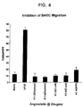
- FIG. 4 shows the inhibition of BAEC migration by angiostatin
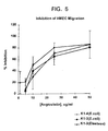
- Figure 5 shows the inhibition of HMEC migration by angiostatin
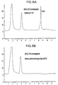
- Figure 6 shows binding of refolded angiostatin to lysine-sepharose columns at pH 8.0 and 10.5
- E . coli strains (Table 1), such as DH10BTM (Life Technologies, Gaithersburg, MD) and TG1 (Amersham Corp., Arlington Heights, IL) are used for transformation of ligation reactions and are the hosts used to prepare plasmid DNA for transfecting mammalian cells.
- E. coli strains such as JM101 (Yanisch-Perron et al., Gene , 33: 103-119, 1985) and MON105 (Obukowicz et al., Appl. and Envir. Micr ., 58: 1511-1523, 1992) can be used for expressing the proteins of the present invention in the cytoplasm or periplasmic space
- DH10BTM Subcloning efficiency cells are purchased as competent cells and are ready for transformation using the manufacturer's protocol, while both E. coli strains JM101 AND MON105 are rendered competent to take up DNA using a CaCl 2 method.
- 20 to 50 mL of cells are grown in LB medium (1% Bacto-tryptone, 0.5% Bacto-yeast extract, 150 mM NaCl) to a density of approximately 1.0 absorbance unit at 600 nanometers (OD600) as measured by a Baush & Lomb Spectronic spectrophotometer (Rochester, NY).
- the cells are collected by centrifugation and resuspended in one-fifth culture volume of CaCl 2 solution [50 mM CaCl 2 , 10 mM Tris-Cl ((10 mM 2-amino-2-(hydroxymethyl) 1,3-propanediol hydrochloride, pH7.4] and are held at 4°C for 30 minutes.
- the cells are again collected by centrifugation and resuspended in one-tenth culture volume of CaCl 2 solution. Ligated DNA is added to 0.2 mL of these cells, and the samples are held at 4°C for 30-60 minutes.
- the samples are shifted to 42°C for two minutes and 1.0 mL of LB is added prior to shaking the samples at 37°C for one hour.
- Cells from these samples are spread on plates (LB medium plus 1.5% Bacto-agar) containing either ampicillin (100 micrograms/mL, ug/mL) when selecting for ampicillin-resistant transformants, or spectinomycin (75 ug/mL) when selecting for spectinomycin-resistant transformants. The plates are incubated overnight at 37°C.
- Colonies are picked and inoculated into LB plus appropriate antibiotic (100 ug/mL ampicillin or 75 ug/mL spectinomycin) and are grown at 37°C while shaking.
- appropriate antibiotic 100 ug/mL ampicillin or 75 ug/mL spectinomycin
- Plasmid DNA can be isolated by a number of different methods and using commercially available kits known to those skilled in the art. Plasmid DNA is isolated using the Promega WizardTM Miniprep kit (Madison, WI), the Qiagen QIAwell Plasmid isolation kits (Chatsworth, CA) or Qiagen Plasmid Midi or Mini kit. These kits follow the same general procedure for plasmid DNA isolation. Briefly, cells are pelleted by centrifugation (5000 x g), the plasmid DNA released with sequential NaOH/acid treatment, and cellular debris is removed by centrifugation (10000 x g).
- the supernatant (containing the plasmid DNA) is loaded onto a column containing a DNA-binding resin, the column is washed, and plasmid DNA eluted. After screening for the colonies with the plasmid of interest, the E. coli cells are inoculated into 50-100 ml of LB plus appropriate antibiotic for overnight growth at 37°C in an air incubator while shaking.
- the purified plasmid DNA is used for DNA sequencing, further restriction enzyme digestion, additional subcloning of DNA fragments and transfection into E. coli, mammalian cells, or other cell types.
- Purified plasmid DNA is resuspended in deionized H 2 O and its concentration is determined by measuring the absorbance at 260/280 nm in a Bausch and Lomb Spectronic 601 UV spectrometer.
- DNA samples are sequenced using ABI PRISMTM DyeDeoxyTM terminator sequencing chemistry (Applied Biosystems Division of Perkin Elmer Corporation, Lincoln City, CA) kits (Part Number 401388 or 402078) according to the manufacturer's suggested protocol usually modified by the addition of 5% DMSO to the sequencing mixture. Sequencing reactions are performed in a DNA thermal cycler (Perkin Elmer Corporation, Norwalk, CT) following the recommended amplification conditions.
- Samples are purified to remove excess dye terminators with Centri-SepTM spin columns (Princeton Separations, Adelphia, NJ) and lyophilized. Fluorescent dye labeled sequencing reactions are resuspended in deionized formamide, and sequenced on denaturing 4.75% polyacrylamide-8M urea gels using ABI Model 373A and Model 377 automated DNA sequencers. Overlapping DNA sequence fragments are analyzed and assembled into master DNA contigs using Sequencher DNA analysis software (Gene Codes Corporation, Ann Arbor, MI).
- E. coli strain MON105 or JM101 harboring the plasmid of interest is grown at 37°C in M9 plus casamino acids medium with shaking in an air incubator Model G25 from New Brunswick Scientific (Edison, NJ). Growth is monitored at OD 600 until it reaches a value of 1.0 at which time nalidixic acid (10 mg/mL) in 0.1 N NaOH is added to a final concentration of 50 ⁇ g/mL. The cultures are then shaken at 37°C for three to four additional hours. A high degree of aeration is maintained throughout THE culture period in order to achieve maximal production of the desired gene product. The cells are examined under a light microscope for the presence of inclusion bodies (IB).
- IB inclusion bodies
- One mL aliquots of the culture are removed for analysis of protein content by boiling the pelleted cells. treating them with reducing buffer and electrophoresis via SDS-PAGE (see Maniatis et al., "Molecular Cloning: A Laboratory Manual", 1982).
- the culture is centrifuged (5000 x g) to pellet the cells.
- the Angiostatin molecules were all scaled up in a 10 L Biostat ETM fermenter (B. Braun Biotech Inc., Allentown PA).
- the fermentation medium used was M9 salts supplemented with 2% casamino acids (Difco Laboratories, Detroit MI) and glucose.
- Approximately one milliliter of a thawed culture was transferred to a 3.8 L Fernbach shake flask containing 1.0 L of medium and then incubated at 37°C at a shaker speed of 250 rpm for 12 hours. This shake flask culture was then used to inoculate a 10 L fermenter containing 9.0 L of medium.
- Glucose was initially batched in at 15 g/l and controlled at 2-5 g/l by the addition of a 50% glucose feed stock.
- the fermentation was harvested four hours post induction by continuous-flow centrifugation.
- the cell paste from a 10L fermentation was resuspended in approximately 6.0 L of 50 mM Tris/150 mM EDTA buffer containing 40 pellets of 50 ml Complete Protease Inhibitor Cocktail (Boehringer Mannheim; Indianapolis, IN) and 600,000 KIU of Trasyol.
- the resuspension was passed once through a Microfluidizer (Newton, MA) at 10,000 psi and temperature maintained under 8°C.
- the recovered homogenate was then spun at 15,000 x G for 25 minutes and mixed to consistency with an Ultr-Turrax mixer.
- the cell paste resuspension was then homogenized by passing the cell suspension two times through a Microfluidizer (Newton, MA) operating at a pressure of 10,000 psi.
- the temperature of the homogenate was maintained less than 6°C by collection in a stainless steel container placed in an ice bath.
- Inclusion bodies were then isolated by centrifugation of the cell homogenate at 15,000 X g for 30 minutes and removal of supernatant. Inclusion bodies were then washed a total of two times by resuspending the inclusion bodies in chilled D.I. and centrifugation at 15,000 X g for 30 minutes.
- Angiostatin Inclusion bodies were then frozen at -70°C.
- the cell pellet from a 330 mL E. coli culture is resuspended in 15 mL of sonication buffer Tris-Cl, pH 8.0 + 1 mM ethylenediaminetetraacetic acid (EDTA). These resuspended cells are sonicated using the microtip probe of a Sonicator Cell Disruptor (Model W-375, Heat Systems-Ultrasonics, Inc., Farmingdale, New York). Three rounds of sonication in sonication buffer followed by centrifugation are employed to disrupt the cells and wash the inclusion bodies (IB). The first round of sonication is a 3 minute burst followed by a 1 minute burst, and the final two rounds of sonication are for 1 minute each.
- Tris-Cl pH 8.0 + 1 mM ethylenediaminetetraacetic acid (EDTA).
- EDTA ethylenediaminetetraacetic acid
- Angiostatin K1-4 inclusion bodies were dissolved in 6M urea, 5 mM DTT, 50 mM Bis-Tris Propane, pH 10.8 at 1.2 mg/ml. This solution was stirred for 2 hours and then 6-amino hexanoic acid (stock 1M) was added to achieve a 6 mM concentration, followed by addition of cystine (stock 0.2M, pH 10.5) to 6 mM. This was mixed for 5 minutes and then the K1-4 angiostatin was diluted to 0.2 mg/ml by addition of 75 mM Bis-Tris propane, pH 10.5. It was then stirred for 72 hours to complete the refolding of the protein as assessed by reverse-phase HPLC or by binding to Lys-HPLC ( Figure 6).
- angiostatin K1-4 Purification of angiostatin K1-4 was achieved using a Sepharose-Lys affinity column followed by Q-Sepharose chromatography. The sample was next dialyzed against 50 mM sodium phosphate, pH 8.0 and then filtered through a 1 uM filter to clarify. This sample was loaded onto a Sepharose-Lys column equilibrated in 50 mM sodium phosphate, pH 8.0. The load was at approximately 1.5 gm of K1-4 per ml of resin.
- Fractions were analyzed by SDS-gel electrophoresis and/or RP-HPLC and pooled. This pool was made 40 mM Tris-Cl, and adjusted to pH 9.0. It was then dialyzed extensively vs. 40 mM Tris-Cl, pH 9.0 and applied to a Pharmacia Q-Sepharose HP column ( ⁇ 5 mgs protein/ml resin) equilibrated in the same buffer. K1-4 was then eluted using a NaCl gradient from 0-0.1M in the equilibrating buffer. Fractions were analyzed by RP-HPLC and/or SDS gel electrophoresis and pooled.
- the folded proteins can be affinity-purified using affinity reagents such as monoclonal antibodies or receptor subunits attached to a suitable matrix. Purification can also be accomplished using any of a variety of chromatographic methods such as: ion exchange, gel filtration or hydrophobic interaction chromatography or reversed phase HPLC. These and other protein purification methods are described in detail in Methods in Enzymology, Volume 182 "Guide to Protein Purification” edited by Murray Deutscher, Academic Press, San Diego, California, 1990.
- the purified protein is analyzed by RP-HPLC, electrospray mass spectrometry, and SDS-PAGE.
- the protein quantitation is done by amino acid composition, RP-HPLC, and Bradford protein determination. In some cases tryptic peptide mapping is performed in conjunction with electrospray mass spectrometry to confirm the identity of the protein.
- Human Burkitt's lymphoma Daudi cells (ATCC) are seeded at 2 x 10 4 cells/well into 96-well tissue culture plates and cells are cultured in the presence or absence of serial doses of rhIFN.alpha. 2b for 3 days. Cultures are pulsed with 3 H-thymidine for the last hour of the culture period, and the 3 H-thymidine uptake, counts per minute (cpm), ARE measured on a Beta-plate reader. All samples are assayed in triplicate.
- HdMVEC human dermal microvascular endothelial cells
- BocEnd, Incell bovine microvascular endothelial cells
- FBS heat-inactivated fetal bovine serum
- antibiotics 100 ug/ml heparin
- endothelial mitogen Biomedical Technologies
- the tube formation assay is performed as previously described (Gately et al., Cancer Res . 56 :4887-4890, 1996).
- the effect of angiogenesis inhibitors can be tested in an in vitro angiogenesis assay; endothelial cell tube formation on matrix.
- Matrigel Becton Dickinson
- MCDB 131 medium supplemented with 2 mM L-glutamine, containing either buffer control or testing compounds.
- Aliquots of 0.5 ml of 1:1 diluted matrigel were plated into individual wells of 2-chambered Tissue-Tek polystyrene slides (Nunc) and allowed to polymerize at 37°C for at least 30 min.
- Endothelial cell tube networks were fixed in 100% methanol at -20°C for 7-10 min, rinsed 4 times with phosphate buffered saline (PBS) and incubated overnight at 4°C with rabbit polyclonal anti-human Factor VIII-related antigen (von Willibrand factor, vWF) (Dako). The next day, the cells were stained with a secondary antibody, Cy3-conjugated goat anti-rabbit IgG F(ab')2 specific (Jackson ImmunoResearch Laboratories).
- Tube formation was visualized using a Nikon microphot-FXA with a fluorescence filter linked to a computer containing image analysis software. Tube formation was measured as follows: Total area encompassed by endothelial cell tubes/Total endothelial cell surface area.
- the endothelial cell migration assay is performed essentially as previously described (Gately et al., Cancer Res. 56: 4887-4890, 1996). To determine the ability of angiostatin to inhibit the migration of endothelial cells, migration assays were performed in a transwell chamber (Costar) containing 8 mm pore size polycarbonate membranes. The cells utilized in the assay were either human microvascular endothelial cells from Emory or bovine endothelial cells (kindly provided by Gately Northwestern University, Evanston, IL).
- the cells were starved overnight before use in MCDB131 + 0.1% BSA (human cells) or DMEM+0.1%BSA (bovine cells), harvested, and resuspended in the same media at 10 6 cells/ml.
- the lower side of the transwells were coated with 0.1% gelatin for 30 minutes at 37°C before addition of 2 x 10 5 cells to the upper chamber.
- the transwell was moved to a well containing the chemoattractant (bFGF or VEGF) in the lower chamber. Migration was allowed to occur overnight at 37°C.
- the membranes were then fixed and stained, and the number of cells that migrated to the lower side of the membrane counted in 3 high powered fields.
- Example 1 Construction of pMON24642 and selection of strains producing high levels of angiostatin K1-4E
- a cDNA encoding angiostatin K1-4E (see e.g., pMON24642-a12.seq, SEQID # 10) was synthesized by polymerase chain reaction using the oligonucleotide primers angec-s1 (angec-sl.seq, SEQID # 1) and angec-n1 (angec-n1.seq, SEQID # 4) and using the human plasminogen cDNA (pMON24624.seq SEQID # 9) as template.
- the resulting DNA was cleaved with restriction endonucleases Afl III and Hin dIII and was ligated into the E.
- Plasmid DNA was isolated from the DH10B bacterial colonies and used to transform E. coli strain MON105 to spectinomycin resistance. Individual antibiotic resistant colonies were inoculated into 1 ml of Luria Broth with 75 ug/ml spectinomycin and grown over night at 37 degrees with shaking. Fifty microliters of the overnight culture were inoculated into one ml of M9 medium with casamino acids and grown for four hours at 37 degrees with shaking. Nalidixic acid (50 ul of a 1 mg/ml aqueous solution) was added and the cultures were incubated at 37 degrees for an additional three hours.
- Example 2 Construction of pMON24643 and selection of strains producing high levels of angiostatin K1-4
- a cDNA encoding angiostatin K1-4 was synthesized by polymerase chain reaction using the oligonucleotide primers angec-s2 (angec-s2.seq, SEQID # 2) and angec-n2 (angec-n2.seq, SEQID # 5) and using the human plasminogen cDNA (pMON24624.seq, SEQID # 9) as template.
- the resulting cDNA was cleaved with restriction endonucleases Afl III and Hin dIII and was ligated into the E. coli expression vector pMON5723 which had been digested with Nco I and Hin dIII. A portion of the ligation reaction was used to transform E. coli strain DH10B to spectinomycin resistance
- Plasmid DNA was isolated from the DH10B bacterial colonies and used to transform E. coli strain MON105 to spectinomycin resistance. Individual antibiotic resistant colonies were inoculated into 1 ml of Luria Broth with 75 ug/ml spectinomycin and grown over night at 37 degrees with shaking. Fifty microliters of the overnight culture were inoculated into one ml of M9 medium with casamino acids and grown for four hours at 37 degrees with shaking. Nalidixic acid (50 ul of a 1 mg/ml aqueous solution) was added and the cultures were incubated at 37 degrees for an additional three hours.
- Example 3 Construction of pMON24644 and selection of strains producing high levels of angiostatin K2-3
- a cDNA encoding angiostatin K2-3 was synthesized by polymerase chain reaction using the oligonucleotide primers angec-s3 (angec-s3.seq, SEQID # 3) and angec-n3 (angec-n3.seq, SEQID # 6) and using the human plasminogen cDNA (pMON24624.seq, SEQID # 9) as template.
- the resulting cDNA was cleaved with restriction endonuclease Afl III and Hin dIII and was ligated into the E. coli expression vector pMON5723 which had been digested with Nco I and Hin dIII. A portion of the ligation reaction was used to transform E. coli strain DH10B to spectinomycin resistance
- Plasmid DNA was isolated from the DH10B bacterial colonies used to transform E. coli strain MON105 to spectinomycin resistance. Individual antibiotic resistant colonies were inoculated into 1 ml of Luria Broth with 75 ug/ml spectinomycin and grown over night at 37 degrees with shaking. Fifty microliters of the overnight culture were inoculated into one ml of M9 medium with casamino acids and grown for four hours at 37 degrees with shaking. Nalidixic acid (50 ul of a 1 mg/ml aqueous solution) was added and the cultures were incubated at 37 degrees for an additional three hours.
- Each cDNA also contained various silent mutations at the third positions of codons two through eight. These mutations were caused by the redundancies in the 5' PCR primer (angec-s3.seq, SEQID # 3), and the particular mutations in these clones result in high level expression of angiostatin. Each cDNA differed from the others and from the wild-type sequence at one or more of these positions.
- Example 4 Construction of pMON24645 and selection of strains producing high levels of angiostatin K2-4
- a cDNA encoding angiostatin K2-4 was synthesized by polymerase chain reaction using the oligonucleotide primers angec-s3 (angec-s3.seq, SEQID # 3) and angec-n2 (angec-n2.seq, SEQID # 5) and using the human plasminogen cDNA (pMON24624.seq, SEQID # 9) as template.
- the resulting cDNA was cleaved with restriction endonuclease Afl III and Hind III and was ligated into the E. coli expression vector pMON5723 which had been digested with Nco I and Hind III. A portion of the ligation reaction was used to transform E. coli strain DH10B to spectinomycin resistance
- Plasmid DNA was isolated from the DH10B bacterial colonies and used to transform E. coli strain MON105 to spectinomycin resistance. Individual antibiotic resistant colonies were inoculated into 1 ml of Luria Broth with 75 ug/ml spectinomycin and grown over night at 37 degrees with shaking. Fifty microliters of the overnight culture were inoculated into one ml of M9 medium with casamino acids and grown for four hours at 37 degrees with shaking. Nalidixic acid (50 ul of a 1 mg/ml aqueous solution) was added and the cultures were incubated at 37 degrees for an additional three hours.
- Each cDNA also contained various silent mutations at the third positions of codons two through eight. These mutations were caused by the redundancies in the 5' PCR primer (angec-s3.seq, SEQID # 3), and the particular mutations in these clones result in high level expression of angiostatin. Each cDNA differed from the others and from the wild-type sequence at one or more of these positions.
- Example 5 Construction of pMON24646 and selection of strains producing high levels of angiostatin K1-3
- a cDNA encoding angiostatin K1-3 was synthesized by polymerase chain reaction using the oligonucleotide primers angec-s2 (angec-s2.seq, SEQID # 2) and angec-n3 (angec-n3.seq, SEQID # 6) and using the human plasminogen cDNA (pMON24624.seq, SEQID # 9) as template.
- the resulting cDNA was cleaved with restriction endonuclease Afl III and Hin dIII and was ligated into the E. coli expression vector pMON5723 which had been digested with Nco I and Hin dIII. A portion of the ligation reaction was used to transform E. coli strain DH10B to spectinomycin resistance.
- Plasmid DNA was isolated from the DH10B bacterial colonies and used to transform E. coli strain MON105 to spectinomycin resistance. Individual antibiotic resistant colonies were inoculated into 1 ml of Luria Broth with 75 ug/ml spectinomycin and grown over night at 37 degrees with shaking. Fifty microliters of the overnight culture were inoculated into one ml of M9 medium with casamino acids and grown for four hours at 37 degrees with shaking. Nalidixic acid (50 ul of a 1 mg/ml aqueous solution) was added and the cultures were incubated at 37 degrees for an additional three hours.
- the sequences of the cDNAs in these plasmids are shown in 24646-A7.seq, 24646-A9.seq and 24646-A10, 24646-A12, 24646-B1, and 24646-B10 (SEQID # 22, SEQID # 23, SEQID # 24, SEQID # 25, SEQID # 26, and SEQID # 27, respectively).
- Each cDNA encoded the expected protein.
- Each cDNA also contained various silent mutations at the third positions of codons two through eight. These mutations were caused by the redundancies in the 5' PCR primer (angec-s2,seq, SEQID # 2), and the particular mutations in these clones result in high level expression of angiostatin.
- Each cDNA differed from the others and from the wild-type sequence at one or more of these positions.
- Example 6 Construction of pMON24649 and of strains producing high levels of angiostatin K1
- a cDNA encoding angiostatin K1 (24649.seq, SEQID # 28) was synthesized by polymerase chain reaction using the oligonucleotide primers angec-sk1 (angec-sk1.seq, SEQID # 7) and angec-n4 (angec-n4.seq, SEQID # 8) and using the cDNA in pMON24646-A7 (SEQID # 22) as template.
- the 5' PCR primer (angec-sk1) contained the same silent mutations in the first eight codons that resulted in high level expression of angiostatin K1-3 from pMON24646-A7.
- the resulting cDNA was cleaved with restriction endonucleases Nco I and Hin dIII and was ligated into the E. coli expression vector pMON5723 which had also been digested with Nco I and Hin dIII. A portion of the ligation reaction was used to transform E. coli strain DH10B to spectinomycin resistance. Plasmid DNA was isolated from the bacterial colonies and analyzed by restriction endonuclease cleavage and DNA sequencing.
- Plasmid DNA was then used to transform E. coli strain MON105 to spectinomycin resistance. Individual colonies were grown over night in Luria Broth containing 75 ug/ml spectinomycin. One ml of the over night culture was used to inoculate 20 ml of M9 medium containing casamino acids. After about three hours of growth at 37 degrees, Nalidixic acid was added and the cultures incubated for a further three hours at 37 degrees. Cultures were screened by phase contrast microscopy for the presence of cytoplasmic inclusion bodies and by SDS polyacrylamide gel electrophoresis for expression of the protein. Unlike other forms of angiostatin, K1 protein was not incorporated into inclusion bodies, but rather remained soluble in the cytoplasm of the bacteria. Nevertheless, the mutations in the first eight codons in pMON24649 conferred high level expression of K1 as they had for K1-3 expression from pMON13646-A7.
- Example 7 Method for refolding angiostatin
- Angiostatin K1-4 inclusion bodies were dissolved in 6M urea, 5 mM DTT, 50 mM Bis-Tris Propane, pH 10.8 at 1.2 mg/ml. This solution was stirred for 2 hours. A stock solution of 1M 6-amino hexanoic acid was then added to achieve a 6 mM concentration, followed by addition of cystine (stock 0.2M, pH 10.5) to 6 mM. This was stirred for 5 minutes. The K1-4 angiostatin was then diluted to 0.2 mg/ml by addition of 75 mM Bis-Tris propane, pH 10.5. It was then stirred for 72 hours to complete the refolding of the protein. Refolded angiostatin was purified by lysine-Sepharose chromatography and Q-Sepharose-HP chromatography. This purified angiostatin was assayed for inhibition of cell migration as shown in figures 3-5.
Landscapes
- Chemical & Material Sciences (AREA)
- Organic Chemistry (AREA)
- Health & Medical Sciences (AREA)
- Life Sciences & Earth Sciences (AREA)
- Engineering & Computer Science (AREA)
- Genetics & Genomics (AREA)
- Wood Science & Technology (AREA)
- General Health & Medical Sciences (AREA)
- Bioinformatics & Cheminformatics (AREA)
- Biochemistry (AREA)
- Zoology (AREA)
- Molecular Biology (AREA)
- Medicinal Chemistry (AREA)
- Biomedical Technology (AREA)
- General Engineering & Computer Science (AREA)
- Chemical Kinetics & Catalysis (AREA)
- Biotechnology (AREA)
- Crystallography & Structural Chemistry (AREA)
- Microbiology (AREA)
- General Chemical & Material Sciences (AREA)
- Analytical Chemistry (AREA)
- Biophysics (AREA)
- Proteomics, Peptides & Aminoacids (AREA)
- Peptides Or Proteins (AREA)
- Preparation Of Compounds By Using Micro-Organisms (AREA)
- Saccharide Compounds (AREA)
Abstract
Description
| Strains | ||
| Designation | Description or Genotype | Referenee/Source |
| DH10B | F- mcrA Δ(mrr-hsdRMS-mcrBC) dlacZΔM15 ΔlacX74 deoR recA1 endA1 araD139 Δ(ara, leu)7697 galU galK λ·rpsL nupG | Life Technologies, Rockville, Maryland |
| MON105 (ATCC # 55204) | F-, lambda-,IN (rrnD, rrnE)1, rpoD+, rpoH358 | Obukowicz et al., Appl. and Envir. Micr., 58: 1511-1523, 1992 |
| JM101 (ATCC # 33876) | delta (pro lac), supE, thi, F'(traD36, proA+B+, lacIq, lacZdeltaM15) | Yanisch-Perron et al., Gene, 33: 103-119, 1985 |
| Plasmids | |||||
| Plasmid | SEQ ID NO. | Marker | Description | Source | |
| pMON5723 | Spec | Generic Spec-resistant E. coli expression vector containing the E. coli recA1 promoter and G10L ribosome binding site | (Olins and Rangwala, Methods Enzymol. 185 (Gene Expression Technol.): 115-119, 1990) | ||
| pMON6875 | Spec | Generic Spec-resistant E. coli expression vector containing the E. coli recA1 promoter and G10L ribosome binding site | (Olins and Rangwala, Methods Enzymol. 185 (Gene Expression Technol.): 115-119, 1990) | ||
| | # | 9 | Amp | Human plasminogen, including the native plasminogen signal sequence, N-terminal peptide and kringles 1-5. BamHI fragment in pMON3360B. (Amino acids -19-537 of plasminogen, nucleotides 55-1752) | This work |
| pMON24642-A12, -B5, - | # | 10, #11, #12 | spec | Human angiostatin(E). AflIII/HindIII fragment in pMON5723. The 5' end has been mutagenized for increased expression. (Amino acids 74-470 of plasminogen nucleotides 331-1464) | This work |
| pMON24643-C10, D5,-D7 | #13, #14, #15 | spec | Human angiostatin(S) (K1-4) AflIII/HindIII fragment in pMON5723. (Amino acids 79-440 of plasminogen, nucleotides 346-1431) | This work | |
| pMON24644-E7, -E9, -F8 | ##16, #17, #18 | spec | K2-3 fragment of human angiostatin(S). AflIII/HindIII fragment in pMON5723. (Amino acids 163-344 of plasminogen, nucleotides 598-1143) | This work | |
| pMON24645-G3, -H5, -H6 | #19, #20, #21 | spec | K2-4 fragment of human angiostatin(S). AflIII/HindIII fragment in pMON5723. (Amino acids 163-440 of plasminogen, nucleotides 598-1431) | This work | |
| pMON24646-A7, -A9, -A10, -A12, -B1, -B10 | ##22, #23, #24, #25, #26, #27 | spec | K1-3 fragment of human angiostatin(S). AflIII/HindIII fragment in pMON5723. (Amino acids 79-344 of plasminogen, nucleotides 346-1143) | This work | |
| pMON24649 | #28 | spec | K1 fragment of human angiostatin(S). NcoI/HindIII fragment in pMON5723. (Amino acids 79-184 of plasminogen, nucleotides 1-269 of pMON24646-A7.seq) | This work | |
| pMON24650 | #29 | Amp | K1 fragment of human angiostatin(S). NcoI/HindIII fragment in pMON3633. (Amino acids 79-184 of plasminogen) | This work |


Polazzi, J.O.
Casperson, G.F.








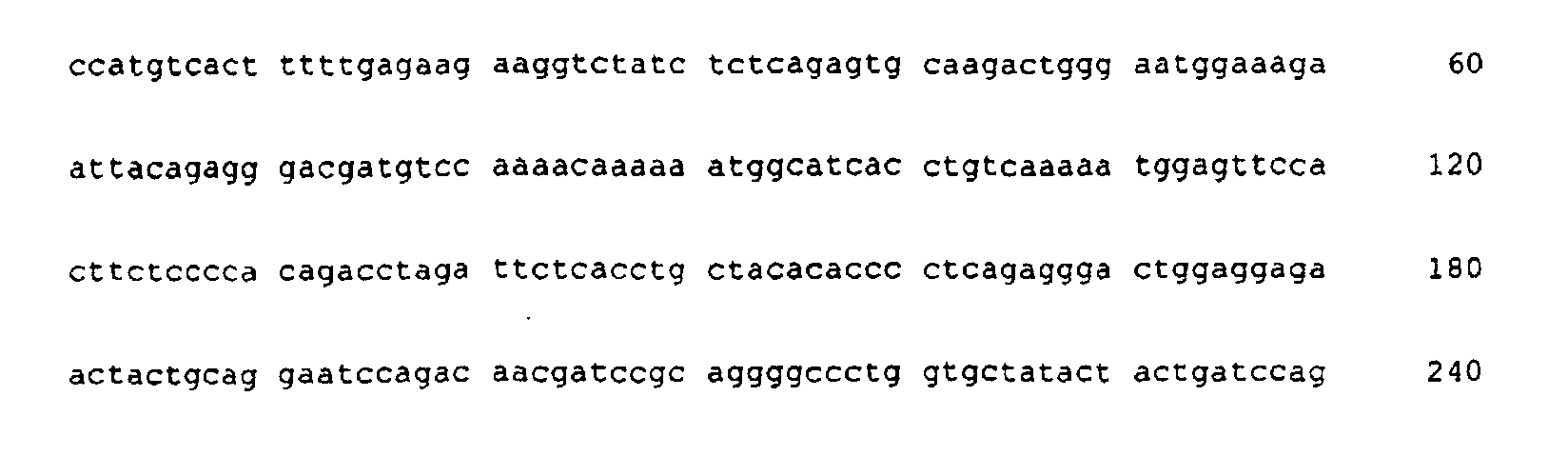





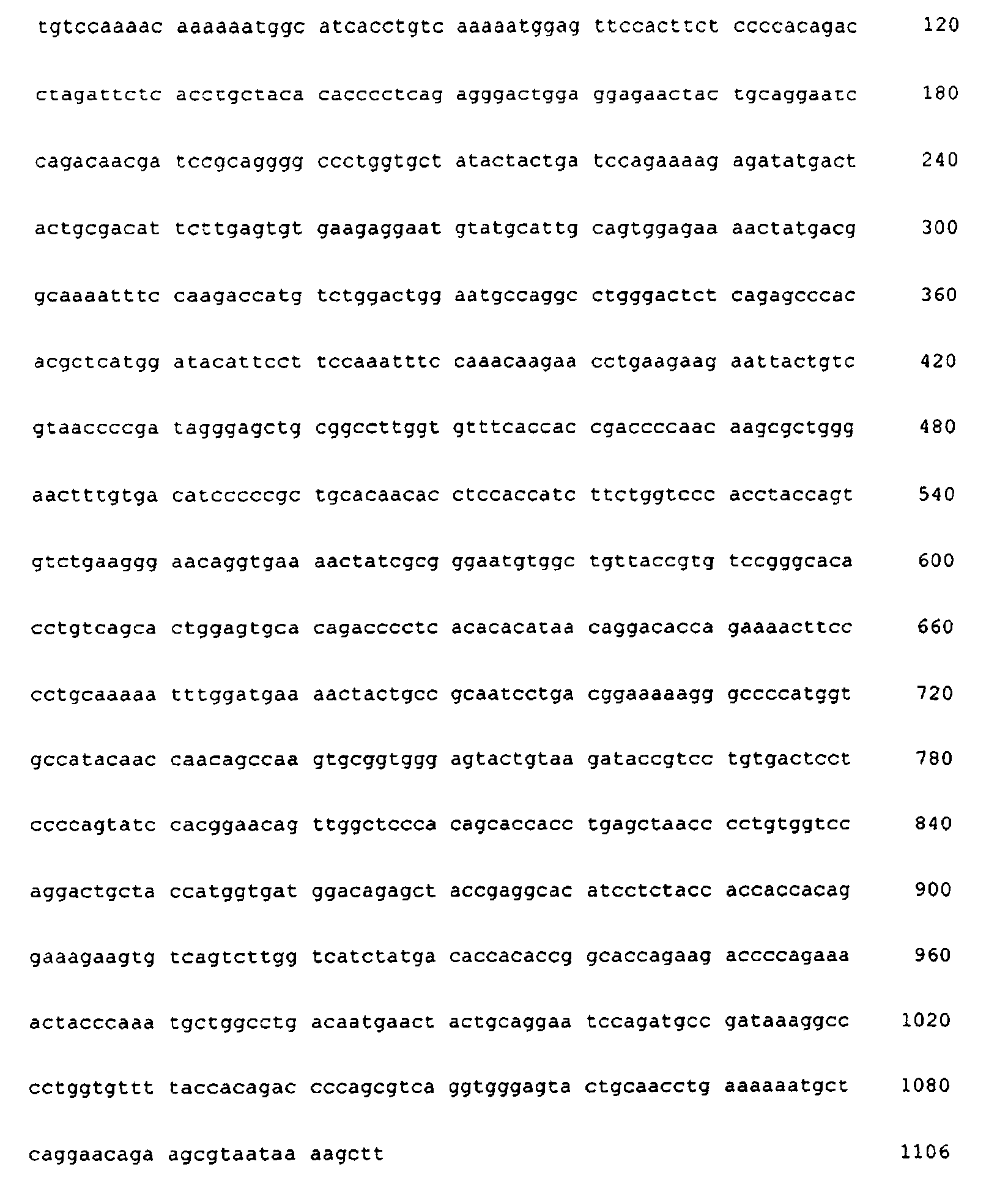



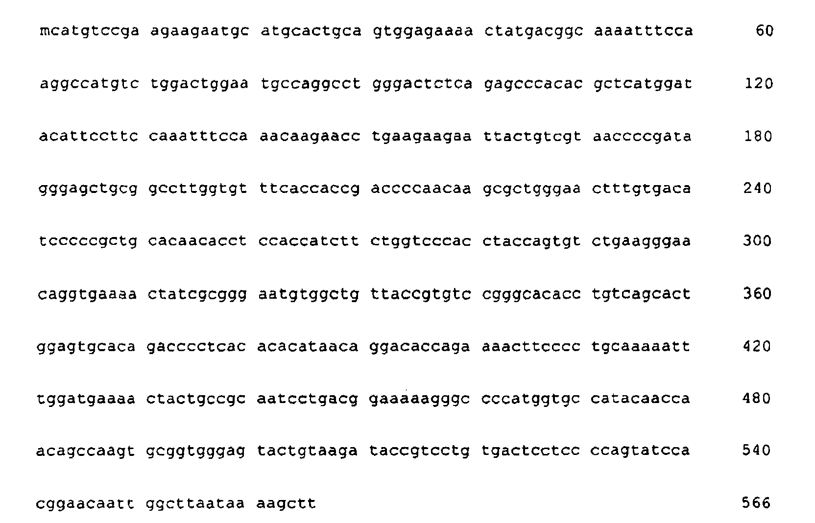



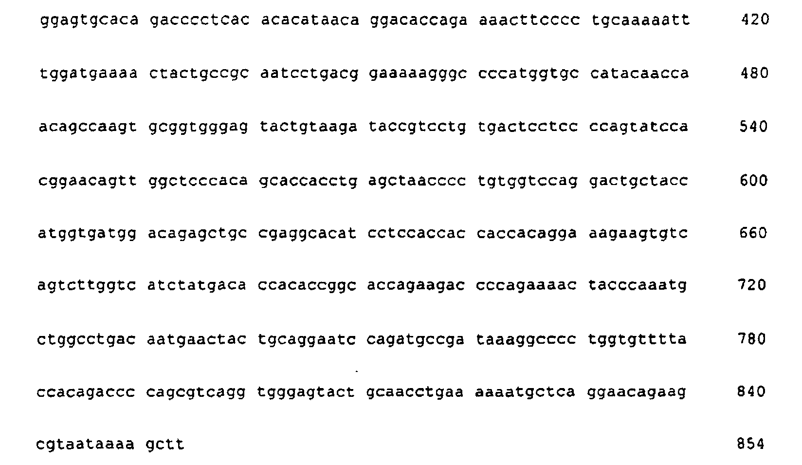
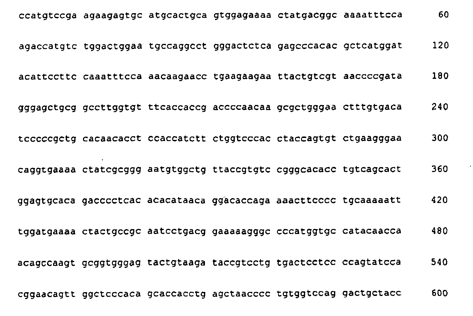
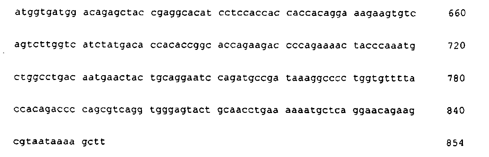


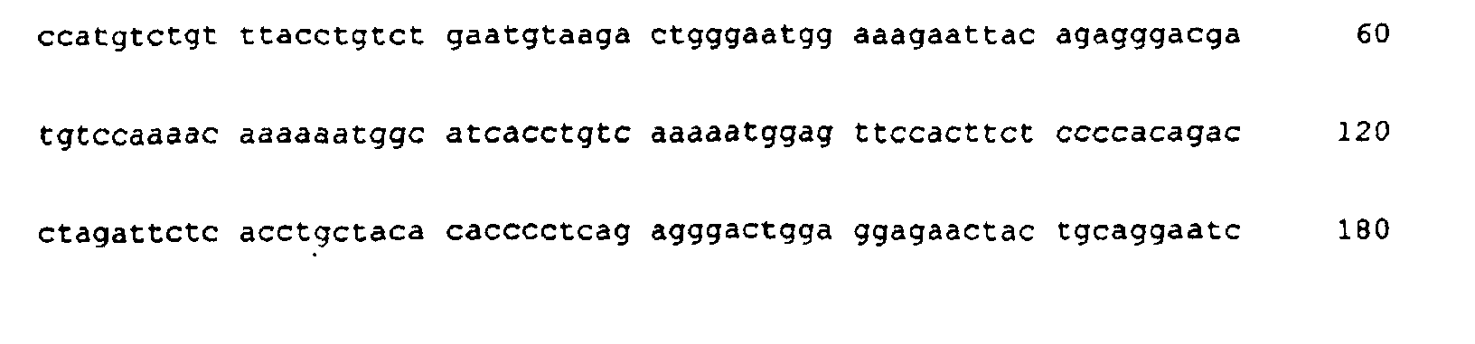


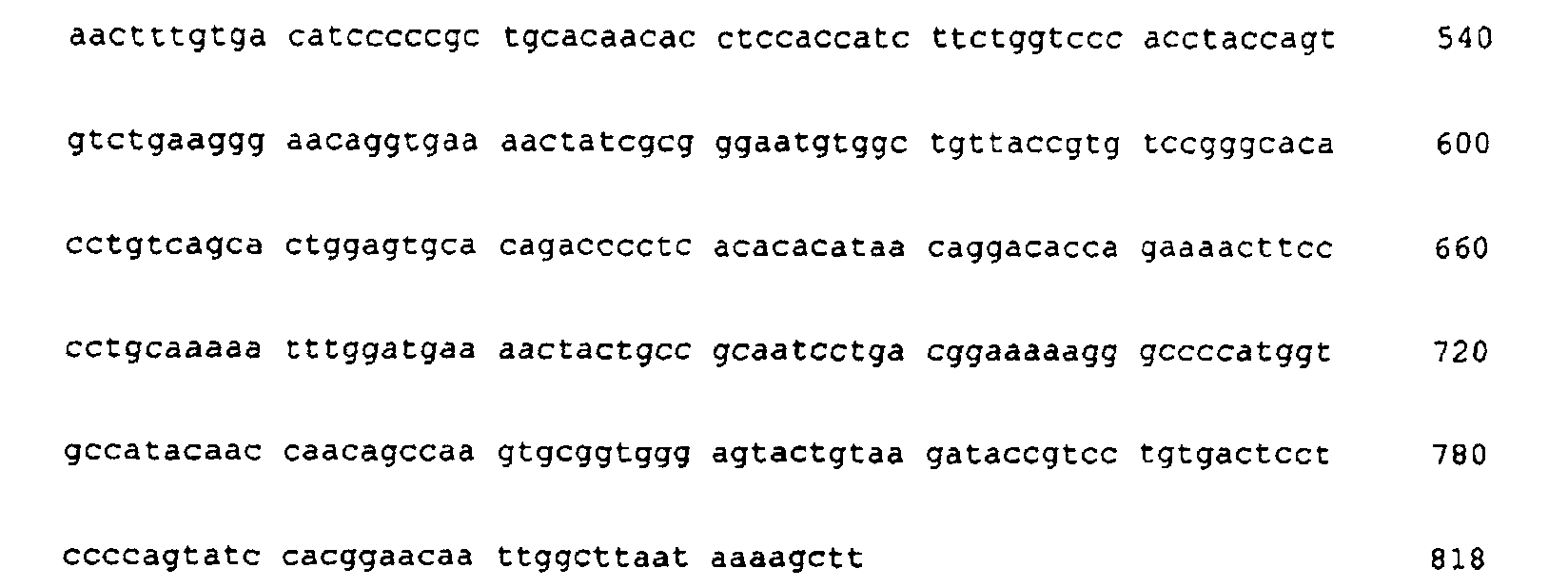
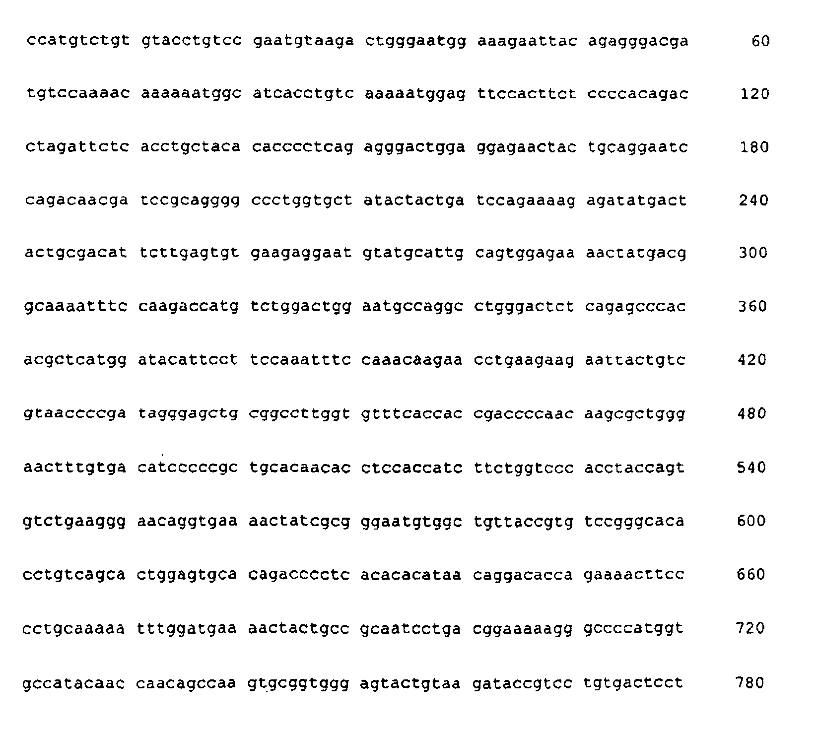

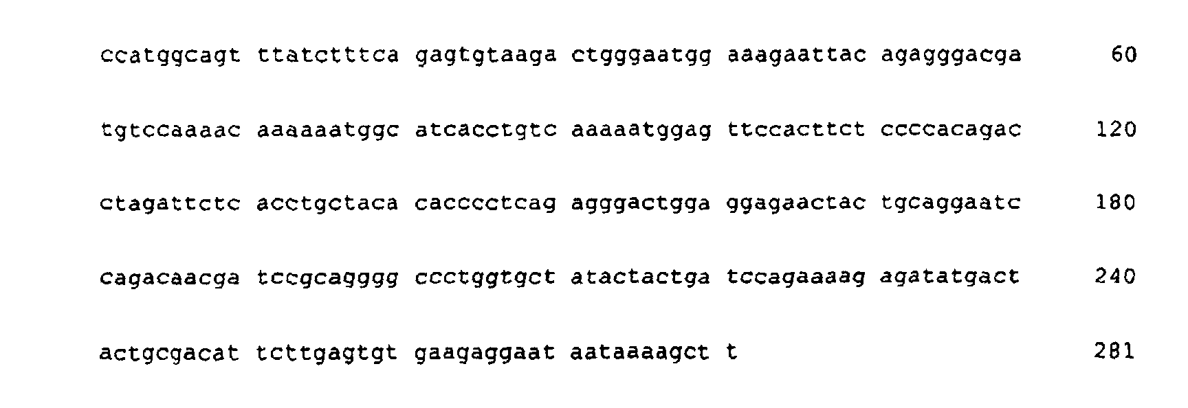
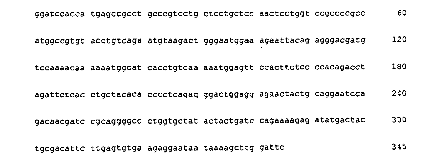
Claims (47)
- A method of producing angiostatin comprising the steps of:(a) culturing host cells that are expressing an angiostatin gene;(b) recovering the product of said gene expression;(c) refolding said gene product at an elevated pH from 9 to 11.5; and(d) isolating properly folded forms of said gene product.
- A method of producing angiostatin according to claim 1 comprising the steps of:(a) culturing host cells that are expressing an angiostatin gene;(b) recovering the product of said gene expression;(c) solubilizing said gene product at an elevated pH from 9 to 11.5;(d) refolding said solubilized gene product at an elevated pH from 9 to 11.5; and(e) isolating properly folded forms of said gene product.
- The method of claim 1 or 2 wherein said elevated pH is from pH 10 to pH 11.
- The method of claim 3 wherein said elevated pH is pH 10.5.
- The method of claim 2 wherein said gene product is present at a concentration of from about 1 to about 20 mg/ml during said solubilization step.
- The method of claim 5 wherein said gene product is present at a concentration of about 6 mg/ml during said solubilization step.
- The method of claim 1 or claim 2 wherein said gene product is present at a concentration of from about 0.1 to about 5 mg/m) during said refolding step.
- The method of claim 7 wherein said gene product is present at a concentration of about 1 mg/ml during said refolding step.
- The method of claim 1 or claim 2 wherein said angiostatin gene product is encoded by a sequence is selected from the group consisting of SEQ ID NO: 10; SEQ ID NO: 11; SEQ ID NO: 12; SEQ ID NO: 13; SEQ ID NO: 14; SEQ ID NO: 15; SEQ ID NO: 16; SEQ ID N7: 10; SEQ ID NO: 18; SEQ ID NO: 19; SEQ ID NO: 20; SEQ ID NO: 21; SEQ ID NO: 22; SEQ ID NO 23; SEQ ID NO: 24; SEQ ID NO: 25; SEQ ID NO: 26; SEQ ID NO: 27; SEQ ID NO: 28; SEQ ID NO: 29.
- The method of claim 2 wherein urea is present at a concentration of from about 4 M to about 10 M during said solubilization step.
- The method of claim 10 wherein urea is present at a concentration of about 6 M during said solubilization step.
- The method of claim 1 or claim 2 wherein urea is present at a concentration of from about 0.5 M to about 3 M during said refolding step.
- The method of claim 12 wherein urea is present at a concentration of about 1 M during said refolding step.
- The method of claim 2 wherein guanidine hydrochloride is present at a concentration of from about 3 M to about 8 M during said solubilization step.
- The method of claim 14 wherein guanidine hydrochloride is present at a concentration of about 5 M during said solubilization step.
- The method of claim 1 or claim 2 wherein guanidine hydrochloride is present at a concentration of from about 0.2 M to about 2 M during said refolding step.
- The method of claim 16 wherein guanidine hydrochloride is present at a concentration of about 0.5 M during said refolding step.
- A method of claim 1 or 2 carried out in a presence of a reducing agent capable of reducing disulfide linkages to sulfhydryl groups.
- A method of claim 18 wherein said reducing agent is selected from the group consisting of DTT, BME, cysteine, and reduced glutathione.
- The method of claim 19 wherein DTT is present at a concentration of from about 2 mM to about 10 mM during said solubilization step.
- The method of claim 20 wherein DTT is present at a concentration of about 6 mM during said solubilization step.
- The method of claim 21 wherein DTT is present at a concentration of from about 0.5 mM to about 2 mM during said refolding step.
- The method of claim 22 wherein DTT is present at a concentration of about 1 mM during said refolding step.
- The method of claim 19 wherein reduced glutathione is present at a concentration of from about 5 mM to about 20 mM during said solubilization step.
- The method of claim 24 wherein reduced glutathione is present at a concentration of about 10 mM during said solubilization step.
- The method of claim 19, wherein reduced glutathione is present at a concentration of from about 1 mM to about 4 mM during said refolding step.
- The method of claim 26 wherein reduced glutathione is present at a concentration of about 2 mM during said refolding step
- The method of claim 19 wherein cysteine is present at a concentration of from about 5 mM to about 20 mM during said solubilization step.
- The method of claim 28 wherein cysteine is present at a concentration of about 10 mM during said solubilization step.
- The method of claim 19 wherein cysteine is present at a concentration of from about 1 mM to about 4 mM during said refolding step.
- The method of claim 30 wherein cysteine is present at a concentration of about 2 mM during said refolding step.
- The method of claim 1 or claim 2 wherein an agent capable of enhancing the interchange of disulfide bonds is present during said refolding step.
- The method of claim 32 wherein said agent is selected from cystine and oxidized glutathione.
- The method of claim 33 wherein cystine is present at a concentration of from about 0.2 mM to about 5 mM during said refolding step.
- The method of claim 34 wherein cystine is present at a concentration of about 1 mM during said refolding step.
- The method of claim 1 or 2 wherein said refolding step is carried out in the presence of a ligand capable of binding kringle domains of said angiostatin gene product.
- The method of claim 36 wherein said ligand capable of binding kringle domains of said angiostatin gene product is selected from the group consisting of 6-amino hexanoic acid, 7-aminoheptanoic acid, 5-amino pentanoic acid, and 4-amino butyric acid.
- The method of claim 37 wherein 6-amino hexanoic acid is present at a concentration of from about 0.2 mM to about 20 mM during said refolding step.
- The method of claim 38 wherein 6-amino hexanoic acid is present at a concentration of about 1 mM during said refolding step.
- The method of claim 1 or claim 2 wherein disulfide bonds are formed through air oxidation during said refolding step.
- The method of claim 40 wherein said air oxidation step is carried out from about 12 hours to about 96 hours.
- The method of claim 41 wherein said air oxidation step is carried out from about 24 hours to about 72 hours.
- The method of claim 42 wherein said air oxidation step is carried on for about 60 hours.
- The method of claim 1 or claim 2 which further includes the step of purifying the angiostatin by a process selected from the group consisting of Lysine-sepharose chromatography, ion-exchange chromatography, hydrophobic interaction chromatography and RP-HPLC.
- The method of claim 1 or claim 2 wherein said angiostatin gene is comprised of DNA coding for animal angiostatin.
- The method of claim 1 or claim 2 wherein said angiostatin gene is comprised of DNA coding for human angiostatin.
- The method of claim 1 or 2 which further includes the step of purifying the angiostatin by a process selected from the group consisting of ion-exchange chromatography, and hydrophobic interaction chromatography.
Applications Claiming Priority (3)
| Application Number | Priority Date | Filing Date | Title |
|---|---|---|---|
| US7124798P | 1998-01-12 | 1998-01-12 | |
| US71247P | 1998-01-12 | ||
| PCT/US1999/000048 WO1999035248A2 (en) | 1998-01-12 | 1999-01-11 | Method of refolding angiostatin |
Publications (2)
| Publication Number | Publication Date |
|---|---|
| EP1047771A2 EP1047771A2 (en) | 2000-11-02 |
| EP1047771B1 true EP1047771B1 (en) | 2003-04-23 |
Family
ID=22100170
Family Applications (1)
| Application Number | Title | Priority Date | Filing Date |
|---|---|---|---|
| EP99901279A Expired - Lifetime EP1047771B1 (en) | 1998-01-12 | 1999-01-11 | Method of refolding angiostatin |
Country Status (8)
| Country | Link |
|---|---|
| EP (1) | EP1047771B1 (en) |
| AT (1) | ATE238414T1 (en) |
| AU (1) | AU2101599A (en) |
| DE (1) | DE69907139T2 (en) |
| DK (1) | DK1047771T3 (en) |
| ES (1) | ES2198879T3 (en) |
| PT (1) | PT1047771E (en) |
| WO (1) | WO1999035248A2 (en) |
Families Citing this family (7)
| Publication number | Priority date | Publication date | Assignee | Title |
|---|---|---|---|---|
| JP2002520367A (en) | 1998-07-14 | 2002-07-09 | ブリストル−マイヤーズ スクイブ カンパニー | Lysine-binding fragment of angiostatin |
| CA2367198A1 (en) * | 1999-12-03 | 2001-06-07 | Entremed, Inc. | Method of producing and purifying angiostatin protein |
| US6723536B2 (en) | 1999-12-03 | 2004-04-20 | Entremed, Inc. | Method of producing and purifying angiostatin |
| IL152804A0 (en) | 2000-05-16 | 2003-06-24 | Bolder Biotechnology Inc | Methods for refolding proteins containing free cysteine residues |
| AU2001285919B2 (en) | 2000-09-05 | 2007-08-23 | Jiangsu Simcere Pharmaceutical R&D Co., Ltd | Recombinant endothelial cell growth inhibitors derived from a mammalian plasminogen |
| GB2455262A (en) * | 2006-09-29 | 2009-06-10 | Proteomtech Inc | Methods for production of recombinant plasminogen and plasmin polypeptides |
| US8617531B2 (en) | 2006-12-14 | 2013-12-31 | Bolder Biotechnology, Inc. | Methods of making proteins and peptides containing a single free cysteine |
Family Cites Families (2)
| Publication number | Priority date | Publication date | Assignee | Title |
|---|---|---|---|---|
| CA2047063A1 (en) * | 1991-07-15 | 1993-01-16 | Warren C. Mackellar | Method for folding tissue plasminogen activators and erivatives thereof |
| ES2246513T3 (en) * | 1996-05-03 | 2006-02-16 | Abbott Laboratories | NEW ANTI-ANGIOGENIC, POLINUCLEOTID PEPTIDES CODING THEM AND ANGIOGENESIS INHIBITION PROCEDURES. |
-
1999
- 1999-01-11 EP EP99901279A patent/EP1047771B1/en not_active Expired - Lifetime
- 1999-01-11 DK DK99901279T patent/DK1047771T3/en active
- 1999-01-11 ES ES99901279T patent/ES2198879T3/en not_active Expired - Lifetime
- 1999-01-11 WO PCT/US1999/000048 patent/WO1999035248A2/en not_active Ceased
- 1999-01-11 DE DE69907139T patent/DE69907139T2/en not_active Expired - Fee Related
- 1999-01-11 AU AU21015/99A patent/AU2101599A/en not_active Abandoned
- 1999-01-11 PT PT99901279T patent/PT1047771E/en unknown
- 1999-01-11 AT AT99901279T patent/ATE238414T1/en not_active IP Right Cessation
Also Published As
| Publication number | Publication date |
|---|---|
| DK1047771T3 (en) | 2003-08-11 |
| WO1999035248A3 (en) | 1999-11-11 |
| EP1047771A2 (en) | 2000-11-02 |
| AU2101599A (en) | 1999-07-26 |
| WO1999035248A2 (en) | 1999-07-15 |
| PT1047771E (en) | 2003-08-29 |
| ATE238414T1 (en) | 2003-05-15 |
| DE69907139T2 (en) | 2004-01-29 |
| ES2198879T3 (en) | 2004-02-01 |
| DE69907139D1 (en) | 2003-05-28 |
Similar Documents
| Publication | Publication Date | Title |
|---|---|---|
| AU766154C (en) | Method of producing mouse and human endostatin | |
| AU743030B2 (en) | Methods for producing heterologous disulfide bond- containing polypeptides in bacterial cells | |
| KR100230156B1 (en) | Tumor necrosis factor inhibitor and preparation method thereof | |
| JP3370094B2 (en) | Expression of a fusion polypeptide that is transported out of the cytoplasm without a leader sequence | |
| FI88932B (en) | FRAMSTAELLNING AV FUNKTIONELLT MAENSKLIGT UROKINASPROTEIN | |
| JP3189052B2 (en) | Polypeptide having anticoagulant activity | |
| JPS63267296A (en) | Interferon conjugate and production thereof | |
| CA2303401A1 (en) | Fusion proteins comprising an angiostatin moiety and their use in anti-tumor treatment | |
| US5708148A (en) | Process for the production and refolding of unfused IGF-II protein in E. coli | |
| Hlodan et al. | Protein folding and its implications for the production of recombinant proteins | |
| AU8022887A (en) | Purification of recombinant interleukin-1 | |
| FI105348B (en) | Process for endoproteolytic treatment of proteins and method for (micro) biological production of proteins | |
| EP1047771A2 (en) | Method of refolding angiostatin | |
| EP0234592A1 (en) | Plasmid containing DNA fragment coding for human immunoglobulin G Fc region protein and use thereof for production of said protein | |
| JPH088870B2 (en) | Recombinant vector system having a metalloproteinase inhibitor sequence and recombinant DNA for the production of metalloproteinase inhibitor | |
| JP2770926B2 (en) | Human placental angiogenesis inducer capable of promoting capillary endothelial cell protease synthesis, DNA synthesis and migration | |
| KR100247668B1 (en) | Stable and Biologically Modified Somatotropin | |
| US5316934A (en) | Effective thrombosis mediated by human prourokinase-like polypeptides with increased binding affinity for thrombosis | |
| MXPA00008209A (en) | Method of producing mouse and human endostatin | |
| Swartz et al. | Methods for producing heterologous disulfide bond-containing polypeptides in bacterial cells | |
| Gal | THE MAJOR GLYCOPROTEIN SECRETED BY TRANSFORMED MOUSE CELLS IS A NOVEL LYSOSOMAL ACID PROTEASE | |
| JPH01265895A (en) | Cell differentiating and inducing factor |
Legal Events
| Date | Code | Title | Description |
|---|---|---|---|
| PUAI | Public reference made under article 153(3) epc to a published international application that has entered the european phase |
Free format text: ORIGINAL CODE: 0009012 |
|
| 17P | Request for examination filed |
Effective date: 20000728 |
|
| AK | Designated contracting states |
Kind code of ref document: A2 Designated state(s): AT BE CH DE DK ES FI FR GB GR IE IT LI LU NL PT SE |
|
| 17Q | First examination report despatched |
Effective date: 20020808 |
|
| GRAH | Despatch of communication of intention to grant a patent |
Free format text: ORIGINAL CODE: EPIDOS IGRA |
|
| GRAH | Despatch of communication of intention to grant a patent |
Free format text: ORIGINAL CODE: EPIDOS IGRA |
|
| GRAA | (expected) grant |
Free format text: ORIGINAL CODE: 0009210 |
|
| AK | Designated contracting states |
Designated state(s): AT BE CH DE DK ES FI FR GB GR IE IT LI LU NL PT SE |
|
| REG | Reference to a national code |
Ref country code: GB Ref legal event code: FG4D |
|
| REG | Reference to a national code |
Ref country code: CH Ref legal event code: EP |
|
| REF | Corresponds to: |
Ref document number: 69907139 Country of ref document: DE Date of ref document: 20030528 Kind code of ref document: P |
|
| REG | Reference to a national code |
Ref country code: IE Ref legal event code: FG4D |
|
| REG | Reference to a national code |
Ref country code: CH Ref legal event code: NV Representative=s name: DR. LUSUARDI AG |
|
| REG | Reference to a national code |
Ref country code: SE Ref legal event code: TRGR |
|
| REG | Reference to a national code |
Ref country code: DK Ref legal event code: T3 |
|
| REG | Reference to a national code |
Ref country code: GR Ref legal event code: EP Ref document number: 20030402824 Country of ref document: GR |
|
| REG | Reference to a national code |
Ref country code: ES Ref legal event code: FG2A Ref document number: 2198879 Country of ref document: ES Kind code of ref document: T3 |
|
| ET | Fr: translation filed | ||
| PLBE | No opposition filed within time limit |
Free format text: ORIGINAL CODE: 0009261 |
|
| STAA | Information on the status of an ep patent application or granted ep patent |
Free format text: STATUS: NO OPPOSITION FILED WITHIN TIME LIMIT |
|
| 26N | No opposition filed |
Effective date: 20040126 |
|
| PGFP | Annual fee paid to national office [announced via postgrant information from national office to epo] |
Ref country code: GB Payment date: 20041210 Year of fee payment: 7 Ref country code: AT Payment date: 20041210 Year of fee payment: 7 |
|
| PGFP | Annual fee paid to national office [announced via postgrant information from national office to epo] |
Ref country code: DK Payment date: 20041213 Year of fee payment: 7 |
|
| PGFP | Annual fee paid to national office [announced via postgrant information from national office to epo] |
Ref country code: LU Payment date: 20041220 Year of fee payment: 7 |
|
| PGFP | Annual fee paid to national office [announced via postgrant information from national office to epo] |
Ref country code: PT Payment date: 20041228 Year of fee payment: 7 |
|
| PGFP | Annual fee paid to national office [announced via postgrant information from national office to epo] |
Ref country code: FI Payment date: 20050103 Year of fee payment: 7 |
|
| PGFP | Annual fee paid to national office [announced via postgrant information from national office to epo] |
Ref country code: FR Payment date: 20050105 Year of fee payment: 7 |
|
| PGFP | Annual fee paid to national office [announced via postgrant information from national office to epo] |
Ref country code: SE Payment date: 20050107 Year of fee payment: 7 |
|
| PGFP | Annual fee paid to national office [announced via postgrant information from national office to epo] |
Ref country code: IE Payment date: 20050111 Year of fee payment: 7 |
|
| PGFP | Annual fee paid to national office [announced via postgrant information from national office to epo] |
Ref country code: GR Payment date: 20050118 Year of fee payment: 7 |
|
| PGFP | Annual fee paid to national office [announced via postgrant information from national office to epo] |
Ref country code: NL Payment date: 20050119 Year of fee payment: 7 Ref country code: ES Payment date: 20050119 Year of fee payment: 7 |
|
| PGFP | Annual fee paid to national office [announced via postgrant information from national office to epo] |
Ref country code: BE Payment date: 20050127 Year of fee payment: 7 |
|
| PGFP | Annual fee paid to national office [announced via postgrant information from national office to epo] |
Ref country code: DE Payment date: 20050131 Year of fee payment: 7 |
|
| PGFP | Annual fee paid to national office [announced via postgrant information from national office to epo] |
Ref country code: CH Payment date: 20050317 Year of fee payment: 7 |
|
| PG25 | Lapsed in a contracting state [announced via postgrant information from national office to epo] |
Ref country code: IE Free format text: LAPSE BECAUSE OF NON-PAYMENT OF DUE FEES Effective date: 20060111 Ref country code: GB Free format text: LAPSE BECAUSE OF NON-PAYMENT OF DUE FEES Effective date: 20060111 Ref country code: FI Free format text: LAPSE BECAUSE OF NON-PAYMENT OF DUE FEES Effective date: 20060111 Ref country code: AT Free format text: LAPSE BECAUSE OF NON-PAYMENT OF DUE FEES Effective date: 20060111 |
|
| PG25 | Lapsed in a contracting state [announced via postgrant information from national office to epo] |
Ref country code: SE Free format text: LAPSE BECAUSE OF NON-PAYMENT OF DUE FEES Effective date: 20060112 Ref country code: ES Free format text: LAPSE BECAUSE OF NON-PAYMENT OF DUE FEES Effective date: 20060112 |
|
| PG25 | Lapsed in a contracting state [announced via postgrant information from national office to epo] |
Ref country code: LU Free format text: LAPSE BECAUSE OF NON-PAYMENT OF DUE FEES Effective date: 20060131 Ref country code: LI Free format text: LAPSE BECAUSE OF NON-PAYMENT OF DUE FEES Effective date: 20060131 Ref country code: FR Free format text: LAPSE BECAUSE OF NON-PAYMENT OF DUE FEES Effective date: 20060131 Ref country code: DK Free format text: LAPSE BECAUSE OF NON-PAYMENT OF DUE FEES Effective date: 20060131 Ref country code: CH Free format text: LAPSE BECAUSE OF NON-PAYMENT OF DUE FEES Effective date: 20060131 Ref country code: BE Free format text: LAPSE BECAUSE OF NON-PAYMENT OF DUE FEES Effective date: 20060131 |
|
| PGFP | Annual fee paid to national office [announced via postgrant information from national office to epo] |
Ref country code: IT Payment date: 20060131 Year of fee payment: 8 |
|
| PG25 | Lapsed in a contracting state [announced via postgrant information from national office to epo] |
Ref country code: PT Free format text: LAPSE BECAUSE OF NON-PAYMENT OF DUE FEES Effective date: 20060711 |
|
| PG25 | Lapsed in a contracting state [announced via postgrant information from national office to epo] |
Ref country code: NL Free format text: LAPSE BECAUSE OF NON-PAYMENT OF DUE FEES Effective date: 20060801 Ref country code: DE Free format text: LAPSE BECAUSE OF NON-PAYMENT OF DUE FEES Effective date: 20060801 |
|
| REG | Reference to a national code |
Ref country code: DK Ref legal event code: EBP |
|
| REG | Reference to a national code |
Ref country code: CH Ref legal event code: PL |
|
| EUG | Se: european patent has lapsed | ||
| GBPC | Gb: european patent ceased through non-payment of renewal fee |
Effective date: 20060111 |
|
| REG | Reference to a national code |
Ref country code: PT Ref legal event code: MM4A Effective date: 20060711 |
|
| NLV4 | Nl: lapsed or anulled due to non-payment of the annual fee |
Effective date: 20060801 |
|
| REG | Reference to a national code |
Ref country code: IE Ref legal event code: MM4A |
|
| REG | Reference to a national code |
Ref country code: FR Ref legal event code: ST Effective date: 20060929 |
|
| REG | Reference to a national code |
Ref country code: ES Ref legal event code: FD2A Effective date: 20060112 |
|
| BERE | Be: lapsed |
Owner name: G.D. *SEARLE & CO. Effective date: 20060131 |
|
| PG25 | Lapsed in a contracting state [announced via postgrant information from national office to epo] |
Ref country code: GR Free format text: LAPSE BECAUSE OF NON-PAYMENT OF DUE FEES Effective date: 20060802 |
|
| PG25 | Lapsed in a contracting state [announced via postgrant information from national office to epo] |
Ref country code: IT Free format text: LAPSE BECAUSE OF NON-PAYMENT OF DUE FEES Effective date: 20070111 |