EP1021701B1 - Hematology reference control and method of preparation - Google Patents
Hematology reference control and method of preparation Download PDFInfo
- Publication number
- EP1021701B1 EP1021701B1 EP97928807A EP97928807A EP1021701B1 EP 1021701 B1 EP1021701 B1 EP 1021701B1 EP 97928807 A EP97928807 A EP 97928807A EP 97928807 A EP97928807 A EP 97928807A EP 1021701 B1 EP1021701 B1 EP 1021701B1
- Authority
- EP
- European Patent Office
- Prior art keywords
- wbc
- cells
- nuclei
- lysing
- fixed
- Prior art date
- Legal status (The legal status is an assumption and is not a legal conclusion. Google has not performed a legal analysis and makes no representation as to the accuracy of the status listed.)
- Expired - Lifetime
Links
- 238000000034 method Methods 0.000 title claims abstract description 50
- 239000013643 reference control Substances 0.000 title abstract description 21
- 238000002360 preparation method Methods 0.000 title description 8
- 210000000265 leukocyte Anatomy 0.000 claims abstract description 142
- 210000004027 cell Anatomy 0.000 claims abstract description 99
- 210000003743 erythrocyte Anatomy 0.000 claims abstract description 94
- 239000003153 chemical reaction reagent Substances 0.000 claims abstract description 33
- 230000002934 lysing effect Effects 0.000 claims description 59
- 230000001413 cellular effect Effects 0.000 claims description 39
- 239000000725 suspension Substances 0.000 claims description 38
- 210000004369 blood Anatomy 0.000 claims description 36
- 239000008280 blood Substances 0.000 claims description 36
- 238000000569 multi-angle light scattering Methods 0.000 claims description 31
- 210000004698 lymphocyte Anatomy 0.000 claims description 30
- 239000000834 fixative Substances 0.000 claims description 20
- 239000000203 mixture Substances 0.000 claims description 19
- 239000006285 cell suspension Substances 0.000 claims description 13
- 239000006228 supernatant Substances 0.000 claims description 10
- 241000251468 Actinopterygii Species 0.000 claims description 9
- 241000271566 Aves Species 0.000 claims description 9
- 210000000805 cytoplasm Anatomy 0.000 claims description 8
- 238000003860 storage Methods 0.000 claims description 8
- 210000001772 blood platelet Anatomy 0.000 claims description 7
- 238000005406 washing Methods 0.000 claims description 7
- 239000012042 active reagent Substances 0.000 claims description 6
- 238000001816 cooling Methods 0.000 claims description 6
- 230000007774 longterm Effects 0.000 claims description 6
- 239000013049 sediment Substances 0.000 claims description 6
- 239000003085 diluting agent Substances 0.000 claims description 4
- 238000004519 manufacturing process Methods 0.000 abstract description 4
- 238000004458 analytical method Methods 0.000 description 14
- 239000002609 medium Substances 0.000 description 11
- 238000001514 detection method Methods 0.000 description 10
- 210000000440 neutrophil Anatomy 0.000 description 10
- 239000000243 solution Substances 0.000 description 10
- WSFSSNUMVMOOMR-UHFFFAOYSA-N Formaldehyde Chemical compound O=C WSFSSNUMVMOOMR-UHFFFAOYSA-N 0.000 description 9
- 210000000601 blood cell Anatomy 0.000 description 9
- 241000270728 Alligator Species 0.000 description 8
- 230000009089 cytolysis Effects 0.000 description 8
- LOKCTEFSRHRXRJ-UHFFFAOYSA-I dipotassium trisodium dihydrogen phosphate hydrogen phosphate dichloride Chemical compound P(=O)(O)(O)[O-].[K+].P(=O)(O)([O-])[O-].[Na+].[Na+].[Cl-].[K+].[Cl-].[Na+] LOKCTEFSRHRXRJ-UHFFFAOYSA-I 0.000 description 8
- 210000003979 eosinophil Anatomy 0.000 description 8
- 239000002953 phosphate buffered saline Substances 0.000 description 8
- 241000283690 Bos taurus Species 0.000 description 7
- 238000000149 argon plasma sintering Methods 0.000 description 7
- 210000003651 basophil Anatomy 0.000 description 7
- 239000000047 product Substances 0.000 description 7
- HEMHJVSKTPXQMS-UHFFFAOYSA-M Sodium hydroxide Chemical compound [OH-].[Na+] HEMHJVSKTPXQMS-UHFFFAOYSA-M 0.000 description 6
- 210000001616 monocyte Anatomy 0.000 description 6
- 241000287828 Gallus gallus Species 0.000 description 5
- 241000277331 Salmonidae Species 0.000 description 5
- 239000008366 buffered solution Substances 0.000 description 5
- 230000008901 benefit Effects 0.000 description 4
- KRKNYBCHXYNGOX-UHFFFAOYSA-N citric acid Chemical compound OC(=O)CC(O)(C(O)=O)CC(O)=O KRKNYBCHXYNGOX-UHFFFAOYSA-N 0.000 description 4
- 238000009826 distribution Methods 0.000 description 4
- 239000000463 material Substances 0.000 description 4
- 230000003278 mimic effect Effects 0.000 description 4
- 239000001488 sodium phosphate Substances 0.000 description 4
- NLXLAEXVIDQMFP-UHFFFAOYSA-N Ammonia chloride Chemical compound [NH4+].[Cl-] NLXLAEXVIDQMFP-UHFFFAOYSA-N 0.000 description 3
- 241000272814 Anser sp. Species 0.000 description 3
- WQZGKKKJIJFFOK-GASJEMHNSA-N Glucose Natural products OC[C@H]1OC(O)[C@H](O)[C@@H](O)[C@@H]1O WQZGKKKJIJFFOK-GASJEMHNSA-N 0.000 description 3
- 239000008121 dextrose Substances 0.000 description 3
- 229910000397 disodium phosphate Inorganic materials 0.000 description 3
- 238000009472 formulation Methods 0.000 description 3
- 230000003287 optical effect Effects 0.000 description 3
- IEQAICDLOKRSRL-UHFFFAOYSA-N 2-[2-[2-[2-[2-[2-[2-[2-[2-[2-[2-[2-[2-[2-[2-[2-[2-[2-[2-[2-[2-[2-(2-dodecoxyethoxy)ethoxy]ethoxy]ethoxy]ethoxy]ethoxy]ethoxy]ethoxy]ethoxy]ethoxy]ethoxy]ethoxy]ethoxy]ethoxy]ethoxy]ethoxy]ethoxy]ethoxy]ethoxy]ethoxy]ethoxy]ethoxy]ethanol Chemical compound CCCCCCCCCCCCOCCOCCOCCOCCOCCOCCOCCOCCOCCOCCOCCOCCOCCOCCOCCOCCOCCOCCOCCOCCOCCOCCOCCO IEQAICDLOKRSRL-UHFFFAOYSA-N 0.000 description 2
- TVZRAEYQIKYCPH-UHFFFAOYSA-N 3-(trimethylsilyl)propane-1-sulfonic acid Chemical compound C[Si](C)(C)CCCS(O)(=O)=O TVZRAEYQIKYCPH-UHFFFAOYSA-N 0.000 description 2
- 229910019142 PO4 Inorganic materials 0.000 description 2
- VMHLLURERBWHNL-UHFFFAOYSA-M Sodium acetate Chemical compound [Na+].CC([O-])=O VMHLLURERBWHNL-UHFFFAOYSA-M 0.000 description 2
- FAPWRFPIFSIZLT-UHFFFAOYSA-M Sodium chloride Chemical compound [Na+].[Cl-] FAPWRFPIFSIZLT-UHFFFAOYSA-M 0.000 description 2
- 238000013019 agitation Methods 0.000 description 2
- 238000004159 blood analysis Methods 0.000 description 2
- 230000001086 cytosolic effect Effects 0.000 description 2
- BNIILDVGGAEEIG-UHFFFAOYSA-L disodium hydrogen phosphate Chemical compound [Na+].[Na+].OP([O-])([O-])=O BNIILDVGGAEEIG-UHFFFAOYSA-L 0.000 description 2
- 235000019800 disodium phosphate Nutrition 0.000 description 2
- 210000003714 granulocyte Anatomy 0.000 description 2
- 229910000403 monosodium phosphate Inorganic materials 0.000 description 2
- 235000019799 monosodium phosphate Nutrition 0.000 description 2
- 230000007935 neutral effect Effects 0.000 description 2
- NBIIXXVUZAFLBC-UHFFFAOYSA-K phosphate Chemical compound [O-]P([O-])([O-])=O NBIIXXVUZAFLBC-UHFFFAOYSA-K 0.000 description 2
- 239000010452 phosphate Substances 0.000 description 2
- XNGIFLGASWRNHJ-UHFFFAOYSA-N phthalic acid Chemical compound OC(=O)C1=CC=CC=C1C(O)=O XNGIFLGASWRNHJ-UHFFFAOYSA-N 0.000 description 2
- 229910000028 potassium bicarbonate Inorganic materials 0.000 description 2
- 239000011736 potassium bicarbonate Substances 0.000 description 2
- TYJJADVDDVDEDZ-UHFFFAOYSA-M potassium hydrogencarbonate Chemical compound [K+].OC([O-])=O TYJJADVDDVDEDZ-UHFFFAOYSA-M 0.000 description 2
- AJPJDKMHJJGVTQ-UHFFFAOYSA-M sodium dihydrogen phosphate Chemical compound [Na+].OP(O)([O-])=O AJPJDKMHJJGVTQ-UHFFFAOYSA-M 0.000 description 2
- 239000000126 substance Substances 0.000 description 2
- KDYFGRWQOYBRFD-UHFFFAOYSA-N succinic acid Chemical compound OC(=O)CCC(O)=O KDYFGRWQOYBRFD-UHFFFAOYSA-N 0.000 description 2
- 238000003260 vortexing Methods 0.000 description 2
- XLYOFNOQVPJJNP-UHFFFAOYSA-N water Substances O XLYOFNOQVPJJNP-UHFFFAOYSA-N 0.000 description 2
- BJEPYKJPYRNKOW-REOHCLBHSA-N (S)-malic acid Chemical compound OC(=O)[C@@H](O)CC(O)=O BJEPYKJPYRNKOW-REOHCLBHSA-N 0.000 description 1
- 229930024421 Adenine Natural products 0.000 description 1
- GFFGJBXGBJISGV-UHFFFAOYSA-N Adenine Chemical compound NC1=NC=NC2=C1N=CN2 GFFGJBXGBJISGV-UHFFFAOYSA-N 0.000 description 1
- 108091003079 Bovine Serum Albumin Proteins 0.000 description 1
- WQZGKKKJIJFFOK-QTVWNMPRSA-N D-mannopyranose Chemical compound OC[C@H]1OC(O)[C@@H](O)[C@@H](O)[C@@H]1O WQZGKKKJIJFFOK-QTVWNMPRSA-N 0.000 description 1
- 241000251152 Ginglymostoma cirratum Species 0.000 description 1
- 239000007836 KH2PO4 Substances 0.000 description 1
- 102000004895 Lipoproteins Human genes 0.000 description 1
- 108090001030 Lipoproteins Proteins 0.000 description 1
- KDXKERNSBIXSRK-UHFFFAOYSA-N Lysine Natural products NCCCCC(N)C(O)=O KDXKERNSBIXSRK-UHFFFAOYSA-N 0.000 description 1
- 239000004472 Lysine Substances 0.000 description 1
- 241000286209 Phasianidae Species 0.000 description 1
- 229920001213 Polysorbate 20 Polymers 0.000 description 1
- 239000013504 Triton X-100 Substances 0.000 description 1
- 229920004890 Triton X-100 Polymers 0.000 description 1
- 229960000643 adenine Drugs 0.000 description 1
- 235000019270 ammonium chloride Nutrition 0.000 description 1
- 239000012736 aqueous medium Substances 0.000 description 1
- 229940098773 bovine serum albumin Drugs 0.000 description 1
- 239000007975 buffered saline Substances 0.000 description 1
- 230000024245 cell differentiation Effects 0.000 description 1
- 238000005119 centrifugation Methods 0.000 description 1
- 239000012612 commercial material Substances 0.000 description 1
- 235000014113 dietary fatty acids Nutrition 0.000 description 1
- 230000007717 exclusion Effects 0.000 description 1
- 229930195729 fatty acid Natural products 0.000 description 1
- 239000000194 fatty acid Substances 0.000 description 1
- 150000004665 fatty acids Chemical class 0.000 description 1
- 239000008098 formaldehyde solution Substances 0.000 description 1
- 239000000815 hypotonic solution Substances 0.000 description 1
- 238000002847 impedance measurement Methods 0.000 description 1
- 230000031700 light absorption Effects 0.000 description 1
- 210000004962 mammalian cell Anatomy 0.000 description 1
- 238000012544 monitoring process Methods 0.000 description 1
- 210000005087 mononuclear cell Anatomy 0.000 description 1
- 229910000402 monopotassium phosphate Inorganic materials 0.000 description 1
- 239000008188 pellet Substances 0.000 description 1
- 230000000704 physical effect Effects 0.000 description 1
- 239000000256 polyoxyethylene sorbitan monolaurate Substances 0.000 description 1
- 235000010486 polyoxyethylene sorbitan monolaurate Nutrition 0.000 description 1
- 235000015497 potassium bicarbonate Nutrition 0.000 description 1
- GNSKLFRGEWLPPA-UHFFFAOYSA-M potassium dihydrogen phosphate Chemical compound [K+].OP(O)([O-])=O GNSKLFRGEWLPPA-UHFFFAOYSA-M 0.000 description 1
- 239000003755 preservative agent Substances 0.000 description 1
- 230000002335 preservative effect Effects 0.000 description 1
- 238000012545 processing Methods 0.000 description 1
- 102000004169 proteins and genes Human genes 0.000 description 1
- 108090000623 proteins and genes Proteins 0.000 description 1
- 238000011002 quantification Methods 0.000 description 1
- 239000001397 quillaja saponaria molina bark Substances 0.000 description 1
- 238000005057 refrigeration Methods 0.000 description 1
- 230000033458 reproduction Effects 0.000 description 1
- 229930182490 saponin Natural products 0.000 description 1
- 150000007949 saponins Chemical class 0.000 description 1
- 239000011734 sodium Substances 0.000 description 1
- 239000001632 sodium acetate Substances 0.000 description 1
- 235000017281 sodium acetate Nutrition 0.000 description 1
- 239000011780 sodium chloride Substances 0.000 description 1
- 238000010186 staining Methods 0.000 description 1
- 239000001384 succinic acid Substances 0.000 description 1
Images


Classifications
-
- G—PHYSICS
- G01—MEASURING; TESTING
- G01N—INVESTIGATING OR ANALYSING MATERIALS BY DETERMINING THEIR CHEMICAL OR PHYSICAL PROPERTIES
- G01N33/00—Investigating or analysing materials by specific methods not covered by groups G01N1/00 - G01N31/00
- G01N33/48—Biological material, e.g. blood, urine; Haemocytometers
- G01N33/50—Chemical analysis of biological material, e.g. blood, urine; Testing involving biospecific ligand binding methods; Immunological testing
- G01N33/96—Chemical analysis of biological material, e.g. blood, urine; Testing involving biospecific ligand binding methods; Immunological testing involving blood or serum control standard
-
- G—PHYSICS
- G01—MEASURING; TESTING
- G01N—INVESTIGATING OR ANALYSING MATERIALS BY DETERMINING THEIR CHEMICAL OR PHYSICAL PROPERTIES
- G01N15/00—Investigating characteristics of particles; Investigating permeability, pore-volume or surface-area of porous materials
- G01N15/10—Investigating individual particles
- G01N15/14—Optical investigation techniques, e.g. flow cytometry
- G01N15/1456—Optical investigation techniques, e.g. flow cytometry without spatial resolution of the texture or inner structure of the particle, e.g. processing of pulse signals
- G01N15/1459—Optical investigation techniques, e.g. flow cytometry without spatial resolution of the texture or inner structure of the particle, e.g. processing of pulse signals the analysis being performed on a sample stream
-
- G—PHYSICS
- G01—MEASURING; TESTING
- G01N—INVESTIGATING OR ANALYSING MATERIALS BY DETERMINING THEIR CHEMICAL OR PHYSICAL PROPERTIES
- G01N33/00—Investigating or analysing materials by specific methods not covered by groups G01N1/00 - G01N31/00
- G01N33/48—Biological material, e.g. blood, urine; Haemocytometers
- G01N33/50—Chemical analysis of biological material, e.g. blood, urine; Testing involving biospecific ligand binding methods; Immunological testing
- G01N33/5002—Partitioning blood components
-
- G—PHYSICS
- G01—MEASURING; TESTING
- G01N—INVESTIGATING OR ANALYSING MATERIALS BY DETERMINING THEIR CHEMICAL OR PHYSICAL PROPERTIES
- G01N33/00—Investigating or analysing materials by specific methods not covered by groups G01N1/00 - G01N31/00
- G01N33/48—Biological material, e.g. blood, urine; Haemocytometers
- G01N33/50—Chemical analysis of biological material, e.g. blood, urine; Testing involving biospecific ligand binding methods; Immunological testing
- G01N33/5005—Chemical analysis of biological material, e.g. blood, urine; Testing involving biospecific ligand binding methods; Immunological testing involving human or animal cells
-
- G—PHYSICS
- G01—MEASURING; TESTING
- G01N—INVESTIGATING OR ANALYSING MATERIALS BY DETERMINING THEIR CHEMICAL OR PHYSICAL PROPERTIES
- G01N15/00—Investigating characteristics of particles; Investigating permeability, pore-volume or surface-area of porous materials
- G01N15/10—Investigating individual particles
- G01N2015/1006—Investigating individual particles for cytology
-
- G—PHYSICS
- G01—MEASURING; TESTING
- G01N—INVESTIGATING OR ANALYSING MATERIALS BY DETERMINING THEIR CHEMICAL OR PHYSICAL PROPERTIES
- G01N15/00—Investigating characteristics of particles; Investigating permeability, pore-volume or surface-area of porous materials
- G01N15/10—Investigating individual particles
- G01N15/14—Optical investigation techniques, e.g. flow cytometry
- G01N2015/1402—Data analysis by thresholding or gating operations performed on the acquired signals or stored data
-
- Y—GENERAL TAGGING OF NEW TECHNOLOGICAL DEVELOPMENTS; GENERAL TAGGING OF CROSS-SECTIONAL TECHNOLOGIES SPANNING OVER SEVERAL SECTIONS OF THE IPC; TECHNICAL SUBJECTS COVERED BY FORMER USPC CROSS-REFERENCE ART COLLECTIONS [XRACs] AND DIGESTS
- Y10—TECHNICAL SUBJECTS COVERED BY FORMER USPC
- Y10T—TECHNICAL SUBJECTS COVERED BY FORMER US CLASSIFICATION
- Y10T436/00—Chemistry: analytical and immunological testing
- Y10T436/10—Composition for standardization, calibration, simulation, stabilization, preparation or preservation; processes of use in preparation for chemical testing
- Y10T436/101666—Particle count or volume standard or control [e.g., platelet count standards, etc.]
-
- Y—GENERAL TAGGING OF NEW TECHNOLOGICAL DEVELOPMENTS; GENERAL TAGGING OF CROSS-SECTIONAL TECHNOLOGIES SPANNING OVER SEVERAL SECTIONS OF THE IPC; TECHNICAL SUBJECTS COVERED BY FORMER USPC CROSS-REFERENCE ART COLLECTIONS [XRACs] AND DIGESTS
- Y10—TECHNICAL SUBJECTS COVERED BY FORMER USPC
- Y10T—TECHNICAL SUBJECTS COVERED BY FORMER US CLASSIFICATION
- Y10T436/00—Chemistry: analytical and immunological testing
- Y10T436/10—Composition for standardization, calibration, simulation, stabilization, preparation or preservation; processes of use in preparation for chemical testing
- Y10T436/106664—Blood serum or blood plasma standard or control
-
- Y—GENERAL TAGGING OF NEW TECHNOLOGICAL DEVELOPMENTS; GENERAL TAGGING OF CROSS-SECTIONAL TECHNOLOGIES SPANNING OVER SEVERAL SECTIONS OF THE IPC; TECHNICAL SUBJECTS COVERED BY FORMER USPC CROSS-REFERENCE ART COLLECTIONS [XRACs] AND DIGESTS
- Y10—TECHNICAL SUBJECTS COVERED BY FORMER USPC
- Y10T—TECHNICAL SUBJECTS COVERED BY FORMER US CLASSIFICATION
- Y10T436/00—Chemistry: analytical and immunological testing
- Y10T436/10—Composition for standardization, calibration, simulation, stabilization, preparation or preservation; processes of use in preparation for chemical testing
- Y10T436/107497—Preparation composition [e.g., lysing or precipitation, etc.]
-
- Y—GENERAL TAGGING OF NEW TECHNOLOGICAL DEVELOPMENTS; GENERAL TAGGING OF CROSS-SECTIONAL TECHNOLOGIES SPANNING OVER SEVERAL SECTIONS OF THE IPC; TECHNICAL SUBJECTS COVERED BY FORMER USPC CROSS-REFERENCE ART COLLECTIONS [XRACs] AND DIGESTS
- Y10—TECHNICAL SUBJECTS COVERED BY FORMER USPC
- Y10T—TECHNICAL SUBJECTS COVERED BY FORMER US CLASSIFICATION
- Y10T436/00—Chemistry: analytical and immunological testing
- Y10T436/10—Composition for standardization, calibration, simulation, stabilization, preparation or preservation; processes of use in preparation for chemical testing
- Y10T436/108331—Preservative, buffer, anticoagulant or diluent
-
- Y—GENERAL TAGGING OF NEW TECHNOLOGICAL DEVELOPMENTS; GENERAL TAGGING OF CROSS-SECTIONAL TECHNOLOGIES SPANNING OVER SEVERAL SECTIONS OF THE IPC; TECHNICAL SUBJECTS COVERED BY FORMER USPC CROSS-REFERENCE ART COLLECTIONS [XRACs] AND DIGESTS
- Y10—TECHNICAL SUBJECTS COVERED BY FORMER USPC
- Y10T—TECHNICAL SUBJECTS COVERED BY FORMER US CLASSIFICATION
- Y10T436/00—Chemistry: analytical and immunological testing
- Y10T436/25—Chemistry: analytical and immunological testing including sample preparation
- Y10T436/25125—Digestion or removing interfering materials
-
- Y—GENERAL TAGGING OF NEW TECHNOLOGICAL DEVELOPMENTS; GENERAL TAGGING OF CROSS-SECTIONAL TECHNOLOGIES SPANNING OVER SEVERAL SECTIONS OF THE IPC; TECHNICAL SUBJECTS COVERED BY FORMER USPC CROSS-REFERENCE ART COLLECTIONS [XRACs] AND DIGESTS
- Y10—TECHNICAL SUBJECTS COVERED BY FORMER USPC
- Y10T—TECHNICAL SUBJECTS COVERED BY FORMER US CLASSIFICATION
- Y10T436/00—Chemistry: analytical and immunological testing
- Y10T436/25—Chemistry: analytical and immunological testing including sample preparation
- Y10T436/2525—Stabilizing or preserving
Definitions
- This invention relates to hematology control compositions and their methods of preparation and use in a reference standard. More particularly, this invention relates to a hematology reference control suspension containing stabilized, white blood cells ("WBC") and the nuclei of mammalian lymphocytes or avian or fish erythrocytes. Even more particularly this invention relates to a hematology reference control suspension containing stabilized WBC that have been subjected to a whole blood lysing process during their preparation such that in their stabilized state they retain multi-angle light scattering properties. This enables the stabilized cells, when utilized in a hematology analyzer that differentiates WBC based solely on multi-angle light scatter signals to produce multi-angle light scatter signal that mimic whole blood WBC signals.
- WBC white blood cells
- the current class of instruments must utilize either impedance, impedance and light scatter (but not necessarily multi-angle light scatter), or light scatter, impedance and radio frequency signals to differentiate and determine cells from one another. Further, these currently available hematology instruments are not able to quantify nucleated red blood cells ("NRBC"). NRBC interfere with an accurate WHC/Diff analysis. The currently available hematology instruments only "flag" for the existence of NRBC in a sample. However, the soon to be released Abbott Cell-Dyn ® 4000 hematology analyzer system will be capable of performing a simultaneous whole blood analysis of WBC/Diff and NRBC.
- NRBC nucleated red blood cells
- the Cell-Dyn ® instrument will perform a simultaneous, whole blood analysis by utilizing only multi-angle light scatter signals, including on occasion fluorescence to differentiate among WBC and NRBC. Consequently, it has become necessary to develop a new hematology control for WBC/Diff and NRBC analysis.
- the control cells of this new reference control must possess all of the multi-angle light scattering capabilities of the cells of the whole blood sample that they are suppose to mimic.
- U. S. Patent 4,704,364 to Carver, et al. and assigned to Coulter Instrument Corp. discloses a method for preparing a three component system which simulates the three major components of human leukocytes. However these simulated cells are detected by an impedance based detection system, not a multi-angle light scatter detection system.
- Carver, et al. use fixed, red blood cells ("RBC") from the nurse shark to simulate human granulocytes; fixed RBC from turkeys to simulate human mononuclear cells; and fixed human RBC to simulate human lymphocytes.
- RBC red blood cells
- the hematology control produced by the teachings of Carver, et al. is useful only for electronic impedance measurement of a three part WBC/Diff since the three components are only distinguishable by size (impedance), not by optical properties.
- ALL or WBC ALL Axial Light Loss (0°) IAS or WBC IAS: Intermediate Angle Scatter (3°-10°) PSS or WBC PSS: Polarized Light Scatter (90°) DSS or WBC DSS: Depolarized Light Scatter (90°) FL3 or WBC FL3: Red Fluorescence (515-545 nm)
- G Granulocyte Cluster M: Monocyte Cluster B: Basophil Cluster L: Lymphocyte Cluster E or Eos: Eosinophil Cluster N: Neutrophils S: Noise signals from RBC Stroma & PLT NRBC: Nucleated Red Blood Cells
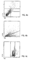
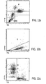
- Figures 4a-4c show the results obtained on a Cell-Dyn ® 4000 analyzer for the simulated WBC control of Carver, et al. and embodied in the Coulter Corp. Product 4C ® Plus Cell control. As can be seen in Figures 4a-4c , the clusters are not identifiable.
- WO9618878 performs blood cell differential analysis (WBC/diff) based on light scattering.
- the method involves gentle lysis of a blood sample, fixation of the WBC and staining of the nuclei. The method does not require the use or preparation of hematology control suspensions.
- U.S. Patent 5,270,208 to Ryan discloses a different method of preparing a hematology reference control for WBC/Diff analysis.
- aldehyde-fixed human WBC's are suspended in an isotonic aqueous medium comprising lipoprotein in an amount sufficient to provide a mixture that gives a WBC signature profile that is substantially similar to that obtained from whole blood.
- Ryan exhibited a WBC distribution of a Coulter Corp. STK-S ® analyzer dot plot, DF1 (abscissa) vs. Volume (ordinate).
- NRBC by multi-angle light scatter (axial light loss, multi-dimensional light scatter and fluorescence.)
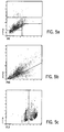
- the results reveal that the WBC component of the Ryan preparation generates a significantly different light scatter signature than that obtained from whole blood.
- the neutrophil components of the product generated much smaller axial light loss and polarized side scatter signals than that of whole blood; the depolarized side scatter signals from neutrophils are much too large to be separated from that of eosinophils; monocyte cluster does not separate from neutrophil cluster at all; the lymphocyte component generate much higher intermediate angle scatter (7°) signals than that of whole blood and thus wiping out the region reserved for basophils by overlapping.
- U. S. Patent 5,320,964 to Young, et al. discloses methods and reagent compositions for preparing leukocyte analogs.
- the Young, et al. lymphocyte analogs are prepared from fixed goose RBC in a hypotonic phosphate buffered solution (15-25 mOsm/kg); the Monocyte analogs are prepared from fixed alligator RBC in a hypotonic buffered solution (15-25 mOsm/kg); the Eosinophil analogs are also prepared from fixed alligator RBC in a hypotonic solution (75-85 mOsm/kg); and the Neutrophil analogs are prepared from alligator RBC in hypotonic buffered solution (45-65 mOsm/kg).
- Both goose RBC and alligator RBC are elliptical, nucleated and have a smooth cell surface.
- Young, et al. claim that the fixed cells prepared according to their procedures simulate at least two different human leukocytes, each having at least two physical properties of a human leukocyte. These properties are selected from: a) volume measured by D. C. current; b) high frequency (RF) size; c) opacity; and d) light scatter. Although they did not specify the type of light scatter, the Young, et al. simulated WBC components prepared from goose and alligator RBC do not have the same characteristics with regard to cell surface structure and cytoplasmic granularity as those of mammalian WBC.
- the osmolarity of the solution is very low (5-25 mOsm/kg) the cytoplasm of the cells will lyse. If, on the other hand, the osmolarity is about (85 mOsm/kg) the cell volume will expand.
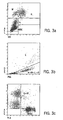
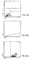
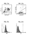
- Figures 18a-18d show the results of a multi-parameter, light scatter based analysis of a control cell solution containing fixed alligator RBC as the control cells.
- the fixed alligator cells generate no detectable PSS signals; the IAS signals fall between the regions of lymphocytes and basophils; and the ALL signals are too low to be counted as either neutrophils or eosinophils.
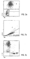
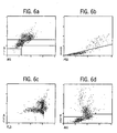
- Figures 6a-6c show the results of another commercially available control, R&D Systems, Inc. CBC-3k TM Hematology Controls. It is not know if the CBC-3k TM product has been patented. As shown, the clusters cannot be identified.
- the present invention first provides a hematology control suspension comprising:
- the hematology control suspension further comprises a nucleated red blood cell (NRBC) fraction wherein the cells of the NRBC fraction are selected from the group consisting of avian erythrocytes, fish erythrocytes and mammalian lymphocytes, and which have been subjected to a gentle lysing environment that lyses the cytoplasm and then a fixing process that fixes the nuclei before the lysing environment has destroyed their desired cellular characteristics so that the NRBC of interest may be differentiated by the exclusive use of polarized and depolarized 90° light scatter and axial light loss signals and combinations thereof and wherein the nuclei possess predetermined cellular multi-angle light scattering characteristics similar to the cellular multi-angle light scattering characteristics of the NRBC of interest.
- NRBC nucleated red blood cell
- the present invention also provides a hematology whole blood control suspension comprising:
- the present invention provides a method of producing a white blood cell (WBC) fraction of a hematology control suspension, the method comprising:
- the present invention further provides a method of producing a nucleated red blood cell fraction of a hematology control suspension comprising:
- Also provided by the present invention is a method of producing a nucleated red blood cell fraction of a hematology control suspension comprising:
- One object of the present invention is to provide a hematology reference control on an automated hematology instruments such as the Abbott Laboratories Cell-Dyn ® 3000 instrument which uses multi-dimensional light scatter WBC/Diff analysis.
- Another objective of the present invention is to provide a hematology reference control on an automated hematology system such as the Abbott Laboratories Cell-Dyn ® 4000 instrument which utilizes multi-dimensional light scatter, axial light loss and fluorescence for simultaneous analysis of WBC/Diff and NRBC.
- an automated hematology system such as the Abbott Laboratories Cell-Dyn ® 4000 instrument which utilizes multi-dimensional light scatter, axial light loss and fluorescence for simultaneous analysis of WBC/Diff and NRBC.
- Yet another objective is to provide a stable hematology reference control which contains stabilized RBC, Platelets, Neutrophils, Lymphocytes, Eosinophils, Basophils and NRBC components which can be used on an automated hematology instruments such as the Cell-Dyn ® 4000 instrument which uses multi-dimensional light scatter, axial light loss and fluorescence for the simultaneous analysis of WBC/Diff and NRBC.
- the present invention relates to the method for the preparation of a stable WBC and NRBC component and the hematology reference control solution containing one or more of these components.
- the method produces hard-fixed and stabilized WBCs and simulated NRBCs which generate similar electro-optical signals to that of human whole blood cells, enabling a complete WBC/Diff/NRBC analysis using the same algorithms utilized on an analyzer for fresh human blood samples.
- the fixed WBC's and simulated NRBCs produced by the method of the present invention are clump-iree and stable in an appropriate plasma-like medium for a long period of time under refrigeration and can be used as Hematology Controls for WBC/Diff/NRBC on a routine clinical hematology instrument, including those utilizing multi-parameter light scatter.
- Figures 12a-12c are multi-angle, light scatter based analyzer WBC cytograms of fixed turkey erythrocyte nuclei produced by the methods of the present invention.
- Figures 13a-13c are multi-angle, light scatter based analyzer WBC cytograms of whole blood spiked with the fixed turkey erythrocyte nuclei of Figures 12a-12c .
- Figures 14a-14c are multi-angle, light scatter based analyzer WBC cytograms of fixed porcine lymphocyte nuclei produced by the methods of the present invention.
- Figures 15a-15c are multi-angle, light scatter based analyzer WBC cytograms of bovine WBC by the method of the present invention.
- Figures 16a-16c are multi-angle, light scatter based analyzer WBC cytograms of fixed bovine WBC spiked with fixed porcine lymphocytes, all produced according to the methods of the present invention.
- Figures 17a-17d are WBC cytograms of fixed bovine WBC produced by the methods of the present invention and run on an analyzer utilizing both impedance and light scatter.
- Figures 18a-18d are WBC cytograms of a control cell solution containing fixed alligator RBC as the control cells and run on a multi-parameter, light scatter based analyzer.
- controls do not work on multi-angle light scatter based hematology analyzers and devised methods for producing controls that will work.
- controls perform on multi-parameter light scatter based systems, but they should also perform on other detection based systems as well. This is because the cells of the new controls are the same cells as is found in a blood sample, only processed. Therefore, they retain the cells' light scattering characteristics.
- the cellular components of the reference control suspensions of this invention substantially mimic the multi-parameter light scattering characteristics of the WBC or NRBC in a whole blood sample when run on a multi-parameter light scatter based detection system.
- multi-parameter, multi-angle, or multi-dimensional light scatter encompasses the exclusive use of ( i.e., excludes non-) polarized and depolarized 90° light scatter and axial light loss signals, and combinations thereof to determine or differentiate the cells of interest. No other non-light based signals are utilized for this determination. Other types of light signals may also be utilized, such as fluorescence; but impedance and other non-light based signals are not used. However, instruments which do use non-light based signals, or combinations of light scatter and impedance for example, can also benefit from the controls of this invention, but it is the multi-angle light scatter based instruments which will benefit the most as there are currently no such controls available.
- the WBC components of the control suspension of this invention are WBC from blood that have been subjected to a very gentle lysing environment (similar to the environment they would have been subjected to in the analyzer had that blood sample been analyzed), and then fixed.
- the NRBC components as well, the nuclei are exposed to a lysing environment and fixed to preserve their cellular characteristics.
- mammalian nucleated blood cells are not the only cells that can be used as the source of the freed nuclei. Avian or fish erythrocytes can also utilized for this fraction.
- control cells to mimic the light scattering characteristics of the cells of interest is due to the fact that the cells are first chosen because of their cellular composition (i . e ., their light scattering characteristics). During the manufacturing process these cellular components of the control cells are preserved by first subjecting them to a gentle lysing environment to lyse RBC; then the remaining WBC and NRBC nuclei are fixed before the lysing environment has destroyed the desired cellular characteristics. This manufacturing process mimics the environment "real" blood cells are subjected to in the interior passages of the analyzer. The control cells, because they are "fixed", are not significantly affected by the lysing environment when they pass through the analyzer, so they retain their desired characteristics.
- control cells are stable in the control solution, where whole blood cells are not.
- control cells need to be subjected to the same or similar lysing environment that the blood cells in a sample see while in an analyzer in order to produce substantially the same refractive index, which primarily determines the light scattering characteristics of a cell. In this way the control cells will appear to the instrument to be the "instrument lysed" cells of interest and be recorded as such.
- the reference control cells, or cellular components retain the appropriate cellular characteristic and therefore, react or behave (scatter light and/or fluoresce) in a manner that is substantially similar to whole blood, they will "appear" to be different cells to the analyzer or be completely unrecognizable.
- the primary methods of this invention subject cells or cellular components to a lysing environment during the reference control manufacturing process and prior to their "fixation" that they will encounter in the analyzer when they are run as reference controls.
- Figures 2a-2c and Figures 3a-3c are Abbott Laboratories Cell-Dyn ® 4000 multi-parameter, light scatter based system cytograms of the WBC/NRBC distributions of clinical blood samples which contain 1.78k/mL NRBC and 40.4k/mL NRBC, respectively. These Figures are presented for comparison purposes with the controls or cells produced by the various processes described and depicted in the Figures herein.
- the fixed WBC and nuclei of avian, fish and mammalian cells used in this invention are inert, they can be resuspended in any buffered saline, which may contain some protein to prevent clumping.
- these fixed cells need to be combined with unfixed, but stabilized RBC to produce a full range hematology control, the cell resuspending medium should be able to protect the stabilized RBC from lysis.
- the formulation below is an example of a resuspension medium that has been found to work well with the control cells of this invention. This formulation is a plasma-like cell resuspension medium that prevents clumping of fixed WBC and nuclei while protecting the RBC components in the control solution from lysis.
- CRSM Plasma-like Cell Resuspension Medium
- CRSM Plasma-like Cell Resuspension Medium
- hematology control produced by the method of present invention on CeU-Dyn ® 4000 hematology instrument, which analyzes WBC/Diff/NRBC by multi-dimensional light scatter, axial light loss and fluorescence, permits monitoring of day to day performances of the system as well as reagents.
- the parameters that the quality of the results can be monitored with the product on the Cell-Dyn ® 4000 system are as follows: Parameter Absolute Cell Counts % of Total WBC Total WBC # of cells/mL % of Total WBC Neutrophils # of cells/mL % of Total WBC Lymphocytes # of cells/mL % of Total WBC Eosinophils # of cells/mL % of Total WBC Monocytes # of cells/mL % of Total WBC Basophils # of cells/mL % of Total WBC NRBC # of cells/mL % of NRBC/100 WBC
- Example 2 The same lysing and fixing reagents used in Example 1 were used but the RBC lysis and WBC fixation were performed according to the following protocol:
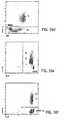
- NRBC fraction was prepared from Turkey erythrocytes according to the following protocol and examples of Cell-Dyn® 4000 instrument NRBC cytograms of the fixed turkey erythrocyte nuclei and a normal human whole blood spiked with the fixed Turkey erythrocyte nuclei were presented in Figures 12a-12e and 13a-13c , respectively.
- NRBC fraction was prepared from trout erythrocytes using the same reagents and protocol described in Example 3. Examples of Cell-Dyn ® 4000 instrument NRBC cytograms of the fixed trout erythrocyte nuclei and a normal whole blood spiked with the fixed trout nuclei are presented in Figures 8a-8c and 9a-9c
- NRBC Fraction was prepared from Porcine Lymphocytes according to the following materials and methods and an example of Cell-Dyn ® 4000 instrument NRBC cytograms of the fixed porcine lymphocyte nuclei is presented in Figures 14a-14c .
- Bovine WBC's are fixed according to the protocol described in Example 1 and mixed with fixed porcine nuclei prepared according to the procedure described in Example 4.
- Examples of Cell-Dyn® 4000 instrument WBC channel cytograms of the fixed bovine WBC's alone and the fixed bovine WBC's mixed with porcine lymphocyte nuclei are presented in Figures 15a-15c and 16a-16f .
- NRBC fraction was prepared from Chicken erythrocytes according to the protocol described in Example 3 and examples of Cell-Dyn ® 4000 instrument NRBC cytograms of the fixed Chicken erythrocyte nuclei and a normal human whole blood spiked with the fixed Chicken erythrocyte nuclei are presented in Figures 10a-10c and Figures 11a-11c .
- the hematology control produced by the method of present invention can also be used to monitor the following parameters on Cell-Dyn ® 3000 or 3500 instruments, which utilize both light scatter and impedance signals, to monitor the following WBC/Diff parameters: Parameter Absolute Cell Counts % of Total WBC Total WBC # of cells/mL % of Total WBC Neutrophils # of cells/mL % of Total WBC Lymphocytes # of cells/mL % of Total WBC Eosinophils # of cells/mL % of Total WBC Monocytes # of cells/mL % of Total WBC Basophils # of cells/mL % of Total WBC
- Figures 17a-17d are WBC cytograms of fixed bovine WBC produced by the methods of the present invention and run on a Cell-Dyn ® 3500 analyzer that conducts the WBC/Diff analysis using light scatter parameters (but no axial light loss or fluorescence) and determined. RBC and platelets by means of impedance. For this figure, the WBC fraction was combined with fixed platelets and stabilized, but unfixed RBC to produce a full range control suspension.
Landscapes
- Health & Medical Sciences (AREA)
- Life Sciences & Earth Sciences (AREA)
- Engineering & Computer Science (AREA)
- Immunology (AREA)
- Chemical & Material Sciences (AREA)
- Hematology (AREA)
- Molecular Biology (AREA)
- Urology & Nephrology (AREA)
- Biomedical Technology (AREA)
- Physics & Mathematics (AREA)
- General Physics & Mathematics (AREA)
- Biochemistry (AREA)
- Pathology (AREA)
- Analytical Chemistry (AREA)
- General Health & Medical Sciences (AREA)
- Microbiology (AREA)
- Cell Biology (AREA)
- Biotechnology (AREA)
- Food Science & Technology (AREA)
- Medicinal Chemistry (AREA)
- Dispersion Chemistry (AREA)
- Tropical Medicine & Parasitology (AREA)
- Investigating Or Analysing Biological Materials (AREA)
- Medicines Containing Material From Animals Or Micro-Organisms (AREA)
- Extrusion Moulding Of Plastics Or The Like (AREA)
- Holo Graphy (AREA)
- Apparatuses And Processes For Manufacturing Resistors (AREA)
Abstract
Description
- This invention relates to hematology control compositions and their methods of preparation and use in a reference standard. More particularly, this invention relates to a hematology reference control suspension containing stabilized, white blood cells ("WBC") and the nuclei of mammalian lymphocytes or
avian or fish erythrocytes. Even more particularly this invention relates to a hematology reference control suspension containing stabilized WBC that have been subjected to a whole blood lysing process during their preparation such that in their stabilized state they retain multi-angle light scattering properties. This enables the stabilized cells, when utilized in a hematology analyzer that differentiates WBC based solely on multi-angle light scatter signals to produce multi-angle light scatter signal that mimic whole blood WBC signals. Currently there are several different brands of automated hematology instruments in the marketplace. These different analyzers utilize varying detection techniques to quantify neutrophils, lymphocytes, monocytes, eosinophils and basophils. Among the detection techniques utilized are: electronic impedance, forward light scatter, polarized 90° angle light scatter, light absorption, radio-frequency and combinations thereof. The different optical bench designs significantly affect the characteristics of the optical signals obtained from stabilized control cells. Since these instruments utilize different detection methods for white blood cell differential analysis ("WBC/Diff"), it has become necessary to utilize different types of WBC/Diff control solutions in order to obtain control cell signatures that are similar to those of whole blood cells when run on that particular type of instrument. - The current class of instruments must utilize either impedance, impedance and light scatter (but not necessarily multi-angle light scatter), or light scatter, impedance and radio frequency signals to differentiate and determine cells from one another. Further, these currently available hematology instruments are not able to quantify nucleated red blood cells ("NRBC"). NRBC interfere with an accurate WHC/Diff analysis. The currently available hematology instruments only "flag" for the existence of NRBC in a sample. However, the soon to be released Abbott Cell-Dyn® 4000 hematology analyzer system will be capable of performing a simultaneous whole blood analysis of WBC/Diff and NRBC. The Cell-Dyn® instrument will perform a simultaneous, whole blood analysis by utilizing only multi-angle light scatter signals, including on occasion fluorescence to differentiate among WBC and NRBC. Consequently, it has become necessary to develop a new hematology control for WBC/Diff and NRBC analysis. The control cells of this new reference control must possess all of the multi-angle light scattering capabilities of the cells of the whole blood sample that they are suppose to mimic.
- There are in the current realm of art, several patents which describe methods and reagent systems for preparing hematology reference control materials for the current class of analyzers, i.e., those that do not perform an exclusive multi-angle light scatter WBC/Diff analysis. The applicants are not aware of any art describing a method or reagent system for the preparation or utilization of a hematology reference control for the multi-angle light scatter analysis of WBC or NRBC.
-
U. S. Patent 4,704,364 to Carver, et al. and assigned to Coulter Instrument Corp., discloses a method for preparing a three component system which simulates the three major components of human leukocytes. However these simulated cells are detected by an impedance based detection system, not a multi-angle light scatter detection system. Carver, et al. use fixed, red blood cells ("RBC") from the nurse shark to simulate human granulocytes; fixed RBC from turkeys to simulate human mononuclear cells; and fixed human RBC to simulate human lymphocytes. The hematology control produced by the teachings of Carver, et al. is useful only for electronic impedance measurement of a three part WBC/Diff since the three components are only distinguishable by size (impedance), not by optical properties. - The three components produced by Carver, et al.'s method do not have similar cell surface structure or cytoplasmic granularity substantially the same as that of human WBC. Therefore, they are not usable as a reference control on a multi-angle light scatter based system. The applicants tested these simulated cells on the soon to be commercially available Cell-Dyn® 4000 analyzer, and found that the light scattering characteristics of the simulated WBC in the Carver, et al. control produced very different multi-angle light scatter signals than that of a normal blood sample.
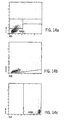
Figures 1a-1c are reproductions of the dot plots obtained on a Cell-Dyn® 4000 hematology analyzer for normal blood. - Abbreviations used to label the axis in the following figures:
ALL or WBC ALL: Axial Light Loss (0°) IAS or WBC IAS: Intermediate Angle Scatter (3°-10°) PSS or WBC PSS: Polarized Light Scatter (90°) DSS or WBC DSS: Depolarized Light Scatter (90°) FL3 or WBC FL3: Red Fluorescence (515-545 nm) G: Granulocyte Cluster M: Monocyte Cluster B: Basophil Cluster L: Lymphocyte Cluster E or Eos: Eosinophil Cluster N: Neutrophils S: Noise signals from RBC Stroma & PLT NRBC: Nucleated Red Blood Cells -
Figures 4a-4c show the results obtained on a Cell-Dyn® 4000 analyzer for the simulated WBC control of Carver, et al. and embodied in the Coulter Corp. Product 4C® Plus Cell control. As can be seen inFigures 4a-4c , the clusters are not identifiable. -
WO9618878 -
U.S. Patent 5,270,208 to Ryan , discloses a different method of preparing a hematology reference control for WBC/Diff analysis. In the Ryan method, aldehyde-fixed human WBC's are suspended in an isotonic aqueous medium comprising lipoprotein in an amount sufficient to provide a mixture that gives a WBC signature profile that is substantially similar to that obtained from whole blood. To support his claim, Ryan exhibited a WBC distribution of a Coulter Corp. STK-S® analyzer dot plot, DF1 (abscissa) vs. Volume (ordinate). It is believed that Ryan's WBC preparation is commercially available under the name of PARA 12® (Low, Normal & High) Tri-level hematology control from Streck Laboratories. The applicants tested this commercial material on a Cell-Dyn® 4000 analyzer which performs a simultaneous analysis of WBC/Diff and - NRBC by multi-angle light scatter (axial light loss, multi-dimensional light scatter and fluorescence.) The results (see
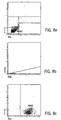
Figures 5a-5c ) reveal that the WBC component of the Ryan preparation generates a significantly different light scatter signature than that obtained from whole blood. As can be seen inFigures 5a-5c the neutrophil components of the product generated much smaller axial light loss and polarized side scatter signals than that of whole blood; the depolarized side scatter signals from neutrophils are much too large to be separated from that of eosinophils; monocyte cluster does not separate from neutrophil cluster at all; the lymphocyte component generate much higher intermediate angle scatter (7°) signals than that of whole blood and thus wiping out the region reserved for basophils by overlapping. -
U. S. Patent 5,320,964 to Young, et al. discloses methods and reagent compositions for preparing leukocyte analogs. The Young, et al. lymphocyte analogs are prepared from fixed goose RBC in a hypotonic phosphate buffered solution (15-25 mOsm/kg); the Monocyte analogs are prepared from fixed alligator RBC in a hypotonic buffered solution (15-25 mOsm/kg); the Eosinophil analogs are also prepared from fixed alligator RBC in a hypotonic solution (75-85 mOsm/kg); and the Neutrophil analogs are prepared from alligator RBC in hypotonic buffered solution (45-65 mOsm/kg). Both goose RBC and alligator RBC are elliptical, nucleated and have a smooth cell surface. Young, et al. claim that the fixed cells prepared according to their procedures simulate at least two different human leukocytes, each having at least two physical properties of a human leukocyte. These properties are selected from: a) volume measured by D. C. current; b) high frequency (RF) size; c) opacity; and d) light scatter. Although they did not specify the type of light scatter, the Young, et al. simulated WBC components prepared from goose and alligator RBC do not have the same characteristics with regard to cell surface structure and cytoplasmic granularity as those of mammalian WBC. Consequently, these cells do not generate the multi-angle light scatter signals for polarized and depolarized 90° light scatter and axial light loss signals that are substantially equivalent, or similar to that of human whole blood WBC. Thus, the Young, et al. product cannot be used as a hematology reference control on a sophisticated hematology instruments that utilizes multi-dimensional light scatter, axial light loss and fluorescence signals, such as the Cell-Dyn® 4000 analyzer, for WBC/Diff and NRBC quantification. In fact, if the cells of Young, et al. are fixed in a hypotonic buffered solution one of two results will occur. first, if the osmolarity of the solution is very low (5-25 mOsm/kg) the cytoplasm of the cells will lyse. If, on the other hand, the osmolarity is about (85 mOsm/kg) the cell volume will expand. -
Figures 18a-18d show the results of a multi-parameter, light scatter based analysis of a control cell solution containing fixed alligator RBC as the control cells. As can be seen the fixed alligator cells generate no detectable PSS signals; the IAS signals fall between the regions of lymphocytes and basophils; and the ALL signals are too low to be counted as either neutrophils or eosinophils. -
Figures 6a-6c show the results of another commercially available control, R&D Systems, Inc. CBC-3k™ Hematology Controls. It is not know if the CBC-3k™ product has been patented. As shown, the clusters cannot be identified. - The present invention first provides a hematology control suspension comprising:
- a white blood cell (WBC) fraction consisting essentially of mammalian WBC which have been subjected to a gentle lysing environment,
- In one embodiment the hematology control suspension further comprises a nucleated red blood cell (NRBC) fraction wherein the cells of the NRBC fraction are selected from the group consisting of avian erythrocytes, fish erythrocytes and mammalian lymphocytes, and which have been subjected to a gentle lysing environment that lyses the cytoplasm and then a fixing process that fixes the nuclei before the lysing environment has destroyed their desired cellular characteristics so that the NRBC of interest may be differentiated by the exclusive use of polarized and depolarized 90° light scatter and axial light loss signals and combinations thereof and wherein the nuclei possess predetermined cellular multi-angle light scattering characteristics similar to the cellular multi-angle light scattering characteristics of the NRBC of interest.
- The present invention also provides a hematology whole blood control suspension comprising:
- a stabilized red blood cell ("RBC") fraction;
- a stabilized platelet ("PLT") fraction;
- a stabilized white blood cell ("WBC") fraction; and
- a resuspension and storage medium,
wherein the WBC fraction consists essentially of mammalian WBC which have been subjected to a gentle lysing environment, wherein red blood cells (RBC) are lysed in the gentle lysing environment, and then a fixing process that fixes the cells before the lysing environment has destroyed the desired cellular characteristics of the mammalian WBC so that subpopulations of WBC may be differentiated by the exclusive use of polarized and depolarized 90° light scatter and axial light loss signals and combinations thereof and wherein the mammalian WBC possess predetermined cellular multi-angle light scattering characteristics similar to the cellular multi-angle light scattering characteristics of WBC of interest In a preferred embodiment; the hematology whole blood control suspension further comprises a nucleated red blood cell ("NRBC") fraction wherein the cells of the NRBC fraction are selected from the group consisting of avian erythrocytes, fish erythrocytes and mammalian lymphocytes, and which have been subjected to a gentle lysing environment that lyses the cytoplasm and then a fixing process that fixes the nuclei before the lysing environment has destroyed their desired cellular characteristics so that the NRBC of interest may be differentiated by the exclusive use of polarized and depolarized 90° light scatter and axial light loss signal and combinations thereof and wherein the nuclei possess predetermined cellular multi-angle light scattering characteristics similar to the cellular multi-angle light scattering characteristics of the NRBC of interest. - In addition, the present invention provides a method of producing a white blood cell (WBC) fraction of a hematology control suspension, the method comprising:
- a. providing mammalian whole blood or buffy coat layer;
- b. combining the whole blood with a lysing reagent for 1 to 5 minutes in a ratio of about 1 part blood or buffy coat layer to 12 parts lysing reagent to provide a gentle lysing environment to eliminate any red blood cells present, wherein the lysing reagent is present in an amount of about 1 part lyse to 12 parts diluent;
- c. combining the lyse and diluent cell suspension of step b with a fixative and incubate at 60 °C to 70 °C for up to 10 minutes to fix and stabilize the cells before the lysing environment has destroyed the desired cellular characteristics of the WBC so that subpopulations of WBC may be differentiated with the control suspension by the exclusive use of polarized and depolarized 90° light scatter and axial light loss signals and combinations thereof;
- d. cooling the mixture of step c to ambient temperature and allowing to sediment;
- e. discarding the supernatant formed in step d and washing the resultant fixed cells sufficient to remove the active reagents of steps b and c;
- f. resuspending the fixed and washed cells in a resuspension medium suitable for long term storage of the fixed cells, wherein the fixed cells exhibit cellular multi-angle light scattering characteristics similar to the cellular multi-angle light scattering characteristics of WBC of interest.
- The present invention further provides a method of producing a nucleated red blood cell fraction of a hematology control suspension comprising:
- a. providing whole blood or a red blood cell layer selected from the groups consisting of avian and fish erythrocytes;
- b. combining the erythrocytes and a lysing reagent for 1 to 5 minutes in a ratio of about 1 part erythrocyte to 12 parts lysing reagent to form a cell suspension;
- c. combining the cell suspension of step b with a fixative and incubate at 60 °C to 70 °C for up to 10 minutes to fix and stabilize the nuclei, wherein the ratio of fixative to cell suspension is 1:8 to 1:11;
- d. cooling the mixture of step c to ambient temperature and allowing the suspension to sediment;
- e. discarding the supernatant formed in step d and washing the resultant fixed nuclei sufficient to remove the active reagents of steps b and c;
- f. resuspending the fixed and washed nuclei in a resuspension medium suitable for long term storage of the fixed nuclei, wherein the fixed nuclei exhibit multi-angle light scattering characteristics similar to the multi-angle light scattering characteristics of nucleated red blood cells of interest so that the nucleated red blood cells of interest may be detected with the control suspension by the exclusive use of polarized and depolarized 90° light scatter and axial light loss signals and combinations thereof.
- Also provided by the present invention is a method of producing a nucleated red blood cell fraction of a hematology control suspension comprising:
- a. providing mammalian lymphocytes;
- b. mixing the lymphocytes and a lysing reagent for 5 minutes to 3 hours in a ratio of 1:5 lymphocytes to lysing reagent to 1:30 lymphocytes to lysing reagent to expose the nuclei of the lymphocytes and form a nuclei suspension;
- c. combining the nuclei suspension of step b with a fixative and incubating at 55 °C to 60 °C for up to 10 minutes to fix and stabilize the nuclei, wherein the ratio of fixative to nuclei suspension is 1:1 to 1:10;
- d. cooling the mixture of step c to ambient temperature and allowing the suspension to sediment;
- e. discarding the supernatant formed in step d and washing the resultant fixed nuclei sufficient to remove the active reagents of steps b and c;
- f. resuspending the fixed and washed nuclei in a resuspension medium suitable for long term storage of the fixed cells, wherein the fixed nuclei exhibit multi-angle light scattering characteristics similar to the multi-angle light scattering characteristics of nucleated red blood cells of interest so that the nucleated red blood cells of interest may be detected with the control suspension by the exclusive use of polarized and depolarized 90° light scatter and axial light loss signals and combinations thereof
- One object of the present invention is to provide a hematology reference control on an automated hematology instruments such as the Abbott Laboratories Cell-Dyn® 3000 instrument which uses multi-dimensional light scatter WBC/Diff analysis.
- Another objective of the present invention is to provide a hematology reference control on an automated hematology system such as the Abbott Laboratories Cell-Dyn® 4000 instrument which utilizes multi-dimensional light scatter, axial light loss and fluorescence for simultaneous analysis of WBC/Diff and NRBC.
- Yet another objective is to provide a stable hematology reference control which contains stabilized RBC, Platelets, Neutrophils, Lymphocytes, Eosinophils, Basophils and NRBC components which can be used on an automated hematology instruments such as the Cell-Dyn® 4000 instrument which uses multi-dimensional light scatter, axial light loss and fluorescence for the simultaneous analysis of WBC/Diff and NRBC.
- As defined above, the present invention relates to the method for the preparation of a stable WBC and NRBC component and the hematology reference control solution containing one or more of these components. The method produces hard-fixed and stabilized WBCs and simulated NRBCs which generate similar electro-optical signals to that of human whole blood cells, enabling a complete WBC/Diff/NRBC analysis using the same algorithms utilized on an analyzer for fresh human blood samples. The fixed WBC's and simulated NRBCs produced by the method of the present invention are clump-iree and stable in an appropriate plasma-like medium for a long period of time under refrigeration and can be used as Hematology Controls for WBC/Diff/NRBC on a routine clinical hematology instrument, including those utilizing multi-parameter light scatter. These advantages represent a substantial improvement over the prior art.
- For a more complete understanding of the present invention, and the advantages thereof, reference is now made to the following descriptions taken in conjunction with the accompanying figures, in which:
-
Figures 1a-1c are WBC cytograms of normal blood run on a multi-angle, light scatter based analyzer. -
Figures 2a-2c are multi-angle light scatter based analyzer WBC/NRBC cytograms of a clinical sample containing 1.78 k/µL NRBC. -
Figures 3a-3c are multi-angle, light scatter based analyzer WBC/NRBC cytograms of a clinical sample containing 40.4 K/µL NRBC. -
Figures 4a-4c are multi-angle, light scatter based analyzer WBC cytograms of Coulter® 4C® Plus Cell Controls. -
Figures 5a-5c are multi-angle, light scatter based analyzer WBC cytograms of Streck Laboratories' PARA 12® Multi-Parameter Hematology Controls, Normal Level. -
Figures 6a-6c are multi-angle, light scatter based analyzer WBC cytograms of R&D Systems' CBC-3K™ Hematology Controls. -
Figures 7a-7c are multi-angle, light scatter based analyzer WBC cytograms of the hematology reference control of the present invention. -
Figures 8a-8c are multi-angle, light scatter based analyzer WBC cytograms of fixed trout erythrocyte nuclei produced by the methods of the present invention. -
Figures 9a-9c are multi-angle, light scatter based analyzer WBC cytograms of normal whole blood spiked with the fixed trout erythrocyte nuclei ofFigures 8a-8c . -
Figures 10a-10c are multi-angle, light scatter based analyzer WBC cytograms of fixed chicken erythrocyte nuclei produced by the methods of the present invention. -
Figures 11a-11c are multi-angle, light scatter based analyzer WBC cytograms whole blood spiked with the fixed chicken erythrocyte nuclei ofFigures 10a-10c . -
Figures 12a-12c are multi-angle, light scatter based analyzer WBC cytograms of fixed turkey erythrocyte nuclei produced by the methods of the present invention. -
Figures 13a-13c are multi-angle, light scatter based analyzer WBC cytograms of whole blood spiked with the fixed turkey erythrocyte nuclei ofFigures 12a-12c . -
Figures 14a-14c are multi-angle, light scatter based analyzer WBC cytograms of fixed porcine lymphocyte nuclei produced by the methods of the present invention. -
Figures 15a-15c are multi-angle, light scatter based analyzer WBC cytograms of bovine WBC by the method of the present invention. -
Figures 16a-16c are multi-angle, light scatter based analyzer WBC cytograms of fixed bovine WBC spiked with fixed porcine lymphocytes, all produced according to the methods of the present invention. -
Figures 17a-17d are WBC cytograms of fixed bovine WBC produced by the methods of the present invention and run on an analyzer utilizing both impedance and light scatter. -
Figures 18a-18d are WBC cytograms of a control cell solution containing fixed alligator RBC as the control cells and run on a multi-parameter, light scatter based analyzer. - Any automated system for the detection and differentiation of cells requires the utilization of reference controls to assure that the system is operating properly. This is true no matter what detection system is employed.
- With the advent of a new detection scheme, mainly that of utilizing only multi-angle/parameter light scatter signals to differentiate and distinguish WBC subpopulations and NRBC in a sample (to the exclusion of all other non-light signals), new controls have become necessary. Current, state of the art reference controls will not perform properly on such multi-parameter systems.
- The present inventors have discovered why the current controls do not work on multi-angle light scatter based hematology analyzers and devised methods for producing controls that will work. Not only with the present invention controls perform on multi-parameter light scatter based systems, but they should also perform on other detection based systems as well. This is because the cells of the new controls are the same cells as is found in a blood sample, only processed. Therefore, they retain the cells' light scattering characteristics.
- The cellular components of the reference control suspensions of this invention substantially mimic the multi-parameter light scattering characteristics of the WBC or NRBC in a whole blood sample when run on a multi-parameter light scatter based detection system.
- For the purposes of this invention multi-parameter, multi-angle, or multi-dimensional light scatter encompasses the exclusive use of (i.e., excludes non-) polarized and depolarized 90° light scatter and axial light loss signals, and combinations thereof to determine or differentiate the cells of interest. No other non-light based signals are utilized for this determination. Other types of light signals may also be utilized, such as fluorescence; but impedance and other non-light based signals are not used. However, instruments which do use non-light based signals, or combinations of light scatter and impedance for example, can also benefit from the controls of this invention, but it is the multi-angle light scatter based instruments which will benefit the most as there are currently no such controls available.
- The WBC components of the control suspension of this invention are WBC from blood that have been subjected to a very gentle lysing environment (similar to the environment they would have been subjected to in the analyzer had that blood sample been analyzed), and then fixed. The same is true for the NRBC components as well, the nuclei are exposed to a lysing environment and fixed to preserve their cellular characteristics. With the NRBC component however, mammalian nucleated blood cells are not the only cells that can be used as the source of the freed nuclei. Avian or fish erythrocytes can also utilized for this fraction.
- The ability of the control cells to mimic the light scattering characteristics of the cells of interest is due to the fact that the cells are first chosen because of their cellular composition (i.e., their light scattering characteristics). During the manufacturing process these cellular components of the control cells are preserved by first subjecting them to a gentle lysing environment to lyse RBC; then the remaining WBC and NRBC nuclei are fixed before the lysing environment has destroyed the desired cellular characteristics. This manufacturing process mimics the environment "real" blood cells are subjected to in the interior passages of the analyzer. The control cells, because they are "fixed", are not significantly affected by the lysing environment when they pass through the analyzer, so they retain their desired characteristics. In addition, the fixed cells are stable in the control solution, where whole blood cells are not. However, it is believed that the control cells need to be subjected to the same or similar lysing environment that the blood cells in a sample see while in an analyzer in order to produce substantially the same refractive index, which primarily determines the light scattering characteristics of a cell. In this way the control cells will appear to the instrument to be the "instrument lysed" cells of interest and be recorded as such.
- It has also been determined that the same cellular components of the present invention, if "fixed" prior to subjecting them to a lysing environment, do not perform satisfactorily in a multi-angle light scattering based system. The cells need to be subjected to a lysing environment before they are "fixed". Later, when they are processed as control cells in the analyzer these processed cells will "appear" to the analyzer, to be the blood cells of interest. This is because the multi-parameter light, scatter based analyzers are only programmed to recognize whole blood cells in the analyzer's internal processing environment. So, unless the reference control cells, or cellular components, retain the appropriate cellular characteristic and therefore, react or behave (scatter light and/or fluoresce) in a manner that is substantially similar to whole blood, they will "appear" to be different cells to the analyzer or be completely unrecognizable.
- Therefore, the primary methods of this invention subject cells or cellular components to a lysing environment during the reference control manufacturing process and prior to their "fixation" that they will encounter in the analyzer when they are run as reference controls.
- In the examples that follow the following reagent formulations were used:
-
- from about 0.75M to about 1.10M ammonium chloride (NH4Cl)
- from about 0.1M to about 0.4M formaldehyde (HCHO)
- from about 10mM to about 25mM sodium acetate (CH3COONa)
- from about 10mM to about 25mM potassium bicarbonate (KHCO3)
- from about 50 mg/L to about 250 mg/L Saponin
- from about 0.2 g/L to about 0.4 g/L Proclin 300
- from about 2.5 g/L to about 5.0 g/L maleic acid, succinic acid or phthalic acid
- from about 10.0 g/L to about 30.0 g/L Brij 35, Tween 20 or Triton X-100
-
- from about 3.0 g/L to about 5.0 g/L monosodium phosphate
- from about 6.0 g/L to about 7.0 g/l disodium phosphate
- from about 100 ml/L to about 200 ml/L formalin (37-40% formaldehyde solution)
-
Figures 2a-2c andFigures 3a-3c are Abbott Laboratories Cell-Dyn® 4000 multi-parameter, light scatter based system cytograms of the WBC/NRBC distributions of clinical blood samples which contain 1.78k/mL NRBC and 40.4k/mL NRBC, respectively. These Figures are presented for comparison purposes with the controls or cells produced by the various processes described and depicted in the Figures herein. - Since the fixed WBC and nuclei of avian, fish and mammalian cells used in this invention are inert, they can be resuspended in any buffered saline, which may contain some protein to prevent clumping. However, these fixed cells need to be combined with unfixed, but stabilized RBC to produce a full range hematology control, the cell resuspending medium should be able to protect the stabilized RBC from lysis. The formulation below is an example of a resuspension medium that has been found to work well with the control cells of this invention. This formulation is a plasma-like cell resuspension medium that prevents clumping of fixed WBC and nuclei while protecting the RBC components in the control solution from lysis.
-
Chemical Conc. Range/L Preferred Conc./L Na2HP04 2.20 - 2.70 g 2.45 g KH2PO4 0.36 - 0.44 g 0.40 g Na3Citrate 330 - 4.05 g 3.68 g Citric Acid 0.41 - 0.51 0.46 g -
Chemical Conc. Range/L Preferred Conc./L Dextrose 4.05 - 4.95 4.50 g Mannose 1.35 - 1.65 1.50 g Adenine 0.30 - 0.55 0.50 g BSA* 15.0-50.0 30.0 g NaCl adjust to 290 ± 15 mOsm/L 0.50 g Proclin 300 (Preservative) 0.15 - 0.45 0.30 g pH adjust pH to 7.2 ± 0.2 with 1 N NaOH adjust pH to 7.2 ± 0.2 with 1 N NaOH Osmolarity 290 ± 15 mOs/L 290 ± 15 mOs/L * Fatty acid free bovine serum albumin - The use of hematology control produced by the method of present invention on CeU-Dyn® 4000 hematology instrument, which analyzes WBC/Diff/NRBC by multi-dimensional light scatter, axial light loss and fluorescence, permits monitoring of day to day performances of the system as well as reagents. The parameters that the quality of the results can be monitored with the product on the Cell-Dyn® 4000 system are as follows:
Parameter Absolute Cell Counts % of Total WBC Total WBC # of cells/mL % of Total WBC Neutrophils # of cells/mL % of Total WBC Lymphocytes # of cells/mL % of Total WBC Eosinophils # of cells/mL % of Total WBC Monocytes # of cells/mL % of Total WBC Basophils # of cells/mL % of Total WBC NRBC # of cells/mL % of NRBC/100 WBC - The lysing reagent and fixative described above are used in this example. Stable and clump-free fixed human WBC's were prepared according to the following protocol and an example of the Cell-Dyn® 4000 instrument WBC cytograms of the fixed human WBC of this example are presented at
Figures 7a-7c - 1. One (1) part of human buffy coat layer was mixed with 5 parts of lysing reagent (pre-warmed at 42°C). The components were mixed immediately by gentle vortexing and allowed to stand at room temperature for 50 seconds to lyse RBC completely.
- 2. One part of the lysed WBC suspension in Step 1 was mixed with ten (10) parts of fixative and immediately placed in a 60° to 70°C water bath and fixed for 10 minutes with gentle mixing.
- 3. The fixed cell suspension was cooled at room temperature, centrifuged at 2,500 rpm at 10°C for 5 minutes to remove the Fixative, washed 3 times using the same centrifuge speed with isotonic phosphate buffered saline at neutral pH (PBS) and then resuspended in CRSM.
- 4. An aliquot of the finished product was run on a Cell-Dyn® 4000 instrument for WBC counts and distribution.
- 5. The fixed cell concentration was adjusted to a final concentration in the resuspension medium of about 7,500/µL for a normal level control.
- The same lysing and fixing reagents used in Example 1 were used but the RBC lysis and WBC fixation were performed according to the following protocol:
- 1. One (1) part of human buffy coat layer was mixed immediately by gentle vortexing with 5 parts of lysing reagent solution and allowed to stand at room temperature for 10 minutes to lyse RBC completely.
- 2. One (1) part of the lysed WBC suspension in Step 1 was mixed with ten (10) parts of Fixative, mixed and allowed to fix at room temperature for 2-3 hours.
- 3. The fixed cell suspension was centrifuged at 2,500 rpm at 10°C for 5 minutes to remove the Fixative, washed 3 times using the same centrifuge speed with phosphate buffered saline at neutral pH, and then resuspended in CRSM.
- 4. An aliquot of the finished product was run on a Cell-Dyn® 4000 instrument for WBC counts and distribution.
- 5. The fixed cell concentration was adjusted to final concentration of about 2,000/µL for low level control.
- NRBC fraction was prepared from Turkey erythrocytes according to the following protocol and examples of Cell-Dyn® 4000 instrument NRBC cytograms of the fixed turkey erythrocyte nuclei and a normal human whole blood spiked with the fixed Turkey erythrocyte nuclei were presented in
Figures 12a-12e and 13a-13c , respectively. -
- 1. Turkey Whole Blood
- 2. Lysing reagent: The same as in Example 1.
- 3. Fixative: w/Dextrose: Monosodium phosphate: 4 gram/L, Disodium phosphate: 6.5 grams/L, Formalin: 150 ml/L, Dextrose 100 g/L, pH about 6.8
- 4. Cell Washing Solution: Phosphate Buffered Solution (PBS)
- 5. CRSM: The same CRSM as listed above
-
- 1. Warm up 10 ml aliquot of the lysine reagent at 37°C.
- 2. Centrifuge turkey whole blood at 3000 rpm for 10 minutes to separate the plasma layer and remove the buffy coat layer. Add the plasma back to the packed RBC layer and mix.
- 3. Add 1.0 ml of the RBC layer to the pre-warmed lysing reagent solution, cap and immediately invert mix 3 times. Vortex at full speed for about 10 seconds to help the lysis the cytoplasm of RBC. Let stand at room temperature for about 10 minutes or until the lysis or cytoplasm is complete. Check under the microscope for completeness of the cytoplasm lysis.
- 4. Centrifuge at 2000 rpm for 15 minutes and siphon off all the supernatant leaving just enough to resuspend the cell button. Resuspend the cells by gentle agitation until no cell clumps are observed.
- 5. Wash the cell 3 times with PBS using the same centrifugation conditions as in Step 4.
- 6. Add 10 ml of fixative, mix and immediately place it in the water bath at 60°C and fix for at least 5 minutes with constant agitation to prevent cell clumping.
- 7. Leave the cell suspension at room temperature for 30 minutes or overnight.
- 8. Mix and repeat Step 4 & 5 twice.
- 9. Resuspend the fixed nuclei in CRSM.
- 10. Run on a Cell-Dyn® 4000 instrument to determine the concentration of FL3+ nuclei and the position of the cluster.
- NRBC fraction was prepared from trout erythrocytes using the same reagents and protocol described in Example 3. Examples of Cell-Dyn® 4000 instrument NRBC cytograms of the fixed trout erythrocyte nuclei and a normal whole blood spiked with the fixed trout nuclei are presented in
Figures 8a-8c and 9a-9c - NRBC Fraction was prepared from Porcine Lymphocytes according to the following materials and methods and an example of Cell-Dyn® 4000 instrument NRBC cytograms of the fixed porcine lymphocyte nuclei is presented in
Figures 14a-14c . - Porcine buffy coat (platelets already removed) layer.
Cyto-Lyse: - Brij 35: 15.0 g/L
- Phthalic Add: 5.5 g/L
-
- 1. Dilute one (1) part of the enriched porcine lymphocyte layer with 5 parts of the lysing reagent and let stand at room temperature for 5 minutes to complete the lysis of the remaining RBC in the cell suspension.
- 2. Centrifuge the cell suspension 3 minutes at 3000 rpm and pour off the supernatant and dislodge the cell button.
- 3. Mix the cells with Cyto-Lyse at 1:10 ratio and leave at room temperature for 2 hours.
- 4. Resuspend the nuclei in Cyto-Lyse and spin for 3 minutes at 3000 rpm and pour off the supernatant.
- 5. Resuspend the nuclei in minimum 2 volumes of Fixative and mix.
- 6. Fix at 60°C for 10 minutes (or for 4 hours at room temperature) and allow to cool at room temperature.
- 7. Centrifuge the nuclei for 3 minutes at 3000 rpm and pour off supernatant.
- 8. Resuspend the cell pellet in PBS to wash the nuclei 3 times.
- 9. Resuspend the nuclei in CRSM.
- Bovine WBC's are fixed according to the protocol described in Example 1 and mixed with fixed porcine nuclei prepared according to the procedure described in Example 4. Examples of Cell-Dyn® 4000 instrument WBC channel cytograms of the fixed bovine WBC's alone and the fixed bovine WBC's mixed with porcine lymphocyte nuclei are presented in
Figures 15a-15c and 16a-16f . - NRBC fraction was prepared from Chicken erythrocytes according to the protocol described in Example 3 and examples of Cell-Dyn® 4000 instrument NRBC cytograms of the fixed Chicken erythrocyte nuclei and a normal human whole blood spiked with the fixed Chicken erythrocyte nuclei are presented in
Figures 10a-10c andFigures 11a-11c . - The hematology control produced by the method of present invention can also be used to monitor the following parameters on Cell-Dyn® 3000 or 3500 instruments, which utilize both light scatter and impedance signals, to monitor the following WBC/Diff parameters:
Parameter Absolute Cell Counts % of Total WBC Total WBC # of cells/mL % of Total WBC Neutrophils # of cells/mL % of Total WBC Lymphocytes # of cells/mL % of Total WBC Eosinophils # of cells/mL % of Total WBC Monocytes # of cells/mL % of Total WBC Basophils # of cells/mL % of Total WBC -
Figures 17a-17d are WBC cytograms of fixed bovine WBC produced by the methods of the present invention and run on a Cell-Dyn® 3500 analyzer that conducts the WBC/Diff analysis using light scatter parameters (but no axial light loss or fluorescence) and determined. RBC and platelets by means of impedance. For this figure, the WBC fraction was combined with fixed platelets and stabilized, but unfixed RBC to produce a full range control suspension.
Fixative: the same as in Example 1
Phosphate Buffered Saline (PBS)
Claims (7)
- A hematology control suspension comprising:a white blood cell (WBC) fraction consisting essentially of mammalian WBC which have been subjected to a gentle lysing environment,wherein red blood cells (RBC) are lysed in the gentle lysing environment, and then a fixing process that fixes the cells before the lysing environment has destroyed the desired cellular characteristics of the mammalian WBC so that subpopulations of WBC may be differentiated by the exclusive use of polarized and depolarized 90° light scatter and axial light loss signals and combinations thereof and wherein the mammalian WBC possess predetermined cellular multi-angle light scattering characteristics similar to the cellular multi-angle light scattering characteristics of WBC of interest
- The hematology control suspension of Claim 1 further comprising a nucleated red blood cell (NRBC) fraction wherein the cells of the NRBC fraction are selected from the group consisting of avian erythrocytes, fish erythrocytes and mammalian lymphocytes, and which have been subjected to a gentle lysing environment that lyses the cytoplasm and then a fixing process that fixes the nuclei before the lysing environment, has destroyed their desired cellular characteristics so that the NRBC of interest may be differentiated by the exclusive use of polarized and depolarized 90° light scatter and axial light loss signals and combinations thereof and wherein the nuclei possess predetermined cellular multi-angle light scattering characteristics similar to the cellular multi-angle light scattering characteristics of the NRBC of interest.
- A hematology whole blood control suspension comprising:a stabilized red blood cell (RBC) fraction;a stabilized platelet (PLT) fraction;a stabilized white blood cell (WBC) fraction; anda resuspension and storage medium,wherein the WBC fraction consists essentially of mammalian WBC which have been subjected to a gentle lysing environment, wherein red blood cells (RBC) are lysed in the gentle lysing environment, and then a fixing process that fixes the cells before the lysing environment has destroyed the desired cellular characteristics of the mammalian WBC so that subpopulations of WBC may be differentiated by the exclusive use of polarized and depolarized 90° light scatter and axial light loss signals and combinations thereof and wherein the mammalian WBC possess predetermined cellular multi-angle light scattering characteristics similar to the cellular multi-angle light scattering characteristics of WBC of interest
- The hematology control suspension of Claim 3 further comprising a nucleated red blood cell (NRBC) fraction wherein the cells of the NRBC fraction are selected from the group consisting of avian erythrocytes, fish erythrocytes and mammalian lymphocytes, and which have been subjected to a gentle lysing environment that lyses the cytoplasm and then a fixing process that fixes the nuclei before the lysing environment has destroyed their desired cellular characteristics so that the NRBC of interest may be differentiated by the exclusive use of polarized and depolarized 90° light scatter and axial light loss signals and combinations thereof and wherein the nuclei possess predetermined cellular multi-angle light scattering characteristics similar to the cellular multi-angle light scattering characteristics of the NRBC of interest.
- A method of producing a white blood cell (WBC) fraction of a hematology control suspension, the method comprising:a. providing mammalian whole blood or buffy coat layer;b. combining the whole blood with a lysing reagent for 1 to 5 minutes in a ratio of about 1 part blood or buffy coat layer to 12 parts lysing reagent to provide a gentle lysing environment to eliminate any red blood cells present, wherein the lysing reagent is present in an amount of about 1 part lyse to 12 parts diluent;c. combining the lyse and diluent cell suspension of step b with a fixative and incubate at 60 °C to 70 °C for up to 10 minutes to fix and stabilize the cells before the lysing environment has destroyed the desired cellular characteristics of the WBC so that subpopulations ofWBC may be differentiated with the control suspension by the exclusive use of polarized and depolarized 90° light scatter and axial light loss signals and combinations thereof;d. cooling the mixture of step c to ambient temperature and allowing to sediment;e. discarding the supernatant formed in step d and washing the resultant fixed cells sufficient to remove the active reagents of steps b and c;f. resuspending the fixed and washed cells in a resuspension medium suitable for long term storage of the fixed cells, wherein the fixed cells exhibit cellular multi-angle light scattering characteristics similar to the cellular multi-angle light scattering characteristics of WBC of interest.
- A method of producing a nucleated red blood cell fraction of a hematology control suspension comprising:a. providing whole blood or a red blood cell layer selected from the groups consisting of avian and fish erythrocytes;b. combining the erythrocytes and a lysing reagent for 1 to 5 minutes in a ratio of about 1 part erythrocyte to 12 parts lysing reagent to form a cell suspension;c. combining the cell suspension of step b with a fixative and incubate at 60 °C to 70 °C for up to 10 minutes to fix and stabilize the nuclei, wherein the ratio of fixative to cell suspension is 1:8 to 1:11;d. cooling the mixture of step c to ambient temperature and allowing the suspension to sediment;e. discarding the supernatant formed in step d and washing the resultant fixed nuclei sufficient to remove the active reagents of steps b and c;f. resuspending the fixed and washed nuclei in a resuspension medium suitable for long term storage of the fixed nuclei, wherein the fixed nuclei exhibit multi-angle light scattering characteristics similar to the multi-angle light scattering characteristics of nucleated red blood cells of interest so that the nucleated red blood cells of interest may be detected with the control suspension by the exclusive use of polarized and depolarized 90° light scatter and axial light loss signals and combinations thereof.
- A method of producing a nucleated red blood cell fraction of a hematology control suspension comprising:a. providing mammalian lymphocytes;b. mixing the lymphocytes and a lysing reagent for 5 minutes to 3 hours in a ratio of 1:5 lymphocytes to lysing reagent to 1:30 lymphocytes to lysing reagent to expose the nuclei of the lymphocytes and form a nuclei suspension;c. combining the nuclei suspension of step b with a fixative and incubating at 55 °C to 60 °C for up to 10 minutes to fix and stabilize the nuclei, wherein the ratio of fixative to nuclei suspension is 1:1 to 1:10;d. cooling the mixture of step c to ambient temperature and allowing the suspension to sediment;e. discarding the supernatant formed in step d and washing the resultant fixed nuclei sufficient to remove the active reagents of steps b and c;f. resuspending the fixed and washed nuclei in a resuspension medium suitable for long term storage of the fixed cells, wherein the fixed nuclei exhibit multi-angle light scattering characteristics similar to the multi-angle light scattering characteristics of nucleated red blood cells of interest so that the nucleated red blood cells of interest may be detected with the control suspension by the exclusive use of polarized and depolarized 90° light scatter and axial light loss signals and combinations thereof.
Applications Claiming Priority (3)
| Application Number | Priority Date | Filing Date | Title |
|---|---|---|---|
| US673672 | 1984-11-21 | ||
| US08/673,672 US5858790A (en) | 1996-06-26 | 1996-06-26 | Hematology reference control and method of preparation |
| PCT/US1997/009567 WO1997049982A1 (en) | 1996-06-26 | 1997-05-29 | Hematology reference control and method of preparation |
Publications (2)
| Publication Number | Publication Date |
|---|---|
| EP1021701A1 EP1021701A1 (en) | 2000-07-26 |
| EP1021701B1 true EP1021701B1 (en) | 2009-09-02 |
Family
ID=24703633
Family Applications (1)
| Application Number | Title | Priority Date | Filing Date |
|---|---|---|---|
| EP97928807A Expired - Lifetime EP1021701B1 (en) | 1996-06-26 | 1997-05-29 | Hematology reference control and method of preparation |
Country Status (8)
| Country | Link |
|---|---|
| US (2) | US5858790A (en) |
| EP (1) | EP1021701B1 (en) |
| JP (2) | JP4549440B2 (en) |
| AT (1) | ATE441846T1 (en) |
| CA (1) | CA2257359C (en) |
| DE (1) | DE69739565D1 (en) |
| ES (1) | ES2330902T3 (en) |
| WO (1) | WO1997049982A1 (en) |
Cited By (2)
| Publication number | Priority date | Publication date | Assignee | Title |
|---|---|---|---|---|
| US11852640B2 (en) | 2017-10-27 | 2023-12-26 | Beckman Coulter, Inc. | Hematology analyzers and methods of operation |
| US12062448B2 (en) | 2016-01-28 | 2024-08-13 | Beckman Coulter, Inc. | Infection detection and differentiation systems and methods |
Families Citing this family (43)
| Publication number | Priority date | Publication date | Assignee | Title |
|---|---|---|---|---|
| US6509192B1 (en) * | 1992-02-24 | 2003-01-21 | Coulter International Corp. | Quality control method |
| US6200500B1 (en) * | 1999-08-20 | 2001-03-13 | Streck Laboratories, Inc. | Hematology control and system for multi-parameter hematology measurements |
| US6221668B1 (en) * | 1999-08-20 | 2001-04-24 | Streck Laboratories, Inc. | Hematology control and system for multi-parameter hematology measurements |
| US6444471B1 (en) | 1999-10-18 | 2002-09-03 | Research & Diagnostic Systems, Inc. | Reticulocyte containing complete blood control |
| US6514763B2 (en) | 2000-04-28 | 2003-02-04 | Hematronix, Inc. | Hematology blood control and method for preparation of same |
| US6653063B2 (en) | 2000-06-12 | 2003-11-25 | Hematronix, Inc. | Hematology composition using preserved white blood cells in combination with source particles to simulate native white blood cells and method of making same |
| WO2001096831A1 (en) * | 2000-06-12 | 2001-12-20 | Hematronix, Inc. | Hematology composition using preserved white blood cells |
| US6448085B1 (en) * | 2001-04-13 | 2002-09-10 | Sysmex Corporation | Quality control material and calibrator for nucleated red blood cell tested on hematology analyzer |
| US6759246B1 (en) * | 2001-11-30 | 2004-07-06 | Research & Diagnostic Systems, Inc. | Hematology control composition including lymphocyte analogs and method for preparation and use |
| US6653137B2 (en) | 2001-12-03 | 2003-11-25 | Streck Laboratories Inc. | Hematology reference control |
| US6723563B2 (en) | 2001-12-03 | 2004-04-20 | Streck Laboratories Inc. | Hematology reference control |
| DE10231308C5 (en) | 2002-07-10 | 2009-10-15 | Cmi Uvk Gmbh | Process and apparatus for recycling stainless steel pickling baths |
| EP1422509A1 (en) * | 2002-11-01 | 2004-05-26 | Streck Laboratories, Inc. | Lysing Reagent and Method of Lysing Red Blood Cells for Hematology |
| US6900058B2 (en) * | 2003-03-11 | 2005-05-31 | Bionostics, Inc. | Control solution for photometric analysis |
| EP1668353B1 (en) * | 2003-10-02 | 2015-03-18 | Beckman Coulter, Inc. | Reference control for optical measurement of nucleated red blood cells of a blood sample |
| JP4873712B2 (en) * | 2003-10-12 | 2012-02-08 | ベックマン コールター, インコーポレイテッド | How to use a reference control for the measurement of nucleated red blood cells |
| US7198953B2 (en) * | 2003-10-12 | 2007-04-03 | Beckman Coulter, Inc. | Method of using a reference control composition for measurement of nucleated red blood cells |
| US7195919B2 (en) * | 2003-12-19 | 2007-03-27 | Beckman Coulter, Inc. | Hematology controls for reticulocytes and nucleated red blood cells |
| US7208319B2 (en) * | 2004-02-10 | 2007-04-24 | Beckman Coulter, Inc. | Method of measurement of nucleated red blood cells |
| EP1714146B1 (en) * | 2004-02-10 | 2022-08-03 | Beckman Coulter, Inc. | Method of measurement of nucleated red blood cells |
| WO2005100979A1 (en) * | 2004-04-07 | 2005-10-27 | Beckman Coulter, Inc. | Reference control containing a nucleated red blood cell component |
| US7354767B2 (en) * | 2006-03-16 | 2008-04-08 | Beckman Coulter, Inc. | Reference control composition containing a nucleated red blood cell component made of non-nucleated blood cells |
| CN101535804B (en) | 2006-11-14 | 2013-04-24 | 贝克曼考尔特公司 | Hematology linearity control composition, system and method of use |
| US7618821B2 (en) * | 2007-04-13 | 2009-11-17 | Streck, Inc. | Simulated blood components and methods |
| CN101311724B (en) * | 2007-05-24 | 2014-11-12 | 深圳迈瑞生物医疗电子股份有限公司 | Single group leucocyte emulation ion, calibrator comprising same and its preparation method |
| CN101311725B (en) * | 2007-05-25 | 2014-03-26 | 深圳迈瑞生物医疗电子股份有限公司 | WBC differential count quality control matter and method for making same |
| CN101561443B (en) * | 2008-04-15 | 2013-08-21 | 深圳迈瑞生物医疗电子股份有限公司 | Five-classification leucocyte simulacrum particle, method for preparing same, and quality control substance and calibration substance containing same |
| US20100086962A1 (en) | 2008-10-08 | 2010-04-08 | Streck, Inc. | Hematology controls and methods |
| CN102109430B (en) | 2009-12-25 | 2013-11-06 | 深圳迈瑞生物医疗电子股份有限公司 | Nucleated red blood cell simulation particle and blood quality control substance and preparation methods and application thereof |
| US8546144B2 (en) | 2010-11-17 | 2013-10-01 | Streck, Inc. | Method of preparing controls for glycated hemoglobin S-A1c derived from healthy blood cells |
| US8551784B2 (en) | 2010-11-17 | 2013-10-08 | Streck, Inc. | Cis di-ahl modified controls for glycated hemoglobin S-A1c derived from healthy blood cells |
| JP6117181B2 (en) * | 2011-04-28 | 2017-04-19 | ベックマン コールター, インコーポレイテッド | Hematology control composition with long-term stability |
| WO2018022991A1 (en) | 2016-07-29 | 2018-02-01 | Streck, Inc. | Suspension composition for hematology analysis control |
| US11791022B2 (en) | 2017-02-28 | 2023-10-17 | Beckman Coulter, Inc. | Cross discipline disease management system |
| CN110402319B (en) * | 2017-05-08 | 2024-01-19 | 哈利伯顿能源服务公司 | Inversion processing of logging data |
| WO2019204804A1 (en) | 2018-04-20 | 2019-10-24 | Beckman Coulter, Inc. | Sepsis infection determination systems and methods |
| US11538566B2 (en) | 2018-05-23 | 2022-12-27 | Beckman Coulter, Inc. | Sample analysis with test determination based on identified condition |
| US11521706B2 (en) | 2018-04-20 | 2022-12-06 | Beckman Coulter, Inc. | Testing and representing suspicion of sepsis |
| WO2019240876A1 (en) | 2018-06-15 | 2019-12-19 | Beckman Coulter, Inc. | Method of determining sepsis in the presence of blast flagging |
| US12023178B2 (en) | 2019-07-12 | 2024-07-02 | Beckman Coulter, Inc. | Method of detecting sepsis using vital signs, including systolic blood pressure, hematology parameters, and combinations thereof |
| CN114096845A (en) | 2019-07-12 | 2022-02-25 | 贝克曼库尔特有限公司 | Systems and methods for assessing immune response to infection |
| US20230062518A1 (en) * | 2020-01-31 | 2023-03-02 | Bio-Rad Laboratories, Inc. | Stable reference materials for automated hematology testing platforms |
| CN116097093A (en) * | 2020-09-03 | 2023-05-09 | 生物辐射实验室股份有限公司 | Preparation of Nucleated RBC (NRBC) analogs for use as reference blood controls in automated blood analyzers |
Citations (1)
| Publication number | Priority date | Publication date | Assignee | Title |
|---|---|---|---|---|
| US5270208A (en) * | 1991-05-09 | 1993-12-14 | Streck Laboratories, Inc. | White blood cell hematology control |
Family Cites Families (13)
| Publication number | Priority date | Publication date | Assignee | Title |
|---|---|---|---|---|
| US3632735A (en) * | 1970-03-11 | 1972-01-04 | Pfizer & Co C | Method of stabilizing fowl red blood cells for use in hematology control compositions |
| US4219440A (en) * | 1979-06-06 | 1980-08-26 | Coulter Electronics, Inc. | Multiple analysis hematology reference control reagent and method of making the same |
| US4704364A (en) * | 1984-05-18 | 1987-11-03 | Coulter Electronics, Inc. | Hematology control compositions for three populations of leukocytes; and methods for their preparation and use in whole blood control systems |
| US4847204A (en) * | 1984-05-24 | 1989-07-11 | Southeast Vetlab, Inc. | Calibrator composition and method of producing and using same for veterinary applications |
| DE3587636T2 (en) * | 1984-05-31 | 1994-03-10 | Coulter Corp | REAGENT SYSTEM AND METHOD FOR DETECTING, LISTING AND CHECKING THE CLASSES AND SUBCLASSES OF BLOOD LEUCOCYTES. |
| US4698312A (en) * | 1986-07-28 | 1987-10-06 | Fisher Scientific Company | Stabilizing blood cells with aromatic polyaldehyde for use in hematology controls and calibrators |
| US4777139A (en) * | 1987-06-25 | 1988-10-11 | Fisher Scientific Company | Hematology control or calibrator with red cell components of enhanced stability |
| IE76319B1 (en) * | 1990-07-23 | 1997-10-22 | Becton Dickinson Co | Preservation of cells as controls or standards in cellular analysis |
| US5262327A (en) * | 1991-05-09 | 1993-11-16 | Streck Laboratories, Inc. | White blood cell hematology control |
| EP0628167B1 (en) * | 1992-02-24 | 2000-08-23 | Coulter Corporation | Hematology control composition for leukocyte analogs; and methods for their preparation and use |
| WO1993017329A1 (en) * | 1992-02-24 | 1993-09-02 | Coulter Corporation | Suspension media for hematological composition and method for its use |
| AU6235994A (en) * | 1993-02-25 | 1994-09-14 | Abbott Laboratories | Multipurpose reagent system for rapid lysis of whole blood samples |
| US5559037A (en) * | 1994-12-15 | 1996-09-24 | Abbott Laboratories | Method for rapid and simultaneous analysis of nucleated red blood cells |
-
1996
- 1996-06-26 US US08/673,672 patent/US5858790A/en not_active Expired - Fee Related
-
1997
- 1997-05-29 EP EP97928807A patent/EP1021701B1/en not_active Expired - Lifetime
- 1997-05-29 ES ES97928807T patent/ES2330902T3/en not_active Expired - Lifetime
- 1997-05-29 AT AT97928807T patent/ATE441846T1/en not_active IP Right Cessation
- 1997-05-29 CA CA002257359A patent/CA2257359C/en not_active Expired - Fee Related
- 1997-05-29 DE DE69739565T patent/DE69739565D1/en not_active Expired - Lifetime
- 1997-05-29 JP JP50302998A patent/JP4549440B2/en not_active Expired - Fee Related
- 1997-05-29 WO PCT/US1997/009567 patent/WO1997049982A1/en active Application Filing
-
1998
- 1998-10-16 US US09/173,823 patent/US6187590B1/en not_active Expired - Fee Related
-
2007
- 2007-07-12 JP JP2007182849A patent/JP4686506B2/en not_active Expired - Fee Related
Patent Citations (1)
| Publication number | Priority date | Publication date | Assignee | Title |
|---|---|---|---|---|
| US5270208A (en) * | 1991-05-09 | 1993-12-14 | Streck Laboratories, Inc. | White blood cell hematology control |
Cited By (2)
| Publication number | Priority date | Publication date | Assignee | Title |
|---|---|---|---|---|
| US12062448B2 (en) | 2016-01-28 | 2024-08-13 | Beckman Coulter, Inc. | Infection detection and differentiation systems and methods |
| US11852640B2 (en) | 2017-10-27 | 2023-12-26 | Beckman Coulter, Inc. | Hematology analyzers and methods of operation |
Also Published As
| Publication number | Publication date |
|---|---|
| JP4549440B2 (en) | 2010-09-22 |
| EP1021701A1 (en) | 2000-07-26 |
| WO1997049982A1 (en) | 1997-12-31 |
| CA2257359C (en) | 2006-03-14 |
| ES2330902T3 (en) | 2009-12-16 |
| DE69739565D1 (en) | 2009-10-15 |
| JP4686506B2 (en) | 2011-05-25 |
| ATE441846T1 (en) | 2009-09-15 |
| US6187590B1 (en) | 2001-02-13 |
| CA2257359A1 (en) | 1997-12-31 |
| US5858790A (en) | 1999-01-12 |
| JP2008026317A (en) | 2008-02-07 |
| JP2000513441A (en) | 2000-10-10 |
Similar Documents
| Publication | Publication Date | Title |
|---|---|---|
| EP1021701B1 (en) | Hematology reference control and method of preparation | |
| AU669692B2 (en) | Suspension media for hematological composition and method for its use | |
| US7285417B2 (en) | Reference control containing a nucleated red blood cell component | |
| EP1766045B1 (en) | Hematology reference control containing an immature granulocyte component | |
| JP2008026317A5 (en) | ||
| EP2002256B1 (en) | Reference control composition containing a nucleated red blood cell component made of non-nucleated blood cells | |
| JP3822625B2 (en) | Hematology control products | |
| EP4097599A1 (en) | Stable reference materials for automated hematology testing platforms | |
| EP1682865B1 (en) | Method of using a reference control composition for measurement of nucleated red blood cells | |
| US6759246B1 (en) | Hematology control composition including lymphocyte analogs and method for preparation and use |
Legal Events
| Date | Code | Title | Description |
|---|---|---|---|
| PUAI | Public reference made under article 153(3) epc to a published international application that has entered the european phase |
Free format text: ORIGINAL CODE: 0009012 |
|
| 17P | Request for examination filed |
Effective date: 19981217 |
|
| AK | Designated contracting states |
Kind code of ref document: A1 Designated state(s): AT BE CH DE ES FR GB IT LI NL |
|
| 17Q | First examination report despatched |
Effective date: 20050629 |
|
| 17Q | First examination report despatched |
Effective date: 20050629 |
|
| GRAP | Despatch of communication of intention to grant a patent |
Free format text: ORIGINAL CODE: EPIDOSNIGR1 |
|
| GRAS | Grant fee paid |
Free format text: ORIGINAL CODE: EPIDOSNIGR3 |
|
| GRAA | (expected) grant |
Free format text: ORIGINAL CODE: 0009210 |
|
| AK | Designated contracting states |
Kind code of ref document: B1 Designated state(s): AT BE CH DE ES FR GB IT LI NL |
|
| REG | Reference to a national code |
Ref country code: CH Ref legal event code: EP |
|
| REF | Corresponds to: |
Ref document number: 69739565 Country of ref document: DE Date of ref document: 20091015 Kind code of ref document: P |
|
| REG | Reference to a national code |
Ref country code: ES Ref legal event code: FG2A Ref document number: 2330902 Country of ref document: ES Kind code of ref document: T3 |
|
| NLV1 | Nl: lapsed or annulled due to failure to fulfill the requirements of art. 29p and 29m of the patents act | ||
| PG25 | Lapsed in a contracting state [announced via postgrant information from national office to epo] |
Ref country code: NL Free format text: LAPSE BECAUSE OF FAILURE TO SUBMIT A TRANSLATION OF THE DESCRIPTION OR TO PAY THE FEE WITHIN THE PRESCRIBED TIME-LIMIT Effective date: 20090902 |
|
| PG25 | Lapsed in a contracting state [announced via postgrant information from national office to epo] |
Ref country code: BE Free format text: LAPSE BECAUSE OF FAILURE TO SUBMIT A TRANSLATION OF THE DESCRIPTION OR TO PAY THE FEE WITHIN THE PRESCRIBED TIME-LIMIT Effective date: 20090902 Ref country code: AT Free format text: LAPSE BECAUSE OF FAILURE TO SUBMIT A TRANSLATION OF THE DESCRIPTION OR TO PAY THE FEE WITHIN THE PRESCRIBED TIME-LIMIT Effective date: 20090902 |
|
| PLBE | No opposition filed within time limit |
Free format text: ORIGINAL CODE: 0009261 |
|
| STAA | Information on the status of an ep patent application or granted ep patent |
Free format text: STATUS: NO OPPOSITION FILED WITHIN TIME LIMIT |
|
| PGFP | Annual fee paid to national office [announced via postgrant information from national office to epo] |
Ref country code: FR Payment date: 20100525 Year of fee payment: 14 Ref country code: ES Payment date: 20100514 Year of fee payment: 14 |
|
| 26N | No opposition filed |
Effective date: 20100603 |
|
| PGFP | Annual fee paid to national office [announced via postgrant information from national office to epo] |
Ref country code: IT Payment date: 20100513 Year of fee payment: 14 Ref country code: DE Payment date: 20100531 Year of fee payment: 14 |
|
| PGFP | Annual fee paid to national office [announced via postgrant information from national office to epo] |
Ref country code: GB Payment date: 20100401 Year of fee payment: 14 |
|
| REG | Reference to a national code |
Ref country code: CH Ref legal event code: PL |
|
| PG25 | Lapsed in a contracting state [announced via postgrant information from national office to epo] |
Ref country code: LI Free format text: LAPSE BECAUSE OF NON-PAYMENT OF DUE FEES Effective date: 20100531 Ref country code: CH Free format text: LAPSE BECAUSE OF NON-PAYMENT OF DUE FEES Effective date: 20100531 |
|
| REG | Reference to a national code |
Ref country code: DE Ref legal event code: R119 Ref document number: 69739565 Country of ref document: DE |
|
| REG | Reference to a national code |
Ref country code: DE Ref legal event code: R119 Ref document number: 69739565 Country of ref document: DE |
|
| GBPC | Gb: european patent ceased through non-payment of renewal fee |
Effective date: 20110529 |
|
| REG | Reference to a national code |
Ref country code: FR Ref legal event code: ST Effective date: 20120131 |
|
| PG25 | Lapsed in a contracting state [announced via postgrant information from national office to epo] |
Ref country code: IT Free format text: LAPSE BECAUSE OF NON-PAYMENT OF DUE FEES Effective date: 20110529 |
|
| PG25 | Lapsed in a contracting state [announced via postgrant information from national office to epo] |
Ref country code: FR Free format text: LAPSE BECAUSE OF NON-PAYMENT OF DUE FEES Effective date: 20110531 |
|
| PG25 | Lapsed in a contracting state [announced via postgrant information from national office to epo] |
Ref country code: GB Free format text: LAPSE BECAUSE OF NON-PAYMENT OF DUE FEES Effective date: 20110529 |
|
| REG | Reference to a national code |
Ref country code: ES Ref legal event code: FD2A Effective date: 20121116 |
|
| PG25 | Lapsed in a contracting state [announced via postgrant information from national office to epo] |
Ref country code: ES Free format text: LAPSE BECAUSE OF NON-PAYMENT OF DUE FEES Effective date: 20110530 |
|
| PG25 | Lapsed in a contracting state [announced via postgrant information from national office to epo] |
Ref country code: DE Free format text: LAPSE BECAUSE OF NON-PAYMENT OF DUE FEES Effective date: 20111130 |