-
The present invention relates to the field of
non-viral gene delivery.
-
Translocation of exogeneous DNA through the nuclear
membrane has been shown to be a major problem in gene
delivery technologies.
The nuclear membrane of eukaryotic cells is freely
permeable to solutes of size up to ca. 9 nm (e.g. 40-60
kDa proteins). Transport of larger molecules through
nuclear pores is signal-mediated, involves shuttle
molecules and requires energy. The basic peptide
derived from the SV40 large T antigen (PKKKRKV) is a
nuclear localization signal (NLS) that mediates binding
of the karyophilic protein to importin-α (Adam and
Gerace, 1991.
Complex formation triggers binding to importin-β and
the ternary complex is then carried through the nuclear
pore with the help of the GTPase Ran. Macromolecules
and particles up to 25 nm have been shown to enter the
nucleus in this way, although the translocation
mechanism is still not fully understood (Panté and
Aebi, 1995; Nigg, 1997; Ohno et al., 1998; Melchior and
Gerace, 1998).
-
Eukaryotic DNA viruses which replicate in the
nucleus seem to be capable of diverting the cell's
nuclear import machinery to their own benefit (Greber
et al., 1997; Greber, 1998). Recombinant viruses
derived therefrom are being used to carry therapeutic
genes into humans. However, the host's immune response
is currently a limitation to the clinical development
of viral gene therapy. Nonviral alternatives using
plasmid DNA and cationic carrier molecules suffer from
a different drawback, the low efficacy of gene
delivery. The main barrier to transgene expression in
vitro, is the nuclear membrane (Zabner et al., 1995;
Labat-Moleur et al., 1996; Hagstrom et al. 1997). Since
breakdown of this membrane during cell division helps
nuclear localization, this obstacle is probably still
more a problem in vivo, where cells can be considered
as resting with respect to the lifetime of DNA.
-
Several attempts to improve entry of plasmid DNA
into the nucleus have been suggested. These include use
of electrostatic binding of DNA to cationic NLS-containing
proteins (Kaneda et al., 1989; Fritz et al.,
1996) or peptides (Collas et al., 1996) or lipids (Remy
et al., 1995)), as well as sequence-specific binding of
DNA to karyophilic proteins (Fominaya, J. & Wels, W.
1996; Langle-Rouault et al., 1998). Yet such
"piggyback" nuclear transport relies on the
unpredictable stability of the complexes within the
cytoplasm. Recent work by the group of Jon Wolff
(Sebestyén et al., 1998) with digitonin-permeabilized
cells demonstrated nuclear accumulation of
fluorescently-labelled DNA that was randomly tagged
with hundreds of NLS peptides covalently attached to
the DNA; nuclei of intact cells did not take up the
modified DNA.
-
It was an object of the present invention to
overcome the problems of the previous methods of
nuclear transport of DNA and to provide an improved
gene delivery system.
-
The present invention is directed to a non-viral
transfection vector comprising a DNA molecule which is
to be delivered into the nucleus of a cell,
characterized that said DNA molecule is equipped with
1-15 conjugates comprising a nuclear localization
signal covalently linked to an oligonucleotide, each of
which NLS-conjugate has been
- i) covalently linked to one or both termini of a
linear DNA molecule, or
- ii) associated with a plasmid DNA molecule by forming
a triple helix, or
- iii) inserted in a plasmid DNA molecule by strand
invasion.
In the following, the nuclear localization signal is
termed "NLS".
NLSs are peptides which are known to be required for
nuclear transport of karyophilic proteins. They are
part of the primary structure of most nuclear proteins
and contain one or two clusters of basic amino acids.
Typically, they are six to eighteen amino acids in
length. -
-
A wide range of NLSs from various organisms has
been characterized, examples of NLSs that can be used
in the present invention are given in WO 95/31557,
which is incorporated herein by reference in its
entirety.
-
The NLS must fulfil the requirements to direct the
DNA or the protein encoded by the DNA, respectively, to
the nucleus.
-
Depending on the organism whose cells are to be
transfected, e.g. an animal or a plant, the NLS is
selected from a karyophilic protein expressed by that
organism or from a virus infectious for that organism.
-
A preferred NLS used in the present invention is
the NLS of the SV40 virus large T antigen.
-
The NLS peptide can be synthesized according to
standard methods of peptide chemistry. In general,the
peptide is identical to the naturally occuring NLS.
However, if desired, e.g. to improve the nuclear
transporting function of the peptide, the peptide may
be modified, e.g. by replacing one or more of the basic
amino acids by other basic amino acids. Another NLS
modification, which has been used in the experiments of
the present invention, is the extension of the NLS
beyond its actual nuclear localization domain by the
N-terminal amino acids of a karyophilic protein,
preferably the authentic protein that the NLS
originates from. The peptide may carry an amino acid
such as cysteine, lysine, tyrosine, threonine, serine
that allows convenient linkage of an oligonucleotide.
-
Generally, the length of the NLS is six to sixty,
preferably six to fourty, even more preferably six to
thirty amino acids.
-
In order to assess whether a selected NLS is
suitable for the cell to be transfected, i.e. that it
directs the protein of interest into the nucleus,
assays are conducted which are known in the art, e.g.
those described by Garcia-Bustos et al., 1991; Sandler
et al., 1989; Citovsky et al., 1992; Sebestyén et al.,
1998). These assays are based on the detection of the
protein in the nucleus, e.g. by histochemical methods.
A useful assay involves the use of a reporter gene, e.g
the luciferase gene; the delivery of the luciferase is
detected by standard luciferase detection methods as
described in the Examples of the present invention.
Preferably, the NLS contains a reactive thiol group
from a cysteine residue and is conjugated to the
oligonucleotide by conventional methods, e.g. by amino-modifying
the oligonucleotide and linking the two
compounds with bifunctional cross linkers like SMCC
(4-(N-maleimidomethyl)-cyclohexane-1-carboxlic acid
N-hydroxysuccinimide ester). Alternative coupling
methods that can be used in the present invention, e.g.
activation of the 5'-phosphate groups of the
oligonucleotide with imidazole or tetrazole in the
presence of a carbodiimide compound and subsequent
dislacement of that group by the terminal amino group
of the NLS, are disclosed in WO 88/05077, which is
incorporated herein by reference in its entirety.
-
Preferably, the DNA molecule is equipped with
1 to 5 NLS conjugates.
-
In the embodiment i) of the present invention, the
DNA molecule is linear. The oligonucleotide part of the
conjugate has a sequence that allows to form a hairpin
and has, preferably at its 5'-end, a cohesive extension
that is ligated, by standard cloning methods, to a
complementary cohesive generated at the end of the DNA
molecule. The linear DNA molecule carries one or two
NLSs covalently linked to the hairpin at the 5' and/or
at 3' terminus of the DNA molecule.
-
This hairpin-forming oligonucleotide is preferably
15 to 60 nucleotides in length. The NLS is conjugated
to the oligonucleotide by coupling the peptide to a
amino-modified nucleotide in the loop of the hairpin.
-
Preferably, in the embodiment i) of the invention,
only one terminus of the linear DNA molecule carries an
oligonucleotide hairpin which has an NLS coupled to it,
the other terminus of the DNA molecule is capped with
an identical or different oligonucleotide hairpin that
does not carry an NLS. In the experiments of the
present invention, is has been surprisingly shown that
a single NLS is sufficient to carry DNA to the nucleus.
Figure 1 depicts an example for the strategy for the
preparation of a dsDNA fragment coupled to an NLS
peptide. A functional luciferase gene of 3380 bp was
cut out of pCMVLuc with XmaI and SalI. Further
digestion with XmnI and BspHI cut the unwanted
restriction fragment into small fragments (970, 875,
768 and 240 bp) which were removed by sucrose gradient
centrifugation. The capped CMVLuc-NLS DNA was obtained
by ligation of the 32P radioactive (*) oligo-peptide and
oligo-cap hairpins to the restriction fragment
-
The following considerations have lead to the
design of the nuclear transport system according to
embodiment i) of the invention:
-
When fully counterion-condensed, a single plasmid
molecule collapses into a sphere of ca. 25 nm (Blessing
et al., 1998)) which is already at the size-exclusion
limit for signal-mediated nuclear import (Ohno et al.,
1998; Gorlich and Mattaj,1996). Plasmid condensation
with cationic lipids or polymers generally leads to
even larger, multimolecular aggregates which reach the
cytoplasm after binding to cell-surface anionic
proteoglycans (Labat-Moleur et al., 1996; Mislick and
Baldeschwieler, 1996) and eventual escape from the
formed vacuoles (Zabner et al., 1995; Plank et al.,
1994; Boussif et al., 1995). Since the particles are
too large to cross an intact nuclear membrane,
transfection of resting cells can reasonably only be
accounted for by uncomplexed DNA that has been released
by exchange with phosphatidylserine (Xu and Szoka,
1996) or heparan sulfate (Labat-Moleur et al., 1996)
present in the vacuolar membrane. Any molecular
information (e.g. NLS) borne by the cationic vector
will thus be lost prior to reaching the nuclear
membrane, hence its ineffectiveness.
-
In sharp contrast to a condensed DNA molecule, a
free hydrated DNA double helix (3 nm) is thin enough to
enter the nuclear pore by diffusion, i.e. with no
energy nor signal requirement. Restricted intracellular
motion of DNA (the cytoplasm is equivalent to 13%
dextran (Luby-Phelps, 1994)) and a rather low nuclear
pore coverage (<10% of the membrane surface (Maul and
Deaven, 1977)) however give this event a low
probability to occur: the nuclear membrane has been
recognized as a major barrier to gene delivery (Zabner
et al., 1995; Labat-Moleur et al., 1996). Quantitative
cytoplasmic microinjection of DNA (Pollard et al.,
1998; Mirzayans et al.,1992; Dowty et al., 1995) indeed
led to less than 0.1% of the DNA being expressed. The
probability for DNA to find and subsequently enter a
nuclear pore can be increased by a bound karyophilic
signal peptide able to dock DNA to the nuclear pore
filaments and help an initial part of the molecule to
cross the membrane. The DNA-karyophilic signal link
needs however be stable, therefore the peptide was
chemically linked to the oligonucleotide.
-
The linear DNA molecule approach i), which is
exemplified in the experiments of the invention, takes
advantage of a chemically controlled pathway to
irreversibly link a NLS-peptide to one end of a gene.
In the experiments of the inventions, in contrast to
the state of the art, the level of transgene expression
following transfection was chosen as a test of success.
-
The major achievement of the linear DNA/NLS
approach is that, for the first time using a nonviral
technology, the amount of DNA required to effectively
transfect cells in vitro has shifted from the microgram
to the nanogram range. Although the improvement
(1-3 orders of magnitude) varies with cell type
(showing that the nuclear membrane is not the only
possible barrier), even the modest enhancements
observed for primary cell cultures look interesting
(1000%) when not presented on a logarithmic scale. To
show that this result was a consequence of the
involvement of the nuclear import machinery, a Lys ⇒
Thr mutation was used which indeed abolished the
beneficial effect brought about by the DNA-bound NLS
sequence.
-
Taking the raised hypothesis further, the last
unanswered question deals with threading of the rest of
the plasmid molecule (Figure 7). Random-walk diffusion
of a micrometer-long molecule would be inefficient and
slow. Fortunately, naked DNA will not remain free in
the nucleus : histones (and eventually basic nuclear
matrix proteins) indeed quickly assemble transfected
DNA into chromatin-like structures (Cereghini and
Yaniv, 1984; Jeong and Stein, 1994), thus providing a
mechanism for pulling and condensing the filamentous
molecule into the nucleus. Following this line of
thinking, too many NLS signals distributed along the
DNA may actually inhibit nuclear entry if the nucleic
acid is longer than the distance separating adjacent
pores (Fig. 7). A straightforward calculation using the
pore density in HeLa cells (Maul and Deaven, 1977)
shows this to become probable above 1 kbp.
-
In embodiment ii) of the invention, the DNA
molecule is a plasmid encoding any protein of interest,
and the NLS peptide is covalently linked to a
oligonucleotide that recognizes a
homopurine/homopyrimidine sequence within the plasmid
in a sequence specific manner (Le Doan et al., 1987;
Moser and Dervan, 1987). The interaction of the
oligonucleotide with the duplex DNA takes place in the
major groove leading to a local triple helix structure
(Fig. 8). The use of a triple-helix structure to obtain
strong association between a duplex molecule and an
oligonucleotide is an attractive method to generate the
NLS-bearing plasmid molecule.
-
Two families of DNA triple helices have been
characterized that differ in their third-strand
sequence composition and relative orientation. In the
pyrimidine family (Py*Pu-Py), a homopyrimidine strand
(Py) is bound (*) parallel to the purine strand of
target duplex through Hoogsteen base pairing. In the
purine family (Pu*Pu-Py), a homopurine strand (Pu) is
bound in an anti-parallel orientation to the purine
strand of target duplex through reverse Hoogsteen base
pairing. In the purine family, it has been shown that
G-rich homopurine oligonucleotides are able to form
triple helices with high stability compatible with
physiological conditions (Debin et al., 1997; Nakanishi
et al., 1998). This type of sequence, which is
preferred in this embodiment of the invention, leads to
a stable non-covalent association between the
NLS-oligonucleotide and the plasmid (Fig. 9).
The selected homopurine-homopyrimidine oligonucleotide
sequence can be introduced in the plasmid by
conventional cloning methods, e.g. as described in
Sambrook et al., 1989, at a specific position outside
of the promoter region, outside of the gene coding for
the protein of interest, and of the polyadenylation
signal. Preferably, the oligonucleotide, which in this
embodiment of the invention is a triple-helix-forming
oligonucleotide (TFO), is between 8 and 40 nucleotides
long. Some examples of G-rich sequences, that are
useful for this embodiment of the invention, e.g. a
nearly perfect 45mer polypurine tract localized in the
gag gene of Friend murine leukemia virus (F-MuLV), are
presented in Fig. 10 (Debin et al., 1997; Nakanishi et
al., 1998; Svinarchuk et al., 1994; Rando et al.,
1994).
-
It has been reported for several G-rich homoPu
sequences that triple helix association can persist
inside cells for 1 to 3 days (Debin et al., 1997;
Svinarchuk et al., 1994). Therefore, for appropriate
homoPu-homo-Py sequences, the NLS-oligo/plasmid
triplexes are expected to be stable enough to remain
undissociated inside the cell until the triplex is
carried to the nucleus. Furthermore, by introducing
several identical homoPu-homo-Py sequences in the
plasmid, the influence of both number and distance
between NLS's on nuclear import can be assessed in
order to optimize the design of the construct.
-
The NLS peptide is covalently linked to the TFO
purine oligonucleotide either at the 5' or on the
3' end of the oligonucleotide according to known
methods, as described above for embodiment i).
Preparation of the plasmid carrying the NLS peptide(s)
simply requires mixing of the NLS-oligonucleotide and
the homoPu-homoPy plasmid in appropriate buffer
conditions, e.g. 20 mM Tris-acetate pH 7.5, 5-10 mM
Mg2+, no K+ ions.
-
In the embodiment iii) of the invention, the
NLS-oligonucleotide conjugate is inserted into the
plasmid DNA by a mechanism that has been termed
"strand-invading mechanism". This mechanism has been
proposed to explain the binding mode of "peptide
nucleic acids" (PNA) to DNA (Nielsen et al., 1991). It
has been described as the strand invasion of a doublestranded
DNA by an oligonucleotide (or a PNA) that
leads to complementary binding between the invading
molecule and the duplex through Watson-Crick
interactions. Local separation of the DNA strands is
required and the final conformation is thought to be a
D-loop structure.
-
In a preferred embodiment of variant iii) of the
invention, the oligonucleotide is chemically modified,
thus displacement of one DNA strand is achieved by the
chemically modified oligonucleotide.
-
The chemical modification allows stabilization of
the D-loop structure over the native duplex. As
described by Schmid and Behr, 1995, invasion has been
observed with synthetic oligonucleotides containing two
spermine residues linked to the C2 position of inosine
(Fig. 11). Stabilization of the D-loop structure is
provided by spermine modifications that lie in the
minor groove and clip both strands together through
interstrand hydrogen bonding between each ammonium
group and the nucleic bases.
-
Stabilization of the D-loop structure may be
achieved by modifying the oligonucleotide with
polycations such as spermine, spermidine or other
polyamines such as (NH-(CH2)n)m-NH2. The
oligonucleotides are preferably 10-50 bases in length
and contain 2-15 polyamines.
-
Since the strand invading mechanism is not
restricted to a particular class of sequences,
embodiment iii) of the invention does not require
providing specific DNA sequences when constructing the
plasmid. However, the oligonucleotide requires a number
of bases that allow the coupling of the respective
number of polyamine molecules, e.g. in the case of
modifying the oligonucleotide with two spermine
molecules the presence of at least two guanine bases
for the chemical coupling of two spermine residues and
the chemical coupling of the NLS peptide to the
spermine- oligonucleotide will be useful.
-
The chemical modification of the oligonucleotide
with the polyamine may be carried out by known methods,
described by (Schmid and Behr, 1995).
-
Embodiment iii) of the invention is schematically
depicted in Fig. 12.
-
In general, the DNA molecule encodes a protein of
interest to be expressed in an animal or plant cell.
Alternatively, the DNA molecule may encode an
inhibiting RNA molecule, e.g. an antisense RNA. In view
of gene therapy applications in humans or animals, the
protein of interest is a therapeutically active
protein. Non limiting examples of DNA molecules are
given in WO 93/07283. In addition to the coding
sequence, the DNA contains regulatory sequences
necessary for its expression in the cell; in the case
of a plasmid, additional sequences, e.g. encoding
selection markers, may be present. There are no
limitations as to the sequence of the DNA. The size of
the DNA is preferably in a range typical for eukaryotic
genes, i.e. 1Kpb tp 1Mbp.
-
Transfection of the cells with the NLS-modified DNA
molecules of the invention may be carried out by any
standard gene delivery technology, e.g. applying the
DNA molecules as such, in a suitable physiological
solution, by methods known for the delivery of "naked"
DNA, or by mixing the DNA containing the NLS-conjugate
with a cationic lipid (Behr, 1994; Ledley, 1996), a
detergent or with a cationic polymer, e.g.
polyethyleneimine, which may contain a cellular
targeting function and/or and endosomolytic function.
Suitable methods are described in WO 93/07283, or in
reviews by Ledley, 1995; Cotten and Wagner, 1993;
Boussif et al.,1995.
-
In the case of delivery of an NLS-equipped plasmid
into the cell, a cationic vector may eventually favour
encounter between the plasmid and oligonucleotide
sequences. In these particles, both NLS-oligonucleotides
and plasmid DNA partners will be
protected from nucleases.
-
The cells may be transfected with the NLS-DNA of
the invention in vitro, ex vivo or in vivo. Examples
for in vivo applications are intramuscular or
intradermal injection of the NLS-DNA or
electropermeabilization of NLS-DNA, both applications
preferably in the absence of gene delivery vehicles, or
gene gun-mediated introduction of NLS-DNA.
Brief description of the figures:
-
- Fig. 1
- Strategy for the preparation of a dsDNA
fragment
- Fig. 2
- Reaction scheme for the chemical coupling
steps leading to the oligonucleotide-peptide
conjugate (oligonucleotide-NLS)
- Fig.3
-
- A. Synthesis of the oligonucleotide-NLS
- B. The oligonucleotide-NLS conjugate is a
substrate of proteinase K
- Fig. 4
- The NLS peptide promotes high and sustained
transfection levels down to 10 ng DNA
- Fig. 5
- Sustained luciferase expression levels are
due to the nuclear localization peptide
- Fig. 6
- Reporter protein activity appears faster with
CMVLuc-NLS
- Fig. 7
- Hypothetical scheme of transgenic linear NLS-DNA
crossing a nuclear pore
- Fig. 8
- Schematic representation of triple-helix
interaction
- Fig. 9
- Non covalent coupling of an NLS peptide to
plasmid DNA through triple helix association
- Fig. 10
- Some examples of G-rich target sequences for
triple-helix formation
- Fig. 11
- Interaction through Watson-Crick pairing
after strand-invasion of a DNA duplex by a
complementary spermine-containing
oligonucleotide
- Fig. 12
- Non-covalent coupling of an NLS peptide to a
plasmid through the strand-invading mechanism
Example 1
Synthesis of an oligonucleotide-NLS equipped luciferase
gene; purification and proof of structure
i) Chemicals, enzymes and oligonucleotides
-
4-(N-maleimidomethyl)-cyclohexane-1-carboxylic acid N-hydroxysuccinimide
ester (SMCC) was purchased from
Sigma. The SAL 34-mer 5' d(TCGATGTCCGCGTTGGCTT
XTGCCAACGCGGACA) oligodeoxynucleotide was synthesized
by Appligene using an amino-modified deoxythymidine (X;
amino-modified dT, Glen Research, USA). The XMA 34-mer
5' d(CCGGCTACCTTGCGAGCTTTTGCTCGCAAGGTAG)
oligodeoxynucleotide, the NLS peptide NH2-PKKKRKVEDPYC
and the mutated-NLS peptide NH2-PKTKRKVEDPYC with
C-terminal amidation were synthesized by Genosys using
the standard F-moc chemistry for the solid phase
synthesis of peptide. Hairpin structures composed of a
loop of four thymines, a stem of 13 base pairs and a
sticky 5'-end were formed by boiling and subsequently
cooling the XMA- or the SAL- oligonucleotides in ice.
XmaI, XmnI, SalI and BspHI restriction endonucleases,
T4 polynucleotide kinase T4 DNA ligase and Exonuclease
III were purchased from New England Biolabs (Ozyme,
France). Linear 22 kDa (ExGen500) and branched 25 kDa
polyethylenimines (PEI) were purchased from Euromedex
(Souffelweyersheim, France) and Fluka (Saint-Quentin
Fallavier, France), respectively. Transfectam®
(dioctadecylamido-glycylspermine, DOGS) was synthesized
as described (Behr et al., 1989).
ii) Preparation of the oligo-peptide conjugate
-
2 OD units (6,46 nmoles, assuming ε260= 309,800 M-1cm-1)
of the aminomodified SAL oligonucleotide in 20 µl
phosphate buffer (10 mM, pH 7.5) was mixed with 40:1
molar excess of the bifunctional crosslinker SMCC in
DMF (30 mM stock solution) and incubated at room
temperature for 2 hours. Excess SMCC was removed using
a Nick-Spin Column (Amersham-Pharmacia, Orsay, France)
which had been equilibrated with PBS. The recovered
oligonucleotide solution (100 µl) was immediately
reacted with tenfold molar excess of NLS-peptide (or
mutated-NLS-peptide) overnight at room temperature,
then stored at -20°C. The oligo-peptide conjugate was
purified by preparative PAGE (20% acrylamide denaturing
gel containing 8 M urea. Electrophoresis was done
3 hours at 60 W). The coupling yield was 30 %, based on
quantification of the radiolabeled oligonucleotides
after migration in a 20% denaturing polyacrylamide gel.
To assess the peptide content, 1 pmol of radiolabeled
oligo-NH2 or oligonucleotide-NLS was incubated in 10 mM
Tris- HCl pH 8, 1 mM EDTA in the presence of proteinase
K (0.5 mg/ml) for 10 min at 37°C, followed by
incubation for 10 min at 65°C and subsequent loading on
a denaturing polyacrylamide gel. Visualization and
quantification were performed with a PhosphorImager 425
(Molecular Dynamics, SA).
iii) Ligation of the CMVLuc restriction fragment to the
hairpin oligonucleotides
-
pCMVLuc plasmid, encoding the Photinus pyralis
luciferase under the control of the cytomegalovirus
enhancer/promoter and followed by the SV40 early
polyadenylation signal was prepared as described in
WO 93/07283 (designated there "pCMVL"), and propagated
and purified as described (Zanta et al., 1997).
XmnI/XmaI double digestion (10 U/µg DNA) was performed
at 37 °C for 2 hours, followed by enzyme heat
inactivation (65°C for 20 min), prior to SalI cleavage
(37°C for 2 hours; 10 U/µg DNA). After removal of the
endonucleases by DNA precipitation, BspHI digestion was
performed for 2 hours at 37°C (10 U/µg DNA). Separation
of the CMVLuc-containing DNA fragment (3380 bp) from
the shorter restriction fragments was performed by
ultracentrifugation (30,000 rpm, 19 hours at 25°C,
Beckman Ultracentrifuge L8/55, France) in a 15-30%
sucrose gradient.
-
The 5'-end of the oligo-peptide carrying the NLS or
the mutated-NLS peptide was radiolabeled with
[γ-32P]-ATP and T4 polynucleotide kinase (1 U/pmol
oligonucleotide at 37°C for 30 min). The 5'-end of the
oligo-cap was phosphorylated similarly with ATP. Excess
[γ-32P]-ATP and ATP were removed using Microspin G-25
columns (Amersham-Pharmacia). Prior to ligation, the
hairpin form of the oligonucleotides presenting a
sticky 5'-end was obtained by boiling and subsequently
cooling the sample in ice. Ligation of the CMVLuc
fragment with the oligo-cap and the oligo-peptide was
performed overnight at 13 °C with a 15-fold molar
excess of each oligonucleotide and T4 DNA ligase
(10,000 U/µg DNA). The excess oligonucleotide was
removed using a Microspin S-400 HR column (Amersham-Pharmacia).
Quantification of the ligase reaction yield
(ca. 80-90 %) was performed by Cerenkov counting
(TRI-CARB 2100 TR Liquid Scintillation Analyzer,
Packard, France). Capping of the CMVLuc fragment was
checked by digestion with Exonuclease III (10 U/µg DNA)
at 37°C. Agarose gel electrophoresis showed the
uncapped and hemicapped fragments to be totally
digested, whereas the capped fragment remained
undigested.
-
If not stated otherwise, the following materials
and methods were used in the Experiments described
below:
i) Cells and cell culture
-
NIH 3T3 murine fibroblasts were purchased from ATCC
(Rockville, MA, USA) and grown in DMEM (Gibco BRL,
Cergy-Pontoise, France). BNL CL.2 murine hepatocytes
(ATCC) were grown in DMEM high glucose (4.5 g/l). HeLa
human cervix epitheloid carcinoma cells were grown in
MEM with Earle's salt (PolyLabo, Strasbourg, France).
Human monocyte-derived macrophages, from an healthy
donor (Hautepierre Hospital, Strasbourg) and isolated
from blood by Ficoll, were grown in RPMI 1640
(Biowhittaker). Dorsal root ganglia (DRG) new born rat
neurons were obtained and grown as described by Lambert
et al., 1996. All cell culture media were supplemented
with 10% FCS (fetal calf serum, Gibco BRL), 2 mM
L-glutamine, 100 units/ml penicillin and 100 µg/ml
streptomycin (Gibco BRL). Cells were maintained at 37°C
in a 5% CO2 humidified atmosphere.
ii) Cell transfection
-
For each cell line used, 10,000 cells/well were
seeded twenty four hours before transfection in
96 multi-well tissue culture plates (Costar, D.
Dutscher, France) in order to reach 60-70% confluence
during transfection. Before transfection, cells were
rinsed and 0.2 ml of fresh culture medium supplemented
(transfection in the presence of serum) or not
(transfection in the absence of serum) with 10% FCS was
added to each well. The desired amount of DNA was
diluted into 46 µL (final volume) of 0.15 M NaCl or
5% glucose solutions. The desired quantity of ExGen500,
25 kDa PEI or Transfectam (from a 1 mM aqueous amine
nitrogen stock solution of PEI or a 2 mM ethanolic
stock solution of Transfectam) was then added to the
DNA-containing solutions, vortexed gently and spun
down. After 10 min, volumes corresponding to 10, 20 or
200 ng of CMVLuc fragment (10 ng/µl DNA) or to the
gene-number corrected amount of plasmid DNA were added
to the cells. The cell culture dish was immediately
centrifuged (Sigma 3K10, Bioblock, France) for 5 min at
1500 rpm (280 g) or 500 rpm for primary neurons. After
2-3 hours, 20 µl of fetal calf serum were added to the
serum-free wells. Cells were cultured for 24 hours and
tested for reporter gene expression. All experiments
were done in duplicate.
iii) Luciferase Assay
-
Luciferase gene expression was measured by a
luminescence assay. The culture medium was discarded
and cell lysate harvested following incubation of cells
for 30 min at room temperature in 100 µl of Lysis
Reagent 1x (Promega, MA, USA). The lysate was vortexed
gently and centrifuged for 5 min at 14,000 rpm at 4°C.
Twenty µl of supernatant were diluted into 100 µl of
luciferase reaction buffer (Promega) and the
luminescence was integrated over 10 seconds (Mediators
PhL, Wien, Austria). Results were expressed as light
units per mg of cell protein (BCA assay, Pierce).
Example 2
Synthesis of a capped gene-peptide conjugate ;
purification and proof of structure
-
The construction was based on ligation of a pair of
hairpin oligonucleotides to unique cohesive termini
generated on the reporter gene, as schematically
depicted in Fig. 1. Incorporation of 32P* into the
modified oligonucleotide allowed us to follow the
reaction kinetics, to purify the fragment of interest
and to verify its structure. The firefly luciferase
reporter gene (Luc) flanked by the cytomegalovirus
(CMV) enhancer/promoter sequence and the SV40
polyadenylation signal was excised from the pCMVLuc
plasmid (Fig. 1). Quadruple endonuclease digestion
ensured straightforward large-scale separation of the
3380 bp CMVLuc fragment from the remaining < 1 kbp
fragments by ultracentrifugation through a 15-30%
sucrose gradient. T4-loops are well-suited for hairpins
and the C-terminal thiol group of the PKKKRKVEDPYC
peptide was conjugated to a thymine with a C(5)-amino
group (Seibel et al., 1995) via an activated
ester/maleimide bifunctional linker (SMCC) as detailed
in Fig. 2: A hairpin oligonucleotide with a free
alkylamino group in the T4 loop (oligo-NH2) was reacted
with the heterobifunctional crosslinker SMCC to give a
thiol-reactive maleimide oligonucleotide (oligo-mal)
which was in turn reacted with the C-terminal
cysteinamide residue of the NLS dodecapeptide).
-
Preliminary experiments showed that the global
reaction yield was highest following activation of
oligo-NH2 with SMCC for 2 h (significant parasitic
maleimide hydrolysis may occur with time).
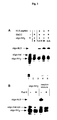
Oligonucleotides 5'-32P radiolabeling and polyacrylamide
gel electrophoresis (Fig. 3A: Polyacrylamide gel
electrophoresis (PAGE) analysis of the formation of the
oligo-peptide conjugate. Aliquots of the reaction
mixture were taken at different time intervals and
analyzed on a 20% denaturing gel after radiolabeling of
the oligonucleotides. Lane 1 : oligo-NH2; lane 2:
oligo-NH2/SMCC reaction mixture after 2 h; lanes 3, 4
and 5: reaction mixture 1.5 h, 3 h or overnight after
addition) showed the peptide conjugation reaction to be
completed after 3 h, giving 30% oligonucleotide-NLS
based on the starting oligonucleotide. Proteinase K
digestion converted oligonucleotide-NLS to a faster
migrating compound, presumably the oligonucleotide
conjugated to the C-terminal aminoacid (oligo-mal-cys),
thus establishing the chimeric nature of the conjugate
(Fig. 3B: Radiolabeled oligo-NH2 or the crude
oligonucleotide-NLS reaction mixture were digested with
proteinase K. Products were analyzed on a 20%
denaturing gel and show total conversion of
oligonucleotide-NLS into a faster migrating band,
presumably oligo-mal-cys). The oligonucleotide-NLS was
purified by electrophoresis. The uncapped CMVLuc
fragment was then simultaneously reacted with a 15-fold
excess of oligo-cap and oligonucleotide-NLS using T4
DNA ligase in previously optimized conditions. Ligation
reaction yield (80-90%) was assessed by quantitative
radioactivity counting and full capping of the CMVLuc
fragment was checked by 3'-exonuclease digestion.
CMVLuc-NLS was purified by gel permeation.
Example 3
Transfection with low amounts of DNA
-
The reporter gene construct was obtained by
chemical/enzymatic synthesis and purified by PAGE/gel
permeation, instead of being amplified in bacteria.
Only limited amounts of DNA could therefore be obtained
and the transfection setup had to be miniaturized. A
convenient 96-well microtiter plate assay developed by
Felgner et al., 1994, was chosen. Using several
optimized cationic lipid formulations, these authors
showed that transfection was best at 2-0.5 µg plasmid
and quickly fell off below 0.25 µg. This result was
confirmed with pCMVLuc and two other cell types
(Table 1, entry 4 and Fig. 4), thus putting our
ultimate goal to obtaining an effective transfection
with less than 200 ng DNA. As a preliminary test, the
relevance to cap the reporter gene at both ends was
investigated, thus avoiding free DNA ends that are
prone to exonuclease-mediated DNA degradation and to
eliciting a DNA repair response. To this end, the
uncapped, hemicapped and fully capped CMVLuc DNAs
(200 ng) were transfected into hepatocytes. (BNL CL.2
hepatocytes were transfected with 200 ng DNA/well.
Complexes were formed with 25 kDa PEI (N/P= 10) in
150mM NaCl, and in the absence of serum.)
Luciferase activities (Table 1) showed both un- and
hemi-capped molecules to give 25-fold less gene
expression than the capped one thus justifying the
choice of our chemical strategy. Thus,this is
experiment shows that free DNA ends decrease
transfection efficacy. (a: Transfection with 20 ng
plasmid.)
Example 4
-
The NLS peptide allows effective transfection with
minute quantities of DNA
-
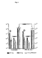
3T3 cells were transfected with decreasing amounts
of DNA, using the NLS-bearing capped gene (CMVLuc-NLS),
the capped gene (CMVLuc) and the corresponding mass-corrected
amount of plasmid DNA (pCMVLuc). While
efficacy very much decreased for `20 ng' plasmid DNA
(in fact 38 ng), as expected (see above), transfection
levels remained remarkably high and constant over the
3T3 cells were transfected with decreasing amounts of
DNA complexed in 150 mM NaCl to a cationic lipid
(Transfectam, N/P=6) or to a cationic polymer (25 kDa
PEI N/P=10) in the absence of serum.range 200-10 ng DNA
with CMVLuc-NLS (Fig. 4: 3T3 cells were transfected
with decreasing amounts of DNA complexed in 150 mM NaCl
to a cationic lipid (Transfectam, N/P=6) or to a
cationic polymer (25 kDa PEI N/P=10) in the absence of
serum.). Comparison with the capped gene lacking the
NLS peptide showed 100-1000 fold more expression when
the nuclear localization signal peptide was present on
the gene. Similar conclusions were obtained, whether
using a cationic lipid (Transfectam, Fig. 4 left) or a
cationic polymer (branched PEI, Fig. 4 right). Further
data (not shown) confirmed that other cationic carrier
molecules behaved similarly, but also that efficacy
dropped fiftyfold within the range 10-1 ng CMVLuc-NLS.
-
Improved transfection involves the cellular nuclear
import machinery. Comparative experiments between
CMVLuc and CMVLuc-NLS, although interesting, do not
really allow one put forward any hypothesis concerning
the mechanism. Nuclear import of DNA and
oligonucleotides has mainly been followed with
fluorescent tags and FISH, in microinjected or
digitonin-permeabilized cells. Proof of active import
relied on inhibition experiments using energy-decoupling
molecules, nuclear pore-binding agglutinins
or mutations in the signal peptide sequence. Gene
expression is an unambiguous proof of nuclear
localization and transfection can be regarded as a
straightforward way of introducing DNA into intact
cells. The Lysine ⇒ Threonine mutation abolishes
nuclear import (Kalderon et al., 1984). Therefore, a
capped linear DNA molecule was synthesized (CMVLuc-mNLS)
with a mutated PKTKRKVEDPYC sequence. Comparative
transfection experiments with HeLa cells are shown in
Fig. 5 (HeLa cells were transfected with various
amounts of DNA complexed to ExGen500 PEI (N/P=5 in a
5% glucose solution) in the presence of 10% serum.).
Irrespective of the amount of DNA used, the Lys ⇒ Thr
mutation brought transfection down to the level
obtained with the capped gene having no peptide at all,
confirming the functionality of the NLS/importin-α
interaction.
Example 5
NLS peptide-mediated transfection enhancement is a
general phenomenon
-
A series of comparative experiments was undertaken with
various cell types. Transfection enhancement due to the
presence of the NLS peptide was always observed
(Table 2, which shows the average enhancement of
transfection with CMVLuc-NLS.Values refer to
transfection with 10-20 ng DNA/well, in the absence of
serum. a. Estimated spread ± 30%; b. In the presence of
10% serum during transfection; c. In 24-well plates
using 200 ng DNA in the presence of 10% serum.)
-
However enhancement factors were spread widely with
cell type (10-1000-fold), with no obvious relation with
tissue origin nor cell type (primary vs. transformed
cells). Nondividing primary cells such as human
monocyte-derived macrophages and rat dorsal root
ganglia neurons showed less impressive enhancement than
3T3 and HeLa cells. However, similar values were also
seen for the easily transfected and fast dividing rat
hepatocyte-derived cell line BNL CL.2.
The general experimental setup for transfection
included 96-well microtiter plates and no serum during
2 hours following addition of the DNA/vector complexes
to the cells. Several experiments were also performed
on a larger scale (24-well plates) or in the presence
of 10% serum during transfection. Table 2 shows the
average enhancement of transfection with CMVLuc-NLS.
(Values refer to transfection with 10-20 ng DNA/well,
in the absence of serum. a: estimated spread ± 30%; b:
in the presence of 10% serum during transfection; c: in
24-well plates using 200 ng DNA in the presence of 10%
serum.). The results obtained (Table 2, b and c)
confirmed the general conclusions derived with the 96-well
setup.
Example 6
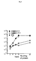
Time course of the transgene expression
-
Active transport of the reporter gene via the
nuclear import machinery could result in faster
appearing expression. More interesting, a lower
intracellular barrier to gene delivery means less
DNA/vector complexes required in the cytoplasm, hence
lower toxicity and more sustained expression. The
kinetics of gene expression was followed in detail up
to 24 h (Fig. 6: HeLa cells were transfected with 10 ng
DNA complexed to ExGen 500 PEI (N/P=5 in 5% glucose
solution) in the absence of serum. Cells were lysed and
luciferase activity was measured at the indicated time
after transfection. The absence of error bars indicates
errors that are smaller than the label on the graph.)
and then daily up to 3 days. Again, transfection with
CMVLuc-NLS was much more effective than with CMVLuc-mNLS.
Remarkably (and reproducibly), NLS-driven
transfection reached its plateau after 12 h, whereas
transfection levels obtained with the plasmid or the
mutated NLS sequence were still increasing
significantly up to 24 h. Previous experiments (Pollard
et al., 1998) showed both transfection and cytoplasmic
injection of DNA/PEI complexes to have superimposable
kinetics of transgene expression, suggesting that the
slow step was intracellular trafficking/nuclear entry
rather than cell entry. Using the nuclear import
machinery seems to speed up the rate-limiting step. For
longer time periods, expression stayed broadly constant
with a low standard deviation. However, the rather
large variability observed upon repeating this
experiment prevented any conclusion from being drawn
(the inconsistent results observed at t >1d may be due
to the fact that cells reached confluency at day 1).

| CELL TYPE | Enhancement factor a |
| BNL CL.2 | 10 |
| 3T3 | 400 b - 1000 |
| HeLa | 200 c - 300 |
| Murine DRG neurons | 30 |
| Human macrophages | 10 |
REFERENCES
-
- Adam, S. A. & Gerace, L. (1991) Cell 66, 837-847.
- Behr, J. P., Demeneix, B., Loeffler, J. P. &
Perez-Mutul, J. (1989) Proc. Natl. Acad. Sci. USA 86,
6982-6986.
- Behr, J. P., (1994), Bioconjugate Chemistry, 5, 382-389
- Blessing, T., Remy, J. S. & Behr, J. P. (1998) Proc.
Natl. Acad. Sci. USA 95, 1427-1431.
- Boussif, O., Lezoualch, F., Zanta, M. A., Mergny, M.
D., Scherman, D., Demeneix, B. & Behr, J. P. (1995)
Proc. Natl. Acad. Sci. USA 92, 7297-7301.
- Cereghini, S. & Yaniv, M. (1984) Embo J. 3, 1243-1253.
- Citovsky, V., et al., (1992), Science 256: 1802
- Collas, P., Husebye, H. & Aleström, P. (1996)
Transgenic Res. 5, 541-548.
- Cotten, M. and Wagner, E., (1993), Curr. Opin.
Biotech., 4, 705-710
- Dean, D. A. (1997) Exp. Cell Res. 230, 293-302.
- Debin, A., Malvy, C., and Svinarchuk, F. (1997) Nucleic
Acids Res. 25, 1965-1974.
- Dowty, M. E., Williams, P., Zhang, G., Hagstrom, J. E.
& Wolff, J. A. (1995) Proc. Natl. Acad. Sci. USA 92,
4572-4576.
- Felgner, J. H., Kumar, R., Sridhar, C. N., Wheeler, C.
J., Tsai, Y. J., Border, R., Ramsey, P., Martin, M. &
Felgner, P. L. (1994) J. Biol. Chem. 269, 2550-2561.
- Fominaya, J. & Wels, W. (1996) J. Biol. Chem. 271,
10560-10568.
- Fritz, J. D., Herweijer, H., Zhang, G. F. & Wolff, J.
A. (1996) Hum. Gene Ther. 7, 1395-1404.
- Garcia-Bustos, et al., (1991) Biochem. Biophys. Acta
1071: 83
- Gorlich, D. & Mattaj, I. W. (1996) Science 271,
1513-1518.
- Greber, U. F., Suomalainen, M., Stidwill, R. P.,
Boucke, K., Ebersold, M. W. & Helenius, A. (1997)
EMBO J. 16, 5998-6007.
- Greber, U. F. (1998) in Self-assembling complexes for
gene delivery : from laboratory to clinical trial,
eds. Kabanov, A. V., Felgner, P. L. & Seymour, L. W.
(John Wiley & Sons Ltd, Chichester), pp. 89-114.
- Hagstrom, J. E., Ludtke, J. J., Bassik, M. C.,
Sebestyén, M. G., Adam, S. A. & Wolff, J. A. (1997)
J. Cell Sci. 110, 2323-2331.
- Jeong, S. & Stein, A. (1994) Nucleic Acids Res. 22,
370-375.
- Kalderon, D., Roberts, B. L., Richardson, W. D. &
Smith, A. E. (1984) Cell 39, 499-509.
- Kaneda, Y., Iwai, K. & Uchida, T. (1989) Science 243,
375-378.
- Labat-Moleur, F., Steffan, A. M., Brisson, C., Perron,
H., Feugeas, O., Furstenberger, P., Oberling, F.,
Brambilla, E. & Behr, J. P. (1996) Gene Therapy 3,
1010-1017.
- Lambert, R. C., Maulet, Y., Dupont, J. L., Mykita, S.,
Craig, P., Volsen, S. & Feltz, A. (1996) Mol Cell
Neurosci 7, 239-246.
- Langle-Rouault, F., Patzel, V., Benavente, A., Taillez,
M., Silvestre, N., Bompard, A., Sczakiel, G., Jacobs,
E. & Rittner, K. (1998) J. Virol. 72, 6181-6185.
- Le Doan, T., Perrouault, L., Praseuth, D., Habhoub, N.,
Decout, J., Thuong, N. T., Lhomme, J. & Hélène, C.
(1987) Nucleic Acids Res. 15, 7749.
- Ledley, F. D., (1995), Human Gene Ther., 6, 1129-1144
- Ledley, F. D., (1996), Pharmaceut Res, 13, 1595-1614.
- Luby-Phelps, K. (1994) Curr. Opin. Cell Biol. 6, 3-9.
- Maul, G. G. & Deaven, L. (1977) J. Cell Biol. 73,
748-760.
- Melchior, F. & Gerace, L. (1998) Trends Cell. Biol. 8,
175-179.
- Mirzayans, R., Aubin, R. A. & Paterson, M. C. (1992)
Mutat. Res. 281, 115-122.
- Mislick, K. A. & Baldeschwieler, J. D. (1996) Proc.
Natl. Acad. Sci. USA 93, 12349-12354.
- Moser, H. E. & Dervan, P. B. (1987) Science 238, 645.
- Nakanishi, M., Weber, K. T., and Guntaka, R. V. (1998)
Nucleic Acids Res. 26, 5218-5222.
- Nielsen, P. E., Egholm, M., Berg, R. H. & Buchardt, O.
(1991) Science 254, 1497-1500.
- Nigg, E. A. (1997) Nature 386, 779-787.
- Ohno, M., Fornerod, M. & Mattaj, I. W. (1998) Cell 92,
327-336.
- Panté, N. & Aebi, U. (1995) J. Cell Sci. Suppl. 19,
1-11.
- Plank, C., Oberhauser, B., Mechtler, K., Koch, C. &
Wagner, E. (1994) J. Biol. Chem. 269, 12918-12924.
- Pollard, H., Remy, J. S., Loussouarn, G., Demolombe,
S., Behr, J. P. & Escande, D. (1998) J. Biol. Chem.
273, 7507-7511.
- Rando R. F., DePaolis L., Durland R.H., Jayaraman K.,
Kessler D.J., Hogan M.E., (1994), Nucleic Acids Res.
Feb 25;22(4):678-85.
- Remy, J. S., Kichler, A., Mordvinov, V., Schuber, F. &
Behr, J. P. (1995) Proc. Natl. Acad. Sci. USA 92,
1744-1748.
- Sambrook, J., Fritsch, E.F. and Maniatis, T., 1989,
Molecular Cloning, Cold Spring Harbor Laboratory
Press (2. Edition).
- Sandler, et al., (1989) J. Cell Biol. 109: 2665
- Schmid, N. & Behr, J. P. (1995) Tetrahedron Lett. 36,
1447-1450
- Sebestyén, M. G., Ludtke, J. J., Bassik, M. C., Zhang,
G., Budker, V., Lukhtanov, E. A., Hagstrom, J. E. &
Wolff, J. A. (1998) Nature Biotech. 16, 80-85.
- Seibel, P., Trappe, J., Villani, G., Klopstock, T.,
Papa, S. & Reichmann, H. (1995) Nucleic Acids Res.
23, 10-17.
- Svinarchuk, F., Bertrand, J. R., and Malvy, C. (1994)
Nucleic Acids Res. 22, 3742-3747.
- Xu, Y. H. & Szoka, F. C. (1996) Biochemistry 35,
5616-5623.
- Zabner, J., Fasbender, A. J., Moninger, T., Poellinger,
K. A. & Welsh, M. J. (1995) J. Biol. Chem. 270,
18997-19007.
- Zanta, M. A., Boussif, O., Adib, A. & Behr, J. P.
(1997) Bioconjugate Chem. 8, 839-844.
-
Annex to the description
-