EP1011723B1 - 88kda tumorigenic growth factor and antagonists - Google Patents
88kda tumorigenic growth factor and antagonists Download PDFInfo
- Publication number
- EP1011723B1 EP1011723B1 EP98926056A EP98926056A EP1011723B1 EP 1011723 B1 EP1011723 B1 EP 1011723B1 EP 98926056 A EP98926056 A EP 98926056A EP 98926056 A EP98926056 A EP 98926056A EP 1011723 B1 EP1011723 B1 EP 1011723B1
- Authority
- EP
- European Patent Office
- Prior art keywords
- cells
- expression
- antisense
- antibody
- agent
- Prior art date
- Legal status (The legal status is an assumption and is not a legal conclusion. Google has not performed a legal analysis and makes no representation as to the accuracy of the status listed.)
- Expired - Lifetime
Links
Images


Classifications
-
- C—CHEMISTRY; METALLURGY
- C07—ORGANIC CHEMISTRY
- C07K—PEPTIDES
- C07K16/00—Immunoglobulins [IGs], e.g. monoclonal or polyclonal antibodies
- C07K16/18—Immunoglobulins [IGs], e.g. monoclonal or polyclonal antibodies against material from animals or humans
- C07K16/28—Immunoglobulins [IGs], e.g. monoclonal or polyclonal antibodies against material from animals or humans against receptors, cell surface antigens or cell surface determinants
- C07K16/2896—Immunoglobulins [IGs], e.g. monoclonal or polyclonal antibodies against material from animals or humans against receptors, cell surface antigens or cell surface determinants against molecules with a "CD"-designation, not provided for elsewhere
-
- A—HUMAN NECESSITIES
- A61—MEDICAL OR VETERINARY SCIENCE; HYGIENE
- A61P—SPECIFIC THERAPEUTIC ACTIVITY OF CHEMICAL COMPOUNDS OR MEDICINAL PREPARATIONS
- A61P35/00—Antineoplastic agents
-
- C—CHEMISTRY; METALLURGY
- C07—ORGANIC CHEMISTRY
- C07K—PEPTIDES
- C07K14/00—Peptides having more than 20 amino acids; Gastrins; Somatostatins; Melanotropins; Derivatives thereof
- C07K14/435—Peptides having more than 20 amino acids; Gastrins; Somatostatins; Melanotropins; Derivatives thereof from animals; from humans
- C07K14/475—Growth factors; Growth regulators
-
- C—CHEMISTRY; METALLURGY
- C07—ORGANIC CHEMISTRY
- C07K—PEPTIDES
- C07K16/00—Immunoglobulins [IGs], e.g. monoclonal or polyclonal antibodies
- C07K16/18—Immunoglobulins [IGs], e.g. monoclonal or polyclonal antibodies against material from animals or humans
- C07K16/22—Immunoglobulins [IGs], e.g. monoclonal or polyclonal antibodies against material from animals or humans against growth factors ; against growth regulators
-
- A—HUMAN NECESSITIES
- A61—MEDICAL OR VETERINARY SCIENCE; HYGIENE
- A61K—PREPARATIONS FOR MEDICAL, DENTAL OR TOILETRY PURPOSES
- A61K39/00—Medicinal preparations containing antigens or antibodies
- A61K2039/505—Medicinal preparations containing antigens or antibodies comprising antibodies
-
- A—HUMAN NECESSITIES
- A61—MEDICAL OR VETERINARY SCIENCE; HYGIENE
- A61K—PREPARATIONS FOR MEDICAL, DENTAL OR TOILETRY PURPOSES
- A61K38/00—Medicinal preparations containing peptides
-
- C—CHEMISTRY; METALLURGY
- C07—ORGANIC CHEMISTRY
- C07K—PEPTIDES
- C07K2317/00—Immunoglobulins specific features
- C07K2317/30—Immunoglobulins specific features characterized by aspects of specificity or valency
- C07K2317/34—Identification of a linear epitope shorter than 20 amino acid residues or of a conformational epitope defined by amino acid residues
-
- C—CHEMISTRY; METALLURGY
- C07—ORGANIC CHEMISTRY
- C07K—PEPTIDES
- C07K2317/00—Immunoglobulins specific features
- C07K2317/70—Immunoglobulins specific features characterized by effect upon binding to a cell or to an antigen
- C07K2317/73—Inducing cell death, e.g. apoptosis, necrosis or inhibition of cell proliferation
-
- C—CHEMISTRY; METALLURGY
- C12—BIOCHEMISTRY; BEER; SPIRITS; WINE; VINEGAR; MICROBIOLOGY; ENZYMOLOGY; MUTATION OR GENETIC ENGINEERING
- C12N—MICROORGANISMS OR ENZYMES; COMPOSITIONS THEREOF; PROPAGATING, PRESERVING, OR MAINTAINING MICROORGANISMS; MUTATION OR GENETIC ENGINEERING; CULTURE MEDIA
- C12N2799/00—Uses of viruses
- C12N2799/02—Uses of viruses as vector
- C12N2799/021—Uses of viruses as vector for the expression of a heterologous nucleic acid
- C12N2799/026—Uses of viruses as vector for the expression of a heterologous nucleic acid where the vector is derived from a baculovirus
Definitions
- This invention relates to cell biology, physiology and medicine, and concerns an 88kDa glycoprotein growth factor ("GP88") and compositions and methods which affect the expression and biological activity of GP88. These compositions and methods are useful for diagnosis and treatment of diseases including cancer.
- GP88 glycoprotein growth factor
- the proliferation and differentiation of cells in multicellular organisms is subject to a highly regulated process (1).
- a distinguishing feature of cancer cells is the absence of control over this process; proliferation and differentiation become deregulated resulting in uncontrolled growth.
- Significant research efforts have been directed toward better understanding this difference between normal and tumor cells.
- One area of research focus is growth factors and, more specifically, autocrine growth stimulation.
- Growth factors are polypeptides which carry messages to cells concerning growth, differentiation, migration and gene expression (2). Typically, growth factors are produced in one cell and act on another cell to stimulate proliferation. However, certain malignant cells, in culture, demonstrate a greater or absolute reliance on an autocrine growth mechanism (3). Malignant cells which observe this autocrine behavior circumvent the regulation of growth factor production by other cells and are therefore unregulated in their growth.
- IGF1 insulin-like growth factors
- IGF2 gastrin-releasing peptide
- GFP gastrin-releasing peptide
- TGF-a transforming growth factors alpha and beta
- EGF epidermal growth factor
- the present invention is directed to a recently discovered growth factor.
- This growth factor was first discovered in the culture medium of highly tumorigenic "PC cells," an insulin-independent variant isolated from the teratoma derived adipogenic cell line 1246. This growth factor is referred to herein as "GP88.”
- GP88 has been purified and structurally characterized (4). Amino acid sequencing of GP88 indicates that GP88 has amino acid sequence similarities with the mouse granulin/epithelin precursor.
- Granulins/epithelins are 6kDa polypeptides and belong to a novel family of double cysteine rich polypeptides (5, 6).
- U.S. Patent No. 5,416,192 (Shoyab et al.) is directed to 6 kDa epithelins, particularly epithelin 1 and epithelin 2. According to Shoyab, both epithelins are encoded by a common 63.5 kDa precursor, which is processed into smaller forms as soon as it is synthesized, so that the only natural products found in biological samples are the 6 kDa forms. Shoyab et al. teaches that the epithelin precursor is biologically inactive.
- the inventor's laboratory has demonstrated that the precursor is not always processed as soon as it is synthesized.
- the precursor i.e., GP88
- the precursor is in fact secreted as an 88kDa glycoprotein with an N-linked carbohydrate moiety of 20kDa (4).
- Analysis of the N-terminal sequence of GP88 indicates that GP88 starts at amino acid 17 of the grn/epi precursor, demonstrating that the first 17 amino acids from the protein sequence deduced from the precursor cDNA correspond to a signal peptide compatible with targeting for membrane localization or for secretion (4).
- GP88 is biologically active and has growth promoting activity, particularly as an autocrine growth factor for the producer cells.
- GP88 glycoprotein
- a further object of the invention is to provide methods for treating diseases associated with a defect in GP88 quantity or activity such as but not limited to cancer in a mammal.
- Another object of the invention is to provide methods for determining the susceptibility of a subject to diseases associated with a defect in GP88 expression or action.
- Yet another object of the invention is to provide methods for measuring susceptibility to GP88 antagonizing therapy.
- Yet another object of the invention is to provide methods, reagents, and kits for the in vitro and in vivo detection of GP88 and tumorigenic activity in cells.
- the present invention provides compositions for diagnosis and treatment of diseases such as but not limited to cancer in which cells exhibit an altered expression of GP88 or altered response to GP88.
- altered expression herein means increased expression or overexpression of GP88 by a factor of at least two-fold, and at times by a factor of 10 or more, based on the level of mRNA or protein as compared to corresponding normal cells or surrounding peripheral cells.
- altered expression also means expression which became unregulated or constitutive without being necessarily elevated.
- increased or altered "response" to GP88 means a condition wherein increase in any of the biological functions (e.g., growth, differentiation, viral infectivity) conferred by GP88 results in the same or equivalent condition as altered expression of GP88.
- GP88 means epithelin/granulin precursor in cell extracts and extracellular fluids, and is intended to include not only GP88 according to the amino acid sequences included in figures 8 or 9, which are of mouse and human origins, but also GP88 of other species.
- the term also includes functional derivatives thereof having additional components such as a carbohydrate moiety including a glycoprotein or other modified structures.
- GP88 is any polypeptide fragment having at least 10 amino-acids present in the above mentioned sequences. Sequences of this length are useful as antigens and for making immunogenic conjugates with carriers for the production of antibodies specific for various epitopes of the entire protein. Such polypeptides are useful in screening such antibodies and in the methods directed to detection of GP88 in biological fluids. It is well known in the art that peptides are useful in generation of antibodies to larger proteins (7). In one embodiment of this invention, it is shown that peptides from 12-19 amino-acids in length have been successfully used to develop antibodies that recognize the full length GP88.
- polypeptide of this invention may exist covalently or non-covalently bound to another molecule.
- it may be fused to one or more other polypeptides via one or more peptide bonds such as glutathione transferase, poly-histidine, or myc tag.
- the polypeptide is sufficiently large to comprise an antigenetically distinct determinant or epitope which can be used as an immunogen to reproduce or test antibodies against GP88 or a functional derivative thereof.
- One embodiment includes the polypeptide substantially free of other mammalian peptides.
- GP88 of the present invention can be biochemically or immunochemically purified from cells, tissues or a biological fluid.
- the polypeptide can be produced by recombinant means in a prokaryotic or eukaryotic expression system and host cells.
- Substantially free of other mammalian polypeptides reflects the fact that the polypeptide can be synthesized in a prokaryotic or a non-mammalian or mammalian eukaryotic organism, if desired.
- methods are well known for the synthesis of polypeptides of desired sequences by chemical synthesis on solid phase supports and their subsequent separation from the support.
- the protein can be purified from tissues or fluids of mammals where it naturally occurs so that it is at least 90% pure (on a weight basis) or even 99% pure, if desired, of other mammalian polypeptides, and is therefore substantially free from them.
- tissue extracts or fluids can be subjected to standard protein purification such as on immunoabsorbants bearing antibodies reactive against the protein.
- standard protein purification such as on immunoabsorbants bearing antibodies reactive against the protein.
- One embodiment of the present invention describes purification methods for the purification of naturally occurring GP88 and of recombinant GP88 expressed in baculovirus infected insect cells.
- purification from such tissues or fluids can be achieved by a combination of standard methods such as but not limited to the ones described in reference (4).
- GP88 is intended to also include functional derivatives.
- functional derivative is meant a “fragment,” “variant,” “analog,” or “chemical derivative” of the protein or glycoprotein as defined below.
- a functional derivative retains at least a portion of the function of the full length GP88 which permits its utility in accordance with the present invention.
- a “fragment” of GP88 refers to any subset of the molecule that is a shorter peptide. This corresponds for example but is not limited to regions such as K19T and S14R for mouse GP88, and E19V and A14R (equivalent to murine K19T and S14R, respectively) for human GP88.
- a "variant" of GP88 refers to a molecule substantially similar to either the entire peptide or a fragment thereof. Variant peptides may be prepared by direct chemical synthesis of the variant peptide using methods known in the art.
- amino acid sequence variants of the peptide can be prepared by modifying the DNA which encodes the synthesized protein or peptide.
- Such variants include, for example, deletions, insertions, or substitutions of residues within the amino-acid sequence of GP88. Any combination of deletion, insertion, and substitution may also be made to arrive at the final construct, provided the final construct possesses the desired activity.
- the mutation that will be made in the DNA encoding the variant peptide must not alter the reading frame and preferably will not create complementary regions that could produce secondary mRNA structures.
- these variants are prepared by site directed mutagenesis (8) of nucleotides in the DNA encoding the peptide molecule thereby producing DNA encoding the variant, and thereafter expressing the DNA in recombinant cell culture.
- the variant typically exhibits the same qualitative biological activity as the nonvariant peptide.
- An “analog” of GP88 protein refers to a non-natural molecule substantially similar to either the entire molecule or a fragment thereof.
- a “chemical derivative” contains additional chemical moieties not normally a part of the peptide or protein.
- Covalent modifications of the peptide are also included within the scope of this invention. Such modifications may be introduced into the molecule by reacting targeted amino-acid residues of the peptide with an organic derivatizing agent that is capable of reacting with selected side chains or terminal amino-acid residues. Most commonly derivatized residues are cysteinyl, histidyl, lysinyl, arginyl, tyrosyl, glutaminyl, asparaginyl and amino terminal residues.
- cross-linking agents include glutaraldehyde, N-hydroxysuccinimide esters, homobifunctional imidoesters, 1,1-bis(-diazoloacetyl)-2-phenylethane, and bifunctional maleimides.
- Derivatizing agents such as methyl-3-[9p-azidophenyl)]dithiopropioimidate yield photoactivatable intermediates that are capable of forming crosslinks in the presence of light.
- reactive water-insoluble matrices such as cyanogen bromide activated carbohydrates and the reactive substrates described in U.S. Patents 3,969,287 and 3,691,016 may be employed for protein immobilization.
- GP88 antigenizing agents
- the GP88 antagonizing agents are antisense oligonucleotides to GP88.
- the antisense oligonucleotides preferably inhibit GP88 expression by inhibiting translation of the GP88 protein.
- such a composition may comprise reagents, factors or hormones that inhibit GP88 expression by regulating GP88 gene transcriptional activity.
- a composition may comprise reagents, factors or hormones that inhibit GP88 post-translational modification and its secretion.
- Such a composition may comprise reagents that act as GP88 antagonists that block GP88 activity by competing with GP88 for binding to GP88 cell surface receptors.
- such a composition may comprise factors or reagents that inhibit the signaling pathway transduced by GP88 once binding to its receptors on diseased cells.
- the composition may comprise reagents that block GP88 action such as an antibody specific to GP88 that neutralizes its biological activity, or an antibody to the GP88 receptor that blocks its activity.
- the antibodies of the invention are preferably used as a treatment for cancer or other diseases in cells which exhibit an increased expression of GP88.
- neutralizing it shall be understood that the antibody has the ability to inhibit or block the normal biological activity of GP88, including GP88's ability to stimulate cell proliferation or to induce tumor growth in experimental animals and in humans.
- An effective amount of anti-GP88 antibody is administered to an animal, including humans, by various routes.
- the anti-GP88 antibody is used as a diagnostic to detect cells which exhibit an altered (increased) expression of GP88 as occurring in diseases such as but not limited to cancers, and to identify diseased cells whose growth is dependent on GP88 and which will respond to GP88 antagonizing therapy.
- the anti-GP88 antibody is used to deliver compounds such as cytotoxic factors or antisense oligonucleotides to cells expressing or responsive to GP88.
- the antisense oligonucleotides of the invention are also used as a treatment for cancer in cells which exhibit an increased expression of GP88.
- An effective amount of the antisense oligonucleotide is administered to an animal, including humans, by various routes.
- the present invention also provides a method for determining the susceptibility to diseases associated with a defect in GP88 expression or action which comprises obtaining a sample of biological fluid or tissue and measuring the amount of GP88 in the fluid or tissue or measuring the susceptibility of the cells to respond to GP88.
- the amount of GP88 being proportional to the susceptibility to the cancer.
- the present invention also provides a method for measuring the degree of severity of cancer which comprises obtaining a sample of biological fluid or tissue and measuring the amount of GP88 in the fluid or tissue sample, the amount of GP88 being proportional to the degree or severity of the cancer.
- the present invention also provides a method for measuring susceptibility to GP88 antagonizing therapy which comprises obtaining a sample of the diseased tissue (biopsy), maintaining the cells derived from the sample in culture, treating the cells derived from the culture with anti-GP88 neutralizing antibody and determining if the neutralizing antibody inhibits the cell growth.
- a sample of the diseased tissue biopsy
- anti-GP88 neutralizing antibody anti-GP88 neutralizing antibody
- ability of the antibody to inhibit cell growth is indicative that the cells are dependent on GP88 to proliferate and is predictive that GP88 antagonizing therapy will be efficacious.
- the present invention also provides a method for determining the susceptibility to cancer associated with an abnormality in GP88 receptor level or activity which comprises obtaining a sample of tissue and measuring the amount of GP88 receptor protein or mRNA in the tissue or measuring the tyrosine kinase activity of the receptor in the tissue (GP88 binding to its receptor induces tyrosine phosphorylation of cellular proteins including the receptor for G88).
- the present invention also provides a method for targeting GP88 antagonizing reagents to the diseased site by conjugating them to an anti-GP88 antibody or an anti-GP88 receptor antibody.
- the invention relates to GP88 and antitumor and antiviral compositions useful for treating and diagnosing diseases linked to altered (increased) expression of GP88.
- this invention is used for treating and diagnosing diseases linked to increased responsiveness to GP88.
- the parent cell line, 1246 is a C3H mouse adipogenic cell line which proliferates and differentiates into adipocytes in a defined medium under stringent regulation by insulin (9, 10).
- the 1246 cells cannot form tumors in a syngeneic animal (C3H mouse) even when injected at a high cell density.
- An insulin independent cell line, 1246-3A was isolated from 1246 cells maintained in insulin-free medium (11). The 1246-3A cells lost the ability to differentiate and form tumors when 10 6 are injected subcutaneously in syngeneic mice.
- a highly tumorigenic cell line, PC was developed from 1246-3A cells by an in vitro-in vivo shuttle technique. The PC cells formed tumors when 10 4 cells were injected into syngeneic mice (12).
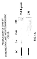
- GP88 is overexpressed in the insulin-independent tumorigenic cell lines relative to the parent non-tumorigenic insulin-dependent cell line. Moreover, the degree of overexpression of GP88 positively correlates with the degree of tumorigenicity of these cells, demonstrating for the first time that GP88 is important in tumorigenesis (FIG. 1). With reference to Figure 1, since GP88 is synthesized by cells but also secreted in culture medium, the level of GP88 was determined in cell lysates and in culture medium (CM). All cells were cultivated in DME/F12 nutrient medium supplemented with 2% fetal bovine serum.
- CM culture medium
- cell lysates were prepared by incubation in buffer containing detergent followed by a 10,000 x g centrifugation. Cell lysate and conditioned medium were normalized by cell number. Samples from cell lysate and conditioned medium were analyzed by Western blot analysis using an anti-GP88 antibody, as explained below.
- FIG. 2 The development of a neutralizing antibody confirmed GP88's key role in tumorigenesis.
- an anti-GP88 antibody directed to the K19T region of mouse GP88 was added to the culture medium, the growth of highly tumorigenic PC cells was inhibited in a dose dependent fashion (FIG. 2).
- PC cells were cultivated in 96 well plates at a density 2 x 10 4 cells/well in DME/F12 medium supplemented with human fibronectin (2 ⁇ g/ml) and human transferrin (10 ⁇ g/ml). Increasing concentrations of anti-GP88 IgG fraction were added to the wells after the cells were attached. Control cells were treated with equivalent concentrations of non-immune IgG. Two days later, 0.25 mCi of 3 H-thymidine was added per well for 6 hrs. Cells were then harvested to count 3 H-thymidine incorporated into DNA as a measure for cell proliferation.
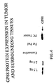
- FIG. 4 Comparison of the expression of GP88 indicates that in vivo GP88 levels in tumors is dramatically higher than in normal tissues (FIG. 4).
- C3H mice were injected with 10 6 PC cells. Tumor bearing mice were euthanized. Tumors, fat pads and connective tissue were collected. Cell lysates were prepared by incubation in buffer containing detergent as described above for FIG. 1. Protein concentration of tissue extracts was determined, and equivalent amounts of proteins for each sample were analyzed by SDS-PAGE followed by Western blot analysis using anti-GP88 antibody to measure the content of GP88 in tissue extracts. The results showed that the level of GP88 in tumor extracts is at least 10-fold higher than in surrounding connective and fat tissues.
- GP88 In normal cells (1246 cells, fibroblasts), the expression of GP88 is regulated, in particular by insulin, and inhibited by fetal bovine serum. In tumorigenic cells, a loss of regulation of normal growth leads to the increased expression of GP88 and the acquisition of GP88 dependence for growth. Therefore, inhibition of GP88 expression and/or action is an effective approach to suppression of tumorigenesis. Detection of an elevated GP88 expression in biopsies provides diagnostic analysis of tumors that are responsive to GP88 inhibition therapy.
- GP88 is also a tumor inducing factor in human cancers. As seen in the 1246-3A cell line, a loss of responsiveness to insulin (or to IGF-I) and a concurrent increase in malignancy has been well documented (13, 14) in several human cancers including but not limited to breast cancers. Specifically, breast carcinoma is accompanied by the acquisition of an insulin/IGF-I autocrine loop, which is also the starting point of the development of tumorigenic properties in the mouse model system discussed above. Furthermore, GP88 expression is elevated in human breast carcinomas. More specifically, with reference to FIG. 5, human GP88 was highly expressed in estrogen receptor positive and also in estrogen receptor negative insulin/IGF-I independent highly malignant cells.
- GP88 is a potent growth factor for mammary epithelial cells (FIG. 6).
- the data in FIG. 5 was obtained by cultivating MCF7, MDA-MB-453 and MDA-MB-468 cells in DME/F12 medium supplemented with 10% fetal bovine serum (FBS).
- FBS fetal bovine serum
- RNA was extracted from each cell line by the RNAzol method and poly-A + RNA prepared.
- GP88 mRNA expression was examined by Northern blot analysis with 3 ⁇ g of poly-A + RNA for each cell line using a 32 P-labeled GP88 cDNA probe.
- RNA can be extracted by a variety of methods (Sambrook , Molecular Biology manual: 35) well known to people of ordinary skill in the art. The method of choice was to extract RNA using RNAzol (Cinnabiotech) or Trizol (Gibco-BRL) solutions which consists of a single step extraction by guanidinium isothiocyanate and phenol -chloroform.
- C57MG cells were cultivated in the presence of increasing concentrations of GP88 purified from PC cells conditioned medium (top panel), and recombinant GP88 expressed in insect cells (bottom panel), to demonstrate the growth stimulating effect of increasing concentrations of GP88 on the growth of the mouse mammary epithelial cell line C57MG.
- GP88 expression is also elevated in human tumors when compared to non-tumorigenic human fibroblasts and other human cell lines. GP88 promotes the growth of mammary carcinoma cells.
- compositions for treating and diagnosing diseases linked to increased expression of GP88 This also will apply to treatment and diagnosis of diseases linked to increased responsiveness to GP88.
- compositions of this invention include anti-GP88 antibodies which neutralize the biological activity of GP88.
- the present invention is also directed to an antibody specific for an epitope of GP88 and the use of such antibody to detect the presence or measure the quantity or concentration of GP88 molecule, a functional derivative thereof or a homologue from different animal species in a cell, a cell or tissue extract, culture medium or biological fluid. Moreover, antibody can be used to target cytotoxic molecules to a specific site.
- the GP88 protein naturally produced or expressed in recombinant form or functional derivative thereof, preferably having at least 9 amino-acids, is obtained and used to immunize an animal for production of polyclonal or monoclonal antibody.
- An antibody is said to be capable of binding a molecule if it is capable of reacting with the molecule to thereby bind the molecule to the antibody.
- the specific reaction is meant to indicate that the antigen will react in a highly selective manner with its corresponding antibody and not with the multitude of other antibodies which may be evoked by other antigens.
- antibody herein includes but is not limited to human and non-human polyclonal antibodies, human and non-human monoclonal antibodies (mAbs), chimeric antibodies, anti-idiotypic antibodies (anti-IdAb) and humanized antibodies.
- Polyclonal antibodies are heterogeneous populations of antibody molecules derived either from sera of animals immunized with an antigen or from chicken eggs.
- Monoclonal antibodies (“mAbs") are substantially homogeneous populations of antibodies to specific antigens. mAbs may be obtained by methods known to those skilled in the art (18, 19, 20 and U.S. Patent No. 4,376,110). Such antibodies may be of any immunological class including IgG, IgM, IgE, IgA, IgD and any subclass thereof.
- the hybridoma producing human and non-human antibodies to GP88 may be cultivated in vitro or in vivo .
- in vivo is the presently preferred method of production. Briefly, cells from the individual hybridomas are injected intraperitoneally into pristane primed Balb/c mice or Nude mice to produce ascites fluid containing high concentrations of the desired mAbs.
- mAbs may be purified from such ascites fluids or from culture supernatants using standard chromatography methods well known to those of skill in the art.
- Human monoclonal Ab to human GP88 can be prepared by immunizing transgenic mice expressing human immunoglobulin genes. Hybridoma produced by using lymphocytes from these transgenic animals will produce human immunoglobulin instead of mouse immunoglobulin.
- a humanized antibody contains the amino-acid sequences for the 6 complementarity-determining regions (CDRs) of the parent murine mAb which are grafted onto a human antibody framework.
- CDRs complementarity-determining regions
- Hybridoma supernatants and sera are screened for the presence of antibody specific for GP88 by any number of immunoassays including dot blots and standard immunoassays (EIA or ELISA) which are well known in the art.
- EIA or ELISA standard immunoassays
- Western blotting to identify the size of the antigen to which the antibody binds.
- One of ordinary skill in the art will know how to prepare and screen such hybridomas without undue experimentation in order to obtain a desired polyclonal or mAb.
- Chimeric antibodies have different portions derived from different animal species.
- a chimeric antibody might have a variable region from a murine mAb and a human immunoglobulin constant region.
- Chimeric antibodies and methods for their production are also known to those skilled in the art (21-24).
- an anti-idiotypic is an antibody which recognizes unique determinants generally associated with the antigen-binding site of an antibody.
- An anti-IdAb can be prepared by immunizing an animal of the same species and genetic type (e.g., mouse strain) as the source of the mAb with the mAb to which an anti-IdAb is being prepared. The immunized animal will recognize and respond to the idiotypic determinants of the immunizing antibody by producing antibody to these idiotypic determinants (the anti-IdAb).
- the anti-IdAb may also be used as an immunogen to produce an immune response in yet another animal, producing a so-called anti-anti-IdAb.
- the anti-anti-IdAb may be epitopically identical to the original mAb which induced the anti-IdAb.
- the anti-anti-IdAb may be epitopically identical to the original mAb which induced the anti-IdAb.
- mAbs generated against GP88 may be used to induce human and non-human anti-IdAbs in suitable animals.
- Spleen cells from such immunized mice are used to produce hybridomas secreting human or non-human anti-Id mAbs.
- the anti-Id mAbs can be coupled to a carrier such as Keyhole Limpet Hemocyanin (KLH) or bovine serum albumin (B S A) and used to immunize additional mice.
- Sera from these mice will contain human or non-human anti-anti-IdAb that have the binding properties of the original mAb specific for a GP88 polypeptide epitope.
- the anti-Id mAbs thus have their own idiotypic epitopes or idiotypes structurally similar to the epitope being evaluated.
- antibody is also meant to include both intact molecules as well as fragments thereof such as, for example, Fab and F(ab')2, which are capable of binding to the antigen.
- Fab and F(ab')2 fragments lack the Fc fragment of intact antibody, clear more rapidly from the circulation and may have less non-specific tissue binding than an intact antibody (28).
- Such fragments are typically produced by proteolytic cleavage, using enzymes such as papain (to generate Fab fragments) and pepsin (to generate F(ab')2 fragments).
- Fab and F(ab')2 and other fragments of the antibodies useful in the present invention may be used for the detection or quantitation of GP88, and for treatment of pathological states related to GP88 expression, according to the methods disclosed herein for intact antibody molecules.
- antibodies that neutralize GP88 activity in vitro can be used to neutralize GP88 activity in vivo to treat diseases associated with increased GP88 expression or increased responsiveness to GP88, such as but not limited to cancer and viral infection.
- a subject preferably a human subject, suffering from disease associated with increased GP88 expression is treated with an antibody to GP88. Such treatment may be performed in conjunction with other anti-cancer or anti-viral therapy.
- a typical regimen comprises administration of an effective amount of the antibody specific for GP88 administered over a period of one or several weeks and including between about one and six months.
- the antibody of the present invention may be administered by any means that achieves its intended purpose.
- administration may be by various routes including but not limited to subcutaneous, intravenous, intradermal, intramuscular, intraperitoneal and oral.
- Parenteral administration can be by bolus injection or by gradual perfusion over time.
- Preparations for parenteral administration include sterile aqueous or non-aqueous solutions, suspensions and emulsions, which may contain auxiliary agents or excipients known in the art.
- Pharmaceutical compositions such as tablets and capsules can also be prepared according to routine methods. It is understood that the dosage of will be dependent upon the age, sex and weight of the recipient, kind of concurrent treatment, if any, frequency of treatment and the nature of the effect desired.
- the ranges of effective doses provided below are not intended to limit the invention and merely represent preferred dose ranges.
- the total dose required for each treatment may be administered by multiple doses or in a single dose.
- Effective amounts of antibody are from about 0.01 ⁇ g to about 100 mg/kg body weight and preferably from about 10 ⁇ g to about 50 mg/kg.
- Antibody may be administered alone or in conjunction with other therapeutics directed to the same disease.
- GP88 neutralizing antibodies can be used in all therapeutic cases where it is necessary to inhibit GP88 biological activity, even though there may not necessarily be a change in GP88 expression, including cases where there is an overexpression of GP88 cell surface receptors and this in turn results in an increased biological activity, or where there is an alteration in GP88 signaling pathways or receptors leading to the fact that the signaling pathways are always "turned on.”
- Neutralizing antibodies to growth factor and to growth factor receptors have been successfully used to inhibit the growth of cells whose proliferation is dependent on this growth factor. This has been the case for IGF-I receptor in human breast carcinoma cells (14) and bombesin for lung cancer (29).
- the antibody to GP88 can also be used to deliver compounds such as, but not limited to, cytotoxic reagents such as toxins, oncotoxins, mitotoxins and immunotoxins, or antisense oligonucleotides, in order to specifically target them to cells expressing or responsive to GP88 (30).
- cytotoxic reagents such as toxins, oncotoxins, mitotoxins and immunotoxins, or antisense oligonucleotides
- One region that allows antigen to develop a neutralizing antibody to GP88 is the 19 amino-acid region defined as K19T in the mouse GP88, and E19V in the human GP88 which is not located within the epithelin/granulin 6 kDa repeats but between these repeats, specifically between granulin A (epithelin 1) and granulin C (5) in what is considered a variant region (see Figure 10).
- the region important for the biological activity of GP88 lies outside of the epithelin repeats.
- the antibodies or fragments of antibodies useful in the present invention may also be used to quantitatively or qualitatively detect the presence of cells which express the GP88 protein. This can be accomplished by immunofluorescence techniques employing a fluorescently labeled antibody (see below) with fluorescent microscopic, flow cytometric, or fluorometric detection. The reaction of antibodies and polypeptides of the present invention may be detected by immunoassay methods well known in the art (20).
- the antibodies of the present invention may be employed histologically as in light microscopy, immunofluorescence or immunoelectron microscopy, for in situ detection of the GP88 protein in tissues samples or biopsies.
- In situ detection may be accomplished by removing a histological specimen from a patient and applying the appropriately labeled antibody of the present invention.
- the antibody (or fragment) is preferably provided by applying or overlaying the labeled antibody (or fragment) to the biological sample.
- Assays for GP88 typically comprise incubating a biological sample such as a biological fluid, a tissue extract, freshly harvested or cultured cells or their culture medium in the presence of a detectably labeled antibody capable of identifying the GP88 protein and detecting the antibody by any of a number of techniques well known in the art.
- a biological sample such as a biological fluid, a tissue extract, freshly harvested or cultured cells or their culture medium
- a detectably labeled antibody capable of identifying the GP88 protein and detecting the antibody by any of a number of techniques well known in the art.
- the biological sample may be treated with a solid phase support or carrier such as nitrocellulose or other solid support capable of immobilizing cells or cell particles or soluble proteins.
- a solid phase support or carrier such as nitrocellulose or other solid support capable of immobilizing cells or cell particles or soluble proteins.
- the support may then be washed followed by treatment with the detectably labeled anti-GP88 antibody. This is followed by wash of the support to remove unbound antibody.
- the amount of bound label on said support may then be detected by conventional means.
- solid phase support is intended any support capable of binding antigen or antibodies such as but not limited to glass, polystyrene polypropylene, nylon, modified cellulose, or polyacrylamide.
- binding activity of a given lot of antibody to the GP88 protein may be determined according to well known methods. Those skilled in the art will be able to determine operative and optimal assay conditions for each determination by employing routine experimentation.
- Detection of the GP88 protein or functional derivative thereof and of a specific antibody for the protein may be accomplished by a variety of immunoassays well known in the art such as enzyme linked immunoassays (EIA) or radioimmunoassays (RIA).
- EIA enzyme linked immunoassays
- RIA radioimmunoassays
- Such immunoassays are useful to detect and quantitate GP88 protein in serum or other biological fluid as well as in tissues, cells, cell extracts, or biopsies.
- the concentration of GP88 is measured in a tissue specimen as a means for diagnosing cancer or other disease associated with increased expression of GP88.
- proportional to an increase in the level of the GP88 protein.
- proportional as used herein is not intended to be limited to a linear or constant relationship between the level of protein and the malignant properties of the cancer.
- proportional as used herein, is intended to indicate that an increased level of GP88 protein is related to appearance, recurrence or display of malignant properties of a cancer or other disease associated with increased expression of GP88 at ranges of concentration of the protein that can be readily determined by one skilled in the art.
- Another embodiment of the invention relates to evaluating the efficacy of anti-cancer or anti-viral drug or agent by measuring the ability of the drug or agent to inhibit the expression or production of GP88.
- the antibodies of the present invention are useful in a method for evaluating anti-cancer or anti-viral drugs in that they can be employed to determine the amount of the GP88 protein in one of the above-mentioned immunoassays. Alternatively, the amount of the GP88 protein produced is measured by bioassay (cell proliferation assay) as described herein. The bioassay and immunoassay can be used in combination for a more precise assessment.
- An additional embodiment is directed to an assay for diagnosing cancers or other diseases associated with an increase in GP88 expression based on measuring in a tissue or biological fluid the amount of mRNA sequences present that encode GP88 or a functional derivative thereof, preferably using an RNA-DNA hybridization assay.
- the presence of certain cancers and the degree of malignancy is proportional to the amount of such mRNA present.
- the source of mRNA will be biopsies and surrounding tissues.
- the preferred technique for measuring the amount of mRNA is a hybridization assay using DNA of complementarity base sequence.
- Another related embodiment is directed to an assay for diagnosing cancers or other diseases associated with an increase in GP88 responsiveness based on measuring on a tissue biopsy whether treatment with anti-GP88 neutralizing antibody will inhibit its growth or other biological activity.
- Another related embodiment is a method for measuring the efficacy of anti-cancer or anti-viral drug or agent which comprises the steps of measuring the agent's effect on inhibiting the expression of mRNA for GP88. Similarly such method can be used to identify or evaluate the efficacy of GP88 antagonizing agents by measuring the ability of said agent to inhibit the production of GP88 mRNA.
- Nucleic acid detection assays can be based on any characteristic of the nucleic acid molecule such as its size, sequence, or susceptibility to digestion by restriction endonucleases. The sensitivity of such assays can be increased by altering the manner in which detection is reported or signaled to the observer.
- labels have been extensively developed and used by those of ordinary skill in the art, including enzymatic, radioisotopic, fluorescent, chemical labels and modified bases.
- PCR polymerase chain reaction
- This invention also provides GP88 antisense components.
- the constitutive expression of antisense RNA in cells has been shown to inhibit the expression of more than 20 genes and the list continues to grow (32-34).
- Possible mechanisms for antisense effects are the blockage of translation or prevention of splicing, both of which have been observed in vitro . Interference with splicing allows the use of intron sequences (30) which should be less conserved and therefore result in greater specificity, inhibiting expression of a gene product of one species but not its homologue in another species.
- antisense component corresponds to an RNA sequence as well as a DNA sequence coding therefor, which is sufficiently complementary to a particular mRNA molecule, for which the antisense RNA is specific, to cause molecular hybridization between the antisense RNA and the mRNA such that translation of the mRNA is inhibited. Such hybridization can occur under in vivo conditions.
- the action of the antisense RNA results in specific inhibition of gene expression in the cells (32-35).
- transfection of tumorigenic cells with DNA antisense to the GP88 cDNA inhibits endogenous GP88 expression and inhibits tumorigenicity of the antisense cDNA transfected cells.
- This antisense DNA must have sufficient complementarity, about 18-30 nucleotides in length, to the GP88 gene so that the antisense RNA can hybridize to the GP88 gene (or mRNA) and inhibit GP88 gene expression regardless of whether the action is at the level of splicing, transcription, or translation.
- the degree of inhibition is readily discernible to one skilled in the art without undue experimentation given the teachings herein and preferably is sufficient to inhibit the growth of cells whose proliferation is dependent on the expression of GP88.
- the antisense RNA approach is but a number of known mechanisms which can be employed to block specific gene expression.
- the antisense components of the present invention may be hybridizable to any of several portions of the target GP88 cDNA, including the coding sequence, 3' or 5' untranslated regions, or other intronic sequences, or to GP88 mRNA.
- the minimal amount of homology required by the present invention is that sufficient to result in hybridization to the GP88 DNA or mRNA and in inhibition of transcription of the DNA, or translation or function of the mRNA, preferably without affecting the function of other mRNA molecules and the expression of other unrelated genes.
- Antisense RNA is delivered to a cell by transformation or transfection via a vector, including retroviral vectors and plasmids, into which has been placed DNA encoding the antisense RNA with the appropriate regulatory sequences including a promoter to result in expression of the antisense RNA in a host cell. Stable transfection of various antisense expression vectors containing GP88 cDNA fragments in the antisense orientation have been performed. One can also deliver antisense components to cells using a retroviral vector. Delivery can also be achieved by liposomes.
- antisense oligonucleotides 32, 36
- Sequences for the antisense oligonucleotides to GP88 are preferably selected as being the ones that have the most potent antisense effects (37, 38).
- Factors that govern a target site for the antisense oligonucleotide sequence are related to the length of the oligonucleotide, binding affinity, and accessibility of the target sequence. Sequences may be screened in vitro for potency of their antisense activity by measuring inhibition of GP88 protein translation and GP88 related phenotype, e.g., inhibition of cell proliferation in cells in culture. In general it is known that most regions of the RNA (5' and 3' untranslated regions, AUG initiation, coding, splice junctions and introns) can be targeted using antisense oligonucleotides.
- the preferred GP88 antisense oligonucleotides are those oligonucleotides which are stable, have a high resilience to nucleases (enzymes that could potentially degrade oligonucleotides), possess suitable pharmacokinetics to allow them to traffic to disease tissue at non-toxic doses, and have the ability to cross through plasma membranes.
- Phosphorothioate antisense oligonucleotides may be used (39). Modifications of the phosphodiescer linkage as well as of the heterocycle or the sugar may provide an increase in efficiency. With respect to modification of the phosphodiester linkage, phophorothioate may be used.
- An N3'-P5' phosphoramidate linkage has been described as stabilizing oligonucleotides to nucleases and increasing the binding to RNA (40).
- Peptide nucleic acid (PNA) linkage is a complete replacement of the ribose and phosphodiester backbone and is stable to nucleases, increases the binding affinity to RNA, and does not allow cleavage by RNAse H.
- the delivery route will be the one that provides the best antisense effect as measured according to the criteria described above.
- In vitro cell culture assays and in vivo tumor growth assays using antisense oligonucleotides have shown that delivery mediated by cationic liposomes, by retroviral vectors and direct delivery are efficient. (36, 41-43)
- Another possible delivery mode is targeting using antibody to cell surface markers for the tumor cells. Antibody to GP88 or to its receptor may serve this purpose.
- the present invention is also directed to DNA expression systems for expressing a recombinant GP88 polypeptide or a functional derivative thereof substantially free of other mammalian DNA sequences.
- DNA may be double or single stranded.
- the DNA sequence should preferably have about 20 or more nucleotides to allow hybridization to another polynucleotide. In order to achieve higher specificity of hybridization, characterized by the absence of hybridization to sequences other than those encoding the GP88 protein or a homologue or functional derivative thereof, a length of at least 50 nucleotides is preferred.
- the present invention is also directed to the above DNA molecules, expressible vehicles or vectors as well as hosts transfected or transformed with the vehicles and capable of expressing the polypeptide.
- hosts may be prokaryotic, preferably bacteria, or eukaryotic, preferably yeast or mammalian cells.
- a preferred vector system includes baculovirus expressed in insect cells.
- the DNA can be incorporated into host organisms by transformation, transduction, transfection, infection or related processes known in the art.
- the invention also provides methods for expression of the nucleic acid sequence. Further, the genetic sequences and oligonucleotides allow identification and cloning of additional polypeptides having sequence homology to the polypeptide GP88 described here.
- An expression vector is a vector which (due to the presence of appropriate transcriptional and/or translational control sequences) is capable of expressing a DNA (or cDNA) molecule which has been cloned into the vector and thereby produces a polypeptide or protein. Expression of the cloned sequence occurs when the expression vector is introduced into an appropriate host cell. If a prokaryotic expression vector is employed, then the appropriate host cell would be any prokaryotic cell capable of expressing the cloned sequence. Similarly, if an eukaryotic expression system is employed, then the appropriate host cell would be any eukaryotic cell capable of expressing the cloned sequence.
- Baculovirus vector for example, can be used to clone GP88 cDNA and subsequently express the cDNA in insect cells.
- a DNA sequence encoding GP88 polypeptide cr its functional derivatives may be recombined with vector DNA in accordance with conventional techniques including blunt-ended or staggered ended termini for ligation, restriction enzyme digestion to provide appropriate termini, filling in cohesive ends as appropriate, alkaline phosphatase treatment to avoid undesirable joining, and ligation with proper enzyme ligases. Techniques for such manipulations are discussed in (35).
- a nucleic acid molecule is capable of expressing a polypeptide if it contains nucleotide sequences which contain transcriptional and translational regulatory information and such sequences are operably linked to nucleotide sequences which encode the polypeptide.
- An operable linkage is a linkage in which the regulatory DNA sequences and the DNA sequence sought to be expressed are connected in such a way as to permit gene expression.
- the precise nature of the regulatory regions needed for gene expression may vary from organism to organism but shall in general include a promoter region, which in prokaryotes contains both the promoter (which directs the initiation of RNA transcription) as well as the DNA sequences which when transcribed into RNA will signal the initiation of protein synthesis. Such regions will normally include those 5' non-coding sequences involved with the initiation of transcription, translation such as the TATA box, capping sequence, CAAT sequence and the like.
- the 3' non-coding region to the gene sequence encoding the protein may be obtained by described methods (screening appropriate cDNA library or PCR amplification). This region may be retained for the presence of transcriptional termination regulatory sequences such as termination and polyadenylation. Thus, by retaining the 3' region naturally contiguous to the DNA sequence coding for the protein, the transcriptional termination signals may be provided. Where the transcription termination signals are not provided or satisfactorily functional in the expression host cells, then a 3' region from another gene may be substituted.
- Two DNA sequences such as a promoter region sequence and GP88 encoding sequence are said to be operably linked if the nature of the linkage between the sequences does not (1) result in the introduction of a frame-shift mutation or (2) interfere with the ability of the promoter sequence to direct transcription of the polypeptide gene sequence.
- the promoter sequences may be prokaryotic, eukaryotic or viral. Suitable promoters are inducible, repressible or constitutive. Examples of suitable prokaryotic promoters are reviewed by (44-46).
- Eukaryotic promoters include but are not limited to the promoter for the mouse methallothionein I gene (47), the TK promoter of Herpes Virus (48), the gene gal4 promoter (49), the SV40 early promoter (50), the mouse mammary tumor virus (MMTV) promoter, and the cytomegalovirus (CMV) promoter (51). Strong promoters are preferred. Examples of such promoters are those which recognize the T3, SP6 and T7 polymerases, the PL promoter of bacteriophage lambda, the recA promoter, the promoter of the mouse methallothionein I gene, the SV40 promoter and the CMV promoter.
- the role of autocrine growth factor production in the loss of differentiation ability and acquisition of tumorigenic properties in mammalian cells has been studied using a murine model system developed by the inventor. It consists of the mouse C3H adipogenic cell line 1246 (9), a series of cell lines which are differentiation-deficient and have increasing tumorigenic properties. 1246 cells proliferate and differentiate in a serum-free defined medium (9). In defined medium, 1246 cells stringently require insulin for proliferation and for differentiation (10). Insulin-like growth factor I (IGF-I) can replace insulin for proliferation but not for differentiation. From 1246 cells maintained in the absence of insulin, insulin-dependent cell lines were isolated (11).
- IGF-I Insulin-like growth factor I
- 1246-3A was particularly studied. 1246-3A cells had lost the ability to differentiate and had become tumorigenic in vivo (11). 1246-3A cells formed tumors when 10 6 cells were injected into syngeneic C3H mice within 6 weeks, whereas 1246 cells were non-tumorigenic. By an in vitro - in vivo shuttle technique, highly tumorigenic insulin-independent cell lines were subsequently isolated and analyzed (12).
- the shuttle technique consisted of subcutaneously injecting 1246-3A cells (10 6 cells/mouse) into syngeneic C3H mice.
- the tumor resulting from the injection of the cells was then minced and plated in primary culture into defined medium deprived of insulin (DME-F12 nutrient medium 1:1 mixture supplemented with fibronectin, transferrin and FGF).
- Cells that had started to grow were subcultured when they reached confluency to be: (1) either frozen in the presence of 10% Fetal Bovine serum and 10% Dimethylsulfoxyde (DMSO) for long term conservation; (2) injected subcutaneously into C3H mice at different cell densities (10 6 , 10 5 , 10 4 cells/mouse). Rate of appearance of tumor and size of tumor was monitored. Tumors that appeared were again put back in culture in the insulin-free medium. Cells growing in these conditions were reinjected back into the animal.
- DME-F12 nutrient medium 1:1 mixture supplemented with fibronectin, transferrin and FGF defined medium deprived
- PC highly tumorigenic cell line named PC was isolated (12). PC cells can form tumors even when 10 4 cells are injected subcutaneously into syngeneic C3H mice.
- the PC cell line has at least the following characteristics:
- GPBB is an autocrine growth factor for the highly tumorigenic PC cells.
- PC cell conditioned medium contained growth promoting activity that was purified by chromatographic techniques (4).
- the purified factor called GP88 precursor was sequenced and shown to be similar to the epithelin/granulin precursor.
- PC cells were cultivated in the presence of GP88 antibody that can neutralize GP88 activity. DNA synthesis of PC cells was measured in the presence of increasing amounts of either the non-immune IgG or the anti-K19T IgG.
- PC cells were plated in 96-well plates at a density of 2x10 4 cells/well in DME/F12 medium supplemented with 2 ⁇ g/ml fibronectin and 10 ⁇ g/ml transferrin (2F medium). After 6 hours when the cells were attached, anti-GP88 IgG fraction was added. 36 hours later, 3 H-thymidine (0.25 ⁇ Ci/ml) was added for an additional 8 hours.
- Anti K19T monoclonal antibody inhibited the growth of PC cells and of rat leukemia cells in a dose dependent fashion.
- anti E19V monoclonal antibody inhibited the growth of the human breast carcinoma cell line MCF7, with an ED 50 of 100 ⁇ g/ml.
- 3F medium consisted of DME-F12 medium supplemented with 2 ⁇ g/ml of human plasma fibronectin and 10 ⁇ g/ml of human plasma transferrin and 1 ng/ml of basic fibroblast growth factor (bFGF).
- bFGF basic fibroblast growth factor
- mice GP88 cDNA probe 311 bp in length starting at nucleotide 551 to 862 (corresponding to amino-acid sequence 160 to 270). The probe was 32 P-labeled by random-priming reaction.
- RNAzol solution (Cinnabiotech) or Trizol solution (Life Technologies) based on a modification of the single step guanidinium isothiocyanante/phenol chloroform method (52).
- RNA was blotted on nitrocellulose membrane (MSI Inc., Westboro, MA) by overnight capillary transfer in 10x SSC (20x SSC 3M NaCl, 0.3M Na Citrate pH 7.0).
- Hybridization was performed overnight at 42°C in the same solution with 10 6 cpm/ml of random-primed 32 P-labeled GP88 cDNA probe. Filters were washed twice for 25 min at 42°C in 2 X SSC and 1% SDS, followed by two 15 min. washes at 56°C in 0.2 X SSC and 1% SDS. Dried filters were exposed to Kodak XAR-5 film (Kodak, Rochester, NY) at -70°C with an intensifying screen (Dupont, Boston, MA). Results were quantitated by densitometric scanning. Ribosomal protein L 32 mRNA was detected as internal standard for normalizing RNA loading.
- Amounts of cell lysate. and culture medium to be analyzed were normalized by cell number of 18 x10 5 and 3 x 10 5 respectively.
- the protease inhibitors 200 ⁇ M PMSF, 1 ⁇ M leupeptin, 0.5 ⁇ M aprotinin, and 1 ⁇ M EDTA were added per sample. Each sample was incubated at 4°C for 4 hours with 5 ⁇ g of affinity purified anti-K19T IgG conjugated to agarose with shaking.
- Precipitates were collected by centrifugation, washed three times with PBS buffer, resuspended in 20 ⁇ l SDS sample buffer containing 5% ⁇ -mercaptoethanol, boiled for 5 min., and then separated by SDS-PAGE on 10% polyacrylamide gels according to the method of Laemmli (56). Proteins were electro transferred to immobilon membranes and GP88 detected using anti K19T antibody conjugated to horseradish peroxidase and detected by enhanced chemiluminescence (ECL).
- ECL enhanced chemiluminescence
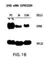
- the levels of GP88 protein expression were examined by Western blot analyses using anti-K19 antibody both in cell lysates and in culture medium of 1246, 1246-3A and PC cells as described above. As shown in FIG. 1A, the level of GP88 was undetectable in the culture medium of 1246 cells, 3T3, and 1246-3A cells and increased dramatically in the culture medium of the highly tumorigenic PC cells. The same results were obtained for GP88 expression in cell lysates.
- GP88 expression was examined in 1246 cells in different culture conditions such as defined medium and serum containing medium. It was shown that the level of expression of GP88 mRNA (measured by Northern blot analysis) and protein (measured by Western blot analysis) in 1246 and in 1246-3A cells is inhibited when the cells are treated with 2% fetal bovine serum indicating the presence of circulating inhibitors of GP88 expression in fetal bovine serum ( Figure 1C). This inhibition of GP88 expression was also observed when the activity of GP88 promoter linked to a luciferase reporter gene was measured indicating that these inhibitors are effective in inhibiting the transcriptional activity of the GP88 gene.
- Such inhibitors can be useful to develop GP88 antagonizing agents which will be useful as anti-tumor or antiviral therapy.
- GP88 mRNA expression is stimulated by EGF in the 1246 cells and by insulin in the human mammary carcinoma cells MDA MB-453.
- GP88 is a potent growth stimulator for mammary epithelial cells (see next paragraph); receptors for GP88 (our studies) and processed form epithelin 1 (54) have been characterized on mammary epithelial cells; breast carcinoma cell lines with different degrees of hormonal dependency are available for study; and lastly there are a growing number of reports emphasizing the importance of the insulin/IGF pathway in the growth control of mammary cells indicating that an escape from this regulation is occurring in malignant breast carcinomas (14, 15). Since PC cells which display an over expression of GP88 are insulin/IGF independent, this would support the rationale of GP88 deregulation in human breast carcinoma.
- MCF-7 estrogen receptor positive
- MDA-MB-453 and 468 being estrogen receptor negative
- ER- estrogen receptor negative
- FIG. 5 shows that GP88 mRNA is expressed in the three cell lines but that the level of expression is higher in the ER- cell lines, MDA-MB-453 and 468, than in the ER+MCF7 cells, indicating that in human breast carcinoma, increased malignancy may be accompanied by increased GP88 expression.
- GP88 had a profound growth promoting effect on the mouse mammary epithelial cell line C57MG. As shown in FIG. 6, a 5-fold increase in DNA synthesis was observed at a concentration of 150ng/ml (2 nM) either with GP88 purified from PC cells or with recombinant GP88 expressed in insect cells.
- GP88 protein purified from PC cell conditioned medium was sequenced after digestion with cyanogen bromide and trypsin. Sequences of N-terminal regions and 6 peptides were obtained (4). Sense and antisense redundant oligonucleotide primers complementary to the obtained amino acid sequences were synthesized and used in the polymerase chain reaction using the touch down PCR method with first strand cDNA of PC cells as template. From the touch down PCR using a primer pair SCV157 and ANG300, a 444 bp amplified product was obtained. This cDNA was then used to screen a lambda-ZAP cDNA library prepared from PC cells in our laboratory.
- GP88 For recombinant GP88 production, the method of choice was to express GP88 in the baculovirus system.
- a full length GP88 cDNA obtained by screening PC cell cDNA library) including the signal peptide was ligated into the baculovirus transfer vector pVL1392 (in Vitrogen, San Diego, CA). Plasmid pVL1392-GP88 was used to co-transfect Sf9 insect cells with baculovirus DNA. Recombinant viruses encoding GP88 were isolated and plaque purified.
- Sf9 cells were seeded in Grace's medium containing 10% fetal bovine serum (FBS) in T 75 cm 2 flasks.
- FBS fetal bovine serum
- rGP88 recombinant GP88
- rGP88 recombinant GP88
- SDS-PAGE analysis of rGP88 indicated that rGP88 migrates faster than PC cell derived GP88 corresponding to an apparent MW of 76 kDa.
- N-terminal sequencing analysis of rGP88 indicated that it was identical to GP88 purified from PC-CM.
- the difference of molecular weight between GP88 and rGP88 is due to a difference in glycosylation status cf GP88 in insect cells.
- biological activity of rGP88 was identical to that of GP88 purified from PC cells, indicating that the different glycosylation status of GP88 in insect cells and mammalian cells did not affect the biological potency of the protein.
- the rGP88 produced from insect cells can be used for biological, and binding studies and to develop monoclonal antibodies to the intact GP88
- the conditioned medium (2000 ml) from PC cells was diluted with the same volume of H 2 O and loaded on a 2.5 ml heparin-sepharose CL-6B column equilibrated in 10 mM sodium phosphate buffer pH 7.4 containing 75 mM NaCl (Pharmacia, Uppsala, Sweden). The column was washed with at least 10 bed volumes of the same equilibration buffer followed by a wash with 10 mM sodium phosphate buffer containing 0.15 M NaCl. The fraction containing GP88 was eluted with 5 bed volumes of 0.4 M NaCl, 10 mM Tris-HCl, pH 7.5. The eluate was stored at -20 C for further purification.
- a synthetic peptide K19T (sequence: KKVIAPRRLPDPQILKSDT) was used to raise the antisera against the GP88 used in the immunoaffinity step.
- the K19T peptide was linked to CNBr-activated Sepharose 4B according to the method provided by the manufacturer (Pharmacia, Uppsala, Sweden).
- the specific anti-K19 antibody was purified using the K19T peptide affinity column by elution at acidic pH.
- anti-K19T IgG was applied to a K19T peptide-Sepharose 4B column equilibrated with 10mM sodium phosphate buffer pH 6.5 (Buffer A) at a flow rate of 0.8 ml/hr, and circulated at 4°C overnight. After washing the column with 7 ml of Buffer A, the conjugate was eluted with 1 ml of HCl, pH 2.9, then 1 ml of HCl, pH 2.5 at a flow rate of about 0.1 ml/min in a tube containing 0 1 ml of 1M sodium phosphate buffer pH 7.0 to neutralize the pH. The concentration of affinity-purified IgG was determined by the absorbance at 280 nm.
- the purified Ab-K19T (1 mg) was then conjugated to 1 ml of agarose beads (Sulfolink coupling gel, Pierce, Rockford, IL) using protocols provided by the manufacturer.
- the final coupled column contained 600 ⁇ g anti-K19T/ml gel.
- the Ab-K19T agarose was packed in a column and washed extensively with PBS.
- the eluate from heparin sepharose CL-6B column was diluted with 3 volumes H 2 O and loaded on the Ab-K19T column. After washing the column with buffer consisting of 750 mM NaCl in 10 mM NaPO4 pH 7.5, the fraction containing GP88 was eluted by elution buffer (150 mM NaCl, pH 2.5 (HCl)).
- This method is also adequate for the purification of recombinant GP88 such as constructed in a baculovirus expression vector and expressed in insect cells. This method is also adequate to purify human GP88 using for the immunoaffinity step human GP88 antibody conjugated to adequate support (sepharose or agarose).
- Peptides corresponding to various regions of mouse and human GP88 were synthesized and conjugated to keyhole limpet Hemocyanin (KLH) by the "glutaraldehyde method.” Peptide KLH conjugate was injected into chinchilla rabbits to raise anti-GP88 antibody. Two peptides, K19T and S14R, listed below, were found to generate neutralizing antibodies. Equivalent regions such as E19V of the human GP88 amino acid sequences were used to develop neutralizing anti-human GP88 monoclonal antibodies.
- KLH keyhole limpet Hemocyanin
- Peptides were as follows: P12T from P 208 to T 219 PDAKTQCPDDST K19T from K 344 to T 362 KKVIAPRRLPDPQILKSDT S14R from S 562 to R 575 SARGTKCLRKKIPR E19V (human GP88) EKAPAHLSLPDPQALKRDV A14R (human GP88) ARRGTKCLRREAPR
- GP88 cDNA was cloned in the antisense orientation in pCMV4 expression vector (Andersson, S., et al, 1989) (51) containing CMV promoter and hGH transcription termination and polyadenylation signals (pCMV4-GP88AS).
- PC cells were co-transfected with the 20 ⁇ g of antisense pCMV4-GP88AS and 2 ⁇ g of pRSVneo expression vector containing the neomycin resistant gene by the calcium phosphate method.
- Control cells were co-transfected with empty pCMV4 vector and pRSVneo as described above. Transfected cells were selected in the presence of neomycin.
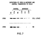
- Neomycin resistant colonies were cloned and cells were assayed first by detecting the presence of pCMV4-GP88AS by PCR. Twenty-four positive neomycin resistant clones containing the antisense pCMV4-GP88AS were isolated. Nine have been isolated and screened for expression of the antisense transcript. Three clones were further characterized. Western blot analysis of cell lysates and conditioned medium using anti-GP88 antiserum (i.e., anti-K19T antibody) was performed in order to determine the level of GP88 expression in transfected antisense cells and control cells (FIG. 7). Culture medium and cell lysates were prepared by immunoprecipitation with anti-K19T antibody. Protein samples corresponding to 3x10 6 cells/lane were analyzed by Western blotting with anti-GP88 antibody. The results indicate that GP88 levels are significantly lower in antisense, than in control, transfected cells particularly for AS1 and AS18 clones.
- PC cells were transfected with a 228 bp antisense cDNA fragment of GP88 including start codon region obtained by digesting with Sma I and Xba I GP88 cDNA clone and cloning the obtained cDNA fragment in the antisense orientation into Xba I and Sma I site of the mammalian expression vector pCMV4 as shown in Figure 11.
- the stable transfection of PC cells was performed by the Calcium Phosphate method (55) in DME medium (Dulbecco's Modified eagle Medium) containing 3.7 g/L sodium bicarbonate supplemented with 10% FBS.
- a calcium phosphate precipitate was made with 20 ⁇ g of plasmid pCMV4 constructed with antisense GP88 cDNA, 2 ⁇ g of plasmid carrying neomycin resistance selectable marker (pRSVNEO), and 20 ⁇ g of pSK as carrier DNA. After 25 minutes, the precipitate was added dropwise to the cells. After 7 hours, the medium was aspirated and the cells were shocked with 10% DMSO in PBS for 2-3 min., washed twice and fed with complete medium (DME supplemented with 10% FBS). One day after transfection, the cells were split 1:3 and selected for resistance to Geneticin (G-418 Sulphate, Gibco-BRL) at 400 ⁇ g/ml.
- G-418 Sulphate Gibco-BRL
- transfected clones were analyzed by two assays as described below:
- the presence of the antisense cDNA construct is tested by PCR analysis of genomic DNA of transfected clones using as primers an oligonucleotide located in the CMV promoter (5'-CCTACTTGGCAGTACATCTACGTA-3') and the other corresponding to the start codon of GP88 cDNA (5'-CGAGAATTCAGGCAGACCATGTGGGTC-3'). These primers would amplify a 551 bp DNA fragment from genomic DNA of transfected cells containing the antisense DNA construct described above.
- the level of GP88 protein in antisense transfected clones was measured by Western blotting analysis of cell lysates and conditioned media collected from the transfected clones using anti-K19T antibody to measure the efficacy by which the antisense GP88 inhibited the endogenous GP88 protein expression.
- Antisense clones that showed the highest degree of inhibition of GP88 expression were selected for analysis of their growth properties in vivo as described below. Analysis of GP88 expression in empty vector control transfected clones was done similarly.
- the presence of the antisense construct in the transfected cells was determined by PCR analysis cf their genomic DNA using as sense primer SP647 (5'-CCTACTTGGCAGTACATCTACGTA-3') corresponding to CMV promoter region and antisense primer SP7 (5'-CGAGRATTCAGGCAGACCATGTGGGTC-3') corresponding to start codon region of GP88.
- the sense primer SP647 and antisense primer AP912 both located in the CMV4 promoter were used to test whether CMV promoter was inserted into the genomic DNA of control transfectants which had been transfected with empty pCMV4 vector.
- the transfectants were lysed by buffer A (100 mM KCl, 10 mM Tris-HCl [pH 8.3], 0.45% Tween 20 and 0.45% NP40) and proteinase K 120 ⁇ g/ml, incubated in 60°C 1 hr, followed by boiling for 15 min. 50-100 ng DNA of each clone was used as template for PCR reaction. Non-transfected PC were used as negative control. Constructed plasmid DNA was used as positive control.
- the PCR reaction was performed in 20 ⁇ l reaction mixture containing 10 mM Tris-HCl pH 9.0, 50 mM KCl, 1.5 mM MgCl2, 0.1% Triton X-100, 0.2 mM dNTPs, 0.5 units Taq DNA polymerase, 3.2 pMol of each primer, and 50-100 ng of template DNA.
- the reaction tubes were heated to 95°C for 3 minutes, and then subjected to 40 cycles of 94°C for 1 minute, 55°C for 2 minutes, and 72°C for 3 minutes with a 10 minutes 72°C extension in a programmable thermal controller. Products were analyzed on a 1% agarose gel stained with ethidium bromide. The expected size of the amplified fragment corresponding to the presence of the antisense construct with the primers chosen was 551 bp.
- transfecting antisense GP88 DNA in the cells is to inhibit endogenous GP88 expression. Therefore, the transfected clones of cells which had been selected by the assays described above (i.e., neomicin resistance and presence of antisense DNA in genomic DNA) were examined to determine the degree of inhibition of endogenous GP88 expression. This was examined by measuring the level of GP88 in cell lysates and in conditioned medium from antisense transfected clones and empty vector transfected control clones by immunoprecipitation and Western blot analysis using anti-K19T antibody by the methods previously described. The transfected clones that displayed the highest degree of inhibition of GP88 expression were further analyzed to examine their growth properties in vitro and in vivo.
- Cells were plated at a density of 3 x 10 4 cells/well (12 well plates, Corning) in 2 ml of DME/F-12 medium (1:1 mixture) supplemented with 2% FBS or 2 ⁇ g/ml human fibronectin and 10 ⁇ g/ml human transferrin. Five days later, cells were washed with PBS, and cell number per well was determined by counting cells from duplicate wells using Coulter Counter after trypsinization.
- mice Six weeks old female C3H mice were used for tumorigenicity assays. Sense/antisense or control transfectants were injected subcutaneously into syngeneic C3H mice at the density of 10 6 cells per animal. The appearance and size of tumors were examined. The mice were killed 45-50 days after injection. Tumors were excised, their weight determined and the tumors were quick frozen and kept at -70°C.
- FIG. 3 shows the picture of tumor bearing mouse detected with 10 6 control transfected cells and of mouse injected with antisense clone. Empty vector control transfected clones maintained similar tumorigenic properties as the parent PC cells, whereas no tumor formation was observed in the mice injected with antisense clones. Even after 90 days, these mice still did not present tumors. This experiment was repeated twice. Moreover, additional clones (2 antisense and 2 control) were also examined. The same results were obtained with other antisense clones.
- the human antisense cDNA construct consisted of a 400 bp cDNA fragment inserted in the antisense orientation in the commercially available pcDNA3 mammalian expression vector (In Vitrogen, San Diego, CA) which contains pCMV4 cytomagelovirus CMV promoter and neomycin resistant gene so that double transfection of pCMV4 and pRSVneo is not required like the ones described above for PC cells.
- the amplified cDNA fragment with EcoRI and BamHI restriction sites was inserted in the antisense orientation in the EcoRI and BamHI sites of the pcDNA3 mammalian expression vector.
- This expression vector construct called pCAS was transfected in the human mammary carcinoma cell line MCF7 and MDA-MB-468 DME-F12 medium supplemented with fetal bovine serum by the calcium phosphate method (55). Selection of transfected cells was done by cultivating the cells in the presence of 800 ⁇ g/ml of Geneticin to select cells that are neomicin resistant.
- Neomicin resistant clones were picked with cloning rings and passaged in medium supplemented with 10% fetal bovine serum (FBS) and with geneticin (800 ⁇ g/ml). Transfected clones selected for their resistance to geneticin were further examined by methods similar to the ones described above for PC cells transfected with mouse GP88 antisense cDNA. The presence of the antisense cDNA construct in genomic DNA was checked by PCR analysis using as primers for the PCR reaction T7 primer in the pCDNA3 expression vector and H-hGP88 primer described above. PCR reaction amplified a 420 bp DNA fragment in cells that expressed the transfected human GP88 antisense DNA fragment.
- GP88 Endogenous GP88 was determined by Western blot analysis of cell lysates and conditioned medium of transfected clones using anti E19V antiserum to select antisense clones with maximum inhibition of GP88 expression. Selected antisense clones were further analyzed to examine their growth properties in vitro and in vivo . Transfection of empty pCDNA3 vector in MCF7 and MDA-MB-468 cells was performed as control for these experiments.
- An alternative method to transfection of antisense cDNA is to use antisense oligonucleotides. It is known in the art that sequences around the translation initiation site (ATG encoding the first methionine) provide good sequences for efficient antisense activity. Secondly, sequences with an adequate GC content and that start with either a G or a C have increased efficiency and stability in forming a hybrid with corresponding sense sequence (32, 37, 38). Based on this rationale, it is anticipated that the following two sequences will be efficacious as antisense oligonucleotides to human GP88.
- the first one is a 22-mer named HGPAS1 starting 11 nucleotides upstream of the first ATG (methionine codon): HGPAS1 (22): 5'-GGGTCCACATGGTCTGCCTGC-3'.
- the second oligomer is a 24 mer named HGPAS2 (24) located 21 nucleotides 3' (downstream) of the first ATG: HGPAS2 (24):5'-GCCACCAGCCCTGCTGTTAAGGCC-3'.
- Other oligonucleotide antisense sequences can be explored by those of ordinary skill given the teachings herein.
- oligonucleotides will be added to breast carcinoma cells in culture or any other human cell types under study in increasing doses. Cells will be collected at various time points (12 hours to several days) to measure the level of expression of GP88 protein by Western blot analysis or EIA using an antihuman GP88 antibody, using techniques known to those of ordinary skill in the art. Control cells will be treated with a nonsense or a sense oligomer.
- the present example provides the following:
- the MCF10A cell line which is a non tumorigenic human mammary epithelial cell line
- MCF7 which is estrogen receptor positive
- MDA-MB-468 which is estrogen receptor negative.
- Expression of GP88 mRNA expression was done by Northern blot analysis of total RNA extracted from these cell lines using a radiolabeled human GP88 cDNA probe.
- GP88 protein was measured by Western blot analysis of immunoprecipitated GP88 using either rabbit anti-human GP88 polyclonal antibody or the anti-human E19V monoclonal antibody. This latter antibody was developed by immunizing mice with human peptide E19V conjugated to protein as described in Example 8.
- Anti-human GP88 antibodies that we have now at our disposal are: polyclonal antihuman GP88 antibody developed in rabbits using as antigen a 37 kDa fragment of human GP88 expressed as a histidine tagged protein in bacteria, and the anti-human GP88 antibody (polyclonal and monoclonal) developed by using as antigen E19V peptide conjugated to keyhole limpet hemocyanin. Development of these antibodies (polyclonal and monoclonal) are described above. All of these antibodies can be used for immunoprecipitation and Western blot analysis of human GP88 in human tissues and cells.
- the Northern blot analysis shows that the expression of GP88 mRNA in the non tumorigenic MCF10A cells is very low and increases at least 5-10 times in the human breast carcinoma cell lines MCF7 and MDA-MB-468 (FIG. 14).
- GP88 protein expression is undetectable in culture medium (CM) collected from the non-tumorigenic MCF10A cells, whereas it increased 10-20 times in media conditioned by the MCF7 cells and by the MDA-MB-468 cells.
- CM culture medium
- GP88 protein is also expressed at high levels in cell lysates of the maligant cells, whereas it is undetected in the non tumorigenic MCF10A cells.
- Human breast carcinoma MDA-MB-468 cells were stably transfected with antisense human GP88 cDNA construct (400 bp fragment inserted into pCDNA3 expression vector). This construct is described in Example 9. Stable antisense clones were isolated and characterized. In particular, antisense clones were selected based on the fact that expression of GP88 was effectively inhibited. This was determined by measuring GP88 expression in antisense cells and empty vector control cells by Western blot analysis of cell extracts and conditioned medium using antiGP88 antibody. Several clones were obtained and characterized. Similar results were obtained with all antisense clones isolated. Data presented here concern one antisense clone called 468AS. As shown in FIG. 15, transfection of antisense GP88 cDNA in MDA-MB-468 cells (468AS) resulted in inhibition of GP88 protein expression when compared to empty vector control MDA-MB-468 cells (468 Cont).
- Antisense human breast 468AS cells which displayed the highest inhibition of GP88 expression, and empty vector control (468 Cont) cells were injected subcutaneously into female nude mice in the breast area at a density of 2 x 10 3 cells/mouse using 4 mice per cell line. Tumor appearance was monitored by visually inspecting the mice. After 4 weeks, the mice were sacrificed, tumors were excised and weighed (see Table 3 below). Effect of inhibition of GP88 expression by antisense GP88cDNA transfection on tumor growth of human breast cancer cells MDA-MB-468 in nude mice Cells Mice bearing tumors Weight of tumors (g) 468 Cent empty 4/4 0.2 ⁇ 0.06 vector 3/4 0.05 ⁇ 0/025 468AS antisense GP88
- an increase in tumorigenic properties is associated with an increase in GP88 expression or an increase in GP88 responsiveness.
- Figure 4 shows that the level of expression of GP88 in tumor tissue is increased when compared to the surrounding tissues. Accordingly, increase of GP88 level can be used as a diagnostic approach to detecting tumor.
- a change (increase) in GP88 expression when compared to the level of GP88 in normal corresponding tissues is indicative of the state of tumorigenicity or malignancy of the tissue biopsy analyzed.
- Increase in expression of GP88 can be measured at the mRNA level or at the protein level.
- GP88 mRNA expression can be measured either by Northern blot analysis, RNAse protection assay or RT-PCR.
- GP88 protein expression is quantitated by ELISA, EIA or RIA using an anti-GP88 antibody.
- the ability to measure GP88 expression in tissue extracts in comparison to corresponding tissues from normal subject can be used to predict the degree of tumorigenicity of a particular cancer or to determine whether this particular cancer will be responsive to anti-GP88 therapy.
- tissue biopsies to be analyzed will be treated with anti-GP88 neutralizing antibodies or anti-GP88 receptor antibodies to see if such treatment inhibits growth of the cells.
- anti-GP88 neutralizing antibodies or anti-GP88 receptor antibodies to see if such treatment inhibits growth of the cells.
- the ability of GP88 antisense oligonucleotides to inhibit growth indicates that expression of GP88 is required for growth in vivo .
- Binding of iodinated GP88 to a variety of cell lines CCL64, 1246, PC and the mammary epithelial C57MG was determined in order to examine the biochemical characteristics of GP88 cell surface receptors using the methods described below.
- affinity purified recombinant GP88 (rGP88) from the culture medium of baculovirus infected SF9 insect cells as ligand. Methods to prepare rGP88 have previously been described.
- Recombinant GP88 was iodinated by the chloramine T method at 4°C. Other known methods can also be applied. Briefly, 1 ⁇ g of GP88 was incubated for 2 min with 125 I Na (100 ⁇ Ci) that had been preincubated for 90 seconds with 2 ⁇ g chloramine T. The reaction was quenched with the addition of 100 ⁇ l saturated tyrosine, 10 ⁇ l of a 1% solution of bovine serum albumin (BSA) and 2 ⁇ g of sodium metabisulfite.
- BSA bovine serum albumin
- the iodinated protein was separated from free Na 125 I by gel filtration on a Sephadex-G50 column that had been preincubated with PBS containing 1 % bovine serum albumin and then extensively washed with PBS to reduce non-specific binding.
- the labeled proteins were eluted with PBS and fractions monitored for radioactivity. Amount of incorporated radioactivity was estimated by TCA precipitation. Specific activity of 125 I-GP88 was typically 30-50 ⁇ Ci/ ⁇ g. This method and other methods for iodination of proteins are well known to people skilled in the art can also be used to iodinate PC derived GP88 rather than rGP88 or any derivatives thereof.
- the example provided here describes binding assay with the mink lung epithelial cell line CCL64 but has also been applied to several cell lines including the 1246, PC cell lines and the mammary epithelial cell line C57MG.
- the binding assays were performed on cell suspension.
- CCL64 cells were cultivated as monolayer in DME medium supplemented with 10% fetal bovine serum (FBS) until they reached confluency. At that time, cells were washed with PBS and detached by incubation with a solution of 0.25 mg/ml of trypsin and 1 mM EDTA. The cells were harvested by centrifugation, washed with culture medium and counted with a hemocytometer.
- FBS fetal bovine serum
- binding assays 10 6 cells were resuspended in 500 ⁇ l of binding buffer consisting of DME medium supplemented with 1 % bovine serum albumin in 1.5 ml ependorf tubes and incubated for 2 hours at 25°C with 10 5 cpm of 125 I-rGP88 and increasing concentrations of cold rGP88 from 1 to 100 ng/ml. Binding was stopped by centrifuging the cells followed by 3 successive washings at 4°C with cold binding buffer followed by centrifugation. Cell pellets were counted with a gamma counter. Scatchard analysis of binding data was carried out by computer Ligand program. Values correspond to the average of three separate experiments with duplicate determinations per experimental point.
- DMS disuccinimidyl suberate
- the cells were resuspended in 200 ⁇ l PBS containing 1 mM disuccinimidyl suberate (DSS,) at room temperature for 15 min. After crosslinking, the cells were centrifuged, washed and extracted with 25 ⁇ l extraction buffer (PBS containing 1% Triton X-100, 0.1% SDS, 0.5 mM phenylmethylsulfonyl fluoride (PMSF) at 4°C. Samples were centrifuged for 5 min at 13,000 x g and 25 ⁇ l of supernatant from each sample was mixed with 4 ⁇ l of 20% SDS and 15 ⁇ l 3x Laemmli's sample buffer (56) containing b-mercaptoethanol and boiled for 5 min.
- DSS disuccinimidyl suberate
- Electrophoresis of samples was carried out on 7% SDS polyacrylamide slab gel according to Laemmli (56) using a minigel apparatus (Bio-Rad, Richmond, CA). The dried gels were exposed to X-ray films for autoradiography at -70°C.
- GP88 receptor Upon binding of GP88 to its cell surface, GP88 receptor is activated by phosphorylation on tyrosine residues resulting in phosphorylation of several signaling molecules including IRS-1, SHC, Grb2 and leading to activation of MAP kinase ERK-2.
Abstract
Description
- This invention relates to cell biology, physiology and medicine, and concerns an 88kDa glycoprotein growth factor ("GP88") and compositions and methods which affect the expression and biological activity of GP88. These compositions and methods are useful for diagnosis and treatment of diseases including cancer.
- Several publications are referenced herein by Arabic numerals within parenthesis. Full citations for these references may be found at the end of the specification immediately preceding the claims.
- The proliferation and differentiation of cells in multicellular organisms is subject to a highly regulated process (1). A distinguishing feature of cancer cells is the absence of control over this process; proliferation and differentiation become deregulated resulting in uncontrolled growth. Significant research efforts have been directed toward better understanding this difference between normal and tumor cells. One area of research focus is growth factors and, more specifically, autocrine growth stimulation.
- Growth factors are polypeptides which carry messages to cells concerning growth, differentiation, migration and gene expression (2). Typically, growth factors are produced in one cell and act on another cell to stimulate proliferation. However, certain malignant cells, in culture, demonstrate a greater or absolute reliance on an autocrine growth mechanism (3). Malignant cells which observe this autocrine behavior circumvent the regulation of growth factor production by other cells and are therefore unregulated in their growth.
- Study of autocrine growth control advances understanding of cell growth mechanisms and leads to important advances in the diagnosis and treatment of cancer. Toward this end, a number of growth factors have been studied, including insulin-like growth factors ("IGF1" and "IGF2"), gastrin-releasing peptide ("GRP"), transforming growth factors alpha and beta ("TGF-a" and "TGF-b"), and epidermal growth factor ("EGF").
- The present invention is directed to a recently discovered growth factor. This growth factor was first discovered in the culture medium of highly tumorigenic "PC cells," an insulin-independent variant isolated from the teratoma derived
adipogenic cell line 1246. This growth factor is referred to herein as "GP88." GP88 has been purified and structurally characterized (4). Amino acid sequencing of GP88 indicates that GP88 has amino acid sequence similarities with the mouse granulin/epithelin precursor. - Granulins/epithelins ("grn/epi") are 6kDa polypeptides and belong to a novel family of double cysteine rich polypeptides (5, 6). U.S. Patent No. 5,416,192 (Shoyab et al.) is directed to 6 kDa epithelins, particularly epithelin 1 and
epithelin 2. According to Shoyab, both epithelins are encoded by a common 63.5 kDa precursor, which is processed into smaller forms as soon as it is synthesized, so that the only natural products found in biological samples are the 6 kDa forms. Shoyab et al. teaches that the epithelin precursor is biologically inactive. - Contrary to the teachings of Shoyab et al., the inventor's laboratory has demonstrated that the precursor is not always processed as soon as it is synthesized. Studies, conducted in part by this inventor, have demonstrated that the precursor (i.e., GP88) is in fact secreted as an 88kDa glycoprotein with an N-linked carbohydrate moiety of 20kDa (4). Analysis of the N-terminal sequence of GP88 indicates that GP88 starts at
amino acid 17 of the grn/epi precursor, demonstrating that the first 17 amino acids from the protein sequence deduced from the precursor cDNA correspond to a signal peptide compatible with targeting for membrane localization or for secretion (4). - Also in contrast to the teachings of Shoyab et al., GP88 is biologically active and has growth promoting activity, particularly as an autocrine growth factor for the producer cells.
- The inventor has now unexpectedly discovered that a glycoprotein (GP88), which is expressed in a tightly regulated fashion in normal cells, is overexpressed and unregulated in highly tumorigenic cells derived from the normal cells, that GP88 acts as a stringently required growth stimulator for the tumorigenic cells and that inhibition of GP88 expression or action in the tumorigenic cells results in an inhibition of the tumorigenic properties of the overproducing cells.
- It is an object of this invention to provide GP88 antagonizing compositions capable of inhibiting the expression or activity of GP88.
- A further object of the invention is to provide methods for treating diseases associated with a defect in GP88 quantity or activity such as but not limited to cancer in a mammal.
- Another object of the invention is to provide methods for determining the susceptibility of a subject to diseases associated with a defect in GP88 expression or action.
- Yet another object of the invention is to provide methods for measuring susceptibility to GP88 antagonizing therapy.
- Yet another object of the invention is to provide methods, reagents, and kits for the in vitro and in vivo detection of GP88 and tumorigenic activity in cells.
- Additional objects and advantages of the invention will be set forth in part in the description that follows, and in part will be obvious from the description, or may be learned by the practice of the invention.
- To achieve the objects and in accordance with the purpose of the invention, as embodied and properly described herein, the present invention provides compositions for diagnosis and treatment of diseases such as but not limited to cancer in which cells exhibit an altered expression of GP88 or altered response to GP88.
- Use of the term "altered expression" herein means increased expression or overexpression of GP88 by a factor of at least two-fold, and at times by a factor of 10 or more, based on the level of mRNA or protein as compared to corresponding normal cells or surrounding peripheral cells. The term "altered expression" also means expression which became unregulated or constitutive without being necessarily elevated. Use of the terms increased or altered "response" to GP88 means a condition wherein increase in any of the biological functions (e.g., growth, differentiation, viral infectivity) conferred by GP88 results in the same or equivalent condition as altered expression of GP88.
- Use of the term "GP88" herein means epithelin/granulin precursor in cell extracts and extracellular fluids, and is intended to include not only GP88 according to the amino acid sequences included in figures 8 or 9, which are of mouse and human origins, but also GP88 of other species. In addition, the term also includes functional derivatives thereof having additional components such as a carbohydrate moiety including a glycoprotein or other modified structures.
- Also intended by the term GP88 is any polypeptide fragment having at least 10 amino-acids present in the above mentioned sequences. Sequences of this length are useful as antigens and for making immunogenic conjugates with carriers for the production of antibodies specific for various epitopes of the entire protein. Such polypeptides are useful in screening such antibodies and in the methods directed to detection of GP88 in biological fluids. It is well known in the art that peptides are useful in generation of antibodies to larger proteins (7). In one embodiment of this invention, it is shown that peptides from 12-19 amino-acids in length have been successfully used to develop antibodies that recognize the full length GP88.
- The polypeptide of this invention may exist covalently or non-covalently bound to another molecule. For example, it may be fused to one or more other polypeptides via one or more peptide bonds such as glutathione transferase, poly-histidine, or myc tag.
- The polypeptide is sufficiently large to comprise an antigenetically distinct determinant or epitope which can be used as an immunogen to reproduce or test antibodies against GP88 or a functional derivative thereof.
- One embodiment includes the polypeptide substantially free of other mammalian peptides. GP88 of the present invention can be biochemically or immunochemically purified from cells, tissues or a biological fluid. Alternatively, the polypeptide can be produced by recombinant means in a prokaryotic or eukaryotic expression system and host cells.
- "Substantially free of other mammalian polypeptides" reflects the fact that the polypeptide can be synthesized in a prokaryotic or a non-mammalian or mammalian eukaryotic organism, if desired. Alternatively, methods are well known for the synthesis of polypeptides of desired sequences by chemical synthesis on solid phase supports and their subsequent separation from the support. Alternatively, the protein can be purified from tissues or fluids of mammals where it naturally occurs so that it is at least 90% pure (on a weight basis) or even 99% pure, if desired, of other mammalian polypeptides, and is therefore substantially free from them. This can be achieved by subjecting the tissue extracts or fluids to standard protein purification such as on immunoabsorbants bearing antibodies reactive against the protein. One embodiment of the present invention describes purification methods for the purification of naturally occurring GP88 and of recombinant GP88 expressed in baculovirus infected insect cells. Alternatively, purification from such tissues or fluids can be achieved by a combination of standard methods such as but not limited to the ones described in reference (4).
- As an alternative to a native purified or recombinant glycoprotein or polypeptide, "GP88" is intended to also include functional derivatives. By functional derivative is meant a "fragment," "variant," "analog," or "chemical derivative" of the protein or glycoprotein as defined below. A functional derivative retains at least a portion of the function of the full length GP88 which permits its utility in accordance with the present invention.
- A "fragment" of GP88 refers to any subset of the molecule that is a shorter peptide. This corresponds for example but is not limited to regions such as K19T and S14R for mouse GP88, and E19V and A14R (equivalent to murine K19T and S14R, respectively) for human GP88.
- A "variant" of GP88 refers to a molecule substantially similar to either the entire peptide or a fragment thereof. Variant peptides may be prepared by direct chemical synthesis of the variant peptide using methods known in the art.
- Alternatively, amino acid sequence variants of the peptide can be prepared by modifying the DNA which encodes the synthesized protein or peptide. Such variants include, for example, deletions, insertions, or substitutions of residues within the amino-acid sequence of GP88. Any combination of deletion, insertion, and substitution may also be made to arrive at the final construct, provided the final construct possesses the desired activity. The mutation that will be made in the DNA encoding the variant peptide must not alter the reading frame and preferably will not create complementary regions that could produce secondary mRNA structures. At the genetic level these variants are prepared by site directed mutagenesis (8) of nucleotides in the DNA encoding the peptide molecule thereby producing DNA encoding the variant, and thereafter expressing the DNA in recombinant cell culture. The variant typically exhibits the same qualitative biological activity as the nonvariant peptide.
- An "analog" of GP88 protein refers to a non-natural molecule substantially similar to either the entire molecule or a fragment thereof.
- A "chemical derivative" contains additional chemical moieties not normally a part of the peptide or protein. Covalent modifications of the peptide are also included within the scope of this invention. Such modifications may be introduced into the molecule by reacting targeted amino-acid residues of the peptide with an organic derivatizing agent that is capable of reacting with selected side chains or terminal amino-acid residues. Most commonly derivatized residues are cysteinyl, histidyl, lysinyl, arginyl, tyrosyl, glutaminyl, asparaginyl and amino terminal residues. Hydroxylation of proline and lysine, phosphorylation of hydroxyl groups of seryl and threonyl residues, methylation of the alpha-amino groups of lysine, histidine, and histidine side chains, acetylation of the N-terminal amine and amidation of the C-terminal carboxylic groups. Such derivatized moieties may improve the solubility, absorption, biological half life and the like. The moieties may also eliminate or attenuate any undesirable side effect of the protein and the like. In addition, derivatization with bifunctional agents is useful for cross-linking the peptide to water insoluble support matrices or to other macromolecular carriers. Commonly used cross-linking agents include glutaraldehyde, N-hydroxysuccinimide esters, homobifunctional imidoesters, 1,1-bis(-diazoloacetyl)-2-phenylethane, and bifunctional maleimides. Derivatizing agents such as methyl-3-[9p-azidophenyl)]dithiopropioimidate yield photoactivatable intermediates that are capable of forming crosslinks in the presence of light. Alternatively, reactive water-insoluble matrices such as cyanogen bromide activated carbohydrates and the reactive substrates described in U.S. Patents 3,969,287 and 3,691,016 may be employed for protein immobilization.
- Use of the term GP88 "antagonizing agents" herein means any composition that inhibits or blocks GP88 expression, production or secretion, or any composition that inhibits or blocks the biological activity of GP88. This can be achieved by any mode of action such as but not limited to the following:
- (A) GP88 antagonizing agents include any reagent or
molecule inhibiting GP88 expression or production including
but not limited to:
- (1) antisense GP88 DNA or RNA molecules that inhibit GP88 expression by inhibiting GP88 translation;
- (2) reagents (hormones, growth factors, small molecules) that inhibit GP88 mRNA and/or protein expression at the transcriptional, translational or post-transaltional levels;
- (3) factors, reagents or hormones that inhibit GP88 secretion;
- (B) GP88 antagonizing agents also include any reagent
or molecule that will inhibit GP88 action or biological
activity such as but not limited to:
- (1) neutralizing antibodies to GP88 that bind the protein and prevent it from exerting its biological activity;
- (2) antibodies to the GP88 receptor that prevent GP88 from binding to its receptor and from exerting its biological activity;
- (3) competitive inhibitors of GP88 binding to its receptors;
- (4) inhibitors of GP88 signaling pathways.
-
- Specific examples presented herein provide a description of preferred embodiments, particularly the use of neutralizing antibodies to inhibit GP88 biological action and the growth of the highly tumorigenic PC cells; the use of antisense GP88 cDNA and antisense GP88 oligonucleotides to inhibit GP88 expression leading to inhibition of the tumorigenic properties of the PC cells; characterization of GP88 receptors on cell surfaces of several cell lines including the mammary epithelial cell line C57MG, the 1246 and PC cell lines and the mink lung epithelial cell line CCL64.
- In one embodiment of the invention, the GP88 antagonizing agents are antisense oligonucleotides to GP88. The antisense oligonucleotides preferably inhibit GP88 expression by inhibiting translation of the GP88 protein.
- Alternatively, such a composition may comprise reagents, factors or hormones that inhibit GP88 expression by regulating GP88 gene transcriptional activity. Such a composition may comprise reagents, factors or hormones that inhibit GP88 post-translational modification and its secretion. Such a composition may comprise reagents that act as GP88 antagonists that block GP88 activity by competing with GP88 for binding to GP88 cell surface receptors. Alternatively, such a composition may comprise factors or reagents that inhibit the signaling pathway transduced by GP88 once binding to its receptors on diseased cells.
- Alternatively, the composition may comprise reagents that block GP88 action such as an antibody specific to GP88 that neutralizes its biological activity, or an antibody to the GP88 receptor that blocks its activity.
- The antibodies of the invention (neutralizing and others) are preferably used as a treatment for cancer or other diseases in cells which exhibit an increased expression of GP88. By the term "neutralizing" it shall be understood that the antibody has the ability to inhibit or block the normal biological activity of GP88, including GP88's ability to stimulate cell proliferation or to induce tumor growth in experimental animals and in humans. An effective amount of anti-GP88 antibody is administered to an animal, including humans, by various routes. In an alternative embodiment, the anti-GP88 antibody is used as a diagnostic to detect cells which exhibit an altered (increased) expression of GP88 as occurring in diseases such as but not limited to cancers, and to identify diseased cells whose growth is dependent on GP88 and which will respond to GP88 antagonizing therapy. In yet another embodiment, the anti-GP88 antibody is used to deliver compounds such as cytotoxic factors or antisense oligonucleotides to cells expressing or responsive to GP88.
- The antisense oligonucleotides of the invention are also used as a treatment for cancer in cells which exhibit an increased expression of GP88. An effective amount of the antisense oligonucleotide is administered to an animal, including humans, by various routes.
- The present invention also provides a method for determining the susceptibility to diseases associated with a defect in GP88 expression or action which comprises obtaining a sample of biological fluid or tissue and measuring the amount of GP88 in the fluid or tissue or measuring the susceptibility of the cells to respond to GP88. In the case of cancer, the amount of GP88 being proportional to the susceptibility to the cancer.
- The present invention also provides a method for measuring the degree of severity of cancer which comprises obtaining a sample of biological fluid or tissue and measuring the amount of GP88 in the fluid or tissue sample, the amount of GP88 being proportional to the degree or severity of the cancer.
- The present invention also provides a method for measuring susceptibility to GP88 antagonizing therapy which comprises obtaining a sample of the diseased tissue (biopsy), maintaining the cells derived from the sample in culture, treating the cells derived from the culture with anti-GP88 neutralizing antibody and determining if the neutralizing antibody inhibits the cell growth. Ability of the antibody to inhibit cell growth is indicative that the cells are dependent on GP88 to proliferate and is predictive that GP88 antagonizing therapy will be efficacious.
- The present invention also provides a method for determining the susceptibility to cancer associated with an abnormality in GP88 receptor level or activity which comprises obtaining a sample of tissue and measuring the amount of GP88 receptor protein or mRNA in the tissue or measuring the tyrosine kinase activity of the receptor in the tissue (GP88 binding to its receptor induces tyrosine phosphorylation of cellular proteins including the receptor for G88).
- The present invention also provides a method for targeting GP88 antagonizing reagents to the diseased site by conjugating them to an anti-GP88 antibody or an anti-GP88 receptor antibody.
- The accompanying drawings, which are incorporated in and constitute a part of this specification, illustrate embodiments of the invention and, together with the description, serve to explain the principles of the invention.
-
- FIG. 1A compares the level of expression of GP88 protein in the 1246, 1246-3A and PC cell lines. Cells were cultured in DME-F12 medium supplemented with 2% fetal bovine serum (FBS). GP88 expression levels were measured by immunoprecipitation and Western blot analysis with anti-K19T antibody.
- FIG. 1B compares the level of GP88 mRNA expression in the 1246, 1246-3A and PC cell lines. mRNA for RPL32 is used as an internal control for equal amounts of RNA loading.
- FIG. 1C compares the expression of GP88 mRNA in 1246 cells (left panel) and in PC cells (right panel) in serum-free and serum containing medium. The results show that GP88 expression in 1246 cells is inhibited by the addition of fetal bovine serum whereas such inhibition is not observed in the highly tumorigenic PC cells.
- FIG. 2 illustrates the effect of treatment of the highly tumorigenic PC cells with increasing concentrations of anti-GP88 neutralizing antibody.
- FIG. 3 shows C3H mice injected subcutaneously with 10° antisense GP88 transfected PC cells (bottom) and with empty vector transfected control PC cells (top).
- FIG. 4 shows in vivo GP88 expression levels in C3H mice tumor tissues and in surrounding normal tissues.
- FIG. 5 shows GP88 mRNA expression levels in estrogen receptor positive and estrogen receptor negative human mammary carcinoma cell lines.
- FIG. 6 shows the effect of increasing concentrations of GP88 on the growth of the mouse mammary epithelial cell line C57.
- FIG. 7 shows the growth properties and tumorigenic ability of PC cells transfected with a cytomegalovirus promoter controlled expression vector containing GP88 in antisense orientation and PC cells transfected with an empty vector.
- FIG. 8 shows the nucleotide and deduced amino-acid sequence cf mouse GP88. Peptide regions used as antigens to raise anti-GP88 antibodies K19T and S14R are underlined. The region cloned in the antisense orientation in the pCMV4 mammalian expression vector is indicated between brackets.
- FIG. 9A shows the nucleotide sequence of human GP88 cDNA. Indicated between brackets is the region cloned in the antisense orientation into the pcDNA3 mammalian expression system; and
- FIG. 9B shows the deduced amino-acid sequence of human GP88. The E19V region used as antigen to develop anti-human GP88 neutralizing antibody is underlined. It also indicates the region A14R equivalent to the mouse S14R region.
- FIG. 10 shows the amino-acid sequence of mouse GP88
arranged to show the 7 and one-half repeats defined as
granulins g, f, B, A, C, D and e (right side). This
representation shows that the region K19T and S14R used to
raise GP88 antibodies for developing anti-GP88 neutralizing
antibodies is found between two epithlin/granulin repeats in
what is considered a variant region. Indicated on the right
hand side is the granulin classification of the repeats
according to Bateman et al (6). Granulin B and granulin A are
also defined as
epithelin 2 and epithelin 1 respectively according to Plowman et al., 1992 (5). - FIG. 11 shows a schematic representation of pCMV4 and a GP88 cDNA clone indicating the restriction sites used to clone GP88 antisense cDNA into the expression vector.
- FIG. 12 shows the cross-linking of 125I-rGP88 to GP88 cell surface receptors on CCL-64 cells. The cross-linking reaction was carried out with disuccinimidyl suberate (DSS). Reaction products were analyzed by SDS-PAGE on a 7% polyacrylamide gel.
- FIG. 13 shows the cross-linking of 125-rGP88 to GP88 cell surface receptors on 3T3 fibroblasts, PC cells and C57MG mammary epithelial cells. The results show that these various cell lines display GP88 cell surface receptors of similar molecular weight as the ones on CCL64 cells (Figure 12).
- FIG. 14 shows GP88 expression levels in
non-tumorigenic MCF 10A and in malignant (MCF 7, MDA-468) human mammary epithelial cells. - FIG 15 shows that GP88 expression is inhibited by antisense GP88 cDNA transfection in human breast carcinoma MDA-468 cells.
-
- Reference will now be made in detail to the presently preferred embodiments of the invention, which, together with the following examples, serve to explain the principles of the invention.
- The invention relates to GP88 and antitumor and antiviral compositions useful for treating and diagnosing diseases linked to altered (increased) expression of GP88. Alternatively this invention is used for treating and diagnosing diseases linked to increased responsiveness to GP88. Using a murine model system consisting of three cell lines, the inventor has shown that cells which overexpress GP88 form tumors. The parent cell line, 1246, is a C3H mouse adipogenic cell line which proliferates and differentiates into adipocytes in a defined medium under stringent regulation by insulin (9, 10). The 1246 cells cannot form tumors in a syngeneic animal (C3H mouse) even when injected at a high cell density. An insulin independent cell line, 1246-3A, was isolated from 1246 cells maintained in insulin-free medium (11). The 1246-3A cells lost the ability to differentiate and form tumors when 106 are injected subcutaneously in syngeneic mice. A highly tumorigenic cell line, PC, was developed from 1246-3A cells by an in vitro-in vivo shuttle technique. The PC cells formed tumors when 104 cells were injected into syngeneic mice (12).
- GP88 is overexpressed in the insulin-independent tumorigenic cell lines relative to the parent non-tumorigenic insulin-dependent cell line. Moreover, the degree of overexpression of GP88 positively correlates with the degree of tumorigenicity of these cells, demonstrating for the first time that GP88 is important in tumorigenesis (FIG. 1). With reference to Figure 1, since GP88 is synthesized by cells but also secreted in culture medium, the level of GP88 was determined in cell lysates and in culture medium (CM). All cells were cultivated in DME/F12 nutrient medium supplemented with 2% fetal bovine serum. When cells reached confluency, culture medium (CM) was collected and cell lysates were prepared by incubation in buffer containing detergent followed by a 10,000 x g centrifugation. Cell lysate and conditioned medium were normalized by cell number. Samples from cell lysate and conditioned medium were analyzed by Western blot analysis using an anti-GP88 antibody, as explained below.
- The development of a neutralizing antibody confirmed GP88's key role in tumorigenesis. When an anti-GP88 antibody directed to the K19T region of mouse GP88 was added to the culture medium, the growth of highly tumorigenic PC cells was inhibited in a dose dependent fashion (FIG. 2). With reference to Figure 2, PC cells were cultivated in 96 well plates at a density 2 x 10⁴ cells/well in DME/F12 medium supplemented with human fibronectin (2 µg/ml) and human transferrin (10 µg/ml). Increasing concentrations of anti-GP88 IgG fraction were added to the wells after the cells were attached. Control cells were treated with equivalent concentrations of non-immune IgG. Two days later, 0.25 mCi of 3H-thymidine was added per well for 6 hrs. Cells were then harvested to count 3H-thymidine incorporated into DNA as a measure for cell proliferation.
- Moreover, when the expression of GP88 was specifically inhibited by antisense GP88 cDNA in PC cells, the production of GP88 was reduced and these PC cells could no longer form tumors in syngeneic C3H mouse. In addition, these PC cells regained responsiveness to insulin. With reference to Figure 3 and Tables 1 and 2, C3H female mice were injected subcutaneously with 106 antisense GP88 transfected PC cells (as explained below) or 106 empty vector transfected PC cells. Mice were monitored daily for tumor appearance. Photographs were taken 45 days after injection of the cells. The results show that mice injected with antisense GP88 PC cells do not develop tumors, in contrast to the mice injected with empty vector transfected PC cells used as control.
COMPARISON OF TUMORIGENIC PROPERTIES OF GP88 ANTISENSE TRANSFECTED CELLS, CONTROL TRANSFECTED CELLS AND PC CELLS CELLS INJECTED AVERAGE DAY OF TUMOR DETECTION NUMBER OF MICE WITH TUMORS AVERAGE TUMOR WEIGHT (g) PC 15±3.0 5/5 9.0±3.2 P14 15±3.7 5/5 7.8±2.7 ASGP88 --- 0/5 --- PC: Control non-transfected cells P-14: Empty vector control transfected PC cells ASGP88: PC cells transfected with expression vector containing GP88 antisense cDNA COMPARISON OF PROPERTIES OF 1246, PC CELLS AND GP88 ANTISENSE CELLS GP88 antisense transfection insulin independence 1246 cells PC cells Antisense GP 88 cellsinsulin responsive for growth and differentiation insulin-independent for growth differentiation deficient recovery of insulin responsiveness for growth (differentiation?) autocrine production of insulin-related factor cell surface insulin receptor expression high cell surface insulin receptor expression very low cell surface insulin receptor expression elevated GP88 expression low GP88 expression constitutively high GP88 expression inhibited by antisense GP88 expression inhibited by serum No inhibition by serum GP88 expression regulated by insulin GP88 expression constitutive recovery of insulin regulation for endogenous GP88 expression non-tumorigenic highly tumorigenic non-tumorigenic - Comparison of the expression of GP88 indicates that in vivo GP88 levels in tumors is dramatically higher than in normal tissues (FIG. 4). C3H mice were injected with 106 PC cells. Tumor bearing mice were euthanized. Tumors, fat pads and connective tissue were collected. Cell lysates were prepared by incubation in buffer containing detergent as described above for FIG. 1. Protein concentration of tissue extracts was determined, and equivalent amounts of proteins for each sample were analyzed by SDS-PAGE followed by Western blot analysis using anti-GP88 antibody to measure the content of GP88 in tissue extracts. The results showed that the level of GP88 in tumor extracts is at least 10-fold higher than in surrounding connective and fat tissues.
- In normal cells (1246 cells, fibroblasts), the expression of GP88 is regulated, in particular by insulin, and inhibited by fetal bovine serum. In tumorigenic cells, a loss of regulation of normal growth leads to the increased expression of GP88 and the acquisition of GP88 dependence for growth. Therefore, inhibition of GP88 expression and/or action is an effective approach to suppression of tumorigenesis. Detection of an elevated GP88 expression in biopsies provides diagnostic analysis of tumors that are responsive to GP88 inhibition therapy.
- GP88 is also a tumor inducing factor in human cancers. As seen in the 1246-3A cell line, a loss of responsiveness to insulin (or to IGF-I) and a concurrent increase in malignancy has been well documented (13, 14) in several human cancers including but not limited to breast cancers. Specifically, breast carcinoma is accompanied by the acquisition of an insulin/IGF-I autocrine loop, which is also the starting point of the development of tumorigenic properties in the mouse model system discussed above. Furthermore, GP88 expression is elevated in human breast carcinomas. More specifically, with reference to FIG. 5, human GP88 was highly expressed in estrogen receptor positive and also in estrogen receptor negative insulin/IGF-I independent highly malignant cells. Also, GP88 is a potent growth factor for mammary epithelial cells (FIG. 6). The data in FIG. 5 was obtained by cultivating MCF7, MDA-MB-453 and MDA-MB-468 cells in DME/F12 medium supplemented with 10% fetal bovine serum (FBS). RNA was extracted from each cell line by the RNAzol method and poly-A+ RNA prepared. GP88 mRNA expression was examined by Northern blot analysis with 3 µg of poly-A+ RNA for each cell line using a 32 P-labeled GP88 cDNA probe.
- For Northern blot analysis of GP88 mRNA expression in rodent cells or tissues (mouse and rats), we used a mouse GP88 cDNA probe 311 bp in length starting at nucleotide 551 to 862 (corresponding to amino-acid sequence 160 to 270). RNA can be extracted by a variety of methods (Sambrook , Molecular Biology manual: 35) well known to people of ordinary skill in the art. The method of choice was to extract RNA using RNAzol (Cinnabiotech) or Trizol (Gibco-BRL) solutions which consists of a single step extraction by guanidinium isothiocyanate and phenol -chloroform.
- For Northern blot analysis of GP88 mRNA expression in human cell lines, a 672 bp human GP88 cDNA probe was developed corresponding to nucleotide 1002 to 1674 (corresponding to amino-acid sequence 334-558) of human GP88. See example 8 for a detailed and specific description of the Northern blot analysis method used in the preferred embodiments.
- With respect to FIG. 6, C57MG cells were cultivated in the presence of increasing concentrations of GP88 purified from PC cells conditioned medium (top panel), and recombinant GP88 expressed in insect cells (bottom panel), to demonstrate the growth stimulating effect of increasing concentrations of GP88 on the growth of the mouse mammary epithelial cell line C57MG.
- A correlation between IGF-1 autocrine production and increased malignancy has also been well established for glioblastomas, teratocarcinomas and breast carcinomas (2, 15, 16, 17). In these cancers, GP88 expression is also elevated in human tumors when compared to non-tumorigenic human fibroblasts and other human cell lines. GP88 promotes the growth of mammary carcinoma cells.
- The invention provides compositions for treating and diagnosing diseases linked to increased expression of GP88. This also will apply to treatment and diagnosis of diseases linked to increased responsiveness to GP88. The compositions of this invention include anti-GP88 antibodies which neutralize the biological activity of GP88.
- The present invention is also directed to an antibody specific for an epitope of GP88 and the use of such antibody to detect the presence or measure the quantity or concentration of GP88 molecule, a functional derivative thereof or a homologue from different animal species in a cell, a cell or tissue extract, culture medium or biological fluid. Moreover, antibody can be used to target cytotoxic molecules to a specific site.
- For use as antigen for development of antibodies, the GP88 protein naturally produced or expressed in recombinant form or functional derivative thereof, preferably having at least 9 amino-acids, is obtained and used to immunize an animal for production of polyclonal or monoclonal antibody. An antibody is said to be capable of binding a molecule if it is capable of reacting with the molecule to thereby bind the molecule to the antibody. The specific reaction is meant to indicate that the antigen will react in a highly selective manner with its corresponding antibody and not with the multitude of other antibodies which may be evoked by other antigens.
- The term antibody herein includes but is not limited to human and non-human polyclonal antibodies, human and non-human monoclonal antibodies (mAbs), chimeric antibodies, anti-idiotypic antibodies (anti-IdAb) and humanized antibodies. Polyclonal antibodies are heterogeneous populations of antibody molecules derived either from sera of animals immunized with an antigen or from chicken eggs. Monoclonal antibodies ("mAbs") are substantially homogeneous populations of antibodies to specific antigens. mAbs may be obtained by methods known to those skilled in the art (18, 19, 20 and U.S. Patent No. 4,376,110). Such antibodies may be of any immunological class including IgG, IgM, IgE, IgA, IgD and any subclass thereof. The hybridoma producing human and non-human antibodies to GP88 may be cultivated in vitro or in vivo. For production of a large amount of mAbs, in vivo is the presently preferred method of production. Briefly, cells from the individual hybridomas are injected intraperitoneally into pristane primed Balb/c mice or Nude mice to produce ascites fluid containing high concentrations of the desired mAbs. mAbs may be purified from such ascites fluids or from culture supernatants using standard chromatography methods well known to those of skill in the art.
- Human monoclonal Ab to human GP88 can be prepared by immunizing transgenic mice expressing human immunoglobulin genes. Hybridoma produced by using lymphocytes from these transgenic animals will produce human immunoglobulin instead of mouse immunoglobulin.
- Since most monoclonal antibodies are derived from murine source and other non-human sources, their clinical efficiency may be limited due to the immunogenicity of rodent mAbs administered to humans, weak recruitment of effector function and rapid clearance from serum (25). To circumvent these problems, the antigen-binding properties of murine antibodies can be conferred to human antibodies through a process called humanization (25). A humanized antibody contains the amino-acid sequences for the 6 complementarity-determining regions (CDRs) of the parent murine mAb which are grafted onto a human antibody framework. The low content of non-human sequences in humanized antibodies (around 5%) has proven effective in both reducing the immunogenicity and prolonging the serum half life in humans. Methods such as the ones using monovalent phage display and combinatorial library strategy (26, 27) for humanization of monoclonal antibodies are now widely applied to the humanization of a variety of antibodies and are known to people skilled in the art. These humanized antibodies and human antibodies developed with transgenic animals as described above are of great therapeutic use for several diseases including but not limited to cancer.
- Hybridoma supernatants and sera are screened for the presence of antibody specific for GP88 by any number of immunoassays including dot blots and standard immunoassays (EIA or ELISA) which are well known in the art. Once a supernatant has been identified as having an antibody of interest, it may be further screened by Western blotting to identify the size of the antigen to which the antibody binds. One of ordinary skill in the art will know how to prepare and screen such hybridomas without undue experimentation in order to obtain a desired polyclonal or mAb.
- Chimeric antibodies have different portions derived from different animal species. For example, a chimeric antibody might have a variable region from a murine mAb and a human immunoglobulin constant region. Chimeric antibodies and methods for their production are also known to those skilled in the art (21-24).
- An anti-idiotypic ("anti-IdAb") is an antibody which recognizes unique determinants generally associated with the antigen-binding site of an antibody. An anti-IdAb can be prepared by immunizing an animal of the same species and genetic type (e.g., mouse strain) as the source of the mAb with the mAb to which an anti-IdAb is being prepared. The immunized animal will recognize and respond to the idiotypic determinants of the immunizing antibody by producing antibody to these idiotypic determinants (the anti-IdAb). The anti-IdAb may also be used as an immunogen to produce an immune response in yet another animal, producing a so-called anti-anti-IdAb. The anti-anti-IdAb may be epitopically identical to the original mAb which induced the anti-IdAb. Thus by using antibodies to the idiotypic determinants of a mAb, it is possible to identify other clones expressing antibodies of identical specificity.
- Accordingly, mAbs generated against GP88 may be used to induce human and non-human anti-IdAbs in suitable animals. Spleen cells from such immunized mice are used to produce hybridomas secreting human or non-human anti-Id mAbs. Further, the anti-Id mAbs can be coupled to a carrier such as Keyhole Limpet Hemocyanin (KLH) or bovine serum albumin (BSA) and used to immunize additional mice. Sera from these mice will contain human or non-human anti-anti-IdAb that have the binding properties of the original mAb specific for a GP88 polypeptide epitope. The anti-Id mAbs thus have their own idiotypic epitopes or idiotypes structurally similar to the epitope being evaluated.
- The term antibody is also meant to include both intact molecules as well as fragments thereof such as, for example, Fab and F(ab')2, which are capable of binding to the antigen. Fab and F(ab')2 fragments lack the Fc fragment of intact antibody, clear more rapidly from the circulation and may have less non-specific tissue binding than an intact antibody (28). Such fragments are typically produced by proteolytic cleavage, using enzymes such as papain (to generate Fab fragments) and pepsin (to generate F(ab')2 fragments). It will be appreciated that Fab and F(ab')2 and other fragments of the antibodies useful in the present invention may be used for the detection or quantitation of GP88, and for treatment of pathological states related to GP88 expression, according to the methods disclosed herein for intact antibody molecules.
- According to the present invention, antibodies that neutralize GP88 activity in vitro can be used to neutralize GP88 activity in vivo to treat diseases associated with increased GP88 expression or increased responsiveness to GP88, such as but not limited to cancer and viral infection. A subject, preferably a human subject, suffering from disease associated with increased GP88 expression is treated with an antibody to GP88. Such treatment may be performed in conjunction with other anti-cancer or anti-viral therapy. A typical regimen comprises administration of an effective amount of the antibody specific for GP88 administered over a period of one or several weeks and including between about one and six months. The antibody of the present invention may be administered by any means that achieves its intended purpose. For example, administration may be by various routes including but not limited to subcutaneous, intravenous, intradermal, intramuscular, intraperitoneal and oral. Parenteral administration can be by bolus injection or by gradual perfusion over time. Preparations for parenteral administration include sterile aqueous or non-aqueous solutions, suspensions and emulsions, which may contain auxiliary agents or excipients known in the art.
Pharmaceutical compositions such as tablets and capsules can also be prepared according to routine methods. It is understood that the dosage of will be dependent upon the age, sex and weight of the recipient, kind of concurrent treatment, if any, frequency of treatment and the nature of the effect desired. The ranges of effective doses provided below are not intended to limit the invention and merely represent preferred dose ranges. However the most preferred dosage will be tailored to the individual subject as is understood and determinable by one skilled in the art. The total dose required for each treatment may be administered by multiple doses or in a single dose. Effective amounts of antibody are from about 0.01µg to about 100 mg/kg body weight and preferably from about 10 µg to about 50 mg/kg. Antibody may be administered alone or in conjunction with other therapeutics directed to the same disease. - According to the present invention and concerning the neutralizing antibody, GP88 neutralizing antibodies can be used in all therapeutic cases where it is necessary to inhibit GP88 biological activity, even though there may not necessarily be a change in GP88 expression, including cases where there is an overexpression of GP88 cell surface receptors and this in turn results in an increased biological activity, or where there is an alteration in GP88 signaling pathways or receptors leading to the fact that the signaling pathways are always "turned on." Neutralizing antibodies to growth factor and to growth factor receptors have been successfully used to inhibit the growth of cells whose proliferation is dependent on this growth factor. This has been the case for IGF-I receptor in human breast carcinoma cells (14) and bombesin for lung cancer (29). The antibody to GP88 can also be used to deliver compounds such as, but not limited to, cytotoxic reagents such as toxins, oncotoxins, mitotoxins and immunotoxins, or antisense oligonucleotides, in order to specifically target them to cells expressing or responsive to GP88 (30).
- One region that allows antigen to develop a neutralizing antibody to GP88 is the 19 amino-acid region defined as K19T in the mouse GP88, and E19V in the human GP88 which is not located within the epithelin/granulin 6 kDa repeats but between these repeats, specifically between granulin A (epithelin 1) and granulin C (5) in what is considered a variant region (see Figure 10). Without wishing to be bound by theory, it is believed that the region important for the biological activity of GP88 lies outside of the epithelin repeats.
- The antibodies or fragments of antibodies useful in the present invention may also be used to quantitatively or qualitatively detect the presence of cells which express the GP88 protein. This can be accomplished by immunofluorescence techniques employing a fluorescently labeled antibody (see below) with fluorescent microscopic, flow cytometric, or fluorometric detection. The reaction of antibodies and polypeptides of the present invention may be detected by immunoassay methods well known in the art (20).
- The antibodies of the present invention may be employed histologically as in light microscopy, immunofluorescence or immunoelectron microscopy, for in situ detection of the GP88 protein in tissues samples or biopsies. In situ detection may be accomplished by removing a histological specimen from a patient and applying the appropriately labeled antibody of the present invention. The antibody (or fragment) is preferably provided by applying or overlaying the labeled antibody (or fragment) to the biological sample. Through the use of such a procedure, it is possible to determine not only the presence of the GP88 protein but also its distribution in the examined tissue. Using the present invention, those of ordinary skill in the art will readily perceive that any wide variety of histological methods (such as staining procedures) can be modified in order to achieve such in situ detection.
- Assays for GP88 typically comprise incubating a biological sample such as a biological fluid, a tissue extract, freshly harvested or cultured cells or their culture medium in the presence of a detectably labeled antibody capable of identifying the GP88 protein and detecting the antibody by any of a number of techniques well known in the art.
- The biological sample may be treated with a solid phase support or carrier such as nitrocellulose or other solid support capable of immobilizing cells or cell particles or soluble proteins. The support may then be washed followed by treatment with the detectably labeled anti-GP88 antibody. This is followed by wash of the support to remove unbound antibody. The amount of bound label on said support may then be detected by conventional means. By solid phase support is intended any support capable of binding antigen or antibodies such as but not limited to glass, polystyrene polypropylene, nylon, modified cellulose, or polyacrylamide.
- The binding activity of a given lot of antibody to the GP88 protein may be determined according to well known methods. Those skilled in the art will be able to determine operative and optimal assay conditions for each determination by employing routine experimentation.
- Detection of the GP88 protein or functional derivative thereof and of a specific antibody for the protein may be accomplished by a variety of immunoassays well known in the art such as enzyme linked immunoassays (EIA) or radioimmunoassays (RIA). Such assays are well known in the art and one of skill will readily know how to carry out such assays using the anti-GP88 antibodies and GP88 protein of the present invention.
- Such immunoassays are useful to detect and quantitate GP88 protein in serum or other biological fluid as well as in tissues, cells, cell extracts, or biopsies. In a preferred embodiment, the concentration of GP88 is measured in a tissue specimen as a means for diagnosing cancer or other disease associated with increased expression of GP88.
- The presence of certain types of cancers and the degree of malignancy are said to be "proportional" to an increase in the level of the GP88 protein. The term "proportional" as used herein is not intended to be limited to a linear or constant relationship between the level of protein and the malignant properties of the cancer. The term "proportional" as used herein, is intended to indicate that an increased level of GP88 protein is related to appearance, recurrence or display of malignant properties of a cancer or other disease associated with increased expression of GP88 at ranges of concentration of the protein that can be readily determined by one skilled in the art.
- Another embodiment of the invention relates to evaluating the efficacy of anti-cancer or anti-viral drug or agent by measuring the ability of the drug or agent to inhibit the expression or production of GP88. The antibodies of the present invention are useful in a method for evaluating anti-cancer or anti-viral drugs in that they can be employed to determine the amount of the GP88 protein in one of the above-mentioned immunoassays. Alternatively, the amount of the GP88 protein produced is measured by bioassay (cell proliferation assay) as described herein. The bioassay and immunoassay can be used in combination for a more precise assessment.
- An additional embodiment is directed to an assay for diagnosing cancers or other diseases associated with an increase in GP88 expression based on measuring in a tissue or biological fluid the amount of mRNA sequences present that encode GP88 or a functional derivative thereof, preferably using an RNA-DNA hybridization assay. The presence of certain cancers and the degree of malignancy is proportional to the amount of such mRNA present. For such assays the source of mRNA will be biopsies and surrounding tissues. The preferred technique for measuring the amount of mRNA is a hybridization assay using DNA of complementarity base sequence.
- Another related embodiment is directed to an assay for diagnosing cancers or other diseases associated with an increase in GP88 responsiveness based on measuring on a tissue biopsy whether treatment with anti-GP88 neutralizing antibody will inhibit its growth or other biological activity.
- Another related embodiment is a method for measuring the efficacy of anti-cancer or anti-viral drug or agent which comprises the steps of measuring the agent's effect on inhibiting the expression of mRNA for GP88. Similarly such method can be used to identify or evaluate the efficacy of GP88 antagonizing agents by measuring the ability of said agent to inhibit the production of GP88 mRNA.
- Nucleic acid detection assays, especially hybridization assays, can be based on any characteristic of the nucleic acid molecule such as its size, sequence, or susceptibility to digestion by restriction endonucleases. The sensitivity of such assays can be increased by altering the manner in which detection is reported or signaled to the observer. A wide variety of labels have been extensively developed and used by those of ordinary skill in the art, including enzymatic, radioisotopic, fluorescent, chemical labels and modified bases.
- One method for overcoming the sensitivity limitation of a nucleic acid for detection is to selectively amplify the nucleic acid prior to performing the assay. This method has been referred as the "polymerase chain reaction" or PCR (31 and U.S. Pat. Nos. 4,683,202 and 4,582,788). The PCR reaction provides a method for selectively increasing the concentration of a particular nucleic acid sequence even when that sequence has not been previously purified and is present only in a single copy in a particular sample.
- This invention also provides GP88 antisense components. The constitutive expression of antisense RNA in cells has been shown to inhibit the expression of more than 20 genes and the list continues to grow (32-34). Possible mechanisms for antisense effects are the blockage of translation or prevention of splicing, both of which have been observed in vitro. Interference with splicing allows the use of intron sequences (30) which should be less conserved and therefore result in greater specificity, inhibiting expression of a gene product of one species but not its homologue in another species.
- The term antisense component corresponds to an RNA sequence as well as a DNA sequence coding therefor, which is sufficiently complementary to a particular mRNA molecule, for which the antisense RNA is specific, to cause molecular hybridization between the antisense RNA and the mRNA such that translation of the mRNA is inhibited. Such hybridization can occur under in vivo conditions. The action of the antisense RNA results in specific inhibition of gene expression in the cells (32-35).
- According to the present invention, transfection of tumorigenic cells with DNA antisense to the GP88 cDNA inhibits endogenous GP88 expression and inhibits tumorigenicity of the antisense cDNA transfected cells. This antisense DNA must have sufficient complementarity, about 18-30 nucleotides in length, to the GP88 gene so that the antisense RNA can hybridize to the GP88 gene (or mRNA) and inhibit GP88 gene expression regardless of whether the action is at the level of splicing, transcription, or translation. The degree of inhibition is readily discernible to one skilled in the art without undue experimentation given the teachings herein and preferably is sufficient to inhibit the growth of cells whose proliferation is dependent on the expression of GP88. One of ordinary skill in the art will recognize that the antisense RNA approach is but a number of known mechanisms which can be employed to block specific gene expression.
- The antisense components of the present invention may be hybridizable to any of several portions of the target GP88 cDNA, including the coding sequence, 3' or 5' untranslated regions, or other intronic sequences, or to GP88 mRNA. As is readily discernible by one of ordinary skill in the art, the minimal amount of homology required by the present invention is that sufficient to result in hybridization to the GP88 DNA or mRNA and in inhibition of transcription of the DNA, or translation or function of the mRNA, preferably without affecting the function of other mRNA molecules and the expression of other unrelated genes.
- Antisense RNA is delivered to a cell by transformation or transfection via a vector, including retroviral vectors and plasmids, into which has been placed DNA encoding the antisense RNA with the appropriate regulatory sequences including a promoter to result in expression of the antisense RNA in a host cell. Stable transfection of various antisense expression vectors containing GP88 cDNA fragments in the antisense orientation have been performed. One can also deliver antisense components to cells using a retroviral vector. Delivery can also be achieved by liposomes.
- For purpose of antisense technology for in vivo therapy, the currently preferred method is to use antisense oligonucleotides (32, 36), instead of performing stable transfection of an antisense cDNA fragment constructed into an expression vector. Antisense oligonucleotides having a size of 15-30 bases in length and with sequences hybridizable to any of several portions of the target GP88 cDNA, including the coding sequence, 3' or 5' untranslated regions, or other intronic sequences, or to GP88 mRNA, are preferred. Sequences for the antisense oligonucleotides to GP88 are preferably selected as being the ones that have the most potent antisense effects (37, 38). Factors that govern a target site for the antisense oligonucleotide sequence are related to the length of the oligonucleotide, binding affinity, and accessibility of the target sequence. Sequences may be screened in vitro for potency of their antisense activity by measuring inhibition of GP88 protein translation and GP88 related phenotype, e.g., inhibition of cell proliferation in cells in culture. In general it is known that most regions of the RNA (5' and 3' untranslated regions, AUG initiation, coding, splice junctions and introns) can be targeted using antisense oligonucleotides.
- The preferred GP88 antisense oligonucleotides are those oligonucleotides which are stable, have a high resilience to nucleases (enzymes that could potentially degrade oligonucleotides), possess suitable pharmacokinetics to allow them to traffic to disease tissue at non-toxic doses, and have the ability to cross through plasma membranes.
- Phosphorothioate antisense oligonucleotides may be used (39). Modifications of the phosphodiescer linkage as well as of the heterocycle or the sugar may provide an increase in efficiency. With respect to modification of the phosphodiester linkage, phophorothioate may be used. An N3'-P5' phosphoramidate linkage has been described as stabilizing oligonucleotides to nucleases and increasing the binding to RNA (40). Peptide nucleic acid (PNA) linkage is a complete replacement of the ribose and phosphodiester backbone and is stable to nucleases, increases the binding affinity to RNA, and does not allow cleavage by RNAse H. Its basic structure is also amenable to modifications that may allow its optimization as an antisense component. With respect to modifications of the heterocycle, certain heterocycle modifications have proven to augment antisense effects without interfering with RNAse H activity. An example of such modification is C-5 thiazole modification. Finally, modification of the sugar may also be considered. 2'-O-propyl and 2'-methoxyethoxy ribose modifications stabilize oligonucleotides to nucleases in cell culture and in vivo. Cell culture and in vivo tumor experiments using these types of oligonucleotides targeted to c-raf-1 resulted in enhanced potency. As general references for antisense oligonucleotides, see (32-34)
- The delivery route will be the one that provides the best antisense effect as measured according to the criteria described above. In vitro cell culture assays and in vivo tumor growth assays using antisense oligonucleotides have shown that delivery mediated by cationic liposomes, by retroviral vectors and direct delivery are efficient. (36, 41-43) Another possible delivery mode is targeting using antibody to cell surface markers for the tumor cells. Antibody to GP88 or to its receptor may serve this purpose.
- The present invention is also directed to DNA expression systems for expressing a recombinant GP88 polypeptide or a functional derivative thereof substantially free of other mammalian DNA sequences. Such DNA may be double or single stranded. The DNA sequence should preferably have about 20 or more nucleotides to allow hybridization to another polynucleotide. In order to achieve higher specificity of hybridization, characterized by the absence of hybridization to sequences other than those encoding the GP88 protein or a homologue or functional derivative thereof, a length of at least 50 nucleotides is preferred.
- The present invention is also directed to the above DNA molecules, expressible vehicles or vectors as well as hosts transfected or transformed with the vehicles and capable of expressing the polypeptide. Such hosts may be prokaryotic, preferably bacteria, or eukaryotic, preferably yeast or mammalian cells. A preferred vector system includes baculovirus expressed in insect cells. The DNA can be incorporated into host organisms by transformation, transduction, transfection, infection or related processes known in the art. In addition to DNA and mRNA sequences encoding the GP88 polypeptide, the invention also provides methods for expression of the nucleic acid sequence. Further, the genetic sequences and oligonucleotides allow identification and cloning of additional polypeptides having sequence homology to the polypeptide GP88 described here.
- An expression vector is a vector which (due to the presence of appropriate transcriptional and/or translational control sequences) is capable of expressing a DNA (or cDNA) molecule which has been cloned into the vector and thereby produces a polypeptide or protein. Expression of the cloned sequence occurs when the expression vector is introduced into an appropriate host cell. If a prokaryotic expression vector is employed, then the appropriate host cell would be any prokaryotic cell capable of expressing the cloned sequence. Similarly, if an eukaryotic expression system is employed, then the appropriate host cell would be any eukaryotic cell capable of expressing the cloned sequence. Baculovirus vector, for example, can be used to clone GP88 cDNA and subsequently express the cDNA in insect cells.
- A DNA sequence encoding GP88 polypeptide cr its functional derivatives may be recombined with vector DNA in accordance with conventional techniques including blunt-ended or staggered ended termini for ligation, restriction enzyme digestion to provide appropriate termini, filling in cohesive ends as appropriate, alkaline phosphatase treatment to avoid undesirable joining, and ligation with proper enzyme ligases. Techniques for such manipulations are discussed in (35).
- A nucleic acid molecule is capable of expressing a polypeptide if it contains nucleotide sequences which contain transcriptional and translational regulatory information and such sequences are operably linked to nucleotide sequences which encode the polypeptide. An operable linkage is a linkage in which the regulatory DNA sequences and the DNA sequence sought to be expressed are connected in such a way as to permit gene expression. The precise nature of the regulatory regions needed for gene expression may vary from organism to organism but shall in general include a promoter region, which in prokaryotes contains both the promoter (which directs the initiation of RNA transcription) as well as the DNA sequences which when transcribed into RNA will signal the initiation of protein synthesis. Such regions will normally include those 5' non-coding sequences involved with the initiation of transcription, translation such as the TATA box, capping sequence, CAAT sequence and the like.
- If desired, the 3' non-coding region to the gene sequence encoding the protein may be obtained by described methods (screening appropriate cDNA library or PCR amplification). This region may be retained for the presence of transcriptional termination regulatory sequences such as termination and polyadenylation. Thus, by retaining the 3' region naturally contiguous to the DNA sequence coding for the protein, the transcriptional termination signals may be provided. Where the transcription termination signals are not provided or satisfactorily functional in the expression host cells, then a 3' region from another gene may be substituted.
- Two DNA sequences such as a promoter region sequence and GP88 encoding sequence are said to be operably linked if the nature of the linkage between the sequences does not (1) result in the introduction of a frame-shift mutation or (2) interfere with the ability of the promoter sequence to direct transcription of the polypeptide gene sequence.
- The promoter sequences may be prokaryotic, eukaryotic or viral. Suitable promoters are inducible, repressible or constitutive. Examples of suitable prokaryotic promoters are reviewed by (44-46).
- Eukaryotic promoters include but are not limited to the promoter for the mouse methallothionein I gene (47), the TK promoter of Herpes Virus (48), the gene gal4 promoter (49), the SV40 early promoter (50), the mouse mammary tumor virus (MMTV) promoter, and the cytomegalovirus (CMV) promoter (51). Strong promoters are preferred. Examples of such promoters are those which recognize the T3, SP6 and T7 polymerases, the PL promoter of bacteriophage lambda, the recA promoter, the promoter of the mouse methallothionein I gene, the SV40 promoter and the CMV promoter.
- It is to be understood that application of the teachings of the present invention to a specific problem or environment will be within the capability of one having ordinary skill in the art in light of the teachings contained herein. The present invention is more fully illustrated by the following non-limiting examples.
- The role of autocrine growth factor production in the loss of differentiation ability and acquisition of tumorigenic properties in mammalian cells has been studied using a murine model system developed by the inventor. It consists of the mouse C3H adipogenic cell line 1246 (9), a series of cell lines which are differentiation-deficient and have increasing tumorigenic properties. 1246 cells proliferate and differentiate in a serum-free defined medium (9). In defined medium, 1246 cells stringently require insulin for proliferation and for differentiation (10). Insulin-like growth factor I (IGF-I) can replace insulin for proliferation but not for differentiation. From 1246 cells maintained in the absence of insulin, insulin-dependent cell lines were isolated (11).
- 1246-3A was particularly studied. 1246-3A cells had lost the ability to differentiate and had become tumorigenic in vivo (11). 1246-3A cells formed tumors when 106 cells were injected into syngeneic C3H mice within 6 weeks, whereas 1246 cells were non-tumorigenic. By an in vitro-in vivo shuttle technique, highly tumorigenic insulin-independent cell lines were subsequently isolated and analyzed (12).
- The shuttle technique consisted of subcutaneously injecting 1246-3A cells (106 cells/mouse) into syngeneic C3H mice. The tumor resulting from the injection of the cells was then minced and plated in primary culture into defined medium deprived of insulin (DME-F12 nutrient medium 1:1 mixture supplemented with fibronectin, transferrin and FGF). Cells that had started to grow were subcultured when they reached confluency to be: (1) either frozen in the presence of 10% Fetal Bovine serum and 10% Dimethylsulfoxyde (DMSO) for long term conservation; (2) injected subcutaneously into C3H mice at different cell densities (106, 105, 104 cells/mouse). Rate of appearance of tumor and size of tumor was monitored. Tumors that appeared were again put back in culture in the insulin-free medium. Cells growing in these conditions were reinjected back into the animal.
- By this in vitro-in vivo shuttle technique insulin-independent cells with increasing tumorigenic properties were obtained. In particular, the highly tumorigenic cell line named PC was isolated (12). PC cells can form tumors even when 104 cells are injected subcutaneously into syngeneic C3H mice.
- The PC cell line has at least the following characteristics:
- 1. These cells represent an excellent model system for tumorigenicity studies: these cells can proliferate in a simple defined medium DME-F12 nutrient mixture supplemented with 2 µg/ml fibronectin and 10 µg/ml transferrin, and they can be injected into syngeneic hosts and do not require the use of nude mice which are expensive and necessitate special handling.
- 2. When the PC cells reach confluency, the cells can be maintained in a complete serum-free and factor-free medium. Their growth is maintained solely by the nutrient medium and the factors that the cells secrete in their conditioned medium, thus conditioned medium is a good source for characterization and purification of factors required for proliferation of tumor cells.
-
- It was shown that PC cell conditioned medium contained growth promoting activity that was purified by chromatographic techniques (4). The purified factor called GP88 precursor was sequenced and shown to be similar to the epithelin/granulin precursor.
- Experiments were then carried out to examine whether the production of GP88 by PC cells stimulated their growth in an autocrine fashion. For this purpose, PC cells were cultivated in the presence of GP88 antibody that can neutralize GP88 activity. DNA synthesis of PC cells was measured in the presence of increasing amounts of either the non-immune IgG or the anti-K19T IgG.
- As shown in FIG. 2, the addition of anti-GP88 IgG inhibited PC cell growth in a dose-dependent manner, directly demonstrating that GP88 production by the PC cells is required for their growth. Non-immune IgG had no effect. Here, PC cells were plated in 96-well plates at a density of 2x10⁴ cells/well in DME/F12 medium supplemented with 2 µg/ml fibronectin and 10 µg/ml transferrin (2F medium). After 6 hours when the cells were attached, anti-GP88 IgG fraction was added. 36 hours later, 3H-thymidine (0.25 µCi/ml) was added for an additional 8 hours. Cells were collected by trypsinization on glass filters by a cell harvester and the radioactivity corresponding to 3H-thymidine incorporated into DNA counted by liquid scintillation counter. Values (FIG. 2) are expressed as % of control corresponding to thymidine incorporation in PC cells treated with equivalent amounts of nonimmune IgG.
- Similar results were obtained using anti K19T and anti E19V monoclonal antibodies. Anti K19T monoclonal antibody inhibited the growth of PC cells and of rat leukemia cells in a dose dependent fashion. Moreover, anti E19V monoclonal antibody inhibited the growth of the human breast carcinoma cell line MCF7, with an ED50 of 100 µg/ml.
- Since GP88 protein was purified from PC cell conditioned medium, experiments were carried out to compare the expression of GP88 mRNA and protein in the three cell lines.
- Comparative tumorigenicity studies of 1246, 1246-3A and PC cell lines in C3H mice showed that PC cells are highly tumorigenic when compared to 1246-3A cells since they can develop tumors when 104 cells/mouse are injected into C3H mice. 1246-3A cells make tumors when injected a 106 cells/mouse, whereas in syngeneic hosts, 1236 cells are non-tumorigenic (12).
- The following methods were used for the studies of comparing the level of GP88 in the model system consisting of the three
cell lines 1246, 1246-3A and PC. - 1246 stock cells were maintained in DME/F12 nutrient medium (1:1 mixture of Dulbecco's modified Eagle medium and Ham's nutrient F12) supplemented with 10% fetal bovine serum (FBS). 1246-3A and PC stock cells were maintained in defined media. For PC stock cells, it consisted of DME-F12 medium supplemented with 2 µg/ml of human plasma fibronectin and 10 µg/ml of human plasma transferrin (2F medium). For 1246-3A cells, the defined medium called 3F medium consisted of DME-F12 medium supplemented with 2 µg/ml of human plasma fibronectin and 10 µg/ml of human plasma transferrin and 1 ng/ml of basic fibroblast growth factor (bFGF). For comparative studies the three cell lines were plated in DME-F12 medium supplemented with 2% FBS.
- For Northern blot analysis of GP88 mRNA expression in rodent cells or tissues (mouse and rats), we used a mouse GP88 cDNA probe 311 bp in length starting at nucleotide 551 to 862 (corresponding to amino-acid sequence 160 to 270). The probe was 32P-labeled by random-priming reaction.
- Total cellular RNA was isolated by RNAzol solution (Cinnabiotech) or Trizol solution (Life Technologies) based on a modification of the single step guanidinium isothiocyanante/phenol chloroform method (52).
- Fifteen or twenty micrograms of total RNA per sample were subjected to electrophoresis on a denaturing 1.2% agarose gel containing 0.22 M formaldehyde in 1x,MOPS (10x MOPS: 0.2 M MOPS, 50
mM NaOAc 10 mM EDTA). RNA was blotted on nitrocellulose membrane (MSI Inc., Westboro, MA) by overnight capillary transfer in 10x SSC (20x SSC = 3M NaCl, 0.3M Na Citrate pH 7.0). The filters were baked at 80°C under vacuum for 2 hrs and then prehybridized at 42°C for 4 hrs in hybridization solution consisting of 50% formamide, 5 X SSPE (1 X SSPE = 0.16 M sodium chloride, 50 mM sodium phosphate pH 7.4, 1 mM EDTA), 1% SDS, 5 X Denhardt's solution ( 1X Denhardt's solution = 0.02 % each of polyvinylpirrolidone, Ficoll and bovine serum albumin), 1 µg/ml poly-A and 100 µg/ml denatured salmon sperm DNA at 42°C. Hybridization was performed overnight at 42°C in the same solution with 106 cpm/ml of random-primed 32P-labeled GP88 cDNA probe. Filters were washed twice for 25 min at 42°C in 2 X SSC and 1% SDS, followed by two 15 min. washes at 56°C in 0.2 X SSC and 1% SDS. Dried filters were exposed to Kodak XAR-5 film (Kodak, Rochester, NY) at -70°C with an intensifying screen (Dupont, Boston, MA). Results were quantitated by densitometric scanning. Ribosomal protein L32 mRNA was detected as internal standard for normalizing RNA loading. - Cells in 10 cm culture dishes were washed once with PBS buffer and lysed on ice for 10 min. with 1 ml PBS buffer containing 1% Triton X-100. Cell lysate was sonicated for 10 seconds on ice, centrifuged at 10,000xg for 10 min., then the supernatant was collected and stored at -70°C until use.
- Amounts of cell lysate. and culture medium to be analyzed were normalized by cell number of 18 x105 and 3 x 10⁵ respectively. The protease inhibitors: 200 µM PMSF, 1 µM leupeptin, 0.5 µM aprotinin, and 1 µM EDTA were added per sample. Each sample was incubated at 4°C for 4 hours with 5 µg of affinity purified anti-K19T IgG conjugated to agarose with shaking. Precipitates were collected by centrifugation, washed three times with PBS buffer, resuspended in 20 µl SDS sample buffer containing 5% β-mercaptoethanol, boiled for 5 min., and then separated by SDS-PAGE on 10% polyacrylamide gels according to the method of Laemmli (56). Proteins were electro transferred to immobilon membranes and GP88 detected using anti K19T antibody conjugated to horseradish peroxidase and detected by enhanced chemiluminescence (ECL).
- When expression of GP88 mRNA was investigated in the three cell lines cultivated in DME/F12 medium supplemented with 2% fetal bovine serum (FBS) our laboratory as a probe the results show that the highest level of mRNA expression for GP88 is found in the highly tumorigenic PC cells (Figure 1B).
- The levels of GP88 protein expression were examined by Western blot analyses using anti-K19 antibody both in cell lysates and in culture medium of 1246, 1246-3A and PC cells as described above. As shown in FIG. 1A, the level of GP88 was undetectable in the culture medium of 1246 cells, 3T3, and 1246-3A cells and increased dramatically in the culture medium of the highly tumorigenic PC cells. The same results were obtained for GP88 expression in cell lysates.
- GP88 expression was examined in 1246 cells in different culture conditions such as defined medium and serum containing medium. It was shown that the level of expression of GP88 mRNA (measured by Northern blot analysis) and protein (measured by Western blot analysis) in 1246 and in 1246-3A cells is inhibited when the cells are treated with 2% fetal bovine serum indicating the presence of circulating inhibitors of GP88 expression in fetal bovine serum (Figure 1C). This inhibition of GP88 expression was also observed when the activity of GP88 promoter linked to a luciferase reporter gene was measured indicating that these inhibitors are effective in inhibiting the transcriptional activity of the GP88 gene. Such inhibitors can be useful to develop GP88 antagonizing agents which will be useful as anti-tumor or antiviral therapy. We have also showed that GP88 mRNA expression is stimulated by EGF in the 1246 cells and by insulin in the human mammary carcinoma cells MDA MB-453.
- Experiments were carried out to examine the level of GP88 expression in breast carcinoma and to examine if GP88 is a growth factor for mammary epithelial cells. The rationale in support of this possibility is: GP88 is a potent growth stimulator for mammary epithelial cells (see next paragraph); receptors for GP88 (our studies) and processed form epithelin 1 (54) have been characterized on mammary epithelial cells; breast carcinoma cell lines with different degrees of hormonal dependency are available for study; and lastly there are a growing number of reports emphasizing the importance of the insulin/IGF pathway in the growth control of mammary cells indicating that an escape from this regulation is occurring in malignant breast carcinomas (14, 15). Since PC cells which display an over expression of GP88 are insulin/IGF independent, this would support the rationale of GP88 deregulation in human breast carcinoma.
- We have investigated the level of expression of GP88 mRNA in three well studied human breast carcinoma cell lines, MCF-7, which is estrogen receptor positive (ER+) , and two cell lines, MDA-MB-453 and 468, being estrogen receptor negative (ER-). MDA-MB-468 has also been characterized as having a defective insulin and insulin-like growth factor signaling (53). FIG. 5 shows that GP88 mRNA is expressed in the three cell lines but that the level of expression is higher in the ER- cell lines, MDA-MB-453 and 468, than in the ER+MCF7 cells, indicating that in human breast carcinoma, increased malignancy may be accompanied by increased GP88 expression.
- We investigated the effect of GP88 on the proliferation of a variety of cell lines including fibroblasts and mammary epithelial cells. We have found that GP88 had a profound growth promoting effect on the mouse mammary epithelial cell line C57MG. As shown in FIG. 6, a 5-fold increase in DNA synthesis was observed at a concentration of 150ng/ml (2 nM) either with GP88 purified from PC cells or with recombinant GP88 expressed in insect cells.
- The ability of GP88 purified from PC cells (upper panel) and recombinant GP88, rGP88 (lower panel), to stimulate DNA synthesis in C57MG cells was measured by incorporation of 3H-thyrnidine in serum-starved quiescent cells. Interestingly, in contrast to the growth promoting effect of GP88 on breast epithelial cells, the 6 kDa epithelin 2 (epi 2) has been reported as a growth inhibitor, at least for MDA-MB-468 cells when given at concentrations up to 100 nM (54). These data suggest that the precursor, i.e., GP88, and the processed form, i.e.,
epi 2, have opposite effects on mammary epithelial cell growth. - GP88 protein purified from PC cell conditioned medium was sequenced after digestion with cyanogen bromide and trypsin. Sequences of N-terminal regions and 6 peptides were obtained (4). Sense and antisense redundant oligonucleotide primers complementary to the obtained amino acid sequences were synthesized and used in the polymerase chain reaction using the touch down PCR method with first strand cDNA of PC cells as template. From the touch down PCR using a primer pair SCV157 and ANG300, a 444 bp amplified product was obtained. This cDNA was then used to screen a lambda-ZAP cDNA library prepared from PC cells in our laboratory. One million unamplified plaques were screened by plaque hybridization with 32P-labeled PCR generated cDNA fragment. Positive clones were isolated by an additional 3 rounds of plaque purification. Full length GP88 cDNA clone was obtained and sequenced. Full length cDNA was 2137 nucleotides in length with the first ATG located at 23 bp from the 5' end, an open reading frame (ORF) 1770 nucleotides long, and a 3'untranslated region having a polyA tail at position 2127. The sequence was identical to the published mouse granulin (5) except for one nucleotide (T instead of G) at position 1071 of GP88 cDNA (position 1056 of mouse granulin), which resulted in the change of amino acid from arginine to leucine, and a nucleotide substitution at position 1483 with no change in amino acid (FIG. 8). This study demonstrated that GP88 is similar to epithelin/granulin precursor and provided a cDNA to pursue our study of GP88 expression.
- For recombinant GP88 production, the method of choice was to express GP88 in the baculovirus system. A full length GP88 cDNA (obtained by screening PC cell cDNA library) including the signal peptide was ligated into the baculovirus transfer vector pVL1392 (in Vitrogen, San Diego, CA). Plasmid pVL1392-GP88 was used to co-transfect Sf9 insect cells with baculovirus DNA. Recombinant viruses encoding GP88 were isolated and plaque purified. For infection and production of recombinant GP88, Sf9 cells were seeded in Grace's medium containing 10% fetal bovine serum (FBS) in T75 cm2 flasks. After infection with recombinant baculovirus-GP88, insect cells were maintained in Grace's medium for 48 hours at 27°C. Conditioned medium was collected by centrifugation and recombinant GP88 (rGP88) was purified by a 2 step purification protocol consisting of heparin-sepharose and immunoaffinity chromatography as described in Example 6. SDS-PAGE analysis of rGP88 indicated that rGP88 migrates faster than PC cell derived GP88 corresponding to an apparent MW of 76 kDa. N-terminal sequencing analysis of rGP88 indicated that it was identical to GP88 purified from PC-CM. The difference of molecular weight between GP88 and rGP88 is due to a difference in glycosylation status cf GP88 in insect cells. As shown in Figure 6, biological activity of rGP88 was identical to that of GP88 purified from PC cells, indicating that the different glycosylation status of GP88 in insect cells and mammalian cells did not affect the biological potency of the protein.
- The rGP88 produced from insect cells can be used for biological, and binding studies and to develop monoclonal antibodies to the intact GP88
- The conditioned medium (2000 ml) from PC cells was diluted with the same volume of H2O and loaded on a 2.5 ml heparin-sepharose CL-6B column equilibrated in 10 mM sodium phosphate buffer pH 7.4 containing 75 mM NaCl (Pharmacia, Uppsala, Sweden). The column was washed with at least 10 bed volumes of the same equilibration buffer followed by a wash with 10 mM sodium phosphate buffer containing 0.15 M NaCl. The fraction containing GP88 was eluted with 5 bed volumes of 0.4 M NaCl, 10 mM Tris-HCl, pH 7.5. The eluate was stored at -20 C for further purification. A synthetic peptide K19T (sequence: KKVIAPRRLPDPQILKSDT) was used to raise the antisera against the GP88 used in the immunoaffinity step. The K19T peptide was linked to CNBr-activated Sepharose 4B according to the method provided by the manufacturer (Pharmacia, Uppsala, Sweden). The specific anti-K19 antibody was purified using the K19T peptide affinity column by elution at acidic pH. Specifically, anti-K19T IgG was applied to a K19T peptide-Sepharose 4B column equilibrated with 10mM sodium phosphate buffer pH 6.5 (Buffer A) at a flow rate of 0.8 ml/hr, and circulated at 4°C overnight. After washing the column with 7 ml of Buffer A, the conjugate was eluted with 1 ml of HCl, pH 2.9, then 1 ml of HCl, pH 2.5 at a flow rate of about 0.1 ml/min in a tube containing 0 1 ml of 1M sodium phosphate buffer pH 7.0 to neutralize the pH. The concentration of affinity-purified IgG was determined by the absorbance at 280 nm.
- The purified Ab-K19T (1 mg) was then conjugated to 1 ml of agarose beads (Sulfolink coupling gel, Pierce, Rockford, IL) using protocols provided by the manufacturer. The final coupled column contained 600 µg anti-K19T/ml gel. The Ab-K19T agarose was packed in a column and washed extensively with PBS. The eluate from heparin sepharose CL-6B column was diluted with 3 volumes H2O and loaded on the Ab-K19T column. After washing the column with buffer consisting of 750 mM NaCl in 10 mM NaPO4 pH 7.5, the fraction containing GP88 was eluted by elution buffer (150 mM NaCl, pH 2.5 (HCl)). To neutralize, 1/10 volume (v/v) 1 M sodium phosphate pH 6.5 buffer was added to the eluate and the protein concentration was determined by amino acid analysis or micro BCA kit (Bio-Rad, Richmond, CA). In general 50 µg of GP88 was purified on a 350 µl column.
- This method is also adequate for the purification of recombinant GP88 such as constructed in a baculovirus expression vector and expressed in insect cells. This method is also adequate to purify human GP88 using for the immunoaffinity step human GP88 antibody conjugated to adequate support (sepharose or agarose).
- Peptides corresponding to various regions of mouse and human GP88 were synthesized and conjugated to keyhole limpet Hemocyanin (KLH) by the "glutaraldehyde method." Peptide KLH conjugate was injected into chinchilla rabbits to raise anti-GP88 antibody. Two peptides, K19T and S14R, listed below, were found to generate neutralizing antibodies. Equivalent regions such as E19V of the human GP88 amino acid sequences were used to develop neutralizing anti-human GP88 monoclonal antibodies.
Peptides were as follows:P12T from P208 to T219 PDAKTQCPDDST K19T from K344 to T362 KKVIAPRRLPDPQILKSDT S14R from S562 to R575 SARGTKCLRKKIPR E19V (human GP88) EKAPAHLSLPDPQALKRDV A14R (human GP88) ARRGTKCLRREAPR - Properties of anti-sera are the following:
- (1) Anti-K19T, anti-S14R and anti-E19V antibody
recognize an 88 kDa GP88 in conditioned medium of cells in
culture, in cell lysates and in tissue extracts.
Tissue distribution of GP88 protein expression
indicates that it is widely expressed and that most tissues
express the unprocessed precursor, i.e., GP88, rather than
processed 6 kDa forms, i.e.,
epi 1 and 2. GP88 is found in serum (mouse serum contains about 150ng 88 kDa GP88/ml), and expressed in adipose tissue, in brain (molecular weight varies between 45 and 88 kDa), in testes, in ovary, in liver and in kidney. - (2) Anti-K19T antibody is a neutralizing antibody. Anti-K19T antibody neutralizes the biological effect of GP88 secreted by PC cells which is required for their proliferation. (FIG. 2). Addition of anti-K19T IgG into culture medium of PC cells results in a dose dependent inhibition of PC cell growth. Non-immune IgG had no effect. Inhibition of cell proliferation of cells expressing GP88 has also been obtained using monoclonal antibody K19T for PC cells, rat leukemia cell lines and using monoclonal antibody E19V for neutralizing human GP88 in human breast carcinomas. This demonstrates that the E19V region of human GP88 (and K19T region of mouse GP88) is a region of great biological importance and that any antibody raised against this region will result in obtaining a neutralizing antibody that can be used for therapy of diseases due to increased GP88 expression or increased responsiveness to GP88. The same is true for the S14R region of mouse GP88 and A14R region of human GP88.
- (3) Anti E19V monoclonal antibody is a neutralizing antibody. We have shown that at a dose of 100 µg/ml, E19V antibody inhibits the growth of the human breast carcinoma cell line MCF7 by 50% using 3H-thymidine incorporation assay as described above for PC cells.
-
- The examples above demonstrate that GP88 is overexpressed by the highly tumorigenic PC cell line. Since the cultivation of PC cells in the presence of neutralizing anti-GP88 antibody had resulted in growth inhibition, it indicated that GP88 is required for the growth of PC cells. In order to test whether GP88 overexpression is responsible for the high tumorigenic properties of PC cells in vivo, we examined the growth properties and the tumorigenic ability of PC cells transfected with a cytomegalovirus promoter controlled expression vector containing GP88 cDNA in antisense orientation in order to obtain high levels of antisense RNA transcription. As control, we used PC cells transfected with empty vector.
- A 228 bp fragment of GP88 cDNA was cloned in the antisense orientation in pCMV4 expression vector (Andersson, S., et al, 1989) (51) containing CMV promoter and hGH transcription termination and polyadenylation signals (pCMV4-GP88AS). PC cells were co-transfected with the 20 µg of antisense pCMV4-GP88AS and 2 µg of pRSVneo expression vector containing the neomycin resistant gene by the calcium phosphate method. Control cells were co-transfected with empty pCMV4 vector and pRSVneo as described above. Transfected cells were selected in the presence of neomycin. Neomycin resistant colonies were cloned and cells were assayed first by detecting the presence of pCMV4-GP88AS by PCR. Twenty-four positive neomycin resistant clones containing the antisense pCMV4-GP88AS were isolated. Nine have been isolated and screened for expression of the antisense transcript. Three clones were further characterized. Western blot analysis of cell lysates and conditioned medium using anti-GP88 antiserum (i.e., anti-K19T antibody) was performed in order to determine the level of GP88 expression in transfected antisense cells and control cells (FIG. 7). Culture medium and cell lysates were prepared by immunoprecipitation with anti-K19T antibody. Protein samples corresponding to 3x10⁶ cells/lane were analyzed by Western blotting with anti-GP88 antibody. The results indicate that GP88 levels are significantly lower in antisense, than in control, transfected cells particularly for AS1 and AS18 clones.
- PC cells were transfected with a 228 bp antisense cDNA fragment of GP88 including start codon region obtained by digesting with Sma I and Xba I GP88 cDNA clone and cloning the obtained cDNA fragment in the antisense orientation into Xba I and Sma I site of the mammalian expression vector pCMV4 as shown in Figure 11. The stable transfection of PC cells was performed by the Calcium Phosphate method (55) in DME medium (Dulbecco's Modified eagle Medium) containing 3.7 g/L sodium bicarbonate supplemented with 10% FBS. 2-4 hours prior to transfection, a calcium phosphate precipitate was made with 20 µg of plasmid pCMV4 constructed with antisense GP88 cDNA, 2 µg of plasmid carrying neomycin resistance selectable marker (pRSVNEO), and 20 µg of pSK as carrier DNA. After 25 minutes, the precipitate was added dropwise to the cells. After 7 hours, the medium was aspirated and the cells were shocked with 10% DMSO in PBS for 2-3 min., washed twice and fed with complete medium (DME supplemented with 10% FBS). One day after transfection, the cells were split 1:3 and selected for resistance to Geneticin (G-418 Sulphate, Gibco-BRL) at 400 µg/ml. Media was changed 2 days later to remove dead cells and every 3-4 days thereafter. 10-14 days after the 1:3 split, colonies were picked with a cloning ring and transferred into 48 well plate, then passaged into 24 well plate, 12 well plate, and 60 mm dish subsequently. The transfectants were analyzed or frozen. Co-transfection of the empty pCMV4 vector and pRSVNEO was used for raising the empty vector control transfectants.
- After being selected by their ability to grow in the presence of Geneticin , the transfected clones were analyzed by two assays as described below:
- The presence of the antisense cDNA construct is tested by PCR analysis of genomic DNA of transfected clones using as primers an oligonucleotide located in the CMV promoter (5'-CCTACTTGGCAGTACATCTACGTA-3') and the other corresponding to the start codon of GP88 cDNA (5'-CGAGAATTCAGGCAGACCATGTGGGTC-3'). These primers would amplify a 551 bp DNA fragment from genomic DNA of transfected cells containing the antisense DNA construct described above.
- Then, the level of GP88 protein in antisense transfected clones was measured by Western blotting analysis of cell lysates and conditioned media collected from the transfected clones using anti-K19T antibody to measure the efficacy by which the antisense GP88 inhibited the endogenous GP88 protein expression. Antisense clones that showed the highest degree of inhibition of GP88 expression were selected for analysis of their growth properties in vivo as described below. Analysis of GP88 expression in empty vector control transfected clones was done similarly.
- The methods used in these various assays will now be described in detail.
- The presence of the antisense construct in the transfected cells was determined by PCR analysis cf their genomic DNA using as sense primer SP647 (5'-CCTACTTGGCAGTACATCTACGTA-3') corresponding to CMV promoter region and antisense primer SP7 (5'-CGAGRATTCAGGCAGACCATGTGGGTC-3') corresponding to start codon region of GP88. The sense primer SP647 and antisense primer AP912 (5'-CTGACGGTTCACTAAACGAGCTC-3') both located in the CMV4 promoter were used to test whether CMV promoter was inserted into the genomic DNA of control transfectants which had been transfected with empty pCMV4 vector.
- For extracting genomic DNA for the PCR analysis, the transfectants were lysed by buffer A (100 mM KCl, 10 mM Tris-HCl [pH 8.3], 0.45% Tween 20 and 0.45% NP40) and
proteinase K 120 µg/ml, incubated in 60°C 1 hr, followed by boiling for 15 min. 50-100 ng DNA of each clone was used as template for PCR reaction. Non-transfected PC were used as negative control. Constructed plasmid DNA was used as positive control. - The PCR reaction was performed in 20 µl reaction mixture containing 10 mM Tris-HCl pH 9.0, 50 mM KCl, 1.5 mM MgCl2, 0.1% Triton X-100, 0.2 mM dNTPs, 0.5 units Taq DNA polymerase, 3.2 pMol of each primer, and 50-100 ng of template DNA. The reaction tubes were heated to 95°C for 3 minutes, and then subjected to 40 cycles of 94°C for 1 minute, 55°C for 2 minutes, and 72°C for 3 minutes with a 10
minutes 72°C extension in a programmable thermal controller. Products were analyzed on a 1% agarose gel stained with ethidium bromide. The expected size of the amplified fragment corresponding to the presence of the antisense construct with the primers chosen was 551 bp. - The function of transfecting antisense GP88 DNA in the cells is to inhibit endogenous GP88 expression. Therefore, the transfected clones of cells which had been selected by the assays described above (i.e., neomicin resistance and presence of antisense DNA in genomic DNA) were examined to determine the degree of inhibition of endogenous GP88 expression. This was examined by measuring the level of GP88 in cell lysates and in conditioned medium from antisense transfected clones and empty vector transfected control clones by immunoprecipitation and Western blot analysis using anti-K19T antibody by the methods previously described. The transfected clones that displayed the highest degree of inhibition of GP88 expression were further analyzed to examine their growth properties in vitro and in vivo.
- Cells were plated at a density of 3 x 10⁴ cells/well (12 well plates, Corning) in 2 ml of DME/F-12 medium (1:1 mixture) supplemented with 2% FBS or 2 µg/ml human fibronectin and 10 µg/ml human transferrin. Five days later, cells were washed with PBS, and cell number per well was determined by counting cells from duplicate wells using Coulter Counter after trypsinization.
- The results showed that the antisense GP88 transfected PC clones showed a slower growth when cultivated either in serum containing medium and in defined medium (2F medium) than the empty vector control PC cells cultured under equivalent conditions. These results are in agreement with the fact that GP88 is required by the PC cells to proliferate. Since its expression is inhibited by the antisense, the growth of the antisense transfected cells is inhibited as a result.
- Six weeks old female C3H mice were used for tumorigenicity assays. Sense/antisense or control transfectants were injected subcutaneously into syngeneic C3H mice at the density of 106 cells per animal. The appearance and size of tumors were examined. The mice were killed 45-50 days after injection. Tumors were excised, their weight determined and the tumors were quick frozen and kept at -70°C.
- In vivo tumorigenicity studies were are carried out by injecting 5x10⁶ antisense and control cells (transfected with empty vector and nontransfected PC cells) subcutaneously in syngeneic C3H female mice. Mice were observed every day for appearance of tumors by scoring the time of tumor appearance and the size of tumor. After 50 days, mice were killed and blood was collected by heart puncture. Tumors were excised, weighed and either fixed for pathological examination or quick frozen in liquid nitrogen. Other organs of the mice were also collected and examined.
- Tumorigenicity for antisense clones and control clones was examined and compared to the wild type PC cells. Table 1 above, shows the results obtained with one of them. FIG. 3 shows the picture of tumor bearing mouse detected with 106 control transfected cells and of mouse injected with antisense clone. Empty vector control transfected clones maintained similar tumorigenic properties as the parent PC cells, whereas no tumor formation was observed in the mice injected with antisense clones. Even after 90 days, these mice still did not present tumors. This experiment was repeated twice. Moreover, additional clones (2 antisense and 2 control) were also examined. The same results were obtained with other antisense clones.
- The approach for stable transfection of antisense GP88 cDNA in PC cells described above has also been applied to inhibiting GP88 expression in human breast carcinoma cell lines.
- In this case, the human antisense cDNA construct consisted of a 400 bp cDNA fragment inserted in the antisense orientation in the commercially available pcDNA3 mammalian expression vector (In Vitrogen, San Diego, CA) which contains pCMV4 cytomagelovirus CMV promoter and neomycin resistant gene so that double transfection of pCMV4 and pRSVneo is not required like the ones described above for PC cells.
- To generate the antisense cDNA fragment the following pairs of primers with appropriate restriction enzymes sites were synthesized and used in the PCR reaction:


- The amplified cDNA fragment with EcoRI and BamHI restriction sites was inserted in the antisense orientation in the EcoRI and BamHI sites of the pcDNA3 mammalian expression vector. This expression vector construct called pCAS was transfected in the human mammary carcinoma cell line MCF7 and MDA-MB-468 DME-F12 medium supplemented with fetal bovine serum by the calcium phosphate method (55). Selection of transfected cells was done by cultivating the cells in the presence of 800 µg/ml of Geneticin to select cells that are neomicin resistant. Neomicin resistant clones were picked with cloning rings and passaged in medium supplemented with 10% fetal bovine serum (FBS) and with geneticin (800 µg/ml). Transfected clones selected for their resistance to geneticin were further examined by methods similar to the ones described above for PC cells transfected with mouse GP88 antisense cDNA. The presence of the antisense cDNA construct in genomic DNA was checked by PCR analysis using as primers for the PCR reaction T7 primer in the pCDNA3 expression vector and H-hGP88 primer described above. PCR reaction amplified a 420 bp DNA fragment in cells that expressed the transfected human GP88 antisense DNA fragment.
- Expression of endogenous GP88 was determined by Western blot analysis of cell lysates and conditioned medium of transfected clones using anti E19V antiserum to select antisense clones with maximum inhibition of GP88 expression. Selected antisense clones were further analyzed to examine their growth properties in vitro and in vivo. Transfection of empty pCDNA3 vector in MCF7 and MDA-MB-468 cells was performed as control for these experiments.
- An alternative method to transfection of antisense cDNA is to use antisense oligonucleotides. It is known in the art that sequences around the translation initiation site (ATG encoding the first methionine) provide good sequences for efficient antisense activity. Secondly, sequences with an adequate GC content and that start with either a G or a C have increased efficiency and stability in forming a hybrid with corresponding sense sequence (32, 37, 38). Based on this rationale, it is anticipated that the following two sequences will be efficacious as antisense oligonucleotides to human GP88. The first one is a 22-mer named HGPAS1 starting 11 nucleotides upstream of the first ATG (methionine codon): HGPAS1 (22): 5'-GGGTCCACATGGTCTGCCTGC-3'. The second oligomer is a 24 mer named HGPAS2 (24) located 21 nucleotides 3' (downstream) of the first ATG: HGPAS2 (24):5'-GCCACCAGCCCTGCTGTTAAGGCC-3'. Other oligonucleotide antisense sequences can be explored by those of ordinary skill given the teachings herein. To judge the efficacy of a sequence to inhibit GP88 expression, oligonucleotides will be added to breast carcinoma cells in culture or any other human cell types under study in increasing doses. Cells will be collected at various time points (12 hours to several days) to measure the level of expression of GP88 protein by Western blot analysis or EIA using an antihuman GP88 antibody, using techniques known to those of ordinary skill in the art. Control cells will be treated with a nonsense or a sense oligomer.
- The present example provides the following:
- 1. Comparison of the expression of GP88 mRNA and protein in non tumorigenic mammary epithelial cells MCF10A and in malignant MCF7 and in MDA-MB-468 cells.
- 2. Growth of malignant MCF7 and MDA-MB-468 cells in the presence of neutralizing anti-human GP88 antibody (anti-human E19V monoclonal antibody) leads to inhibition of proliferation of cells.
- 3. Proliferation in vitro and tumorigenicity in vivo of human breast carcinoma cells are inhibited by inhibiting GP88 expression by antisense GP88 DNA.
-
- We investigated the expression of GP88 mRNA and protein in three human breast cell lines: The MCF10A cell line, which is a non tumorigenic human mammary epithelial cell line, and in two human breast carcinoma cell lines, MCF7 which is estrogen receptor positive and MDA-MB-468 which is estrogen receptor negative.
Expression of GP88 mRNA expression was done by Northern blot analysis of total RNA extracted from these cell lines using a radiolabeled human GP88 cDNA probe. - Expression of GP88 protein was measured by Western blot analysis of immunoprecipitated GP88 using either rabbit anti-human GP88 polyclonal antibody or the anti-human E19V monoclonal antibody. This latter antibody was developed by immunizing mice with human peptide E19V conjugated to protein as described in Example 8.
- Anti-human GP88 antibodies that we have now at our disposal are: polyclonal antihuman GP88 antibody developed in rabbits using as antigen a 37 kDa fragment of human GP88 expressed as a histidine tagged protein in bacteria, and the anti-human GP88 antibody (polyclonal and monoclonal) developed by using as antigen E19V peptide conjugated to keyhole limpet hemocyanin. Development of these antibodies (polyclonal and monoclonal) are described above. All of these antibodies can be used for immunoprecipitation and Western blot analysis of human GP88 in human tissues and cells.
- The Northern blot analysis shows that the expression of GP88 mRNA in the non tumorigenic MCF10A cells is very low and increases at least 5-10 times in the human breast carcinoma cell lines MCF7 and MDA-MB-468 (FIG. 14).
- The results of the Western blot analysis of the three cell lines show that GP88 protein expression is undetectable in culture medium (CM) collected from the non-tumorigenic MCF10A cells, whereas it increased 10-20 times in media conditioned by the MCF7 cells and by the MDA-MB-468 cells. In addition to being secreted in the culture medium of the breast carcinoma cells, GP88 protein is also expressed at high levels in cell lysates of the maligant cells, whereas it is undetected in the non tumorigenic MCF10A cells.
- These data confirm that human breast carcinoma cells overexpress GP88 when compared to non tumorigenic human mammary cells.
- Experiments were carried out in which human breast carcinoma MCF7 cells were incubated with different doses of anti-human GP88 monoclonal antibody (anti-E19V peptide IgG fraction). Proliferation was measured by [3H] thymidine incorporation into DNA. Control cells consisted of cells incubated with the same amount of unrelated mouse IgG. Results show that incubation of MCF7 cells with 25 µg/ml of anti-GP88 monoclonal antibody leads to a 50% inhibition of thymidine incorporation, whereas 100 µg/ml resulted in an 80% inhibition of proliferation. Similar results were found as indicated above for MDA-MB-468 cells. These data again confirm that (i) anti-human E19V antibody neutralizes human GP88, and (ii) malignant human cells which secrete high levels of GP88 in culture medium and which require GP88 to proliferate can be effectively inhibited by treatment with anti-human GP88 neutralizing antibody, thus demonstrating that inhibition of GP88 action on tumor cells is an effective therapy for inhibiting tumor growth of cells overexpressing GP88.
- Human breast carcinoma MDA-MB-468 cells were stably transfected with antisense human GP88 cDNA construct (400 bp fragment inserted into pCDNA3 expression vector). This construct is described in Example 9. Stable antisense clones were isolated and characterized. In particular, antisense clones were selected based on the fact that expression of GP88 was effectively inhibited. This was determined by measuring GP88 expression in antisense cells and empty vector control cells by Western blot analysis of cell extracts and conditioned medium using antiGP88 antibody. Several clones were obtained and characterized. Similar results were obtained with all antisense clones isolated. Data presented here concern one antisense clone called 468AS. As shown in FIG. 15, transfection of antisense GP88 cDNA in MDA-MB-468 cells (468AS) resulted in inhibition of GP88 protein expression when compared to empty vector control MDA-MB-468 cells (468 Cont).
- Measurement of the proliferation rate of antisense GP88 and empty vector control cells indicated that antisense cells had an 80% inhibition of cell proliferation when compared to the empty vector transfected cells. Our data with all the clones analyzed also showed that the extent of growth inhibition was directly correlated to the degree of inhibition of expression of GP88 in the antisense clones.
- Antisense human breast 468AS cells which displayed the highest inhibition of GP88 expression, and empty vector control (468 Cont) cells were injected subcutaneously into female nude mice in the breast area at a density of 2 x 10³ cells/mouse using 4 mice per cell line. Tumor appearance was monitored by visually inspecting the mice. After 4 weeks, the mice were sacrificed, tumors were excised and weighed (see Table 3 below).
Effect of inhibition of GP88 expression by antisense GP88cDNA transfection on tumor growth of human breast cancer cells MDA-MB-468 in nude mice Cells Mice bearing tumors Weight of tumors (g) 468 Cent empty 4/4 0.2 ± 0.06 vector 3/4 0.05 ± 0/025 468AS antisense GP88 - The results show that inhibition of GP88 expression resulted in a 75% inhibition of tumor growth for human breast carcinoma cells.
- This experiment demonstrated that inhibition of GP88 expression in human breast carcinoma cell lines leads to inhibition of tumorigenicity.
- In teratoma and in breast cancer, an increase in tumorigenic properties is associated with an increase in GP88 expression or an increase in GP88 responsiveness.
- Moreover, Figure 4 shows that the level of expression of GP88 in tumor tissue is increased when compared to the surrounding tissues. Accordingly, increase of GP88 level can be used as a diagnostic approach to detecting tumor. In human tumor biopsies, a change (increase) in GP88 expression when compared to the level of GP88 in normal corresponding tissues is indicative of the state of tumorigenicity or malignancy of the tissue biopsy analyzed. Increase in expression of GP88 can be measured at the mRNA level or at the protein level. GP88 mRNA expression can be measured either by Northern blot analysis, RNAse protection assay or RT-PCR.
- GP88 protein expression is quantitated by ELISA, EIA or RIA using an anti-GP88 antibody.
- The ability to measure GP88 expression in tissue extracts in comparison to corresponding tissues from normal subject can be used to predict the degree of tumorigenicity of a particular cancer or to determine whether this particular cancer will be responsive to anti-GP88 therapy.
- For diagnosis of diseases associated with increase in GP88 responsiveness, tissue biopsies to be analyzed will be treated with anti-GP88 neutralizing antibodies or anti-GP88 receptor antibodies to see if such treatment inhibits growth of the cells. Alternatively, the ability of GP88 antisense oligonucleotides to inhibit growth indicates that expression of GP88 is required for growth in vivo.
- Binding of iodinated GP88 to a variety of cell lines CCL64, 1246, PC and the mammary epithelial C57MG was determined in order to examine the biochemical characteristics of GP88 cell surface receptors using the methods described below. For these studies we used affinity purified recombinant GP88 (rGP88) from the culture medium of baculovirus infected SF9 insect cells as ligand. Methods to prepare rGP88 have previously been described.
- Recombinant GP88 (rGP88) was iodinated by the chloramine T method at 4°C. Other known methods can also be applied. Briefly, 1 µg of GP88 was incubated for 2 min with 125I Na (100 µCi) that had been preincubated for 90 seconds with 2 µg chloramine T. The reaction was quenched with the addition of 100 µl saturated tyrosine, 10 µl of a 1% solution of bovine serum albumin (BSA) and 2 µg of sodium metabisulfite. After addition of 100 µl PBS, the iodinated protein was separated from free Na125I by gel filtration on a Sephadex-G50 column that had been preincubated with PBS containing 1 % bovine serum albumin and then extensively washed with PBS to reduce non-specific binding. The labeled proteins were eluted with PBS and fractions monitored for radioactivity. Amount of incorporated radioactivity was estimated by TCA precipitation. Specific activity of 125I-GP88 was typically 30-50 µCi/µg. This method and other methods for iodination of proteins are well known to people skilled in the art can also be used to iodinate PC derived GP88 rather than rGP88 or any derivatives thereof.
- The example provided here describes binding assay with the mink lung epithelial cell line CCL64 but has also been applied to several cell lines including the 1246, PC cell lines and the mammary epithelial cell line C57MG. The binding assays were performed on cell suspension. CCL64 cells were cultivated as monolayer in DME medium supplemented with 10% fetal bovine serum (FBS) until they reached confluency. At that time, cells were washed with PBS and detached by incubation with a solution of 0.25 mg/ml of trypsin and 1 mM EDTA. The cells were harvested by centrifugation, washed with culture medium and counted with a hemocytometer. For binding assays, 106 cells were resuspended in 500 µl of binding buffer consisting of DME medium supplemented with 1 % bovine serum albumin in 1.5 ml ependorf tubes and incubated for 2 hours at 25°C with 105 cpm of 125I-rGP88 and increasing concentrations of cold rGP88 from 1 to 100 ng/ml. Binding was stopped by centrifuging the cells followed by 3 successive washings at 4°C with cold binding buffer followed by centrifugation. Cell pellets were counted with a gamma counter. Scatchard analysis of binding data was carried out by computer Ligand program. Values correspond to the average of three separate experiments with duplicate determinations per experimental point.
- Scatchard analysis of binding of 125I-GP88 to CCL64 cells was curvilinear corresponding to the presence of two classes of cell surface receptors: a high affinity class with a Kd of 4.3 ± 1.5 x 10⁻¹¹ M, 560 ± 170 sites/cell and a low affinity class of receptors with a Kd of 3.9 ± 1.9 x 10⁻⁹M, 17,000 ± 5900 sites/cell.
- Cross-linking of 125I-GP88 to cell surface receptors was carried out using disuccinimidyl suberate (DSS). For cross-linking studies, 5x10⁵ cells were suspended in 250 µl of binding buffer in eppendorf tubes. 125I-GP88 was added in 50 µl of binding buffer (DME medium with 1% BSA) with or without 100 fold excess unlabeled GP88 ligand. Binding was performed as described in the previous paragraph. At the end of the incubation period, the cells were washed one time with 0.2% BSA-DME and one time with PBS before cross-linking was carried out. Dissucinimidye suberate (DSS) was dissolved in DMSO at a 100 mM just prior to use. The cells were resuspended in 200 µl PBS containing 1 mM disuccinimidyl suberate (DSS,) at room temperature for 15 min. After crosslinking, the cells were centrifuged, washed and extracted with 25 µl extraction buffer (PBS containing 1% Triton X-100, 0.1% SDS, 0.5 mM phenylmethylsulfonyl fluoride (PMSF) at 4°C. Samples were centrifuged for 5 min at 13,000 x g and 25 µl of supernatant from each sample was mixed with 4 µl of 20% SDS and 15 µl 3x Laemmli's sample buffer (56) containing b-mercaptoethanol and boiled for 5 min. Electrophoresis of samples was carried out on 7% SDS polyacrylamide slab gel according to Laemmli (56) using a minigel apparatus (Bio-Rad, Richmond, CA). The dried gels were exposed to X-ray films for autoradiography at -70°C.
- As shown in Figure 12, autoradiographic analysis revealed the presence of one major cross-linked band with a molecular weight of 190-195 kDa. This would correspond to a molecular weight for the unbound receptor of about 110 kDa for the major band. The intensity of the major cross-linked band was decreased in the lanes where binding was carried out in the presence of excess cold GP88. Cross-linked band could not be detected if experiment and gel electrophoresis was performed in the absence of DSS. Additional experiments using samples treated or not with b-mercaptoethanol prior to performing the electrophoresis indicated that the cell surface receptors for epithelin/granulin precursor are monomeric.
- Cross-linking of 125I-GP88 to cell surface receptors were also carried out with 3T3, PC cells and the mammary epithelial cells C57MG by the same method. The results of these experiments indicated similar cross-linking pattern for GP88 in all cell lines tested suggesting the presence of cell surface binding sites with similar size in fibroblastic and epithelial cells (Figure 13)
- Experiments to determine the characteristics of the signal transduction pathway activated by GP88 after binding to its cell surface receptors were carried out with GP88 in the mammary epithelial cell line C57MG. We have shown that anti-K19 T antibody can immunoprecipitate the complex formed by GP88 and its cell surface receptors on various cell types and in the mammary epithelial cells C57MG in particular. This feature has allowed us to further characterize biochemical properties of GP88 receptor and the signal transduction it mediates leading to growth stimulation. we have shown that GP88 receptors belong to the tyrosine kinase family of receptors. Upon binding of GP88 to its cell surface, GP88 receptor is activated by phosphorylation on tyrosine residues resulting in phosphorylation of several signaling molecules including IRS-1, SHC, Grb2 and leading to activation of MAP kinase ERK-2.
-
- 1. Alberts, B., Bray, D., Lewis, J., Raff, M., Roberts, K., Watson, J.D., 1983 Molecular Biology of the Cell. Garland Publishing Inc. NY.
- 2. Cross, M and Dexter, T (1991) Growth Factors in development, transformation and tumorigenesis. Cell, 64, 271-280.
- 3. Sporn, M.B. and Todaro, G.J. (1980) Autocrine secretion and malignant transformation. New. Engl. J. Med. 303, 878-880.
- 4. Zhou, J., Gao, G., Crabb, J.W. and Serrero, G. (1993) Purification of an autocrine growth factor homologous with mouse epithelin precursor from a highly tumorigenic cell line. J. Biol. Chem. 268, 10863-10869.
- 5. Plowman, G.D., Green, J.M. Neubauer, M.G., Buckley, S.D., McDonald, V.L., Todaro, G.J., Shoyab, M. (1992). The epithelin precursor encodes two proteins with opposing activities on epithelial cell growth. J. Biol. Chem. 267, 13073-13078.
- 6. Bateman, A., Belcourt, D., Bennett, H., Lazure, C. and Solomon, S. (1990) Granulins, a novel class of peptide from leucocytes. Biochem. Biophys. Res. Commun. 173, 1161-1168.
- 7. Nestor, JJ Jr. Newman, SR, DeLustro, B., Todaro, GJ, Schreiber, AB,1985, A synthetic fragment of rat transforming growth factor alpha with receptor binding and antigenic properties. Biochem. Biophys. Res. Commun. 129, 226-232.
- 8. Adelman, JP, Hayflick, JS, Vasser, M, Seeburg, PH (1983) In vitro deletional mutagenesis for bacterial production of the 20,000 dalton form of human pituitary growth hormone. DNA, 2, 183-193.
- 9. Serrero, G. and J.C. Khoo (1982) An in vitro model to study adipose differentiation in serum-free medium. Anal. Biochem. 120:351-359.
- 10. Serrero, G. (1984). Study of a teratoma-derived
adipogenic cell line 1246 and isolation of an insulin independent variant in serum-free medium. In: Hormcnally Defined Media, a Tool in Cell Biology, (G. Fisher and R.J. Wieser, eds.), Springer-Verlag, New York, Berlin, Heidelberg, Tokyo, pp. 310-313. - 11. Serrero, G. (1985) Tumorigenicity associated with
loss of response to insulin and of differentiation ability in
the
adipogenic cell line 1246. In Vitro Cell. Devel. Biol. 21:537-540. - 12. Serrero, G., Zhou, J., Mills, D. and Lepak, N. (1991) Decreased transforming growth factor b response and binding in insulin-independent, teratoma-derived cell lines with increased tumorigenic properties. J. Cell Physiol. 149 503-511.
- 13. Arteaga, C.L. (1992) Interference with the IGF system as a strategy to inhibit breast cancer growth. Breast Cancer Res. Treat 22, 101-106.
- 14. Arteaga, C.L., and Osborne, C.k. (1989) Growth inhibition of human breast cancer cells in vitro with an antibody against the type 1 somatomedin receptor. Cancer Res. 49, 6237-6241.
- 15. Schofield, PN, Granerus, M, Tally, M, Engstrom, W (1994) The biological effects of a high molecular weight form of IGF-II in a pluripotential human teratocarcinoma cell line. Anticancer Res. 14, 533-538.
- 16. Trojan, J, Johnson, TR, Rudin, SD, Blossey, BK, Kelley, KM, Shevelev, A, Abdul-Karim, FW, Anthony, DD, Tykocinski, ML, Ilan, J et al (1994) Gene therapy of murine teratocarcinoma: separate functions for insulin-like growth factors I and II in immunogenicity and differentiation. Proc. Natl. Acad. Sci. USA 91, 6088-6092.
- 17. Trojan, J, Johnson, TR, Rudin, SD, Ilan, J, Tykocinski, ML, Ilan, J (1993) Treatment of rat glioblastomas by immunogenic C6 cells expressing antisense insulin-like growth factor I RNA. Science, 259, 94-97.
- 18. Kohler, G and Milstein, C. 1975: Continuous culture of fused cells secreting antibody of predefined specificity. Nature 256, 495-497.
- 19. de St. Groth, F. and Scheidegger, D. (1980) Production of monoclonal antibodies. Strategy and tactics. J. Immunol. Methods. 35 1-21.
- 20. Harlow, E. and Lane D. (1988) Antibodies. A laboratory manual. Cold Spring Harbor Laboratory. Cold Spring Harbor, NY.
- 21. Cadilly, S . , Riggs, A.D., Pande, H., Shively, J.E., Holmes, W.E., Rey. M. Perry, L.J., Wetzel, R., Heyneker, H.L. (1984) Generation of antibody activity from immunoglobulin polypeptide chains produced in Escherichia Coli. Proc. Natl. Acad. Sci. USA. 81, 3273-3277.
- 22. Morrison SL, Johnson, MJ., Herzenberg, LA, Oi, VT (1984) Chimeric human antibody molecules: mouse antigen-binding domains with human constant region domains. Proc. Natl. Acad. Sci USA. 81, 6851-6855.
- 23. Liu, AY, Robinson, RR, Hellstrom, KE., Murray, ED Jr., Chang, CP, Hellstrom, T. (1987) Chimeric mouse-human IgG antibody that can mediate lysis of cancer cells. Proc. Natl. Acad. Sci. USA 84, 3439-3443.
- 24. Better, M, Chang, CP, Robinson, RR Horwitz, AH. 1988. Escherichia Coli secretion of an active antibody fragment. Science, 240, 1041-1043.
- 25. Riechmann, L., Clark, M., Waldmann, H. and Winter, G. (1988) Reshaping human antibodies for therapy. Nature, 332, 323-327.
- 26. Baca, M., Presta, L.G., O'Connor, S.J., Wells, J.A. (1997) Antibody humanization using monovalent phage display. J. Biol. Chem. 272, 10678-10684.
- 27. Rosok, M.J., Yelton, D.E., Harris, L.J., Bajorath, J., Hellstrom, K-E., Hellstrom, I., Cruz, G.A., Kristensson, K., Lin, H., Huse, W.D., and Glaser, S.M. (1966) A combinatorial library strategy for the rapid humanization of anticarcinoma BR96 Fab. J. Biol. Chem. 271, 22611-22618.
- 28. Wahl, R.L., Parker, C.W., Philpott, G.W., J. Nucl. Med. 1983, 24, 316-325 Improved radioimaging and tumor locations with monoclonal F(ab')2.
- 29. Mulshine, J.L., Avis, I., Treston, A.M., Mobley, C., Kaspryzyk, P., Carrasquillo, J.A., Larson, S.M., Nakanishi, Y., Merchant, B., Minna, J.D., et al (1988) Clinical use of a monoclonal antibody to bombesin-like peptide in patients with lung cancer. Ann. NY Acad. Sci. 547, 360-372.
- 30. Munroe, S.H., 1988, EMBO. J. 7:2523-2532 Antisense RNA inhibits splicing of prem-RNA in vitro.
- 31. Mullis, KB, Faloona, FA (1987) Specific synthesis of DNA in vitro via polymerase catalyzed chain reaction. Met. Enzymol. 155, 335-350.
- 32. Mercola, D., and Cohen, J.S. (1995) Antisense
approaches to cancer gene therapy.
Cancer Gene Therapy 2 47-59. - 33. Wagner, R.W. (1994) Nature, 372, 333-335 Gene Inhibition using antisense aligodeoxy-nucleotides.
- 34. Wagner, R.W. (1995) The state of the art in antisense research. Nature Medicine 1, 1116-1118
- 35. Sambrook et al. Molecular Cloning. A Laboratory Manual. Cold Spring Harbour, NY (1989)
- 36. Brysch, W. and Schlingensiepen, K.H. (1994) Design and application of antisense oligonucleotides in cell culture, in vivo and as therapeutic agents. Cell Mol. Neurobiol. 14, 557-568.
- 37. Helene, C (1991) Rational design of sequence-specific oncogene inhibitors based on antisense and antigene nucleotides. Eur. J. Cancer 27, 1466-1471.
- 38. Giles, R.V., Spiller, D.G., Green, J.A., Clark, R.E. and Tidd, D.M. (1995) Optimization of antisense oligodeoxynucleotide structure for targeting bcr-abl mRNA. Blood 86, 744-754.
- 39. Thaler, D.S., Liu, S. and Tombline, G. (1996) Extending the chemistry that supports genetic information transfer in vivo: phophorothioate DNA, phosphorothioate RNA, 2-O-methyl RNA and methylphosphonate DNA. Proc. Natl. Acad. Sci. USA. 93, 1352-1356.
- 40. Gryaznov, S., Skorski, T., Cucco, C., Nieborovska-Skorska, M., Chiu, C.Y., Lloyd, D., Chen, J.K., Koziolkiewicz, M. and Calbretta, B. (1996) Oligonucleotide N3'-->P5' phosphoramidates as antisense agents. Nucleic Acid Res. 24, 1508-1514.
- 41. Lappalainen, K., Urtti, A., Soderling, E., Jaaskelainen, I., Syrjanen, K. and Syrjanen, S. (1994) Cationic liposomes improve the stability and intracellular delivery of antisense oligonucleotides into CaSki cells. Biochim. Biophys. Acta. 1196, 201-208.
- 42. Ensoli, B. et al. (1994) Block of AIDS-Kaposi's sarcoma KS cell growth, angiogenesis and lesion formation in nude mice by antisense oligonucleotide targeting basic fibroblast growth factor. A novel strategy for the therapy of KS. J. Clin. Inves. 94, 1736-1746.
- 43. Peng, B., Methta, NH., Fernandes, H., Chou, CC. and Raveche, E. (1995) Growth inhibition of malignant CD5+B (b-1) cells by antisense IL-10 oligonucleotide. Leukemia Res. 19 159-167.
- 44. Glick, B.R. (1987) J. Ind. Microbiol, 1:277-282.
- 45. Cenatiempo, Y., (1986) Biochimie, 68: 595-516 Prokaryotic gene expression in vitro: transcription-translation coupled systems.
- 46. Gottesman, S (1984) Ann. Rev. Genet. 18:415-442 Bacterial regulation: global regulatory networks.
- 47. Hamer, D.H. and Walling, M., 1982, J Mol Appli. Gen. 1: 273-288 Regulation in vivo of a closed mammalian gene: cadmium induces the trancription of a mouse metallothionein gene in SV40 vectors.
- 48. McKnight, S., 1982, Cell, 31: 355-365 Functional relationships between transcriptional control signals of the thymidine kinase gene of herpes siplex virus.
- 49. Johnston, S.A. and Hopper, J.E. 1982 Proc. Natl. Acad. Sci. USA, 79, 6971-6975 Isolation of the yeast regulatory gene, GAL4 and analysis of its dosage effects on the galactose/melibiose regulon.
- 50. Benoist, C., and Chambon, P., 1981 Nature, 290: 304-310 In vivo sequence requirements of the SV40 early promoter region.
- 51. Andersson, S., Davis, D.L., Dahlback, H., Jornvalif, H. and Russell, D.W. 91989) Cloning, Structure and Expression of the Microchondrial cytochrome P450 sterl 26 hydroxylase a bile acid biosynthetic enzyme, J. Biol. Chem. 264, 8222-8229.
- 52. Chomczinski, P, Sacchi, N (1987) Single-step method of RNA isolation by acid guanidinium thiocyanate-phenol-chloroform extraction. Anal. Biochem. 162, 156-159.
- 53. Sepp-Lorenzino, L., Rosen, N., Lebwohl, D.E. (1994) Insulin and insulin-like growth factor signaling are defective in the MDA-MB-468 human breast cancer cell line. Cell Growth Diff. 5, 1077-1083.
- 54. Culouscou, J-M., Carlton, G.W., and Shoyab, M. (1993) Biochemical analysis of the epithelin receptor. J. Biol. Chem. 268, 10458-10462.
- 55. Ausubel, F.M., Brent, R.E., Moore, D.D., Smith, J.A., Seidman, J.G., and Struhl, K. (1987). Current Protocols in Molecular Biology, Greene Publishing and Wiley Interscience, New York.
- 56. Laemmli. UK (1970) Cleavage of structural proteins during assembly of the head of bacteriophage T4. Nature, 227, 680-685.
- 57. Siegall, C.B. 1994 Targeted Toxins as Anticancer Agents, Cancer, 74 1006-1012.
-
Claims (25)
- A pharmaceutical composition comprising a GP88 antagonizing agent and a pharmaceutically acceptable carrier.
- A composition according to claim 1 wherein said agent is an anti-GP88 antibody.
- A composition according to claim 2 wherein said antibody is a neutralizing antibody.
- A composition according to claim 3 wherein said antibody is made against a GP88 sequence between aminoacid 334 and 362.
- A composition according to claim 2 wherein said antibody is selected from the group consisting of anti-K19T, anti-S14R, anti-E19V and anti-A14R antibodies.
- A composition according to claim 1 wherein said agent is an antisense oligonucleotide.
- A composition according to claim 6 wherein said antisense oligonucleotide is an antisense RNA sequence having sufficient complementarity to GP88 mRNA to cause hybridisation between said antisense RNA and said GP88 mRNA and inhibition of the translation of said GP88 mRNA.
- A composition according to claim 6 wherein said antisense oligonucleotide is an antisense DNA sequence having sufficient complementarity to said GP88 mRNA to inhibit expression of said GP88.
- A composition according to claim 1 wherein said agent is a vector suitable for expressing an antisense nucleotide sequence having sufficient complementarity to GP88 mRNA to inhibit expression of GP88 in cells.
- A composition according to claim 1 wherein said agent is a reagent that inhibits GP88 mRNA or protein expression.
- A composition according to claim 1 wherein said agent is a reagent that inhibits GP88 protein secretion.
- A composition according to claim 1 wherein said agent is an antagonist that inhibits GP88 binding to its receptor.
- Use of a GP88 antagonizing agent wherein said agent inhibits the production or biological activity of GP88 in the manufacture of a medicament for treating diseases associated with increased expression of GP88.
- The use according to claim 13 wherein said disease is cancer.
- The use according to claim 13 wherein said agent is an antisense oligonucleotide at least 20 nucleotides in length and having sufficient complementarity to GP88 DNA to inhibit GP88 expression.
- The use according to claim 13 wherein said agent is antisense RNA.
- The use according to claim 13 wherein said agent is an anti-GP88 antibody.
- A method for aiding the in vitro diagnosis of tumorigenicity comprising the steps of measuring the level of GP88 expression in tissue extracts or biological fluids; measuring the level of GP88 expression in corresponding normal or peripheral tissues; and determining whether the measured level of GP88 expression in tissue extracts or biological fluids is higher than the level in corresponding normal or peripheral tissues.
- A method according to claim 18 wherein said measuring step comprises measuring GP88 mRNA expression.
- A method according to claim 18 where said measuring step comprises measuring GP protein expression.
- A method for aiding diagnosis of tumourigenicity comprising the steps of measuring GP88 binding to its cell surface receptors or measuring GP88 receptors expression.
- Use of a GP88 antagonizing agent wherein said agent inhibits biological activity of GP88 in the manufacture of a medicament for treating diseases associated with alteration cf GP88 biological activity.
- The use according to claim 22 wherein said agent is a neutralising anti-GP88 antibody.
- The use of a GP88 antagonising agent in the manufacture of an agent for diagnosing tumorigenicity wherein the diagnosis includes the steps of measuring the level of GP88 expression in tissue extracts or biological fluids; measuring the level of GP88 expression in corresponding normal or peripheral tissues; and determining whether the measured level of GP88 expression in tissue extracts or biological fluids is higher than the level in corresponding normal or peripheral tissues.
- The use of a GP88 antagonising agent in the manufacture of an agent for diagnosing diseases associated with alteration in GP88 biological action wherein the diagnosis includes the steps of measuring GP88 binding to its cell surface receptors or measuring GP88 receptors expression.
Priority Applications (2)
| Application Number | Priority Date | Filing Date | Title |
|---|---|---|---|
| SI9830571T SI1011723T1 (en) | 1997-05-23 | 1998-05-22 | 88kda tumorigenic growth factor and antagonists |
| EP03076801A EP1356824A3 (en) | 1997-05-23 | 1998-05-22 | 88KDA tumorigenic growth factor and antagonists |
Applications Claiming Priority (5)
| Application Number | Priority Date | Filing Date | Title |
|---|---|---|---|
| US86307997A | 1997-05-23 | 1997-05-23 | |
| US863079 | 1997-05-23 | ||
| US08/991,862 US6309826B1 (en) | 1997-05-23 | 1997-12-16 | 88kDa tumorigenic growth factor and antagonists |
| US991862 | 1997-12-16 | ||
| PCT/US1998/010555 WO1998052607A1 (en) | 1997-05-23 | 1998-05-22 | 88kda tumorigenic growth factor and antagonists |
Related Child Applications (1)
| Application Number | Title | Priority Date | Filing Date |
|---|---|---|---|
| EP03076801A Division EP1356824A3 (en) | 1997-05-23 | 1998-05-22 | 88KDA tumorigenic growth factor and antagonists |
Publications (3)
| Publication Number | Publication Date |
|---|---|
| EP1011723A1 EP1011723A1 (en) | 2000-06-28 |
| EP1011723A4 EP1011723A4 (en) | 2001-12-05 |
| EP1011723B1 true EP1011723B1 (en) | 2003-11-19 |
Family
ID=27127743
Family Applications (2)
| Application Number | Title | Priority Date | Filing Date |
|---|---|---|---|
| EP03076801A Withdrawn EP1356824A3 (en) | 1997-05-23 | 1998-05-22 | 88KDA tumorigenic growth factor and antagonists |
| EP98926056A Expired - Lifetime EP1011723B1 (en) | 1997-05-23 | 1998-05-22 | 88kda tumorigenic growth factor and antagonists |
Family Applications Before (1)
| Application Number | Title | Priority Date | Filing Date |
|---|---|---|---|
| EP03076801A Withdrawn EP1356824A3 (en) | 1997-05-23 | 1998-05-22 | 88KDA tumorigenic growth factor and antagonists |
Country Status (10)
| Country | Link |
|---|---|
| US (3) | US6309826B1 (en) |
| EP (2) | EP1356824A3 (en) |
| AT (1) | ATE254476T1 (en) |
| AU (1) | AU7797898A (en) |
| CA (1) | CA2290602C (en) |
| DE (1) | DE69819911T2 (en) |
| DK (1) | DK1011723T3 (en) |
| ES (1) | ES2213906T3 (en) |
| PT (1) | PT1011723E (en) |
| WO (1) | WO1998052607A1 (en) |
Families Citing this family (22)
| Publication number | Priority date | Publication date | Assignee | Title |
|---|---|---|---|---|
| US20100111928A1 (en) * | 1997-05-23 | 2010-05-06 | A & G Pharmaceutical, Inc. | Methods and kits for diagnosis tumorgenicity |
| US7091047B2 (en) * | 1997-05-23 | 2006-08-15 | A&G Pharmaceutical, Inc. | Methods and kits for diagnosing tumorigenicity |
| US6309826B1 (en) | 1997-05-23 | 2001-10-30 | Ginette Serrero | 88kDa tumorigenic growth factor and antagonists |
| US8512960B2 (en) * | 1997-05-23 | 2013-08-20 | A&G Pharmaceutical, Inc. | 88kDa tumorigenic growth factor and antagonists |
| US6720159B1 (en) | 1997-12-16 | 2004-04-13 | A&G Pharmaceutical, Inc. | 88KDA tumorigenic growth factor and antagonists |
| US20030215445A1 (en) * | 1997-05-23 | 2003-11-20 | Ginette Serrero | Methods and compositions for inhibiting the growth of hematopoietic malignant cells |
| US6881548B2 (en) * | 1997-05-23 | 2005-04-19 | A&G Pharmaceutical, Inc. | Methods and kits for diagnosing tumorigenicity and determining resistance to the antineoplastic effects of antiestrogen therapy |
| US6872703B2 (en) | 2000-06-07 | 2005-03-29 | Ajinomoto Co., Inc. | Insulin receptor-related receptor binding protein |
| AU2007229401B2 (en) * | 2001-06-15 | 2011-10-06 | A & G Pharmaceuticals, Inc. | Methods and kits for diagnosing tumorigenicity and determining resistance to the antineoplastic efffects of antiestrogen therapy |
| US7411045B2 (en) * | 2002-11-19 | 2008-08-12 | A&G Pharmaceutical, Inc. | Autocrine growth factor receptor antibodies and methods |
| ES2369695T3 (en) * | 2003-02-26 | 2011-12-05 | A & G PHARMACEUTICAL, INC. | METHODS TO INCREASE THE PROLIFERATION OF CELLS B. |
| WO2005000240A2 (en) * | 2003-06-23 | 2005-01-06 | A & G Pharmaceutical, Inc. | Compositions and methods for restoring sensitivity of tumor cells to antitumor therapy and inducing apoptosis |
| EP1849480A3 (en) * | 2003-06-23 | 2013-06-26 | A & G Pharmaceutical, Inc. | Compositions and methods for restoring sensitivity of tumor cells to antitumor therapy and inducing apoptosis |
| WO2005009363A2 (en) * | 2003-07-21 | 2005-02-03 | Medimmune, Inc. | Treatment of pre-cancerous conditions and prevention of cancer using pcdgf-based therapies |
| US7674460B2 (en) * | 2003-08-01 | 2010-03-09 | A&G Pharmaceutical, Inc. | Compositions and methods for restoring sensitivity to treatment with HER2 antagonists |
| US20050244839A1 (en) * | 2004-04-29 | 2005-11-03 | Cheung Siu T | Granulin-epithelin precursor (GEP) overexpression as a target for diagnosis, prognosis and treatment of hepatocellular carcinoma (HCC) |
| US20080145369A1 (en) * | 2004-10-13 | 2008-06-19 | Ginette Serrero | Autocrine Growth Factor Receptors and Methods |
| DE102005033237B4 (en) * | 2005-07-15 | 2007-09-20 | Siemens Ag | Method for determining and correcting misalignments and offsets of the sensors of an inertial measurement unit in a land vehicle |
| DK2687223T3 (en) * | 2006-05-30 | 2017-10-23 | Mayo Foundation | Detection and treatment of dementia |
| EP2687223B1 (en) * | 2006-05-30 | 2017-07-12 | Mayo Foundation For Medical Education And Research | Detecting and treating dementia |
| US8057579B2 (en) * | 2008-11-10 | 2011-11-15 | General Electric Company | Method, apparatus, and system for acid gas removal |
| KR101229821B1 (en) | 2010-11-09 | 2013-02-05 | 가톨릭대학교 산학협력단 | Anti-Sense Nucleic Acid of Inhibiting Expression of Granulin-Epithelin-Precursor Gene and Pharmaceutical Composition Containing Thereof |
Family Cites Families (8)
| Publication number | Priority date | Publication date | Assignee | Title |
|---|---|---|---|---|
| US5416192A (en) * | 1990-04-03 | 1995-05-16 | Bristol-Myers Squibb Company | Epithelins: novel cysteine-rich growth modulating proteins |
| US6720159B1 (en) * | 1997-12-16 | 2004-04-13 | A&G Pharmaceutical, Inc. | 88KDA tumorigenic growth factor and antagonists |
| US6309826B1 (en) * | 1997-05-23 | 2001-10-30 | Ginette Serrero | 88kDa tumorigenic growth factor and antagonists |
| US20030215445A1 (en) * | 1997-05-23 | 2003-11-20 | Ginette Serrero | Methods and compositions for inhibiting the growth of hematopoietic malignant cells |
| US6881548B2 (en) * | 1997-05-23 | 2005-04-19 | A&G Pharmaceutical, Inc. | Methods and kits for diagnosing tumorigenicity and determining resistance to the antineoplastic effects of antiestrogen therapy |
| US7091047B2 (en) * | 1997-05-23 | 2006-08-15 | A&G Pharmaceutical, Inc. | Methods and kits for diagnosing tumorigenicity |
| JP2004507465A (en) | 2000-08-11 | 2004-03-11 | ワイス | Methods of treating estrogen receptor positive carcinoma |
| US7411045B2 (en) * | 2002-11-19 | 2008-08-12 | A&G Pharmaceutical, Inc. | Autocrine growth factor receptor antibodies and methods |
-
1997
- 1997-12-16 US US08/991,862 patent/US6309826B1/en not_active Expired - Lifetime
-
1998
- 1998-05-22 EP EP03076801A patent/EP1356824A3/en not_active Withdrawn
- 1998-05-22 EP EP98926056A patent/EP1011723B1/en not_active Expired - Lifetime
- 1998-05-22 AU AU77978/98A patent/AU7797898A/en not_active Abandoned
- 1998-05-22 DK DK98926056T patent/DK1011723T3/en active
- 1998-05-22 PT PT98926056T patent/PT1011723E/en unknown
- 1998-05-22 ES ES98926056T patent/ES2213906T3/en not_active Expired - Lifetime
- 1998-05-22 AT AT98926056T patent/ATE254476T1/en active
- 1998-05-22 DE DE69819911T patent/DE69819911T2/en not_active Expired - Lifetime
- 1998-05-22 WO PCT/US1998/010555 patent/WO1998052607A1/en active Search and Examination
- 1998-05-22 CA CA2290602A patent/CA2290602C/en not_active Expired - Lifetime
-
2001
- 2001-03-21 US US09/813,156 patent/US6670183B2/en not_active Expired - Lifetime
-
2003
- 2003-06-30 US US10/607,974 patent/US7592318B2/en not_active Expired - Fee Related
Also Published As
| Publication number | Publication date |
|---|---|
| EP1356824A2 (en) | 2003-10-29 |
| EP1011723A4 (en) | 2001-12-05 |
| WO1998052607A1 (en) | 1998-11-26 |
| CA2290602A1 (en) | 1998-11-26 |
| US7592318B2 (en) | 2009-09-22 |
| DE69819911T2 (en) | 2004-09-02 |
| US6670183B2 (en) | 2003-12-30 |
| AU7797898A (en) | 1998-12-11 |
| ES2213906T3 (en) | 2004-09-01 |
| DK1011723T3 (en) | 2004-03-22 |
| US6309826B1 (en) | 2001-10-30 |
| DE69819911D1 (en) | 2003-12-24 |
| US20040131618A1 (en) | 2004-07-08 |
| ATE254476T1 (en) | 2003-12-15 |
| EP1356824A3 (en) | 2004-02-04 |
| CA2290602C (en) | 2015-10-06 |
| PT1011723E (en) | 2004-04-30 |
| EP1011723A1 (en) | 2000-06-28 |
| US20020061859A1 (en) | 2002-05-23 |
Similar Documents
| Publication | Publication Date | Title |
|---|---|---|
| EP1011723B1 (en) | 88kda tumorigenic growth factor and antagonists | |
| AU2010246504B2 (en) | Methods and kits for diagnosing tumorigenicity | |
| EP1404365B1 (en) | Methods and compositions for inhibiting, preventing, or reducing tumor cell growth in patients resistant to the antineoplastic effects of antiestrogen therapy | |
| US7368428B2 (en) | Compositions and methods for restoring sensitivity of tumor cells to antitumor therapy and inducing apoptosis | |
| AU2002315060A1 (en) | Diagnosing tumorigenicity and determining resistance to anticancer therapy | |
| AU2002312462A1 (en) | Methods and compositions for inhibiting, preventing, or reducing tumor cell growth in patients resistant to the antineoplastic effects of antiestrogen therapy | |
| US8512960B2 (en) | 88kDa tumorigenic growth factor and antagonists | |
| US6824775B2 (en) | 88kDa tumorigenic growth factor and antagonists | |
| US20100111928A1 (en) | Methods and kits for diagnosis tumorgenicity | |
| AU2007229401B2 (en) | Methods and kits for diagnosing tumorigenicity and determining resistance to the antineoplastic efffects of antiestrogen therapy | |
| EP1849480A2 (en) | Compositions and methods for restoring sensitivity of tumor cells to antitumor therapy and inducing apoptosis |
Legal Events
| Date | Code | Title | Description |
|---|---|---|---|
| PUAI | Public reference made under article 153(3) epc to a published international application that has entered the european phase |
Free format text: ORIGINAL CODE: 0009012 |
|
| 17P | Request for examination filed |
Effective date: 19991223 |
|
| AK | Designated contracting states |
Kind code of ref document: A1 Designated state(s): AT BE CH CY DE DK ES FI FR GB GR IE IT LI LU MC NL PT SE |
|
| AX | Request for extension of the european patent |
Free format text: AL PAYMENT 19991223;LT PAYMENT 19991223;LV PAYMENT 19991223;MK PAYMENT 19991223;RO PAYMENT 19991223;SI PAYMENT 19991223 |
|
| RIC1 | Information provided on ipc code assigned before grant |
Free format text: 7A 61K 39/395 A, 7A 61K 31/70 B, 7A 01N 43/04 B, 7G 01N 33/53 B, 7G 01N 33/574 B, 7C 12N 15/63 B, 7C 12Q 1/68 B, 7G 01N 33/68 B, 7C 07K 16/22 -, 7C 12N 15/11 - |
|
| A4 | Supplementary search report drawn up and despatched |
Effective date: 20011023 |
|
| AK | Designated contracting states |
Kind code of ref document: A4 Designated state(s): AT BE CH CY DE DK ES FI FR GB GR IE IT LI LU MC NL PT SE |
|
| 17Q | First examination report despatched |
Effective date: 20020211 |
|
| GRAH | Despatch of communication of intention to grant a patent |
Free format text: ORIGINAL CODE: EPIDOS IGRA |
|
| GRAH | Despatch of communication of intention to grant a patent |
Free format text: ORIGINAL CODE: EPIDOS IGRA |
|
| GRAA | (expected) grant |
Free format text: ORIGINAL CODE: 0009210 |
|
| AK | Designated contracting states |
Kind code of ref document: B1 Designated state(s): AT BE CH CY DE DK ES FI FR GB GR IE IT LI LU MC NL PT SE |
|
| AX | Request for extension of the european patent |
Extension state: AL LT LV MK RO SI |
|
| REG | Reference to a national code |
Ref country code: GB Ref legal event code: FG4D |
|
| REG | Reference to a national code |
Ref country code: SE Ref legal event code: TRGR |
|
| REG | Reference to a national code |
Ref country code: CH Ref legal event code: EP |
|
| REF | Corresponds to: |
Ref document number: 69819911 Country of ref document: DE Date of ref document: 20031224 Kind code of ref document: P |
|
| REG | Reference to a national code |
Ref country code: IE Ref legal event code: FG4D |
|
| REG | Reference to a national code |
Ref country code: GR Ref legal event code: EP Ref document number: 20040400352 Country of ref document: GR |
|
| REG | Reference to a national code |
Ref country code: CH Ref legal event code: NV Representative=s name: R. A. EGLI & CO. PATENTANWAELTE |
|
| REG | Reference to a national code |
Ref country code: DK Ref legal event code: T3 |
|
| REG | Reference to a national code |
Ref country code: PT Ref legal event code: SC4A Free format text: AVAILABILITY OF NATIONAL TRANSLATION Effective date: 20040219 |
|
| PGFP | Annual fee paid to national office [announced via postgrant information from national office to epo] |
Ref country code: CY Payment date: 20040521 Year of fee payment: 7 |
|
| PGFP | Annual fee paid to national office [announced via postgrant information from national office to epo] |
Ref country code: MC Payment date: 20040526 Year of fee payment: 7 |
|
| ET | Fr: translation filed | ||
| REG | Reference to a national code |
Ref country code: ES Ref legal event code: FG2A Ref document number: 2213906 Country of ref document: ES Kind code of ref document: T3 |
|
| PLBE | No opposition filed within time limit |
Free format text: ORIGINAL CODE: 0009261 |
|
| STAA | Information on the status of an ep patent application or granted ep patent |
Free format text: STATUS: NO OPPOSITION FILED WITHIN TIME LIMIT |
|
| 26N | No opposition filed |
Effective date: 20040820 |
|
| REG | Reference to a national code |
Ref country code: SI Ref legal event code: IF |
|
| PG25 | Lapsed in a contracting state [announced via postgrant information from national office to epo] |
Ref country code: CY Free format text: LAPSE BECAUSE OF NON-PAYMENT OF DUE FEES Effective date: 20050522 |
|
| PG25 | Lapsed in a contracting state [announced via postgrant information from national office to epo] |
Ref country code: MC Free format text: LAPSE BECAUSE OF NON-PAYMENT OF DUE FEES Effective date: 20050531 |
|
| LTLA | Lt: lapse of european patent or patent extension |
Effective date: 20050522 |
|
| REG | Reference to a national code |
Ref country code: SI Ref legal event code: KO00 Effective date: 20060330 |
|
| PGFP | Annual fee paid to national office [announced via postgrant information from national office to epo] |
Ref country code: AT Payment date: 20120510 Year of fee payment: 15 |
|
| PGFP | Annual fee paid to national office [announced via postgrant information from national office to epo] |
Ref country code: LU Payment date: 20130529 Year of fee payment: 16 Ref country code: IE Payment date: 20130528 Year of fee payment: 16 |
|
| PGFP | Annual fee paid to national office [announced via postgrant information from national office to epo] |
Ref country code: FI Payment date: 20130528 Year of fee payment: 16 Ref country code: PT Payment date: 20130521 Year of fee payment: 16 Ref country code: GR Payment date: 20130530 Year of fee payment: 16 |
|
| PGFP | Annual fee paid to national office [announced via postgrant information from national office to epo] |
Ref country code: CH Payment date: 20140527 Year of fee payment: 17 Ref country code: SE Payment date: 20140527 Year of fee payment: 17 Ref country code: NL Payment date: 20140524 Year of fee payment: 17 |
|
| PGFP | Annual fee paid to national office [announced via postgrant information from national office to epo] |
Ref country code: BE Payment date: 20140530 Year of fee payment: 17 Ref country code: DK Payment date: 20140602 Year of fee payment: 17 |
|
| REG | Reference to a national code |
Ref country code: PT Ref legal event code: MM4A Free format text: LAPSE DUE TO NON-PAYMENT OF FEES Effective date: 20141124 |
|
| PG25 | Lapsed in a contracting state [announced via postgrant information from national office to epo] |
Ref country code: LU Free format text: LAPSE BECAUSE OF NON-PAYMENT OF DUE FEES Effective date: 20140522 |
|
| REG | Reference to a national code |
Ref country code: AT Ref legal event code: MM01 Ref document number: 254476 Country of ref document: AT Kind code of ref document: T Effective date: 20140522 |
|
| PG25 | Lapsed in a contracting state [announced via postgrant information from national office to epo] |
Ref country code: PT Free format text: LAPSE BECAUSE OF NON-PAYMENT OF DUE FEES Effective date: 20141124 Ref country code: FI Free format text: LAPSE BECAUSE OF NON-PAYMENT OF DUE FEES Effective date: 20140522 Ref country code: GR Free format text: LAPSE BECAUSE OF NON-PAYMENT OF DUE FEES Effective date: 20141203 |
|
| REG | Reference to a national code |
Ref country code: GR Ref legal event code: ML Ref document number: 20040400352 Country of ref document: GR Effective date: 20141203 |
|
| PG25 | Lapsed in a contracting state [announced via postgrant information from national office to epo] |
Ref country code: AT Free format text: LAPSE BECAUSE OF NON-PAYMENT OF DUE FEES Effective date: 20140522 |
|
| PG25 | Lapsed in a contracting state [announced via postgrant information from national office to epo] |
Ref country code: IE Free format text: LAPSE BECAUSE OF NON-PAYMENT OF DUE FEES Effective date: 20140522 |
|
| REG | Reference to a national code |
Ref country code: DK Ref legal event code: EBP Effective date: 20150531 |
|
| REG | Reference to a national code |
Ref country code: CH Ref legal event code: PL |
|
| PG25 | Lapsed in a contracting state [announced via postgrant information from national office to epo] |
Ref country code: LI Free format text: LAPSE BECAUSE OF NON-PAYMENT OF DUE FEES Effective date: 20150531 Ref country code: CH Free format text: LAPSE BECAUSE OF NON-PAYMENT OF DUE FEES Effective date: 20150531 |
|
| REG | Reference to a national code |
Ref country code: NL Ref legal event code: MM Effective date: 20150601 |
|
| PG25 | Lapsed in a contracting state [announced via postgrant information from national office to epo] |
Ref country code: SE Free format text: LAPSE BECAUSE OF NON-PAYMENT OF DUE FEES Effective date: 20150523 |
|
| PG25 | Lapsed in a contracting state [announced via postgrant information from national office to epo] |
Ref country code: NL Free format text: LAPSE BECAUSE OF NON-PAYMENT OF DUE FEES Effective date: 20150601 Ref country code: DK Free format text: LAPSE BECAUSE OF NON-PAYMENT OF DUE FEES Effective date: 20150531 |
|
| REG | Reference to a national code |
Ref country code: FR Ref legal event code: PLFP Year of fee payment: 19 |
|
| REG | Reference to a national code |
Ref country code: FR Ref legal event code: PLFP Year of fee payment: 20 |
|
| PG25 | Lapsed in a contracting state [announced via postgrant information from national office to epo] |
Ref country code: BE Free format text: LAPSE BECAUSE OF NON-PAYMENT OF DUE FEES Effective date: 20150531 |
|
| PGFP | Annual fee paid to national office [announced via postgrant information from national office to epo] |
Ref country code: DE Payment date: 20170516 Year of fee payment: 20 Ref country code: GB Payment date: 20170517 Year of fee payment: 20 Ref country code: FR Payment date: 20170530 Year of fee payment: 20 |
|
| PGFP | Annual fee paid to national office [announced via postgrant information from national office to epo] |
Ref country code: ES Payment date: 20170615 Year of fee payment: 20 Ref country code: IT Payment date: 20170522 Year of fee payment: 20 |
|
| REG | Reference to a national code |
Ref country code: DE Ref legal event code: R071 Ref document number: 69819911 Country of ref document: DE |
|
| REG | Reference to a national code |
Ref country code: GB Ref legal event code: PE20 Expiry date: 20180521 |
|
| PG25 | Lapsed in a contracting state [announced via postgrant information from national office to epo] |
Ref country code: GB Free format text: LAPSE BECAUSE OF EXPIRATION OF PROTECTION Effective date: 20180521 |
|
| REG | Reference to a national code |
Ref country code: ES Ref legal event code: FD2A Effective date: 20200724 |
|
| PG25 | Lapsed in a contracting state [announced via postgrant information from national office to epo] |
Ref country code: ES Free format text: LAPSE BECAUSE OF EXPIRATION OF PROTECTION Effective date: 20180523 |