EP1009824B1 - COMPOSITIONS AND METHODS FOR MODULATING CELLULAR NF-kappaB ACTIVATION - Google Patents
COMPOSITIONS AND METHODS FOR MODULATING CELLULAR NF-kappaB ACTIVATION Download PDFInfo
- Publication number
- EP1009824B1 EP1009824B1 EP98908593A EP98908593A EP1009824B1 EP 1009824 B1 EP1009824 B1 EP 1009824B1 EP 98908593 A EP98908593 A EP 98908593A EP 98908593 A EP98908593 A EP 98908593A EP 1009824 B1 EP1009824 B1 EP 1009824B1
- Authority
- EP
- European Patent Office
- Prior art keywords
- seq
- agent
- iκbα
- ubiquitination
- peptide
- Prior art date
- Legal status (The legal status is an assumption and is not a legal conclusion. Google has not performed a legal analysis and makes no representation as to the accuracy of the status listed.)
- Expired - Lifetime
Links
Images


Classifications
-
- C—CHEMISTRY; METALLURGY
- C07—ORGANIC CHEMISTRY
- C07K—PEPTIDES
- C07K14/00—Peptides having more than 20 amino acids; Gastrins; Somatostatins; Melanotropins; Derivatives thereof
- C07K14/435—Peptides having more than 20 amino acids; Gastrins; Somatostatins; Melanotropins; Derivatives thereof from animals; from humans
- C07K14/46—Peptides having more than 20 amino acids; Gastrins; Somatostatins; Melanotropins; Derivatives thereof from animals; from humans from vertebrates
- C07K14/47—Peptides having more than 20 amino acids; Gastrins; Somatostatins; Melanotropins; Derivatives thereof from animals; from humans from vertebrates from mammals
- C07K14/4701—Peptides having more than 20 amino acids; Gastrins; Somatostatins; Melanotropins; Derivatives thereof from animals; from humans from vertebrates from mammals not used
- C07K14/4702—Regulators; Modulating activity
-
- A—HUMAN NECESSITIES
- A61—MEDICAL OR VETERINARY SCIENCE; HYGIENE
- A61P—SPECIFIC THERAPEUTIC ACTIVITY OF CHEMICAL COMPOUNDS OR MEDICINAL PREPARATIONS
- A61P29/00—Non-central analgesic, antipyretic or antiinflammatory agents, e.g. antirheumatic agents; Non-steroidal antiinflammatory drugs [NSAID]
-
- A—HUMAN NECESSITIES
- A61—MEDICAL OR VETERINARY SCIENCE; HYGIENE
- A61P—SPECIFIC THERAPEUTIC ACTIVITY OF CHEMICAL COMPOUNDS OR MEDICINAL PREPARATIONS
- A61P31/00—Antiinfectives, i.e. antibiotics, antiseptics, chemotherapeutics
- A61P31/12—Antivirals
-
- A—HUMAN NECESSITIES
- A61—MEDICAL OR VETERINARY SCIENCE; HYGIENE
- A61P—SPECIFIC THERAPEUTIC ACTIVITY OF CHEMICAL COMPOUNDS OR MEDICINAL PREPARATIONS
- A61P35/00—Antineoplastic agents
-
- A—HUMAN NECESSITIES
- A61—MEDICAL OR VETERINARY SCIENCE; HYGIENE
- A61P—SPECIFIC THERAPEUTIC ACTIVITY OF CHEMICAL COMPOUNDS OR MEDICINAL PREPARATIONS
- A61P37/00—Drugs for immunological or allergic disorders
- A61P37/02—Immunomodulators
-
- A—HUMAN NECESSITIES
- A61—MEDICAL OR VETERINARY SCIENCE; HYGIENE
- A61P—SPECIFIC THERAPEUTIC ACTIVITY OF CHEMICAL COMPOUNDS OR MEDICINAL PREPARATIONS
- A61P37/00—Drugs for immunological or allergic disorders
- A61P37/02—Immunomodulators
- A61P37/06—Immunosuppressants, e.g. drugs for graft rejection
-
- A—HUMAN NECESSITIES
- A61—MEDICAL OR VETERINARY SCIENCE; HYGIENE
- A61K—PREPARATIONS FOR MEDICAL, DENTAL OR TOILETRY PURPOSES
- A61K38/00—Medicinal preparations containing peptides
Definitions
- the present invention relates generally to compositions for modulating the activation of nuclear factor ⁇ B (NF- ⁇ B).
- the invention is more particularly related to agents that modulate ubiquitination of phosphorylated I ⁇ B ⁇ and/or I ⁇ B ⁇ , to methods for identifying such agents and to the use of such agents in the manufacture of a medicament for treating diseases associated with NF- ⁇ B activation.
- Modulating agents encompassed by the present invention include peptides that comprise a recognition domain for E3 ubiquitin ligase, wherein the agent consists of the amino acid sequence of SEQ ID NO.5, SEQ ID NO.6, SEQ ID NO.7, SEQ ID NO.8, SEQ ID NO.9 or residues 21-41 of SEQ ID NO.1.
- NF- ⁇ B is a transcription factor that plays a pivotal role in the highly specific pattern of gene expression observed for immune, inflammatory and acute phase response genes, including interleukin 1, interleukin 8, tumor necrosis factor and certain cell adhesion molecules. Like other members of the Rel family of transcriptional activators, NF- ⁇ B is sequestered in an inactive form in the cytoplasm of most cell types. A variety of extracellular stimuli including mitogens, cytokines, antigens, stress inducing agents, UV light and viral proteins initiate a signal transduction pathway that ultimately leads to NF- ⁇ B release and activation.
- I ⁇ B inhibitor proteins
- I ⁇ B ⁇ and I ⁇ B ⁇ referred to herein as I ⁇ B
- Activation and nuclear translocation of NF- ⁇ B occurs following signal-induced phosphorylation of I ⁇ B, which leads to proteolysis via the ubiquitin pathway.
- the stimulus-induced phosphorylation at serines 32 and 36 renders the inhibitor a target for ubiquitination at lysines 21 and 22, resulting in degradation.
- phosphorylation of I ⁇ B ⁇ at serines 19 and 23 renders the inhibitor a target for ubiquitination at lysine 9.
- neither the site at which I ⁇ Bs are recognized by the ubiquitin system, nor the component(s) of the ubiquitin system mediating I ⁇ B recognition have been identified.
- Degradation of a protein via the ubiquitin pathway proceeds by two discrete and successive steps: (a) covalent attachment of multiple ubiquitin molecules to the protein substrate, and (b) degradation of the targeted protein by the 26S proteasome complex.
- the ubiquitin pathway consists of several components that act in concert and in a hierarchical manner (for reviews, see Ciechanover, Cell 79:13, 1994 ; Hochstrasser, Curr. Op. Cell. Biol. 7:215, 1995 ; Jentsch and Schlenker, Cell 82:881, 1995 ; Deshaies, Trends Cell Biol. 5:428, 1995 ).
- One such component a single E1 enzyme, carries out activation of ubiquitin.
- E2 enzymes probably bind to the ligase E3 ( Reiss and Hersko, J. Biol. Chem. 265:3685, 1990 ; Dohmen et al., Proc. Natl. Acad. Sci. USA 88:7351, 1991 ) and it appears that each E2 enzyme can act with one or more E3 proteins ( Nuber et al., J. Biol. Chem. 271:2795, 1996 ; Orian et al., J. Biol. Chem. 270:21707,1995 ; Stancovski et al., Mol. Cell. Biol. 15:7106, 1995 ; Gonen et al., J. Biol. Chem. 271:302,1996 ).
- E3 enzymes ubiquitin ligases
- Mammalian E3 ⁇ (UBR1 in yeast) and E3 ⁇ recognize protein substrates via their free N-terminal amino acid residues ("N-end rule”; Varshavsky, Cell 69:725, 1992 ; Hershko and Ciechanover, Ann. Rev. Biochem. 61:761, 1992 ).
- Cdc53 is probably an E3 involved in targeting phosphorylated G1 cyclins ( Willems et al., Cell 86:453, 1996 ).
- E6-AP is involved in recognition of p53 ( Scheffner et al., Cell 75:495, 1993 ), and a series of unique E6-AP homologous proteins have been identified ( Huibregtse et al., Proc. Natl. Acad Sci. USA 92:2563, 1995 ): Nedd4 is involved the degradation of the epithelial Na + channel ( Staub et al, Embo J. 15:2371, 1996 ) and RSP5 (NIP1) is involved in tagging the permeases Gap1 and Furl ( Hein et al., Mol. Microbiol. 18:77, 1995 ), whereas Publ 1 targets Cdc25 ( Nefsky and Beach, EMBO J.
- the ligases represent a large, mostly unraveled family of enzymes and, except for the mode of recognition of the "N-end rule” ligases (E3 ⁇ and E3 ⁇ ), the recognition motifs of all other known substrates of the ubiquitin system have not been identified.
- Didonato J. et al. Mol. Cell Biol. 1996, 16(4), 1295-1304 relates to in vivo ubiquitination. It describes how stimulated cells pools expressing either WT or 32A/36A I ⁇ B with TNF ⁇ in the absence of presence of ZLLP-CHO proteasome inhibitor resulted in appearance of high molecular weight forms of WT-HA-I ⁇ B but not of HA I ⁇ B ⁇ (32A/36A) in vivo.
- the present invention provides compositions and methods for modulating the activation of nuclear factor ⁇ B (NF- ⁇ B) by modulating ubiquitination of phosphorylated I ⁇ B ⁇ and/or I ⁇ B ⁇ .
- NF- ⁇ B nuclear factor ⁇ B
- a method for identifying an agent that modulates ubiquitination of I ⁇ B ⁇ and/or I ⁇ B ⁇ comprising: (a) incubating a candidate agent with an I ⁇ B polypeptide and a cellular extract, wherein the step of incubating is carried out under conditions and for a time sufficient to allow phosphorylation of the I ⁇ B polypeptide and formation of a complex comprising phosphorylated I ⁇ B polypeptide and NF- ⁇ B; and (b) subsequently measuring the ability of the candidate agent to modulate ubiquitination of the complex by comparing inhibition of ubiquitination by the candidate agent to inhibition of ubiquitination by one or more polypeptides consisting of the amino acid sequence of SEQ ID NO.
- SEQ ID NO. 6 SEQ ID NO. 7, SEQ ID NO. 8 or SEQ ID NO. 9, and therefrom identifying an agent that modulates ubiquitination of I ⁇ B ⁇ and/or I ⁇ B ⁇ ; wherein said agent is a fragment of I ⁇ B ⁇ , a fragment of I ⁇ B ⁇ , a drug or an antibody.
- the present invention also provides agents that modulate ubiquitination of I ⁇ B ⁇ and/or I ⁇ B ⁇ .
- agents are peptides that comprise a recognition domain for E3 ubiquitin ligase and consist of the peptides recited in SEQ ID NO: 5-SEQ ID NO:9 or residues 21-41 of SEQ ID NO.1.
- Isolated DNA molecules and recombinant expression vectors encoding peptide agents, as well as host cells transformed or transfected with such an expression vector, are also provided.
- the present invention provides pharmaceutical compositions comprising one or more agents that modulate ubiquitination of I ⁇ B ⁇ and/or I ⁇ B ⁇ in combination with a pharmaceutically acceptable carrier.
- the present invention also provides, within further aspects, the use of an agent of the invention in the manufacture of a medicament for treating a patient afflicted with a disorder associated with NF- ⁇ B activation, comprising administering to a patient a pharmaceutical composition as described above.
- Disorders associated with NF- ⁇ B activation include inflammatory diseases, autoimmune diseases, cancer and viral infection.
- the present invention is generally directed to compositions useful for modulating the activation of nuclear factor ⁇ B (NF- ⁇ B) and for treating diseases associated with such activation.
- the invention is directed to agents that modulate ubiquitination of phosphorylated I ⁇ B (i.e., I ⁇ B ⁇ and/or I ⁇ B ⁇ ), and to methods for identifying such agents.
- I ⁇ B associated with NF- ⁇ B is activated (i.e., phosphorylated), rendering I ⁇ B a target for degradation and thereby releasing and activating NF- ⁇ B.
- phosphorylated and NF- ⁇ B-associated I ⁇ B is recognized by a specific ubiquitin ligase, E3.
- E3 ubiquitin ligase
- the N-terminal signal-induced phosphorylation site that is functionally conserved between I ⁇ B ⁇ and I ⁇ B ⁇ constitutes the E3 recognition motif and is distinct from the nearby ubiquitination site.
- Peptides corresponding to this motif, and variants thereof, as described in claim 3 inhibit the ubiquitination of I ⁇ B and its subsequent degradation, and such peptides are modulating agents within the scope of the present invention.
- I ⁇ B is targeted for degradation by phosphorylation at serines 32 and 36, while altered forms of I ⁇ B ⁇ that contain alanine residues at positions 32 and 36 are not subject to ubiquitin conjugation.
- phosphorylation at serines 19 and 23 is required for ubiquitination of I ⁇ B ⁇ .
- free I ⁇ B is recognized by the ubiquitin system in a non-discriminatory manner ( i.e., phosphorylation is not required).
- the ubiquitination assay described herein allows regulation of I ⁇ B ubiquitination that corresponds to the regulation observed in vivo.
- I ⁇ B polypeptides for use in a ubiquitination assay as described herein may be native human I ⁇ B ⁇ (SEQ ID NO: 1) or I ⁇ B ⁇ (SEQ ID NO:3), or may be a variant of a native protein.
- a variant is a polypeptide that contains one or more substitutions and/or modifications.
- Variants include truncated polypeptides and polypeptides containing additional amino acid sequences that have minimal influence on the activity of the polypeptide.
- variants may contain additional amino acid sequences at the amino and/or carboxy termini. Such sequences may be used, for example, to facilitate purification or detection of the polypeptide.
- Polypeptide variants of I ⁇ B are modified such that the ability of the variant to be phosphorylated and ubiquitinated within a ubiquitination assay as described herein is not substantially diminished.
- the I ⁇ B polypeptide is labeled.
- 35 S may be incorporated into a I ⁇ B polypeptide by in vitro translation of the polypeptide in the presence of 35 S-methionine, using standard techniques.
- An I ⁇ B polypeptide may generally be prepared from DNA encoding the polypeptide by expression of the DNA in cultured host cells or by translation using an in vitro system such as wheat germ extract. If host cells are employed, such cells are preferably are bacteria, yeast, baculovirus-infected insect cells or mammalian cells. The recombinant DNA may be cloned into any expression vector suitable for use within the host cell, using techniques well known to those of ordinary skill in the art. In vitro translation of polypeptide may generally be performed according to the manufacturer's instructions.
- DNA sequences expressed in this manner may encode native I ⁇ B ⁇ or I ⁇ B ⁇ , or may encode portions or variants of a native I ⁇ B.
- DNA molecules encoding variants may generally be prepared using standard mutagenesis techniques, such as oligonucleotide-directed site-specific mutagenesis. Sections of the DNA sequence may also, or alternatively, be removed to permit preparation of truncated polypeptides and DNA encoding additional sequences such as "tags" may be added to the 5' or 3' end of the DNA molecule.
- DNA encoding an I ⁇ B polypeptide may also encode an epitope, such that the recombinant protein contains the epitope at the N- or C-terminus. Epitopes such as glutathione-S transferase protein (GST), HA (hemaglutinin)-tag, FLAG and Histidine-tag may be added using techniques well known to those of ordinary skill in the art.
- GST glutathione-S transferase protein
- I ⁇ B polypeptides may be used without purification following in vitro translation.
- a polypeptide may be isolated in substantially pure form.
- An I ⁇ B polypeptide may be isolated to a purity of at least 80% by weight, preferably to a purity of at least 95% by weight, and more preferably to a purity of at least 99% by weight.
- purification may be achieved using, for example, the representative purification method described herein or the standard techniques of ammonium sulfate fractionation, SDS-PAGE electrophoresis, and affinity chromatography.
- cellular extracts from stimulated or non-stimulated Jurkat, HeLa, THP-1 or endothelial cells are incubated in vitro with an I ⁇ B polypeptide in the presence of ATP and the phosphatase inhibitor okadaic acid.
- Cellular extracts may generally be prepared according to the method of Alkalay et al., Proc. Natl. Acad. Sci. USA 92:10599, 1995 .
- I ⁇ B polypeptide may be incubated with HeLa or Jurkat cell extract, ATP and okadaic acid. Incubation for 90 minutes at 30°C is generally sufficient to allow phosphorylation of the I ⁇ B polypeptide.
- the pI ⁇ B/NF- ⁇ B complex may be immunopurified with, for example, anti-p65 antibodies and subjected to in vitro ubiquitination in a cell free system, as described by Alkalay et al., Proc. Natl. Acad Sci. USA 92:10599, 1995 .
- the level of ubiquitination may then be evaluated using the well known techniques of SDS-PAGE, followed by autoradiography.
- a wild type 35 S-pI ⁇ B ⁇ polypeptide generates multiply ubiquitinated species in the presence of ATPyS ( see Figure 1A , lane 4).
- Neither 35 S-labeled S32/36A mutant of I ⁇ B ⁇ (lane 1), nor the non-phosphorylated wild type 35 S-I ⁇ B ⁇ (lane 2) are ubiquitinated.
- free forms of either mutant or wild type I ⁇ B ⁇ are readily conjugated ( Figure 1B ).
- a free (but not a complex-associated) lysine 21, 22 mutant of I ⁇ B ⁇ can be ubiquitinated in vitro.
- the ubiquitination assay described herein targets only I ⁇ B polypeptides that are complex-associated and appropriately phosphorylated.
- a ubiquitination assay as described above may be used to identify agents that modulate ubiquitination of I ⁇ B.
- Modulating agents may include antibodies (e.g. , monoclonal), peptides and other drugs that stimulate or, preferably, inhibit ubiquitination of an I ⁇ B ⁇ and/or I ⁇ B ⁇ polypeptide.
- such agents may be identified by including a candidate modulating agent in the ubiquitination reaction, which may otherwise be performed as described above, and evaluating the effect of the agent on the level of ubiquitination.
- a suitable concentration of candidate agent for use in such an assay generally ranges from about 0.1 ⁇ M to about 1 mM.
- a peptidase inhibitor such as Bestatin (40 ⁇ g/mL) may also be added, and the amount of peptide preferably ranges from about 10 ⁇ M to about 1 mM.
- a candidate agent that results in a statistically significant effect on the level of ubiquitination is a modulating agent.
- modulating agents within the scope of the present invention are peptides that comprise a recognition domain for E3 ubiquitin ligase and which consist of the amino acid sequence of SEQ ID NO.5, SEQ ID NO. 6, SEQ ID NO. 7, SEQ ID NO. 8, SEQ ID NO. 9 or residues 21-41 of SEQ ID NO. 1.
- Such peptides may be derived from the N-terminal signaling domain (residues 1 to 54 of native I ⁇ B ⁇ or I ⁇ B ⁇ ) and, at minimum, should contain the signaling phosphorylation site (residues 32 to 36 of native I ⁇ B ⁇ or residues 19 to 23 of native I ⁇ B ⁇ ).
- Peptide modulating agents may generally be prepared using standard automated synthesis techniques or by expression of recombinant DNA encoding the desired peptide. Such agents may differ in sequence from native I ⁇ B ⁇ and I ⁇ B ⁇ due to one or more substitutions and/or modifications, as described above, provided that the peptide variant inhibits ubiquitination of an I ⁇ B polypeptide.
- peptide modulating agents should be phosphorylated; preferably at both of the native phosphorylation sites (e.g., serines 32 and 36 of I ⁇ B ⁇ ), although singly phosphorylated peptides may be employed.
- Phosphorylated peptides may be prepared using well known techniques. For example phosphoserine residues may be incorporated into a peptide during synthesis. Alternatively, a peptide may be phosphorylated using standard techniques following synthesis.
- peptide modulating agents may be prepared using standard techniques, incorporating amino acids and/or amino acid analogs.
- active groups of amino acids and/or amino acid analogs may be protected as necessary using, for example, a t-butyldicarbonate (t-BOC) group or a fluorenylmethoxy carbonyl (FMOC) group.
- t-BOC t-butyldicarbonate
- FMOC fluorenylmethoxy carbonyl
- Amino acids and amino acid analogs may be purchased commercially (e.g., Sigma Chemical Co.; Advanced Chemtec) or synthesized using methods known in the art.
- Peptides may be synthesized using a solid phase method, in which the peptides are attached to a resin such as 4-methylbenzhydrylamine (MBHA), 4-(oxymethyl)-phenylacetamido methyl- and 4-(hydroxymethyl)phenoxy methyl-copoly(styrene-1% divinylbenzene) (Wang resin), all of which are commercially available, or to p-nitrobenzophenone oxime polymer (oxime resin) which can be synthesized as described by De Grado and Kaiser, J. Org. Chem. 47:3258, 1982 .
- a resin such as 4-methylbenzhydrylamine (MBHA), 4-(oxymethyl)-phenylacetamido methyl- and 4-(hydroxymethyl)phenoxy methyl-copoly(styrene-1% divinylbenzene) (Wang resin), all of which are commercially available, or to p-nitrobenzophenone oxime polymer (oxime resin) which can be synthesized as described
- Selective modification of the reactive groups in a peptide can also impart desirable characteristics.
- Peptides can be manipulated while still attached to the resin to obtain N-terminal modified compounds such as an acetylated peptide or can be removed from the resin using hydrogen fluoride or an equivalent cleaving agent and then modified.
- Compounds synthesized containing the C-terminal carboxy group can be modified after cleavage from the resin or, in some cases, prior to solution phase synthesis.
- Methods for modifying the N-terminus or C-terminus of a peptide are well known in the art and include, for example, methods for acetylation of the N-terminus or amidation of the C-terminus.
- methods for modifying side chains of the amino acids or amino acid analogs are well known to those skilled in the art of peptide synthesis. The choice of modifications made to reactive groups present on the peptide will be determined by the desired characteristics.
- a modulating agent may also be a cyclic peptide.
- a cyclic peptide can be obtained by inducing the formation of a covalent bond between, for example, the amino group at the N-terminus of the peptide and the carboxyl group at the C-terminus.
- a cyclic peptide can be obtained by forming a covalent bond between a terminal reactive group and a reactive amino acid side chain or between two reactive side chains. It will be apparent to those of skill in the art that a cyclic peptide is selected based on the desired properties. For example, a cyclic peptide may provide increased stability, increased solubility, decreased immunogenicity or decreased clearance in vivo.
- a newly synthesized peptide can be purified using a method such as reverse phase high performance liquid chromatography (RP-HPLC) or other methods of separation based on size or charge. Furthermore, a purified peptide can be characterized using these and other well known methods such as amino acid analysis and mass spectrometry.
- RP-HPLC reverse phase high performance liquid chromatography
- peptide modulating agents are provided in Table I.
- Table I Representative Peptide Modulating Agents Peptide Sequence pp7 CDS*GLDS*M pp 11 CDDRHDS*GLDS*M pp15 CDDRHDS*GLDS*MKDEE pp19 CERLLDDRHDS*GLDS*MKDEE pp21 CKKERLLDDRHDS*GLDS*MKDEE * indicates phosphorylated residue
- ubiquitin-dependent in vitro degradation assay Such an assay may generally be performed as described by Alkalay et al., Proc. Natl. Acad. Sci. USA 92:10599, 1995 . Within this assay, pI ⁇ B ⁇ from stimulated cells is degraded in vitro in a ubiquitin-dependent manner, whereas non-phosphorylated I ⁇ B ⁇ from the same cell extract is not subject to degradation. Modulating agents that inhibit ubiquitination of I ⁇ B ⁇ should also result in stabilization of pI ⁇ B ⁇ within such an in vitro degradation assay.
- Modulating agents as described herein may generally be used to specifically inhibit cellular NF- ⁇ B functions. Such inhibition may generally be demonstrated by microinjection of the agent (e.g., about 5 mg/mL of a peptide agent) into a suitable cell (e.g., HeLa cell or primary human vascular endothelial cell). Following microinjection, cells are stimulated (e.g., with TNF ⁇ ) and incubated to allow NF- ⁇ B activation. In HeLa cells, TNF ⁇ induces rapid nuclear translocation of NF- ⁇ B into the nucleus, which may be detected by staining with p65-specific antibodies. Modulating agents induce a statistically significant decrease in NF- ⁇ B translocation, and may reduce such translocation to undetectable levels.
- the agent e.g., about 5 mg/mL of a peptide agent
- a suitable cell e.g., HeLa cell or primary human vascular endothelial cell.
- TNF ⁇ e.g., TNF
- HWEC Primary human vascular endothelial cells respond to TNF ⁇ stimulation by surface expression of NF- ⁇ B regulated adhesion proteins such as ICAM-1, V-CAM-1 and E-selectin ( Read et al., Immunity 2:493,1995 ; Chen et al., J. Immunol 155:3538, 1995 ).
- E-selectin expression is particularly NF- ⁇ B dependent and is the major inducible endothelial adhesion molecule for initial neutrophil attachment and rolling on activated endothelium.
- Stimulated cells may be fixed and stained to detect expression of one or more NF- ⁇ B regulated adhesion proteins. Microinjection of a modulating agent results in a statistically significant inhibition of such expression, but does not affect the expression of NF- ⁇ B independent adhesion proteins, such as ICAM2.
- Modulating agents may also be used to modulate ubiquitination of I ⁇ B ⁇ and/or I ⁇ B ⁇ in a patient, thereby modulating NF- ⁇ B cellular function in vivo.
- a "patient" may be any mammal, including a human, and may be afflicted with a disease associated with NF- ⁇ B activation, or may be free of detectable disease. Accordingly, the treatment may be of an existing disease or may be prophylactic. Diseases associated with NF- ⁇ B activation include inflammatory diseases, autoimmune diseases, cancer and viral infection.
- a pharmaceutical composition may be a sterile aqueous or non-aqueous solution, suspension or emulsion, which additionally comprises a physiologically acceptable carrier (i.e ., a non-toxic material that does not interfere with the activity of the active ingredient).
- a physiologically acceptable carrier i.e ., a non-toxic material that does not interfere with the activity of the active ingredient.
- Any suitable carrier known to those of ordinary skill in the art may be employed in the pharmaceutical compositions of the present invention.
- Representative carriers include physiological saline solutions, gelatin, water, alcohols, natural or synthetic oils, saccharide solutions, glycols, injectable organic esters such as ethyl oleate or a combination of such materials.
- a pharmaceutical composition may additionally contain preservatives and/or other additives such as, for example, antimicrobial agents, anti-oxidants, chelating agents and/or inert gases, and/or other active ingredients.
- a pharmaceutical composition may comprise a polynucleotide encoding a modulating agent (such that the modulating agent is generated in situ ) in combination with a physiologically acceptable carrier.
- the polynucleotide may be present within any of a variety of delivery systems known to those of ordinary skill in the art, including nucleic acid, bacterial and viral expression systems, as well as colloidal dispersion systems, including liposomes.
- Appropriate nucleic acid expression systems contain the necessary polynucleotide sequences for expression in the patient (such as a suitable promoter and terminating signal). DNA may also be "naked," as described, for example, in Ulmer et al., Science 259:1745-1749, 1993 .
- the retroviral vector is a derivative of a murine or avian retrovirus including, but not limited to, Moloney murine leukemia virus (MoMuLV), Harvey murine sarcoma virus (HaMuSV), murine mammary tumor virus (MuMTV), and Rous Sarcoma Virus (RSV).
- MoMuLV Moloney murine leukemia virus
- HaMuSV Harvey murine sarcoma virus
- MuMTV murine mammary tumor virus
- RSV Rous Sarcoma Virus
- a retroviral vector may additionally transfer or incorporate a gene for a selectable marker (to aid in the identification or selection of transduced cells) and/or a gene that encodes the ligand for a receptor on a specific target cell (to render the vector target specific).
- retroviral vectors can be made target specific by inserting a nucleotide sequence encoding a sugar, a glycolipid, or a protein. Targeting may also be accomplished using an antibody, by methods known to those of ordinary skill in the art.
- Viral vectors are typically non-pathogenic (defective), replication competent viruses, which require assistance in order to produce infectious vector particles.
- This assistance can be provided, for example, by using helper cell lines that contain plasmids that encode all of the structural genes of the retrovirus under the control of regulatory sequences within the LTR, but that are missing a nucleotide sequence which enables the packaging mechanism to recognize an RNA transcript for encapsulation.
- helper cell lines include (but are not limited to) ⁇ 2, PA317 and PA12.
- a retroviral vector introduced into such cells can be packaged and vector virion produced.
- the vector virions produced by this method can then be used to infect a tissue cell line, such as NIH 3T3 cells, to produce large quantities of chimeric retroviral virions.
- colloidal dispersion systems include macromolecule complexes, nanocapsules, microspheres, beads, and lipid-based systems including oil-in-water emulsions, micelles, mixed micelles, and liposomes.
- a preferred colloidal system for use as a delivery vehicle in vitro and in vivo is a liposome (i.e ., an artificial membrane vesicle). It has been shown that large unilamellar vesicles (LUV), which range in size from 0.2-4.0 ⁇ m can encapsulate a substantial percentage of an aqueous buffer containing large macromolecules.
- LUV large unilamellar vesicles
- RNA, DNA and intact virions can be encapsulated within the aqueous interior and be delivered to cells in a biologically active form ( Fraley, et al., Trends Biochem. Sci. 6:77, 1981 ).
- liposomes In addition to mammalian cells, liposomes have been used for delivery of polynucleotides in plant, yeast and bacterial cells.
- a liposome In order for a liposome to be an efficient gene transfer vehicle, the following characteristics should be present: (1) encapsulation of the genes of interest at high efficiency while not compromising their biological activity; (2) preferential and substantial binding to a target cell in comparison to non-target cells; (3) delivery of the aqueous contents of the vesicle to the target cell cytoplasm at high efficiency; and (4) accurate and effective expression of genetic information ( Mannino, et al., Biotechniques 6:882, 1988 ).
- the targeting of liposomes can be classified based on anatomical and mechanistic factors.
- Anatomical classification is based on the level of selectivity and may be, for example, organ-specific, celt-specific, and/or organelle-specific.
- Mechanistic targeting can be distinguished based upon whether it is passive or active. Passive targeting utilizes the natural tendency of liposomes to distribute to cells of the reticuloendothelial system (RES) in organs which contain sinusoidal capillaries.
- RES reticuloendothelial system
- Active targeting involves alteration of the liposome by coupling the liposome to a specific ligand such as a monoclonal antibody, sugar, glycolipid, or protein, or by changing the composition or size of the liposome in order to achieve targeting to organs and cell types other than the naturally occurring sites of localization.
- a specific ligand such as a monoclonal antibody, sugar, glycolipid, or protein
- the pharmaceutical compositions may be administered intravenously, intraperitoneally, intramuscularly, subcutaneously, intracavity or transdermally. Between 1 and 6 doses may be administered daily.
- a suitable dose is an amount that is sufficient to show improvement in the symptoms of a patient afflicted with a disease associated with NF- ⁇ B activation. Such improvement may be detected by monitoring inflammatory responses (e.g ., edema, transplant rejection, hypersensitivity) or through an improvement in clinical symptoms associated with the disease.
- the amount of modulating agent present in a dose, or produced in situ by DNA present in a dose ranges from about 1 ⁇ g to about 100 mg per kg of host. Suitable dose sizes will vary with the size of the patient, but will typically range from about 10 mL to about 500 mL for 10-60 kg animal.
- This Example illustrates a representative ubiquitination assay, and the use of such an assay to evaluate candidate modulating agents.
- HA-tagged I ⁇ B ⁇ or HA-tagged I ⁇ B ⁇ cDNAs were translated in vitro in wheat germ extract in the presence of 35 S-methionine according to the manufacturer's instructions (Promega, Madison, WI).
- To phosphorylate I ⁇ B ⁇ or I ⁇ B ⁇ 1 ⁇ l of the extract containing the labeled protein was incubated for 90 minutes at 30°C in a reaction mixture having a final volume of 30 ⁇ l: 100 ⁇ g HeLa or Jurkat cell extract (prepared as described by Alkalay et al., Proc. Natl. Acad. Sci.
- Doubly phosphorylated I ⁇ B ⁇ peptides inhibited the pI ⁇ B conjugation reaction (lanes 7, 11-14) at an IC 50 of 5 ⁇ M.
- the sequences of these peptides are provided in Table I, above, and in SEQ ID NOs:5-9.
- singly phosphorylated peptides inhibited the pI ⁇ B ⁇ conjugation at an IC 50 of 400 ⁇ M.
- the minimal size peptide tested (pp7, lane 14), merely spanning the signaling phosphorylation site, was sufficient to effectively inhibit the ubiquitination, although at somewhat higher IC 50 (10 ⁇ M).
- a peptide comprising residues 21 to 41 of SEQ ID NO:1 comprises a recognition domain for E3 ubiquitin ligase.
- lysine residues 21 and 22 are not essential for inhibition, implying that the ubiquitin-system recognition site is distinct from the actual conjugation site.
- Peptide modulating agents were found to abolish the ubiquitination of the pI ⁇ B ⁇ related substrate pI ⁇ B ⁇ ( Figure 1D ). Similar to the conjugation of pI ⁇ B ⁇ , the specific conjugation of the I ⁇ B ⁇ also required an associated NF- ⁇ B complex (not shown) and prior phosphorylation at the I ⁇ B ⁇ -homologous residues Ser 19 and 23. An I ⁇ B ⁇ substrate prepared in the absence of phosphatase inhibitors was not subject to ubiquitination ( Figure 1D , lane 1). Peptide modulating agents affected pI ⁇ B ⁇ ubiquitination at an IC 50 that was similar to that observed for pI ⁇ B ⁇ ( Figure 1D , lanes 4-7). Hence, it appears that the same enzyme(s) target both I ⁇ Bs for ubiquitin-dependent degradation.
- the inhibitory pI ⁇ B ⁇ peptides were tested in a complementary ubiquitin-dependent in vitro degradation assay ( Orian et al., J. Biol. Chem. 270:21707, 1995 ; Stancovski et al., Mol. Cell. Biol. 15:7106, 1995 ). Using this assay, only pI ⁇ B ⁇ derived from stimulated cells is degraded in vitro in a ubiquitin-dependent manner, whereas the non-phosphorylated I ⁇ B ⁇ from the same cell extract is not subject to degradation.
- This Example illustrates the identification of a specific E3 that is responsible for recognition of pI ⁇ B polypeptides.
- ubiquitin system enzymes E1, a specific E2 derived from the ubiquitin system fraction I, E2F1 ( Alkalay et al., Proc. Natl. Acad. Sci. USA 92:10599, 1995 ; Chen et al., Cell 84:853, 1996 ) and a Fraction II-component E3.
- E1 a specific E2 derived from the ubiquitin system fraction I
- E2F1 Alkalay et al., Proc. Natl. Acad. Sci. USA 92:10599, 1995 ; Chen et al., Cell 84:853, 1996
- Fraction II-component E3 Fraction II-component
- Peptides were coupled to NHS-Sepharose ® (Pharmacia) according to the manufacturer's instructions at a concentration of 2 mg/ml. 100 ⁇ g of HeLa extract were incubated with 2.5 ⁇ l coupled resin in the presence of 0.1% NP40 and 3% ovalbumin for 1 hour at 4°C. The resin was discarded and the unbound material tested in the ubiquitination assay described above.
- I ⁇ B ⁇ lysine residues 21 and 22 were dispensable for retaining the E3 (compare Figure 3A , lane 7 to lane 4), emphasizing the distinction between the substrate recognition and conjugation site.
- the peptide column depletion was found to be specific for the I ⁇ B E3, as all flow-through fractions maintained full activity in random HeLa protein conjugation (as detected by measuring the conjugation of 125 I ubiquitin, Figure 3B ). This indicates that a specific E3 is responsible for recognition of the pI ⁇ Bs at the identified motif.
- This Example illustrates the inhibition of cellular NF- ⁇ B activation by microinjection of peptide modulating agents.
- HeLa cells were plated on a grid coverslips (Cellocate, Eppendorf) 18 hours before microinjection. Microinjection was performed with a 22 amino acid pI ⁇ B ⁇ peptide (pp21; Table I and SEQ ID NO:9) or a control phospho-Fos peptide (SEQ ID NO:10) using a semi-automated apparatus (Eppendorf). Peptides were injected into the cell cytoplasm at a concentration of 5 mg/ml in 100 mM KCl, 5mM Na 2 HPO 4 (pH 7.2), and immediately activated with TNF ⁇ (200 units/mL) for either 20 minutes (for NF- ⁇ B translocation) or 3 hours (for E-selectin expression). Following activation, the cells were fixed and stained with p65 specific antibodies ( Mercurio et al., Genes & Dev. 7:705, 1993 ; Santa Cruz) or monoclonal anti-E-selectin antibodies (R&D Systems).
- p65 specific antibodies Mer
- TNF ⁇ induces rapid nuclear translocation of NF- ⁇ B into the nucleus, as shown by the p65 nuclear staining of 90% of the cells ( see Figure 4G , column 2).
- the pp21 peptide abolished TNF ⁇ -stimulated NF- ⁇ B activation in 50%-70% of the microinjected cells in several experiments ( see representative fields in Figures 4A and 4B ; and Figure 4G , column 3).
- the control pp-Fos peptide had no effect on the rate of NF- ⁇ B induced nuclear translocation, as compared to non-microinjected cells ( Figures 4C and 4G , column 4).
- the I ⁇ B-E3 inhibitory peptide was microinjected into primary human vascular endothelial cells (HUVEC; Chen et al, J. Immunol 155:3538, 1995 ). These cells respond to TNF ⁇ stimulation by surface expression of NF- ⁇ B regulated adhesion proteins, such as E-selectin.
- HUVEC cells were plated, microinjected and stimulated as described above. Three hours post stimulation the cells were fixed and stained for expression of the NF- ⁇ B dependent E-selectin. 75%-85% of the HUVEC cells were intensely stained for E-selectin following TNF ⁇ stimulation in several experiments.
- Microinjection of the pp21 peptide resulted in the inhibition of E-selectin expression in 70%-80% of the microinjected cells ( Figure 4D ; and Figure 4H , column 3).
- the control pp-Fos peptide had no effect on E-selectin expression, as compared to non-microinjected cells ( Figures 4F and 4H , column 4).
- Microinjection of a control, S32/36A substituted I ⁇ B ⁇ peptide had no effect on the rate of E-selectin expression (data not shown).
- the recognition domain for E3 ubiquitin ligase is a short sequence, centered around the two signal-acquired phosphoserines conserved in both I ⁇ Bs, representing the first biologically relevant E3 recognition motif.
- the specificity in I ⁇ B recognition is supported by the context of the phosphorylated substrate: an associated cellular complex masks the substrate from non-specific E3s. This feature restricts the NF- ⁇ B inhibitor degradation to the post-stimulation phase, at which it is exposed through site-specific phosphorylation event(s) to the specific ligase.
- NF- ⁇ B activation and its resultant function can be specifically abolished by in vivo inhibition of the I ⁇ B ligase, using a modulating agent as provided herein.
Abstract
Description
- The present invention relates generally to compositions for modulating the activation of nuclear factor κB (NF-κB). The invention is more particularly related to agents that modulate ubiquitination of phosphorylated IκBα and/or IκBβ, to methods for identifying such agents and to the use of such agents in the manufacture of a medicament for treating diseases associated with NF-κB activation. Modulating agents encompassed by the present invention include peptides that comprise a recognition domain for E3 ubiquitin ligase, wherein the agent consists of the amino acid sequence of SEQ ID NO.5, SEQ ID NO.6, SEQ ID NO.7, SEQ ID NO.8, SEQ ID NO.9 or residues 21-41 of SEQ ID NO.1.
- NF-κB is a transcription factor that plays a pivotal role in the highly specific pattern of gene expression observed for immune, inflammatory and acute phase response genes, including
interleukin 1,interleukin 8, tumor necrosis factor and certain cell adhesion molecules. Like other members of the Rel family of transcriptional activators, NF-κB is sequestered in an inactive form in the cytoplasm of most cell types. A variety of extracellular stimuli including mitogens, cytokines, antigens, stress inducing agents, UV light and viral proteins initiate a signal transduction pathway that ultimately leads to NF-κB release and activation. - Important modulators of NF-κB activation are the inhibitor proteins IκBα and IκBβ (referred to herein as IκB), which associate with (and thereby inactivate) NF-κB in vivo. Activation and nuclear translocation of NF-κB occurs following signal-induced phosphorylation of IκB, which leads to proteolysis via the ubiquitin pathway. For IκBα, the stimulus-induced phosphorylation at serines 32 and 36 renders the inhibitor a target for ubiquitination at lysines 21 and 22, resulting in degradation. Similarly, phosphorylation of IκBβ at serines 19 and 23 renders the inhibitor a target for ubiquitination at
lysine 9. However, neither the site at which IκBs are recognized by the ubiquitin system, nor the component(s) of the ubiquitin system mediating IκB recognition have been identified. - Degradation of a protein via the ubiquitin pathway proceeds by two discrete and successive steps: (a) covalent attachment of multiple ubiquitin molecules to the protein substrate, and (b) degradation of the targeted protein by the 26S proteasome complex. The ubiquitin pathway consists of several components that act in concert and in a hierarchical manner (for reviews, see Ciechanover, Cell 79:13, 1994; Hochstrasser, Curr. Op. Cell. Biol. 7:215, 1995; Jentsch and Schlenker, Cell 82:881, 1995; Deshaies, Trends Cell Biol. 5:428, 1995). One such component, a single E1 enzyme, carries out activation of ubiquitin. Several major species of E2 enzymes have been characterized in mammalian cells, plants, and yeast. E2 enzymes probably bind to the ligase E3 (Reiss and Hersko, J. Biol. Chem. 265:3685, 1990; Dohmen et al., Proc. Natl. Acad. Sci. USA 88:7351, 1991) and it appears that each E2 enzyme can act with one or more E3 proteins (Nuber et al., J. Biol. Chem. 271:2795, 1996; Orian et al., J. Biol. Chem. 270:21707,1995; Stancovski et al., Mol. Cell. Biol. 15:7106, 1995; Gonen et al., J. Biol. Chem. 271:302,1996 ).
- Only few E3 enzymes (ubiquitin ligases) have been described. Mammalian E3α (UBR1 in yeast) and E3β recognize protein substrates via their free N-terminal amino acid residues ("N-end rule"; Varshavsky, Cell 69:725, 1992; Hershko and Ciechanover, Ann. Rev. Biochem. 61:761, 1992). Cdc53 is probably an E3 involved in targeting phosphorylated G1 cyclins (Willems et al., Cell 86:453, 1996). E6-AP is involved in recognition of p53 (Scheffner et al., Cell 75:495, 1993), and a series of unique E6-AP homologous proteins have been identified (Huibregtse et al., Proc. Natl. Acad Sci. USA 92:2563, 1995): Nedd4 is involved the degradation of the epithelial Na+ channel (Staub et al, Embo J. 15:2371, 1996) and RSP5 (NIP1) is involved in tagging the permeases Gap1 and Furl (Hein et al., Mol. Microbiol. 18:77, 1995), whereas
Publ 1 targets Cdc25 (Nefsky and Beach, EMBO J. 15:1301, 1996). Several other E3 enzymes that have been recently isolated appear to be involved in the degradation of c-Fos, a subset of muscle proteins, and in the processing of p105, the NF-κB precursor (Orian et al., J. Biol. Chem. 270:21707, 1995; Stancovski et al., Mol. Cell. Biol. 15:7106, 1995; Gonen et al., J. Biol. Chem. 271:302,1996). Thus, it appears that the ligases represent a large, mostly unraveled family of enzymes and, except for the mode of recognition of the "N-end rule" ligases (E3α and E3β), the recognition motifs of all other known substrates of the ubiquitin system have not been identified. - Accordingly, there is a need in the art for an improved understanding of IκB degradation via the ubiquitin pathway, and for the identification of modulators of this degradation process for use in treating diseases associated with activation of NF-κB. The present invention fulfills these needs and further provides other related advantages.
- Didonato J. et al. Mol. Cell Biol. 1996, 16(4), 1295-1304, relates to in vivo ubiquitination. It describes how stimulated cells pools expressing either WT or 32A/36A IκB with TNFα in the absence of presence of ZLLP-CHO proteasome inhibitor resulted in appearance of high molecular weight forms of WT-HA-IκB but not of HA IκBα (32A/36A) in vivo.
- Ubiquitination assays using whole IκBα are described in Chen, Z.J. et al., Cell, Cell Press, vol. 84, 1996, 853-862 and Chou-Chi, L.H. et. al., Biochem. and Biophys. Res. Comm. vol. 215, No. 1, 1999, 292-301...
- Chen, Z. et al., Genes and Development, vol. 9, 1995, 1586-1597, describes an IκBα ΔC mutant lacking 75 amino acids at the carboxy-terminus of IκBα.
- Briefly stated, the present invention provides compositions and methods for modulating the activation of nuclear factor κB (NF-κB) by modulating ubiquitination of phosphorylated IκBα and/or IκBβ.
- Within one aspect, a method for identifying an agent that modulates ubiquitination of IκBα and/or IκBβ is provided, comprising: (a) incubating a candidate agent with an IκB polypeptide and a cellular extract, wherein the step of incubating is carried out under conditions and for a time sufficient to allow phosphorylation of the IκB polypeptide and formation of a complex comprising phosphorylated IκB polypeptide and NF-κB; and (b) subsequently measuring the ability of the candidate agent to modulate ubiquitination of the complex by comparing inhibition of ubiquitination by the candidate agent to inhibition of ubiquitination by one or more polypeptides consisting of the amino acid sequence of SEQ ID NO. 5, SEQ ID NO. 6, SEQ ID NO. 7, SEQ ID NO. 8 or SEQ ID NO. 9, and therefrom identifying an agent that modulates ubiquitination of IκBα and/or IκBβ; wherein said agent is a fragment of IκBα, a fragment of IκBβ, a drug or an antibody.
- The present invention also provides agents that modulate ubiquitination of IκBα and/or IκBβ. Such agents are peptides that comprise a recognition domain for E3 ubiquitin ligase and consist of the peptides recited in SEQ ID NO: 5-SEQ ID NO:9 or residues 21-41 of SEQ ID NO.1. Isolated DNA molecules and recombinant expression vectors encoding peptide agents, as well as host cells transformed or transfected with such an expression vector, are also provided.
- Within another aspect, the present invention provides pharmaceutical compositions comprising one or more agents that modulate ubiquitination of IκBα and/or IκBβ in combination with a pharmaceutically acceptable carrier.
- The present invention also provides, within further aspects, the use of an agent of the invention in the manufacture of a medicament for treating a patient afflicted with a disorder associated with NF-κB activation, comprising administering to a patient a pharmaceutical composition as described above. Disorders associated with NF-κB activation include inflammatory diseases, autoimmune diseases, cancer and viral infection.
- These and other aspects of the present invention will become apparent upon reference to the following detailed description and attached drawings.
-
-
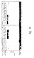
Figures 1A-1D are autoradiograms depicting the results of SDS-PAGE analysis of ubiquitination assays performed in the presence and absence of representative modulating agents. Unless otherwise indicated, the substrate was an 35S-labelled, HA-tagged IκB polypeptide that was phosphorylated and NF-κB complex-associated. - In
Figure 1A ,lane 1 shows the ubiquitination of an IκBα polypeptide that contains alanine residues at positions 32 and 36 (S32/36A; SEQ ID NO:13) andlane 2 shows the ubiquitination of a non-phosphorylated wild-type IκBα polypeptide (SEQ ID NO:12). In lanes 3-14, the ubiquitination substrate was wild-type IκBα (SEQ ID NO:12). Inlane 3, ubiquitination was performed in the absence of ATP; and in lanes 4-14 the reaction was performed in the presence of ATPγS with (lanes 5-14) or without (lane 4) a candidate peptide modulating agent. The candidate agents shown are: 400µM c-Fos phosphopeptide (ppFos (SEQ ID NO:10), lane 5); 400µM serine 32, 36 to alanine substituted IκBα peptide (pp21S/A (SEQ ID NO:11), lane 6); 40µM doubly phosphorylated IκBα peptide (pp21 (SEQ ID NO:9), lane 7); 400µM non-phosphorylated, IκBα peptide (p21 (SEQ ID NO:9), lane 8); 100µM singly phosphorylated IκBα peptides (ppS32 (SEQ ID NO:9),lane 9; ppS36 (SEQ ID NO:9), lane 10); and 40µM shorter, doubly phosphorylated IκBα peptides (pp19 (SEQ ID NO:8), lane 11); pp15 (SEQ ID NO:7), lane 12; pp1 (SEQ ID NO:6), lane 13; pp7 (SEQ ID NO:5), lane 14). - In
Figure 1B , the ubiquitination substrate was free wild type IκBα (SEQ ID NO:12, lanes 1-3) or free S32/36A substituted IκBα (SEQ ID NO:13, lanes 4-6). The reaction was performed in the absence (lanes 1 and 4) or presence (lanes lanes - In
Figure 1C , the ubiquitination of bulk cellular proteins in HeLa extract is shown.Lane 1 shows the ubiquitination in the absence of ATP, andlane 5 shows the ubiquitination in the presence of ATP. In lanes 3-5, candidate modulating agents were added: 40µM doubly phosphorylated IκBα peptide (pp21 (SEQ ID NO:9), lane 2); 400µM c-Fos phosphopeptide (ppFos (SEQ ID NO:10), lane 3); and 400µM non-phosphorylated IκBα peptide (p21 (SEQ ID NO:9), lane 4). - In
Figure 1D , the ubiquitination substrate was phosphorylated (lanes 2-7) or non-phosphorylated (lane 1) wild type IκBβ (SEQ ID NO:14). Reactions were performed in the absence (lane 2) or presence (lanes 1, 3-7) of ATPγS, and with (lanes 4-7) or without (lanes 1-3) a candidate peptide modulating agent. The candidate agents shown are: 40µM doubly phosphorylated IκBα peptide (pp21 (SEQ ID NO:9), lane 4); 400µM c-Fos phosphopeptide (ppFos (SEQ ID NO:10), lane 5); 40µM doubly phosphorylated IκBα peptide (pp19 (SEQ ID NO:8), lane 6); and 400µM non-phosphorylated IκBα peptide (p21 (SEQ ID NO:9), lane 7). -
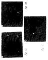
Figure 2 is an autoradiogram depicting the results of an in vitro ubiquitin-dependent degradation assay performed using extracts from stimulated HeLa cells. In each lane of the SDS-PAGE, the level of phosphorylated (upper band) and non-phosphorylated (lower band) HA-tagged IκBα polypeptide (SEQ ID NO:12) following the degradation assay is shown.Lane 1 shows the level of these polypeptides following a degradation assay performed without ATP. In lanes 2-6, ATP was included in the reaction mixture. 40µM candidate modulating agents were added to the reactions shown in lanes 3-6: doubly phosphorylated IκBα peptide (pp21 (SEQ ID NO:9), lane 3); doubly phosphorylated IκBα peptide (pp19 (SEQ ID NO:8), lane 4); c-Fos phosphopeptide (ppFos (SEQ ID NO:10), lane 5); and non-phosphorylated IκBα peptide (p21 (SEQ ID NO:9), lane 6). -
Figure 3A is an autoradiogram depicting the results of SDS-PAGE analysis of ubiquitination assays performed using flow-through fractions of HeLa cell lysate fractionated over modulating agent columns. In each case, the substrate was a 33S-labelled, HA-tagged IκBα polypeptide (SEQ ID NO: 12) that was phosphorylated and NF-κB complex-associated.Lane 1 shows the level of ubiquitination using a non-fractionated extract. In lanes 2-9, the extract was fractionated over a peptide-Sepharose® column. The peptides used were: c-Fos phosphopeptide (ppFos (SEQ ID NO:10), lane 2); serine 32, 36 to alanine substituted IκBα peptide (pp21 S/A (SEQ ID NO:11), lane 3); doubly phosphorylated IκBα peptide (pp21 (SEQ ID NO:9), lanes 4-6); and doubly phosphorylated IκBα peptide (pp19 (SEQ ID NO:8), lanes 7-9). In addition, reticulocyte Fraction II (160 µg) was added to the ubiquitination reactions shown inlanes lanes -
Figure 3B is an autoradiogram showing the ubiquitination of bulk cellular proteins in HeLa extract.Lane 1 shows the ubiquitination in the absence of ATP, andlane 2 shows the ubiquitination in the presence of ATP, but without candidate modulating agent. In lanes 3-6, candidate modulating agents were added: 40µM doubly phosphorylated IκBα peptide (pp19 (SEQ ID NO:8), lane 3); 400µM c-Fos phosphopeptide (ppFos (SEQ ID NO:10), lane 4); 400µM serine 32, 36 to alanine substituted IκBα peptide (pp21S/A (SEQ ID NO:11), lane 5); and 40µM doubly phosphorylated IκBα peptide (pp21 (SEQ ID NO:9), lane 6). -
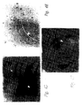
Figures 4A-4F are micrographs showing the effect of candidate modulating agents on nuclear NF-κB translocation. InFigures 4A-C , pp21 (Figures 4A and 4B ) or ppFos (Figure 4C ) was microinjected into the cytoplasm of HeLa cells. Cells were then activated immediately with TNFα and immunostained with anti-p65 antibodies. InFigures 4D-F , pp21 (Figure 4D ) or ppFos (Figure 4F ) was injected into the cytoplasm of human vascular endothelial cells (HUVEC). Cells were then activated immediately with TNFα and immunostained with anti-E-selectin antibodies.Figure 4E is a phase contrast photograph ofFigure 4D . In each micrograph, the injected cells are marked by large arrows. A non-injected, E-selectin negative cell is marked by a small arrow inFigures 4D and 4E . -
Figures 4G and 4H are graphs presenting a summary of the microinjection experiments shown inFigures 4A - 4F . InFigure 4G , the percent of HeLa cells displaying nuclear p65 staining is shown. 90 and 42 cells were microinjected with pp21 and ppFos, respectively.Figure 4H shows the percent of HUVEC displaying E-selectin staining. 160 and 36 cells were microinjected with pp21 and ppFos, respectively. For each graph,column 1 shows the level in the absence of candidate modulating agent and TNFα activation. Columns 2-4 show the level following TNFα activation in the absence of candidate modulating agent (column 2) or in the presence of pp21 (column 3) or ppFos (column 4). - As noted above, the present invention is generally directed to compositions useful for modulating the activation of nuclear factor κB (NF-κB) and for treating diseases associated with such activation. In particular, the invention is directed to agents that modulate ubiquitination of phosphorylated IκB (i.e., IκBα and/or IκBβ), and to methods for identifying such agents.
- In response to a stimulus, IκB associated with NF-κB is activated (i.e., phosphorylated), rendering IκB a target for degradation and thereby releasing and activating NF-κB. It has been found, within the context of the present invention, that phosphorylated and NF-κB-associated IκB is recognized by a specific ubiquitin ligase, E3. The N-terminal signal-induced phosphorylation site that is functionally conserved between IκBα and IκBβ constitutes the E3 recognition motif and is distinct from the nearby ubiquitination site. Peptides corresponding to this motif, and variants thereof, as described in
claim 3, inhibit the ubiquitination of IκB and its subsequent degradation, and such peptides are modulating agents within the scope of the present invention. - An in vitro ubiquitination assay that reproduces the in vivo ubiquitination of IκBα with high fidelity is described herein. In vivo, IκB is targeted for degradation by phosphorylation at serines 32 and 36, while altered forms of IκBα that contain alanine residues at positions 32 and 36 are not subject to ubiquitin conjugation. Similarly, phosphorylation at serines 19 and 23 is required for ubiquitination of IκBβ. However, free IκB is recognized by the ubiquitin system in a non-discriminatory manner (i.e., phosphorylation is not required). The ubiquitination assay described herein allows regulation of IκB ubiquitination that corresponds to the regulation observed in vivo.
- IκB polypeptides for use in a ubiquitination assay as described herein may be native human IκBα (SEQ ID NO: 1) or IκBβ (SEQ ID NO:3), or may be a variant of a native protein. As used herein, a variant is a polypeptide that contains one or more substitutions and/or modifications. Variants include truncated polypeptides and polypeptides containing additional amino acid sequences that have minimal influence on the activity of the polypeptide. In particular, variants may contain additional amino acid sequences at the amino and/or carboxy termini. Such sequences may be used, for example, to facilitate purification or detection of the polypeptide. Polypeptide variants of IκB are modified such that the ability of the variant to be phosphorylated and ubiquitinated within a ubiquitination assay as described herein is not substantially diminished. Preferably, the IκB polypeptide is labeled. For example, 35S may be incorporated into a IκB polypeptide by in vitro translation of the polypeptide in the presence of 35S-methionine, using standard techniques.
- An IκB polypeptide may generally be prepared from DNA encoding the polypeptide by expression of the DNA in cultured host cells or by translation using an in vitro system such as wheat germ extract. If host cells are employed, such cells are preferably are bacteria, yeast, baculovirus-infected insect cells or mammalian cells. The recombinant DNA may be cloned into any expression vector suitable for use within the host cell, using techniques well known to those of ordinary skill in the art. In vitro translation of polypeptide may generally be performed according to the manufacturer's instructions.
- The DNA sequences expressed in this manner may encode native IκBα or IκBβ, or may encode portions or variants of a native IκB. DNA molecules encoding variants may generally be prepared using standard mutagenesis techniques, such as oligonucleotide-directed site-specific mutagenesis. Sections of the DNA sequence may also, or alternatively, be removed to permit preparation of truncated polypeptides and DNA encoding additional sequences such as "tags" may be added to the 5' or 3' end of the DNA molecule. For example, DNA encoding an IκB polypeptide may also encode an epitope, such that the recombinant protein contains the epitope at the N- or C-terminus. Epitopes such as glutathione-S transferase protein (GST), HA (hemaglutinin)-tag, FLAG and Histidine-tag may be added using techniques well known to those of ordinary skill in the art.
- Expressed IκB polypeptides may be used without purification following in vitro translation. Alternatively, a polypeptide may be isolated in substantially pure form. An IκB polypeptide may be isolated to a purity of at least 80% by weight, preferably to a purity of at least 95% by weight, and more preferably to a purity of at least 99% by weight. In general, such purification may be achieved using, for example, the representative purification method described herein or the standard techniques of ammonium sulfate fractionation, SDS-PAGE electrophoresis, and affinity chromatography.
- Within a ubiquitination assay as described herein, cellular extracts from stimulated or non-stimulated Jurkat, HeLa, THP-1 or endothelial cells are incubated in vitro with an IκB polypeptide in the presence of ATP and the phosphatase inhibitor okadaic acid. Cellular extracts may generally be prepared according to the method of Alkalay et al., Proc. Natl. Acad. Sci. USA 92:10599, 1995. The incubation is performed under conditions sufficient to result in phosphorylation of the IκB polypeptide (at serines 32 and 36 for IκBα and variants thereof) and association of the phosphorylated polypeptide (pIκB) with the cellular-derived NF-κB complex. For example, IκB polypeptide may be incubated with HeLa or Jurkat cell extract, ATP and okadaic acid. Incubation for 90 minutes at 30°C is generally sufficient to allow phosphorylation of the IκB polypeptide. Following this incubation, the pIκB/NF-κB complex may be immunopurified with, for example, anti-p65 antibodies and subjected to in vitro ubiquitination in a cell free system, as described by Alkalay et al., Proc. Natl. Acad Sci. USA 92:10599, 1995. The level of ubiquitination may then be evaluated using the well known techniques of SDS-PAGE, followed by autoradiography.
- Under these conditions, a wild type 35S-pIκBα polypeptide generates multiply ubiquitinated species in the presence of ATPyS (see
Figure 1A , lane 4). Neither 35S-labeled S32/36A mutant of IκBα (lane 1), nor the non-phosphorylated wild type 35S-IκBα (lane 2) are ubiquitinated. However, free forms of either mutant or wild type IκBα are readily conjugated (Figure 1B ). Similarly, a free (but not a complex-associated) lysine 21, 22 mutant of IκBα can be ubiquitinated in vitro. Thus, unlike ubiquitination assays performed using free IκB polypeptides, the ubiquitination assay described herein targets only IκB polypeptides that are complex-associated and appropriately phosphorylated. - A ubiquitination assay as described above may be used to identify agents that modulate ubiquitination of IκB. Modulating agents may include antibodies (e.g., monoclonal), peptides and other drugs that stimulate or, preferably, inhibit ubiquitination of an IκBα and/or IκBβ polypeptide. In general, such agents may be identified by including a candidate modulating agent in the ubiquitination reaction, which may otherwise be performed as described above, and evaluating the effect of the agent on the level of ubiquitination. A suitable concentration of candidate agent for use in such an assay generally ranges from about 0.1 µM to about 1 mM. For peptide candidate agents, a peptidase inhibitor such as Bestatin (40 µg/mL) may also be added, and the amount of peptide preferably ranges from about 10 µM to about 1 mM. A candidate agent that results in a statistically significant effect on the level of ubiquitination is a modulating agent.
- As noted above, it has been found, within the context of the present invention, that complex-associated IκB is recognized by a specific ubiquitin ligase, E3. Accordingly, modulating agents within the scope of the present invention are peptides that comprise a recognition domain for E3 ubiquitin ligase and which consist of the amino acid sequence of SEQ ID NO.5, SEQ ID NO. 6, SEQ ID NO. 7, SEQ ID NO. 8, SEQ ID NO. 9 or residues 21-41 of SEQ ID NO. 1. Such peptides may be derived from the N-terminal signaling domain (
residues 1 to 54 of native IκBα or IκBβ) and, at minimum, should contain the signaling phosphorylation site (residues 32 to 36 of native IκBα or residues 19 to 23 of native IκBβ). Peptide modulating agents may generally be prepared using standard automated synthesis techniques or by expression of recombinant DNA encoding the desired peptide. Such agents may differ in sequence from native IκBα and IκBβ due to one or more substitutions and/or modifications, as described above, provided that the peptide variant inhibits ubiquitination of an IκB polypeptide. - For maximal inhibition, peptide modulating agents should be phosphorylated; preferably at both of the native phosphorylation sites (e.g., serines 32 and 36 of IκBα), although singly phosphorylated peptides may be employed. Phosphorylated peptides may be prepared using well known techniques. For example phosphoserine residues may be incorporated into a peptide during synthesis. Alternatively, a peptide may be phosphorylated using standard techniques following synthesis.
- In general, peptide modulating agents may be prepared using standard techniques, incorporating amino acids and/or amino acid analogs. During synthesis, active groups of amino acids and/or amino acid analogs may be protected as necessary using, for example, a t-butyldicarbonate (t-BOC) group or a fluorenylmethoxy carbonyl (FMOC) group. Amino acids and amino acid analogs may be purchased commercially (e.g., Sigma Chemical Co.; Advanced Chemtec) or synthesized using methods known in the art. Peptides may be synthesized using a solid phase method, in which the peptides are attached to a resin such as 4-methylbenzhydrylamine (MBHA), 4-(oxymethyl)-phenylacetamido methyl- and 4-(hydroxymethyl)phenoxy methyl-copoly(styrene-1% divinylbenzene) (Wang resin), all of which are commercially available, or to p-nitrobenzophenone oxime polymer (oxime resin) which can be synthesized as described by De Grado and Kaiser, J. Org. Chem. 47:3258, 1982. Those skilled in the art will realize that the choice of amino acids and/or amino acid analogs will depend, in part, on the specific physical, chemical or biological characteristics desired. Such characteristics are determined, in part, by the method of administration and the target location within a patient.
- Selective modification of the reactive groups in a peptide can also impart desirable characteristics. Peptides can be manipulated while still attached to the resin to obtain N-terminal modified compounds such as an acetylated peptide or can be removed from the resin using hydrogen fluoride or an equivalent cleaving agent and then modified. Compounds synthesized containing the C-terminal carboxy group (Wang resin) can be modified after cleavage from the resin or, in some cases, prior to solution phase synthesis. Methods for modifying the N-terminus or C-terminus of a peptide are well known in the art and include, for example, methods for acetylation of the N-terminus or amidation of the C-terminus. Similarly, methods for modifying side chains of the amino acids or amino acid analogs are well known to those skilled in the art of peptide synthesis. The choice of modifications made to reactive groups present on the peptide will be determined by the desired characteristics.
- A modulating agent may also be a cyclic peptide. A cyclic peptide can be obtained by inducing the formation of a covalent bond between, for example, the amino group at the N-terminus of the peptide and the carboxyl group at the C-terminus. Alternatively, a cyclic peptide can be obtained by forming a covalent bond between a terminal reactive group and a reactive amino acid side chain or between two reactive side chains. It will be apparent to those of skill in the art that a cyclic peptide is selected based on the desired properties. For example, a cyclic peptide may provide increased stability, increased solubility, decreased immunogenicity or decreased clearance in vivo.
- A newly synthesized peptide can be purified using a method such as reverse phase high performance liquid chromatography (RP-HPLC) or other methods of separation based on size or charge. Furthermore, a purified peptide can be characterized using these and other well known methods such as amino acid analysis and mass spectrometry.
- Some representative examples of peptide modulating agents are provided in Table I.
Table I Representative Peptide Modulating Agents Peptide Sequence pp7 CDS*GLDS*M pp 11 CDDRHDS*GLDS*M pp15 CDDRHDS*GLDS*MKDEE pp19 CERLLDDRHDS*GLDS*MKDEE pp21 CKKERLLDDRHDS*GLDS*MKDEE * indicates phosphorylated residue - Further characterization of modulating agents may be achieved using a ubiquitin-dependent in vitro degradation assay. Such an assay may generally be performed as described by Alkalay et al., Proc. Natl. Acad. Sci. USA 92:10599, 1995. Within this assay, pIκBα from stimulated cells is degraded in vitro in a ubiquitin-dependent manner, whereas non-phosphorylated IκBα from the same cell extract is not subject to degradation. Modulating agents that inhibit ubiquitination of IκBα should also result in stabilization of pIκBα within such an in vitro degradation assay.
- Modulating agents as described herein may generally be used to specifically inhibit cellular NF-κB functions. Such inhibition may generally be demonstrated by microinjection of the agent (e.g., about 5 mg/mL of a peptide agent) into a suitable cell (e.g., HeLa cell or primary human vascular endothelial cell). Following microinjection, cells are stimulated (e.g., with TNFα) and incubated to allow NF-κB activation. In HeLa cells, TNFα induces rapid nuclear translocation of NF-κB into the nucleus, which may be detected by staining with p65-specific antibodies. Modulating agents induce a statistically significant decrease in NF-κB translocation, and may reduce such translocation to undetectable levels.
- Primary human vascular endothelial cells (HWEC) respond to TNFα stimulation by surface expression of NF-κB regulated adhesion proteins such as ICAM-1, V-CAM-1 and E-selectin (Read et al., Immunity 2:493,1995; Chen et al., J. Immunol 155:3538, 1995). E-selectin expression is particularly NF-κB dependent and is the major inducible endothelial adhesion molecule for initial neutrophil attachment and rolling on activated endothelium. Stimulated cells may be fixed and stained to detect expression of one or more NF-κB regulated adhesion proteins. Microinjection of a modulating agent results in a statistically significant inhibition of such expression, but does not affect the expression of NF-κB independent adhesion proteins, such as ICAM2.
- Modulating agents may also be used to modulate ubiquitination of IκBα and/or IκBβ in a patient, thereby modulating NF-κB cellular function in vivo. As used herein, a "patient" may be any mammal, including a human, and may be afflicted with a disease associated with NF-κB activation, or may be free of detectable disease. Accordingly, the treatment may be of an existing disease or may be prophylactic. Diseases associated with NF-κB activation include inflammatory diseases, autoimmune diseases, cancer and viral infection.
- Treatment refers to administration of a modulating agent as described herein. For administration to a patient, one or more such compounds are generally formulated as a pharmaceutical composition. A pharmaceutical composition may be a sterile aqueous or non-aqueous solution, suspension or emulsion, which additionally comprises a physiologically acceptable carrier (i.e., a non-toxic material that does not interfere with the activity of the active ingredient). Any suitable carrier known to those of ordinary skill in the art may be employed in the pharmaceutical compositions of the present invention. Representative carriers include physiological saline solutions, gelatin, water, alcohols, natural or synthetic oils, saccharide solutions, glycols, injectable organic esters such as ethyl oleate or a combination of such materials. Optionally, a pharmaceutical composition may additionally contain preservatives and/or other additives such as, for example, antimicrobial agents, anti-oxidants, chelating agents and/or inert gases, and/or other active ingredients.
- Alternatively, a pharmaceutical composition may comprise a polynucleotide encoding a modulating agent (such that the modulating agent is generated in situ) in combination with a physiologically acceptable carrier. In such pharmaceutical compositions, the polynucleotide may be present within any of a variety of delivery systems known to those of ordinary skill in the art, including nucleic acid, bacterial and viral expression systems, as well as colloidal dispersion systems, including liposomes. Appropriate nucleic acid expression systems contain the necessary polynucleotide sequences for expression in the patient (such as a suitable promoter and terminating signal). DNA may also be "naked," as described, for example, in Ulmer et al., Science 259:1745-1749, 1993.
- Various viral vectors that can be used to introduce a nucleic acid sequence into the targeted patient's cells include, but are not limited to, vaccinia or other pox virus, herpes virus, retrovirus, or adenovirus. Techniques for incorporating DNA into such vectors are well known to those of ordinary skill in the art. Preferably, the retroviral vector is a derivative of a murine or avian retrovirus including, but not limited to, Moloney murine leukemia virus (MoMuLV), Harvey murine sarcoma virus (HaMuSV), murine mammary tumor virus (MuMTV), and Rous Sarcoma Virus (RSV). A retroviral vector may additionally transfer or incorporate a gene for a selectable marker (to aid in the identification or selection of transduced cells) and/or a gene that encodes the ligand for a receptor on a specific target cell (to render the vector target specific). For example, retroviral vectors can be made target specific by inserting a nucleotide sequence encoding a sugar, a glycolipid, or a protein. Targeting may also be accomplished using an antibody, by methods known to those of ordinary skill in the art.
- Viral vectors are typically non-pathogenic (defective), replication competent viruses, which require assistance in order to produce infectious vector particles. This assistance can be provided, for example, by using helper cell lines that contain plasmids that encode all of the structural genes of the retrovirus under the control of regulatory sequences within the LTR, but that are missing a nucleotide sequence which enables the packaging mechanism to recognize an RNA transcript for encapsulation. Such helper cell lines include (but are not limited to) ψ2, PA317 and PA12. A retroviral vector introduced into such cells can be packaged and vector virion produced. The vector virions produced by this method can then be used to infect a tissue cell line, such as NIH 3T3 cells, to produce large quantities of chimeric retroviral virions.
- Another targeted delivery system for polynucleotides is a colloidal dispersion system. Colloidal dispersion systems include macromolecule complexes, nanocapsules, microspheres, beads, and lipid-based systems including oil-in-water emulsions, micelles, mixed micelles, and liposomes. A preferred colloidal system for use as a delivery vehicle in vitro and in vivo is a liposome (i.e., an artificial membrane vesicle). It has been shown that large unilamellar vesicles (LUV), which range in size from 0.2-4.0 µm can encapsulate a substantial percentage of an aqueous buffer containing large macromolecules. RNA, DNA and intact virions can be encapsulated within the aqueous interior and be delivered to cells in a biologically active form (Fraley, et al., Trends Biochem. Sci. 6:77, 1981). In addition to mammalian cells, liposomes have been used for delivery of polynucleotides in plant, yeast and bacterial cells. In order for a liposome to be an efficient gene transfer vehicle, the following characteristics should be present: (1) encapsulation of the genes of interest at high efficiency while not compromising their biological activity; (2) preferential and substantial binding to a target cell in comparison to non-target cells; (3) delivery of the aqueous contents of the vesicle to the target cell cytoplasm at high efficiency; and (4) accurate and effective expression of genetic information (Mannino, et al., Biotechniques 6:882, 1988).
- The targeting of liposomes can be classified based on anatomical and mechanistic factors. Anatomical classification is based on the level of selectivity and may be, for example, organ-specific, celt-specific, and/or organelle-specific. Mechanistic targeting can be distinguished based upon whether it is passive or active. Passive targeting utilizes the natural tendency of liposomes to distribute to cells of the reticuloendothelial system (RES) in organs which contain sinusoidal capillaries. Active targeting, on the other hand, involves alteration of the liposome by coupling the liposome to a specific ligand such as a monoclonal antibody, sugar, glycolipid, or protein, or by changing the composition or size of the liposome in order to achieve targeting to organs and cell types other than the naturally occurring sites of localization.
- Routes and frequency of administration, as well doses, will vary from patient to patient. In general, the pharmaceutical compositions may be administered intravenously, intraperitoneally, intramuscularly, subcutaneously, intracavity or transdermally. Between 1 and 6 doses may be administered daily. A suitable dose is an amount that is sufficient to show improvement in the symptoms of a patient afflicted with a disease associated with NF-κB activation. Such improvement may be detected by monitoring inflammatory responses (e.g., edema, transplant rejection, hypersensitivity) or through an improvement in clinical symptoms associated with the disease. In general, the amount of modulating agent present in a dose, or produced in situ by DNA present in a dose, ranges from about 1 µg to about 100 mg per kg of host. Suitable dose sizes will vary with the size of the patient, but will typically range from about 10 mL to about 500 mL for 10-60 kg animal.
- The following Examples are offered by way of illustration and not by way of limitation.
- This Example illustrates a representative ubiquitination assay, and the use of such an assay to evaluate candidate modulating agents.
- HA-tagged IκBα or HA-tagged IκBβ cDNAs (Haskill et al., Cell 65:1281-1289, 1991) were translated in vitro in wheat germ extract in the presence of 35S-methionine according to the manufacturer's instructions (Promega, Madison, WI). To phosphorylate IκBα or IκBβ, 1 µl of the extract containing the labeled protein was incubated for 90 minutes at 30°C in a reaction mixture having a final volume of 30 µl: 100 µg HeLa or Jurkat cell extract (prepared as described by Alkalay et al., Proc. Natl. Acad. Sci. USA 92:10599, 1995), 2mM ATP and 1 µM okadaic acid. During this incubation, the labeled IκB polypeptide was phosphorylated at serines 32 and 36, and associated with the endogenous NF-κB complex (data not shown).
- Following incubation, 1 µl of anti-p65 serum was added, and the NF-κB immune complex was immobilized to Protein A-Sepharose® and subjected to in vitro ubiquitination in HeLa cell extract as described by Alkalay et al.. Ubiquitinated proteins were separated by SDS-PAGE and visualized by autoradiography.
- As shown in
Figure 1A , only wild type 35S-pIκBα generated multiply ubiquitinated species (lane 4). Neither 35S-labeled S32/36A mutant of IκBα (lane 1) nor the non-phosphorylated wild type 35S-IκBα (lane 2) were ubiquitinated, and no ubiquitination of pIκBα was seen in the absence of ATP (lane 3). - The physiological relevance of this assay was further documented by comparison of in vitro ubiquitination of free 35S-IκB to that of a complex-associated, phosphorylated substrate. Whereas a complex-associated S32/36A mutant was not subject to ubiquitin conjugation in accordance with its in vivo fate, free forms of either mutant or wild type IκBα were readily conjugated (
Figure 1B ). Similarly, only free, but not a complex-associated lysine 21, 22 mutant of IκBα could be ubiquitinated in vitro (data not shown). Thus, while the free IκBα is recognized by the ubiquitin system in a non-discriminatory manner, the complex-associated inhibitor is masked unless it is appropriately phosphorylated. - To identify the IκBα-ubiquitin ligase recognition motif, various peptides were added at varying concentrations to the reaction mixtures in the presence of the peptidase inhibitor Bestatin (40 µg/ml). The peptides spanned the N-terminal signaling domain of the protein, and were phosphorylated at one or both serine residues (32 and 36), or were unmodified or serine-substituted. These peptides were included in the ubiquitination reaction at different concentrations and tested for inhibition of pIκBα specific ubiquitination. When conjugation of free IκBα was monitored, the translated protein was added directly to the conjugation reaction mixture.
- Only peptides that were phosphorylated at both serine 32 and 36 (pIκBα peptides) effectively inhibited pIκBα ubiquitination (
Figure 1A ,lanes 7, 11-14). A c-Fos phosphopeptide (ppFos, lane 5), a serine 32, 36 to alanine substituted IκBα peptide (p21 S/A, lane 6) and a non-phosphorylated peptide (p21, lane 8) had no detectable effect on the ubiquitination of pIκB at a concentration of 400 µM. The IC50 of the phosphorylated IκBα peptides were calculated and representative inhibitory concentrations are shown inFigure 1A . Doubly phosphorylated IκBα peptides inhibited the pIκB conjugation reaction (lanes 7, 11-14) at an IC50 of 5 µM. The sequences of these peptides are provided in Table I, above, and in SEQ ID NOs:5-9. In contrast, singly phosphorylated peptides (lanes 9, 10) inhibited the pIκBα conjugation at an IC50 of 400 µM. The minimal size peptide tested (pp7, lane 14), merely spanning the signaling phosphorylation site, was sufficient to effectively inhibit the ubiquitination, although at somewhat higher IC50 (10 µM). Thus, a peptide comprising residues 21 to 41 of SEQ ID NO:1 comprises a recognition domain for E3 ubiquitin ligase. Interestingly, lysine residues 21 and 22 are not essential for inhibition, implying that the ubiquitin-system recognition site is distinct from the actual conjugation site. - The specificity of the peptide modulating agents was tested in two other ubiquitin-conjugation reactions: the conjugation of free wild type (
Figure 1B lanes 1-3) or S32/36A mutant IκBα (Figure 1B , lanes 4-6) and the ubiquitin conjugation to the bulk of cellular proteins in HeLa extract (detected by 125I-labeled ubiquitin according to Alkalay et al.,Figure 1C ). Neither reaction was affected by the addition of a peptide modulating agent or a control peptide. - Peptide modulating agents were found to abolish the ubiquitination of the pIκBα related substrate pIκBβ (
Figure 1D ). Similar to the conjugation of pIκBα, the specific conjugation of the IκBβ also required an associated NF-κB complex (not shown) and prior phosphorylation at the IκBα-homologous residues Ser 19 and 23. An IκBβ substrate prepared in the absence of phosphatase inhibitors was not subject to ubiquitination (Figure 1D , lane 1). Peptide modulating agents affected pIκBβ ubiquitination at an IC50 that was similar to that observed for pIκBα (Figure 1D , lanes 4-7). Hence, it appears that the same enzyme(s) target both IκBs for ubiquitin-dependent degradation. - The inhibitory pIκBα peptides were tested in a complementary ubiquitin-dependent in vitro degradation assay (Orian et al., J. Biol. Chem. 270:21707, 1995; Stancovski et al., Mol. Cell. Biol. 15:7106, 1995). Using this assay, only pIκBα derived from stimulated cells is degraded in vitro in a ubiquitin-dependent manner, whereas the non-phosphorylated IκBα from the same cell extract is not subject to degradation. Incorporation of the conjugation-inhibitory phosphopeptide modulating agents into the degradation assay resulted in stabilization of the pIκBα substrate (
Figure 2 ,lanes 3, 4) whereas the non-phosphorylated peptide agent or a control phospho-Fos peptide had no effect on the specific pIκBα degradation (lanes 5, 6). Trimming the peptides at Lys 21/22 did not diminish the degradation inhibitory effect (lane 4), indicating that the peptides do not abolish pIκBα degradation by exhausting the ubiquitin-proteasome system as conjugatable substrates. - This Example illustrates the identification of a specific E3 that is responsible for recognition of pIκB polypeptides.
- pIκBα-ubiquitin conjugation and degradation requires a full complement of the ubiquitin system enzymes: E1, a specific E2 derived from the ubiquitin system fraction I, E2F1 (Alkalay et al., Proc. Natl. Acad. Sci. USA 92:10599, 1995; Chen et al., Cell 84:853, 1996) and a Fraction II-component E3. To identify the ubiquitin system component involved in the substrate recognition, HeLa lysate was fractionated over IκBα-phosphopeptide modulating agent columns, and the flow-through fractions were assayed for pIκBα conjugation. Peptides were coupled to NHS-Sepharose® (Pharmacia) according to the manufacturer's instructions at a concentration of 2 mg/ml. 100 µg of HeLa extract were incubated with 2.5 µl coupled resin in the presence of 0.1% NP40 and 3% ovalbumin for 1 hour at 4°C. The resin was discarded and the unbound material tested in the ubiquitination assay described above.
- Whereas a flow-through fraction from a control phosphopeptide column and an S32/36A peptide column retained full IκBαconjugation capacity (
Figure 3A ,lanes 2, 3) flow-through fractions from two different pIκBα peptides lost their IκBα specific conjugation capacity (lanes 4, 7). The depleted conjugating activity could be complemented by reticulocyte Fraction II (lanes 5, 8) that contains all the known species of E3 enzymes (Ciechanover, Cell 79:13, 1994). Complementation could not be obtained by the addition of Fraction I or Fraction I and E1 (lanes Figure 3A ,lane 7 to lane 4), emphasizing the distinction between the substrate recognition and conjugation site. The peptide column depletion was found to be specific for the IκB E3, as all flow-through fractions maintained full activity in random HeLa protein conjugation (as detected by measuring the conjugation of 125I ubiquitin,Figure 3B ). This indicates that a specific E3 is responsible for recognition of the pIκBs at the identified motif. - This Example illustrates the inhibition of cellular NF-κB activation by microinjection of peptide modulating agents.
- HeLa cells were plated on a grid coverslips (Cellocate, Eppendorf) 18 hours before microinjection. Microinjection was performed with a 22 amino acid pIκBα peptide (pp21; Table I and SEQ ID NO:9) or a control phospho-Fos peptide (SEQ ID NO:10) using a semi-automated apparatus (Eppendorf). Peptides were injected into the cell cytoplasm at a concentration of 5 mg/ml in 100 mM KCl, 5mM Na2HPO4 (pH 7.2), and immediately activated with TNFα (200 units/mL) for either 20 minutes (for NF-κB translocation) or 3 hours (for E-selectin expression). Following activation, the cells were fixed and stained with p65 specific antibodies (Mercurio et al., Genes & Dev. 7:705, 1993; Santa Cruz) or monoclonal anti-E-selectin antibodies (R&D Systems).
- In the absence of peptide modulating agent, TNFα induces rapid nuclear translocation of NF-κB into the nucleus, as shown by the p65 nuclear staining of 90% of the cells (see
Figure 4G , column 2). The pp21 peptide abolished TNFα-stimulated NF-κB activation in 50%-70% of the microinjected cells in several experiments (see representative fields inFigures 4A and 4B ; andFigure 4G , column 3). In contrast, the control pp-Fos peptide had no effect on the rate of NF-κB induced nuclear translocation, as compared to non-microinjected cells (Figures 4C and4G , column 4). - To further assess the functional consequences of NF-κB inhibition, the IκB-E3 inhibitory peptide was microinjected into primary human vascular endothelial cells (HUVEC; Chen et al, J. Immunol 155:3538, 1995). These cells respond to TNFα stimulation by surface expression of NF-κB regulated adhesion proteins, such as E-selectin. HUVEC cells were plated, microinjected and stimulated as described above. Three hours post stimulation the cells were fixed and stained for expression of the NF-κB dependent E-selectin. 75%-85% of the HUVEC cells were intensely stained for E-selectin following TNFα stimulation in several experiments. Microinjection of the pp21 peptide resulted in the inhibition of E-selectin expression in 70%-80% of the microinjected cells (
Figure 4D ; andFigure 4H , column 3). In contrast, the control pp-Fos peptide had no effect on E-selectin expression, as compared to non-microinjected cells (Figures 4F and4H , column 4). Microinjection of a control, S32/36A substituted IκBα peptide had no effect on the rate of E-selectin expression (data not shown). - These results demonstrate that the subunit-specific degradation of the signal-induced phosphorylated IκBα and IκBβ is mediated by a specific E3. The recognition domain for E3 ubiquitin ligase is a short sequence, centered around the two signal-acquired phosphoserines conserved in both IκBs, representing the first biologically relevant E3 recognition motif. The specificity in IκB recognition is supported by the context of the phosphorylated substrate: an associated cellular complex masks the substrate from non-specific E3s. This feature restricts the NF-κB inhibitor degradation to the post-stimulation phase, at which it is exposed through site-specific phosphorylation event(s) to the specific ligase. NF-κB activation and its resultant function can be specifically abolished by in vivo inhibition of the IκB ligase, using a modulating agent as provided herein.
-
- SEQ ID NO: 1 is amino acid sequence of IκBα
- SEQ ID NO:2 is DNA sequence of IκBα
- SEQ ID NO:3 is amino acid sequence of IκBβ
- SEQ ID NO:4 is DNA sequence of IκBβ
- SEQ ID NO:5 is amino acid sequence of pp7
- SEQ ID NO:6 is amino acid sequence of pp11
- SEQ ID NO:7 is amino acid sequence of pp15
- SEQ ID NO: 8 is amino acid sequence of pp 19
- SEQ ID NO:9 is amino acid sequence of pp21
- SEQ ID NO: 10 is amino acid sequence of phospho-Fos peptide
- SEQ ID NO:11 is amino acid sequence of pp21 S/A
- SEQ ID NO: 12 is amino acid sequence of HA-tagged IκBα
- SEQ ID NO: 13 is amino acid sequence of HA-tagged S32, 36 IκBα
- SEQ ID NO:14 is amino acid sequence of HA-tagged IκBβ
-
- (1) GENERAL INFORMATION:
- (i) APPLICANTs: Alkalay, Irit
Ben-Neriah, Yinon
Ciechanover, Aaron
Manning, Anthony
Mercurio, Frank
Yaron, Avraham - (ii) TITLE OF INVENTION: COMPOSITIONS AND METHODS FOR MODULATING CELLULAR NF-κB ACTIVATION
- (iii) NUMBER OF SEQUENCES: 14
- (iv) CORRESPONDENCE ADDRESS:
- (A) ADDRESSEE: SEED and BERRY LLP
- (B) STREET: 6300 Columbia Center, 701 Fifth Avenue
- (C) CITY: Seattle
- (D) STATE: Washington
- (E) COUNTRY: USA
- (F) ZIP: 98104
- (v) COMPUTER READABLE FORM:
- (A) MEDIUM TYPE: Floppy disk
- (B) COMPUTER: IBM PC compatible
- (C) OPERATING SYSTEM: PC-DOS/MS-DOS
- (D) SOFTWARE: PatentIn Release #1.0, Version #1.30
- (vi) CURRENT APPLICATION DATA:
- (A) APPLICATION NUMBER: US
- (B) FILING DATE: 14-FEB-1997
- (C) CLASSIFICATION:
- (viii) ATTORNEY/AGENT INFORMATION:
- (A) NAME: Maki, David J.
- (B) REGISTRATION NUMBER: 31,392
- (C) REFERENCE/DOCKET NUMBER: 860098.418
- (ix) TELECOMMUNICATION INFORMATION:
- (A) TELEPHONE: (206) 622-4900
- (B) TELEFAX: (206) 682-6031
- (i) APPLICANTs: Alkalay, Irit
- (2) INFORMATION FOR SEQ ID NO:1:
- (i) SEQUENCE CHARACTERISTICS:
- (A) LENGTH: 317 amino acids
- (B) TYPE: amino acid
- (C) STRANDEDNESS:
- (D) TOPOLOGY: linear
- (xi) SEQUENCE DESCRIPTION: SEQ ID NO:1:


- (i) SEQUENCE CHARACTERISTICS:
- (2) INFORMATION FOR SEQ ID NO:2:
- (i) SEQUENCE CHARACTERISTICS:
- (A) LENGTH: 1550 base pairs
- (B) TYPE: nucleic acid
- (C) STRANDEDNESS: single
- (D) TOPOLOGY: linear
- (xi) SEQUENCE DESCRIPTION: SEQ ID NO:2:


- (i) SEQUENCE CHARACTERISTICS:
- (2) INFORMATION FOR SEQ ID NO:3:
- (i) SEQUENCE CHARACTERISTICS:
- (A) LENGTH: 359 amino acids
- (B) TYPE: amino acid
- (C) STRANDEDNESS:
- (D) TOPOLOGY: linear
- (xi) SEQUENCE DESCRIPTION: SEQ ID NO:3:


- (i) SEQUENCE CHARACTERISTICS:
- (2) INFORMATION FOR SEQ ID NO:4:
- (i) SEQUENCE CHARACTERISTICS:
- (A) LENGTH: 1212 base pairs
- (B) TYPE: nucleic acid
- (C) STRANDEDNESS: single
- (D) TOPOLOGY: linear
- (xi) SEQUENCE DESCRIPTION: SEQ ID NO:4:


- (i) SEQUENCE CHARACTERISTICS:
- (2) INFORMATION FOR SEQ ID NO:5:
- (i) SEQUENCE CHARACTERISTICS:
- (A) LENGTH: 8 amino acids
- (B) TYPE: amino acid
- (C) STRANDEDNESS:
- (D) TOPOLOGY: linear
- (xi) SEQUENCE DESCRIPTION: SEQ ID NO:5:

- (i) SEQUENCE CHARACTERISTICS:
- (2) INFORMATION FOR SEQ ID NO:6:
- (i) SEQUENCE CHARACTERISTICS:
- (A) LENGTH: 12 amino acids
- (B) TYPE: amino acid
- (C) STRANDEDNESS:
- (D) TOPOLOGY: linear
- (xi) SEQUENCE DESCRIPTION: SEQ ID NO:6:

- (i) SEQUENCE CHARACTERISTICS:
- (2) INFORMATION FOR SEQ ID NO:7:
- (i) SEQUENCE CHARACTERISTICS:
- (A) LENGTH: 16 amino acids
- (B) TYPE: amino acid
- (C) STRANDEDNESS:
- (D) TOPOLOGY: linear
- (xi) SEQUENCE DESCRIPTION: SEQ ID NO:7:

- (i) SEQUENCE CHARACTERISTICS:
- (2) INFORMATION FOR SEQ ID NO:8:
- (i) SEQUENCE CHARACTERISTICS:
- (A) LENGTH: 20 amino acids
- (B) TYPE: amino acid
- (C) STRANDEDNESS:
- (D) TOPOLOGY: linear
- (xi) SEQUENCE DESCRIPTION: SEQ ID NO:8:

- (i) SEQUENCE CHARACTERISTICS:
- (2) INFORMATION FOR SEQ ID NO:9:
- (i) SEQUENCE CHARACTERISTICS:
- (A) LENGTH: 22 amino acids
- (B) TYPE: amino acid
- (C) STRANDEDNESS:
- (D) TOPOLOGY: linear
- (xi) SEQUENCE DESCRIPTION: SEQ ID NO:9:

- (i) SEQUENCE CHARACTERISTICS:
- (2) INFORMATION FOR SEQ ID NO:10:
- (i) SEQUENCE CHARACTERISTICS:
- (A) LENGTH: 19 amino acids
- (B) TYPE: amino acid
- (C) STRANDEDNESS:
- (D) TOPOLOGY: linear
- (xi) SEQUENCE DESCRIPTION: SEQ ID NO:10:

- (i) SEQUENCE CHARACTERISTICS:
- (2) INFORMATION FOR SEQ ID NO:11:
- (i) SEQUENCE CHARACTERISTICS:
- (A) LENGTH: 22 amino acids
- (B) TYPE: amino acid
- (C) STRANDEDNESS:
- (D) TOPOLOGY: linear
- (xi) SEQUENCE DESCRIPTION: SEQ ID NO:11:

- (i) SEQUENCE CHARACTERISTICS:
- (2) INFORMATION FOR SEQ ID NO:12:
- (i) SEQUENCE CHARACTERISTICS:
- (A) LENGTH: 347 amino acids
- (B) TYPE: amino acid
- (C) STRANDEDNESS:
- (D) TOPOLOGY: linear
- (xi) SEQUENCE DESCRIPTION: SEQ ID NO:12:


- (i) SEQUENCE CHARACTERISTICS:
- (2) INFORMATION FOR SEQ ID NO:13:
- (i) SEQUENCE CHARACTERISTICS:
- (A) LENGTH: 347 amino acids
- (B) TYPE: amino acid
- (C) STRANDEDNESS:
- (D) TOPOLOGY: linear
- (xi) SEQUENCE DESCRIPTION: SEQ ID NO:13:


- (i) SEQUENCE CHARACTERISTICS:
- (2) INFORMATION FOR SEQ ID NO:14:
- (i) SEQUENCE CHARACTERISTICS:
- (A) LENGTH: 389 amino acids
- (B) TYPE: amino acid
- (C) STRANDEDNESS:
- (D) TOPOLOGY: linear
- (xi) SEQUENCE DESCRIPTION: SEQ ID NO:14:


- (i) SEQUENCE CHARACTERISTICS:
Claims (13)
- A method for identifying an agent that modulates ubiquitination of IκBα and/or IκBβ, comprising:(a) incubating a candidate agent with an IκB polypeptide and a cellular extract, wherein the step of incubating is carried out under conditions and for a time sufficient to allow phosphorylation of the IκB polypeptide and formation of a complex comprising phosphorylated IκB polypeptide and NF-κB; and(b) subsequently measuring the ability of the candidate agent to modulate ubiquitination of the complex by comparing inhibition of ubiquitination by the candidate agent to inhibition of ubiquitination by one or more polypeptides consisting of the amino acid sequence of SEQ ID NO:5, SEQ ID NO:6, SEQ ID NO:7, SEQ ID NO:8 or SEQ ID NO:9, and therefrom identifying an agent that modulates ubiquitination of IκBα and/or IκBβ;wherein said agent is a fragment of IκBα, a fragment of IκBβ, a drug or an antibody.
- A method according to claim 1, wherein the fragment comprises a recognition domain for E3 ubiquitin ligase which comprises at least residues 32 to 36 of native IkBα or residues 19 to 23 of native IkBβ.
- An agent that modulates ubiquitination of IκBα and/or IκBβ, wherein the agent is a fragment of IκBα that comprises a recognition domain for E3 ubiquitin ligase, wherein the agent consists of the amino acid sequence of SEQ ID NO.5, SEQ ID NO.6, SEQ ID NO.7, SEQ ID NO.8, SEQ ID NO.9 or residues 21-41 of SEQ ID NO.1.
- An agent according to claim 3, wherein each of the serines of the peptide is phosphorylated.
- An agent according to claim 3, wherein the agent consists of residues 21-41 of SEQ ID NO. 1.
- An isolated DNA molecule encoding a polypeptide that, when appropriately phosphorylated, is an agent according to at least one of claims 3 to 5.
- A recombinant expression vector comprising a DNA molecule according to claim 6.
- A host cell transformed or transfected with an expression vector according to claim 7.
- A host cell according to claim 8, wherein the host cell is selected from the group consisting of bacteria, yeast, baculovirus infected insect cells and mammalian cells.
- A pharmaceutical composition comprising an agent according to any of claims 3 to 5 in combination with a pharmaceutically acceptable carrier.
- An agent according to any one of claims 3 to 5, for use in therapy.
- Use of an agent, according to any one of claims 3 to 5 in the manufacture of a medicament for treating a patient afflicted with a disorder associated with NF-κB activation, wherein said disorder is an inflammatory disease, cancer, an autoimmune disease or a viral infection.
- An agent according to claim 11, wherein the disorder is selected from the group consisting of inflammatory diseases, autoimmune diseases, cancer and viral infection.
Applications Claiming Priority (3)
| Application Number | Priority Date | Filing Date | Title |
|---|---|---|---|
| US802322 | 1997-02-18 | ||
| US08/802,322 US5932425A (en) | 1997-02-18 | 1997-02-18 | Compositions and methods for modulating cellular NF-κB activation |
| PCT/US1998/003163 WO1998036070A1 (en) | 1997-02-18 | 1998-02-18 | COMPOSITIONS AND METHODS FOR MODULATING CELLULAR NF-λB ACTIVATION |
Publications (2)
| Publication Number | Publication Date |
|---|---|
| EP1009824A1 EP1009824A1 (en) | 2000-06-21 |
| EP1009824B1 true EP1009824B1 (en) | 2008-04-02 |
Family
ID=25183386
Family Applications (1)
| Application Number | Title | Priority Date | Filing Date |
|---|---|---|---|
| EP98908593A Expired - Lifetime EP1009824B1 (en) | 1997-02-18 | 1998-02-18 | COMPOSITIONS AND METHODS FOR MODULATING CELLULAR NF-kappaB ACTIVATION |
Country Status (9)
| Country | Link |
|---|---|
| US (1) | US5932425A (en) |
| EP (1) | EP1009824B1 (en) |
| JP (2) | JP4560616B2 (en) |
| AT (1) | ATE391177T1 (en) |
| AU (1) | AU6659198A (en) |
| CA (1) | CA2281920C (en) |
| DE (1) | DE69839326T2 (en) |
| HK (1) | HK1028784A1 (en) |
| WO (1) | WO1998036070A1 (en) |
Families Citing this family (24)
| Publication number | Priority date | Publication date | Assignee | Title |
|---|---|---|---|---|
| JP4695735B2 (en) * | 1996-08-26 | 2011-06-08 | シグナル ファーマシューティカルズ, エルエルシー | Stimulation-inducible I (κ) B kinase [IKK] signalsome |
| US6896899B2 (en) * | 1996-12-31 | 2005-05-24 | Antioxidant Pharmaceuticals Corp. | Pharmaceutical preparations of glutathione and methods of administration thereof |
| DE69739663D1 (en) | 1996-12-31 | 2009-12-31 | Antioxidant Pharmaceuticals Co | PHARMACEUTICAL GLUTATHION PREPARATIONS AND METHODS OF ADMINISTRATION |
| CA2330210A1 (en) * | 1998-06-13 | 1999-12-23 | Zarpex Biosciences Limited | Proteasomal activity |
| JP4633259B2 (en) * | 1998-12-10 | 2011-02-16 | シグナル ファーマシューティカルズ, エルエルシー | Compounds and methods for modulating NF-κB activation |
| US6468755B1 (en) | 1999-08-10 | 2002-10-22 | Joslin Diabetes Center, Inc. | Method for identifying compounds for treatment of insulin resistance |
| US6630312B2 (en) | 1999-08-10 | 2003-10-07 | Joslin Diabetes Center, Inc. | Method for identifying compounds for treatment of insulin resistance |
| US20030104622A1 (en) * | 1999-09-01 | 2003-06-05 | Robbins Paul D. | Identification of peptides that facilitate uptake and cytoplasmic and/or nuclear transport of proteins, DNA and viruses |
| FR2801902B1 (en) * | 1999-12-01 | 2005-03-04 | Centre Nat Rech Scient | METHODS OF FUNCTIONAL SCREENING OF AGENTS FOR MODULATING THE ACTIVITY OF UBIQUITIN-SCF LIGASE COMPLEXES AND THEIR APPLICATIONS |
| WO2001079555A2 (en) | 2000-04-14 | 2001-10-25 | Millennium Pharmaceuticals, Inc. | Roles of jak/stat family members in tolerance induction |
| US20030044852A1 (en) * | 2000-08-10 | 2003-03-06 | Joslin Diabetes Center, Inc., A Massachusetts Corporation | Methods for treatment of insulin resistance |
| US20060287337A1 (en) * | 2001-11-09 | 2006-12-21 | Proteologics, Inc. | Trans-golgi network-associated processes, methods and compositions related thereto |
| AU2002349784A1 (en) * | 2001-12-03 | 2003-06-17 | Asahi Kasei Pharma Corporation | Nf-kappab activating genes |
| FR2859733A1 (en) * | 2003-09-15 | 2005-03-18 | Centre Nat Rech Scient | Cell-based screen for compounds that modulate ubiquitin hydrolase, useful for treating e.g. cancer, neurodegeneration and infections, uses a yeast that expresses Met4 under control of a repressible promoter |
| WO2005085731A1 (en) * | 2004-03-02 | 2005-09-15 | The Procter & Gamble Company | A method for curing high internal phase emulsions |
| FR2867783B1 (en) * | 2004-03-16 | 2008-02-01 | Cytomics Systems | METHOD OF SCREENING AGENT MODULATING THE UBIQUITINATION OF THE IKBALPHA PROTEIN AND MEANS FOR CARRYING OUT SAID METHOD |
| US20070060537A1 (en) * | 2005-04-28 | 2007-03-15 | Proteologics, Inc. | Methods and compositions for inhibiting viral infections |
| US20100292306A1 (en) * | 2005-05-24 | 2010-11-18 | Carlson C George | Compositions And Methods For The Treatment Of Muscular Dystrophy |
| WO2007101202A1 (en) * | 2006-02-27 | 2007-09-07 | Research Development Foundation | Cell-targeted ikb and methods for the use thereof |
| AR066016A1 (en) * | 2007-04-11 | 2009-07-15 | Alcon Res Ltd | USE OF AN ALFA TNF INHIBITOR TOGETHER WITH AN ANTIHISTAMINE TO TREAT ALLERGIC RHINITIS AND ALLERGIC CONJUNCTIVITIS |
| US20090182035A1 (en) * | 2007-04-11 | 2009-07-16 | Alcon Research, Ltd. | Use of a combination of olopatadine and cilomilast to treat non-infectious rhinitis and allergic conjunctivitis |
| WO2010044892A1 (en) * | 2008-10-17 | 2010-04-22 | President And Fellows Of Harvard College | Diagnostic method based on large scale identification of post-translational modification of proteins |
| JP5721722B2 (en) * | 2009-10-01 | 2015-05-20 | アルコン リサーチ, リミテッド | Olopatadine composition and use thereof |
| SG11202106517WA (en) | 2018-12-28 | 2021-07-29 | Regeneron Pharma | Treatment of respiratory disorders with arachidonate15-lipoxygenase (alox15) inhibitors |
Family Cites Families (6)
| Publication number | Priority date | Publication date | Assignee | Title |
|---|---|---|---|---|
| US5122463A (en) * | 1990-05-17 | 1992-06-16 | Massachusetts Institute Of Technology | Methods for trans-destabilization of specific proteins in vivo and dna molecules useful therefor |
| CA2148258A1 (en) * | 1992-10-29 | 1994-05-11 | Nicholas Barrie La Thangue | Transcription factor dp-1 |
| DE4243770A1 (en) * | 1992-12-23 | 1994-06-30 | Behringwerke Ag | Process for the production of recombinant and synthetic peptides and their use |
| IL114615A0 (en) * | 1995-07-16 | 1995-11-27 | Yeda Res & Dev | Modulators of the function of fas receptors and other proteins |
| WO1997000690A1 (en) * | 1995-06-23 | 1997-01-09 | Tularik, Inc. | Interleukin-1 receptor-associated protein kinase and assays |
| JP4695735B2 (en) * | 1996-08-26 | 2011-06-08 | シグナル ファーマシューティカルズ, エルエルシー | Stimulation-inducible I (κ) B kinase [IKK] signalsome |
-
1997
- 1997-02-18 US US08/802,322 patent/US5932425A/en not_active Expired - Lifetime
-
1998
- 1998-02-18 JP JP53601398A patent/JP4560616B2/en not_active Expired - Fee Related
- 1998-02-18 CA CA2281920A patent/CA2281920C/en not_active Expired - Fee Related
- 1998-02-18 DE DE69839326T patent/DE69839326T2/en not_active Expired - Lifetime
- 1998-02-18 WO PCT/US1998/003163 patent/WO1998036070A1/en active Application Filing
- 1998-02-18 EP EP98908593A patent/EP1009824B1/en not_active Expired - Lifetime
- 1998-02-18 AT AT98908593T patent/ATE391177T1/en not_active IP Right Cessation
- 1998-02-18 AU AU66591/98A patent/AU6659198A/en not_active Abandoned
-
2000
- 2000-08-17 HK HK00105172A patent/HK1028784A1/en not_active IP Right Cessation
-
2009
- 2009-11-06 JP JP2009255576A patent/JP2010063461A/en not_active Withdrawn
Non-Patent Citations (2)
| Title |
|---|
| CHEN Z J; PARENT L; MANIATIS T: "SITE-SPECIFIC PHOSPHORYLATION OF IKAPPABALPHA BY A NOVEL UBIQUITINATION-DEPENDENT PROTEIN KINASE ACTIVITY", CELL, CELL PRESS, vol. 84, 22 March 1996 (1996-03-22), CAMBRIDGE, NA, US, pages 853 - 862, XP002037788 * |
| LI CHOU-CHI H; DAI REN-MING; LONGO DAN L: "Inactivation of NF-kappa-B inhibitor I-kappa-B-alpha: Ubiquitin-dependent proteolysis and its degradation product", BIOCHEMICAL AND BIOPHYSICAL RESEARCH COMMUNICATIONS, vol. 215, no. 1, 4 October 1999 (1999-10-04), UNITED STATES, pages 292 - 301 * |
Also Published As
| Publication number | Publication date |
|---|---|
| JP2010063461A (en) | 2010-03-25 |
| DE69839326T2 (en) | 2009-04-16 |
| EP1009824A1 (en) | 2000-06-21 |
| WO1998036070A1 (en) | 1998-08-20 |
| AU6659198A (en) | 1998-09-08 |
| JP4560616B2 (en) | 2010-10-13 |
| DE69839326D1 (en) | 2008-05-15 |
| US5932425A (en) | 1999-08-03 |
| CA2281920C (en) | 2010-05-04 |
| ATE391177T1 (en) | 2008-04-15 |
| JP2001512319A (en) | 2001-08-21 |
| HK1028784A1 (en) | 2001-03-02 |
| CA2281920A1 (en) | 1998-08-20 |
Similar Documents
| Publication | Publication Date | Title |
|---|---|---|
| EP1009824B1 (en) | COMPOSITIONS AND METHODS FOR MODULATING CELLULAR NF-kappaB ACTIVATION | |
| US7414120B2 (en) | Compounds and methods for modulating activation of NF-κB | |
| AU726383B2 (en) | Stimulus-inducible I (KAPPA)B kinase (IKK) signalsome | |
| CN100482795C (en) | IREN protein, its preparation and use | |
| US20070065804A1 (en) | Method for PR-39 peptide mediated selective inhibition of IkappaBalpha degradation | |
| US20120076809A1 (en) | Method to inhibit the biological activity of calcineurin with selective peptides | |
| US7449563B2 (en) | Methods and compositions for destabilizing microtubules | |
| AU2599001A (en) | Method for pr-39 peptide mediated selective inhibition of ikappabalpha degradation | |
| WO1997030712A1 (en) | METHOD FOR INHIBITING NF-'kappa'B ACTIVATION | |
| US7169604B1 (en) | Method for PR-39 peptide mediated selective inhibition of IκBα degradation |
Legal Events
| Date | Code | Title | Description |
|---|---|---|---|
| PUAI | Public reference made under article 153(3) epc to a published international application that has entered the european phase |
Free format text: ORIGINAL CODE: 0009012 |
|
| 17P | Request for examination filed |
Effective date: 19990916 |
|
| AK | Designated contracting states |
Kind code of ref document: A1 Designated state(s): AT BE CH DE DK ES FI FR GB GR IE IT LI LU MC NL PT SE |
|
| RIN1 | Information on inventor provided before grant (corrected) |
Inventor name: YARON, AVRAHAM Inventor name: CIECHANOVER, AARON Inventor name: BEN-NERIAH, YINON Inventor name: ALKALAY, IRIT Inventor name: MERCURIO, FRANK Inventor name: MANNING, ANTHONY |
|
| 17Q | First examination report despatched |
Effective date: 20040312 |
|
| RAP1 | Party data changed (applicant data changed or rights of an application transferred) |
Owner name: YISSUM RESEARCH DEVELOPMENT COMPANY OF THE HEBREW Owner name: SIGNAL PHARMACEUTICALS LLC |
|
| 17Q | First examination report despatched |
Effective date: 20040312 |
|
| GRAP | Despatch of communication of intention to grant a patent |
Free format text: ORIGINAL CODE: EPIDOSNIGR1 |
|
| GRAS | Grant fee paid |
Free format text: ORIGINAL CODE: EPIDOSNIGR3 |
|
| GRAA | (expected) grant |
Free format text: ORIGINAL CODE: 0009210 |
|
| AK | Designated contracting states |
Kind code of ref document: B1 Designated state(s): AT BE CH DE DK ES FI FR GB GR IE IT LI LU MC NL PT SE |
|
| REG | Reference to a national code |
Ref country code: GB Ref legal event code: FG4D |
|
| REG | Reference to a national code |
Ref country code: IE Ref legal event code: FG4D Ref country code: CH Ref legal event code: EP |
|
| REF | Corresponds to: |
Ref document number: 69839326 Country of ref document: DE Date of ref document: 20080515 Kind code of ref document: P |
|
| REG | Reference to a national code |
Ref country code: HK Ref legal event code: GR Ref document number: 1028784 Country of ref document: HK |
|
| REG | Reference to a national code |
Ref country code: CH Ref legal event code: NV Representative=s name: PATENTANWAELTE SCHAAD, BALASS, MENZL & PARTNER AG |
|
| NLV1 | Nl: lapsed or annulled due to failure to fulfill the requirements of art. 29p and 29m of the patents act | ||
| PG25 | Lapsed in a contracting state [announced via postgrant information from national office to epo] |
Ref country code: PT Free format text: LAPSE BECAUSE OF FAILURE TO SUBMIT A TRANSLATION OF THE DESCRIPTION OR TO PAY THE FEE WITHIN THE PRESCRIBED TIME-LIMIT Effective date: 20080902 Ref country code: NL Free format text: LAPSE BECAUSE OF FAILURE TO SUBMIT A TRANSLATION OF THE DESCRIPTION OR TO PAY THE FEE WITHIN THE PRESCRIBED TIME-LIMIT Effective date: 20080402 Ref country code: FI Free format text: LAPSE BECAUSE OF FAILURE TO SUBMIT A TRANSLATION OF THE DESCRIPTION OR TO PAY THE FEE WITHIN THE PRESCRIBED TIME-LIMIT Effective date: 20080402 Ref country code: ES Free format text: LAPSE BECAUSE OF FAILURE TO SUBMIT A TRANSLATION OF THE DESCRIPTION OR TO PAY THE FEE WITHIN THE PRESCRIBED TIME-LIMIT Effective date: 20080713 |
|
| PG25 | Lapsed in a contracting state [announced via postgrant information from national office to epo] |
Ref country code: AT Free format text: LAPSE BECAUSE OF FAILURE TO SUBMIT A TRANSLATION OF THE DESCRIPTION OR TO PAY THE FEE WITHIN THE PRESCRIBED TIME-LIMIT Effective date: 20080402 |
|
| ET | Fr: translation filed | ||
| PG25 | Lapsed in a contracting state [announced via postgrant information from national office to epo] |
Ref country code: SE Free format text: LAPSE BECAUSE OF FAILURE TO SUBMIT A TRANSLATION OF THE DESCRIPTION OR TO PAY THE FEE WITHIN THE PRESCRIBED TIME-LIMIT Effective date: 20080702 Ref country code: DK Free format text: LAPSE BECAUSE OF FAILURE TO SUBMIT A TRANSLATION OF THE DESCRIPTION OR TO PAY THE FEE WITHIN THE PRESCRIBED TIME-LIMIT Effective date: 20080402 |
|
| PLBE | No opposition filed within time limit |
Free format text: ORIGINAL CODE: 0009261 |
|
| STAA | Information on the status of an ep patent application or granted ep patent |
Free format text: STATUS: NO OPPOSITION FILED WITHIN TIME LIMIT |
|
| PG25 | Lapsed in a contracting state [announced via postgrant information from national office to epo] |
Ref country code: BE Free format text: LAPSE BECAUSE OF FAILURE TO SUBMIT A TRANSLATION OF THE DESCRIPTION OR TO PAY THE FEE WITHIN THE PRESCRIBED TIME-LIMIT Effective date: 20080402 |
|
| 26N | No opposition filed |
Effective date: 20090106 |
|
| PG25 | Lapsed in a contracting state [announced via postgrant information from national office to epo] |
Ref country code: MC Free format text: LAPSE BECAUSE OF NON-PAYMENT OF DUE FEES Effective date: 20090228 |
|
| PG25 | Lapsed in a contracting state [announced via postgrant information from national office to epo] |
Ref country code: GR Free format text: LAPSE BECAUSE OF FAILURE TO SUBMIT A TRANSLATION OF THE DESCRIPTION OR TO PAY THE FEE WITHIN THE PRESCRIBED TIME-LIMIT Effective date: 20080703 |
|
| PG25 | Lapsed in a contracting state [announced via postgrant information from national office to epo] |
Ref country code: LU Free format text: LAPSE BECAUSE OF NON-PAYMENT OF DUE FEES Effective date: 20090218 |
|
| PGFP | Annual fee paid to national office [announced via postgrant information from national office to epo] |
Ref country code: IT Payment date: 20110225 Year of fee payment: 14 |
|
| PG25 | Lapsed in a contracting state [announced via postgrant information from national office to epo] |
Ref country code: IT Free format text: LAPSE BECAUSE OF NON-PAYMENT OF DUE FEES Effective date: 20120218 |
|
| PGFP | Annual fee paid to national office [announced via postgrant information from national office to epo] |
Ref country code: IE Payment date: 20130226 Year of fee payment: 16 Ref country code: CH Payment date: 20130225 Year of fee payment: 16 Ref country code: DE Payment date: 20130227 Year of fee payment: 16 Ref country code: FR Payment date: 20130311 Year of fee payment: 16 Ref country code: GB Payment date: 20130227 Year of fee payment: 16 |
|
| REG | Reference to a national code |
Ref country code: DE Ref legal event code: R119 Ref document number: 69839326 Country of ref document: DE |
|
| REG | Reference to a national code |
Ref country code: CH Ref legal event code: PL |
|
| GBPC | Gb: european patent ceased through non-payment of renewal fee |
Effective date: 20140218 |
|
| PG25 | Lapsed in a contracting state [announced via postgrant information from national office to epo] |
Ref country code: CH Free format text: LAPSE BECAUSE OF NON-PAYMENT OF DUE FEES Effective date: 20140228 Ref country code: LI Free format text: LAPSE BECAUSE OF NON-PAYMENT OF DUE FEES Effective date: 20140228 |
|
| REG | Reference to a national code |
Ref country code: FR Ref legal event code: ST Effective date: 20141031 |
|
| REG | Reference to a national code |
Ref country code: IE Ref legal event code: MM4A |
|
| REG | Reference to a national code |
Ref country code: DE Ref legal event code: R119 Ref document number: 69839326 Country of ref document: DE Effective date: 20140902 |
|
| PG25 | Lapsed in a contracting state [announced via postgrant information from national office to epo] |
Ref country code: IE Free format text: LAPSE BECAUSE OF NON-PAYMENT OF DUE FEES Effective date: 20140218 Ref country code: DE Free format text: LAPSE BECAUSE OF NON-PAYMENT OF DUE FEES Effective date: 20140902 Ref country code: FR Free format text: LAPSE BECAUSE OF NON-PAYMENT OF DUE FEES Effective date: 20140228 Ref country code: GB Free format text: LAPSE BECAUSE OF NON-PAYMENT OF DUE FEES Effective date: 20140218 |