EP1009484B1 - Method for corneal laser surgery - Google Patents
Method for corneal laser surgery Download PDFInfo
- Publication number
- EP1009484B1 EP1009484B1 EP97945439A EP97945439A EP1009484B1 EP 1009484 B1 EP1009484 B1 EP 1009484B1 EP 97945439 A EP97945439 A EP 97945439A EP 97945439 A EP97945439 A EP 97945439A EP 1009484 B1 EP1009484 B1 EP 1009484B1
- Authority
- EP
- European Patent Office
- Prior art keywords
- flap
- cornea
- tissue
- plug
- path
- Prior art date
- Legal status (The legal status is an assumption and is not a legal conclusion. Google has not performed a legal analysis and makes no representation as to the accuracy of the status listed.)
- Expired - Lifetime
Links
- 238000000034 method Methods 0.000 title abstract description 21
- 238000002430 laser surgery Methods 0.000 title abstract description 3
- 210000004087 cornea Anatomy 0.000 claims abstract description 32
- 230000035876 healing Effects 0.000 claims description 3
- 230000001154 acute effect Effects 0.000 claims description 2
- 230000000452 restraining effect Effects 0.000 claims 1
- 230000004438 eyesight Effects 0.000 abstract description 9
- 230000003292 diminished effect Effects 0.000 abstract 1
- 210000001519 tissue Anatomy 0.000 description 32
- 238000012937 correction Methods 0.000 description 17
- 208000001491 myopia Diseases 0.000 description 5
- 230000004379 myopia Effects 0.000 description 5
- 238000001356 surgical procedure Methods 0.000 description 5
- 201000006318 hyperopia Diseases 0.000 description 4
- 230000004305 hyperopia Effects 0.000 description 4
- 206010020675 Hypermetropia Diseases 0.000 description 3
- 230000006735 deficit Effects 0.000 description 3
- 210000001525 retina Anatomy 0.000 description 3
- 239000002775 capsule Substances 0.000 description 2
- 210000003038 endothelium Anatomy 0.000 description 2
- 210000000981 epithelium Anatomy 0.000 description 2
- 230000003287 optical effect Effects 0.000 description 2
- 230000002159 abnormal effect Effects 0.000 description 1
- 210000003484 anatomy Anatomy 0.000 description 1
- 210000004045 bowman membrane Anatomy 0.000 description 1
- 230000007547 defect Effects 0.000 description 1
- 230000007812 deficiency Effects 0.000 description 1
- 230000002950 deficient Effects 0.000 description 1
- 235000012489 doughnuts Nutrition 0.000 description 1
- 239000012528 membrane Substances 0.000 description 1
- 238000004321 preservation Methods 0.000 description 1
- 210000001747 pupil Anatomy 0.000 description 1
- 208000014733 refractive error Diseases 0.000 description 1
- 210000003786 sclera Anatomy 0.000 description 1
Images
Classifications
-
- A—HUMAN NECESSITIES
- A61—MEDICAL OR VETERINARY SCIENCE; HYGIENE
- A61F—FILTERS IMPLANTABLE INTO BLOOD VESSELS; PROSTHESES; DEVICES PROVIDING PATENCY TO, OR PREVENTING COLLAPSING OF, TUBULAR STRUCTURES OF THE BODY, e.g. STENTS; ORTHOPAEDIC, NURSING OR CONTRACEPTIVE DEVICES; FOMENTATION; TREATMENT OR PROTECTION OF EYES OR EARS; BANDAGES, DRESSINGS OR ABSORBENT PADS; FIRST-AID KITS
- A61F9/00—Methods or devices for treatment of the eyes; Devices for putting in contact-lenses; Devices to correct squinting; Apparatus to guide the blind; Protective devices for the eyes, carried on the body or in the hand
- A61F9/007—Methods or devices for eye surgery
- A61F9/008—Methods or devices for eye surgery using laser
- A61F9/00825—Methods or devices for eye surgery using laser for photodisruption
- A61F9/00836—Flap cutting
-
- A—HUMAN NECESSITIES
- A61—MEDICAL OR VETERINARY SCIENCE; HYGIENE
- A61F—FILTERS IMPLANTABLE INTO BLOOD VESSELS; PROSTHESES; DEVICES PROVIDING PATENCY TO, OR PREVENTING COLLAPSING OF, TUBULAR STRUCTURES OF THE BODY, e.g. STENTS; ORTHOPAEDIC, NURSING OR CONTRACEPTIVE DEVICES; FOMENTATION; TREATMENT OR PROTECTION OF EYES OR EARS; BANDAGES, DRESSINGS OR ABSORBENT PADS; FIRST-AID KITS
- A61F9/00—Methods or devices for treatment of the eyes; Devices for putting in contact-lenses; Devices to correct squinting; Apparatus to guide the blind; Protective devices for the eyes, carried on the body or in the hand
- A61F9/007—Methods or devices for eye surgery
- A61F9/008—Methods or devices for eye surgery using laser
-
- A—HUMAN NECESSITIES
- A61—MEDICAL OR VETERINARY SCIENCE; HYGIENE
- A61F—FILTERS IMPLANTABLE INTO BLOOD VESSELS; PROSTHESES; DEVICES PROVIDING PATENCY TO, OR PREVENTING COLLAPSING OF, TUBULAR STRUCTURES OF THE BODY, e.g. STENTS; ORTHOPAEDIC, NURSING OR CONTRACEPTIVE DEVICES; FOMENTATION; TREATMENT OR PROTECTION OF EYES OR EARS; BANDAGES, DRESSINGS OR ABSORBENT PADS; FIRST-AID KITS
- A61F9/00—Methods or devices for treatment of the eyes; Devices for putting in contact-lenses; Devices to correct squinting; Apparatus to guide the blind; Protective devices for the eyes, carried on the body or in the hand
- A61F9/007—Methods or devices for eye surgery
- A61F9/008—Methods or devices for eye surgery using laser
- A61F9/00825—Methods or devices for eye surgery using laser for photodisruption
- A61F9/00827—Refractive correction, e.g. lenticle
-
- A—HUMAN NECESSITIES
- A61—MEDICAL OR VETERINARY SCIENCE; HYGIENE
- A61F—FILTERS IMPLANTABLE INTO BLOOD VESSELS; PROSTHESES; DEVICES PROVIDING PATENCY TO, OR PREVENTING COLLAPSING OF, TUBULAR STRUCTURES OF THE BODY, e.g. STENTS; ORTHOPAEDIC, NURSING OR CONTRACEPTIVE DEVICES; FOMENTATION; TREATMENT OR PROTECTION OF EYES OR EARS; BANDAGES, DRESSINGS OR ABSORBENT PADS; FIRST-AID KITS
- A61F9/00—Methods or devices for treatment of the eyes; Devices for putting in contact-lenses; Devices to correct squinting; Apparatus to guide the blind; Protective devices for the eyes, carried on the body or in the hand
- A61F9/007—Methods or devices for eye surgery
- A61F9/008—Methods or devices for eye surgery using laser
- A61F2009/00861—Methods or devices for eye surgery using laser adapted for treatment at a particular location
- A61F2009/00872—Cornea
Definitions
- the present invention pertains generally to ophthalmic surgery which is useful for correcting vision deficiencies. More particularly, the present invention pertains to apparatus which surgically corrects the vision of a patient by removing portions of the stroma to reshape the cornea.
- Vision impairment can occur for many reasons, and be the result of many causes.
- One, all too common, cause for vision impairment results from a defective condition of the eye which occurs when the refractive characteristics of the cornea do not cause parallel rays of light to focus on the retina.
- myopia i.e. near-sightedness
- hypermetropia i.e. far-sightedness
- Both myopic and hyperopic conditions result in varying degrees of vision impairment and, as is well known, in most cases the conditions are correctable.
- WO-A-9409849 is considered to represent the closest prior art, and discloses computer controlled laser surgical apparatus adapted and programmed to excise a layer of corneal tissue by directing a focused laser beam along a path, the path defining the cut surface.
- the previously removed anterior portion of the cornea is then repositioned on the cornea to cover the photodisruption.
- This procedure like the procedure disclosed in Bille et al. '586, has as its objective the removal of only stromal tissue with the consequent preservation of anterior corneal tissue.
- a significant downside for the "flap and zap" procedure is the possibility that the previously removed anterior portion of the cornea may again become detached. While the intrastromal procedure disclosed by Bille et al. does not lead to this detachment problem it can, in some cases, require extensive laser photodisruption and be time consuming.
- the "flap and zap" procedure can be made more effective and efficient if the "flap” that is created can somehow be repositioned in an interlocking relationship with the undisturbed corneal tissue.
- the present invention recognizes that it would be desirable if, first, a "flap" with an interlockable configuration is created. The flap could then be lifted to expose the corneal tissue that is to be removed and, next, after the desired amount of corneal tissue is removed, the flap could be repositioned and interlocked with undisturbed corneal tissue to hold the "flap" in place during the healing process.
- the surgical procedure employed must be capable of removing corneal tissue having a thickness which is accurate to within less than ten microns. Furthermore, this degree of accuracy applies for any refractive correction regardless of the total amount of correction required.
- An object of the present invention is to provide apparatus for corneal laser surgery which creates an interlocking flap that can be lifted to remove a predetermined volume of tissue from the stroma and then repositioned in an interlocking relationship with undisturbed corneal tissue to hold the flap in place during subsequent healing.
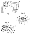
- an apparatus 10 for generating a laser beam 12 is shown. Specifically, the laser beam 12 is shown being directed onto an eye 14 of a patient 16.
- the apparatus 10 is capable of generating a pulsed laser beam 12 having physical characteristics similar to those of the laser beams generated by a laser system as disclosed and claimed in U.S. Patent No.4,764,930 , which is also assigned to the assignee of the present invention.
- the present invention contemplates the use of a pulsed laser beam 12 which has pulses with durations as long as a few nanoseconds or as short as only a few femtoseconds.
- Figure 2 shows the anatomical structure of eye 14 and, specifically, that the cornea 18 is anterior to the pupil 20, the iris 22, and the sclera 24. Additionally, Figure 2 indicates that the optical axis 26 of eye 14 passes through the cornea 18. Consequently, the tissue of cornea 18 is transparent to visible light.
- the cornea 18 includes five anatomically definable layers of tissue. Going in a direction from anterior to posterior in Figure 3 , the tissue layers of the cornea are: epithelium 26, Bowman's membrane 28, stroma 30, Decemet's membrane 32 and endothelium 34. Of these, the stroma 30 is of most importance for the present invention as it contains the only tissue which is to be removed for correction of the patient's vision.
- the correction of a myopic condition can be accomplished by the removal of a predetermined volume of stromal tissue.
- the particular volume of stromal tissue to be removed for the correction of myopia will depend on the amount of correction required and will be a lens or lentoid shaped volume.
- Such a lentoid volume 36 is shown in cross section in Figure 3 .
- the lentoid volume 36 will be defined by an anterior surface 38 and a posterior surface 40. Together, the anterior surface 38 and the posterior surface 40 will completely enclose or encapsulate the lentoid volume 36 of stromal tissue 30 which is to be removed.
- the anterior surface 38 may be convex in shape and the posterior surface 40 may be concave in its shape.
- the computer controlled apparatus 10 is programmed to create a section of corneal tissue which can be moved to expose, and thereby establish access to, the capsule volume 36 that is to be removed.
- a layer 72 of corneal tissue is created which has an undercut region 74 that structurally interacts, or interlocks, with an intact overlap region 76 of the cornea.
- the idea here is to have the undercut region 74 interlock with the overlap region 76 so that the layer 72 does not unintentionally move in an anterior direction.
- the anterior direction is to be considered as the general direction taken from the posterior surface 78 of cornea 18 (i.e. endothelium 34) toward the anterior surface 80 of cornea 18 (i.e. epithelium 26).
- the posterior direction is taken to be from the anterior surface 80 back toward the posterior surface 78.
- a reference axis 82 For a description of how to create the layer 72, it is best to first identify a reference axis 82 from which distances and directions can be taken. For this purpose, consider the reference axis 82 to be oriented generally perpendicular to the anterior surface 80 of cornea 18, and to intersect the anterior surface 80 at a start point 84. For purposes of the present invention, the corneal incisions are made using a laser light beam.
- the creation of layer 72 begins by cutting the corneal tissue along a path 86 which is oriented at an angle 88 from the reference axis 82, and which extends from the start point 84 to a turn point 90.
- the path 86 can result in several different configurations for layer 72.
- One configuration is as shown in Figure 4 .
- the path 86 is essentially a straight line.
- the straight line is set at an acute angle 88 relative to the reference axis 82, and it extends from the start point 84 to the turn point 90.
- the undercut region 74 is then formed by cutting back toward the reference axis 82 along a path 92 which generally parallels the posterior surface 78.
- the path 86 can be something other than a straight line, so long as the start point 88 is generally located on the reference axis 82 and the turn point 90 is distanced from the axis 82 to create the undercut region 74.
- a plurality of different cuts each along the different paths 86 and 92, need to be made in order to create the layer 72.
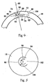
- Reference to Figure 5 shows what the result of these several cuts might be.
- a plurality of start points 84 can be selected to establish a periphery 94.
- the periphery 94 is curvilinear and can be generally defined by a radius of curvature 96.
- a plurality of turn points 90 can be selected to establish a periphery 98.
- the periphery 98 is also curvilinear and can be generally defined by a radius of curvature 100.
- the radius of curvature 100 must be greater than the radius of curvature 96. Additionally, as shown in Figure 5 , the peripheries 94 and 98 do not close. Thus, the hinge area 102 is created and the layer 72 can be lifted as a flap in rotation about the hinge 102.
- the flap 72 can be mechanically lifted to expose an underlying capsule volume 36.
- the volume 36 can have many different sizes and shapes depending on the particular optical problem being confronted.
- the volume 36 can be removed by procedures well known in the pertinent art.
- the layer 72 can be repositioned. When so repositioned, it is intended that the undercut region 74 will interact with overlap region 76 to restrain any further movement of the layer 72 in an anterior direction.
Landscapes
- Health & Medical Sciences (AREA)
- Ophthalmology & Optometry (AREA)
- Heart & Thoracic Surgery (AREA)
- Vascular Medicine (AREA)
- Optics & Photonics (AREA)
- Surgery (AREA)
- Engineering & Computer Science (AREA)
- Biomedical Technology (AREA)
- Physics & Mathematics (AREA)
- Nuclear Medicine, Radiotherapy & Molecular Imaging (AREA)
- Life Sciences & Earth Sciences (AREA)
- Animal Behavior & Ethology (AREA)
- General Health & Medical Sciences (AREA)
- Public Health (AREA)
- Veterinary Medicine (AREA)
- Laser Surgery Devices (AREA)
Abstract
Description
- The present invention pertains generally to ophthalmic surgery which is useful for correcting vision deficiencies. More particularly, the present invention pertains to apparatus which surgically corrects the vision of a patient by removing portions of the stroma to reshape the cornea.
- Vision impairment can occur for many reasons, and be the result of many causes. One, all too common, cause for vision impairment results from a defective condition of the eye which occurs when the refractive characteristics of the cornea do not cause parallel rays of light to focus on the retina. When the eye is at rest, and the rays of light focus in front of the retina, the condition is known as myopia (i.e. near-sightedness). On the other hand, when the rays of light focus behind the retina, the condition is known as hypermetropia or hyperopia (i.e. far-sightedness). Both myopic and hyperopic conditions result in varying degrees of vision impairment and, as is well known, in most cases the conditions are correctable.
- Spectacles or eyeglasses are commonly used to correct myopic or hyperopic conditions. For various reasons, however, many persons who suffer with these conditions prefer not to wear eyeglasses. Fortunately for these individuals, it is known that surgical procedures can be employed which will reshape the cornea in ways that are effective in changing its refractive characteristics. For example,
U.S. Patent NO. 4,665,913 which issued to L'Esperance for an invention entitled "Method for Ophthalmological Surgery", andU.S. Patent No. 4,669,466 which issued to L'Esperance for an invention entitled "Method and Apparatus for Analysis and correction of Abnormal Refractive Errors of the Eye" both disclose a laser system which photoablates corneal tissue from the anterior surface of the eye. In a different manner,U.S. Patent No. 4,988,348 which issued to Bille for an invention entitled "Method for Reshaping the Cornea", and which is assigned to the same assignee as the present invention, discloses a procedure whereby corneal tissue is first removed to correct vision, and then the newly created surface is smoothed. -
WO-A-9409849 - Rather than remove and reshape portions of the anterior portion of the eye to correct refractive defects, some procedures for reshaping the cornea have suggested intrastromal photoablation for removal of only stromal tissue. As an example of such a procedure,
U.S. Patent No. 4,907,586, which issued to Bille et al. for an invention entitled "Method for Reshaping the Eye" discloses an intrastromal photodisruption technique for reshaping the cornea. Another example of a procedure which is intended to essentially remove only stromal tissue is the so-called "flap and zap" procedure. For this procedure, an anterior portion of the cornea is removed and a portion of the exposed stroma is then photoablated. The previously removed anterior portion of the cornea is then repositioned on the cornea to cover the photodisruption. This procedure, like the procedure disclosed in Bille et al. '586, has as its objective the removal of only stromal tissue with the consequent preservation of anterior corneal tissue. A significant downside for the "flap and zap" procedure, however, is the possibility that the previously removed anterior portion of the cornea may again become detached. While the intrastromal procedure disclosed by Bille et al. does not lead to this detachment problem it can, in some cases, require extensive laser photodisruption and be time consuming. - It is appreciated by the present invention that the "flap and zap" procedure can be made more effective and efficient if the "flap" that is created can somehow be repositioned in an interlocking relationship with the undisturbed corneal tissue. To accomplish this, the present invention recognizes that it would be desirable if, first, a "flap" with an interlockable configuration is created. The flap could then be lifted to expose the corneal tissue that is to be removed and, next, after the desired amount of corneal tissue is removed, the flap could be repositioned and interlocked with undisturbed corneal tissue to hold the "flap" in place during the healing process.
- The use of laser systems for ophthalmic surgical procedures, such as for other procedures contemplated for the present invention, is particularly appropriate due to the extreme precision required when corneal tissue is to be removed. Specifically, depending on the diameter and the general shape of the tissue volume to be removed, it is known that the removal of a layer of stromal tissue which is only approximately ten microns thick will result in a one diopter change. More practically, by way of example, the removal of a lens shaped volume of tissue which is four millimeters in diameter and approximately fifty microns thick at its center will result in a refractive correction of approximately four diopters. In almost all cases, for precise vision corrections which can stay within a one diopter accuracy, the surgical procedure employed must be capable of removing corneal tissue having a thickness which is accurate to within less than ten microns. Furthermore, this degree of accuracy applies for any refractive correction regardless of the total amount of correction required.
- It happens that the correction of myopia requires removal of a differently shaped volume of corneal tissue than does the correction of hyperopia. Also, the limits of potential correction are different. Specifically, for a myopic correction it is known that a lentoid or lens shaped volume of stromal tissue needs to be removed. At the present time, myopic corrections of up to approximately thirty diopters can be reasonably expected. On the other hand, corrections of hyperopic conditions can be made up to only about fifteen diopters. Furthermore, for a hyperopic correction a generally doughnut shaped volume of stromal tissue, rather than a lens or lentoid shaped volume, needs to be removed.
- An object of the present invention is to provide apparatus for corneal laser surgery which creates an interlocking flap that can be lifted to remove a predetermined volume of tissue from the stroma and then repositioned in an interlocking relationship with undisturbed corneal tissue to hold the flap in place during subsequent healing.
- In accordance with the present invention, apparatus is provided as in claim 1 appended hereto.
- The novel features of this invention, as well as the invention itself, both as to its structure and its operation will be best understood from the accompanying drawings, taken in conjunction with the accompanying description, in which similar reference characters refer to similar parts, and in which:
-
Figure 1 is a perspective view of a patient being treated with the apparatus of the present invention; -
Figure 2 is a perspective view of an eye; -
Figure 3 is a cross sectional view of the cornea of the eye as seen along the line 3-3 inFigure 2 showing a representative portion of stromal tissue to be removed for the correction of myopia; -
Figure 4 is a cross sectional view of the cornea of an eye; and -
Figure 5 is a plan view of the portion of the cornea shown in Figure 9 looking at the anterior surface thereof in a posterior direction. - Referring initially to
Figure 1 , anapparatus 10 for generating alaser beam 12 is shown. Specifically, thelaser beam 12 is shown being directed onto aneye 14 of apatient 16. For purposes of the present invention, theapparatus 10 is capable of generating apulsed laser beam 12 having physical characteristics similar to those of the laser beams generated by a laser system as disclosed and claimed inU.S. Patent No.4,764,930 , which is also assigned to the assignee of the present invention. Furthermore, the present invention contemplates the use of apulsed laser beam 12 which has pulses with durations as long as a few nanoseconds or as short as only a few femtoseconds. -
Figure 2 shows the anatomical structure ofeye 14 and, specifically, that thecornea 18 is anterior to thepupil 20, theiris 22, and thesclera 24. Additionally,Figure 2 indicates that theoptical axis 26 ofeye 14 passes through thecornea 18. Consequently, the tissue ofcornea 18 is transparent to visible light. - In
Figure 3 it can be seen that thecornea 18 includes five anatomically definable layers of tissue. Going in a direction from anterior to posterior inFigure 3 , the tissue layers of the cornea are:epithelium 26, Bowman'smembrane 28,stroma 30, Decemet'smembrane 32 andendothelium 34. Of these, thestroma 30 is of most importance for the present invention as it contains the only tissue which is to be removed for correction of the patient's vision. - As indicated above, the correction of a myopic condition can be accomplished by the removal of a predetermined volume of stromal tissue. As also indicated above, the particular volume of stromal tissue to be removed for the correction of myopia will depend on the amount of correction required and will be a lens or lentoid shaped volume. Such a
lentoid volume 36 is shown in cross section inFigure 3 . As shown, it is to also be appreciated that thelentoid volume 36 will be defined by ananterior surface 38 and aposterior surface 40. Together, theanterior surface 38 and theposterior surface 40 will completely enclose or encapsulate thelentoid volume 36 ofstromal tissue 30 which is to be removed. To obtain the lens shape of thelentoid volume 36 it will be understood and further appreciated that, when consideringlentoid volume 36 in a direction from anterior to posterior, theanterior surface 38 may be convex in shape and theposterior surface 40 may be concave in its shape. - According to the present invention, the computer controlled
apparatus 10 is programmed to create a section of corneal tissue which can be moved to expose, and thereby establish access to, thecapsule volume 36 that is to be removed. For this particular procedure alayer 72 of corneal tissue is created which has an undercutregion 74 that structurally interacts, or interlocks, with anintact overlap region 76 of the cornea. The idea here is to have the undercutregion 74 interlock with theoverlap region 76 so that thelayer 72 does not unintentionally move in an anterior direction. For purposes of this disclosure, the anterior direction is to be considered as the general direction taken from theposterior surface 78 of cornea 18 (i.e. endothelium 34) toward theanterior surface 80 of cornea 18 (i.e. epithelium 26). Contrarily, the posterior direction is taken to be from theanterior surface 80 back toward theposterior surface 78. - For a description of how to create the
layer 72, it is best to first identify areference axis 82 from which distances and directions can be taken. For this purpose, consider thereference axis 82 to be oriented generally perpendicular to theanterior surface 80 ofcornea 18, and to intersect theanterior surface 80 at astart point 84. For purposes of the present invention, the corneal incisions are made using a laser light beam. - The creation of
layer 72 begins by cutting the corneal tissue along apath 86 which is oriented at anangle 88 from thereference axis 82, and which extends from thestart point 84 to aturn point 90. In practice, depending upon the particular desires of the operator, thepath 86 can result in several different configurations forlayer 72. One configuration, of course, is as shown inFigure 4 . For this particular configuration, thepath 86 is essentially a straight line. In this case, the straight line is set at anacute angle 88 relative to thereference axis 82, and it extends from thestart point 84 to theturn point 90. The undercutregion 74 is then formed by cutting back toward thereference axis 82 along apath 92 which generally parallels theposterior surface 78. Again, depending on the desires of the operation, thepath 86 can be something other than a straight line, so long as thestart point 88 is generally located on thereference axis 82 and theturn point 90 is distanced from theaxis 82 to create the undercutregion 74. - The implication from the above disclosure is that a plurality of different cuts, each along the
different paths layer 72. Reference toFigure 5 , shows what the result of these several cuts might be. InFigure 5 , it will be seen that a plurality of start points 84 can be selected to establish aperiphery 94. As shown, theperiphery 94 is curvilinear and can be generally defined by a radius ofcurvature 96. Further, it can be appreciated by reference toFigure 5 that a plurality of turn points 90 can be selected to establish aperiphery 98. As shown, theperiphery 98 is also curvilinear and can be generally defined by a radius ofcurvature 100. In order for the undercutregion 74 to be established, it will be understood that the radius ofcurvature 100 must be greater than the radius ofcurvature 96. Additionally, as shown inFigure 5 , theperipheries hinge area 102 is created and thelayer 72 can be lifted as a flap in rotation about thehinge 102. - Once the
flap 72 has been created it can be mechanically lifted to expose anunderlying capsule volume 36. As indicated above, thevolume 36 can have many different sizes and shapes depending on the particular optical problem being confronted. In any case, once exposed, thevolume 36 can be removed by procedures well known in the pertinent art. Importantly, after thevolume 36 is removed, thelayer 72 can be repositioned. When so repositioned, it is intended that the undercutregion 74 will interact withoverlap region 76 to restrain any further movement of thelayer 72 in an anterior direction.
Claims (3)
- Computer controlled laser surgical apparatus (10) adapted and programmed to create automatically an interlocking flap (72) of corneal tissue by
establishing a reference axis (82), said reference axis intersecting the anterior surface of the cornea (18) at a first point (84) and being substantially normal to the anterior surface;
cutting through the cornea along a first path (86) from said first point to a second point (90), said first path being oriented at an acute angle from said reference axis, said first path being substantially straight;
back cutting through the cornea along a second path (92) from said second point to a third point, said second path intersecting said axis and being oriented substantially perpendicular thereto to create said flap (72); and
repeating said establishing step, said cutting step, and said back cutting step as required to create an undercut region for said flap (72) with a hinge between said flap and the cornea, said undercut region restraining movement of said flap in a direction toward the anterior surface of the cornea during healing between said flap and the cornea. - Laser apparatus as claimed in Claim 1 wherein said repeated first points establish a first periphery (94) having a first radius of curvature, and wherein said repeated second points establish a second periphery (98) having a second radius of curvature, and further wherein said second radius of curvature is greater than said first radius of curvature.
- Laser apparatus as claimed in Claim 1 wherein said back cutting step is accomplished before said cutting step.
Priority Applications (1)
| Application Number | Priority Date | Filing Date | Title |
|---|---|---|---|
| EP08005039A EP1941849B1 (en) | 1996-10-02 | 1997-10-01 | Apparatus for corneal laser surgery |
Applications Claiming Priority (4)
| Application Number | Priority Date | Filing Date | Title |
|---|---|---|---|
| US08/725,070 US6110166A (en) | 1995-03-20 | 1996-10-02 | Method for corneal laser surgery |
| US725070 | 1996-10-02 | ||
| PCT/US1997/017754 WO1998014244A1 (en) | 1996-10-02 | 1997-10-01 | Method for corneal laser surgery |
| CA002266044A CA2266044C (en) | 1996-10-02 | 1999-03-31 | Method for corneal laser surgery |
Related Child Applications (1)
| Application Number | Title | Priority Date | Filing Date |
|---|---|---|---|
| EP08005039A Division EP1941849B1 (en) | 1996-10-02 | 1997-10-01 | Apparatus for corneal laser surgery |
Publications (3)
| Publication Number | Publication Date |
|---|---|
| EP1009484A1 EP1009484A1 (en) | 2000-06-21 |
| EP1009484A4 EP1009484A4 (en) | 2006-05-31 |
| EP1009484B1 true EP1009484B1 (en) | 2008-07-23 |
Family
ID=32963014
Family Applications (1)
| Application Number | Title | Priority Date | Filing Date |
|---|---|---|---|
| EP97945439A Expired - Lifetime EP1009484B1 (en) | 1996-10-02 | 1997-10-01 | Method for corneal laser surgery |
Country Status (6)
| Country | Link |
|---|---|
| US (1) | US6110166A (en) |
| EP (1) | EP1009484B1 (en) |
| JP (1) | JP2002500522A (en) |
| CA (1) | CA2266044C (en) |
| DE (2) | DE69738854D1 (en) |
| WO (1) | WO1998014244A1 (en) |
Families Citing this family (165)
| Publication number | Priority date | Publication date | Assignee | Title |
|---|---|---|---|---|
| US7892226B2 (en) * | 1995-03-20 | 2011-02-22 | Amo Development, Llc. | Method of corneal surgery by laser incising a contoured corneal flap |
| US6551307B2 (en) * | 2001-03-23 | 2003-04-22 | Gholam A. Peyman | Vision correction using intrastromal pocket and flap |
| US6881197B1 (en) * | 1996-10-25 | 2005-04-19 | Anamed, Inc. | Sutureless implantable device and method for treatment of glaucoma |
| US6554424B1 (en) | 1999-03-01 | 2003-04-29 | Boston Innovative Optices, Inc. | System and method for increasing the depth of focus of the human eye |
| DE19938203A1 (en) | 1999-08-11 | 2001-02-15 | Aesculap Meditec Gmbh | Method and device for correcting visual defects in the human eye |
| CA2331223C (en) * | 2000-03-27 | 2011-08-16 | Intralase Corp. | A method of corneal surgery by laser incising a contoured corneal flap |
| US8668735B2 (en) | 2000-09-12 | 2014-03-11 | Revision Optics, Inc. | Corneal implant storage and delivery devices |
| CA2421948C (en) | 2000-09-12 | 2009-12-22 | Anamed, Inc. | System for packaging and handling an implant and method of use |
| US6451006B1 (en) * | 2001-02-14 | 2002-09-17 | 20/10 Perfect Vision Optische Geraete Gmbh | Method for separating lamellae |
| AU2002307430A1 (en) * | 2001-04-19 | 2002-11-05 | Intralase Corp. | Method and system for photodisruption of tissue of the eye |
| US7568365B2 (en) | 2001-05-04 | 2009-08-04 | President & Fellows Of Harvard College | Method and apparatus for micromachining bulk transparent materials using localized heating by nonlinearly absorbed laser radiation, and devices fabricated thereby |
| DE10124358C1 (en) * | 2001-05-18 | 2002-10-17 | Wavelight Laser Technologie Ag | Laser system for eye surgery, e.g. cornea transplantation, has laser pulse focus controlled for providing rebated cut within cornea |
| US6610050B2 (en) * | 2001-07-27 | 2003-08-26 | 20/10 Perfect Vision, Optische Geraete Gmbh | Laser beam delivery system with multiple focal points |
| US6610051B2 (en) * | 2001-10-12 | 2003-08-26 | 20/10 Perfect Vision Optische Geraete Gmbh | Device and method for performing refractive surgery |
| US7101364B2 (en) | 2001-10-12 | 2006-09-05 | 20/10 Perfect Vision Optische Geraete Gmbh | Method and apparatus for intrastromal refractive surgery |
| US9370446B2 (en) | 2001-11-07 | 2016-06-21 | Gholam A. Peyman | Method of altering the refractive properties of an eye |
| US9814567B2 (en) | 2001-11-07 | 2017-11-14 | Gholam A. Peyman | Method of altering the refractive properties of an eye |
| US20050149006A1 (en) * | 2001-11-07 | 2005-07-07 | Peyman Gholam A. | Device and method for reshaping the cornea |
| US20070088415A1 (en) * | 2001-11-07 | 2007-04-19 | Minu Llc | Method of treating the eye using controlled heat delivery |
| US9681942B2 (en) * | 2001-11-07 | 2017-06-20 | Gholam A. Peyman | Method for prevention of rejection and sever encapsulation of a supportive or functioning implant |
| US20070142828A1 (en) * | 2001-11-07 | 2007-06-21 | Minu, Llc | Method and system for altering the refractive properties of the eye |
| US9681984B2 (en) | 2001-11-07 | 2017-06-20 | Gholam A. Peyman | Method of altering the refractive properties of an eye |
| CN100446739C (en) * | 2002-03-23 | 2008-12-31 | Amo发展有限责任公司 | System for improving material processing using laser beam |
| DE10237945A1 (en) * | 2002-08-20 | 2004-03-11 | Quintis Gmbh | Laser-based device for non-mechanical, three-dimensional trepanation in corneal transplants |
| DE10323422B4 (en) * | 2002-08-23 | 2022-05-05 | Carl Zeiss Meditec Ag | Device and method for measuring an optical breakthrough in a tissue |
| WO2004026198A2 (en) | 2002-08-23 | 2004-04-01 | Carl Zeiss Meditec Ag | Device and method for measuring an optical penetration in a tissue |
| US20040044355A1 (en) * | 2002-08-28 | 2004-03-04 | Nevyas Herbert J. | Minimally invasive corneal surgical procedure for the treatment of hyperopia |
| US7628810B2 (en) | 2003-05-28 | 2009-12-08 | Acufocus, Inc. | Mask configured to maintain nutrient transport without producing visible diffraction patterns |
| US7351241B2 (en) * | 2003-06-02 | 2008-04-01 | Carl Zeiss Meditec Ag | Method and apparatus for precision working of material |
| US7131968B2 (en) * | 2003-06-02 | 2006-11-07 | Carl Zeiss Meditec Ag | Apparatus and method for opthalmologic surgical procedures using a femtosecond fiber laser |
| US20050046794A1 (en) | 2003-06-17 | 2005-03-03 | Silvestrini Thomas A. | Method and apparatus for aligning a mask with the visual axis of an eye |
| DE10332815B4 (en) * | 2003-07-18 | 2020-10-22 | Carl Zeiss Meditec Ag | Method and apparatus for forming curved cut surfaces in a transparent material |
| DE10334108B4 (en) * | 2003-07-25 | 2018-05-09 | Carl Zeiss Meditec Ag | Apparatus for forming a closed, curved cut surface |
| DE10334110A1 (en) * | 2003-07-25 | 2005-02-17 | Carl Zeiss Meditec Ag | Apparatus and method for forming curved cut surfaces in a transparent material |
| DE10334109A1 (en) * | 2003-07-25 | 2005-02-17 | Carl Zeiss Meditec Ag | Method of forming cuts in a transparent material such as the cornea of the eye in laser surgery using an arrangement of partial grids |
| US20050178394A1 (en) * | 2003-08-21 | 2005-08-18 | Intralens Vision, Inc. | Method for keratophakia surgery |
| US7226443B1 (en) * | 2003-11-07 | 2007-06-05 | Alcon Refractivehorizons, Inc. | Optimization of ablation correction of an optical system and associated methods |
| DE10358927B4 (en) * | 2003-12-16 | 2021-09-09 | Carl Zeiss Meditec Ag | Laser device and method for material processing by means of laser radiation |
| DE102004014181A1 (en) | 2004-03-23 | 2005-10-06 | Carl Zeiss Meditec Ag | Material processing device and method |
| DE102004018628A1 (en) * | 2004-04-16 | 2005-11-03 | Carl Zeiss Meditec Ag | Device and method for detecting eye movements |
| US7238176B2 (en) * | 2004-04-29 | 2007-07-03 | 20/10 Perfect Vision Optische Geraete Gmbh | Method for intrastromal photodisruption of dome-shaped surfaces |
| US10835371B2 (en) | 2004-04-30 | 2020-11-17 | Rvo 2.0, Inc. | Small diameter corneal inlay methods |
| US8057541B2 (en) | 2006-02-24 | 2011-11-15 | Revision Optics, Inc. | Method of using small diameter intracorneal inlays to treat visual impairment |
| US7776086B2 (en) | 2004-04-30 | 2010-08-17 | Revision Optics, Inc. | Aspherical corneal implant |
| US20060020259A1 (en) * | 2004-07-20 | 2006-01-26 | Klaus Baumeister | System for performing a corneal transplantation |
| US20060064077A1 (en) * | 2004-08-13 | 2006-03-23 | Peyman Gholam A | Method for correcting hyperopia and presbyopia using a laser and an inlay outside the visual axis of eye |
| US7584756B2 (en) * | 2004-08-17 | 2009-09-08 | Amo Development, Llc | Apparatus and method for correction of aberrations in laser system optics |
| US7892225B2 (en) * | 2004-12-17 | 2011-02-22 | Technolas Perfect Vision Gmbh | Devices and methods for separating layers of materials having different ablation thresholds |
| US8394084B2 (en) | 2005-01-10 | 2013-03-12 | Optimedica Corporation | Apparatus for patterned plasma-mediated laser trephination of the lens capsule and three dimensional phaco-segmentation |
| DE102005001249A1 (en) * | 2005-01-11 | 2006-07-20 | Carl Zeiss Meditec Ag | Safety mechanism for a laser treatment device |
| US20070027438A1 (en) * | 2005-07-26 | 2007-02-01 | Frieder Loesel | System and method for compensating a corneal dissection |
| US20070106285A1 (en) | 2005-11-09 | 2007-05-10 | Ferenc Raksi | Laser scanner |
| US10555805B2 (en) | 2006-02-24 | 2020-02-11 | Rvo 2.0, Inc. | Anterior corneal shapes and methods of providing the shapes |
| US9402714B2 (en) * | 2006-03-06 | 2016-08-02 | Amo Development, Llc | Method of transplanting a cornea |
| DE102006053580A1 (en) * | 2006-03-10 | 2007-09-13 | Carl Zeiss Meditec Ag | System for the treatment or diagnosis of the eye |
| US20070219541A1 (en) * | 2006-03-14 | 2007-09-20 | Intralase Corp. | System and method for ophthalmic laser surgery on a cornea |
| US20070219542A1 (en) * | 2006-03-15 | 2007-09-20 | Toru Yahagi | Surgical procedure and instrumentation for intrastromal implants of lens or strengthening materials |
| US8182471B2 (en) * | 2006-03-17 | 2012-05-22 | Amo Manufacturing Usa, Llc. | Intrastromal refractive correction systems and methods |
| WO2007143111A2 (en) * | 2006-06-01 | 2007-12-13 | University Of Southern California | Method and apparatus to guide laser corneal surgery with optical measurement |
| US20070280994A1 (en) * | 2006-06-01 | 2007-12-06 | Cunanan Crystal M | Ocular Tissue Separation Areas With Barrier Regions For Inlays Or Other Refractive Procedures |
| US9955867B2 (en) | 2006-07-26 | 2018-05-01 | Shui T. Lai | Intrastromal surgery correcting low order and high order aberrations of the eye |
| US8382744B2 (en) * | 2006-08-23 | 2013-02-26 | Szymon Suckewer | Method and device for cornea reshaping by intrastromal tissue removal |
| US20080082086A1 (en) * | 2006-09-05 | 2008-04-03 | Kurtz Ronald M | System and method for resecting corneal tissue |
| US20080058841A1 (en) * | 2006-09-05 | 2008-03-06 | Kurtz Ronald M | System and method for marking corneal tissue in a transplant procedure |
| US20080114386A1 (en) * | 2006-11-09 | 2008-05-15 | Bernardino Iliakis | Method of providing corneal tissue and method of determining the bioburden of laboratory providing same |
| US8685006B2 (en) * | 2006-11-10 | 2014-04-01 | Carl Zeiss Meditec Ag | Treatment apparatus for surgical correction of defective eyesight, method of generating control data therefore, and method for surgical correction of defective eyesight |
| EP2529712B9 (en) * | 2006-11-10 | 2014-09-10 | Carl Zeiss Meditec AG | Device for the corrective surgery of defective vision of an eye and method for generating control data therefor |
| ATE478642T1 (en) * | 2006-11-10 | 2010-09-15 | Zeiss Carl Meditec Ag | PLANNING DEVICE FOR PREPARING CONTROL DATA FOR A TREATMENT DEVICE FOR OPERATIVE VISION CORRECTION TREATMENT DEVICE FOR OPERATIVE VISION CORRECTION AND METHOD FOR PREPARING CONTROL DATA THEREFOR |
| EP2649971B1 (en) | 2007-03-13 | 2016-08-31 | Optimedica Corporation | Apparatus for creating ocular surgical and relaxing incisions |
| US9271828B2 (en) | 2007-03-28 | 2016-03-01 | Revision Optics, Inc. | Corneal implant retaining devices and methods of use |
| US8162953B2 (en) | 2007-03-28 | 2012-04-24 | Revision Optics, Inc. | Insertion system for corneal implants |
| US9549848B2 (en) | 2007-03-28 | 2017-01-24 | Revision Optics, Inc. | Corneal implant inserters and methods of use |
| DE102007019813A1 (en) | 2007-04-26 | 2008-10-30 | Carl Zeiss Meditec Ag | Apparatus and method for creating cut surfaces in the cornea of an eye for correction of ametropia |
| US20080300893A1 (en) * | 2007-06-01 | 2008-12-04 | Arizona Public Service Company | Assistance program enrollment method and computer readable code |
| US20090118716A1 (en) * | 2007-11-07 | 2009-05-07 | Intralase, Inc. | System and method for scanning a pulsed laser beam |
| US8142423B2 (en) * | 2007-11-07 | 2012-03-27 | Amo Development, Llc. | System and method for incising material |
| DE102007053281A1 (en) * | 2007-11-08 | 2009-05-14 | Carl Zeiss Meditec Ag | A treatment device for operative vision correction of an eye, a method for generating control data therefor and methods for surgical correction of defective vision of an eye |
| DE102007053283B4 (en) * | 2007-11-08 | 2019-08-29 | Carl Zeiss Meditec Ag | Treatment device for operative vision correction of an eye and method for generating control data therefor |
| US8231612B2 (en) * | 2007-11-19 | 2012-07-31 | Amo Development Llc. | Method of making sub-surface photoalterations in a material |
| US7717908B2 (en) * | 2007-12-17 | 2010-05-18 | Technolas Perfect Vision Gmbh | Method patterns for intrastromal refractive surgery |
| US7717907B2 (en) * | 2007-12-17 | 2010-05-18 | Technolas Perfect Vision Gmbh | Method for intrastromal refractive surgery |
| US9108270B2 (en) * | 2008-01-02 | 2015-08-18 | Amo Development, Llc | System and method for scanning a pulsed laser beam |
| US9101446B2 (en) | 2008-01-02 | 2015-08-11 | Intralase Corp. | System and method for scanning a pulsed laser beam |
| US8740888B2 (en) * | 2008-01-18 | 2014-06-03 | Technolas Perfect Vision Gmbh | Computer control for bio-mechanical alteration of the cornea |
| US9539143B2 (en) | 2008-04-04 | 2017-01-10 | Revision Optics, Inc. | Methods of correcting vision |
| DE102008017772B4 (en) | 2008-04-04 | 2021-10-07 | Carl Zeiss Meditec Ag | Device for forming cut surfaces in a transparent material |
| WO2009124268A2 (en) | 2008-04-04 | 2009-10-08 | Revision Optics, Inc. | Corneal inlay design and methods of correcting vision |
| DE102008056489A1 (en) | 2008-11-06 | 2010-05-12 | Carl Zeiss Meditec Ag | Method for treating the incision surfaces in the cornea comprises forming a partial volume and subjecting at least one incision surface to a radiation from an additional radiation source after removing the volume |
| DE102008017771A1 (en) | 2008-04-04 | 2009-10-08 | Carl Zeiss Meditec Ag | Curved sectional areas smoothing method for cornea of eye of patient, involves defining partial volume by curved sectional areas produced by excimer laser, and interrupting sectional areas after removing of partial volume |
| US20090299345A1 (en) * | 2008-05-27 | 2009-12-03 | Bille Josef F | System and method for reshaping a cornea using a combination of liob and structural change procedures |
| US20110022037A1 (en) * | 2009-01-06 | 2011-01-27 | Bille Josef F | System and Method for Minimizing the Side Effects of Refractive Corrections Using Line or Dot Cuts for Incisions |
| US8377048B2 (en) * | 2009-01-06 | 2013-02-19 | Technolas Perfect Vision Gmbh | Minimizing the side-effects of refractive corrections using statistically determined irregularities in intrastromal incisions |
| DE102009005482A1 (en) | 2009-01-21 | 2010-07-22 | Carl Zeiss Meditec Ag | Device and method for generating control data for the surgical ametropia correction of an eye |
| US20100191229A1 (en) * | 2009-01-27 | 2010-07-29 | Bille Josef F | Methods for Employing Intrastromal Corrections in Combination with Surface Refractive Surgery to Correct Myopic/Hyperopic Presbyopia |
| US8496651B2 (en) * | 2009-01-27 | 2013-07-30 | Technolas Perfect Vision Gmbh | System and method for refractive surgery with augmentation by intrastromal corrective procedure |
| US8366701B2 (en) * | 2009-01-27 | 2013-02-05 | Technolas Perfect Vision Gmbh | System and method for correcting higher order aberrations with changes in intrastromal biomechanical stress distributions |
| US20100217247A1 (en) * | 2009-02-20 | 2010-08-26 | Bille Josef F | System and Methods for Minimizing Higher Order Aberrations Introduced During Refractive Surgery |
| CN102387763B (en) * | 2009-04-01 | 2014-03-12 | 威孚莱有限公司 | Device for cutting the corneal flap in the cornea of the eye |
| US8337489B2 (en) * | 2009-04-01 | 2012-12-25 | Wavelight Ag | Apparatus for cutting a flap in the cornea of an eye |
| DE102009015911A1 (en) | 2009-04-03 | 2010-10-07 | Carl Zeiss Meditec Ag | Device and method for removing a lenticle from the cornea |
| WO2011020074A1 (en) | 2009-08-13 | 2011-02-17 | Acufocus, Inc. | Corneal inlay with nutrient transport structures |
| US10004593B2 (en) | 2009-08-13 | 2018-06-26 | Acufocus, Inc. | Intraocular lens with elastic mask |
| RU2012108951A (en) | 2009-08-13 | 2013-09-20 | Акуфокус, Инк. | HIDDED INTRAOCULAR IMPLANTS AND LENSES |
| US20110224657A1 (en) * | 2009-09-18 | 2011-09-15 | Amo Development, Llc | Registration of Corneal Flap With Ophthalmic Measurement and/or Treatment Data for Lasik and Other Procedures |
| US8425499B2 (en) * | 2010-01-22 | 2013-04-23 | Wavelight Ag | Apparatus for cutting a human cornea |
| DE102010031348B4 (en) | 2010-07-14 | 2022-10-13 | Carl Zeiss Meditec Ag | Control data generation for the ophthalmic surgical treatment of ametropia |
| US8469948B2 (en) | 2010-08-23 | 2013-06-25 | Revision Optics, Inc. | Methods and devices for forming corneal channels |
| US9233025B2 (en) | 2010-09-25 | 2016-01-12 | Gregory John Roy Spooner | Laser apparatus and method for refractive surgery |
| CN103415268B (en) * | 2011-02-15 | 2016-04-06 | 视乐有限公司 | Device for assisting implantation of corneal prostheses in human eyes |
| WO2012135073A2 (en) | 2011-03-25 | 2012-10-04 | Board Of Trustees Of Michigan State University | Adaptive laser system for ophthalmic use |
| JP5838598B2 (en) * | 2011-05-31 | 2016-01-06 | 株式会社ニデック | Ophthalmic laser surgery device |
| MX343719B (en) | 2011-10-10 | 2016-11-17 | Wavelight Gmbh | Device and process for surgery on the human eye. |
| DE102011085047A1 (en) | 2011-10-21 | 2013-04-25 | Carl Zeiss Meditec Ag | Producing cuts in a transparent material by means of optical radiation |
| AU2012325705B2 (en) | 2011-10-21 | 2017-07-20 | Revision Optics, Inc. | Corneal implant storage and delivery devices |
| DE102011085046A1 (en) | 2011-10-21 | 2013-04-25 | Carl Zeiss Meditec Ag | Generation of cut surfaces in a transparent material by means of optical radiation |
| US9301806B2 (en) | 2011-10-21 | 2016-04-05 | Nusite Technologies Llc | Methods and patterns for increasing amplitude of accommodations in a human lens |
| WO2013082545A1 (en) | 2011-12-02 | 2013-06-06 | Acufocus, Inc. | Ocular mask having selective spectral transmission |
| US9204962B2 (en) | 2013-03-13 | 2015-12-08 | Acufocus, Inc. | In situ adjustable optical mask |
| US9427922B2 (en) | 2013-03-14 | 2016-08-30 | Acufocus, Inc. | Process for manufacturing an intraocular lens with an embedded mask |
| US11338059B2 (en) | 2014-05-12 | 2022-05-24 | Gholam A. Peyman | Method of corneal and scleral inlay crosslinking and preservation |
| US11565023B2 (en) | 2014-05-12 | 2023-01-31 | Gholam A. Peyman | Method of corneal transplantation or corneal inlay implantation with cross-linking |
| US9427355B1 (en) | 2014-05-12 | 2016-08-30 | Gholam A. Peyman | Corneal transplantation with a cross-linked cornea |
| US9937033B1 (en) | 2014-05-12 | 2018-04-10 | Gholam A. Peyman | Corneal lenslet implantation with a cross-linked cornea |
| US10206569B1 (en) | 2014-05-12 | 2019-02-19 | Gholam A. Peyman | Corneal intraocular pressure sensor and a surgical method using the same |
| US11666777B2 (en) | 2014-05-12 | 2023-06-06 | Gholam A. Peyman | Photodynamic therapy technique for preventing damage to the fovea of the eye or another body portion of a patient |
| US10314690B1 (en) | 2014-05-12 | 2019-06-11 | Gholam A. Peyman | Method of corneal transplantation or corneal inlay implantation with cross-linking |
| US12383393B2 (en) | 2014-05-12 | 2025-08-12 | Gholam A. Peyman | Ablatable corneal inlay for correction of refractive errors and/or presbyopia |
| US11648261B2 (en) | 2014-05-12 | 2023-05-16 | Gholam A. Peyman | Method of treating, reducing, or alleviating a medical condition in a patient |
| US12396889B2 (en) | 2014-05-12 | 2025-08-26 | Gholam A. Peyman | Lamellar corneal autologous or homologous graft in refractive surgery |
| US11259914B2 (en) | 2014-05-12 | 2022-03-01 | Gholam A. Peyman | Molding or 3-D printing of a synthetic refractive corneal lenslet |
| US10881503B2 (en) | 2014-05-12 | 2021-01-05 | Gholam A. Peyman | Method of corneal transplantation or corneal inlay implantation with cross-linking |
| US10925889B2 (en) | 2014-05-12 | 2021-02-23 | Gholam A. Peyman | Method of treating, reducing, or alleviating a medical condition in a patient |
| US10709546B2 (en) | 2014-05-12 | 2020-07-14 | Gholam A. Peyman | Intracorneal lens implantation with a cross-linked cornea |
| US9744029B1 (en) | 2014-05-12 | 2017-08-29 | Gholam A. Peyman | Method of preventing capsular opacification and fibrosis utilizing an accommodative intraocular lens implant |
| US11045352B2 (en) | 2014-05-12 | 2021-06-29 | Gholam A. Peyman | Methods for treatment of dry eye and other acute or chronic inflammatory processes |
| US10195081B1 (en) | 2014-05-12 | 2019-02-05 | Gholam A. Peyman | Method of prevention of capsular opacification and fibrosis after cataract extraction and/or prevention of fibrosis around a shunt or stent after glaucoma surgery |
| US10583221B2 (en) | 2014-05-12 | 2020-03-10 | Gholam A. Peyman | Method of corneal transplantation or corneal inlay implantation with cross-linking |
| US10278920B1 (en) | 2014-05-12 | 2019-05-07 | Gholam A. Peyman | Drug delivery implant and a method using the same |
| WO2016049442A1 (en) | 2014-09-25 | 2016-03-31 | Amo Development, Llc | Systems for lenticular laser incision |
| US10709611B2 (en) | 2014-09-25 | 2020-07-14 | Amo Development, Llc | Systems and methods for lenticular laser incision |
| EP3220859B8 (en) | 2014-11-19 | 2020-06-10 | AcuFocus, Inc. | Fracturable mask for treating presbyopia |
| WO2016144404A1 (en) | 2015-03-12 | 2016-09-15 | Revision Optics, Inc. | Methods of correcting vision |
| KR101603571B1 (en) * | 2015-08-26 | 2016-03-25 | 정영택 | Incision part with a reinforcement formed in the cornea for the removal of lenticule resected by vision correction surgery |
| DE102016218564A1 (en) * | 2015-09-30 | 2017-03-30 | Carl Zeiss Meditec Ag | Eye surgery procedure |
| ES2972581T3 (en) | 2015-10-05 | 2024-06-13 | Acufocus Inc | Intraocular lens molding methods |
| WO2017070637A1 (en) | 2015-10-23 | 2017-04-27 | The Trustees Of Columbia University In The City Of New York | Laser induced collagen crosslinking in tissue |
| EP3384342B1 (en) | 2015-11-24 | 2021-08-25 | AcuFocus, Inc. | Toric small aperture intraocular lens with extended depth of focus |
| US11497403B2 (en) | 2016-06-10 | 2022-11-15 | The Trustees Of Columbia University In The City Of New York | Devices, methods, and systems for detection of collagen tissue features |
| CA3029425A1 (en) * | 2016-06-29 | 2018-01-04 | Amo Development, Llc | Lenticular laser incision for low myopia and/or hyperopia patients |
| DE102016116267B4 (en) | 2016-08-01 | 2025-11-13 | Carl Zeiss Meditec Ag | Device for the surgical correction of refractive errors in an eye and method for generating control data for this purpose |
| SI3509541T1 (en) | 2016-09-12 | 2022-04-29 | Gebauer-Klopotek Patent Verwaltungs-UG (haftungsbeschraenkt) | Lenticules for intrastromal corneal implantation |
| US11666481B1 (en) | 2017-12-01 | 2023-06-06 | The Trustees Of Columbia University In The City Of New York | Diagnosis and treatment of collagen-containing tissues |
| US11364110B2 (en) | 2018-05-09 | 2022-06-21 | Acufocus, Inc. | Intraocular implant with removable optic |
| US12350144B2 (en) | 2018-07-02 | 2025-07-08 | Gebauer-Klopotek Patent Verw Altungs-Ug | Stabilization of collagen scaffolds |
| US10842674B2 (en) * | 2018-08-07 | 2020-11-24 | Amo Development, Llc | High speed corneal lenticular incision using a femtosecond laser |
| EP3840702A4 (en) * | 2018-08-22 | 2022-05-11 | AMO Development, LLC | SYSTEMS AND METHODS FOR LASER LENTICULAR INCISION |
| DE102019103848B4 (en) * | 2019-02-15 | 2023-03-09 | Schwind Eye-Tech-Solutions Gmbh | Method for controlling an ophthalmic surgical laser and treatment device |
| US11110007B2 (en) | 2019-03-15 | 2021-09-07 | Amo Development, Llc | Ophthalmic laser surgical method and system for forming corneal lenticule with side tab for easy extraction |
| US12226478B2 (en) | 2019-04-28 | 2025-02-18 | Gholam A. Peyman | Method of treating, reducing, or alleviating a medical condition in a patient |
| US11707518B2 (en) | 2019-04-28 | 2023-07-25 | Gholam A. Peyman | Method of treating, reducing, or alleviating a medical condition in a patient |
| DE102019214020A1 (en) | 2019-09-13 | 2021-03-18 | Carl Zeiss Meditec Ag | Method and device for generating control data for an ophthalmic laser therapy device |
| AU2020410995A1 (en) | 2019-12-27 | 2022-07-21 | Gebauer- Klopotek Patent Verwaltungs-UG (haftungsbeschränkt) | Stabilization of collagen scaffolds |
| DE102023208088A1 (en) | 2023-08-24 | 2025-02-27 | Carl Zeiss Meditec Ag | Method and device for generating control data with an optimized cutting geometry and cutting sequence for a correction of the refraction of an eye |
| WO2026047622A1 (en) | 2024-09-02 | 2026-03-05 | Gebauer-Klopotek Patent Verwaltungs-Ug | Birefringent intracorneal lenticule |
| DE102024208418A1 (en) | 2024-09-05 | 2026-03-05 | Carl Zeiss Meditec Ag | Procedures for functional testing and calibration of an ophthalmic laser therapy device |
Family Cites Families (20)
| Publication number | Priority date | Publication date | Assignee | Title |
|---|---|---|---|---|
| US3769963A (en) * | 1972-03-31 | 1973-11-06 | L Goldman | Instrument for performing laser micro-surgery and diagnostic transillumination of living human tissue |
| FR2442622A1 (en) * | 1978-06-08 | 1980-06-27 | Aron Rosa Daniele | OPHTHALMOLOGICAL SURGERY APPARATUS |
| DE3069080D1 (en) * | 1979-11-28 | 1984-10-04 | Lasag Ag | Observation device for eye-treatment |
| US4633866A (en) * | 1981-11-23 | 1987-01-06 | Gholam Peyman | Ophthalmic laser surgical method |
| US4770172A (en) * | 1983-11-17 | 1988-09-13 | Lri L.P. | Method of laser-sculpture of the optically used portion of the cornea |
| US4718418A (en) * | 1983-11-17 | 1988-01-12 | Lri L.P. | Apparatus for ophthalmological surgery |
| US4732148A (en) * | 1983-11-17 | 1988-03-22 | Lri L.P. | Method for performing ophthalmic laser surgery |
| US4773414A (en) * | 1983-11-17 | 1988-09-27 | Lri L.P. | Method of laser-sculpture of the optically used portion of the cornea |
| US4665913A (en) | 1983-11-17 | 1987-05-19 | Lri L.P. | Method for ophthalmological surgery |
| US4601288A (en) * | 1984-02-24 | 1986-07-22 | Myers John D | Laser device and method |
| US4538608A (en) * | 1984-03-23 | 1985-09-03 | Esperance Jr Francis A L | Method and apparatus for removing cataractous lens tissue by laser radiation |
| US4580559A (en) * | 1984-07-24 | 1986-04-08 | Esperance Francis A L | Indirect ophthalmoscopic photocoagulation delivery system for retinal surgery |
| US4669466A (en) * | 1985-01-16 | 1987-06-02 | Lri L.P. | Method and apparatus for analysis and correction of abnormal refractive errors of the eye |
| US4655913A (en) * | 1985-04-22 | 1987-04-07 | Boersma Donald J | Adjustable drain cover |
| AU606315B2 (en) * | 1985-09-12 | 1991-02-07 | Summit Technology, Inc. | Surface erosion using lasers |
| US4842599A (en) * | 1986-10-28 | 1989-06-27 | Ann M. Bronstein | Prosthetic cornea and method of implantation therefor |
| US4907586A (en) * | 1988-03-31 | 1990-03-13 | Intelligent Surgical Lasers | Method for reshaping the eye |
| US4988348A (en) * | 1989-05-26 | 1991-01-29 | Intelligent Surgical Lasers, Inc. | Method for reshaping the cornea |
| US5647865A (en) * | 1991-11-01 | 1997-07-15 | Swinger; Casimir A. | Corneal surgery using laser, donor corneal tissue and synthetic material |
| WO1994009849A1 (en) * | 1992-10-26 | 1994-05-11 | Swinger Casimir A | Method of performing ophthalmic surgery |
-
1996
- 1996-10-02 US US08/725,070 patent/US6110166A/en not_active Expired - Lifetime
-
1997
- 1997-10-01 EP EP97945439A patent/EP1009484B1/en not_active Expired - Lifetime
- 1997-10-01 DE DE69738854T patent/DE69738854D1/en not_active Expired - Lifetime
- 1997-10-01 JP JP51686098A patent/JP2002500522A/en not_active Ceased
- 1997-10-01 DE DE1009484T patent/DE1009484T1/en active Pending
- 1997-10-01 WO PCT/US1997/017754 patent/WO1998014244A1/en not_active Ceased
-
1999
- 1999-03-31 CA CA002266044A patent/CA2266044C/en not_active Expired - Lifetime
Also Published As
| Publication number | Publication date |
|---|---|
| CA2266044C (en) | 2003-11-18 |
| DE69738854D1 (en) | 2008-09-04 |
| WO1998014244A1 (en) | 1998-04-09 |
| EP1009484A4 (en) | 2006-05-31 |
| US6110166A (en) | 2000-08-29 |
| EP1009484A1 (en) | 2000-06-21 |
| JP2002500522A (en) | 2002-01-08 |
| DE1009484T1 (en) | 2001-02-08 |
| CA2266044A1 (en) | 2000-10-01 |
Similar Documents
| Publication | Publication Date | Title |
|---|---|---|
| US6110166A (en) | Method for corneal laser surgery | |
| US11602457B2 (en) | Treatment apparatus for operatively correcting defective vision of an eye, method for generating control data therefor, and method for operatively correcting defective vision of an eye | |
| US7892226B2 (en) | Method of corneal surgery by laser incising a contoured corneal flap | |
| EP1159033B8 (en) | A universal implant blank for modifying corneal curvature | |
| EP1645222B1 (en) | Ophthalmologic surgery system | |
| US5647865A (en) | Corneal surgery using laser, donor corneal tissue and synthetic material | |
| EP0796065B1 (en) | System for corneal reprofiling | |
| US5919185A (en) | Universal implant blank for modifying corneal curvature and methods of modifying corneal curvature therewith | |
| US20050222679A1 (en) | Bifocal implant and method for altering the refractive properties of the eye | |
| US20030014042A1 (en) | Method of creating stromal pockets for corneal implants | |
| US8496651B2 (en) | System and method for refractive surgery with augmentation by intrastromal corrective procedure | |
| WO2003022168A1 (en) | Ablatable intracorneal inlay with predetermined refractive properties | |
| US6063072A (en) | Methods and systems for correction of hyperopia and/or astigmatism using ablative radiation | |
| CA2331223C (en) | A method of corneal surgery by laser incising a contoured corneal flap | |
| US6551306B1 (en) | Refractive laser ablation through topography | |
| US6582445B1 (en) | Trephine for lamellar keratectomy | |
| EP1462074A1 (en) | Ablation depth control system for corneal surgery | |
| EP1941849B1 (en) | Apparatus for corneal laser surgery | |
| WO1996029115A1 (en) | Method for corneal laser surgery | |
| US20220117786A1 (en) | Method and apparatus for treating ocular tissue |
Legal Events
| Date | Code | Title | Description |
|---|---|---|---|
| PUAI | Public reference made under article 153(3) epc to a published international application that has entered the european phase |
Free format text: ORIGINAL CODE: 0009012 |
|
| 17P | Request for examination filed |
Effective date: 19990429 |
|
| AK | Designated contracting states |
Kind code of ref document: A1 Designated state(s): DE FR GB |
|
| EL | Fr: translation of claims filed | ||
| DET | De: translation of patent claims | ||
| A4 | Supplementary search report drawn up and despatched |
Effective date: 20060418 |
|
| GRAP | Despatch of communication of intention to grant a patent |
Free format text: ORIGINAL CODE: EPIDOSNIGR1 |
|
| GRAS | Grant fee paid |
Free format text: ORIGINAL CODE: EPIDOSNIGR3 |
|
| RAP1 | Party data changed (applicant data changed or rights of an application transferred) |
Owner name: AMO DEVELOPMENT, LLC |
|
| GRAA | (expected) grant |
Free format text: ORIGINAL CODE: 0009210 |
|
| AK | Designated contracting states |
Kind code of ref document: B1 Designated state(s): DE FR GB |
|
| REG | Reference to a national code |
Ref country code: GB Ref legal event code: FG4D |
|
| REF | Corresponds to: |
Ref document number: 69738854 Country of ref document: DE Date of ref document: 20080904 Kind code of ref document: P |
|
| PLBE | No opposition filed within time limit |
Free format text: ORIGINAL CODE: 0009261 |
|
| STAA | Information on the status of an ep patent application or granted ep patent |
Free format text: STATUS: NO OPPOSITION FILED WITHIN TIME LIMIT |
|
| 26N | No opposition filed |
Effective date: 20090424 |
|
| REG | Reference to a national code |
Ref country code: FR Ref legal event code: PLFP Year of fee payment: 20 |
|
| PGFP | Annual fee paid to national office [announced via postgrant information from national office to epo] |
Ref country code: GB Payment date: 20160926 Year of fee payment: 20 |
|
| PGFP | Annual fee paid to national office [announced via postgrant information from national office to epo] |
Ref country code: FR Payment date: 20160926 Year of fee payment: 20 |
|
| PGFP | Annual fee paid to national office [announced via postgrant information from national office to epo] |
Ref country code: DE Payment date: 20161031 Year of fee payment: 20 |
|
| REG | Reference to a national code |
Ref country code: DE Ref legal event code: R071 Ref document number: 69738854 Country of ref document: DE |
|
| REG | Reference to a national code |
Ref country code: GB Ref legal event code: PE20 Expiry date: 20170930 |
|
| PG25 | Lapsed in a contracting state [announced via postgrant information from national office to epo] |
Ref country code: GB Free format text: LAPSE BECAUSE OF EXPIRATION OF PROTECTION Effective date: 20170930 |